


Bahasa
Halaman
Hukum

interdisciplinary
Toxicity of lead: A review with recent updatesGagan FLORA, Deepesh GUPTA, Archana TIWARISchool of Biotechnology, Rajiv Gandhi Proudyogiki Vishwavidyalaya, Bhopal, M.P. INDIA
ITX050212R01 • Received: 02 April 2012 • Revised: 13 April 2012 • Accepted: 20 April 2012
ABSTRACTLead poisoning has been recognized as a major public health risk, particularly in developing countries. Though various occupational and public health measures have been undertaken in order to control lead exposure, cases of lead poisoning are still reported. Exposure to lead produces various deleterious effects on the hematopoietic, renal, reproductive and central nervous system, mainly through increased oxidative stress. These alterations play a prominent role in disease manifestations. Modulation of cellular thiols for protection against reactive oxygen species (ROS) has been used as a therapeutic strategy against lead poisoning. N-acetylcysteine, α-lipoic acid, vitamin E, quercetin and a few herbal extracts show prophylaxis against the majority of lead mediated injury in both in vitro and in vivo studies. This review provides a comprehensive account of recent updates describing health effects of lead exposure, relevant biomarkers and mechanisms involved in lead toxicity. It also updates the readers about recent advances in chelation therapy and newer therapeutic strategies, like nanoencapsulation, to treat lead induced toxic manifestations.
KEY WORDS: antioxidants; reactive oxygen species; lead toxicity
LIST OF ABBREVIATIONSPb: Lead; ROS: Reactive oxygen species; GSH: Glutathione; GSSG: glutathione disulfide; ALAD: δ-aminolevulinic acid dehydratase; ALAS: δ-aminolevulinic acid synthetase; ZPP: zinc protoporphyrin; GPx: glutathione peroxidase; CAT: Catalase; SOD: Superoxide dismutase; DMSA: 2,3-dimercaptosuccinic acid; MiADMSA: Monoisoamy dimercaptosuccinic acid; BBB: Blood-brain barrier; PPR: Paired-Pulse Reactions; EPSP: Excitatory postsynaptic potential; PS: Population spike
Correspondence address: Gagan Flora, M.TechSchool of Biotechnology, Rajiv Gandhi Proudyogiki Vishwavidyalaya, Bhopal, M.P. INDIA. E-MAIL: [email protected]
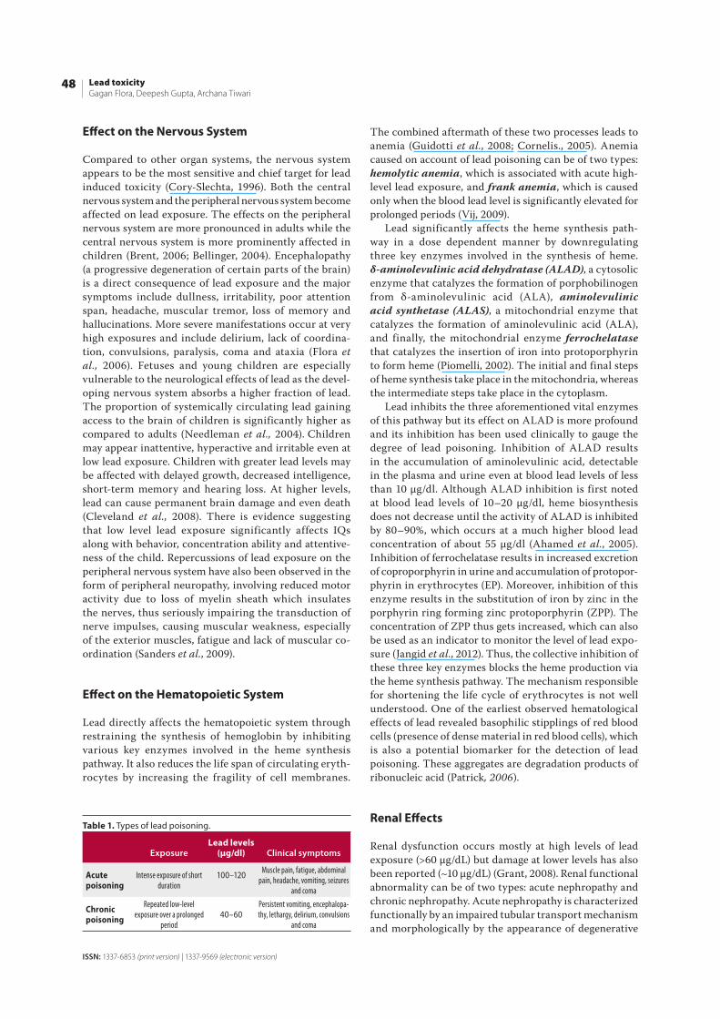
coal combustion, lead-based paints, lead containing pipes or lead-based solder in water supply systems, battery recycling, grids and bearings, etc. Although lead toxicity is a highly explored and comprehensively published topic, complete control and prevention over lead exposure is still far from being achieved. There is no such level of lead that appears to be necessary or beneficial to the body and no “safe” level of exposure to lead has been found. Lead toxic-ity is a particularly insidious hazard with the potential of causing irreversible health effects. It is known to interfere with a number of body functions and it is primarily affect-ing the central nervous, hematopoietic, hepatic and renal system producing serious disorders (Kalia & Flora, 2005). Acute toxicity is related to occupational exposure and is quite uncommon. Chronic toxicity on the other hand is much more common and occurs at blood lead levels of about 40–60 ug/dL. It can be much more severe if not treated in time and is characterized by persistent vomit-ing, encephalopathy, lethargy, delirium, convulsions and coma (Flora et al., 2006; Pearce, 2007).
Introduction
Lead (Pb) is ubiquitous and one of the earliest metals discovered by the human race. Unique properties of lead, like softness, high malleability, ductility, low melting point and resistance to corrosion, have resulted in its widespread usage in different industries like automobiles, paint, ceramics, plastics, etc. This in turn has led to a manifold rise in the occurrence of free lead in biological systems and the inert environment.
Lead is regarded as a potent occupational toxin and its toxicological manifestations are well known. The non biodegradable nature of lead is the prime reason for its prolonged persistence in the environment. Human expo-sure to lead occurs through various sources like leaded gasoline, industrial processes such as lead smelting and
Interdiscip Toxicol. 2012; Vol. 5(2): 47–58. doi: 10.2478/v10102-012-0009-2Published online in:www.intertox.sav.sk & www.versita.com/science/medicine/it/
Copyright © 2012 SETOX & IEPT, SASc.This is an Open Access article distributed under the terms of the Creative Commons Attribu-tion License (http://creativecommons.org/licenses/by/2.0), which permits unrestricted use, distribution, and reproduction in any medium, provided the original work is properly cited.
REVIEW ARTICLE
48Gagan Flora, Deepesh Gupta, Archana Tiwari Lead toxicity
ISSN: 1337-6853 (print version) | 1337-9569 (electronic version)
Eff ect on the Nervous System
Compared to other organ systems, the nervous system appears to be the most sensitive and chief target for lead induced toxicity (Cory-Slechta, 1996). Both the central nervous system and the peripheral nervous system become affected on lead exposure. The effects on the peripheral nervous system are more pronounced in adults while the central nervous system is more prominently affected in children (Brent, 2006; Bellinger, 2004). Encephalopathy (a progressive degeneration of certain parts of the brain) is a direct consequence of lead exposure and the major symptoms include dullness, irritability, poor attention span, headache, muscular tremor, loss of memory and hallucinations. More severe manifestations occur at very high exposures and include delirium, lack of coordina-tion, convulsions, paralysis, coma and ataxia (Flora et al., 2006). Fetuses and young children are especially vulnerable to the neurological effects of lead as the devel-oping nervous system absorbs a higher fraction of lead. The proportion of systemically circulating lead gaining access to the brain of children is significantly higher as compared to adults (Needleman et al., 2004). Children may appear inattentive, hyperactive and irritable even at low lead exposure. Children with greater lead levels may be affected with delayed growth, decreased intelligence, short-term memory and hearing loss. At higher levels, lead can cause permanent brain damage and even death (Cleveland et al., 2008). There is evidence suggesting that low level lead exposure significantly affects IQs along with behavior, concentration ability and attentive-ness of the child. Repercussions of lead exposure on the peripheral nervous system have also been observed in the form of peripheral neuropathy, involving reduced motor activity due to loss of myelin sheath which insulates the nerves, thus seriously impairing the transduction of nerve impulses, causing muscular weakness, especially of the exterior muscles, fatigue and lack of muscular co-ordination (Sanders et al., 2009).
Eff ect on the Hematopoietic System
Lead directly affects the hematopoietic system through restraining the synthesis of hemoglobin by inhibiting various key enzymes involved in the heme synthesis pathway. It also reduces the life span of circulating eryth-rocytes by increasing the fragility of cell membranes.
The combined aftermath of these two processes leads to anemia (Guidotti et al., 2008; Cornelis., 2005). Anemia caused on account of lead poisoning can be of two types: hemolytic anemia, which is associated with acute high-level lead exposure, and frank anemia, which is caused only when the blood lead level is significantly elevated for prolonged periods (Vij, 2009).
Lead significantly affects the heme synthesis path-way in a dose dependent manner by downregulating three key enzymes involved in the synthesis of heme. δ-aminolevulinic acid dehydratase (ALAD), a cytosolic enzyme that catalyzes the formation of porphobilinogen from δ-aminolevulinic acid (ALA), aminolevulinic acid synthetase (ALAS), a mitochondrial enzyme that catalyzes the formation of aminolevulinic acid (ALA), and finally, the mitochondrial enzyme ferrochelatase that catalyzes the insertion of iron into protoporphyrin to form heme (Piomelli, 2002). The initial and final steps of heme synthesis take place in the mitochondria, whereas the intermediate steps take place in the cytoplasm.
Lead inhibits the three aforementioned vital enzymes of this pathway but its effect on ALAD is more profound and its inhibition has been used clinically to gauge the degree of lead poisoning. Inhibition of ALAD results in the accumulation of aminolevulinic acid, detectable in the plasma and urine even at blood lead levels of less than 10 μg/dl. Although ALAD inhibition is first noted at blood lead levels of 10–20 μg/dl, heme biosynthesis does not decrease until the activity of ALAD is inhibited by 80–90%, which occurs at a much higher blood lead concentration of about 55 μg/dl (Ahamed et al., 2005). Inhibition of ferrochelatase results in increased excretion of coproporphyrin in urine and accumulation of protopor-phyrin in erythrocytes (EP). Moreover, inhibition of this enzyme results in the substitution of iron by zinc in the porphyrin ring forming zinc protoporphyrin (ZPP). The concentration of ZPP thus gets increased, which can also be used as an indicator to monitor the level of lead expo-sure (Jangid et al., 2012). Thus, the collective inhibition of these three key enzymes blocks the heme production via the heme synthesis pathway. The mechanism responsible for shortening the life cycle of erythrocytes is not well understood. One of the earliest observed hematological effects of lead revealed basophilic stipplings of red blood cells (presence of dense material in red blood cells), which is also a potential biomarker for the detection of lead poisoning. These aggregates are degradation products of ribonucleic acid (Patrick, 2006).
Renal Eff ects
Renal dysfunction occurs mostly at high levels of lead exposure (>60 μg/dL) but damage at lower levels has also been reported (~10 μg/dL) (Grant, 2008). Renal functional abnormality can be of two types: acute nephropathy and chronic nephropathy. Acute nephropathy is characterized functionally by an impaired tubular transport mechanism and morphologically by the appearance of degenerative
Table 1. Types of lead poisoning.
ExposureLead levels
(μg/dl) Clinical symptoms
Acute poisoning
Intense exposure of short
duration
100–120 Muscle pain, fatigue, abdominal
pain, headache, vomiting, seizures
and coma
Chronic poisoning
Repeated low-level
exposure over a prolonged
period
40–60Persistent vomiting, encephalopa-
thy, lethargy, delirium, convulsions
and coma
49Also available online on PubMed Central
Interdisciplinary Toxicology. 2012; Vol. 5(2): 47–58
Copyright © 2012 SETOX & Institute of Experimental Pharmacology and Toxicology, SASc.
changes in the tubular epithelium along with the occur-rence of nuclear inclusion bodies containing lead protein complexes. It does not cause protein to appear in the urine but can give rise to abnormal excretion of glucose, phosphates and amino acids, a combination referred to as Fanconi’s syndrome. Chronic nephropathy on the other hand, is much more severe and can lead to irreversible functional and morphological changes. It is characterized by glomerular and tubulointerstitial changes, resulting in renal breakdown, hypertension and hyperuricemia (Rastogi, 2008).
Cardiovascular Eff ects
Both chronic and acute lead poisoning causes cardiac and vascular damage with potentially lethal consequences including hypertension and cardiovascular disease (Navas-Acien et al., 2007). Low level lead exposure can contribute to hypertension in both animals and humans (ATSDR, 2005). Other major disorders include ischemic coronary heart disease, cerebrovascular accidents and peripheral vascular disease. Although evidence of causal relationship of lead exposure and hypertension was reported, it applies only in cases of cardiovascular out-comes of lead toxicity (Navas-Acien et al., 2007).
Reproductive Health Eff ects
Lead causes a number of adverse effects on the reproduc-tive system in both men and women. Common effects seen in men include: reduced libido, abnormal spermato-genesis (reduced motility and number), chromosomal damage, infertility, abnormal prostatic function and changes in serum testosterone. Women on the other hand, are more susceptible to infertility, miscarriage, premature membrane rupture, pre-eclampsia, pregnancy hypertension and premature delivery (Flora et al., 2011). Moreover, during the gestation period, direct influence of lead on the developmental stages of the fetus has also been reported (Saleh et al., 2009).
Eff ect on Bone
The primary site of lead storage in the human body are bones (Renner, 2010; Silbergeld et al., 1993). There are two compartments in bones where lead is believed to be stored. The exchangeable pool present at the surface of bone and the non-exchangeable pool located deeper in the cortical bone. Lead can enter into plasma at ease from the exchangeable pool but can leave the non-exchangeable pool and move to the surface only when bone is actively being re-absorbed (Patrick, 2006). Stable lead isotope methodology showed that bones contibute around 40–70% of lead released into blood in adults. In adults, 85–95% of the lead is stored in bones, in contrast to 70% in children, resulting in higher concentration of lead in
soft tissues in children. The storage and the mobilization of lead in bones depends on several factors, like dose/rate of lead exposure, age, pregnancy, gestation and race.
Mechanism of toxicity
Lead is probably the most extensively studied heavy metal. Studies carried out in this field have reported the presence of various cellular, intracellular and molecular mechanisms behind the toxicological manifestations caused by lead in the body.
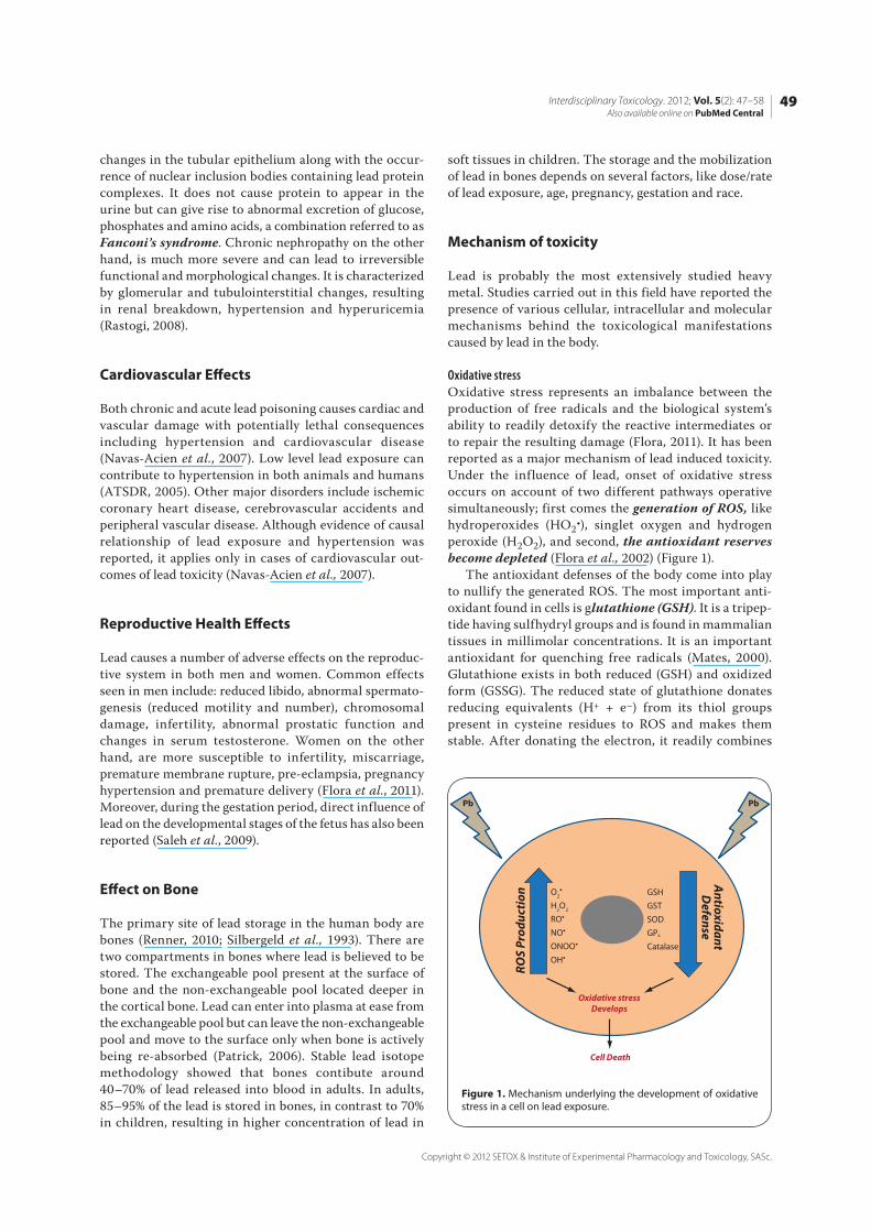
Oxidative stressOxidative stress represents an imbalance between the production of free radicals and the biological system’s ability to readily detoxify the reactive intermediates or to repair the resulting damage (Flora, 2011). It has been reported as a major mechanism of lead induced toxicity. Under the influence of lead, onset of oxidative stress occurs on account of two different pathways operative simultaneously; first comes the generation of ROS, like hydroperoxides (HO2•), singlet oxygen and hydrogen peroxide (H2O2), and second, the antioxidant reserves become depleted (Flora et al., 2002) (Figure 1).
The antioxidant defenses of the body come into play to nullify the generated ROS. The most important anti-oxidant found in cells is glutathione (GSH). It is a tripep-tide having sulfhydryl groups and is found in mammalian tissues in millimolar concentrations. It is an important antioxidant for quenching free radicals (Mates, 2000). Glutathione exists in both reduced (GSH) and oxidized form (GSSG). The reduced state of glutathione donates reducing equivalents (H+ + e–) from its thiol groups present in cysteine residues to ROS and makes them stable. After donating the electron, it readily combines
ROS
Prod
uctio
n
AntioxidantD
efense
GSHGSTSODGPx
Catalase
O2
H2O2
RONOONOOOH
Oxidative stressDevelops
Cell Death
Pb Pb
Figure 1. Mechanism underlying the development of oxidative stress in a cell on lead exposure.
50Gagan Flora, Deepesh Gupta, Archana Tiwari Lead toxicity
ISSN: 1337-6853 (print version) | 1337-9569 (electronic version)
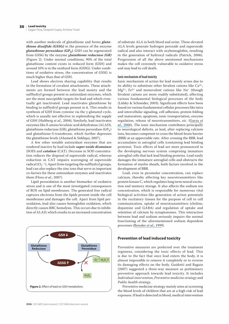
with another molecule of glutathione and forms gluta-thione disulfide (GSSG) in the presence of the enzyme glutathione peroxidase (GPX). GSH can be regenerated from GSSG by the enzyme glutathione reductase (GR) (Figure 2). Under normal conditions, 90% of the total glutathione content exists in reduced form (GSH) and around 10% is in the oxidized form (GSSG). Under condi-tions of oxidative stress, the concentration of GSSG is much higher than that of GSH.
Lead shows electron sharing capability that results in the formation of covalent attachments. These attach-ments are formed between the lead moiety and the sulfhydryl groups present in antioxidant enzymes, which are the most susceptible targets for lead and which even-tually get inactivated. Lead inactivates glutathione by binding to sulfhydryl groups present in it. This results in synthesis of GSH from cysteine via the γ-glutamyl cycle, which is usually not effective in replenishing the supply of GSH (Hultberg et al., 2004). Similarly, lead inactivates enzymes like δ-amino levulinic acid dehydratase (ALAD), glutathione reductase (GR), glutathione peroxidase (GPX) and glutathione-S-transferase, which further depresses the glutathione levels (Ahamed & Siddiqui, 2007).
A few other notable antioxidant enzymes that are rendered inactive by lead include super oxide dismutase (SOD) and catalase (CAT). Decrease in SOD concentra-tion reduces the disposal of superoxide radical, whereas reduction in CAT impairs scavenging of superoxide radical (O2– •). Apart from targeting the sulfhydryl groups, lead can also replace the zinc ions that serve as important co-factors for these antioxidant enzymes and inactivates them (Flora et al., 2007).
Lipid peroxidation is another biomarker of oxidative stress and is one of the most investigated consequences of ROS on lipid membranes. The generated free radical captures electrons from the lipids present inside the cell membranes and damages the cell. Apart from lipid per-oxidation, lead also causes hemoglobin oxidation, which directly causes RBC hemolysis. This occurs due to inhibi-tion of ALAD, which results in an increased concentration
of substrate ALA in both blood and urine. These elevated ALA levels generate hydrogen peroxide and superoxide radical and also interact with oxyhemoglobin, resulting in the generation of hydroxyl radicals (Patrick, 2006). Progression of all the above mentioned mechanisms makes the cell extremely vulnerable to oxidative stress and may lead to cell death.
Ionic mechanism of lead toxicityIonic mechanism of action for lead mainly arises due to its ability to substitute other bivalent cations like Ca2+, Mg2+, Fe2+ and monovalent cations like Na+ (though bivalent cations are more readily substituted), affecting various fundamental biological processes of the body (Lidsky & Schneider, 2003). Significant effects have been found on various fundamental cellular processes like intra and intercellular signaling, cell adhesion, protein folding and maturation, apoptosis, ionic transportation, enzyme regulation, release of neurotransmitters, etc. (Garza et al., 2006). The ionic mechanism contributes principally to neurological deficits, as lead, after replacing calcium ions, becomes competent to cross the blood-brain barrier (BBB) at an appreciable rate. After crossing the BBB, lead accumulates in astroglial cells (containing lead binding proteins). Toxic effects of lead are more pronounced in the developing nervous system comprising immature astroglial cells that lack lead binding proteins. Lead easily damages the immature astroglial cells and obstructs the formation of myelin sheath, both factors involved in the development of BBB.
Lead, even in picomolar concentration, can replace calcium, thereby affecting key neurotransmitters like protein kinase C, which regulates long term neural excita-tion and memory storage. It also affects the sodium ion concentration, which is responsible for numerous vital biological activities like generation of action potentials in the excitatory tissues for the purpose of cell to cell communication, uptake of neurotransmitters (choline, dopamine and GABA) and regulation of uptake and retention of calcium by synaptosomes. This interaction between lead and sodium seriously impairs the normal functioning of the aforementioned sodium dependent processes (Bressler et al., 1999).
Prevention of lead induced toxicity
Preventive measures are preferred over the treatment regimens, considering the toxic effects of lead. This is due to the fact that once lead enters the body, it is almost impossible to remove it completely or to reverse its damaging effects on the body. Guidotti and Ragain (2007) suggested a three-way measure as preliminary preventive approach towards lead toxicity. It includes Individual intervention, Preventive medicine strategy and Public health strategy.
Preventive medicine strategy mainly aims at screening the blood levels of children that are at a high risk of lead exposure. If lead is detected in blood, medical intervention
GSH
GSSG
GSH
GlutathioneReductase
GlutathionePeroxidase
O2– , H2O2
H2O2
Figure 2. Eff ect of lead on GSH metabolism.
51Also available online on PubMed Central
Interdisciplinary Toxicology. 2012; Vol. 5(2): 47–58
Copyright © 2012 SETOX & Institute of Experimental Pharmacology and Toxicology, SASc.
is carried out with the aim to control undesirable out-comes of poisoning and prevent further accumulation of lead.
Public health strategy has a much larger sphere of influence and acts at a population level with a target to reduce the risk of lead exposure in habitable regions. Various preventive strategies have been suggested by the public health services for controlling lead. The most important of them include: prohibition of setting up industries dealing with lead close to habitable areas and completely banning the use of lead where appropriate replacement is available.
Apart from the above mentioned preliminary strate-gies, nutrition also plays an important role in prevention of lead induced toxicity. Studies have shown that uptake of certain nutrients like mineral elements, flavonoids and vitamins can provide protection from the environmen-tal lead as well as from the lead already present in the body. These nutrients play a pivotal role in restoring the imbalanced prooxidant/oxidant ratio that arises due to oxidative stress. Although the mechanism by which these nutrients restore the delicate prooxidant/oxidant ratio is still unclear, significant data are available suggesting a protective role of nutrients against lead poisoning (Hsu & Guo, 2002).
Role of antioxidants in protecting lead induced oxidative stress
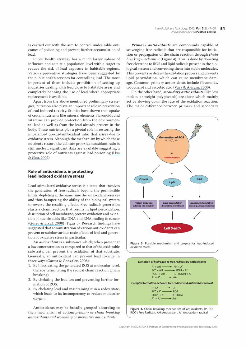
Lead stimulated oxidative stress is a state that involves the generation of free radicals beyond the permissible limits, depleting at the same time the antioxidant reserves and thus hampering the ability of the biological system to reverse the resulting effects. Free radicals generation starts a chain reaction that results in lipid peroxidation, disruption of cell membrane, protein oxidation and oxida-tion of nucleic acids like DNA and RNA leading to cancer (Gurer & Ercal, 2000) (Figur 3). Research findings have suggested that administration of various antioxidants can prevent or subdue various toxic effects of lead and genera-tion of oxidative stress in particular.
An antioxidant is a substance which, when present at a low concentration as compared to that of the oxidizable substrate, can prevent the oxidation of that substrate. Generally, an antioxidant can prevent lead toxicity in three ways (Garcia & Gonzalez, 2008):1. By inactivating the generated ROS at molecular level,
thereby terminating the radical chain reaction (chain breaking).
2. By chelating the lead ion and preventing further for-mation of ROS.
3. By chelating lead and maintaining it in a redox state, which leads to its incompetency to reduce molecular oxygen.
Antioxidants may be broadly grouped according to their mechanism of action: primary or chain breaking antioxidants and secondary or preventive antioxidants.
Primary antioxidants are compounds capable of scavenging free radicals that are responsible for initia-tion or propagation of the chain reaction through chain breaking mechanism (Figure 4). This is done by donating free electrons to ROS and lipid radicals present in the bio-logical system and converting them into stable molecules. This prevents or delays the oxidation process and prevents lipid peroxidation, which can cause membrane dam-age. Common primary antioxidants include flavonoids, tocopherol and ascorbic acid (Vaya & Aviram, 2000).
On the other hand, secondary antioxidants (like low molecular weight polyphenols) are those which mainly act by slowing down the rate of the oxidation reaction. The major difference between primary and secondary
Generation of ROSO2 H2O2
Cell Death
Pb Pb
Oxidative stress
ProteinLipid
DNA
Protein oxidationaltering the function
Lipid peroxidationdisrupting membrane
Nucleic acid oxidationcausing cancer/mutation
Figure 3. Possible mechanism and targets for lead-induced oxidative stress.
Donation of hydrogen to free radicals by antioxidants
Complex formation between free radical and antioxidant radical
R + AH RH + ARO + AH ROH + AROO + AH ROOH + AA + A AA
R +A RARO +A ROAROO + A ROOAA + A AA
Figure 4. Chain breaking mechanism of antioxidants. R•, RO•, ROO•: Free Radicals, AH: Antioxidant, A•: Antioxidant radical
52Gagan Flora, Deepesh Gupta, Archana Tiwari Lead toxicity
ISSN: 1337-6853 (print version) | 1337-9569 (electronic version)
antioxidants is that the latter do not convert free radicals into stable molecules. They are capable of chelating heavy metals like lead (Wanasundara & Shahidi, 2005).
Natural antioxidants and the present status
Naturally occurring antioxidants and their role in quenching free radicals generated in the body under various pathologic conditions have been an active area of research. Studies have revealed that antioxidants possess the ability of both preventing and curing the damage caused by the generation of free radicals in the body. Natural antioxidants can be categorized into enzymatic and non enzymatic.
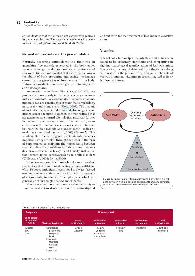
Enzymatic antioxidants like SOD, CAT, GPX are produced endogenously in the cells, whereas non enzy-matic antioxidants like carotenoids, flavonoids, vitamins, minerals, etc. are constituents of many fruits, vegetables, nuts, grains and some meats (Flora, 2009). The amount of antioxidants present under normal physiological con-ditions is just adequate to quench the free radicals that are generated at a normal physiological rate. Any further increment in the concentration of free radicals (due to environmental or natural causes) can cause an imbalance between the free radicals and antioxidants, leading to oxidative stress (Blokhina et al., 2003) (Figure 5). This is where the role of exogenous antioxidants becomes important. They are taken through the diet or in the form of supplements to maintain the homeostasis between free radicals and antioxidants and thus prevent various deleterious effects, like heavy metal toxicity, inflamma-tion, cancer, aging, cardiovascular and brain disorders (Willcox et al., 2004; Pietta, 2000).
It has been reported that those who take an antioxidant rich diet are at the forefront of reaping various health ben-efits. To boost antioxidant levels, food is always favored over supplements mainly because it contains thousands of antioxidants, in contrast to supplements, which are generally rich in a single or a few antioxidants.
This review will now incorporate a detailed study of some natural antioxidants that have been investigated
and put forth for the treatment of lead induced oxidative stress.
Vitamins
The role of vitamins (particularly B, C and E) has been found to be extremely significant and competitive in fighting toxicological manifestations of lead poisoning. These vitamins may chelate lead from the tissues along with restoring the pro/antioxidant balance. The role of various prominent vitamins in preventing lead toxicity has been discussed.
Table 2. Classification of natural antioxidants.
Enzymatic Non-enzymatic
Endogenous antioxidants
(Cellular) Phyto-antioxidantsHerbal
antioxidantsAntioxidant
vitaminsAntioxidant
mineralsAntioxidant hormones
Thiolantioxidants
CatalaseSODGP X
Carotenoids β -carotene, Lycopene LuteinFlavonoids Quercetin Catechin GarlicLipoic acid
GinkgoCurcumin
ThiaminePyridoxine
Ascorbic acidα tocopherol
Selenium Zinc
Melatonin GlutathioneThioredoxin
AntioxidantsFree RadicalsDynamic
homeostaticbalance
Imbalance
Oxidative Stress
Tissue injury & cell death
Figure 5. Under normal physiological conditions, there is a bal-ance between free radicals and antioxidants and any deviation from it can cause oxidative stress leading to cell death.
53Also available online on PubMed Central
Interdisciplinary Toxicology. 2012; Vol. 5(2): 47–58
Copyright © 2012 SETOX & Institute of Experimental Pharmacology and Toxicology, SASc.
Vitamin B (Pyridoxine and Thiamine)Vitamin B6 (pyridoxine) and vitamin B1 (thiamine) are reported to have essential characteristics that can cure the deleterious effects of lead toxicity. Pyridoxine is an important co-factor which participates in the metabolic trans-sulfuration pathway which is responsible for the synthesis of cysteine from dietary methionine.
Vitamin B6 acts also as an antioxidant by stimulat-ing the production of GSH and as a moderate chelator (Ahamed & Siddiqui, 2007). Chelation of lead by vitamin B6 could be attributed to the presence of the ring in the nitrogen atom or to the interference of vitamin B6 with the absorption of lead. Vitamin B1 (thiamine) has also been reported to exert protective efficacy against short-term implications of lead poisoning. Senapati et al. (2000) reported the protective role of thiamine hydrochloride on lead-induced endogenous lipid peroxidation in rat hepatic and renal tissues. They revealed a significant decrement in the levels of lead in liver and kidney.
Ascorbic acid (vitamin C)Ascorbic acid is probably the most widely studied vitamin when it comes to the prevention of lead induced oxidative stress. Its property of quenching ROS along with metal chelation makes it a potential detoxifying agent for lead (Das & Saha, 2010; Tariq, 2007). A recent study done by Chang et al. (2011) showed the defensive effect of ascorbic acid on oxidative stress, developed in the hippocampus of lead exposed suckling rats. They reported that intro-duction of ascorbic acid during pregnancy and lactation caused to some extent amelioration of oxidative stress in the developing hippocampus. Shan et al. (2009) examined the defensive effects of ascorbic acid and thiamine against the toxic effects of lead on testes of mice. Exposure to lead exhibited a significant decrease in epididymal sperm count and motility, along with the induction of apoptosis through activation of caspase-3, Fas/Fas-L and Bcl-2. Co-administration of ascorbic acid and thiamine reverted the oxidative stress in a concentration dependent manner, as well as DNA damage and apoptosis induced by lead in rat liver cells (Wang et al., 2007). Supplementation
of ascorbic acid in combination with silymarin was able to reduce acute hepatotoxic lead toxicity (Shalan et al., 2005).
Vitamin E (α-tocopherol)Vitamin E is a fat soluble vitamin with numerous bio-logical functions (Flora, 2002). It possesses powerful anti-oxidative properties, operative in the membrane to prevent lipid peroxidation by obstructing the free radical chain reaction. Sajitha et al., (2010) reported that vitamin E administered to rats counteracted the deleterious effect of lead by scavenging free radicals and thus preventing oxidative stress. Lead induced ALAD inhibition in the erythrocytes was found to be reversed by the treatment with vitamin E (Rendon-Ramirez et al., 2007). Vitamin E was also found to be helpful in restoring thyroid dysfunc-tion by maintaining the hepatic cell membrane archi-tecture disrupted indirectly by lead induced lipid per-oxidation. Effect of vitamin E in combination with other antioxidants has been found to be more pronounced than its individual administration. Flora et al. (2003) reported that co-administration of vitamin E with monoisoamyl derivative (MiADMSA), which is a thiol chelator, exerts an elevated recovery from lead burden in rats. Interestingly, α-tocopherol is capable of reducing ferric iron to ferrous iron (i.e. to act as a pro-oxidant). Moreover, the ability of α-tocopherol to act as a pro-oxidant (reducing agent) or antioxidant depends on whether all of the α-tocopherol becomes consumed in the conversion from ferric to fer-rous iron or whether, following this interaction, residual α-tocopherol is available to scavenge the resultant ROS (Yamamoto & Nike, 1988).
Flavonoids
Flavonoids are naturally occurring polyphenolic com-pounds. They are the main constituents of fruits, veg-etables and certain beverages (Youdim et al., 2002). The anti-oxidative nature of flavonoids has been extensively investigated. These compounds, like other anti-oxidants,
O
A C
B1
2
3
45
6
7
8 1'
2'
3'
6'
5'
4'
O
O
OH
OH
OH
HO
OH
A C
B
Figure 6. General structure of fl avonoids. Figure 7. General structure of quercetin.
54Gagan Flora, Deepesh Gupta, Archana Tiwari Lead toxicity
ISSN: 1337-6853 (print version) | 1337-9569 (electronic version)
can cure or prevent oxidative stress by chelating redox active metal ions and also by terminating the free radical chain reaction (Terao, 2009; Rice-Evans, 2001).
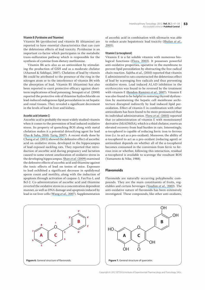
The capacity of flavonoids to act as antioxidants depends upon their molecular structure (Figure 6). Their general structure includes a diphenylpropane moiety composed of two or more aromatic rings (A and B), each having at least one aromatic hydroxyl group connected via a carbon chain. The chain consists of three carbons that combine with an oxygen and two carbons of one of the aromatic rings (A ring) to form a third 6-member ring (C ring) known as the pyran ring (Larson et al. 2012).
The metal chelating ability of flavonoids arises from the appropriate positioning of the functional groups that include both the hydroxyl groups of ring-B and the 5-hydroxy group of ring-A. The electron donating capa-bility of flavonoid molecules to scavenge ROS rests with the 3’,4’-catechol (dihydroxy) structure (B-ring). Another structural feature that contributes to the anti-oxidative nature is the presence of 2, 3 double bond in conjugation with a 4-oxo group in the C-ring (Heim et al., 2002; Ng et al., 2000; Dugas et al., 2000).
QuercetinQuercetin is a ubiquitously distributed and comprehen-sively explored bioflavonoid. Dietary sources of quercetin include fruits, vegetables and tea. The chemical name for quercetin is 3,3’,4’,5,7-pentahydroxyflavone. The pres-ence of multiple hydroxyl groups in its chemical structure and conjugated electrons account for its antioxidant and metal chelating property (Figure 7).
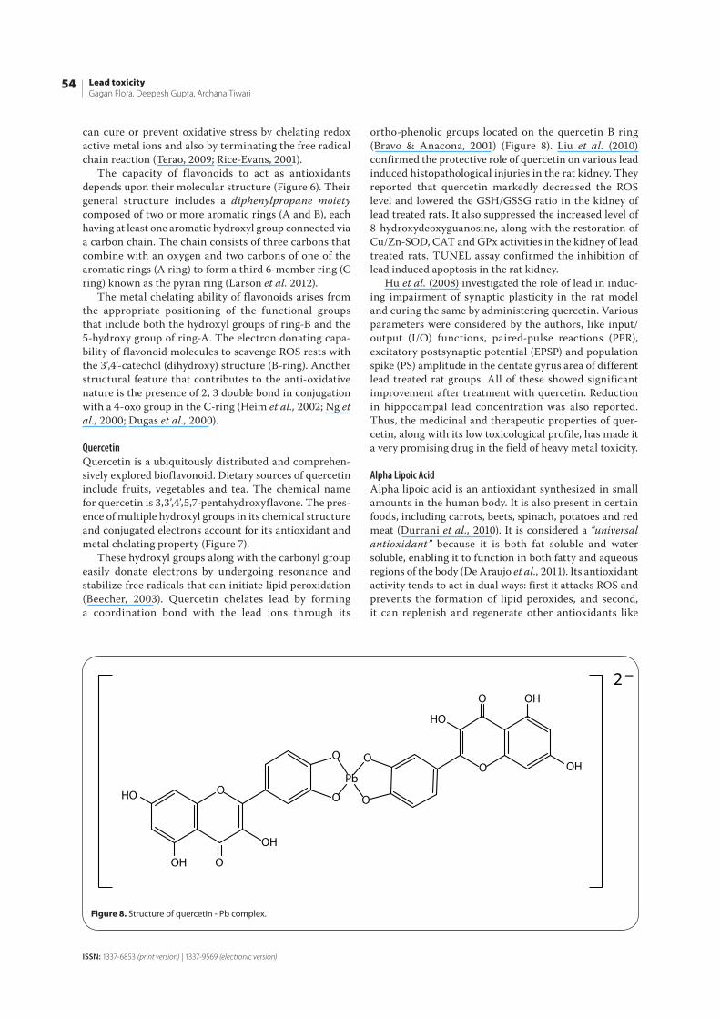
These hydroxyl groups along with the carbonyl group easily donate electrons by undergoing resonance and stabilize free radicals that can initiate lipid peroxidation (Beecher, 2003). Quercetin chelates lead by forming a coordination bond with the lead ions through its
ortho-phenolic groups located on the quercetin B ring (Bravo & Anacona, 2001) (Figure 8). Liu et al. (2010) confirmed the protective role of quercetin on various lead induced histopathological injuries in the rat kidney. They reported that quercetin markedly decreased the ROS level and lowered the GSH/GSSG ratio in the kidney of lead treated rats. It also suppressed the increased level of 8-hydroxydeoxyguanosine, along with the restoration of Cu/Zn-SOD, CAT and GPx activities in the kidney of lead treated rats. TUNEL assay confirmed the inhibition of lead induced apoptosis in the rat kidney.
Hu et al. (2008) investigated the role of lead in induc-ing impairment of synaptic plasticity in the rat model and curing the same by administering quercetin. Various parameters were considered by the authors, like input/output (I/O) functions, paired-pulse reactions (PPR), excitatory postsynaptic potential (EPSP) and population spike (PS) amplitude in the dentate gyrus area of different lead treated rat groups. All of these showed significant improvement after treatment with quercetin. Reduction in hippocampal lead concentration was also reported. Thus, the medicinal and therapeutic properties of quer-cetin, along with its low toxicological profile, has made it a very promising drug in the field of heavy metal toxicity.
Alpha Lipoic AcidAlpha lipoic acid is an antioxidant synthesized in small amounts in the human body. It is also present in certain foods, including carrots, beets, spinach, potatoes and red meat (Durrani et al., 2010). It is considered a “universal antioxidant” because it is both fat soluble and water soluble, enabling it to function in both fatty and aqueous regions of the body (De Araujo et al., 2011). Its antioxidant activity tends to act in dual ways: first it attacks ROS and prevents the formation of lipid peroxides, and second, it can replenish and regenerate other antioxidants like
OH
OOH
HO OO
O
HO
O OH
OHOO
O
Pb
2 –
Figure 8. Structure of quercetin - Pb complex.
55Also available online on PubMed Central
Interdisciplinary Toxicology. 2012; Vol. 5(2): 47–58
Copyright © 2012 SETOX & Institute of Experimental Pharmacology and Toxicology, SASc.
vitamin C and E (Haleagrahara et al., 2011). Lipoic acid has mostly been used in combination with other chelating agents like 2,3-dimercaptosuccinic acid (DMSA), due to the fact that lipoic acid itself does not have metal chelating ability but it can consistently tackle the generated oxida-tive stress. Sivaprasad et al. (2002, 2003, 2004) explored the histopathological implications of lead exposure on kidneys, liver and erythrocyte membranes and showed that lipoic acid greatly improved the condition by revers-ing the developed oxidative stress. Lipoic acid was found to be more effective in removing lead from the brain compared to any other organs (liver, kidneys and other soft tissues) (Pande and Flora., 2002).
Herbal antioxidants
The ability of herbal antioxidants to act as useful clini-cal medicine is due to their low cost and few side effects. However, actual implementation of herbal antioxidants as potential medicines has been highly limited. This is due to the longer treatment durations associated with it, which makes it a preventive rather than therapeutic measure. Apart from this, herbal drugs also suffer from a serious drawback of poor bioavailability in the body and require much higher and repetitive doses to maintain the thera-peutic threshold in the body. A few herbal antioxidants that have been reported to provide protection against lead induced oxidative stress will be discussed.
GarlicGarlic is a medicinal plant that has been an inseparable part of Indian culinary for over 5000 years. Besides its use as a condiment, it is credited to have remarkable thera-peutic and pharamcological properties. Its active agent is allicin, which imparts its characteristic odor as well as medicinal properties (Sharma et al., 2010). Garlic can prevent oxidative stress by chelating lead ions and scav-enging free radicals. Senapati et al. (2001) reported the prophylactic efficacy of garlic extract in reducing the lead burden from soft tissues. In another study, Pourjafar et al. (2007) further confirmed the ability of garlic to reduce the lead burden from the liver, kidney, blood and bone. The protective efficacy of aqueous garlic extract was studied against lead induced hepatic injury in rats. The results clearly indicated the ameliorative ability of garlic towards hepatic injury caused by lead due to generated oxidative stress (Kilikdar et al., 2011).
CurcuminCurcumin is a yellow-colored polyphenolic compound and the principal active component of turmeric, which is obtained from the plant Curcuma longa. There are reports of antioxidant, radical scavenging and metal chelating effects of curcumin in metal toxicity (Sethi et al., 2009; Agarwal et al., 2010; Singh et al., 2010; Rao et al., 2008). Shukla et al. (2003) reported for the first time the protec-tive effect of curcumin against lead-induced neurotoxicity in rats by showing significant improvement in the levels
of various biomarkers of oxidative stress (GSH, SOD and CAT levels) in different regions of the brain. Daniel et al. (2004) provided insight into the chelating properties of curcumin by showing a remarkable reduction in levels of lead in rat brains. The above mentioned results on curative effects of curcumin on lead neurotoxicity were successfully reproduced by Dairam et al. (2007) in male rats. In spite of these commendable properties, the major drawback associated with the use of curcumin is its low bioavailability. This is due to its poor aqueous dispersion and poor absorption from the intestine coupled with a high degree of metabolism of curcumin in the liver and rapid elimination in bile (Maiti et al., 2007).
Centella asiaticaCentella asiatica, popularly known as Indian Pennywort, is well known for its various medicinal applications and for treating metal toxicity (Flora & Gupta, 2007). Introduction of C. asiatica along with chelating agents like DMSA have shown very promising results regarding treatment of lead induced oxidative stress in rats (Saxena & Flora, 2006). Independent administration of C. asiatica has also been studied regarding its ameliorative ability against lead induced oxidative stress. The results found were quite encouraging (Ponnusamy et al., 2008; Sainath et al., 2011). It has been hypothesized that C. asiatica can cross the BBB and restore the levels of altered neurotrans-mitters and can also restore the impaired prooxidant/antioxidant balance arising due to lead exposure (Hussin et al., 2007).
Recent Strategies
The major drawback in the usefulness of antioxidants is their poor bioavailability due to low solubility and rapid clearance. Although the pharmacological safety of most of the therapeutics promises a great potential for treat-ment and prevention of various diseases, their relatively low bioavailability is a major hurdle for clinical develop-ment. Novel approaches to overcome the problem of low bioavailability of these antioxidants are being developed. These approaches include improved formulations for better delivery such as liposomes, micelles, phospholipid complexes and nanoparticles (Anand et al., 2007).
Liposomal NanoparticlesLipid-based nanoencapsulation systems enhance the performance of antioxidants by improving their solubility, bioavailability and by preventing unwanted interactions with other food components. The main lipid-based nano-encapsulation systems that can be used for the delivery of nutraceuticals are nanoliposomes, nanocochleates and archaeosomes (Mozafari et al., 2008). Nanoliposome technology presents exciting opportunities for food tech-nologists in areas such as encapsulation and controlled release of antioxidants, as well as the enhanced bioavail-ability, stability and shelf-life of sensitive ingredients. Application of nanoliposomes as carrier vehicles of
56Gagan Flora, Deepesh Gupta, Archana Tiwari Lead toxicity
ISSN: 1337-6853 (print version) | 1337-9569 (electronic version)
nutrients, nutraceuticals, enzymes, food additives and food antimicrobials was reported (Mozafari et al., 2006). Liposomes are phospholipid bilayers that have closed in upon themselves to form a tiny bubble or vesicle. These nanoparticles have hydrophilic heads pointing outward that make them water soluble, while the hydrophobic tails of both lipid layers interact. The inside of the liposome is also water soluble, enabling it to protect soluble drugs, cosmetics or biomolecules intended for delivery into cells. The outer or inner membrane structures can be altered with charged ligands or target-specific molecules, such as antigens, for specialized use. Encapsulation of curcumin in liposomes with their hydrophilic and hydrophobic properties should offer an excellent antioxidant potential. While these studies suggest that liposomal curcumin is bioactive, the increased bioavailability of encapsulated curcumin in vivo still needs to be conclusively demon-strated (Gandhi et al., 2011).
NanoencapsulationNanoencapsulation of antioxidants provides improved biodistribution and bioavailability of poorly-soluble therapeutics through solubilization. Many vehicles have been developed for encapsulation and delivery of thera-peutics, including solid nanoparticles, micelles, lipid polymer vesicles (polymersomes) and nanohydrogels. In recent years, biodegradable polymeric nanoparticles have attracted considerable attention. In spite of the develop-ment of various synthetic and semi-synthetic polymers, natural polymers are still widely used. Some of them are: gums (acacia, guar, etc.), chitosan, gelatin, sodium algi-nate, albumin etc. Polymeric nanoparticles for controlled release and targeted delivery of functional compounds have been reported in the literature (Zigoneanu et al., 2008). They are synthesized using polymers and sur-factants and include alginic acid, polylactic-co-glycolic acid and chitosan. A recent study using a polymer-based nanoparticle of curcumin found that its molecular activity was similar to free curcumin in a pancreatic cell line (Bisht et al., 2007). Encapsulation of curcumin in a pluronic block copolymer demonstrated a slow and sustained release of curcumin and showed anticancer activity comparable with free curcumin (Sahu et al., 2010). Properties such as biodegradability, low toxicity and good biocompatibility make nanoparticles suitable for use in biomedical and pharmaceutical manipulations. Thus, nanotechnology-based approaches to increase curcumin delivery, bioavailability and high therapeutic potential in vivo are gradually evolving (Choudhuri et al., 2005). There are several lines of evidence from in vitro, in vivo, preclinical and clinical studies to suggest that curcumin has a great potential in the prevention and treatment of various diseases, yet the potential role of nanoencapsulated curcumin has not been thoroughly explored. In the near future, enhanced bioavailability of curcumin by nanoencapsulation is likely to bring this promising natural product to the forefront of therapeutic agents for treatment of human diseases.
Conclusion
Lead poisoning has been known to mankind since antiq-uity, although the situation got aggravated since the 18th century during the industrial revolution. It was the period when various important qualities of lead were discovered that made it one of the most widely used industrial metals. Lead has no known biological function in the body and once it enters the body, it is known to cause severe health effects that might be irreversible. It affects almost all the major organ systems of the body like hematopoietic, renal, nervous and cardiovascular systems. Various molecular, cellular and intracellular mechanisms have been proposed to explain the toxicological profile of lead that includes generation of oxidative stress, ionic mechanism and apoptosis. Of these oxidative stress has been found to be more pronounced and much more severe. Lead causes generation of ROS which results in critical damage to various biomolecules like DNA, enzymes, proteins and membrane based lipids, while simultaneously it impairs the antioxidant defense system. Chelation therapy has so far been used as the mainstay of the treatment that involves quenching of lead from different sites of the body and expels it through urine. Prevention is regarded as the best approach, involving incorporation of various natural and synthetic antioxidants. Various naturally occurring anti-oxidants (nutrient antioxidants) like vitamins, flavonoids and herbal antioxidants have been reported for the preven-tion and treatment of lead induced toxicity and oxidative stress in particular. They have the ability to scavange ROS at molecular level and chelate lead ions, thereby reversing the toxic effects. These antioxidants were also reported to provide an elevated therapeutic impact when administered with chelating agents like DMSA, which is a thiol chela-tor. Nevertheless, we do recommend that the presence and possible beneficial effects of antagonists be carefully considered, as an antioxidant may become a pro-oxidant in the presence of certain other molecules. For example, chlorophylls may overwhelm the antioxidant effect of phenolics due to photosensitized oxidation, while transi-tion metal ions, as those of iron and copper, may render conditions favoring oxidation. Synergism among different phenolic antioxidants and between phenolics and non-phenolics should also be considered in all application areas .
The latest addition to preventive regimens is the use of nanoencapsulation or liposome mediated drug delivery, which deals with the problem of low systemic bioavail-ability of certain natural hydrophobic antioxidants, like curcumin. Compared to conventional drugs, this approach also reduces the dosage needed to maintain its therapeutic threshold in the body.
REFERENCES
Agarwal R, Goel SK, Behari JR. (2010). Detoxifi cation and antioxidant eff ects of curcumin in rats experimentally exposed to mercury. J Appl Toxicol 30: 457–468.
57Also available online on PubMed Central
Interdisciplinary Toxicology. 2012; Vol. 5(2): 47–58
Copyright © 2012 SETOX & Institute of Experimental Pharmacology and Toxicology, SASc.
Agency for Toxic Substances and Disease Registry (ATSDR). (2005) Toxicolog-ical profi le for lead. (Draft for Public Comment). Atlanta, GA: U.S. Depart-ment of Health and Human Services, Public Health Service; pp.43–59.
Ahamed M, Siddiqui MKJ. (2007). Environmental lead toxicity and nutritional factors. Clin Nut 26: 400–408.
Ahamed M, Siddiqui MKJ. (2007). Low level lead exposure and oxidative stress: Current opinions. Clin Chim Acta 383: 57–64.
Ahamed M, Verma S, Kumar A, Siddiqui MK. (2005). Environmental exposure to lead and its correlation with biochemical indices in children. Sci Total En-viron 346: 48–55.
Anand P, Kunnumakkara AB, Newman RA, Aggarwal BB. (2007). Bioavailabil-ity of curcumin: problems and promises. Mol. Pharm. 4: 807–818.
Ayres JG. (2008). The eff ects of Inhaled materials on the lung and other tar-get organs. Occupational Hygiene (Blackwell Publishing Ltd). pp. 47–58.
Beecher GR. (2003). Overview of dietary fl avonoids: nomenclature, occur-rence and intake. J Nutr 133: 3248S–3254S.
Bellinger DC. (2004). Lead. Pediatrics 113: 1016–1022.
Bisht S, Feldmann G, Soni S, Ravi R, Karikar C, Maitra A, Maitra A. (2007). Poly-meric nanoparticle-encapsulated curcumin (“nanocurcumin”): A novel strategy for human cancer therapy. J Nanobiotech 5: 3–21.
Blokhina O, Virolainen E, Fagerstedt KV. (2003). Antioxidants, Oxidative Dam-age and Oxygen Deprivation Stress: a Review. Ann Bot 91: 179–194.
Bravo A, Anacona JR. (2001). Metal complexes of the fl avonoid quercetin: an-tibacterial properties. Trans Met Chem 26: 20–23.
Brent JA. (2006). Review of: “Medical Toxicology”. Clin Toxicol 44: 355–355.
Bressler J, Kim KA, Chakraborti T, Goldstein G. (1999). Molecular mechanisms of lead neurotoxicity. Neurochem Res 24: 595–600.
Chang BJ, Jang BJ, Son TG, Cho IH, Quan FS, Choe NH, Nahm SS, Lee JH. (2012). Ascorbic acid ameliorates oxidative damage induced by maternal low level lead exposure in the hippocampus of rat pups during gestation and lactation. Fd Chem Toxicol 52: 104–108.
Choudhuri T, Pal S, Das T, Sa G. (2005). Curcumin selectively induces apop-tosis in deregulated cyclin D1-expressed cells at G2 phase of cell cycle in a p53-dependent manner. J Biol Chem 280: 20059–20068.
Cleveland LM, Minter ML, Cobb KA, Scott AA, German VF. (2008). Lead haz-ards for pregnant women and children: Part 1: immigrants and the poor shoulder most of the burden of lead exposure in this country. Part 1 of a two-part article details how exposure happens, whom it aff ects, and the harm it can do. Am J Nurs 108: 40–49; quiz 50.
Cornelis R. (2005). Handbook of elemental speciation II: species in the environ-ment, food, medicine & occupational health. Wiley.
Cory-Slechta DA. (1996). Legacy of lead exposure: consequences for the cen-tral nervous system. Otolaryngol Head Neck Surg 114: 224–226.
Dairam A, Limson JL, Watkins GM, Antunes E, Daya S. (2007). Curcuminoids, curcumin, and demethoxycurcumin reduce lead-induced memory defi cits in male Wistar rats. J Agric Food Chem 55: 1039–1044.
Daniel S, Limson JL, Dairam A, Watkins GM, Daya S. (2004). Through metal binding, curcumin protects against lead- and cadmium-induced lipid per-oxidation in rat brain homogenates and against lead-induced tissue dam-age in rat brain. J Inorg Biochem 98: 266–275.
Das KK, Saha S. (2010). L-ascorbic acid and alpha tocopherol supplementa-tion and antioxidant status in nickel- or lead-exposed rat brain tissue. J Ba-sic Clin Physiol Pharmacol 21: 325–346.
De Araujo DP, Lobato Rde F, Cavalcanti JR, Sampaio LR, Araujo PV, Silva MC, Neves KR, Fonteles MM, Sousa FC, Vasconcelos SM. (2011). The contribu-tions of antioxidant activity of lipoic acid in reducing neurogenerative pro-gression of Parkinson’s disease: a review. Int J Neurosci 121: 51–57.
Dugas AJ Jr, Castaneda-Acosta J, Bonin GC, Price KL, Fischer NH, Winston GW. (2000). Evaluation of the total peroxyl radical-scavenging capacity of fl avo-noids: structure-activity relationships. J Nat Prod 63: 327–331.
Durrani AI, Schwartz H, Nagl M, Sontag G. (2010). Determination of free α-lipoic acid in foodstuff s by HPLC coupled with CEAD and ESI-MS. Fd Chem 120: 1143–1148.
Flora SJS. (2002). Nutritional components modify metal absorption, toxic re-sponse and chelation therapy. J Nut Environ Med 12: 53–67.
Flora SJS. (2009). Structural, chemical and biological aspects of antioxidants for strategies against metal and metalloid exposure. Oxid Med Cell Longev 2: 191–206.
Flora SJS. (2011) Arsenic induced oxidative stress and its reversibility. Free Rad Biol Med 51: 257–281.
Flora SJS, Flora G, Saxena G. (2006). Environmental occurrence, health eff ects and management of lead poisoning. (In: José, S. C, José, S., eds. Lead. Am-sterdam: Elsevier Science B.V.). pp. 158–228.
Flora SJ, Pande M, Mehta A. (2003). Benefi cial eff ect of combined adminis-tration of some naturally occurring antioxidants (vitamins) and thiol chela-tors in the treatment of chronic lead intoxication. Chem Biol Interact 145: 267–280.
Flora SJ, Flora G, Saxena G. (2007). Mishra, M. Arsenic and lead induced free radical generation and their reversibility following chelation. Cell Mol Biol (Noisy-le-grand) 53: 26–47.
Flora SJS, Gupta R. (2007). Benefi cial eff ects of Centella asiatica aqueous ex-tract against arsenic-induced oxidative stress and essential metal status in rats. Phytother Res 21: 980–988
Flora SJS, Saxena G, Mehta A. (2007). Reversal of lead-induced neuronal apoptosis by chelation treatment in rats: role of reactive oxygen species and intracellular Ca2+. J Pharmacol Exp Ther 322: 108–116.
Flora SJS, Pachauri V, Saxena G. (2011). Arsenic, cadmium and lead. Reproduc-tive and Developmental Toxicology. (Academic Press) pp 415–438.
Gandhi P, Khan Z, Chakraverty N. (2011).Soluble curcumin: A promising oral supplement for health management. J Appl Pharma Sci 1: 01–07.
Garcia MTA, Gonzalez ELM. (2008). Toxic eff ects of perinatal lead exposure on the brain of rats: Involvement of oxidative stress and the benefi cial role of antioxidants. Fd Chem Toxicol 46: 2089–2095.
Garza A, Vega R, Soto E. (2006). Cellular mechanisms of lead neurotoxicity. Med Sci Monit 12: RA57–65.
Grant LD. (2008). Lead and compounds. Environmental Toxicants (John Wiley & Sons, Inc.). pp. 757–809.
Guidotti TL, McNamara J, Moses MS. (2008). The interpretation of trace ele-ment analysis in body fl uids. Indian J Med Res 128: 524–532;.
Guidotti TL, Ragain L. (2007). Protecting children from toxic exposure: three strategies. Pediatr Clin North Am 54: 227–235.
Gurer H, Ercal N. (2000).Can antioxidants be benefi cial in the treatment of lead poisoning? Free Radic Biol Med 29: 927–945.
Haleagrahara N, Jackie T, Chakravarthi S, Kulur AB. (2011). Protective eff ect of alpha-lipoic acid against lead acetate-induced oxidative stress in the bone marrow of rats. Internat J Pharmacol 7: 217–227.
Heim KE, Tagliaferro AR, Bobliya DJ. (2002). Flavonoid antioxidants: chemistry, metabolism and structure activity relationships. J Nut Biochem 13: 572–584
Hsu PC, Guo YL. (2002). Antioxidant nutrients and lead toxicity. Toxicology 180: 33–44.
Hu P, Wang M, Chen WH, Liu J, Chen L, Yin ST, Yong W, Chen JT, Wang HL, Ruan DY. (2008). Quercetin relieves chronic lead exposure-induced impair-ment of synaptic plasticity in rat dentate gyrus in vivo. Naunyn Schmiede-bergs Arch Pharmacol 378: 43–51.
Hultberg B, Andersson A, Isaksson A. (2001). Interaction of metals and thiols in cell damage and glutathione distribution: potentiation of mercury toxic-ity by dithiothreitol. Toxicology 156: 93–100.
Hussin M, Hamid AA, Mohamad S, Saan N, Ismail M, Bejo MH. (2007). Protec-tive eff ect of Centella asiatica extract and powder on oxidative stress in rats. Fd Chem 100: 535–541.
Jangid AP, John PJ, Yadav D, Mishra S, Sharma P. (2012). Impact of chronic lead exposure on selected biological markers. Indian J Clin Biochem 27: 83–89.
Kalia K, Flora SJ. (2005). Strategies for safe and eff ective therapeutic mea-sures for chronic arsenic and lead poisoning. J Occup Health 47: 1–21.
Kilikdar D, Mukherjee D, Mitra E, Ghosh AK, Basu A, Chandra AM, Bandyoap-dhyay D. (2011). Protective eff ect of aqueous garlic extract against lead-in-duced hepatic injury in rats. Indian J Exp Biol 49: 498–510.
Larson AJ, Symons JD, Jalili T. (2012). Therapeutic potential of quercetin to decrease blood pressure: review of effi cacy and mechanisms. Adv Nutr 3(1): 39–46.
Lidsky TI, Schneider JS. (2003). Lead neurotoxicity in children: basic mecha-nisms and clinical correlates. Brain 126: 5–19.
Liu CM, Ma JQ, Sun YZ. (2010). Quercetin protects the rat kidney against oxi-dative stress-mediated DNA damage and apoptosis induced by lead. Envi-ron Toxicol Pharmacol 30: 264–271.
Maiti K, Mukherjee K, Gantait A, Saha BP, Mukherjee PK. (2007). Curcumin-phospholipid complex: Preparation, therapeutic evaluation and pharma-cokinetic study in rats. Int J Pharm 330: 155–163.
58Gagan Flora, Deepesh Gupta, Archana Tiwari Lead toxicity
ISSN: 1337-6853 (print version) | 1337-9569 (electronic version)
Mates JM. (2000). Eff ects of antioxidant enzymes in the molecular control of reactive oxygen species toxicology. Toxicology 153: 83–104.
Mozafari MR, Flanagan J, Matia-Merino L, Awati A, Omri A, Suntres Z, Singh H. (2006). Recent trends in the lipid-based nanoencapsulation of antioxidants and their role in foods. J Sci Food Agric. 86: 2038–2045.
Mozafari MR, Johnson C, Hatziantoniou S, Demetzos C. (2008). Nanolipo-somes and their applications in food nanotechnology. J Liposome Res. 18(4): 309–327
Navas-Acien A, Guallar E, Silbergeld EK, Rothenberg SJ. (2007). Lead expo-sure and cardiovascular disease--a systematic review. Environ Health Per-spect 115: 472–482.
Needleman H. (2004). Lead poisoning. Annu Rev Med 55: 209–222.Ng TB, Liu F, Wang ZT. (2000). Antioxidative activity of natural products from
plants. Life Sci 66: 709–723.Pande M, Flora SJ. (2002). Lead induced oxidative damage and its response
to combined administration of alpha-lipoic acid and succimers in rats. Toxi-cology 177: 187–196.
Paramera EI, Konteles SJ, Karathanos VT. (2011). Stability and release proper-ties of curcumin encapsulated in Saccharomyces cerevisiae, b-cyclodextrin and modifi ed starch. Fd Chem. 125: 913–922.
Patrick L. (2006) Lead toxicity part II: the role of free radical damage and the use of antioxidants in the pathology and treatment of lead toxicity. Altern Med Rev 11: 114–127;.
Patrick L. (2006). Lead toxicity, a review of the literature. Part 1: Exposure, evaluation, and treatment. Altern Med Rev 11: 2–22.
Pearce JM. (2007). Burton’s line in lead poisoning. European neurology 57: 118–9
Pietta PG. (2000). Flavonoids as antioxidants. J Nat Prod 63: 1035–1042.Piomelli S. (2002). Childhood lead poisoning. Pediatr Clin North Am 49: 1285–
1304.Ponnusamy K, Mohan M, Nagaraja HS. (2008). Protective antioxidant eff ect of
Centella asiatica biofl avonoids on lead acetate induced neurotoxicity. Med J Malaysia 63 Suppl A: 102.
Pourjafar M, Aghbolaghi PA, Shakhse-Niaie M. (2007). Eff ect of garlic along with lead acetate administration on lead burden of some tissues in mice. Pak J Biol Sci 10: 2772–2774.
Rao MV, Jhala DD, Patel Chettiar SS. (2008). Cytogenetic alteration induced by nickel and chromium in human blood cultures and its amelioration by curcumin. Int J Hum Genet. 8: 301–305.
Rastogi SK. (2008). Renal eff ects of environmental and occupational lead ex-posure. Indian J Occup Environ Med 12: 103–106.
Rendon-Ramirez A, Cerbon-Solorzano J, Maldonado-Vega M, Quintanar-Escorza MA, Calderon-Salinas JV. (2007).Vitamin-E reduces the oxidative damage on delta-aminolevulinic dehydratase induced by lead intoxication in rat erythrocytes. Toxicol In Vitro 21: 1121–1126;.
Renner R. (2010). Exposure on tap: Drinking water as an overlooked source of lead. Environ Health Perspect 118: A68–A74.
Rice-Evans C. (2001). Flavonoid antioxidants. Curr Med Chem 8: 797–807.Sahu A, Kasoju N, Goswami P, Bora U. (2011). Encapsulation of curcumin in
pluronic block copolymer micelles for drug delivery applications. J Bioma-ter Appl 25 : 619–39
Sainath SB, Meena R, Supriya C, Reddy KP, Reddy PS. (2011). Protective role of Centella asiatica on lead-induced oxidative stress and suppressed repro-ductive health in male rats. Environ Toxicol Pharmacol 32: 146–154.
Sajitha GR, Jose R, Andrews A, Ajantha KG, Augustine P, Augusti KT. (2010). Garlic oil and vitamin E prevent the adverse eff ects of lead acetate and eth-anol separately as well as in combination in the drinking water of rats. In-dian J Clin Biochem 25: 280–288.
Saleh HA, El-Aziz GA, El-Fark MM, El-Gohary M. (2009).Eff ect of maternal lead exposure on craniofacial ossifi cation in rat fetuses and the role of antioxi-dant therapy. Anat Histol Embryol 38: 392–399.
Sanders T, Liu Y, Buchner V, Tchounwou PB. (2009). Neurotoxic eff ects and biomarkers of lead exposure: A Review. Res Environ Health 24: 15–45.
Saxena G, Flora SJ. (2006). Changes in brain biogenic amines and haem bio-synthesis and their response to combined administration of succimers and Centella asiatica in lead poisoned rats. J Pharm Pharmacol 58: 547–559.
Senapati SK, Dey S, Dwivedi SK, Swarup D. (2001). Eff ect of garlic (Allium sa-tivum L.) extract on tissue lead level in rats. J Ethnopharmacol 76: 229–232.
Senapati SK, Dey S, Dwivedi SK, Patra RC, Swarup D. (2004). Eff ect of thiamine hydrochloride on lead induced lipid peroxidation in rat liver and kidney. Vet Hum Toxicol 42: 236–7.
Sethi P, Jyoti A, Hussain E, Sharma D. (2009). Curcumin attenuates aluminium-induced functional neurotoxicity in rats. Pharmacol Biochem Behav 93: 31–39.
Shalan MG, Mostafa MS, Hassouna MM, El-Nabi S E, El-Refaie A. (2005). Ame-lioration of lead toxicity on rat liver with vitamin C and silymarin supple-ments. Toxicology 206: 1–15.
Shan G, Tang T, Zhang X. (2009). The protective eff ect of ascorbic acid and thiamine supplementation against damage caused by lead in the testes of mice. J Huazhong Univ Sci Technolog Med Sci 29: 68–72.
Sharma V, Sharma A, Kansal L. (2010). The eff ect of oral administration of Al-lium sativum extracts on lead nitrate induced toxicity in male mice. Fd Chem Toxicol 48: 928–936.
Shukla PK, Khanna VK, Khan MY, Srimal RC. (2003). Protective eff ect of cur-cumin against lead neurotoxicity in rat. Hum Exp Toxicol 22: 653–658.
Silbergeld EK, Sauk J, Somerman M, Todd A, McNeill F, Fowler B, Fontaine A, van Buren J. (1993). Lead in bone: storage site, exposure source, and target organ. Neurotoxicology 14: 225–236.
Singh P, Sankhla V. (2010). In situ protective eff ect of curcumin on cadmium chloride induced genotoxicity in bone marrow chromosomes of Swiss al-bino mice. J Cell Mol Biol 8: 57–64.
Sivaprasad R, Nagaraj M, Varalakshmi P. (2002). Lipoic acid in combination with a chelator ameliorates lead-induced peroxidative damage in rat kid-ney. Arch Toxicol 76: 437–441.
Sivaprasad R, Nagaraj M, Varalakshmi P. (2003). Combined effi cacies of lipoic acid and meso-2,3-dimercaptosuccinic acid on lead-induced erythrocyte membrane lipid peroxidation and antioxidant status in rats. Hum Exp Toxi-col 22: 183–192.
Sivaprasad R, Nagaraj M, Varalakshmi P. (2004). Combined effi cacies of lipoic acid and 2,3-dimercaptosuccinic acid against lead-induced lipid peroxida-tion in rat liver. J Nutr Biochem 15: 18–23.
Suresh D, Manjunatha H, Srinivasan K. (2007). Eff ect of heat processing of spices on the concentrations of their bioactive principles: Turmeric (Cur-cuma longa), red pepper (Capsicum annuum) and black pepper (Piper ni-grum). J Fd Composition Analysis 20: 346–351.
Tariq SA. (2007). Role of ascorbic acid in scavenging free radicals and lead toxicity from biosystems. Mol Biotechnol 37: 62–65.
Terao J. (2009). Dietary fl avonoids as antioxidants. Forum Nutr 61: 87–94.Vaya J, Aviram M. (2000). Nutritional antioxidants mechanisms of action,
analyses of activities and medical applications. Curr Med Chem-Immunol Endo Metabolic Agents 1: 99–117.
Vij AG. (2009). Hemopoietic, hemostatic and mutagenic eff ects of lead and possible prevention by zinc and vitamin C. Al Ameen J Med Sci 2: 27–36.
Wanasundara PKJPD, Shahidi F. (2005). Antioxidants: Science, Technology, and Applications. Bailey’s Industrial Oil and Fat Products: John Wiley & Sons, Inc. pp
Wang C, Liang J, Zhang C, Bi Y, Shi X, Shi Q. (2007). Eff ect of ascorbic acid and thiamine supplementation at diff erent concentrations on lead toxicity in liver. Ann Occup Hyg 51: 563–569.
Willcox JK, Ash SL, Catignani GL. (2004). Antioxidants and prevention of chronic disease. Crit Rev Food Sci Nutr 44: 275–295.
Youdim KA, Spencer JP, Schroeter H, Rice-Evans C. (2002). Dietary fl avonoids as potential neuroprotectants. Biol Chem 383(3–4): 503–519.
Zigoneanu IG, Astete CE, Sabliov CM. (2008). Nanoparticles with entrapped α-tocopherol: synthesis, characterization, and controlled release. Nano-technol. 19: 105606: 8p.
Top Related
Copyright © 2022 FDOKUMEN

