


![bronchial-hygiene-therapy.ppt [Read-Only] - Semantic Scholar](https://static.fdokumen.com/doc/165x107/6317b9679076d1dcf80beb6a/bronchial-hygiene-therapyppt-read-only-semantic-scholar.jpg)
Bahasa
Halaman
Hukum

Thiocyanate Transport in Resting and IL-4-Stimulated HumanBronchial Epithelial Cells: Role of Pendrin and AnionChannels1
Nicoletta Pedemonte,*† Emanuela Caci,* Elvira Sondo,* Antonella Caputo,* Kerry Rhoden,‡
Ulrich Pfeffer,§ Michele Di Candia,† Roberto Bandettini,¶ Roberto Ravazzolo,*Olga Zegarra-Moran,* and Luis J. V. Galietta2*†
SCN� (thiocyanate) is an important physiological anion involved in innate defense of mucosal surfaces. SCN� is oxidized by H2O2,a reaction catalyzed by lactoperoxidase, to produce OSCN� (hypothiocyanite), a molecule with antimicrobial activity. Given theimportance of the availability of SCN� in the airway surface fluid, we studied transepithelial SCN� transport in the human bronchialepithelium. We found evidence for at least three mechanisms for basolateral to apical SCN� flux. cAMP and Ca2� regulatory pathwayscontrolled SCN� transport through cystic fibrosis transmembrane conductance regulator and Ca2�-activated Cl� channels, respec-tively, the latter mechanism being significantly increased by treatment with IL-4. Stimulation with IL-4 also induced the strong up-regulation of an electroneutral SCN�/Cl� exchange. Global gene expression analysis with microarrays and functional studies indicatedpendrin (SLC26A4) as the protein responsible for this SCN� transport. Measurements of H2O2 production at the apical surface ofbronchial cells indicated that the extent of SCN� transport is important to modulate the conversion of this oxidant molecule by thelactoperoxidase system. Our studies indicate that the human bronchial epithelium expresses various SCN� transport mechanisms underresting and stimulated conditions. Defects in SCN� transport in the airways may be responsible for susceptibility to infections and/ordecreased ability to scavenge oxidants. The Journal of Immunology, 2007, 178: 5144–5153.
T ransport of anions like Cl� and HCO3� is important in
the airway epithelium to control electrolyte and fluid se-cretion and to regulate pH (1–3). In particular, Cl� trans-
port is recognized as one of the important factors in the regulationof airway surface hydration and therefore in mucociliary clearance(4). Transepithelial anion transport is mediated by a series of chan-nels and transporters which operate in coordination with mem-brane proteins devoted to cation transport. In cystic fibrosis (CF),3
the function of the cystic fibrosis transmembrane conductance reg-ulator (CFTR) Cl� channel is severely reduced thus causing adeficit in anion permeability (5). In the airways, this defect, to-gether with the dysregulation of the Na� channel ENaC (6), causes animpairment of mucociliary clearance and of other antimicrobial mech-anisms favoring the colonization of the lungs by antibiotic-resistant
bacteria (4, 5). Anion transport in epithelial cells may be mediated byother membrane proteins in addition to CFTR (7). The role and mo-lecular identity of such proteins is only partially clear.
Anion transport is regulated by acute and chronic stimuli. Acuteactivation of Cl� transport is triggered by signals that elevate in-tracellular cAMP or Ca2� (2). The increase of cAMP activatesCFTR through phosphorylation of its R domain (8). In contrast,Ca2� elevation causes the activation of Ca2�-activated Cl� chan-nels, a different class of membrane proteins whose identity iscontroversial (7). Chronic regulation seems also important asdemonstrated by experiments on cultured cells where treatmentwith IL-4/IL-13 causes an up-regulation of Cl� transport, par-ticularly of the Ca2�-dependent component (9, 10).
It has been recently postulated that SCN� (thiocyanate)transport may also play a very important role in the pathophys-iology of the airway epithelium (11). SCN� is a pseudohalidenormally present in the blood at 30 –100 �M and secreted insaliva and milk at concentrations up to 0.5–1.6 mM (12–14). Insuch fluids, SCN� has an antimicrobial role (15–17). Indeed, itis oxidized by H2O2, a reaction catalyzed by lactoperoxidase, toproduce OSCN� (hypothiocyanite), a molecule with bacteri-cidal or bacteriostatic activity (18). Recent studies have shownthat: 1) lactoperoxidase is secreted on the airway surface (19,20); 2) the airway epithelium has the ability to generate H2O2
at the apical membrane through dual oxidases 1 and 2 (DUOX1and DUOX2) (21–23); 3) polarized preparations of airway ep-ithelium transport SCN� from the basolateral to the apical sidein a way that involves CFTR (11); CF cells show a defectivebacterial killing due to lack of SCN� transport (24). Such ob-servations suggest that the lactoperoxidase/H2O2/SCN� antimi-crobial system is present also in the airways where it could playan important role in innate defense function. They also implythat CFTR dysfunction in CF could favor bacterial colonization
*Laboratory of Molecular Genetics, Istituto Giannina Gaslini, Genova, Italy; †Ad-vanced Biotechnology Center, Genova, Italy; ‡Unita Operativa Medical Genetics,Policlinico Orsola-Malpighi, Bologna, Italy; §Functional Genomics, National CancerResearch Institute, Genova, Italy; and ¶Laboratorio Centrale di Analisi, Istituto Gi-annina Gaslini, Genova, Italy
Received for publication September 12, 2006. Accepted for publication February1, 2007.
The costs of publication of this article were defrayed in part by the payment of pagecharges. This article must therefore be hereby marked advertisement in accordancewith 18 U.S.C. Section 1734 solely to indicate this fact.1 This work was supported by grants from Telethon-Italy (GGP05103), the CysticFibrosis Foundation Therapeutics, Comitato Interministeriale Programmazione Eco-nomica Regione Liguria (Biofarma 2), and the National Institutes of Health (P30DK072517).2 Address correspondence and reprint requests to Dr. Luis J. V. Galietta, Laboratoriodi Genetica Molecolare, L.go Gerolamo Gaslini 5, 16147 Genova, Italy. E-mailaddress: [email protected] Abbreviations used in this paper: CF, cystic fibrosis; FRT, Fischer rat thyroid;siRNA, short-interfering RNA; YFP, yellow fluorescent protein; CFTR, cystic fibrosistransmembrane conductance regulator; PDS, pendrin.
Copyright © 2007 by The American Association of Immunologists, Inc. 0022-1767/07/$2.00
The Journal of Immunology
www.jimmunol.org
by causing a deficit in SCN� transport and that other anionchannels and transporters may compensate this deficit by pro-viding an alternate route for SCN�.
In our study, we investigated the mechanisms responsible forSCN� transport in human bronchial epithelial cells under restingand cytokine-stimulated conditions to assess the involvement ofCFTR and that of other transport mechanisms. We have found thatbronchial epithelial cells possess at least three mechanisms forSCN� transport. Particularly, interesting is the finding that IL-4causes a dramatic up-regulation of an electroneutral transport forCl� and SCN�. Global analysis of gene expression by microarraysand functional studies indicate that this SCN� transport is medi-ated by pendrin (25), an anion transporter whose expression/func-tion in the respiratory system has not been previously reported.Our findings indicate a novel function for pendrin and anion chan-nels in the innate defense of mucosal surfaces.
Materials and MethodsCulture of Fischer rat thyroid (FRT) and bronchial epithelialcells
FRT cells were cultured in Coon’s-modified F12 culture medium supple-mented with 10% FCS. Human bronchial epithelial cells were collectedand cultured as previously described (26). Briefly, dissected bronchi wereplaced overnight at 4°C in a Hanks’ solution containing protease XIV. Thebronchi were then gently removed from the protease solution, and epithe-lial cells were collected by flushing energically the bronchial lumen withHanks’ solution. Detached epithelial cell layers were pelleted by centrif-ugation and resuspended in a small volume of saline solution containingtrypsin. After 5–10 min at 37°C, single cells were dissociated by repeatedpipetting. After this step, trypsin was neutralized with a culture mediumcontaining serum. Cells were then pelleted by centrifugation and plated inculture flasks in a serum-free culture medium consisting in a 1:1 mixture ofLHC9 and RPMI 1640 (26). After two to five passages, cells were frozenin several aliquots. When needed, a single aliquot of bronchial cells wasthawed and cultured for 1–2 more passages in the serum-free medium. Atthe end, the cells were plated at high density (500,000 cells/cm2) on Snap-well (12-mm diameter) or Transwell (24.5-mm diameter) porous supports(code 3801 and 3450, respectively; Corning Costar). After 24 h, the serum-free medium was replaced with an enriched mixture containing DMEM/F12 (1:1) plus 2% FCS and various hormones and supplements (26). Gen-eration of differentiated epithelia was checked by measuring dailytransepithelial electrical resistance and potential difference with an epithe-lial volt ohmmeter (World Precision Instruments). Experiments were usu-ally done at 10–14 days after plating, when epithelial resistance and po-tential difference were 1.5–2.5 k� � cm2 and �40 to �50 mV, respectively.
Gene expression analysis by microarrays
Total RNA was extracted using TRIzol reagent (Invitrogen Life Technol-ogies) followed by purification with RNeasy Mini kit (Qiagen) accordingto the manufacturer’s instructions. cDNA synthesis was performed usingT7-(dT)24 oligonucleotide primers and the Custom SuperScript Double-Stranded cDNA Synthesis kit (Invitrogen Life Technologies). Double-stranded cDNAs were extracted with phenol-chloroform-isoamyl alcohol(25:24:1), ethanol precipitated, and used to prepare cRNAs using the Bio-array High Yield RNA Transcription kit (Affymetrix) according to themanufacturer’s instructions. cRNAs were purified using the RNeasy Minikit (Qiagen), controlled by agarose gel electrophoresis and RNA 6000 PicoAssay (Agilent Technologies) and subjected to fragmentation for 35 min at94°C in fragmentation buffer (40 mM Tris-acetate (pH 8.1), 100 mMCH3COOH, 30 mM Mg(CH3COO)2.
Labeled cRNA was used for hybridization of GeneChip Human GenomeU133 plus 2 arrays (Affymetrix). The experiment consisted of four bio-logical replicates (different donors) for control and IL-4-treated cells. Hy-bridization and scanning was conducted on the Affymetrix platform. Datawere normalized following the guanine cytosine robust multiarray averageprocedure (27) of Bioconductor 1.8 (28) (www.bioconductor.org). Nor-malization was set to “invariant.set.” Statistically significant expressionchanges were determined using permutation tests, significance analysis ofmicroarrays (29) (www-stat.stanford.edu/�tibs/SAM/) considering treatedand untreated cells from the same donor as a pair. Genes regulated at least2-fold in comparison to untreated controls were considered. The � valuewas set to return a median false significant number �1. Annotations were
obtained through the DAVID database (http://david.niaid.nih.gov/david/�/index.htm) (30).
Gene expression analysis by RT-PCR
For real-time quantitative PCR, 1 �g of total RNA was retrotranscribedwith both random hexamer and oligo(dT) primers using the AdvantageRT-for-PCR kit (BD Clontech). Quantitation of transcripts for desiredgenes was conducted using inventoried Assays-on-Demand provided byApplied Biosystems (specifically: Hs001166504_m1 for pendrin, andHs00187842_m1 for �2-microglobulin as endogenous control). PCR wasperformed using the ABI Prism 7700 Sequence Detection System (AppliedBiosystems). Reaction conditions were: 2 min at 50°C for UNG activationand 10 min hot start at 95°C, followed by 40 cycles, each consisting indenaturation at 95°C for 15 s, and annealing/extension at 60°C for 1 min.Changes in transcript levels were quantified by using the comparative cyclethreshold method (Sequence Detection System Chemistry Guide; AppliedBiosystems). Each sample was run in triplicate. Data were analyzed byusing the Sequence Detector Systems version 2.0 software (AppliedBiosystems).
Cloning of human pendrin
Total RNA extracted from bronchial epithelial cells was retrotranscribed tocDNA as described above. Amplification of full-length pendrin codingsequence was obtained in a reaction containing: 7 �l of bronchial cellcDNA, 2 mM Mg2�, 20 �M dNTPs, 1 �M forward and reverse primers,0.25 �l of AmpliTaq Gold (PerkinElmer), in a final volume of 25 �l.Forward primer sequence was: 5�-CCACTGCCTTCTGAGAGC-3�. Re-verse primer sequence was: 5�-CCTAGAAGCAGTCTTAGTGC-3�. ThePCR consisted in a first step at 95°C for 12 min, followed by 35 cycles of95°C for 30 s, 60°C for 30 s, 72°C for 2 min, and a final step of 72°C for7 min. The amplification product was cloned in the pcDNA3.1 TOPOvector (Invitrogen Life Technologies) following the manufacturer’s in-structions. The resulting plasmids extracted from bacterial colonies wereanalyzed with SacII digestion to identify the constructs with the correctorientation of the insert. Full sequencing of the insert was then conductedto confirm identity of the cloned product with human pendrin (SLC26A4).
Transfection in FRT cells and analysis of anion transport byfluorescence
The plasmid vector carrying pendrin-coding sequence and neomycin-re-sistance gene was transfected in FRT cells already expressing the yellowfluorescent protein (YFP)-H148Q/I152L by means of the Lipofectamine2000 reagent (Invitrogen Life Technologies). Cells were then treated with0.75 mg/ml G418 and 0.5 mg/ml hygromycin B to select clones with stablecoexpression of pendrin and the YFP. Determination of pendrin activitywas conducted by plating the cells in 96-well microplates (50,000 cells/well). After 24 h, the cells were washed three times with 200 �l of PBS (inmM: 137 NaCl, 2.7 KCl, 8.1 Na2HPO4, 1.5 KH2PO4, 1 CaCl2, 0.5 MgCl2).After washing, cells were left for 15–30 min at 37°C in 60 �l of PBS perwell. The microplate was then transferred to a BMG Fluostar Galaxy platereader equipped with excitation and emission optical filters for YFP (ex-citation: HQ500/20X, 500 � 10 nm; emission: HQ535/30 M, 535 � 15nm; Chroma) and injection pumps. The assay consisted, for each well, inthe continuous reading of fluorescence for 14 s. At 2 s from start, the platereader injected 165 �l of a modified PBS in which Cl� was replaced by I�,SCN�, NO3
�, or Br�. The decrease of cell fluorescence caused by additionof quenching anions was analyzed to calculate anion transport. Briefly, thefluorescence decay phase was fitted with an exponential function to derivethe maximal slope. Maximal slopes were converted to rates of variation ofintracellular anion concentration (in millimoles per second) using the equa-tion: d[X�]/dt � KX[d(F/F0)/dt], where [X�] is the anion concentration, KX
is the affinity constant of YFP for a given anion, and F/F0 is the ratio of thecell fluorescence at a given time vs initial fluorescence. Activity in cellstransfected with pendrin was compared with that in null cells or in cellsexpressing CFTR. Before the assay, CFTR was preactivated with 20 �Mforskolin.
Transepithelial SCN� and Cl� transport
FRT cells (with and without expression of pendrin or CFTR) or bronchialepithelial cells were plated on Transwell permeable supports (500,000cells/cm2) and cultured with the appropriate medium (1.5 ml on the apicalside, 2.5 ml on the basolateral side). After 10–12 days, the Transwellinserts with cells were washed on both sides with PBS and then transferredto a 6-well plate seating on the top of metal block heated at 37°C. ThreeTranswell inserts were processed at each time. Each well (basolateral side)contained 2 ml of PBS plus 10 mM glucose and 0.4 �Ci of 36Cl� or
5145The Journal of Immunology
S14CN� (total concentration of SCN�: 95 �M). The top of the Transwell(apical side) instead received 1 ml of PBS. For bronchial cells, the apicalsolution also contained 10 �M amiloride to block the epithelial sodiumchannel. After incubation of cells for 15 min, the apical fluid was removedand replaced with 1 ml of the same solution preheated at 37°C. This pro-cess was repeated every 2 min. The apical fluid collected in the first threetime points was discarded. Subsequently, the fluid collected every 2 minwas placed in scintillation vials for radioactivity determination.
Short-circuit current recordings
Bronchial epithelial cells or FRT cells plated on Snapwell supports weremounted in a self-contained Ussing chamber system (vertical diffusionchamber; Corning Costar). FRT cells were studied with a transepithelialCl� gradient. Accordingly, the basolateral solution contained (in milli-moles): 130 NaCl, 2.7 KCl, 1.5 KH2PO4, 1 CaCl2, 0.5 MgCl2, 10 sodium-HEPES (pH 7.3) and 10 glucose. For the apical side, this solution wasinstead modified by replacing half of NaCl with sodium gluconate andincreasing CaCl2 to 2 mM to compensate for calcium buffering caused bygluconate. The basolateral membrane was permeabilized with 250 �g/mlamphotericin B. For, human airway epithelial cells, both apical and baso-lateral chambers contained (in millimoles): 126 NaCl, 0.38 KH2PO4, 2.1K2HPO4, 1 MgSO4, 1 CaCl2, 24 NaHCO3, and 10 glucose (basolateralmembrane not permeabilized). During experiments, solutions in bothchambers were continuously bubbled with air (FRT cells) or with 5% CO2
in air (bronchial cells). The hemichambers were connected to DVC-1000voltage clamps (World Precision Instruments) via Ag/AgCl electrodesand 1 M KCl agar bridges. Transepithelial currents were digitized usingPowerLab 4/25 and 2/25 data acquisition systems and stored on Macin-tosh computers. All measurements were done at 37°C.
Immunodetection of pendrin protein
Bronchial epithelial cells or FRT cells plated on Snapwell supports werefixed in paraformaldehyde (4%) and permeabilized with Triton X-100(0.2%). Fixed epithelia were then incubated with anti-pendrin Abs (31) ata concentration of 0.25 �g/ml for 2 h at 37°C. After washings with PBS,cells were incubated with a Cy3-labeled anti-rabbit secondary Ab (Molec-ular Probes). At the end, the porous membrane with the stained epitheliumwas detached from the plastic support and mounted on a slide using theVectaShield mounting medium with 4�,6�-diamidino-2-phenylindole (Vec-tor Laboratories). Photographs were acquired with a fluorescence micro-scope equipped with a digital camera and the IPLab software.
Transfection with short-interfering RNA (siRNA)
Previous unpublished studies from our laboratory have shown that bestconditions for siRNA transfer to bronchial epithelial cells require trans-fection at the time of cell plating on permeable supports. Accordingly,bronchial cells were coplated on the apical side of Transwell inserts in2 ml of LHC9/RPMI 1640 medium without antibiotics together with500 �l of OPTI-MEM medium (Invitrogen Life Technologies) contain-ing preformed complexes of siRNA (20 –100 nM final concentration)and 20 �l of Lipofectamine 2000 (Invitrogen Life Technologies).Stealth RNA interference against human pendrin (code HSS107795) andcorresponding negative control (code 12935-200) were from InvitrogenLife Technologies. The anti-pendrin siRNA was chosen as the one induc-ing the most effective silencing in FRT cells expressing human pendrin.The negative control was a nontargeting siRNA having a guanine cytosinecontent matching that of the antipendrin duplex. During transfection, thebasolateral side contained 2.5 ml of LHC9/RPMI 1640 medium withoutantibiotics. siRNA complexes were removed after 24 h. TransepithelialSCN� transport was conducted after 10–12 days.
Measurement of H2O2
Generation of H2O2 was measured with the Amplex Red Hydrogen Per-oxide/Peroxidase Assay kit (Molecular Probes/Invitrogen Life Technolo-gies). Bronchial epithelial cells were plated and cultured on Snapwell per-meable supports as for short-circuit current recordings. After 10–14 days,the Snapwell supports containing the cells were washed with PBS andpositioned in a 6-well plate, four supports each time. The well (basolateralside) contained 2 ml of PBS plus 10 mM glucose. The top of the Snapwell(apical side) instead received 250 �l of PBS without glucose. After 20 minof equilibration at 37°C, 200 �l of the apical solution were replaced withan equal volume of PBS containing Amplex Red reagent and HRP, to afinal concentration of 25 �M and 0.1 U/ml. After addition of reagents,measurement of H2O2 started immediately by reading fluorescence with aBMG Fluostar Galaxy plate reader. The reader was programmed to readfluorescence from each one of the four Snapwell supports every 6 s for 10
min. Excitation and emission wavelengths were 544 and 590 nm, respec-tively. After this step, UTP (final concentration 100 �M) was added to theapical solution and fluorescence reading was continued for further 10 min.
Intracellular-free Ca2� detection
Bronchial epithelial cells, cultured on Snapwell supports, were placed in asix-well plate after washing apical and basolateral sides with PBS. The well(basolateral side) contained 2 ml of PBS plus glucose 10 mM. The apicalside of the Snapwell support contained instead 200 �l of PBS. Cells wereincubated for 1 h at 37°C with 4 �M Fluo-4/AM (Molecular Probes) and2 mM probenecid. After Fluo-4 loading, cells were washed leaving 200 �land 2 ml of PBS plus probenecid in the apical and basolateral sides, re-spectively. The multiwell plate containing the Snapwell supports was thentransferred to the microplate reader for fluorescence measurement. Theassay was run for one Snapwell at a time and consisted in continuousfluorescence reading for 35 s. Excitation and emission wavelengths were485 and 520 nm, respectively. At 8 s, the plate reader was programmed toinject 330 �l of PBS plus UTP (100 �M final concentration) on theapical side.
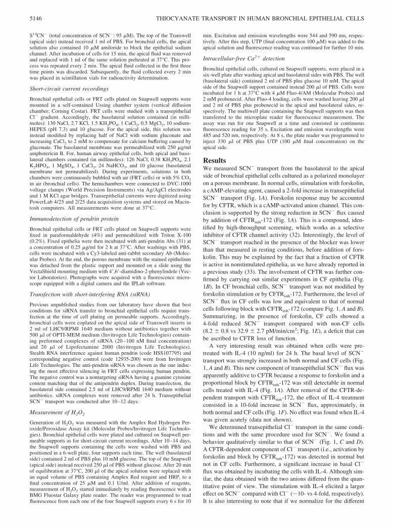
ResultsWe measured SCN� transport from the basolateral to the apicalside of bronchial epithelial cells cultured as a polarized monolayeron a porous membrane. In normal cells, stimulation with forskolin,a cAMP-elevating agent, caused a 2-fold increase in transepithelialSCN� transport (Fig. 1A). Forskolin response may be accountedfor by CFTR, which is a cAMP-activated anion channel. This con-clusion is supported by the strong reduction in SCN� flux causedby addition of CFTRinh-172 (Fig. 1A). This is a compound, iden-tified by high-throughput screening, which works as a selectiveinhibitor of CFTR channel activity (32). Interestingly, the level ofSCN� transport reached in the presence of the blocker was lowerthan that measured in resting conditions, before addition of fors-kolin. This may be explained by the fact that a fraction of CFTRis active in nonstimulated epithelia, as we have already reported ina previous study (33). The involvement of CFTR was further con-firmed by carrying out similar experiments in CF epithelia (Fig.1B). In CF bronchial cells, SCN� transport was not modified byforskolin stimulation or by CFTRinh-172. Furthermore, the level ofSCN� flux in CF cells was low and equivalent to that of normalcells following block with CFTRinh-172 (compare Fig. 1, A and B).Summarizing, in the presence of forskolin, CF cells showed a4-fold reduced SCN� transport compared with non-CF cells(8.2 � 0.8 vs 32.9 � 2.7 pM/min/cm2; Fig. 1E), a deficit that canbe ascribed to CFTR loss of function.
A very interesting result was obtained when cells were pre-treated with IL-4 (10 ng/ml) for 24 h. The basal level of SCN�
transport was strongly increased in both normal and CF cells (Fig.1, A and B). This new component of transepithelial SCN� flux wasapparently additive to CFTR because a response to forskolin and aproportional block by CFTRinh-172 was still detectable in normalcells treated with IL-4 (Fig. 1A). After removal of the CFTR-de-pendent transport with CFTRinh-172, the effect of IL-4 treatmentconsisted in a 10-fold increase in SCN� flux, approximately, inboth normal and CF cells (Fig. 1F). No effect was found when IL-4was given acutely (data not shown).
We determined transepithelial Cl� transport in the same condi-tions and with the same procedure used for SCN�. We found abehavior qualitatively similar to that of SCN� (Fig. 1, C and D).A CFTR-dependent component of Cl� transport (i.e., activation byforskolin and block by CFTRinh-172) was detected in normal butnot in CF cells. Furthermore, a significant increase in basal Cl�
flux was obtained by incubating the cells with IL-4. Although sim-ilar, the data obtained with the two anions differed from the quan-titative point of view. The stimulation with IL-4 elicited a largereffect on SCN� compared with Cl� (�10- vs 4-fold, respectively).It is also interesting to note that if we normalize for the different
5146 THIOCYANATE TRANSPORT IN HUMAN BRONCHIAL EPITHELIAL CELLS
concentrations used in the experiments to mimic physiologicalconditions (95 �M SCN� vs 137 mM Cl� in the basolateral so-lution), it appears that permeability for SCN�, in particular afterIL-4 treatment, is even higher than that for Cl�.
The strong up-regulation of basal anion transport by IL-4, de-tected by radioisotopic technique, was particularly interesting be-
cause we had not detected in previous studies a similar effect dur-ing short-circuit current recordings (9), as also shown in Fig. 1G.In agreement with our previous findings, we found that treatmentwith IL-4 caused a dramatic reduction of Na� absorption as evi-dent from the reduced effect of the ENaC blocker, amiloride. Uponblock of ENaC with amiloride, and of CFTR with CFTRinh-172,we observed that the residual current was very small and not dif-ferent between control and IL-4-treated cells (Fig. 1G). This con-trasts with the high Cl� and SCN� fluxes evoked by IL-4 treat-ment. Therefore, we hypothesized that the strong effect elicited byIL-4 on basal SCN� and Cl� transport is mediated by stimulationof a nonelectrogenic anion transporter, possibly by up-regulationof transcription of the corresponding gene.
To possibly identify this transporter, we performed a globalanalysis of gene expression by means of Affymetrix U133 Plus 2.0microarrays. This last version of microarrays contains 54,000probe sets corresponding to 47,000 human transcripts. ThemRNA was extracted from untreated and IL-4-treated cells of cul-tured polarized epithelia of four different individuals. We lookedfor possible up-regulation of anion channels and transporters. Wefound no up-regulation of Cl� channels of the ClC family likeCLC2 (data not shown). The same negative findings were foundfor putative Ca2�-dependent Cl� channels of the ClCA or bestro-phin family. Actually, the genes belonging to the latter familyappeared as poorly expressed in our cells (data not shown). Inter-estingly, the only anion transporter that appeared strongly up-reg-ulated by IL-4 was SLC26A4, also known as pendrin (25) (seeTable I). The up-regulation consisted in a 23-fold increase ofSLC26A4 signal in cytokine-treated cells. This finding was sur-prising because pendrin expression in airway epithelial cells hasnot been previously reported. Pendrin is a transporter which me-diates anion exchange at the apical membrane of cells in the kid-ney, inner ear, and thyroid follicle (34–37). When mutated, thependrin gene is responsible for Pendred syndrome or nonsyn-dromic deafness (38, 39). Besides pendrin, no other known aniontransporter was strongly up-regulated by IL-4. Many other mem-bers of the SLC26 family, like SLC26A1, SLC26A6, SLC26A7,SLC26A8, SLC26A9, and SLC26A10, were expressed at a lowlevel in our cells and not affected by IL-4. SLC26A2, a sulfatetransporter, was the only member of the same family with a sig-nificant expression under basal levels and a modest induction by
FIGURE 1. Anion transport in resting and IL-4-stimulated bronchialepithelial cells. Transepithelial transport of SCN� (A and B) and Cl� (Cand D) in bronchial epithelial cells. Highly differentiated epithelia weregenerated from normal (A and C) or CF bronchi (B and D) on permeablesupports. Cells were kept under resting conditions or treated for 24 h with10 ng/ml IL-4. After equilibrating the cells with S14CN� or 36Cl� in thebasolateral medium, the radioactive tracer transported to the apical sidewas collected and measured. Apical amiloride (10 �M) was maintainedthroughout the experiments. As indicated by arrows, cells during the ex-periment were first stimulated with forskolin (20 �M) and then treated withthe selective CFTR blocker inh-172 (20 �M). E, CFTR-dependent trans-epithelial SCN� flux. Basolateral to apical SCN� transport was measuredin normal and CF epithelia during maximal stimulation with forskolin (6min after forskolin addition). F, CFTR-independent SCN� flux. Transep-ithelial SCN� transport was measured in normal and CF epithelia in thepresence of inh-172 (20 �M). Cells were in resting conditions (�) ortreated with IL-4 (f). G, representative traces from Ussing chamber ex-periments on resting (top) and IL-4 treated (bottom) bronchial epithelialcells. Except for Ussing chamber experiments, all data are presented asmean � SEM of four to six experiments. ���, A statistically significantdifference (p � 0.001).
Table I. Expression of SLC26 transporters and other genesa
GeneMicroarray RT-PCR
Control IL-4 IL-4/control IL-4/control
SLC26A1 4.9 5 AbsentSLC26A2 106.9 244.5 2.29 3.07SLC26A3 5.6 5.6 Absent AbsentSLC26A4 139.1 3150.5 22.65 21.72SLC26A6 35.2 58.2 1.65 1.06SLC26A7 6.9 7 Absent AbsentSLC26A8 3.8 3.8 Absent AbsentSLC26A9 6.6 6.3 Absent AbsentSLC26A10 4.4 4.5 AbsentSLC26A11 293.3 458.3 1.56SLC4A1 6.3 6.4 AbsentSLC4A2 196.7 168 0.85SLC4A3 5.2 5.2 AbsentSLC5A5 70.4 98.2 1.39 0.97DUOX1 94.7 188.7 1.99 1.7DUOX2 895.9 2592.6 2.89 2.4LPO 4.2 4.3 Absent
a Expression of SLC26 transporters and other genes in resting and IL-4-treatedbronchial epithelial cells by microarray and real-time RT-PCR.
5147The Journal of Immunology
IL-4. Similarly, other anion transporters belonging to the SLC4family (anion exchangers) were poorly expressed and/or not af-fected by cytokine treatment. To confirm the findings obtainedwith the microarrays, we determined the changes in expression ofspecific genes by real-time RT-PCR. We found that pendrinmRNA was indeed strongly up-regulated after treatment with IL-4(24.7- � 4.9-fold, n � 6). Other transporters of the same family,including SLC26A3, A6, A7, A8, and A9 were instead poorlyexpressed and not up-regulated. We also measured by the samemethod the expression of SLC5A5, the Na�/I� symporter, whichis also able to mediate SCN� uptake (11). This gene was notup-regulated by IL-4 in agreement with data deriving from mi-croarrays (Table I).
To demonstrate the expression of full-length pendrin mRNA inbronchial cells, we conducted an RT-PCR using two primers al-lowing amplification of the whole coding sequence. The reactiongenerated a fragment of 2500 bp, compatible with the expectedpendrin amplification product (data not shown). This fragment wascloned in a plasmid and the resulting clones were fully sequenced.Determination of nucleotide sequence revealed that the clones con-tained the full coding sequence of pendrin, as reported in GenBank(accession number: NM_000441).
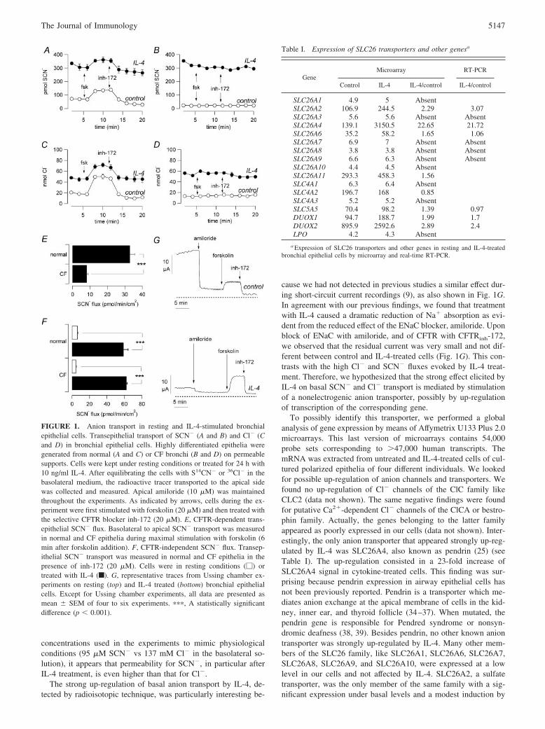
To determine the functional properties of cloned pendrin wedecided to take advantage of the halide and pseudohalide sensi-tivity of YFPs (40, 41). Accordingly, we transfected the expressionvector carrying the pendrin-coding sequence in FRT cells that al-ready expressed the YFP-H148Q/I152L (41). FRT cells are thyroidcells that have lost expression of endogenous pendrin. The result-ing cell line coexpressing the two desired proteins was used influorescence assays using a microplate reader equipped with sy-ringe pumps for liquid injection during the assay. Injection in thewell of a modified PBS containing I� instead of Cl� generated adecrease in fluorescence that was much faster in pendrin-express-ing cells compared with null cells (Fig. 2A). This is in agreementwith the reported ability of pendrin to mediate exchange of Cl�
with I� (25, 39). In the same way, a fast fluorescence quenchingwas also obtained by injecting a SCN�- or a NO3
�-rich solution
thus indicating that pendrin is also able to transport these anions(Fig. 2A). We calculated anion fluxes by taking into account thedifferent affinity of the YFP for the different anions (41). Afterthis correction, the rates of transport for the different anions inpendrin-expressing cells were comparable (Fig. 2B). We per-formed the same type of experiments also in FRT cells express-ing CFTR. In this case, we found that I� and SCN� transportrates were lower than those measured for NO3
� and Br� (Fig.2C). This is in agreement with the CFTR pore being partiallyblocked by such anions (42).
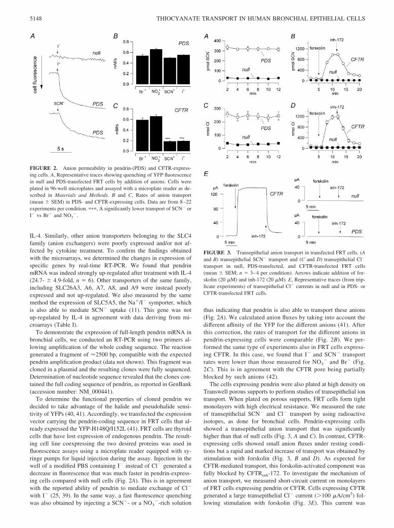
The cells expressing pendrin were also plated at high density onTranswell porous supports to perform studies of transepithelial iontransport. When plated on porous supports, FRT cells form tightmonolayers with high electrical resistance. We measured the rateof transepithelial SCN� and Cl� transport by using radioactiveisotopes, as done for bronchial cells. Pendrin-expressing cellsshowed a transepithelial anion transport that was significantlyhigher than that of null cells (Fig. 3, A and C). In contrast, CFTR-expressing cells showed small anion fluxes under resting condi-tions but a rapid and marked increase of transport was obtained bystimulation with forskolin (Fig. 3, B and D). As expected forCFTR-mediated transport, this forskolin-activated component wasfully blocked by CFTRinh-172. To investigate the mechanism ofanion transport, we measured short-circuit current on monolayersof FRT cells expressing pendrin or CFTR. Cells expressing CFTRgenerated a large transepithelial Cl� current (100 �A/cm2) fol-lowing stimulation with forskolin (Fig. 3E). This current was
FIGURE 2. Anion permeability in pendrin-(PDS) and CFTR-express-ing cells. A, Representative traces showing quenching of YFP fluorescencein null and PDS-transfected FRT cells by addition of anions. Cells wereplated in 96-well microplates and assayed with a microplate reader as de-scribed in Materials and Methods. B and C, Rates of anion transport(mean � SEM) in PDS- and CFTR-expressing cells. Data are from 8–22experiments per condition. ���, A significantly lower transport of SCN� orI� vs Br� and NO3
�.
FIGURE 3. Transepithelial anion transport in transfected FRT cells. (Aand B) transepithelial SCN� transport and (C and D) transepithelial Cl�
transport in null, PDS-transfected, and CFTR-transfected FRT cells(mean � SEM; n � 3–4 per condition). Arrows indicate addition of for-skolin (20 �M) and inh-172 (20 �M). E, Representative traces (from trip-licate experiments) of transepithelial Cl� currents in null and in PDS- orCFTR-transfected FRT cells.
5148 THIOCYANATE TRANSPORT IN HUMAN BRONCHIAL EPITHELIAL CELLS
sensitive to CFTRinh-172. On the contrary, cells expressing pen-drin did not show a significant Cl� current compared with nullcells (Fig. 3E). The finding that CFTR- and pendrin (PDS) ex-pressing cells show significant rates of anion transport using ra-dioactive tracers, but only CFTR cells display measurable Cl�
currents, supports the present knowledge that pendrin mediateselectroneutral anion transport (43).
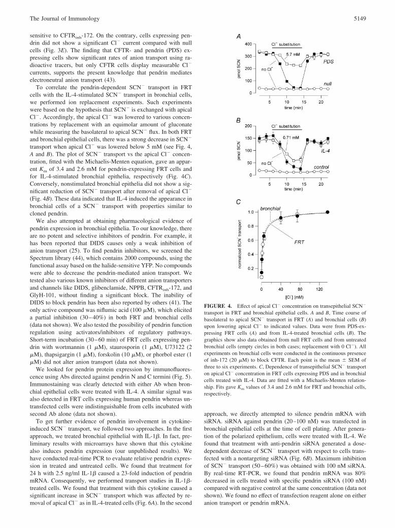
To correlate the pendrin-dependent SCN� transport in FRTcells with the IL-4-stimulated SCN� transport in bronchial cells,we performed ion replacement experiments. Such experimentswere based on the hypothesis that SCN� is exchanged with apicalCl�. Accordingly, the apical Cl� was lowered to various concen-trations by replacement with an equimolar amount of gluconatewhile measuring the basolateral to apical SCN� flux. In both FRTand bronchial epithelial cells, there was a strong decrease in SCN�
transport when apical Cl� was lowered below 5 mM (see Fig. 4,A and B). The plot of SCN� transport vs the apical Cl� concen-tration, fitted with the Michaelis-Menten equation, gave an appar-ent Km of 3.4 and 2.6 mM for pendrin-expressing FRT cells andfor IL-4-stimulated bronchial epithelia, respectively (Fig. 4C).Conversely, nonstimulated bronchial epithelia did not show a sig-nificant reduction of SCN� transport after removal of apical Cl�
(Fig. 4B). These data indicated that IL-4 induced the appearance inbronchial cells of a SCN� transport with properties similar tocloned pendrin.
We also attempted at obtaining pharmacological evidence ofpendrin expression in bronchial epithelia. To our knowledge, thereare no potent and selective inhibitors of pendrin. For example, ithas been reported that DIDS causes only a weak inhibition ofanion transport (25). To find pendrin inhibitors, we screened theSpectrum library (44), which contains 2000 compounds, using thefunctional assay based on the halide-sensitive YFP. No compoundswere able to decrease the pendrin-mediated anion transport. Wetested also various known inhibitors of different anion transportersand channels like DIDS, glibenclamide, NPPB, CFTRinh-172, andGlyH-101, without finding a significant block. The inability ofDIDS to block pendrin has been also reported by others (41). Theonly active compound was niflumic acid (100 �M), which eliciteda partial inhibition (30–40%) in both FRT and bronchial cells(data not shown). We also tested the possibility of pendrin functionregulation using activators/inhibitors of regulatory pathways.Short-term incubation (30–60 min) of FRT cells expressing pen-drin with wortmannin (1 �M), staurosporin (1 �M), U73122 (2�M), thapsigargin (1 �M), forskolin (10 �M), or phorbol ester (1�M) did not alter anion transport (data not shown).
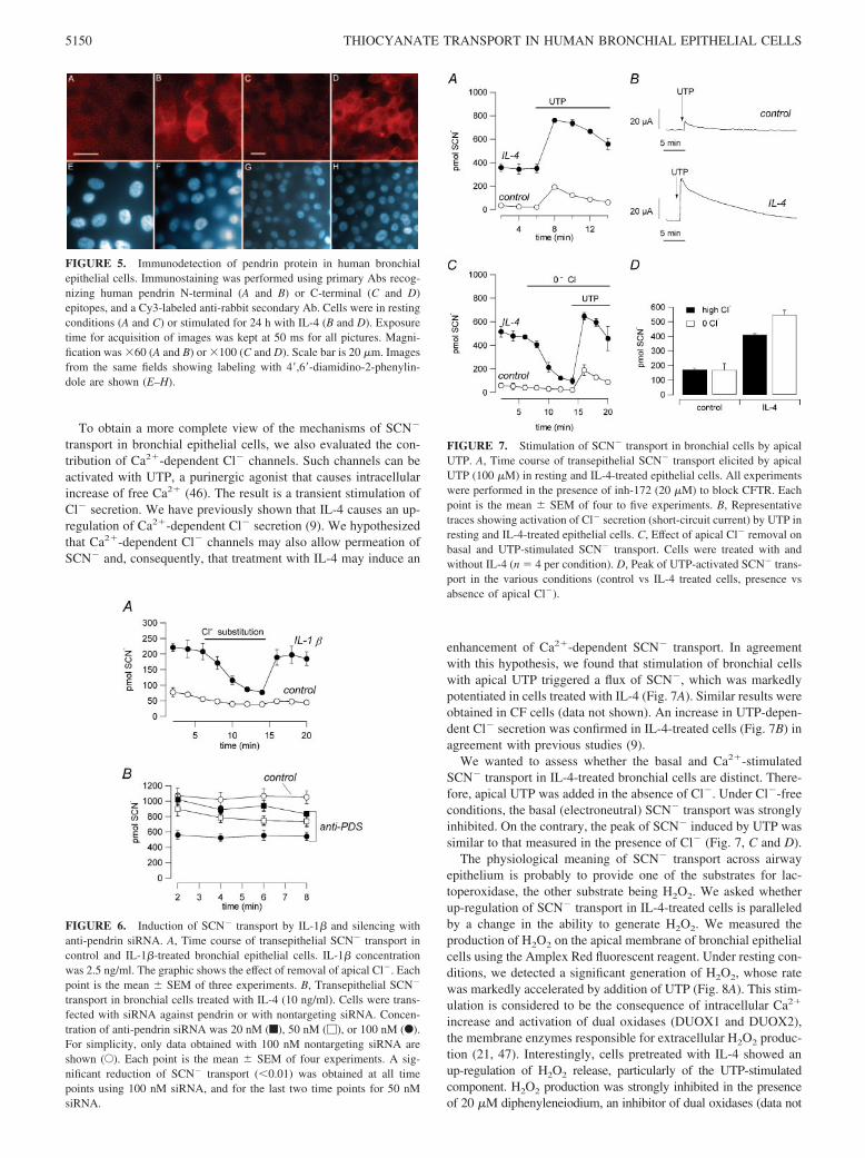
We looked for pendrin protein expression by immunofluores-cence using Abs directed against pendrin N and C termini (Fig. 5).Immunostaining was clearly detected with either Ab when bron-chial epithelial cells were treated with IL-4. A similar signal wasalso detected in FRT cells expressing human pendrin whereas un-transfected cells were indistinguishable from cells incubated withsecond Ab alone (data not shown).
To get further evidence of pendrin involvement in cytokine-induced SCN� transport, we followed two approaches. In the firstapproach, we treated bronchial epithelial with IL-1�. In fact, pre-liminary results with microarrays have shown that this cytokinealso induces pendrin expression (our unpublished results). Wehave conducted real-time PCR to evaluate relative pendrin expres-sion in treated and untreated cells. We found that treatment for24 h with 2.5 ng/ml IL-1� caused a 23-fold induction of pendrinmRNA. Consequently, we performed transport studies in IL-1�-treated cells. We found that treatment with this cytokine caused asignificant increase in SCN� transport which was affected by re-moval of apical Cl� as in IL-4-treated cells (Fig. 6A). In the second
approach, we directly attempted to silence pendrin mRNA withsiRNA. siRNA against pendrin (20–100 nM) was transfected inbronchial epithelial cells at the time of cell plating. After genera-tion of the polarized epithelium, cells were treated with IL-4. Wefound that treatment with anti-pendrin siRNA generated a dose-dependent decrease of SCN� transport with respect to cells trans-fected with a nontargeting siRNA (Fig. 6B). Maximum inhibitionof SCN� transport (50–60%) was obtained with 100 nM siRNA.By real-time RT-PCR, we found that pendrin mRNA was 80%decreased in cells treated with specific pendrin siRNA (100 nM)compared with negative control at the same concentration (data notshown). We found no effect of transfection reagent alone on eitheranion transport or pendrin mRNA.
FIGURE 4. Effect of apical Cl� concentration on transepithelial SCN�
transport in FRT and bronchial epithelial cells. A and B, Time course ofbasolateral to apical SCN� transport in FRT (A) and bronchial cells (B)upon lowering apical Cl� to indicated values. Data were from PDS-ex-pressing FRT cells (A) and from IL-4-treated bronchial cells (B). Thegraphics show also data obtained from null FRT cells and from untreatedbronchial cells (empty circles in both cases; replacement with 0 Cl�). Allexperiments on bronchial cells were conducted in the continuous presenceof inh-172 (20 �M) to block CFTR. Each point is the mean � SEM ofthree to six experiments. C, Dependence of transepithelial SCN� transporton apical Cl� concentration in FRT cells expressing PDS and in bronchialcells treated with IL-4. Data are fitted with a Michaelis-Menten relation-ship. Fits gave Km values of 3.4 and 2.6 mM for FRT and bronchial cells,respectively.
5149The Journal of Immunology
To obtain a more complete view of the mechanisms of SCN�
transport in bronchial epithelial cells, we also evaluated the con-tribution of Ca2�-dependent Cl� channels. Such channels can beactivated with UTP, a purinergic agonist that causes intracellularincrease of free Ca2� (46). The result is a transient stimulation ofCl� secretion. We have previously shown that IL-4 causes an up-regulation of Ca2�-dependent Cl� secretion (9). We hypothesizedthat Ca2�-dependent Cl� channels may also allow permeation ofSCN� and, consequently, that treatment with IL-4 may induce an
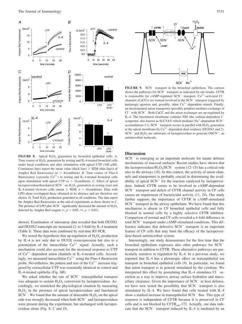
enhancement of Ca2�-dependent SCN� transport. In agreementwith this hypothesis, we found that stimulation of bronchial cellswith apical UTP triggered a flux of SCN�, which was markedlypotentiated in cells treated with IL-4 (Fig. 7A). Similar results wereobtained in CF cells (data not shown). An increase in UTP-depen-dent Cl� secretion was confirmed in IL-4-treated cells (Fig. 7B) inagreement with previous studies (9).
We wanted to assess whether the basal and Ca2�-stimulatedSCN� transport in IL-4-treated bronchial cells are distinct. There-fore, apical UTP was added in the absence of Cl�. Under Cl�-freeconditions, the basal (electroneutral) SCN� transport was stronglyinhibited. On the contrary, the peak of SCN� induced by UTP wassimilar to that measured in the presence of Cl� (Fig. 7, C and D).
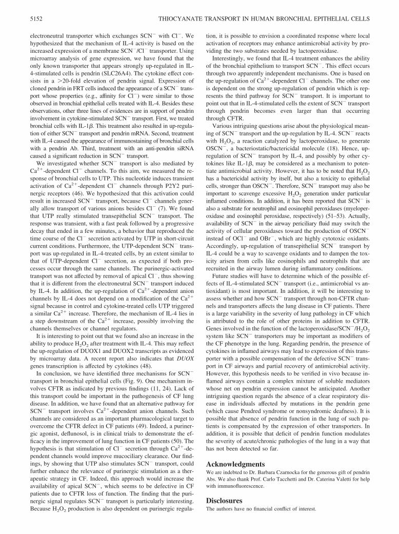
The physiological meaning of SCN� transport across airwayepithelium is probably to provide one of the substrates for lac-toperoxidase, the other substrate being H2O2. We asked whetherup-regulation of SCN� transport in IL-4-treated cells is paralleledby a change in the ability to generate H2O2. We measured theproduction of H2O2 on the apical membrane of bronchial epithelialcells using the Amplex Red fluorescent reagent. Under resting con-ditions, we detected a significant generation of H2O2, whose ratewas markedly accelerated by addition of UTP (Fig. 8A). This stim-ulation is considered to be the consequence of intracellular Ca2�
increase and activation of dual oxidases (DUOX1 and DUOX2),the membrane enzymes responsible for extracellular H2O2 produc-tion (21, 47). Interestingly, cells pretreated with IL-4 showed anup-regulation of H2O2 release, particularly of the UTP-stimulatedcomponent. H2O2 production was strongly inhibited in the presenceof 20 �M diphenyleneiodium, an inhibitor of dual oxidases (data not
FIGURE 5. Immunodetection of pendrin protein in human bronchialepithelial cells. Immunostaining was performed using primary Abs recog-nizing human pendrin N-terminal (A and B) or C-terminal (C and D)epitopes, and a Cy3-labeled anti-rabbit secondary Ab. Cells were in restingconditions (A and C) or stimulated for 24 h with IL-4 (B and D). Exposuretime for acquisition of images was kept at 50 ms for all pictures. Magni-fication was �60 (A and B) or �100 (C and D). Scale bar is 20 �m. Imagesfrom the same fields showing labeling with 4�,6�-diamidino-2-phenylin-dole are shown (E–H).
FIGURE 6. Induction of SCN� transport by IL-1� and silencing withanti-pendrin siRNA. A, Time course of transepithelial SCN� transport incontrol and IL-1�-treated bronchial epithelial cells. IL-1� concentrationwas 2.5 ng/ml. The graphic shows the effect of removal of apical Cl�. Eachpoint is the mean � SEM of three experiments. B, Transepithelial SCN�
transport in bronchial cells treated with IL-4 (10 ng/ml). Cells were trans-fected with siRNA against pendrin or with nontargeting siRNA. Concen-tration of anti-pendrin siRNA was 20 nM (f), 50 nM (�), or 100 nM (F).For simplicity, only data obtained with 100 nM nontargeting siRNA areshown (E). Each point is the mean � SEM of four experiments. A sig-nificant reduction of SCN� transport (�0.01) was obtained at all timepoints using 100 nM siRNA, and for the last two time points for 50 nMsiRNA.
FIGURE 7. Stimulation of SCN� transport in bronchial cells by apicalUTP. A, Time course of transepithelial SCN� transport elicited by apicalUTP (100 �M) in resting and IL-4-treated epithelial cells. All experimentswere performed in the presence of inh-172 (20 �M) to block CFTR. Eachpoint is the mean � SEM of four to five experiments. B, Representativetraces showing activation of Cl� secretion (short-circuit current) by UTP inresting and IL-4-treated epithelial cells. C, Effect of apical Cl� removal onbasal and UTP-stimulated SCN� transport. Cells were treated with andwithout IL-4 (n � 4 per condition). D, Peak of UTP-activated SCN� trans-port in the various conditions (control vs IL-4 treated cells, presence vsabsence of apical Cl�).
5150 THIOCYANATE TRANSPORT IN HUMAN BRONCHIAL EPITHELIAL CELLS
shown). Examination of microarray data revealed that both DUOX1and DUOX2 transcripts are increased (2- to 3-fold) by IL-4 treatment(Table I). These data were confirmed by real-time RT-PCR.
We tested the hypothesis that up-regulation of H2O2 productionby IL-4 is not only due to DUOX overexpression but also to apotentiation of the intracellular Ca2� signal. Actually, such amechanism could also account for the increased response to UTPof Ca2�-dependent anion channels in IL-4-treated cells. Accord-ingly, we measured intracellular Ca2� using the Fluo-4 fluorescentprobe. Nevertheless, the pattern and size of the Ca2� increase trig-gered by extracellular UTP was essentially identical in control andIL-4-treated epithelia (Fig. 8B).
We asked whether the rate of SCN� transepithelial transportwas adequate to sustain H2O2 conversion by lactoperoxidase. Ac-cordingly, we mimicked the physiological situation by measuringH2O2 in the presence of apical lactoperoxidase and basolateralSCN�. We found that the amount of detectable H2O2 on the apicalside was strongly decreased when both SCN� and lactoperoxidasewere present during the experiment, but unchanged with lactoper-oxidase alone (Fig. 8, C and D).
DiscussionSCN� is emerging as an important molecule for innate defensemechanisms of mucosal surfaces. Recent studies have shown thatthe lactoperoxidase/H2O2/SCN� system (12–15) has a critical rolealso in the airways (18). In this contest, the activity of anion chan-nels and transporters is probably crucial in determining the avail-ability of apical SCN� for the reaction catalyzed by lactoperoxi-dase. Indeed, CFTR seems to be involved in cAMP-dependentSCN� transport and deficit of CFTR channel activity in CF cellscauses an impairment of bactericidal activity (11, 24). Our studyfurther supports the importance of CFTR in cAMP-stimulatedSCN� transport in the airway epithelium. We have found that thismechanism is absent in CF bronchial epithelial cells and fullyblocked in normal cells by a highly selective CFTR inhibitor.Comparison of normal and CF cells revealed a 4-fold difference intotal SCN� transport under cAMP-stimulated conditions. This dif-ference indicates that defective SCN� transport is an importantfeature of CF cells that may limit the efficacy of the lactoperoxi-dase-based antimicrobial system.
Interestingly, our study demonstrates for the first time that thebronchial epithelium expresses also other pathways for SCN�
transport in addition to CFTR. These alternative pathways are par-ticularly sensitive to regulation by IL-4. In a previous study, wereported that IL-4 has a pleiotropic effect on transepithelial iontransport in bronchial epithelial cells (9). In particular, we foundthat anion transport is in general stimulated by the cytokine. Weinterpreted this effect by postulating that IL-4 stimulates Cl� se-cretion as a way to improve airway surface hydration and muco-ciliary clearance. Given the importance of SCN� in host defense,we have now tested the possibility that SCN� transport is alsostimulated by IL-4. We have found that cells treated with IL-4show a marked increase in transepithelial SCN� flux. This type ofresponse is independent of CFTR because it is preserved in CFcells and is not blocked by CFTRinh-172. Actually, our data indi-cate that the SCN� transport induced by IL-4 is mediated by an
FIGURE 8. Apical H2O2 generation by bronchial epithelial cells. A,Time course of H2O2 generation by resting and IL-4-treated bronchial cellsunder basal conditions and after stimulation with apical UTP (100 �M).Continuous lines report the mean value (thick line) � SEM (thin lines) ofAmplex Red fluorescence (n � 4/condition). B, Time course of Fluo-4fluorescence (cytosolic Ca2�) in resting and IL-4-treated bronchial cellsupon stimulation with apical UTP (n � 3/condition). C, Effect of apicallactoperoxidase/basolateral SCN� on H2O2 generation in resting (top) andIL-4-treated (bottom) cells (mean � SEM; n � 4/condition). Data withLPO alone overlapped those obtained in its absence and are therefore notshown. D, Total H2O2 production generated in all conditions. The data reportthe Amplex Red fluorescence at the end of experiments as those shown in C.The presence of LPO plus SCN� significantly decreased the amount of H2O2
detected by Amplex Red reagent (�, p � 0.05; ��, p � 0.01).
FIGURE 9. SCN� transport in the bronchial epithelium. The cartoonshows the pathways for SCN� transport as indicated by our results. CFTRis responsible for cAMP-regulated SCN� transport. Ca2�-activated Cl�
channels (CaCCs) are instead involved in the SCN� transport triggered bypurinergic agonists and, possibly, other Ca2�-dependent stimuli. Finally,an electroneutral anion transporter (possibly pendrin) mediates exchange ofCl� with SCN�. Both CaCC and the anion exchanger are up-regulated byIL-4. The basolateral membrane contains NIS (the sodium-dependent I�
symporter, also known as SLC5A5) which mediates Na�-dependent SCN�
accumulation (11). SCN� transport occurs in parallel with H2O2 generationat the apical membrane by Ca2�-dependent dual oxidases (DUOX1 and 2).SCN� and H2O2 are substrates of lactoperoxidase to generate OSCN�, anantimicrobial molecule.
5151The Journal of Immunology
electroneutral transporter which exchanges SCN� with Cl�. Wehypothesized that the mechanism of IL-4 activity is based on theincreased expression of a membrane SCN�/Cl� transporter. Usingmicroarray analysis of gene expression, we have found that theonly known transporter that appears strongly up-regulated in IL-4-stimulated cells is pendrin (SLC26A4). The cytokine effect con-sists in a 20-fold elevation of pendrin signal. Expression ofcloned pendrin in FRT cells induced the appearance of a SCN� trans-port whose properties (e.g., affinity for Cl�) were similar to thoseobserved in bronchial epithelial cells treated with IL-4. Besides theseobservations, other three lines of evidences are in support of pendrininvolvement in cytokine-stimulated SCN� transport. First, we treatedbronchial cells with IL-1�. This treatment also resulted in up-regula-tion of either SCN� transport and pendrin mRNA. Second, treatmentwith IL-4 caused the appearance of immunostaining of bronchial cellswith a pendrin Ab. Third, treatment with an anti-pendrin siRNAcaused a significant reduction in SCN� transport.
We investigated whether SCN� transport is also mediated byCa2�-dependent Cl� channels. To this aim, we measured the re-sponse of bronchial cells to UTP. This nucleotide induces transientactivation of Ca2�-dependent Cl� channels through P2Y2 puri-nergic receptors (46). We hypothesized that this activation couldresult in increased SCN� transport, because Cl� channels gener-ally allow transport of various anions besides Cl� (7). We foundthat UTP really stimulated transepithelial SCN� transport. Theresponse was transient, with a fast peak followed by a progressivedecay that ended in a few minutes, a behavior that reproduced thetime course of the Cl� secretion activated by UTP in short-circuitcurrent conditions. Furthermore, the UTP-dependent SCN� trans-port was up-regulated in IL-4-treated cells, by an extent similar tothat of UTP-dependent Cl� secretion, as expected if both pro-cesses occur through the same channels. The purinergic-activatedtransport was not affected by removal of apical Cl�, thus showingthat it is different from the electroneutral SCN� transport inducedby IL-4. In addition, the up-regulation of Ca2�-dependent anionchannels by IL-4 does not depend on a modification of the Ca2�
signal because in control and cytokine-treated cells UTP triggereda similar Ca2� increase. Therefore, the mechanism of IL-4 lies ina step downstream of the Ca2� increase, possibly involving thechannels themselves or channel regulators.
It is interesting to point out that we found also an increase in theability to produce H2O2 after treatment with IL-4. This may reflectthe up-regulation of DUOX1 and DUOX2 transcripts as evidencedby microarray data. A recent report also indicates that DUOXgenes transcription is affected by cytokines (48).
In conclusion, we have identified three mechanisms for SCN�
transport in bronchial epithelial cells (Fig. 9). One mechanism in-volves CFTR as indicated by previous findings (11, 24). Lack ofthis transport could be important in the pathogenesis of CF lungdisease. In addition, we have found that an alternative pathway forSCN� transport involves Ca2�-dependent anion channels. Suchchannels are considered as an important pharmacological target toovercome the CFTR defect in CF patients (49). Indeed, a puriner-gic agonist, deflunosol, is in clinical trials to demonstrate the ef-ficacy in the improvement of lung function in CF patients (50). Thehypothesis is that stimulation of Cl� secretion through Ca2�-de-pendent channels would improve mucociliary clearance. Our find-ings, by showing that UTP also stimulates SCN� transport, couldfurther enhance the relevance of purinergic stimulation as a ther-apeutic strategy in CF. Indeed, this approach would increase theavailability of apical SCN�, which seems to be defective in CFpatients due to CFTR loss of function. The finding that the puri-nergic signal regulates SCN� transport is particularly interesting.Because H2O2 production is also dependent on purinergic regula-
tion, it is possible to envision a coordinated response where localactivation of receptors may enhance antimicrobial activity by pro-viding the two substrates needed by lactoperoxidase.
Interestingly, we found that IL-4 treatment enhances the abilityof the bronchial epithelium to transport SCN�. This effect occursthrough two apparently independent mechanisms. One is based onthe up-regulation of Ca2�-dependent Cl� channels. The other oneis dependent on the strong up-regulation of pendrin which is rep-resents the third pathway for SCN� transport. It is important topoint out that in IL-4-stimulated cells the extent of SCN� transportthrough pendrin becomes even larger than that occurringthrough CFTR.
Various intriguing questions arise about the physiological mean-ing of SCN� transport and the up-regulation by IL-4. SCN� reactswith H2O2, a reaction catalyzed by lactoperoxidase, to generateOSCN�, a bacteriostatic/bactericidal molecule (18). Hence, up-regulation of SCN� transport by IL-4, and possibly by other cy-tokines like IL-1�, may be considered as a mechanism to poten-tiate antimicrobial activity. However, it has to be noted that H2O2
has a bactericidal activity by itself, but also a toxicity to epithelialcells, stronger than OSCN�. Therefore, SCN� transport may also beimportant to scavenge excessive H2O2 generation under particularinflamed conditions. In addition, it has been reported that SCN� isalso a substrate for neutrophil and eosinophil peroxidases (myeloper-oxidase and eosinophil peroxidase, respectively) (51–53). Actually,availability of SCN� in the airway periciliary fluid may switch theactivity of cellular peroxidases toward the production of OSCN�
instead of OCl� and OBr�, which are highly cytotoxic oxidants.Accordingly, up-regulation of transepithelial SCN� transport byIL-4 could be a way to scavenge oxidants and to dampen the tox-icity arisen from cells like eosinophils and neutrophils that arerecruited in the airway lumen during inflammatory conditions.
Future studies will have to determine which of the possible ef-fects of IL-4-stimulated SCN� transport (i.e., antimicrobial vs an-tioxidant) is most important. In addition, it will be interesting toassess whether and how SCN� transport through non-CFTR chan-nels and transporters affects the lung disease in CF patients. Thereis a large variability in the severity of lung pathology in CF whichis attributed to the role of other proteins in addition to CFTR.Genes involved in the function of the lactoperoxidase/SCN�/H2O2
system like SCN� transporters may be important as modifiers ofthe CF phenotype in the lung. Regarding pendrin, the presence ofcytokines in inflamed airways may lead to expression of this trans-porter with a possible compensation of the defective SCN� trans-port in CF airways and partial recovery of antimicrobial activity.However, this hypothesis needs to be verified in vivo because in-flamed airways contain a complex mixture of soluble mediatorswhose net on pendrin expression cannot be anticipated. Anotherintriguing question regards the absence of a clear respiratory dis-ease in individuals affected by mutations in the pendrin gene(which cause Pendred syndrome or nonsyndromic deafness). It ispossible that absence of pendrin function in the lung of such pa-tients is compensated by the expression of other transporters. Inaddition, it is possible that deficit of pendrin function modulatesthe severity of acute/chronic pathologies of the lung in a way thathas not been detected so far.
AcknowledgmentsWe are indebted to Dr. Barbara Czarnocka for the generous gift of pendrinAbs. We also thank Prof. Carlo Tacchetti and Dr. Caterina Valetti for helpwith immunofluorescence.
DisclosuresThe authors have no financial conflict of interest.
5152 THIOCYANATE TRANSPORT IN HUMAN BRONCHIAL EPITHELIAL CELLS
References1. Smith, J. J., and M. J. Welsh. 1993. Fluid and electrolyte transport by cultured
human airway epithelia. J. Clin. Invest. 91: 1590–1597.2. Boucher, R. C. 1994. Human airway ion transport: part two. Am. J. Respir. Crit.
Care Med. 150: 581–593.3. Coakley, R. D., B. R. Grubb, A. M. Paradiso, J. T. Gatzy, L. G. Johnson,
S. M. Kreda, W. K. O’Neal, and R. C. Boucher. 2003. Abnormal surface liquidpH regulation by cultured cystic fibrosis bronchial epithelium. Proc. Natl. Acad.Sci. USA 100: 16083–16088.
4. Matsui, H., B. R. Grubb, R. Tarran, S. H. Randell, J. T. Gatzy, C. W. Davis, andR. C. Boucher. 1998. Evidence for periciliary liquid layer depletion, not abnormalion composition, in the pathogenesis of cystic fibrosis airways disease. Cell 95:1005–1015.
5. Boucher, R. C. 2004. New concepts of the pathogenesis of cystic fibrosis lungdisease. Eur. Respir. J. 23: 146–158.
6. Boucher, R. C., C. U. Cotton, J. T. Gatzy, M. R. Knowles, and Y. R. Yankaskas.1988. Evidence for reduced Cl� and increased Na� permeability in cystic fibrosishuman primary cell cultures. J. Physiol. 405: 77–103.
7. Jentsch, T. J., V. Stein, F. Weinreich, and A. A. Zdebik. 2002. Molecular struc-ture and physiological function of chloride channels. Physiol. Rev. 82: 503–568.
8. Sheppard, D. N., and M. J. Welsh. 1999. Structure and function of the CFTRchloride channel. Physiol. Rev. 79: S23–S45.
9. Galietta, L. J. V., P. Pagesy, C. Folli, E. Caci, L. Romio, B. Costes, E. Nicolis,G. Cabrini, M. Goossens, R. Ravazzolo, and O. Zegarra-Moran. 2002. IL-4 is apotent modulator of ion transport in the human bronchial epithelium in vitro.J. Immunol. 168: 839–845.
10. Danahay, H., H. Atherton, G. Jones, R. J. Bridges, and C. T. Poll. 2002. Inter-leukin-13 induces a hypersecretory ion transport phenotype in human bronchialepithelial cells. Am. J. Physiol. 282: L226–L236.
11. Fragoso, M. A., V. Fernandez, R. Forteza, R. H. Randell, M. Salathe, andG. E. Conner. 2004. Transcellular thiocyanate transport by human airway epi-thelia. J. Physiol. 561: 183–194.
12. Lundquist, P., B. Kagedal, and L. Nilsson. 1995. An improved method for de-termination of thiocyanate in plasma and urine. Eur. J. Clin. Chem. Clin. Bio-chem. 33: 343–349.
13. Schultz, C. P., M. K. Ahmed, C. Dawes, and H. H. Mantsch. 1996. Thiocyanatelevels in human saliva: quantitation by Fourier transform infrared spectroscopy.Anal. Biochem. 240: 7–12.
14. Tsuge, K., M. Kataoka, and Y. Seto. 2000. Cyanide and thiocyanate levels inblood and saliva of healthy adult volunteers. J. Health Sci. 46: 343–350.
15. Cowman, R. A., S. S. Baron, S. D. Obenauf, and J. J. Byrnes. 1983. Evidence forthiocyanate-sensitive peroxidase activity in human saliva. J. Clin. Microbiol. 18:1177–1182.
16. Gaya, P., M. Medina, and M. Nunez. 1991. Effect of the lactoperoxidase systemon Listeria monocytogenes behavior in raw milk at refrigeration temperatures.Appl. Environ. Microbiol. 57: 3355–3360.
17. Ihalin, R., V. Loimaranta, and J. Tenovuo. 2006. Origin, structure, and biologicalactivities of peroxidases in human saliva. Arch. Biochem. Biophys. 445: 261–268.
18. Ratner, A. J., and A. Prince. 2000. Lactoperoxidase: new recognition of an “old”enzyme in airway defenses. Am. J. Respir. Cell Mol. Biol. 22: 642–644.
19. Gerson, C., J. Sabater, M. Scuri, A. Torbati, R. Coffey, J. W. Abraham,I. Lauredo, R. Forteza, A. Wanner, M. Salathe, et al. 2000. The lactoperoxidasesystem functions in bacterial clearance of airways. Am. J. Respir. Cell Mol. Biol.22: 665–671.
20. Wijkstrom-Frei, C., S. El-Chemaly, R. Ali-Rachedi, C. Gerson, M. A. Cobas,R. Forteza, M. Salathe, and G. E. Conner. 2003. Lactoperoxidase and humanairway host defense. Am. J. Respir. Cell Mol. Biol. 29: 206–212.
21. Forteza, R., M. Salathe, F. Miot, R. Forteza, and G. E. Conner. 2005. Regulatedhydrogen peroxide production by Duox in human airway epithelial cells.Am. J. Respir. Cell Mol. Biol. 32: 462–469.
22. Ameziane-El-Hassani, R., S. Morand, J. L. Boucher, Y. M. Frapart, D. Apos-tolou, D. Agnandji, S. Gnidehou, R. Ohayon, M. S. Noel-Hudson, J. Francon,K. Lalaoui, A. Virion, and C. Dupuy. 2005. Dual oxidase-2 has an intrinsicCa2�-dependent H2O2-generating activity. J. Biol. Chem. 280: 30046–30054.
23. Schwarzer, C., T. E. Machen, B. Illek, and H. Fischer. 2004. NADPH oxidase-dependent acid production in airway epithelial cells. J. Biol. Chem. 279:36454–36461.
24. Moskwa, P., D. Lorentzen, K. J. D. A. Excoffon, J. Zabner, P. B. McCray,W. M. Nauseef, C. Dupuy, and B. Banfi. 2007. A novel host defense system ofairways is defective in cystic fibrosis. Am. J. Respir. Crit. Care Med. 175:174–183.
25. Scott, D. A., R. Wang, T. M. Kreman, V. C. Sheffield, and L. P. Karniski. 1999.The Pendred syndrome gene encodes a chloride-iodide transport protein. Nat.Genet. 21: 440–443.
26. Galietta, L. J. V., L. Musante, L. Romio, U. Caruso, A. Fantasia, A. Gazzolo,L. Romano, O. Sacco, G. A. Rossi, L. Varesio, and O. Zegarra-Moran. 1998. Anelectrogenic amino acid transporter in the apical membrane of cultured humanbronchial epithelial cells. Am. J. Physiol. 275: L917–L923.
27. Irizarry, R. A., B. M. Bolstad, F. Collin, L. M. Cope, B. Hobbs, and T. P. Speed.2003. Summaries of Affymetrix GeneChip probe level data. Nucleic Acids Res.31: e15.
28. Zhang, J., V. Carey, and R. Gentleman. 2003. An extensible application forassembling annotation for genomic data. Bioinformatics 19: 155–156.
29. Tusher, V. G., R. Tibshirani, and G. Chu. 2001. Significance analysis of microar-rays applied to the ionizing radiation response. Proc. Natl. Acad. Sci. USA 98:5116–5121.
30. Dennis, G. Jr., B. T. Sherman, D. A. Hosack, J. Yang, W. Gao, H. C. Lane, andR. A. Lempicki. 2002. DAVID: database for annotation, visualization, and inte-grated discovery. Genome Biol. 4: P3.
31. Skubis-Zegadlo, J., A. Nikodemska, E. Przytula, M. Mikula, K. Bardadin,J. Ostrowski, B. E. Wenzel, and B. Czarnocka. 2005. Expression of pendrin inbenign and malignant human thyroid tissues. Br. J. Cancer 93: 144–151.
32. Taddei, A., C. Folli, O. Zegarra-Moran, P. Fanen, A. S. Verkman, andL. J. V. Galietta. 2004. Altered channel gating mechanism for CFTR inhibition bya high-affinity thiazolidinone blocker. FEBS Lett. 558: 52–56.
33. Caci, E., C. Folli, O. Zegarra-Moran, T. Ma, M. F. Springsteel, R. E. Sammelson,M. H. Nantz, M. J. Kurth, A. S. Verkman, and L. J. V. Galietta. 2003. CFTRactivation in human bronchial epithelial cells by novel benzoflavone and ben-zimidazolone compounds. Am. J. Physiol. 285: L180–L188.
34. Everett, L. A., H. Morsli, D. K. Wu, and E. D. Green. 1999. Expression patternof the mouse ortholog of the Pendred’s syndrome gene (Pds) suggests a key rolefor pendrin in the inner ear. Proc. Natl. Acad. Sci. USA 96: 9727–9732.
35. Royaux, I. E., K. Suzuki, A. Mori, R. Katoh, L. A. Everett, L. D. Kohn, andE. D. Green. 2000. Pendrin, the protein encoded by the Pendred syndrome gene(PDS), is an apical porter of iodide in the thyroid and is regulated by thyroglob-ulin in FRTL-5 cells. Endocrinology 141: 839–845.
36. Royaux, I. E., S. M. Wall, L. P. Karniski, L. A. Everett, K. Suzuki,M. A. Knepper, and E. D. Green. 2001. Pendrin, encoded by the Pendred syn-drome gene, resides in the apical region of renal intercalated cells and mediatesbicarbonate secretion. Proc. Natl. Acad. Sci. USA 98: 4221–4226.
37. Soleimani, M., T. Greeley, S. Petrovic, Z. Wang, H. Amlal, P. Kopp, andC. E. Burnham. 2001. Pendrin: an apical Cl�/OH�/HCO3
� exchanger in thekidney cortex. Am. J. Physiol. 280: F356–F364.
38. Everett, L. A., B. Glaser, J. C. Beck, J. R. Idol, A. Buchs, M. Heyman, F. Adawi,E. Hazani, E. Nassir, A. D. Baxevanis, et al. 1997. Pendred syndrome is causedby mutations in a putative sulphate transporter gene (PDS). Nat. Genet. 17:411–422.
39. Scott, D. A., R. Wang, T. M. Kreman, M. Andrews, J. M. McDonald,J. R. Bishop, R. J. Smith, L. P. Karniski, and V. C. Sheffield. 2000. Functionaldifferences of the PDS gene product are associated with phenotypic variation inpatients with Pendred syndrome and non-syndromic hearing loss (DFNB4). Hum.Mol. Genet. 9: 1709–1715.
40. Jayaraman, S., P. Haggie, R. M. Wachter, S. J. Remington, and A. S. Verkman.2000. Mechanism and cellular applications of a green fluorescent protein-basedhalide sensor. J. Biol. Chem. 275: 6047–6050.
41. Galietta, L. J. V., P. M. Haggie, and A. S. Verkman. 2001. Green fluorescentprotein-based halide indicators with improved chloride and iodide affinities.FEBS Lett. 499: 220–224.
42. Linsdell, P., J. A. Tabcharani, and J. W. Hanrahan. 1997. Multi-Ion mechanismfor ion permeation and block in the cystic fibrosis transmembrane conductanceregulator chloride channel. J. Gen. Physiol. 110: 365–377.
43. Dossena, S., A. Maccagni, V. Vezzoli, C. Bazzini, M. L. Garavaglia, G. Meyer,J. Furst, M. Ritter, L. Fugazzola, L. Persani, et al. 2005. The expression ofwild-type pendrin (SLC26A4) in human embryonic kidney (HEK 293 Phoenix)cells leads to the activation of cationic currents. Eur. J. Endocrinol. 153:693–699.
44. Pedemonte, N., T. Diena, E. Caci, E. Nieddu, M. Mazzei, R. Ravazzolo,O. Zegarra-Moran, and L. J. V. Galietta. 2005. Antihypertensive 1,4-dihydropy-ridines as correctors of the cystic fibrosis transmembrane conductance regulatorchannel gating defect caused by cystic fibrosis mutations. Mol. Pharmacol. 68:1736–1746.
45. Dossena, S., V. Vezzoli, N. Cerutti, C. Bazzini, M. Tosco, C. Sironi,S. Rodighiero, G. Meyer, U. Fascio, J. Furst, et al. 2006. Functional character-ization of wild-type and a mutated form of SLC26A4 identified in a patient withPendred syndrome. Cell. Physiol. Biochem. 17: 245–256.
46. Mason, S. J., A. M. Paradiso, and R. C. Boucher. 1991. Regulation of transep-ithelial ion transport and intracellular calcium by extracellular ATP in humannormal and cystic fibrosis airway epithelium. Br. J. Pharmacol. 103: 1649–1656.
47. Geiszt, M., J. Witta, J. Baffi, K. Lekstrom, and T. L. Leto. 2003. Dual oxidasesrepresent novel hydrogen peroxide sources supporting mucosal surface host de-fense. FASEB J. 17: 1502–1504.
48. Harper, R. W., C. Xu, J. P. Eiserich, Y. Chen, C. Y. Kao, P. Thai, H. Setiadi, andR. Wu. 2005. Differential regulation of dual NADPH oxidases/peroxidases,Duox1 and Duox2, by Th1 and Th2 cytokines in respiratory tract epithelium.FEBS Lett. 579: 4911–4917.
49. Tarran, R., M. E. Loewen, A. M. Paradiso, J. C. Olsen, M. A. Gray, B. E. Argent,R. C. Boucher, and S. E. Gabriel. 2002. Regulation of murine airway surfaceliquid volume by CFTR and Ca2�-activated Cl� conductances. J. Gen. Physiol.120: 407–418.
50. Deterding, R., G. Retsch-Bogart, L. Milgram, R. Gibson, C. Daines, P. L. Zeitlin,C. Milla, B. Marshall, L. Lavange, J. Engels, et al. 2005. Safety and tolerabilityof denufosol tetrasodium inhalation solution, a novel P2Y2 receptor agonist:results of a phase 1/phase 2 multicenter study in mild to moderate cystic fibrosis.Pediatr. Pulmonol. 39: 339–348.
51. Thomas, E. L., and M. Fishman. 1986. Oxidation of chloride and thiocyanate byisolated leukocytes. J. Biol. Chem. 261: 9694–9702.
52. Slungaard, A., and J. R. Mahoney. 1991. Thiocyanate is the major substrate foreosinophil peroxidase in physiologic fluids: implications for cytotoxicity. J. Biol.Chem. 266: 4903–4910.
53. van Dalen, C. J., and A. J. Kettle. 2001. Substrates and products of eosinophilperoxidase. Biochem. J. 358: 233–239.
5153The Journal of Immunology
Top Related
Copyright © 2022 FDOKUMEN

