


Bahasa
Halaman
Hukum

lable at ScienceDirect
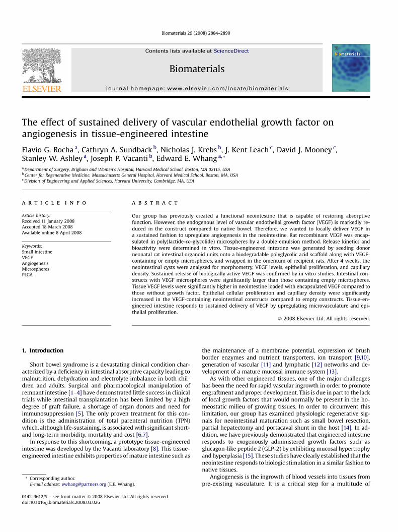
Biomaterials 29 (2008) 2884–2890
lable at ScienceDirect
Contents lists avaiContents lists avaiBiomaterials
journal homepage: www.elsevier .com/locate/biomater ia ls
Biomaterials
journal homepage: www.elsevier .com/locate/biomater ia ls
The effect of sustained delivery of vascular endothelial growth factor onangiogenesis in tissue-engineered intestine
Flavio G. Rocha a, Cathryn A. Sundback b, Nicholas J. Krebs b, J. Kent Leach c, David J. Mooney c,Stanley W. Ashley a, Joseph P. Vacanti b, Edward E. Whang a,*
a Department of Surgery, Brigham and Women’s Hospital, Harvard Medical School, Boston, MA 02115, USAb Center for Regenerative Medicine, Massachusetts General Hospital, Harvard Medical School, Boston, MA, USAc Division of Engineering and Applied Sciences, Harvard University, Cambridge, MA, USA
a r t i c l e i n f o
Article history:Received 11 January 2008Accepted 18 March 2008Available online 8 April 2008
Keywords:Small intestineVEGFAngiogenesisMicrospheresPLGA
* Corresponding author.E-mail address: [email protected] (E.E. Whan
0142-9612/$ – see front matter � 2008 Elsevier Ltd.doi:10.1016/j.biomaterials.2008.03.026
a b s t r a c t
Our group has previously created a functional neointestine that is capable of restoring absorptivefunction. However, the endogenous level of vascular endothelial growth factor (VEGF) is markedly re-duced in the construct compared to native bowel. Therefore, we wanted to locally deliver VEGF ina sustained fashion to upregulate angiogenesis in the neointestine. Rat recombinant VEGF was encap-sulated in poly(lactide-co-glycolide) microspheres by a double emulsion method. Release kinetics andbioactivity were determined in vitro. Tissue-engineered intestine was generated by seeding donorneonatal rat intestinal organoid units onto a biodegradable polyglycolic acid scaffold along with VEGF-containing or empty microspheres, and wrapped in the omentum of recipient rats. After 4 weeks, theneointestinal cysts were analyzed for morphometry, VEGF levels, epithelial proliferation, and capillarydensity. Sustained release of biologically active VEGF was confirmed by in vitro studies. Intestinal con-structs with VEGF microspheres were significantly larger than those containing empty microspheres.Tissue VEGF levels were significantly higher in neointestine loaded with encapsulated VEGF compared tothose without growth factor. Epithelial cellular proliferation and capillary density were significantlyincreased in the VEGF-containing neointestinal constructs compared to empty constructs. Tissue-en-gineered intestine responds to sustained delivery of VEGF by upregulating microvasculature and epi-thelial proliferation.
� 2008 Elsevier Ltd. All rights reserved.
1. Introduction
Short bowel syndrome is a devastating clinical condition char-acterized by a deficiency in intestinal absorptive capacity leading tomalnutrition, dehydration and electrolyte imbalance in both chil-dren and adults. Surgical and pharmacological manipulation ofremnant intestine [1–4] have demonstrated little success in clinicaltrials while intestinal transplantation has been limited by a highdegree of graft failure, a shortage of organ donors and need forimmunosuppression [5]. The only proven treatment for this con-dition is the administration of total parenteral nutrition (TPN)which, although life-sustaining, is associated with significant short-and long-term morbidity, mortality and cost [6,7].
In response to this shortcoming, a prototype tissue-engineeredintestine was developed by the Vacanti laboratory [8]. This tissue-engineered intestine exhibits properties of mature intestine such as
g).
All rights reserved.
the maintenance of a membrane potential, expression of brushborder enzymes and nutrient transporters, ion transport [9,10],generation of vascular [11] and lymphatic [12] networks and de-velopment of a mature mucosal immune system [13].
As with other engineered tissues, one of the major challengeshas been the need for rapid vascular ingrowth in order to promoteengraftment and proper development. This is due in part to the lackof local growth factors that would normally be present in the ho-meostatic milieu of growing tissues. In order to circumvent thislimitation, our group has examined physiologic regenerative sig-nals for neointestinal maturation such as small bowel resection,partial hepatectomy and portacaval shunt in the host [14]. In ad-dition, we have previously demonstrated that engineered intestineresponds to exogenously administered growth factors such asglucagon-like peptide 2 (GLP-2) by exhibiting mucosal hypertrophyand hyperplasia [15]. These studies have clearly established that theneointestine responds to biologic stimulation in a similar fashion tonative tissues.
Angiogenesis is the ingrowth of blood vessels into tissues frompre-existing vasculature. It is a critical step for a multitude of
F.G. Rocha et al. / Biomaterials 29 (2008) 2884–2890 2885
important biologic functions such as fetal development, woundhealing, and reperfusion after ischemia [16]. It is characterized bya series of steps involving endothelial cells, cytokines, growth fac-tors, and extracellular matrix. One of the key mediators of angio-genesis is the vascular endothelial growth factor (VEGF) whosemitogenic effects include migration and proliferation of endothelialcells, stabilization of neovessels and tissue remodeling [17]. In ad-dition, VEGF has been shown to promote smooth muscle growthand stimulate nerve regeneration after injury [18,19]. VEGF hasa limited half-life in circulation and developing tissues require localand persistent exposure to the growth factor to prevent atrophy ofneovessels [20]. Previously, we have demonstrated that our tissue-engineered intestine exhibits lower levels of VEGF and basicfibroblast growth factor (bFGF) and a fixed capillary densitythroughout its maturation compared to native juvenile bowel [11].
Therefore, in the present study we applied a polymeric micro-sphere delivery vehicle to encapsulate VEGF and provide a sus-tained angiogenic signal to the maturing neointestine.
2. Methods
2.1. Microsphere preparation
Microspheres were prepared using a double emulsion/solvent extraction tech-nique [21]. Briefly, emulsion #1 was created by sonicating 3 mg of rat recombinantVEGF165 (R&D Systems, Minneapolis, MN) with a poly(lactide-co-glycolide) (PLGA)solution (0.5 g of PLGA, 85:15 high viscosity) (Medisorb, Cincinnati, OH) dissolved in10 ml of ethyl acetate (EtAc) (Sigma, St. Louis, MO). Emulsion #2 was produced bydissolving 0.5 g polyvinyl alcohol (PVA) (Sigma) in 9.3 ml double distilled water(ddH20) and 0.7 ml EtAc; 1 ml of emulsion #2 was added to emulsion #1 and themixture vortexed. The vortexed solution was added to an organic extraction buffer(186 ml ddH20, 14 ml EtAc, 0.2 g PVA) and stirred for 3 h to evaporate the EtAc. Thesolution was filtered through a 0.45 mm membrane pre-conditioned with 70% eth-anol (EtOH); the filtrate was centrifuged to pellet the microspheres, and the su-pernatant was discarded. The microspheres were flash-frozen in liquid nitrogen andlyophilized overnight. For radiolabeled studies, 1 mCi of 125I-VEGF was added to themicrospheres (Perkin–Elmer, Boston, MA).
2.2. Polymer fabrication
Polyglycolic acid scaffold tubes were fabricated as previously described [8]. Non-woven 2 mm thick sheets of polyglycolic acid (PGA) (Smith and Nephew, Heslington,NY) were fashioned into 10 mm long cylinders with an outer diameter of 5 mm. Theedges were sealed with a 10% PLGA solution. To reinforce the fiber structure, thetubes were sprayed with a 5% poly-L-lactide (1.06 i.v.) solution (Birmingham Poly-mers, Pelham, AL). The lyophilized tubes were washed with 500 ml Hank’s balancedsalt solution (HBSS) (Gibco, Gaithersburg, MD). The tubes were sterilized by im-mersion in 100% EtOH for 20 min and coated with 1:100 solution of type I collagen(Vitrogen, Palo Alto, CA).
2.3. Organoid separation
Organoid units were prepared according to the method of Evans et al. [22]. Briefly,total small intestine from 3- to 5-day-old neonatal Lewis rat pups (Charles RiverLaboratories, Wilmington MA) was irrigated with ice-cold HBSS before being openedlengthwise along the antimesenteric border and cut into 1–2 mm sections. The tissuewas washed three times in HBSS at 4 �C with vigorous shaking and enzymaticallydigested with 0.25 mg/ml of dispase (Boehringer Mannheim, Indianapolis, IN) and800 U/ml of collagenase XI (Sigma) on an orbital shaker at 80 revolutions/min. Thedigestion was stopped by the addition of 10 ml of high-glucose Dulbecco’s ModifiedEagle Medium (DMEM) (Gibco) containing 4% fetal bovine serum (FBS) and 4% sor-bitol and the mixture centrifuged at 150 g for 5 min. The supernatant was removedand the above procedure repeated three times. Organoid units were reconstituted inhigh-glucose DMEM with 10% FBS and counted by hemocytometer.
2.4. Construct implantation
Organoid units (100,000) were loaded into each tubular polymer scaffold, withempty or VEGF-containing microspheres, and allowed to adhere for 1 h at 4 �C.Twelve 200 g adult male Lewis rats were anesthetized with 50 mg/kg pentobarbitaland a 1.5 cm upper abdominal incision was made in order to exteriorize the greateromentum. Polymer constructs were wrapped in the omentum and secured with 6-OProlene sutures (Ethicon, Piscataway, NJ) before being returned to the peritonealcavity and the abdominal wall was closed in two layers. Studies were approved bythe Harvard Medical Area Committee on Animals and followed National Institutes ofHealth guidelines.
2.5. Neointestine harvest
At 4 weeks post-implantation, all animals were injected intraperitoneally with50 mg/kg of 5-bromo-2-deoxyuridine (BrDU) (Sigma) 1 h before sacrifice. Afterlaparotomy, neointestinal cysts were separated from surrounding tissues and theirweight and two radii (long axis a, short axis b) were recorded. Volume of constructswas calculated using a formula for an oblate spheroid (V¼ (4/3)a2b). They weredivided into two pieces along their long axis with one section placed in 10% formalinovernight for paraffin embedding and histology while the other section was snap-frozen in liquid nitrogen for protein analysis.
2.6. In vitro characterization of microspheres
Release kinetics were determined by placing microspheres containing 125I-VEGFin a simulated body fluid consisting of phosphate-buffered saline (PBS) containingcalcium and magnesium at 37 �C on a shaker table. At pre-determined timepoints,the entire solution was removed and analyzed for radioactivity using a Packard g
counter (GMI Inc., Ramsey, MN). A fresh aliquot of PBS was added and the quantity of125I-VEGF released was determined by subtracting the radioactivity in each samplefrom the total radioactivity at the beginning of the experiment. Bioactivity of en-capsulated VEGF was determined using a human umbilical vein endothelial cells(HUVEC) (Cell Applications, San Diego, CA) proliferation assay [23]. HUVECs(1�104 cells) were cultured and passaged in standard endothelial cell mediumconditions with growth supplement onto six-well plates and allowed to adhere for24 h. The plates were then replaced with filtered supplement-free medium condi-tioned with VEGF released from microspheres or empty microspheres, supplement-free medium alone or supplement-free medium containing 10 ng/ml VEGF; all cellsremained in culture for an additional 72 h. The cells were harvested with a 0.05%trypsin solution and counted. This cycle was continued until the microspheres hadundergone 10 successive rounds of HUVEC subculturing and medium conditioning.
2.7. Histology and immunohistochemistry
Deparaffinized and rehydrated 10 mm thick sections were unmasked using mi-crowave antigen retrieval with 0.1 M citrate buffer, pH 6.0 for 10 min. Sections wererinsed and then blocked with 10% normal goat serum in PBS for 30 min at roomtemperature. Sections were incubated with primary antibody (goat anti-CD31 forvessel staining, diluted 1:1000) (Santa Cruz Biotechnology, Santa Cruz, CA) or(mouse anti-BrDU for epithelial proliferation, diluted 1:50) (Sigma) with 0.1 M PBScontaining 5% normal horse serum and 0.3% Triton X-100, for 1 h at 25 �C. Afterwashing in PBS, sections were incubated with the secondary antibody (biotinylatedhorse anti-goat antiserum for CD31 or goat anti-mouse for BrDU) diluted 1:200 inPBS for 1 h at room temperature. After a brief rinse with PBS, the sections wereincubated with 0.3% hydrogen peroxide (H2O2) in methanol for 30 min to quenchendogenous peroxidase activity. After rinsing, the sections were reacted with avi-din–biotin–horseradish peroxidase complex for 1 h. After washing in 0.1 M Tris–HCL-buffered saline, the sections were incubated in 0.05% 3,30-diaminobenzidinesolution containing nickel ammonium sulfate (0.2%) for 5–10 min and counter-stained with Mayer’s hematoxylin and mounted. CD31 positive capillaries in 10random high power fields were counted and normalized to the slide viewing area.BrDU positively stained cells were calculated as a fraction of the total number of cellsper crypt and expressed as a percentage (10 crypts/slide were counted).
2.8. Terminal deoxynucleotide transferase mediated dUTP nick-end labeling (TUNEL)
Deparaffinized and rehydrated 10 mm thick sections were exposed to TUNELmixture containing equilibration buffer, biotinylated nucleotide mix and terminaldeoxynucleotidyl transferase for 1 h in a humidified chamber at room temperature(Roche, Indianapolis, IN). Endogenous peroxidase activity was blocked by immersingthe sections in 0.3% H2O2 for 3 min at room temperature. Sections were then in-cubated with anti-digoxigenin and diaminobenzidine chromogen substrate kitsaccording to the manufacturer’s instructions for 30 min. The number of cells withTUNEL positivity and morphological features of apoptosis were counted in five fieldsand averaged.
2.9. Enzyme-linked immunosorbent assay (ELISA)
Tissue VEGF was extracted as previously described [24]. Frozen samples wereminced and homogenized in 5 ml of extraction buffer consisting of PBS, 0.05% TritonX-100 and 1 mM protease inhibitor 4,2-aminoethylbenzenesulfonylfluoride (Sigma).The mix was sonicated and centrifuged at 1500g for 10 min at 4 �C. The total proteinconcentration was determined by the bichinchoninic acid method (BCA) (Sigma).Quantikine ELISA kit for rat VEGF was purchased (R&D Systems) and utilizedaccording to the manufacturer’s instructions with standards of known concentrationand positive and negative controls. The concentration of VEGF in each sample wascalculated as picograms per milligram total protein.
2.10. Statistical analysis
All continuous data were expressed as the mean� SEM and p< 0.05 was takenas significant. p Values were estimated using Student’s t-test and when applicable,
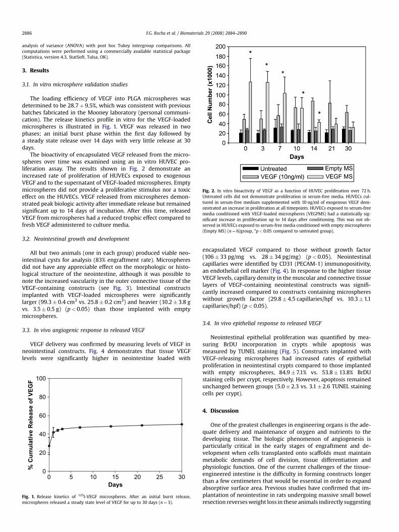
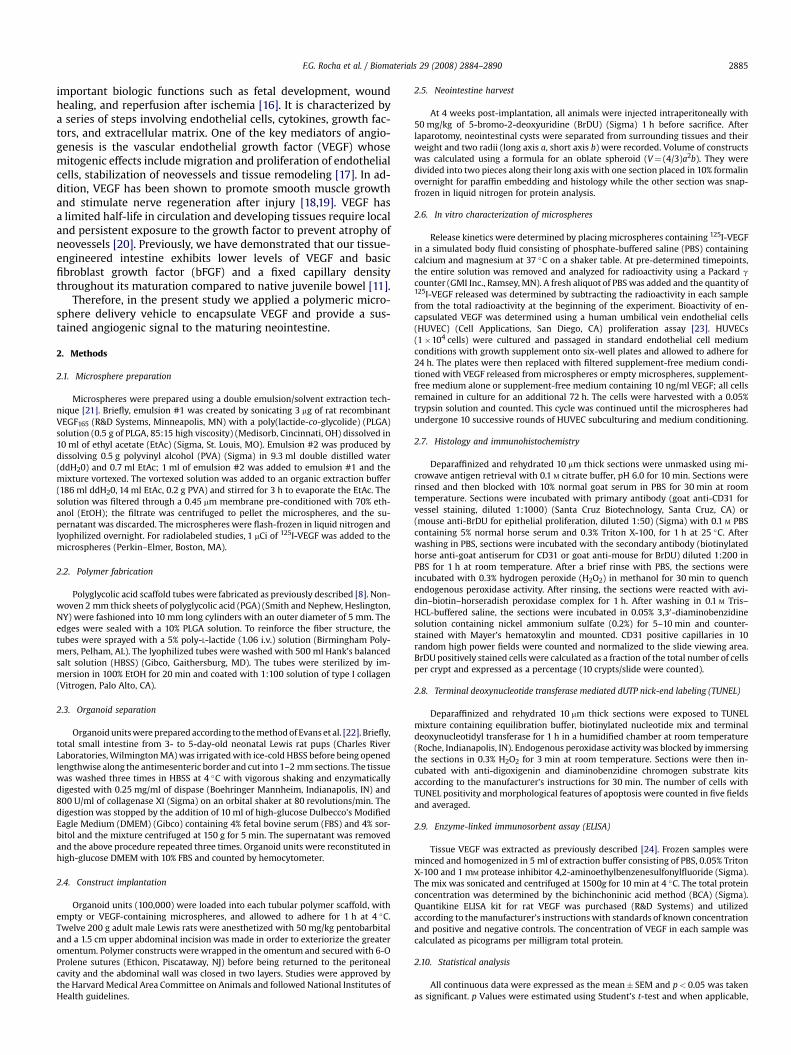
Fig. 2. In vitro bioactivity of VEGF as a function of HUVEC proliferation over 72 h.Untreated cells did not demonstrate proliferation in serum-free media. HUVECs cul-tured in serum-free medium supplemented with 10 ng/ml of exogenous VEGF dem-onstrated an increase in proliferation at all timepoints. HUVECs exposed to serum-freemedia conditioned with VEGF-loaded microspheres (VEGFMS) had a statistically sig-nificant increase in proliferation up to 14 days after conditioning. This was not ob-served in HUVECs exposed to serum-free media conditioned with empty microspheres(Empty MS) (n¼ 6/group, *p< 0.05 compared to untreated group).
F.G. Rocha et al. / Biomaterials 29 (2008) 2884–28902886
analysis of variance (ANOVA) with post hoc Tukey intergroup comparisons. Allcomputations were performed using a commercially available statistical package(Statistica, version 4.3, StatSoft, Tulsa, OK).
3. Results
3.1. In vitro microsphere validation studies
The loading efficiency of VEGF into PLGA microspheres wasdetermined to be 28.7þ 9.5%, which was consistent with previousbatches fabricated in the Mooney laboratory (personal communi-cation). The release kinetics profile in vitro for the VEGF-loadedmicrospheres is illustrated in Fig. 1. VEGF was released in twophases; an initial burst phase within the first day followed bya steady state release over 14 days with very little release at 30days.
The bioactivity of encapsulated VEGF released from the micro-spheres over time was examined using an in vitro HUVEC pro-liferation assay. The results shown in Fig. 2 demonstrate anincreased rate of proliferation of HUVECs exposed to exogenousVEGF and to the supernatant of VEGF-loaded microspheres. Emptymicrospheres did not provide a proliferative stimulus nor a toxiceffect on the HUVECs. VEGF released from microspheres demon-strated peak biologic activity after immediate release but remainedsignificant up to 14 days of incubation. After this time, releasedVEGF from microspheres had a reduced trophic effect compared tofresh VEGF administered to culture media.
3.2. Neointestinal growth and development
All but two animals (one in each group) produced viable neo-intestinal cysts for analysis (83% engraftment rate). Microspheresdid not have any appreciable effect on the morphologic or histo-logical structure of the neointestine, although it was possible tonote the increased vascularity in the outer connective tissue of theVEGF-containing constructs (see Fig. 3). Intestinal constructsimplanted with VEGF-loaded microspheres were significantlylarger (99.3� 0.4 cm3 vs. 25.8� 0.2 cm3) and heavier (10.2� 3.8 gvs. 3.5� 0.5 g) (p< 0.05) than those implanted with emptymicrospheres.
3.3. In vivo angiogenic response to released VEGF
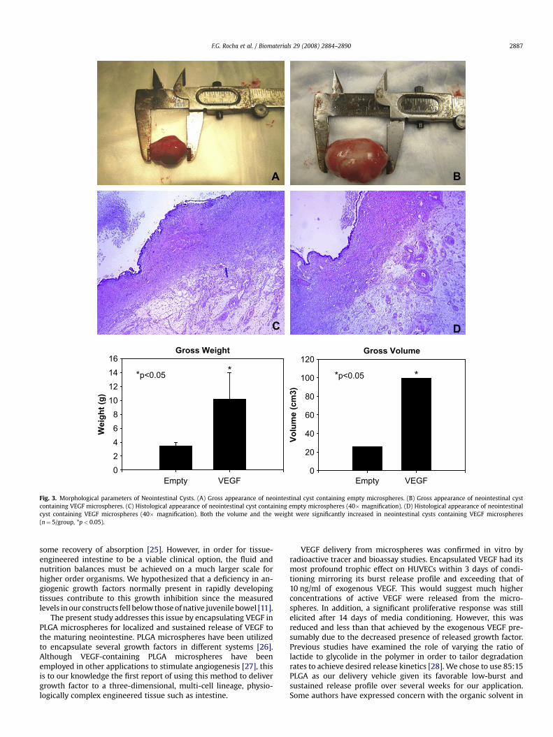
VEGF delivery was confirmed by measuring levels of VEGF inneointestinal constructs. Fig. 4 demonstrates that tissue VEGFlevels were significantly higher in neointestine loaded with
Days
0 5 10 15 20 25 30
Cu
mu
lative R
elease o
f V
EG
F
0
20
40
60
80
100
Fig. 1. Release kinetics of 125I-VEGF microspheres. After an initial burst release,microspheres released a steady state level of VEGF for up to 30 days (n¼ 3).
encapsulated VEGF compared to those without growth factor(106� 33 pg/mg vs. 28� 34 pg/mg) (p< 0.05). Neointestinalcapillaries were identified by CD31 (PECAM-1) immunopositivity,an endothelial cell marker (Fig. 4). In response to the higher tissueVEGF levels, capillary density in the muscular and connective tissuelayers of VEGF-containing neointestinal constructs was signifi-cantly increased compared to constructs containing microsphereswithout growth factor (29.8� 4.5 capillaries/hpf vs. 10.3�1.1capillaries/hpf) (p< 0.05).
3.4. In vivo epithelial response to released VEGF
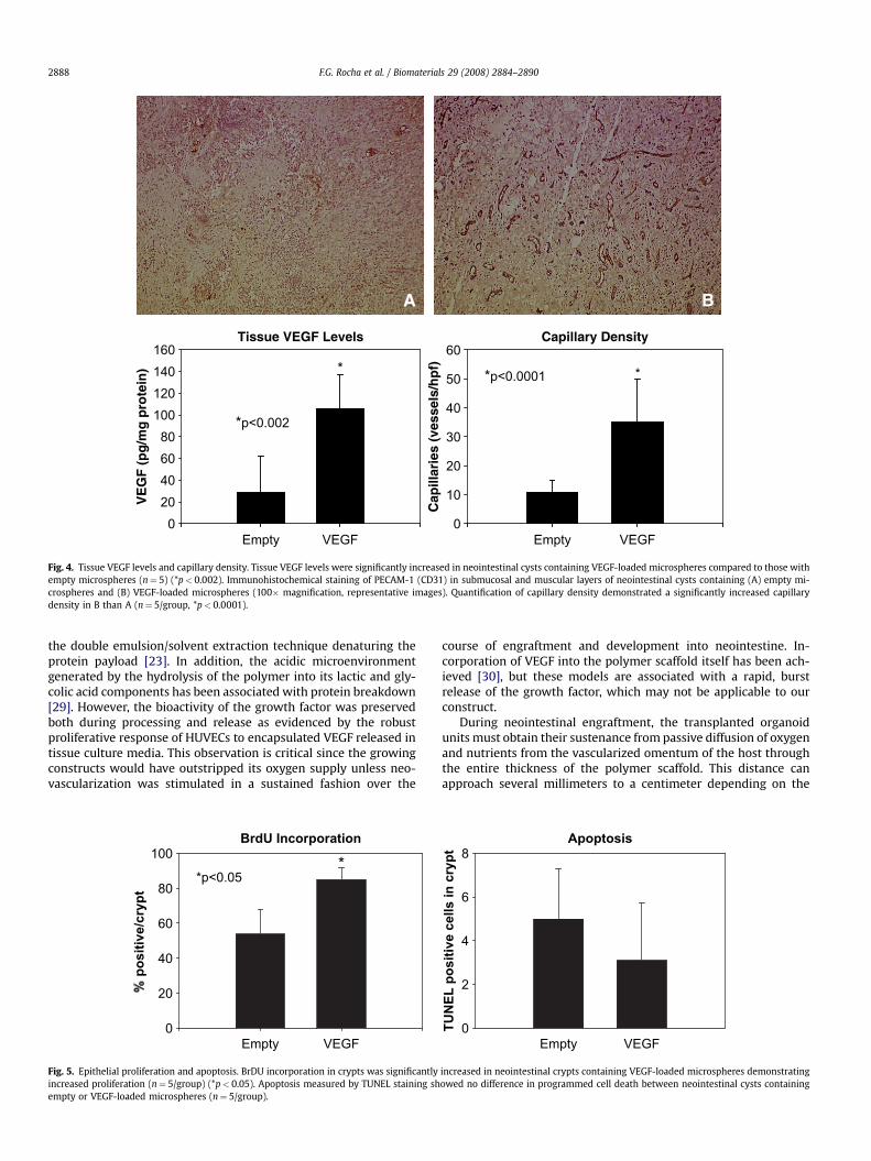
Neointestinal epithelial proliferation was quantified by mea-suring BrDU incorporation in crypts while apoptosis wasmeasured by TUNEL staining (Fig. 5). Constructs implanted withVEGF-releasing microspheres had increased rates of epithelialproliferation in neointestinal crypts compared to those implantedwith empty microspheres, 84.9�7.1% vs. 53.8� 13.8% BrDUstaining cells per crypt, respectively. However, apoptosis remainedunchanged between groups (5.0� 2.3 vs. 3.1�2.6 TUNEL stainingcells per crypt).
4. Discussion
One of the greatest challenges in engineering organs is the ade-quate delivery and maintenance of oxygen and nutrients to thedeveloping tissue. The biologic phenomenon of angiogenesis isparticularly critical in the early stages of engraftment and de-velopment when cells transplanted onto scaffolds must maintainmetabolic demands of cell division, tissue differentiation andphysiologic function. One of the current challenges of the tissue-engineered intestine is the difficulty in forming constructs longerthan a few centimeters that would be essential in order to expandabsorptive surface area. Previous studies have confirmed that im-plantation of neointestine in rats undergoing massive small bowelresection reverses weight loss in these animals indirectly suggesting
* *
Empty
Vo
lu
me (cm
3)
Weig
ht (g
)
VEGFEmpty VEGF
Gross VolumeGross Weight
*p<0.05*p<0.05
0
20
40
60
80
100
120
0246810121416
A B
C D
Fig. 3. Morphological parameters of Neointestinal Cysts. (A) Gross appearance of neointestinal cyst containing empty microspheres. (B) Gross appearance of neointestinal cystcontaining VEGF microspheres. (C) Histological appearance of neointestinal cyst containing empty microspheres (40� magnification). (D) Histological appearance of neointestinalcyst containing VEGF microspheres (40� magnification). Both the volume and the weight were significantly increased in neointestinal cysts containing VEGF microspheres(n¼ 5/group, *p< 0.05).
F.G. Rocha et al. / Biomaterials 29 (2008) 2884–2890 2887
some recovery of absorption [25]. However, in order for tissue-engineered intestine to be a viable clinical option, the fluid andnutrition balances must be achieved on a much larger scale forhigher order organisms. We hypothesized that a deficiency in an-giogenic growth factors normally present in rapidly developingtissues contribute to this growth inhibition since the measuredlevels in our constructs fell below those of native juvenile bowel [11].
The present study addresses this issue by encapsulating VEGF inPLGA microspheres for localized and sustained release of VEGF tothe maturing neointestine. PLGA microspheres have been utilizedto encapsulate several growth factors in different systems [26].Although VEGF-containing PLGA microspheres have beenemployed in other applications to stimulate angiogenesis [27], thisis to our knowledge the first report of using this method to delivergrowth factor to a three-dimensional, multi-cell lineage, physio-logically complex engineered tissue such as intestine.
VEGF delivery from microspheres was confirmed in vitro byradioactive tracer and bioassay studies. Encapsulated VEGF had itsmost profound trophic effect on HUVECs within 3 days of condi-tioning mirroring its burst release profile and exceeding that of10 ng/ml of exogenous VEGF. This would suggest much higherconcentrations of active VEGF were released from the micro-spheres. In addition, a significant proliferative response was stillelicited after 14 days of media conditioning. However, this wasreduced and less than that achieved by the exogenous VEGF pre-sumably due to the decreased presence of released growth factor.Previous studies have examined the role of varying the ratio oflactide to glycolide in the polymer in order to tailor degradationrates to achieve desired release kinetics [28]. We chose to use 85:15PLGA as our delivery vehicle given its favorable low-burst andsustained release profile over several weeks for our application.Some authors have expressed concern with the organic solvent in
**
Empty
Cap
illar
ies
(ves
sels
/hpf
)
VE
GF
(pg/
mg
prot
ein)
VEGFEmpty VEGF
Capillary DensityTissue VEGF Levels
*p<0.0001
*p<0.002
0
10
20
30
40
50
60
020406080100120140160
A B
Fig. 4. Tissue VEGF levels and capillary density. Tissue VEGF levels were significantly increased in neointestinal cysts containing VEGF-loaded microspheres compared to those withempty microspheres (n¼ 5) (*p< 0.002). Immunohistochemical staining of PECAM-1 (CD31) in submucosal and muscular layers of neointestinal cysts containing (A) empty mi-crospheres and (B) VEGF-loaded microspheres (100� magnification, representative images). Quantification of capillary density demonstrated a significantly increased capillarydensity in B than A (n¼ 5/group, *p< 0.0001).
F.G. Rocha et al. / Biomaterials 29 (2008) 2884–28902888
the double emulsion/solvent extraction technique denaturing theprotein payload [23]. In addition, the acidic microenvironmentgenerated by the hydrolysis of the polymer into its lactic and gly-colic acid components has been associated with protein breakdown[29]. However, the bioactivity of the growth factor was preservedboth during processing and release as evidenced by the robustproliferative response of HUVECs to encapsulated VEGF released intissue culture media. This observation is critical since the growingconstructs would have outstripped its oxygen supply unless neo-vascularization was stimulated in a sustained fashion over the
BrdU Incorporation
Empty
po
sitive/cryp
t
0
20
40
60
80
100
VEGF
**p<0.05
Fig. 5. Epithelial proliferation and apoptosis. BrDU incorporation in crypts was significantlyincreased proliferation (n¼ 5/group) (*p< 0.05). Apoptosis measured by TUNEL staining shempty or VEGF-loaded microspheres (n¼ 5/group).
course of engraftment and development into neointestine. In-corporation of VEGF into the polymer scaffold itself has been ach-ieved [30], but these models are associated with a rapid, burstrelease of the growth factor, which may not be applicable to ourconstruct.
During neointestinal engraftment, the transplanted organoidunits must obtain their sustenance from passive diffusion of oxygenand nutrients from the vascularized omentum of the host throughthe entire thickness of the polymer scaffold. This distance canapproach several millimeters to a centimeter depending on the
TU
NE
L p
ositive cells in
cryp
t
0
2
4
6
8
Empty VEGF
Apoptosis
increased in neointestinal crypts containing VEGF-loaded microspheres demonstratingowed no difference in programmed cell death between neointestinal cysts containing
F.G. Rocha et al. / Biomaterials 29 (2008) 2884–2890 2889
dimensions of the cylindrical scaffold dimensions and polymerspraying time of the PLLA coating. Since new blood vessel forma-tion occurs at a rate of less than 1 mm per day, local application ofan endothelial cell mitogen such as VEGF is required to accelerateneovessel growth and facilitate nutrient transport [31]. Increasedblood vessel formation was noted in the outer layers of the neo-intestine including in the host omentum suggesting trans-polymerdiffusion of released VEGF, although this was not directly measuredin this study. It has been demonstrated that pre-encapsulated VEGFin microspheres incorporated into the polymer scaffold yieldedVEGF concentrations of 10 ng/ml up to 2 cm away from the scaffold[32]. This approach may be able to support a larger intestinal seg-ment by providing a gradient of growth factor concentration overa wider area thereby supporting neovessels at progressive stages ofdevelopment.
Microsphere incubation with organoid units did not have anydetrimental effect on the morphology or ultrastructure of theresulting neointestinal construct. Spatial orientation of mucosa toserosa as well as villus–crypt architecture was preserved. Thissuggests the presence of degrading PLGA does not alter the at-tachment of the organoids to the PGA scaffold, nor the migration ofcells to their appropriate topographic sites (inner mucosa for epi-thelium and outer serosa for mesenchymal elements). Although theintercellular signaling for cell migration in organoid units trans-planted into scaffolds remains elusive, it seems that angiogenicstimuli are not involved.
A significant increase in the size and weight of the constructsexposed to VEGF microspheres was noted signifying a trophic effecton neointestinal tissues. This was confirmed by the presence oftissue VEGF levels that were significantly higher than controls andcomparable to those of juvenile native jejunum [11]. In addition,a higher density of capillary networks in both the muscular andserosal layers of the neointestine was achieved by the application ofVEGF-releasing microspheres. In rodents, as the juvenile intestinegrows rapidly during weaning, metabolic demands are met byaugmenting capillary networks in the bowel wall in order to sup-port the lengthening tissue [33]. Therefore, VEGF-containing neo-intestinal constructs are able to mimic the in vivo angiogenicmaturation of growing intestinal tissue. However, the functionalityof the engineered tissue when anastomosed to native gut and ac-tual bowel perfusion will require further investigation.
In response to increased vascularity and therefore greater accessto nutrients and oxygen, the neointestinal epithelium responded byupregulating cellular proliferation as evidenced by BrDU in-corporation into rapidly dividing progenitor cells in the cryptcompartment. No effect was noted in cellular apoptotic rates sug-gesting a lack of neoplastic transformation. This implies that theenhanced neointestine has the capability to regenerate its mucosathrough repopulation from crypt cells. Under normal conditions,the gut is constantly exposed to a harsh environment consisting ofosmotic loads, digestive enzymes, and bacterial toxins, but it is ableto survive despite these challenges. This is achieved by a constantturnover of villus tip cells and by migration of mucosal cells adja-cent to denuded areas. Further investigation will be needed todetermine if the angiogenic and regenerative properties of growthfactor enriched neointestine will translate into improved absorp-tive capacity and resistance to injury.
5. Conclusion
In summary, sustained polymeric delivery of VEGF with polymermicrospheres to the tissue-engineered intestine is feasible. Theproliferative effects on the capillary network and epithelial cryptsof the tissue-engineered intestine may serve to enhance its resil-ience and functional capacity. The ability to deliver other criticalgrowth factors such as bFGF and GLP-2 is likely to further optimize
the neointestine and may ultimately help to create an engineeredorgan of vital biologic function in desperate demand.
Acknowledgements
The authors wish to acknowledge the technical assistance of JanRounds. Portions of this manuscript were presented at the Ameri-can Gastroenterological Association/Digestive Disease Week An-nual Meeting, May 23, 2006. This work was funded by a NationalInstitutes of Health F-32 Grant DK65483 (FGR) and Association forAcademic Surgery/Karl Storz Endoscopy Research Fellowship (FGR).
References
[1] Warner BW, Chaet MS. Nontransplant surgical options for management of theshort bowel syndrome. J Pediatr Gastroenterol Nutr 1993;17:1–12.
[2] Thompson JS. Surgical considerations in the short bowel syndrome. SurgGynecol Obstet 1993;176:89–101.
[3] Byrne TA, Persinger RL, Young LS, Ziegler TR, Wilmore DW. A new treatmentfor patients with short-bowel syndrome: growth hormone, glutamine, anda modified diet. Ann Surg 1995;222:243–55.
[4] Scolapio JS, Camilleri M, Fleming CR, Oenning LV, Burton DD, Sebo TJ, et al.Effect of growth hormone, glutamine, and diet on adaptation in short bowelsyndrome: a randomized, controlled study. Gastroenterology 1997;113:1074–81.
[5] Todo S, Reyes J, Furukawa H, Abu-Elmagd K, Lee RG, Tzakis A, et al. Outcomeanalysis of 71 clinical intestinal transplantations. Ann Surg 1995;222:270–9.
[6] Leaseburge LA, Winn NJ, Schloerb PR. Liver test alterations with total paren-teral nutrition and nutritional status. JPEN J Parenter Enteral Nutr 1992;26:348–52.
[7] Howard L, Hassan N. Home parenteral nutrition: 25 years later. GastroenterolClin North Am 1998;27:481–512.
[8] Organ GM, Mooney DJ, Hansen LK, Schloo B, Vacanti JP. Transplantation ofenterocytes utilizing polymer-cell constructs to produce a neointestine.Transplant Proc 1992;24(6):3009–11.
[9] Choi RS, Riegler M, Pothoulakis C, Kim BS, Mooney D, Vacanti M, et al. Studiesof brush border enzymes, basement membrane components, and electro-physiology of tissue-engineered neointestine. J Pediatr Surg 1998;33(7):991–7.
[10] Tavakkolizadeh A, Berger UV, Stephen AE, Kim BS, Mooney D, Hediger MA,et al. Tissue-engineered neomucosa: morphology, enterocyte dynamics, andSGLT1 expression topography. Transplantation 2003;75:181–5.
[11] Gardner-Thorpe J, Grikscheit TC, Ito H, Perez A, Ashley SW, Vacanti JP, et al.Angiogenesis in tissue-engineered small intestine. Tissue Eng 2003;9:1255–61.
[12] Duxbury MS, Grikscheit TC, Gardner-Thorpe J, Rocha FG, Ito H, Perez A, et al.Transplantation 2004;77(8):1162–6.
[13] Perez A, Grikscheit TC, Blumberg RS, Ashley SW, Vacanti JP, Whang EE. Tissue-engineered small intestine: ontogeny of the immune system. Transplantation2002;74:619–23.
[14] Kim SS, Kaihara S, Benvenuto M, Choi RS, Kim BS, Mooney DJ, et al. Re-generative signals for tissue-engineered small intestine. Transplant Proc 1999;31:657–60.
[15] Ramsanahie A, Duxbury MS, Grikscheit TC, Perez A, Rhoads DB, Gardner-Thorpe J, et al. Effect of GLP-2 on mucosal morphology and SGLT1 expressionin tissue-engineered neointestine. Am J Physiol Gastrointest Liver Physiol2003;285(6):G1345–52.
[16] Folkman J, Shing Y. Angiogenesis. J Biol Chem 1992;267:10931–4.[17] Banai S, Jaklitsch MT, Shou M, Lazarous DF, Scheinowitz M, Biro S, et al.
Angiogenic-induced enhancement of collateral blood flow to ischemic myo-cardium by vascular endothelial growth factor in dogs. Circulation 1994;89:2183–9.
[18] Meirer R, Gurunlouglu R, Siemionow M. Neurogenic perspective on vascularendothelial growth factor: review of the literature. J Reconstr Microsurg 2001;17(8):625–30.
[19] Burgess WH, Maciag T. The heparin-binding (fibroblast) growth factor familyof proteins. Annu Rev Biochem 1989;58:575–606.
[20] Lazarous DF, Shou M, Scheinowitz M, Hodge E, Thirumurti V, Kitsiou AN, et al.Comparative effects of basic fibroblast growth factor and vascular endothelialgrowth factor on coronary collateral development and arterial response toinjury. Circulation 1996;94:1074–82.
[21] Rosca ID, Watari F, Uo M. Microparticle formation and its mechanism in singleand double emulsion fashion. J Controlled Release 2004;99:271–80.
[22] Evans GS, Flint N, Somers AS, Eyden B, Potten CS. The development ofa method for the preparation of rat intestinal epithelial cell primary cultures.J Cell Sci 1992;101:219–31.
[23] King TW, Patrick CW. Development and in vitro characterization of vascularendothelial growth factor (VEGF)-loaded poly(DL-lactic-co-glycolic acid)/poly(ethylene glycol) microspheres using a solid encapsulation/single emul-sion/solvent technique. J Biomed Mater Res 2000;51(3):383–90.
[24] Ishii H, Oota I, Takuma T, Inomata K. Developmental expression of vascularendothelial growth factor in the masseter muscle of rats. Arch Oral Biol 2001;46:77–82.
F.G. Rocha et al. / Biomaterials 29 (2008) 2884–28902890
[25] Grikscheit TC, Siddique A, Ochoa E, Srinivasan A, Alsberg E, Hodin RA, et al.Tissue-engineered small intestine improves recovery after massive smallbowel resection. Ann Surg 2004;240(5):748–54.
[26] Cohen S, Yoshioka T, Lucarelli M, Hwang LH, Langer R. Controlled deliverysystems for proteins based on poly(lactic/glycolic acid) microspheres. PharmRes 1991;8:713–20.
[27] Cleland JL, Duenas ET, Park A, Daugherty A, Kahn J, Kowalski J, et al. De-velopment of poly-(D,L-lactide-coglycolide) microsphere formulations con-taining recombinant human vascular endothelial growth factor to promotelocal angiogenesis. J Controlled Release 2001;72:13–24.
[28] Chung TW, Tsai YL, Hsieh JH, Tsai WJ. Different ratios of lactide and glycolide inPLGA affect the surface property and protein delivery characteristics of the PLGAmicrospheres with hydrophobic additives. J Microencapsul 2006;23(1):15–27.
[29] Fu K, Pack DW, Klivanov AM, Langer R. Visual evidence of acidic environmentwithin degrading poly(lactic-co-glycolic acid) (PLGA) microspheres. PharmRes 2000;17:100–6.
[30] Murphy WL, Peters MC, Kohn DH, Mooney DJ. Sustained release of vascularendothelial growth factor from mineralized poly(lactide-co-glycolide) scaf-folds for tissue engineering. Biomaterials 2000;21:2521–7.
[31] Mikos AG, Sarakinos G, Ingber DE, Vacanti J, Langer R. Prevascularization ofporous biodegradable polymer sponges. Biotech Bioeng 1993;42:716–23.
[32] Ennett AB, Kaigler D, Mooney DJ. Temporally regulated delivery of VEGFin vitro and in vivo. J Biomed Mater Res 2006;79(1):176–84.
[33] Unthank JL, Lash JM, Bohlen HG. Maturation of the rat intestinal microvas-culature from juvenile to early adult life. Am J Physiol Gastrointest Liver 1990;259:G282–9.
Top Related
Copyright © 2022 FDOKUMEN

