


Bahasa
Halaman
Hukum


Mutation Research 634 (2007) 60–68
Available online at www.sciencedirect.com
The chlorophenoxy herbicide dicamba and its commercialformulation banvel® induce genotoxicity and cytotoxicity
in Chinese hamster ovary (CHO) cells
Norma V. Gonzalez, Sonia Soloneski, Marcelo L. Larramendy ∗Catedra de Citologıa, Facultad de Ciencias Naturales y Museo, Universidad Nacional de La Plata, La Plata, Argentina
Received 21 December 2006; received in revised form 5 June 2007; accepted 8 June 2007Available online 17 June 2007
Abstract
The sister chromatid exchange (SCE) frequency, the cell-cycle progression analysis, and the single cell gel electrophoresistechnique (SCGE, comet assay) were employed as genetic end-points to investigate the geno- and citotoxicity exerted by dicamba andone of its commercial formulation banvel® (dicamba 57.71%) on Chinese hamster ovary (CHO) cells. Log-phase cells were treatedwith 1.0–500.0 �g/ml of the herbicides and harvested 24 h later for SCE and cell-cycle progression analyses. All concentrationsassessed of both test compounds induced higher SCE frequencies over control values. SCEs increased in a non-dose-dependentmanner neither for the pure compound (r = 0.48; P > 0.05) nor for the commercial formulation (r = 0.58, P > 0.05). For the 200.0 �g/mland 500.0 �g/ml dicamba doses and the 500.0 �g/ml banvel® dose, a significant delay in the cell-cycle progression was found. Aregression test showed that the proliferation rate index decreased as a function of either the concentration of dicamba (r = −0.98,P < 0.05) or banvel® (r = −0.88, P < 0.01) titrated into cultures in the 1.0–500.0 �g/ml dose-range. SCGE performed on CHOcells after a 90 min pulse-treatment of dicamba and banvel® within a 50.0–500.0 �g/ml dose-range revealed a clear increase indicamba-induced DNA damage as an enhancement of the proportion of slightly damaged and damaged cells for all concentrationsused (P < 0.01); concomitantly, a decrease of undamaged cells was found over control values (P < 0.01). In banvel®-treated cells,
a similar overall result was registered. Dicamba induced a significant increase both in comet length and width over control values(P < 0.01) regardless of its concentration whereas banvel® induced the same effect only within 100.0–500.0 �g/ml dose range(P < 0.01). As detected by three highly sensitive bioassays, the present results clearly showed the capability of dicamba and banvel®to induce DNA and cellular damage on CHO cells.© 2007 Elsevier B.V. All rights reserved.
omatid
Keywords: Dicamba; Banvel®; Chlorophenoxy herbicides; Sister chr1. Introduction
Dicamba is a selective systemic herbicide, absorbedby the leaves and roots, acts as an auxin-like growth
∗ Corresponding author at: Facultad de Ciencias Naturales y Museo,Calle 64 Nro. 3 esq. 120, 1900 La Plata, Argentina.Tel.: +54 221 424 9049.
E-mail address: m [email protected] (M.L. Larramendy).
1383-5718/$ – see front matter © 2007 Elsevier B.V. All rights reserved.doi:10.1016/j.mrgentox.2007.06.001
exchanges; Cell-cycle kinetics; Comet assay
regulator causing uncontrolled growth. It is used to con-trol annual and perennial broad-leaved weeds and bushspecies, e.g. cereals, maize, sorghum, sugarcane, aspara-gus, perennial seed grasses, turf, pastures, rangeland,and non-crop land [1]. Being ranked as class III chem-
ical (slightly hazardous) by WHO (http://www.who.int/ipcs/publications/pesticides hazard/en/) and included incategory D (not classifiable as to human carcinogenicityby EPA [2], dicamba has been proved to have a promot-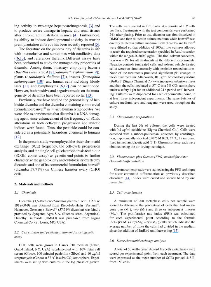
tion Re
itaip
w(bd(pmlHg
bfwiais[
ea(cd(c
2
2
1HpDC
2a
Gssi
N.V. Gonzalez et al. / Muta
ng activity in two-stage hepatocarcinogenesis [3] ando produce severe damage in hepatic and renal tissuesfter chronic administration in mice [4]. Furthermore,ts ability to induce developmental toxicity in murinereimplantation embryos has been recently reported [5].
The literature on the genotoxicity of dicamba is rifeith inconclusive and sometimes with conflictive data
[6,13], and references therein). Different assays haveeen performed to study the mutagenicity properties oficamba. Among them, bioassays employing bacteriaBacillus subtilis rec A [8], Salmonella typhimurium [9]),lants (Arabidopsis thaliana [7]), insects (Drosophilaelanogaster [10]) and human cells including fibrob-
asts [11] and lymphocytes [6,12] can be mentioned.owever, both positive and negative results on the muta-enicity of dicamba have been reported so far [13].
Previously, we have studied the genotoxicity of her-icide dicamba and the dicamba containing commercialormulation banvel® in in vitro human lymphocytes. Weere able to demonstrate that dicamba is a DNA damag-
ng agent since enhancement of the frequency of SCEs,lterations in both cell-cycle progression and mitoticndices were found. Thus, the pesticide could be con-idered as a potentially hazardous chemical to humans12].
In the present study we employed the sister chromatidxchange (SCE) frequency, the cell-cycle progressionnalysis, and the single cell gel electrophoresis techniqueSCGE, comet assay) as genetic end-points to furtherharacterize the genotoxicity and cytotoxicity exerted byicamba and one of its commercial formulation banvel®
dicamba 57.71%) on Chinese hamster ovary (CHO)ells.
. Materials and methods
.1. Chemicals
Dicamba (3,6-Dichloro-2-methoxybenzoic acid, CAS n◦
918-00-9) was obtained from Riedel-de-Haen (Pestanal®;annover, Germany). Banvel® (57.71% dicamba) was kindlyrovided by Syngenta Agro S.A. (Buenos Aires, Argentina).imethyl sulfoxide (DMSO) was purchased from Sigmahemical Co. (St. Louis, MO, USA).
.2. Cell cultures and pesticide treatment for cytogeneticssay
CHO cells were grown in Ham’s F10 medium (Gibco,rand Island, NY, USA) supplemented with 10% fetal calf
erum (Gibco), 100 units/ml penicillin (Gibco) and 10 �g/mltreptomycin (Gibco) at 37 ◦C in a 5% CO2 atmosphere. Exper-ments were set up with cultures in the log phase of growth.
search 634 (2007) 60–68 61
The cells were seeded in T75 flasks at a density of 106 cellsper flask. Treatments with the test compounds were performed24 h after plating. Prior to use, dicamba was first dissolved inDMSO and then diluted in culture medium while banvel® wasdirectly diluted in culture medium. Both dicamba and banvel®
were diluted so that addition of 100 �l into cultures allowedto reach the required concentration specified in Results sectionwithin the range 0.0–500.0 �g/ml. The final solvent concentra-tion was <1% for all treatments in the different experiments.Negative controls (untreated cells and solvent vehicle-treatedcells) were run simultaneously with pesticide-treated cultures.None of the treatments produced significant pH changes inthe culture medium. Afterwards, 10 �g/ml bromodeoxyuridine(BrdUrd) (Sigma Chemical Co.) was incorporated into culturesand then the cells incubated at 37 ◦C in a 5% CO2 atmosphereunder a safety light for an additional 24 h period until harvest-ing. Cultures were duplicated for each experimental point, inat least three independent experiments. The same batches ofculture medium, sera and reagents were used throughout thestudy.
2.3. Chromosome preparations
During the last 3 h of culture, the cells were treatedwith 0.2 �g/ml colchicine (Sigma Chemical Co.). Cells weredetached with a rubber-policeman, collected by centrifuga-tion, hypotonically shocked (0.075 M KCl, 37 ◦C, 17 min) andfixed in methanol/acetic acid (3:1). Chromosome spreads wereobtained using the air-drying technique.
2.4. Fluorescence plus Giemsa (FPG) method for sisterchromatid differentiation
Chromosome spreads were stained using the FPG techniquefor sister chromatid differentiation as previously describedelsewhere [14]. Slides were coded and scored blind by oneresearcher.
2.5. Cell-cycle kinetics
A minimum of 200 metaphase cells per sample werescored to determine the percentage of cells that had under-gone one (M1), two (M2) and three or subsequent mitoses(M3+). The proliferative rate index (PRI) was calculatedfor each experimental point according to the formulaPRI = [(%M1) + 2(%M2) + 3(%M3+)]/100, which indicated theaverage number of times the cells had divided in the mediumsince the addition of BrdUrd until harvesting [15].
2.6. Sister chromatid exchange analysis
A total of 50 well-spread diploid M2 cells metaphases werescored per experimental point from each treatment. The datawere expressed as the mean number of SCEs per cell ± S.E.from 150 cells.
tion Re
62 N.V. Gonzalez et al. / Muta2.7. Cell cultures and pesticide treatment for single cellgel electrophoresis (SCGE)
Prior to test chemical treatment, exponentially CHO cellwere detached by a rubber-policeman, collected by centrifu-gation, resuspended in complete culture medium, and thencounted. Afterwards, aliquots containing 3.5 × 10⁵ cells/mlwere incubated for 90 min at 37 ◦C in a 5% CO2 atmo-sphere in culture medium containing the test compounds.Both dicamba and banvel® were used at a final concentrationbetween 50.0 and 500.0 �g/ml. The final solvent concen-tration was <1% for all the treatments in all experiments.Negative controls (untreated cells and solvent vehicle-treatedcells) were run simultaneously with pesticide-treated cultures.None of the treatments produced significant pH changes inthe culture medium. Thereafter, the cells were washed twicewith pesticide-free complete culture medium. The SCGE andcell viability assays were performed immediately after the90 min treatment. Cultures were duplicated for each experi-mental point, during at least three independent experiments.The same batches of cultures medium, sera and reagents wereused throughout the study.
2.8. Cell viability assay
Cell viability was determined using the ethidium bro-mide/acridine orange assay described elsewhere [16]. Briefly,one aliquot of 5 �l of a 1:1 freshly prepared mixture of ethidiumbromide (100 �g/ml, Sigma Chemical Co.) and acridine orange(100 �g/ml, Sigma Chemical Co.) was mixtured with 50 �l ofthe cell suspension. Afterwards, cells were analyzed using anOlympus BX50 fluorescence photomicroscope equipped withan appropriate filter combination. Viable cells appeared green-fluorescent whereas orange-stained nuclei indicated dead cells.At least, 500 cells were counted per experimental point, andresults expressed as percentage of viable cells among allcells. Cell viability was expressed as proportion of livingcells.
2.9. Single cell gel electrophoresis (SCGE) assay
The SCGE assay was performed following the alkaline pro-cedure described by Singh and collaborators [17] with minormodifications. Briefly, two solutions containing 0.5% normalmelting agarose (NMA) and 0.5% low melting agarose (LMA)solution in Ca2+–Mg2+-free PBS were performed. Seventy-fivemicroliters NMA were transferred onto 100% ethanol pre-cleaned slide, spread evenly, and placed at 37 ◦C for 20–30 min.Afterwards, 95 �l of LMA together with 7.0 × 10³ cells (20 �lcell suspension + 75 �l LMA) was applied, covered with acoverslip and placed at 4 ◦C for 15 min. After this layer had
solidified, a third layer of 75 �l LMA was added and slideswere immersed in ice-cold freshly prepared lysis solution (1%sodium sarcocinate, 2.5 M NaCl, 100 mM Na2EDTA, 10 mMTris pH 10.0, 1% Triton X-100, 10% DMSO) and then lysedat darkness for an overnight period (4 ◦C). When this periodsearch 634 (2007) 60–68
concluded, slides were placed in a horizontal electrophore-sis buffer (1 mM Na2EDTA, 300 mM NaOH) for 20 min at4 ◦C to allow the cellular DNA to unwind, followed by elec-trophoresis in the same buffer and temperature for 20 minat 25 V and 250 mA. Finally, the slides were neutralizedwith a solution comprising 0.4 M Tris–HCl, pH 7.5, stainedwith 4′6-diamino-2-phenylindole (DAPI) (Vectashield mount-ing medium H1200; Vector Laboratories, Burlingame, CA,USA). Slides were coded and scored blind by one cytogenetist.Analysis of the slides was performed under an OlympusBX50 fluorescence photomicroscope equipped with an appro-priate filter combination. The cellular nucleus diameters of thenucleus plus migrated DNA were individually measured usinga calibration scale with an X63 fluorescence objective from50 randomly selected cells per experimental point for eachexperiment. Cells were visually graded into four categoriesas suggested elsewhere [18,19] depending on DNA damagelevel as undamaged, slightly damaged, damaged, and highlydamaged, respectively.
2.10. Statistical analysis
The two-tailed Student’s t-test was used to compare SCEfrequencies and measures from comets between treated andcontrol groups. A χ2-test was employed for cell-cycle pro-gression. The non-parametric Kruskal–Wallis test was used tocompare the effect of a pesticide over control cells in cometassay experiments while individual comparisons between pairsof data were performed using the Mann–Whitney test. The levelof significance chosen was 0.05 unless otherwise indicated.
3. Results
Since no difference of cell-cycle progression, SCEsand PRI values were observed between negative con-trols (untreated and DMSO-treated cells), pooled dataare presented for control values.
Fig. 1 shows the results of SCE analysis in CHOcells treated for 24 h with different doses of dicamba orthe dicamba-containing commercial herbicide banvel®.The SCE frequencies observed either in dicamba- orbanvel®-treated cultures were significantly higher thanthose of control cultures (P < 0.05, P < 0.01, respec-tively). SCE frequencies induced by the pure compoundand the commercial formulation increased in a non-dose-dependent manner (r = 0.48, P > 0.05, and r = 0.58,P > 0.05 for dicamba and banvel®, respectively). Themaximal SCEs increase over control values wasdetected when a 200.0 �g/ml dicamba dose was tested(12.55 ± 0.55 SCEs/cell versus 9.33 ± 0.51 SCEs/cell)
(Fig. 1). In the banvel®-treated cultures the highest SCEsvalue was observed when a 500.0 �g/ml dose was used(13.40 ± 0.55 SCEs/cell versus 9.47 ± 0.50 SCEs/cell)(Fig. 1).
N.V. Gonzalez et al. / Mutation Research 634 (2007) 60–68 63
Fig. 1. Effect of the chlorophenoxy dicamba (black bars) and its com-mercial formulation banvel® (empty bars) on SCE frequency fromCHO cells. Cultures were harvested at 24 h from pesticide treatmentand the frequency of SCEs were determined in 50 M2 mitoses foreiad
otdcd(qws5hMpdd
Fig. 3. Effect of the chlorophenoxy dicamba and its commercial for-mulation banvel® on proliferative rate index (PRI) from CHO cells.Cultures were harvested at 24 h from pesticide treatment and the PRIswere determined in 300 mitoses for each experimental point. For each
Fhce*
ach experimental point. For each herbicide, pool data from threendependent experiments are reported as mean SCE values ± S.E. (y-xis) and plotted against the herbicide concentration (0.0–500.0 �g/mlose-range; x-axis). *P < 0.05; **P < 0.01.
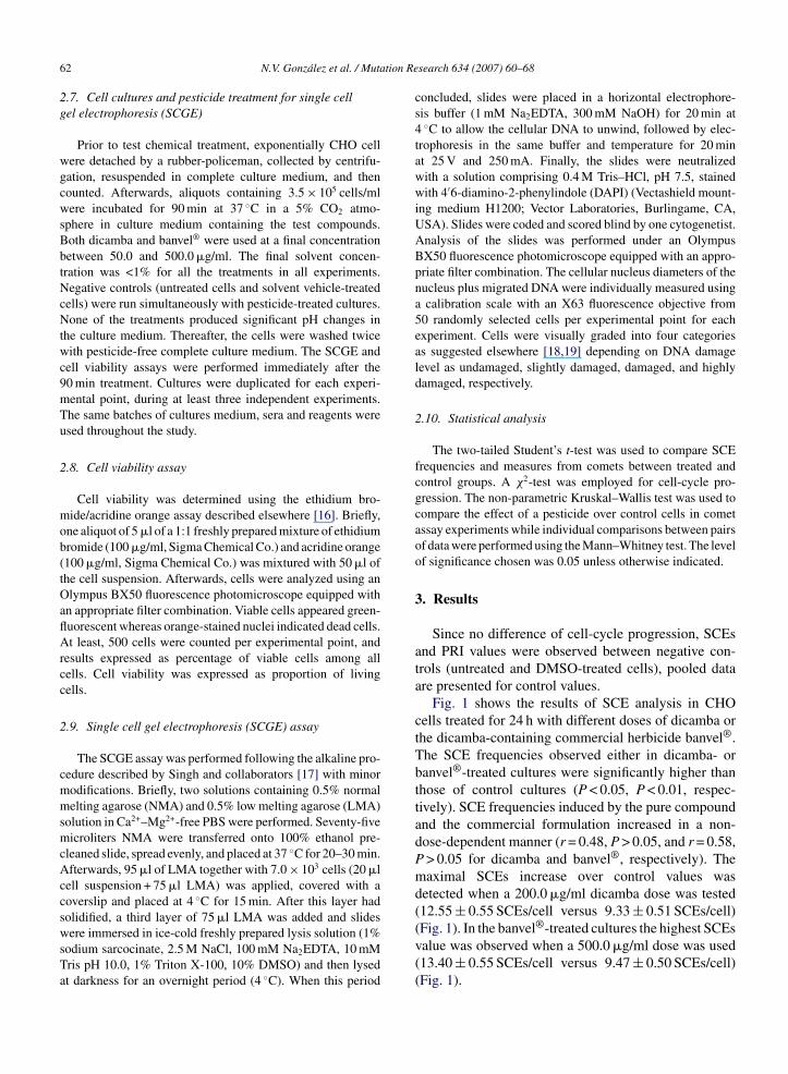
Cytotoxicity, measured as cell-cycle kinetics, wasbserved in those dicamba- (Fig. 2A) and banvel®-reated (Fig. 2B) cultures since both a significantelay in cell-cycle progression (Fig. 2) and a signifi-ant reduction of the PRI were induced (Fig. 3). Foricamba, a significant increase in the frequency of M1P < 0.001) and a significant decrease in the M2 fre-uency (P < 0.001) were registered in cultures titratedith both 200.0 �g/ml and 500 �g/ml (Fig. 2A). A
imilar effect was only found in cultures treated with00.0 �g/ml banvel® (P < 0.001) (Fig. 2B). On the otherand, no significant alteration in the frequencies of
1, M2, and M3+ was observed for both test com-ounds within 1.0–100.0 �g/ml and 1.0–200.0 �g/ml oficamba and banvel®, respectively (P > 0.05) (Fig. 2). Aecrease in the PRI was observed in cultures treated with
ig. 2. Effect of the chlorophenoxy dicamba and its commercial formulatioarvested at 24 h from pesticide treatment and the proportion of cells in first (ell divisions (black circles, M3+) were determined in 200 mitoses for each expxperiments are reported as mean frequencies ± S.E. (y-axis) and plotted agaiP < 0.01.
herbicide, pool data from three independent experiments are reportedas mean frequencies ± S.E. (y-axis) and plotted against the herbicideconcentration (0.0–500.0 �g/ml dose-range; x-axis). *P < 0.05.
200.0–500.0 �g/ml and 500.0 �g/ml of dicamba andbanvel®, respectively. However, statistical significancewas only achieved after treatment with 500.0 �g/ml ofdicamba (P < 0.05) (Fig. 3). A regression test showedthat the PRI decreased as a function of either theconcentration of the dicamba (r = −0.98, P < 0.05) orbanvel® (r = −0.88, P < 0.01) titrated into cultures in the1.0–500.0 �g/ml dose-range (Fig. 3).
Table 1 summarizes the results of the SCGE per-formed on CHO cells after a 90 min pulse-treatmentof dicamba and banvel® in a 50.0–500.0 �g/ml
dose-range. Since no differences of frequency of undam-aged/damaged cells were observed between negativecontrols (untreated and DMSO-treated cells) pooled dataare presented for control cultures. The SCGE revealedn banvel® on cell-cycle progression from CHO cells. Cultures werewhite circles, M1), second (gray circles, M2), and third or subsequenterimental point. For each herbicide, pool data from three independentnst the herbicide concentration (0.0–500.0 �g/ml dose-range; x-axis).

64 N.V. Gonzalez et al. / Mutation Research 634 (2007) 60–68
Table 1Frequencies of undamaged, slightly damaged (non-migrant) and damaged (migrant) in control, dicamba- and banvel®-treated CHO cellsa
Doses (�g/ml) Number of cells examined Percentage of cellsb Viabilityc
Undamaged Slightly damaged Damaged
Dicamba0 300 80.33 ± 0.33 19.66 ± 0.33 0.00 ± 0.00 96.50 ± 0.50
50 150 10.00 ± 1.33* 77.00 ± 2.33* 13.00 ± 3.67* 96.00 ± 2.10100 150 9.00 ± 3.00* 73.00 ± 2.33* 18.00 ± 3.33* 91.00 ± 1.20200 150 8.00 ± 0.67* 81.00 ± 1.00* 11.00 ± 0.33* 96.75 ± 0.75500 150 7.50 ± 0.33* 85.00 ± 0.33* 7.50 ± 0.33* 91.75 ± 2.50
Banvel®
0 300 81.33 ± 0.67 17.67 ± 0.67 1.00 ± 0.67 96.50 ± 0.5050 150 11.00 ± 2.33* 77.00 ± 0.33* 12.00 ± 2.67* 92.50 ± 1.40
100 150 9.00 ± 1.67* 72.00 ± 2.00* 19.00 ± 0.33* 93.50 ± 1.50200 150 13.00 ± 4.33* 79.00 ± 3.67* 8.00 ± 0.67* 90.50 ± 2.50500 150 1.00 ± 0.33* 75.00 ± 1.67* 24.00 ± 1.33* 91.00 ± 1.20
a Cells were treated with test compounds and harvested 24 h thereafter, and processed following procedure for SCGE. Electrophoresis wasperformed at 4 ◦C for 20 min at 25 V and 250 mA. Cells were stained with DAPI.
b Undamaged cells, width and length <30 �m; slightly damaged cells, width and length 31–45 �m; damaged cells, width and length >45 �m;
results are expressed as mean values ± S.E. of the mean.c Results are expressed as mean values ± S.E. of the mean.* P < 0.01.
a clear increase in dicamba-induced DNA damage asan enhancement of the proportion of slightly damagedand damaged cells for all concentrations used (P < 0.01);concomitantly, a decrease of undamaged cells was foundover control values (P < 0.01). In banvel®-treated cells,a similar overall result was registered since a signifi-cant increase in the frequency of slightly damaged anddamaged cells for all concentrations used (P < 0.01). Theresults demonstrated that cultures showed the lowestproportion of undamaged cells and the highest fre-quency of damaged cells when treated with 500.0 �g/mlbanvel®. Furthermore SCGE revealed that banvel® ata dose of 500.0 �g/ml induced a seven-fold decreasein the frequency of undamaged cells and a three-foldenhancement of damaged cells with regard to the samedicamba concentration (Table 1). Effects of dicambaand banvel® concentrations on DNA damage, measuredby comet length and width, are presented in Fig. 4.Dicamba induced a significant increase both in cometlength (Fig. 4A) and width (Fig. 4B) over control val-ues (P < 0.01) regardless of its concentration. Banvel®
induced significant enhancement over control values(P < 0.01) either in comet length (Fig. 4C) or comet width(Fig. 4D) in the 100.0–500.0 �g/ml dose range. A regres-sion analysis revealed a positive relationship betweendicamba concentration and the frequency of DNA-
strand breaks as measured as comet length (r = 0.81,P < 0.01). On the other hand, no statistical associa-tion was observed between dicamba concentrations andcomet width (r = 0.58, P > 0.05), and between banvel®concentrations and comet length (r = 0.29, P > 0.05) orwidth (r = 0.29, P > 0.05).
4. Discussion
The aim of this study was to evaluate the geno-toxic and cytotoxic effects of dicamba and its technicalformulation banvel® in CHO cells. The investigationwas conducted using the sister chromatid exchange(SCE) frequency, the cell-cycle progression analysis,and the SCGE assay as genetic end-points. Both chem-icals induced higher SCE frequencies and a delay inthe cell-cycle progression compared to control values,as well as DNA-strand breaks revealed by the cometassay. Thus, either dicamba- or banvel®-treated cultures’results found, at least under the experimental conditionsused in the present work, showed the ability of thesecompounds to induce genotoxicity and cytotoxicity invitro.
Diverse genetic end-points and test-systems havebeen utilized for research on dicamba, and results emerg-ing from those investigations, although being diverse,have been also inconclusive. Specifically concerning ani-mal systems, dicamba has been demonstrated to causean increase in SCE frequency in human peripheral bloodlymphocytes in vitro [6,12] but failed to induce genetic
damage such as structural chromosomal aberrations inrat bone marrow cells [13] or sex-linked recessive lethalmutations in D. melanogaster [20]. CHO cells havebeen recently applied in pesticides soil clays-mediated
N.V. Gonzalez et al. / Mutation Research 634 (2007) 60–68 65
Fig. 4. Effect of the chlorophenoxy dicamba (A and B) and its commercial formulation banvel® (C and D) on the length of the comet tails (A–C)and on the diameter (B–D) at the estimated trailing edge of comet tails in CHO cells. Cultures were harvested at 90 min from herbicide treatmenta Electroc 0 cellsi d plottex
tawsa
biristenFt1ttt
nd processed following procedure for single cell gel electrophoresis.ells stained with DAPI. DNA migration (�m) were determined in 15ndependent experiments are reported as DNA migration (y-axis) an-axis). *P < 0.01.
oxicity assessments, including dicamba among othersgricultural agents [21,22]. In the current investigation,e have combined the extensively used CHO in vitro
ystem and three highly sensitive bioassays for geno-nd cytotoxicity [23,24].
Our results demonstrated that all dicamba andanvel® doses titrated into CHO cultures induced a sim-lar level of SCE frequency in a non-dose-dependentelationship. This is in contrast with our previous find-ngs on human lymphocytes in vitro which exhibitedimilar augments only for the 200.0 �g/ml dicamba andhe 500.0 �g/ml banvel® doses. Furthermore, the high-st concentration tested in CHO cells (500.0 �g/ml) didot cause cell-death as it resulted in human lymphocytes.urthermore, it is worth mentioning that the latter when
reated with doses ranging from 10.0 to 100.0 �g/ml and
0.0 to 200.0 �g/ml of dicamba and banvel®, respec-ively, failed to show any sign of cytotoxicity [12]. Thus,he survival of CHO cells together with their higher resis-ance to dicamba and banvel® deleterious effects whenphoresis was performed at 4 ◦C for 20 min at 25 V and 250 mA, andfor each experimental point. For each herbicide, pool data from threed against the herbicide concentration (0.0–500.0 �g/ml dose-range;
compared to human lymphocytes, point out that this cellline constitutes a well suited system to investigate thesechlorobenzoic compounds. However, the possibility thathuman lymphocytes are able to repair the damage intro-duced into their DNA by low doses of the pesticideduring G0 cannot be ruled out.
To our knowledge this is the first time the cell-cyclekinetics is used as an alternative approach to quantifydicamba and banvel® cytotoxicity. The results presentedhere showed that pure pesticide treatment of 200.0 �g/mland 500.0 �g/ml delayed significantly the cell-cycle pro-gression and produced an overall valuable PRI reduction,results obtained for the maximal banvel® concentrationassayed only. These three test compound doses rendereda matched combination of M1 increase and M2 reductionthat differed from our previous results on human lym-
phocytes in vitro which exhibited a varied combinationof M1, M2, and M3+ reductions and/or increases [12].Plausible explanation for this discrepancy could be mostprobably related to differences both in the harvesting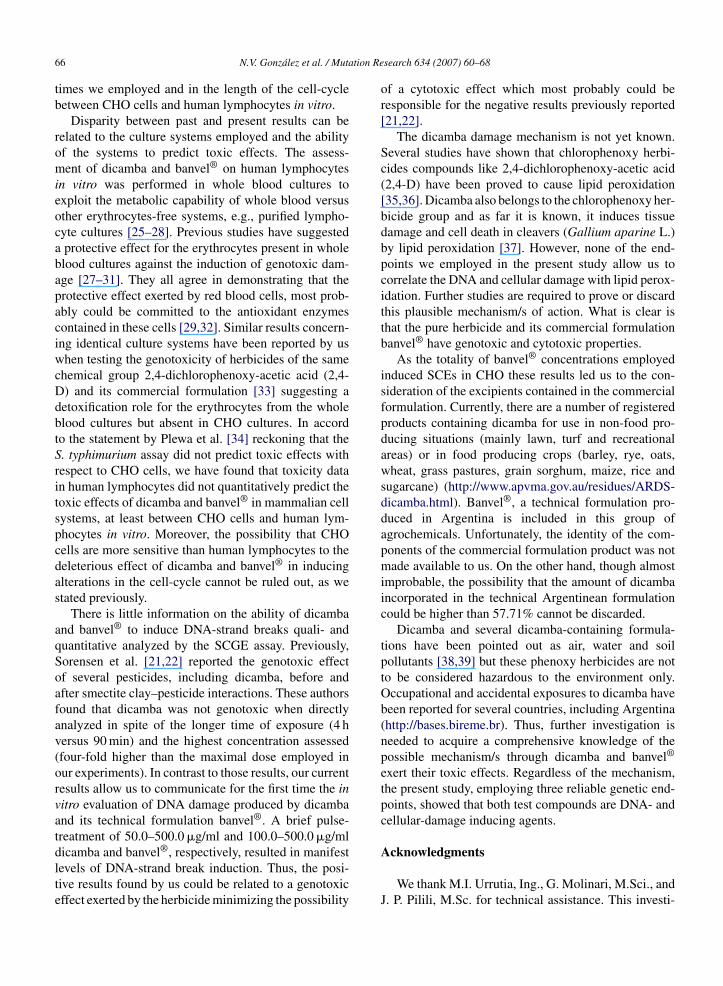
tion Re
66 N.V. Gonzalez et al. / Mutatimes we employed and in the length of the cell-cyclebetween CHO cells and human lymphocytes in vitro.
Disparity between past and present results can berelated to the culture systems employed and the abilityof the systems to predict toxic effects. The assess-ment of dicamba and banvel® on human lymphocytesin vitro was performed in whole blood cultures toexploit the metabolic capability of whole blood versusother erythrocytes-free systems, e.g., purified lympho-cyte cultures [25–28]. Previous studies have suggesteda protective effect for the erythrocytes present in wholeblood cultures against the induction of genotoxic dam-age [27–31]. They all agree in demonstrating that theprotective effect exerted by red blood cells, most prob-ably could be committed to the antioxidant enzymescontained in these cells [29,32]. Similar results concern-ing identical culture systems have been reported by uswhen testing the genotoxicity of herbicides of the samechemical group 2,4-dichlorophenoxy-acetic acid (2,4-D) and its commercial formulation [33] suggesting adetoxification role for the erythrocytes from the wholeblood cultures but absent in CHO cultures. In accordto the statement by Plewa et al. [34] reckoning that theS. typhimurium assay did not predict toxic effects withrespect to CHO cells, we have found that toxicity datain human lymphocytes did not quantitatively predict thetoxic effects of dicamba and banvel® in mammalian cellsystems, at least between CHO cells and human lym-phocytes in vitro. Moreover, the possibility that CHOcells are more sensitive than human lymphocytes to thedeleterious effect of dicamba and banvel® in inducingalterations in the cell-cycle cannot be ruled out, as westated previously.
There is little information on the ability of dicambaand banvel® to induce DNA-strand breaks quali- andquantitative analyzed by the SCGE assay. Previously,Sorensen et al. [21,22] reported the genotoxic effectof several pesticides, including dicamba, before andafter smectite clay–pesticide interactions. These authorsfound that dicamba was not genotoxic when directlyanalyzed in spite of the longer time of exposure (4 hversus 90 min) and the highest concentration assessed(four-fold higher than the maximal dose employed inour experiments). In contrast to those results, our currentresults allow us to communicate for the first time the invitro evaluation of DNA damage produced by dicambaand its technical formulation banvel®. A brief pulse-treatment of 50.0–500.0 �g/ml and 100.0–500.0 �g/ml
dicamba and banvel®, respectively, resulted in manifestlevels of DNA-strand break induction. Thus, the posi-tive results found by us could be related to a genotoxiceffect exerted by the herbicide minimizing the possibilitysearch 634 (2007) 60–68
of a cytotoxic effect which most probably could beresponsible for the negative results previously reported[21,22].
The dicamba damage mechanism is not yet known.Several studies have shown that chlorophenoxy herbi-cides compounds like 2,4-dichlorophenoxy-acetic acid(2,4-D) have been proved to cause lipid peroxidation[35,36]. Dicamba also belongs to the chlorophenoxy her-bicide group and as far it is known, it induces tissuedamage and cell death in cleavers (Gallium aparine L.)by lipid peroxidation [37]. However, none of the end-points we employed in the present study allow us tocorrelate the DNA and cellular damage with lipid perox-idation. Further studies are required to prove or discardthis plausible mechanism/s of action. What is clear isthat the pure herbicide and its commercial formulationbanvel® have genotoxic and cytotoxic properties.
As the totality of banvel® concentrations employedinduced SCEs in CHO these results led us to the con-sideration of the excipients contained in the commercialformulation. Currently, there are a number of registeredproducts containing dicamba for use in non-food pro-ducing situations (mainly lawn, turf and recreationalareas) or in food producing crops (barley, rye, oats,wheat, grass pastures, grain sorghum, maize, rice andsugarcane) (http://www.apvma.gov.au/residues/ARDS-dicamba.html). Banvel®, a technical formulation pro-duced in Argentina is included in this group ofagrochemicals. Unfortunately, the identity of the com-ponents of the commercial formulation product was notmade available to us. On the other hand, though almostimprobable, the possibility that the amount of dicambaincorporated in the technical Argentinean formulationcould be higher than 57.71% cannot be discarded.
Dicamba and several dicamba-containing formula-tions have been pointed out as air, water and soilpollutants [38,39] but these phenoxy herbicides are notto be considered hazardous to the environment only.Occupational and accidental exposures to dicamba havebeen reported for several countries, including Argentina(http://bases.bireme.br). Thus, further investigation isneeded to acquire a comprehensive knowledge of thepossible mechanism/s through dicamba and banvel®
exert their toxic effects. Regardless of the mechanism,the present study, employing three reliable genetic end-points, showed that both test compounds are DNA- andcellular-damage inducing agents.
Acknowledgments
We thank M.I. Urrutia, Ing., G. Molinari, M.Sci., andJ. P. Pilili, M.Sc. for technical assistance. This investi-
tion Re
gvCISA
R
[
[
[
[
[
[
[
[
[
[
[
[
[
[
[
[
[
[
N.V. Gonzalez et al. / Muta
ation was supported by Grants from the National Uni-ersity of La Plata (Grant number 11/N496), Nationalouncil of Scientific and Technological Research (CON-
CET) (Grant number PIP 6386), and National Agency ofcientific and Technological Promotion (BID 1728/OC-R-PICT 2004 number 26116).
eferences
[1] IARC Occupational Exposures in Insecticide Application, andSome Pesticides, vol. 53, International Agency for Research onCancer, Lyon, 1991.
[2] EPA Compendium of Registered Pesticides, vol. 2, US Govern-ment Printing Office, Washington, DC, 1974.
[3] P. Espandiari, H.P. Glauert, E.Y. Lee, L.W. Robertson,Promoting activity of the herbicide dicamba (2-methoxy-3, 6-dichlorobenzoic acid) in two stage hepatocarcinogenesis, Int. J.Oncol. 14 (1999) 79–84.
[4] A. Romero, G. James, M. Shibayama, K. Auki, L. Jimenez, Tox-icidad cronica en ratones del herbicida dicamba y su derivado2-metoxi-3,6- diclorobenzaldoxima, Rev. Sociedad QuımicaMexico 47 (2003) 77–80.
[5] A.R. Greenlee, T.M. Ellis, R.L. Berg, Low-dose agrochemicalsand lawn-care pesticides induce developmental toxicity in murinepreimplantation embryos, Environ. Health Perspect. 112 (2004)703–709.
[6] P. Perocco, G. Ancora, P. Rani, A.M. Valenti, M. Mazzullo, A.Colacci, S. Grilli, Evaluation of genotoxic effects of the herbicidedicamba using in vivo and in vitro test systems, Environ. Mol.Mutagen. 15 (1990) 131–135.
[7] J. Filkowski, J. Besplug, P. Burke, I. Kovalchuk, O. Kovalchuk,Genotoxicity of 2,4-D and dicamba revealed by transgenicArabidopsis thaliana plants harboring recombination and pointmutation markers, Mutat. Res. 542 (2003) 23–32.
[8] Z. Leifer, T. Kada, M. Mandel, E. Zeiger, R. Stafford, H.S.Rosenkranz, An evaluation of tests using DNA repair-deficientbacteria for predicting genotoxicity and carcinogenicity. A reportof the U.S. EPAs Gene-Tox Program, Mutat. Res. 87 (1981)211–297.
[9] L.D. Kier, D.J. Brusick, A.E. Auletta, E.S. Von Halle,M.M. Brown, V.F. Simmon, V. Dunkel, J. McCann, K.Mortelmans, M. Prival, T.K. Rao, V. Ray, The Salmonellatyphimurium/mammalian microsomal assay. A report of the U.S.Environmental Protection Agency Gene-Tox Program, Mutat.Res. 168 (1986) 69–240.
10] W.R. Lee, S. Abrahamson, R. Valencia, E.S. von Halle, F.E.Wurgler, S. Zimmering, The sex-linked recessive lethal test formutagenesis in Drosophila melanogaster. A report of the U.S.Environmental Protection Agency Gene-Tox Program, Mutat.Res. 123 (1983) 183–279.
11] A.D. Mitchell, D.A. Casciano, M.L. Meltz, D.E. Robinson, R.H.San, G.M. Williams, E.S. Von Halle, Unscheduled DNA synthe-sis tests. A report of the U.S. Environmental Protection AgencyGene-Tox Program, Mutat. Res. 123 (1983) 363–410.
12] N.V. Gonzalez, S. Soloneski, M.L. Larramendy, Genotoxicity
analysis of the phenoxy herbicide dicamba in mammalian cellsin vitro, Toxicol. In Vitro 20 (2006) 1481–1487.13] P. Hrelia, F. Vigagni, F. Maffei, M. Morotti, A. Colacci, P. Perocco,S. Grilli, G. Cantelli-Forti, Genetic safety evaluation of pesticidesin different short-term tests, Mutat. Res. 321 (1994) 219–228.
[
search 634 (2007) 60–68 67
14] M.L. Larramendy, S. Knuutila, Immunophenotype and sisterchromatid differentiation: a combined methodology for analyz-ing cell proliferation in unfractionated lymphocyte cultures, Exp.Cell Res. 188 (1990) 209–213.
15] L. Lamberti, P. Bigatti-Ponzetto, G. Ardito, Cell kinetics and sisterchromatid exchange frequency in human lymphocytes, Mutat.Res. 120 (1983) 193–199.
16] A.J. McGahon, S.J. Martın, R.P. Bissonnette, A. Mahboubi, Y.Shi, R.J. Mogil, W.K. Nishioka, D.K. Green, The end of the (cell)line: methods for the study of apoptosis in vitro, Methods CellBiol. 46 (1995) 153–185.
17] N.P. Singh, Microgel electrophoresis of DNA from individualcells: principles and methodology, in: G.P. Pfeifer (Ed.), Tech-nologies for Detection of DNA Damage and Mutations, PlenumPress, New York, 1996, pp. 3–24.
18] P. Lebailly, C. Vigreux, T. Godard, F. Sichel, E. Bar, J.Y. LeTalaer,M. Henry-Amar, P. Gauduchon, Assessment of DNA damageinduced in vitro by etoposide and two fungicides (carbendazimand chlorothalonil) in human lymphocytes with the comet assay,Mutat. Res. 375 (1997) 205–217.
19] M. Gonzalez, S. Soloneski, M.A. Reigosa, M.L. Larramendy,Effect of dithiocarbamate pesticide zineb and its commercialformulation, azzurro. IV. DNA damage and repair kineticsassessed by single cell gel electrophoresis (SCGE) assay on Chi-nese hamster ovary (CHO) cells, Mutat. Res. 543 (2003) 145–154.
20] M.D. Waters, S. Nesnow, V.F. Simmon, A.D. Mitchell, T.A. Jor-genson, R. Valencia, Pesticide Chemist and Modern Toxicology,vol., American Chemical Society, Washington, D.C., 1981.
21] K.C. Sorensen, J.W. Stucki, R.E. Warner, M.J. Plewa, Alter-ation of mammalian-cell toxicity of pesticides by structuraliron(II) in ferruginous smectite, Environ. Sci. Technol. 38 (2004)4383–4389.
22] K.C. Sorensen, J.W. Stucki, R.E. Warner, E.D. Wagner, M.J.Plewa, Modulation of the genotoxicity of pesticides reacted withredox-modified smectite clay, Environ. Mol. Mutagen. 46 (2005)174–181.
23] P. Moller, Genotoxicity of environmental agents assessed by thealkaline comet assay, Basic Clin. Pharmacol. Toxicol. 96 (2005)1–42.
24] S. Soloneski, M. Gonzalez, E. Piaggio, M.A. Reigosa, M.L.Larramendy, Effect of dithiocarbamate pesticide zineb and itscommercial formulation azzurro. III. Genotoxic evaluation onChinese hamster ovary (CHO) cells, Mutat. Res. 514 (2002)201–212.
25] H. Norppa, M. Sorsa, O. Pfaffli, H. Vainio, Styrene and styreneoxide induce SCEs and are metabolised in human lymphocytecultures, Carcinogenesis 1 (1980) 357–361.
26] M.R. Abdel-Fadil, C.G. Palmer, N. Heerena, Effect of tem-perature variation on sister-chromatid exchange and cell-cycleduration in cultured lymphocytes, Mutat. Res. 104 (1982)267–273.
27] S. Soloneski, M. Gonzalez, E. Piaggio, M. Apezteguıa, M.A.Reigosa, M.L. Larramendy, Effect of dithiocarbamate pesticidezineb and its commercial formulation azzurro. I. Genotoxicevaluation on cultured human lymphocytes exposed in vitro,Mutagenesis 16 (2001) 487–493.
28] S. Soloneski, M.A. Reigosa, M.L. Larramendy, Effect of dithio-carbamate pesticide zineb and its commercial formulationazzurro. II. Clastogenesis on immunophenotyped human lympho-cytes assessed by the micronucleus test, Environ. Mol. Mutagen.40 (2002) 57–62.

tion Re
[
[
[
[
[
[
[
[
[
[
68 N.V. Gonzalez et al. / Muta
29] J.H. Ray, L.C. Altenburg, Sister-chromatid exchange induction bysodium selenite: dependence on the presence of red blood cellsor red cell lysate, Mutat. Res. 54 (1978) 343–354.
30] H. Norppa, H. Vainio, M. Sorsa, Metabolic activation ofstyrene by erythrocytes detected as increased sister chromatidexchanges in cultured human lymphocytes, Cancer Res. 43 (1983)3579–3582.
31] M.L. Larramendy, M.A. Reigosa, Variation in sister chromatidexchange frequencies between human and pig whole blood,plasma leukocyte, and mononuclear leukocyte cultures, Environ.Mutagen. 8 (1986) 543–554.
32] L.C. Altenburg, J.H. Ray, C.E. Smart, F.B. Moore, Rubber sol-vent: a clastogenetic agent that fails to induce sister-chromatidexchanges, Mutat. Res. 67 (1979) 331–341.
33] M. Gonzalez, S. Soloneski, M.A. Reigosa, M.L. Larramendy,
Effect of the herbicide 2,4-dichlorophenoxyacetic acid (2,4-D)and its derivative 2,4-D dichlorophenoxyacetic acid dimethy-lamine salt (2,4-D DMA). I. Genotoxic evaluation on Chinesehamster ovary (CHO) cells, Toxicol. In Vitro 19 (2005)289–297.[
search 634 (2007) 60–68
34] M.J. Plewa, Y. Kargalioglu, D. Vankerk, R.A. Minear, E.D. Wag-ner, Mammalian cell cytotoxicity and genotoxicity analysis ofdrinking water disinfection by-products, Environ. Mol. Mutagen.40 (2002) 134–142.
35] C. Balague, N. Sturtz, R. Duffard, A.M. Evangelista de Duffard,Effect of 2,4-dichlorophenoxyacetic acid herbicide on escherichiacoli growth, chemical composition, and cellular envelope, Envi-ron. Toxicol. 16 (2001) 43–53.
36] P. Duchnowicz, M. Koter, Damage to the erythrocyte membranecaused by chlorophenoxyacetic herbicides, Cell. Mol. Biol. Lett.8 (2003) 25–30.
37] K. Grossmann, J. Kwiatkowski, S. Tresch, Auxin herbicidesinduce H(2)O(2) overproduction and tissue damage in cleavers(Galium aparine L.), J. Exp. Bot. 52 (2001) 1811–1816.
38] T.K. George, D. Waite, K. Liber, J. Sproull, Toxicity of a complex
mixture of atmospherically transported pesticides to Ceriodaph-nia dubia, Environ. Monit. Assess. 85 (2003) 309–326.39] C. Gibb, T. Satapanajaru, S.D. Comfort, P.J. Shea, Remediatingdicamba-contaminated water with zerovalent iron, Chemosphere54 (2004) 841–848.
Top Related
Copyright © 2022 FDOKUMEN

