
![Bis{2-amino-2-oxo- N -[(1 E )-1-(pyridin-2-yl-κ N )ethylidene]acetohydrazidato-κ 2 N ′, O 1 }nickel(II)](https://static.fdokumen.com/doc/165x107/632cc300a7940c776c01fe7e/bis2-amino-2-oxo-n-1-e-1-pyridin-2-yl-k-n-ethylideneacetohydrazidato-k.jpg)
![Synthesis, Chiral High Performance Liquid Chromatographic Resolution and Enantiospecific Activity of a Potent New Geranylgeranyl Transferase Inhibitor, 2Hydroxy3-imidazo[1,2- a ]pyridin-3-yl-2-phosphonopropionic](https://static.fdokumen.com/doc/165x107/63139c8685333559270c5139/synthesis-chiral-high-performance-liquid-chromatographic-resolution-and-enantiospecific.jpg)
![2-[5-Methyl-2-(propan-2-yl)phenoxy]- N ′-{2-[5-methyl-2-(propan-2-yl)phenoxy]acetyl}acetohydrazide](https://static.fdokumen.com/doc/165x107/6344862303a48733920aed56/2-5-methyl-2-propan-2-ylphenoxy-n-2-5-methyl-2-propan-2-ylphenoxyacetylacetohydrazide.jpg)
![Synthesis and antiproliferative properties of N3/8-disubstituted 3,8-diazabicyclo[3.2.1]octane analogues of 3,8-bis[2-(3,4,5-trimethoxyphenyl)pyridin-4-yl]methyl-piperazine](https://static.fdokumen.com/doc/165x107/6336de581c5ab7fce205727f/synthesis-and-antiproliferative-properties-of-n38-disubstituted-38-diazabicyclo321octane.jpg)
Bahasa
Halaman
Hukum


Address correspondence to Gamal A El-Hiti, School of Chemistry, Cardiff University,
Main Building, Park Place, Cardiff, CF10 3AT, UK. E-mail: [email protected];
SYNTHESIS OF A SERIES OF DIPHENYL (ARYLAMINO)(PYRIDIN-3-
YL)METHYLPHOSPHONATES AS POTENTIAL ANTIMICROBIAL
AGENTS
Mohamed F. Abdel-Megeed1, Gamal A. El-Hiti
1,2,*, Badr E. Badr
3, and Mohamed M.
Azaam1
1 Department of Chemistry, Faculty of Science, Tanta University, Tanta 31527, Egypt
2 School of Chemistry, Cardiff University, Main Building, Park Place, Cardiff, CF10 3AT, UK
3 Department of Botany, Faculty of Science, Tanta University, Tanta 31527, Egypt
e-mail: [email protected] ([email protected]); Tel: +44(0)2920870601
2
Abstract Series of diphenyl 1-(arylamino)(pyridin-3-yl)methylphosphonates were obtained in
high yields from the reactions of nicotinaldehyde with aromatic amines and
triphenylphosphite in the presence of titanium tetrachloride as a catalyst. The structures of
the synthesized compounds were confirmed by IR, 1H NMR and mass spectral data and their
purities were confirmed by elemental analyses. The synthesized α-aminophosphonates showed
moderate to high antimicrobial activities against Escherichia coli (NCIM2065) as a Gram-
negative bacterium, Bacillus subtilis (PC1219) and Staphylococcus aureus (ATCC25292) as
Gram-positive bacteria and Candida albicans and Saccharomyces cerevisiae as fungi, at
various concentrations (10–100 μg/mL). The lethal dose of the synthesized compounds was
also determined and indicated that most compounds are safe to use.
Keywords α-Aminophosphonate, nicotinaldehyde, minimum inhibitory concentrations,
antimicrobial properties, lethal dose
INTRODUCTION
Heterocyclic compounds containing nitrogen play an important role in medicinal
chemistry and have been intensively used in drug development.1 The pyridine moiety has
been found in various biologically active compounds, natural products and
pharmaceuticals.24
On the other hand, various synthetic processes have been developed for
the production of α-aminophosphonates.57
However, the most efficient method involves a
one pot Mannich-type8 process of carbonyl compounds, amines and diphenyl phosphite in the
presence of a Lewis acid catalyst. Such process is high yielding, general, simple and
accommodates various substituents into α-aminophosphonates.912
α-Aminophosphonates, and in particular the ones having heterocyclic moieties, show
very interesting biological activities1315
and have been used as antibacterial,16,17
3
antifungal,16,17
anticancer,1820
anti-HIV enzyme inhibitors,16
antibiotics5 and herbicidal
agents.13
Therefore, the synthesis of α-aminophosphonates having a pyridine moiety in an
attempt to improve the biological activities of the synthesized compounds is always of
interest.
We have developed simple and efficient synthetic procedures for the syntheses of a
range of biological active heterocycles compounds2129
as part of our continuing work in
organic synthesis.3037
Recently, we have shown that various novel -aminophosphonates can
be produced efficiently, in high yields in a one-step reaction, and such compounds have
proven to be good antimicrobial and anticancer reagents.3840
The present work was aimed to
synthesize a series of α-aminophosphonates containing pyridine moiety with the hope that
new antimicrobial agents could be developed. We now report the successful synthesis of a
range of diphenyl (arylamino)(pyridin-3-yl)methylphosphonates and their antimicrobial
properties.
RESULT AND DISCUSSION
Chemistry
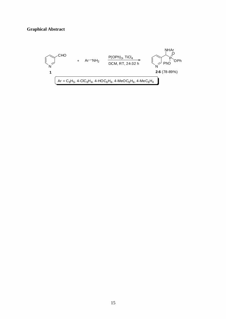
The reactions of nicotinaldehyde (1; 2 mole equiv.) with arylamines (aniline,
4-chloroaniline, 4-hydroxyaniline, 4-anisidine and 4-toluidine; 4 mole equiv.) and
triphenylphosphite (3 mole equiv.) in the presence of titanium tetrachloride (TiCl4), as a
Lewis acid, were carried out in dichloromethane (DCM) at room temperature for 24-32 h
under identical conditions. The crude products obtained were purified by crystallization from
ethanol to give the corresponding α-aminophosphonates 2–6 (Scheme 1) in 78-89% yields.
The reaction represented in Scheme 1 is general, simple, high yielding, involves easy work-up
and accommodates various substituents to produce substituted α-aminophosphonates
efficiently.
4
Scheme 1 here
The structures of α-aminophosphonates 2–6 were confirmed by IR, 1H NMR and mass
spectroscopy and their purities were confirmed by elemental analyses. The IR spectra of 2–6
are characterized by the presence of absorption bands within the 3424–3305 cm-1
region
corresponding to the stretching vibrations of the NH groups. The bands within the 15991582
cm-1
region are due to the stretching vibration of the C=N groups of the pyridine ring. While,
the absorption bands at the 1361–1320 cm-1
region are due to the symmetric stretching
vibrations of the P=O groups and absorption bands within the 869–801 cm-1
region are
attributed to the P–O–C groups.
The 1H NMR spectra of α-aminophosphonates 2–6 showed a characteristic
exchangeable singlet within the 9.68–8.88 ppm region due to NH protons. While, the CH
protons resonated as doublets (J = 13–16 Hz) within the 6.56–4.85 ppm region. The structures
of 2–6 were also confirmed by lowresolution mass spectra and the molecular or pseudo
molecular ions were confirmed by the highresolution mass spectra. Moreover, the elemental
analyses of 2–6 were consistent with the suggested structures (See experimental section for
details).
Antimicrobial Activities
α-Aminophosphonates 2–6 were screened for their in vitro antibacterial and antifungal
activities against Escherichia coli (NCIM2065) as a Gram-negative bacterium, Bacillus
subtilis (PC1219) and Staphylococcus aureus (ATCC25292) as Gram-positive bacteria and
Schccaromycies cerevisiae and Candida albicans as fungi. The inhibition zones were
measured in triplicates and the results are reported in Table S 1 (Supplemental Materials).

5
The results showed that compounds 2–6 showed moderate to high antimicrobial
activities against the tested organisms. It was found that compound 4 was the most effective
while compound 5 was the least active one.
Minimum Inhibitory Concentrations (MICs)
The inhibition zone was measured in triplicates in four different concentrations (10-
1000 μg/mL) and the mean value ± standard deviation (SD) is recorded in Table S 2
(Supplemental Materials). It is clear that compound 6 showed the highest antimicrobial
activities at low concentration (10 μg/mL). The compounds could be arranged according to
their MIC as follows: 6 > 3, 4 > 2 > 5.
The Lethal Dose
Cytotoxic anticancer substances have unique problems that come primarily from the
lack of safety and side effects. Therefore, cytotoxicity lethal dose (LD50) of
α-aminophosphonates 2–6 was determined to the larvae of Artemia salina using brine shrimp
lethality bioassay. The LD50 of compounds 2–6 are represented in Figures S 1S 5
(Supplemental Materials).
α-Aminophosphonates 26 showed low toxicity and they are safe to be used in vivo. It
was found that α-aminophosphonates 2, 3 and 6 are safe to be used because they exhibited
high values of lethal dose. On the other hand, α-aminophosphonate 4 and 5 exhibited small
values of lethal dose.
CONCLUSIONS
A convenient method for the synthesis of diphenyl 1-(arylamino)(pyridin-3-
yl)methylphosphonates was developed. The synthesized compounds exhibit a remarkable
6
inhibition of the growth of Gram-positive, Gram-negative bacteria and fungi at relatively low
concentrations. The lethal dose of the synthesized compounds indicated that most of the
synthesized compounds are safe and are promising for their uses as in vivo antimicrobial
reagents.
EXPERIMENTAL
General Experimental. Melting point determinations were performed by the open capillary
method using an Electrothermal MEL–TEMP II apparatus, at Tanta, Egypt, and are reported
uncorrected. IR spectra were recorded on a Perkin–Elmer 1430 Spectrophotometer, at Tanta,
Egypt, using KBr disc technique. 1H NMR Spectra were recorded on a Bruker AC400
spectrometer operating at 400 MHz at Tanta, Egypt. 31
P NMR Spectra were recorded on a
Bruker AC500 spectrometer operating at 202.48 MHz at Cardiff, UK. The spectra were
recorded in DMSO–d6. Chemical shifts are reported in parts per million (ppm) relative to
TMS. Assignments of signals are based on integration values and expected chemical shift
values and have not been rigorously confirmed. Low-resolution mass spectra were recorded
on a Waters GCT Premier spectrometer, at Cardiff, UK, and high-resolution mass spectra
were recorded on a Waters LCT Premier XE instrument at Cardiff, UK. Microanalysis was
performed by analytical service at both the Universities of Tanta and Cairo, Egypt. Analytical
thin layer chromatography (TLC) was performed on EM silica gel F254 sheet (0.2 mm) with
petroleum ether (40–60 C)/acetone (5:2 by volume) as a developing eluent. The spots were
detected with UV Lamp Model UV GL–58. Reagents and solvents were obtained from
commercial sources and used without purification.
Chemistry
General Procedure for the Synthesis of Diphenyl (arylamino)(pyridin-3-
yl)methylphosphonates 2-6. A mixture of 1 (1.07 g, 10.0 mmol), aromatic amine (20.0

7
mmol), triphenylphosphite (4.65 g, 15.0 mmol) and titanium tetrachloride (TiCl4; 0.19 g; 1.0
mmol) in DCM (10 mL) was stirred at room temperature for 24–32 h during which the
progress of the reaction was monitored by TLC. The solvent was removed under reduced
pressure and the residue obtained was treated with methanol (20 mL) and then filtered to
remove the solid materials. A mixture of water (10 mL) and DCM (20 mL) was added to the
filtrate and the organic phase was separated and dried over anhydrous Na2SO4. The solvent
was removed under reduced pressure to give the crude product which recrystallized from
ethanol to give the pure products 2–6 as white solids.
Diphenyl (phenylamino)(pyridin-3-yl)methylphosphonate (2). Reaction time: 24 h; Yield:
81%; mp; 112114 C. Selected IR data (KBr): 3390 (NH), 1596 (C=N), 1320 (P=O) and 823
(P–O–C) cm-1
. 1H NMR (MeCOMe/DMSO-d6): 8.44–6.18 (m, 20 H, Ar–H and NH) and
5.25 (2 d, J = 11 Hz, 1 H, CH) ppm. 31
P NMR (DMSO-d6): 15.98 ppm. AP+–MS: m/z (%)
418 ([M + 2 H]+, 28), 417 ([M + H]
+, 100), 368 (4), 327 (12), 269 (18), 229 (51) and 199 (15).
HRMS (AP+): m/z [M + H]
+ calcd for C24H22N2O3P: 417.1368; found: 417.1351. Anal calcd
for C24H21N2O3P (416.41): C, 69.22; H, 5.08; N, 6.73; P, 7.44. Found: C, 69.25; H, 5.10; N,
6.74; P, 7.35.
Diphenyl (4-chlorophenylamino)(pyridin-3-yl)methylphosphonate (3). Reaction time: 28
h; Yield: 78%; mp; 135137 C. Selected IR data (KBr): 3305 (NH), 1588 (C=N), 1361
(P=O) and 820 (P-O–C) cm-1
. 1H NMR (DMSO-d6): 8.88–6.97 (m, 19 H, Ar-H and NH)
and 5.75 (2 d, J = 13 Hz, 1 H, CH) ppm. 31
P NMR (DMSO-d6): 16.07 pm. AP+–MS: m/z
(%) 453 ([M37
Cl + H]+, 33), 451 ([M
35Cl + H]
+, 100), 417 (12), 385 (27), 368 (30), 327 (32)
and 249 (11). HRMS (AP+): m/z [M + H]
+ calcd for C24H21
35ClN2O3P: 451.0978; found:

8
451.0963. Anal calcd for C24H20ClN2O3P (450.85): C, 63.94; H, 4.47; N, 6.21; P, 6.87.
Found: C, 64.20; H, 4.21; N, 6.16; P, 6.85.
Diphenyl (4-hydroxyphenylamino)(pyridin-3-yl)methylphosphonate (4). Reaction time:
24 h; Yield: 81%; mp; 205207 C. Selected IR data (KBr): 3424 (NH/OH), 1585 (C=N),
1334 (P=O) and 869 (P–O–C) cm-1
. 1H NMR (DMSO-d6): 8.90–6.93 (m, 20 H, Ar–H, OH
and NH) and 5.73 (2 d, J = 15 Hz, 1 H, CH) ppm. 31
P NMR (DMSO-d6): 15.06 ppm. EI–
MS: m/z (%) 433 ([M + H]+, 4), 432 (M
+, 16), 339 (26), 246 (66) and 199 (100). HRMS (EI):
m/z [M]+ calcd for C24H21N2O4P: 432.1239; found: 432.1241. Anal calcd for C24H21N2O4P
(432.41): C, 66.66; H, 4.90; N, 6.48; P, 7.16. Found: C, 66.70; H, 4.92; N, 6.45; P, 7.15.
Diphenyl (4-methoxyphenylamino)(pyridin-3-yl)methylphosphonate (5). Reaction time:
30 h; Yield: 88%; mp; 160162 C. Selected IR data (KBr): 3422 (NH), 1582 (C=N), 1325
(P=O) and 801 (P–O–C) cm-1
. 1H NMR (DMSO-d6): 8.93–6.71 (m, 19 H, Ar-H and NH),
5.69 (2 d, J = 16 Hz, 1 H, CH) and 4.17 (s, 3 H, OCH3) ppm. 31
P NMR (DMSO-d6): 15.06
ppm. AP+–MS: m/z (%) 447 ([M + H]
+, 3), 415 (2), 294 (5), 193 (6), 140 (8), 117 (22), 100
(100), 97 (57) and 77 (47). HRMS (AP+): m/z [M + H]
+ calcd for C25H24N2O4P: 447.1474;
found: 447.1478. Anal calcd for C25H23N2O4P (446.43): C, 67.26; H, 5.19; N, 6.27; P, 6.94.
Found: C, 67.22; H, 5.02; N, 6.57; P, 6.90.
Diphenyl (4-methylphenylamino)(pyridin-3-yl)methylphosphonate (6). Reaction time: 32
h; Yield: 89%; mp; 194196 C. Selected IR data (KBr): 3391 (NH), 1599 (C=N), 1343
(P=O) and 836 (P-O–C) cm-1
. 1H NMR (DMSO-d6): 8.73–6.66 (m, 19 H, Ar-H and NH), 4.85
(2 d, J = 14 Hz, 1 H, CH) and 2.29 (s, 3 H, CH3) ppm. 31
P NMR (DMSO-d6): 16.06 ppm.
AP+–MS: m/z (%) 472 ([M + MeCNH]
+, 9), 432 ([M + 2 H]
+, 25), 431 ([M + H]
+, 100), 368

9
(12), 327 (17) and 294 (6). HRMS (AP+): m/z [M + H]
+ calcd for C25H24N2O3P: 431.1525;
found: 431.1506. Anal calcd for C25H23N2O3P (430.43): C, 69.76; H, 5.39; N, 6.51; P, 7.20.
Found: C, 69.72; H, 5.34; N, 6.48; P, 7.15.
Biological assay
Gram–negative bacteria. After Gram-staining procedure, Gram-negative cells appear pink.
The Gram–negative bacterium used in this study was E. coli which is known as the back bone
example for Gram–negative bacteria and cause urinary infection, wound infection and
gastroenteritis.
Gram–positive bacteria. The thick cell wall of a Gram-positive organism retains the crystal
violet dye used in the Gram–staining procedure, so the stained cells appear purple under
magnification. Gram–positive bacteria used in this study were B. subtilis (PC1219) and S.
aureus (ATCC25292). B. subtilis are mostly involved in Urinary infection, wound, ulceration
and septicemia. S. aureus is the mild stone of Gram-positive bacteria and it is a causative
agent of pneumonia, meningitis and food poisoning.
Fungi. Pathogenic fungi spatially yeasts are responsible for a number of diseases in human,
animals. A number of pathogenic strains of fungi are represented in C. albicans and S.
cerevisiae. The tested organisms were obtained from the culture collection of Bacteriology
Unit, Department of Botany, Faculty of Science, Tanta University, Egypt.
Determination of Minimum inhibitory concentrations (MICs). The antimicrobial activities
of the tested samples were determined by measuring the diameter of zone of inhibition

10
expressed in millimeter. The inhibition zones were measured in triplicates and expressed as
mean SD.42
The Lethal Dose. Brine shrimps lethality bioassay is very simple bench-top assay used to
measure cytotoxicity of plant extracts as well as the synthesized compounds. Three replicates
were used for each concentration and living larvae were counted after 72 h. All data were
expressed as mean SD.43
REFERENCES
[1] Advances in Nitrogen Heterocycles, Moody, C. J. (Ed.) JAI Press Inc., London, 1998,
Vol 3.
[2] Blum, C. A.; Ellis, J. L.; Loh, C.; Ng, P. Y.; Perni, R. B.; Stein, R. L. J. Med. Chem.
2011, 54, 417–432.
[3] Marson, C. M. Chem. Rev. 2011, 111, 7121–7156.
[4] Meanwell, N. A. J. Med. Chem. 2011, 54, 2529–2591.
[5] Qian, C.; Huang, T. J. Org. Chem. 1998, 63, 4125–4128.
[6] Ranu, B. C.; Hajra, A.; Jana, U. Org. Lett. 1999, 1, 1141–1143.
[7] Chandrasekhar, S.; Prakash, S. J.; Jagadeshwar, V.; Narsihmulu, Ch. Tetrahedron Lett.
2001, 42, 5561–5563.
[8] Wang, Q. M.; Li, Z. G.; Hung, R. Q.; Cheng, J. R. Heteroatom Chem. 2001, 12, 68–
72.
[9] El Sayed, I.; El Kosy, S. M.; Hawata, M. A.; El Gokha, A. A.-A.; Tolan, A.; Abd El-
Sattar, M. M. J. Am. Sci. 2011, 7, 357–361.
[10] Caldés, C.; Vilanova, B.; Adrover, M.; Muñoz, F.; Donoso, J. Bioorg. Med. Chem.
2011, 19, 4536–4543.

11
[11] Ning, L.; Wang, W.; Liang, Y.; Peng, H.; Fu, L.; He, H. J. Med. Chem. 2012, 47, 379–
384.
[12] Reddy, C. B.; Kumar, K. S.; Kumar, M. A.; Reddy, M. V. N.; Krishna, B. S.; Naveen,
M.; Arunasree, M. K.; Reddy, C. S.; Raju, C. N.; Reddy C. D. J. Med. Chem. 2012,
47, 553–559.
[13] Moonen, K.; Laureyn, I.; Stevens, C. V. Chem. Rev. 2004, 104, 6177–6216.
[14] Schug, K. A.; Lindner, W. Chem. Rev. 2005, 105, 67–114.
[15] Hassal, C. H.; Hahn, E. F. Antibiotics, Vol VI, Springer, Berlin, 1983, pp 1–11.
[16] Hu, D.; Wan, Q. Q.; Yang, S.; Song, B. A.; Bhadury, P. S.; Jin, L. H.; Yan, K.; Liu,
F.; Chen, Z.; Xue, W. J. Agr. Food Chem. 2008, 56, 998–1001.
[17] Zhou, J. Fan, H. T.; Song, B. A.; Jin, L. H.; Bhadury, P. S.; Hu, D. Y.; Yang, S.
Phosphorus Sulfur Silicon Relat. Elem. 2011, 186, 81–87.
[18] Kafarski, P.; Lejczak, B. Curr. Med. Chem. Anti-Cancer Agents 2001, 1, 301–312.
[19] Jing-Zi, L.; Bao-An, S.; Hui-Tao, F.; Bhadury, P. S.; Wan, W.-T.; Yang, S.; Xu, W.;
Wu, J.; Jin, L.-H.; Wei, X.; Hu, D.-Y.; Zeng, S. Eur. J. Med. Chem. 2010, 45, 5108–
5112.
[20] Rezaei, Z.; Firouzabadi, H.; Iranpoor, N.; Ghaderi, A.; Jafari, M. R.; Jafari, A. A.;
Zare, H. R. Eur. J. Med. Chem. 2009, 44, 4266–4275.
[21] Smith, K.; El-Hiti, G. A.; Abdo, M. A.; Abdel-Megeed, M. F. J. Chem. Soc., Perkin
Trans. 1 1995, 10291033.
[22] (a) Smith, K.; El-Hiti, G. A.; Abdel-Megeed, M. F.; Abdo, M. A. J. Org. Chem.1996,
61, 647–655; (b) ibid. 656–661.
[23] El-Hiti, G. A. Synthesis 2003, 2799–2804.
[24] El-Hiti, G. A. Synthesis 2004, 363368.
[25] Smith, K.; El-Hiti, G. A.; Abdel-Megeed, M. F. Synthesis 2004, 21212130.

12
[26] Smith, K.; El-Hiti, G. A.; Hegazy, A. S. Synthesis 2005, 29512961.
[27] Smith, K.; El-Hiti, G. A.; Hegazy, A. S. Synthesis 2010, 13711380.
[28] Smith, K.; El-Hiti, G. A.; Hegazy, A. S. Chem. Commun. 2010, 46, 27902792.
[29] Browne, K. A.; Deheyn, D. D.; El-Hiti, G. A.; Smith, K.; Weeks, I. J. Am. Chem. Soc.
2011, 133, 1463714648.
[30] Smith, K.; El-Hiti, G. A.; Hammond, M. E. W.; Bahzad, D.; Li, Z.; Siquet, C. J.
Chem. Soc. Perkin Trans. 1 2000, 27452752.
[31] Smith, K.; Roberts, S. D.; El-Hiti, G. A. Org. Biomol. Chem. 2003, 1, 15521559.
[32] Smith, K.; Ewart, G. M.; El-Hiti, G. A.; Randles, K. R. Org. Biomol. Chem. 2004, 2,
31503154.
[33] Smith, K.; El-Hiti, G. A. Curr. Org. Chem. 2006, 10, 16031625.
[34] Smith, K.; Ajarim, M. D.; El-Hiti, G. A.; Peters, C. Top. Catal. 2009, 52, 16961700.
[35] Smith, K.; Ajarim, M. D.; El-Hiti, G. A. Catal. Lett. 2010, 134, 270278.
[36] Smith, K.; El-Hiti, G. A. Green Chem. 2011, 13, 15791608.
[37] Smith, K.; Al-Khalaf, A. K. H.; El-Hiti, G. A.; Pattisson, S. Green Chem. 2012, 14,
11031110.
[38] Abdel-Megeed, M. F.; Badr, B. E.; Azaam, M. M.; El-Hiti, G. A. Bioorg. Med. Chem.
2012, 20, 2252.
[39] Abdel-Megeed, M. F.; Badr, B. E.; Azaam, M. M.; El-Hiti, G. A. Phosphorous Sulfur
Silicon Relat. Elem. 2012, 187, 1201.
[40] Abdel-Megeed, M. F.; Badr, B. E.; Azaam, M. M.; El-Hiti, G. A. Phosphorous Sulfur
Silicon Relat. Elem. 2012, in press; doi: 10.1080/10426507.2012.690117.
[41] Pridham, T. G.; Lindenfelser, L. A.; Shotwell, O. L.; Stodola, F.; Benedict, R. G.;
Foley, C.; Jacks, P. W.; Zaumeeyer, W. J.; Perston, W. H.; Mitchell, J. W.
Phytopathology 1956, 46, 568–575.
13
[42] Hsueh, P. R.; Chang, J. C.; Teng, L. J.; Yang, P. C.; Ho, S. W.; Hsieh, W. C.; Luh, K.
T. J. Clin. Microbiol. 1997, 35, 1021–1023.
[43] Meyer, B. N.; Ferrigni, N. R.; Putnam, J. E.; Jacobsen, L. B.; Nichols, D. E.;
McLaughlin, J. L. Planta Med. 1982, 45, 31–34.

14
N
CHO P(OPh)3, TiCl4
DCM, RT, 24-32 hAr NH2+
N
NHAr
POPh
PhO
O
2-6Ar = C6H5, 4-ClC6H4, 4-HOC6H4, 4-MeOC6H4, 4-MeC6H41
Scheme 1
15
Graphical Abstract
N
CHO P(OPh)3, TiCl4
DCM, RT, 24-32 hAr NH2+
N
NHAr
POPh
PhO
O
2-6 (78-89%)
Ar = C6H5, 4-ClC6H4, 4-HOC6H4, 4-MeOC6H4, 4-MeC6H4
1
Top Related
Copyright © 2022 FDOKUMEN

