


Bahasa
Halaman
Hukum

Available online at www.sciencedirect.com
Structural and dynamic mechanisms for the function andinhibition of the M2 proton channel from influenza A virusJun Wang1, Jade Xiaoyan Qiu2, Cinque Soto2 and William F DeGrado1,2
The M2 proton channel from influenza A virus, a prototype for a
class of viral ion channels known as viroporins, conducts
protons along a chain of water molecules and ionizable
sidechains, including His37. Recent studies highlight a delicate
interplay between protein folding, proton binding, and proton
conduction through the channel. Drugs inhibit proton
conduction by binding to an aqueous cavity adjacent to M2’s
proton-selective filter, thereby blocking access of proton to the
filter, and altering the energetic landscape of the channel and
the energetics of proton-binding to His37.
Addresses1 Department of chemistry, University of Pennsylvania, 231 south, 34th
st, Philadelphia, PA 19104, USA2 Department of Biochemistry and Biophysics, School of Medicine,
University of Pennsylvania, 422 Curvie Blvd, Philadelphia, PA 19104,
USA
Corresponding author: DeGrado, William F
Current Opinion in Structural Biology 2011, 21:68–80
This review comes from a themed issue on
Folding and Binding
Edited by Michael Gilson and Sheena Radford
Available online 17th January 2011
0959-440X/$ – see front matter
# 2010 Elsevier Ltd. All rights reserved.
DOI 10.1016/j.sbi.2010.12.002
IntroductionProtein function requires correct folding into a native
ensemble of three-dimensional structures with finely
orchestrated dynamic interconversion between confor-
mational substates. Thus, protein sequences reflect
a compromise between stability and function [1,2].
The thermodynamics of folding has been studied for
very few membrane proteins, and the relationships among
stability, dynamics, and function remain largely unex-
plored for this class of proteins [3,4]. Here, we examine
these relationships for the M2 proton channel [5] in
comparison to the potassium channel KcsA [6–8], focus-
ing on the interplay between folding energetics, per-
meant ion binding/translocation, and inhibition by the
binding of pore-blocking drugs.
M2 is a multifunctional, modular proteinThe M2 protein, which was discovered as the target of the
anti-influenza drugs amantadine and rimantadine [9–11],
Current Opinion in Structural Biology 2011, 21:68–80
has multiple functions [5,12,13] associated with different
regions of the sequence of this short 97-residue protein.
Influenza viruses gain access to cells via receptor-
mediated endocytosis, which places the virus within an
acidifying endosome. M2 facilitates diffusion of protons
into the interior of the endosomally entrapped virus as the
endosome matures, leading to uncoating of the viral RNA
from the matrix protein M1 [14]. M2 is also important for
delaying acidification of the late Golgi in some strains of
the virus [15,16].
M2’s functions are compartmentalized into parsimonious
sequences, consisting of:
(A) Residues 1–24 comprise a short unstructured N-
terminal region important for incorporation into the
virion [17] in influenza A virus, but entirely missing
in influenza B virus [18,19].
(B) Residues 25–46 encompass the transmembrane
(TM) helix that is necessary and sufficient for
tetramerization, proton conductance and drug-bind-
ing [20–23]. Drug-resistant mutations map to pore-
lining residues of this TM helix (particularly S31N,
V27A, and L26F) [24–26]. A secondary binding site
on the outside of the TM helices is observed when
the drug is present at very high concentrations in
micelles [27] or bilayers [28�], but electrophysio-
logical studies and the drug sensitivities of reverse-
engineered viruses showed that this site does not
contribute to the pharmacological inhibition of the
channel [24–26]. Much work on the structure and
function of M2 has been conducted on fragments
spanning from residues 22 to 46, or closely related
sequences, which we call M2TM.
(C) Residues 47–61 define a cytoplasmic amphiphilic
helix involved in cholesterol-binding [29], mem-
brane localization, budding and scission [12]. This
sequence is not required for channel formation or
drug-binding [20,25,29]. Together, the TM and
cytoplasmic helices are often studied as a single
peptide (approximately residues 20–60), defined as
M2TM + cyto peptides.
(D) Residues 61–97 comprise a disordered tail that
interacts with the matrix protein, M1 [30].
Structure determination of M2’s membrane-interacting domainsThe membrane-interactive domains of M2 were ident-
ified by limited proteolysis [21], which identified both
M2TM + cyto as a metastable product and M2TM as a
www.sciencedirect.com
Structural and dynamic mechanisms of the M2 proton channel Wang et al. 69
final cleavage product (Wang J and DeGrado WF, unpub-
lished result). This, and related studies [20,22,31],
showed that the TM domain was both necessary and
sufficient to form tetramers and bind amantadine in
micelles and bilayers. M2TM has also been the subject
of numerous studies using optical spectroscopy (IR [32],
Raman [33], CD [22], fluorescence [34], solid state NMR
(SSNMR) [23,35–37], X-ray crystallography [38,39��],isothermal calorimetry [20], analytical ultracentrifugation
[40,41], and surface plasmon resonance [42]) to examine
pH activation and drug-binding. Recently, a second series
of structural studies focused on the M2TM + cyto frag-
ment, which also includes the cytoplasmic amphiphilic
helix [27,43,44,45�].
Before discussing the structural basis for M2’s functions,
it is first important to consider the various models and
structures proposed for M2 using different techniques,
their resolution, and the degree to which the presence or
absence of the C-terminal cytoplasmic helix might influ-
ence the structure of the pore-forming tetramer. We
compare the findings with the potassium channel KcsA
[6–8], which has two cytoplasmic domains, consisting of
an N-terminal membrane-interactive helix as well as a C-
terminal helical bundle domain. Almost all high-resol-
ution structural work on KcsA has been accomplished
with constructs lacking its N-terminal and C-terminal
cytoplasmic helices. Lower resolution site-directed spin
label EPR studies and a 3.8 A crystal structure [46]
showed that the N-terminal cytoplasmic domain formed
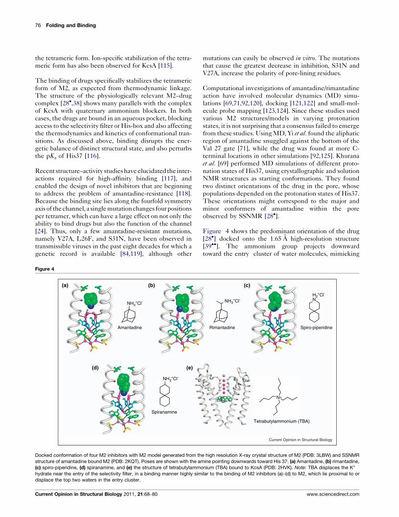
Figure 1
Val27 valve
Gly34
His37His-box
Trp41Trp basket
Asp44
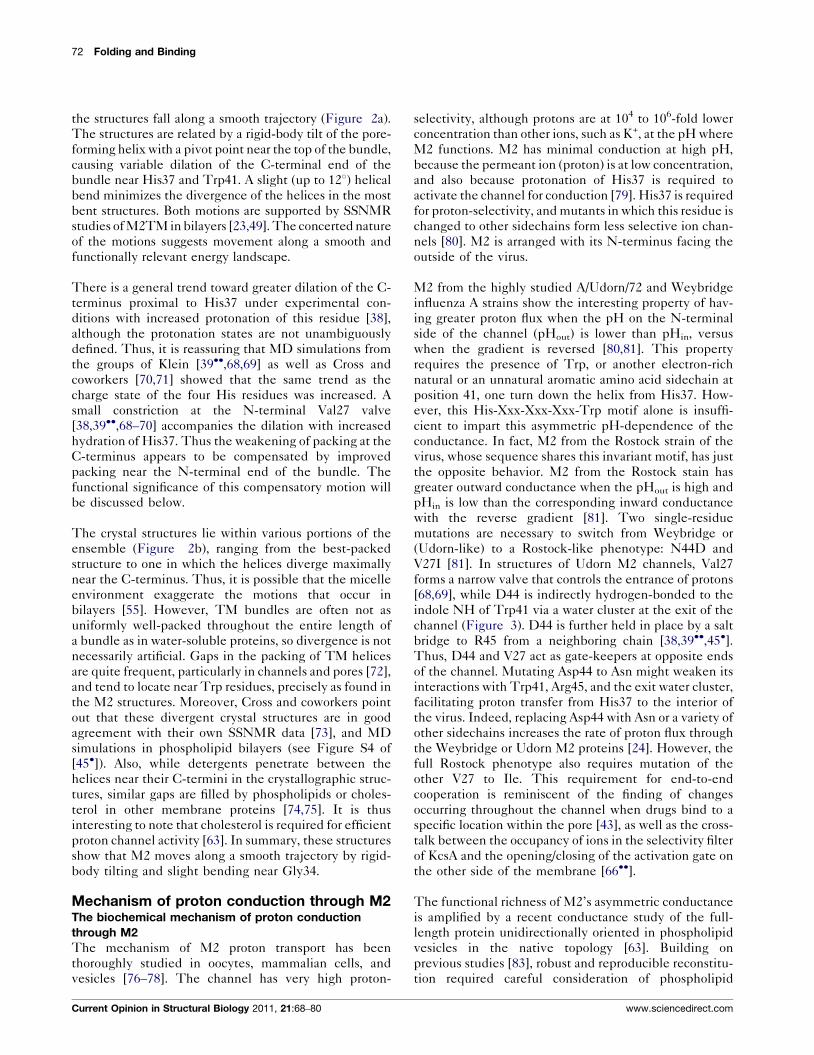
(a) crystallography (b) solu
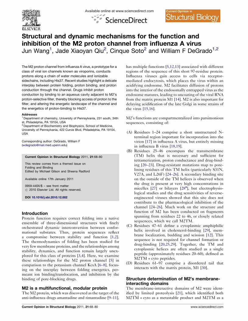
Comparison of (a) crystal (PDB: 3LBW), (b) solution NMR (PDB: 2RLF), and
while residues 49–60 are colored in yellow. Critical residues His37 (in cyan) an
the crystal structure. Water clusters are shown in pink.
www.sciencedirect.com
a helix bound to the membrane surface, while the C-
terminal cytoplasmic domain forms a water soluble four-
helix bundle [47]. The presence of the C-terminal four-
helix bundle has little effect on the channel pore, except
to close one end of the bundle slightly more tightly near
the activation gate. This is consistent with the fact that
the basic structural features, conductance mechanism,
and gating mechanism inferred from the structure of
KcsA have proven to be general across many mammalian
K+ channels. These proteins have very similar channel-
forming regions and differ most markedly in their pen-
dant cytoplasmic domains, which help modulate the
energetics and kinetics of interconversion of the confor-
mational states underlying gating. Crystallographic struc-
tures of the membrane-spanning domains have been
invaluable for defining these conformational states, while
functional measurements and other lower resolution
methods, such as fluorescence spectroscopy, EPR,
solution and solid-state NMR, are indispensable to assign
these conformations to discrete functional states and to
map their populations and kinetics of interconversion.
The structures of M2TM have been extensively studied
by X-ray crystallography and SSNMR. The highest-resol-
ution structure is a 1.65 A crystal structure [39��] of the TM
domain (M2TM) from micelles (Figure 1a), which comp-
lements earlier crystallographic structures at intermediate
resolution (2.0–3.5 A) [38]. The 1.65 A-resolution structure
precisely pinpoints water molecules and sidechains that
form a pathway for proton translocation. Functional water
tion NMR (c) solid state NMR
Current Opinion in Structural Biology
(c) solid state NMR (PDB: 2L0J) structures. Backbone is colored in grey,
d Trp41 (in green) are the key to the extensive hydrogen bond network in
Current Opinion in Structural Biology 2011, 21:68–80
70 Folding and Binding
molecules are often very well-ordered, and this structure
abounds with waters whose thermal factors are on par with
the most ordered backbones. As further discussed below,
the most important residues required for proton-channel
function are His37 and Trp41, which associate to form a
‘His-box’ and a ‘Trp-basket’ (Figure 1a). In the crystal
structures of M2TM obtained under other conditions [38],
the bundle is more splayed at the cytoplasmic end, partially
or fully disrupting the His-box.
M2 has been studied by SSNMR in aligned phospholipid
mutilayers [48–50] to evaluate the orientation of individ-
ual amides via a 15N-based two-dimensional experiment,
PISEMA, in which the N–H dipolar coupling and the 15N
chemical shift anisotropy are correlated [36]. This method
defines the crossing angle, kinking, and rotation of a
monomeric helix relative to the membrane normal. How-
ever, many structures of the tetramer can be devised that
are consistent with these main-chain restraints, so early
SSNMR models of M2TM (2H95, 1NYJ, 2KAD) have
now been superceded by more recent, high-resolution
crystallographic (3LBW), SSNMR (2KQT, 2L0J) and
solution NMR structures (2RLF). Hong and coworkers
recently used experimental sidechain dihedral restraints
and REDOR distance measurements together with the
previous dipolar coupling data of Cross and coworkers
[49] to obtain a well-defined structure for the M2TM–amantadine complex [28�] (2KQT), which is within 1.0 A
rmsd of the backbone structure of the high-resolution
crystallographic structure of M2TM (3LBW) [28�].
The M2TM + cyto fragment was recently studied by
solution [27,44] and solid-state NMR (SSNMR)
[43,45�]. The solution structure is at moderate resolution,
being defined by 20 inter-monomer NOEs (0.47 per
residue), 23 dihedral restraints, and 27 residual dipolar
couplings [27] (Figure 1b). The SSNMR structure (2L0J)
[45�], based on only backbone orientation and membrane
depths for the C-terminal helix from EPR studies [51]
(Figure 1c), was computed by MD calculations, using
these restraints plus reasonable (but nevertheless
hypothetical) distance restraints [45�]. The solution and
SSNMR structures are in reasonable agreement in the
TM region; they feature left-crossing TM bundles that
place His37 and Trp41 in the pore. However, the cyto-
plasmic helices differ significantly. In the solution struc-
ture [27], it forms a tetrameric bundle extending into the
cytoplasm beyond the end of the TM domain (Figure 1b),
while the SSNMR structure places the cytoplasmic helix
against the C-terminal end of the bundle exposed to the
headgroup region of the bilayer [45�] (Figure 1c).
One may ask ‘What is the role of all the detailed inter-
actions formed by the cytoplasmic helices in these struc-
tures? Surely, they must be essential for proton channel
activity!’ To answer this question, the five hydrophobic
residues (F47, F48, I51, Y52, F55) in the cytoplasmic
Current Opinion in Structural Biology 2011, 21:68–80
helix involved in these interactions were simultaneously
changed to Ala in the full-length protein [12,20,29,52].
The surface expression level, proton flux, pH-activation,
and drug-binding of this quintuple mutant were indis-
tinguishable from WT, the only change being its inability
to promote virus budding and vesicle fission [12,20,29,52].
Thus, the interactions of the cytoplasmic domain in these
structures have a subtle effect (if any) on the proton
channel activity of the TM domain. They also have little
effect on the structure of the TM domain; the overall
backbone structures of M2TM are in good agreement
with the corresponding TM domains seen in structures of
M2TM + cyto constructs, showing small (rmsd 1–2 A)
differences within the range associated with subtle
changes in bilayer composition [23,53–55]. Thus, studies
with the TM construct, which have been conducted in
much greater resolution, should provide relevant insight.
The structures of the longer M2TM + cyto constructs help
inform the mechanisms of vesicle budding and scission. In
a budding virus, M2 localizes to the neck of the budding
membrane at a region of extreme curvature that topologi-
cally resembles a donut-hole. Such saddle-shaped surfaces
have negative Gaussian curvature characterized by orthog-
onally directed negative and positive local curvature. The
cytoplasmic helix is rich in Arg and hydrophobic residues
that can promote negative Gaussian curvature [56], and in
the SSNMR structure the hydrophobic and positively
charged sidechains of the M2 cytoplasmic helix are well
positioned to promote negative Gaussian curvature by
interacting with phospholipid headgroups. The cyto-
plasmic helices in the solution NMR structure [27] are
less well oriented for this function, but this conformation
might serve another functional role. A longer helical bun-
dle is observed in the corresponding BM2 protein from
influenza B virus [57]. Also, the C-terminal cytoplasmic
helical bundle in KcsA has recently been suggested to
dissociate, and its individual helices interact with the
membrane during gating [58]. Thus, the full functional
role of the M2’s cytoplasmic helix remains a fruitful area for
further investigation.
TM structures and dynamicsNMR studies on M2TM and M2TM + cyto have shown
that the TM helical motions are strongly modulated by
pH, being greatest at acidic pH where the protein func-
tions. The pH-dependent broadening of peaks in the
amide region of the solution NMR [27], aligned SSNMR
[59], and magic angle spinning SSNMR [60] spectra is
indicative of backbone conformational fluctuations in the
microsecond to millisecond time scale, and the critical
residues His37 and Trp41 also show pH-dependent
motions in the microsecond regime [23,27,61�]. SSNMR
is particularly well suited to examine dynamics of M2TM
and M2TM + cyto, because one can access both the slow-
exchange to the rapid exchange regimes on the same
sample by simply altering the temperature [62]. It also
www.sciencedirect.com
Structural and dynamic mechanisms of the M2 proton channel Wang et al. 71
allows the exploration of different bilayers to evaluate
their effects on dynamics [54,60].
Conformation and dynamics of M2 are also modulated
by the chain length of the lipid; short chain lipids tend to
increase the crossing angle of the helix relative to the
membrane normal [53], allowing a better match be-
tween the hydrophobic width of the peptide and the
bilayer. Hong and coworkers have used cholesterol-rich
bilayers to freeze out rapid uniaxial rotation and mini-
mize contributions from exchange-broadening over a
wide range of temperatures [54]. The addition of cho-
lesterol is also interesting because it is required for
maximal proton channel activity [63] and is known to
stabilize the tetramer [64]. At ambient temperature,
M2TM has two conformations at pH 7.5, and a third
conformation at pH 4.5 [60]. The binding of drugs and
the composition of the surrounding membrane bilayer
also have a large effect on the fraction of each confor-
mational form [60].
The M2TM + cyto construct has also been examined in
bilayers by SSNMR. Cross and coworkers used a mem-
brane rich in the nonbilayer-forming phospholipid DOPE
(DOPC/DOPE, 4:1) to achieve good reconstitution and
alignment in multilayers [45�]. Two conformations exist
in these preparations, based upon doubling of many of the
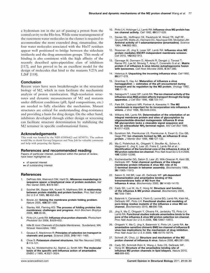
Figure 2
(a)
(b)axial view
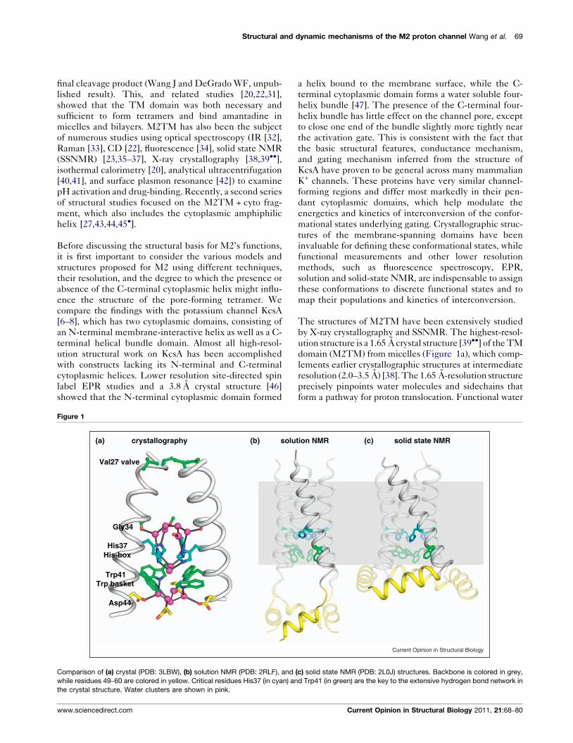
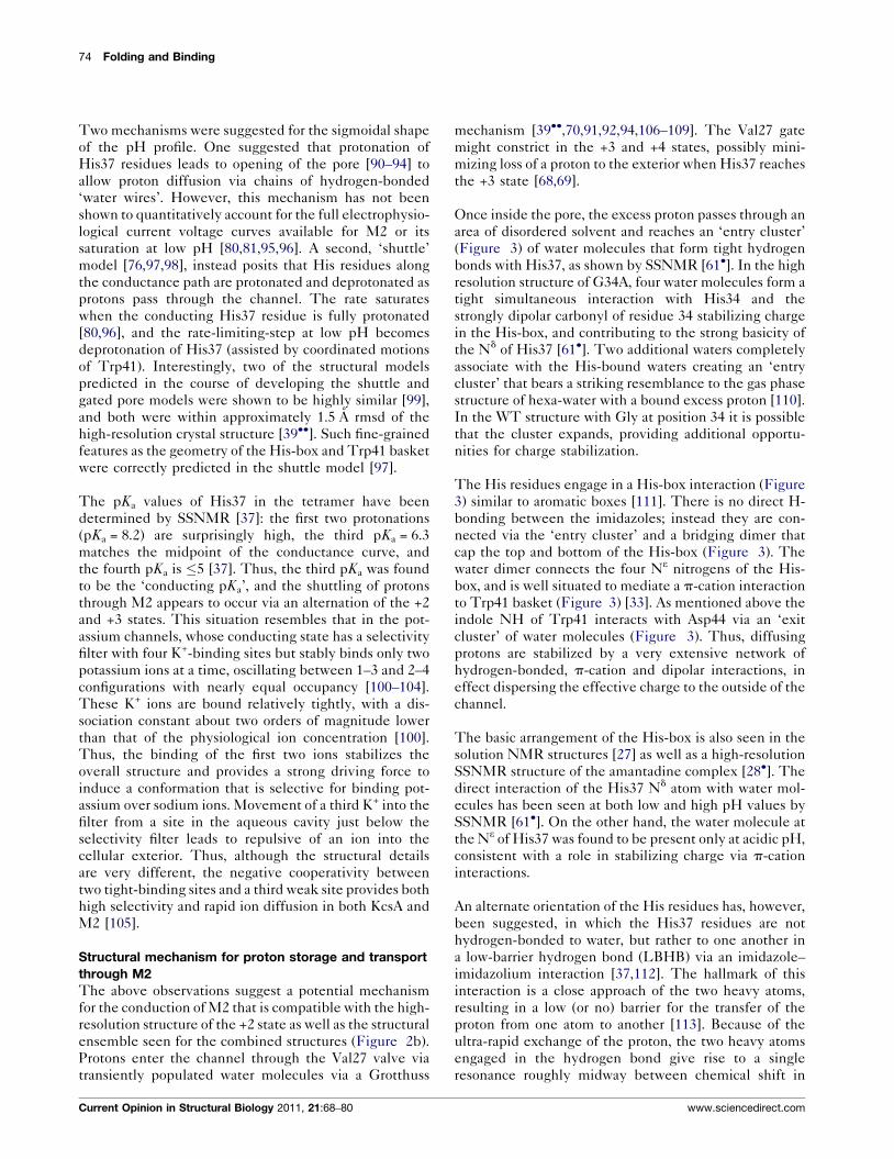
Motions of pore forming helices of KcsA and M2 shown by overlaying KcsA
(residues 86–122) (PDB codes: 1R3J, 2HVK, 3EFF, 3F5W, 3F7V, 3F7Y, 3FB5
25–46) (PDB codes: 2RLF, 3C9J, 2KQT, 2L0J, 3LBW, and three symmetric s
3BKD). Figures are adapted from [126].
www.sciencedirect.com
peaks, including S31 (see Figure S3 of the supplementary
material from [45�]).
The multiconformational behavior seen in these studies is
similar to that seen in parallel solution NMR and SSNMR
studies of KcsA, which has multiple structures associated
with its gating between resting and conducting states. The
opening of the ‘activation gate’ occurs on the low ms time
scale [65], and involves large changes in packing of the
pore-forming helices, resulting in a 20-A increase in the
diameter of the cytoplasmic section of the pore that leads to
the selectivity filter [66��]. The structures of the open and
closed forms of this activation gate, and possible intermedi-
ates along the trajectory, have recently been elucidated by
X-ray crystallography in KcsA [66��], as well as the Na/K
channel [67]. The opening motion involves bending and
rigid-body tilting of the pore-forming helix (Figure 2a).
The use of a minimal construct, lacking all of the cyto-
plasmic domains was the key to obtaining the full family of
conformational states [68]. These truncations removed
short segments that, in earlier studies, had engaged in
crystallographic packing interactions that otherwise inter-
fered with the marginally stable open activation gate.
Figure 2b shows the corresponding ensemble of the TM
regions of all recent structures of M2 obtained by X-ray
crystallography, solution NMR, and SSNMR. As for KcsA,
side view
1R3J
3EFF
3FB5
3F7Y
3F7V
3FB6
3FB8
3FB7
3F5W
2HVK
3BKD, chain H
3C9J
3BKD, chain F
3BKD, chain G
2L0J
3LBW
2KQT
2RLE
Current Opinion in Structural Biology
and M2 structures from different sources. (a) Ten structures for KcsA
, 3FB6, 3FB7, 3FB8). (b) Eight structures were analyzed for M2 (residues
tructures derived from three different helices of the asymmetric structure
Current Opinion in Structural Biology 2011, 21:68–80
72 Folding and Binding
the structures fall along a smooth trajectory (Figure 2a).
The structures are related by a rigid-body tilt of the pore-
forming helix with a pivot point near the top of the bundle,
causing variable dilation of the C-terminal end of the
bundle near His37 and Trp41. A slight (up to 128) helical
bend minimizes the divergence of the helices in the most
bent structures. Both motions are supported by SSNMR
studies of M2TM in bilayers [23,49]. The concerted nature
of the motions suggests movement along a smooth and
functionally relevant energy landscape.
There is a general trend toward greater dilation of the C-
terminus proximal to His37 under experimental con-
ditions with increased protonation of this residue [38],
although the protonation states are not unambiguously
defined. Thus, it is reassuring that MD simulations from
the groups of Klein [39��,68,69] as well as Cross and
coworkers [70,71] showed that the same trend as the
charge state of the four His residues was increased. A
small constriction at the N-terminal Val27 valve
[38,39��,68–70] accompanies the dilation with increased
hydration of His37. Thus the weakening of packing at the
C-terminus appears to be compensated by improved
packing near the N-terminal end of the bundle. The
functional significance of this compensatory motion will
be discussed below.
The crystal structures lie within various portions of the
ensemble (Figure 2b), ranging from the best-packed
structure to one in which the helices diverge maximally
near the C-terminus. Thus, it is possible that the micelle
environment exaggerate the motions that occur in
bilayers [55]. However, TM bundles are often not as
uniformly well-packed throughout the entire length of
a bundle as in water-soluble proteins, so divergence is not
necessarily artificial. Gaps in the packing of TM helices
are quite frequent, particularly in channels and pores [72],
and tend to locate near Trp residues, precisely as found in
the M2 structures. Moreover, Cross and coworkers point
out that these divergent crystal structures are in good
agreement with their own SSNMR data [73], and MD
simulations in phospholipid bilayers (see Figure S4 of
[45�]). Also, while detergents penetrate between the
helices near their C-termini in the crystallographic struc-
tures, similar gaps are filled by phospholipids or choles-
terol in other membrane proteins [74,75]. It is thus
interesting to note that cholesterol is required for efficient
proton channel activity [63]. In summary, these structures
show that M2 moves along a smooth trajectory by rigid-
body tilting and slight bending near Gly34.
Mechanism of proton conduction through M2The biochemical mechanism of proton conduction
through M2
The mechanism of M2 proton transport has been
thoroughly studied in oocytes, mammalian cells, and
vesicles [76–78]. The channel has very high proton-
Current Opinion in Structural Biology 2011, 21:68–80
selectivity, although protons are at 104 to 106-fold lower
concentration than other ions, such as K+, at the pH where
M2 functions. M2 has minimal conduction at high pH,
because the permeant ion (proton) is at low concentration,
and also because protonation of His37 is required to
activate the channel for conduction [79]. His37 is required
for proton-selectivity, and mutants in which this residue is
changed to other sidechains form less selective ion chan-
nels [80]. M2 is arranged with its N-terminus facing the
outside of the virus.
M2 from the highly studied A/Udorn/72 and Weybridge
influenza A strains show the interesting property of hav-
ing greater proton flux when the pH on the N-terminal
side of the channel (pHout) is lower than pHin, versus
when the gradient is reversed [80,81]. This property
requires the presence of Trp, or another electron-rich
natural or an unnatural aromatic amino acid sidechain at
position 41, one turn down the helix from His37. How-
ever, this His-Xxx-Xxx-Xxx-Trp motif alone is insuffi-
cient to impart this asymmetric pH-dependence of the
conductance. In fact, M2 from the Rostock strain of the
virus, whose sequence shares this invariant motif, has just
the opposite behavior. M2 from the Rostock stain has
greater outward conductance when the pHout is high and
pHin is low than the corresponding inward conductance
with the reverse gradient [81]. Two single-residue
mutations are necessary to switch from Weybridge or
(Udorn-like) to a Rostock-like phenotype: N44D and
V27I [81]. In structures of Udorn M2 channels, Val27
forms a narrow valve that controls the entrance of protons
[68,69], while D44 is indirectly hydrogen-bonded to the
indole NH of Trp41 via a water cluster at the exit of the
channel (Figure 3). D44 is further held in place by a salt
bridge to R45 from a neighboring chain [38,39��,45�].Thus, D44 and V27 act as gate-keepers at opposite ends
of the channel. Mutating Asp44 to Asn might weaken its
interactions with Trp41, Arg45, and the exit water cluster,
facilitating proton transfer from His37 to the interior of
the virus. Indeed, replacing Asp44 with Asn or a variety of
other sidechains increases the rate of proton flux through
the Weybridge or Udorn M2 proteins [24]. However, the
full Rostock phenotype also requires mutation of the
other V27 to Ile. This requirement for end-to-end
cooperation is reminiscent of the finding of changes
occurring throughout the channel when drugs bind to a
specific location within the pore [43], as well as the cross-
talk between the occupancy of ions in the selectivity filter
of KcsA and the opening/closing of the activation gate on
the other side of the membrane [66��].
The functional richness of M2’s asymmetric conductance
is amplified by a recent conductance study of the full-
length protein unidirectionally oriented in phospholipid
vesicles in the native topology [63]. Building on
previous studies [83], robust and reproducible reconstitu-
tion required careful consideration of phospholipid
www.sciencedirect.com
Structural and dynamic mechanisms of the M2 proton channel Wang et al. 73
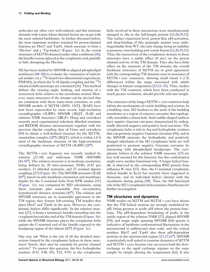
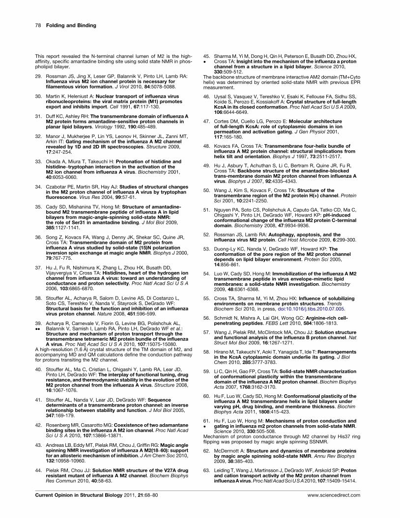
Figure 3
Gly/Ala 34
Entry cluster
His 37 Box
Bridging Dimer
Bridging cluster
Asp 44
Trp 41 Basket
Current Opinion in Structural Biology
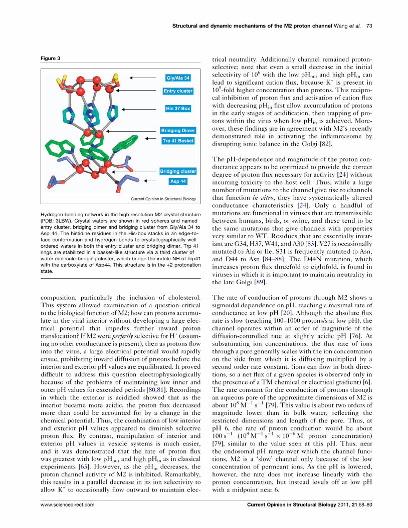
Hydrogen bonding network in the high resolution M2 crystal structure
(PDB: 3LBW). Crystal waters are shown in red spheres and named
entry cluster, bridging dimer and bridging cluster from Gly/Ala 34 to
Asp 44. The histidine residues in the His-box stacks in an edge-to-
face conformation and hydrogen bonds to crystallographically well
ordered waters in both the entry cluster and bridging dimer. Trp 41
rings are stabilized in a basket-like structure via a third cluster of
water molecule-bridging cluster, which bridge the indole NH of Trp41
with the carboxylate of Asp44. This structure is in the +2 protonation
state.
composition, particularly the inclusion of cholesterol.
This system allowed examination of a question critical
to the biological function of M2; how can protons accumu-
late in the viral interior without developing a large elec-
trical potential that impedes further inward proton
translocation? If M2 were perfectly selective for H+ (assum-
ing no other conductance is present), then as protons flow
into the virus, a large electrical potential would rapidly
ensue, prohibiting inward diffusion of protons before the
interior and exterior pH values are equilibrated. It proved
difficult to address this question electrophysiologically
because of the problems of maintaining low inner and
outer pH values for extended periods [80,81]. Recordings
in which the exterior is acidified showed that as the
interior became more acidic, the proton flux decreased
more than could be accounted for by a change in the
chemical potential. Thus, the combination of low interior
and exterior pH values appeared to diminish selective
proton flux. By contrast, manipulation of interior and
exterior pH values in vesicle systems is much easier,
and it was demonstrated that the rate of proton flux
was greatest with low pHout and high pHin as in classical
experiments [63]. However, as the pHin decreases, the
proton channel activity of M2 is inhibited. Remarkably,
this results in a parallel decrease in its ion selectivity to
allow K+ to occasionally flow outward to maintain elec-
www.sciencedirect.com
trical neutrality. Additionally channel remained proton-
selective; note that even a small decrease in the initial
selectivity of 106 with the low pHout and high pHin can
lead to significant cation flux, because K+ is present in
105-fold higher concentration than protons. This recipro-
cal inhibition of proton flux and activation of cation flux
with decreasing pHin first allow accumulation of protons
in the early stages of acidification, then trapping of pro-
tons within the virus when low pHin is achieved. More-
over, these findings are in agreement with M2’s recently
demonstrated role in activating the inflammasome by
disrupting ionic balance in the Golgi [82].
The pH-dependence and magnitude of the proton con-
ductance appears to be optimized to provide the correct
degree of proton flux necessary for activity [24] without
incurring toxicity to the host cell. Thus, while a large
number of mutations to the channel give rise to channels
that function in vitro, they have systematically altered
conductance characteristics [24]. Only a handful of
mutations are functional in viruses that are transmissible
between humans, birds, or swine, and these tend to be
the same mutations that give channels with properties
very similar to WT. Residues that are essentially invar-
iant are G34, H37, W41, and A30 [83]. V27 is occasionally
mutated to Ala or Ile, S31 is frequently mutated to Asn,
and D44 to Asn [84–88]. The D44N mutation, which
increases proton flux threefold to eightfold, is found in
viruses in which it is important to maintain neutrality in
the late Golgi [89].
The rate of conduction of protons through M2 shows a
sigmoidal dependence on pH, reaching a maximal rate of
conductance at low pH [20]. Although the absolute flux
rate is slow (reaching 100–1000 protons/s at low pH), the
channel operates within an order of magnitude of the
diffusion-controlled rate at slightly acidic pH [76]. At
subsaturating ion concentrations, the flux rate of ions
through a pore generally scales with the ion concentration
on the side from which it is diffusing multiplied by a
second order rate constant. (ions can flow in both direc-
tions, so a net flux of a given species is observed only in
the presence of a TM chemical or electrical gradient) [6].
The rate constant for the conduction of protons through
an aqueous pore of the approximate dimensions of M2 is
about 108 M�1 s�1 [79]. This value is about two orders of
magnitude lower than in bulk water, reflecting the
restricted dimensions and length of the pore. Thus, at
pH 6, the rate of proton conduction would be about
100 s�1 (108 M�1 s�1 � 10�6 M proton concentration)
[79], similar to the value seen at this pH. Thus, near
the endosomal pH range over which the channel func-
tions, M2 is a ‘slow’ channel only because of the low
concentration of permeant ions. As the pH is lowered,
however, the rate does not increase linearly with the
proton concentration, but instead levels off at low pH
with a midpoint near 6.
Current Opinion in Structural Biology 2011, 21:68–80
74 Folding and Binding
Two mechanisms were suggested for the sigmoidal shape
of the pH profile. One suggested that protonation of
His37 residues leads to opening of the pore [90–94] to
allow proton diffusion via chains of hydrogen-bonded
‘water wires’. However, this mechanism has not been
shown to quantitatively account for the full electrophysio-
logical current voltage curves available for M2 or its
saturation at low pH [80,81,95,96]. A second, ‘shuttle’
model [76,97,98], instead posits that His residues along
the conductance path are protonated and deprotonated as
protons pass through the channel. The rate saturates
when the conducting His37 residue is fully protonated
[80,96], and the rate-limiting-step at low pH becomes
deprotonation of His37 (assisted by coordinated motions
of Trp41). Interestingly, two of the structural models
predicted in the course of developing the shuttle and
gated pore models were shown to be highly similar [99],
and both were within approximately 1.5 A rmsd of the
high-resolution crystal structure [39��]. Such fine-grained
features as the geometry of the His-box and Trp41 basket
were correctly predicted in the shuttle model [97].
The pKa values of His37 in the tetramer have been
determined by SSNMR [37]: the first two protonations
(pKa = 8.2) are surprisingly high, the third pKa = 6.3
matches the midpoint of the conductance curve, and
the fourth pKa is �5 [37]. Thus, the third pKa was found
to be the ‘conducting pKa’, and the shuttling of protons
through M2 appears to occur via an alternation of the +2
and +3 states. This situation resembles that in the pot-
assium channels, whose conducting state has a selectivity
filter with four K+-binding sites but stably binds only two
potassium ions at a time, oscillating between 1–3 and 2–4
configurations with nearly equal occupancy [100–104].
These K+ ions are bound relatively tightly, with a dis-
sociation constant about two orders of magnitude lower
than that of the physiological ion concentration [100].
Thus, the binding of the first two ions stabilizes the
overall structure and provides a strong driving force to
induce a conformation that is selective for binding pot-
assium over sodium ions. Movement of a third K+ into the
filter from a site in the aqueous cavity just below the
selectivity filter leads to repulsive of an ion into the
cellular exterior. Thus, although the structural details
are very different, the negative cooperativity between
two tight-binding sites and a third weak site provides both
high selectivity and rapid ion diffusion in both KcsA and
M2 [105].
Structural mechanism for proton storage and transport
through M2
The above observations suggest a potential mechanism
for the conduction of M2 that is compatible with the high-
resolution structure of the +2 state as well as the structural
ensemble seen for the combined structures (Figure 2b).
Protons enter the channel through the Val27 valve via
transiently populated water molecules via a Grotthuss
Current Opinion in Structural Biology 2011, 21:68–80
mechanism [39��,70,91,92,94,106–109]. The Val27 gate
might constrict in the +3 and +4 states, possibly mini-
mizing loss of a proton to the exterior when His37 reaches
the +3 state [68,69].
Once inside the pore, the excess proton passes through an
area of disordered solvent and reaches an ‘entry cluster’
(Figure 3) of water molecules that form tight hydrogen
bonds with His37, as shown by SSNMR [61�]. In the high
resolution structure of G34A, four water molecules form a
tight simultaneous interaction with His34 and the
strongly dipolar carbonyl of residue 34 stabilizing charge
in the His-box, and contributing to the strong basicity of
the Nd of His37 [61�]. Two additional waters completely
associate with the His-bound waters creating an ‘entry
cluster’ that bears a striking resemblance to the gas phase
structure of hexa-water with a bound excess proton [110].
In the WT structure with Gly at position 34 it is possible
that the cluster expands, providing additional opportu-
nities for charge stabilization.
The His residues engage in a His-box interaction (Figure
3) similar to aromatic boxes [111]. There is no direct H-
bonding between the imidazoles; instead they are con-
nected via the ‘entry cluster’ and a bridging dimer that
cap the top and bottom of the His-box (Figure 3). The
water dimer connects the four Ne nitrogens of the His-
box, and is well situated to mediate a p-cation interaction
to Trp41 basket (Figure 3) [33]. As mentioned above the
indole NH of Trp41 interacts with Asp44 via an ‘exit
cluster’ of water molecules (Figure 3). Thus, diffusing
protons are stabilized by a very extensive network of
hydrogen-bonded, p-cation and dipolar interactions, in
effect dispersing the effective charge to the outside of the
channel.
The basic arrangement of the His-box is also seen in the
solution NMR structures [27] as well as a high-resolution
SSNMR structure of the amantadine complex [28�]. The
direct interaction of the His37 Nd atom with water mol-
ecules has been seen at both low and high pH values by
SSNMR [61�]. On the other hand, the water molecule at
the Ne of His37 was found to be present only at acidic pH,
consistent with a role in stabilizing charge via p-cation
interactions.
An alternate orientation of the His residues has, however,
been suggested, in which the His37 residues are not
hydrogen-bonded to water, but rather to one another in
a low-barrier hydrogen bond (LBHB) via an imidazole–imidazolium interaction [37,112]. The hallmark of this
interaction is a close approach of the two heavy atoms,
resulting in a low (or no) barrier for the transfer of the
proton from one atom to another [113]. Because of the
ultra-rapid exchange of the proton, the two heavy atoms
engaged in the hydrogen bond give rise to a single
resonance roughly midway between chemical shift in
www.sciencedirect.com
Structural and dynamic mechanisms of the M2 proton channel Wang et al. 75
imidazole–imidazolium pairs generally seen for the donor
and acceptor in the 15N NMR spectrum at 270 K and
above [112]. At lower temperatures, two peaks of equal
intensity can be observed for dimers that deviate from
true symmetry, although the exchange in the dimers is
rapid throughout the temperature range [112]. The His37
residues of M2TM have also been proposed to form a
direct LBHB in the +2 state, although the interpretation
was ambiguous due to exchange broadening, which
obscured key diagnostic peaks [37]. The chemical shifts
assigned to imidazole–imidazolium interactions in this
study might instead arise from interactions between
His37 and water molecules [61�]. Moreover, exchange
broadening is generally indicative of microsecond to
millisecond processes with significantly larger energetic
barriers than that associated with a LBHB, so its presence
is not good evidence for a LBHB. A second argument for a
LBHB came from quantum mechanical (QM) calcu-
lations in which the imidazoles were found to form stable
pairwise interactions [45�]. This computed geometry was
used to guide the MD simulation of the aligned SSNMR
structure of M2TM + cyto [45�]. However, when water
was included in more extensive QM simulations
[39��,106], the imidazoles were instead found to interact
via bridging water molecules, as in the crystal structure.
The chemical shifts computed from the crystal structure
(supporting information of [39��]) are also in agreement
with the same aligned SSNMR data [45�]. Thus, the His-
box is clearly formed in at least one conformational form
of the channel. It remains possible that there is a direct
interaction of imidazoles in some alternate conformers,
but direct evidence for this interaction is not definitive.
Clearly, the viral membrane mimetic bilayer, with its lack
of exchange broadening from axial rotational motions, is
well suited for future investigations of this issue [54].
In summary, the recently published high-resolution struc-
ture of M2TM, together with many other biochemical
and SSNMR studies of M2TM and M2TM + cyto in
bilayers, explains how protons enter and are stabilized
in the channel, and provides a rationale for the high
stability of the +2 state. Furthermore, upon reaching
the third protonation state, the highly ordered structure
of the His-box is destabilized [61�], and the structure of
the protein becomes more dynamic, allowing efflux of a
proton past Trp41 and into the interior. The mechanism
of this final step remains an important challenge. SSNMR
[114] and IR spectroscopy [32] studies show an increase in
hydration in the +3 state, consistent with the structural
and MD studies showing dilation of the C-terminal end of
the bundle [68]. Raman spectroscopy [33] shows a pro-
tonated His–Trp interaction which forms with a pKa near
6, and presumably reflects residual tertiary interactions
retained in the time-averaged structure of the +3 state.
Another challenge will be to refine the original shuttle
mechanism which suggested protonation and deprotona-
www.sciencedirect.com
tion at the Nd and Ne of the imidazole occurred by either a
water-mediated tautomerization or a ring-flip as recently
proposed in greater detail in [61�].
Thermodynamic coupling between the freeenergy of tetramer assembly, proton-binding,conduction, and drug-bindingM2 is a finely tuned machine, with multiple conformational
states whose stability and kinetics of interconversion are
tightly controlled by pH. Thus, its sequence has evolved to
mediate these processes, rather than to simply form a stable
static tetramer [2]. Indeed, mutation of each of M2’s pore-
lining or interfacial positions to either Ala or Phe either had
no effect on or enhanced the free energy of tetramerization
of M2TM [41]. The only exceptions were at a single
position in the pore where substitution with Phe was not
sterically feasible, or at the polar residue His37, where
mutation to Ala or Phe was strongly destabilizing. Thus,
the primary determinant for the strong conservation of
sequence in M2 reflects the need to maintain the energetic
and kinetic balance between its multiple conformational
substates. To assess how mutations affect a more restricted
set of specific conformations of the tetramer, the binding of
amantadine to the channel was measured to a set of variants
in which the substitution was not located directly within
the binding site [41]. In micelles and bilayers, all of the
mutations destabilized amantadine binding or were iso-
energetic.
The pKas of His37 in the tetramer are strongly perturbed,
so pH should control the stability of the tetramer in a
biphasic manner. The pKas of the first two protonations of
the tetramer (8) are greater than those of the correspond-
ing monomer (app. 6.5), so in the pH range of about 8–6.5
the stability of the tetramer should gradually increase
with decreasing pH. On the other hand, because the pKa
values for the third and fourth protonations of the tetra-
mer are lower than those of the monomer, stability should
gradually decrease with decreasing pH below 6. Overall,
the equilibrium constant for the dissociation of the tetra-
mer (Kobs) at a given pH is:
Kobs ¼ð1þ ð½Hþ�=KmonÞÞ4K tet
1þ ½Hþ�
K1þ ½H
þ�2K1K2þ ½Hþ�3
K1K2K3þ ½Hþ�4
K1K2K3K4
where Ktet is the dissociation constant for the neutral
tetramer, Kmon is the dissociation constant for the proto-
nated monomer, and K1–K4 are the dissociation constants
of the monoprotonated, diprotonated, triprotonated and
tetraprotonated His37 in the tetramer.
Analytical ultracentrifugation showed the expected maxi-
mum in stability near pH 6.5 for both M2TM and full
length M2 [20], confirming the high pKa values for the first
two protonations of His37 and helping to define conditions
under which the protein could be studied while retaining
Current Opinion in Structural Biology 2011, 21:68–80
76 Folding and Binding
the tetrameric form. Ion-specific stabilization of the tetra-
meric form has also been observed for KcsA [115].
The binding of drugs specifically stabilizes the tetrameric
form of M2, as expected from thermodynamic linkage.
The structure of the physiologically relevant M2–drug
complex [28�,38] shows many parallels with the complex
of KcsA with quaternary ammonium blockers. In both
cases, the drugs are bound in an aqueous pocket, blocking
access to the selectivity filter or His-box and also affecting
the thermodynamics and kinetics of conformational tran-
sitions. As discussed above, binding disrupts the ener-
getic balance of distinct structural state, and also perturbs
the pKa of His37 [116].
Recent structure–activity studieshave elucidatedthe inter-
actions required for high-affinity binding [117], and
enabled the design of novel inhibitors that are beginning
to address the problem of amantadine-resistance [118].
Because the binding site lies along the fourfold symmetry
axis of the channel, a single mutation changes four positions
per tetramer, which can have a large effect on not only the
ability to bind drugs but also the function of the channel
[24]. Thus, only a few amantadine-resistant mutations,
namely V27A, L26F, and S31N, have been observed in
transmissible viruses in the past eight decades for which a
genetic record is available [84,119], although other
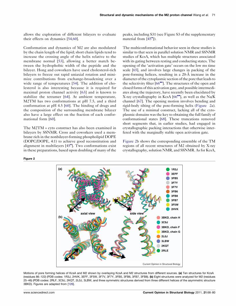
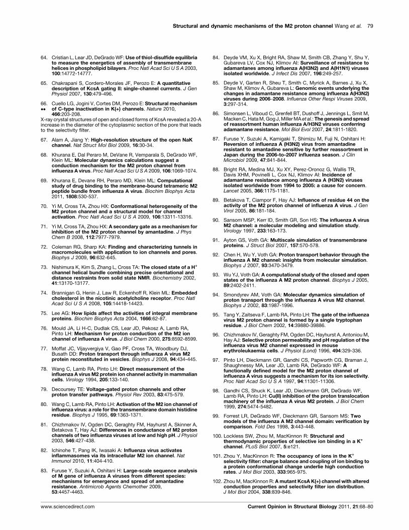
Figure 4
NH3+Cl-
NH3+Cl-
Amantadine
Spiranamine
(a) (b)
(d) (e)
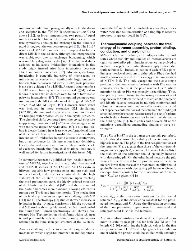
Docked conformation of four M2 inhibitors with M2 model generated from th
structure of amantadine bound M2 (PDB: 2KQT). Poses are shown with the am
(c) spiro-piperidine, (d) spiranamine, and (e) the structure of tetrabutylammo
hydrate near the entry of the selectivity filter, in a binding manner highly sim
displace the top two waters in the entry cluster.
Current Opinion in Structural Biology 2011, 21:68–80
mutations can easily be observed in vitro. The mutations
that cause the greatest decrease in inhibition, S31N and
V27A, increase the polarity of pore-lining residues.
Computational investigations of amantadine/rimantadine
action have involved molecular dynamics (MD) simu-
lations [69,71,92,120], docking [121,122] and small-mol-
ecule probe mapping [123,124]. Since these studies used
various M2 structures/models in varying protonation
states, it is not surprising that a consensus failed to emerge
from these studies. Using MD, Yi et al. found the aliphatic
region of amantadine snuggled against the bottom of the
Val 27 gate [71], while the drug was found at more C-
terminal locations in other simulations [92,125]. Khurana
et al. [69] performed MD simulations of different proto-
nation states of His37, using crystallographic and solution
NMR structures as starting conformations. They found
two distinct orientations of the drug in the pore, whose
populations depended on the protonation states of His37.
These orientations might correspond to the major and
minor conformers of amantadine within the pore
observed by SSNMR [28�].
Figure 4 shows the predominant orientation of the drug
[28�] docked onto the 1.65 A high-resolution structure
[39��]. The ammonium group projects downward
toward the entry cluster of water molecules, mimicking
Current Opinion in Structural Biology
N+
Rimantadine Spiro-piperidine
Tetrabutylammonium (TBA)
NH3+Cl-
H2+Cl-
N
(c)
e high resolution X-ray crystal structure of M2 (PDB: 3LBW) and SSNMR
ine pointing downwards toward His 37. (a) Amantadine, (b) rimantadine,
nium (TBA) bound to KcsA (PDB: 2HVK). Note: TBA displaces the K+
ilar to the binding of M2 inhibitors (a)–(d) to M2, which lie proximal to or
www.sciencedirect.com
Structural and dynamic mechanisms of the M2 proton channel Wang et al. 77
a hydronium ion in the act of passing a proton from the
central cavity to the His-box. While some rearrangement of
the outermost water molecules in the cluster is required to
accommodate the more extended drug, rimantadine, the
four water molecules associated with the His37 residues
appear well positioned to bridge between the sidechain
imidazole and the drug ammonium groups. This mode of
binding is also consistent with the high affinity of the
recently described spiro-piperidine class of inhibitors
[117], and has proved to be an excellent model for the
design of molecules that bind to the mutants V27A and
L26F [118].
ConclusionRecent years have seen breakthroughs in the structural
biology of M2, which in turn facilitate the mechanistic
analysis of proton translocation. High-resolution struc-
tures and dynamic measurements of M2 and mutants
under different conditions (pH, lipid compositions, etc.)
are needed to fully elucidate the mechanism. Mutant
structures are critical for understanding drug-resistance
and providing a basis for drug design. On the other hand,
inhibitors developed through either design or screening
can facilitate structure determination by stabilizing the
M2 tetramer in specific conformational forms.
AcknowledgementsThis work was funded by the NIH (GM56423 and AI74571). The authorswould like to thank Gevorg Grigoryan and Nate Joh for valuable commentsand help with preparing the figures.
References and recommended readingPapers of particular interest, published within the period of review,have been highlighted as:
� of special interest�� of outstanding interest
References1. DePristo MA, Weinreich DM, Hartl DL: Missense meanderings in
sequence space: a biophysical view of protein evolution. NatRev Genet 2005, 6:678-687.
2. Soichet BK, Baase WA, Kuroki R, Matthews BW: A relationshipbetween protein stability and protein function. Proc Natl AcadSci U S A 1995, 92:452-456.
3. Bowie JU: Solving the membrane protein folding problem.Nature 2005, 438:581-589.
4. Stanley AM, Fleming KG: The process of folding proteins intomembranes: challenges and progress. Arch Biochem Biophys2008, 469:46-66.
5. Pinto LH, Lamb RA: Influenza virus proton channels. PhotochemPhotobiol Sci 2006, 5:629-632.
6. Hille B: Ionic Channels of Excitable Membranes. Sunderland, MA:Sinauer Associates; 1992.
7. Gouaux E, Mackinnon R: Principles of selective ion transport inchannels and pumps. Science 2005, 310:1461-1465.
8. Choe S: Potassium channel structures. Nat Rev Neurosci 2002,3:115-121.
9. Hay AJ, Wolstenholme AJ, Skehel JJ, Smith MH: The molecularbasis of the specific anti-influenza action of amantadine.EMBO J 1985, 4:3021-3024.
www.sciencedirect.com
10. Pinto LH, Holsinger LJ, Lamb RA: Influenza virus M2 protein hasion channel activity. Cell 1992, 69:517-528.
11. Davies WL, Hoffmann CE, Paulshock M, Wood TR, Haff RF,Grunert RR, Watts JC, Hermann EC, Neumayer EM, McGahen JW:Antiviral activity of 1-adamantanamine (amantadine). Science1964, 144:862-863.
12. Rossman JS, Jing X, Leser GP, Lamb RA: Influenza virus M2protein mediates ESCRT-independent membrane scission.Cell 2010, 142:902-913.
13. Gannage M, Dormann D, Albrecht R, Dengjel J, Torossi T,Ramer PC, Lee M, Strowig T, Arrey F, Conenello G et al.: Matrixprotein 2 of influenza A virus blocks autophagosome fusionwith lysosomes. Cell Host Microbe 2009, 6:367-380.
14. Helenius A: Unpacking the incoming influenza virus. Cell 1992,69:577-578.
15. Grambas S, Hay AJ: Maturation of influenza a virushemagglutinin — estimates of the pH encountered duringtransport and its regulation by the M2 protein. Virology 1992,190:11-18.
16. Sakaguchi T, Leser GP, Lamb RA: The ion channel activity of theinfluenza virus M(2) protein affects transport through the Golgiapparatus. J Cell Biol 1996, 133:733-747.
17. Park EK, Castrucci MR, Portner A, Kawaoka Y: The M2ectodomain is important for its incorporation into influenza Avirions. J Virol 1998, 72:2449-2455.
18. Williams MA, Lamb RA: Determination of the orientation of anintegral membrane protein and sites of glycosylation byoligonucleotide-directed mutagenesis: influenza B virusNB glycoprotein lacks a cleavable signal sequence andhas an extracellular NH2-terminal region. Mol Cell Biol 1986,6:4317-4328.
19. Sunstrom NA, Premkumar LS, Premkumar A, Ewart G, Cox GB,Gage PW: Ion channels formed by NB, an influenza B virusprotein. J Membr Biol 1996, 150:127-132.
20. Ma C, Polishchuk AL, Ohigashi Y, Stouffer AL, Schon A,Magavern E, Jing X, Lear JD, Freire E, Lamb RA et al.:Identification of the functional core of the influenza A virus A/M2 proton-selective ion channel. Proc Natl Acad Sci U S A 2009,106:12283-12288.
21. Kochendoerfer GG, Salom D, Lear JD, Wilk-Orescan R, Kent SB,DeGrado WF: Total chemical synthesis of the integralmembrane protein influenza A virus M2: role of itsC-terminal domain in tetramer assembly. Biochemistry 1999,38:11905-11913.
22. Salom D, Hill BR, Lear JD, DeGrado WF: pH-dependenttetramerization and amantadine binding of thetransmembrane helix of M2 from theinfluenza A virus. Biochemistry 2000, 39:14160-14170.
23. Cady SD, Luo W, Hu F, Hong M: Structure and functionof the influenza A M2 proton channel. Biochemistry 2009,48:7356-7364.
24. Balannik V, Carnevale V, Fiorin G, Levine BG, Lamb RA, Klein ML,DeGrado WF, Pinto LH: Functional studies and modeling ofpore-lining residue mutants of the influenza a virus M2 ionchannel. Biochemistry 2010, 49:696-708.
25. Jing X, Ma C, Ohigashi Y, Oliveira FA, Jardetzky TS, Pinto LH,Lamb RA: Functional studies indicate amantadine binds to thepore of the influenza A virus M2 proton-selective ion channel.Proc Natl Acad Sci U S A 2008, 105:10967-10972.
26. Ohigashi Y, Ma C, Jing X, Balannick V, Pinto LH, Lamb RA: Anamantadine-sensitive chimeric BM2 ion channel of influenza Bvirus has implications for the mechanism of drug inhibition.Proc Natl Acad Sci U S A 2009, 106:18775-18779.
27. Schnell JR, Chou JJ: Structure and mechanism of the M2proton channel of influenza A virus. Nature 2008, 451:591-595.
28.�
Cady SD, Schmidt-Rohr K, Wang J, Soto CS, DeGrado WF,Hong M: Structure of the amantadine binding site ofinfluenza M2 proton channels in lipid bilayers. Nature 2010,463:689-692.
Current Opinion in Structural Biology 2011, 21:68–80
78 Folding and Binding
This report revealed the N-terminal channel lumen of M2 is the high-affinity, specific amantadine binding site using solid state NMR in phos-pholipid bilayer.
29. Rossman JS, Jing X, Leser GP, Balannik V, Pinto LH, Lamb RA:Influenza virus M2 ion channel protein is necessary forfilamentous virion formation. J Virol 2010, 84:5078-5088.
30. Martin K, Heleniust A: Nuclear transport of influenza virusribonucleoproteins: the viral matrix protein (M1) promotesexport and inhibits import. Cell 1991, 67:117-130.
31. Duff KC, Ashley RH: The transmembrane domain of influenza AM2 protein forms amantadine-sensitive proton channels inplanar lipid bilayers. Virology 1992, 190:485-489.
32. Manor J, Mukherjee P, Lin YS, Leonov H, Skinner JL, Zanni MT,Arkin IT: Gating mechanism of the influenza A M2 channelrevealed by 1D and 2D IR spectroscopies. Structure 2009,17:247-254.
33. Okada A, Miura T, Takeuchi H: Protonation of histidine andhistidine–tryptophan interaction in the activation of theM2 ion channel from influenza A virus. Biochemistry 2001,40:6053-6060.
34. Czabotar PE, Martin SR, Hay AJ: Studies of structural changesin the M2 proton channel of influenza A virus by tryptophanfluorescence. Virus Res 2004, 99:57-61.
35. Cady SD, Mishanina TV, Hong M: Structure of amantadine-bound M2 transmembrane peptide of influenza A in lipidbilayers from magic-angle-spinning solid-state NMR:the role of Ser31 in amantadine binding. J Mol Biol 2009,385:1127-1141.
36. Song Z, Kovacs FA, Wang J, Denny JK, Shekar SC, Quine JR,Cross TA: Transmembrane domain of M2 protein frominfluenza A virus studied by solid-state (15)N polarizationinversion spin exchange at magic angle NMR. Biophys J 2000,79:767-775.
37. Hu J, Fu R, Nishimura K, Zhang L, Zhou HX, Busath DD,Vijayvergiya V, Cross TA: Histidines, heart of the hydrogen ionchannel from influenza A virus: toward an understanding ofconductance and proton selectivity. Proc Natl Acad Sci U S A2006, 103:6865-6870.
38. Stouffer AL, Acharya R, Salom D, Levine AS, Di Costanzo L,Soto CS, Tereshko V, Nanda V, Stayrook S, DeGrado WF:Structural basis for the function and inhibition of an influenzavirus proton channel. Nature 2008, 451:596-599.
39.��
Acharya R, Carnevale V, Fiorin G, Levine BG, Polishchuk AL,Balannik V, Samish I, Lamb RA, Pinto LH, DeGrado WF et al.:Structure and mechanism of proton transport through thetransmembrane tetrameric M2 protein bundle of the influenzaA virus. Proc Natl Acad Sci U S A 2010, 107:15075-15080.
A high-resolution (1.6 A) crystal structure of the TM domain of M2, andaccompanying MD and QM calculations define the conduction pathwayfor protons transiting the M2 channel.
40. Stouffer AL, Ma C, Cristian L, Ohigashi Y, Lamb RA, Lear JD,Pinto LH, DeGrado WF: The interplay of functional tuning, drugresistance, and thermodynamic stability in the evolution of theM2 proton channel from the influenza A virus. Structure 2008,16:1067-1076.
41. Stouffer AL, Nanda V, Lear JD, DeGrado WF: Sequencedeterminants of a transmembrane proton channel: an inverserelationship between stability and function. J Mol Biol 2005,347:169-179.
42. Rosenberg MR, Casarotto MG: Coexistence of two adamantanebinding sites in the influenza A M2 ion channel. Proc Natl AcadSci U S A 2010, 107:13866-13871.
43. Andreas LB, Eddy MT, Pielak RM, Chou J, Griffin RG: Magic anglespinning NMR investigation of influenza A M2(18–60): supportfor an allosteric mechanism of inhibition. J Am Chem Soc 2010,132:10958-10960.
44. Pielak RM, Chou JJ: Solution NMR structure of the V27A drugresistant mutant of influenza A M2 channel. Biochem BiophysRes Commun 2010, 40:58-63.
Current Opinion in Structural Biology 2011, 21:68–80
45.�
Sharma M, Yi M, Dong H, Qin H, Peterson E, Busath DD, Zhou HX,Cross TA: Insight into the mechanism of the influenza a protonchannel from a structure in a lipid bilayer. Science 2010,330:509-512.
The backbone structure of membrane interactive AM2 domain (TM+Cytohelix) was determined by oriented solid-state NMR with previous EPRmeasurement.
46. Uysal S, Vasquez V, Tereshko V, Esaki K, Fellouse FA, Sidhu SS,Koide S, Perozo E, Kossiakoff A: Crystal structure of full-lengthKcsA in its closed conformation. Proc Natl Acad Sci U S A 2009,106:6644-6649.
47. Cortes DM, Cuello LG, Perozo E: Molecular architectureof full-length KcsA: role of cytoplasmic domains in ionpermeation and activation gating. J Gen Physiol 2001,117:165-180.
48. Kovacs FA, Cross TA: Transmembrane four-helix bundle ofinfluenza A M2 protein channel: structural implications fromhelix tilt and orientation. Biophys J 1997, 73:2511-2517.
49. Hu J, Asbury T, Achuthan S, Li C, Bertram R, Quine JR, Fu R,Cross TA: Backbone structure of the amantadine-blockedtrans-membrane domain M2 proton channel from influenza Avirus. Biophys J 2007, 92:4335-4343.
50. Wang J, Kim S, Kovacs F, Cross TA: Structure of thetransmembrane region of the M2 protein H(+) channel. ProteinSci 2001, 10:2241-2250.
51. Nguyen PA, Soto CS, Polishchuk A, Caputo GA, Tatko CD, Ma C,Ohigashi Y, Pinto LH, DeGrado WF, Howard KP: pH-inducedconformational change of the influenza M2 protein C-terminaldomain. Biochemistry 2008, 47:9934-9936.
52. Rossman JS, Lamb RA: Autophagy, apoptosis, and theinfluenza virus M2 protein. Cell Host Microbe 2009, 6:299-300.
53. Duong-Ly KC, Nanda V, DeGrado WF, Howard KP: Theconformation of the pore region of the M2 proton channeldepends on lipid bilayer environment. Protein Sci 2005,14:856-861.
54. Luo W, Cady SD, Hong M: Immobilization of the influenza A M2transmembrane peptide in virus envelope-mimetic lipidmembranes: a solid-state NMR investigation. Biochemistry2009, 48:6361-6368.
55. Cross TA, Sharma M, Yi M, Zhou HX: Influence of solubilizingenvironments on membrane protein structures. TrendsBiochem Sci 2010, in press, doi:10.1016/j.tibs.2010.07.005.
56. Schmidt N, Mishra A, Lai GH, Wong GC: Arginine-rich cell-penetrating peptides. FEBS Lett 2010, 584:1806-1813.
57. Wang J, Pielak RM, McClintock MA, Chou JJ: Solution structureand functional analysis of the influenza B proton channel. NatStruct Mol Biol 2009, 16:1267-1271.
58. Hirano M, Takeuchi Y, Aoki T, Yanagida T, Ide T: Rearrangementsin the KcsA cytoplasmic domain underlie its gating. J BiolChem 2010, 285:3777-3783.
59. Li C, Qin H, Gao FP, Cross TA: Solid-state NMR characterizationof conformational plasticity within the transmembranedomain of the influenza A M2 proton channel. Biochim BiophysActa 2007, 1768:3162-3170.
60. Hu F, Luo W, Cady SD, Hong M: Conformational plasticity of theinfluenza A M2 transmembrane helix in lipid bilayers undervarying pH, drug binding, and membrane thickness. BiochimBiophys Acta 2011, 1808:415-423.
61.�
Hu F, Luo W, Hong M: Mechanisms of proton conduction andgating in influenza m2 proton channels from solid-state NMR.Science 2010, 330:505-508.
Mechanism of proton conductance through M2 channel by His37 ringflipping was proposed by magic angle spinning SSNMR.
62. McDermott A: Structure and dynamics of membrane proteinsby magic angle spinning solid-state NMR. Annu Rev Biophys2009, 38:385-403.
63. Leiding T, Wang J, Martinsson J, DeGrado WF, Arskold SP: Protonand cation transport activity of the M2 proton channel frominfluenza A virus. Proc Natl AcadSciU S A 2010,107:15409-15414.
www.sciencedirect.com
Structural and dynamic mechanisms of the M2 proton channel Wang et al. 79
64. Cristian L, Lear JD, DeGrado WF: Use of thiol-disulfide equilibriato measure the energetics of assembly of transmembranehelices in phospholipid bilayers. Proc Natl Acad Sci U S A 2003,100:14772-14777.
65. Chakrapani S, Cordero-Morales JF, Perozo E: A quantitativedescription of KcsA gating II: single-channel currents. J GenPhysiol 2007, 130:479-496.
66.��
Cuello LG, Jogini V, Cortes DM, Perozo E: Structural mechanismof C-type inactivation in K(+) channels. Nature 2010,466:203-208.
X-ray crystal structures of open and closed forms of KcsA revealed a 20-Aincrease in the diameter of the cytoplasmic section of the pore that leadsto the selectivity filter.
67. Alam A, Jiang Y: High-resolution structure of the open NaKchannel. Nat Struct Mol Biol 2009, 16:30-34.
68. Khurana E, Dal Peraro M, DeVane R, Vemparala S, DeGrado WF,Klein ML: Molecular dynamics calculations suggest aconduction mechanism for the M2 proton channel frominfluenza A virus. Proc Natl Acad Sci U S A 2009, 106:1069-1074.
69. Khurana E, Devane RH, Peraro MD, Klein ML: Computationalstudy of drug binding to the membrane-bound tetrameric M2peptide bundle from influenza A virus. Biochim Biophys Acta2011, 1808:530-537.
70. Yi M, Cross TA, Zhou HX: Conformational heterogeneity of theM2 proton channel and a structural model for channelactivation. Proc Natl Acad Sci U S A 2009, 106:13311-13316.
71. Yi M, Cross TA, Zhou HX: A secondary gate as a mechanism forinhibition of the M2 proton channel by amantadine. J PhysChem B 2008, 112:7977-7979.
72. Coleman RG, Sharp KA: Finding and characterizing tunnels inmacromolecules with application to ion channels and pores.Biophys J 2009, 96:632-645.
73. Nishimura K, Kim S, Zhang L, Cross TA: The closed state of a H+
channel helical bundle combining precise orientational anddistance restraints from solid state NMR. Biochemistry 2002,41:13170-13177.
74. Brannigan G, Henin J, Law R, Eckenhoff R, Klein ML: Embeddedcholesterol in the nicotinic acetylcholine receptor. Proc NatlAcad Sci U S A 2008, 105:14418-14423.
75. Lee AG: How lipids affect the activities of integral membraneproteins. Biochim Biophys Acta 2004, 1666:62-87.
76. Mould JA, Li H-C, Dudlak CS, Lear JD, Pekosz A, Lamb RA,Pinto LH: Mechanism for proton conduction of the M2 ionchannel of influenza A virus. J Biol Chem 2000, 275:8592-8599.
77. Moffat JC, Vijayvergiya V, Gao PF, Cross TA, Woodbury DJ,Busath DD: Proton transport through influenza A virus M2protein reconstituted in vesicles. Biophys J 2008, 94:434-445.
78. Wang C, Lamb RA, Pinto LH: Direct measurement of theinfluenza A virus M2 protein ion channel activity in mammaliancells. Virology 1994, 205:133-140.
79. Decoursey TE: Voltage-gated proton channels and otherproton transfer pathways. Physiol Rev 2003, 83:475-579.
80. Wang C, Lamb RA, Pinto LH: Activation of the M2 ion channel ofinfluenza virus: a role for the transmembrane domain histidineresidue. Biophys J 1995, 69:1363-1371.
81. Chizhmakov IV, Ogden DC, Geraghty FM, Hayhurst A, Skinner A,Betakova T, Hay AJ: Differences in conductance of M2 protonchannels of two influenza viruses at low and high pH. J Physiol2003, 546:427-438.
82. Ichinohe T, Pang IK, Iwasaki A: Influenza virus activatesinflammasomes via its intracellular M2 ion channel. NatImmunol 2010, 11:404-410.
83. Furuse Y, Suzuki A, Oshitani H: Large-scale sequence analysisof M gene of influenza A viruses from different species:mechanisms for emergence and spread of amantadineresistance. Antimicrob Agents Chemother 2009,53:4457-4463.
www.sciencedirect.com
84. Deyde VM, Xu X, Bright RA, Shaw M, Smith CB, Zhang Y, Shu Y,Gubareva LV, Cox NJ, Klimov AI: Surveillance of resistance toadamantanes among influenza A(H3N2) and A(H1N1) virusesisolated worldwide. J Infect Dis 2007, 196:249-257.
85. Deyde V, Garten R, Sheu T, Smith C, Myrick A, Barnes J, Xu X,Shaw M, Klimov A, Gubareva L: Genomic events underlying thechanges in adamantane resistance among influenza A(H3N2)viruses during 2006–2008. Influenza Other Respi Viruses 2009,3:297-314.
86. Simonsen L, Viboud C, Grenfell BT, Dushoff J, Jennings L, Smit M,Macken C, Hata M, Gog J, Miller MA et al.: The genesis and spreadof reassortment human influenza A/H3N2 viruses conferringadamantane resistance. Mol Biol Evol 2007, 24:1811-1820.
87. Furuse Y, Suzuki A, Kamigaki T, Shimizu M, Fuji N, Oshitani H:Reversion of influenza A (H3N2) virus from amantadineresistant to amantadine sensitive by further reassortment inJapan during the 2006-to-2007 influenza season. J ClinMicrobiol 2009, 47:841-844.
88. Bright RA, Medina MJ, Xu XY, Perez-Oronoz G, Wallis TR,Davis XHM, Povinelli L, Cox NJ, Klimov AI: Incidence ofadamantane resistance among influenza A (H3N2) virusesisolated worldwide from 1994 to 2005: a cause for concern.Lancet 2005, 366:1175-1181.
89. Betakova T, Ciampor F, Hay AJ: Influence of residue 44 on theactivity of the M2 proton channel of influenza A virus. J GenVirol 2005, 86:181-184.
90. Sansom MSP, Kerr ID, Smith GR, Son HS: The influenza A virusM2 channel: a molecular modeling and simulation study.Virology 1997, 233:163-173.
91. Ayton GS, Voth GA: Multiscale simulation of transmembraneproteins. J Struct Biol 2007, 157:570-578.
92. Chen H, Wu Y, Voth GA: Proton transport behavior through theinfluenza A M2 channel: insights from molecular simulation.Biophys J 2007, 93:3470-3479.
93. Wu YJ, Voth GA: A computational study of the closed and openstates of the influenza A M2 proton channel. Biophys J 2005,89:2402-2411.
94. Smondyrev AM, Voth GA: Molecular dynamics simulation ofproton transport through the influenza A virus M2 channel.Biophys J 2002, 83:1987-1996.
95. Tang Y, Zaitseva F, Lamb RA, Pinto LH: The gate of the influenzavirus M2 proton channel is formed by a single tryptophanresidue. J Biol Chem 2002, 14:39880-39886.
96. Chizhmakov IV, Geraghty FM, Ogden DC, Hayhurst A, Antoniou M,Hay AJ: Selective proton permeability and pH regulation of theinfluenza virus M2 channel expressed in mouseerythroleukaemia cells. J Physiol (Lond) 1996, 494:329-336.
97. Pinto LH, Dieckmann GR, Gandhi CS, Papworth CG, Braman J,Shaughnessy MA, Lear JD, Lamb RA, DeGrado WF: Afunctionally defined model for the M2 proton channel ofinfluenza A virus suggests a mechanism for its ion selectivity.Proc Natl Acad Sci U S A 1997, 94:11301-11306.
98. Gandhi CS, Shuck K, Lear JD, Dieckmann GR, DeGrado WF,Lamb RA, Pinto LH: Cu(II) inhibition of the proton translocationmachinery of the influenza A virus M2 protein. J Biol Chem1999, 274:5474-5482.
99. Forrest LR, DeGrado WF, Dieckmann GR, Sansom MS: Twomodels of the influenza A M2 channel domain: verification bycomparison. Fold Des 1998, 3:443-448.
100. Lockless SW, Zhou M, MacKinnon R: Structural andthermodynamic properties of selective ion binding in a K+
channel. PLoS Biol 2007, 5:e121.
101. Zhou Y, MacKinnon R: The occupancy of ions in the K+
selectivity filter: charge balance and coupling of ion binding toa protein conformational change underlie high conductionrates. J Mol Biol 2003, 333:965-975.
102. Zhou M, MacKinnon R: A mutant KcsA K(+) channel with alteredconduction properties and selectivity filter ion distribution.J Mol Biol 2004, 338:839-846.
Current Opinion in Structural Biology 2011, 21:68–80
80 Folding and Binding
103. Morais-Cabral JH, Zhou Y, MacKinnon R: Energetic optimizationof ion conduction rate by the K+ selectivity filter. Nature 2001,414:37-42.
104. Berneche S, Roux B: Energetics of ion conduction through theK+ channel. Nature 2001, 414:73-77.
105. Phongphanphanee S, Rungrotmongkol T, Yoshida N,Hannongbua S, Hirata F: Proton transport through theinfluenza A M2 channel: three-dimensional referenceinteraction site model study. J Am Chem Soc 2010,132:9782-9788.
106. Carnevale V, Fiorin G, Levine BG, DeGrado WF, Klein ML: Multipleproton confinement in the M2 channel from the influenza Avirus. J Phys Chem C 2010, 114:20856-20863.
107. Zhou HX: Diffusion-influenced transport of ions across atransmembrane channel with an internal binding site. J PhysChem Lett 2010, 1:1973-1976.
108. Wu Y, Voth GA: Computational studies of proton transportthrough the M2 channel. FEBS Lett 2003, 552:23-27.
109. Voth GA: The computer simulation of proton transport inbiomolecular systems. Front Biosci 2003, 8:1384-1397.
110. Headrick JM, Diken EG, Walters RS, Hammer NI, Christie RA,Cui J, Myshakin EM, Duncan MA, Johnson MA, Jordan KD:Spectral signatures of hydrated proton vibrations in waterclusters. Science 2005, 308:1765-1769.
111. Waters ML: Aromatic interactions in model systems. Curr OpinChem Biol 2002, 6:736-741.
112. Song XJ, McDermott AE: Proton transfer dynamics and N–Hbond lengthening in N–H center dot center dot center dot Nmodel systems: a solid-state NMR study. Magn Reson Chem2001, 39:S37-S43.
113. Perrin CL, Nielson JB: ‘‘Strong’’ hydrogen bonds in chemistryand biology. Annu Rev Phys Chem 1997, 48:511-544.
114. Luo W, Hong M: Conformational changes of an ionchannel detected through water–protein interactions usingsolid-state NMR spectroscopy. J Am Chem Soc 2010,132:2378-2384.
115. Renart ML, Triano I, Poveda JA, Encinar JA, Fernandez AM, Ferrer-Montiel AV, Gomez J, Gonzalez Ros JM: Ion binding to KcsA:implications in ion selectivity and channel gating. Biochemistry2010, 49:9480-9487.
Current Opinion in Structural Biology 2011, 21:68–80
116. Hu J, Fu R, Cross TA: The chemical and dynamical influence ofthe anti-viral drug amantadine on the M2 proton channeltransmembrane domain. Biophys J 2007, 93:276-283.
117. Wang J, Cady SD, Balannik V, Pinto LH, DeGrado WF, Hong M:Discovery of spiro-piperidine inhibitors and their modulationof the dynamics of the M2 proton channel from influenza Avirus. J Am Chem Soc 2009, 131:8066-8076.
118. Balannik V, Wang J, Ohigashi Y, Jing X, Magavern E, Lamb RA,Degrado WF, Pinto LH: Design and pharmacologicalcharacterization of inhibitors of amantadine-resistantmutants of the M2 ion channel of influenza A virus.Biochemistry 2009, 48:11872-11882.
119. Furuse Y, Suzuki A, Kamigaki T, Oshitani H: Evolution of the Mgene of the influenza A virus in different host species: large-scale sequence analysis. Virol J 2009, 6:67.
120. Qin G, Yu K, Shi T, Luo C, Li G, Zhu W, Jiang H: How doesinfluenza virus a escape from amantadine? J Phys Chem B2010, 114:8487-8493.
121. Eleftheratos S, Spearpoint P, Ortore G, Kolocouris A, Martinelli A,Martin S, Hay A: Interaction of aminoadamantane derivativeswith the influenza A virus M2 channel — docking using a poreblocking model. Bioorg Med Chem Lett 2010, 20:4182-4187.
122. Wang JF, Wei DQ, Chou KC: Insights from investigating theinteractions of adamantane-based drugs with the M2 protonchannel from the H1N1 swine virus. Biochem Biophys ResCommun 2009, 388:413-417.
123. Kozakov D, Chuang GY, Beglov D, Vajda S: Where doesamantadine bind to the influenza virus M2 proton channel?Trends Biochem Sci 2010, 35:471-475.
124. Chuang GY, Kozakov D, Brenke R, Beglov D, Guarnieri F, Vajda S:Binding hot spots and amantadine orientation in the influenzaA virus M2 proton channel. Biophys J 2009, 97:2846-2853.
125. Laohpongspaisan C, Rungrotmongkol T, Intharathep P,Malaisree M, Decha P, Aruksakunwong O, Sompornpisut P,Hannongbua S: Why amantadine loses its function ininfluenza m2 mutants: MD simulations. J Chem Inf Model 2009,49:847-852.
126. Grigoryan G, Moore DT, DeGrado WF: TransmembraneCommunication: General Principles and Lessons from theStructure and Function of the M2 Proton Channel, K+Channels, and Integrin Receptors. Annu Rev Biochem. 2011, 80.
www.sciencedirect.com
Top Related
Copyright © 2022 FDOKUMEN

