


Bahasa
Halaman
Hukum

ORIGINAL INVESTIGATION
Stroke lesion in cortical neural circuits and post-stroke incidence of major depressive episode: A 4-month prospective study
LUISA TERRONI 1 , EDSON AMARO JR 2 , DAN V. IOSIFESCU 3 , GISELA TINONE 4 , JO Ã O RICARDO SATO 5 , CLAUDIA COSTA LEITE 2 , MATILDES F. M. SOBREIRO 6 , MARA CRISTINA SOUZA LUCIA 7 , MILBERTO SCAFF 4 & REN É RIO FR Á GUAS 8
1 Liaison Psychiatry Group, Department and Institute of Psychiatry, Clinical Hospital, Medical School, University of S ã o Paulo, S ã o Paulo, Brazil, 2 Department of Radiology, Clinical Hospital, Medical School, University of S ã o Paulo, S ã o Paulo, Brazil, 3 Mood and Anxiety Disorders Program, Mount Sinai School of Medicine, New York, NY, USA, 4 Department of Neurology, Clinical Hospital, Medical School, University of São Paulo, São Paulo, Brazil, 5 Center of Mathematics, Computation and Cognition, Federal University of ABC and Department of Radiology, Clinical Hospital, Medical School, University of São Paulo, São Paulo, Brazil, 6 Liaison Psychiatry Group, Department and Institute of Psychiatry, Clinical Hospital, Medical School, University of São Paulo, São Paulo, Brazil, 7 Department of Neurology, Division of Psychology, Clinical Hospital, Medical School, University of São Paulo, Brazil, and 8 Liaison Psychiatry Group, Laboratory of Psychiatric Neuroimaging (LIM-21), Department and Institute of Psychiatry, Clinical Hospital, Medical School, University of São Paulo, São Paulo, Brazil
Abstract Objective. Little is known about the relevance of lesion in neural circuits reported to be associated with major depressive disorder. We investigated the association between lesion stroke size in the limbic-cortical-striatal-pallidal-thalamic (LCSPT) circuit and incidence of major depressive episode (MDE). Methods. We enrolled 68 patients with fi rst-ever ischemic stroke and no history of major depressive disorder. Neurological and psychiatric examinations were performed at three time-points. We diagnosed major depressive episode, following DSM-IV criteria. Lesion location and volume were determined with mag-netic resonance imaging, using a semi-automated method based on the Brodmann Cytoarchitectonic Atlas. Results. Twenty-one patients (31%) experienced major depressive episode. Larger lesions in the left cortical regions of the LCSPT circuit (3,760 vs. 660 mm 3 ; P � 0.004) were associated with higher incidence of MDE. Secondary analyses revealed that major depressive episode was associated with larger lesions in areas of the medial prefrontal cortex including the ventral (BA24) and dorsal anterior cingulate cortex (BA32) and subgenual cortex (BA25); and also the subiculum (BA28/36) and amygdala (BA34). Conclusions Our fi ndings indicate that depression due to stroke is aetiologically related to the disruption of the left LCSPT circuit and support the relevance of the medial prefrontal cortex dysfunction in the pathophysiology of depression.
Key words: Cingulate gyrus , depression disorder , magnetic resonance imaging , prefrontal cortex , stroke
Introduction
The reported prevalence of major depression within 3 months after stroke ranges from 22 to 31% (Robinson et al. 1984b; Astrom et al. 1993; Terroni et al. 2003; Spalletta et al. 2005). Efforts to identify biological and psychosocial mechanisms have provided evidence that the aetiology of post-stroke depression is multifactorial (Robinson et al. 1984a; Astrom et al. 1993; Andersen et al. 1995; Vataja et al. 2001). How-ever, research focusing on stroke location has been a fruitful strategy in understanding the pathophysiology
of post-stroke depression (Robinson et al. 1984a; Mayberg et al. 1988; Astrom et al. 1993; Vataja et al. 2001; Vataja et al. 2004). From a clinical perspective, knowing which patients are at increased risk of devel-oping post-stroke depression may ameliorate the prevention, detection, and early treatment of depres-sion, consequently reducing its the negative impact on the recovery of stroke patients (Ramasubbu and Kennedy 1994).
Although there is no consensus about the relationship between lesion location and post-stroke depression
Correspondence: Luisa Terroni, Departamento e Instituto de Psiquiatria HCFMUSP-SP, R. Dr. Ov í dio Pires de Campos, 785 – 3 ° andar, Caixa Postal 3671, CEP 05403-010, S ã o Paulo, SP, Brazil. Tel: � 55 11 30697804. Fax: � 55 11 30697804. E-mail: [email protected]
(Received 25 May 2010 ; accepted 7 February 2011 )
The World Journal of Biological Psychiatry, 2011; 12: 539–548
ISSN 1562-2975 print/ISSN 1814-1412 online © 2011 Informa HealthcareDOI: 10.3109/15622975.2011.562242
Wor
ld J
Bio
l Psy
chia
try
Dow
nloa
ded
from
info
rmah
ealth
care
.com
by
UN
ICA
MP
on 0
4/05
/13
For
pers
onal
use
onl
y.
540 L. Terroni et al.
depression, we also investigated the relationship between lesions in specifi c brain areas incorporated in those circuits and incidence of major depressive episodes.
Methods
Patients
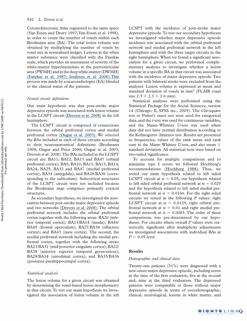
We screened 326 male and female patients, 18 years of age or older, consecutively admitted to the Neurology Unit of a University Hospital with a diag-nosis of ischemic stroke between August of 2002 and May of 2008. The diagnosis of stroke was made by a neurologist in accordance with the World Health Organization criteria (WHO 1989) and confi rmed by MRI. A psychiatrist administered the modules for mood episodes, psychotic symptoms and substance use disorders of the Structured Clinical Interview (SCID) for Diagnostic and Statistical Manual of Mental Disorders fourth edition (DSM-IV) to investigate past and current psychiatric disorders (American Psychiatric Association 1994; First 1995). This interview was performed with the patient and a family member/caregiver present when possible. Patients with previous history of stroke or other cen-tral nervous system diseases (i.e. amyotrophic lateral sclerosis, subarachnoid haemorrhage, Binswanger ' s disease, brain tumours, or multiple sclerosis) were excluded from the study, as were those with infraten-torial stroke, a severe clinical condition that impeded the interview, Cushing ' s syndrome, alcohol or drug dependence in the last 12 months, previous history of major depressive episode or bipolar disorder, cur-rent major depressive episode or bipolar disorder with pre-stroke onset, psychotic disorder, dementia, or aphasia that impeded the interview. On the basis of these criteria, we excluded 234 patients (Figure 1): history stroke, infratentorial stroke, greater than 2-week interval between stroke occurrence and screening interview, or haemorrhagic transformation of stroke ( N � 89); drug/alcohol dependence, psy-choses, delirium, history of major depressive episode, current major depressive episode with pre-stroke onset, dysthymia, or bipolar disorder ( N � 54); aphasia that impeded the interview ( N � 37); neu-rological diseases or severe clinical condition that impeded the interview ( N � 22); and other reasons ( N � 32). Of the remaining 92 patients, fi ve declined to participate in the study. Of those 87 patients, 19 were later excluded due to problems during the MRI acquisition: image was inappropriate for use in the present study ( N � 15); claustrophobia ( N � 3); and haemorrhagic transformation of stroke ( N � 1). Therefore, we enrolled a fi nal sample of 68 patients. Nine patients dropped out after the fi rst time-point
(Singh et al. 1998; Carson et al. 2000; Bhogal et al. 2004; Hackett and Anderson 2005), some studies using computed tomography, magnetic resonance imaging (MRI) (Robinson et al. 1984a; Astrom et al. 1993; Vataja et al. 2001, 2004) and PET imaging of cortical S2 serotonin receptors (Mayberg et al. 1988) have suggested that post-stroke depression is associ-ated with the proximity of the lesion to the frontal lobe and with left hemisphere stroke. In addition, studies in major depressive disorder with PET (Baxter et al. 1989; Drevets et al. 1992), and with catecholamine depletion (Hasler et al. 2008), have found abnormal prefrontal function, more commonly in the left than in the right hemisphere.
MRI studies (Vataja et al. 2001, 2004) have reported a high prevalence of post-stroke depression in lesions affecting some structures of the prefronto-subcortical circuit, particularly in the left hemisphere, a circuit that has been reported to be involved in various neu-ropsychiatry syndromes, including depression. Recent studies have highlighted the specifi c relevance of the limbic-cortical-striatal-pallidal-thalamic (LCSPT) circuit in the pathophysiology of major depressive dis-order (Drevets et al. 2008; Hasler et al. 2008). Based on evidence from animal studies, Drevets et al. (2008) proposed that in addition to the LCPSPT circuit, two other circuits are essential for emotional regulation. The fi rst, the orbital prefrontal network, is involved in a system of reward, aversion, and relative value. The second, the medial prefrontal network, has con-nections with limbic and visceral control structures that are involved in introspective functions such as mood and emotion, and visceral reactions to emo-tional stimuli such as autonomic regulation and neu-roendocrine responses. The prefrontal cortex, which includes areas belonging to all these circuits, has been implicated in response to treatment for major depres-sive disorder (Mayberg et al. 1997; Brody et al. 1999; Pizzagalli et al. 2001).
To our knowledge, there have been no studies investigating the relationship between stroke lesions in LCSPT circuit and the development of post-stroke depression. Therefore, the primary aim of the pres-ent study was to investigate the association between lesion volume in the left LCSPT circuit and the inci-dence of major depressive episode in the fi rst four months after stroke. We focused on the left hemi-sphere as the left lateralization of stroke has been repeatedly associated with post-stroke depression (Robinson et al. 1984a; Astrom et al. 1993; Vataja et al. 2001, 2004; Bhogal et al. 2004). As secondary hypothesis, we investigated the association between the incidence of major depressive episode and lesions involving cortical regions of the orbital and medial prefrontal networks. When cortical circuits were found to be statistically associated with post-stroke
Wor
ld J
Bio
l Psy
chia
try
Dow
nloa
ded
from
info
rmah
ealth
care
.com
by
UN
ICA
MP
on 0
4/05
/13
For
pers
onal
use
onl
y.

Stroke lesion and major depression 541
MRI methods
The MRIs were acquired in general within two weeks after stroke (9.34 � 6.87; range 1 – 43 days). All images were acquired using a 1.5-Tesla system (GE-Horizon LX). The imaging protocol included axial spoiled gradient recalled acquisition in steady state (SPGR, TR � 27 ms; fl ip angle � 45 ° ; voxel size � 0.94 � 0.94 � 1.5 mm), axial fl uid attenu-ated inversion recovery (FLAIR, TR � 133 ms; TE � 8400 ms; TI � 2100 ms; voxel size � 0.94 � 0.94 � 5 mm), axial diffusion-weighted image (TR � 8000 ms; b value � 1000 s/mm 2 ; voxel size � 1.8 � 1.8 � 5 mm), T2-weighted fast spin echo (TR � 4500 ms; TE � 100 – 120 ms; voxel size � 0.94 � 0.94 � 5 mm). All images were acquired in the bicommissural plane.
Lesion location and volume quantifi cation were determined using a semi-automated method. Ini-tially, SPGR and axial FLAIR acquisitions were both normalized to the Montreal Neurological Institute template (Evans et al. 1993) using linear transforma-tion with 12 degrees of freedom and 15 nonlinear interactions implemented in Statistical Parametric Mapping (SPM5,Wellcome Trust for Neuroimaging, London, http://www.fi l.ion.ucl.ac.uk/spm/) (Friston et al. 1996), and based on coordinates referenced in the Talairach and Tournoux Atlas (Talairach and Tornoux 1988). During this process, all images were sampled to 2.3 � 2.3 � 2.6 mm. Lesion delin-eation was performed by a trained psychiatrist (LT) using a mouse device to trace the ischemic lesion and analyzing all slices of each FLAIR image using MRIcro Software (http://www.sph.sc.edu/comd/rorden/mricro.html) (Rorden and Brett 2000). All lesions delineations of each patient were reviewed by a neuroradiologist (EAJ), blinded for clinical data and psychiatric diagnoses.
RF had elaborated the main hypothesis of the study related to the lesions in the LCSPT and post-stroke depression but did not disclose it to the raters of MRI lesions (LT, EA) while lesion delineation was taking place, in order to preserve the blinding. Both raters delineating MRI lesions (LT, EAJ) were only aware they would study size and location of the lesions using the Brodmann Map in relation to post stroke depression. Both raters (LT, EA) were also blind to the clinical outcome status (post stroke depression episodes) of the patients while delineating lesions on MRIs. After fi nishing the lesion delinea-tion process, RF (who had not participated in lesion delineation) revealed the specifi c hypothesis to be tested. Diffusion-weighted images were also ana-lyzed in order to distinguish between other possible differential diagnoses. The regions of interest were then analyzed automatically using the Brodmann
examination and four patients dropped out after the second time-point examination. The institutional review board of the Clinics Hospital approved the study protocol and, written informed consent was obtained from all participating patients.
Clinical assessments
The enrolled patients were evaluated at three dif-ferent time-points: between post-stroke days 5 and 25 (mean � SD; 12.0 � 4.5; range, 5 – 23 days); between post-stroke days 30 and 59 (37.0 � 6.0; 30 – 57 days); and, between post-stroke days 80 and 110 (91.6 � 5.4; 83 – 108 days). In all three evalua-tions, the diagnosis of major depressive episode was made by an experienced psychiatrist (LT), blinded for imaging data, using the SCID for DSM-IV, Axis I disorders (American Psychiatric Association 1994; First 1995). In the fi rst evaluation in the Neurology Unit, as we described in a previous work (Terroni et al. 2009), the diagnosis of major depres-sive episode was made considering a period of 1 week, as have been done in others studies (Robinson et al. 1984a; Astrom et al. 1993; Caeiro et al. 2006). The 31-item Hamilton Rating Scale for Depression (HAM – D-31) (Williams 1988; Jamerson et al. 2003) was used at every visit to assess the severity of depres-sive symptoms. At all three time-points, a neurologist (GT), certifi ed by the National Institutes of Health to administer the National Institutes of Health Stroke Scale (NIHSS) (Brott et al. 1989), blinded for imaging data and psychiatric diagnoses, evaluated the stroke severity using the NIHSS and the impair-ment of activities of daily living using the Barthel Index (Herndon 1997). Cognitive performance was assessed at the fi rst and third time-points with the Mini-Mental State Examination (MMSE) (Folstein et al. 1975) administered by a neuropsychologist (MS) blinded for imaging data and psychiatric diagnoses.
Inclusion criteria not met (n=234) No agreement to participate (n=5)
Enrolled patients followed for 4 months (n = 87)
Problems in acquisition of MRI scans (n=19)
Clinical diagnosis of stroke confirmed by MRI (n = 326)
Final sample First evaluation: 5–25 days after stroke (n = 68)
Second evaluation: 30–57 days after stroke (n = 59) Third evaluation: 80–110 days after stroke (n = 55)
Figure 1. Patient fl ow.
Wor
ld J
Bio
l Psy
chia
try
Dow
nloa
ded
from
info
rmah
ealth
care
.com
by
UN
ICA
MP
on 0
4/05
/13
For
pers
onal
use
onl
y.
542 L. Terroni et al.
LCSPT with the incidence of post-stroke major depressive episode. To test our secondary hypotheses we investigated whether major depressive episode incidence was associated with the orbital prefrontal network and medial prefrontal network in the left hemisphere and with the three target circuits in the right hemisphere. When we found a signifi cant asso-ciation for a given circuit, we performed comple-mentary analyses to investigate whether a lesion volume in a specifi c BA in that circuit was associated with the incidence of major depressive episode. Two patients with bilateral stroke were excluded from the analyses. Lesion volume is expressed as mean and standard deviation of voxels in mm 3 (FLAIR voxel size 2.3 � 2.3 � 2.6 mm).
Statistical analyses were performed using the Statistical Package for the Social Sciences, version 14 (Chicago: Il, SPSS inc., 2005). The Chi - square test or Fisher ' s exact test were used for categorical data, and the t -test was used for continuous variables, and the Mann – Whitney U -test was used when data did not have normal distribution according to the Kolmogorov – Smirnov test. Results are presented as frequencies, values of the rank mean and rank sum in the Mann – Whitney U -test, and also mean � standard deviation. All statistical tests were based on two-tailed signifi cance.
To account for multiple comparisons and to minimize type I errors we followed Hochberg ’ s recommendations (Hochberg 1988). Thus, we tested our main hypothesis related to left sided LCSPT circuit at α � 0.05, our hypothesis related to left sided orbital prefrontal network at α � 0.025 and the hypothesis related to left sided medial pre-frontal network at α � 0.0166. For the right sided circuits we tested at the following P values: right LCSPT circuit at α � 0.0125, right orbital pre-frontal network at α � 0.01 and right medial pre-frontal network at α � 0.0083. The order of these comparisons was pre-determined by our hypo-theses. For circuits where overall P values were sta-tistically signifi cant after multiplicity adjustments we investigated associations with individual BAs at P � 0.05 level.
Results
Demographic and clinical data
Twenty-one patients (31%) were diagnosed with a new-onset major depressive episode, including seven at the time of the fi rst evaluation, fi ve at the second and, nine at the third evaluation. The depressed patients were comparable to those without major depressive episode in terms of sociodemographic, clinical, neurological, lesions in white matter, and
Cytoarchitectonic Atlas registered to the same space (Van Essen and Drury 1997; Van Essen et al. 1998), in order to count the number of voxels within each Brodmann area (BA). The total lesion volume was obtained by multiplying the number of voxels by voxel size in normalized images. Lesions in the white matter substance were classifi ed with the Fazekas scale, which provides an assessment of severity of the white-matter hyperintensities in the periventricular area (PWMH) and in the deep white-matter (DWMH) (Fazekas et al. 1987; Iosifescu et al. 2006).This process was made by a neuroradiologist (EA) blinded to the clinical status of the patients.
Neural circuit: defi nitions
Our main hypothesis was that post-stroke major depressive episode was associated with lesion volume in the LCSPT circuit (Drevets et al. 2008) in the left hemisphere.
The LCSPT circuit is composed of connections between the orbital prefrontal cortex and medial prefrontal cortex (Ongur et al. 2003). We selected the BAs included in each of these circuits according to their neuroanatomical defi nitions (Brodmann 1909; Ongur and Price 2000; Ongur et al. 2003; Drevets et al. 2008). The BAs included in the LCSPT circuit are: BA11, BA12, BA13 and BA47 (orbital prefrontal cortex); BA9, BA10, BA11, BA13, BA14, BA24, BA25, BA32 and BA47 (medial prefrontal cortex); BA34 (amygdala); and BA28/BA36 (corre-sponding to the subiculum). Subcortical structures of the LCSPT circuit were not included because the Brodmann map comprises primarily cortical structures.
As secondary hypotheses, we investigated the asso-ciation between post-stroke major depressive episode and two networks (Drevets et al. 2008). The orbital prefrontal network includes the orbital prefrontal cortex together with the following areas: BA20 (infe-rior temporal cortex); BA13/BA14 (insula); BA44/BA45 (frontal operculum); BA27/BA34 (olfactory cortex); and BA43 (taste cortex). The second, the medial prefrontal network including the medial pre-frontal cortex, together with the following areas: BA23/BA31 (mid/posterior cingulate cortex); BA22/BA38 (anterior superior temporal gyrus/sulcus); BA28/BA34 (entorhinal cortex); and BA35/BA36 (posterior parahippocampal cortex).
Statistical analysis
The lesion volume for a given circuit was obtained by determining the voxel-based lesion morphometry in that circuit. To test our main hypothesis we inves-tigated the association of lesion volume in the left
Wor
ld J
Bio
l Psy
chia
try
Dow
nloa
ded
from
info
rmah
ealth
care
.com
by
UN
ICA
MP
on 0
4/05
/13
For
pers
onal
use
onl
y.
Stroke lesion and major depression 543
cognitive aspects, with the exception of an increased rate of Diabetes Mellitus among depressed patients (Table I). Thirty-nine patients (57.4%) had suffered a left hemisphere stroke, 27 (39.7%) had suffered a right hemisphere stroke and 2 (2.9%) had suffered a bilateral stroke.
There were no signifi cant differences between the patients included in the fi nal study ( N � 68) and those who had been excluded because of problems in the MRI acquisitions ( N � 19) in terms of socio-demographic, neurological, and cognitive measures, except for the fact that NIHSS scores were lower in the studied patients at the fi rst time-point, but not at the second and third time-points.
Analysis of lesion location and volume in neural circuit
Patients with fi rst major depressive episode after stroke had larger lesion volume in the left LCSPT circuit than non-depressed patients (respectively 3,760 vs. 660 mm 3 ; P � 0.004; Table II). The inci-dence of major depressive episode was also associ-ated with lesion volume in the left orbital prefrontal network ( P � 0.037), our secondary hypothesis, but this comparison did not reach statistical signi-fi cance after Hochberg ' s multiplicity adjustments ( P � 0.025). Complementary analyses revealed that major depressive episode incidence was associated with specifi c areas of the left LCSPT circuit (Table III) including the ventral anterior cingulate cortex (BA24), subgenual cortex (BA25), subiculum (BA28/BA36), amygdala (BA34), and dorsal anterior cingulate cortex (BA32).
Due to the increased rate of diabetes mellitus among depressed patients and its potential con-founding effect, we investigate the association between diabetes and stroke location. The presence of diabetes mellitus was not associated with lesions in the LCSPT circuit ( P � 1.00). The distribution of handedness among patients with left hemisphere lesions was similar between those with and without major depressive episode. Among patients with left hemisphere lesions all the 12 (100%) patients with major depressive episode were right-handed and regarding the non-depressed patients 25 (92.6%) were right-handed and 2 (7.4%) were left-handed ( P � 1.00)
Discussion
In this 4-month prospective study, we found an asso-ciation between the post-stroke incidence of major depressive episode and lesion volume in the left LCSPT circuit. Although this neural circuit was pre-viously described to be affected in major depressive
Table I. Demographic and clinical characteristics of patients with and without major depressive episode after stroke.
Major depressive episode
YesN (%)
NoN (%) P value
Patients 21 (31) 47 (69)Sex
Female 10 (48) 22 (47)Male 11 (52) 25 (53) 0.951
Marriedyes 15 (71) 29 (62)no 6 (29) 18 (38) 0.606
Level of educational�8 years of schooling 16 (76) 32 (68)�9 years of schooling 5 (24) 15 (32) 0.435
Employedyes 11 (52) 32 (68)no 10 (48) 15 (32) 0.215
Left hemisphere lesionyes 13 (62) 28 (59.6)no 8 (38) 19 (40.4) 0.856
Right hemisphere lesionyes 9 (42.9) 20 (42.6)no 12 (57.1) 27 (57.5) 0.981
Handenessb
Right-handed 21 (100) 44 (93.6)Left-handed 0 (0) 3 (6.4) 0.547
HTNc
Yes 12 (57.1) 22 (50)No 9 (42.9) 22 (50) 0.590
DMb,c
Yes 6 (28,6) 3 (6.8)No 15 (71.4) 41 (93.2) 0.048
CHDb,c
Yes 1 (5) 1 (2.2)No 19 (95) 44 (97.8) 0.524
Dysphasiab,d
Yes 1 (4.8) 8 (19)No 20 (95.2) 34 (81) 0.251
White-matter hyperintensity scoree
PWMH 0.182PWMH � 0 2 (9.5) 2 (4.3)PWMH � 1 13 (61.9) 39 (83)PWMH � 2 5 (23.8) 6 (12.8)PWMH � 3 1 (4.8) 0 (0)
DWMH 0.486DWMH � 0 6 (28.6) 12 (25.5)DWMH � 1 10 (47.6) 23 (48.9)DWMH � 2 3 (14.3) 11 (23.4)DWMH � 3 2 (9.5) 1 (2.1)
Mean (SD) Mean (SD)
Age, years 53.8 (15.8) 49.8 (12.8) 0.280NIHSSa
First time-point 3.7 (3.0) 2.7 (2.7) 0.134Second time-point 2.8 (2.4) 2.4 (2.7) 0.379Third time-point 2 (1.8) 1.9 (1.9) 0.825
Barthel Indexa
First time-point 86 (24.8) 92.6 (19.1) 0.123Second time-point 91.3 (17.4) 93.3 (17.1) 0.366Third time-point 97.4 (5.4) 96.4 (9.5) 0.901
(Continued)
Wor
ld J
Bio
l Psy
chia
try
Dow
nloa
ded
from
info
rmah
ealth
care
.com
by
UN
ICA
MP
on 0
4/05
/13
For
pers
onal
use
onl
y.

544 L. Terroni et al.
Table I. (Continued)
Major depressive episode
YesN (%)
NoN (%) P value
MMSEFirst time-point 23.5 (4.2) 23.7 (4.5) 0.933Third time-point 24.6 (4.3) 24.4 (3.7) 0.900
HAM-DFirst time-point 23 (2.9) 5.7 (3.9) <0.001Second time-point 22.2 (6.8) 5.7 (4.6) <0.001Third time-point 20.3 (4) 5.1 (5) <0.001
NIHSS, National Institutes of Health Stroke Scale; MMSE, Mini-Mental State Examination; HAM-D, Hamilton Rating Scale for Depression, 31-item version; HTN, hypertension; DM, diabetes mellitus; CHD, coronary heart disease; PWMH, periventricular hyperintensity; DWMH, deep white-matter hyperintensity. aMann–Whitney test; bFischer’s exact test; cN � 65, three cases with no data; dN � 63, fi ve cases with no data; eFazekas Score.
disorder, to our knowledge ours is the fi rst study to directly test the importance of lesions in this circuit in relation to post-stroke depression. The importance of the LCSPT circuit activity in the pathophysiology of major depressive disorder has recently been con-fi rmed in a catecholamine depletion study (Hasler et al. 2008). The metabolism of the LCSPT circuit
was increased in remitted major depressive disorder subjects in response to catecholamine depletion but decreased or remained unchanged in healthy sub-jects (Hasler et al. 2008). The association of post-stroke depression with lesions in neuronal circuits has previously been investigated with MRI studies (Vataja et al. 2001, 2004). These studies reported a higher proportion and/or larger volume of infarcts in the prefrontal-subcortical circuit of post-stroke depressed patients compared with those non-depressed, with differences found particularly in the frontal lobes, caudate, pallidum, and the genu of the internal capsule, with left hemisphere predominance. The prefrontal-subcortical circuit has been associ-ated with behavioral syndromes such as executive dysfunction, irritability, disinhibition and apathy (Cummings 1993).
Our complementary analyses revealed that fi ve of the LCSPT areas in the left hemisphere including the ventral anterior cingulate cortex (BA24), subgenual cortex (BA25), dorsal anterior cingulate cortex (BA32), amygdala (BA34), and subiculum (BA28/BA36) were individually associated with the post-stroke incidence of major depressive episode. The ventral anterior cingulate cortex (BA24), subgenual cortex (BA25) and the dorsal anterior cingulate cor-tex (BA32) are located in the medial prefrontal cortex, confi rming the relevance of the frontal lobe for the pathophysiology of post-stroke depression (Robinson et al. 1984a; Mayberg et al. 1988; Vataja et al. 2001, 2004). The medial prefrontal cortex
Table III. Complementary analysis of the association of lesion volume in areas of the LCSPT in the left hemisphere and incidence of major depressive episode.
Major depressive episodea
P valuebYes (n � 12)c No (n � 27)c
Amygdala (BA34) 87 (160) 4 (21) 0.01024.08 (289) 18.19 (491)
Ventral anterior cingulate cortex (BA24)
690 (1,990) 0 (0) 0.03222.25 (267) 19.00 (513)
Subgenual cortex (BA25)
280 (710) 4 (20) 0.03823.00 (276) 18.67 (504)
Hippocampal subiculum (BA28)
39 (130) 0 (0) 0.03222.25 (267) 19.00 (513)
Dorsal anterior cingulate cortex (BA32)
740 (2,190) 5 (24)22.92 (275) 18.70 (505) 0.043
Hippocampal subiculum (BA36)
40 (120) 0 (0) 0.03222.25 (267) 19.00 (513)
BA, Brodmann area. aMeasurement of size is in mm3 (mean � SD) in the upper line and value of mean rank (rank sum) in the below line. bMann–Whitney test without multiple comparison adjustment. cFinal sample in the group of patients.
Table II. Association between lesion volume in neural circuits and four-month incidence of major depressive episode after ischemic stroke.
Major depressive episodea
P valuedYesb Noc
Left hemisphere (N � 39)e
N � 12 (30.8%) N � 27 (69.2%)
LCSPT 3,760 (5,840) 660 (3,080) 0.00426.21 (314.5) 17.24 (465.5)
OPFN 4,900 (10,080) 1,680 (6,230) 0.03725.25 (303) 17.67 (477)
MPFN 4,380 (6,350) 860 (3,170) 0.06524.58 (295) 17.96 (485)
Right hemisphere (N � 27)e
N � 8 (29.6%) N � 19 (70.4%)
LCSPT 1,270 (1,610) 6,170 (15,650) 0.69714.88 (119) 13.63 (259)
OPFN 3,900 (4,120) 6,300 (10,830) 0.66715.00 (120) 13.58 (258)
MPFN 3,450 (2,720) 7,010 (15,840) 0.43515.81 (126.5) 13.24 (251.5)
LCSPT, limbic-cortical-striatal-pallidal-thalamic circuit; OPFN, orbital prefrontal network; MPFN, medial prefrontal network. aMeasurement of size is in mm3 (mean � SD) in the upper line and value of mean rank (rank sum) in the below line. bOne patient with bilateral stroke was excluded of this analysis. cOne patient with bilateral stroke was excluded of this analysis. dMann–Whitney test with P values of the Hochberg’s recommen-dations: left sided LCSPT circuit at α � 0.05, left sided OPFN at α � 0.025, left sided MPFN at α � 0.01667, right LCSPT circuit at α � 0.0125, right OPFN at α � 0.01 and right MPFN at α � 0.0083. eFinal sample in the group of patients.
Wor
ld J
Bio
l Psy
chia
try
Dow
nloa
ded
from
info
rmah
ealth
care
.com
by
UN
ICA
MP
on 0
4/05
/13
For
pers
onal
use
onl
y.

Stroke lesion and major depression 545
amygdala activity decreases after antidepressant treatment/symptom remission (Fu et al. 2004). Furthermore, functional coupling among the neural pathways of amygdala connections in the fronto-striato-thalamic circuits has been shown to increase after treatment with antidepressants (Chen et al. 2008). It has also been suggested that the subiculum plays a role in the pathophysiology of depression and hippocampal neuroplasticity (Bessa et al. 2009), albeit this is less extensively established than it is for the amygdala. Taking into account the relevance of the medial prefrontal cortex, subiculum, and amygdala, the consequences of stroke lesions in the LCSPT circuit, in terms of the prognosis and treatment of post-stroke depression, merit further investigation.
Our results also provide evidence that stroke lateralization is relevant for post-stroke depression. Despite some controversy (Singh et al. 1998; Carson et al. 2000; Bhogal et al. 2004; Yu et al. 2004; Hackett and Anderson 2005), major depression has been reported to be associated with left lesions in the fi rst months after stroke (Robinson et al. 1984a; Astrom et al. 1993; Herrmann et al. 1995; Beblo et al. 1999; Vataja et al. 2001, 2004). In our study, major depressive episode was associated with the volume of lesions in the LCSPT circuit of the left hemisphere but not with the volume of lesions in neural circuits in the right hemisphere. Other studies using MRI have also supported that left hemisphere location of stroke is relevant to the occurrence and severity of depression after three to four months after stroke (Vataja et al. 2001, 2004), and a study with PET found that the ratio of ipsilateral to con-tralateral binding specifi c to S2 was correlated with severity of depressive symptoms one year after stroke in patients with left-hemisphere strokes but not those with right-hemisphere strokes (Mayberg et al. 1988). Of note, the relevance of left side location of stroke may not be generalized for all conditions of post-stroke depression. For example, a literature review suggested that left side stroke lesions contribute to the development of post-stroke depression among inpatients, in contrast to community patients, where right side lesions were associated with post-stroke depression (Bhogal et al. 2004). Similarly, left side location was relevant for the development of depres-sion during the acute but not chronic phase after stroke. In agreement with this review, we sampled inpatients and investigated the incidence of post-stroke depression in a period of 4 months (mean 91.6 � 5.4; 83 – 108 days) after stroke.
Certain limitations of our study should be consid-ered. First, we excluded patients with hemorrhagic or infratentorial stroke, as well as those with a history of stroke or major depressive disorder. Consequently, it might not be possible to generalize our results to
exhibits abnormal activity during periods of rest in depressive subjects (Soares and Mann 1997) and participates in the proposed default system, a net-work that subserves the mental processes when the individual is not engaged in any specifi c goal-oriented task. The medial prefrontal cortex exerts modulation over visceral control structures in the hypothalamus and brainstem, which dysfunction can lead to disturbances in autonomic regulation, as well as to neuroendocrine responses associated with mood disorders (Drevets et al. 2008; Sheline et al. 2009). The medial prefrontal cortex has been reported to be involved in the process of contextual association network (Bar et al. 2007). Lesions in the medial prefrontal cortex reduce the ability to disen-gage the focus of attention from one task in order to move to another task. This phenomenon would explain the depressive rumination and inability of broadly association in patients with depression (Bar 2009). The medial prefrontal cortex has also been shown to play a role in the response to treatment for depression. Hypermetabolism (Mayberg et al. 1997) and hyperactivity (Pizzagalli et al. 2001) in the ros-tral anterior cingulate cortex and lower metabolism in the left ventral anterior cingulate (Brody et al. 1999) have been associated with better antidepres-sant response. In addition, changes in medial pre-frontal cortex activity might be a condition for the amelioration of depression after treatment with antidepressants (Mayberg et al. 2000), with chronic high-frequency deep brain stimulation (Johansen-Berg et al. 2008), or with cognitive behavioral ther-apy (Goldapple et al. 2004). It is important to note that the ventral anterior cingulate cortex (BA24), the subgenual cortex (BA25), the dorsal anterior cingu-late cortex (BA32), the amygdala (BA34), and the subiculum (BA28/BA36) are also part of the medial prefrontal network and the BA34 is also present in the orbital prefrontal network. Conse-quently, although these networks did not present an association with post-stroke major depressive epi-sode, lesions in the above mentioned structures may also be relevant for post-stroke major depressive epi-sode by disrupting the medial prefrontal and the orbital prefrontal network.
The two other regions of the left LCSPT circuit in which lesion volume was associated with post-stroke incidence of major depressive episode, the amygdala and the subiculum, have also been reported to play an important role in post-stroke depression and major depressive disorder. Lesions in the amygdala have previously been associated with post-stroke depression (Vataja et al. 2001, 2004). In addi-tion, studies of primary depression have indicated that amygdala hyperactivation (Peluso et al. 2009) is associated with depressive state, as well as that
Wor
ld J
Bio
l Psy
chia
try
Dow
nloa
ded
from
info
rmah
ealth
care
.com
by
UN
ICA
MP
on 0
4/05
/13
For
pers
onal
use
onl
y.
546 L. Terroni et al.
subgenual cortex, amygdala and subiculum, were found to be associated with major depressive episode incidence within the 4-month period following a fi rst-ever ischemic stroke. Our fi ndings suggest a neurobiological basis and a pathophysiological explanation for post-stroke depression.
Acknowledgements
We would like to thank all the patients and their families. We thank the psychiatrists Dr Patricia Mattos and Dr Mary Sauh Yeh for support in screen-ing patients, Rafael Izbicki for auxiliary in statistical analyses; the neurologist Dr Fabio Yamamoto, Dr Adriana Conforto and Dr Rodrigo Carvalho for support in neurology unit.
Statement of Interest
Dr Terroni received support from Coordena ç ã o de Aperfei ç oamento de Pessoal de N í vel Superior (CAPES) Foundation and Conselho Nacional de Desenvolvimento Cient í fi co e Tecnol ó gico (CNPq). Dr Iosifescu has received research support from Aspect Medical Systems, Forest Laboratories and Janssen Pharmaceutica; he has received honoraria from Eli Lilly & Co., Forest Laboratories, Pfi zer, Inc, and Reed Medical Education. The other authors report no biomedical fi nancial interests or potential confl icts of interest.
References
American Psychiatric Association. 1994. Diagnostic and Statisti-cal Manual of Mental Disorders (DSM-IV). 4th ed. Washing-ton, DC: American Psychiatric Association.
Andersen G, Vestergaard K, Ingemann-Nielsen M, Lauritzen L. 1995. Risk factors for post-stroke depression. Acta Psychiatr Scand 92(3):193 – 198.
Astrom M, Adolfsson R, Asplund K. 1993. Major depression in stroke patients. A 3-year longitudinal study. Stroke 24(7):976 – 982.
Bar M. 2009. A cognitive neuroscience hypothesis of mood and depression. Trends Cogn Sci 13(11):456 – 463.
Bar M, Aminoff E, Mason M, Fenske M. 2007. The units of thought. Hippocampus 17(6):420 – 428.
Baxter LR Jr, Schwartz JM, Phelps ME, Mazziotta JC, Guze BH, Selin CE, et al. 1989. Reduction of prefrontal cortex glucose metabolism common to three types of depression. Arch Gen Psychiatry 46(3):243 – 250.
Beblo T, Wallesch CW, Herrmann M. 1999. The crucial role of frontostriatal circuits for depressive disorders in the postacute stage after stroke. Neuropsychiatry Neuropsychol Behav Neurol 12(4):236 – 246.
Bessa JM, Ferreira D, Melo I, Marques F, Cerqueira JJ, Palha JA, et al. 2009. The mood-improving actions of antidepressants do not depend on neurogenesis but are associated with neuronal remodeling. Mol Psychiatry 14(8):764 – 773, 739.
Bhogal SK, Teasell R, Foley N, Speechley M. 2004. Lesion location and poststroke depression: systematic review of the
such patients. Second, our stroke patients were recruited from a public teaching hospital and were younger than those evaluated in studies conducted in developed countries (Conforto et al. 2008). The mean age of our sample was 51 � 13.8 years, whereas that reported for other samples has ranged from approximately 57 to approximately 73 years (Robinson et al. 1984a; Astrom et al. 1993; Vataja et al. 2001). Third, although we investigated a highly selected sample, we did not evaluate in our study some fac-tors that may infl uence the analysis including use of psychotropic medication, family history of psychiat-ric illness; and post-stroke social support (Robinson et al. 1984a; Astrom et al. 1993; Vataja et al. 2001, 2004). Fourth, we investigated cerebral structures using the Brodmann map (Brodmann 1909), which does not allow the investigation of white matter lesions and lesions of subcortical neuroanatomical structures. Of note, in a previous study (Vataja et al. 2004) stroke location in the pallidum was the only independent correlate of post-stroke depression in a logistic regression analysis. However, even though we did not evaluate subcortical structures in the cur-rent study, the association of post-stroke major depressive episode with lesions in the amygdala and the hippocampal subiculum, both cardinal limbic structures, reinforces the relevance of disruption of the LCSPT for the occurrence of major depressive episode after stroke. In addition, using the Brodmann map does permit a reliable comparison of cortical areas among different studies (Ongur et al. 2003). Fifth, although the MRIs were acquired in general within 2 weeks after stroke there was a large range (1 – 43 days) in the time interval of the MRI acquisi-tions. However, no time interval differences were found among the four groups of patients: left hemi-sphere stroke/depressed, left hemisphere stroke/non-depressed, right hemisphere stroke/depressed and right hemisphere stroke/non-depressed ( P � 0.861). Sixth, we did not attempt to determine whether post-stroke depression was associated with other possible risk factors such as cortical and subcortical atrophy, and silent infarcts (Astrom et al. 1993; Vataja et al. 2001). It should be borne in mind that vascular risk factors such as sedentary lifestyle, smoking and the use of certain medications, none of which were included in our analysis, have also been associated with an increased risk of depression (Lustman and Clouse 2005; Iosifescu et al. 2006, 2007). Finally, any interpretation of our fi ndings should take into consideration the fact that our analyses and results were limited to the risk of depression only during the fi rst four months after stroke.
In conclusion, the volume of lesions affecting the LCSPT circuit in the left hemisphere, especially the ventral and dorsal anterior cingulate cortex,
Wor
ld J
Bio
l Psy
chia
try
Dow
nloa
ded
from
info
rmah
ealth
care
.com
by
UN
ICA
MP
on 0
4/05
/13
For
pers
onal
use
onl
y.
Stroke lesion and major depression 547
Herndon R, ed. 1997. Handbook of neurology rating scales. New York: Demos Vermande.
Herrmann M, Bartels C, Schumacher M, Wallesch CW. 1995. Poststroke depression. Is there a pathoanatomic correlate for depression in the postacute stage of stroke? Stroke 26(5):850 – 856.
Hochberg Y. 1988. A sharper Bonferroni procedure for multiple tests of signifi cance. Biometrika 75(4):800 – 802.
Iosifescu DV, Renshaw PF, Lyoo IK, Lee HK, Perlis RH, Papakostas GI, et al. 2006. Brain white-matter hyperintensities and treatment outcome in major depressive disorder. Br J Psychiatry 188:180 – 185.
Iosifescu DV, Renshaw PF, Dougherty DD, Lyoo IK, Lee HK, Fraguas R, et al. 2007. Major depressive disorder with anger attacks and subcortical MRI white matter hyperintensities. J Nerv Ment Dis 195(2):175 – 178.
Jamerson BD, Krishnan KR, Roberts J, Krishen A, Modell JG. 2003. Effect of bupropion SR on specifi c symptom clusters of depression: analysis of the 31-item Hamilton Rating Scale for depression. Psychopharmacol Bull 37(2):67 – 78.
Johansen-Berg H, Gutman DA, Behrens TE, Matthews PM, Rushworth MF, Katz E, et al. 2008. Anatomical connectivity of the subgenual cingulate region targeted with deep brain stimulation for treatment-resistant depression. Cereb Cortex 18(6):1374 – 1383.
Lustman PJ, Clouse RE. 2005. Depression in diabetic patients: the relationship between mood and glycemic control. J Diabe-tes Complications 19(2):113 – 122.
Mayberg HS, Robinson RG, Wong DF, Parikh R, Bolduc P, Starkstein SE, et al. 1988. PET imaging of cortical S2 serot-onin receptors after stroke: lateralized changes and relationship to depression. Am J Psychiatry 145(8):937 – 943.
Mayberg HS, Brannan SK, Mahurin RK, Jerabek PA, Brickman JS, Tekell JL, et al. 1997. Cingulate function in depression: a potential predictor of treatment response. Neuroreport 8(4): 1057 – 1061.
Mayberg HS, Brannan SK, Tekell JL, Silva JA, Mahurin RK, McGinnis S, Jerabek PA. 2000. Regional metabolic effects of fl uoxetine in major depression: serial changes and relationship to clinical response. Biol Psychiatry 48(8):830 – 943.
Ongur D, Price JL. 2000. The organization of networks within the orbital and medial prefrontal cortex of rats, monkeys and humans. Cereb Cortex 10(3):206 – 219.
Ongur D, Ferry AT, Price JL. 2003. Architectonic subdivision of the human orbital and medial prefrontal cortex. J Comp Neurol 460(3):425 – 449.
Peluso MA, Glahn DC, Matsuo K, Monkul ES, Najt P, Zamarripa F, et al. 2009. Amygdala hyperactivation in untreated depressed individuals. Psychiatry Res 173(2):158 – 161.
Pizzagalli D, Pascual-Marqui RD, Nitschke JB, Oakes TR, Larson CL, Abercrombie HC, et al. 2001. Anterior cingulate activity as a predictor of degree of treatment response in major depression: evidence from brain electrical tomography analysis. Am J Psychiatry 158(3):405 – 415.
Ramasubbu R, Kennedy SH. 1994. Factors complicating the diagnosis of depression in cerebrovascular disease, Part I – Phenomenological and nosological issues. Can J Psychiatry 39(10):596 – 600.
Robinson RG, Kubos KL, Starr LB, Rao K, Price TR. 1984a. Mood disorders in stroke patients. Importance of location of lesion. Brain 107( Pt 1):81 – 93.
Robinson RG, Starr LB, Lipsey JR, Rao K, Price TR. 1984b. A two-year longitudinal study of post-stroke mood disorders: dynamic changes in associated variables over the fi rst six months of follow-up. Stroke 15(3):510 – 517.
Rorden C, Brett M. 2000. Stereotaxic display of brain lesions. Behav Neurol 12(4):191 – 200.
methodological limitations in the literature. Stroke 35(3):794 – 802.
Brodmann K. 1909. Brodmann’s localisation in the cerebral cortex. 3rd ed. New York: Springer. p 298.
Brody AL, Saxena S, Silverman DH, Alborzian S, Fairbanks LA, Phelps ME, et al. 1999. Brain metabolic changes in major depressive disorder from pre- to post-treatment with paroxet-ine. Psychiatry Res 91(3):127 – 139.
Brott T, Adams HP Jr, Olinger CP, Marler JR, Barsan WG, Biller J, et al. 1989. Measurements of acute cerebral infarction: a clinical examination scale. Stroke 20(7):864 – 870.
Caeiro L, Ferro JM, Santos CO, Figueira ML. 2006. Depression in acute stroke. J Psychiatr Neuroscience 31(6):377 – 383.
Carson AJ, MacHale S, Allen K, Lawrie SM, Dennis M, House A, Sharpe M. 2000. Depression after stroke and lesion location: a systematic review. Lancet 356(9224):122 – 126.
Chen CH, Suckling J, Ooi C, Fu CH, Williams SC, Walsh ND, et al. 2008. Functional coupling of the amygdala in depressed patients treated with antidepressant medication. Neuropsy-chopharmacology 33(8):1909 – 1918.
Conforto AB, de Paulo RB, Patroclo CB, dos Apostolos Pereira SL, de Souza Miyahara H, da Fonseca CB, et al. 2008. Stroke management in a university hospital in the largest South American city. Arq Neuropsiquiatr 66(2B):308 – 311.
Cummings JL. 1993. Frontal-subcortical circuits and human behavior. Arch Neurol 50(8):873 – 880.
Drevets WC, Videen TO, Price JL, Preskorn SH, Carmichael ST, Raichle ME. 1992. A functional anatomical study of unipolar depression. J Neurosci 12(9):3628 – 3641.
Drevets WC, Price JL, Furey ML. 2008. Brain structural and functional abnormalities in mood disorders: implications for neurocircuitry models of depression. Brain Struct Funct 213(1 – 2):93 – 118.
Evans AC, Collins DL, Mills SR, Brown ED, Kelly RL, Peters TM. 1993. 3D statistical neuroanatomical models from 305 MRI volumes. Proceedings of IEEE-Nuclear Science Sympo-sium and Medical Imaging Conference: p 1813 – 1817.
Fazekas F, Chawluk JB, Alavi A, Hurtig HI, Zimmerman RA. 1987. MR signal abnormalities at 1.5 T in Alzheimer’s demen-tia and normal aging. AJR Am J Roentgenol 149(2):351 – 356.
First MB SR, Gibbson M, Williams JBW. 1995. Structured clinical interview for axis I DSM-IV disorders (Version 2.0) – patient edition. New York: Biometrics Research Department, New York State Psychiatric Institute.
Folstein MF, Folstein SE, McHugh PR. 1975. “Mini-mental state”. A practical method for grading the cognitive state of patients for the clinician. J Psychiatr Res 12(3):189 – 198.
Friston KJ, Frith CD, Fletcher P, Liddle PF, Frackowiak RS. 1996. Functional topography: multidimensional scaling and func-tional connectivity in the brain. Cereb Cortex 6(2):156 – 164.
Fu CH, Williams SC, Cleare AJ, Brammer MJ, Walsh ND, Kim J, et al. 2004. Attenuation of the neural response to sad faces in major depression by antidepressant treatment: a pro-spective, event-related functional magnetic resonance imaging study. Arch Gen Psychiatry 61(9):877 – 889.
Goldapple K, Segal Z, Garson C, Lau M, Bieling P, Kennedy S, Mayberg H. 2004. Modulation of cortical-limbic pathways in major depression: treatment-specifi c effects of cognitive behavior therapy. Arch Gen Psychiatry 61(1):34 – 41.
Hackett ML, Anderson CS. 2005. Predictors of depression after stroke: a systematic review of observational studies. Stroke 36(10):2296 – 2301.
Hasler G, Fromm S, Carlson PJ, Luckenbaugh DA, Waldeck T, Geraci M, et al. 2008. Neural response to catecholamine deple-tion in unmedicated subjects with major depressive disorder in remission and healthy subjects. Arch Gen Psychiatry 65(5):521 – 531.
Wor
ld J
Bio
l Psy
chia
try
Dow
nloa
ded
from
info
rmah
ealth
care
.com
by
UN
ICA
MP
on 0
4/05
/13
For
pers
onal
use
onl
y.
548 L. Terroni et al.
Van Essen DC, Drury HA. 1997. Structural and functional analy-ses of human cerebral cortex using a surface-based atlas. J Neurosci 17(18):7079 – 7102.
Van Essen DC, Drury HA, Joshi S, Miller MI. 1998. Functional and structural mapping of human cerebral cortex: solutions are in the surfaces. Proc Natl Acad Sci USA 95(3):788 – 795.
Vataja R, Pohjasvaara T, Leppavuori A, Mantyla R, Aronen HJ, Salonen O, et al. 2001. Magnetic resonance imaging correlates of depression after ischemic stroke. Arch Gen Psychiatry 58(10):925 – 931.
Vataja R, Leppavuori A, Pohjasvaara T, Mantyla R, Aronen HJ, Salonen O, et al. 2004. Poststroke depression and lesion loca-tion revisited. J Neuropsychiatry Clin Neurosci 16(2):156 – 162.
WHO. 1989. Stroke – 1989. Recommendations on stroke preven-tion, diagnosis, and therapy. Report of the WHO Task Force on Stroke and other Cerebrovascular Disorders. Stroke 20(10): 1407 – 1431.
Williams JB. 1988. A structured interview guide for the Hamilton Depression Rating Scale. Arch Gen Psychiatry 45(8):742 – 747.
Yu L, Liu CK, Chen JW, Wang SY, Wu YH, Yu SH. 2004. Relation-ship between post-stroke depression and lesion location: a meta-analysis. Kaohsiung J Med Sci 20(8):372 – 380.
Sheline YI, Barch DM, Price JL, Rundle MM, Vaishnavi SN, Snyder AZ, et al. 2009. The default mode network and self-referential processes in depression. Proc Natl Acad Sci USA 106(6):1942 – 1947.
Singh A, Herrmann N, Black SE. 1998. The importance of lesion location in poststroke depression: a critical review. Can J Psychiatry 43(9):921 – 927.
Soares JC, Mann JJ. 1997. The functional neuroanatomy of mood disorders. J Psychiatr Res 31(4):393 – 432.
Spalletta G, Ripa A, Caltagirone C. 2005. Symptom profi le of DSM-IV major and minor depressive disorders in fi rst-ever stroke patients. Am J Geriatr Psychiatry 13(2):108 – 115.
Talairach J, Tornoux P. 1988. Co-planar stereotaxic atlas of the human brain. New York: Thieme Medical Publishers Inc.
Terroni L, Leite CC, Tinone G, Fraguas R. 2003. [Poststroke depression: risk factors and antidepressant treatment]. Rev Assoc Med Bras 49(4):450 – 459.
Terroni L, Fraguas R, Lucia M, Tinone G, Mattos P, Iosifescu DV, Scaf M. 2009. Importance of retardation and fatigue/interest domains for the diagnosis of major depressive episode after stroke: a four months prospective study. Rev Bras Psiquiatr 31(3):202 – 207.
Wor
ld J
Bio
l Psy
chia
try
Dow
nloa
ded
from
info
rmah
ealth
care
.com
by
UN
ICA
MP
on 0
4/05
/13
For
pers
onal
use
onl
y.
Top Related
Copyright © 2022 FDOKUMEN

