


Bahasa
Halaman
Hukum

Stable Myocardial-Specific AAV6-S100A1 Gene TherapyResults in Chronic Functional Heart Failure RescueSven T. Pleger, MD*; Patrick Most, MD*; Matthieu Boucher, PhD; Stephen Soltys, BS;
J. Kurt Chuprun, PhD; Wiebke Pleger, BS; Erhe Gao, MD; Abhijit Dasgupta, PhD;Giuseppe Rengo, MD; Andrew Remppis, MD; Hugo A. Katus, MD; Andrea D. Eckhart, PhD;
Joseph E. Rabinowitz, PhD; Walter J. Koch, PhD
Background—The incidence of heart failure is ever-growing, and it is urgent to develop improved treatments. An attractiveapproach is gene therapy; however, the clinical barrier has yet to be broken because of several issues, including the lackof an ideal vector supporting safe and long-term myocardial transgene expression.
Methods and Results—Here, we show that the use of a recombinant adeno-associated viral (rAAV6) vector containing anovel cardiac-selective enhancer/promoter element can direct stable cardiac expression of a therapeutic transgene, thecalcium (Ca2�)-sensing S100A1, in a rat model of heart failure. The chronic heart failure–rescuing properties ofmyocardial S100A1 expression, the result of improved sarcoplasmic reticulum Ca2� handling, included improvedcontractile function and left ventricular remodeling. Adding to the clinical relevance, long-term S100A1 therapy hadunique and additive beneficial effects over �-adrenergic receptor blockade, a current pharmacological heart failuretreatment.
Conclusions—These findings demonstrate that stable increased expression of S100A1 in the failing heart can be used forlong-term reversal of LV dysfunction and remodeling. Thus, long-term, cardiac-targeted rAAV6-S100A1 gene therapymay be of potential clinical utility in human heart failure. (Circulation. 2007;115:2506-2515.)
Key Words: gene therapy � heart failure � long-term care � S100A1 protein
Cardiovascular disease remains a leading cause of mortalityworldwide.1,2 In particular, chronic heart failure (HF) contin-
ues to represent an enormous clinical challenge because HF mor-tality and incidence continue to rise.2,3 Although therapy hasconsiderably improved HF care over the last 2 decades, existingtreatments are not ideal because they often fail to support myocar-dium and increase global cardiac function.1–3 Therefore, noveltherapeutic approaches to target the underlying molecular defects ofventricular dysfunction in HF are needed. One hallmark moleculardefect in failing myocardium is dysfunctional intracellular calcium(Ca2�) handling, and several Ca2�-cycling proteins have beenidentified as potential targets for reversing failing myocytefunction.4,5
Clinical Perspective p 2515S100A1 is a Ca2�-sensing protein of the EF-hand type and
a positive inotropic regulator of myocardial function in vitro
and in vivo.6–10c Consistent with S100A1 being a key playerin cardiac contractile function, data generated from S100A1knockout mice demonstrate that the loss of S100A1 expres-sion leads to an inability of the heart to adapt to acute orchronic hemodynamic stress in vivo.11,12 Importantly,S100A1-mediated effects on cardiac contractile function donot interfere with basic regulatory mechanisms of myocardialcontractility9 and have been found to be independent of�-adrenergic receptor (�AR) signaling.6–8,11 Functional prop-erties of S100A1 in cardiomyocytes are caused mainly byincreased sarcoplasmic reticulum (SR) Ca2�-ATPase(SERCA2a) activity, diminished diastolic SR Ca2� leak, andaugmented systolic open probability of the ryanodine recep-tor, causing an overall gain in SR Ca2� cycling.12–14 Thisdemonstrates a potential distinct mechanism of action forS100A1 altering Ca2� handling in both phases of SR function.
Received October 21, 2006; accepted March 9, 2007.From the Center for Translational Medicine (S.T.P., P.M., M.B., S.S., J.K.C., W.P., E.G., G.R., A.D.E., J.E.R., W.J.K.), George Zallie and Family
Laboratory of Cardiovascular Gene Therapy (S.T.P., M.B., J.K.C., W.P., E.G., G.R., W.J.K.), Eugene Feiner Laboratory of Vascular Biology andThrombosis (A.D.E.), and Division of Biostatistics, Department of Pharmacology and Experimental Therapeutics (A.D.), Thomas Jefferson University,Philadelphia, Pa, and the Medizinische Universitätsklinik und Poliklinik III (S.T.P., P.M., A.R., H.A.K.), Laboratory for Cardiac Stem Cell and GeneTherapy, Otto Meyerhof Zentrum, Universität zu Heidelberg, Heidelberg, Germany.
*The first 2 authors contributed equally to this work.The online-only Data Supplement, consisting of Methods, is available with this article at http://circ.ahajournals.org/cgi/content/full/
CIRCULATIONAHA.106.671701/DC1.Correspondence to Walter J. Koch or Joseph E. Rabinowitz, Center for Translational Medicine and George Zallie and Family Laboratory of
Cardiovascular Gene Therapy, Thomas Jefferson University, 1025 Walnut St, Room 317, Philadelphia, PA 19107. E-mail [email protected] [email protected]
© 2007 American Heart Association, Inc.
Circulation is available at http://www.circulationaha.org DOI: 10.1161/CIRCULATIONAHA.106.671701
2506 by guest on August 5, 2015http://circ.ahajournals.org/Downloaded from
S100A1 is highly and preferentially expressed in thehealthy heart, whereas it is found to be significantly down-regulated in HF.12,13,16 Previous studies by our laboratoryhave shown that targeting S100A1 expression in HF is apromising strategy to recover deranged intracellular Ca2�
cycling and to improve contractile function of failing cardio-myocytes.13,17a Cardiac adenoviral-mediated S100A1 genetherapy rescued myocardial performance in a rat HF model invivo and ex vivo.13 These data suggest that chronic S100A1gene delivery to failing myocardium may be therapeutic.However, before this potential target and others for HF genetherapy are realized,17 safe, efficient, and reproducible genetherapy vector systems must be established and tested. It isbecoming increasingly clear that recombinant adeno-associated viral (rAAV) vectors have properties amenable tofuture human use and that S100A1 delivered to myocardiumusing these vectors may indeed fulfill the currently unmetpromise of HF gene therapy. Accordingly, in this study, wetested the chronic effects of S100A1 gene therapy in HF andselectively targeted S100A1 gene expression to myocardiumusing a rAAV6 vector engineered with a novel cardiac-specific enhancer/promoter element. Finally, to further en-hance potential clinical relevance, S100A1 HF gene therapywas compared with and added to pharmacological �AR-blocker treatment.
MethodsConstruction of �-Cardiac ActinEnhancer/Elongation Factor 1� PromoterGenomic DNA was isolated from mouse (C57/BL6) muscle, andpolymerase chain reaction amplification was performed using PfuUl-
tra (Stratagene, La Jolla, Calif) with the following primer pair:forward, 5�-A G G A A T T C T A A A T T T A C G T C T G C T TC C T G T C A A T G G G C-3�; and reverse, 5�-C C A G A C T A GT C A G C T G C T T T T C C T T C A G T T C A C A C C A G-3�.This resulted in a 324-bp fragment containing the �-cardiac actinmyocyte-enhancer factor-2 (MEF2) domain.18 The fragment con-tained an extra MEF2 element built into the forward primer and 2enhancer myogenic differentiation antigen consensus sequences.This fragment was cloned in place of the cytomegalovirus (CMV)enhancer sequence in pCpGLacZ (InvivoGen, San Diego, Calif),resulting in pCpGa-cardLacZ. The human S100A1 gene was clonedby polymerase chain reaction from the plasmid (pBSSK�) withPfuUltra (Stratagene) using the following primer pair: forward,5�-CTGCCATGGGCTCTGAGCTGGAGACGGCG-3�; and re-verse, 5�- C A G C T A G C T C A T T C A A C T G T T C T C C CC A G A A G A A A T T-3�. This resulted in a 290-bp fragment thatreplaced the LacZ gene in pCpGa-cardLacZ, resulting in pCpG�-cardS100. This plasmid was digested with EcoRI, and the 5�overhang was filled in with Klenow (New England Biolabs, Beverly,Mass) and cloned into pTRUFR in place of the HSVtk enhancer/promoter neo gene, resulting in a packaging construct containingCMV-driving green fluorescent protein (GFP) and �-cardiac actinenhancer/elongation factor 1� (EF1�) promoter–driving S100A1(see Figure 1A). Detailed procedures for production and purificationof AAV constructs are given in the online-only Data Supplement.
Myocardial Infarction and Evaluation of In VivoCardiac FunctionProcedures were performed as described previously.13,17a The de-tailed experimental procedures for left ventricular (LV) cryoinfarc-tion, echocardiography, and hemodynamic analysis of cardiac func-tion are given in the online-only Data Supplement.
Myocardial In Vivo Gene DeliveryAll animal procedures and experiments were performed in accor-dance with the guidelines of the Institutional Animal Care and UseCommittee of Thomas Jefferson University. Myocardial gene trans-
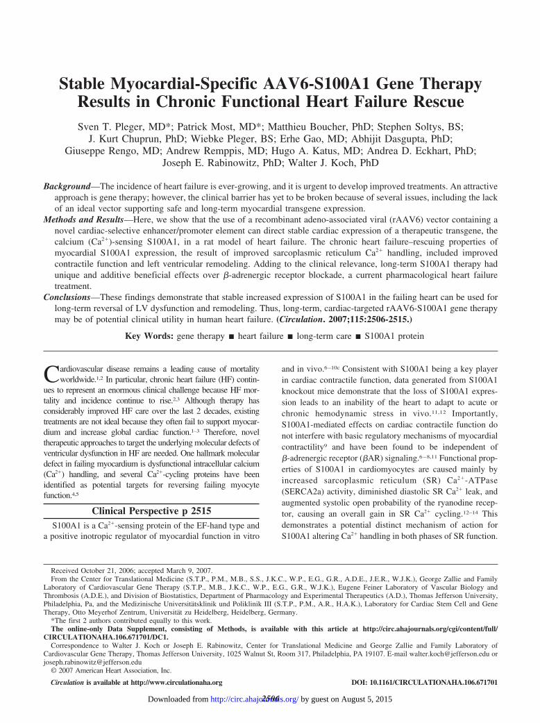
Figure 1. Cardioselective rAAV6-mediated in vivo gene transfer. A, AAV6/S100A1 construct used. Shown are thepositions of the 2 independent trans-genic cassettes: GFP driven by the CMVpromoter and S100A1 driven by the�-cardiac actin enhancer/EF1� promoter.B, Representative Western blot analysisof GFP expression in rat tissues 8 weeksafter in vivo intracoronary delivery withAAV6/S100A1. C, Representative West-ern blot of human S100A1 protein pres-ent only in the heart and absent in otherrat tissues harboring the GFP transgene8 weeks after AAV6/S100A1 delivery. D,Representative GFP fluorescencemicroscopy (left), light microscopy (mid-dle), and overlay of both (right) of LVmyocardium 8 weeks after intracoronaryAAV6/S100A1 delivery. Magnification,�40. E, S100A1 immunohistochemistryin LV tissue from AAV6/GFP- (left, con-trol) and AAV6/S100A1-treated (right)rats (8 weeks after gene delivery). Magni-fication, �40.
Pleger et al Chronic S100A1 Heart Failure Gene Therapy 2507
by guest on August 5, 2015http://circ.ahajournals.org/Downloaded from
fer to 10-week post–myocardial infarction (MI) rats was achieved aspreviously described13,17a with some modifications. Briefly, undergeneral anesthesia (2% isoflurane, vol/vol), a midline cervicalincision was made, and the animal was cooled to 29°C with icepacks. A P-50 catheter (Becton Dickinson, Sparks, Md) was ad-vanced into the aortic root via the right carotid artery, and theascending aorta was looped with 2-0 silk. Then, 1.2 mg adenosinewas injected into the right ventricle with a 301⁄2-gauge needle. Theascending aorta was clamped, and 2.5�1011 particles of the AAVconstruct and 8 �g of substance P (Sigma Chemical Co, St Louis,Mo) were rapidly injected into the aortic root, allowing coronaryperfusion. After 2 minutes, the aortic clamp was released, a bolus ofdobutamine (30 �g IA) was administered, and the animal wasrewarmed with a heating pad.
Histology, Western Blotting, and Real-TimePolymerase Chain ReactionImmunohistochemistry, assessment of infarct size, Western blotanalysis, and quantitative real-time polymerase chain reaction wereperformed as previously reported.13,17a
Ca2� Transient Analysis and ContractileParameters of Isolated Adult Rat CardiomyocytesIsolation of adult rat cardiomyocytes, assessment of contractileparameters, and Ca2� transients of isolated cardiomyocytes weredone as described.13,17a
Statistical AnalysisData are summarized as mean�SEM. Comparisons were made usingt tests or ANOVA as appropriate. When multiple observations existper animal, we used a mixed-effects model with random intercept foreach animal to account for the repeated measurements. The statisticalsignificance of each effect was then determined from Wald statisticsderived from the mixed-effects models. A Bonferroni correction wasapplied to the probability values whenever multiple comparisonsarose. Statistical analysis was performed with the Graph PadPrismsoftware and the R statistical package (www.r-project.org). For alltests, P�0.05 is considered statistically significant after Bonferronicorrections if needed.
The authors had full access to and take full responsibility for theintegrity of the data. All authors have read and agree to themanuscript as written.
ResultsCardioselective S100A1 Gene Delivery With aNovel �-Cardiac Actin EnhancerTo engineer a putative myocardium-selective promoter, a316-bp fragment of the �-cardiac actin gene enhancer con-taining 2 MEF2 sequences and 2 enhancer MyoD consensussequences was amplified from mouse genomic DNA andligated to the EF1� promoter within an AAV shuttle plasmid(Figure 1A). The human S100A1 cDNA was then cloned intothis plasmid. The final construct (AAV6-S100A1) also con-tains a separate transgene cassette with the CMV promoterdriving expression of the green fluorescent protein (GFP)marker gene (Figure 1A). To first examine whether thisvector supports stable cardiac expression in vivo, AAV6-S100A1 was delivered to normal rats (n�4) via intracoronarydelivery.13,17a Consistent with a CMV-driven transgene, 8weeks after gene delivery, GFP expression was found outsidethe heart with appreciable levels in liver, lung, and skeletalmuscle (Figure 1B). In contrast to these findings, the humanisoform of S100A1 was detectable only in cardiac homoge-nates, indicating that S100A1 expression driven by the
combination of the �-cardiac actin enhancer and the EF1�promoter is cardioselective (Figure 1C).
Infection of myocardium by our novel AAV6 vector asassessed by GFP fluorescence of cardiac sections was globalin nature but not homogeneous throughout the heart (Figure1D), which is consistent with previous gene delivery studiesusing this intracoronary delivery method.13,17a This distribu-tion pattern also was evident with S100A1 expression asconfirmed by immunohistochemistry using an antibody spe-cific for the human isoform of S100A1 (Figure 1E).
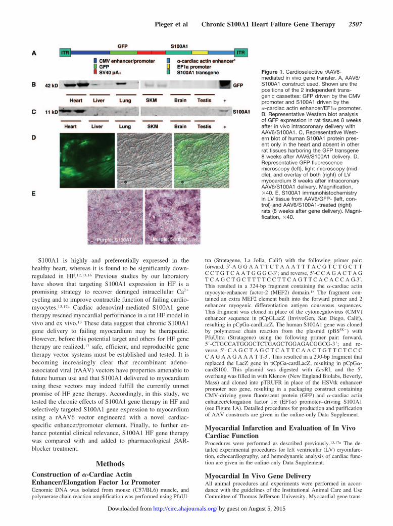
Characterization of Cardiac Dysfunction After MIand Before Gene DeliveryTo induce chronic HF in rats, we used a cryoinfarct model,which leads to HF in 10 to 12 weeks.13 Ten weeks after MI,cardiac function was assessed to determine pre–gene therapystatus in these rats compared with sham animals. All MI ratswere found to have significant LV dysfunction and wererandomized into 5 treatment groups (Figure 2). Global HFwas evident because of significantly diminished ejectionfraction compared with sham animals (Figure 2A). Moreover,significant post-MI remodeling was apparent as determinedby LV dilatation (Figure 2D). Importantly, all post-MI ratshad similar HF (Figure 2 and data not shown); thus, allrandomized groups had equal pre–gene therapy status. Inaddition to treating HF rats with AAV6-S100A1, separategroups also were treated with the �1AR antagonist, metopro-lol (250 mg · kg�1 · d�1 in the drinking water) beginning 10weeks after MI. All groups were then followed up over 2months.
S100A1 Gene Therapy Improves In Vivo CardiacFunction Long Term and Reverses LVRemodeling in HFThe in vivo functional consequences of chronic cardioselec-tive S100A1 gene therapy in HF were determined by echo-cardiography and with closed-chest cardiac catheterization.All HF groups still had significantly impaired cardiac func-tion compared with sham rats (Figure 2B, 2E, and 2F) 18weeks after MI; however, rats treated at 10 weeks after MIwith S100A1 had significantly improved cardiac function asassessed by percent ejection fraction (Figure 2B and 2C).Representative M-mode echocardiographic recordings areshown in Figure 2G. Of interest, the significant �30%improvement in global cardiac function after 8 weeks ofcardiac-selective S100A1 expression was seen with or with-out metoprolol (Figure 2C). In contrast to this improvementwith S100A1, HF rats treated with only GFP or saline hadfurther deterioration of cardiac function over this period(Figure 2C), whereas metoprolol induced no improvementbut also no further functional decline (Figure 2C).
Both cardiac S100A1 gene therapy and �-blocker treat-ment significantly attenuated further LV chamber dilatationas measured by 18-week post-MI LV diastolic dimensioncompared with pretreatment (10 week after-MI) values,whereas progressive dilatation occurred in saline- and GFP-treated HF rats (Figure 2D and 2E). S100A1 expression withconcurrent metoprolol treatment did not further attenuate LVremodeling over either treatment alone (Figure 2E). Finally,
2508 Circulation May 15, 2007
by guest on August 5, 2015http://circ.ahajournals.org/Downloaded from
the anterior wall (at the site of the infarct) was similarlythinned in all HF groups at 18 weeks after MI and wasunchanged after treatment (Figure 2F), which is not surpris-ing because gene delivery was performed 10 weeks after MIwhen expansion and scaring of the infarct are complete.19
After echocardiographic assessment of cardiac function,terminal cardiac catheterization was performed to measure18-week post-MI hemodynamics. As expected, LV contrac-tility and relaxation as measured by the maximal rate of LVpressure rise (dP/dt) and fall (�dP/dt), respectively, was
Figure 2. S100A1 gene therapy recovers cardiac function in HF. A, Ejection fraction (EF%) 10 weeks after MI before gene treatmentand (B) 8 weeks after in vivo intracoronary AAV6/S100A1 gene delivery with or without metoprolol treatment. C, Percent change inalteration of EF after 8 weeks. D, LV chamber dimensions were similar before gene delivery in all HF groups. E, Effect of AAV6/S100A1and/or �-blocker treatment on LV chamber dimensions after 8 weeks of treatment. F, Anterior wall thickness (AWTS) 18 weeks after MI.Sham, n�11; HF/saline, n�14; HF/GFP, n�11; HF/S100A1, n�12; HF/GFP-metoprolol (�), n�9; and HF/S100A1-metoprolol (�), n�9.Data are presented as mean�SEM. Bar�8 mm. G, Representative raw traces of M-mode echocardiography 8 weeks after gene deliv-ery in the 6 experimental groups. *P�0.05 vs HF/saline, HF/GFP, or HF/GFP-metoprolol groups; #P�0.05 vs sham; &P�0.05 vs HF/sa-line or HF/GFP groups, ANOVA analysis and Bonferroni test between all groups.
Pleger et al Chronic S100A1 Heart Failure Gene Therapy 2509
by guest on August 5, 2015http://circ.ahajournals.org/Downloaded from
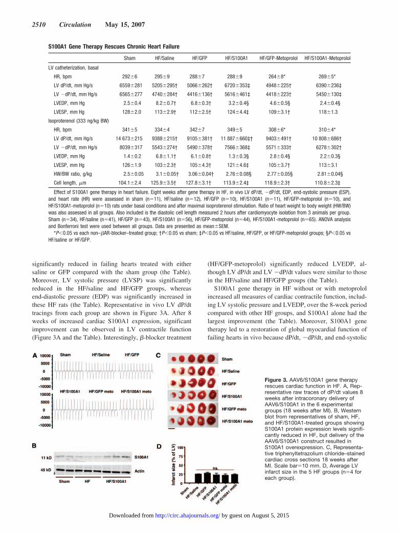
significantly reduced in failing hearts treated with eithersaline or GFP compared with the sham group (the Table).Moreover, LV systolic pressure (LVSP) was significantlyreduced in the HF/saline and HF/GFP groups, whereasend-diastolic pressure (EDP) was significantly increased inthese HF rats (the Table). Representative in vivo LV dP/dttracings from each group are shown in Figure 3A. After 8weeks of increased cardiac S100A1 expression, significantimprovement can be observed in LV contractile function(Figure 3A and the Table). Interestingly, �-blocker treatment
(HF/GFP-metoprolol) significantly reduced LVEDP, al-though LV dP/dt and LV �dP/dt values were similar to thosein the HF/saline and HF/GFP groups (the Table).
S100A1 gene therapy in HF without or with metoprololincreased all measures of cardiac contractile function, includ-ing LV systolic pressure and LVEDP, over the 8-week periodcompared with other HF groups, and S100A1 alone had thelargest improvement (the Table). Moreover, S100A1 genetherapy led to a restoration of global myocardial function offailing hearts in vivo because dP/dt, �dP/dt, and end-systolic
S100A1 Gene Therapy Rescues Chronic Heart Failure
Sham HF/Saline HF/GFP HF/S100A1 HF/GFP-Metoprolol HF/S100A1-Metoprolol
LV catheterization, basal
HR, bpm 292�6 295�9 288�7 288�9 264�8* 269�5*
LV dP/dt, mm Hg/s 6559�281 5205�295† 5066�262† 6720�353‡ 4948�225† 6390�236‡
LV �dP/dt, mm Hg/s 6565�277 4740�284† 4416�136† 5616�461‡ 4418�223† 5450�130‡
LVEDP, mm Hg 2.5�0.4 8.2�0.7† 6.8�0.3† 3.2�0.4§ 4.6�0.5§ 2.4�0.4§
LVESP, mm Hg 128�2.0 113�2.9† 112�2.5† 124�4.4‡ 109�3.1† 118�1.3
Isoproterenol (333 ng/kg BW)
HR, bpm 341�5 334�4 342�7 349�5 308�6* 310�4*
LV dP/dt, mm Hg/s 14 673�215 9388�215† 9105�381† 11 887�660‡† 9403�491† 10 808�686†
LV �dP/dt, mm Hg/s 8039�317 5543�274† 5490�378† 7566�368‡ 5571�333† 6278�302†
LVEDP, mm Hg 1.4�0.2 6.8�1.1† 6.1�0.8† 1.3�0.3§ 2.8�0.4§ 2.2�0.3§
LVESP, mm Hg 126�1.9 103�2.3† 105�4.3† 121�4.6‡ 105�3.7† 113�3.1
HW/BW ratio, g/kg 2.5�0.05 3.1�0.05† 3.06�0.04† 2.76�0.08§ 2.77�0.05§ 2.81�0.04§
Cell length, �m 104.1�2.4 125.9�3.5† 127.8�3.1† 113.9�2.4‡ 118.9�2.3† 110.8�2.3‡
Effect of S100A1 gene therapy in heart failure. Eight weeks after gene therapy in HF, in vivo LV dP/dt, �dP/dt, EDP, end-systolic pressure (ESP),and heart rate (HR) were assessed in sham (n�11), HF/saline (n�12), HF/GFP (n�10), HF/S100A1 (n�11), HF/GFP-metoprolol (n�10), andHF/S100A1-metoprolol (n�10) rats under basal conditions and after maximal isoproterenol stimulation. Ratio of heart weight to body weight (HW/BW)was also assessed in all groups. Also included is the diastolic cell length measured 2 hours after cardiomyocyte isolation from 3 animals per group.Sham (n�34), HF/saline (n�41), HF/GFP (n�43), HF/S100A1 (n�56), HF/GFP-metoprolol (n�44), HF/S100A1-metoprolol (n�65). ANOVA analysisand Bonferroni test were used between all groups. Data are presented as mean�SEM.
*P�0.05 vs each non–�AR-blocker–treated group; †P�0.05 vs sham; ‡P�0.05 vs HF/saline, HF/GFP, or HF/GFP-metoprolol groups; §P�0.05 vsHF/saline or HF/GFP.
Figure 3. AAV6/S100A1 gene therapyrescues cardiac function in HF. A, Rep-resentative raw traces of dP/dt values 8weeks after intracoronary delivery ofAAV6/S100A1 in the 6 experimentalgroups (18 weeks after MI). B, Westernblot from representatives of sham, HF,and HF/S100A1-treated groups showingS100A1 protein expression levels signifi-cantly reduced in HF, but delivery of theAAV6/S100A1 construct resulted inS100A1 overexpression. C, Representa-tive triphenyltetrazolium chloride–stainedcardiac cross sections 18 weeks afterMI. Scale bar�10 mm. D, Average LVinfarct size in the 5 HF groups (n�4 foreach group).
2510 Circulation May 15, 2007
by guest on August 5, 2015http://circ.ahajournals.org/Downloaded from
pressure could not be statistically distinguished from healthy,sham-operated animals, although the EDP remained elevated(the Table). When failing hearts were challenged with amaximal dose of isoproterenol, chronic S100A1 overexpres-sion continued to improve cardiac performance in vivo (theTable). Heart rate was not affected by MI or in gene therapygroups; therefore, heart rate was not responsible for thefunctional improvements seen with S100A1 but, as expected,was significantly reduced by metoprolol (the Table).
Cardioselective S100A1 expression was confirmed byWestern blotting, and levels from whole-heart homogenatescan be seen in Figure 3B. In HF (18 weeks after MI),significant loss of cardiac S100A1 protein levels comparedwith sham levels occurs, and AAV6-S100A1 gene deliverydriving S100A1 expression only in the heart with the�-cardiac actin enhancer/EF1� promoter not only restoresnormal S100A1 levels but also increases S100A1 proteinexpression (Figures 3B and 4A). S100A1 protein overexpres-sion was not evident in other tissues in HF rats, whereas GFPhad the same expression pattern in rat tissues outside the heartas in Figure 1 (data not shown).
To determine whether all hearts had similar injury, weassessed LV infarct size within a subset of hearts from eachgroup (n�4) via triphenyltetrazolium chloride (TTC) stain-ing. Representative TTC-stained cardiac sections from eachgroup are shown in Figure 3C. Analysis revealed an averageinfarct size of 22.9�0.9% of the LV (33.2�1.4% of the LVfree wall), which was similar in all groups (Figure 3D).
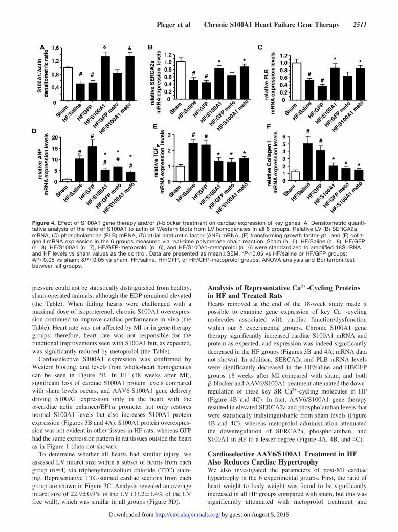
Analysis of Representative Ca2�-Cycling Proteinsin HF and Treated RatsHearts removed at the end of the 18-week study made itpossible to examine gene expression of key Ca2�-cyclingmolecules associated with cardiac function/dysfunctionwithin our 6 experimental groups. Chronic S100A1 genetherapy significantly increased cardiac S100A1 mRNA andprotein as expected, and expression was indeed significantlydecreased in the HF groups (Figures 3B and 4A; mRNA datanot shown). In addition, SERCA2a and PLB mRNA levelswere significantly decreased in the HF/saline and HF/GFPgroups 18 weeks after MI compared with sham, and both�-blocker and AAV6/S100A1 treatment attenuated the down-regulation of these key SR Ca2�-cycling molecules in HF(Figure 4B and 4C). In fact, AAV6/S100A1 gene therapyresulted in elevated SERCA2a and phospholamban levels thatwere statistically indistinguishable from sham levels (Figure4B and 4C), whereas metoprolol administration attenuatedthe downregulation of SERCA2a, phospholamban, andS100A1 in HF to a lesser degree (Figure 4A, 4B, and 4C).
Cardioselective AAV6/S100A1 Treatment in HFAlso Reduces Cardiac HypertrophyWe also investigated the parameters of post-MI cardiachypertrophy in the 6 experimental groups. First, the ratio ofheart weight to body weight was found to be significantlyincreased in all HF groups compared with sham, but this wassignificantly attenuated with metoprolol treatment and
Figure 4. Effect of S100A1 gene therapy and/or �-blocker treatment on cardiac expression of key genes. A, Densitometric quanti-tative analysis of the ratio of S100A1 to actin of Western blots from LV homogenates in all 6 groups. Relative LV (B) SERCA2amRNA, (C) phospholamban (PLB) mRNA, (D) atrial natriuretic factor (ANF) mRNA, (E) transforming growth factor-�1, and (F) colla-gen I mRNA expression in the 6 groups measured via real-time polymerase chain reaction. Sham (n�6), HF/Saline (n�8), HF/GFP(n�8), HF/S100A1 (n�7), HF/GFP-metoprolol (n�6), and HF/S100A1-metoprolol (n�6) were standardized to amplified 18S rRNAand HF levels vs sham values as the control. Data are presented as mean�SEM. *P�0.05 vs HF/saline or HF/GFP groups;#P�0.05 vs sham; &P�0.05 vs sham, HF/saline, HF/GFP, or HF/GFP-metoprolol groups, ANOVA analysis and Bonferroni testbetween all groups.
Pleger et al Chronic S100A1 Heart Failure Gene Therapy 2511
by guest on August 5, 2015http://circ.ahajournals.org/Downloaded from
S100A1 (the Table). Ventricular atrial natriuretic factormRNA expression typically associated with cardiac hypertro-phy also was significantly increased 18 weeks after MI in allanalyzed groups compared with sham (Figure 4D); however,both �-blocker treatment and S100A1 gene therapy signifi-cantly reduced cardiac atrial natriuretic factor expression inHF (Figure 4D). On the cellular level, cardiac hypertrophywas reflected by a significantly increased length of isolatedcardiomyocytes 18 weeks after MI, which interestingly wassignificantly reduced only in the AAV6/S100A1 groups (theTable). We also examined cardiac mRNA levels of trans-forming growth factor-�1 and collagen I mRNA as molecularmarkers of remodeling. Both were significantly elevated inthe HF/saline and HF/GFP groups and were significantlyreduced with metoprolol and chronic S100A1 gene therapy inHF (Figure 4E and 4F).
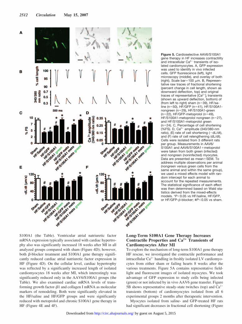
Long-Term S100A1 Gene Therapy IncreasesContractile Properties and Ca2� Transients ofCardiomyocytes After MITo explore the mechanism of long-term S100A1 gene therapyHF rescue, we investigated the contractile performance andintracellular Ca2� handling in freshly isolated LV cardiomyo-cytes from either sham or failing hearts 8 weeks after thevarious treatments. Figure 5A contains representative field-light and fluorescent images of isolated myocytes. We tookadvantage of GFP expression to study cells being infected(green) or not infected by in vivo AAV6 gene transfer. Figure5B shows representative steady-state twitches (top) and Ca2�
transients (bottom) of cardiomyocytes isolated from all 6experimental groups 2 months after therapeutic intervention.
Myocytes isolated from saline- and GFP-treated HF ratshad significant decreases in fractional cell shortening (Figure
Figure 5. Cardioselective AAV6/S100A1gene therapy in HF increases contractilityand intracellular Ca2� transients of iso-lated cardiomyocytes. A, GFP expressionwas used to identify in vivo infectedcells. GFP fluorescence (left), lightmicroscopy (middle), and overlay of both(right). Scale bar�100 �m. B, Represen-tative raw traces of fractional shortening(percent change in cell length, shown asdownward deflection, top) and originaltraces of representative [Ca2�]I transients(shown as upward deflection, bottom) of(from left to right) sham (n�39), HF/sa-line (n�50), HF/GFP (n�41), HF/S100A1-nongreen (n�29), HF/S100A1-green(n�22), HF/GFP-metoprolol (n�49),HF/S100A1-metoprolol nongreen (n�27),and HF/S100A1-metoprolol green(n�24). C, Percentage of cell shortening(%FS). D, Ca2� amplitude (340/380-nmratio), (E) rate of cell shortening (�dL/dt),and (F) rate of cell relengthening (dL/dt).Cells were isolated from 3 different ratsper group. Measurements in AAV6/S100A1 and AAV6/S100A1�metoprololwere taken from both green (infected)and nongreen (noninfected) myocytes.Data are presented as mean�SEM. Toaddress multiple observations per animal(nongreen versus green cells from thesame animal and within the same group),we used a mixed effects model with ran-dom intercept for each animal toaccount for the repeated measurements.The statistical significance of each effectwas then determined based on Wald sta-tistics derived from the mixed-effectsmodels. *P�0.05 vs HF/saline, HF/GFP,or HF/GFP-�-blocker; #P�0.05 vs sham.
2512 Circulation May 15, 2007
by guest on August 5, 2015http://circ.ahajournals.org/Downloaded from
5C), the amplitude of the [Ca2�]i transient (Figure 5D), therate of myocyte shortening (�dL/dt; Figure 5E), and the rateof myocyte relengthening (dL/dt; Figure 5F) compared withmyocytes isolated from sham hearts. Metoprolol treatmentalone did not affect contractile properties of isolated failingmyocytes under our conditions because [Ca2�]i transients,percent fractional shortening, dL/dt, and �dL/dt were similarto the HF/saline and HF/GFP groups (Figure 5C through 5F).Data using only infected cells (green) from AAV6/S100A1-treated HF rats showed that S100A1 overexpression com-pletely rescues myocyte dysfunction because contractile pa-rameters and [Ca2�]i transients were similar in these HF cellscompared with nonfailing myocytes (Figure 5C through 5F).Furthermore, the therapeutic effect on isolated myocytes fromAAV6/S100A1 gene therapy in HF was preserved underadditional �-blocker administration (Figure 5C through 5F).Interestingly, noninfected cardiomyocytes (nongreen) ob-tained from AAV6/S100A1-treated rats showed a trend to-ward improved contractile properties and [Ca2�]i transientscompared with the HF/saline, HF/GFP, and HF/GFP–�-blocker groups (Figure 5C through 5F).
DiscussionThe data presented above demonstrate the use of a novelrAAV6 vector containing a cardioselective promoter to sup-port chronic therapeutic gene expression in the failing heart.Using this novel vector in a previously described chronicpost-MI rat HF model,13 we show that cardioselective long-term S100A1 gene therapy can reverse global in vivo cardiacdysfunction and attenuate LV remodeling. Notably, improvedfunction in HF after cardioselective S100A1 treatment re-mained at least 8 weeks after in vivo gene delivery, providingevidence for a sustained therapeutic effect. This finding is inline with previous results providing proof of concept foreffective adenovirus-mediated S100A1 gene therapy of HF,supporting S100A1 as a novel therapeutic target for HF.13
Moreover, S100A1-mediated recovery of functional proper-ties of failing myocardium was preserved with additionalpharmacological �AR-blocker treatment. Thus, S100A1 geneaddition could represent a viable future clinical approach forHF treatment and add to existing drug treatment.
Our choice for directing cardioselective gene expressionwas a sequence from the proximal enhancer region of the�-cardiac actin gene. Previously, this region that contains aMEF2 sequence was shown to direct heart but not skeletalmuscle expression.18 A second MEF2 site was added topotentially drive stronger gene expression in the heart. As ourresults show, the enhancer element cloned in front of theEF1� promoter produces robust expression of S100A1 thatwas specifically localized to the normal and failing rat heartafter in vivo intracoronary delivery. Tissue selectivity wasconfirmed by including a second transgene cassette in thisrAAV6 vector containing CMV-GFP; indeed, GFP expres-sion was found in several extracardiac sites after intracoro-nary delivery. We excluded that posttranscriptional regulationlimits S100A1 expression to cardiac tissue because S100A1mRNA overexpression was not present in organs such aslung, liver, testis, and skeletal muscle, whereas S100A1mRNA expression was increased 3-fold in isolated cardio-
myocytes from rAAV6-S100A1–treated HF rats (data notshown). To the best of our knowledge, this is the firstdemonstration of long-term, myocardium-specific gene ex-pression after in vivo gene delivery and represents the use ofa vector that, after being tested in larger animal models, couldhave potential clinical utility.
In our HF model, rats were found to have significant LVdysfunction and remodeling at 10 weeks after MI, which wasin line with previous results13 and similar in all randomizedtreatment groups. Consistent with clinical HF develop-ment,20–22 cardiac contractile function deteriorated furtherover the 2-month treatment period in control groups. How-ever, S100A1 enhancement after cardioselective AAV6-mediated gene delivery led to functional recovery of thefailing rat heart seen globally with increased percent ejectionfraction and dP/dt and lower EDP; HF rescue also was seenin individual ventricular cardiomyocytes. Moreover, en-hanced contractile function of S100A1-treated failing heartswas preserved under maximal �AR stimulation, furtherdemonstrating the therapeutic potential of S100A1. Impor-tantly, S100A1 is reduced in the failing rat heart,13 consistentwith human HF,16 and restoration to the supranormal levelsresponsible for the rescue seen in this study reveals a criticalrole for this protein in Ca2�-dependent cardiac regulation andfunction. These findings are in line with our recent datagenerated from S100A1 knockout mice demonstrating thatthe loss of S100A1 protein contributes significantly to theprogressive deterioration of cardiac function in post-MI HF,whereas preservation of cardiac S100A1 protein isprotective.12
Functional recovery of myocardial function in AAV6/S100A1-treated HF rats was accompanied by mitigated LVchamber dilatation and cardiac hypertrophy. These data are inline with previous findings13 and might be explained inmultiple ways. First, there may be an indirect effect resultingfrom enhanced contractile function of the heart; thus, areduced biomechanical overload may result in a reverseremodeling situation. Additionally, improved [Ca2�]i han-dling in failing cardiomyocytes resulting from rAAV6-S100A1 treatment might beneficially affect myocardial apo-ptosis, hypertrophy, or gene expression by modulatingvarious Ca2�-dependent signaling pathways involving cal-cineurin, calmodulin kinase, or protein kinase C isoforms (�,�, �).23 Alternatively, because the loss of S100A1 haspreviously been shown to be permissive for the induction ofgenes involved in cardiac hypertrophy,12,13,24 increasedS100A1 levels may more directly influence the silencing ofthese genes, leading to attenuation of hypertrophy and ac-companied dilatation. Regardless of the mechanism, it isapparent that enhanced expression of S100A1 induces thesebeneficial effects chronically on the failing heart.
An interesting finding at the myocyte level was that whenonly rAAV6-S100A1–infected cardiomyocytes (green cells)were studied, a complete restoration of [Ca2�]i transientsoccurred, and contractile parameters as S100A1-treated HFmyocytes had similar values to healthy cells 8 weeks aftergene therapy. Moreover, data from isolated cardiomyocytesalso reveal a potential indirect therapeutic effect of cardiacS100A1 gene delivery on cells that were not infected in vivo
Pleger et al Chronic S100A1 Heart Failure Gene Therapy 2513
by guest on August 5, 2015http://circ.ahajournals.org/Downloaded from
because these non-GFP myocytes displayed a trend towardincreased functional properties. This is especially interestingbecause functional recovery of the failing heart globally wasachieved despite inhomogeneous gene delivery being in linewith previous studies.13,17a Our data using GFP expression inmyocytes showed �40% infection rate; thus, there may be anindirect effect of S100A1-overexpressing myocytes to im-prove the function of neighboring myocytes. Furthermore, thereduced wall stress and regression of maladaptive hypertro-phy may allow noninfected myocytes to recover on their own.Alternatively, in vivo AAV6/S100A1 gene delivery might beunderestimated by GFP coexpression because the brightnessof GFP fluorescence varied substantially between isolatedmyocytes (data not shown), or GFP expression might notfully match S100A1 overexpression levels because 2 differ-ent promoters were used to drive S100A1 and GFPexpression.
To increase potential clinical relevancy, we added a �AR-blocker component. Metoprolol administration in HF signif-icantly attenuated LV remodeling, reduced cardiac hypertro-phy, lowered EDP, and prevented further deterioration ofcardiac function in HF. However, �AR-blocker treatment didnot affect functional properties of isolated failing cardiomyo-cytes and failed to recover functional properties of in vivoglobal cardiac function under our conditions. These findingsare in line with observations in several rodent HF andpost-MI models showing that selective �1-blockade attenu-ates post-MI structural remodeling without concomitant im-provement in myocardial function.25–27
Clinical studies such as the Metoprolol Controlled-ReleaseRandomized Intervention Trial in Heart Failure (MERIT-HF)have proved that �-blocker therapy in patients with HF notonly can attenuate pathological remodeling of the heart butalso may actually improve patient outcomes.28,29 Therefore,preservation of S100A1-mediated positive inotropic effectsunder �-blocker treatment, as observed in this study, suggestspotentially additive action of both strategies and supports apotential future clinical application of S100A1 gene therapyin HF, which could become safer with a cardioselectiveapproach like that described here.
Because short-term in vivo strategies already proved ther-apeutic effects of myocardial S100A1 gene delivery in bothacute MI and in overt HF,13,17a a potential goal of the presentstudy was to investigate long-term actions of S100A1 in HF.This is especially important because cytosolic Ca2� overloadand excess cAMP generation under chronic pharmacologicalinotropic treatment in HF are associated with increasedmortality.30,31 Long-term S100A1 gene therapy resulted inrecovery of contractile function in HF, which was mediated,at least largely, by rescued intracellular Ca2� turnover and SRCa2� cycling mechanistically similar to adenoviral S100A1gene therapy.13,17a Importantly, S100A1 protein can decreaseCa2� spark activity in ventricular cardiomyocytes underdiastolic conditions, which might contribute to attenuatedetrimental diastolic Ca2� overload and SR Ca2� leakage inHF through S100A1.12,13,32 Therefore, targeting both ryano-dine receptor open probability and SERCA2a activity byS100A112–14,32 might be advantageous compared with solely
increasing SERCA2a function, which can result in impairedsurvival in response to ischemic events.33
To summarize, our study shows that long-term in vivocardioselective S100A1 gene therapy is feasible with the�-cardiac actin enhancer/EF1� promoter in a rAAV6 vectorand that it is indeed therapeutic. Moreover, effects of S100A1gene therapy in HF are preserved under �-blocker treatmentin vivo and indicate that both treatment strategies might beadditive in HF.
Sources of FundingThis research was supported in part by grants from the DeutscheForschungsgemeinschaft (Mo 1066/1–1 to Dr Most, 1083/1–1 to DrRemppis), Bundesministerium für Bildung und Forschung(01GU0527 to Dr Most), Pennsylvania–Delaware Affiliate of theAmerican Heart Association (fellowship to Dr Pleger), Lilly-Stipendium of the Deutsche Gesellschaft für Kardiologie (DrPleger), American Heart Association (scientist development grantSTG 053 0127N to Dr Rabinowitz), and the National Institutes ofHealth (R01 HL56205 and P01 HL075443-project 2 to Dr Koch,W.W. Smith Professor of Medicine).
DisclosuresDrs Katus, Koch, Remppis, Most, Rabinowitz, and Pleger have apatent pending on the use of S100A1 in treating heart disease and onthe use of the �-cardiac actin enhancer/EF1� promoter. The otherauthors report no conflicts.
References1. Heart Disease and Stroke Statistics. Dallas, Tex: American Heart Asso-
ciation; 2005.2. Cohn JN, Bristow MR, Chien KR, Colucci WS, Frazier OH, Leinwand
LA, Lorell BH, Moss AJ, Sonnenblick EH, Walsh RA, Mockrin SC,Reinlib L. Report of the National Heart, Lung, and Blood Institute SpecialEmphasis Panel on Heart Failure Research. Circulation. 1997;95:766–770.
3. Rich MW. Epidemiology, pathophysiology, and etiology of congestiveheart failure in older adults. J Am Geriatr Soc. 1997;45:968–974.
4. del Monte F, Hajjar R. Targeting calcium cycling proteins in heart failurethrough gene transfer. J Physiol. 2003;546:49–61.
5. Wehrens XH, Marks AR. Novel therapeutic approaches for heart failureby normalizing calcium cycling. Nat Rev Drug Discov. 2004;3:565–573.
6. Most P, Ehlermann P, Bernotat J, Pleger ST, Boerries M, Reppel M,Mandinova A, Niroomand F, Pieske B, Janssen PML, Zeitz O,Eschenhagen T, Karczewski P, Smith GL, Koch WJ, Kattus HA, RemppisA. S100A1: a regulator of myocardial contractility. Proc Natl Acad SciU S A. 2001;98:13889–13894.
7. Most P, Remppis A, Pleger ST, Löffler E, Ehlermann P, Bernotat J,Kleuss C, Heierhorst J, Ruiz P, Witt H, Karczewski P, Mao L, RockmanHA, Duncan SJ, Katus HA, Koch WJ. Transgenic overexpression of theCa2� binding protein S100A1 in the heart leads to increased in vivomyocardial contractile performance. J Biol Chem.2003;278:33809–33817.
8. Most P, Boerries M, Eicher C, Schweda C, Völkers M, Wedel T, SollnerS, Katus HA, Remppis A, Aebi U, Koch WJ, Schoenenberger CA.Distinct subcellular location of the Ca2� binding protein S100A1 differ-entially modulates Ca2� cycling in ventricular rat cardiomyocytes. J CellSci. 2005;118:421–431.
9. Remppis A, Pleger ST, Most P, Lindenkamp J, Ehlermann P, Schweda C,Löffler E, Weichenhan D, Zimmermann W, Eschenhagen T, Koch WJ,Katus HA. S100A1 gene transfer: a strategy to strengthen engineeredcardiac grafts. J Gene Med. 2004;6:387–394.
10. Remppis A, Most P, Löffler E, Ehlermann P, Bernotat J, Pleger S,Boerries M, Reppel M, Fischer J, Koch WJ, Smith G, Katus HA. Thesmall EF-hand Ca2� binding protein S100A1 increases contractility andCa2� cycling in rat cardiac myocytes. Basic Res Cardiol. 2002;97:I56-I62.
10a. Most P, Remppis A, Pleger ST, Katus HA, Koch WJ. S100A1: a novelinotropic regulator of cardiac performance: transition from molecular
2514 Circulation May 15, 2007
by guest on August 5, 2015http://circ.ahajournals.org/Downloaded from
physiology to pathophysiological relevance. Am J Physiol Regul IntegrComp Physiol. In press.
10b. Pleger ST, Boucher M, Most P, Koch WJ. Targeting myocardial�-adrenergic receptor signaling and calcium cycling for heart failure genetherapy. J Card Fail. In press.
10c. Most P, Koch WJ. S100A1: a calcium-modulating inotropic prototype forfuture clinical heart failure therapy. Future Cardiol. 2007;3:5–11.
11. Du XJ, Cole TJ, Tenis N, Gao XM, Kontgen F, Kemp BE, Heierhorst J.Impaired cardiac contractility response to hemodynamic stress inS100A1-deficient mice. Mol Cell Biol. 2002;22:2821–2829.
12. Most P, Seifert H, Gao E, Funakoshi H, Völkers M, Heierhorst J,Remppis A, Pleger ST, DeGeorge BR Jr, Eckhart AD, Feldman AM,Koch WJ. Cardiac S100A1 protein levels determine contractile per-formance and propensity towards heart failure after myocardial infarction.Circulation. 2006;114:1258–1268.
13. Most P, Pleger ST, Völkers M, Heidt B, Boerries M, Weichenhan D,Löffler E, Janssen PM, Eckhart AD, Martini J, Williams ML, Katus HA,Remppis A, Koch WJ. Cardiac adenoviral S100A1 gene delivery rescuesfailing myocardium. J Clin Invest. 2004;114:1550–1563.
14. Kettlewell S, Most P, Currie S, Koch WJ, Smith GL. S100A1 increasesthe gain of excitation-contraction coupling in isolated rabbit ventricularcardiomyocytes. J Mol Cell Cardiol. 2005;39:900–910.
15. Deleted in proof.16. Remppis A, Greten T, Schafer BW, Hunziker P, Erne P, Katus HA,
Heizmann CW. Altered expression of the Ca2�-binding protein S100A1in human cardiomyopathy. Biochim Biophys Acta. 1996;1313:253–257.
17. Williams ML, Koch WJ. Viral-based myocardial gene therapyapproaches to alter cardiac function. Annu Rev Physiol. 2004;66:49–75.
17a.Pleger ST, Remppis A, Heidt B, Volkers M, Chuprun JK, Kuhn M, ZhouRH, Gao E, Szabo G, Weichenhan D, Muller OJ, Eckhart AD, Katus HA,Koch WJ, Most P. S100A1 gene therapy preserves in vivo cardiacfunction after myocardial infarction. Mol Ther. 2005;12:1120–1129.
18. Lemonnier M, Buckingham M. Characterization of a cardiac-specificenhancer, which directs a-cardiac actin gene transcription in the mouseadult heart. J Biol Chem. 2004;279:55651–55658.
19. Mukherjee R, Brinsa TA, Dowdy KB, Scott AA, Baskin JM, DeschampsAM, Lowry AS, Escobar GP, Lucas DG, Yarbrough WM, Zile MR,Spinale FG. Myocardial infarct expansion and matrix metalloproteinaseinhibition. Circulation. 2003;107:618–625.
20. Katz AM. Proliferative signaling and disease progression in heart failure.Circ J. 2002;66:225–231.
21. Esler M. Measurement of sympathetic nervous system activity in heartfailure: the role of norepinephrine kinetics. Heart Fail Rev. 2000;5:17–25.
22. Sabbah HN, Sharov VG, Goldstein S. Cell death, tissue hypoxia and theprogression of heart failure. Heart Fail Rev. 2000;5:131–138.
23. Deleted in proof.24. Tsoporis JN, Marks A, Zimmer DB, McMahon C, Parker TG. The
myocardial protein S100A1 plays a role in the maintenance of normalgene expression in the adult heart. Mol Cell Biochem. 2003;242:27–33.
25. Omerovic E, Bollano E, Soussi B, Waagstein F. Selective beta1-blockadeattenuates post-infarct remodelling without improvement in myocardialenergy metabolism and function in rats with heart failure. Eur J HeartFail. 2003;5:725–732.
26. Yang Y, Tang Y, Ruan Y, Wang Y, Gao R, Chen J, Chen Z. Comparisonof metoprolol with low, middle and high doses of carvedilol in preventionof postinfarction left ventricular remodelling in rats. Jpn Heart J. 2003;44:979–988.
27. Ahmet I, Krawczyk M, Heller P, Moon C, Lakatta EG, Talan MI.Beneficial effects of chronic pharmacological manipulation of beta-adre-noreceptor subtype signaling in rodent dilated ischemic cardiomyopathy.Circulation. 2004;110:1083–1090.
28. Janosi A, Ghali JK, Herlitz J, Czuriga I, Klibaner M, Wikstrand J,Hjalmarson A, for the MERIT-HF Study Group. Metoprolol CR/XL inpostmyocardial infarction patients with chronic heart failure: experiencesfrom MERIT-HF. Am Heart J. 2003;146:721–728.
29. Williams RE. Early initiation of beta blockade in heart failure: issues andevidence. Clin Hypertens. 2005;7:520–528.
30. Katz A. Potential deleterious effects of inotropic agents in the therapy ofchronic heart failure. Circulation. 1986;73:III-184–III-190. Abstract.
31. Engelhardt S, Hein L, Wiesmann F, Lohse MJ. Progressive hypertrophyand heart failure in �1-adrenergic receptor transgenic mice. Proc NatlAcad Sci U S A. 1999;96:7059–7064.
32. Völkers M, Loughrey CM, Macquaide N, Remppis A, DeGeorge BR Jr,Wegner FV, Friedrich O, Fink RH, Koch WJ, Smith GL, Most P. S100A1decreases calcium spark frequency and alters their characteristics inpermeabilized adult ventricular cardiomyocytes. Cell Calcium. 2007;41:135–143.
33. Chen Y, Escoubet B, Prunier F, Amour J, Simonides WS, Vivien B,Lenoir C, Heimburger M, Choqueux C, Gellen B, Riou B, Michel JB,Franz WM, Mercadier JJ. Constitutive cardiac overexpression of sarco-plasmic/endoplasmic reticulum Ca2�-ATPase delays myocardial failureafter myocardial infarction in rats at a cost of increased acute arrhythmias.Circulation. 2004;9:1898–1903.
CLINICAL PERSPECTIVEIn the present study, we provide proof of concept for the long-term therapeutic effectiveness of cardiac S100A1 genetherapy in the context of a clinically relevant experimental model of overt heart failure. Taking advantage ofadeno-associated virus serotype 6 gene transfer in combination with a novel cardiomyocyte-specific enhancer/promoter,cardiac S100A1 gene delivery restored contractile performance and reversed cardiac remodeling of chronically failinghearts. This translational approach stems from the observation of diminished S100A1 protein levels in failing humanmyocardium and underscores the significant therapeutic potential of S100A1. Given the fact that S100A1-mediatedtherapeutic effects in our study lasted for months without detrimental effects, it is important to point out that the inotropicmolecular support conveyed through S100A1 does not rely on �-adrenergic receptor signaling involving, for example,cAMP, for which long-term clinical use has been proved to be deleterious in failing human hearts. Rather, S100A1-mediated inotropy is based on balanced improvement of sarcoplasmic reticulum Ca2� cycling, targeting both the cardiacryanodine receptor isoform and the Ca2�-ATPase/phospholamban complex that is directly translated into enhancedcontractile performance. In the present study, this therapeutic modality exerted long-term therapeutic effects that weresuperior even to metoprolol, a clinically established and approved heart failure therapy in humans. Of note, metoprolol wasable only to prevent but not reverse progressive deterioration of contractile function (as S100A1 gene therapy did) in ourexperimental setting. Thus, this study enables preclinical testing of adeno-associated virus serotype 6–S100A1 genetherapy in large-animal studies, and clinical use of S100A1 heart failure therapy is now within reach.
Pleger et al Chronic S100A1 Heart Failure Gene Therapy 2515
by guest on August 5, 2015http://circ.ahajournals.org/Downloaded from
Andrea D. Eckhart, Joseph E. Rabinowitz and Walter J. KochPleger, Erhe Gao, Abhijit Dasgupta, Giuseppe Rengo, Andrew Remppis, Hugo A. Katus,
Sven T. Pleger, Patrick Most, Matthieu Boucher, Stephen Soltys, J. Kurt Chuprun, WiebkeHeart Failure Rescue
Stable Myocardial-Specific AAV6-S100A1 Gene Therapy Results in Chronic Functional
Print ISSN: 0009-7322. Online ISSN: 1524-4539 Copyright © 2007 American Heart Association, Inc. All rights reserved.
is published by the American Heart Association, 7272 Greenville Avenue, Dallas, TX 75231Circulation doi: 10.1161/CIRCULATIONAHA.106.671701
2007;115:2506-2515; originally published online April 30, 2007;Circulation.
http://circ.ahajournals.org/content/115/19/2506World Wide Web at:
The online version of this article, along with updated information and services, is located on the
http://circ.ahajournals.org/content/suppl/2007/04/27/CIRCULATIONAHA.106.671701.DC1.htmlData Supplement (unedited) at:
http://circ.ahajournals.org//subscriptions/
is online at: Circulation Information about subscribing to Subscriptions:
http://www.lww.com/reprints Information about reprints can be found online at: Reprints:
document. Permissions and Rights Question and Answer this process is available in the
click Request Permissions in the middle column of the Web page under Services. Further information aboutOffice. Once the online version of the published article for which permission is being requested is located,
can be obtained via RightsLink, a service of the Copyright Clearance Center, not the EditorialCirculationin Requests for permissions to reproduce figures, tables, or portions of articles originally publishedPermissions:
by guest on August 5, 2015http://circ.ahajournals.org/Downloaded from
Top Related
Copyright © 2022 FDOKUMEN

