


Bahasa
Halaman
Hukum

Université Victor Segalen Bordeaux 2
Année 2010 Thèse n° 1783
Thèse pour le
DOCTORAT DE L'UNIVERSITÉ DE BORDEAUX 2
Mention : Sciences, Technologie, Santé
Option : Neurosciences
Présentée et soutenue publiquement
Le 17 décembre 2010
Par Jean-Yves ROTGÉ
Né le 08 Octobre 1978 à Sarreguemines (57)
Rôle des voies thalamo-corticales dans le trouble obsessionnel-compulsif
Approches méta-analytique et physiopathologique chez l'homme et l'animal
Directeur de thèse
Professeur Pierre BURBAUD
Membres du jury
Professeur Pierre-Michel LLORCA Président-Rapporteur
Professeur Vincent CAMUS Rapporteur
Professeur Michèle ALLARD Examinateur
Docteur David BELIN Examinateur

2

3
A mes parents, Martine et Alain
A mes frères, Jean-Rémy et Jonathan
Leurs compagnes, Halima et Emma
Mes neveux, Lina et Yanis
A Virginie

4
5
REMERCIEMENTS
A Monsieur le Professeur Pierre-Michel Llorca,
Vous me faites l'honneur d'accepter de présider le jury de ma thèse et d'en être le rapporteur.
Je vous remercie infiniment d'avoir accepté cette double tâche et d'apporter votre savoir et
votre expérience à l'examen de ce travail. Veuillez trouver ici le témoignage de mon profond
respect.
A Monsieur le Professeur Vincent Camus,
Je suis très honoré de pouvoir soumettre cette thèse à votre jugement et je vous remercie pour
l'intérêt que vous avez témoigné pour ce travail en acceptant d'en être le rapporteur. Veuillez
trouver ici le témoignage de mon profond respect.
A Madame le Professeur Michèle Allard,
Je tiens à te témoigner toute ma reconnaissance pour m'avoir accueilli et guidé dans le monde
de la neuro-imagerie avec bienveillance et attention. Travailler au sein de ton équipe m'a
permis de bénéficier de tes grandes connaissances scientifiques et celles de tes collaborateurs.
J'espère sincèrement pouvoir bénéficier encore de ta grande expertise en imagerie à l'avenir.
Reçois ici le témoignage de ma gratitude et de mon admiration.
A Monsieur le Docteur David Belin,
Je vous remercie d'accepter de consacrer une partie de votre temps à juger cette thèse et
d'apporter la qualité et la richesse de vos compétences et connaissances scientifiques à
l'examen de ce travail. Veuillez trouver ici le témoignage de mon profond respect.
A Monsieur le Professeur Pierre Burbaud,
Je te remercie de m'avoir accueilli dans ton équipe de recherche où j'ai pu bénéficier à maintes
reprises de tes grandes connaissances en neurophysiologie et de ton enthousiasme
scientifique. Dans cette aventure passionnante qu’est la recherche, nous avons partagé
ensemble des moments joyeux, d’autres plus difficiles, tu m'as toujours guidé avec attention,
bienveillance et gentillesse. Je te suis infiniment reconnaissant pour tout ce que tu m’as
apporté.

6
7
A Monsieur le Professeur Bernard Bioulac,
Je tiens à vous exprimer ma reconnaissance et ma gratitude pour m'avoir accueilli dans votre
laboratoire et m'avoir fait profiter de l'immensité de vos connaissances tant scientifiques
qu'humaines. Je vous remercie également de votre soutien et de tout l'intérêt que vous
manifestez quant à l'évolution de mon travail.
A Monsieur le Docteur Jean-René Cazalets,
Je souhaite t'exprimer tout le plaisir que j'ai eu à travailler dans le laboratoire que tu diriges et
ma reconnaissance pour m'y avoir accueilli. Je te remercie de m'avoir soutenu et de m'avoir
donné les moyens de réaliser ces travaux.
A Monsieur le Professeur Bruno Aouizerate,
Je te remercie d'avoir dirigé ma thèse de médecine et d'avoir encadré mon année de master 2
avec enthousiasme, qui a suscité chez moi un intérêt grandissant pour la recherche en
psychiatrie. J'ai également pu apprécier la qualité et l'étendue de tes compétences cliniques
lors de mon internat. Je te suis reconnaissant pour ta disponibilité, tes conseils et tes
encouragements tout au long de ces années.
A Monsieur le Docteur Nematollah Jaafari,
Ton aide et la qualité de tes connaissances cliniques et scientifiques m'ont été très précieuses
pour mener à bien ce travail. J'espère sincèrement que l'avenir me permettra de pouvoir
bénéficier encore de ta grande expertise sur le TOC. Trouve ici le témoignage de ma profonde
gratitude et de mon amitié.

8

9
A Luc Mallet, Antoine Pélissolo et Anne-Hélène Clair,
Recevez mes remerciements pour votre collaboration et votre aide précieuse dans le
développement de la tâche de vérification. Soyez assurés de ma profonde gratitude.
A Bixente Dilharreguy,
Je tiens à te remercier chaleureusement pour ton travail très précieux dans toutes les étapes de
l'étude d'imagerie, du design aux analyses statistiques. Merci aussi pour ton enthousiasme et
ton amour inégalé du soleil lorrain.
A Tho Hai NGuyen et Hughes Orignac,
Je veux vous remercier de votre participation et de votre implication quotidienne lors des
manipulations et des soins de nos amies.
A Sandra Dovéro et Laura Cardoit,
Je souhaite saluer votre travail et votre aide en histologie, au son de radio Chérie FM, vos plus
belles émotions... Résiste, prouve que tu existes !
A mes amis,
A Charly, pour ton soutien et pour avoir éteint la pression à coups de cochons volants.
A Michèle et Pierre Amère, votre humour est salvateur.
Aux copains de l'équipe, Dominique, Nicolas, Véronique et Thomas.
Aux copains de Charles Perrens, Sophie, Caroline, Candice, Clémentine et les autres.
Aux copains du Big Up du 5227, Bébé, Mac Chétrit, Aude, Stéphanie, Claire, Loïc, Thomas,
Cyril, Rachida, Steeve, Sylvia, Jojo carotte et les autres.
Aux copains du 5231, Isabelle, Gwenaelle, Martine, Olivier, Wafaa, Solange et les autres.
Je tiens également à exprimer ma sympathie et toute mon estime à Geneviève, Elisabeth,
Jean-Louis et l'ensemble des membres des laboratoires 5543, 5227 et 5231 qui m'ont
accompagné tout au long de ces années.

10

11
SOMMAIRE GÉNÉRAL
Résumé...................................................................................................................................................15!
Abstract .................................................................................................................................................16!
Liste des communications ....................................................................................................................17!
Liste des publications ...........................................................................................................................21!
Introduction ..........................................................................................................................................25!
1.! Approche physiopathologique actuelle........................................................................................29!
1.1.! Bases phénoménologiques......................................................................................................29!
1.2.! Boucles orbitofrontales et cingulaires antérieures ..............................................................30!
2.! Objectif ...........................................................................................................................................43!
3.! Altérations anatomiques des voies thalamo-corticales dans le TOC ........................................45!
3.1.! Modifications des volumes cérébraux ..................................................................................45!
3.1.1.! Introduction.......................................................................................................................45!
3.1.2.! Méthodes ...........................................................................................................................45!
3.1.3.! Résultats et discussion.......................................................................................................45!
3.1.4.! Conclusion.........................................................................................................................46!
3.2.! Modifications de densité de matière grise ............................................................................57!
3.2.1.! Introduction.......................................................................................................................57!
3.2.2.! Méthodes ...........................................................................................................................57!
3.2.3.! Résultats et discussion.......................................................................................................58!
3.2.4.! Conclusion.........................................................................................................................58!
3.3.! Relation entre les volumes thalamiques et orbitofrontaux.................................................71!
3.3.1.! Introduction.......................................................................................................................71!
3.3.2.! Méthodes ...........................................................................................................................71!
3.3.3.! Résultats et discussion.......................................................................................................71!
3.3.4.! Conclusion.........................................................................................................................72!
12
4.! Altérations fonctionnelles des voies thalamo-corticales dans le TOC ......................................81!
4.1.! Modifications fonctionnelles lors de la provocation des symptômes .................................81!
4.1.1.! Introduction.......................................................................................................................81!
4.1.2.! Méthodes ...........................................................................................................................81!
4.1.3.! Résultats et discussion.......................................................................................................81!
4.1.4.! Conclusion.........................................................................................................................82!
4.2.! Rôle des noyaux thalamiques dans les comportements répétitifs chez le primate ...........91!
4.2.1.! Introduction.......................................................................................................................91!
4.2.2.! Méthodes ...........................................................................................................................91!
4.2.3.! Résultats et discussion.......................................................................................................93!
4.2.4.! Conclusion.........................................................................................................................94!
4.3.! Une tâche originale pour l'étude du comportement de vérification ................................125!
4.3.1.! Introduction.....................................................................................................................125!
4.3.2.! Méthodes .........................................................................................................................125!
4.3.3.! Résultats et discussion.....................................................................................................125!
4.3.4.! Conclusion.......................................................................................................................126!
4.4.! Etude du comportement de vérification en imagerie fonctionnelle .................................137!
4.4.1.! Introduction.....................................................................................................................137!
4.4.2.! Méthodes .........................................................................................................................137!
4.4.3.! Résultats et discussion.....................................................................................................139!
4.4.4.! Conclusion.......................................................................................................................140!
5.! Relation entre les altérations anatomiques et fonctionnelles...................................................173!
5.1.! Introduction ..........................................................................................................................173!
5.2.! Méthodes ...............................................................................................................................173!
5.3.! Résultats et discussion..........................................................................................................173!
5.4.! Conclusion.............................................................................................................................173!
6.! Discussion .....................................................................................................................................177!
Conclusion ...........................................................................................................................................189!
Références ...........................................................................................................................................191!
Annexe A : Critères diagnostiques du TOC ........................................................................................197!
Annexe B : Traitements pharmacologiques du TOC...........................................................................199!
Annexe C : Matériels supplémentaires de l’article « Meta-analysis of brain volume changes in
obsessive-compulsive disorder » ..........................................................................................................275!
13
SOMMAIRE DES ARTICLES
1. Apport actuel des neurosciences à travers une nouvelle lecture clinique du trouble
obsessionnel compulsif. L’Encéphale 2007 ............................................................................. 35
2. Meta-analysis of brain volume changes in obsessive-compulsive disorder. Biological
Psychiatry 2009........................................................................................................................ 47
3. Gray matter alterations in obsessive-compulsive disorder: an anatomic likelihood
estimation meta-analysis. Neuropsychopharmacology 2010 ................................................... 59
4. Inverse relationship between thalamic and orbitofrontal volumes in obsessive-compulsive
disorder. Progress in Neuro-Psychopharmacology and Biological Psychiatry 2009 ............. 73
5. Provocation of obsessive-compulsive symptoms: a quantitative voxel-based meta-analysis
of functional neuroimaging studies. Journal of Psychiatry & Neuroscience 2008 ................. 83
6. Associative-limbic thalamus and behavior. En Préparation ............................................... 95
7. Deficient interactions between error, cognition and behavior in obsessive-compulsive
disorder. En Préparation........................................................................................................ 141
8. Anatomical alterations and symptom-related functional activity in obsessive-compulsive
disorder are correlated in the lateral orbitofrontal cortex. Biological Psychiatry 2010......... 175
9. The glutamate-based genetic immune hypothesis in obsessive-compulsive disorder. An
integrative approach from genes to symptoms. Neuroscience 2010...................................... 179

14
15
Résumé
Le trouble obsessionnel-compulsif (TOC) est un trouble anxieux fréquent et invalidant. Pour
un grand nombre de patients, il existe une résistance aux thérapeutiques actuellement
disponibles, soulignant toute l'importance de mieux préciser la physiopathologie du TOC. Le
principal objectif de cette thèse est d’étudier les altérations anatomiques et fonctionnelles des
voies thalamo-corticales intéressant le cortex orbitofrontal (COF) et le cortex cingulaire
antérieur (CCA) dans le TOC. Pour cela, nous avons utilisé plusieurs outils complémentaires
permettant d’appréhender cette problématique sous différents angles méthodologiques.
Concernant les altérations anatomiques associées au TOC, nous avons rapporté les données de
méta-analyses des études de neuro-imagerie volumétrique et morphométrique ainsi que les
résultats d'une étude originale d'imagerie volumétrique. Une diminution du volume
orbitofrontal, une augmentation du volume thalamique et une relation entre ces modifications
de volumes ont été observées chez les patients avec TOC comparativement aux témoins. Les
modifications de densité de matière grise concernaient le COF et le putamen dans le sens
d'une augmentation et les cortex pariétal et préfrontal dorsolatéral dans le sens d'une
diminution dans le TOC.
Concernant les altérations fonctionnelles associées au TOC, nous avons détaillé un travail de
méta-analyse des études d'imagerie fonctionnelle, un travail expérimental chez le primate
basé sur des manipulations pharmacologiques intra-cérébrales, puis un travail expérimental
chez l'homme reposant sur le développement d'une tâche comportementale originale couplée à
l'imagerie fonctionnelle. Dans notre méta-analyse, nous avons décrit la participation
fonctionnelle de régions comme le COF, le thalamus et le striatum lorsque des symptômes
obsessionnels et compulsifs étaient provoqués chez des patients. Chez le primate subhumain,
nous avons montré qu'une hyperactivation du noyau ventral-antérieur, par levée de l'inhibition
GABAergique, entraînait l'apparition de comportements pseudo-compulsifs. Ensuite, à l'aide
d'une tâche originale qui mettait les sujets en situation de vérifier, nous avons mis en évidence
que les dysfonctions orbitofrontales associées au doute lors de la prise de décision n'étaient
pas modulées ni par les informations contextuelles (signaux d'erreur), ni par la réponse
comportementale chez les patients avec TOC comparativement à des sujets témoins.
Enfin, la superposition des cartes morphométriques et fonctionnelles a trouvé une relation
entre les altérations anatomiques et fonctionnelles au sein du COF.
Nos résultats soulignent toute l'importance des voies thalamo-orbitofrontales dans la
physiopathologie du TOC.
16
Abstract
Role of the thalamocortical networks in obsessive-compulsive disorder
Obsessive-compulsive disorder (OCD) is a frequent and disabling anxiety disorder. Available
treatments are effective for most patients but impairing residual symptoms and treatment
resistance are common in OCD patients. Therefore, a better understanding of OCD
pathophysiology is essential for further improvement of therapeutic strategies. The main goal
of my thesis was to assess the anatomical and funtional thalamocortical alterations associated
with OCD. Concerning the anatomical thalamocortical alterations associated with OCD, we
conducted two meta-analyses of anatomical neuroimaging studies and an original volumetric
neuroimaging study. We reported a smaller thalamic volume and a greater orbitofrontal
volume, but also an inverse relationship between the volume changes in OCD patients
compared with healthy subjects. Furthermore, we showed that gray matter density within the
orbitofrontal cortex and the putamen were enhanced in OCD. Concerning the functional
thalamocortical alterations associated with OCD, we reported data coming from a meta-
analysis of functional neuroimaging studies, an experimental study in subhuman primates
using local brain pharmacological manipulations and an event-related neuroimaging study in
OCD patients. In our meta-analysis, we showed that the orbitofrontal cortex, the thalamus and
the striatum were involved in the mediation of OCD symptoms. In subhuman primates, the
pharmacologically induced overactivity within the ventralanterior thalamic nucleus leaded to
the emergence of compulsive-like behaviors. Then, in our neuroimaging study, we found that
doubt-related orbitofrontal dysfunctions were not modulated by neither error signals nor
compulsive-like behaviors in OCD patients, compared with healthy subjects. Finally, we
described by using meta-analytic data that anatomical and functional brain alterations overlap
with the lateral orbitofrontal cortex in OCD. In conclusion, our results suggest that the
thalamo-orbitofrontal network may play a primary role in the genesis and mediation of OCD
symptoms.
17
Liste des communications
Rotgé JY, Clair AH, Jaafari N, Hantouche EG, Pelissolo A, Goillandeau M, Pochon JB,
Guehl D, Burbaud P, Mallet L, Aouizerate B. Etude du comportement de vérification dans le
trouble obsessionnel-compulsif. 5ème Congrès de l’Encéphale, Paris, 2007.
Rotgé JY, Clair AH, Jaafari N, Hantouche EG, Pelissolo A, Goillandeau M, Pochon JB,
Guehl D, Burbaud P, Mallet L, Aouizerate B. A challenging task for assessment of checking
behaviors in obsessive-compulsive disorder. 8ème Colloque de la Société des Neurosciences,
Montpellier, 2007.
Rotgé JY, Clair AH, Jaafari N, Hantouche EG, Pelissolo A, Goillandeau M, Pochon JB,
Guehl D, Burbaud P, Mallet L, Aouizerate B. A challenging task for assessment of checking
behaviors in obsessive-compulsive disorder. 20th European College of
NeuroPsychopharmacology Congress, Vienna, 2007. European Neuropsychopharmacology
2007;17(S4):300-301.
Rotgé JY, Clair AH, Jaafari N, Hantouche EG, Pelissolo A, Goillandeau M, Pochon JB,
Guehl D, Burbaud P, Mallet L, Aouizerate B. A challenging task for assessment of checking
behaviors in obsessive-compulsive disorder. 2nd
Meeting of West European Societies of
Biological Psychiatry, Strasbourg, 2007. European Archives of Psychiatry and Clinical
Neuroscience 2007;257(S2):43.
Aouizerate B, Rotgé JY, Amestoy V, Tignol J, Bioulac B, Burbaud P, Guehl D. Role of the
thalamus in the expression of behavior in the monkey. 2nd Meeting of West European Societies
of Biological Psychiatry, Strasbourg, 2007. European Archives of Psychiatry and Clinical
Neuroscience 2007;257(S2):40.
Aouizerate B, Rotgé JY, Amestoy V, Tignol J, Bioulac B, Burbaud P, Guehl D. Role of the
thalamus in the expression of behavior in the monkey. 6ème
Congrès de l’Encéphale, Paris,
2008.
Rotgé JY, Clair AH, Jaafari N, Hantouche EG, Pelissolo A, Goillandeau M, Pochon JB,
Guehl D, Burbaud P, Mallet L, Aouizerate B. A challenging task for assessment of checking
behaviors in obsessive-compulsive disorder. 12th
International Congress of Parkinson’s
Disease and Movement Disorders, Chicago, 2008. Movement Disorders 23(S1):S252-253.
18
Aouizerate B, Rotgé JY, Amestoy V, Tignol J, Bioulac B, Burbaud P, Guehl D. Associative-
limbic thalamus and behavior. 12th
International Congress of Parkinson’s Disease and
Movement Disorders, Chicago, 2008. Movement Disorders 23(S1):S252.
Rotgé JY, Clair AH, Jaafari N, Hantouche EG, Pelissolo A, Goillandeau M, Pochon JB,
Guehl D, Burbaud P, Mallet L, Aouizerate B. A challenging task for assessment of checking
behaviors in obsessive-compulsive disorder. 6th CIC Meeting, Clermont-Ferrand, 2008.
Fundamental & Clinical Pharmacology 2008;22(S1):6.
Guehl D, Rotgé JY, Amestoy V, Tignol J, Bioulac B, Burbaud P, Aouizerate B. Associative-
limbic thalamus and behavior. 6th CIC Meeting, Clermont-Ferrand, 2008. Fundamental &
Clinical Pharmacology 2008;22(S1):1.
Rotgé JY, Guehl D, Dilharreguy B, Tignol J, Bioulac B, Allard M, Burbaud P, Aouizerate B.
Méta-analyse des modifications de volumes cérébraux dans le trouble obsessionnel-
compulsif. 7ème Congrès de l’Encéphale, Paris, 2009.
Rotgé JY, Guehl D, Dilharreguy B, Allard M, Tignol J, Aouizerate B, Bioulac B, Burbaud P.
Inverse relationship between thalamic and orbitofrontal volumes in obsessive-compulsive
disorder: magnetic resonance imaging study and meta-regression. 7th CIC Meeting, Marseille,
2009.
Rotgé JY, Guehl D, Dilharreguy B, Allard M, Tignol J, Aouizerate B, Bioulac B, Burbaud P.
Inverse relationship between thalamic and orbitofrontal volumes in obsessive-compulsive
disorder: magnetic resonance imaging study and meta-regression. 13th International Congress
Meeting of Parkinson’s Disease and Movement Disorders, Paris, 2009. Movement Disorder
2009;24(S1):S501.
Lambrecq V, Sibon I, Loiseau H, Jeannin S, Rotgé JY, Guehl D, Burbaud P. Acute
blepharospasm and torticollis associated with an ependymoma of the lateral ventricle. 13th
International Congress Meeting of Parkinson’s Disease and Movement Disorders, Paris,
2009. Movement Disorder 2009;24(S1):S102.
Langbour N, Michel V, Dilharreguy B, Guehl D, Rotgé JY, Allard M, Burbaud P. Disruption
of sensorimotor integration in writer’s cramp. 13th International Congress Meeting of
Parkinson’s Disease and Movement Disorders, Paris, 2009. Movement Disorder
2009;24(S1):S94.
19
Rotgé JY, Lambrecq V, Marchal C, Pedespan JM, Burbaud P, Rougier A, Michel V.
Evaluation diagnostique des crises non-épileptiques chez 219 patients pharmaco-résistants.
Journées Françaises d’Epilepsie, Marseille, 2009.
Rotgé JY, Langbour N, Jaafari N, Guehl D, Bioulac B, Allard M, Aouizerate B, Burbaud P.
Anatomical alterations and symptom-related functional activity in obsessive-compulsive
disorder are correlated in the lateral orbitofrontal cortex. 8th CIC Meeting, Bordeaux, 2010.
Fundamental & Clinical Pharmacology 2010;24(S1):10-11.
Langbour N, Michel V, Dilharreguy B, Guehl D, Rotgé JY, Allard M, Burbaud P. Disruption
of sensorimotor integration in writer’s cramp. 8th CIC Meeting, Bordeaux, 2010. Fundamental
& Clinical Pharmacology 2010;24(S1):11-12.
Frasca M, Vibert N, Rotgé JY, Rigalleau F, Jaafari N. Evaluation d’une tâche simple de
comparaison d’images comme outil diagnostique potentiel du trouble obsessionnel compulsif.
Congrès de Psychiatrie et de Neurologie de Langue Française, Lille, 2010.
Lambrecq V, Rotge JY, Guehl D, Burbaud P, Michel V. Intérêt de l’électroconvulsivothérapie
dans l’état de mal épileptique. Journées Françaises d’Epilepsie, Grenoble, 2010.

20
21
Liste des publications
Aouizerate B, Rotgé JY, Martin-Guehl C, Cuny E, Rougier A, Guehl D, Burbaud P,
Bioulac B, Tignol J. A systematic review of psychosurgical treatments for obsessive-
compulsive disorder: does deep brain stimulation represent the future trend in
psychosurgery? Clinical Neuropsychiatry 2006;3:391-403.
Aouizerate B, Rotgé JY, Bioulac B, Tignol J. Apport actuel des neurosciences à travers une
nouvelle lecture clinique du trouble obsessionnel-compulsif. [Present contribution of
neurosciences to a new clinical reading of obsessive-compulsive disorder.] L’Encéphale
2007;33:203-210.
Rotgé JY, Tignol J, Aouizerate B. Améliorer la prise en charge de la dépression en soins
primaires : revue et perspectives. [Improving the management of depression in primary care:
review and prospects.] L’Encéphale 2007;33:552-560.
Rotgé JY, Aouizerate B, Tignol J. Les antipsychotiques atypiques dans le premier épisode
psychotique : revue de la littérature. [Atypical antipsychotics in first-episode psychosis: a
review.] L’Encéphale 2008;34:194-204
Guehl D, Benazzouz A, Aouizerate B, Cuny E, Rotgé JY, Rougier A, Tignol J, Bioulac B,
Burbaud P. Neuronal correlates of obsessions in the caudate nucleus. Biological Psychiatry
2008;63:557-562.
Rotgé JY, Clair AH, Jaafari N, Hantouche EG, Pelissolo A, Goillandeau M, Pochon JB,
Guehl D, Burbaud P, Mallet L, Aouizerate B. A challenging task for assessment of checking
behaviors in obsessive-compulsive disorder. Acta Psychiatrica Scandinavica 2008;117:465-
473.
Rotgé JY, Guehl D, Dilharreguy B, Cuny E, Tignol J, Bioulac B, Allard M, Burbaud P,
Aouizerate B. Provocation of obsessive-compulsive symptoms : a quantitative voxel-based
meta-analysis of functional neuroimaging studies. Journal of Psychiatry & Neuroscience
2008;33:405-412.
Jaafari N, Brzozoweski M, Rotgé JY, Sharov I, Bates H, Paillot C, Debaene B, Camus V, El
Hage W, Quentin S, Millet B, Senon JL. ECT as a "therapeutic test" to differentiate
pharmaco-resistant depression from dementia in the elderly: a pilot study. Primary Care &
Community Psychiatry 2008;13:155-161.
22
Rotgé JY, Guehl D, Dilharreguy B, Tignol J, Bioulac B, Allard M, Burbaud P, Aouizerate B.
Meta-analysis of brain volume changes in obsessive-compulsive disorder. Biological
Psychiatry 2009;65:75-83.
Lambrecq V, Rotgé JY, Guehl D, Bardinet E, Machado S, Cuny E, Bioulac B, Yelnik J,
Aouizerate B, Burbaud P. Lesions in the associative striatum improve obsessive-compulsive
disorder. Biological Psychiatry 2009;65:e11-13.
Jaafari N, Bachollet MS, Paillot C, Amiel A, Rotgé JY, Lafay N, Quentin S, Wassouf I,
Camus V, Senon JL, El Hage W. Obsessive-compulsive disorder in a patient with Twiddler’s
syndrome. Pacing and Clinical Electrophysiology 2009;32:399-402.
Aouizerate B, Cuny E, Bardinet E, Yelnik J, Martin-Guehl C, Rotgé JY, Rougier A,
Bioulac B, Tignol J, Mallet L, Burbaud P, Guehl D. Distinct striatal targets for treating
obsessive-compulsive disorder and major depression. Case report. Journal of Neurosurgery
2009;111:775-779.
Rotge JY, Dilharreguy B, Aouizerate B, Martin-Guehl C, Guehl D, Jaafari N, Langbour N,
Bioulac B, Tignol J, Allard M, Burbaud P. Inverse relationship between thalamic and
orbitofrontal volumes in obsessive-compulsive disorder. Progress in
Neuropsychopharmacology & Biological Psychiatry 2009;33:682-687.
Rotgé JY, Lambrecq V, Marchal C, Pedespan JM, Burbaud P, Rougier A, Michel V.
Conversion disorder and coexisting non-epileptic seizures in refractoy seizure patients.
Epilepsy & Behavior 2009;16;350-352.
Rotgé JY, Langbour N, Jaafari N, Guehl D, Bioulac B, Aouizerate B, Allard M, Burbaud P.
Anatomical alterations and symptom-related functional activity in obsessive-compulsive
disorder are correlated in the lateral orbitofrontal cortex. Biological Psychiatry 2010;67:e37-
38.
Rotgé JY, Langbour N, Guehl D, Bioulac B, Jaafari N, Allard M, Aouizerate B, Burbaud P.
Gray matter alterations in obsessive-compulsive disorder: an anatomic likelihood estimation
meta-analysis. Neuropsychopharmacology 2010;35:686-691.
Rotgé JY, Aouizerate B, Tignol J, Bioulac B, Burbaud P, Guehl D. The glutamate-based
genetic immune hypothesis in obsessive-compulsive disorder. An integrative approach from
genes to symptoms. Neuroscience 2010;165:408-417.
23
Lambrecq V, Sibon I, Loiseau H, Jeannin S, Dousset V, Rotgé JY, Guehl D, Burbaud P.
Acute blepharospasm and torticollis associated with an ependymoma of the lateral ventricle.
Movement Disorders 2010;25:653-655.
Quentin S, Voyer M, Daniel ML, Rachid F, Paillard C, Wassouf I, Sharov I, Depond B,
Rotgé JY, Senon JL, Jaafari N. Intérêt de l’électroconvulsivothérapie (ECT) chez les sujets
âgés souffrant d’une pathologie démentielle : une revue de la littérature. [ECT practice in the
elderly suffering from dementia: a review of literature]. NPG Neurologie – Psychiatrie –
Gériatrie In Press.
Bourredjem A, Pelissolo A, Rotgé JY, Jaafari N, Machefaux S, Quentin S, Bui E, Bruno N,
Pochon JB, Polosan M, Baup N, Papetti F, Chereau I, Arbus C, Mallet L, Tezenas du
Montcel S. Reliability of the video Clinical Global Impression Scale (CGI) in Obsessive
Compulsive Disorder. Psychiatry Research In Press.
Rotgé JY, Grabot D, Aouizerate B, Lépine JP, Tignol J. Childhood history of behavioral
inhibition and comorbidity status in 256 adults with social phobia. Journal of Affective
Disorders In Press.
Rotgé JY. Individual determination of the surgical target for deep brain stimulation in
obsessive-compulsive disorder. Medical Hypotheses In Press.
Jaafari N, Aouizerate B, Tignol J, Guehl D, Bioulac B, Burbaud P, Rotgé JY. Relationship
between insightlessness and uncertainty in obsessive-compulsive disorder. Psychopathology
In Press.
Aouizerate B, Rotgé JY, Lambrecq V, Amestoy V, Langbour N, Dovero S, Cardoit L,
Tignol J, Bioulac B, Burbaud P, Guehl D. Associative-limbic thalamus and behavior. En
Préparation.
Rotgé JY, Dilharreguy B, Langbour N, Guehl D, Bioulac B, Martin-Guehl C, Jaafari N,
Aouizerate B, Allard M, Burbaud P. Deficient interactions between error, cognition and
behavior in obsessive-compulsive disorder. En Préparation.

24
25
Introduction
Le trouble obsessionnel-compulsif (TOC) est un trouble anxieux défini par la présence
d’obsessions et de compulsions. Les obsessions sont des idées, pensées ou images intrusives
et récurrentes qui entraînent une anxiété importante. Les compulsions sont des comportements
ou des actes mentaux répétitifs ou ritualisés visant à neutraliser ou à réduire la charge
anxieuse provoquée par l’émergence des pensées obsédantes (critères diagnostiques présentés
en Annexe A, American Psychiatric Association, 1994).
Le TOC est une maladie fréquente qui a une prévalence vie entière de 2 à 3 % avec un
sex-ratio proche de 1 (Karno et al., 1988 ; Stein, 2002 ; Ruscio et al., 2010). L’âge de début
de la maladie a une distribution bimodale ; il se situe autour de la puberté chez la plupart des
patients (12-14 ans), mais peut être plus tardif (20-22 ans) (Rasmussen et Tsuang, 1986).
Le TOC est une maladie hétérogène sur le plan de l'expression symptomatique. Il est
pourtant désormais classique de regrouper les obsessions selon leurs thématiques et les
compulsions selon la nature du comportement. Ainsi, quatre groupes de symptômes
différents sont habituellement décrits : 1) les obsessions à thématiques agressive, sexuelle,
religieuse ou somatique avec des compulsions de vérification (63 % des patients), 2) les
obsessions de symétrie ou d’exactitude avec des rituels d’ordre, de rangement ou de comptage
(28 %), 3) les obsessions de contamination avec des compulsions de lavage ou de nettoyage
(50 %), 4) les obsessions et compulsions d’accumulation ou de collection (18 %) (Rasmussen
et Eisen, 1992 ; Leckman et al., 1997 ; Bloch et al., 2008).
Le TOC est une maladie d'évolution chronique avec généralement des phases
d’exacerbation symptomatique entrecoupées de périodes de rémission et, en général, une
aggravation progressive des symptômes au fil du temps (Rasmussen et Tsuang, 1984 ; Skoog
et Skoog, 1999). Le TOC est généralement accompagné d’un retentissement familial, social et
professionnel avec par exemple une augmentation des séparations conjugales, une
augmentation du taux de chômage et une diminution de la qualité de vie (Karno et al., 1988 ;
Koran et al., 1996).
Le TOC est une maladie fréquemment comorbide à d'autres troubles neuro-
psychiatriques. Les principales affections associées au TOC sont la dépression majeure
(67 %), les phobies spécifiques (22 %), la phobie sociale (18 %), les troubles des conduites
alimentaires (17 %), le trouble panique (12 %), les troubles liés à la consommation d’alcool
(14 %), le trouble bipolaire de l’humeur (13 %) et le syndrome de Gilles de la Tourette (7 %)
26
(Pigott et al., 1994 ; Rasmussen et Eisen, 1992). La présence de ces comorbidités peut rendre
difficile la prise en charge thérapeutique des patients souffrant de TOC.
Les traitements ayant fait la preuve de leur efficacité pour réduire l’intensité des
symptômes obsessionnels-compulsifs sont psychothérapeutiques, pharmacologiques ou
chirurgicaux. Le traitement psychothérapeutique repose principalement sur les techniques de
thérapie cognitive et comportementale avec la technique d’exposition à la situation anxiogène
et prévention de la réponse compulsive. Les traitements pharmacologiques efficaces sont les
antidépresseurs inhibiteurs de la recapture de la sérotonine (la clomipramine et les inhibiteurs
sélectifs de la recapture de la sérotonine [ISRS]). Des stratégies d’association
médicamenteuse, notamment avec les antipsychotiques, peuvent s’avérer pertinentes pour les
patients non ou partiellement répondeurs aux IRS (Rotgé, 2007). Cependant, un tiers des
patients sont non-répondeurs à ces différentes stratégies thérapeutiques et près de la moitié
continuent d’avoir un retentissement significatif des symptômes obsessionnels et compulsifs
sur leur vie quotidienne (Pallanti et Quercioli, 2006). Une synthèse des données actuelles sur
le traitement pharmacologique du TOC est présentée en Annexe B. Cette revue montre la
difficulté à proposer des alternatives thérapeutiques aux traitements actuellement disponibles
au grand nombre de patients souffrant de TOC pharmacorésistant. Cette situation a conduit à
l’utilisation de traitements chirurgicaux comme la stimulation cérébrale profonde de
structures sous-corticales, en particulier le striatum ventral ou le noyau sous-thalamique. Si
cette chirurgie a permis une amélioration significative de la symptomatologie chez certains
patients avec un TOC résistant aux traitements habituels, la rémission de la symptomatologie
n’est cependant pas obtenue chez tous les patients opérés et, là encore, un nombre non
négligeable de patients sont non-répondeurs à la stimulation (Aouizerate et al., 2004b ;
Aouizerate et al., 2005 ; Mallet et al., 2008 ; Greenberg et al., 2010).
Les caractéristiques cliniques que nous venons d'évoquer sont à mettre en rapport avec la
physiopathologie du TOC. En effet, l'hétérogénéité de l'expression symptomatique pourrait
être sous-tendue par une hétérogénéité physiopathologique (Van den Heuvel et al., 2009). En
outre, la présence de comorbidités peut compliquer la compréhension de la physiopathologie
du TOC. Par exemple, l'association du TOC à certaines comorbidités comme la chorée de
Sydenham a contribué à la récente description du PANDAS (« pediatric autoimmune
neuropsychiatric disorders associated with streptococcal infection ») comme une entité neuro-
psychiatrique distincte qui a une physiopathologie distincte (Swedo et al., 1989). Enfin,
l'observation d'une réponse thérapeutique a conduit à l'élaboration d'hypothèses
physiopathologiques basées sur les mécanismes d'action des traitements utilisés. La réponse
27
aux traitements sérotoninergiques et anti-dopaminergiques a contribué au développement
d'hypothèses monoaminergiques, alors que les travaux en chirurgie vont davantage dans le
sens d'un défaut de modulation au sein des boucles fronto-sous-corticales (Greenberg et al.,
2000). Mais, comme nous l'avons vu, un certain nombre de patients ne connaissent pas
d'amélioration symptomatique significative avec ces différents traitements. Cette situation
souligne toute la complexité de la physiopathologie du TOC et la nécessité d'approfondir nos
connaissances physiopathologiques actuelles pour améliorer les stratégies thérapeutiques déjà
existantes ou développer de nouvelles voies thérapeutiques. Les dernières avancées dans le
domaine de la neuro-imagerie apparaissent particulièrement pertinentes pour venir enrichir
ces connaissances physiopathologiques.
Le premier chapitre de cette thèse a pour objet de montrer comment nos connaissances
phénoménologiques et physiologiques ont amené à considérer l'implication des circuits
fronto-sous-corticaux dans le TOC. Le deuxième chapitre expose les objectifs de la thèse.
Notre travail consacrée aux altérations anatomiques des voie thalamo-corticales associées au
TOC est présentée dans le troisième chapitre. Nous y décrivons les résultats de deux méta-
analyses d'imagerie anatomique et d'une étude originale de neuro-imagerie volumétrique.
Dans le quatrième chapitre, nous exposons nos résultats visant à identifier les altérations
fonctionnelles des voies thalamo-corticales dans le TOC à travers des approches
méthodologiques complémentaires. Nous y détaillons un travail de méta-analyse des études
d'imagerie fonctionnelle, un travail expérimental chez le primate basé sur des manipulations
pharmacologiques intra-cérébrales, puis un travail expérimental chez l'homme reposant sur le
développement d'une tâche comportementale originale couplée à l'imagerie fonctionnelle.
Dans le cinquième chapitre, nous montrons comment les relations entre les altérations
anatomiques et les altérations fonctionnelles permettent un éclairage nouveau sur la
physiopathologie du TOC. Enfin, dans notre sixième et dernier chapitre, nous tentons de
montrer l'intérêt d'insérer ces données de physiopathologie cérébrale dans un modèle plus
élargi prenant en considération les résultats obtenus dans d'autres domaines de recherche
physiopathologique.

28
29
1. Approche physiopathologique actuelle
1.1. Bases phénoménologiques
L’approche phénoménologique du TOC est essentielle pour comprendre les processus
cognitifs, émotionnels et motivationnels engagés dans l’expression des symptômes
obsessionnels et compulsifs. Les obsessions sont des idées intrusives apparaissant le plus
souvent au patient comme un phénomène pathologique, émanant de sa propre activité
psychique et persistant malgré ses efforts pour s’en débarrasser. Elles sont ainsi de caractère
« égodystonique » dans la mesure où le contenu des pensées obsédantes est considéré comme
étranger au patient, en désaccord avec ses propres croyances et valeurs. Les compulsions sont
des comportements répétitifs auxquels le patient ne peut résister, se sentant dans l’obligation
de les accomplir. Elles traduisent, en général, la lutte contre les obsessions avec le but de
réduire ou neutraliser la charge anxieuse associée à la pensée obsédante.
Les obsessions pourraient être liées à une surestimation des conséquences négatives
auxquelles une action peut exposer dans certaines situations (Pigott et al., 1994 ; Rasmussen
et Eisen, 1992). Le doute obsessionnel, qui est à distinguer du contenu même des pensées
obsédantes, peut être conçu comme la perception permanente ou récurrente par le patient
d’être en situation d’erreur (« something is wrong » des Anglo-Saxons) (Schwartz, 1998).
Ainsi, le doute naît de l’erreur que le patient aurait commise en surévaluant le risque de
survenue d’événements préjudiciables suite à l’exécution d’une action.
Par exemple, dans un cas d’obsession à thème agressif, le patient peut craindre de
provoquer un accident de la voie publique en conduisant. Son questionnement va alors se
porter sur la qualité de l’évaluation qu’il a pu faire de la probabilité de survenue du supposé
accident. L’absence de réponse à cette question génère le doute obsessionnel qui entraîne
l’anxiété.
Les compulsions apparaissent alors comme des réponses comportementales destinées à
soulager l’anxiété provoquée par l’obsession en tentant de mettre fin aux signaux d’erreur
perçus. Elles visent à prévenir ou à réduire le risque de survenue des conséquences prédites
comme négatives de certaines actions. Il s’agit aussi de s’assurer par la vérification que le
risque de survenue d’un événement grave dans une situation donnée est réellement surestimé.
Le patient porte toute son attention sur l’acte compulsif qu’il est en train d’accomplir afin de
s’affranchir de la moindre erreur dans son exécution, de pouvoir en extraire toutes les
informations indispensables à la levée du doute obsessionnel et en conserver une trace
30
mnésique. En ce qui concerne l’exemple choisi, le patient doit revenir à plusieurs reprises sur
le lieu supposé de l’accident pour gagner en certitude sur l’absence de sa survenue.
Le soulagement ressenti, une fois la réponse compulsive émise, reste souvent transitoire,
l’incertitude étant aussitôt réalimentée par le flot des préoccupations obsédantes. Le patient
est alors conduit à reproduire en boucle ce comportement pour obtenir un soulagement
durable qui peut être perçu comme une forme de récompense.
Ces aspects phénoménologiques supposent l’exacerbation de certaines fonctions
cognitives comme le sens donné à l’information reçue et à sa représentation, à l’anticipation
et à la détection des erreurs, mais aussi l’implication des processus émotionnels,
motivationnels et de récompense dans le TOC (Aouizerate et al., 2004). Cette approche
phénoménologique suggère ainsi la participation des boucles orbitofrontales et cingulaires
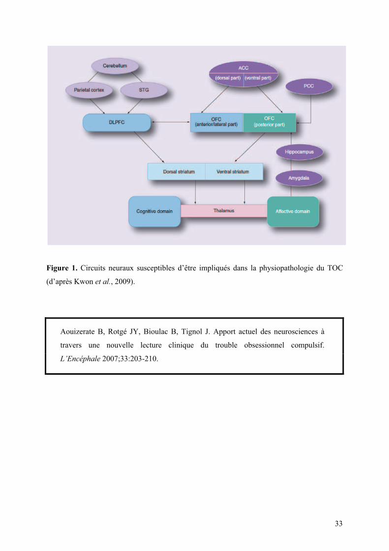
antérieures dans la physiopathologie du TOC (voir Figure 1, page 33) (Schwartz, 1998 ;
Aouizerate et al., 2004a).
1.2. Boucles orbitofrontales et cingulaires antérieures
Le cortex orbitofrontal (COF) comprend deux régions fonctionnellement bien distinctes.
La première, ventrale et latérale, est impliquée dans un certain nombre de fonctions cognitives
comme la détection des erreurs (Rosenkilde et al., 1981 ; Rubin et al., 1992) ou la sélection et
la comparaison de stimuli environnementaux selon la signification qui leur est attribuée
(Ramnani et Owen, 2004). Ces processus sont des éléments essentiels de la prise de décision
permettant de répondre au mieux aux buts et objectifs fixés (Krawczyk, 2002). La seconde
région du COF, ventrale et médiane, joue un rôle majeur dans la gestion des processus
émotionnels et motivationnels, autre aspect important de la prise de décision (Davidson et
Irwin, 1999). Suite à la présentation d’un stimulus et à la saisie des informations relatives à ce
dernier, cette région ventro-médiane permet l’expression d’un état émotionnel et
motivationnel interne qui va entraîner l’émergence d’un comportement de telle sorte que
l’expérience émotionnelle ressentie et le comportement généré soient les plus adaptés au
contexte. Cette adéquation résulte de la mise en jeu de processus de modulation et ou
d’inhibition tenant compte du sens conféré au stimulus (Phillips et al., 2003). Le cortex
préfrontal ventro-médian permet également de pouvoir se construire des représentations
mentales de tels états émotionnels en dehors de toute exposition aux stimuli. Sur la base des
expériences passées, ces représentations jouent un rôle important dans les décisions et les
comportements qui en résultent (Davidson et Irwin, 1999).
31
Le cortex cingulaire antérieur (CCA) est un centre d’intégration cognitif et émotionnel
qui se situe au carrefour des systèmes associatif et limbique. Il se compose de deux régions,
l’une dorsale à orientation cognitive, l’autre ventrale et rostrale plus spécifiquement impliquée
dans les dimensions émotionnelles et motivationnelles du comportement (Bush et al., 2000).
La région dorsale participe à un grand nombre de fonctions cognitives, dont la détection des
erreurs. Il peut s’agir d’une détection de l’erreur par le sujet lui-même, une fois la réponse
comportementale émise, à moins que cette détection de l’erreur n’émane d’une information
extérieure (Holroyd et al., 2004). Cette région dorsale du CCA intervient également dans la
gestion des situations dites de « haut conflit » qui placent le sujet en condition d’erreur
potentielle. Elle permet la sélection de réponses comportementales destinées à éviter ou
corriger l’erreur commise par « haut contrôle cognitif » impliquant un recrutement du cortex
préfrontal dorsolatéral (Kerns et al., 2004). Tout cela requiert de façon impérative la mise en
jeu des ressources attentionnelles et mnésiques (mémoire de travail), l’objectif étant pour le
sujet de fixer son attention sur la séquence comportementale qu’il accomplit et d’accroître ses
performances (Procyk et al., 2000). Le CCA dorsal joue également un rôle important dans le
processus d’anticipation qui fait appel à une certaine flexibilité cognitive (Bush et al., 2000).
L’anticipation permet de réduire la probabilité de faire une erreur dans une situation donnée
par l’établissement de la relation entre la réponse comportementale et ses conséquences. Elle
permet en quelque sorte de prendre les devants et de se prémunir du risque d’erreur dans une
tâche comportementale donnée. Ce phénomène d’anticipation renvoie également aux
représentations que le sujet s’est construit au fil des expériences accumulées dans des
situations proches (Davidson et Irwin, 1999). La région ventrale et rostrale du CCA est
étroitement connectée au COF ventromédian et autres structures limbiques comme
l’amygdale, l’hippocampe ou le striatum ventral (voir Figure 1, page 33) (Bush et al., 2000).
Elle permet d’attirer l’attention sur le vécu émotionnel de façon à en prendre conscience ou
connaissance et à pouvoir lui attribuer plus clairement certains qualificatifs. Ce CCA ventral
est aussi impliqué dans la régulation des réponses neurovégétatives et neuroendocriniennes
entourant la perception des émotions par ses projections sur l’hypothalamus (Bush et al.,
2000).
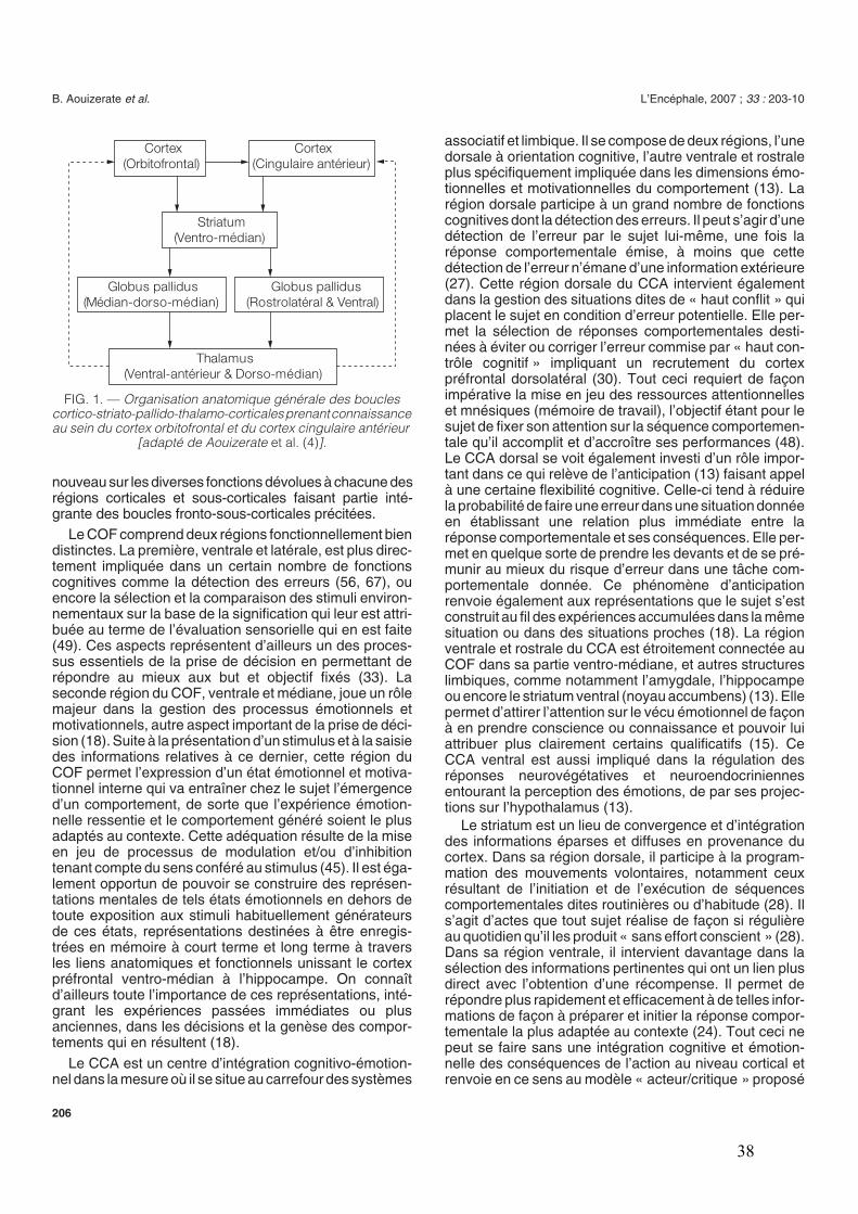
Le striatum est un lieu de convergence et d’intégration des informations éparses et
diffuses en provenance du cortex (voir Figure 1, page 33). Dans sa région dorsale, il participe
à la programmation des mouvements volontaires, notamment ceux résultant de l’initiation et
de l’exécution de séquences comportementales dites routinières (Jogs et al., 1999). Il s’agit
d’actes que tout sujet réalise de façon si régulière au quotidien qu’il les produit « sans effort
32
conscient ». Dans sa région ventrale, il intervient davantage dans la sélection des informations
pertinentes qui ont un lien plus direct avec l’obtention d’une récompense. Il permet de
répondre plus rapidement et efficacement à de telles informations de façon à préparer et initier
la réponse comportementale la plus adaptée au contexte (Hassani et al., 2001). Tout cela ne
peut se faire sans une intégration cognitive et émotionnelle des conséquences de l’action au
niveau cortical et renvoie en ce sens au modèle « acteur/critique » (Sutton et Barto, 1998).
L’« acteur », en émettant un comportement, exerce en quelque sorte un contrôle sur
l’environnement. Il reçoit en retour un certain nombre de renseignements par l’évaluation des
conséquences de l’action (« critique »), lui permettant d’ajuster la réponse comportementale
qui suit et de répondre aux attentes qu’il s’était fixées à l’origine.
Pallidum et thalamus, quant à eux, n’ont fait l’objet que de peu de travaux s’intéressant
précisément à leurs propriétés fonctionnelles. Il a néanmoins pu être identifié au sein du
pallidum externe des territoires limbiques impliqués dans l’apparition de comportements
stéréotypés ainsi que des territoires plus associatifs qui participent à la production d’une
hyperactivité désordonnée (Grabli et al., 2004).
Sur la base de nos connaissances physiologiques des boucles fronto-sous-corticales, la
phénoménologie du TOC conduit à faire l'hypothèse de l'implication des boucles cingulaires
antérieures et orbitofrontales dans la médiation des symptômes obsessionnels et compulsifs.
L'utilisation de la neuro-imagerie en psychiatrie permet de tester ces hypothèses mais aussi
d'affiner notre connaissance des régions cérébrales impliquées dans le TOC, selon les
différentes techniques de neuro-imagerie employées et leurs spécificités.
33
Figure 1. Circuits neuraux susceptibles d’être impliqués dans la physiopathologie du TOC
(d’après Kwon et al., 2009).
Aouizerate B, Rotgé JY, Bioulac B, Tignol J. Apport actuel des neurosciences à
travers une nouvelle lecture clinique du trouble obsessionnel compulsif.
L’Encéphale 2007;33:203-210.

34
L’Encéphale,
33 :
2007, Mars-Avril
203
PSYCHOPHYSIOLOGIE
Apport actuel des neurosciences à travers une nouvelle lecture clinique du trouble obsessionnel compulsif
B. AOUIZERATE
(1)
, J.Y. ROTGÉ
(1)
, B. BIOULAC
(2)
, J. TIGNOL
(1)
(1) Service de Psychiatrie d’Adultes du Professeur Tignol, Université Victor Segalen Bordeaux 2, Centre Hospitalier Charles Perrens, CentreCarreire, 121, rue de la Béchade, 33076 Bordeaux, France.(2) Laboratoire de Neurophysiologie (CNRS UMR 5543), Université Victor Segalen Bordeaux 2.
Travail reçu le 18 août 2006 et accepté le 16 novembre 2006.
Tirés à part :
B. Aouizerate (à l’adresse ci-dessus).
Present contribution of neurosciences to a new clinical reading of obsessive-compulsive disorder
Obsessive-compulsive disorder (OCD), that affects 2 to 3 % of the general population, is characterized by recurrent intrusivethoughts and repetitive, time-consuming behaviors. It is a severely incapacitating mental illness that causes profound impair-ment in psychosocial functioning and quality of life. Although the physiopathology of OCD is still far from resolved, the existenceof a biological basis for OCD is now clearly established and should be interpreted from phenomenological considerations,on the one hand, and in the light of our increasing knowledge of the physiology of cortico-subcortical functional loops, on theother. In a phenomenological view, the heart of the obsessional process is the subject’s underlying impression that « somethingis wrong ». In other words, obsessions may be thought of as the permanent perception of a mistake and/or error in certainbehavioral situations. Compulsions occur as behavioral responses aimed at relieving the tensions or anxiety generated bythe situation. If obtained, this relief may be felt to be a form of reward. Nevertheless, it is only transient, thereby creating afeeling of considerable anxiety. This contributes to immediately reproducing the behavior in a cyclic manner, on the basis ofan internal motivational state through an expectation of the reward. Therefore, it can be assumed that several malfunctioningprocesses are altered within the OCD : 1) error recognition ; and, 2) emotion and motivation. This suggests that there is adysfunction of the brain regions mediating these cognitive and emotional functions. Experimental neurophysiology in laboratoryanimals indicates the central role of the fronto-subcortical circuits originating in the orbitofrontal and anterior cingulate cortices,respectively. The orbitofrontal cortex (OFC) and ventromedial areas are involved in appraisal of the emotional and motivationalvalues of environmental information, and in integrating the subject’s prior experience, which is crucial in decision-making.The OFC also contributes to the selection, comparison and judgment of stimuli and error detection. The anterior cingulatecortex (ACC) is comprised of 1) a ventral or affective region that could keep attention on the internal emotional and motivationalstatus and regulation of autonomic responses, and 2) a dorsal and cognitive region that serves a wide range of functionsincluding attention, working memory, error detection, conflict monitoring, response selection, and anticipation of incominginformation. Ventral striatum, that is intimately connected to the OFC and ACC, participates in the preparation, initiation andexecution of behavioral responses oriented toward reward delivery following the cognitive and emotional integration of beha-viorally relevant information at the cortical level. Functional imaging research in humans has shown an increased functionalactivity in the OFC, ACC, head of the caudate nucleus and thalamus in OCD patients. These functional abnormalities havebeen found in basal conditions and during provocation tests. Moreover, the therapeutic efficacy of antidepressants with pre-ponderant serotonin-reuptake inhibiting properties and cognitive-behavioral therapies seems to be associated with a pro-gressive reduction in activity of the OFC, ACC and the caudate nucleus. Therefore, these observations are suggestive of theresponsibility of 5HT neurotransmission in the dysfunction of the frontal-subcortical loops that emanate from the OFC andACC. However, several lines of research suggest that the dopamine system, with which 5HT interacts, may play a major rolein the expression of OC symptoms. In conclusion, it seems that in OCD there is a dysfunction of the brain regions that belongto the orbitofrontal and anterior cingulate loops in view of evidence obtained from separate and complementary approaches.
Key words :
Anterior cingulate cortex ; Emotion/motivation ; Error detection ; Obsessive-compulsive disorder ; Orbitofrontal cortex.
35
B. Aouizerate
et al.
L’Encéphale, 2007 ;
33 :
203-10
204
Résumé.
Le trouble obsessionnel compulsif (TOC) est uneaffection fréquente touchant 2 à 3 % de la population géné-rale. Il s’agit d’un trouble anxieux invalidant d’évolution le plussouvent chronique. Les obsessions sont définies par l’irrup-tion intrusive et incessante dans la pensée d’une idée quientraîne une anxiété importante. Les compulsions sont descomportements, répétitifs ou ritualisés, visant à neutraliserou à réduire la charge anxieuse provoquée par l’émergencedes pensées obsédantes. L’approche phénoménologique duTOC oriente vers l’altération d’un certain nombre de fonc-tions, détection des erreurs, processus émotionnels, motiva-tionnels et de récompense. Ceci suggère l’implication des cir-cuits cortico-striato-pallido-thalamo-corticaux dans laphysiopathologie de cette affection, et notamment ceux met-tant en jeu le cortex orbito-frontal et le cortex cingulaire anté-rieur dans l’étiopathogénie du TOC, à la lumière de nos con-naissances actuelles des relations structure-fonction issuesdes données de la neurophysiologie expérimentale chezl’animal, complétées par les travaux de neuro-imagerie fonc-tionnelle chez l’homme. C’est précisément cette démarchephysiopathologique interrogeant en permanence ce que l’onsait de la clinique du TOC que nous nous proposons d’adop-ter dans ce travail de revue.
Mots clés :
Cortex cingulaire antérieur ; Cortex orbitofrontal ; Détec-tion de l’erreur ; Émotion/motivation ; Trouble obsessionnel compul-sif.
INTRODUCTION
Le trouble obsessionnel compulsif (TOC) est une affec-tion psychiatrique relativement fréquente. En populationgénérale, des études comme l’«
Epidemiologic Catch-ment Area
» (ECA) réalisées aux États-Unis sous l’égidede l’Institut National de la Santé Mentale (
National Instituteof Mental Health
, NIMH) indiquent une prévalence du TOCde 1,9 à 3,3 % sur la vie entière (55, 68) et de 0,7 à 2,1 %sur 6 mois (9). Il figure ainsi au 4
e
rang derrière la dépres-sion majeure, les phobies et l’abus/dépendance des subs-tances psychoactives (53). Si le TOC semble affecter defaçon égale hommes et femmes (50), des différences sontmalgré tout observées pour ce qui est de l’âge de débutde la maladie plus précoce chez les hommes (17-18 ans)que chez les femmes (21-23 ans) (11, 52).
Le TOC est rarement une pathologie isolée, mais estle plus souvent associé à d’autres affections psychiatri-ques parmi lesquelles les troubles de l’humeur, avec ladépression majeure (67 %) ou le trouble bipolaire (13 %),et les troubles anxieux, qu’il s’agisse des phobies spéci-fiques (22 %), de la phobie sociale (18 %) ou du troublepanique (12 %). Une forte comorbidité vie entière est éga-lement retrouvée avec les troubles des conduites alimen-taires (17 %) ou ceux liés à l’utilisation de substances psy-choactives comme l’alcool (14 %), sans oublier un lienprivilégié avec la maladie de Gilles de la Tourette dans7 % des cas (46, 53).
En dépit de l’hétérogénéité clinique de la maladie, lessymptômes obsessionnels compulsifs répondent à une
classification en quatre facteurs. Le premier regroupe lesobsessions à thème agressif, sexuel, religieux ou soma-tique et compulsions de vérification. Le second corres-pond aux obsessions de symétrie, d’ordre et d’exactitudeou encore celles dites « à pensées magiques », qui fontintervenir la notion d’un malheur qui pourrait frapper lesproches du sujet, ainsi qu’aux conduites compulsivesd’ordre, de rangement et de comptage. Le troisième com-prend les obsessions de contamination, saleté et souillureet les compulsions de lavage et de nettoyage. Le qua-trième enfin est constitué des obsessions/compulsionscentrées sur l’accumulation/collection (35). En termes defréquence et par ordre décroissant, sont retrouvées lesobsessions de contamination (45 %), les obsessionssomatiques (36 %), les obsessions d’ordre et de symétrie(31 %), puis les obsessions à thématique agressive(28 %) et sexuelle (26 %). Les compulsions de vérificationsont les plus fréquentes (63 %). Viennent ensuite lesrituels de lavage/nettoyage (50 %), puis ceux ayant pourobjet le fait de compter (36 %), d’aligner/ranger (28 %) etd’accumuler/collectionner (18 %) (53). Les obsessions/compulsions appartenant aux premiers et deuxièmes fac-teurs sont associées à un âge de début assez précoce dela maladie, avant 16 ans (23, 42). Les obsessions/com-pulsions correspondant au premier facteur se caractéri-sent aussi par une comorbidité plus importante, notam-ment avec la dépression, alors que les obsessions/compulsions liées aux second et troisième facteurs entre-tiennent des relations plus étroites avec, respectivement,les troubles bipolaires et le trouble panique avec agora-phobie d’une part, et les troubles des conduites alimen-taires de l’autre (23).
Si le TOC, après un début le plus souvent insidieux,s’exprime initialement par intermittence avec des phasesd’exacerbation symptomatique entrecoupées de périodesde rémission, son évolution tend davantage vers la chro-nicité après plusieurs années. Ceci n’exclut pas la possi-bilité d’une aggravation des manifestations au fil du temps(51, 63). Le retentissement sur le fonctionnement socio-professionnel et familial est alors important, d’où une fré-quence plus marquée des sujets séparés, divorcés ousans emploi (29), et une détérioration majeure de la qualitéde vie (32). Mais le développement récent de la psycho-pharmacologie, avec l’avènement des antidépresseurs dela classe des inhibiteurs de la recapture de la sérotonine(IRS), et l’essor des thérapies cognitivo-comportementa-les (TCC), ont permis de transformer le pronostic péjoratifde la maladie dans un nombre non négligeable de cas (70-80 %) (21, 47).
Dans ce contexte, on comprend mieux l’engouementcroissant des neurosciences cliniques et expérimentales,ces deux dernières décennies, pour une meilleure com-préhension de l’étiopathogénie du TOC. La recherche, etnotamment l’enrichissement permanent de nos connais-sances sur les relations structure-fonction, ainsi quel’apport récent de la neuro-imagerie, permettent actuelle-ment de mieux appréhender la physiopathologie de cetrouble et de proposer une hypothèse basée essentielle-ment sur l’approche anatomo-fonctionnelle. Cette démar-
36
L’Encéphale, 2007 ;
33 :
203-10 Apport actuel des neurosciences à travers une nouvelle lecture clinique du trouble obsessionnel compulsif
205
che vise, dans un premier temps, à essayer de définir lesprocessus cognitifs et/ou émotionnels occupant une placecentrale dans la pathogénie du TOC, au vu de ce que l’onsait de sa clinique. Elle tente ensuite d’identifier les régionsou structures cérébrales sous-tendant ces divers proces-sus et dont le fonctionnement serait
a priori
altéré dans leTOC, en confrontant les données de la neurophysiologieexpérimentale chez l’animal à celles de la neuro-imageriefonctionnelle chez l’homme. En perspective, certainsdéterminants de ces perturbations sont abordés avec unbref regard porté sur les aspects neurochimiques commetrait d’union entre processus dysfonctionnels et régionscérébrales d’intérêt. C’est cette réflexion physiopatholo-gique, faisant référence de façon incessante à la clinique,que nous tentons de développer dans les paragraphes quisuivent.
BASES PHÉNOMÉNOLOGIQUES
Les obsessions sont définies par l’irruption intrusive etincessante dans la pensée d’une idée, d’une impulsion oud’une représentation apparaissant le plus souvent au sujetcomme un phénomène pathologique, émanant de sa pro-pre activité psychique et persistant malgré tous ses effortspour s’en débarrasser. Elles sont ainsi le plus souvent decaractère « égodystonique » en ce sens que le sujet inter-prète le contenu des pensées obsédantes comme étran-ger à lui-même, en désaccord avec ses propres croyanceset valeurs. Les compulsions sont des comportementsrépétitifs auxquels le sujet ne peut résister, se sentantdans l’obligation de les accomplir. Elles traduisent, engénéral, la lutte contre les obsessions, leur seul but étantde réduire et/ou neutraliser la charge anxieuse résultantde l’émergence des pensées obsédantes.
Ces définitions amènent à considérer que les obses-sions sont plus précisément liées à une surestimation desconséquences négatives auxquelles une action peutexposer dans certaines situations (46, 53). Le douteobsessionnel, qui est à distinguer du contenu même despensées obsédantes, peut être conçu comme la percep-tion permanente ou récurrente par le sujet d’être en situa-tion d’erreur («
something is wrong
» des anglo-saxons)(61). En d’autres termes, le doute naît de l’erreur que lesujet aurait commise en surévaluant le risque de survenued’un événement défavorable et préjudiciable suite à l’exé-cution d’un comportement donné. À titre d’exemple, dansle cas d’obsessions à thème d’exactitude, dont le contenuest parfois mental, le sujet peut être assiégé par la peurde ne pas comprendre précisément ce qu’il lit ou ce quel’interlocuteur peut dire lors d’un échange verbal. Il perçoitalors des signaux d’erreur qui viennent alimenter le douteobsessionnel le conduisant à s’interroger sur ses capaci-tés même de compréhension. Dans le cadre d’obsessionsportant sur des thèmes agressifs, le sujet émet la crainte,par exemple, de provoquer en conduisant, par négligence,un accident de la voie publique. La question que le sujetse pose alors, sans pouvoir y apporter de réponse, portesur la qualité de l’évaluation qu’il a pu faire de la probabilitéde survenue du supposé accident. Il est clair que ce che-
minement de la pensée, de forme purement interrogativeet probabiliste, s’applique parfaitement aux autres typesd’obsessions précédemment évoqués. C’est précisémentsur cette absence de réponse précise à la question poséeque repose le doute obsessionnel générant l’anxiété. Lescompulsions apparaissent alors comme des réponsescomportementales destinées à soulager l’anxiété provo-quée par la mise en situation en tentant de mettre fin auxsignaux d’erreur que le sujet repère. Elles visent soit à pré-venir, comme c’est le cas des compulsions de collectionet d’accumulation, soit à réduire, par la réalisation derituels de lavage et de nettoyage, les conséquences pré-dites comme négatives de certaines actions. Il s’agit ausside s’assurer, par la vérification, que le risque de survenued’un événement grave dans une situation donnée est réel-lement surestimé. Le sujet porte toute son attention surl’acte compulsif qu’il est en train d’accomplir afin des’affranchir de la moindre erreur dans son exécution, depouvoir en extraire toutes les informations indispensablesà la levée du doute obsessionnel et en conserver une tracemnésique. En ce qui concerne les exemples choisis, lesujet se doit de lire et relire de façon inlassable ce qui luisemble échapper à sa propre compréhension, de la mêmefaçon qu’il va demander à l’interlocuteur de répéter à plu-sieurs reprises ce qu’il a pu dire, afin d’avoir plus de cer-titude sur sa capacité à comprendre. Il en sera de mêmepour le risque exact de survenue de l’accident, avec desretours répétés sur son lieu supposé. Néanmoins, le sou-lagement ressenti, une fois la réponse compulsive émise,reste souvent transitoire, l’incertitude étant aussitôt réali-mentée par le flot des préoccupations obsédantes. Lesujet est alors amené à reproduire en boucle ce compor-tement sur la base d’un état émotionnel et motivationnelinterne orienté vers l’obtention d’un soulagement plusdurable qui peut être conçu comme une forme de récom-pense.
Ces aspects phénoménologiques supposent l’exacer-bation d’un certain nombre de fonctions cognitives dansle TOC, qu’elles soient liées au sens attribué à l’informa-tion reçue et à sa représentation, à l’anticipation, à ladétection des erreurs, à l’attention ou à la mémoire de tra-vail. Ils suggèrent également toute l’importance des pro-cessus émotionnels, motivationnels et de récompense(3). Une telle approche oriente donc vers le rôle potentieldes circuits cortico-striato-pallido-thalamo-corticaux dansla physiopathologie de cette affection. Parmi ceux-ci, lescircuits trouvant leur origine au niveau du cortex orbito-frontal (COF) et du cortex cingulaire antérieur (CCA), etdont l’organisation anatomique est illustrée sur la
figure 1
,ont été proposés comme étroitement impliqués dans laproduction des symptômes obsessionnels compulsifs (3,61).
APPROCHE ANATOMO-FONCTIONNELLE
Aspects fonctionnels
Les données actuelles de la neurophysiologie expéri-mentale chez l’animal ont contribué à apporter un éclairage
37
B. Aouizerate
et al.
L’Encéphale, 2007 ;
33 :
203-10
206
nouveau sur les diverses fonctions dévolues à chacune desrégions corticales et sous-corticales faisant partie inté-grante des boucles fronto-sous-corticales précitées.
Le COF comprend deux régions fonctionnellement biendistinctes. La première, ventrale et latérale, est plus direc-tement impliquée dans un certain nombre de fonctionscognitives comme la détection des erreurs (56, 67), ouencore la sélection et la comparaison des stimuli environ-nementaux sur la base de la signification qui leur est attri-buée au terme de l’évaluation sensorielle qui en est faite(49). Ces aspects représentent d’ailleurs un des proces-sus essentiels de la prise de décision en permettant derépondre au mieux aux but et objectif fixés (33). Laseconde région du COF, ventrale et médiane, joue un rôlemajeur dans la gestion des processus émotionnels etmotivationnels, autre aspect important de la prise de déci-sion (18). Suite à la présentation d’un stimulus et à la saisiedes informations relatives à ce dernier, cette région duCOF permet l’expression d’un état émotionnel et motiva-tionnel interne qui va entraîner chez le sujet l’émergenced’un comportement, de sorte que l’expérience émotion-nelle ressentie et le comportement généré soient le plusadaptés au contexte. Cette adéquation résulte de la miseen jeu de processus de modulation et/ou d’inhibitiontenant compte du sens conféré au stimulus (45). Il est éga-lement opportun de pouvoir se construire des représen-tations mentales de tels états émotionnels en dehors detoute exposition aux stimuli habituellement générateursde ces états, représentations destinées à être enregis-trées en mémoire à court terme et long terme à traversles liens anatomiques et fonctionnels unissant le cortexpréfrontal ventro-médian à l’hippocampe. On connaîtd’ailleurs toute l’importance de ces représentations, inté-grant les expériences passées immédiates ou plusanciennes, dans les décisions et la genèse des compor-tements qui en résultent (18).
Le CCA est un centre d’intégration cognitivo-émotion-nel dans la mesure où il se situe au carrefour des systèmes
associatif et limbique. Il se compose de deux régions, l’unedorsale à orientation cognitive, l’autre ventrale et rostraleplus spécifiquement impliquée dans les dimensions émo-tionnelles et motivationnelles du comportement (13). Larégion dorsale participe à un grand nombre de fonctionscognitives dont la détection des erreurs. Il peut s’agir d’unedétection de l’erreur par le sujet lui-même, une fois laréponse comportementale émise, à moins que cettedétection de l’erreur n’émane d’une information extérieure(27). Cette région dorsale du CCA intervient égalementdans la gestion des situations dites de « haut conflit » quiplacent le sujet en condition d’erreur potentielle. Elle per-met la sélection de réponses comportementales desti-nées à éviter ou corriger l’erreur commise par « haut con-trôle cognitif » impliquant un recrutement du cortexpréfrontal dorsolatéral (30). Tout ceci requiert de façonimpérative la mise en jeu des ressources attentionnelleset mnésiques (mémoire de travail), l’objectif étant pour lesujet de fixer son attention sur la séquence comportemen-tale qu’il accomplit et d’accroître ses performances (48).Le CCA dorsal se voit également investi d’un rôle impor-tant dans ce qui relève de l’anticipation (13) faisant appelà une certaine flexibilité cognitive. Celle-ci tend à réduirela probabilité de faire une erreur dans une situation donnéeen établissant une relation plus immédiate entre laréponse comportementale et ses conséquences. Elle per-met en quelque sorte de prendre les devants et de se pré-munir au mieux du risque d’erreur dans une tâche com-portementale donnée. Ce phénomène d’anticipationrenvoie également aux représentations que le sujet s’estconstruit au fil des expériences accumulées dans la mêmesituation ou dans des situations proches (18). La régionventrale et rostrale du CCA est étroitement connectée auCOF dans sa partie ventro-médiane, et autres structureslimbiques, comme notamment l’amygdale, l’hippocampeou encore le striatum ventral (noyau accumbens) (13). Ellepermet d’attirer l’attention sur le vécu émotionnel de façonà en prendre conscience ou connaissance et pouvoir luiattribuer plus clairement certains qualificatifs (15). CeCCA ventral est aussi impliqué dans la régulation desréponses neurovégétatives et neuroendocriniennesentourant la perception des émotions, de par ses projec-tions sur l’hypothalamus (13).
Le striatum est un lieu de convergence et d’intégrationdes informations éparses et diffuses en provenance ducortex. Dans sa région dorsale, il participe à la program-mation des mouvements volontaires, notamment ceuxrésultant de l’initiation et de l’exécution de séquencescomportementales dites routinières ou d’habitude (28). Ils’agit d’actes que tout sujet réalise de façon si régulièreau quotidien qu’il les produit « sans effort conscient » (28).Dans sa région ventrale, il intervient davantage dans lasélection des informations pertinentes qui ont un lien plusdirect avec l’obtention d’une récompense. Il permet derépondre plus rapidement et efficacement à de telles infor-mations de façon à préparer et initier la réponse compor-tementale la plus adaptée au contexte (24). Tout ceci nepeut se faire sans une intégration cognitive et émotion-nelle des conséquences de l’action au niveau cortical etrenvoie en ce sens au modèle « acteur/critique » proposé
FIG. 1. —
Organisation anatomique générale des boucles cortico-striato-pallido-thalamo-corticales prenant connaissance au sein du cortex orbitofrontal et du cortex cingulaire antérieur
[adapté de Aouizerate
et al. (4)
].
Cortex
(Orbitofrontal)
Cortex
(Cingulaire antérieur)
Striatum
(Ventro-médian)
Globus pallidus
(Médian-dorso-médian)
Globus pallidus
(Rostrolatéral & Ventral)
Thalamus
(Ventral-antérieur & Dorso-médian)
38
L’Encéphale, 2007 ;
33 :
203-10 Apport actuel des neurosciences à travers une nouvelle lecture clinique du trouble obsessionnel compulsif
207
par Sutton et Barto (64). L’« acteur », en émettant un com-portement, exerce en quelque sorte un contrôle, une pres-sion sur l’environnement. Il reçoit, en retour, un certainnombre de renseignements à travers une évaluation desconséquences de l’action, ou « critique », lui permettantd’ajuster la réponse comportementale qui suit et de répon-dre aux attentes qu’il s’était fixées à l’origine.
Pallidum et thalamus, quant à eux, n’ont fait l’objet quede peu de travaux s’intéressant précisément à leurs pro-priétés fonctionnelles ; ont pu néanmoins être identifiés,au sein du pallidum externe, des territoires limbiques impli-qués dans l’apparition de comportements stéréotypés,ainsi que des territoires plus associatifs qui participent àla production d’une hyperactivité désordonnée (22).L’exploration du thalamus centrée sur les noyaux ventral-antérieur et dorso-médian a permis de mieux comprendreleur importance dans l’émergence de deux types deréponses, les unes purement émotionnelles avec leurscorrélats neurovégétatifs, les autres d’expression com-portementale marquées par l’émergence de séquencesrépétitives, complexes et finalisées (5). Ceci n’a riend’étonnant si l’on considère que ces deux noyaux thala-miques sont connus pour entretenir des liens anatomiquesprivilégiés avec les COF et CCA (3, 4)
(figure 1)
.
Neuro-imagerie fonctionnelle
La neurophysiologie clinique, à travers la neuro-image-rie chez l’homme, a permis d’apporter un éclairage nou-veau sur la physiopathologie du TOC. Les principaux tra-vaux menés dans ce cadre n’ont fait que soulignerdavantage l’importance des circuits orbito-frontal et cin-gulaire antérieur dans la survenue des symptômes obses-sionnels compulsifs.
Les recherches menées dans le domaine fonctionnelà l’aide de la tomographie par émission de positrons(TEP), de la tomographie d’émission monophotonique(TEMP), ou encore de la résonance magnétique nucléairefonctionnelle (IRMf), ont permis de montrer que plusieursrégions cérébrales des patients atteints de TOC présen-taient une activité anormale, qu’il s’agisse de la régionorbitale du cortex préfrontal, du CCA, du striatum (noyaucaudé) ou du thalamus.
L’une des premières études réalisées a permis de met-tre en évidence, grâce à l’utilisation de la TEP et du[
18
F]fluorodéoxyglucose, une augmentation du métabo-lisme du COF gauche, avec une tendance non significa-tive à droite, chez des patients atteints de TOC (6). Dansune seconde étude publiée par les mêmes auteurs un anplus tard, une augmentation, cette fois-ci, bilatérale dumétabolisme au niveau du COF et de la tête du noyaucaudé a été rapportée chez les patients obsessionnels (7).Des résultats similaires ont été obtenus, chez des patientsprésentant un TOC depuis l’enfance, avec une augmen-tation du métabolisme du COF, encore que limitée à gau-che, s’accompagnant d’une élévation bilatérale du méta-bolisme au sein du CCA, le métabolisme des noyauxcaudés n’étant, en revanche, pas modifié. Une corrélation
positive entre l’activité orbitale droite et la sévérité dessymptômes obsessionnels compulsifs a été égalementnotée (65). Une augmentation bilatérale de l’activité tha-lamique a été enfin retrouvée chez des patients présentantun diagnostic de TOC (44, 58).
Un grand nombre de travaux ont été réalisés en utilisantla TEMP et la mesure du débit sanguin cérébral pourapprécier l’activité cérébrale régionale. Bien que dansl’étude initiale de Machlin
et al.
(38), aucune altération del’activité au sein du COF ou du noyau caudé n’ait été miseen évidence, une augmentation bilatérale du débit sanguinmesuré par le [
99m
Tc]HMPAO au niveau de la région orbi-tale du cortex préfrontal a été retrouvée, contrastant avecune diminution de l’activité au niveau de la tête des noyauxcaudés, ces anomalies fonctionnelles étant positivementcorrélées à l’intensité des symptômes obsessionnelscompulsifs (57). Les mêmes résultats ont été obtenus pardes études plus récentes, encore que l’augmentationd’activité puisse prédominer du côté droit, voire mêmeintéresser le CCA et/ou le thalamus (2, 34, 62).
Zohar
et al.
(69) ont, les premiers, étudié les effets dela provocation de symptômes obsessionnels compulsifspar exposition à un objet « contaminant » sur l’activité céré-brale régionale. Ils ont pu montrer une diminution du débitsanguin cérébral mesuré au niveau cortical par le
133
Xechez des patients souffrant de TOC, cette réduction del’activité corticale étant directement reliée à la gravité desmanifestations cliniques. Néanmoins, l’ensemble des tra-vaux ultérieurs va dans le sens d’une augmentation del’activité au niveau du COF, du CCA, du noyau caudé etdu thalamus droits et/ou gauches, qui serait positivementcorrélée à la sévérité des symptômes lors d’épreuves deprovocation, en utilisant du
15
O pour mesurer le débit san-guin régional (17, 54). Des résultats tout à fait similairesont été obtenus à l’aide de l’IRMf avec une activation bila-térale du COF, du CCA et du noyau caudé lors de proto-coles de provocation chez des patients obsessionnels (1,12). Néanmoins, ces régions cérébrales semblent fairel’objet d’une activation différente selon la nature des mani-festations présentées. C’est ainsi qu’une augmentationd’activité au niveau des COF et CCA ventral a été retrouvéechez les patients dits « accumulateurs » par opposition aux« vérificateurs » chez lesquels une activation préférentielledu CCA dorsal a été rapportée (40). Par contre, les patients« accumulateurs » montrent une activité moins importanteau sein du noyau caudé et du thalamus comparés à ceuxne présentant pas cette forme de TOC (60).
L’efficacité des traitements antidépresseurs par IRS etdes TCC est aujourd’hui clairement établie dans la priseen charge du TOC (21, 47). Plusieurs auteurs se sont inté-ressés à l’influence de ces diverses stratégies thérapeu-tiques sur l’activité cérébrale régionale en utilisant lestechniques de neuro-imagerie fonctionnelle précitées.Dans une première étude, il a été mis en évidence unediminution du métabolisme du COF et du noyau caudégauche chez des patients souffrant de TOC améliorés par13 à 21 semaines de traitement par clomipramine (10).Des résultats comparables ont été retrouvés sur unepopulation de patients obsessionnels traités pendant 8-
39
B. Aouizerate
et al.
L’Encéphale, 2007 ;
33 :
203-10
208
16 semaines par fluoxétine ou paroxétine avec efficacité(26, 59). La diminution progressive du métabolisme orbito-frontal est corrélée au degré d’amélioration clinique soustraitement par clomipramine ou fluoxétine prescrit pen-dant un an à des patients atteints de TOC (66). Il a étérapporté la même diminution du métabolisme au niveaudu noyau caudé droit chez des patients avec un diagnosticde TOC répondant favorablement à 8-12 semaines detraitement par fluoxétine, paroxétine ou TCC (8, 59). Cetteréduction progressive de l’activité fonctionnelle est encoreretrouvée au niveau du thalamus chez des patients avecTOC bénéficiant d’un traitement de 12 semaines par flu-voxamine (25).
Certains auteurs se sont enfin consacrés à mieux définirle rôle du CCA dans la pathogénie du TOC, dans la pers-pective d’établir des relations plus intimes avec le proces-sus de détection des erreurs. C’est ainsi qu’une activitéfonctionnelle plus importante au sein du CCA a été obser-vée chez les patients avec TOC lors de tâches compor-tementales les exposant à des erreurs éventuelles. Cetteactivation du CCA est provoquée par le fait, ou la seuleconviction, d’avoir commis une erreur dans une situationdonnée, sollicitant ainsi tant la région rostrale que dorsaledu CCA (20, 39).
Sur la base de ces considérations, et en dépit des dif-férences méthodologiques conduisant parfois à des résul-tats divergents, l’ensemble de ces travaux suggère queles symptômes obsessionnels compulsifs sont avant toutle fait d’un dysfonctionnement des boucles prenant nais-sance au niveau des COF et CCA.
PERSPECTIVES
Au-delà de cette approche anatomo-fonctionnelle sug-gérant l’existence d’une hyperactivité au sein des bouclesorbito-frontale et cingulaire antérieure dans le TOC, il estintéressant d’essayer de mieux identifier les déterminantsde cette hyperactivité en se basant sur un certain nombrede considérations issues de la neurochimie. On peut aus-sitôt évoquer le rôle central de la transmission sérotoni-nergique dans la genèse des manifestations obsession-nelles compulsives. Ainsi, les antidépresseurs, quibloquent de façon préférentielle la recapture de la séro-tonine, ont été démontrés les plus efficaces dans la priseen charge du TOC (47). Il a été également montré que leseffets thérapeutiques des IRS sont associés à une réduc-tion progressive de l’activité au sein de régions cérébralescomme le COF, le CCA, le noyau caudé ou encore le tha-lamus (8, 10, 25, 26, 66). L’administration d’agents phar-macologiques, comme la m-chlorophénylpypérazine ou lesumatriptan, agissant par activation des autorécepteurs5HT
1D
présents au niveau des terminaisons axonales desneurones sérotoninergiques du raphé, est susceptible deprovoquer une exacerbation de la sévérité des symptô-mes chez les patients avec TOC (4). Enfin, on connaîtl’influence modulatrice du système sérotoninergique surl’expression du comportement et sa flexibilité (16, 37), pro-bablement
via
ses interactions fonctionnelles avec la
transmission dopaminergique, dont on peut ainsi suppo-ser l’implication dans la pathogénie du TOC. Les antipsy-chotiques atypiques, en tant que bloqueurs des récep-teurs de la dopamine, réduisent la sévérité dessymptômes obsessionnels compulsifs lorsqu’ils sont uti-lisés en complément des IRS en cas d’efficacité partiellede ces derniers (14, 41). Il a été également rapporté l’exis-tence de perturbations fonctionnelles de ce système deneurotransmission dopaminergique, laissant présagerd’une augmentation de son activité dans le TOC, commeen atteste la mise en évidence par neuro-imagerie d’uneélévation de la densité du transporteur de la dopaminecontrastant avec une réduction du nombre des récepteursD2 au sein du striatum (19, 31). Enfin, le rôle de la trans-mission dopaminergique dans la gestion des fonctionsémotionnelles, motivationnelles et de récompense estbien établi (36), sans omettre son implication dans le pro-cessus de « critique » permettant une évaluation des con-séquences du comportement (43) si l’on fait référence aumodèle « acteur/critique » précédemment développé(64). Dans ce contexte, les obsessions peuvent être con-sidérées comme résultant de la mise en jeu excessive ouinappropriée de ce processus de « critique » dans certai-nes situations (3, 4).
CONCLUSION
On perçoit toute la pertinence d’une démarche derecherche neurophysiologique permettant d’apporter unéclairage nouveau sur les bases physiopathologiques duTOC en partant de sa clinique. Une telle stratégie pourraitavoir des conséquences thérapeutiques dans la mesureoù elle est susceptible d’ouvrir de nouvelles perspectivessur l’abord cognitif des pensées obsédantes et en souli-gner l’intérêt, en complément de ce qui est plus couram-ment pratiqué à travers les thérapies comportementales,qui s’adressent davantage aux conduites compulsives enessayant de contenir progressivement leur émergence.Cette approche cognitive est d’autant plus importante quecertaines formes d’obsessions sont particulièrementrebelles aux antidépresseurs bloquant la recapture de lasérotonine, notamment les obsessions à thématiquesexuelle et religieuse ou à « pensées magiques » (3) ; cecin’a rien d’étonnant si l’on considère qu’il s’agit de préoc-cupations où la nature même de l’événement redouté ren-voie aussitôt à des considérations d’ordre éthique etmoral. On peut alors comprendre que le besoin de leverle doute lié à la surestimation du risque de survenue decette catégorie d’événements soit encore plus impérieuxet difficile à réduire que celui suscité par d’autres préoc-cupations. Une autre implication thérapeutique est le choixde la cible pour la stimulation cérébrale profondeaujourd’hui proposée dans les formes sévères, chroni-ques et résistantes de TOC avec des résultats encoura-geants bien que portant sur un nombre encore très limitéde cas (3).
40
L’Encéphale, 2007 ;
33 :
203-10 Apport actuel des neurosciences à travers une nouvelle lecture clinique du trouble obsessionnel compulsif
209
Références
1. ADLER CM, McDONOUGH-RYAN P, SAX KW
et al
. fMRI of neu-ronal activation with symptom provocation in unmedicated patientswith obsessive compulsive disorder. J Psychiatr Res 2000 ; 34 :317-24.
2. ALPTEKIN K, DEGIRMENCI B, KIVIRCIK B
et al
. Tc-99m HMPAObrain perfusion SPECT in drug-free obsessive-compulsive patientswithout depression. Psychiatr Res 2001 ; 107 : 51-6.
3. AOUIZERATE B, GUEHL D, CUNY E
et al
. Pathophysiology ofobsessive-compulsive disorder : a necessary link between pheno-menology, neuropsychology, imagery and physiology. Prog Neuro-biol 2004 ; 72 : 195-221.
4. AOUIZERATE B, GUEHL D, CUNY E
et al.
Updated overview of theputative role of serotoninergic system in obsessive-compulsive dis-order. Neuropsychiatr Dis Treat 2005 ; 1 : 231-43.
5. AOUIZERATE B, ROTGE JY, AMESTOY V
et al
. Role of the thala-mus in compulsive-like behaviors in the primate (en préparation).
6. BAXTER LR Jr, PHELPS ME, MAZZIOTTA JC
et al
. Local cerebralglucose metabolic rates in obsessive-compulsive disorder. A com-parison with rates in unipolar depression and in normal controls. ArchGen Psychiatry 1987 ; 44 : 211-8.
7. BAXTER LR Jr, SCHWARTZ JM, MAZZIOTTA JC
et al
. Cerebralglucose metabolic rates in nondepressed patients with obsessive-compulsive disorder. Am J Psychiatry 1988 ; 145 : 1560-3.
8. BAXTER LR Jr, SCHWARTZ JM, BERGMAN KS
et al
. Caudate glu-cose metabolic rate changes with both drug and behavior therapyfor obsessive-compulsive disorder. Arch Gen Psychiatry 1992 ; 49 :681-9.
9. BEBBINGTON PE. Epidemiology of obsessive-compulsive disor-der. Br J Psychiatry (Suppl) 1998 : 2-6.
10. BENKELFAT C, NORDAHL TE, SEMPLE WE
et al
. Local cerebralglucose metabolic rates in obsessive-compulsive disorder. Patientstreated with clomipramine. Arch Gen Psychiatry 1990 ; 47 : 840-8.
11. BOGETTO F, VENTURELLO S, ALBERT U
et al
. Gender-relatedclinical differences in obsessive-compulsive disorder. Eur Psychiatry1999 ; 14 : 434-41.
12. BREITER HC, RAUCH SL, KWONG KK
et al
. Functional magneticresonance imaging of symptom provocation in obsessive-compul-sive disorder. Arch Gen Psychiatry 1996 ; 53 : 595-606.
13. BUSH G, LUU P, POSNER MI. Cognitive and emotional influencesin anterior cingulate cortex. Trends Cogn Sci 2000 ; 4 : 215-22.
14. BYSTRITSKY A, ACKERMAN DL, ROSEN RM
et al
. Augmentationof serotonin reuptake inhibitors in refractory obsessive-compulsivedisorder using adjunctive olanzapine : a placebo-controlled trial. JClin Psychiatry 2004 ; 65 : 565-8.
15. CARTER R. Mapping the mind. The Orion Publishing Group Limited(London), 1998.
16. CLARKE HF, DALLEY JW, CROFTS HS
et al
. Cognitive inflexibilityafter prefrontal serotonin depletion. Science 2004 ; 304 : 878-80.
17. COTTRAUX J, GERARD D, CINOTTI L
et al
. A controlled positronemission tomography study of obsessive and neutral auditory sti-mulation in obsessive-compulsive disorder with checking rituals.Psychiatry Res 1996 ; 60 : 101-12.
18. DAVIDSON RJ, IRWIN W. The functional neuroanatomy of emotionand affective style. Trends Cogn Sci 1999 ; 3 : 11-21.
19. DENYS D, VAN DER WEE N, JANSSEN J
et al
. Low level ofdopaminergic D(2) receptor binding in obsessive-compulsive disor-der. Biol Psychiatry 2004 ; 55 : 1041-5.
20. FITZGERALD KD, WELSH RC, GEHRING WJ
et al
. Error-relatedhyperactivity of the anterior cingulate cortex in obsessive-compul-sive disorder. Biol Psychiatry 2005 ; 57 : 287-94.
21. FOA EB, FRANKLIN ME, MOSER J. Context in the clinic : how welldo cognitive-behavioral therapies and medications work incombination ? Biol Psychiatry 2002 ; 52 : 987-97.
22. GRABLI D, MCCAIRN K, HIRSCH EC
et al
. Behavioural disordersinduced by external globus pallidus dysfunction in primates :I. Behavioural study. Brain 2004 ; 127 : 2039-54.
23. HASLER G, LASALLE-RICCI VH, RONQUILLO JG
et al
. Obses-sive-compulsive disorder symptom dimensions show specific rela-tionships to psychiatric comorbidity. Psychiatr Res 2005 ; 135 :121-32.
24. HASSANI OK, CROMWELL HC, SCHULTZ W. Influence of expec-tation of different rewards on behavior-related neuronal activity inthe striatum. J Neurophysiol 2001 ; 85 : 2477-89.
25. HO PIAN KL, VAN MEGEN HJ, RAMSEY NF
et al
. Decreased tha-lamic blood flow in obsessive-compulsive disorder patients respon-ding to fluvoxamine. Psychiatr Res 2005 ; 138 : 89-97.
26. HOEHN-SARIC R, PEARLSON GD, HARRIS GJ
et al
. Effects offluoxetine on regional cerebral blood flow in obsessive-compulsivepatients. Am J Psychiatry 1991 ; 148 : 1243-5.
27. HOLROYD CB, NIEUWENHUIS S, YEUNG N
et al
. Dorsal anteriorcingulate cortex shows fMRI response to internal and external errorsignals. Nat Neurosci 2004 ; 7 : 497-8.
28. JOG MS, KUBOTA Y, CONNOLLY CI
et al
. Building neural repre-sentations of habits. Science 1999 ; 286 : 1745-9.
29. KARNO M, GOLDING JM, SORENSON SB
et al
. The epidemiologyof obsessive-compulsive disorder in five US communities. Arch GenPsychiatry 1988 ; 45 : 1094-9.
30. KERNS JG, COHEN JD, MACDONALD AW, 3RD
et al
. Anterior cin-gulate conflict monitoring and adjustments in control. Science 2004 ;303 : 1023-6.
31. KIM CH, KOO MS, CHEON KA
et al
. Dopamine transporter densityof basal ganglia assessed with [
123
I]IPT SPET in obsessive-com-pulsive disorder. Eur J Nucl Med Mol Imaging 2003 ; 30 : 1637-43.
32. KORAN LM, THIENEMANN ML, DAVENPORT R. Quality of life forpatients with obsessive-compulsive disorder. Am J Psychiatry 1996 ;153 : 783-8.
33. KRAWCZYK DC. Contributions of the prefrontal cortex to the neuralbasis of human decision making. Neurosci Biobehav Rev 2002 ; 26 :631-64.
34. LACERDA AL, DALGALARRONDO P, CAETANO D
et al
. Elevatedthalamic and prefrontal regional cerebral blood flow in obsessive-com-pulsive disorder : a SPECT study. Psychiatr Res 2003 ; 123 : 125-34.
35. LECKMAN JF, GRICE DE, BOARDMAN J
et al
. Symptoms of obses-sive-compulsive disorder. Am J Psychiatry 1997 ; 154 : 911-7.
36. LE MOAL M. Mesocorticolimbic dopaminergic neurons. Functionaland regulatory roles.
In
: Bloom FE, Kupfer DJ, eds. Psychophar-macology : The fourth generation of progress. New York : RavenPress, 1995 : 283-94.
37. LUCKI I. The spectrum of behaviors influenced by serotonin. BiolPsychiatry 1998 ; 44 : 151-62.
38. MACHLIN SR, HARRIS GJ, PEARLSON GD
et al
. Elevated medial-frontal cerebral blood flow in obsessive-compulsive patients : aSPECT study. Am J Psychiatry 1991 ; 148 : 1240-2.
39. MALTBY N, TOLIN DF, WORHUNSKY P
et al
. Dysfunctional actionmonitoring hyperactivates frontal-striatal circuits in obsessive-com-pulsive disorder : an event-related fMRI study. Neuroimage 2005 ;24 : 495-503.
40. MATAIX-COLS D, WOODERSON S, LAWRENCE N
et al
. Distinctneural correlates of washing, checking, and hoarding symptomdimensions in obsessive-compulsive disorder. Arch Gen Psychiatry2004 ; 61 : 564-76.
41. MCDOUGLE CJ, EPPERSON CN, PELTON GH
et al
. A double-blind, placebo-controlled study of risperidone addition in serotoninreuptake inhibitor-refractory obsessive-compulsive disorder. ArchGen Psychiatry 2000 ; 57 : 794-801.
42. MILLET B, KOCHMAN F, GALLARDA T
et al
. Phenomenologicaland comorbid features associated in obsessive-compulsivedisorder : influence of age of onset. J Affect Disord 2004 ; 79 : 241-6.
43. O’DOHERTY J, DAYAN P, SCHULTZ J
et al
. Dissociable roles ofventral and dorsal striatum in instrumental conditioning. Science2004 ; 304 : 452-4.
44. PERANI D, COLOMBO C, BRESSI S
et al
. [
18
F]FDG PET study inobsessive-compulsive disorder. A clinical/metabolic correlationstudy after treatment. Br J Psychiatry 1995 ; 166 : 244-50.
45. PHILLIPS ML, DREVETS WC, RAUCH SL
et al
. Neurobiology ofemotion perception I : The neural basis of normal emotion percep-tion. Biol Psychiatry 2003 ; 54 : 504-14.
46. PIGOTT TA, L’HEUREUX F, DUBBERT B
et al
. Obsessive compul-sive disorder : comorbid conditions. J Clin Psychiatry 1994 ; 55(Suppl) : 15-27.
47. PIGOTT TA, SEAY SM. A review of the efficacy of selective serotoninreuptake inhibitors in obsessive-compulsive disorder. J Clin Psy-chiatry 1999 ; 60 : 101-6.
41
B. Aouizerate
et al.
L’Encéphale, 2007 ;
33 :
203-10
210
48. PROCYK E, TANAKA YL, JOSEPH JP. Anterior cingulate activityduring routine and non-routine sequential behaviors in macaques.Nat Neurosci 2000 ; 3 : 502-8.
49. RAMNANI N, OWEN AM. Anterior prefrontal cortex : insights intofunction from anatomy and neuroimaging. Nat Rev Neurosci 2004 ;5 : 184-94.
50. RASMUSSEN SA, TSUANG MT. The epidemiology of obsessivecompulsive disorder. J Clin Psychiatry 1984 ; 45 : 450-7.
51. RASMUSSEN SA, TSUANG MT. Clinical characteristics and familyhistory in DSM III obsessive-compulsive disorder. Am J Psychiatry1986 ; 143 : 317-22.
52. RASMUSSEN SA, EISEN JL. Epidemiology of obsessive compul-sive disorder. J Clin Psychiatry 1990 ; 51 (Suppl) : 10-3.
53. RASMUSSEN SA, EISEN JL. The epidemiology and differentialdiagnosis of obsessive compulsive disorder. J Clin Psychiatry 1992 ;53 (Suppl) : 4-10.
54. RAUCH SL, JENIKE MA, ALPERT NM
et al
. Regional cerebral bloodflow measured during symptom provocation in obsessive-compul-sive disorder using oxygen 15-labeled carbon dioxide and positronemission tomography. Arch Gen Psychiatry 1994 ; 51 : 62-70.
55. ROBINS LN, HELZER JE, WEISSMAN MM
et al.
Lifetime preva-lence of specific psychiatric disorders in three sites. Arch Gen Psy-chiatry 1984 ; 41 : 949-58.
56. ROSENKILDE CE, BAUER RH, FUSTER JM. Single cell activity inventral prefrontal cortex of behaving monkeys. Brain Res 1981 ;209 : 375-94.
57. RUBIN RT, VILLANUEVA-MEYER J, ANANTH J
et al
. Regionalxenon 133 cerebral blood flow and cerebral technetium 99 mHMPAO uptake in unmedicated patients with obsessive-compulsivedisorder and matched normal control subjects. Determination byhigh-resolution single-photon emission computed tomography. ArchGen Psychiatry 1992 ; 49 : 695-702.
58. SAXENA S, BRODY AL, HO ML
et al
. Cerebral metabolism in majordepression and obsessive-compulsive disorder occurring sepa-rately and concurrently. Biol Psychiatry 2001 ; 50 : 159-70.
59. SAXENA S, BRODY AL, HO ML
et al
. Differential cerebral metabolicchanges with paroxetine treatment of obsessive-compulsive disor-der
vs
major depression. Arch Gen Psychiatry 2002 ; 59 : 250-61.60. SAXENA S, BRODY AL, MAIDMENT KM
et al
. Cerebral glucosemetabolism in obsessive-compulsive hoarding. Am J Psychiatry2004 ; 161 : 1038-48.
61. SCHWARTZ JM. A role of volition and attention in the generation ofnew brain circuitry. Toward a neurobiology of mental force. J Cons-cious Studies 1999 ; 6 : 115-42.
62. SIMPSON S, BALDWIN B. Neuropsychiatry and SPECT of an acuteobsessive-compulsive syndrome patient. Br J Psychiatry 1995 ;166 : 390-2.
63. SKOOG G, SKOOG I. A 40-year follow-up of patients with obsessive-compulsive disorder. Arch Gen Psychiatry 1999 ; 56 : 121-7.
64. SUTTON RS, BARTO AG. Reinforcement Learning – An Introduc-tion. Cambridge, Massachusetts : MIT Press, 1998.
65. SWEDO SE, SCHAPIRO MB, GRADY CL
et al
. Cerebral glucosemetabolism in childhood-onset obsessive-compulsive disorder.Arch Gen Psychiatry 1989 ; 46 : 518-23.
66. SWEDO SE, PIETRINI P, LEONARD HL
et al
. Cerebral glucosemetabolism in childhood-onset obsessive-compulsive disorder.Revisualization during pharmacotherapy. Arch Gen Psychiatry1992 ; 49 : 690-4.
67. THORPE SJ, ROLLS ET, MADDISON S. The orbitofrontal cortex :neuronal activity in the behaving monkey. Exp Brain Res 1983 ; 49 :93-115.
68. WEISSMAN MM, BLAND RC, CANINO GJ
et al
. The cross nationalepidemiology of obsessive compulsive disorder. The Cross NationalCollaborative Group. J Clin Psychiatry 1994 ; 55 (Suppl) : 5-10.
69. ZOHAR J, INSEL TR, BERMAN KF
et al
. Anxiety and cerebral bloodflow during behavioral challenge. Dissociation of central from peri-pheral and subjective measures. Arch Gen Psychiatry 1989 ; 46 :505-10.
42
43
2. Objectif
Le niveau de résistance aux traitements habituels rencontré dans le TOC commande une
meilleure compréhension des mécanismes physiopathologiques de cette affection. Il s'agit
d'une étape indispensable à la nécessaire amélioration des stratégies thérapeutiques actuelles.
Le principal objectif de ma thèse est d’étudier et de définir le rôle des voies thalamo-
corticales intéressant le COF et le CCA dans la pathogénie du TOC en vue d'en dégager les
implications pour une meilleure compréhension générale de la physiopathologie du TOC.
Pour cela, j’ai utilisé plusieurs outils complémentaires permettant d’appréhender cette
problématique sous différents angles méthodologiques. Ainsi, je présenterai des travaux de
méta-analyse des études de neuro-imagerie et des travaux expérimentaux chez l'animal et chez
l'homme. En me basant sur ces résultats, je propose dans cette thèse d'apporter quelques
éléments de réponse concernant 1) l'identification des altérations cérébrales anatomiques
associées au TOC, 2) l'identification des altérations cérébrales fonctionnelles associées au
TOC, 3) la relation entre les altérations anatomiques et fonctionnelles.

44
45
3. Altérations anatomiques des voies thalamo-corticales dans le TOC
3.1. Modifications des volumes cérébraux
3.1.1. Introduction
Plusieurs études ont rapporté des différences de volumes de certaines régions cérébrales
entre des patients avec TOC et des témoins. Cependant, les résultats apparaissent fortement
discordants d’une étude à l’autre selon les définitions anatomiques employées et les
caractéristiques cliniques des populations incluses. Par exemple, les résultats concernant le
volume striatal dans le TOC vont tantôt dans le sens d’une diminution (Robinson et al., 1995 ;
Rosenberg et al., 1997), tantôt dans le sens d’une augmentation (Scarone et al., 1992 ; Giedd
et al., 2000), voire d’une absence de modification (Aylward et al., 1996 ; Kang et al., 2004 ;
Szeszko et al., 2004 ; Riffkin et al., 2005 ; Atmaca et al., 2006 ; Atmaca et al., 2007). Dans le
but d’obtenir une information quantitative et objective de cette littérature discordante, nous
avons réalisé une méta-analyse des études d’IRM volumétrique dans le TOC.
3.1.2. Méthodes
Une recherche systématique par mots-clé a été réalisée pour recenser les études d’IRM
volumétrique publiées dans le TOC. L’ensemble des données cliniques et para-cliniques de
chacune de ces études a été extraite. La taille d’effet, correspondant à la différence de
moyennes standardisées, a été calculée pour chaque région cérébrale dont le volume a été
mesuré par au moins trois équipes de recherche différentes. L’hétérogénéité entre les études a
également été testée. Enfin, pour toute différence ou hétérogénéité significative, nous avons
mené une méta-régression afin d’expliquer la différence ou l’hétérogénéité par les variables
cliniques (âge, sexe, durée de la maladie, traitements médicamenteux, présence de symptômes
dépressifs, présence de tics et sévérité de la maladie).
3.1.3. Résultats et discussion
Les données recueillies sont issues de 21 études ayant inclus 371 patients avec TOC et
407 témoins. Cinq régions cérébrales ont régulièrement été évaluées dans le TOC ; il s’agit du
CCA, du COF, du thalamus, du putamen et du noyau caudé. Les résultats montrent qu’il
existe une diminution du volume du COF bilatéral ainsi qu’une augmentation du volume du
thalamus bilatéral dans le TOC. Le CCA gauche tend à être diminué de volume. Il n’existe

46
pas de différence significative pour les autres régions cérébrales, notamment le striatum. Les
tests d’hétérogénéité sont significatifs pour le CCA gauche et pour le striatum bilatéral.
L’hétérogénéité des résultats pour le CCA, probablement expliquée par la grande variabilité
des définitions anatomiques employées, ne permet pas de conclure clairement en faveur d’une
modification du volume du CCA dans le TOC. Enfin, les analyses de méta-régression
montrent que les modifications du volume thalamique sont positivement corrélées à la
sévérité de la symptomatologie obsessionnelle et compulsive. Il n’existe pas d’autre
association significative.
3.1.4. Conclusion
Les résultats de ce travail suggèrent l’existence d’anomalies anatomiques des voies
thalamo-orbitofrontales dans le TOC. Le COF est diminué de volume, alors que le thalamus
est augmenté de volume. L’augmentation du volume thalamique est positivement corrélée à la
sévérité de la symptomatologie.
Rotgé JY, Guehl D, Dilharreguy B, Tignol J, Bioulac B, Allard M, Burbaud P,
Aouizerate B. Meta-analysis of brain volume changes in obsessive-compulsive
disorder. Biological Psychiatry 2009;65:75-83.
Les matériels supplémentaires sont présentés en Annexe C.
Meta-Analysis of Brain Volume Changes inObsessive-Compulsive DisorderJean-Yves Rotge, Dominique Guehl, Bixente Dilharreguy, Jean Tignol, Bernard Bioulac, Michele Allard,Pierre Burbaud, and Bruno Aouizerate
Background: Many neuroimaging studies exploring the volumes of brain structures in obsessive-compulsive disorder (OCD) have been
published in the past 2 decades. In this study, we attempted to provide a complete overview of structural alterations in OCD by meta-
analyzing magnetic resonance imaging (MRI) data.
Methods: We conducted a systematic search of MRI studies that reported volumetric measurements in both OCD patients and healthy
subjects. Data were entered into the meta-analysis through calculation of the standardized mean differences (SMDs) between the volumes
of cerebral regions in OCD patients and the corresponding volumes in control subjects. We then performed a meta-regression to explore the
influence of clinical covariates on effect sizes.
Results: Although no volumetric differences were found for the whole brain, intracranial region, gray matter, or prefrontal cortex, OCD
patients did show a reduced volume of the left anterior cingulate cortex (ACC) and the left and right orbitofrontal cortex (OFC). No significant
volumetric differences within the basal ganglia were observed, although the left and right thalamic volumes were significantly increased in
OCD patients. The severity of obsessive or compulsive symptoms correlated significantly with the effect sizes for the left and right thalamus.
Conclusions: Our findings indicate volumetric differences between OCD patients and control subjects in the cortical and thalamic regions,
suggesting that structural alteration of the thalamocortical pathways may contribute to the functional disruptions of frontosubcortical
circuits observed in OCD.
Key Words: Basal ganglia, meta-analysis, obsessive-compulsive
disorder, prefrontal cortex, structural imaging, thalamus
Obsessive-compulsive disorder (OCD) is a disabling anx-iety disorder that affects 2%–3% of the general popula-tion. It is characterized by the presence of intrusive and
distressing thoughts, images, or ideas, termed obsessions, andrepetitive or ritualized behaviors, known as compulsions (1).Recent advances in several research areas have provided strongevidence for the involvement of neurobiological abnormalities inthe genesis of OCD symptomatology (2). For instance, neuro-psychological studies have demonstrated the existence of severalcognitive dysfunctions in OCD, such as reduced capacity forimplicit learning of location sequences, which suggests thepresence of frontosubcortical alterations (3). This is supported byneuroimaging investigations that have reported functional abnor-malities within these corticosubcortical circuits in OCD patients(2). More specifically, functional imaging studies have implicatedthe corticosubcortical loops, which originate in the anteriorcingulate cortex (ACC) and orbitofrontal cortex (OFC), in thegenesis or mediation of OCD symptoms (4,5).
During the past few decades, numerous magnetic resonance
imaging (MRI) studies have compared volumes of brain regionsin OCD patients with those of healthy control subjects. However,findings regarding these volumetric measurements remain incon-sistent across investigations. For example, two MRI studies havefound that adults (6) and children (7) with OCD have a smallercaudate nucleus compared with control participants, two otherstudies reported a bigger volume of the caudate nucleus in eitheradults (8) or children (9), and six others (10–15) failed to find anydifference in either clinical population, compared with controlsubjects. Despite several reviews, a quantitative overview has yetto be published that provides a clear and objective view ofstructural abnormalities in OCD.
The aim of this meta-analysis was to determine which brainregions are altered in OCD. To this end, we first integrated resultsfrom all MRI studies that compared regional brain volumesbetween OCD patients and healthy control subjects. We thenexplored the influence of several clinical variables (age, sex,duration of illness, current medication status, comorbid condi-tions, and symptom severity) on the meta-analysis effect sizes.
Methods and Materials
Data Sources and Study Selection ProcessWe searched the MEDLINE and PsycINFO databases through
March 2008, without limits on year of publication, using the Keywords obsessive compulsive disorder and any of the followingterms: imaging, neuroimaging, structural imaging, magnetic
resonance imaging, or MRI. The reference lists of identifiedarticles and review articles were also searched to obtain addi-tional papers. Studies were considered for inclusion if 1) theywere published in English in a peer-reviewed journal, 2) theyused MRI, 3) they reported volumetric measurements (meansand SD) of any region of interest, 4) they compared a group ofOCD patients to a healthy control group, and 5) they includedsubjects with a primary diagnosis of OCD based on DSM-III,
DSM-III-R, DSM-IV, or DSM-IV-R criteria. Studies delineatingbrain regions with voxel-based morphometry (VBM) were sys-
From the Laboratoire Mouvement Adaptation Cognition, (J-YR, DG, BB, PB,
BA) Centre National de la Recherche Scientifique (CNRS) Unité Mixte de
Recherche (UMR) 5227, Université Bordeaux 2; Service Universitaire de
Psychiatrie (J-YR, JT, BA), Centre Hospitalier Charles Perrens; Service de
Neurophysiologie Clinique (DG, BB, PB), Centre Hospitalier Universitaire
Pellegrin; Laboratoire d’Imagerie Moléculaire et Fonctionnelle (BD, MA),
CNRS UMR 5231, Université Bordeaux 2; and Service de Médecine Nu-
cléaire (MA), Centre Hospitalier Universitaire Pellegrin, Bordeaux,
France.
Address reprint requests to Jean-Yves Rotge, M.D., Laboratoire Mouvement
Adaptation Cognition, CNRS UMR 5227, Université Bordeaux 2, 146 rue
Léo-Saignat, 33076 Bordeaux, France; e-mail: [email protected]
bordeaux2.fr.
Received April 21, 2008; revised June 19, 2008; accepted June 24, 2008.
BIOL PSYCHIATRY 2009;65:75–830006-3223/09/$36.00doi:10.1016/j.biopsych.2008.06.019 © 2009 Society of Biological Psychiatry
47
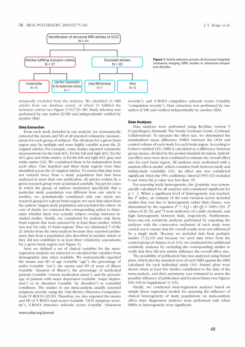
tematically excluded from the analyses. We identified 41 MRIarticles from our database search, of which 21 fulfilled theinclusion criteria (see Figure 1) (6,7,10–28). Study selection wasperformed by one author (J-YR) and independently verified byanother (BA).
Data ExtractionFrom each study included in our analysis, we systematically
extracted the means and SD of all reported volumetric measure-ments for each group of subjects. The divisions for a given brainregion may be multiple and were highly variable across the 21original articles. For example, some studies reported volumetricmeasurements for the total ACC, for the left and right ACC, for theACC gray and white matter, or for the left and right ACC gray andwhite matter (12). We considered them to be independent fromeach other. One hundred and three brain regions were thusidentified across the 21 original articles. To ensure that data werenot entered twice from a study population that had beenanalyzed in more than one publication, all articles written by agiven research group were scrutinized carefully. Except for casesin which the group of authors mentioned specifically that aparticular study population was different from one article toanother, we systematically considered only one article perresearch group for a given brain region; we used data taken fromthe authors’ largest study population and excluded the others. Incase of doubt, we contacted the corresponding author to deter-mine whether there was actually subject overlap between in-cluded studies. Finally, we considered for analysis only thosebrain regions that were assessed by at least three studies, whichwas true for only 15 brain regions. Thus we eliminated 7 of the21 articles from the meta-analysis because they reported prelim-inary data from a population also described in another article orthey did not contribute to at least three volumetric assessmentsfor a given brain region (see Figure 1).
Next we defined a set of clinical variables for the meta-regression analysis; we extracted or calculated the variables fromdemographic data when available. We systematically exportedthe means and SD of age (variable “age”), the percentage ofmales (variable “sex”), the means and SD of years of illness(variable “duration of illness”), the percentage of medicatedpatients (variable “current medication status”), and the percent-age of patients with major depression (variable “major depres-sion”) or tic disorders (variable “tic disorders”) as comorbidconditions. The studies in our meta-analysis usually assessedsymptom severity using the Yale-Brown Obsessive-CompulsiveScale (Y-BOCS) (29,30). Therefore, we also exported the meansand SD of Y-BOCS total scores (variable “OCD symptom sever-ity”), Y-BOCS obsessive subscale scores (variable “obsession
severity”), and Y-BOCS compulsive subscale scores (variable“compulsion severity”). Data extraction was performed by oneauthor (J-YR) and verified independently by another (BA).
Data Analyses
Data analyses were performed using RevMan, version 5(Copenhagen, Denmark; The Nordic Cochrane Centre, CochraneCollaboration). To measure the effect size, we determined thestandardized mean difference (SMD) between the OCD andcontrol volume of each study for each brain region. According toCohen’s method (31), SMD is calculated as a difference betweengroup means, divided by the pooled standard deviation. Individ-ual effect sizes were then combined to estimate the overall effectsize for each brain region. All analyses were performed with arandom-effects model, which considers both between-study andwithin-study variability (32). An effect size was consideredsignificant when the 95% confidence interval (95% CI) excluded0 and when the p value was less than .05.
For assessing study heterogeneity, the Q statistic was system-atically calculated for all analyses and considered significant forp , .1. When a significant level of heterogeneity was reached,the I2 index, an estimate of the total variation across includedstudies that was due to heterogeneity rather than chance, wasdetermined by the equation I2 5 [(Q – df)/Q] 3 100% (33). I2
values of 25, 50, and 75 were indicative of a mild, moderate, andhigh heterogeneity between trials, respectively. Furthermore,leave-one-out sensitivity analyses performed by repeating theanalyses with the consecutive exclusion of each study werecarried out to ensure that the overall results were not influencedby a single study. Because we included data from pediatricstudies (7,12,19) and because we used data twice from thecontrol group of Atmaca et al. (14), we conducted two additionalsensitivity analyses by excluding the corresponding studies toverify that they did not unduly influence the overall effect size.
The possibility of publication bias was analyzed using funnelplots, which plot the standard error of each SMD against the SMDcalculated for each individual study (34). Funnel plots weredrawn when at least five studies contributed to the data of themeta-analysis, and their asymmetry was estimated to assess thepossible influence of publication and location biases (see FiguresS10–S18 in Supplement 1) (35).
Finally, we conducted meta-regression analyses based onsimple linear regression models for assessing the influence ofclinical heterogeneity of study populations on meta-analysiseffect sizes. Regression analyses were performed only whenSMDs or heterogeneity were significant.
Identification of structural MRI articles of OCD
N = 41
Less than 3 assessments
for the studied brain volumes
N = 7
Included articles
N = 14
Excluded articles
N = 20
Using VBM techniques
N = 7
Articles fulfilling inclusion criteria
N = 21
Others
N = 13
Figure 1. Article selection process of structural magneticresonance imaging (MRI) studies in obsessive-compul-sive disorder.
76 BIOL PSYCHIATRY 2009;65:75–83 J.-Y. Rotge et al.
www.sobp.org/journal
48
Results
Among 21 MRI articles fulfilling our inclusion criteria, 15investigations, from 14 articles, contributed to at least threeassessments for a given cerebral region (Figure 1) (6,7,10–16,18,19,23,25,28). These primary results and characteristics aresummarized in Table 1. Included studies comprised a total of 371OCD patients and 407 healthy control subjects. Study popula-tions differed markedly in several demographic and clinicalcharacteristics. The mean age ranged from 12 to 42.2 for OCDsamples and from 12 to 34.5 for control samples. The percentageof males in each sample varied from 0% to 78% for both OCD andcontrol groups. The mean Y-BOCS total scores ranged from 17.6,reflective of moderate symptomatology, to 31.3, indicative ofsevere symptomatology (29). Furthermore, there was significantclinical heterogeneity between study populations. Some of theincluded studies comprised treatment-naïve (7,12,15,19) or treat-ment-refractory patients (14) (Table 1). The comorbidity status ofOCD subjects was also highly variable between included studies.In particular, major depression and tic disorder were comorbid toOCD in three (6,11,15) and two studies (11,15), respectively.However, both of these comorbid conditions were diagnosed inless than 15% of the OCD population in each study. Only fivestudies reported data from OCD patients without any other AxisI psychiatric condition or tic disorders (13,14,16,18,25).
The following 15 brain volumes were assessed by at leastthree studies: whole brain volume, intracranial volume, graymatter volume, prefrontal cortex volume, left and right ACCvolumes, left and right OFC volumes, left and right thalamusvolumes, left and right putamen volumes, and volumes of total,left, and right caudate nucleus. Volumetric analysis parametersfor these brain regions are summarized in Table 2. Detailedmeta-analyses for each of these regions are available in FiguresS1–S9 in Supplement 1. Overall analyses are reported in Table 3.The SMDs indicate a volume reduction for the left ACC, left andright OFC, and a volume increase for the left and right thalamusin OCD patients relative to control subjects. Between-studyheterogeneity, as assessed by the Q test, was significant for theleft ACC (I2 5 56%) and for the total (I2 5 68%) and right caudatenucleus (I2 5 50%). Repeated sensitivity analyses showed nomarked difference concerning the overall findings after theexclusion of each single study, except for the right OFC and thetotal caudate nucleus. The significance of the overall effect sizefor the right OFC was lost after the exclusion of data by Szeszkoet al. (18) (test for overall effect: SMD 5 –0.54, 95% CI 5 –1.13to .04, p 5 .07; test for heterogeneity: x
25 6.98, df 5 3, p 5 .07)
or data corresponding to the treatment-refractory patients’ groupdescribed by Atmaca et al. (14) (test for overall effect: SMD 5
–.36, 95% CI 5 –.79 to .05, p 5 .09; test for heterogeneity: x2
5
4.92, df 5 3, p 5 .18). With regard to the total caudate nucleus,the exclusion of data by Szeszko et al. (12) led to a significantoverall effect size (test for overall effect: SMD 5 –.60, 95% CI 5
–1.02 to –.18, p 5 .005; test for heterogeneity: x2
5 .29, df 5 1,p 5 .59). However, this analysis was performed with data fromonly two studies (6,7) and must be interpreted cautiously.Because the control participants’ data in the studies of Atmacaet al. (14) were entered twice in several meta-analyses, weconducted a sensitivity analysis by excluding them to assess theirimpact on our results. They did not influence the overall results,except for the right OFC, as described earlier. Because we useddata from pediatric MRI studies (7,12,19), we also conducted asensitivity analysis by removing them. No change was detectedin our findings, except for the total caudate nucleus. The
exclusion of pediatric data (7,12) led to a significant effect sizethat was calculated from the only remaining study (6) (test forindividual effect: SMD 5 –.70, 95% CI 5 –1.26 to –.14, p 5 .01;test for heterogeneity: not applicable). An additional analysisbased on the exclusion of adult data to explore brain volumechanges in children with OCD found no difference betweenthose data and the main findings of our meta-analysis.
Meta-regression analyses were then applied to each meta-analysis with significant heterogeneity or SMD to explore theinfluence of each clinical variable—age, sex, duration of illness,current medication status, major depression, tic disorders, OCDsymptom severity, obsession severity, and compulsion severity—onthe effect sizes. No significant correlation was found for brainregions with significant heterogeneity, that is, the left ACC andthe total and right caudate nucleus. Regression analyses did notindicate any significant correlation concerning cortical regionsfor which meta-analyses showed a smaller volume in OCD, thatis, the left ACC and the left and right OFC. Finally, we found asignificant relationship for the right thalamus effect size with thepercentage of males (r 5 .99, p , .05) and a statistical trend withthe Y-BOCS total score (r 5 .96, p 5 .05). We also found asignificant relationship for the left thalamus effect size with theY-BOCS obsession score (r 5 .99, p , .05) and a statistical trendwith the illness duration (r 5 .96, p 5 .17). The significantcorrelations that we report here are based on regression analysesusing only three data points, and therefore, they should beinterpreted cautiously, even if regression coefficients areremarkably high. Because of the lack of available data, wewere not able to compute controls for intracranial volume anestimate for the brain size, as a putative confounding variable.For the same reason, no multivariable regression model couldbe implemented.
Discussion
This meta-analysis provides a quantitative evaluation of volu-metric alterations in OCD by integrating outcomes from 14 MRIcase-control studies that included 371 patients and 407 healthycontrol subjects. Our results indicate the existence of a reliablysmaller volume in both the ACC and OFC, as well as an increaseof thalamic volume in OCD patients relative to healthy controlsubjects, suggesting that the thalamocortical pathway plays aprimary role in OCD pathophysiology. Our findings show nosignificant volumetric difference between OCD patients andhealthy subjects for whole brain, intracranial region, gray matter,prefrontal cortex, right ACC, putamen, and the caudate nucleus.Finally, meta-regression analyses reveal that the effect sizescalculated for the thalamus are partially explained by the severityof obsessive-compulsive symptoms.
Our findings support and extend previous neuroimagingstudies that have demonstrated the involvement of frontosubcor-tical circuits originating within the ACC and OFC in the patho-physiology of OCD. A recent meta-analysis of functional neuro-imaging data has implicated both the ACC and OFC during asymptom-provocation task in OCD patients, suggesting thatthese regions share a critical function in the genesis or mediationof obsessive-compulsive symptoms (5). The ACC may be subdi-vided into an affective module, comprising the most ventral part,and in a cognitive module, comprising the most dorsal part (40).The affective portion of the ACC, through its connections withthe limbic system, is implicated in the assessment of emotionalinformation and the regulation of emotional responses and maymediate the expression of anxiety observed in OCD (2). The
J.-Y. Rotge et al. BIOL PSYCHIATRY 2009;65:75–83 77
www.sobp.org/journal
49
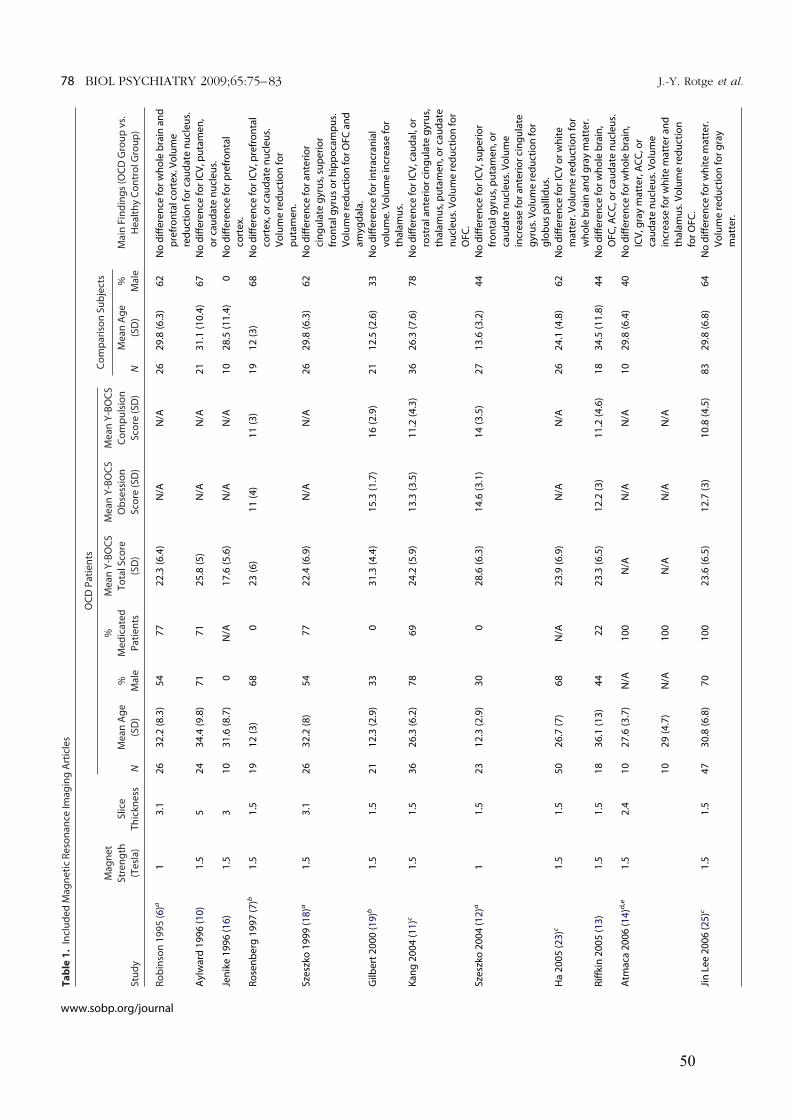
Ta
ble
1.
Incl
ud
ed
Ma
gn
eti
cR
eso
na
nce
Ima
gin
gA
rtic
les
Stu
dy
Ma
gn
et
Str
en
gth
(Te
sla
)
Slic
e
Th
ickn
ess
OC
DP
ati
en
tsC
om
pa
riso
nS
ub
ject
s
Ma
inF
ind
ing
s(O
CD
Gro
up
vs.
He
alt
hy
Co
ntr
olG
rou
p)
N
Me
an
Ag
e
(SD
)
%
Ma
le
%
Me
dic
ate
d
Pa
tie
nts
Me
an
Y-B
OC
S
To
talS
core
(SD
)
Me
an
Y-B
OC
S
Ob
sess
ion
Sco
re(S
D)
Me
an
Y-B
OC
S
Co
mp
uls
ion
Sco
re(S
D)
N
Me
an
Ag
e
(SD
)
%
Ma
le
Ro
bin
son
19
95
(6)a
13
.12
63
2.2
(8.3
)5
47
72
2.3
(6.4
)N
/AN
/A2
62
9.8
(6.3
)6
2N
od
iffe
ren
cefo
rw
ho
leb
rain
an
d
pre
fro
nta
lco
rte
x.V
olu
me
red
uct
ion
for
cau
da
ten
ucl
eu
s.
Ayl
wa
rd1
99
6(1
0)
1.5
52
43
4.4
(9.8
)7
17
12
5.8
(5)
N/A
N/A
21
31
.1(1
0.4
)6
7N
od
iffe
ren
cefo
rIC
V,p
uta
me
n,
or
cau
da
ten
ucl
eu
s.
Jen
ike
19
96
( 16
)1
.53
10
31
.6(8
.7)
0N
/A1
7.6
(5.6
)N
/AN
/A1
02
8.5
(11
.4)
0N
od
iffe
ren
cefo
rp
refr
on
tal
cort
ex.
Ro
sen
be
rg1
99
7(7
)b1
.51
.51
91
2(3
)6
80
23
(6)
11
(4)
11
(3)
19
12
(3)
68
No
dif
fere
nce
for
ICV
,pre
fro
nta
l
cort
ex,
or
cau
da
ten
ucl
eu
s.
Vo
lum
ere
du
ctio
nfo
r
pu
tam
en
.
Sze
szko
19
99
(18
)a1
.53
.12
63
2.2
(8)
54
77
22
.4(6
.9)
N/A
N/A
26
29
.8(6
.3)
62
No
dif
fere
nce
for
an
teri
or
cin
gu
late
gyr
us,
sup
eri
or
fro
nta
lgyr
us
or
hip
po
cam
pu
s.
Vo
lum
ere
du
ctio
nfo
rO
FC
an
d
am
ygd
ala
.
Gilb
ert
20
00
(19
)b1
.51
.52
11
2.3
(2.9
)3
30
31
.3(4
.4)
15
.3(1
.7)
16
(2.9
)2
11
2.5
(2.6
)3
3N
od
iffe
ren
cefo
rin
tra
cra
nia
l
vo
lum
e.V
olu
me
incr
ea
sefo
r
tha
lam
us.
Ka
ng
20
04
( 11
)c1
.51
.53
62
6.3
(6.2
)7
86
92
4.2
(5.9
)1
3.3
(3.5
)1
1.2
(4.3
)3
62
6.3
(7.6
)7
8N
od
iffe
ren
cefo
rIC
V,c
au
da
l,o
r
rost
rala
nte
rio
rci
ng
ula
teg
yru
s,
tha
lam
us,
pu
tam
en
,or
cau
da
te
nu
cle
us.
Vo
lum
ere
du
ctio
nfo
r
OF
C.
Sze
szko
20
04
( 12
)a1
1.5
23
12
.3(2
.9)
30
02
8.6
(6.3
)1
4.6
(3.1
)1
4(3
.5)
27
13
.6(3
.2)
44
No
dif
fere
nce
for
ICV
,su
pe
rio
r
fro
nta
lgyr
us,
pu
tam
en
,or
cau
da
ten
ucl
eu
s.V
olu
me
incr
ea
sefo
ra
nte
rio
rci
ng
ula
te
gyr
us.
Vo
lum
ere
du
ctio
nfo
r
glo
bu
sp
alli
du
s.
Ha
20
05
( 23
)c1
.51
.55
02
6.7
(7)
68
N/A
23
.9(6
.9)
N/A
N/A
26
24
.1(4
.8)
62
No
dif
fere
nce
for
ICV
or
wh
ite
ma
tte
r.V
olu
me
red
uct
ion
for
wh
ole
bra
ina
nd
gra
ym
att
er.
Rif
fkin
20
05
(13
)1
.51
.51
83
6.1
(13
)4
42
22
3.3
(6.5
)1
2.2
(3)
11
.2(4
.6)
18
34
.5(1
1.8
)4
4N
od
iffe
ren
cefo
rw
ho
leb
rain
,
OF
C,A
CC
,or
cau
da
ten
ucl
eu
s.
Atm
aca
20
06
(14
)d,e
1.5
2.4
10
27
.6(3
.7)
N/A
10
0N
/AN
/AN
/A1
02
9.8
(6.4
)4
0N
od
iffe
ren
cefo
rw
ho
leb
rain
,
ICV
,gra
ym
att
er,
AC
C,o
r
cau
da
ten
ucl
eu
s.V
olu
me
incr
ea
sefo
rw
hit
em
att
er
an
d
tha
lam
us.
Vo
lum
ere
du
ctio
n
for
OF
C.
10
29
(4.7
)N
/A1
00
N/A
N/A
N/A
Jin
Le
e2
00
6( 2
5)c
1.5
1.5
47
30
.8(6
.8)
70
10
02
3.6
(6.5
)1
2.7
(3)
10
.8(4
.5)
83
29
.8(6
.8)
64
No
dif
fere
nce
for
wh
ite
ma
tte
r.
Vo
lum
ere
du
ctio
nfo
rg
ray
ma
tte
r.
78 BIOL PSYCHIATRY 2009;65:75–83 J.-Y. Rotge et al.
www.sobp.org/journal
50
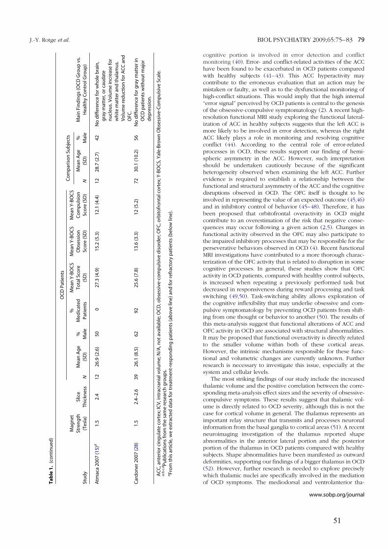
cognitive portion is involved in error detection and conflictmonitoring (40). Error- and conflict-related activities of the ACChave been found to be exacerbated in OCD patients comparedwith healthy subjects (41–43). This ACC hyperactivity maycontribute to the erroneous evaluation that an action may bemistaken or faulty, as well as to the dysfunctional monitoring ofhigh-conflict situations. This would imply that the high internal“error signal” perceived by OCD patients is central to the genesisof the obsessive-compulsive symptomatology (2). A recent high-resolution functional MRI study exploring the functional lateral-ization of ACC in healthy subjects suggests that the left ACC ismore likely to be involved in error detection, whereas the rightACC likely plays a role in monitoring and resolving cognitiveconflict (44). According to the central role of error-relatedprocesses in OCD, these results support our finding of hemi-spheric asymmetry in the ACC. However, such interpretationshould be undertaken cautiously because of the significantheterogeneity observed when examining the left ACC. Furtherevidence is required to establish a relationship between thefunctional and structural asymmetry of the ACC and the cognitivedisruptions observed in OCD. The OFC itself is thought to beinvolved in representing the value of an expected outcome (45,46)and in inhibitory control of behavior (45–48). Therefore, it hasbeen proposed that orbitofrontal overactivity in OCD mightcontribute to an overestimation of the risk that negative conse-quences may occur following a given action (2,5). Changes infunctional activity observed in the OFC may also participate tothe impaired inhibitory processes that may be responsible for theperseverative behaviors observed in OCD (4). Recent functionalMRI investigations have contributed to a more thorough charac-terization of the OFC activity that is related to disruption in somecognitive processes. In general, these studies show that OFCactivity in OCD patients, compared with healthy control subjects,is increased when repeating a previously performed task butdecreased in responsiveness during reward processing and taskswitching (49,50). Task-switching ability allows exploration ofthe cognitive inflexibility that may underlie obsessive and com-pulsive symptomatology by preventing OCD patients from shift-ing from one thought or behavior to another (50). The results ofthis meta-analysis suggest that functional alterations of ACC andOFC activity in OCD are associated with structural abnormalities.It may be proposed that functional overactivity is directly relatedto the smaller volume within both of these cortical areas.However, the intrinsic mechanisms responsible for these func-tional and volumetric changes are currently unknown. Furtherresearch is necessary to investigate this issue, especially at thesystem and cellular levels.
The most striking findings of our study include the increasedthalamic volume and the positive correlation between the corre-sponding meta-analysis effect sizes and the severity of obsessive-compulsive symptoms. These results suggest that thalamic vol-ume is directly related to OCD severity, although this is not thecase for cortical volume in general. The thalamus represents animportant relay structure that transmits and processes neuronalinformation from the basal ganglia to cortical areas (51). A recentneuroimaging investigation of the thalamus reported shapeabnormalities in the anterior lateral portion and the posteriorportion of the thalamus in OCD patients compared with healthysubjects. Shape abnormalities have been manifested as outwarddeformities, supporting our findings of a bigger thalamus in OCD(52). However, further research is needed to explore preciselywhich thalamic nuclei are specifically involved in the mediationof OCD symptoms. The mediodorsal and ventrolanterior tha-T
ab
le1
.(c
on
tin
ue
d)
Stu
dy
Ma
gn
et
Str
en
gth
(Te
sla
)
Slic
e
Th
ickn
ess
OC
DP
ati
en
tsC
om
pa
riso
nS
ub
ject
s
Ma
inF
ind
ing
s(O
CD
Gro
up
vs.
He
alt
hy
Co
ntr
olG
rou
p)
N
Me
an
Ag
e
(SD
)
%
Ma
le
%
Me
dic
ate
d
Pa
tie
nts
Me
an
Y-B
OC
S
To
talS
core
(SD
)
Me
an
Y-B
OC
S
Ob
sess
ion
Sco
re(S
D)
Me
an
Y-B
OC
S
Co
mp
uls
ion
Sco
re(S
D)
N
Me
an
Ag
e
(SD
)
%
Ma
le
Atm
aca
20
07
( 15
)d1
.52
.41
22
6.9
(2.6
)5
00
27
.3(4
.9)
15
.2(5
.3)
12
.1(4
.4)
12
28
.7(2
.7)
42
No
dif
fere
nce
for
wh
ole
bra
in,
gra
ym
att
er,
or
cau
da
te
nu
cle
us.
Vo
lum
ein
cre
ase
for
wh
ite
ma
tte
ra
nd
tha
lam
us.
Vo
lum
ere
du
ctio
nfo
rA
CC
an
d
OF
C.
Ca
rdo
ne
r2
00
7(2
8)
1.5
2.4
–2
.63
92
6.1
(8.5
)6
29
22
5.6
(7.8
)1
3.6
(3.3
)1
2(5
.2)
72
30
.1(1
0.2
)5
6N
od
iffe
ren
cefo
rg
ray
ma
tte
rin
OC
Dp
ati
en
tsw
ith
ou
tm
ajo
r
de
pre
ssio
n.
AC
C,a
nte
rio
rci
ng
ula
teco
rte
x;IC
V,i
ntr
acr
an
ialv
olu
me
;N/A
,no
ta
va
ilab
le;O
CD
,ob
sess
ive
-co
mp
uls
ive
dis
ord
er;
OF
C,o
rbit
ofr
on
talc
ort
ex;
Y-B
OC
S,Y
ale
-Bro
wn
Ob
sess
ive
-Co
mp
uls
ive
Sca
le.
a,b
,c,d
Pu
blic
ati
on
sfr
om
the
sam
ere
sea
rch
gro
up
s.eF
rom
this
art
icle
,we
ext
ract
ed
da
tafo
rtr
ea
tme
nt-
resp
on
din
gp
ati
en
ts(a
bo
ve
line
)a
nd
for
refr
act
ory
pa
tie
nts
(be
low
line
).
J.-Y. Rotge et al. BIOL PSYCHIATRY 2009;65:75–83 79
www.sobp.org/journal
51
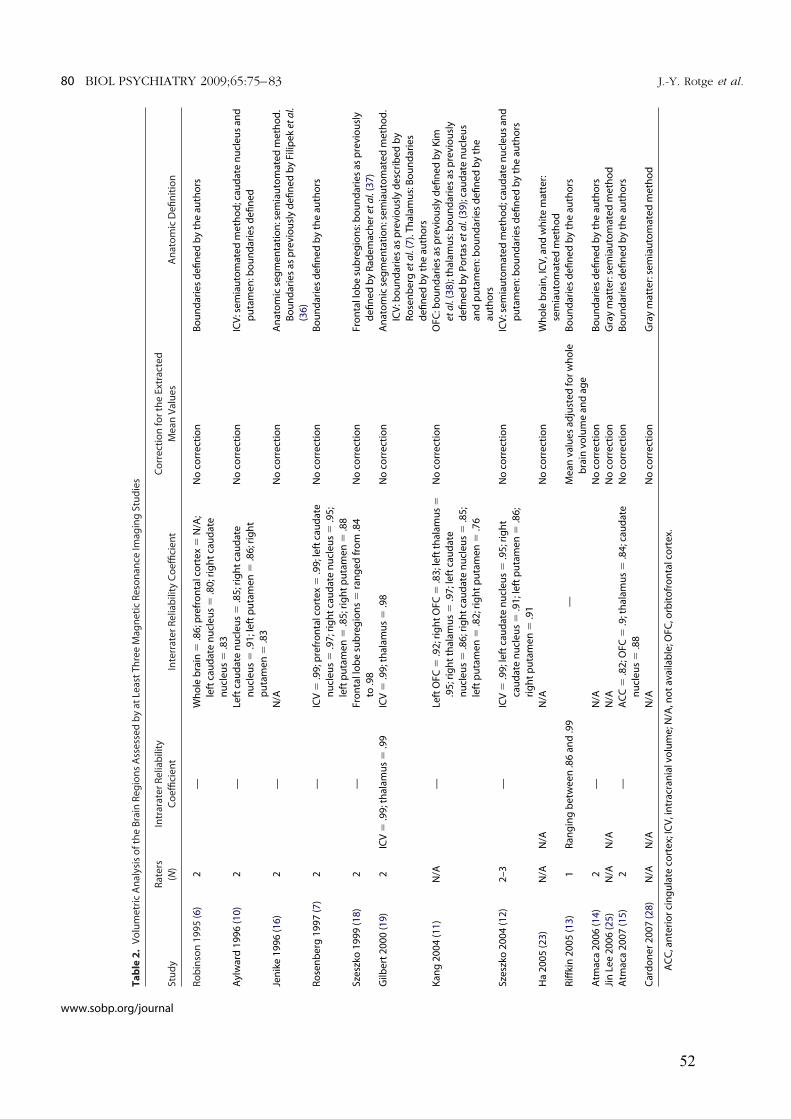
Ta
ble
2.
Vo
lum
etr
icA
na
lysi
so
fth
eB
rain
Re
gio
ns
Ass
ess
ed
by
at
Le
ast
Th
ree
Ma
gn
eti
cR
eso
na
nce
Ima
gin
gS
tud
ies
Stu
dy
Ra
ters
(N)
Intr
ara
ter
Re
liab
ility
Co
effi
cie
nt
Inte
rra
ter
Re
liab
ility
Co
effi
cie
nt
Co
rre
ctio
nfo
rth
eE
xtra
cte
d
Me
an
Va
lue
sA
na
tom
icD
efi
nit
ion
Ro
bin
son
19
95
(6)
2—
Wh
ole
bra
in5
.86
;pre
fro
nta
lco
rte
x5
N/A
;
left
cau
da
ten
ucl
eu
s5
.80
;rig
ht
cau
da
te
nu
cle
us
5.8
3
No
corr
ect
ion
Bo
un
da
rie
sd
efi
ne
db
yth
ea
uth
ors
Ayl
wa
rd1
99
6(1
0)
2—
Le
ftca
ud
ate
nu
cle
us
5.8
5;r
igh
tca
ud
ate
nu
cle
us
5.9
1;l
eft
pu
tam
en
5.8
6;r
igh
t
pu
tam
en
5.8
3
No
corr
ect
ion
ICV
:se
mia
uto
ma
ted
me
tho
d;c
au
da
ten
ucl
eu
sa
nd
pu
tam
en
:bo
un
da
rie
sd
efi
ne
d
Jen
ike
19
96
(16
)2
—N
/AN
oco
rre
ctio
nA
na
tom
icse
gm
en
tati
on
:se
mia
uto
ma
ted
me
tho
d.
Bo
un
da
rie
sa
sp
rev
iou
sly
de
fin
ed
by
Fili
pe
ket
al.
(36
)
Ro
sen
be
rg1
99
7( 7
)2
—IC
V5
.99
;pre
fro
nta
lco
rte
x5
.99
;le
ftca
ud
ate
nu
cle
us
5.9
7;r
igh
tca
ud
ate
nu
cle
us
5.9
5;
left
pu
tam
en
5.8
5;r
igh
tp
uta
me
n5
.88
No
corr
ect
ion
Bo
un
da
rie
sd
efi
ne
db
yth
ea
uth
ors
Sze
szko
19
99
( 18
)2
—F
ron
tall
ob
esu
bre
gio
ns
5ra
ng
ed
fro
m.8
4
to.9
8
No
corr
ect
ion
Fro
nta
llo
be
sub
reg
ion
s:b
ou
nd
ari
es
as
pre
vio
usl
y
de
fin
ed
by
Ra
de
ma
che
ret
al.
(37
)
Gilb
ert
20
00
( 19
)2
ICV
5.9
9;t
ha
lam
us
5.9
9IC
V5
.99
;th
ala
mu
s5
.98
No
corr
ect
ion
An
ato
mic
seg
me
nta
tio
n:s
em
iau
tom
ate
dm
eth
od
.
ICV
:bo
un
da
rie
sa
sp
rev
iou
sly
de
scri
be
db
y
Ro
sen
be
rget
al.
( 7).
Th
ala
mu
s:B
ou
nd
ari
es
de
fin
ed
by
the
au
tho
rs
Ka
ng
20
04
(11
)N
/A—
Le
ftO
FC
5.9
2;r
igh
tO
FC
5.8
3;l
eft
tha
lam
us
5
.95
;rig
ht
tha
lam
us
5.9
7;l
eft
cau
da
te
nu
cle
us
5.8
6;r
igh
tca
ud
ate
nu
cle
us
5.8
5;
left
pu
tam
en
5.8
2;r
igh
tp
uta
me
n5
.76
No
corr
ect
ion
OF
C:b
ou
nd
ari
es
as
pre
vio
usl
yd
efi
ne
db
yK
im
eta
l.( 3
8);
tha
lam
us:
bo
un
da
rie
sa
sp
rev
iou
sly
de
fin
ed
by
Po
rta
set
al.
( 39
);ca
ud
ate
nu
cle
us
an
dp
uta
me
n:b
ou
nd
ari
es
de
fin
ed
by
the
au
tho
rs
Sze
szko
20
04
(12
)2
–3
—IC
V5
.99
;le
ftca
ud
ate
nu
cle
us
5.9
5;r
igh
t
cau
da
ten
ucl
eu
s5
.91
;le
ftp
uta
me
n5
.86
;
rig
ht
pu
tam
en
5.9
1
No
corr
ect
ion
ICV
:se
mia
uto
ma
ted
me
tho
d;c
au
da
ten
ucl
eu
sa
nd
pu
tam
en
:bo
un
da
rie
sd
efi
ne
db
yth
ea
uth
ors
Ha
20
05
(23
)N
/AN
/AN
/AN
oco
rre
ctio
nW
ho
leb
rain
,IC
V,a
nd
wh
ite
ma
tte
r:
sem
iau
tom
ate
dm
eth
od
Rif
fkin
20
05
( 13
)1
Ra
ng
ing
be
twe
en
.86
an
d.9
9—
Me
an
va
lue
sa
dju
ste
dfo
rw
ho
le
bra
inv
olu
me
an
da
ge
Bo
un
da
rie
sd
efi
ne
db
yth
ea
uth
ors
Atm
aca
20
06
(14
)2
—N
/AN
oco
rre
ctio
nB
ou
nd
ari
es
de
fin
ed
by
the
au
tho
rs
Jin
Le
e2
00
6( 2
5)
N/A
N/A
N/A
No
corr
ect
ion
Gra
ym
att
er:
sem
iau
tom
ate
dm
eth
od
Atm
aca
20
07
(15
)2
—A
CC
5.8
2;O
FC
5.9
;th
ala
mu
s5
.84
;ca
ud
ate
nu
cle
us
5.8
8
No
corr
ect
ion
Bo
un
da
rie
sd
efi
ne
db
yth
ea
uth
ors
Ca
rdo
ne
r2
00
7( 2
8)
N/A
N/A
N/A
No
corr
ect
ion
Gra
ym
att
er:
sem
iau
tom
ate
dm
eth
od
AC
C,a
nte
rio
rci
ng
ula
teco
rte
x;IC
V,i
ntr
acr
an
ialv
olu
me
;N/A
,no
ta
va
ilab
le;O
FC
,orb
ito
fro
nta
lco
rte
x.
80 BIOL PSYCHIATRY 2009;65:75–83 J.-Y. Rotge et al.
www.sobp.org/journal
52
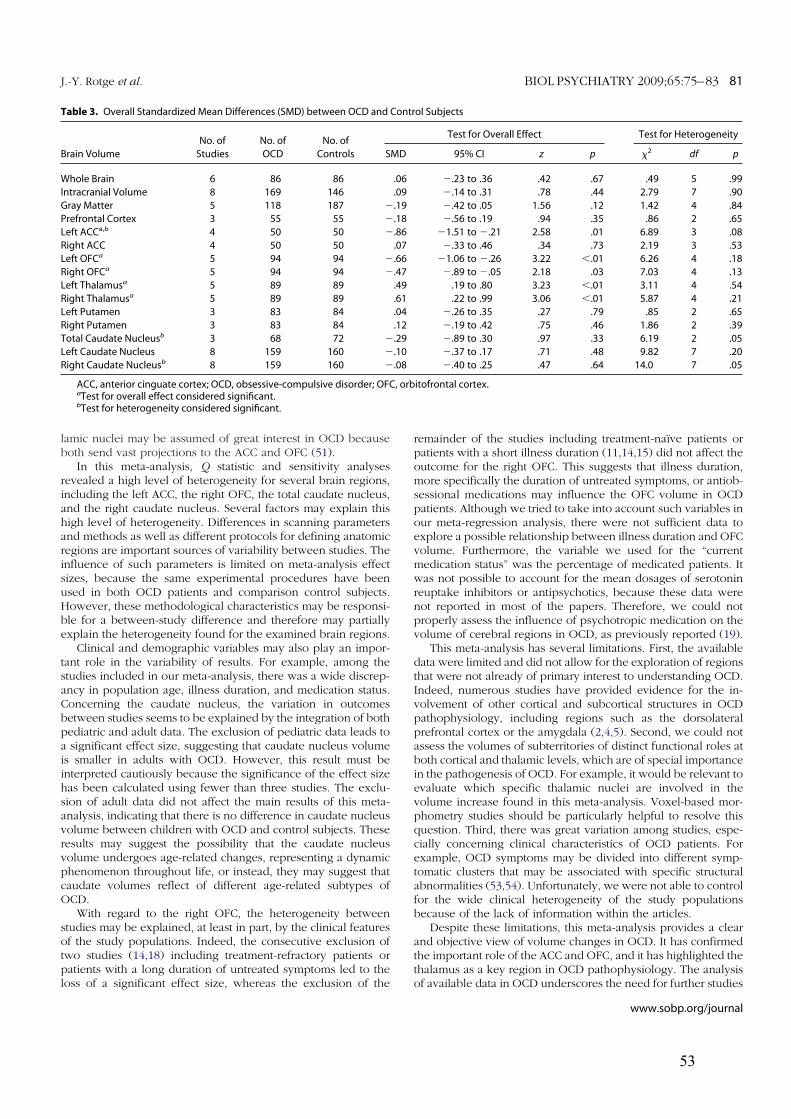
lamic nuclei may be assumed of great interest in OCD becauseboth send vast projections to the ACC and OFC (51).
In this meta-analysis, Q statistic and sensitivity analysesrevealed a high level of heterogeneity for several brain regions,including the left ACC, the right OFC, the total caudate nucleus,and the right caudate nucleus. Several factors may explain thishigh level of heterogeneity. Differences in scanning parametersand methods as well as different protocols for defining anatomicregions are important sources of variability between studies. Theinfluence of such parameters is limited on meta-analysis effectsizes, because the same experimental procedures have beenused in both OCD patients and comparison control subjects.However, these methodological characteristics may be responsi-ble for a between-study difference and therefore may partiallyexplain the heterogeneity found for the examined brain regions.
Clinical and demographic variables may also play an impor-tant role in the variability of results. For example, among thestudies included in our meta-analysis, there was a wide discrep-ancy in population age, illness duration, and medication status.Concerning the caudate nucleus, the variation in outcomesbetween studies seems to be explained by the integration of bothpediatric and adult data. The exclusion of pediatric data leads toa significant effect size, suggesting that caudate nucleus volumeis smaller in adults with OCD. However, this result must beinterpreted cautiously because the significance of the effect sizehas been calculated using fewer than three studies. The exclu-sion of adult data did not affect the main results of this meta-analysis, indicating that there is no difference in caudate nucleusvolume between children with OCD and control subjects. Theseresults may suggest the possibility that the caudate nucleusvolume undergoes age-related changes, representing a dynamicphenomenon throughout life, or instead, they may suggest thatcaudate volumes reflect of different age-related subtypes ofOCD.
With regard to the right OFC, the heterogeneity betweenstudies may be explained, at least in part, by the clinical featuresof the study populations. Indeed, the consecutive exclusion oftwo studies (14,18) including treatment-refractory patients orpatients with a long duration of untreated symptoms led to theloss of a significant effect size, whereas the exclusion of the
remainder of the studies including treatment-naïve patients orpatients with a short illness duration (11,14,15) did not affect theoutcome for the right OFC. This suggests that illness duration,more specifically the duration of untreated symptoms, or antiob-sessional medications may influence the OFC volume in OCDpatients. Although we tried to take into account such variables inour meta-regression analysis, there were not sufficient data toexplore a possible relationship between illness duration and OFCvolume. Furthermore, the variable we used for the “currentmedication status” was the percentage of medicated patients. Itwas not possible to account for the mean dosages of serotoninreuptake inhibitors or antipsychotics, because these data werenot reported in most of the papers. Therefore, we could notproperly assess the influence of psychotropic medication on thevolume of cerebral regions in OCD, as previously reported (19).
This meta-analysis has several limitations. First, the availabledata were limited and did not allow for the exploration of regionsthat were not already of primary interest to understanding OCD.Indeed, numerous studies have provided evidence for the in-volvement of other cortical and subcortical structures in OCDpathophysiology, including regions such as the dorsolateralprefrontal cortex or the amygdala (2,4,5). Second, we could notassess the volumes of subterritories of distinct functional roles atboth cortical and thalamic levels, which are of special importancein the pathogenesis of OCD. For example, it would be relevant toevaluate which specific thalamic nuclei are involved in thevolume increase found in this meta-analysis. Voxel-based mor-phometry studies should be particularly helpful to resolve thisquestion. Third, there was great variation among studies, espe-cially concerning clinical characteristics of OCD patients. Forexample, OCD symptoms may be divided into different symp-tomatic clusters that may be associated with specific structuralabnormalities (53,54). Unfortunately, we were not able to controlfor the wide clinical heterogeneity of the study populationsbecause of the lack of information within the articles.
Despite these limitations, this meta-analysis provides a clearand objective view of volume changes in OCD. It has confirmedthe important role of the ACC and OFC, and it has highlighted thethalamus as a key region in OCD pathophysiology. The analysisof available data in OCD underscores the need for further studies
Table 3. Overall Standardized Mean Differences (SMD) between OCD and Control Subjects
Brain Volume
No. of
Studies
No. of
OCD
No. of
Controls
Test for Overall Effect Test for Heterogeneity
SMD 95% CI z p x2 df p
Whole Brain 6 86 86 .06 2.23 to .36 .42 .67 .49 5 .99
Intracranial Volume 8 169 146 .09 2.14 to .31 .78 .44 2.79 7 .90
Gray Matter 5 118 187 2.19 2.42 to .05 1.56 .12 1.42 4 .84
Prefrontal Cortex 3 55 55 2.18 2.56 to .19 .94 .35 .86 2 .65
Left ACCa,b 4 50 50 2.86 21.51 to 2.21 2.58 .01 6.89 3 .08
Right ACC 4 50 50 .07 2.33 to .46 .34 .73 2.19 3 .53
Left OFCa 5 94 94 2.66 21.06 to 2.26 3.22 ,.01 6.26 4 .18
Right OFCa 5 94 94 2.47 2.89 to 2.05 2.18 .03 7.03 4 .13
Left Thalamusa 5 89 89 .49 .19 to .80 3.23 ,.01 3.11 4 .54
Right Thalamusa 5 89 89 .61 .22 to .99 3.06 ,.01 5.87 4 .21
Left Putamen 3 83 84 .04 2.26 to .35 .27 .79 .85 2 .65
Right Putamen 3 83 84 .12 2.19 to .42 .75 .46 1.86 2 .39
Total Caudate Nucleusb 3 68 72 2.29 2.89 to .30 .97 .33 6.19 2 .05
Left Caudate Nucleus 8 159 160 2.10 2.37 to .17 .71 .48 9.82 7 .20
Right Caudate Nucleusb 8 159 160 2.08 2.40 to .25 .47 .64 14.0 7 .05
ACC, anterior cinguate cortex; OCD, obsessive-compulsive disorder; OFC, orbitofrontal cortex.aTest for overall effect considered significant.bTest for heterogeneity considered significant.
J.-Y. Rotge et al. BIOL PSYCHIATRY 2009;65:75–83 81
www.sobp.org/journal
53
to 1) explore volume changes in other brain regions of interest,2) explore volume changes according to OCD subtypes, and 3)explore the molecular and cellular mechanisms underlying suchvolume changes.
J-Y Rotge was supported by a grant from the Fondation pour
la Recherche Médicale (Medical Research Foundation). We
thank Pr. Jun Soo Kwon from the Department of Psychiatry,
Seoul National University College of Medicine (Seoul, Republic of
Korea) for his collaboration.
The authors report no biomedical financial interests or po-
tential conflicts of interest.
Supplementary material cited in this article is available
online.
1. Stein DJ (2002): Obsessive-compulsive disorder. Lancet 360:397– 405.2. Aouizerate B, Guehl D, Cuny E, Rougier A, Bioulac B, Tignol J, et al. (2004):
Pathophysiology of obsessive-compulsive disorder: A necessary linkbetween phenomenology, neuropsychology, imagery and physiology.Prog Neurobiol 72:195–221.
3. Kathmann N, Rupertseder C, Hauke W, Zaudig M (2005): Implicit se-quence learning in obsessive-compulsive disorder: Further support forthe fronto-striatal dysfunction model. Biol Psychiatry 58:239 –244.
4. Menzies L, Chamberlain SR, Laird AR, Thelen SM, Sahakian BJ, BullmoreET (2008): Integrating evidence from neuroimaging and neuropsycho-logical studies of obsessive-compulsive disorder: The orbitofronto-stri-atal model revisited. Neurosci Biobehav Rev 32:525–549.
5. Rotge JY, Guehl D, Dilharreguy B, Cuny E, Tignol J, Bioulac B, et al. (inpress): Provocation of obsessive-compulsive symptoms: A quantitativevoxel-based meta-analysis of functional neuroimaging studies. J Psychi-atry Neurosci.
6. Robinson D, Wu H, Munne RA, Ashtari M, Alvir JMJ, Lerner G, et al. (1995):Reduced caudate nucleus volume in obsessive-compulsive disorder.Arch Gen Psychiatry 52:393–398.
7. Rosenberg DR, Keshavan MS, O’Hearn KM, Dick EL, Bagwell WW, Sey-mour AB, et al. (1997): Frontostriatal measurement in treatment-naïvechildren with obsessive-compulsive disorder. Arch Gen Psychiatry 54:824 – 830.
8. Scarone S, Colombo C, Livian S, Abbruzzese M, Ronchi P, Locatelli M, etal. (1992): Increased right caudate nucleus size in obsessive-compulsivedisorder: Detection with magnetic resonance imaging. Psychiatry Res45:115–121.
9. Giedd JN, Rapoport JL, Garvey MA, Perlmutter S, Swedo SE (2000): MRIassessment of children with obsessive-compulsive disorder or tics asso-ciated with streptococcal infection. Am J Psychiatry 157:281–283.
10. Aylward EH, Harris GJ, Hoehn-Saric R, Barta PE, Machlin SR, Pearlson GD(1996): Normal caudate nucleus in obsessive-compulsive disorder as-sessed by quantitative neuroimaging. Arch Gen Psychiatry 53:577–584.
11. Kang DH, Kim JJ, Choi JS, Kim YI, Kim CW, Youn T, et al. (2004): Volumetricinvestigation of the frontal-subcortical circuitry in patients with obses-sive-compulsive disorder. J Neuropsychiatry Clin Neurosci 16:342–349.
12. Szeszko PR, MacMillan S, McMeniman M, Chen S, Baribault K, Lim KO, etal. (2004): Brain structural abnormalities in psychotropic drug-naivepediatric patients with obsessive-compulsive disorder. Am J Psychiatry161:1049 –1056.
13. Riffkin J, Yücel M, Maruff P, Wood SJ, Soulsby B, Olver J, et al. (2005): Amanual and automated MRI study of anterior cingulate and orbito-frontal cortices, and caudate nucleus in obsessive-compulsive disorder:Comparison with healthy controls and patients with schizophrenia.Psychiatry Res 138:99 –113.
14. Atmaca M, Yildirim H, Ozdemir H, Aydin A, Tezcan E, Ozler S (2006):Volumetric MRI assessment of brain regions in patients with refractoryobsessive-compulsive disorer. Prog Neuropsychoph 30:1051–1057.
15. Atmaca M, Yildirim H, Ozdemir H, Tezcan E, Poyraz AK (2007): VolumetricMRI study of key brain regions implicated in obsessive-compulsive dis-order. Prog Neuro-psychoph 31:46 –52.
16. Jenike MA, Breiter HC, Baer L, Kennedy DN, Savage CR, Olivares MJ, et al.(1996): Cerebral structural abnormalities in obsessive-compulsive disor-der. Arch Gen Psychiatry 53:625– 632.
17. Stein DJ, Coetzer R, Lee M, Davids B, Bouwer C (1997): Magnetic reso-nance brain imaging in women with obsessive-compulsive disorderand trichotillomania. Psychiatry Res 74:177–182.
18. Szeszko PR, Robinson D, Alvir JMJ, Bilder RM, Lencz T, Ashtari M, et al.(1999): Orbital frontal and amygdala volume reductions in obsessive-compulsive disorder. Arch Gen Psychiatry 56:913–919.
19. Gilbert AR, Moore GJ, Keshavan MS, Paulson LAD, Narula V, MacMasterFP, et al. (2000): Decrease in thalamic volumes of pediatric patients withobsessive-compulsive disorder who are taking paroxetine. Arch GenPsychiatry 57:449 – 456.
20. Kwon JS, Shin YW, Kim CW, Kim YI, Youn T, Han MH, et al. (2003):Similarity and disparity of obsessive-compulsive disorder and schizo-phrenia in MR volumetric abnormalities of the hippocampus-amygdalacomplex. J Neurol Neurosurg Psychiatry 74:962–964.
21. Kim JJ, Youn T, Lee JM, Kim IY, Kim SI, Kwon JS (2003): Morphometricabnormality of the insula in schizophrenia: A comparison with obses-sive-compulsive disorder and normal control using MRI. Schizophr Res60:191–198.
22. Choi JS, Kang DH, Kim JJ, Ha TH, Lee JM, Youn T, et al. (2004): Left anteriorsubregion of orbitofrontal cortex volume reduction and impaired orga-nizational strategies in obsessive-compulsive disorder. J Psychiatr Res38:193–199.
23. Ha TH, Yoon U, Jin Lee K, Shin YW, Lee JM, Kim IY, et al. (2005): Fractaldimension of cerebral cortical surface in schizophrenia and obessive-compulsive disorder. Neurosci Lett 384:172–176.
24. Choi JS, Kim HS, Yoo SY, Ha TH, Chang JH, Kim YY, et al. (2006): Morpho-metric alterations of anterior superior temporal cortex in obsessive-compulsive disorder. Depress Anxiety 23:290 –296.
25. Jin Lee K, Shin YW, Wee H, Kim YY, Kwon JS (2006): Gray matter volumereduction in obsessive-compulsive disorder with schizotypal personal-ity trait. Prog Neuropsychopharmacol Biol Psychiatry 30:1146 –1149.
26. MacMaster FP, Russell A, Mirza Y, Keshavan MS, Banerjee SP, Bhandari R,et al. (2006): Pituitary volume in pediatric obsessive-compulsive disor-der. Biol Psychiatry 59:252–257.
27. Choi JS, Kim HS, Yoo SY, Ha TH, Chang JH, Kim YY, et al. (2006): Morpho-metric alterations of anterior superior temporal cortex in obsessive-compulsive disorder. Depress Anxiety 23:290 –296.
28. Cardoner N, Soriano-Mas C, Pujol J, Alonso P, Harrison BJ, Deus J, et al.(2007): Brain structural correlates of depressive comorbidity in obses-sive-compulsive disorder. NeuroImage 38:413– 421.
29. Goodman WK, Price LH, Rasmussen SA, Mazure C, Fleischmann RL, HillCL, et al. (1989): The Yale-Brown Obsessive Compulsive Scale. I. Devel-opment, use, and reliability. Arch Gen Psychiatry 46:1006 –11.
30. Goodman WK, Price LH, Rasmussen SA, Mazure C, Delgado P, HeningerGR, et al. (1989): The Yale-Brown Obsessive Compulsive Scale. II. Validity.Arch Gen Psychiatry 46:10012–10016.
31. Cohen J (1988): Statistical Power Analysis for the Behavioral Sciences, 2nded. New York: Academic Press.
32. DerSimonian R, Laird N (1986): Meta-analysis in clinical trials. Control ClinTrials 7:177–188.
33. Higgins JPT, Thompson SG, Deeks JJ, Altman DG (2003): Measuringinconsistency in meta-analyses. BMJ 327:557–560.
34. Green S, Higgins J (2006): Cochrane Handbook for Systematic Reviews ofInterventions 4.2.6. Chichester, UK: Wiley.
35. Egger M, Davey Smith G, Schneider M, Minder C (1997): Bias in meta-analysis detected by a simple, graphical test. BMJ 315:629 – 634.
36. Filipek PA, Richelme C, Kennedy DN, Caviness VS (1994): The youngadult human brain: An MRI-based morphometric analysis. Cereb Cortex4:344 –360.
37. Rademacher J, Galaburda AM, Kennedy DN, Filipek PA, Caviness VS Jr(1992): Human cerebral cortex: Localization, parcellation, and mor-phometry with magnetic resonance imaging. J Cogn Neurosci 4:352–374.
38. Kim JJ, Kwon JS, Kang DH, Youn T, Han MH, Chang KH (2002) Frontalsubstructural abnormalities evaluated by the parcellation of MRIin patients with schizophrenia. J Korean Neuropsychiatr Assoc 41:998 –1009.
39. Portas CM, Goldstein JM, Shenton ME, Hokama HH, Wible CG, Fischer I,et al. (1998): Volumetric evaluation of the thalamus in schizophrenicmale patients using magnetic resonance imaging. Biol Psychiatry 43:649 – 659.
40. Bush G, Luu P, Posner MI (2000): Cognitive and emotional influences inanterior cingulate cortex. Trends Cogn Sci 4:215–222.
82 BIOL PSYCHIATRY 2009;65:75–83 J.-Y. Rotge et al.
www.sobp.org/journal
54
41. Ursu S, Stenger VA, Shear MK, Jones MR, Carter CS (2003): Overactiveaction monitoring in obsessive-compulsive disorder: Evidence fromfunctional magnetic resonance imaging. Psychol Sci 14:347–353.
42. Fitzgerald KD, Welsh RC, Gehring WJ, Abelson JL Himle JA, Liberzon I, etal. (2005): Error-related hyperactivity of the anterior cingulate cortex inobsessive-compulsive disorder. Biol Psychiatry 57:287–294.
43. Maltby N, Tolin DF, Worhunsky P, O’Keefe TM, Kiehl KA (2005): Dysfunc-tional action monitoring hyperactivates frontal-striatal circuits in obses-sive-compulsive disorder: An event-related fMRI study. NeuroImage 24:495–503.
44. Lütcke H, Frahm J (2008): Lateralized anterior cingulate function duringerror processing and conflict monitoring as revealed by high-resolutionfMRI. Cereb Cortex 18:508 –515.
45. O’Doherty JP (2007): Lights, camembert, action! The role of humanorbitofrontal cortex in encoding stimuli, rewards, and choices. An N YAcad Sci 1121:254 –272.
46. Murray EA, O’Doherty JP, Schoenbaum G (2007): What we know and donot know about the functions of the orbitofrontal cortex after 20 yearsof cross-species studies. J Neurosci 27:8166 – 8169.
47. Zald DH, Kim SW (1996): Anatomy and function of the orbital frontalcortex II: Function and relevance to obsessive-compulsive disorder.J Neuropsychiatry Clin Neurosci 8:249 –261.
48. Elliott R, Dolan RJ, Frith CD (2000): Dissociable functions in the medial
and lateral orbitofrontal cortex: Evidence from human neuroimaging
studies. Cereb Cortex 10:308 –317.
49. Remijnse PL, Nielen MMA, VanBalkom AJLM, Cath DC, VanOppen P, Uylings
HBM, et al. (2006): Reduced orbitofrontal-striatal activity on a reversal learning
task in obsessive-compulsive disorder. Arch Gen Psychiatry 63:1225–1236.
50. Gu BM, Park JY, Kang DH, Lee SJ, Yoo SY, Jo HJ, et al. (2008): Neural
correlates of cognitive inflexibility during task-switching in obsessive-
compulsive disorder. Brain 131:155–164.
51. McFarland NR, Haber SN (2002): Thalamic relay nuclei of the basal gan-
glia form both reciprocal and non reciprocal cortical connections, link-
ing multiple frontal cortical areas. J Neurosci 22:8117– 8132.
52. Kang DH, Kim SH, Kim CW, Choi JS, Jang JH, Jung MH, et al. (2008):
Thalamus surface shape deformity in obsessive-compulsive disorder
and schizophrenia. Neuroreport 19:609 – 613.
53. Baer L (1994): Factor analysis of symptom subtypes of obsessive com-
pulsive disorder and their relation to personality and tic disorders. J Clin
Psychiatry 55(Suppl):18 –23.
54. Leckman JF, Grice DE, Boardman J, Zhang H, Vitale A, Bondi C, et al.
(1997): Symptoms of obsessive-compulsive disorder. Am J Psychiatry
154:911–917.
J.-Y. Rotge et al. BIOL PSYCHIATRY 2009;65:75–83 83
www.sobp.org/journal
55

56
57
3.2. Modifications de densité de matière grise
3.2.1. Introduction
Les études d’IRM volumétrique qui ont contribué à l’identification des atteintes
anatomiques dans le TOC souffrent de plusieurs limites méthodologiques. En particulier, il est
nécessaire de définir a priori les régions d’intérêt pour lesquelles on a fait l’hypothèse d’une
modification de volume. Une autre méthode, l’imagerie morphométrique (en anglais, « voxel-
based morphometry » [VBM]), permet d’évaluer les modifications de densité de matière grise
sur l’ensemble du cerveau sans se limiter à des structures d’intérêt préalablement définies.
Non seulement l’IRM morphométrique pourrait ainsi mettre en évidence des altérations
structurelles inattendues dans des régions cérébrales peu explorées en IRM volumétrique,
mais sa grande résolution anatomique pourrait aussi permettre d’identifier plus précisément
les sous-territoires altérés au sein d’une région cérébrale donnée. Les études VBM publiées à
ce jour dans le TOC trouvent des résultats qui ne sont pas toujours concordants d’une étude à
l’autre. Afin de réaliser, ici aussi, une évaluation quantitative de ces résultats, nous avons
effectué une méta-analyse des études VBM comparant la densité de matière grise de patients
souffrant de TOC et de témoins.
3.2.2. Méthodes
Nous avons utilisé une méthode de méta-analyse appelée « activation likelihood
estimation » (ALE) qui permet de prendre en considération la dispersion tridimensionnelle
des différentes zones (appelées « clusters ») significativement différentes. Chaque cluster est
défini par les coordonnées de son point maximal (appelé « focus »), par son intensité (t- ou z-
score) et par son volume. Le principe des méta-analyses ALE est de modéliser chaque focus
comme le centre d’une loi gaussienne avec l’intensité d’activation pour ordonnée et le volume
pour abscisse. De cette façon, il est possible de prendre en considération ces paramètres au
sein d’un volume tridimensionnel. Pour réaliser cette méta-analyse, une recherche
systématique par mots-clé a été réalisée pour identifier les études d’IRM morphométrique
publiées dans le TOC. Les paramètres d’intérêt sus-cités ont été extraits pour chacune des
études. Lorsque ces données n’étaient pas rapportées dans un des articles sélectionnés, nous
les avons systématiquement demandées auprès de l’auteur pour les correspondances.
58
3.2.3. Résultats et discussion
Les données recueillies sont issues de 10 articles publiés ayant rapporté 66 foci différents
chez 343 patients avec TOC et 318 témoins. Nous avons trouvé une diminution de la densité
de matière grise au niveau du cortex pariétal et des cortex préfrontal dorsolatéral et préfrontal
antérieur. A l’inverse, une augmentation de la densité de matière grise a été observée dans le
putamen et dans la partie latérale du COF. Nous avons ensuite comparé les cartes
tridimensionnelles obtenues chez les enfants et chez les adultes, nous n’avons trouvé aucune
différence significative entre ces populations. La principale limite de ce travail a été
l’incapacité de prendre en considération les variables cliniques susceptibles d’expliquer, au
moins en partie, ces modifications de densité de matière grise. L’examen des seuils
statistiques utilisés a montré que ceux-ci n’affectaient pas les résultats observés. Enfin, une
analyse en « leave-one-out », consistant à exclure de l’analyse tour à tour une des différentes
études inclues, a montré que ces résultats ne sont pas influencés par une seule étude.
3.2.4. Conclusion
Ces résultats montrent l’existence d’une diminution de substance grise dans un réseau
dorsal et, à l’inverse, une augmentation de substance dans le COF et le putamen. De façon
intéressante, nous trouvons ici une augmentation de densité de matière grise dans la partie
latérale du COF alors que précédemment nous avions décrit une diminution du volume
orbitofrontal dans son ensemble. Ce résultat suggère la possibilité d’un remaniement tissulaire
local au profit d’un contenu plus dense. Cette hypothèse est présentée plus précisément dans
l’article de discussion générale.
Rotgé JY, Langbour N, Guehl D, Bioulac B, Jaafari N, Allard M, Aouizerate B,
Burbaud P. Gray matter alterations in obsessive-compulsive disorder: an anatomic
likelihood estimation meta-analysis. Neuropsychopharmacology 2010;35:686-691.

Gray Matter Alterations in Obsessive–Compulsive Disorder:
An Anatomic Likelihood Estimation Meta-Analysis
Jean-Yves Rotge*,1,2, Nicolas Langbour1, Dominique Guehl1,3, Bernard Bioulac1,3, Nematollah Jaafari4,
Michele Allard5,6,7, Bruno Aouizerate2 and Pierre Burbaud1,3
1Laboratoire Mouvement Adaptation Cognition, CNRS UMR 5227, Universite Bordeaux 2, Bordeaux, France; 2Service Universitaire de
Psychiatrie, Centre Hospitalier Charles Perrens, Bordeaux, France; 3Service de Neurophysiologie Clinique, Centre Hospitalier Universitaire
Pellegrin, Bordeaux, France; 4CIC-P U 802, Service Hospitalo-Universitaire de Psychiatrie Adulte, Centre Hospitalier Henri Laborit, Centre
Hospitalier La Milletrie, Universite de Poitiers, Poitiers, France; 5Laboratoire d’Imagerie Moleculaire et Fonctionnelle, CNRS UMR 5231, Universite
Bordeaux 2, Bordeaux, France; 6Service de Medecine Nucleaire, Centre Hospitalier Universitaire Pellegrin, Bordeaux, France; 7Ecole Pratique des
Hautes Etudes, Bordeaux, France
Many voxel-based morphometry (VBM) studies have found abnormalities in gray matter density (GMD) in obsessive–compulsive
disorder (OCD). Here, we performed a quantitative meta-analysis of VBM studies contrasting OCD patients with healthy controls (HC).
A literature search identified 10 articles that included 343 OCD patients and 318 HC. Anatomic likelihood estimation meta-analyses
were performed to assess GMD changes in OCD patients relative to HC. GMD was smaller in parieto-frontal cortical regions, including
the supramarginal gyrus, the dorsolateral prefrontal cortex, and the orbitofrontal cortex, and greater in the basal ganglia (putamen) and
the anterior prefrontal cortex in OCD patients relative to HC. No significant differences were found between children and adults. Our
findings indicate differences in GMD in parieto-frontal areas and the basal ganglia between OCD patients and HC. We conclude that
structural abnormalities within the prefrontal-basal ganglia network are involved in OCD pathophysiology.
Neuropsychopharmacology (2010) 35, 686–691; doi:10.1038/npp.2009.175; published online 4 November 2009
Keywords: basal ganglia; meta-analysis; obsessive–compulsive disorder; parietal cortex; prefrontal cortex; voxel-based morphometry
�
�
�
�
�
�
�
�
�
�
�
�
�
�
�
�
�
�
�
�
�
�
�
�
�
�
�
�
�
�
�
�
�
�
�
�
�
�
�
�
�
�
�
�
�
�
�
�
INTRODUCTION
Obsessive–compulsive disorder (OCD) is a disabling anxietydisorder characterized by a chronic course. Over recentdecades, several neuroimaging studies have contributed toidentify changes in the brain that may mediate the disorder.For instance, functional abnormalities have been consis-tently described within frontosubcortical loops, whichoriginate in the orbitofrontal cortex (OFC) (Menzies et al,2008a).A recently developed whole-brain technique, called voxel-
based morphometry (VBM), allows voxel-wise betweengroup comparisons of the local concentration of graymatter, often referred to as ‘gray matter density’ (GMD)(Ashburner and Friston, 2000). VBM allows researchers tolocate structural abnormalities with high anatomic resolu-tion, which contributes to the better understanding of OCDpathophysiology. However, results from VBM studies haveremained inconsistent and controversial. For example,some studies have found that OCD patients have greater
OFC compared with control subjects (Valente et al, 2005;Szeszko et al, 2008; Yoo et al, 2008), and some other studiesreported a smaller OFC in OCD patients (Pujol et al, 2004;Christian et al, 2008; Lazaro et al, 2009). Thus, a quanti-tative overview of VBM findings in OCD is necessary.Here, we conducted an anatomic likelihood estimation
(ALE) meta-analysis of VBM studies contrasting OCDpatients and healthy controls (HC). The ALE method is apowerful voxel-based meta-analytic technique, which wasoriginally designed for not only functional neuroimagingstudies (Turkeltaub et al, 2002), but it is also appropriate forVBM studies (Ellison-Wright et al, 2008; Glahn et al, 2008).The aim of the present meta-analysis was to identify brainareas implicated across published VBM studies in OCD.Furthermore, to assess neurodevelopmental GMD changesin OCD, we compared structural abnormalities betweenchildren and adults.
MATERIALS AND METHODS
Literature Search and Study Selection
We searched the MEDLINE and PsycINFO databasesthrough April 2009, without limits on the year of publica-tion, using the keywords ‘obsessive compulsive disorder,’‘imaging,’ ‘neuroimaging,’ ‘structural imaging,’ ‘magnetic
Received 19 August 2009; revised 28 September 2009; accepted 29September 2009
*Correspondence: Dr J-Y Rotge, Laboratoire Mouvement AdaptationCognition, CNRS UMR 5227, Universite Bordeaux 2, 146 rue Leo-Saignat, Bordeaux, Aquitaine 33076, France, Tel: + 33 05 57 57 15 51,Fax: + 33 05 56 90 14 21, E-mail: [email protected]
Neuropsychopharmacology (2010) 35, 686–691
& 2010 Nature Publishing Group All rights reserved 0893-133X/10 $32.00
www.neuropsychopharmacology.org
59
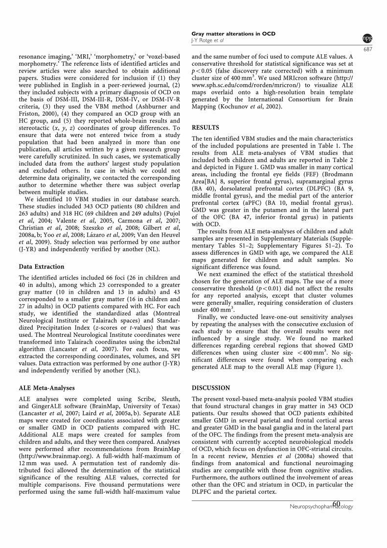
resonance imaging,’ ‘MRI,’ ‘morphometry,’ or ‘voxel-basedmorphometry.’ The reference lists of identified articles andreview articles were also searched to obtain additionalpapers. Studies were considered for inclusion if (1) theywere published in English in a peer-reviewed journal, (2)they included subjects with a primary diagnosis of OCD onthe basis of DSM-III, DSM-III-R, DSM-IV, or DSM-IV-Rcriteria, (3) they used the VBM method (Ashburner andFriston, 2000), (4) they compared an OCD group with anHC group, and (5) they reported whole-brain results andstereotactic (x, y, z) coordinates of group differences. Toensure that data were not entered twice from a studypopulation that had been analyzed in more than onepublication, all articles written by a given research groupwere carefully scrutinized. In such cases, we systematicallyincluded data from the authors’ largest study populationand excluded others. In case in which we could notdetermine data originality, we contacted the correspondingauthor to determine whether there was subject overlapbetween multiple studies.We identified 10 VBM studies in our database search.
These studies included 343 OCD patients (80 children and263 adults) and 318 HC (69 children and 249 adults) (Pujolet al, 2004; Valente et al, 2005, Carmona et al, 2007;Christian et al, 2008; Szeszko et al, 2008; Gilbert et al,2008a, b; Yoo et al, 2008; Lazaro et al, 2009; Van den Heuvelet al, 2009). Study selection was performed by one author(J-YR) and independently verified by another (NL).
Data Extraction
The identified articles included 66 foci (26 in children and40 in adults), among which 23 corresponded to a greatergray matter (10 in children and 13 in adults) and 43corresponded to a smaller gray matter (16 in children and27 in adults) in OCD patients compared with HC. For eachstudy, we identified the standardized atlas (MontrealNeurological Institute or Talairach spaces) and Standar-dized Precipitation Index (z-scores or t-values) that wasused. The Montreal Neurological Institute coordinates weretransformed into Talairach coordinates using the icbm2talalgorithm (Lancaster et al, 2007). For each focus, weextracted the corresponding coordinates, volumes, and SPIvalues. Data extraction was performed by one author (J-YR)and independently verified by another (NL).
ALE Meta-Analyses
ALE analyses were completed using Scribe, Sleuth,and GingerALE software (BrainMap, University of Texas)(Lancaster et al, 2007; Laird et al, 2005a, b). Separate ALEmaps were created for coordinates associated with greateror smaller GMD in OCD patients compared with HC.Additional ALE maps were created for samples fromchildren and adults, and they were then compared. Analyseswere performed after recommendations from BrainMap(http://www.brainmap.org). A full-width half-maximum of12mm was used. A permutation test of randomly dis-tributed foci allowed the determination of the statisticalsignificance of the resulting ALE values, corrected formultiple comparisons. Five thousand permutations wereperformed using the same full-width half-maximum value
and the same number of foci used to compute ALE values. Aconservative threshold for statistical significance was set atpo0.05 (false discovery rate corrected) with a minimumcluster size of 400mm3. We used MRIcron software (http://www.sph.sc.edu/comd/rorden/mricron/) to visualize ALEmaps overlaid onto a high-resolution brain templategenerated by the International Consortium for BrainMapping (Kochunov et al, 2002).
RESULTS
The ten identified VBM studies and the main characteristicsof the included populations are presented in Table 1. Theresults from ALE meta-analyses of VBM studies thatincluded both children and adults are reported in Table 2and depicted in Figure 1. GMD was smaller in many corticalareas, including the frontal eye fields (FEF) (BrodmannArea[BA] 8, superior frontal gyrus), supramarginal gyrus(BA 40), dorsolateral prefrontal cortex (DLPFC) (BA 9,middle frontal gyrus), and the medial part of the anteriorprefrontal cortex (aPFC) (BA 10, medial frontal gyrus).GMD was greater in the putamen and in the lateral partof the OFC (BA 47, inferior frontal gyrus) in patientswith OCD.The results from ALE meta-analyses of children and adult
samples are presented in Supplementary Materials (Supple-mentary Tables S1–2; Supplementary Figures S1–2). Toassess differences in GMD with age, we compared the ALEmaps generated for children and adult samples. Nosignificant difference was found.We next examined the effect of the statistical threshold
chosen for the generation of ALE maps. The use of a moreconservative threshold (po0.01) did not affect the resultsfor any reported analysis, except that cluster volumeswere generally smaller, requiring consideration of clustersunder 400mm3.Finally, we conducted leave-one-out sensitivity analyses
by repeating the analyses with the consecutive exclusion ofeach study to ensure that the overall results were notinfluenced by a single study. We found no markeddifferences regarding cerebral regions that showed GMDdifferences when using cluster size o400mm3. No sig-nificant differences were found when comparing eachgenerated ALE map to the overall ALE map (Figure 1).
DISCUSSION
The present voxel-based meta-analysis pooled VBM studiesthat found structural changes in gray matter in 343 OCDpatients. Our results showed that OCD patients exhibitedsmaller GMD in several parietal and frontal cortical areasand greater GMD in the basal ganglia and in the lateral partof the OFC. The findings from the present meta-analysis areconsistent with currently accepted neurobiological modelsof OCD, which focus on dysfunction in OFC-striatal circuits.In a recent review, Menzies et al (2008a) showed thatfindings from anatomical and functional neuroimagingstudies are compatible with those from cognitive studies.Furthermore, the authors outlined the involvement of areasother than the OFC and striatum in OCD, in particular theDLPFC and the parietal cortex.
Gray matter alterations in OCD
J-Y Rotge et al
687
Neuropsychopharmacology60
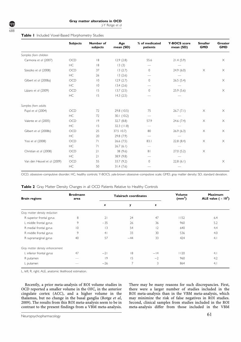
Recently, a prior meta-analysis of ROI volume studies inOCD reported a smaller volume in the OFC, in the anteriorcingulate cortex (ACC), and a higher volume in thethalamus, but no change in the basal ganglia (Rotge et al,2009). The results from this ROI meta-analysis seem to be incontrast to the present findings from a VBM meta-analysis.
There may be many reasons for such discrepancies. First,there were a larger number of studies included in theROI meta-analysis than in the VBM meta-analysis, whichmay minimize the risk of false negatives in ROI studies.Second, clinical samples from studies included in the ROImeta-analysis differ from those included in the VBM
Table 1 Included Voxel-Based Morphometry Studies
Subjects Number of
subjects
Age
mean (SD)
% of medicated
patients
Y-BOCS score
mean (SD)
Smaller
GMD
Greater
GMD
Samples from children
Carmona et al (2007) OCD 18 12.9 (2.8) 55.6 21.4 (5.9) X
HC 18 13 (3) F F
Szeszko et al (2008) OCD 37 13 (2.7) 0 24.9 (6.0) X X
HC 26 13 (2.6) F F
Gilbert et al (2008a) OCD 10 12.9 (2.7) 0 26.5 (5.4) X
HC 10 13.4 (2.6) F F
Lazaro et al (2009) OCD 15 13.7 (2.5) 0 25.9 (5.6) X
HC 15 14.3 (2.5) F F
Samples from adults
Pujol et al (2004) OCD 72 29.8 (10.5) 75 26.7 (7.1) X X
HC 72 30.1 (10.2) F F
Valente et al (2005) OCD 19 32.7 (8.8) 57.9 24.6 (7.4) X X
HC 15 32.3 (11.8) F F
Gilbert et al (2008b) OCD 25 37.5 10.7) 80 26.9 (6.3) X X
HC 20 29.8 (7.9) F F
Yoo et al (2008) OCD 71 26.6 (7.5) 83.1 22.8 (8.4) X X
HC 71 26.7 (6.1) F F
Christian et al (2008) OCD 21 38 (9.6) 81 27.0 (5.2) X
HC 21 38.9 (9.8) F F
Van den Heuvel et al (2009) OCD 55 33.7 (9.2) 0 22.8 (6.1) X
HC 50 31.4 (7.6) F F
OCD, obsessive–compulsive disorder; HC, healthy controls; Y-BOCS, yale-brown obsessive–compulsive scale; GMD, gray matter density; SD, standard deviation.
Table 2 Gray Matter Density Changes in all OCD Patients Relative to Healthy Controls
Brain regions
Brodmann
areaTalairach coordinates Volume
(mm3)
Maximum
ALE value (�103)
x y z
Gray matter density reduction
R superior frontal gyrus 8 21 24 47 1152 6.4
L middle frontal gyrus 9 ÿ35 26 36 960 5.2
R medial frontal gyrus 10 13 54 12 640 4.4
R middle frontal gyrus 9 41 33 30 536 4.0
R supramarginal gyrus 40 57 ÿ44 33 424 4.1
Gray matter density enhancement
L inferior frontal gyrus 47 ÿ21 18 ÿ14 1120 4.1
R putamen F 19 15 ÿ2 960 4.2
L putamen F ÿ26 7 3 864 4.1
L, left; R, right; ALE, anatomic likelihood estimation.
Gray matter alterations in OCD
J-Y Rotge et al
688
Neuropsychopharmacology 61
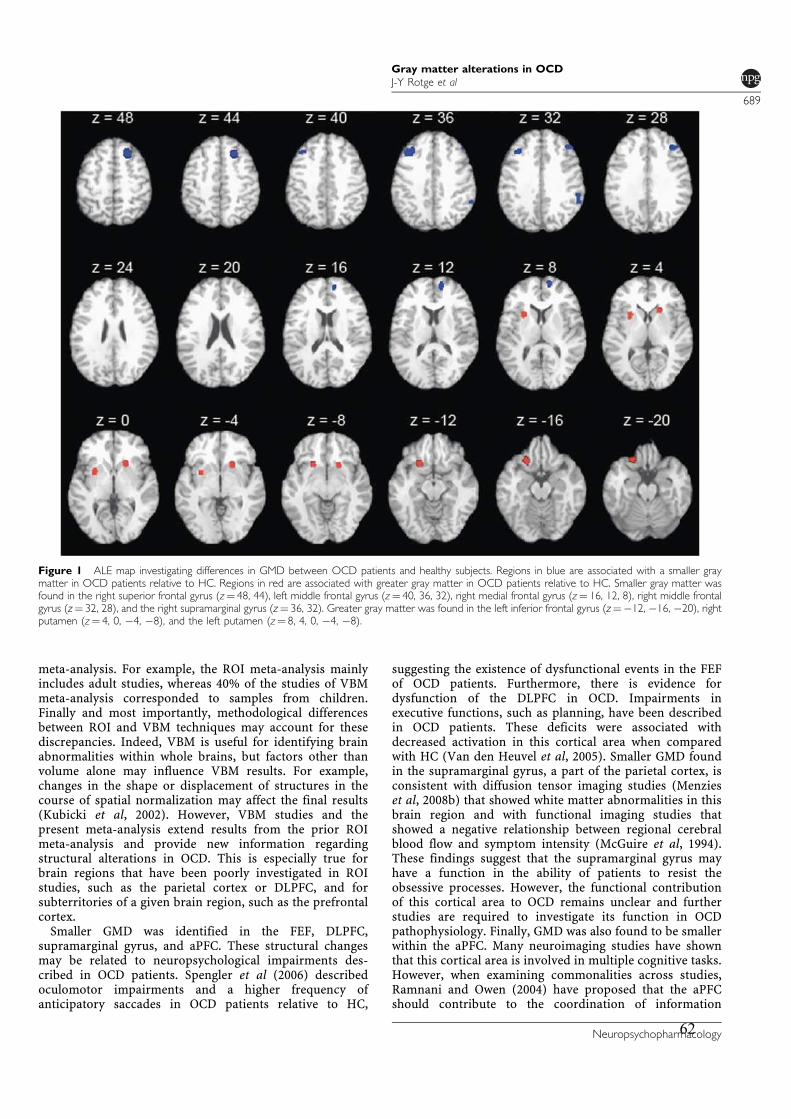
meta-analysis. For example, the ROI meta-analysis mainlyincludes adult studies, whereas 40% of the studies of VBMmeta-analysis corresponded to samples from children.Finally and most importantly, methodological differencesbetween ROI and VBM techniques may account for thesediscrepancies. Indeed, VBM is useful for identifying brainabnormalities within whole brains, but factors other thanvolume alone may influence VBM results. For example,changes in the shape or displacement of structures in thecourse of spatial normalization may affect the final results(Kubicki et al, 2002). However, VBM studies and thepresent meta-analysis extend results from the prior ROImeta-analysis and provide new information regardingstructural alterations in OCD. This is especially true forbrain regions that have been poorly investigated in ROIstudies, such as the parietal cortex or DLPFC, and forsubterritories of a given brain region, such as the prefrontalcortex.Smaller GMD was identified in the FEF, DLPFC,
supramarginal gyrus, and aPFC. These structural changesmay be related to neuropsychological impairments des-cribed in OCD patients. Spengler et al (2006) describedoculomotor impairments and a higher frequency ofanticipatory saccades in OCD patients relative to HC,
suggesting the existence of dysfunctional events in the FEFof OCD patients. Furthermore, there is evidence fordysfunction of the DLPFC in OCD. Impairments inexecutive functions, such as planning, have been describedin OCD patients. These deficits were associated withdecreased activation in this cortical area when comparedwith HC (Van den Heuvel et al, 2005). Smaller GMD foundin the supramarginal gyrus, a part of the parietal cortex, isconsistent with diffusion tensor imaging studies (Menzieset al, 2008b) that showed white matter abnormalities in thisbrain region and with functional imaging studies thatshowed a negative relationship between regional cerebralblood flow and symptom intensity (McGuire et al, 1994).These findings suggest that the supramarginal gyrus mayhave a function in the ability of patients to resist theobsessive processes. However, the functional contributionof this cortical area to OCD remains unclear and furtherstudies are required to investigate its function in OCDpathophysiology. Finally, GMD was also found to be smallerwithin the aPFC. Many neuroimaging studies have shownthat this cortical area is involved in multiple cognitive tasks.However, when examining commonalities across studies,Ramnani and Owen (2004) have proposed that the aPFCshould contribute to the coordination of information
Figure 1 ALE map investigating differences in GMD between OCD patients and healthy subjects. Regions in blue are associated with a smaller graymatter in OCD patients relative to HC. Regions in red are associated with greater gray matter in OCD patients relative to HC. Smaller gray matter wasfound in the right superior frontal gyrus (z¼ 48, 44), left middle frontal gyrus (z¼ 40, 36, 32), right medial frontal gyrus (z¼ 16, 12, 8), right middle frontalgyrus (z¼ 32, 28), and the right supramarginal gyrus (z¼ 36, 32). Greater gray matter was found in the left inferior frontal gyrus (z¼ÿ12, ÿ16, ÿ20), rightputamen (z¼ 4, 0, ÿ4, ÿ8), and the left putamen (z¼ 8, 4, 0, ÿ4, ÿ8).
Gray matter alterations in OCD
J-Y Rotge et al
689
Neuropsychopharmacology62
processing and information transfer between multipleoperations across the supramodal cortex. In regard toOCD phenomenology, it could be assumed that dysfunctionof the aPFC could play a role in deficits in cognitiveflexibility, in particular in cognitive reallocation, which maycontribute to maintenance of repetitive behaviors. However,the specific function of the aPFC in OCD remains unknownand awaits clarification.Greater GMD was identified in the bilateral putamen,
which is a part of the striatum. This finding is consistentwith neurobiological models of OCD and with functionalneuroimaging studies suggesting that dysfunction in theputamen contributes to the pathogenesis of OCD (Menzieset al, 2008a; Rauch et al, 1997). Furthermore, OCD symptomseverity was associated with metabolic activity in theputamen (Kwon et al, 2003), whereas decreased metabolicactivity in the putamen after pharmacological treatment wasassociated with an improvement in cognitive function inOCD patients (Kang et al, 2003). GMD was also found to begreater in the lateral part of the OFC in OCD patientscompared with HC. The OFC is thought to have a functionin several cognitive processes, such as the selection,comparison, and judgment of motivationally salient stimuli,and in the motivational aspects of decision making. Inparticular, the OFC has a function in error prediction byencoding the representation of the value of an expectedoutcome, which is used to anticipate the positive andnegative consequences that are likely to follow a givenaction (O’Doherty, 2007; Wallis, 2007). The OFC alsoincreases the evaluation of the occurrence of negativeconsequences that may follow an action in OCD and,therefore, mediates obsessive thoughts that lead to repeti-tive or ritualistic behaviors intended to prevent negativeconsequences as a way to relieve anxiety. The OFC is likelyrelated to deficits of cognitive regulation of emotionallysalient information. Accordingly, Chamberlain et al(2008)recently showed that, in comparison with HC, lateralOFC activation is reduced during a task that assessesbehavioral flexibility in OCD patients and in unaffectedrelatives. These findings suggest that the lateral OFC couldhave a central function in cognitive flexibility deficits and,thus, in the genesis of pathological habits (Chamberlainet al, 2008).In the present meta-analysis, we failed to show any
significant differences in GMD between children and adults.This was an unexpected result because many factors shouldreasonably contribute to GMD changes with aging. First,children and adults differ for different clinical variables,such as illness duration, age of onset, or treatmentintervention, which may potentially influence neuroimagingfindings. Second, there are several lines of evidence in theliterature suggesting that OCD is a neurodevelopmentaldisorder. Rosenberg and Keshavan (1998) proposed thatOCD may be underlined by ‘a developmentally mediatednetwork dysplasia in prefrontal cortical circuits’. Thisneurodevelopmental view may be supported by differencesin GMD between children and adults. One possibleexplanation for the absence of any significant differencesbetween pediatric and adult samples may be the lownumber of foci, which may contribute to false negativeresults. Further hypothesis-driven ROI studies could behelpful to resolve this issue.
The present meta-analysis has several limitations. First,our meta-analysis is subject to publication bias. Studies thatfailed to show significant effects may not have beenpublished. In the present meta-analysis, we identified only10 VBM studies and we did not identify any articles withoutan effect. Second, we failed to consider clinical variables,such as symptom dimensions, medication status, comor-bidity status, and symptom severity scores. Although thesevariables may be associated with differential structuralabnormalities, the low number of VBM studies did not allowus to control for these factors. However, the absence ofmarked difference when consecutively excluding each studyargues in favor of the robustness of our findings despite theclinical heterogeneity of study populations.In conclusion, the present meta-analysis of VBM studies
showed that OCD patients exhibited gray matter abnorm-alities in parieto-frontal cortical areas and in the basalganglia. Further structural neuroimaging studies are requi-red to assess gray matter changes associated with clinicalcorrelates. Furthermore, functional studies may be helpfulto elucidate the specific function of these areas in thegenesis of OCD symptoms.
ACKNOWLEDGEMENTS
J-Y Rotge was supported by a grant from the Fondation pourla Recherche Medicale (Medical Research Foundation).
DISCLOSURE
The authors declare no conflict of interest.
REFERENCES
Ashburner J, Friston KJ (2000). Voxel-based morphometryFthemethods. NeuroImage 11: 805–821.
Carmona S, Bassas N, Rovira M, Gispert JD, Soliva JC, Prado Met al (2007). Pediatric OCD structural brain deficits in conflicmonitoring circuits: a voxel-based morphometry study. NeurosciLett 421: 218–223.
Chamberlain SR, Menzies L, Hampshire A, Suckling J, FinebergNA, del Campo N et al (2008). Orbitofrontal dysfunction inpatients with obsessive-compulsive disorder and their unaffectedrelatives. Science 321: 421–422.
Christian CJ, Lencz T, Robinson DG, Burdick KE, Ashtari M,Malhotra AK et al (2008). Gray matter structural alterations inobsessive-compulsive disorder: relationship to neuropsycholo-gical functions. Psychiatry Res 164: 123–131.
Ellison-Wright I, Glahn DC, Laird AR, Thelen SM, Bullmore ET(2008). The anatomy of first-episode and chronic schizophrenia:an anatomical likelihood estimation meta-analysis. Am JPsychiatry 165: 1015–1023.
Gilbert AR, Keshavan MS, Diwadkar V, Nutche J, Macmaster F,Easter PC et al (2008a). Gray matter differences betweenpediatric obsessive-compulsive disorder patients and high-risksiblings: a preliminary voxel-based morphometry study. Neu-rosci Lett 435: 45–50.
Gilbert AR, Mataix-Cols D, Almeida JR, Lawrence N, Nutche J,Diwadkar V et al (2008b). Brain structure and symptomdimension relationships in obsessive-compulsive disorder: avoxel-based morphometry study. J Affect Disord 109: 117–126.
Glahn DC, Laird AR, Ellison-Wright I, Thelen SM, Robinson JL,Lancaster JL et al (2008). Meta-analysis of gray matter anomalies
Gray matter alterations in OCD
J-Y Rotge et al
690
Neuropsychopharmacology 63
in schizophrenia: application of anatomic likelihood estimationand network analysis. Biol Psychiatry 64: 774–781.
Kang DH, Kwon JS, Kim JJ, Youn T, Park HJ, Kim MS et al (2003).Brain glucose metabolic changes associated with neuropsycho-logical improvements after 4 months of treatment in patientswith obsessive-compulsive disorder. Acta Psychiatr Scand 107:291–297.
Kochunov P, Lancaster J, Thompson P, Toga AW, Brewer P,Hardies J et al (2002). An optimized individual target brain inthe Talairach coordinate system. NeuroImage 17: 922–927.
Kubicki M, Shenton ME, Salisbury DF, Hirayasu Y, Kasai K,Kikinis R et al (2002). Voxel-based morphometric analysis ofgray matter in first episode schizophrenia. NeuroImage 17:1711–1719.
Kwon JS, Kim JJ, Lee DW, Lee JS, Lee DS, Kim MS et al (2003).Neural correlates of clinical symptoms and cognitive cognitivedysfunctions in obsessive-compulsive disorder. Psychiatry Res122: 37–47.
Laird AR, Fox M, Price CJ, Glahn DC, Uecker AM, Lancaster JL et al(2005a). ALE meta-analysis: controlling the false discovery rate andperforming statistical contrasts. Hum Brain Mapp 25: 155–164.
Laird AR, Lancaster JL, Fox PT (2005b). BrainMap: the socialevolution of a functional neuroimaging database. Neuroinfor-matics 3: 65–78.
Lancaster JL, Tordesillas-Gutierrez D, Martinez M, Salinas F, EvansA, Zilles K et al (2007). Bias between MNI and Talairachcoordinates analyzed using the ICBM-152 brain template. HumBrain Mapp 28: 1194–1205.
Lazaro L, Bargallo N, Castro-Fornieles J, Falcon C, Andres S,Calvo R et al (2009). Brain changes in children and adolescentswith obsessive-compulsive disorder before and after treatment: avoxel-based morphometric MRI study. Psychiatry Res 172: 140–146.
McGuire PK, Bench CJ, Frith CD, Marks IM, Frackowiak RS, DolanRJ (1994). Functional anatomy of obsessive-compulsive phe-nomena. Br J Psychiatry 164: 459–468.
Menzies L, Chamberlain SR, Laird AR, Thelen SM, Sahakian BJ,Bullmore ET (2008a). Integrating evidence from neuroimagingand neuropsychological studies of obsessive-compulsive dis-order: the orbitofronto-striatal model revisited. Neurosci Biobe-hav Rev 32: 525–549.
Menzies L, Williams GB, Chamberlain SR, Ooi C, Fineberg N,Suckling J et al (2008b). White matter abnormalities in patientswith obsessive-compulsive disorder and their first-degreerelatives. Am J Psychiatry 165: 1308–1315.
O’Doherty JP (2007). Lights, camembert, action! The role of humanorbitofrontal cortex in encoding stimuli, rewards, and choices.Ann NY Acad Sci 1121: 254–272.
Pujol J, Soriano-Mas C, Alonso P, Cardoner N, Menchon JM, DeusJ et al (2004). Mapping structural brain alterations in obsessive-compulsive disorder. Arch Gen Psychiatry 61: 720–730.
Ramnani N, Owen AM (2004). Anterior prefrontal cortex: insightsinto function from anatomy and neuroimaging. Nat RevNeurosci 5: 184–194.
Rauch SL, Savage CR, Alpert NM, Dougherty D, Kendrick A,Curran T et al (1997). Probing striatal function in obsessive-compulsive disorder: a PET study of implicit sequence learning.J Neuropsychiatry Clin Neurosci 9: 568–573.
Rosenberg DR, Keshavan MS (1998). Towar a neurodevelopmentalmodel of obsessive-compulsive disorder. Biol Psychiatry 43:623–640.
Rotge JY, Guehl D, Dilharreguy B, Tignol J, Bioulac B, Allard Met al (2009). Meta-analysis of brain volume changes in obsessive-compulsive disorder. Biol Psychiatry 65: 75–83.
Spengler D, Trillenberg P, Sprenger A, Nagel M, Kordon A,Junghanns K et al (2006). Evidence from increased anticipationof predictive saccades for a dysfunction of fronto-striatalcircuits in obsessive-compulsive disorder. Psychiatry Res 143:77–88.
Szeszko PR, Christian C, MacMaster F, Lencz T, Mirza Y,Taormina SP et al (2008). Gray matter structural alterations inpsychotropic drug-naıve pediatric obsessive-compulsive disor-der: an optimized voxel-based morphometry study. Am JPsychiatry 165: 1299–1307.
Turkeltaub PE, Eden GF, Jones KM, Zeffiro TA (2002). Meta-analysis of the functional neuroanatomy of single-word reading:method and validation. NeuroImage 16: 765–780.
Valente AA, Miguel EC, Castro CC, Amaro E, Duran FLS,Buchpiguel CA et al (2005). Regional gray matter abnormalitiesin obsessive-compulsive disorder: a voxel-based morphometrystudy. Biol Psychiatry 58: 479–487.
Van den Heuvel OA, Remijnse PL, Mataix-Cols D, Vrenken H,Groenewegen HJ, Uylings HB et al (2009). The major symptomdimensions of obsessive-compulsive disorder are mediated bypartially distinct neural systems. Brain 132: 853–868.
Van den Heuvel OA, Veltman DJ, Groenewegen HJ, Cath DC, vanBalkom AJ, van Hartskamp J et al (2005). Frontal-striataldysfunction during planning in obsessive-compulsive disorder.Arch Gen Psychiatry 62: 301–309.
Wallis JD (2007). Orbitofrontal cortex and its contribution todecision-making. Annu Rev Neurosci 30: 31–56.
Yoo SY, Roh MS, Choi JS, Kang DH, Ha TH, Lee JM et al(2008). Voxel-based morphometry study of gray matterabnormalities in obsessive-compulsive disorder. J Korean MedSci 23: 24–30.
Supplementary Information accompanies the paper on the Neuropsychopharmacology website (http://www.nature.com/npp)
Gray matter alterations in OCD
J-Y Rotge et al
691
Neuropsychopharmacology64
65
Gray matter alterations in obsessive compulsive disorder:
An anatomic likelihood estimation meta-analysis
Rotge JY, Langbour N, Guehl D, Bioulac B, Jaafari N, Allard M, Aouizerate B, Burbaud P.
Supplementary Materials
Supplementary Results
Results from the ALE meta-analysis of VBM studies that included OCD and healthy children
are reported in Table S1 and depicted in Figure S1. Results from the ALE meta-analysis of
VBM studies that included OCD and healthy adults are reported in Table S2 and depicted in
Figure S2.
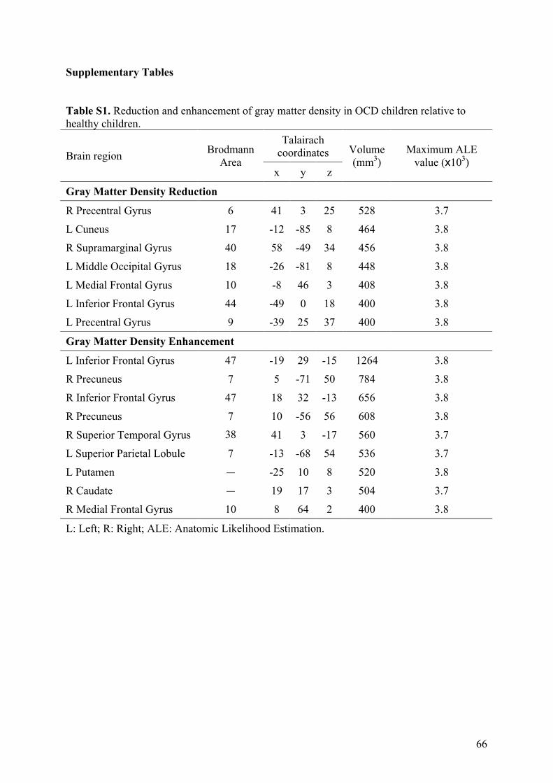
66
Supplementary Tables
Table S1. Reduction and enhancement of gray matter density in OCD children relative to healthy children.
Talairach coordinates Brain region
Brodmann Area
x y z
Volume (mm3)
Maximum ALE value (x103)
Gray Matter Density Reduction
R Precentral Gyrus 6 41 3 25 528 3.7
L Cuneus 17 -12 -85 8 464 3.8
R Supramarginal Gyrus 40 58 -49 34 456 3.8
L Middle Occipital Gyrus 18 -26 -81 8 448 3.8
L Medial Frontal Gyrus 10 -8 46 3 408 3.8
L Inferior Frontal Gyrus 44 -49 0 18 400 3.8
L Precentral Gyrus 9 -39 25 37 400 3.8
Gray Matter Density Enhancement
L Inferior Frontal Gyrus 47 -19 29 -15 1264 3.8
R Precuneus 7 5 -71 50 784 3.8
R Inferior Frontal Gyrus 47 18 32 -13 656 3.8
R Precuneus 7 10 -56 56 608 3.8
R Superior Temporal Gyrus 38 41 3 -17 560 3.7
L Superior Parietal Lobule 7 -13 -68 54 536 3.7
L Putamen — -25 10 8 520 3.8
R Caudate — 19 17 3 504 3.7
R Medial Frontal Gyrus 10 8 64 2 400 3.8
L: Left; R: Right; ALE: Anatomic Likelihood Estimation.
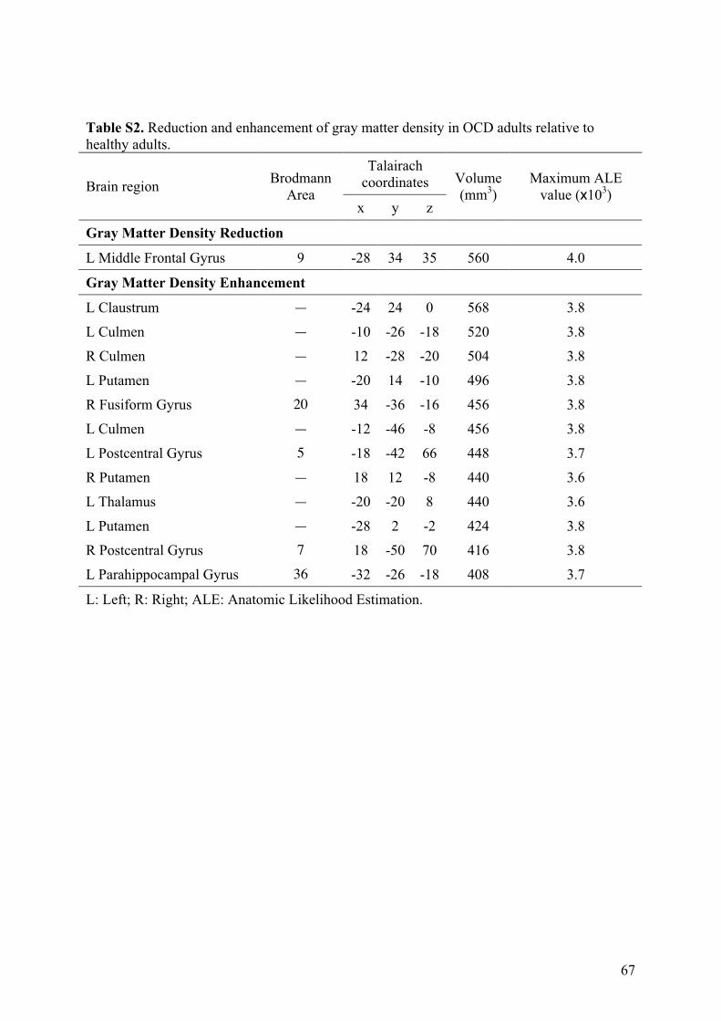
67
Table S2. Reduction and enhancement of gray matter density in OCD adults relative to healthy adults.
Talairach coordinates Brain region
Brodmann Area
x y z
Volume (mm3)
Maximum ALE value (x103)
Gray Matter Density Reduction
L Middle Frontal Gyrus 9 -28 34 35 560 4.0
Gray Matter Density Enhancement
L Claustrum — -24 24 0 568 3.8
L Culmen — -10 -26 -18 520 3.8
R Culmen — 12 -28 -20 504 3.8
L Putamen — -20 14 -10 496 3.8
R Fusiform Gyrus 20 34 -36 -16 456 3.8
L Culmen — -12 -46 -8 456 3.8
L Postcentral Gyrus 5 -18 -42 66 448 3.7
R Putamen — 18 12 -8 440 3.6
L Thalamus — -20 -20 8 440 3.6
L Putamen — -28 2 -2 424 3.8
R Postcentral Gyrus 7 18 -50 70 416 3.8
L Parahippocampal Gyrus 36 -32 -26 -18 408 3.7
L: Left; R: Right; ALE: Anatomic Likelihood Estimation.
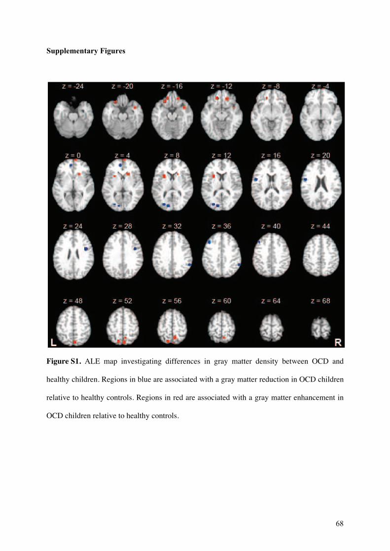
68
Supplementary Figures
Figure S1. ALE map investigating differences in gray matter density between OCD and
healthy children. Regions in blue are associated with a gray matter reduction in OCD children
relative to healthy controls. Regions in red are associated with a gray matter enhancement in
OCD children relative to healthy controls.
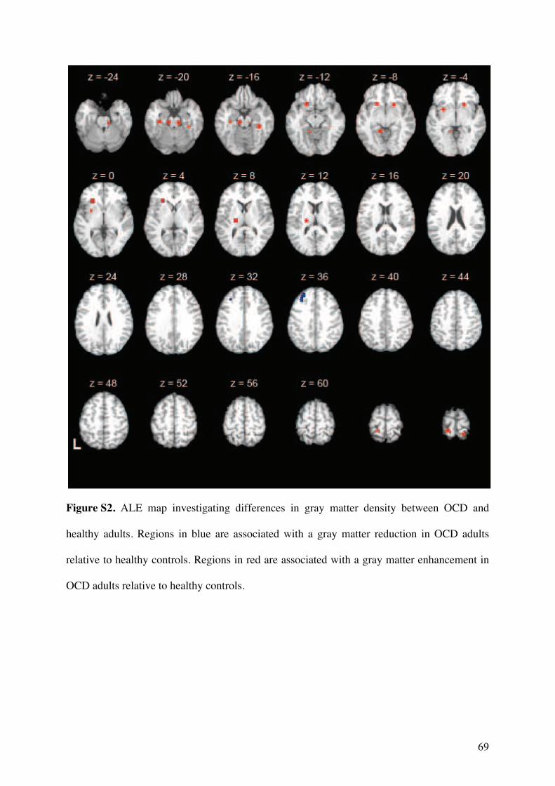
69
Figure S2. ALE map investigating differences in gray matter density between OCD and
healthy adults. Regions in blue are associated with a gray matter reduction in OCD adults
relative to healthy controls. Regions in red are associated with a gray matter enhancement in
OCD adults relative to healthy controls.

70
71
3.3. Relation entre les volumes thalamiques et orbitofrontaux
3.3.1. Introduction
Nous avons vu que le volume du COF est diminué alors que le volume thalamique est
augmenté dans le TOC. Ces deux régions cérébrales sont connues pour entretenir d’étroites
relations directes et réciproques. Il apparaît donc légitime de se demander si ces altérations
anatomiques sont des phénomènes indépendants ou liés. En d’autres termes, existe-t-il une
relation entre les modifications de volume du COF et celles du thalamus dans le TOC ?
3.3.2. Méthodes
Nous avons utilisé deux méthodes différentes et complémentaires pour répondre à la
question posée. Tout d’abord, nous avons réalisé une étude d’IRM volumétrique incluant 16
patients souffrant de TOC sans comorbidité psychiatrique et 16 témoins appariés en âge, sexe
et niveau d’étude. Nous avons mesuré le volume du COF, du thalamus ainsi que d’une région
corticale différente du COF comme région témoin (le CCA a été choisi compte tenu de sa
possible implication dans la maladie). Ensuite, nous avons effectué une analyse de méta-
régression en réutilisant les données présentées dans la méta-analyse précédente (page 45).
3.3.3. Résultats et discussion
Nous n’avons trouvé aucune différence volumétrique significative entre les patients et
les témoins. En revanche, nous avons trouvé une corrélation négative entre les volumes du
COF et du thalamus chez les patients, mais pas chez les témoins. Il n’y avait pas de
corrélation significative entre les volumes du CCA et du thalamus dans aucun groupe. Ces
résultats ont été confirmés par analyse de méta-régression comprenant toutes les études dans
lesquelles les volumes des couples COF-thalamus et CCA-thalamus ont été mesurés au sein
d’une même population. Les tailles d’effet rapportées pour le COF étaient négativement
corrélées à celles du thalamus. Aucune corrélation n’a été retrouvée entre les volumes du
CCA et du thalamus. La principale limite de cette étude a été le petit nombre de patients
inclus, cependant la confirmation de nos observations par l’analyse de méta-régression a
permis de renforcer les résultats trouvés.
72
3.3.4. Conclusion
Ces résultats suggèrent que les altérations anatomiques du COF et du thalamus sont
dépendantes l’une de l’autre, de manière relativement spécifique.
Rotgé JY, Dilharreguy B, Aouizerate B, Martin-Guehl C, Guehl D, Jaafari N,
Langbour N, Bioulac B, Tignol J, Allard M, Burbaud P. Inverse relationship
between thalamic and orbitofrontal volumes in obsessive-compulsive disorder.
Progress in Neuro-Psychopharmacology and Biological Psychiatry 2009;33:682-
687.
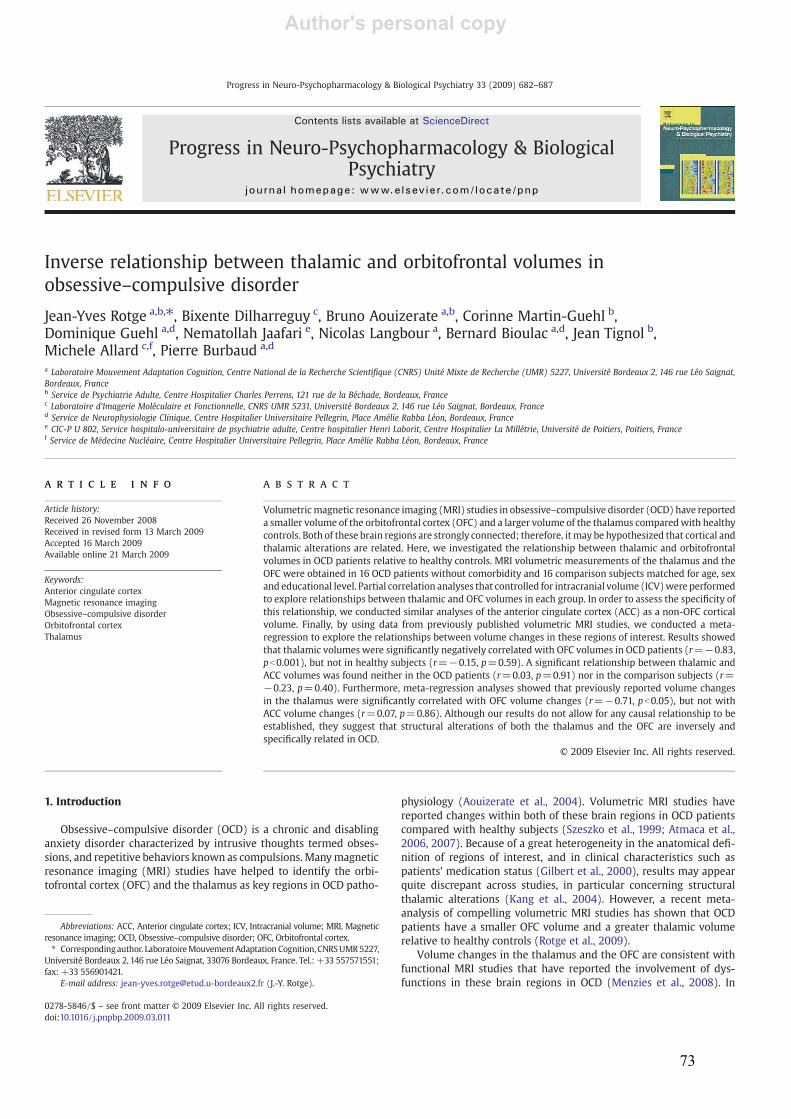
Author's personal copy
Inverse relationship between thalamic and orbitofrontal volumes inobsessive–compulsive disorder
Jean-Yves Rotge a,b,⁎, Bixente Dilharreguy c, Bruno Aouizerate a,b, Corinne Martin-Guehl b,Dominique Guehl a,d, Nematollah Jaafari e, Nicolas Langbour a, Bernard Bioulac a,d, Jean Tignol b,Michele Allard c,f, Pierre Burbaud a,d
a Laboratoire Mouvement Adaptation Cognition, Centre National de la Recherche Scientifique (CNRS) Unité Mixte de Recherche (UMR) 5227, Université Bordeaux 2, 146 rue Léo Saignat,
Bordeaux, Franceb Service de Psychiatrie Adulte, Centre Hospitalier Charles Perrens, 121 rue de la Béchade, Bordeaux, Francec Laboratoire d'Imagerie Moléculaire et Fonctionnelle, CNRS UMR 5231, Université Bordeaux 2, 146 rue Léo Saignat, Bordeaux, Franced Service de Neurophysiologie Clinique, Centre Hospitalier Universitaire Pellegrin, Place Amélie Rabba Léon, Bordeaux, Francee CIC-P U 802, Service hospitalo-universitaire de psychiatrie adulte, Centre hospitalier Henri Laborit, Centre Hospitalier La Millétrie, Université de Poitiers, Poitiers, Francef Service de Médecine Nucléaire, Centre Hospitalier Universitaire Pellegrin, Place Amélie Rabba Léon, Bordeaux, France
a b s t r a c ta r t i c l e i n f o
Article history:
Received 26 November 2008
Received in revised form 13 March 2009
Accepted 16 March 2009
Available online 21 March 2009
Keywords:
Anterior cingulate cortex
Magnetic resonance imaging
Obsessive–compulsive disorder
Orbitofrontal cortex
Thalamus
Volumetric magnetic resonance imaging (MRI) studies in obsessive–compulsive disorder (OCD) have reported
a smaller volume of the orbitofrontal cortex (OFC) and a larger volume of the thalamus comparedwith healthy
controls. Both of these brain regions are strongly connected; therefore, it may be hypothesized that cortical and
thalamic alterations are related. Here, we investigated the relationship between thalamic and orbitofrontal
volumes in OCD patients relative to healthy controls. MRI volumetric measurements of the thalamus and the
OFC were obtained in 16 OCD patients without comorbidity and 16 comparison subjects matched for age, sex
and educational level. Partial correlation analyses that controlled for intracranial volume (ICV)were performed
to explore relationships between thalamic and OFC volumes in each group. In order to assess the specificity of
this relationship, we conducted similar analyses of the anterior cingulate cortex (ACC) as a non-OFC cortical
volume. Finally, by using data from previously published volumetric MRI studies, we conducted a meta-
regression to explore the relationships between volume changes in these regions of interest. Results showed
that thalamic volumes were significantly negatively correlated with OFC volumes in OCD patients (r=−0.83,
pb0.001), but not in healthy subjects (r=−0.15, p=0.59). A significant relationship between thalamic and
ACC volumes was found neither in the OCD patients (r=0.03, p=0.91) nor in the comparison subjects (r=
−0.23, p=0.40). Furthermore, meta-regression analyses showed that previously reported volume changes
in the thalamus were significantly correlated with OFC volume changes (r=−0.71, pb0.05), but not with
ACC volume changes (r=0.07, p=0.86). Although our results do not allow for any causal relationship to be
established, they suggest that structural alterations of both the thalamus and the OFC are inversely and
specifically related in OCD.
© 2009 Elsevier Inc. All rights reserved.
1. Introduction
Obsessive–compulsive disorder (OCD) is a chronic and disabling
anxiety disorder characterized by intrusive thoughts termed obses-
sions, and repetitive behaviors known as compulsions. Manymagnetic
resonance imaging (MRI) studies have helped to identify the orbi-
tofrontal cortex (OFC) and the thalamus as key regions in OCD patho-
physiology (Aouizerate et al., 2004). Volumetric MRI studies have
reported changes within both of these brain regions in OCD patients
compared with healthy subjects (Szeszko et al., 1999; Atmaca et al.,
2006, 2007). Because of a great heterogeneity in the anatomical defi-
nition of regions of interest, and in clinical characteristics such as
patients' medication status (Gilbert et al., 2000), results may appear
quite discrepant across studies, in particular concerning structural
thalamic alterations (Kang et al., 2004). However, a recent meta-
analysis of compelling volumetric MRI studies has shown that OCD
patients have a smaller OFC volume and a greater thalamic volume
relative to healthy controls (Rotge et al., 2009).
Volume changes in the thalamus and the OFC are consistent with
functional MRI studies that have reported the involvement of dys-
functions in these brain regions in OCD (Menzies et al., 2008). In
Progress in Neuro-Psychopharmacology & Biological Psychiatry 33 (2009) 682–687
Abbreviations: ACC, Anterior cingulate cortex; ICV, Intracranial volume; MRI, Magnetic
resonance imaging; OCD, Obsessive–compulsive disorder; OFC, Orbitofrontal cortex.
⁎ Correspondingauthor. LaboratoireMouvementAdaptationCognition,CNRSUMR5227,
Université Bordeaux 2, 146 rue Léo Saignat, 33076 Bordeaux, France. Tel.: +33 557571551;
fax: +33 556901421.
E-mail address: [email protected] (J.-Y. Rotge).
0278-5846/$ – see front matter © 2009 Elsevier Inc. All rights reserved.
doi:10.1016/j.pnpbp.2009.03.011
Contents lists available at ScienceDirect
Progress in Neuro-Psychopharmacology & BiologicalPsychiatry
j ourna l homepage: www.e lsev ie r.com/ locate /pnp
73
Author's personal copy
particular, a functional overactivation of both regions has been des-
cribed in OCD patients during rest and during a task that provokes
obsessive and compulsive symptoms (Menzies et al., 2008; Rotge
et al., 2008). However, cognitive operations performed during these
paradigms remain unclear. Recently, event-related functional MRI
studies have reported OFC underactivaction related to impairments
in cognitive flexibility (Chamberlain et al., 2005; Remijnse et al., 2006;
Gu et al., 2008; Chamberlain et al., 2008) and in response inhibition
(Roth et al., 2007), which may be viewed as the central features of
OCD responsible for the production of maladaptive behaviors. Inter-
estingly, in their study, Roth et al. showed that OFC activation during
response inhibition was negatively correlated with OCD symptom
severity, whereas thalamic activation was positively correlated with
OCD symptom severity. This pattern suggested that functional abnor-
malities of the two regions are closely, but inversely, related to the
severity of OCD symptoms (Roth et al., 2007).
The frontal lobe, including the OFC, and the thalamus are well-
known to be strongly connected (Steriade et al., 1997; Herrero et al.,
2002). Indeed, the thalamusacts as a relaycenter that projects to cortical
areas and receives reciprocal information. These signals to various
thalamic nuclei provide positive feedback to the “correct input”, while
simultaneously suppressing irrelevant information (Steriade, 1970;
Steriade et al., 1997; Herrero et al., 2002). The thalamus maintains a
two-way dialogue with the frontal lobe, including the OFC, by which
it is thought to be implicated in cognitive and emotional functions,
such as outcome evaluation, decision making, and inhibitory control,
and therefore, in the modulation of behavioral responses to the envi-
ronment. Because of the volume changes observed in both the OFC
and the thalamus in OCD, and because of the connections between
these brain regions, it may be hypothesized that structural alterations in
the OFC and the thalamus may represent related phenomena. If so,
volume changes observed in cortical areas may be related to volume
changes in the thalamus. Here, we tested this hypothesis by exploring
the relationship between thalamic and orbitofrontal volumes in OCD
patients compared with healthy control subjects.
2. Materials and methods
2.1. Subjects and clinical assessments
Sixteen adults with a primary diagnosis of OCD seen at outpatient
clinics in Bordeaux and Poitiers, France, and 16 healthy volunteers
recruited from the local community through notice board advertise-
ments were included in the present study. The 16 case-control pairs
were matched for age, sex, handedness and educational level. All par-
ticipants were evaluated for OCD and associated psychopathology with
the Mini-International Neuropsychiatric Interview (version 5.0.0)
(Sheehan et al., 1998) and Structured Clinical Interview for DSM-IV
axis II personality disorders (version 2.0) (Maffei et al., 1997). Two
exclusion criteria forOCDpatients andhealthycomparison subjectswere
applied: the presence of other current major psychiatric disorders, in-
cluding major depressive disorder and personality disorder; and the
presence of a neurological disorder or a significant medical condition.
Clinical assessmentswere performed to determine the severity of symp-
toms of different conditions: the Yale–Brown Obsessive–Compulsive
Scale (Y-BOCS) (Goodman et al., 1989) for OCD symptoms, theHamilton
Depression Rating Scale (HDRS) (Hamilton, 1967) for depressive symp-
toms and the Hamilton Anxiety Rating Scale (HARS) (Hamilton, 1959)
for anxiety symptoms. OCD patients were not included in the final
cohort if they did not have clinically significant OCD symptoms, as
indicated by a Y-BOCS score b16, or if they had clinically significant
depressive symptoms or anxiety, as indicated by HDRS or HARS scores
N7. All OCD patients (n=16) were receiving treatment with serotonin
reuptake inhibitors, and 3 of them (18.8%)were also receiving low doses
of atypical antipsychotics (risperidone=1 mg). All healthy controls
(n=16) were free of any medication at the time of the study.
The protocol of the present study was reviewed and approved by
the local ethics authorities for work involving humans (“Comité Con-
sultatif de la Protection des Personnes dans la Recherche Biomédicale
Bordeaux B”). All participants gave informed written consent after
the goals, procedures and potential risks of the study were presented
and discussed.
2.2. MRI acquisition
All volumetric MRI scans were conducted at Bordeaux 2 University
(1.5-T, Philips Medical System, Best, Netherlands). A T1-weighted
gradient echo pulse sequence was used to obtain 25 4-mm-thick con-
tiguous horizontal images covering the whole brain (voxel size=
1×1×4 mm, echo time=2.5 ms, repetition time=23.1 ms, acquisi-
tion matrix=256×256 pixels, field of view=256 mm, flip angle=
90°). Axial slices were aligned parallel to the intercommissural line.
Before volumetric measurements, MRI scans of both groups were
scrutinized carefully to exclude structural abnormalities.
2.3. Volumetric measurements
Tracing was performed manually with the National Institutes of
Health image software (National Institutes of Health, USA, available at
http://rsb.info.nih.gov/nih-image/) on images resampled to 1 mm3
voxels and realigned parallel with the intercommissural line. Mea-
surements were completed in the coronal plane by an operator (J-YR)
who was blind to the subjects' group membership. Intracranial vol-
umes (ICV), a common estimate of brain size, were measured using
a semiautomated segmentation approach, as previously described
(Rosenberg et al., 1997). We defined three regions of interest (ROIs)
for the thalamus and the OFC: total, left, and right. Neuroanatomical
boundaries of ROIs used in the present study strictly followed a pre-
viously published volumetric MRI study by Kang et al. (2004). Briefly,
the thalamus was delineated by the internal capsule, the third ven-
tricle, the habenular nucleus, the lateral ventricle, the crux fornix, the
zona inserta and the red nucleus. For the OFC, the medial and lateral
boundaries were constituted by the frontomarginal sulcus, lateral
orbital sulcus, inferior margin of the circular sulcus of the insula,
inferior medial point of the cingulate sulcus, and the superior rostral
sulcus; and, the anterior and posterior boundaries were respectively
defined by the most anterior part of the frontal pole and the most
posterior part of the posterior medial orbitofrontal gyrus (see Kang
et al., 2004 for more details).
We also measured the volumes of a non-OFC cortical region to
ensure that a possible relationship was not a non-specific effect. For
this, we chose the anterior cingulate cortex (ACC), another prefrontal
region believed to be involved in OCD pathophysiology (Aouizerate
et al., 2004). Neuroanatomical boundaries of the ACC strictly followed
prior volumetric studies by Atmaca et al. (2006). Briefly, the ACC was
delineated by the cingulate sulcus, the most anterior part of the genu
of the corpus callosum, the deepest point of the callosal sulcus, and
the anterior commissure that indicated the posterior limit of the ACC
(see Atmaca et al., 2006 for more details). Some sample images are
presented in Supplementary material (Fig. S1). Finally, intra-rater
reliabilities were assessed on 20 MRI scans randomly selected among
the included subjects (10 scans in each group of subjects).
2.4. Statistical analyses
Statistical analyses were performed with SPSS software (release
16.0, Chicago, USA). Potential differences in the sociodemographic and
clinical characteristics of the patient and control groups were exam-
ined using χ2 tests and t tests for independent samples. Intrarater
reliability coefficients were computed using intra-class correlations.
To test between-group differences in thalamic, OFC and ACC volumes,
measured volumes for each brain region were entered into analyses
683J.-Y. Rotge et al. / Progress in Neuro-Psychopharmacology & Biological Psychiatry 33 (2009) 682–687
74
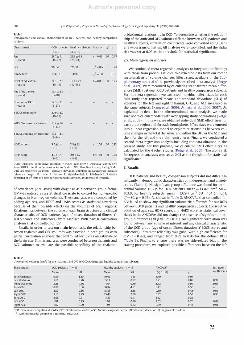
Author's personal copy
of covariance (ANCOVAs) with diagnosis as a between-group factor.
ICV was entered as a statistical covariate to control for non-specific
changes in brain region volumes. These analyses were completed by
adding age, sex, and HDRS and HARS scores as statistical covariates
because of their possible effects on the volumes of brain regions.
Relationships between the volume of each brain structure and clinical
characteristics of OCD patients (age of onset, duration of illness, Y-
BOCS scores and subscores) were assessed with partial correlation
analyses that controlled for ICV.
Finally, in order to test our main hypothesis, the relationship be-
tween thalamic and OFC volumes was assessed in both groups with
partial correlation analyses that controlled for ICV as an estimate of
the brain size. Similar analyses were conducted between thalamic and
ACC volumes to evaluate the possible specificity of the thalamo-
orbitofrontal relationship in OCD. To determine whether the relation-
ship of thalamic and OFC volumes differed between OCD patients and
healthy subjects, correlation coefficients were converted using Fish-
er's r-to-z transformation. All analyses were two-tailed, and the alpha
risk was set at 0.05 as the threshold for statistical significance.
2.5. Meta-regression analyses
We conducted meta-regression analyses to integrate our findings
with those from previous studies. We relied on data from our recent
meta-analysis of volume changes. Effect sizes, available in the Sup-
plementary material of the previously described meta-analysis (Rotge
et al., 2009), were measured by calculating standardized mean differ-
ences (SMD) between OCD patients and healthy comparison subjects.
For the meta-regression, we extracted individual effect sizes for each
MRI study that reported means and standard deviations (SDs) of
volumes for the left and right thalamus, OFC, and ACC measured in
the same subjects (Kang et al., 2004; Atmaca et al., 2006, 2007). As
explained in detail in the aforementioned meta-analysis, we made
sure not to calculate SMDs with overlapping study populations (Rotge
et al., 2009). In this way, we obtained individual SMD effect sizes for
each brain region and for each hemisphere. Effect sizes were entered
into a linear regression model to explore relationships between vol-
ume changes in the total thalamus, and either the OFC or the ACC, and
then, for the left and the right hemispheres. Finally, we conducted a
second meta-regression analysis including the data obtained in the
present study. For this purpose, we calculated SMD effect sizes as
calculated for the 4 other studies (Rotge et al., 2009). The alpha risk
for regression analyses was set at 0.05 as the threshold for statistical
significance.
3. Results
OCD patients and healthy comparison subjects did not differ sig-
nificantly in demographic characteristics or in depression and anxiety
scores (Table 1). No significant group difference was found for intra-
cranial volume (ICV): for OCD patients, mean=1334.9 cm3, SD=
103.0; for healthy subjects, mean=1329.7 cm3, SD=164 (t=0.11,
df=30, p=0.92). As shown in Table 2, ANCOVAs that controlled for
ICV failed to show any significant volumetric difference for our ROIs
between OCD patients and healthy comparison subjects. Consecutive
addition of age, sex, HDRS score, and HARS score, as statistical cova-
riates in the ANCOVAs did not change the absence of significant inter-
group differences (all p valuesN0.05). No significant correlation was
found between any volume of interest and any clinical characteristic
of the OCD group (age of onset, illness duration, Y-BOCS scores and
subscores). Intrarater reliability was good, with high coefficients for
ICV (r=0.99), and ranged from 0.89 to 0.96 for the defined ROIs
(Table 2). Finally, to ensure there was no side-related bias in the
tracing procedure, we explored possible differences between the left
Table 1
Demographic and clinical characteristics of OCD patients and healthy comparison
subjects.
Characteristic OCD patients Healthy subjects Statistic df p
(n=16) (n=16)
Age 28.7±9.4 29.9±6.8 t=0.43 30 0.67
(years) (18–47) (20–44)
Sex 9M/7F 7M/9F χ2=0.5 1 0.48
Handedness 15R/1L 16R/0L χ2=1.0 1 0.31
Level of education 14.3±2.1 15.1±2.1 t=0.88 30 0.25
(years) (10–18) (12–18)
Age of OCD onset 16.4±5.4 – – – –
(years) (8–28)
Duration of OCD 12.3±7.1 – – – –
(years) (5–27)
Y-BOCS total score 20.9±3.2 – – – –
(16–25)
Y-BOCS obsession subscore 10.4±1.6 – – – –
(8–13)
Y-BOCS compulsion subscore 10.5±2.1 – – – –
(8–14)
HDRS score 3.5±1.4 2.8±1.4 t=1.54 30 0.13
(2–6) (1–6)
HARS score 4.0±1.4 2.9±1.7 t=1.93 30 0.06
(1–6) (1–6)
OCD: Obsessive–compulsive disorder. Y-BOCS: Yale–Brown Obsessive–Compulsive
Scale. HDRS: Hamilton Depression Rating Scale. HARS: Hamilton Anxiety Rating Scale.
Data are presented as mean±standard deviation. Numbers in parentheses indicate
reference ranges. M: male, F: female. R: right-handed, L: left-handed. Statistics
consisted of χ2 tests or t tests for independent samples. df: degrees of freedom.
Table 2
Unweighted volumes (cm3) for the thalamus and OFC in OCD patients and healthy comparison subjects.
Brain region OCD patients (n=16) Healthy subjects (n=16) ANCOVAa Intrarater
coefficientsMean SD Mean SD F(df 1, 30) p
Total thalamus 10.99 1.40 10.06 1.49 3.49 0.07 –
Left thalamus 5.61 0.76 5.13 0.82 3.21 0.08 0.94
Right thalamus 5.38 0.66 4.94 0.69 3.62 0.07 0.93
Total OFC 30.08 5.06 30.84 4.81 0.47 0.50 –
Left OFC 14.93 2.86 15.35 2.38 0.50 0.48 0.96
Right OFC 15.15 2.29 15.49 2.50 0.37 0.55 0.93
Total ACC 3.68 0.51 3.86 0.71 1.62 0.21 –
Left ACC 1.81 0.25 1.91 0.36 2.02 0.17 0.89
Right ACC 1.87 0.29 1.94 0.42 0.69 0.41 0.91
OCD: Obsessive–compulsive disorder. OFC: Orbitofrontal cortex. ACC: Anterior cingulate cortex. SD: Standard deviation. df: degrees of freedom.a With intracranial volume as a statistical covariate.
684 J.-Y. Rotge et al. / Progress in Neuro-Psychopharmacology & Biological Psychiatry 33 (2009) 682–687
75
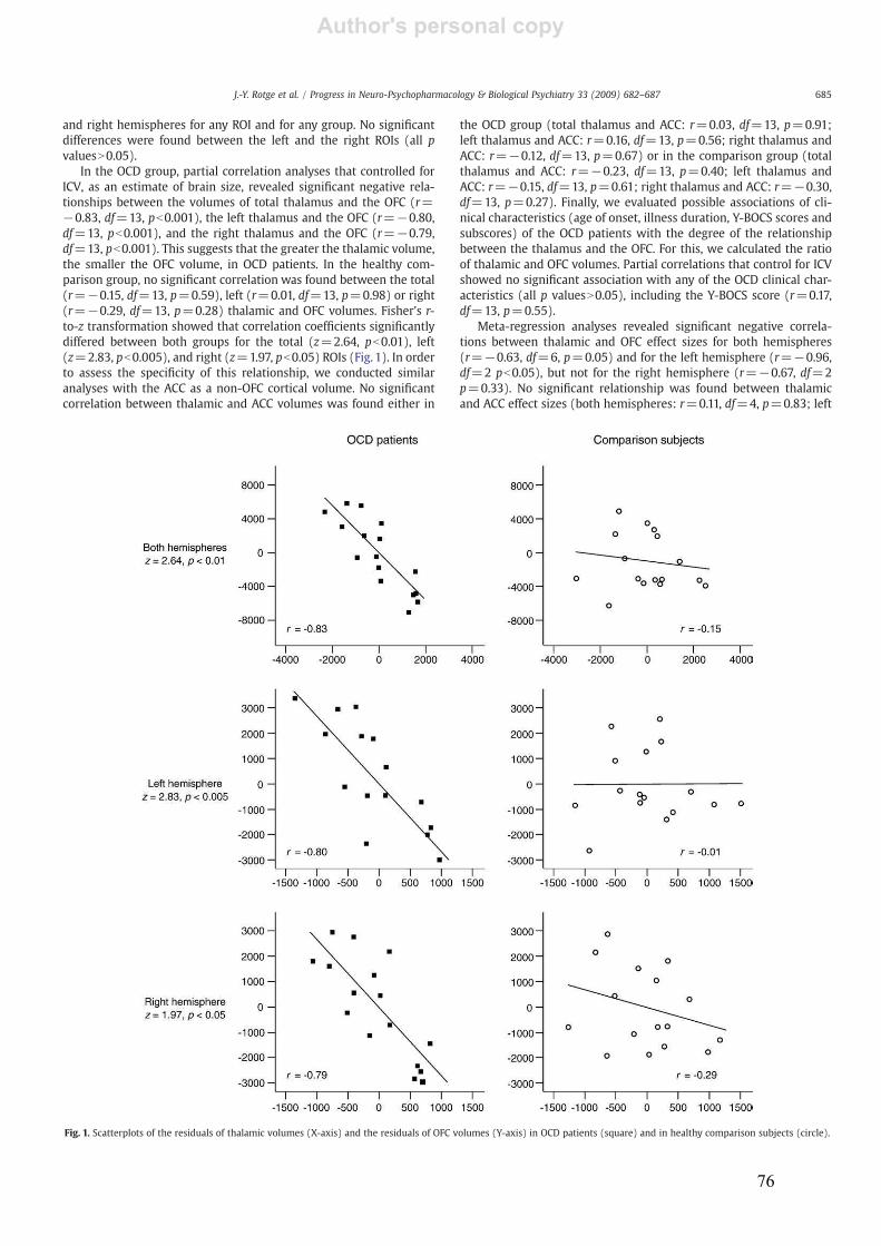
Author's personal copy
and right hemispheres for any ROI and for any group. No significant
differences were found between the left and the right ROIs (all p
valuesN0.05).
In the OCD group, partial correlation analyses that controlled for
ICV, as an estimate of brain size, revealed significant negative rela-
tionships between the volumes of total thalamus and the OFC (r=
−0.83, df=13, pb0.001), the left thalamus and the OFC (r=−0.80,
df=13, pb0.001), and the right thalamus and the OFC (r=−0.79,
df=13, pb0.001). This suggests that the greater the thalamic volume,
the smaller the OFC volume, in OCD patients. In the healthy com-
parison group, no significant correlation was found between the total
(r=−0.15, df=13, p=0.59), left (r=0.01, df=13, p=0.98) or right
(r=−0.29, df=13, p=0.28) thalamic and OFC volumes. Fisher's r-
to-z transformation showed that correlation coefficients significantly
differed between both groups for the total (z=2.64, pb0.01), left
(z=2.83, pb0.005), and right (z=1.97, pb0.05) ROIs (Fig. 1). In order
to assess the specificity of this relationship, we conducted similar
analyses with the ACC as a non-OFC cortical volume. No significant
correlation between thalamic and ACC volumes was found either in
the OCD group (total thalamus and ACC: r=0.03, df=13, p=0.91;
left thalamus and ACC: r=0.16, df=13, p=0.56; right thalamus and
ACC: r=−0.12, df=13, p=0.67) or in the comparison group (total
thalamus and ACC: r=−0.23, df=13, p=0.40; left thalamus and
ACC: r=−0.15, df=13, p=0.61; right thalamus and ACC: r=−0.30,
df=13, p=0.27). Finally, we evaluated possible associations of cli-
nical characteristics (age of onset, illness duration, Y-BOCS scores and
subscores) of the OCD patients with the degree of the relationship
between the thalamus and the OFC. For this, we calculated the ratio
of thalamic and OFC volumes. Partial correlations that control for ICV
showed no significant association with any of the OCD clinical char-
acteristics (all p valuesN0.05), including the Y-BOCS score (r=0.17,
df=13, p=0.55).
Meta-regression analyses revealed significant negative correla-
tions between thalamic and OFC effect sizes for both hemispheres
(r=−0.63, df=6, p=0.05) and for the left hemisphere (r=−0.96,
df=2 pb0.05), but not for the right hemisphere (r=−0.67, df=2
p=0.33). No significant relationship was found between thalamic
and ACC effect sizes (both hemispheres: r=0.11, df=4, p=0.83; left
Fig. 1. Scatterplots of the residuals of thalamic volumes (X-axis) and the residuals of OFC volumes (Y-axis) in OCD patients (square) and in healthy comparison subjects (circle).
685J.-Y. Rotge et al. / Progress in Neuro-Psychopharmacology & Biological Psychiatry 33 (2009) 682–687
76
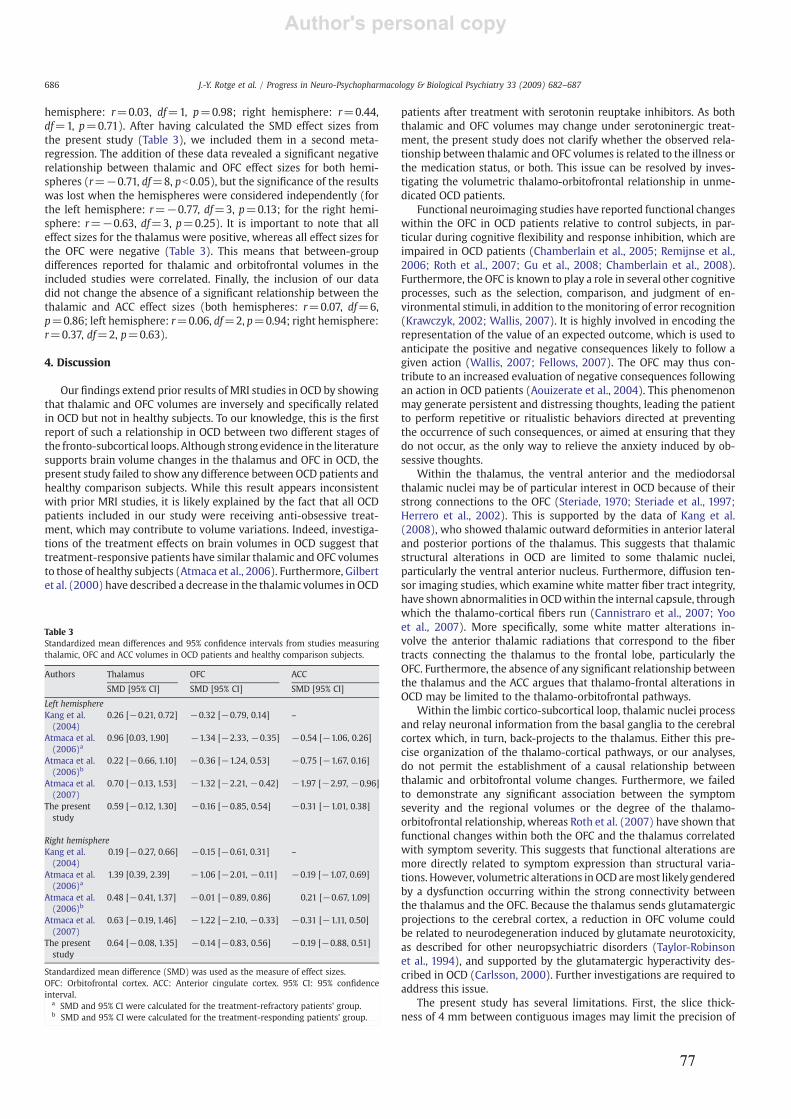
Author's personal copy
hemisphere: r=0.03, df=1, p=0.98; right hemisphere: r=0.44,
df=1, p=0.71). After having calculated the SMD effect sizes from
the present study (Table 3), we included them in a second meta-
regression. The addition of these data revealed a significant negative
relationship between thalamic and OFC effect sizes for both hemi-
spheres (r=−0.71, df=8, pb0.05), but the significance of the results
was lost when the hemispheres were considered independently (for
the left hemisphere: r=−0.77, df=3, p=0.13; for the right hemi-
sphere: r=−0.63, df=3, p=0.25). It is important to note that all
effect sizes for the thalamus were positive, whereas all effect sizes for
the OFC were negative (Table 3). This means that between-group
differences reported for thalamic and orbitofrontal volumes in the
included studies were correlated. Finally, the inclusion of our data
did not change the absence of a significant relationship between the
thalamic and ACC effect sizes (both hemispheres: r=0.07, df=6,
p=0.86; left hemisphere: r=0.06, df=2, p=0.94; right hemisphere:
r=0.37, df=2, p=0.63).
4. Discussion
Our findings extend prior results of MRI studies in OCD by showing
that thalamic and OFC volumes are inversely and specifically related
in OCD but not in healthy subjects. To our knowledge, this is the first
report of such a relationship in OCD between two different stages of
the fronto-subcortical loops. Although strong evidence in the literature
supports brain volume changes in the thalamus and OFC in OCD, the
present study failed to show any difference between OCD patients and
healthy comparison subjects. While this result appears inconsistent
with prior MRI studies, it is likely explained by the fact that all OCD
patients included in our study were receiving anti-obsessive treat-
ment, which may contribute to volume variations. Indeed, investiga-
tions of the treatment effects on brain volumes in OCD suggest that
treatment-responsive patients have similar thalamic and OFC volumes
to those of healthy subjects (Atmaca et al., 2006). Furthermore, Gilbert
et al. (2000) have described a decrease in the thalamic volumes in OCD
patients after treatment with serotonin reuptake inhibitors. As both
thalamic and OFC volumes may change under serotoninergic treat-
ment, the present study does not clarify whether the observed rela-
tionship between thalamic and OFC volumes is related to the illness or
the medication status, or both. This issue can be resolved by inves-
tigating the volumetric thalamo-orbitofrontal relationship in unme-
dicated OCD patients.
Functional neuroimaging studies have reported functional changes
within the OFC in OCD patients relative to control subjects, in par-
ticular during cognitive flexibility and response inhibition, which are
impaired in OCD patients (Chamberlain et al., 2005; Remijnse et al.,
2006; Roth et al., 2007; Gu et al., 2008; Chamberlain et al., 2008).
Furthermore, the OFC is known to play a role in several other cognitive
processes, such as the selection, comparison, and judgment of en-
vironmental stimuli, in addition to themonitoring of error recognition
(Krawczyk, 2002; Wallis, 2007). It is highly involved in encoding the
representation of the value of an expected outcome, which is used to
anticipate the positive and negative consequences likely to follow a
given action (Wallis, 2007; Fellows, 2007). The OFC may thus con-
tribute to an increased evaluation of negative consequences following
an action in OCD patients (Aouizerate et al., 2004). This phenomenon
may generate persistent and distressing thoughts, leading the patient
to perform repetitive or ritualistic behaviors directed at preventing
the occurrence of such consequences, or aimed at ensuring that they
do not occur, as the only way to relieve the anxiety induced by ob-
sessive thoughts.
Within the thalamus, the ventral anterior and the mediodorsal
thalamic nuclei may be of particular interest in OCD because of their
strong connections to the OFC (Steriade, 1970; Steriade et al., 1997;
Herrero et al., 2002). This is supported by the data of Kang et al.
(2008), who showed thalamic outward deformities in anterior lateral
and posterior portions of the thalamus. This suggests that thalamic
structural alterations in OCD are limited to some thalamic nuclei,
particularly the ventral anterior nucleus. Furthermore, diffusion ten-
sor imaging studies, which examine white matter fiber tract integrity,
have shown abnormalities in OCDwithin the internal capsule, through
which the thalamo-cortical fibers run (Cannistraro et al., 2007; Yoo
et al., 2007). More specifically, some white matter alterations in-
volve the anterior thalamic radiations that correspond to the fiber
tracts connecting the thalamus to the frontal lobe, particularly the
OFC. Furthermore, the absence of any significant relationship between
the thalamus and the ACC argues that thalamo-frontal alterations in
OCD may be limited to the thalamo-orbitofrontal pathways.
Within the limbic cortico-subcortical loop, thalamic nuclei process
and relay neuronal information from the basal ganglia to the cerebral
cortex which, in turn, back-projects to the thalamus. Either this pre-
cise organization of the thalamo-cortical pathways, or our analyses,
do not permit the establishment of a causal relationship between
thalamic and orbitofrontal volume changes. Furthermore, we failed
to demonstrate any significant association between the symptom
severity and the regional volumes or the degree of the thalamo-
orbitofrontal relationship, whereas Roth et al. (2007) have shown that
functional changes within both the OFC and the thalamus correlated
with symptom severity. This suggests that functional alterations are
more directly related to symptom expression than structural varia-
tions. However, volumetric alterations inOCD aremost likely gendered
by a dysfunction occurring within the strong connectivity between
the thalamus and the OFC. Because the thalamus sends glutamatergic
projections to the cerebral cortex, a reduction in OFC volume could
be related to neurodegeneration induced by glutamate neurotoxicity,
as described for other neuropsychiatric disorders (Taylor-Robinson
et al., 1994), and supported by the glutamatergic hyperactivity des-
cribed in OCD (Carlsson, 2000). Further investigations are required to
address this issue.
The present study has several limitations. First, the slice thick-
ness of 4 mm between contiguous images may limit the precision of
Table 3
Standardized mean differences and 95% confidence intervals from studies measuring
thalamic, OFC and ACC volumes in OCD patients and healthy comparison subjects.
Authors Thalamus OFC ACC
SMD [95% CI] SMD [95% CI] SMD [95% CI]
Left hemisphere
Kang et al.
(2004)
0.26 [−0.21, 0.72] −0.32 [−0.79, 0.14] –
Atmaca et al.
(2006)a0.96 [0.03, 1.90] −1.34 [−2.33, −0.35] −0.54 [−1.06, 0.26]
Atmaca et al.
(2006)b0.22 [−0.66, 1.10] −0.36 [−1.24, 0.53] −0.75 [−1.67, 0.16]
Atmaca et al.
(2007)
0.70 [−0.13, 1.53] −1.32 [−2.21, −0.42] −1.97 [−2.97, −0.96]
The present
study
0.59 [−0.12, 1.30] −0.16 [−0.85, 0.54] −0.31 [−1.01, 0.38]
Right hemisphere
Kang et al.
(2004)
0.19 [−0.27, 0.66] −0.15 [−0.61, 0.31] –
Atmaca et al.
(2006)a1.39 [0.39, 2.39] −1.06 [−2.01, −0.11] −0.19 [−1.07, 0.69]
Atmaca et al.
(2006)b0.48 [−0.41, 1.37] −0.01 [−0.89, 0.86] 0.21 [−0.67, 1.09]
Atmaca et al.
(2007)
0.63 [−0.19, 1.46] −1.22 [−2.10, −0.33] −0.31 [−1.11, 0.50]
The present
study
0.64 [−0.08, 1.35] −0.14 [−0.83, 0.56] −0.19 [−0.88, 0.51]
Standardized mean difference (SMD) was used as the measure of effect sizes.
OFC: Orbitofrontal cortex. ACC: Anterior cingulate cortex. 95% CI: 95% confidence
interval.a SMD and 95% CI were calculated for the treatment-refractory patients' group.b SMD and 95% CI were calculated for the treatment-responding patients' group.
686 J.-Y. Rotge et al. / Progress in Neuro-Psychopharmacology & Biological Psychiatry 33 (2009) 682–687
77
Author's personal copy
volumetric measurements. The thickness of the slices reflects the fact
that they were originally collected for a different purpose. Despite this
limitation, recent insight into volume changes in OCD (Rotge et al.,
2009) prompted us to use the tissue samples in the present study. The
lack of precision would have affected both groups equally, and was
mitigated by the fact that volumetric measurements were made in a
blindedmanner. Therefore, our findings are unlikely to be significantly
affected by the slice thickness limitation. Second, meta-regression
analyses were conducted with only four or five data points for the left
and right hemispheres. Therefore, results of such analyses should
be interpreted with caution, even if the regression coefficients are
remarkably high (r ranged from −0.77 to −0.63).
In conclusion, our findings show an inverse and specific relation-
ship between thalamic and OFC volumes in OCD, suggesting that
structural variations described within these brain regions are related
in OCD. However, owing to the small sample sizes of the included
subjects, these results are preliminary and should be replicated with
larger samples. Furthermore, wewere not able to explore the effects of
anti-obsessive treatments on the volumetric thalamo-orbitofrontal
relationship. These effects warrant investigation. Finally, no causal
relationship between OFC and thalamic volumes in OCD patients was
established in the present study. Resolving this issue should provide
us with a better understanding of OCD pathophysiology.
Acknowledgements
J-Y Rotge was supported by a grant from the Fondation pour la
Recherche Médicale (Medical Research Fundation).
Appendix A. Supplementary data
Supplementary data associated with this article can be found, in
the online version, at doi:10.1016/j.pnpbp.2009.03.011.
References
Aouizerate B, Guehl D, Cuny E, Rougier A, Bioulac B, Tignol J, et al. Pathophysiology ofobsessive–compulsive disorder: a necessary link between phenomenology,neuropsychology, imagery and physiology. Prog Neurobiol 2004;72:195–221.
Atmaca M, Yildirim H, Ozdemir H, Aydin A, Tezcan E, Ozler S. Volumetric MRIassessment of brain regions in patients with refractory obsessive–compulsivedisorder. Prog Neuro-psychoph 2006;30:1051–7.
Atmaca M, Yildirim H, Ozdemir H, Tezcan E, Poyraz AK. Volumetric MRI study of keybrain regions implicated in obsessive–compulsive disorder. Prog Neuro-psychoph2007;31:46–52.
Cannistraro PA, Makris N, Howard JD, Wedig MM, Hodge SM, Wilhelm S, et al. Adiffusion tensor imaging study of white matter in obsessive–compulsive disorder.Depress Anxiety 2007;24:440–6.
Carlsson ML. On the role of cortical glutamate in obsessive–compulsive disorder andattention-deficit hyperactivity disorder, two phenomenologically antitheticalconditions. Acta Psychiatr Scand 2000;102:401–13.
Chamberlain SR, Blackwell AD, Fineberg NA, Robbins TW, Sahakian BJ. The neuropsy-chology of obsessive compulsive disorder: the importance of failures in cognitiveand behavioural inhibition as candidate endophenotypic markers. NeurosciBiobehav Rev 2005;29:399–419.
Chamberlain SR, Menzies L, Hampshire A, Suckling J, Fineberg NA, del Campo N, et al.Orbitofrontal dysfunction in patients with obsessive–compulsive disorder and theirunaffected relatives. Science 2008;321:421–2.
Fellows LK. Advances in understanding ventromedial prefrontal function: the accountantjoins the executive. Neurology 2007;68:991–5.
Gilbert AR,MooreGJ, KeshavanMS, Paulson LAD, Narula V,MacMaster FP, et al. Decreasein thalamic volumes of pediatric patients with obsessive–compulsive disorder whoare taking paroxetine. Arch Gen Psychiatry 2000;57:449–56.
Goodman WK, Price LH, Rasmussen SA, Mazure C, Fleischmann RL, Hill CL, et al. TheYale–Brown Obsessive Compulsive Scale, I: development, use and reliability. ArchGen Psychiatry 1989;46:1006–11.
Gu BM, Park JY, Kang DH, Lee SJ, Yoo SY, Jo HJ, et al. Neural correlates of cognitiveinflexibility during task-switching in obsessive–compulsive disorder. Brain 2008;131:155–64.
Hamilton M. Development of a rating scale for primary depressive illness. Br J Soc ClinPsychol 1967;6:278–96.
HamiltonM. The assessment of anxiety states by rating. Br J Med Psychol 1959;32:50–5.Herrero MT, Barcia C, Navarro JM. Functional anatomy of thalamus and basal ganglia.
Child's Nerv Syst 2002;18:386–404.Kang DH, Kim JJ, Choi JS, Kim YI, Kim CW, Youn T, et al. Volumetric investigation of
the frontal-subcortical circuitry in patients with obsessive–compulsive disorder.J Neuropsychiatry Clin Neurosci 2004;16:342–9.
Kang DH, Kim SH, Kim CW, Choi JS, Jang JH, Jung MH, Lee, et al. Thalamus surfaceshape deformity in obsessive–compulsive disorder and schizophrenia. Neuroreport2008;19:609–13.
Krawczyk DC. Contributions of the prefrontal cortex to the neural basis of humandecisions making. Neurosci Biobehav Rev 2002;26:631–64.
Maffei C, Fossati A, Agostoni I, Barraco A, Bagnato M, Deborah D, et al. Interrater reliabilityand internal consistency of the structured clinical interview for DSM-IV axis IIpersonality disorders (SCID-II), version 2.0. J Personal Disord 1997;11:279–84.
Menzies L, Chamberlain SR, Laird AR, Thelen SM, Sahakian BJ, Bullmore ET. Integratingevidence from neuroimaging and neuropsychological studies of obsessive–compulsive disorder: the orbitofronto-striatal model revisited. Neurosci BiobehavRev 2008;32:525–49.
Remijnse PL, Nielen MM, van Balkom AJ, Cath DC, van Oppen P, Uylings HB, et al.Reduced orbitofrontal–striatal activity on a reversal learning task in obsessive–compulsive disorder. Arch Gen Psychiatry 2006;63:1225–36.
Rosenberg DR, Keshavan MS, O'Hearn KM, Dick EL, Bagwell WW, Seymour AB, et al.Frontostriatal measurement in treatment-naïve children with obsessive–compulsivedisorder. Arch Gen Psychiatry 1997;54:824–30.
Rotge JY, Guehl D, Dilharreguy B, Cuny E, Tignol J, Bioulac B, et al. Provocation of obsessive–compulsive symptoms: a quantitative voxel-basedmeta-analysis of functional neuro-imaging studies. J Psychiatry Neurosci 2008;33:405–12.
Rotge JY, Guehl D, Dilharreguy B, Tignol J, Bioulac B, AllardM, et al.Meta-analysis of brainvolume changes in obsessive–compulsive disorder. Biol Psychiatry 2009;65:75–83.
Roth RM, SaykinAJ, Flashman LA, PixleyHS,West JD,MamourianAC. Event-related fMRI ofresponse inhibition in obsessive–compulsive disorder. Biol Psychiatry 2007;62:901–9.
Sheehan DV, Lecrubier Y, Sheehan KH, Amorim P, Janavs J, Weiller E, et al. The Mini-International Neuropsychiatric Interview (M.I.N.I.): the development and valida-tion of a structured diagnostic psychiatric interview for DSM-IV and ICD-10. J ClinPsychiatry 1998;59:22–33.
SteriadeM. Ascending control of thalamic and cortical responsiveness. Int RevNeurobiol1970;12:87-144.
Steriade M, Jones EG, McCormick DA. Thalamus. Volume I: organisation and function.Oxford: Elsevier Science Ltd; 1997.
Szeszko PR, Robinson D, Alvir JMJ, Bilder RM, Lencz T, Ashtari M, et al. Orbital frontal andamygdala volume reductions in obsessive–compulsive disorder. Arch Gen Psychia-try 1999;56:913–9.
Taylor-Robinson SD, Weeks RA, Sargentoni J, Marcus CD, Bryant DJ, Harding AE, et al.Evidence for glutamate excitotoxicity in Huntington's diseasewith protonmagneticresonance spectroscopy. Lancet 1994;343:1170.
Wallis JD. Orbitofrontal cortex and its contribution to decision-making. AnnuRevNeurosci2007;30:31–56.
Yoo SY, Jang JH, Shin YW, Kim DJ, Park HJ, MoonWJ, et al. White matter abnormalities indrug-naïve patients with obsessive–compulsive disorder: a diffusion tensor studybefore and after citalopram treatment. Acta Psychiatr Scand 2007;116:211–9.
687J.-Y. Rotge et al. / Progress in Neuro-Psychopharmacology & Biological Psychiatry 33 (2009) 682–687
78
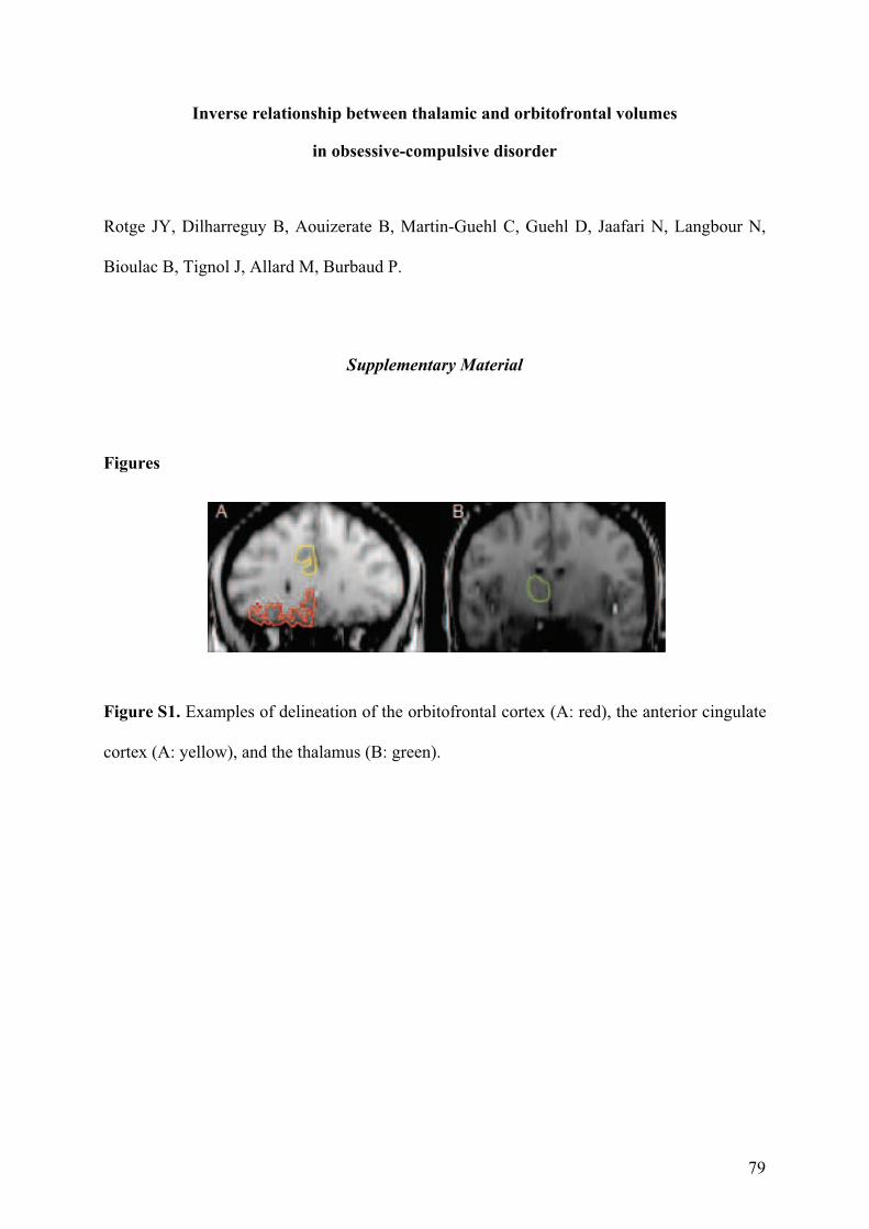
79
Inverse relationship between thalamic and orbitofrontal volumes
in obsessive-compulsive disorder
Rotge JY, Dilharreguy B, Aouizerate B, Martin-Guehl C, Guehl D, Jaafari N, Langbour N,
Bioulac B, Tignol J, Allard M, Burbaud P.
Supplementary Material
Figures
Figure S1. Examples of delineation of the orbitofrontal cortex (A: red), the anterior cingulate
cortex (A: yellow), and the thalamus (B: green).

80
81
4. Altérations fonctionnelles des voies thalamo-corticales dans le TOC
4.1. Modifications fonctionnelles lors de la provocation des symptômes
4.1.1. Introduction
Plusieurs études d’imagerie fonctionnelle ont rapporté des modifications fonctionnelles
dans le TOC à travers différents paradigmes expérimentaux. L’activité fonctionnelle à l’état
dit « de repos » est modifiée chez les patients souffrant de TOC comparativement aux
témoins, en particulier il a été rapporté une hyperactivité du COF, du CCA, du thalamus et
des ganglions de la base (Menzies et al., 2008 ; Kwon et al., 2009). Cependant, le paradigme
visant à provoquer des symptômes obsessionnels et compulsifs chez les patients semble être
plus pertinent pour identifier les réseaux neuraux fonctionnellement impliqués dans
l’expression de la symptomatologie. La plupart des études d’imagerie fonctionnelle basée sur
la provocation des symptômes a révélé l’importance du rôle des boucles cortico-sous-
corticales prenant naissance au sein du COF et du CCA. Toutefois, l’implication fonctionnelle
de nombreuses autres régions cérébrales comme le cortex temporal a été rapportée de façon
moins systématique et plus inconsistante. Dans le but de proposer une vision objective et
quantitative de ces résultats, nous avons réalisé une méta-analyse des études utilisant le
paradigme de provocation des symptômes obsessionnels et compulsifs.
4.1.2. Méthodes
Nous avons utilisé la méthode ALE décrite précédemment (page 57). Après une
recherche systématique par mots-clé pour identifier les études d’imagerie fonctionnelle, nous
avons extrait les coordonnées de chaque focus, ainsi que l’intensité et le volume de chaque
cluster.
4.1.3. Résultats et discussion
Les résultats montrent des activations significatives dans le COF, le CCA et le thalamus
lors de la provocation de symptômes obsessionnels et compulsifs chez les patients avec un
TOC. Des activations significatives sont également retrouvées pour les cortex temporo-
pariétal et préfrontal dorsolatéral gauches. Ces résultats suggèrent l’implication des COF,
CCA et thalamus dans la médiation des symptômes obsessionnels et compulsifs. Les régions
pariétales et préfrontales dorso-latérales connues pour participer au détournement actif et
82
volontaire de l’attention vers un autre objet pourraient participer aux efforts déployés par les
patients pour résister aux processus obsessionnels induits. Une analyse en « leave-one-out »
consistant à exclure de l’analyse une à une les différentes études inclues montre que ces
résultats ne sont pas influencés par une seule étude, à l’exception des données inclues dans
l’étude de Mataix-Cols et al., la seule qui rapporte des contrastes inter-groupes. L’exclusion
de cette étude met en évidence des activations significatives dans d’autres régions cérébrales
(cortex insulaire, cortex cingulaire postérieur, cortex pariétal et noyau caudé). Cependant, ces
différences doivent être considérées avec prudence car aucun cluster correspondant n’excède
10 mm3.
4.1.4. Conclusion
Cette méta-analyse confirme la participation fonctionnelle des boucles préfronto-sous-
corticales du COF et du CCA lors de la provocation des symptômes obsessionnels et
compulsifs. Elle souligne également la participation d’un réseau préfronto-dorso-pariétal qui
jouerait un possible rôle dans les efforts faits pour résister aux symptômes.
Rotgé JY, Guehl D, Dilharreguy B, Cuny E, Tignol J, Bioulac B, Allard M,
Burbaud P, Aouizerate B. Provocation of obsessive-compulsive symptoms: a
quantitative voxel-based meta-analysis of functional neuroimaging studies. Journal
of Psychiatry & Neuroscience 2008;33:405-12.
J Psychiatry Neurosci 2008;33(5) 405
Objective: Recent functional magnetic resonance imaging (fMRI) and positron emission tomography (PET) studies based on the symp-
tom provocation paradigm have explored neural correlates of the cognitive and emotional processes associated with the emergence of
obsessive–compulsive disorder (OCD) symptoms. Although most studies showed the involvement of cortico–subcortical loops originat-
ing in the orbitofrontal cortex and the anterior cingulate cortex, an increased activity within numerous other regions of the brain has in-
consistently been reported across studies. To provide a quantitative estimation of the cerebral activation patterns related to the perform-
ance of the symptom provocation task by OCD patients, we conducted a voxel-based meta-analysis. Methods: We searched the
PubMed and MEDLINE databases for studies that used fMRI and PET and that were based on the symptom provocation paradigm. We
entered data into a paradigm-driven activation likelihood estimation meta-analysis. Results: We found significant likelihoods of activation
in cortical and subcortical regions of the orbitofrontal and anterior cingulate loops. The left dorsal frontoparietal network, including the
dorsolateral prefrontal cortex and precuneus, and the left superior temporal gyrus also demonstrated significant likelihoods of activation.
Conclusion: Consistent results across functional neuroimaging studies suggest that the orbitofrontal and anterior cingulate cortices are
involved in the mediation of obsessive–compulsive symptoms. Based on recent literature, we suggest that activations within the dorsal
frontoparietal network might be related to patients’ efforts to resist the obsessive processes induced by the provocation task. Further re-
search should elucidate the specific neural correlates of the various cognitive and emotional functions altered in OCD.
Objectif : De récentes études d’imagerie par résonance magnétique fonctionnelle (IRMf) et de tomographie par émission de positrons
(TEP) utilisant un paradigme de provocation de symptômes ont exploré les corrélats neuronaux des processus cognitifs et affectifs asso-
ciés à l’apparition de symptômes du trouble obsessionnel–compulsif (TOC). Même si la plupart des études ont révélé l’importance du rôle
de boucles corticales-sous-corticales émanant du cortex orbitofrontal et du cortex cingulaire antérieur, une augmentation de l’activité dans
de nombreuses autres régions du cerveau a été rapportée de façon moins systématique. Afin de produire une estimation quantitative des
activations cérébrales liées à l’exécution de tâches de provocation de symptômes par des patients atteints de TOC, nous avons procédé à
une méta-analyse des données de la littérature. Méthodes : Nous avons effectué dans les bases de données PubMed et MEDLINE une
recherche d’études ayant utilisé l’IRMf et la TEP et qui reposaient sur le paradigme de provocation des symptômes. Nous avons entré les
données dans une méta-analyse d’estimation de la probabilité d’activation. Résultats : Nous avons constaté des probabilités importantes
d’activation dans les régions corticales et sous-corticales des boucles des cortex orbitofrontale et cingulaire antérieure. L’étude a aussi
révélé des probabilités importantes d’activation d’un réseau préfronto–dorso–pariétal gauche, incluant le cortex préfrontal dorsolatéral et le
précunéus, ainsi que de le gyrus temporal supérieur gauche. Conclusion : Les résultats obtenus indiquent que les cortex orbitofrontal et
Review PaperExamen critique
Provocation of obsessive–compulsive symptoms: a quantitative voxel-based meta-analysis of functional
neuroimaging studies
Jean-Yves Rotge, MD; Dominique Guehl, MD, PhD; Bixente Dilharreguy, PhD;Emmanuel Cuny, MD, PhD; Jean Tignol, MD; Bernard Bioulac, MD, PhD;
Michele Allard, MD, PhD; Pierre Burbaud, MD, PhD; Bruno Aouizerate, MD, PhD
Rotge, Guehl, Cuny, Bioulac, Burbaud, Aouizerate — Laboratoire Mouvement Adaptation Cognition, Université Bordeaux 2;
Rotge, Tignol, Aouizerate — Service Universitaire de Psychiatrie, Centre Hospitalier Charles Perrens; Guehl, Bioulac,
Burbaud — Service de Neurophysiologie Clinique, Centre Hospitalier Universitaire Pellegrin; Dilharreguy, Allard — Laboratoire
d’Imagerie Moléculaire et Fonctionnelle, Université Bordeaux 2; Cuny — Service de Neurochirurgie; Allard — Service de
Médecine Nucléaire, Centre Hospitalier Universitaire Pellegrin, Bordeaux, France
Correspondence to: Dr. J.-Y. Rotge, Laboratoire Mouvement Adaptation Cognition, CNRS UMR 5227, Université Bordeaux 2,146 rue Léo-Saignat, 33076 Bordeaux, France; fax (33) 05 56 90 14 21; [email protected]
J Psychiatry Neurosci 2008;33(5):405–12.
Medical subject headings: obsessive-compulsive disorder; magnetic resonance imaging; positron-emission tomography.
Submitted Aug. 7, 2007; Revised Nov. 29, 2007; Accepted Jan. 14, 2008
© 2008 Canadian Medical Association
83
Rotge et al.
406 Rev Psychiatr Neurosci 2008;33(5)
cingulaire antérieure jouent un rôle dans la médiation des symptômes obsessionnels et compulsifs. Compte tenu de publications récentes,
nous suggérons que les activations observées dans le réseau préfronto–dorso–pariétal dorsal pourraient être liées aux efforts déployés
par les patients pour résister aux processus obsessionnels induits par la provocation de symptômes. D’autres recherches devraient clari-
fier les corrélats neuronux spécifiquement liés aux diverses fonctions cognitives et affectives affectées dans le TOC.
Introduction
Obsessive–compulsive disorder (OCD) is a chronic andhighly disabling condition that affects 2%–3% of the generalpopulation. This anxiety disorder is characterized by thepresence of obsession in the form of intrusive and distressingthoughts, ideas or images, and by the urge to perform repeti-tive or ritualistic behaviours known as compulsions.1,2 It hasbeen proposed that obsessions are related to the increasedevaluation of negative consequences following an action thatan individual with OCD wrongly interprets as erroneous orfaulty. This persistent high “error signal,” which is consid-ered to be the core of OCD symptomatology, may initiate thepathological doubt that is usually associated with increasedanxiety.2–5 Compulsions may then occur as behavioural out-puts aiming to relieve or to neutralize the anxiety induced bythe obsessional process. The relief that may be equated with aform of reward is only transient because performed behav-iours do not definitively stop the “error signal,” which leadsto a cyclic reproduction of these behaviours.2 This phenom-enologic view suggests that several cognitive processes suchas error detection, together with emotional and motivationalfunctions, may be altered in OCD.2,5
Remarkable advances in functional neuroimaging have re-cently provided results that showed the participation of sev-eral regions of the brain in the cognitive and emotionalprocesses altered in OCD, and their overactivity in patientswith the disorder.2,6 Indeed, most neuroimaging studies havereported an increased functional activity in the cortico–striato–pallido–thalamo–cortical circuits originating withinboth the orbitofrontal and the anterior cingulate cortices inOCD patients compared with individuals without the disor-der. Furthermore, this basal cortico–subcortical overactivitymay be reduced in conjunction with the clinical improvementobtained with efficient treatment.2,7 A recent meta-analysisbased on the calculation of regional effect sizes across neuro-imaging studies that compared functional activity in patientswith OCD and healthy individuals found consistent and sig-nificant changes in the left orbitofrontal cortex and bilateralcaudate nucleus.8 However, a meta-analysis using the activa-tion likelihood estimation method may provide further in-sight, especially regarding the possible roles of other regionsof the brain in the pathophysiology of OCD. Voxel-basedanalysis, which allows a high spatial level of distinction,quantitatively evaluates converging activation foci acrossfunctional imaging studies by modelling each activation focusas the centre of a normal probability distribution.9 Moreover,recent neuroimaging studies involving patients with OCDhave focused on the symptom provocation task, which entailsthe presentation of obsessional stimuli leading to the produc-
tion of OCD symptoms. Such experimental protocols seem tobe more relevant to elicit neural patterns of activations relatedto the emergence of obsessive–compulsive symptoms thanparadigms based on measuring functional activity during a“resting state.” We conducted a paradigm-driven meta-analysis using the activation likelihood estimation method toprovide a complete overview of the cerebral networks activatedduring the symptom provocation task in patients with OCD.
Methods
Literature search and study selection
We searched the PubMed and MEDLINE databases for stud-ies that used functional magnetic resonance imaging (fMRI)and positron emission tomography (PET) to examine brainactivity during a symptom provocation task in patients withOCD. Our keywords for the literature search included“obsessive–compulsive disorder” and any of the followingterms: “functional magnetic resonance imaging,” “positronemission tomography” and “symptom provocation task.” Toobtain additional papers, we manually searched the referencelists of the studies we retrieved.
We included studies in our analysis if they were publishedin English in a peer-reviewed journal, were restricted to fMRIand PET (C15O2, H2
15O and 15O2) scanning techniques thatmeasure highly interrelated biological phenomena,10 includedpatients with a primary diagnosis of OCD, were based on asymptom provocation paradigm involving the presentationof obsessional stimuli, described activations at a conventionallevel of significance and reported the coordinates of activa-tion maxima in a standardized stereotactic space.
One author (J.-Y.R.) identified all articles (n = 8)11–18 thatwere published before July 2007 and met our inclusion criter-ia. Another (B.A.) independently verified and confirmed thestudy selection.
Data abstraction, coding and correction
We abstracted the coordinates and the volumes of each of102 activation foci and the corresponding statistical valuesfrom conditional contrasts (“symptom provocation condition”minus “neutral condition”). We entered the data into Submitsoftware (BrainMap, University of Texas).19 Because data onconditional contrasts were unavailable for patients with OCDin the study by Mataix-Cols and colleagues, we consideredthe between-group contrast (“patients with OCD” minus “in-dividuals without OCD”).15 To renormalize coordinates spa-tially into a single stereotactic space, we converted MontréalNeurological Institute coordinates to Talairach coordinates
84
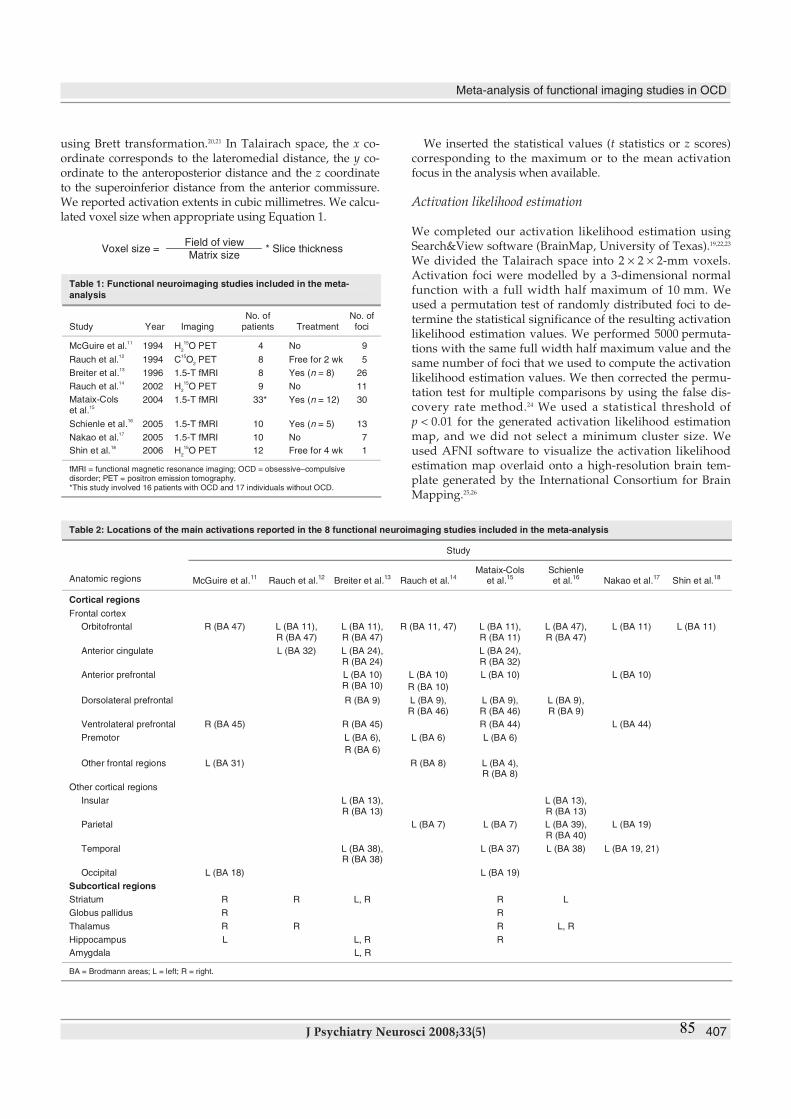
Meta-analysis of functional imaging studies in OCD
J Psychiatry Neurosci 2008;33(5) 407
using Brett transformation.20,21 In Talairach space, the x co-ordinate corresponds to the lateromedial distance, the y co-ordinate to the anteroposterior distance and the z coordinateto the superoinferior distance from the anterior commissure.We reported activation extents in cubic millimetres. We calcu-lated voxel size when appropriate using Equation 1.
We inserted the statistical values (t statistics or z scores)corresponding to the maximum or to the mean activationfocus in the analysis when available.
Activation likelihood estimation
We completed our activation likelihood estimation usingSearch&View software (BrainMap, University of Texas).19,22,23
We divided the Talairach space into 2 × 2 × 2-mm voxels.Activation foci were modelled by a 3-dimensional normalfunction with a full width half maximum of 10 mm. Weused a permutation test of randomly distributed foci to de-termine the statistical significance of the resulting activationlikelihood estimation values. We performed 5000 permuta-tions with the same full width half maximum value and thesame number of foci that we used to compute the activationlikelihood estimation values. We then corrected the permu-tation test for multiple comparisons by using the false dis-covery rate method.24 We used a statistical threshold ofp < 0.01 for the generated activation likelihood estimationmap, and we did not select a minimum cluster size. Weused AFNI software to visualize the activation likelihoodestimation map overlaid onto a high-resolution brain tem-plate generated by the International Consortium for BrainMapping.25,26
Field of viewVoxel size =
Matrix size* Slice thickness
Table 1: Functional neuroimaging studies included in the meta-analysis
Study Year ImagingNo. of
patients TreatmentNo. offoci
McGuire et al.11
1994 H2
15O PET 4 No 9
Rauch et al.12
1994 C15O
2PET 8 Free for 2 wk 5
Breiter et al.13
1996 1.5-T fMRI 8 Yes (n = 8) 26
Rauch et al.14
2002 H2
15O PET 9 No 11
Mataix-Colset al.
15
2004 1.5-T fMRI 33* Yes (n = 12) 30
Schienle et al.16
2005 1.5-T fMRI 10 Yes (n = 5) 13
Nakao et al.17
2005 1.5-T fMRI 10 No 7
Shin et al.18
2006 H2
15O PET 12 Free for 4 wk 1
fMRI = functional magnetic resonance imaging; OCD = obsessive–compulsivedisorder; PET = positron emission tomography.*This study involved 16 patients with OCD and 17 individuals without OCD.
Table 2: Locations of the main activations reported in the 8 functional neuroimaging studies included in the meta-analysis
Study
Anatomic regions McGuire et al.11
Rauch et al.12
Breiter et al.13
Rauch et al.14
Mataix-Colset al.
15Schienleet al.
16Nakao et al.
17Shin et al.
18
Cortical regions
Frontal cortex
Orbitofrontal R (BA 47) L (BA 11),R (BA 47)
L (BA 11),R (BA 47)
R (BA 11, 47) L (BA 11),R (BA 11)
L (BA 47),R (BA 47)
L (BA 11) L (BA 11)
Anterior cingulate L (BA 32) L (BA 24),R (BA 24)
L (BA 24),R (BA 32)
Anterior prefrontal L (BA 10)R (BA 10)
L (BA 10)
R (BA 10)
L (BA 10) L (BA 10)
Dorsolateral prefrontal R (BA 9) L (BA 9),R (BA 46)
L (BA 9),R (BA 46)
L (BA 9),R (BA 9)
Ventrolateral prefrontal R (BA 45) R (BA 45) R (BA 44) L (BA 44)
Premotor L (BA 6),
R (BA 6)
L (BA 6) L (BA 6)
Other frontal regions L (BA 31) R (BA 8) L (BA 4),R (BA 8)
Other cortical regions
Insular L (BA 13),R (BA 13)
L (BA 13),R (BA 13)
Parietal L (BA 7) L (BA 7) L (BA 39),R (BA 40)
L (BA 19)
Temporal L (BA 38),R (BA 38)
L (BA 37) L (BA 38) L (BA 19, 21)
Occipital L (BA 18) L (BA 19)
Subcortical regions
Striatum R R L, R R L
Globus pallidus R R
Thalamus R R R L, R
Hippocampus L L, R R
Amygdala L, R
BA = Brodmann areas; L = left; R = right.
85
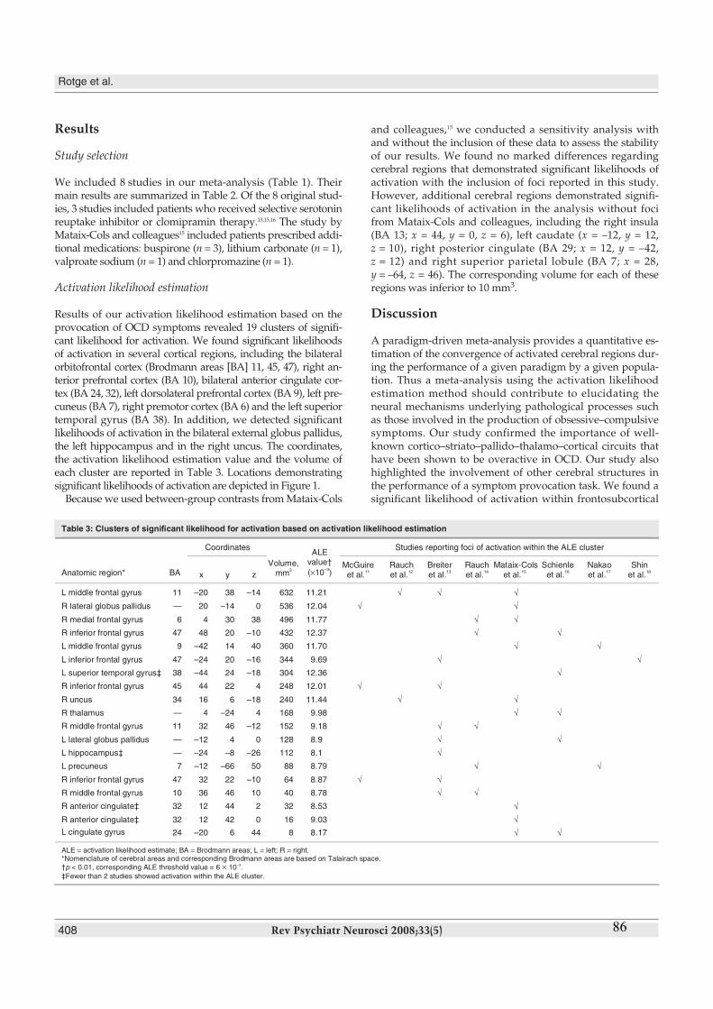
Results
Study selection
We included 8 studies in our meta-analysis (Table 1). Theirmain results are summarized in Table 2. Of the 8 original stud-ies, 3 studies included patients who received selective serotoninreuptake inhibitor or clomipramin therapy.13,15,16 The study byMataix-Cols and colleagues15 included patients prescribed addi-tional medications: buspirone (n = 3), lithium carbonate (n = 1),valproate sodium (n = 1) and chlorpromazine (n = 1).
Activation likelihood estimation
Results of our activation likelihood estimation based on theprovocation of OCD symptoms revealed 19 clusters of signifi-cant likelihood for activation. We found significant likelihoodsof activation in several cortical regions, including the bilateralorbitofrontal cortex (Brodmann areas [BA] 11, 45, 47), right an-terior prefrontal cortex (BA 10), bilateral anterior cingulate cor-tex (BA 24, 32), left dorsolateral prefrontal cortex (BA 9), left pre-cuneus (BA 7), right premotor cortex (BA 6) and the left superiortemporal gyrus (BA 38). In addition, we detected significantlikelihoods of activation in the bilateral external globus pallidus,the left hippocampus and in the right uncus. The coordinates,the activation likelihood estimation value and the volume ofeach cluster are reported in Table 3. Locations demonstratingsignificant likelihoods of activation are depicted in Figure 1.
Because we used between-group contrasts from Mataix-Cols
and colleagues,15 we conducted a sensitivity analysis withand without the inclusion of these data to assess the stabilityof our results. We found no marked differences regardingcerebral regions that demonstrated significant likelihoods ofactivation with the inclusion of foci reported in this study.However, additional cerebral regions demonstrated signifi-cant likelihoods of activation in the analysis without focifrom Mataix-Cols and colleagues, including the right insula(BA 13; x = 44, y = 0, z = 6), left caudate (x = –12, y = 12,z = 10), right posterior cingulate (BA 29; x = 12, y = –42,z = 12) and right superior parietal lobule (BA 7; x = 28,y = –64, z = 46). The corresponding volume for each of theseregions was inferior to 10 mm3.
Discussion
A paradigm-driven meta-analysis provides a quantitative es-timation of the convergence of activated cerebral regions dur-ing the performance of a given paradigm by a given popula-tion. Thus a meta-analysis using the activation likelihoodestimation method should contribute to elucidating theneural mechanisms underlying pathological processes suchas those involved in the production of obsessive–compulsivesymptoms. Our study confirmed the importance of well-known cortico–striato–pallido–thalamo–cortical circuits thathave been shown to be overactive in OCD. Our study alsohighlighted the involvement of other cerebral structures inthe performance of a symptom provocation task. We found asignificant likelihood of activation within frontosubcortical
Rotge et al.
408 Rev Psychiatr Neurosci 2008;33(5)
Table 3: Clusters of significant likelihood for activation based on activation likelihood estimation
Coordinates Studies reporting foci of activation within the ALE cluster
Anatomic region* BA x y z
Volume,mm
3
ALEvalue†
(×10–3
)McGuireet al.
11
Rauchet al.
12
Breiteret al.
13
Rauchet al.
14
Mataix-Colset al.
15
Schienleet al.
16
Nakaoet al.
17
Shinet al.
18
L middle frontal gyrus 11 –20 38 –14 632 11.21 √ √ √
R lateral globus pallidus — 20 –14 0 536 12.04 √ √
R medial frontal gyrus 6 4 30 38 496 11.77 √ √
R inferior frontal gyrus 47 48 20 –10 432 12.37 √ √
L middle frontal gyrus 9 –42 14 40 360 11.70 √ √
L inferior frontal gyrus 47 –24 20 –16 344 9.69 √ √
L superior temporal gyrus‡ 38 –44 24 –18 304 12.36 √
R inferior frontal gyrus 45 44 22 4 248 12.01 √ √
R uncus 34 16 6 –18 240 11.44 √ √
R thalamus — 4 –24 4 168 9.98 √ √
R middle frontal gyrus 11 32 46 –12 152 9.18 √ √
L lateral globus pallidus — –12 4 0 128 8.9 √ √
L hippocampus‡ — –24 –8 –26 112 8.1 √
L precuneus 7 –12 –66 50 88 8.79 √ √
R inferior frontal gyrus 47 32 22 –10 64 8.87 √ √
R middle frontal gyrus 10 36 46 10 40 8.78 √ √
R anterior cingulate‡ 32 12 44 2 32 8.53 √
R anterior cingulate‡ 32 12 42 0 16 9.03 √
L cingulate gyrus 24 –20 6 44 8 8.17 √ √
ALE = activation likelihood estimate; BA = Brodmann areas; L = left; R = right.*Nomenclature of cerebral areas and corresponding Brodmann areas are based on Talairach space.†p < 0.01, corresponding ALE threshold value = 6 × 10⁻³.
‡Fewer than 2 studies showed activation within the ALE cluster.
86
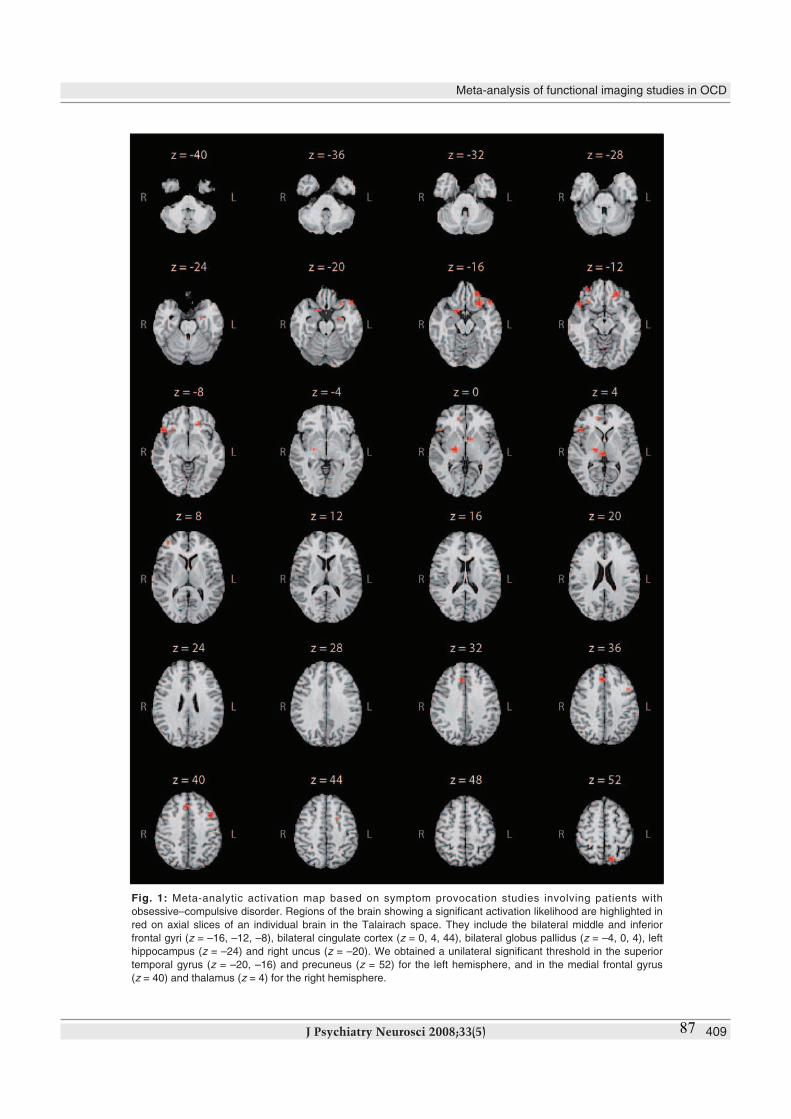
Meta-analysis of functional imaging studies in OCD
J Psychiatry Neurosci 2008;33(5) 409
Fig. 1: Meta-analytic activation map based on symptom provocation studies involving patients with
obsessive–compulsive disorder. Regions of the brain showing a significant activation likelihood are highlighted in
red on axial slices of an individual brain in the Talairach space. They include the bilateral middle and inferior
frontal gyri (z = –16, –12, –8), bilateral cingulate cortex (z = 0, 4, 44), bilateral globus pallidus (z = –4, 0, 4), left
hippocampus (z = –24) and right uncus (z = –20). We obtained a unilateral significant threshold in the superior
temporal gyrus (z = –20, –16) and precuneus (z = 52) for the left hemisphere, and in the medial frontal gyrus
(z = 40) and thalamus (z = 4) for the right hemisphere.
87
loops with high concordance across included studies. Wealso found significant results in unexpected regions of thebrain, such as the temporal and parietal cortices.
The orbitofrontal cortex is commonly distinguished by itsmedial and lateral parts.27 The medial region extensively con-nects with the paralimbic–limbic system, including the insu-lar cortex, cingulate gyrus, amygdala and hypothalamus.27–30
This area is highly involved in encoding the representation ofthe value of an expected outcome, which is used to anticipatethe positive and negative consequences that are likely to fol-low a given action.28,30 The lateral orbital region is more con-nected to higher neocortical systems, particularly the dorso-lateral prefrontal cortex. The lateral orbital region is thoughtto play a role in several cognitive processes, such as the selec-tion, comparison and judgment of environmental stimuli, butalso in the monitoring of error recognition.29,30 These orbito-frontal regions, which are known to be overactive in patientswith OCD, might contribute to an increased evaluation of theoccurrence of negative consequences following an action.2,8
This phenomenon might generate obsessive thoughts thatlead to repetitive or ritualistic behaviours intended to pre-vent negative consequences as a way to relieve anxiety.2
Different versions of the Stroop paradigm have largelycontributed to distinguishing a cognitive division (the mostdorsal part) and an affective division (the most ventral part)of the anterior cingulate cortex.31,32 Several neuroimagingstudies have shown that the cognitive division is involved inmonitoring conflict and detecting error, whereas the affectivedivision is involved in processing emotion, including the as-sessment of emotional information and the regulation ofemotional responses.32 The cognitive division has extensiveconnections with the dorsolateral prefrontal and the parietalcortices, as well as with the primary, premotor and supple-mentary motor areas. The affective division projects moredirectly to the orbitofrontal cortex, amygdala, nucleus accum-bens, hypothalamus and hippocampus, which demonstrateda significant likelihood of activation in our analysis.33 Over-activity in the cognitive division, as observed in patients withOCD, may be involved in the dysfunctional monitoring ofhigh-conflict situations, placing the patient in a potential pos-ition to make an error. Overactivity may also be involved inthe increased evaluation among patients with OCD of actionsthey wrongly interpret as erroneous or faulty.2,34,35 Therefore,the cognitive division seems to be involved in the generationof the high “error signal” that is considered to be the core ofOCD symptomatology. On the other hand, because of itsclosed connections with the limbic system, the affective div-ision might mediate the clinical expression of anxiety symp-toms associated with the obsessional process.2–5
The orbitofrontal and anterior cingulate cortices havestrong connections through the ventral striatum, the pal-lidum and the medial thalamus. The different levels of thesecortico–subcortical loops are thought to be involved in thepathogenesis of OCD. According to our results, the externalglobus pallidus might play a direct role in the production ofobsessive–compulsive symptoms; however, the mechanismsby which it contributes to the emergence of these symptomsremain a matter of debate. It has been reported that OCD
symptoms occur following pallidal lesions; however, micro-injections of a GABAergic antagonist in monkeys inducedoveractivity in the limbic part of the external globus pallidus,causing the monkeys to repeat particular behaviours.36–38 Thisapparent discrepancy could be related to the precise regionsof the external globus pallidus that might be more directly in-volved in OCD. Our results showed no significant likelihoodof activation in the ventral striatum, which is considered tobe a crossroads where cognitive, emotional and motivationalinformation abounds via its multiple connections within thefrontosubcortical loops. However, there are strong argu-ments in the literature for considering the striatum, especiallythe caudate nucleus and nucleus accumbens, to be involvedin the mediation of OCD symptoms.
In another study, our research team found that neuronalactivity in the caudate nucleus differed according to the pres-ence of OCD symptoms.39 We found an increase in firingrates and neuronal irregularities in 2 of 3 patients with OCDwho had surgery for deep brain stimulation. Both patientsexperienced obsessions during the neuronal recordings.39
Furthermore, several studies have reported the efficacy ofdeep brain stimulation with the striatal region as a surgicaltarget among patients with OCD that is resistant to treat-ment.40–44 The main reason for the lack of striatal activation inour present meta-analysis could be our choice of a conserva-tive threshold of α = 0.01 to minimize type I error. Amongthe 8 studies included in our analysis, 4 demonstrated activa-tion within the right striatum and 2 demonstrated activationin the left striatum (Table 2). When we applied a less conserv-ative threshold of α = 0.05, we identified additional regions,including the caudate nucleus (3 foci, x, y, z: 12, –4, 20; 8, 0, 8;–12, 12, 10). Because the number of foci varied across thestudies (owing to heterogeneity of experimental procedures)and because the activation likelihood estimation method at-tributed equal weight to the findings of each study, it seemedadvisable to use a conservative threshold to identify the re-gions of the brain most commonly involved in the perform-ance of the OCD symptom provocation task.
The significance of the likelihood activation that we ob-served in the left-sided superior temporal gyrus, precuneusand dorsolateral prefrontal cortex raises the question of theirrespective roles in the pathophysiology of OCD. The superiortemporal gyrus is included in the anterior part of the temporalpole. The temporal pole receives extensive connections fromthe sensory primary and association cortical areas, and it hasdense connections with the affective division of the anteriorcingulate cortex, orbitofrontal cortex, amygdala and hypothal-amus.45 Evidence suggests that the ventral portion of the tem-poral pole is involved in coupling visceral emotional re-sponses with complex visual stimuli, such as those used in theOCD symptom provocation task.46 However, the superiortemporal gyrus might be more specifically involved in thepathophysiology of OCD. A morphometric study reportedthat, compared with individuals without OCD, patients withthe disorder had a reduced volume of grey matter in the an-terior part of the superior temporal gyrus.47 Maihöfner andcolleagues48 found increased spontaneous magnetoencephalo-graphic activity in the left superior temporal gyrus in OCD
Rotge et al.
410 Rev Psychiatr Neurosci 2008;33(5) 88
Meta-analysis of functional imaging studies in OCD
J Psychiatry Neurosci 2008;33(5) 411
patients compared with individuals without the disorder.Nevertheless, in both studies, neither the volume of the brainnor the increased magnetoencephalographic activity werecorrelated with the severity of OCD symptoms, as measuredwith the Yale–Brown Obsessive Compulsive Scale.47,48 Con-sequently, these data suggest that the left superior temporalgyrus is involved in the mediation of the anxiety manifesta-tions commonly experienced by patients with OCD ratherthan in the genesis of obsessive–compulsive symptoms.
The precuneus corresponds to the medial part of the pos-terior parietal lobule. Its corticocortical connections includeother parietal areas, dorsolateral prefrontal cortex, premotorcortex and both the cognitive and affective divisions of theanterior cingulate cortex. The subcortical projections of theprecuneus target the dorsolateral caudate nucleus and puta-men.49 Together with the dorsolateral prefrontal and anteriorcingulate cortices, this parietal region was found to be in-volved in self-generated thoughts and in the active realloca-tion of attentional resources.49–53 It has been reported that thisdorsal frontoparietal network may be related to the active in-hibition of the attentional processes associated with a cogni-tive task and to the voluntary shift of attention across sensorymodalities.54–57 Thus the likelihood of activation within thesecortical areas might be related to patients’ efforts to disregardor to turn their attention away from the obsessive and dis-tressing thoughts induced by the symptom provocation task.This possible interpretation is supported by the negative cor-relations found between the intensity of OCD symptoms in-duced during the provocation procedure and the activationsfound within the dorsal frontoparietal network.10,15
Our results suggest that the cerebral activations observedduring the performance of a symptom provocation paradigmwere related to several cognitive operations or emotions experi-enced by patients. First, the cortico–subcortical loops originatingin the orbitofrontal cortex and in the cognitive division of the an-terior cingulate cortex seem to play a major role in the cognitiveprocesses underlying the emergence of OCD symptoms.Second, the affective division of the anterior cingulate cortexand the left superior temporal gyrus may be more directly in-volved, through their connections with limbic structures, in theclinical manifestations of anxiety that are usually associatedwith common symptoms of OCD. Third, the left dorsal fronto-parietal network may be involved in patients’ attempts to turntheir attention away from their obsessive thoughts.
Limitations
Our findings have to be interpreted in light of several limita-tions. First, many patients who participated in the selectedstudies were prescribed medications to treat OCD, which mayhave influenced the functional activity in the regions of thebrain involved in the mediation of OCD symptoms. Of the8 included studies, 5 involved unmedicated patients, 2 in-cluded both medicated and unmedicated patients, 1 includedmedicated patients only (Table 1). The small number of fociand studies, and the population heterogeneity concerningmedication status, did not make it possible to properly assessthe potential effect of medication on likelihoods of activation
during the performance of the OCD symptom provocation task.Second, the provocation of symptoms belonging to different
symptomatic clusters may have caused differential patterns offunctional activation.58–60 The stimuli presented may, by them-selves, induce activations through different cerebral networks.60
Unfortunately, the paucity of articles on symptom provocationin patients with OCD did not make it possible to control thewide clinical heterogeneity of the study populations.
Finally, only a very few papers have reported coordinates ofactivation during obsessive–compulsive symptom provocationin healthy individuals. Thus, to date, no statistical comparisonof activation likelihood estimation maps between patients withOCD and healthy individuals can reasonably be performed.
Conclusion
Our study seems to be helpful in highlighting the cortical andsubcortical areas involved in performing the OCD symptomprovocation task. Our findings confirmed the importance ofthe cortico–subcortical loops originating in the orbitofrontaland anterior cingulate cortices in the pathogenesis of OCD.They also provide new arguments concerning the putativeimplication of the temporal cortex in the anxiety manifesta-tions generated by obsessions, especially the hypotheticalrole of the left dorsal frontoparietal network in resistance toobsessions. To better understand how they interact and leadto the emergence of a given obsessive–compulsive symptom,further research should attempt to determine which regionsof the brain are specifically involved in the different cognitiveand emotional functions altered in OCD.
References
1. Stein DJ. Obsessive-compulsive disorder. Lancet 2002;360:397-405.2. Aouizerate B, Guehl D, Cuny E, et al. Pathophysiology of obsessive-
compulsive disorder: a necessary link between phenomenology,neuropsychology, imagery and physiology. Prog Neurobiol 2004;72:195-221.
3. Schwartz JM. Neuroanatomical aspects of cognitive-behavioural ther-apy response in obsessive-compulsive disorder. An evolving perspec-tive on brain and behaviour. Br J Psychiatry Suppl 1998;35:38-44.
4. Schwartz JM. A role of volition and attention in the generation of newbrain circuitry. Toward a neurobiology of mental force. J Conscious-ness Stud 1999;6:115-42.
5. Pitman RK. A cybernetic model of obsessive-compulsive psycho-pathology. Compr Psychiatry 1987;28:334-43.
6. Aouizerate B, Guehl D, Cuny E, et al. Updated overview of the pu-tative role of the serotoninergic system in obsessive-compulsivedisorder. Neuropsychiatr Dis Treat 2005;1:231-43.
7. Baxter LR, Schwartz JM, Bergman KS, et al. Caudate glucose meta-bolic rate changes with both drug and behavior therapy for obsessive-
Competing interests: None declared.
Contributors: Dr. Rotge designed the study. Drs. Rotge andAouizerate acquired the data, which all authors analyzed. Dr. Rotgewrote the article, which Drs. Guehl, Dilharreguy, Cuny, Tignol,Bioulac, Allard, Burbaud and Aouizerate reviewed. All authors gavefinal approval for publication.
Acknowledgements: Dr. Rotge is supported by a grant from theFondation pour la recherche médicale (Medical Research Founda-tion). We thank Dr. Ray Cooke for providing advice on English-language wording.
89
Rotge et al.
412 Rev Psychiatr Neurosci 2008;33(5)
compulsive disorder. Arch Gen Psychiatry 1992;49:681-9.8. Whiteside SP, Port JD, Abramowitz JS. A meta-analysis of func-
tional neuroimaging in obsessive-compulsive disorder. PsychiatryRes 2004;132:69-79.
9. Laird AR, McMillan KM, Lancaster JL, et al. A comparison oflabel-based review and ALE meta-analysis in the Stroop task. HumBrain Mapp 2005;25:6-21.
10. Ito H, Ibaraki M, Kanno I, et al. Changes in cerebral blood flow andcerebral oxygen metabolism during neural activation measured bypositron emission tomography: comparison with blood oxygena-tion level-dependent contrast measured by functional magnetic res-onance imaging. J Cereb Blood Flow Metab 2005;25:371-7.
11. McGuire PK, Bench CJ, Frith CD, et al. Functional anatomy ofobsessive-compulsive phenomena. Br J Psychiatry 1994;164:459-68.
12. Rauch SL, Jenike MA, Alpert NM, et al. Regional cerebral bloodflow measured during symptom provocation in obsessive-compulsive disorder using oxygen 15-labeled carbon dioxide andpositron emission tomography. Arch Gen Psychiatry 1994;51:62-70.
13. Breiter HC, Rauch SL, Kwong KK, et al. Functional magnetic res-onance imaging of symptom provocation in obsessive-compulsivedisorder. Arch Gen Psychiatry 1996;53:595-606.
14. Rauch SL, Shin LM, Dougherty DD, et al. Predictors of fluvoxamineresponse in contamination-related obsessive compulsive disorder: a PET symptom provocation study. Neuropsychopharmacology 2002;27:782-91.
15. Mataix-Cols D, Wooderson S, Lawrence N, et al. Distinct neuralcorrelates of washing, checking, and hoarding symptom dimen-sions in obsessive-compulsive disorder. Arch Gen Psychiatry 2004;61:564-76.
16. Schienle A, Schäfer A, Stark R, et al. Neural responses of OCD pa-tients towards disorder-relevant, generally disgust-inducing andfear-inducing pictures. Int J Psychophysiol 2005;57:69-77.
17. Nakao T, Nakagawa A, Yoshiura T, et al. Brain activation of pa-tients with obsessive-compulsive disorder during neuropsycho-logical and symptom provocation tasks before and after symptomimprovement: a functional magnetic resonance imaging study. BiolPsychiatry 2005;57:901-10.
18. Shin YW, Kwon JS, Kang DH, et al. Altered neural circuit forworking memory before and after symptom provocation in pa-tients with obsessive-compulsive disorder. Acta Psychiatr Scand2006;113:420-9.
19. Laird AR, Lancaster JL, Fox PT. BrainMap: the social evolution of afunctional neuroimaging database. Neuroinformatics 2005;3:65-78.
20. Talairach J, Tournoux P. Co-planar stereotactic atlas of the humanBrain. New York: Thieme Medical Publishers; 1988.
21. Brett M. The MNI brain and the Talairach atlas. Cambridge, England:MRC Cognition and Brain Sciences Unit; 1999. Available:www.mrc-cbu.cam.ac.uk/Imaging/mnispace.html (accessed 2008June 25).
22. Turkeltaub PE, Eden GF, Jones KM, et al. Meta-analysis of thefunctional neuroanatomy of single-word reading: method andvalidation. Neuroimage 2002;16:765-80.
23. Laird AR, Fox M, Price CJ, et al. ALE meta-analysis: controlling thefalse discovery rate and performing statistical contrasts. Hum BrainMapp 2005;25:155-64.
24. Genovese CR, Lazar NA, Nichols T. Thresholding of statisticalmaps in functional neuroimaging using the false discovery rate.Neuroimage 2002;15:870-8.
25. Cox RW. AFNI: software for analysis and visualization of func-tional magnetic resonance neuroimages. Comput Biomed Res 1996;29:162-73.
26. Kochunov P, Lancaster J, Thompson P, et al. An optimized indi-vidual target brain in the Talairach coordinate system. Neuroimage2002;17:922-7.
27. Kringelbach ML, Rolls ET. The functional neuroanatomy of the hu-man orbitofrontal cortex: evidence from neuroimaging and neuro-psychology. Prog Neurobiol 2004;72:341-72.
28. Fellows LK. Advances in understanding ventromedial prefrontalfunction: the accountant joins the executive. Neurology 2007;68:991-5.
29. Krawczyk DC. Contributions of the prefrontal cortex to the neuralbasis of human decision making. Neurosci Biobehav Rev 2002;26:631-64.
30. Wallis JD. Orbitofrontal cortex and its contribution to decision-making. Annu Rev Neurosci 2007;30:31-56.
31. Whalen PJ, Bush G, McNally RJ, et al. The emotional counting Stroopparadigm: a functional magnetic resonance imaging probe of the an-terior cingulate affective division. Biol Psychiatry 1998;44:1219-28.
32. Bush G, Luu P, Posner MI. Cognitive and emotional influences inanterior cingulate cortex. Trends Cogn Sci 2000;4:215-22.
33. Devinsky O, Morrell MJ, Vogt BA. Contributions of anterior cingu-late cortex to behaviour. Brain 1995;118:279-306.
34. Holroyd CB, Nieuwenhuis S, Yeung N, et al. Dorsal anterior cin-gulate cortex shows fMRI response to internal and external errorsignals. Nat Neurosci 2004;7:497-8.
35. Fitzgerald KD, Welsh RC, Gehring WJ, et al. Error-related hyper-activity of the anterior cingulate cortex in obsessive-compulsivedisorder. Biol Psychiatry 2005;57:287-94.
36. Rodrigo Escalona P, Adair JC, Roberts BB, et al. Obsessive-compulsive disorder following bilateral globus pallidus infarction.Biol Psychiatry 1997;42:410-2.
37. Demirkol A, Erdem H, Inan L, et al. Bilateral globus pallidus le-sions in a patient with Tourette syndrome and related disorders.Biol Psychiatry 1999;46:863-7.
38. Grabli D, McCairn D, Hirsch EC, et al. Behavioural disorders in-duced by external globus pallidus dysfunction in primates: I. Behav-ioural study. Brain 2004;127:2039-54.
39. Guehl D, Benazzouz A, Aouizerate B, et al. Neuronal correlates ofobsessions in the caudate nucleus. Biol Psychiatry 2008;63:557-62.
40. Aouizerate B, Rotge JY, Martin-Guehl C, et al. A systematic reviewof psychosurgical treatments for obsessive-compulsive disorder:Does deep brain stimulation represent the future trend in psycho-surgery? Clin Neuropsychiatry 2006;3:391-403.
41. Aouizerate B, Cuny E, Martin-Guehl C, et al. Deep brain stimulationof the ventral caudate nucleus in the treatment of obsessive-compulsive disorder and major depression. J Neurosurg 2004;101:682-6.
42. Aouizerate B, Martin-Guehl C, Cuny E, et al. Deep brain stimula-tion for OCD and major depression. Am J Psychiatry 2005;162:2192.
43. Sturm V, Lenartz D, Koulousakis A, et al. The nucleus accumbens:a target for deep brain stimulation in obsessive-compulsive andanxiety-disorders. J Chem Neuroanat 2003;26:293-9.
44. Greenberg BD, Malone DA, Friehs GM, et al. Three-year outcomesin deep brain stimulation for highly resistant obsessive-compulsivedisorder. Neuropsychopharmacology 2006;31:2384-93.
45. Barbas H, Medalla M, Alade O, et al. Relationship of prefrontalconnections to inhibitory systems in superior temporal areas in therhesus monkey. Cereb Cortex 2005;15:1356-70.
46. Olson IR, Plotzker A, Ezzyat Y. The enigmatic temporal pole: a re-view of findings on social and emotional processing. Brain 2007;130:1718-31.
47. Choi JS, Kim HS, Yoo SY, et al. Morphometric alterations of an-terior superior temporal cortex in obsessive-compulsive disorder.Depress Anxiety 2006;23:290-6.
48. Maihöfner C, Sperling W, Kaltenhäuser M, et al. Spontaneousmagnetoencephalographic activity in patients with obsessive-compulsive disorder. Brain Res 2007;1129:200-5.
49. Cavanna AE, Trimble MR. The precuneus: a review of its func-tional anatomy and behavioural correlates. Brain 2006;129:564-83.
50. McGuire PK, Paulesu E, Frackowiak RS, et al. Brain activity duringstimulus independent thought. Neuroreport 1996;7:2095-9.
51. Den Ouden HEM, Frith U, Frith C, et al. Thinking about inten-tions. Neuroimage 2005;28:787-96.
52. Mazoyer P, Wicker B, Fonlupt P. A neural network elicited by para-metric manipulation of the attention load. Neuroreport 2002;13:2331-4.
53. Fugelsang JA, Dunbar KN. Brain-based mechanisms underlyingcomplex causal thinking. Neuropsychologia 2005;43:1204-13.
54. Corbetta M, Shulman GL. Control of goal-directed and stimulus-driven attention in the brain. Nat Rev Neurosci 2002;3:201-15.
55. McKiernan KA, D’Angelo BR, Kaufman JN, et al. Interrupting the“stream of consciousness”: an fMRI investigation. Neuroimage2006;29:1185-91.
56. Yantis S, Schwarzbach J, Serences JT, et al. Transient neural activityin human parietal cortex during spatial attention shifts. Nat Neurosci2002;5:995-1002.
57. Shomstein S, Yantis S. Control of attention shifts between visionand audition in human cortex. J Neurosci 2004;24:10702-6.
58. Baer L. Factor analysis of symptom subtypes of obsessive compul-sive disorder and their relation to personality and tic disorders. JClin Psychiatry 1994;55(suppl):18-23.
59. Leckman JF, Grice DE, Boardman J, et al. Symptoms of obsessive-compulsive disorder. Am J Psychiatry 1997;154:911-7.
60. Mataix-Cols D, Cullen S, Lange K, et al. Neural correlates of anx-iety associated with obsessive-compulsive symptom dimensions innormal volunteers. Biol Psychiatry 2003;53:482-93.
90
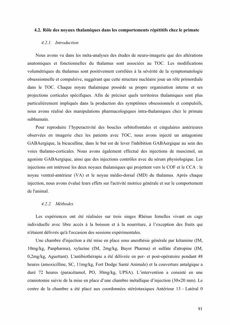
91
4.2. Rôle des noyaux thalamiques dans les comportements répétitifs chez le primate
4.2.1. Introduction
Nous avons vu dans les méta-analyses des études de neuro-imagerie que des altérations
anatomiques et fonctionnelles du thalamus sont associées au TOC. Les modifications
volumétriques du thalamus sont positivement corrélées à la sévérité de la symptomatologie
obsessionnelle et compulsive, suggérant que cette structure nucléaire joue un rôle primordiale
dans le TOC. Chaque noyau thalamique possède sa propre organisation interne et ses
projections corticales spécifiques. Afin de préciser quels territoires thalamiques sont plus
particulièrement impliqués dans la production des symptômes obsessionnels et compulsifs,
nous avons réalisé des manipulations pharmacologiques intra-thalamiques chez le primate
subhumain.
Pour reproduire l’hyperactivité des boucles orbitofrontales et cingulaires antérieures
observées en imagerie chez les patients avec TOC, nous avons injecté un antagoniste
GABAergique, la bicuculline, dans le but est de lever l'inhibition GABAergique au sein des
voies thalamo-corticales. Nous avons également effectué des injections de muscimol, un
agoniste GABAergique, ainsi que des injections contrôles avec du sérum physiologique. Les
injections ont intéressé les deux noyaux thalamiques qui projettent vers le COF et le CCA : le
noyau ventral-antérieur (VA) et le noyau médio-dorsal (MD) du thalamus. Après chaque
injection, nous avons évalué leurs effets sur l'activité motrice générale et sur le comportement
de l'animal.
4.2.2. Méthodes
Les expériences ont été réalisées sur trois singes Rhésus femelles vivant en cage
individuelle avec libre accès à la boisson et à la nourriture, à l’exception des fruits qui
n'étaient délivrés qu'à l'occasion des sessions expérimentales.
Une chambre d'injection a été mise en place sous anesthésie générale par kétamine (IM,
10mg/kg, Panpharma), xylazine (IM, 2mg/kg, Bayer Pharma) et sulfate d'atropine (IM,
0,2mg/kg, Aguettant). L'antibiothérapie a été délivrée en per- et post-opératoire pendant 48
heures (amoxicilline, SC, 11mg/kg, Fort Dodge Santé Animale) et la couverture antalgique a
duré 72 heures (paracétamol, PO, 30mg/kg, UPSA). L’intervention a consisté en une
craniotomie suivie de la mise en place d’une chambre métallique d’injection (30!20 mm). Le
centre de la chambre a été placé aux coordonnées stéréotaxiques Antérieur 13 – Latéral 0
92
(A13 – L0) sur l’atlas de Szabo de façon à permettre l’accès aux MD et VA. Une plaque de
fixation permettant de maintenir la chambre d’injection a été scellée au crâne à l’aide de
ciment dentaire (Ominium dentaire).
Les microinjections intra-thalamiques de bicuculline (Bicuculline Méthiodide, Sigma) et
de muscimol (Muscimol, Sigma), dilués dans du sérum physiologique 0,9 ‰ (10µg/µl pour la
bicuculline et 1µg/µl pour le muscimol), ont été faites avec une canule d’acier inoxydable de
26 gauges (26G, Phymep). La canule a été descendue avec un microdescendeur au sein des
noyaux thalamiques d’intérêt. Elle était reliée par un tube de polyéthylène (référence 8010,
Phymep) à une seringue Hamilton (10µl), contrôlée par une pompe Harvard (Harvard
Apparatus). Un guide-canule facilitait le passage de la dure-mère et permettait une descente
rectiligne de la canule. Le volume injecté a été de 1 à 3µl, à une vitesse de 1µl/min. Une fois
la substance pharmacologique délivrée, la canule était laissée en place pendant cinq minutes,
puis retirée lentement afin d’éviter tout reflux. Des microinjections intra-thalamiques de
sérum physiologique, réalisées de façon strictement identique aux microinjections de
bicuculline et de muscimol, étaient effectuées aux coordonnées où la solution active entraînait
une modification comportementale de l’animal.
Après chaque situation, avec ou sans injection, nous avons évalué l'activité motrice
générale et le comportement de l'animal dans une cage enrichie en nourriture et jouets
pendant 45 minutes. L'activité motrice générale de l’animal a été quantifiée de façon
automatique avec le système « Vigiprimate » (ViewPoint). Ce système basé sur l’analyse des
changements de contraste des pixels de l’image permet de décomposer l'activité de l'animal en
trois niveaux : 1) mouvements fins de faible amplitude (par exemple, les mouvements des
doigts) ; 2) mouvements plus amples sans déplacement (par exemple, la préhension de la
nourriture) ; 3) mouvements de grande amplitude (par exemple, les déplacements). Les
comportements de l'animal (observation, alimentation, exploration, toilettage, recherche fine
de nourriture et manipulation de jouet) ont été quantifiés dans leur durée et leur fréquence en
utilisant un clavier éthologique et le logiciel « LabWatcher » (ViewPoint).
Après le sacrifice de l'animal, nous avons effectué une vérification histologique des
sites d’injections thalamiques. Les cerveaux ont été conservés au congélateur avant d’être
coupés au cryostat. La vérification histologique a été faite sur coupes coronales de 50 µm
passant par le thalamus, révélées par un biomarqueur cérébral, l’acétylcholinestérase (Sigma).
93
4.2.3. Résultats et discussion
Pour la bicuculline, nous avons réalisé un total de 8 injections dans le VA et 17 dans le
MD. Pour le muscimol, 7 injections ont été faites, dont 4 dans le VA et 3 dans le MD.
Les injections de bicuculline dans le VA ont induit la production de comportements
complexes, finalisés, répétitifs et envahissants. Trois types de comportements ont pu être
individualisés : 1) le toilettage répétitif, 2) la recherche fine d’arachides sous les barreaux de
la cage alors que l’animal a à sa disposition des fruits frais et des arachides et 3) la
manipulation fine et méticuleuse des jouets auxquels l’animal est habituellement indifférent.
Dans le cas du MD, nous avons observé des manifestations neuro-végétatives avec
vomissements, tremblements, dysrythmie cardiaque, ainsi qu’un effondrement de l’activité
motrice des animaux.
Les injections de muscimol ont entraîné une hyperactivité motrice importante caractérisée
par de nombreux shifts comportementaux. Ces manifestations comportementales ont été
observées après injections dans le VA et dans le MD.
Les injections « contrôles » n’ont pas induit de modifications comportementales
particulières, c’est-à-dire que l’animal se nourrissait puis se mettait en position d’attente
jusqu’à la fin de l’évaluation.
Les effets observés des injections sont reproductibles d'un animal à l'autre. Cependant,
le nombre d'injections réalisées est relativement faible, ce qui s'explique par le petit volume
des structures cibles. Bien que ce travail ait déjà fait l'objet d'une soumission dans un journal à
comité de lecture, nous avons suspendu le processus de publication afin d'enrichir nos
données. Ainsi, un quatrième animal sera également inclus dans l'étude. Chez cet animal,
nous envisageons également de procéder à l’injection d’un traceur antérograde et rétrograde
(Biotin-dextran, Sigma) aux sites d’injection d'intérêt. Ceci permettra de préciser les
territoires corticaux et sous-corticaux de projection des noyaux thalamiques.
94
4.2.4. Conclusion
Ce travail suggère qu’une hyperactivité du noyau ventral-antérieur du thalamus est plus
particulièrement impliquée dans la production de comportement répétitifs, analogues aux
comportements compulsifs, chez le primate subhumain.
Aouizerate B, Rotgé JY, Lambrecq V, Amestoy V, Langbour N, Dovero S,
Cardoit L, Tignol J, Bioulac B, Burbaud P, Guehl D. Associative-limbic thalamus
and behavior. En préparation.
95
Associative-limbic thalamus and behavior
B Aouizerate1,2, JY Rotge1,2, V Lambrecq1,3, V Amestoy1, N Langbour1, S Dovero1, L
Cardoit1, J Tignol2, B Bioulac1,3, P Burbaud1,3, D Guehl1,3
1Laboratoire Mouvement, Adaptation, Cognition, CNRS UMR 5227, Université Bordeaux 2, Bordeaux, 2Service
Universitaire de Psychiatrie Adulte, Centre Hospitalier Charles Perrens, Bordeaux, 3Service d’Explorations
Fonctionnelles du Système Nerveux, Centre Hospitalier Universitaire, Bordeaux
Corresponding author: Dr. Bruno Aouizerate, Service Universitaire de Psychiatrie Adulte,
CNRS UMR 5227, Université Victor Segalen Bordeaux 2, Centre Hospitalier Charles
Perrens, 121 rue de la Béchade, 33076 Bordeaux, France. Tel: + 33 (5) 57 57 15 86; fax: + 33
(5) 56 90 14 21; email: [email protected]
Manuscript information:
Number of text page: 23
Number of figures: 3
Word and character counts:
Number of words in the abstract: 250
Number of characters in the paper: 42 097
Supporting information:
Number of tables: 2
Number of figures: 2
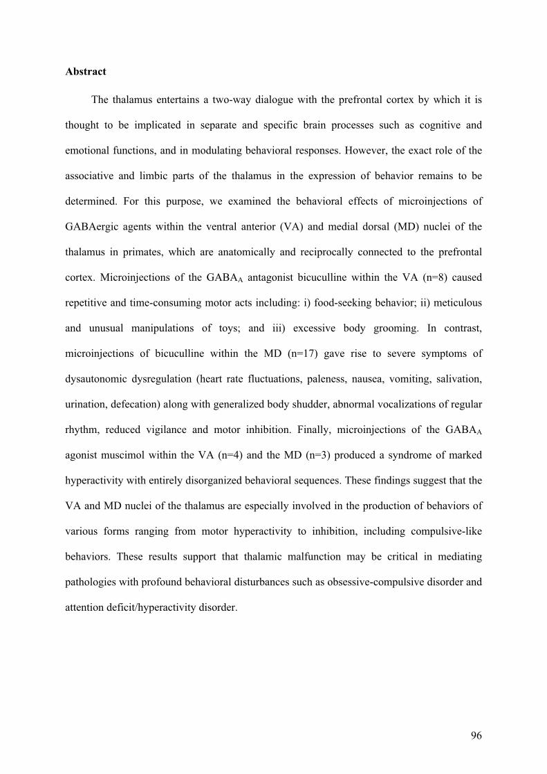
96
Abstract
The thalamus entertains a two-way dialogue with the prefrontal cortex by which it is
thought to be implicated in separate and specific brain processes such as cognitive and
emotional functions, and in modulating behavioral responses. However, the exact role of the
associative and limbic parts of the thalamus in the expression of behavior remains to be
determined. For this purpose, we examined the behavioral effects of microinjections of
GABAergic agents within the ventral anterior (VA) and medial dorsal (MD) nuclei of the
thalamus in primates, which are anatomically and reciprocally connected to the prefrontal
cortex. Microinjections of the GABAA antagonist bicuculline within the VA (n=8) caused
repetitive and time-consuming motor acts including: i) food-seeking behavior; ii) meticulous
and unusual manipulations of toys; and iii) excessive body grooming. In contrast,
microinjections of bicuculline within the MD (n=17) gave rise to severe symptoms of
dysautonomic dysregulation (heart rate fluctuations, paleness, nausea, vomiting, salivation,
urination, defecation) along with generalized body shudder, abnormal vocalizations of regular
rhythm, reduced vigilance and motor inhibition. Finally, microinjections of the GABAA
agonist muscimol within the VA (n=4) and the MD (n=3) produced a syndrome of marked
hyperactivity with entirely disorganized behavioral sequences. These findings suggest that the
VA and MD nuclei of the thalamus are especially involved in the production of behaviors of
various forms ranging from motor hyperactivity to inhibition, including compulsive-like
behaviors. These results support that thalamic malfunction may be critical in mediating
pathologies with profound behavioral disturbances such as obsessive-compulsive disorder and
attention deficit/hyperactivity disorder.
97
Introduction
The thalamus, a nuclear complex located in the diencephalon, is widely thought to be a
relay center projecting to one or a few well-defined cortical areas that send back information
to various thalamic nuclei and provide positive feedback to the “correct input”, while
simultaneously suppressing irrelevant information (1-3). The thalamus has for long been
regarded as one of the chief subcortical structures subserving the analysis and integration of
both sensory and motor functions (4-7). It is also thought to be a highly evolved cognitive and
emotional gateway to the prefrontal cortex, owing to reciprocal connections with the three
major cortical regions including the orbitofrontal cortex (OFC), the dorsolateral prefrontal
cortex (DLPFC) and the anterior cingulate cortex (ACC) (8-11). A large body of evidence
points to the involvement of the prefrontal cortex in several cognitive and emotional
subprocesses underlying behavioral responses in close relationships with thalamic inputs (12-
16). However, although topographically organized connections between the thalamic nuclei
and the prefrontal cortex are well known, the specific role of the thalamus and its respective
associative and limbic territories in the expression of behavior is still far from being
determined. To date, understanding of the thalamus and behavior has mainly been limited to
behavioral changes occurring after thalamic lesions. Clinical reports of patients with thalamic
infarcts have described the emergence of multiple behavioral patterns, including
perseverations in thought and output speech with apathy and amnesia, or disinhibition
syndromes with labile affect, hyperactivity, excessive use of objects and inability to regulate
interactions with the environment (17-19). Furthermore, there has been relatively little
experimental work on thalamic lesions in laboratory animals leading to behavioral
impairment related to severe cognitive memory deficits with disruption in object-reward
associations and habit formation (20).
98
In the present study, we investigated the role of the associative and limbic thalamus in
behavior. For this purpose, we used an experimental approach primarily based on the
examination of behavioral changes following pharmacological manipulations of the thalamic
GABAergic system in subhuman primates. Microinjections of the GABAA antagonist
bicuculline were performed to remove the endogenous GABAergic inhibitory tone on
thalamic activity, and therefore to induce disinhibition of thalamo-cortical projections.
Alternatively, we examined the behavioral consequences of local microinjections of
muscimol, a GABAA agonist. The ventral anterior (VA) and medial dorsal (MD) nuclei of the
thalamus were chosen as targets because they are both strongly connected to the OFC,
DLFPC and ACC (8-11).
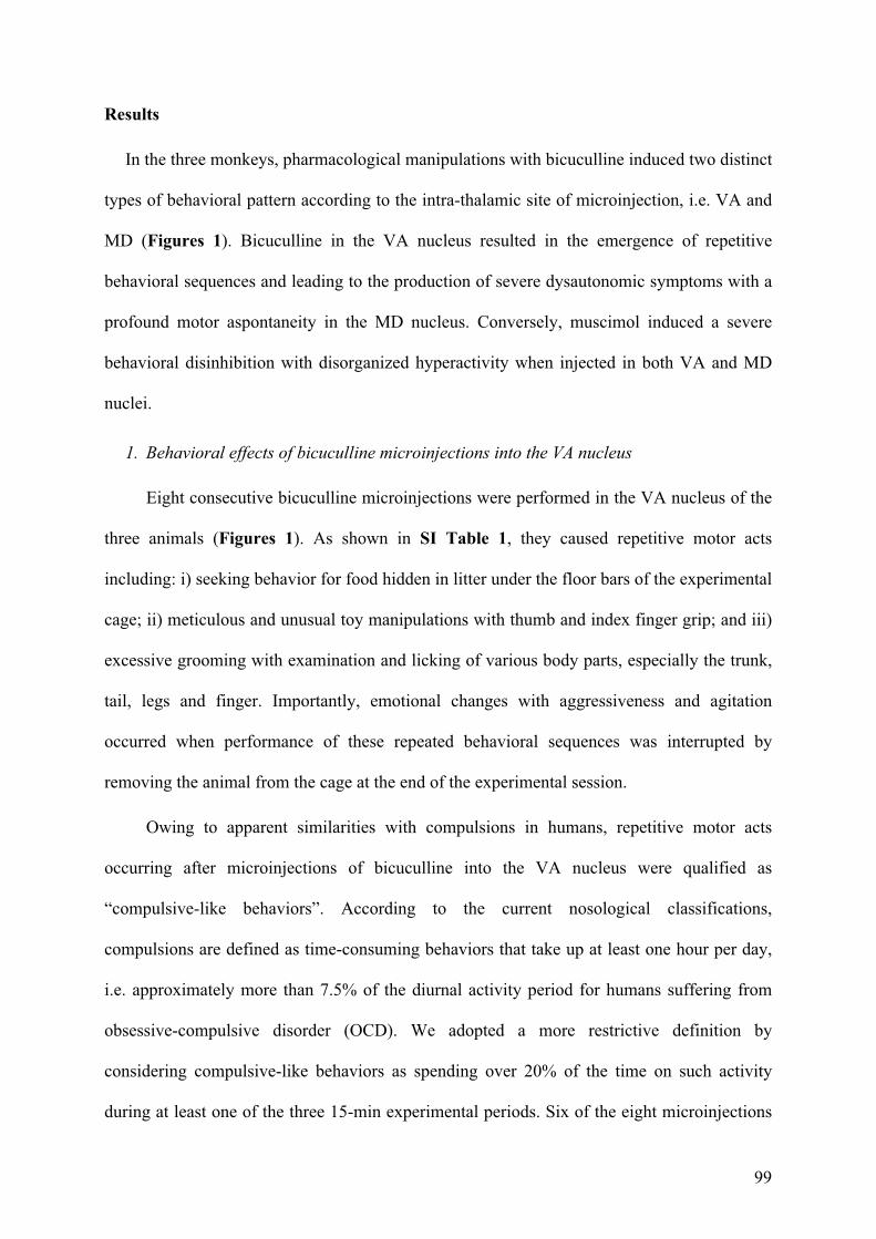
99
Results
In the three monkeys, pharmacological manipulations with bicuculline induced two distinct
types of behavioral pattern according to the intra-thalamic site of microinjection, i.e. VA and
MD (Figures 1). Bicuculline in the VA nucleus resulted in the emergence of repetitive
behavioral sequences and leading to the production of severe dysautonomic symptoms with a
profound motor aspontaneity in the MD nucleus. Conversely, muscimol induced a severe
behavioral disinhibition with disorganized hyperactivity when injected in both VA and MD
nuclei.
1. Behavioral effects of bicuculline microinjections into the VA nucleus
Eight consecutive bicuculline microinjections were performed in the VA nucleus of the
three animals (Figures 1). As shown in SI Table 1, they caused repetitive motor acts
including: i) seeking behavior for food hidden in litter under the floor bars of the experimental
cage; ii) meticulous and unusual toy manipulations with thumb and index finger grip; and iii)
excessive grooming with examination and licking of various body parts, especially the trunk,
tail, legs and finger. Importantly, emotional changes with aggressiveness and agitation
occurred when performance of these repeated behavioral sequences was interrupted by
removing the animal from the cage at the end of the experimental session.
Owing to apparent similarities with compulsions in humans, repetitive motor acts
occurring after microinjections of bicuculline into the VA nucleus were qualified as
“compulsive-like behaviors”. According to the current nosological classifications,
compulsions are defined as time-consuming behaviors that take up at least one hour per day,
i.e. approximately more than 7.5% of the diurnal activity period for humans suffering from
obsessive-compulsive disorder (OCD). We adopted a more restrictive definition by
considering compulsive-like behaviors as spending over 20% of the time on such activity
during at least one of the three 15-min experimental periods. Six of the eight microinjections
100
induced repeated behaviors that fulfilled these duration criteria as early as the first 15 minutes
(T1) after the animal was placed in the experimental cage (i.e. between 15 to 30 minutes after
bicuculline was infused into the VA nucleus), and which persisted over the two subsequent
15-min periods (T2, T3) in all of them. The two other microinjections produced behavioral
changes at T1, but met the predefined criterion only at either T2 and T3 or T3 (SI Table 1).
Finally, statistical analysis showed that bicuculline resulted in an exacerbation of the
three abovementioned behaviors, which were performed for a significantly longer time
compared to when saline was administered at the same anatomical site of the VA nucleus
(t=9.57, p<0.0001) (Figure 2a).
2. Behavioral effects of bicuculline microinjections into the MD nucleus
Seventeen bicuculline microinjections were consecutively performed within the MD
nucleus across the three animals (Figures 1). As shown in SI Table 1, they induced severe
symptoms of dysautonomic dysregulation including heart rate fluctuations, paleness, nausea,
vomiting, salivation, urination, defecation, generalized body shudder, and abnormal
vocalizations, which abruptly appeared within 5-15 min following administration. For two
microinjections (B24, B25), a dose of the benzodiazepine component diazepam (0.5 mg/kg,
intramuscular), as GABAA agonist had to be administered owing to the intensity of the
dysautonomic episodes, but this only partially antagonized the effects of bicuculline. As
illustrated in SI Figures 4, measurements of the R-R space became highly variable after
bicuculline microinjection, as reflected by a wide distribution of values collected over 60 sec
(range: 0.18-0.73 sec) and contrasting with the relatively stable profile found during the same
time-period immediately before bicuculline was injected (range: 0.24-0.30 sec). As shown in
SI Figures 5, the visual aspect of the individual acoustic spectrum showed that bicuculline
induced vocalizations of a wider frequency range (484-1394 Hz) with higher peak
fundamental frequency (890.4 Hz) compared to those spontaneously produced by the animal
101
(frequency range: 555-579 Hz; peak fundamental frequency: 559.0 Hz) (SI Figures 5a, b).
Bicuculline also resulted in vocalizations that occurred more frequently and regularly, as
attested by their higher number over 90 sec of audio recording along with a narrower range
for the time intervals separating each of them (after vs. before bicuculline: vocalization
number: 25 vs. 10; time interval range: 1.65-5.21 vs. 3.24-20.06 sec) (SI Figures 5c, d).
These clinical manifestations were followed by a profound motor inhibition with
marked fatigue and reduced vigilance. This led to a substantial reduction in spontaneous
displacement and exploratory behaviors within the experimental cage, which were marked as
early as T1 for 10 microinjections, and extended at T2 for two of them, or T2 and T3 for five
of them. For the five other microinjections, such behavioral changes started at T1 but became
prominent at T2 for one of them, at T2 and T3 for three others or at T3 for the other one (SI
Table 1).
Finally, statistical analysis showed that bicuculline significantly suppressed both
displacement and exploratory activities when compared to saline injected at the same
anatomical site of the MD (“high” activity duration: t=6.29, p<0.0001; exploratory activity
duration: t=7.43, p<0.0001) (Figure 2b).
3. Behavioral effects of muscimol microinjections into the VA and MD nuclei
Seven consecutive muscimol microinjections were performed within the VA nucleus for
four of them and the MD nucleus for the three others in one of the three studied animals
(Figures 1). As shown in SI Table 2, they induced a syndrome of marked motor
hyperactivity with disruptive behavior in both intra-thalamic sites. There was a major increase
in exploratory activity and excessive displacement movements in the experimental cage with
entirely disorganized behavioral sequences due to successive and rapid shifting from one
behavior to another. Such profound behavioral disturbances were observed at T1, T2 and T3
with 6 of the 7 microinjections and were limited to T1 with the remaining one (SI Table 2).
102
Statistical analyses showed a significant increase in both displacement and exploratory
activities when muscimol was infused into either VA or MD nucleus compared to that found
after saline injected at the same anatomical site (VA: “high” activity duration: t=11.64,
p<0.0001; exploratory activity duration: t=13.83, p<0.0001) (MD: “high” activity duration:
t=4.82, p<0.0001; exploratory activity duration: t=7.23, p<0.0001) (Figure 3a). Muscimol
also resulted in a significantly higher number of times that the specific behavioral sequences
occurred (behavior frequency: VA: t=9.49, p<0.0001; MD: t=2.24, p<0.03) (Figure 3b).
Comparative analysis of the three microinjections performed within the VA and MD nuclei,
where muscimol produced locomotor effects throughout the three time-periods considered
(T1, T2 and T3), showed significantly more pronounced behavioral changes in the VA than in
the MD (“high” activity duration: t=13.84, p<0.0001; exploratory activity duration: t=9.48,
p<0.0001; behavior frequency: t=5.40, p<0.0001).
4. Behavioral effects of bicuculline microinjections in other thalamic sites
Eighteen bicuculline microinjections were performed in motor thalamic sites including
the pallidal thalamus (VLo: ventrolateral thalamic nucleus, pars oralis) for six of them or its
cerebellar territory (VLc: ventrolateral thalamic nucleus, pars caudalis/VPLo: ventral
posterior lateral thalamic nucleus, pars oralis) for the remainder. They gave rise to either
severe postural abnormalities like cervical dystonia with ipsilateral inclination and head
rotation or myoclonic jerks involving the contralateral upper and lower limbs. Moreover, two
other microinjections done at the limit between VLc/VPLo, VA and MD induced the
association of three successive kinds of effects: immediate but transient dystonic limb spasms
followed by the abovementioned repetitive behavioral sequences and then by motor inhibition
in the experimental cage.
103
Discussion
To our knowledge, this study is the first to show that the VA nucleus of the thalamus is
highly involved in the emergence of repetitive behaviors. Microinjections of the GABAA
antagonist bicuculline produced three major kinds of behavioral sequences, including
repeated food-seeking and body grooming along with unusual manipulation of available toys.
However, there was a profound motor inhibition with severe dysautonomic manifestations
such as heart rate variation, paleness, salivation, vomiting, urination and defecation when
bicuculline microinjections were performed within the MD nucleus. Conversely, a marked
hyperactivity with profoundly disorganized and incompletely executed behaviors was
observed in response to the GABAA agonist muscimol into both the VA and MD. This
suggests that both the associative and limbic territories of the thalamus could play a major
role in the production of a wide variety of behaviors by processing their cognitive and
emotional determinants in the subhuman primate.
This exacerbation of spontaneously committed motor acts in response to bicuculline
injected into the VA nucleus shares some similarities with compulsions in humans. Behaviors
were time-consuming while substantially interfering with the routine activity of the animals.
They also met our more restrictive duration criteria than those currently used for defining
clinically compulsive behaviors in human OCD. Interestingly, marked agitation with
aggressiveness appeared when ongoing behavior was interrupted at the end of the
experimental session. This can be assimilated to the rebound of anxiety symptoms
experienced by OCD subjects when they are unexpectedly asked to stop recurrent ritualized
behaviors. However, the behavioral alterations observed in our study were not of a similar
type to those seen in OCD, except for the grooming behavior which is relatively close to the
cleaning and washing compulsions experienced by humans. In a recent study examining the
behavioral effects of bicuculline within the limbic territory of the external globus pallidus in
104
primates, there was an intense expression of behaviors belonging to the animal’s usual
repertoire such as tail- or finger-biting and licking of the cage bars (21). These abnormal
behaviors were analogous to those we observed in the thalamic VA nucleus, although they
were considered as stereotypies, therefore differing from the more complexly organized
behavioral sequences occurring in our study. This may be due to both anatomical and
functional reasons. The limbic territory in the ventromedial portion of the external globus
pallidus is densely connected to the pars reticularis of the substantia nigra (21-24), which in
turn sends vast projections to the thalamic VA nucleus (8). Consequently, the behavioral
effects of injecting bicuculline into the GPe share similar features with those reported in our
study. However, information arising from the basal ganglia is not simply carried throughout
the VA nucleus but is further processed before reaching the cortical level (3, 26-28). This
organization could be responsible for the more organized behavioral sequences observed in
the VA than in the GPe.
The inhibitory neurotransmitter GABA in the thalamus is involved in the intrinsic
mechanisms regulating the activity of the glutamatergic thalamic neurons that innervate
cortical areas. In particular, the VA nucleus includes two major types of GABAergic cells,
referred to as interneurons or local circuit neurons, and also receives GABAergic afferents
from the globus pallidus, the pars reticularis of the substantia nigra and the thalamic reticular
nucleus (8, 29, 30). Therefore, as a GABAA receptor antagonist, bicuculline may be expected
to reduce local GABAergic tone and to result in cortical activation by disinhibition of the
thalamo-cortical neurons. In our study, bicuculline caused repetitive and meticulous bodily
grooming and toy manipulation. It would therefore seem that bicuculline caused excessive
attention to be centered on either environmental or external self-related stimuli. Both the
DLPFC and ACC, which are clearly associated with the thalamic VA nucleus (8, 9, 31), play
a pivotal role in a wide range of cognitive functions including attention to action and selection
105
of appropriate behavioral responses to specific situations (14, 32, 33). Thus, bicuculline could
trigger the recruitment of attentional processes by overactivation of both the DLPFC and
ACC, with alterations in flexibility and inhibition of behavioral responses, thereby leading to
a greater sensitivity to normally ignored stimuli and to the repetition of a given behavior.
Conversely, microinjections of the GABAA receptor agonist muscimol into the VA resulted in
the emergence of profoundly disorganized behaviors with severe motor hyperactivity. In view
of behavioral analogies with attention deficit/hyperactivity disorder (ADHD), we propose that
the attention capacity of the monkeys was impaired, causing difficulties in directing and
maintaining attention to the performance of specific behaviors. Such cognitive and behavioral
abnormalities may be related to the putative reducing effects of muscimol on DLPFC and
ACC activities. In the present study, bicuculline in the VA nucleus also exacerbated reward-
directed behaviors with repeated sequences of food-seeking. This was presumably due to an
enhanced activity within the two cortical and subcortical regions, the OFC and ventral
striatum. The VA nucleus is strongly connected to the OFC and ventral striatum (9, 31, 34).
On one hand, the OFC is highly involved in processing the reward value of environmental
stimuli, and the reward motivation and representation of the incoming reward (14, 35, 36). On
the other hand, the ventral striatum, which receives numerous projections from the OFC, is
responsible for the preparation, initiation and control of voluntary behaviors oriented towards
reward delivery (37-39) requiring extraction of reward-related information from a large
variety of stimuli (40, 41). Finally, bicuculline produced aggressive reactions when the
execution of ongoing behavioral sequences was abruptly interrupted. The thalamic VA
nucleus has anatomical and functional relationships with limbic structures such as the
amygdala (42, 43), which has been consistently identified as playing a crucial role in both the
perception of emotionally salient information and the production of emotional responses (44-
46). Therefore, an abnormally elevated activity within the amygdala, as thought to occur in
106
response to bicuculline injected into the VA nucleus, could produce the emergence of
aggressiveness like that observed in our study.
In our work, bicuculline microinjections within the MD nucleus of the thalamus gave
rise to a severe form of dysautonomia with vocalizations of a regular rhythm, resulting in
profound anergia with successive and short phases of lowered vigilance. Anatomically, the
MD nucleus has bidirectional connections with the amygdala through its lateral and
basolateral nuclei (46-48). In turn, these nuclei project to the central nucleus of the amygdala,
which then extensively innervates a wide range of brain target structures that underlie specific
clinical aspects of emotional responses (46, 49, 50). The connections between the central
nucleus of the amygala and both the lateral hypothalamus and related dorsal motor nucleus of
the vagus could be responsible for stimulating the sympathetic and parasympathetic
autonomic nervous systems, with the emergence of various symptoms such as heart rate
variation, paleness, salivation, vomiting, urination, defecation or body shudder in response to
bicuculline. Projections of the central nucleus of the amygdala to the periacqueductal gray
region, which have been implicated particularly in the elicitation of vocalization (49-52),
could mediate the atypical vocal reaction to bicuculline. Among the brain regions that could
participate in the constellation of clinical symptoms observed in our study, the ACC receiving
input from the thalamic MD nucleus and sending outflow to the amygdala (8-11, 13) is
another candidate as critical site for the regulation of emotional responses with their
autonomic components (12, 13, 15, 46).
As observed in the VA nucleus, muscimol caused severe but less marked behavioral
abnormalities in the MD with inappropriate and disruptive hyperactivity. Our observation
confirms the results of previous studies in rodents showing a dose-dependent increase in
locomotor response to muscimol in the MD nucleus (53, 54). Thus, it can be assumed that the
MD shares, at least partially with the VA, some common cortical projection areas such as the
107
DLPFC and ACC (8-11), which are of special importance in regulating cognitive functions
and controlling attentional processes (14, 32, 33).
To date, the involvement of the thalamic nuclei VA and MD in the expression of
behavior has received little attention. The present study aimed at investigating the behavioral
consequences of pharmacological manipulations of the GABAergic system within either the
VA or MD in the subhuman primate. Our findings show that the VA is particularly involved
in the production of compulsive-like behaviors, whereas the MD participates rather in the
regulation of emotional responses with their autonomic concomitants. However, both the VA
and MD play a central role in the emergence of disruptive and inappropriate hyperactivity,
suggesting possible impairment in the processing of cognitive information. Therefore, our
work in monkeys underlines the critical importance of the thalamus in behavioral disturbances
that have formal similarities with psychiatric conditions such as OCD and ADHD. Further
studies are now required to better identify the cognitive and emotional determinants of such
behavioral alterations and their anatomical substrates.
108
Materials and methods
1. Animals
Experiments were conducted on three female monkeys (Macaca mulatta) weighing 4-6
kg that were housed in individual primate cages. They had ad libitum access to water and
food without supplementation with fruits. Their care was supervised by veterinarians skilled
in the maintenance of subhuman primates, in strict accordance with the European Community
Council Directive for experimental procedures in animals. The light-dark cycle (lights on
from 7 a.m. to 7 p.m.), temperature (22°C), and humidity (60%) were kept constant in the
animal room. The animals were allowed at least two weeks to acclimatize to the animal room
before starting any manipulation. They were then trained to be seated in a primate chair with
their head restrained and to remain quiet during palpation of various parts of the body.
2. Surgery
The surgical procedure was performed under general anesthesia using ketamine (IM, 10
mg/kg, Panpharma, Fougères, France), xylazine (IM, 2 mg/kg, Bayer Pharma, Puteaux,
France) and atropine sulfate (IM, 0.2 mg/kg, Aguettant, Lyon, France). Additional doses of
ketamine and xylazine were administered as necessary to maintain optimal anesthesia. A
stainless steel chamber (Narishige, diameter 19 mm) was implanted on the interhemispheric
line, over both the right and left hemispheres under aseptic conditions. The center of the
cylinder was stereotaxically positioned at A13 and L0 in the three monkeys, according to the
atlas of Szabo and Cowan (55) and the position of the anterior commissure predetermined
with structural magnetic resonance imaging data. A head-holder was embedded with dental
cement (Omnium Dentaire, Bordeaux, France) around the chamber in order to immobilize the
head of the monkeys for the experiments with drug microinjections. Antibiotics (amoxicillin,
subcutaneously, 11 mg/kg, Fort Dodge Santé Animale, Tours, France) and analgesics
109
(Paracetamol, per os, 30 mg/kg, UPSA, Agen, France) were given for 48-72 hours after
surgery. Animals were left to recover 10-15 days before starting the experiments.
3. Drugs and drug administration
Both bicuculline and muscimol (Sigma, Lyon, France) were dissolved in sterile 0.9%
NaCl solution (saline) at 10 mg/ml and 1 mg/ml, respectively. The intra-thalamic
microinjections were performed in monkeys with their head fixed and their body loosely
restrained by a plastic apparatus. Before microinjection experimentation began, extracellular
neuronal recordings of single-cell activity using tungsten microelectrodes isolated with epoxy
(impedance 1-1.5 M" at 1 kHz) were performed in order to delineate the dorsal border of the
thalamus. The electrode was moved with a micromanipulator (Narishige, MO-95, Tokyo,
Japon) in 5-10 µm increments. Neuronal activity was amplified (10 000 !), filtered (300 Hz –
3 kHz) and displayed on an oscilloscope. Spikes were selected from background activity with
a window discriminator, and then processed though an analogue-digital interface before being
stored on a microcomputer. At each session, the cannula for microinjection (26-gauge
stainless steel) (Phymep, Paris, France) was connected to a Hamilton microsyringe (10 µl) via
a 30-cm long polyethylene tube (Phymep, Paris, France) filled with the substance to be
injected, and was then descended through the dura mater into the thalamus with the
micromanipulator. Either bicuculline (1-3 µl) or muscimol (2 µl) was delivered manually by
progressive pressure microinjection at a rate of 1 µl/min in 1 µl increments. The cannula was
maintained in position at the chosen site for at least 5 minutes after completion of the
microinjection to prevent drawback of the drug. At the sites selected according to the
behavioral effects of bicuculline, we injected saline as control substance by using a similar
experimental procedure. Additional microinjections of muscimol were performed on the last
monkey.
110
4. Heart rate recording
To record the heart rate, R-R intervals were monitored with a tactile sensor fixed on the
tail while the animal was seated in the primate chair with the head and body restrained.
Recordings were made for 60 sec immediately before and after bicuculline was injected into
the MD nucleus. Data obtained with a PowerLab system were analyzed using Chart 5.0
software (ADI instruments, Phymep, Paris, France).
5. Vocal analysis
Spectral analysis of vocalizations was performed on 90-sec audio recording
immediately before and after bicuculline was injected into the MD nucleus using Matlab 7.0.1
software. The temporal patterns of vocal productions were also examined across both
experimental conditions.
6. Behavioral activity
As described elsewhere (56), behavioral activity was recorded 10-15 min after each
microinjection with a camera placed in front of the experimental cage during a 45-min
session, which was divided into three consecutive 15-min periods (T1, T2 and T3). During
this period, the animals were placed in a 1.0 ! 1.5 m clear plastic cage with a metal-barred
floor. Behavioral activity was analyzed with Vigiprimate software (Viewpoint, Lyon, France),
which discriminates three levels of movements: (1) “low”, corresponding to immobility or
absence of movement, (2) “middle”, corresponding to movement without displacement, such
as grooming, toy manipulation, food intake, etc.; and, (3) “high”, corresponding to
displacement within the cage which may represent a measurement of exploration. These
measures were performed after the sensitivity parameters for movement detection were finely
adjusted for each of the three animals.
111
To further characterize spontaneous behaviors and their occurrence, each specific
behavior was counted by both direct observation and examination on videotape and quantified
in terms of frequency and duration using Labwatcher software (Viewpoint, Lyon, France).
Five major kinds of behaviors were analyzed: grooming, food-seeking, food intake, toy
manipulation, and exploration. Measurements were made for the 45 min of experimental
session over 3-min intervals and conducted by the same trained researcher (B.A.).
7. General experimental design
Ten to 15 days after surgery, the animal was acclimatized to be seated in the primate
chair placed in the microinjection room where the stainless steel chamber with dura mater was
cleaned with the head completely immobilized for 10-15 min. However, at this first stage, no
pharmacological microinjection was performed. The animal was then transferred to the room
containing the experimental cage where behaviors were monitored for a daily 45-min session
after optimal sensitivity setting for movement detection, as determined on the first three
consecutive sessions. After this initial 3-week habituation period, the experimental sessions
with intra-thalamic microinjections of bicuculline began. They included three successive
observation sequences as follows: 1) Period 1 (5 min) after the animal was transferred to the
microinjection room with the cannula inserted into the intra-thalamic site and before the
microinjection of bicuculline was performed; 2) Period 2 (5 min) after microinjection of
bicuculline was performed and before the cannula was removed; 3) Period 3 (5 min) after the
apparatus for movement restriction was removed but with the animal still seated in the
primate chair; and 4) Period 4 (45 min) after the animal was transferred to the experimental
cage for recording of behavioral activity over three successive 15-min sub-periods (T1, T2
and T3). Thereafter, the animal returned to the home cage. At the sites selected according to
the behavioral effects of bicuculline, either muscimol or saline were also administered. These
microinjection sessions were separated by at least one additional session in the baseline
112
condition, in which all the previously described manipulations were performed but without
descending the cannula into the brain to detect any acquisition of contextually conditioned
behaviors. Animals were systematically manipulated by the same researchers (B.A., J.Y.R.,
D.G.) across all experiments in order to avoid possible context-related behavioral changes.
8. Histological verifications
At the end of the experiments, monkeys were deeply anesthetized with an overdose of
ketamine and perfused through the ascending aorta with 500 ml of 0.9 % saline, followed by 2
l of 4% paraformaldehyde in phosphate buffer (ph 7.4) as fixative. The brain was removed
from the skull, frozen, and serially sliced into 50-µm coronal sections. Locations of
microinjection sites were then reconstructed from cresyl violet-stained and calbindin-
immunoreactive sections, on the basis of a schematic representation taking into account all
behavioral data collected from the three monkeys and the type of microinjection performed in
each structure of interest.
9. Statistical analyses
Paired Student’s t-tests were used for intra-structure comparisons of the effects of both
active pharmacological treatments (bicuculline, muscimol) on behavioral activity scores to
those of the control substance (saline) within each of the intra-thalamic sites considered (VA,
MD). Paired Student’s t-tests were also performed for inter-structure comparisons of
behavioral changes produced by muscimol between both intra-thalamic sites (VA, MD). For
all statistical analyses, p<0.05 was used as the threshold for statistical significance.
Acknowledgments
The authors thank Dr. Ray Cooke for providing advice on English-language wording.
This work was supported in part by a grant from the “Région Aquitaine”. JY Rotge was
supported by a PhD grant from the “Fondation pour la Recherche Médicale”.
113
References
1. Steriade M (1970) Int Rev Neurobiol 12, 87-144.
2. Herrero MT, Barcia C, Navarro JM (2002) Childs Nerv Syst 18, 386-404.
3. Sommer MA (2003) Curr Opin Neurobiol 13, 663-670.
4. Lenz FA, Kwan HC, Dostrovsky JO, Tasker RR, Murphy JT, Lenz YE (1990) Brain 113,
1795-1821.
5. Lenz FA, Byl NN (1999) J Neurophysiol 82, 3204-3212.
6. MacMillan ML, Dostrovsky JO, Lozano AM, Hutchison WD (2004) J Neurophysiol 91,
1085-1090.
7. Kim JH, Greenspan JD, Coghill RC, Ohara S, Lenz FA (2007) J Neurosci 27, 4995-5004.
8. Bentivoglio M, Kultas-Ilinsly K, Ilinsly I (1993) in Neurobiology of cingulate cortex and
limbic thalamus: A comprehensive handbook, eds Vogt BA, Gabriel M (Birkhäuser,
Boston), pp 71-122.
9. Cummings JL (1993) Arch Neurol 50, 873-880.
10. Barbas H (2000) Brain Res Bull 52, 319-330.
11. McFarland NR, Haber SN (2002) J Neurosci 22, 8117-8132.
12. Devinsky O, Morrell MJ, Vogt BA (1995) Brain 118, 279-306.
13. Bush G, Luu P, Posner MI (2000) Trends Cogn Sci 4, 215-222.
14. Krawczyk DC (2002) Neurosci Biobehav Rev 26, 631-664.
15. Aouizerate B, Guehl D. Cuny E, Rougier A, Bioulac B, Tignol J, Burbaud P (2004) Prog
Neurobiol 72, 195-221.
16. Michelet T, Bioulac B, Guehl D, Escola L, Burbaud P (2007) J Neurosci 27, 7482-7489.
17. Eslinger PJ, Warner GC, Grattan LM, Easton JD (1991) Neurology 41, 450-452.
18. Mori E (2002) Ann N Y Acad Sci 977, 141-148.
19. Carrera E, Bogousslavsky J (2006) Neurology 66, 1817-1823.
114
20. Bentivoglio M (1997) in Thalamus, eds Steriade M, Jones EG, McCormick DA (Elsevier,
Oxford), pp 689-720.
21. Grabli D, McCairn K, Hirsch EC, Agid Y, Feger J, Francois C, Tremblay L (2004) Brain
127, 2039-2054.
22. Haber SN, Lynd-Balta E, Mitchell SJ (1993) J Comp Neurol 329, 111-128.
23. Francois C, Yelnik J, Percheron G, Fenelon G (1994) Exp Brain Res 102, 305-318.
24. Francois C, Grabli D, McCairn K, Jan C, Karachi C, Hirsch EC, Feger J, Tremblay L
(2004) Brain 127, 2055-2070.
25. Lenz FA, Tasker RR, Kwan HC, Schnider S, Kwong R, Murayama Y, Dostrovsky JO,
Murphy JT (1988) J Neurosci 8, 754-764.
26. Lenz FA, Kwan HC, Martin RL, Tasker RR, Dostrovsky JO, Lenz YE (1994) Brain 117,
531-543.
27. Sherman SM, Guillery RW (2002) Philos Trans R Soc Lond B Biol Sci 357, 1695-1708.
28. Ilinsky IA, Kultas-Ilinsky K (2002) Mov Disord 17, S9-14.
29. Jones EG (1985) in The Thalamus, eds Jones EG (Plenum, New-York).
30. Zikopoulos B, Barbas H (2006) J Neurosci 26, 7348-7361.
31. Cavada C, Company T, Hernandez-Gonzalez A, Reinoso-Suarez F (1995) J Chem
Neuroanat 8, 245-257.
32. Jueptner M, Stephan KM, Frith CD, Brooks DJ, Frackowiak RS, Passingham RE (1997)
J Neurophysiol 77, 1313-1324.
33. Procyk E, Tanaka YL, Joseph JP (2000) Nat Neurosci 3, 502-508.
34. Gimenez-Amaya JM, McFarland NR, de las Heras S, Haber SN (1995) J Comp Neurol
354, 127-149.
35. Tremblay L, Schultz W (1999) Nature 398, 704-708.
36. Roesch MR, Olson CR (2004) Science 304, 307-310.
115
37. Hollerman, J. R., Tremblay, L., & Schultz, W. (1998) J Neurophysiol 80, 947-963.
38. Tremblay L, Hollerman JR, Schultz W (1998) J Neurophysiol 80, 964-977.
39. Hassani OK, Cromwell HC, Schultz W (2001) J Neurophysiol 85, 2477-2489.
40. Schultz W, Tremblay L, Hollerman JR (2000) Cereb Cortex 10, 272-284.
41. Bar-Gad I, Bergman H (2001) Curr Opin Neurobiol 11, 689-695.
42. Aggleton JP, Burton MJ, Passingham RE (1980) Brain Res 190, 347-368.
43. Xiao D, Barbas H (2004) Thalamus Relat Syst 2, 325-343.
44. Davidson RJ, Irwin W (1999) Trends Cogn Sci 3, 11-21.
45. Davidson RJ (2002) Biol Psychiatry 51, 68-80.
46. Baxter MG, Murray EA (2002) Nat Rev Neurosci 3, 563-573.
47. Krettek JE, Price JL (1977) J Comp Neurol 171, 157-191.
48. van Vulpen EH, Verwer RW (1989) Brain Res 500, 389-394.
49. Davis M (1997) J Neuropsychiatry Clin Neurosci 9, 382-402.
50. Davis M (1999) in Neurobiology of mental illness, eds Charney DS, Nestler EJ, Bunney
BS (Oxford University Press, New York) pp 463-474.
51. Jurgens U (1994) Behav Brain Res 62, 107-117.
52. Dujardin E, Jurgens U (2005) Brain Res 1034, 114-131.
53. Young KA, Hicks PB, Randall PK, Wilcox RE (1994) J Neural Transm 98, 119-132.
54. Churchill L, Zahm DS, Duffy P, Kalivas PW (1996) Neuroscience 70, 103-112.
55. Szabo J, Cowan WM (1984) J Comp Neurol 222, 265-300.
56. Perier C, Tremblay L, Feger J, Hirsch EC (2002) J Neurosci 22, 8711-8719.
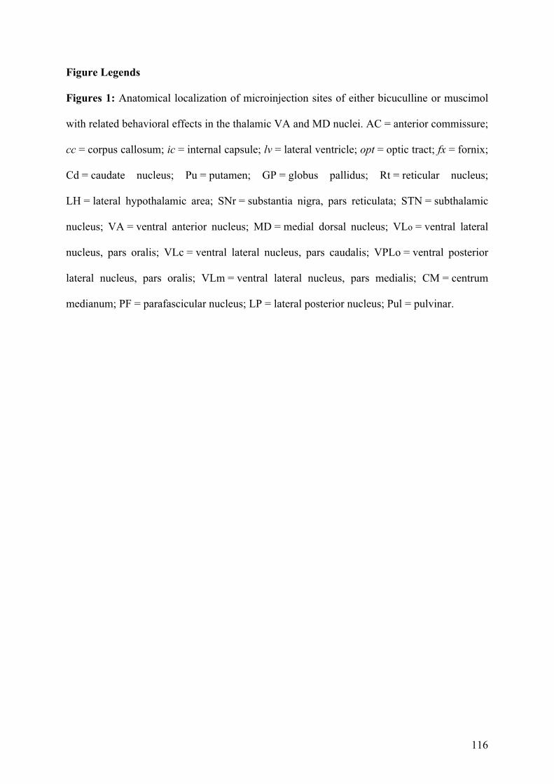
116
Figure Legends
Figures 1: Anatomical localization of microinjection sites of either bicuculline or muscimol
with related behavioral effects in the thalamic VA and MD nuclei. AC = anterior commissure;
cc = corpus callosum; ic = internal capsule; lv = lateral ventricle; opt = optic tract; fx = fornix;
Cd = caudate nucleus; Pu = putamen; GP = globus pallidus; Rt = reticular nucleus;
LH = lateral hypothalamic area; SNr = substantia nigra, pars reticulata; STN = subthalamic
nucleus; VA = ventral anterior nucleus; MD = medial dorsal nucleus; VLo = ventral lateral
nucleus, pars oralis; VLc = ventral lateral nucleus, pars caudalis; VPLo = ventral posterior
lateral nucleus, pars oralis; VLm = ventral lateral nucleus, pars medialis; CM = centrum
medianum; PF = parafascicular nucleus; LP = lateral posterior nucleus; Pul = pulvinar.
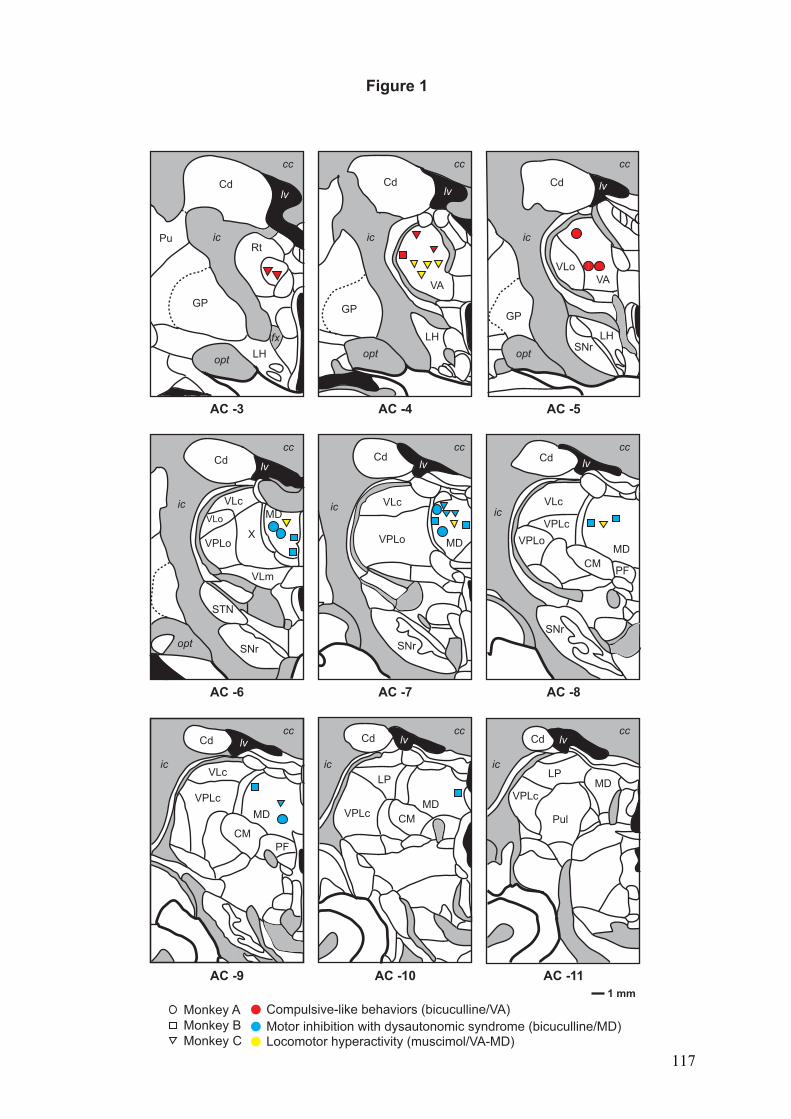
1 mm
Monkey A
Monkey B
Monkey C
Cd
Pu
GP
icRt
LHopt
lv
cc
fx
Cdlv
GP
opt
LH
ic
cc
AC -3 AC -4
Cd lv
opt
GP
ic
VLo
LHSNr
AC -5
Cdlv
VLo
VPLo
VLc
X
STN
SNr
VLm
opt
ic
cc
AC -6
Cd
VLc
VPLo
SNr
ic
lv
cc
AC -7
Cdlv
SNr
VLc
VPLo
VPLc
CM
ic
cc
PF
AC -8
VLc
Cd
VPLc
ic
lvcc
CM
PF
AC -9
VPLc
Cd lv
LP
CM
ic
cc
AC -10
Cd
VPLc
LP
lv
ic
Pul
AC -11
cc
cc
Compulsive-like behaviors (bicuculline/VA)
Motor inhibition with dysautonomic syndrome (bicuculline/MD)Locomotor hyperactivity (muscimol/VA-MD)
VAVA
MD
MDMD
MD
MD
MD
Figure 1
117
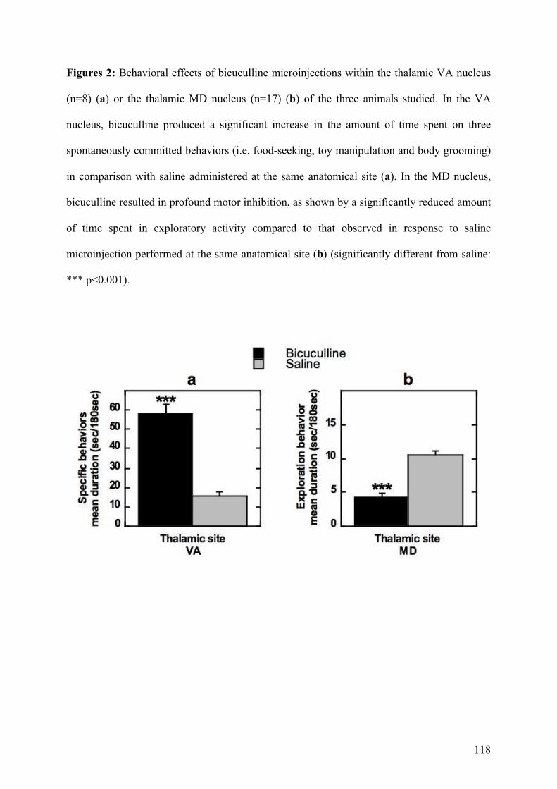
118
Figures 2: Behavioral effects of bicuculline microinjections within the thalamic VA nucleus
(n=8) (a) or the thalamic MD nucleus (n=17) (b) of the three animals studied. In the VA
nucleus, bicuculline produced a significant increase in the amount of time spent on three
spontaneously committed behaviors (i.e. food-seeking, toy manipulation and body grooming)
in comparison with saline administered at the same anatomical site (a). In the MD nucleus,
bicuculline resulted in profound motor inhibition, as shown by a significantly reduced amount
of time spent in exploratory activity compared to that observed in response to saline
microinjection performed at the same anatomical site (b) (significantly different from saline:
*** p<0.001).
119
Figures 3: Behavioral effects of muscimol microinjections within the thalamic VA nucleus
(n=4) (a, b) or the thalamic MD nucleus (n=3) (a, b) in one of the three animals studied. In
the VA nucleus, muscimol produced motor hyperactivity, as attested by a significantly greater
amount of time spent in exploratory behavior than that found after saline administration at the
same anatomical site (a). Muscimol also caused a rapid shift from one behavioral sequence to
another, as suggested by a significantly higher frequency of the different types of specific
behavior recorded (b). In the MD nucleus, muscimol gave rise to similar behavioral changes,
although less pronounced than those observed in the VA nucleus (a, b) (significantly different
from saline: *** p<0.001, * p<0.05; significantly different from muscimol injected into the
VA: ### p<0.001).
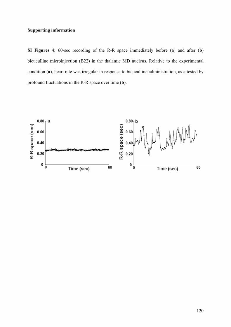
120
Supporting information
SI Figures 4: 60-sec recording of the R-R space immediately before (a) and after (b)
bicuculline microinjection (B22) in the thalamic MD nucleus. Relative to the experimental
condition (a), heart rate was irregular in response to bicuculline administration, as attested by
profound fluctuations in the R-R space over time (b).
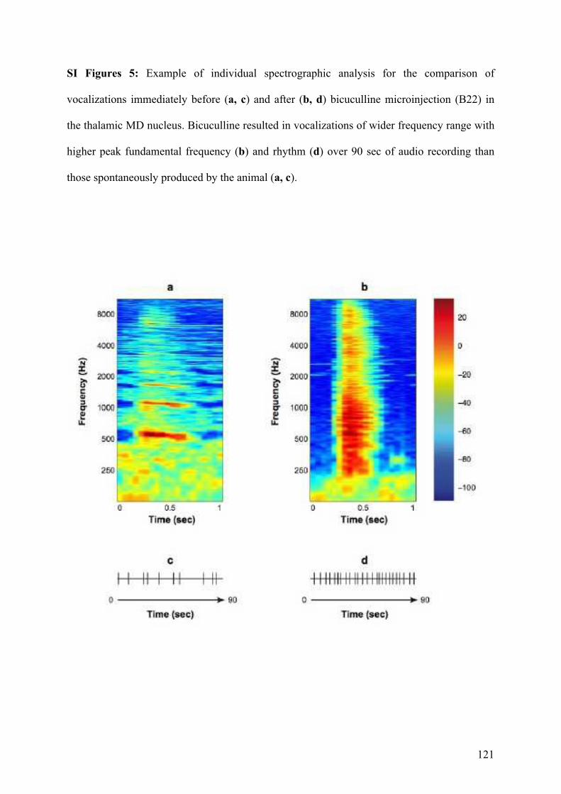
121
SI Figures 5: Example of individual spectrographic analysis for the comparison of
vocalizations immediately before (a, c) and after (b, d) bicuculline microinjection (B22) in
the thalamic MD nucleus. Bicuculline resulted in vocalizations of wider frequency range with
higher peak fundamental frequency (b) and rhythm (d) over 90 sec of audio recording than
those spontaneously produced by the animal (a, c).
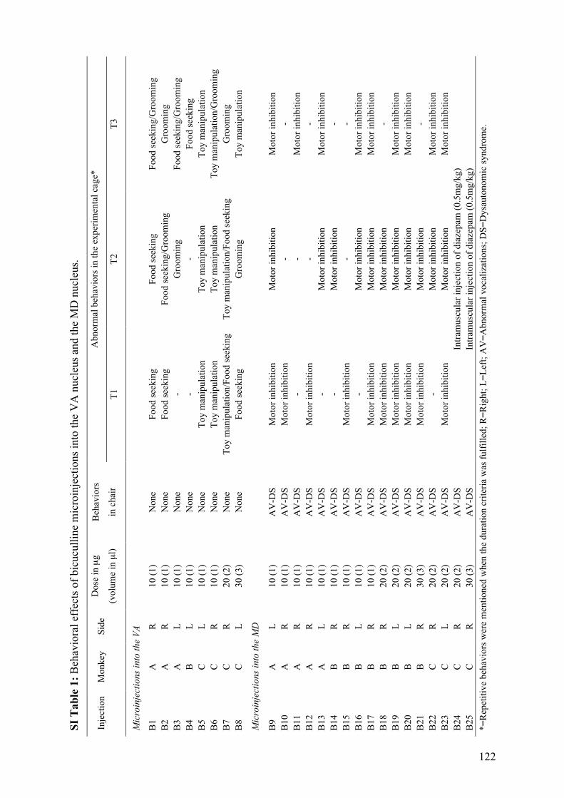
SI T
ab
le 1
: B
ehav
iora
l ef
fect
s o
f b
icu
cull
ine
mic
roin
ject
ion
s in
to t
he
VA
nu
cleu
s an
d t
he
MD
nu
cleu
s.
Ab
no
rmal
beh
avio
rs i
n t
he
exp
erim
enta
l ca
ge*
In
ject
ion
M
on
key
S
ide
Do
se i
n µ
g
(vo
lum
e in
µl)
Beh
avio
rs
in c
hai
r T
1
T2
T
3
Mic
roin
ject
ion
s in
to t
he
VA
B1
A
R
1
0 (
1)
No
ne
Fo
od
see
kin
g
Fo
od
see
kin
g
Fo
od
see
kin
g/G
roo
min
g
B2
A
R
1
0 (
1)
No
ne
Fo
od
see
kin
g
Fo
od
see
kin
g/G
roo
min
g
Gro
om
ing
B3
A
L
1
0 (
1)
No
ne
- G
roo
min
g
Fo
od
see
kin
g/G
roo
min
g
B4
B
L
1
0 (
1)
No
ne
- -
Fo
od
see
kin
g
B5
C
L
1
0 (
1)
No
ne
To
y m
anip
ula
tio
n
To
y m
anip
ula
tio
n
To
y m
anip
ula
tio
n
B6
C
R
1
0 (
1)
No
ne
To
y m
anip
ula
tio
n
To
y m
anip
ula
tio
n
To
y m
anip
ula
tio
n/G
roo
min
g
B7
C
R
2
0 (
2)
No
ne
To
y m
anip
ula
tio
n/F
oo
d s
eek
ing
T
oy
man
ipu
lati
on
/Fo
od
see
kin
g
Gro
om
ing
B8
C
L
3
0 (
3)
No
ne
Fo
od
see
kin
g
Gro
om
ing
T
oy
man
ipu
lati
on
Mic
roin
ject
ion
s in
to t
he
MD
B9
A
L
1
0 (
1)
AV
-DS
M
oto
r in
hib
itio
n
Mo
tor
inh
ibit
ion
M
oto
r in
hib
itio
n
B1
0
A
R
10
(1
) A
V-D
S
Mo
tor
inh
ibit
ion
-
-
B1
1
A
R
10
(1
) A
V-D
S
- -
Mo
tor
inh
ibit
ion
B1
2
A
R
10
(1
) A
V-D
S
Mo
tor
inh
ibit
ion
-
-
B1
3
A
L
10
(1
) A
V-D
S
- M
oto
r in
hib
itio
n
Mo
tor
inh
ibit
ion
B1
4
B
R
10
(1
) A
V-D
S
- M
oto
r in
hib
itio
n
-
B1
5
B
R
10
(1
) A
V-D
S
Mo
tor
inh
ibit
ion
-
-
B1
6
B
L
10
(1
) A
V-D
S
- M
oto
r in
hib
itio
n
Mo
tor
inh
ibit
ion
B1
7
B
R
10
(1
) A
V-D
S
Mo
tor
inh
ibit
ion
M
oto
r in
hib
itio
n
Mo
tor
inh
ibit
ion
B1
8
B
R
20
(2
) A
V-D
S
Mo
tor
inh
ibit
ion
M
oto
r in
hib
itio
n
-
B1
9
B
L
20
(2
) A
V-D
S
Mo
tor
inh
ibit
ion
M
oto
r in
hib
itio
n
Mo
tor
inh
ibit
ion
B2
0
B
L
20
(2
) A
V-D
S
Mo
tor
inh
ibit
ion
M
oto
r in
hib
itio
n
Mo
tor
inh
ibit
ion
B2
1
B
R
30
(3
) A
V-D
S
Mo
tor
inh
ibit
ion
M
oto
r in
hib
itio
n
-
B2
2
C
R
20
(2
) A
V-D
S
- M
oto
r in
hib
itio
n
Mo
tor
inh
ibit
ion
B2
3
C
L
20
(2
) A
V-D
S
Mo
tor
inh
ibit
ion
M
oto
r in
hib
itio
n
Mo
tor
inh
ibit
ion
B2
4
C
R
20
(2
) A
V-D
S
Intr
amu
scu
lar
inje
ctio
n o
f d
iaze
pam
(0
.5m
g/k
g)
B2
5
C
R
30
(3
) A
V-D
S
Intr
amu
scu
lar
inje
ctio
n o
f d
iaze
pam
(0
.5m
g/k
g)
*=
Rep
etit
ive
beh
avio
rs w
ere
men
tio
ned
wh
en t
he
du
rati
on
cri
teri
a w
as f
ulf
ille
d;
R=
Rig
ht;
L=
Lef
t; A
V=
Ab
no
rmal
vo
cali
zati
on
s; D
S=
Dy
sau
ton
om
ic s
yn
dro
me.
122
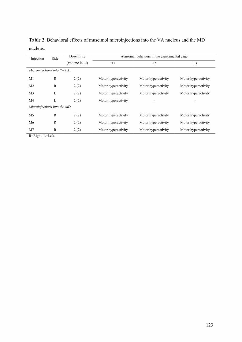
123
Table 2. Behavioral effects of muscimol microinjections into the VA nucleus and the MD
nucleus.
Abnormal behaviors in the experimental cage Injection Side
Dose in µg
(volume in µl) T1 T2 T3
Microinjections into the VA
M1 R 2 (2) Motor hyperactivity Motor hyperactivity Motor hyperactivity
M2 R 2 (2) Motor hyperactivity Motor hyperactivity Motor hyperactivity
M3 L 2 (2) Motor hyperactivity Motor hyperactivity Motor hyperactivity
M4 L 2 (2) Motor hyperactivity - -
Microinjections into the MD
M5 R 2 (2) Motor hyperactivity Motor hyperactivity Motor hyperactivity
M6 R 2 (2) Motor hyperactivity Motor hyperactivity Motor hyperactivity
M7 R 2 (2) Motor hyperactivity Motor hyperactivity Motor hyperactivity
R=Right; L=Left.

124
125
4.3. Une tâche originale pour l'étude du comportement de vérification
4.3.1. Introduction
Dans le but d'étudier les déterminants cognitifs et émotionnels associés à l’expression de
symptômes obsessionnels et compulsifs, nous avons élaboré une tâche originale qui permet
l'expression de la réponse compulsive aux idées obsédantes, à l'inverse des tâches de
provocation symptomatique classiquement utilisées. Elle consiste en une tâche d'appariement
retardé d'images ("delayed matching-to-sample") à laquelle s'ajoute la possibilité illimitée de
vérifier le choix effectué. Le sujet doit, après avoir comparé deux images d'apparition
retardée, décider si les images sont identiques ou différentes. Après sa prise de décision, le
sujet a la possibilité de vérifier sa décision en revoyant les images (voir la Figure 1 de
l'article, page 129). L'objectif de notre étude était de valider cette tâche comme outil
expérimental capable de discriminer des patterns comportementaux différents de prise de
décision et de vérification au sein de trois populations : patients TOC avec compulsions de
vérification, patients TOC sans compulsion de vérification et témoins.
4.3.2. Méthodes
Nous avons inclus 50 patients avec TOC dont 36 avec compulsions de vérification et 50
sujets témoins appariés en âge, sexe et niveau d’étude. Après un entraînement de 10 essais
effectués avec l'expérimentateur, chaque participant a réalisé un total de 50 essais randomisés
qui comprenaient 25 couples d'images identiques et 25 couples d'images différentes. Nous
avons extrait les données correspondant au nombre d'essais réussis, au nombre de
vérifications et à la durée du temps de réponse lors de la prise de décision pour chaque
participant. Les analyses statistiques ont consisté en ANOVAs et tests post-hoc de Newman-
Keuls.
4.3.3. Résultats et discussion
Le nombre d'essais réussis était similaire entre patients et témoins. Le nombre de
vérifications était plus important chez les patients avec TOC, en particulier ceux souffrant de
compulsions de vérification, par rapport aux témoins. Les essais avec couples d'images
identiques entraînaient plus de vérifications que les essais avec couples d'images différentes
chez les patients par rapport les témoins. Il existait un allongement du temps de réponse lors
de la prise de décision lorsque celle-ci précédait un comportement de vérification chez les
126
patients et chez les témoins. Cet allongement était significativement plus important chez les
patients, notamment s'ils avaient des compulsions de vérification. Enfin, le nombre de
vérifications restait stable au cours de la passation de la tâche chez les témoins alors qu'il
augmentait au fur et à mesure des essais chez les patients, en particulier ceux avec
compulsions de vérification.
Bien que nous n'ayons pas évalué l'incertitude lors de la passation de la tâche, nous
pensons que l'allongement du temps de réponse précédant l'émission d'un comportement de
vérification pourrait être en lien avec le degré d'incertitude au moment de la prise de décision.
D'autres données que nous présenterons dans le chapitre suivant vont dans le sens de cette
interprétation. Le comportement de vérification observé chez les patients lors de la passation
de cette tâche partage d'autres caractéristiques avec les comportements compulsifs puisqu'il
s'agit d'un comportement répété et possiblement précédé par un sentiment d'incertitude. Le
pattern ascendant du nombre de vérifications au cours de la tâche observé chez les patients
pourrait ainsi être vu comme une stratégie comportementale visant à réduire la charge
anxieuse associée au doute.
Des limites méthodologiques sont à souligner. Une évaluation neuropsychologique aurait
été nécessaire. En effet, il est probable qu'un nombre certain de patients ayant fait des
épisodes dépressifs par le passé ou prenant des psychotropes, notamment antipsychotiques, au
moment de l'étude aient des altérations neurocognitives avec des répercussions sur leurs
capacités de discrimination visuelle ou encore de mémoire de travail.
4.3.4. Conclusion
La tâche de vérification semble être un outil expérimental intéressant pour étudier le
comportement de vérification et ses déterminants cognitifs et émotionnels.
Rotgé JY, Clair AH, Jaafari N, Hantouche EG, Pelissolo A, Goillandeau M,
Pochon JB, Guehl D, Bioulac B, Burbaud P, Tignol J, Mallet L, Aouizerate B. A
challenging task for assessment of checking behaviors in obsessive-compulsive
disorder. Acta Psychiatrica Scandinavica 2008;117:465-73.
A challenging task for assessment ofchecking behaviors in obsessive–compulsivedisorder
Rotge JY, Clair AH, Jaafari N, Hantouche EG, Pelissolo A,Goillandeau M, Pochon JB, Guehl D, Bioulac B, Burbaud P, Tignol J,Mallet L, Aouizerate B. A challenging task for assessment of checkingbehaviors in obsessive–compulsive disorder.
Objective: The present study concerns the objective and quantitativemeasurement of checking activity, which represents the mostfrequently observed compulsions in obsessive–compulsive disorder(OCD). To address this issue, we developed an instrumental taskproducing repetitive checking in OCD subjects.Method: Fifty OCD subjects and 50 normal volunteers (NV) wereadministered a delayed matching-to-sample task that offered theunrestricted opportunity to verify the choice made. Response accuracy,number of verifications, and response time for choice taken to reflectthe degree of uncertainty and doubt were recorded over 50 consecutivetrials.Results: Despite similar levels of performance, patients with OCDdemonstrated a greater number of verifications and a longer responsetime for choice before checking than NV. Such behavioral patternswere more pronounced in OCD subjects currently experiencingchecking compulsions.Conclusion: The present task might be of special relevance for thequantitative assessment of checking behaviors and for determiningrelationships with cognitive processes.
J. Y. Rotge1,2, A. H. Clair3,N. Jaafari4, E. G. Hantouche5,A. Pelissolo5,6, M. Goillandeau1,J. B. Pochon3, D. Guehl1,7,B. Bioulac1,7, P. Burbaud1,7,J. Tignol2, L. Mallet3,B. Aouizerate1,2
1Movement, Adaptation, Cognition Laboratory, CNRS
UMR 5227, Bordeaux 2 University, Bordeaux, France,2Academic Department of Adult Psychiatry, Charles
Perrens Hospital, Bordeaux, France, 3Clinical
Investigation Center, Behavior, Emotion and Basal
Ganglia Laboratory, INSERM IFR 70, PitiØ-SalpÞtri�re
Hospital, Paris, France, 4Academic Department of Adult
Psychiatry, Henri Laborit University Hospital, Poitiers,
France, 5Academic Department of Adult Psychiatry,
PitiØ-SalpÞtri�re University Hospital, Paris, France,6Personnality and Adaptive Behaviors Laboratory, CNRS
UMR 7593, PitiØ-SalpÞtri�re University Hospital, Paris,
France and 7Academic Department of Neurophysiology,
Pellegrin University Hospital, Bordeaux, France
Key words: checking behavior; delayed matching-to-
sample task; doubt; obsessive–compulsive disorder
Dr Bruno Aouizerate, Academic Department of Adult
Psychiatry, CNRS UMR 5227, Bordeaux 2 University,
Charles Perrens Hospital, 121 rue de la BØchade, 33076
Bordeaux, France.
E-mail: [email protected]
Accepted for publication January 30, 2008
Significant outcomes
• The present task allows one to assess the propensity to develop checking activity in obsessive–compulsive disorder and to characterize some cognitive and emotional aspects of this repetitivebehavior.
Limitations
• No neuropsychological test was performed to explore visuospatial abilities and to eliminate deficitsrelated to spatial working memory and recognition in subjects with obsessive–compulsive disorder.
• Most of the study subjects with obsessive–compulsive disorder were administered with psychotropicmedications. Some among these agents were able to alter cognitive and motor functions, and tointerfere with the task performance.
Acta Psychiatr Scand 2008: 117: 465–473All rights reservedDOI: 10.1111/j.1600-0447.2008.01173.x
Copyright Ó 2008 The AuthorsJournal Compilation Ó 2008 Blackwell Munksgaard
ACTA PSYCHIATRICA
SCANDINAVICA
465127
Introduction
Obsessive–compulsive disorder (OCD) is a rela-tively frequent anxiety disorder that affects 2–3%of the general population (1). It is characterized bypersistent, intrusive and unpleasant thoughts,impulses or images. Recurrent, time-consumingbehaviors are performed in a ritualistic fashionaccording to rigid rules to reduce the anxiety andsevere distress caused by obsessions.Phenomenological considerations suggest that
the core of the obsessional process is the sensationthat Ôsomething is wrongÕ (2, 3). Obsessions may beunderstood as the consequence of an excessiveattribution of negative consequences upon expo-sure to certain behavioral situations (4, 5). Path-ological doubt, a prominent phenomenon in OCD,may be related to the permanent perception of amistake and ⁄or error in the initial representation,relating to the cognitive overestimation of thelikelihood of aversive events occurring in responseto the subjectÕs action. Pathological doubt may beconsidered not only in terms of cognitive con-structs, but also in terms of the subjectÕs emotionalreaction to feelings of uncertainty generated byperceived Ôerror detectionÕ signals (6, 7). Compul-sions may be construed as behavioral responsesintending to relieve the tensions or anxiety causedby high levels of self-doubt (8–10). If obtained, thisrelief is only transient and leads immediately toreproducing the behavior in an excessive andunreasonable manner. Given these considerations,repetitive checking behaviors, which are the mostfrequent form of compulsions in patients withOCD (1, 11), may be thought of as reducing therisk of error in the estimated probability ofnegative events in specific behavioral situations,thereby relieving the uncertainty and obsessionaldoubt (8–10).Contemporary strategies for assessment of OCD
severity use a wide variety of psychometric ratingscales. There are several self-report and assessor-rated instruments that examine different types ofobsessions and compulsions (checking, washing,etc.) or which are designed to measure the symp-toms present, regardless of their thematic content.They also provide information on the efficacy ofconventional treatments in OCD. However,administration of these rating scales requires clini-cians who are extensively trained in evaluatingpsychopathology and who are well versed ingeneral clinical interview technique. To be able toprobe the pathophysiological bases of checkingcompulsions, a tool is required that would allow usto assess objectively the propensity to repeatedchecking.
Aims of the study
We sought to investigate checking behaviors inOCD through an objective approach. For thispurpose, we developed an instrumental task thatspecifically offered the opportunity to verify oncethe choice was made. We examined whether thepresent task was able to discriminate checking andchoice behavioral patterns in two subgroups ofpatients with OCD who actively experiencedchecking compulsions (OCD CC+) or not (OCDCC)) and normal healthy volunteers (NV).
Material and methods
Subjects
Participants were 50 adult patients with a primarydiagnosis of OCD (28 women, 22 men, mean age40.3 years, SD 11.8, range 23–65) seen at threespecialized university hospitals (Charles Perrens,Henri Laborit and Pitie-Salpetriere) and 50 normalhealthy volunteers (NV) (28 women, 22 men, meanage 40.5 years, SD 12.4, range 19–63) who wererecruited from the local community by newspaperadvertisements and word of mouth. All subjectsgave their written informed consent. The groupsdid not differ significantly in age or gender. Theywere evaluated for OCD and associated psychopa-thology with the Mini-International Neuropsychi-atric Interview (MINI) (version 5.0.0) (12).Patients with OCD with comorbid diagnoses werenot excluded from the study, except for those withcurrent mood episodes, provided that OCD wasthe dominant disorder. Healthy controls wereexcluded if they had a history of Axis I psychiatricdisorders. All 50 normal volunteers were free ofmedication at the time of study, whereas 45patients with OCD were receiving psychotropicdrugs, especially antidepressants with serotoninreuptake-inhibiting properties (n = 45) alone orcombined with atypical antipsychotics (n = 12),benzodiazepines (n = 12), mood stabilizers(n = 8) and buspirone (n = 5). Twenty of the 50patients with OCD were receiving cognitive-behav-ioral therapies.
Clinical assessment
Instruments included: i) the Yale–Brown Obses-sive–Compulsive Scale (Y-BOCS) symptom check-list, a list of more than 50 common obsessions andcompulsions, which was used to identify patientswith OCD with checking compulsions (OCDCC+) (n = 36) (23 women, 13 men, mean age41.6 years, SD 11.6, range 20–65) or not (OCD
Rotge et al.
466128
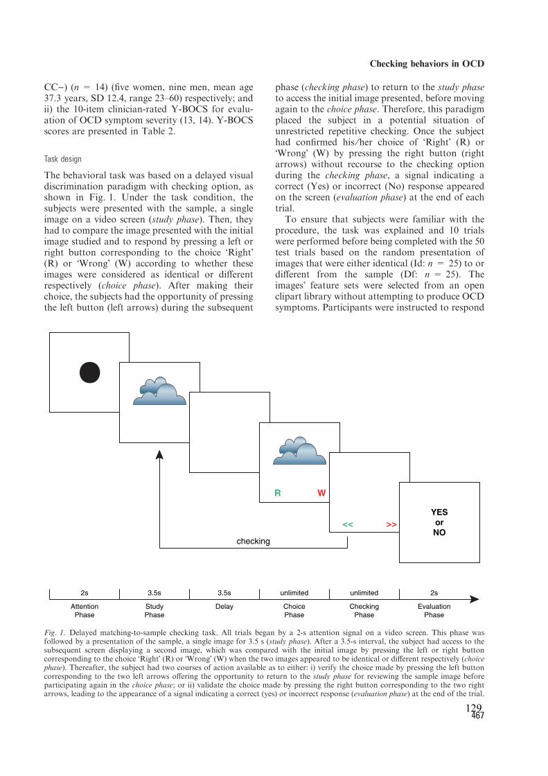
CC)) (n = 14) (five women, nine men, mean age37.3 years, SD 12.4, range 23–60) respectively; andii) the 10-item clinician-rated Y-BOCS for evalu-ation of OCD symptom severity (13, 14). Y-BOCSscores are presented in Table 2.
Task design
The behavioral task was based on a delayed visualdiscrimination paradigm with checking option, asshown in Fig. 1. Under the task condition, thesubjects were presented with the sample, a singleimage on a video screen (study phase). Then, theyhad to compare the image presented with the initialimage studied and to respond by pressing a left orright button corresponding to the choice ÔRightÕ(R) or ÔWrongÕ (W) according to whether theseimages were considered as identical or differentrespectively (choice phase). After making theirchoice, the subjects had the opportunity of pressingthe left button (left arrows) during the subsequent
phase (checking phase) to return to the study phaseto access the initial image presented, before movingagain to the choice phase. Therefore, this paradigmplaced the subject in a potential situation ofunrestricted repetitive checking. Once the subjecthad confirmed his ⁄her choice of ÔRightÕ (R) orÔWrongÕ (W) by pressing the right button (rightarrows) without recourse to the checking optionduring the checking phase, a signal indicating acorrect (Yes) or incorrect (No) response appearedon the screen (evaluation phase) at the end of eachtrial.To ensure that subjects were familiar with the
procedure, the task was explained and 10 trialswere performed before being completed with the 50test trials based on the random presentation ofimages that were either identical (Id: n = 25) to ordifferent from the sample (Df: n = 25). TheimagesÕ feature sets were selected from an openclipart library without attempting to produce OCDsymptoms. Participants were instructed to respond
R
<<
W
>>
YES
or
NOchecking
2s 3.5s 3.5s 2sunlimitedunlimited
Study Choice Checking EvaluationAttention Delay
Phase Phase Phase Phase Phase
Fig. 1. Delayed matching-to-sample checking task. All trials began by a 2-s attention signal on a video screen. This phase wasfollowed by a presentation of the sample, a single image for 3.5 s (study phase). After a 3.5-s interval, the subject had access to thesubsequent screen displaying a second image, which was compared with the initial image by pressing the left or right buttoncorresponding to the choice ÔRightÕ (R) or ÔWrongÕ (W) when the two images appeared to be identical or different respectively (choicephase). Thereafter, the subject had two courses of action available as to either: i) verify the choice made by pressing the left buttoncorresponding to the two left arrows offering the opportunity to return to the study phase for reviewing the sample image beforeparticipating again in the choice phase; or ii) validate the choice made by pressing the right button corresponding to the two rightarrows, leading to the appearance of a signal indicating a correct (yes) or incorrect response (evaluation phase) at the end of the trial.
Checking behaviors in OCD
467129
Ôas efficiently and correctly as possibleÕ. Responseaccuracy (expressed as the number of correcttrials), number of verifications and response timefor choice (defined as the time elapsing from theappearance of the image matching the sample tothe moment the choice was made) were monitoredduring the 50 consecutive trials.
Data analyses
Level of performance was compared among thethree groups by using a one-way analysis of variance(anova). The number of verifications and responsetime for choice making were examined by using atwo-way anova with repeated measures on the lastfactor followed by a one-way anova to determinedifferences between groups according to trial type,i.e. presenting comparison images identical (Id) toand different (Df) from the sample or leading to theproduction of checking behaviors (CB+) or not(CB)). The differences for each subject in either thenumber of verifications between trials Id and Df orthe response time for choice between trials CB+andCB) were analyzed by a one-way anova. Newman–Keuls tests were used for post hoc analyses whereappropriate. To describe the changes in bothnumber of verifications and response time forchoice across all 50 trials performed during thetask, linear regression lines were computed for eachgroup. To determine whether there was a progres-sive rise in these behavioral markers, as expected inpatients with OCD, correlation coefficients for eachgroup were converted by using FisherÕs r-to-ztransformation. To test this directional hypothesis,a one-tailed a = 0.05 was used. PearsonÕs correla-tionswere performed to establishwhether therewereany relationships between the behavioral parame-ters studied and the clinical severity of OCD. Ana = 0.05 (two-tailed) was used as the threshold forstatistical significance for both anova and correla-tion analyses.
Results
Both subgroups of patients with OCD had similarillness duration (OCD CC+: mean duration23.1 years, SD 12.5, range 4–48; OCD CC):mean duration 22.1 years, SD 14.1, range 4–50)and psychiatric comorbidity (Table 1). They didnot significantly differ in their Y-BOCS scores(Table 2). Eight patients with OCD (OCD CC+:n = 7; OCD CC): n = 1) were in remissiondefined as a Y-BOCS score less than 16 (15).However, none of the patients in this study reachedclinical recovery, as indicated by a Y-BOCS scoreless than 8 (15), except one OCD CC).
We first explored the level of performance in thethree groups of subjects OCD CC+, OCD CC)and NV, as measured by the number of correcttrials. No significant difference was found betweenthe groups [group effect, F(2,97) = 0.04,P > 0.96] (Fig. 2a). Thus, the three groups ofsubjects had a similar level of performance on thetask. With regard to the number of verificationsduring the behavioral task, there was a significantdifference between groups [group effect,
Table 1. Per cent of lifetime and current DSM-IV Axis I comorbidities in patients
with OCD
OCD CC+
(n = 36)
OCD CC)
(n = 14)
n % n %
Lifetime comorbidities
Mood disorders
Major depression 25 69.4 11 78.6
Bipolar I disorder 0 0 0 0
Bipolar II disorder 3 8.3 2 14.3
Anxiety disorders
Panic disorder 1 2.8 0 0
Panic disorder with agoraphobia 2 5.6 0 0
Agoraphobia without panic disorder 0 0 0 0
Social phobia 2 5.6 1 7.1
Post-traumatic stress disorder 1 2.8 0 0
Generalized anxiety disorder 0 0 0 0
Substance use disorders
Alcohol abuse ⁄ dependence 0 0 0 0
Drug abuse ⁄ dependence 0 0 0 0
Any eating disorder 0 0 0 0
Any psychotic disorder 0 0 0 0
Current comorbidities
Mood disorders
Major depression 0 0 0 0
Bipolar I disorder 0 0 0 0
Bipolar II disorder 0 0 0 0
Anxiety disorders
Panic disorder 1 2.8 0 0
Panic disorder with agoraphobia 2 5.6 0 0
Agoraphobia without panic disorder 0 0 0 0
Social phobia 2 5.6 1 7.1
Post-traumatic stress disorder 1 2.8 0 0
Generalized anxiety disorder 0 0 0 0
Substance use disorders
Alcohol abuse ⁄ dependence 0 0 0 0
Drug abuse ⁄ dependence 0 0 0 0
Any eating disorder 0 0 0 0
Any psychotic disorder 0 0 0 0
Table 2. Symptom severity scores in patients with OCD
OCD CC+ (n = 36) OCD CC) (n = 14)
Mean SD Range Mean SD Range
Y-BOCS total score 21.8 6.8 9–36 21.9 6.7 7–31
Y-BOCS obsessive subscale score 10.5 4.0 4–18 11.4 3.3 4–15
Y-BOCS compulsive subscale score 11.3 3.7 3–18 10.6 4.0 3–16
OCD CC+, patients with OCD with checking compulsions; OCD CC), patients with
OCD without checking compulsions; Y-BOCS, Yale–Brown Obsessive–compulsive
score.
Rotge et al.
468130
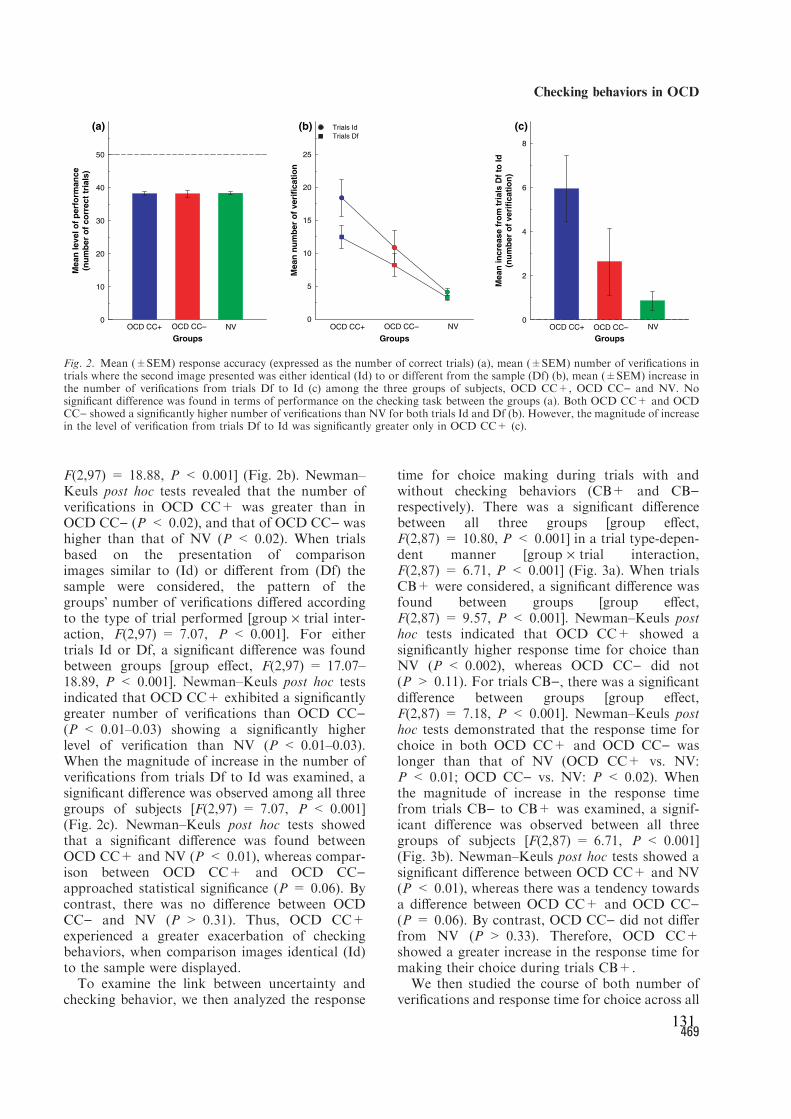
F(2,97) = 18.88, P < 0.001] (Fig. 2b). Newman–Keuls post hoc tests revealed that the number ofverifications in OCD CC+ was greater than inOCD CC) (P < 0.02), and that of OCD CC) washigher than that of NV (P < 0.02). When trialsbased on the presentation of comparisonimages similar to (Id) or different from (Df) thesample were considered, the pattern of thegroupsÕ number of verifications differed accordingto the type of trial performed [group · trial inter-action, F(2,97) = 7.07, P < 0.001]. For eithertrials Id or Df, a significant difference was foundbetween groups [group effect, F(2,97) = 17.07–18.89, P < 0.001]. Newman–Keuls post hoc testsindicated that OCD CC+ exhibited a significantlygreater number of verifications than OCD CC)(P < 0.01–0.03) showing a significantly higherlevel of verification than NV (P < 0.01–0.03).When the magnitude of increase in the number ofverifications from trials Df to Id was examined, asignificant difference was observed among all threegroups of subjects [F(2,97) = 7.07, P < 0.001](Fig. 2c). Newman–Keuls post hoc tests showedthat a significant difference was found betweenOCD CC+ and NV (P < 0.01), whereas compar-ison between OCD CC+ and OCD CC)approached statistical significance (P = 0.06). Bycontrast, there was no difference between OCDCC) and NV (P > 0.31). Thus, OCD CC+experienced a greater exacerbation of checkingbehaviors, when comparison images identical (Id)to the sample were displayed.To examine the link between uncertainty and
checking behavior, we then analyzed the response
time for choice making during trials with andwithout checking behaviors (CB+ and CB)respectively). There was a significant differencebetween all three groups [group effect,F(2,87) = 10.80, P < 0.001] in a trial type-depen-dent manner [group · trial interaction,F(2,87) = 6.71, P < 0.001] (Fig. 3a). When trialsCB+ were considered, a significant difference wasfound between groups [group effect,F(2,87) = 9.57, P < 0.001]. Newman–Keuls posthoc tests indicated that OCD CC+ showed asignificantly higher response time for choice thanNV (P < 0.002), whereas OCD CC) did not(P > 0.11). For trials CB), there was a significantdifference between groups [group effect,F(2,87) = 7.18, P < 0.001]. Newman–Keuls posthoc tests demonstrated that the response time forchoice in both OCD CC+ and OCD CC) waslonger than that of NV (OCD CC+ vs. NV:P < 0.01; OCD CC) vs. NV: P < 0.02). Whenthe magnitude of increase in the response timefrom trials CB) to CB+ was examined, a signif-icant difference was observed between all threegroups of subjects [F(2,87) = 6.71, P < 0.001](Fig. 3b). Newman–Keuls post hoc tests showed asignificant difference between OCD CC+ and NV(P < 0.01), whereas there was a tendency towardsa difference between OCD CC+ and OCD CC)(P = 0.06). By contrast, OCD CC) did not differfrom NV (P > 0.33). Therefore, OCD CC+showed a greater increase in the response time formaking their choice during trials CB+.We then studied the course of both number of
verifications and response time for choice across all
Me
an
le
ve
l o
f p
erf
orm
an
ce
(nu
mb
er
of
co
rre
ct
tria
ls)
0
10
20
30
40
50
(a) (b) (c)
Groups
OCD CC+ OCD CC– NV
Me
an
in
cre
as
e f
rom
tri
als
Df
to I
d
(nu
mb
er
of
ve
rifi
ca
tio
n)
0
2
4
6
8
OCD CC+ OCD CC– NV
Groups
Me
an
nu
mb
er
of
ve
rifi
ca
tio
n
0
5
10
15
20
25
Trials Id
Trials Df
OCD CC+ OCD CC– NV
Groups
Fig. 2. Mean (±SEM) response accuracy (expressed as the number of correct trials) (a), mean (±SEM) number of verifications intrials where the second image presented was either identical (Id) to or different from the sample (Df) (b), mean (±SEM) increase inthe number of verifications from trials Df to Id (c) among the three groups of subjects, OCD CC+, OCD CC) and NV. Nosignificant difference was found in terms of performance on the checking task between the groups (a). Both OCD CC+ and OCDCC) showed a significantly higher number of verifications than NV for both trials Id and Df (b). However, the magnitude of increasein the level of verification from trials Df to Id was significantly greater only in OCD CC+ (c).
Checking behaviors in OCD
469131
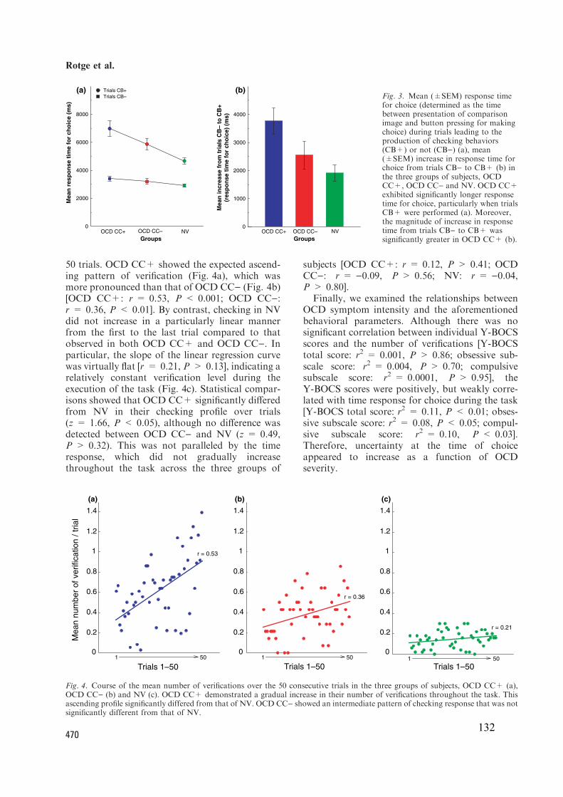
50 trials. OCD CC+ showed the expected ascend-ing pattern of verification (Fig. 4a), which wasmore pronounced than that of OCD CC) (Fig. 4b)[OCD CC+: r = 0.53, P < 0.001; OCD CC):r = 0.36, P < 0.01]. By contrast, checking in NVdid not increase in a particularly linear mannerfrom the first to the last trial compared to thatobserved in both OCD CC+ and OCD CC). Inparticular, the slope of the linear regression curvewas virtually flat [r = 0.21, P > 0.13], indicating arelatively constant verification level during theexecution of the task (Fig. 4c). Statistical compar-isons showed that OCD CC+ significantly differedfrom NV in their checking profile over trials(z = 1.66, P < 0.05), although no difference wasdetected between OCD CC) and NV (z = 0.49,P > 0.32). This was not paralleled by the timeresponse, which did not gradually increasethroughout the task across the three groups of
subjects [OCD CC+: r = 0.12, P > 0.41; OCDCC): r = )0.09, P > 0.56; NV: r = )0.04,P > 0.80].Finally, we examined the relationships between
OCD symptom intensity and the aforementionedbehavioral parameters. Although there was nosignificant correlation between individual Y-BOCSscores and the number of verifications [Y-BOCStotal score: r2 = 0.001, P > 0.86; obsessive sub-scale score: r2 = 0.004, P > 0.70; compulsivesubscale score: r2 = 0.0001, P > 0.95], theY-BOCS scores were positively, but weakly corre-lated with time response for choice during the task[Y-BOCS total score: r2 = 0.11, P < 0.01; obses-sive subscale score: r2 = 0.08, P < 0.05; compul-sive subscale score: r2 = 0.10, P < 0.03].Therefore, uncertainty at the time of choiceappeared to increase as a function of OCDseverity.
0
2000
4000
6000
8000
(a) (b)
Groups
Trials CB+
Trials CB–
Mean
resp
on
se t
ime f
or
ch
oic
e (
ms)
NVOCD CC–OCD CC+
Mean
in
cre
ase f
rom
tri
als
CB
– t
o C
B+
(resp
on
se t
ime f
or
ch
oic
e)
(ms)
0
1000
2000
3000
4000
NVOCD CC–OCD CC+
Groups
Fig. 3. Mean (±SEM) response timefor choice (determined as the timebetween presentation of comparisonimage and button pressing for makingchoice) during trials leading to theproduction of checking behaviors(CB+) or not (CB)) (a), mean(±SEM) increase in response time forchoice from trials CB) to CB+ (b) inthe three groups of subjects, OCDCC+, OCD CC) and NV. OCD CC+exhibited significantly longer responsetime for choice, particularly when trialsCB+ were performed (a). Moreover,the magnitude of increase in responsetime from trials CB) to CB+ wassignificantly greater in OCD CC+ (b).
501
Trials 1–50
Me
an
nu
mb
er
of
ve
rifica
tio
n /
tria
l
1.4
(a) (b) (c)
1.2
1
0.8
0.2
0.6
0.4
0
r = 0.53
501
Trials 1–50
1.4
1.2
1
0.8
0.2
0.6
0.4
0
r = 0.36
501
Trials 1–50
1.4
1.2
1
0.8
0.2
0.6
0.4
0
r = 0.21
Fig. 4. Course of the mean number of verifications over the 50 consecutive trials in the three groups of subjects, OCD CC+ (a),OCD CC) (b) and NV (c). OCD CC+ demonstrated a gradual increase in their number of verifications throughout the task. Thisascending profile significantly differed from that of NV. OCD CC) showed an intermediate pattern of checking response that was notsignificantly different from that of NV.
Rotge et al.
470132
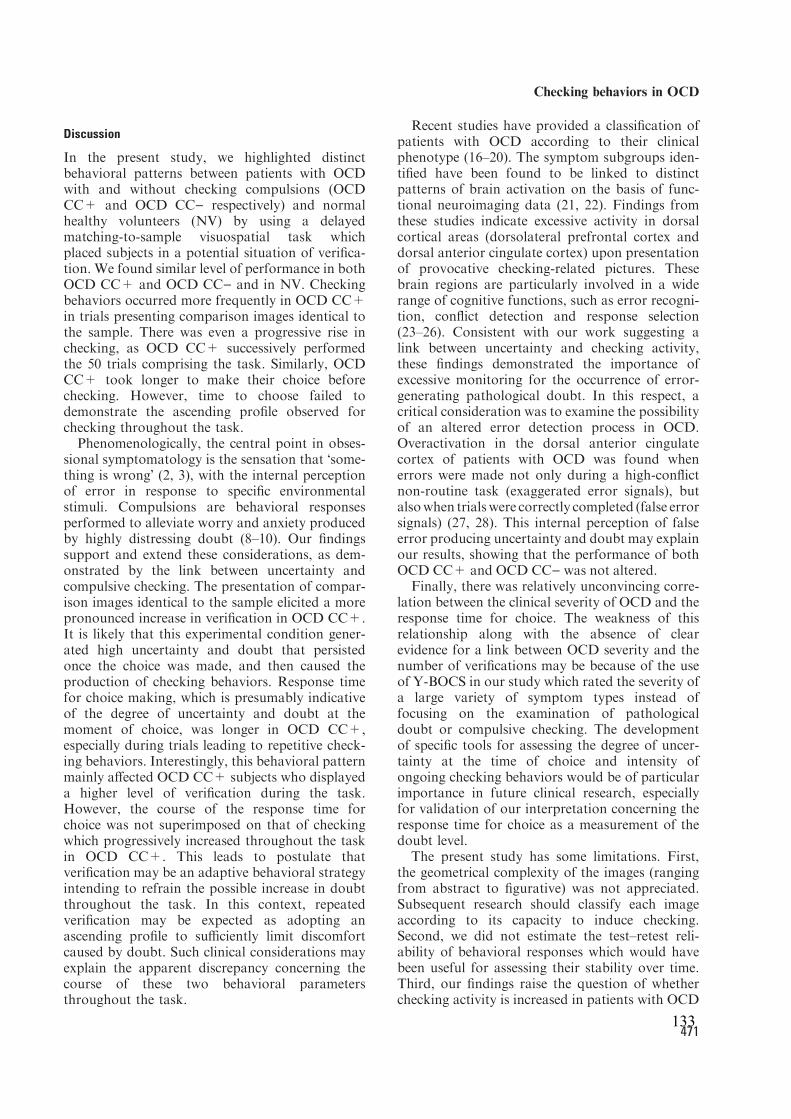
Discussion
In the present study, we highlighted distinctbehavioral patterns between patients with OCDwith and without checking compulsions (OCDCC+ and OCD CC) respectively) and normalhealthy volunteers (NV) by using a delayedmatching-to-sample visuospatial task whichplaced subjects in a potential situation of verifica-tion. We found similar level of performance in bothOCD CC+ and OCD CC) and in NV. Checkingbehaviors occurred more frequently in OCD CC+in trials presenting comparison images identical tothe sample. There was even a progressive rise inchecking, as OCD CC+ successively performedthe 50 trials comprising the task. Similarly, OCDCC+ took longer to make their choice beforechecking. However, time to choose failed todemonstrate the ascending profile observed forchecking throughout the task.Phenomenologically, the central point in obses-
sional symptomatology is the sensation that Ôsome-thing is wrongÕ (2, 3), with the internal perceptionof error in response to specific environmentalstimuli. Compulsions are behavioral responsesperformed to alleviate worry and anxiety producedby highly distressing doubt (8–10). Our findingssupport and extend these considerations, as dem-onstrated by the link between uncertainty andcompulsive checking. The presentation of compar-ison images identical to the sample elicited a morepronounced increase in verification in OCD CC+.It is likely that this experimental condition gener-ated high uncertainty and doubt that persistedonce the choice was made, and then caused theproduction of checking behaviors. Response timefor choice making, which is presumably indicativeof the degree of uncertainty and doubt at themoment of choice, was longer in OCD CC+,especially during trials leading to repetitive check-ing behaviors. Interestingly, this behavioral patternmainly affected OCD CC+ subjects who displayeda higher level of verification during the task.However, the course of the response time forchoice was not superimposed on that of checkingwhich progressively increased throughout the taskin OCD CC+. This leads to postulate thatverification may be an adaptive behavioral strategyintending to refrain the possible increase in doubtthroughout the task. In this context, repeatedverification may be expected as adopting anascending profile to sufficiently limit discomfortcaused by doubt. Such clinical considerations mayexplain the apparent discrepancy concerning thecourse of these two behavioral parametersthroughout the task.
Recent studies have provided a classification ofpatients with OCD according to their clinicalphenotype (16–20). The symptom subgroups iden-tified have been found to be linked to distinctpatterns of brain activation on the basis of func-tional neuroimaging data (21, 22). Findings fromthese studies indicate excessive activity in dorsalcortical areas (dorsolateral prefrontal cortex anddorsal anterior cingulate cortex) upon presentationof provocative checking-related pictures. Thesebrain regions are particularly involved in a widerange of cognitive functions, such as error recogni-tion, conflict detection and response selection(23–26). Consistent with our work suggesting alink between uncertainty and checking activity,these findings demonstrated the importance ofexcessive monitoring for the occurrence of error-generating pathological doubt. In this respect, acritical consideration was to examine the possibilityof an altered error detection process in OCD.Overactivation in the dorsal anterior cingulatecortex of patients with OCD was found whenerrors were made not only during a high-conflictnon-routine task (exaggerated error signals), butalsowhen trialswere correctly completed (false errorsignals) (27, 28). This internal perception of falseerror producing uncertainty and doubt may explainour results, showing that the performance of bothOCD CC+ and OCD CC) was not altered.Finally, there was relatively unconvincing corre-
lation between the clinical severity of OCD and theresponse time for choice. The weakness of thisrelationship along with the absence of clearevidence for a link between OCD severity and thenumber of verifications may be because of the useof Y-BOCS in our study which rated the severity ofa large variety of symptom types instead offocusing on the examination of pathologicaldoubt or compulsive checking. The developmentof specific tools for assessing the degree of uncer-tainty at the time of choice and intensity ofongoing checking behaviors would be of particularimportance in future clinical research, especiallyfor validation of our interpretation concerning theresponse time for choice as a measurement of thedoubt level.The present study has some limitations. First,
the geometrical complexity of the images (rangingfrom abstract to figurative) was not appreciated.Subsequent research should classify each imageaccording to its capacity to induce checking.Second, we did not estimate the test–retest reli-ability of behavioral responses which would havebeen useful for assessing their stability over time.Third, our findings raise the question of whetherchecking activity is increased in patients with OCD
Checking behaviors in OCD
471133
intending to compensate a possible impairment invisuospatial ability, thereby maintaining a normallevel of performance at the behavioral task. How-ever, deficits related to spatial working memoryand spatial recognition have not been consistentlyfound in patients with OCD (29–31). Their perfor-mance accuracy was even reported as higher thanthat of controls on a delayed matching-to-sampletask (32). Despite having comparable levels ofresponse accuracy, both of our subgroups ofpatients with OCD differed in their checkingpattern. Therefore, the checking process might besomething other than just a compensatorymechanism in the phase of spatial workingmemory in the present task. Fourth, only OCDsubjects with remitted and uncomplicated mooddisorders were included in our study, therebyminimizing the potential influence of this factoron cognitive functions. Finally, it may be prema-ture to rule out the possibly deleterious effects ofcurrent medications on neuropsychological perfor-mance. Nonetheless, the power of the study wasinadequate to examine each drug separately.In conclusion, this study using a delayed match-
ing-to-sample task and proposing the option ofchoice checking provides an objective quantifica-tion of checking in patients with OCD. For futurepsychophysiological research, such an experimen-tal approach would be helpful for understandingthe cognitive and emotional determinants ofchecking and for identifying its anatomical sub-strates.
Acknowledgements
The authors thank Dr Ray Cooke for providing advice on the
English language. This work was supported in part by a grant
from the ÔAgence Nationale de la RechercheÕ. JY Rotge was
supported by a PhD grant from the ÔFondation pour la
Recherche MedicaleÕ.
Declaration of interest
All authors reported no biomedical financial interests or
potential conflicts of interest.
References
1. Rasmussen SA, Eisen JL. The epidemiology and differential
diagnosis of obsessive compulsive disorder. J Clin Psychi-
atry 1992;53:4–10.
2. Schwartz JM. Neuroanatomical aspects of cognitive-
behavioural therapy response in obsessive–compulsive
disorder. An evolving perspective on brain and behaviour.
Br J Psychiatry Suppl 1998;35:38–44.
3. Schwartz JM. A role of volition and attention in the gen-
eration of new brain circuitry. Toward a neurobiology of
mental force. J Consciousness Stud 1999;6:115–142.
4. Salkovskis PM. Obsessional–compulsive problems: a cog-
nitive-behavioural analysis. Behav Res Ther 1985;23:571–
583.
5. Salkovskis PM. Understanding and treating obsessive–
compulsive disorder. Behav Res Ther 1999;37:29–52.
6. Foa EB, Franklin ME, Kozac MJ. Psychosocial treatments
for obsessive–compulsive disorder. Literature review. In:
Swinson RP, Antony MM, Rachman S, Richter MA, eds.
Obsessive–compulsive disorder. New York: Theory, Re-
search, and Treatment, Guilford Press, 1998:258–276.
7. Tolin DF, Abramowitz JS, Brigidi BD, Foa EB. Intolerance
of uncertainty in obsessive–compulsive disorder. J Anxiety
Disord 2003;17:233–242.
8. Aouizerate B, Guehl D, Cuny E et al. Pathophysiology of
obsessive–compulsive disorder: a necessary link between
phenomenology, neuropsychology, imagery and physiol-
ogy. Prog Neurobiol 2004;72:195–221.
9. Aouizerate B, Guehl D, Cuny E et al. Updated overview of
the putative role of the serotoninergic system in obsessive–
compulsive disorder. Neuropsychiatr Dis Treat
2005;1:231–243.
10. Aouizerate B, Rotge JY, Martin-guehl C et al. Neurosur-
gical treatments of obsessive–compulsive disorder. Clin
Neuropsychiatry 2006;3:391–403.
11. Rasmussen SA, Eisen JL. Epidemiology of obsessive com-
pulsive disorder. J Clin Psychiatry 1990;51:10–13.
12. Sheehan DV, Lecrubier Y, Sheehan KH et al. The Mini-
International Neuropsychiatric Interview (M.I.N.I.): the
development and validation of a structured diagnostic
psychiatric interview for DSM-IV and ICD-10. J Clin
Psychiatry 1998;59:22–33.
13. Goodman WK, Price LH, Rasmussen SA et al. The
Yale–Brown Obsessive Compulsive Scale. I. Develop-
ment, use, and reliability. Arch Gen Psychiatry 1989;
46:1006–1011.
14. Goodman WD, Price LH, Rasmussen SA et al. The Yale–
Brown Obsessive Compulsive Scale. II. Validity. Arch Gen
Psychiatry 1989;46:1012–1016.
15. Pallanti S, Hollander E, Bienstock C et al. Treatment non-
response in OCD: methodological issues and operational
definitions. Int J Neuropsychopharmacol 2002;5:181–191.
16. Baer L. Factor analysis of symptom subtypes of obsessive
compulsive disorder and their relation to personality and
tic disorders. J Clin Psychiatry 1994;55:18–23.
17. Leckman JF, Grice DE, Boardman J et al. Symptoms of
obsessive–compulsive disorder. Am J Psychiatry 1997;
154:911–917.
18. Summerfeldt LJ, Richter MA, Antony MM, Swinson RP.
Symptom structure in obsessive–compulsive disorder: a
confirmatory factor-analytic study. Behav Res Ther
1999;37:297–311.
19. Mataix-cols D, Rauch SL, Manzo PA, Jenike MA, Baer L.
Use of factor-analyzed symptom dimensions to predict
outcome with serotonin reuptake inhibitors and placebo in
the treatment of obsessive–compulsive disorder. Am J
Psychiatry 1999;156:1409–1416.
20. Mataix-cols D, Rauch SL, Baer L et al. Symptom stability
in adult obsessive–compulsive disorder: data from a nat-
uralistic two-year follow-up study. Am J Psychiatry
2002;159:263–268.
21. Mataix-cols D, Cullen S, Lange K et al. Neural correlates
of anxiety associated with obsessive–compulsive symptom
dimensions in normal volunteers. Biol Psychiatry
2003;53:482–493.
22. Mataix-cols D, Wooderson S, Lawrence N, Brammer MJ,
Speckens A, Phillips ML. Distinct neural correlates of
Rotge et al.
472134
washing, checking, and hoarding symptom dimensions in
obsessive–compulsive disorder. Arch Gen Psychiatry 2004;
61:564–576.
23. Devinsky O, Morrell MJ, Vogt BA. Contributions of
anterior cingulate cortex to behaviour. Brain 1995;
118:279–306.
24. Bush G, Luu P, Posner MI. Cognitive and emotional influ-
ences in anterior cingulate cortex. Trends Cogn Sci 2000;
4:215–222.
25. Ito S, Stuphorn V, Brown JW, Schall JD. Performance
monitoring by the anterior cingulate cortex during saccade
countermanding. Science 2003;302:120–122.
26. Kerns JG, Cohen JD, Macdonald AW 3rd, Cho RY, Stenger
VA, Carter CS. Anterior cingulate conflict monitoring and
adjustments in control. Science 2004;303:1023–1026.
27. Fitzgerald KD, Welsh RC, Gehring WJ et al. Error-related
hyperactivity of the anterior cingulate cortex in obsessive–
compulsive disorder. Biol Psychiatry 2005;57:287–294.
28. Maltby N, Tolin DF, Worhunsky P, OÕkeefe TM, Kiehl KA.
Dysfunctional action monitoring hyperactivates frontal-
striatal circuits in obsessive–compulsive disorder: an event-
related fMRI study. Neuroimage 2005;24:495–503.
29. Head D, Bolton D, Hymas N. Deficit in cognitive shifting
ability in patients with obsessive–compulsive disorder. Biol
Psychiatry 1989;25:929–937.
30. Purcell R, Maruff P, Kyrios M, Pantelis C. Cognitive def-
icits in obsessive–compulsive disorder on tests of frontal-
striatal function. Biol Psychiatry 1998;43:348–357.
31. Van der wee NJ, Ramsey NF, Jansma JM et al. Spatial
working memory deficits in obsessive compulsive disorder
are associated with excessive engagement of the medial
frontal cortex. Neuroimage 2003;20:2271–2280.
32. Ciesielski KT, Hamalainen MS, Lesnik PG, Geller DA,
Ahlfors SP. Increased MEG activation in OCD reflects a
compensatory mechanism specific to the phase of a visual
working memory task. Neuroimage 2005;24:1180–1191.
Checking behaviors in OCD
473135

136
137
4.4. Etude du comportement de vérification en imagerie fonctionnelle
4.4.1. Introduction
Le comportement de vérification vise à répéter une action pour examiner si celle-ci est
bien telle qu'elle devrait être. Ce comportement fait classiquement suite à l'incertitude
résultant d'une pensée intrusive quant au caractère erroné de l'action et ses possibles
conséquences préjudiciables pour le sujet (Wu et al., 2009). Le comportement de vérification,
par l'obtention d'informations supplémentaires sur la bonne réalisation de l'action, permet en
général d'effacer cette incertitude et l'anxiété qui l'accompagne. Inversement, le fait d'avoir été
confronté à une action véritablement erronée peut augmenter la probabilité de ressentir de
l'incertitude lors de la prochaine exposition à cette action. A titre d'exemple, après avoir fermé
la porte de mon appartement, je peux être dans l'incertitude quant au fait que la porte soit bien
fermée. La pensée selon laquelle la porte ne serait pas fermée est associée au risque de
cambriolage, conséquence préjudiciable de l'action erronée. Vérifier que la porte est
réellement bien fermée me donne suffisamment d'éléments d'information pour mettre fin à
l'incertitude. A l'inverse, si j'ai été victime d'un cambriolage après avoir mal fermé la porte, la
probabilité de vérifier la fermeture de la porte sera augmentée les prochaines fois que je
quitterai mon appartement. Cet exemple illustre l'interaction réciproque entre les signaux
d'erreur et le comportement de vérification sur l'incertitude. Dans le TOC dont une
caractéristique peut être la répétition inadaptée de ce comportement, nous formulons
l'hypothèse que ces interactions sont déficientes et que l'intolérance à l'incertitude et ses
corrélats neuraux ne sont modulés ni par les signaux d'erreur et ni par le comportement de
vérification. Pour tester cette hypothèse, nous avons conduit une étude utilisant la tâche de
vérification couplée à l'outil d'imagerie par résonance magnétique fonctionnelle (IRMf).
4.4.2. Méthodes
Quatorze patients avec TOC de vérification et 14 témoins appariés en âge, sexe et niveau
d'étude ont été inclus. Les évaluations psychiatriques ont été réalisées avec les échelles
psychométriques suivantes : (1) la "Mini-International Neuropsychiatric Interview" qui
explore la présence de troubles psychiatriques selon les critères du DSM-IV, (2) la
"Structured Clinical Interview for DSM-IV Axis II Personality Disorders" qui explore la
présence des troubles de la personnalité, (3) la Yale-Brown Obsessive-Compulsive Scale (Y-
BOCS) qui mesure la sévérité de la symptomatologie obsessionnelle et compulsive, (4) la
138
Hamilton Depression Rating Scale qui mesure la sévérité de la symptomatologie dépressive et
(5) la Hamilton Anxiety Rating Scale qui mesure la sévérité de la symptomatologie anxieuse.
L'ensemble des patients prenait un traitement par inhibiteur de la recapture de la sérotonine,
mais ne souffrait d'aucun autre trouble psychiatrique au moment de l'étude.
Dans le but d'obtenir un nombre comparable d'événements dans les deux groupes et afin
d'augmenter l'incertitude au moment de la prise de décision, nous avons modifié la version de
la tâche décrite précédemment (page 167). Dans cette version, l'ensemble des couples
d'images était composé d'images identiques sans que les participants n'en soient informés et
les feedbacks (erreur ou succès) étaient contrôlés et distribués de façon semi-randomisée avec
un ratio de 1 sur 3 pour les erreurs par rapport aux succès. Immédiatement après la réalisation
de la tâche de vérification, il était demandé aux participants de scorer l'intensité du doute et de
l'anxiété associée ("intolérance à l'incertitude") au moment de la prise de décision sur une
échelle allant de 0 à 10. Au même moment, il était demandé aux participants s'ils avaient
remarqué quelque chose de particulier afin de s'assurer qu'aucun n'avait perçu que les images
présentées étaient systématiquement identiques.
Les acquisitions IRM étaient réalisées sur une machine Philips 1,5 Tesla (Laboratoire
Imagerie Moléculaire et Fonctionnelle, CNRS UMR 5231) et consistaient en une séquence
EPI (echo-planar imaging) de 10 minutes. Le traitement des images (interpolation temporelle
["slice timing"], correction des mouvements ["realignment"], normalisation sur un atlas MNI
et lissage ["smoothing"]) a été réalisé avec le logiciel SPM5 (Statistical Parametric Mapping).
Pour l'analyse statistique individuelle, nous avons construit pour chaque condition un
régresseur correspondant à une fenêtre rectangulaire ajustée à l'événement et de même durée.
Afin d'identifier les activations à chaque condition, les régresseurs ont été convolués à une
réponse hémodynamique au sein d'un modèle linéaire généralisé. Dans ce modèle, nous
avons défini deux conditions d'intérêt : (1) prise de décision suivie de vérification (DV+) et
(2) prise de décision non suivie de vérification (DV-). Nous avons créé les contrastes
DV+ > DV- et DV- > DV+ qui ont été analysés au niveau de l'individu et du groupe. Les
différences inter-groupes ont été évaluées avec un t-test pour échantillons indépendants. Le
seuil de confiance a été fixé à un p < 0,05 corrigé pour comparaisons multiples. Un seuil à
p < 0,001 non corrigé a été considéré pour notre région d'intérêt, à savoir le COF.
Nous avons ensuite réalisé des analyses de régression multiple pour évaluer la relation
entre les activations correspondant aux contrastes DV+ > DV- et DV- > DV+ et les variables
cliniques (scores de sévérité du TOC et scores d'intensité du doute ressenti).
139
Pour les analyses en région d'intérêt (ROI), nous avons pris en considération les résultats
sur cerveau entier. Notre ROI a été fonctionnellement défini par une sphère d'un rayon de
5 mm centré sur le pic d'activation de la région orbitofrontale significative suite aux analyses
de régression. Pour tester notre hypothèse, nous avons extrait la valeur moyenne du ROI pour
les prises de décision selon que celles-ci suivent une erreur ou un succès ou que celles-ci
précèdent ou suivent un comportement de vérification. Les données extraites ont été rentrées
dans une ANOVA à deux facteurs (groupes x événements).
4.4.3. Résultats et discussion
Sur le plan comportemental, nous avons trouvé un nombre similaire de réponses correctes
et de vérifications dans les deux groupes en accord avec la nouvelle version de la tâche. Le
temps de réponse pour la prise de décision n'était pas différent entre les deux groupes. Les
scores de doute étaient positivement corrélés aux nombres de vérifications dans les deux
groupes. Le nombre de vérifications était augmenté de 19 % dans les essais réalisés après une
erreur comparativement aux essais réalisés après un succès chez les témoins alors qu'il restait
stable (-1,5 %) chez les patients avec TOC.
Concernant les données d'imagerie, les comparaisons inter-groupes sur cerveau entier
n'ont pas trouvé de différence significative pour nos contrastes d'intérêt. Les analyses de
régression n'ont pas montré de résultats significatifs entre les activations de nos contrastes
d'intérêt et les scores de sévérité de la symptomatologie obsessionnelle et compulsive (Y-
BOCS). Enfin, nous avons mis en évidence une relation positive entre les activations d'un
groupe de voxels intéressant le COF latéral et le cortex préfrontal latéral gauche et les scores
d'intensité du doute ressenti lors de la prise de décision dans les deux groupes.
La région du COF latéral associée aux scores de doute a été définie comme notre région
d'intérêt. Il existait une augmentation de la valeur moyenne du ROI lors de prise de décision
qui conduisait à un comportement de vérification par rapport à la prise de décision non suivie
de vérification chez les témoins, mais pas chez les patients. La valeur moyenne du ROI était
augmentée chez les témoins versus les patients pour les essais faisant suite à une erreur.
Enfin, après un comportement de vérification, les moyennes étaient significativement
abaissées comparativement à la prise de décision précédant une vérification chez les témoins,
celles-ci restant stables chez les patients avec TOC.
Notre étude est cohérente avec de précédentes études d'IRM fonctionnelle dans le TOC
qui ont montré qu'une hypoactivation du COF latéral est associée à des altérations cognitives
intéressant la flexibilité cognitive et l'inhibition comportementale (Roth et al., 2007 ;
140
Chamberlain et al., 2008). Notre étude étend ces résultats en montrant que ce
dysfonctionnement orbitofrontal n'est modulé ni par l'erreur ni par la réalisation de
comportements de type compulsif. Cependant, ces résultats doivent être considérés avec
plusieurs limites. Si l'absence de comorbidités psychiatriques, et notamment dépressive, est
un point clé de notre recrutement, nous ne pouvons pas exclure un effet potentiel des
psychotropes dans cette étude. La prise de psychotropes pourrait également être un facteur
expliquant l'absence de différence inter-groupe. De plus, l'absence d'évaluation
neuropsychologique gène l'interprétation des résultats, ceux-ci pouvant être modifiés en cas
d'altérations neurocognitives.
4.4.4. Conclusion
Nos résultats suggèrent que l'activité orbitofrontale en lien avec l'incertitude lors de la
prise de décision n'est modulée par les signaux d'erreur ni par la réponse comportementale
dans le TOC.
Rotgé JY, Dilharreguy B, Langbour N, Guehl D, Bioulac B, Martin-Guehl C,
Jaafari N, Aouizerate B, Allard M, Burbaud P. Deficient interactions between error,
cognition and behavior in obsessive-compulsive disorder. En préparation.
141
Deficient interactions between error, cognition and behavior
in obsessive-compulsive disorder
Jean-Yves Rotge, M.D.,a,b Bixente Dilharreguy, Ph.D.,c Nicolas Langbour, M.S.,a Dominique
Guehl, M.D., Ph.D.,a,e Bernard Bioulac, M.D., Ph.D.,a Corinne Martin-Guehl, M.D., Ph.D.,b
Nematollah Jaafari, M.D.,f Bruno Aouizerate, M.D., Ph.D.,b Michele Allard, M.D., Ph.D.,c,d
Pierre Burbaud, M.D., Ph.D.a,e
aInstitut des Maladies Neurodégénératives, Université Bordeaux 2, Centre National de la
Recherche Scientifique, Bordeaux, France; bService de Psychiatrie, Hôpital Charles Perrens,
Bordeaux, France; cLaboratoire d’Imagerie Moléculaire et Fonctionnelle, Université Bordeaux
2, ERT 5231 CNRS, Bordeaux, France; dService de Médecine Nucléaire, Hôpital Pellegrin,
Bordeaux, France; eService de Neurophysiologie, Hôpital Pellegrin, Bordeaux, France;
fService de Psychiatrie, Hôpital La Millétrie, Université de Poitiers, Poitiers, France.
Corresponding author: Jean-Yves Rotge, Institut des Maladies Neurodégénératives,
Université Bordeaux 2, Centre National de la Recherche Scientifique, 146 rue Léo-Saignat,
33076 Bordeaux, France. Phone: (33) 05 57 57 15 51. Fax: (33) 05 56 90 14 21. E-mail:
Number of Tables: 2; Number of Figures: 3; Number of Words in the Abstract: N/A;
Number of Words in the Text: N/A
Keywords: Functional magnetic resonance imaging; Obsessive-compulsive disorder;
Orbitofrontal cortex.
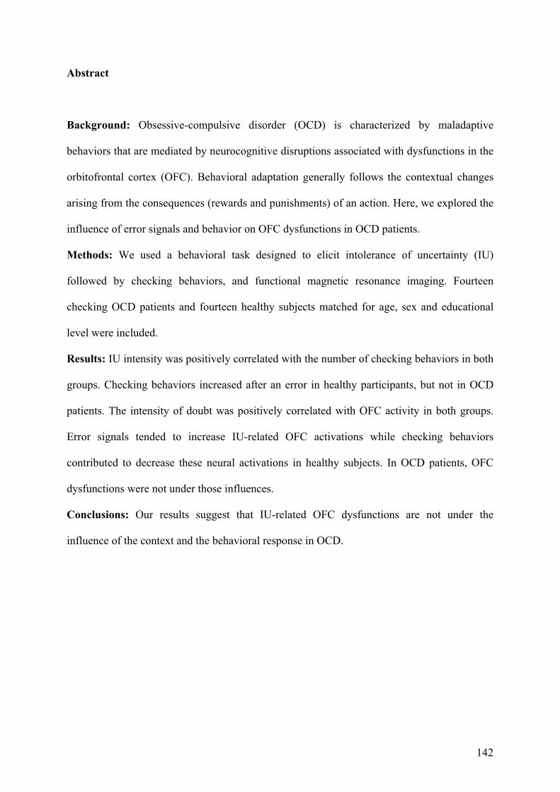
142
Abstract
Background: Obsessive-compulsive disorder (OCD) is characterized by maladaptive
behaviors that are mediated by neurocognitive disruptions associated with dysfunctions in the
orbitofrontal cortex (OFC). Behavioral adaptation generally follows the contextual changes
arising from the consequences (rewards and punishments) of an action. Here, we explored the
influence of error signals and behavior on OFC dysfunctions in OCD patients.
Methods: We used a behavioral task designed to elicit intolerance of uncertainty (IU)
followed by checking behaviors, and functional magnetic resonance imaging. Fourteen
checking OCD patients and fourteen healthy subjects matched for age, sex and educational
level were included.
Results: IU intensity was positively correlated with the number of checking behaviors in both
groups. Checking behaviors increased after an error in healthy participants, but not in OCD
patients. The intensity of doubt was positively correlated with OFC activity in both groups.
Error signals tended to increase IU-related OFC activations while checking behaviors
contributed to decrease these neural activations in healthy subjects. In OCD patients, OFC
dysfunctions were not under those influences.
Conclusions: Our results suggest that IU-related OFC dysfunctions are not under the
influence of the context and the behavioral response in OCD.
143
Introduction
Obsessive-compulsive disorder (OCD) is a chronic affection associated with highly disabling
impairment. The hallmark of OCD is the presence of obsessions in the form of intrusive and
distressing thoughts and the urge to perform repetitive behaviors known as compulsions to
reduce the anxiety and distress caused by obsessions (1). Available treatments with serotonin
reuptake inhibitors or cognitive behavior therapy are effective for most patients but impairing
residual symptoms and treatment resistance are common among OCD patients. A better
understanding of OCD pathophysiology is essential for further improvement of therapeutic
strategies.
Neuroimaging advances have described anatomical and functional brain alterations associated
with OCD. Anatomical modifications within the orbitofrontal cortex (OFC) and subcortical
regions such as the striatum (2). Moreover, functional magnetic resonance imaging (fMRI)
studies have shown that dysfunctions in the OFC in OCD are related to neurocognitive
disruptions, for example, in cognitive flexibility (3) or behavioral inhibition (4), which are
assumed to play a central role in the emergence of OCD symptoms. Reduced activity of the
OFC were found in OCD during both cognitive flexibility and response inhibition. Flexibility-
related OFC dysfunction was also observed in unaffected relatives, suggesting that OFC
underactivation could also represent a relevant vulnerability biomarker for OCD (3). Finally,
anatomical and functional alterations overlap within the OFC, suggesting the central role of
this brain region in OCD (5).
Many other neuroimaging studies have attempted to investigate brain alterations involved in
the production of OCD symptoms, in particular by using the symptom provocation task which
144
entails the presentation of obsessional stimuli. Although the symptom provocation task seems
relevant to elicit neural patterns of brain activations related to the emergence of obsessive-
compulsive symptoms, it suffers from two major limitations. First, it does not allow the
behavioral response to the obsessive thoughts, which is the usual construct of the obsessive-
compulsive symptoms, as daily experienced by OCD patients. Second, the symptom
provocation task involves many cognitive or emotional processes unrelated to the mediation
of OCD symptoms. In particular, some brain activations are likely related to patients’ efforts
to disregard or to turn their attention away from the obsessive and distressing thoughts,
especially when the behavioral response is not allowed (6). Attempting to deal with these
limitations, our research group has created a delayed matching-to-sample task with the
unrestricted possibility to check a decision (the checking task[CT]) (7). During the CT,
checking behaviors performed by OCD patients share similar features with checking
compulsions: i) checking behaviors are repeated in OCD patients compared with healthy
controls, especially when they suffer from checking compulsions; ii) checking behaviors are
driven by distressing uncertainty that is enhanced in OCD patients compared with healthy
controls; and iii) checking behaviors increase throughout CT in OCD patients but not in
healthy controls, as for attempting to reduce the cumulative distress caused by uncertainty (7).
"The way an individual perceives information in uncertain or ambiguous situations and
responds to this information with a set of cognitive, emotional and behavioral reactions" is
generally referred to intolerance of uncertainty (IU) (8).
The checking behavior consists in the repetition of an action that might be mistaken and be
responsible for prejudicial consequences. The checking behavior allows to obtain more
information and provides relief for the felt uncertainty on the accuracy of the action. External
error signals, as a consequence of actually mistaken action, may increase the likelihood to feel
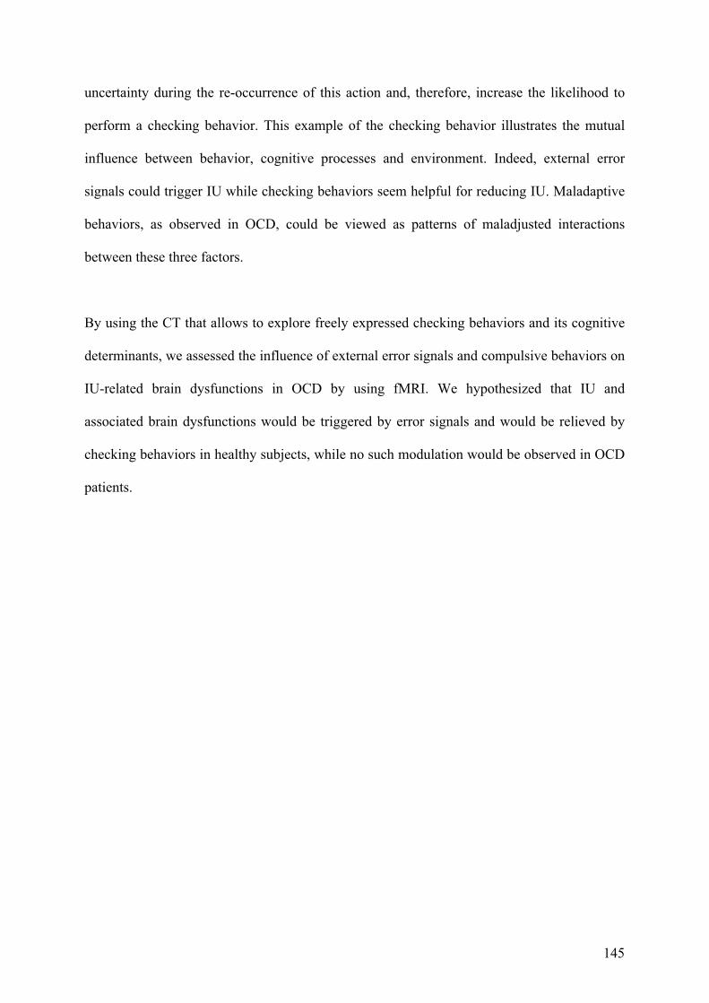
145
uncertainty during the re-occurrence of this action and, therefore, increase the likelihood to
perform a checking behavior. This example of the checking behavior illustrates the mutual
influence between behavior, cognitive processes and environment. Indeed, external error
signals could trigger IU while checking behaviors seem helpful for reducing IU. Maladaptive
behaviors, as observed in OCD, could be viewed as patterns of maladjusted interactions
between these three factors.
By using the CT that allows to explore freely expressed checking behaviors and its cognitive
determinants, we assessed the influence of external error signals and compulsive behaviors on
IU-related brain dysfunctions in OCD by using fMRI. We hypothesized that IU and
associated brain dysfunctions would be triggered by error signals and would be relieved by
checking behaviors in healthy subjects, while no such modulation would be observed in OCD
patients.
146
Materials and Methods
Participants
Sixteen OCD patients who primarily suffered from checking compulsions were recruited
through outpatient clinics in Bordeaux and Poitiers (France) and 16 healthy volunteers were
recruited from the local community through notice board advertisements. The 16 case-control
pairs were matched for age, sex, handedness and educational level.
OCD patients were diagnosed according to DSM-IV criteria (9) using the Mini-International
Neuropsychiatric Interview (MINI) (10). Patients suffering from other current Axis-1 or Axis-
2 disorders, as explored with the MINI and the Structured Clinical Interview for DSM-IV axis
II personality disorders (SCID-II) (11), were not included. The Yale-Brown Obsessive-
Compulsive Scale (Y-BOCS) was used to assess OCD symptom severity (12). The Y-BOCS
symptom checklist allowing to identify obsessions and compulsions was used to ensure that
checking compulsions were the primary OCD symptom. OCD patients without clinically
significant symptoms, as indicated by a Y-BOCS < 16, were not included in the present study.
Depression and anxiety symptoms were assessed with the Hamilton Depression Rating Scale
(HDRS) (13) and the Hamilton Anxiety Rating Scale (HARS) (14). OCD patients with
clinically significant depression or anxiety symptoms, as indicated by a HDRS or HARS
scores > 7, were not included in the present study. All OCD patients were being treated with
serotonin reuptake inhibitors and three of them were also receiving low doses of atypical
antipsychotics (risperidone: 1mg). No patient was actively involved in psychotherapy for
OCD at the time of participation.
147
Healthy comparison subjects passed the MINI, the SCID-II, the HDRS and the HARS. They
were excluded if they had a past or current psychiatric disorder, personality disorder,
neurological illness, or active medical problem. They were free of any psychotropic
medication for at least six months.
The protocol of the present study was reviewed and approved by the local ethics authorities
for work involving humans (“Comité Consultatif de la Protection des Personnes dans la
Recherche Biomédicale Bordeaux B”). All participants gave informed written consent after
the goals, procedures and potential risks of the study were presented and discussed.
Task
The CT consists of a delayed matching-to-sample task with the opportunity to check a
decision (see Figure 1 for description) (7). Briefly, two cartoon pictures delayed by an
interval were displayed after an attention signal. Subjects were required to compare both
images and to choose if they were identical or different. After having made his decision, the
subject has an unrestricted possibility to check his decision by repeating the trial and the
possibility to correct his answer. After having validated his decision, the subject was informed
of his response accuracy. Subjects were asked to be the more accurate as possible (7). Each
participant was trained during 20 minutes with the original version of the CT a week before
MRI acquisition protocol.
In the present study, we used a derived version of the CT with the aim to elicit IU in healthy
subjects and to have a similar number of checking behaviors in both groups for statistical
purposes. The derived version was defined on the basis of prior reported data (7) and
supplementary unpublished data obtained in 18 healthy subjects for testing purpose. Thus, we
148
implemented the presentation of strictly identical images without the subject’s knowledge.
Furthermore, either positive or negative feedback was semi-randomized on the basis of an 3:1
ratio whatever subjects' responses.
Inter-event intervals ranging from 2 to 8 seconds in 2-seconds steps were generated on the
basis of a geometric probability distribution in order to improve detection of blood-
oxygenation level dependent (BOLD) signals (15). In order not to influence the visuo-spatial
discrimination ability, the presentation of the sample and the following memory delay were
time-locked for 3.5 seconds. Subjects first practiced three trials to be familiar to the MRI
environment before completing twenty randomized display trials.
Response accuracy, responses times and the number of checking behaviors were recorded.
Under imaging condition, trials generated with Presentation software (Neurobehavioral
Systems, Albany, California) were visually cued with a mirror attached to the head coil and a
front-projection. Responses were made with the index finger of their right hand using an
MRI-compatible response device.
Immediately after having completed the MRI task, subjects were asked to rate the intensity of
the experienced doubt feeling during decision making with a ten point analog scale based on
self-reporting (1-3: "weak", 4-6: "mild", 7-9: "strong"). A response of zero was equated with
complete absence of doubt or uncertainty whereas ten was corresponding to a doubt feeling
ever previously experienced by the participant, as the subjective measurement of the
intolerance of uncertainty (IU score) (8). In the same time, we also asked the participants
about CT to make sure that they did not suspect that the displayed pictures were all identical.
149
Behavioral data analyses
For descriptive statistics, we used !2 tests or t tests for independent samples. Correlations
between behavioral variables were assessed with Pearson's tests. All analyses were two-tailed,
and the alpha risk was set at 0.05 as the threshold for statistical significance. Statistical
analyses were performed with SPSS software (Release 16.0, Chicago, IL, USA).
Imaging data acquisition
MRI acquisition was performed on a Gyroscan ACS NT Power track 6000 1.5 T MRI system
(Philips Medical System, Best, Netherlands) with a standard circularly head coil. To reduce
motion artifacts, the head was immobilized using foam pads. Each participant lay supine on
the MR system patient table in a dimly illuminated room and wore foam earplugs.
For the structural imaging, a whole-brain T1-weighted sequence was first acquired for
anatomical reference (TR = 0.231s, TE = 2.5ms, flip angle = 90°, 25 sections). Scans were
reviewed by a neuroradiologist to rule out structural pathology and assess scan quality. For
the functional imaging, a total of 237 volumes were then registered using a T2-weighted
gradient echo-planar imaging (EPI) sequence with 25 slices covering the whole brain (slice
thickness = 4 mm; no gap; repetition time = 2500 ms; echo time = 60 ms; flip angle = 90°;
matrix size = 64*64 mm; voxel size = 2*2*4 mm). The orientation of the sagittal slices was
parallel to the AC-PC line. The scanner automatically discarded the first 3 measurements
before the task started.
150
Imaging data analyses
fMRI data were preprocessed using standard procesures implemented in the Statistical
Parametric Mapping package (SPM5; Wellcome Department of Cognitive Neurology,
University College, London) after verification for artifacts and excessive motion. Slice time
correction, realignment and normalization into the Montreal Neurological Institute standard
atlas space were performed. Spatial smoothing was executed with a full-width-at-half
maximum gaussian filter of 8 mm. Two experimental conditions: (1) decision followed by a
checking behavior (DFCB+), (2) decision not followed by a checking behavior (DFCB-) were
modeled by a standard box-car function convolved with a canonical hemodynamic response
function in the general linear model. Other events of the task were modeled as events of no
interest. The six movement parameters were also included in the model as regressors of no
interest.
We computed the following t-contrasts: DFCB+ > DFCB- and DFCB- > DFCB+. Linear
contrasts were first performed at a single subject level and were then entered into a second
level random effects analyses to obtain mean t-images. Within-group and between-group
analyses consisted in one-sample and two-sample t tests to compare signal changes in the
mentioned conditions. Because HARS scores were significantly different between both
groups, they were entered in our group level analyses as covariates.
Positive and negative relationships between clinical variables (IU score and Y-BOCS score)
and brain activity corresponding to our contrasts of interest were assessed using multiple
regression analysis as implemented in SPM5 for each group and for all participants together.
For all analyses, the threshold for significance was set at p < 0.05 FDR (false discovery rate)
corrected for multiple comparisons. An uncorrected p < 0.001 with a minimum cluster extent
151
of 30 contiguous voxels was accepted as significant for our a priori region of interest, namely
the OFC.
For the region of interest (ROI) analyses, we considered significant results from whole brain
analyses interesting the OFC in accordance with our a priori hypotheses. The OFC-ROI was
functionally defined on the basis of whole brain regression analyses by centering spheres on
the respective peak voxels with a radius of 5 mm. We used the REX toolbox
(http://web.mit.edu/swg/software.htm) for extracting the mean values of unsmoothed images
corresponding to our conditions of interest (DFCB+ and DFCB-) for each subject. According
to our hypoheses, we examined the effect of the error and of the checking behavior on the
ROI mean values between OCD group and control group. Statistical analyses consisted in a
two-way (group x conditions of interest) ANOVA with post-hoc t tests.
152
Results
Participant characteristics
From the 32 included participants, two (one in each group) were excluded because of having
performed no checking behavior during the task and two (one in each group) because of
excessive motion during scanning. Demographic and clinical characteristics of the final
samples are presented in Table 1. Groups did not differ with respect to age, sex, handedness
and educational level.
Behavioral performance
Behavioral performances are summarized in Table 2. The total duration for completing all 20
trials were variable between subjects according to the number of checking behaviors,
however, there was no difference between groups. As expected by using the present version
of CT, the number of checking behaviors and the response time for decision making were
similar in both groups. IU scores during decision making were positively significantly
correlated with the number of checking behaviors in both groups (control group: r = 0.86,
p < 0.01; OCD group: r = 0.86, p < 0.01). Finally, in accordance with our hypotheses, we
found a positive relationship between the course of error signals and the number of checking
behaviors throughout the task in healthy controls, but not in OCD patients (control group:
r = 0.62, p < 0.01; OCD group: r = 0.28, p = 0.23). Checking behaviors remained stable in
OCD group whatever they are performed after a success or after an error (t = -0.11, p = 0.92),
while checking behaviors increased after an error in healthy participants (t = 2.66, p < 0.05 )
(see Figure 2).
153
Whole brain fMRI analyses
Within-group analyses are presented in Supplementary Materials. Between-group analyses
did not reveal any significant difference with an alpha risk fixed at 0.05 FDR corrected.
We found no significant correlation between symptom severity (Y-BOCS scores) and whole
brain activity for our contrasts of interest in OCD group. IU scores were positively correlated
with the activity in the lateral OFC and the lateral prefrontal cortex (maximum: x=-30, y=48,
z=0; BA 10, 11, 45) for the DFCB+ > DFCB- contrast in the 28 participants and in each
group independently ([OCD group: maximum x=-32, y=50, z=0; BA 10, 11, 13];[control
group: maximum x=-34, y=51, z=2; BA 10]) (Figure 3A).
ROI analyses
The maximum peak of the cluster for which regression analyses were significant was defined
as region of interest. We extracted mean values for our conditions of interest (DFCB+ and
DFCB-) and we seperated our events according to the nature of the preceding feedback. Also,
we extracted mean values for decision making that occured after a checking behavior. The
ANOVA corrected model was significant (D = 2.3, p < 0.05) and revealed a significant group
effect (D = 5.1, p < 0.05), a trend for the condition effect (D = 2.5, p = 0.06), but no
significant interaction (D = 1.2, p = 0.32). ROI mean values were higher during DFCB+ than
during DFCB- in healthy controls (t = 2.77, p < 0.05), but not in OCD patients (t = 1.27,
p = 0.23) (Figure 3B). There was no difference between healthy controls and OCD patients
(DFCB+: t = 1.61, p = 0.12; DFCB-: t = 0.22, p = 0.83). After an error, healthy controls
exhibited a more enhanced ROI mean values than OCD patients (t = 2.06, p < 0.05). There
was no significant difference when considering DFCB+ after a success or after an error in

154
both groups although a trend was observed in healthy controls (control group: t = 1.78,
p = 0.09; OCD group: t = -0.64, p = 0.53) (Figure 3C). Finally, ROI mean values were
smaller during a decision making occuring after a checking behavior than during a decision
making leading to a checking behavior in healthy controls (t = 3.11, p < 0.05), but not in OCD
patients (t = 0.37, p = 0.72). There was no between-group difference during a decision making
occuring after a checking behavior (t = 0.12, p = 0.91) (Figure 3D).
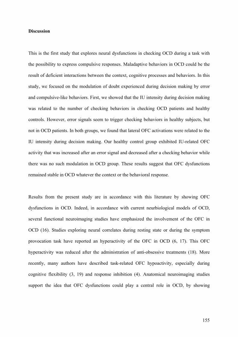
155
Discussion
This is the first study that explores neural dysfunctions in checking OCD during a task with
the possibility to express compulsive responses. Maladaptive behaviors in OCD could be the
result of deficient interactions between the context, cognitive processes and behaviors. In this
study, we focused on the modulation of doubt experienced during decision making by error
and compulsive-like behaviors. First, we showed that the IU intensity during decision making
was related to the number of checking behaviors in checking OCD patients and healthy
controls. However, error signals seem to trigger checking behaviors in healthy subjects, but
not in OCD patients. In both groups, we found that lateral OFC activations were related to the
IU intensity during decision making. Our healthy control group exhibited IU-related OFC
activity that was increased after an error signal and decreased after a checking behavior while
there was no such modulation in OCD group. These results suggest that OFC dysfunctions
remained stable in OCD whatever the context or the behavioral response.
Results from the present study are in accordance with this literature by showing OFC
dysfunctions in OCD. Indeed, in accordance with current neurbiological models of OCD,
several functional neuroimaging studies have emphasized the involvement of the OFC in
OCD (16). Studies exploring neural correlates during resting state or during the symptom
provocation task have reported an hyperactivity of the OFC in OCD (6, 17). This OFC
hyperactivity was reduced after the administration of anti-obsessive treatments (18). More
recently, many authors have described task-related OFC hypoactivity, especially during
cognitive flexibility (3, 19) and response inhibition (4). Anatomical neuroimaging studies
support the idea that OFC dysfunctions could play a central role in OCD, by showing
156
concordant volume changes in the OFC (20). Furthermore, we recently showed that
anatomical and functional alterations overlap within the lateral OFC (21).
In this study, we focused on the interactions between the context, cognition and the behavior.
We hypothesized that maladaptive behaviors observed in OCD could be related to
maladaptive interactions between these three factors. Only very few neurocognitive studies
have explored such interactions. We found a positive relationship between the intensity of IU
and the number of checking behaviors in both groups. Accordingly, in a recent study, it has
been shown that IU has contributed to mediate compulsive checking behaviors in OCD
patients (22).
We have shown that error signals may contribute to enhance checking behaviors in healthy
subjects, but not in OCD. This result is in accordance with those by Julien et al. who have
shown that intrusive thoughts in OCD patients were less linked to the context of their
occurrence than in healthy subjects (23). Also, an electrophysiological study reported no
modulation of the error-related brain activity by enhanced error signals in OCD patients in
comparison with healthy subjects (24). Another possible explanation may lie in the present
modified version of CT that aimed to exacerbate IU. Indeed, error signals may achieve the
increase of IU in healthy subjects, but not in OCD patients because IU was already at its
highest level and, thus, we cannot exclude formally the contribution of error signals in the
performance of checking behaviors.
Checking behavior is efficient in healthy subjects for decreasing IU-related OFC activity, but
not in OCD patients. To our knowledge, this is the first demonstration of the inefficiency of
the behavior response to influence distressing thoughts and their neural correlates in OCD.
157
This may account for a deficit in the top-down control of the affective drive associated with
IU and therefore for a deficit in the down-regulation of the IU-related OFC activity, leading to
the repetition of the behavior for obtaining this down-regulation. This hypothesis should be
further tested. For example, this experimental situation could be relevant for testing how
cognitive-behavioral techniques associated with the task would allow OCD patients to
efficiently check.
The present study has several limitations, including a modest sample size. First, although all
included OCD patients were suffering from checking compulsions and had no psychiatric
comorbid condition which was the strength of our recruitment, all included OCD patients
were under serotonin reuptake inhibitors. Psychotropic medications were not stopped because
of obvious ethical reasons. We cannot rule out the influence of medications in our results,
especially concerning the absence of significant difference in our between-group analyses.
However, this limitation should be tempered because all patients had clinically significant
OCD symptoms, as illustrated by a Y-BOCS score > 16 for each patient. Second, we do not
acquire baseline brain activity that is known to differ between OCD patients and healthy
control subjects (17). We cannot exclude its potential effect in task-related activity, especially
in the OFC. Finally, there was no neuropsychological assessments to eliminate any
neurocognitive alterations, especially working memory dysfunctions that may have influenced
behavioral performances (25).
In conclusion, our study support and extend previous findings by showing that intolerance of
uncertainty-related OFC dysfunctions are not influenced by neither error signals nor
compulsive-like checking behaviors in OCD patients in comparison with healthy subjects.
Our results argue for the existence of deficient interactions between the context, cognition and
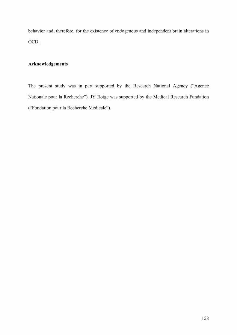
158
behavior and, therefore, for the existence of endogenous and independent brain alterations in
OCD.
Acknowledgements
The present study was in part supported by the Research National Agency (“Agence
Nationale pour la Recherche”). JY Rotge was supported by the Medical Research Fundation
(“Fondation pour la Recherche Médicale”).
159
References
1. Stein DJ (2002): Obsessive-compulsive disorder. Lancet. 360:397-405.
2. Rotge JY, Langbour N, Guehl D, Bioulac B, Jaafari N, Allard M, et al. Gray matter
alterations in obsessive-compulsive disorder: an anatomic likelihood estimation meta-
analysis. Neuropsychopharmacology. 35:686-691.
3. Chamberlain SR, Menzies L, Hampshire A, Suckling J, Fineberg NA, del Campo N, et
al. (2008): Orbitofrontal dysfunction in patients with obsessive-compulsive disorder and their
unaffected relatives. Science. 321:421-422.
4. Roth R, Saykin A, Flashman L, Pixley H, West J, Mamourian A (2007): Event-
Related Functional Magnetic Resonance Imaging of Response Inhibition in Obsessive-
Compulsive Disorder. Biol Psychiatry. 62:901-909.
5. Rotge JY, Langbour N, Jaafari N, Guehl D, Bioulac B, Aouizerate B, et al.
Anatomical alterations and symptom-related functional activity in obsessive-compulsive
disorder are correlated in the lateral orbitofrontal cortex. Biol Psychiatry. 67:e37-38.
6. Rotge JY, Guehl D, Dilharreguy B, Cuny E, Tignol J, Bioulac B, et al. (2008):
Provocation of obsessive-compulsive symptoms: a quantitative voxel-based meta-analysis of
functional neuroimaging studies. J Psychiatry Neurosci. 33:405-412.
7. Rotge JY, Clair AH, Jaafari N, Hantouche EG, Pelissolo A, Goillandeau M, et al.
(2008): A challenging task for assessment of checking behaviors in obsessive-compulsive
disorder. Acta psychiatrica Scandinavica. 117:465-473.
8. Dugas MJ, Gagnon F, Ladouceur R, Freeston MH (1998): Generalized anxiety
disorder: a preliminary test of a conceptual model. Behav Res Ther. 36:215-226.
160
9. American Psychiatric Association., American Psychiatric Association. Task Force on
DSM-IV. (1994): Diagnostic and statistical manual of mental disorders : DSM-IV. 4th ed.
Washington, DC: American Psychiatric Association.
10. Sheehan DV, Lecrubier Y, Sheehan KH, Amorim P, Janavs J, Weiller E, et al. (1998):
The Mini-International Neuropsychiatric Interview (M.I.N.I.): the development and validation
of a structured diagnostic psychiatric interview for DSM-IV and ICD-10. J Clin Psychiatry.
59 Suppl 20:22-33;quiz 34-57.
11. Maffei C, Fossati A, Agostoni I, Barraco A, Bagnato M, Deborah D, et al. (1997):
Interrater reliability and internal consistency of the structured clinical interview for DSM-IV
axis II personality disorders (SCID-II), version 2.0. J Pers Disord. 11:279-284.
12. Goodman WK, Price LH, Rasmussen SA, Mazure C, Fleischmann RL, Hill CL, et al.
(1989): The Yale-Brown Obsessive Compulsive Scale. I. Development, use, and reliability.
Arch Gen Psychiatry. 46:1006-1011.
13. Hamilton M (1960): A rating scale for depression. J Neurol Neurosurg Psychiatry.
23:56-62.
14. Hamilton M (1959): The assessment of anxiety states by rating. Br J Med Psychol.
32:50-55.
15. Hagberg GE, Zito G, Patria F, Sanes JN (2001): Improved detection of event-related
functional MRI signals using probability functions. Neuroimage. 14:1193-1205.
16. Menzies L, Chamberlain SR, Laird AR, Thelen SM, Sahakian BJ, Bullmore ET
(2008): Integrating evidence from neuroimaging and neuropsychological studies of obsessive-
compulsive disorder: the orbitofronto-striatal model revisited. Neurosci Biobehav Rev.
32:525-549.
161
17. Baxter LR, Jr., Phelps ME, Mazziotta JC, Guze BH, Schwartz JM, Selin CE (1987):
Local cerebral glucose metabolic rates in obsessive-compulsive disorder. A comparison with
rates in unipolar depression and in normal controls. Arch Gen Psychiatry. 44:211-218.
18. Nakao T, Nakagawa A, Yoshiura T, Nakatani E, Nabeyama M, Yoshizato C, et al.
(2005): Brain activation of patients with obsessive-compulsive disorder during
neuropsychological and symptom provocation tasks before and after symptom improvement:
a functional magnetic resonance imaging study. Biol Psychiatry. 57:901-910.
19. Remijnse PL, Nielen MM, van Balkom AJ, Cath DC, van Oppen P, Uylings HB, et al.
(2006): Reduced orbitofrontal-striatal activity on a reversal learning task in obsessive-
compulsive disorder. Arch Gen Psychiatry. 63:1225-1236.
20. Rotge JY, Guehl D, Dilharreguy B, Tignol J, Bioulac B, Allard M, et al. (2009): Meta-
analysis of brain volume changes in obsessive-compulsive disorder. Biol Psychiatry. 65:75-
83.
21. Rotge JY, Langbour N, Jaafari N, Guehl D, Bioulac B, Aouizerate B, et al. (2010):
Anatomical alterations and symptom-related functional activity in obsessive-compulsive
disorder are correlated in the lateral orbitofrontal cortex. Biol Psychiatry. 67:e37-38.
22. Lind C, Boschen MJ (2009): Intolerance of uncertainty mediates the relationship
between responsibility beliefs and compulsive checking. Journal of Anxiety Disorders.
23:1047-1052.
23. Julien D, O'Connor KP, Aardema F (2009): Intrusions related to obsessive-compulsive
disorder: a question of content or context? Journal of clinical psychology. 65:709-722.
24. Endrass T, Schuermann B, Kaufmann C, Spielberg R, Kniesche R, Kathmann N
(2010): Performance monitoring and error significance in patients with obsessive-compulsive
disorder. Biol Psychol. 84:257-263.

162
25. Nakao T, Nakagawa A, Nakatani E, Nabeyama M, Sanematsu H, Yoshiura T, et al.
(2009): Working memory dysfunction in obsessive-compulsive disorder: a
neuropsychological and functional MRI study. J Psychiatr Res. 43:784-791.
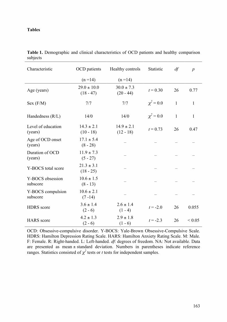
163
Tables
Table 1. Demographic and clinical characteristics of OCD patients and healthy comparison subjects
Characteristic OCD patients Healthy controls Statistic df p
(n =14) (n =14)
Age (years) 29.0 ± 10.0
(18 - 47) 30.0 ± 7.3 (20 - 44)
t = 0.30 26 0.77
Sex (F/M) 7/7 7/7 !2 = 0.0 1 1
Handedness (R/L) 14/0 14/0 !2 = 0.0 1 1
Level of education (years)
14.3 ± 2.1 (10 - 18)
14.9 ± 2.1 (12 - 18)
t = 0.73 26 0.47
Age of OCD onset (years)
17.1 ± 5.4 (8 - 28)
– – – –
Duration of OCD (years)
11.9 ± 7.3 (5 - 27)
– – – –
Y-BOCS total score 21.3 ± 3.1 (18 - 25)
– – – –
Y-BOCS obsession subscore
10.6 ± 1.5 (8 - 13)
– – – –
Y-BOCS compulsion subscore
10.6 ± 2.1 (7 -14)
– – – –
HDRS score 3.6 ± 1.4
(2 - 6) 2.6 ± 1.4
(1 - 4) t = -2.0 26 0.055
HARS score 4.2 ± 1.3
(2 - 6) 2.9 ± 1.8
(1 - 6) t = -2.3 26 < 0.05
OCD: Obsessive-compulsive disorder. Y-BOCS: Yale-Brown Obsessive-Compulsive Scale. HDRS: Hamilton Depression Rating Scale. HARS: Hamilton Anxiety Rating Scale. M: Male. F: Female. R: Right-handed. L: Left-handed. df: degrees of freedom. NA: Not available. Data are presented as mean ± standard deviation. Numbers in parentheses indicate reference ranges. Statistics consisted of !2 tests or t tests for independent samples.

164
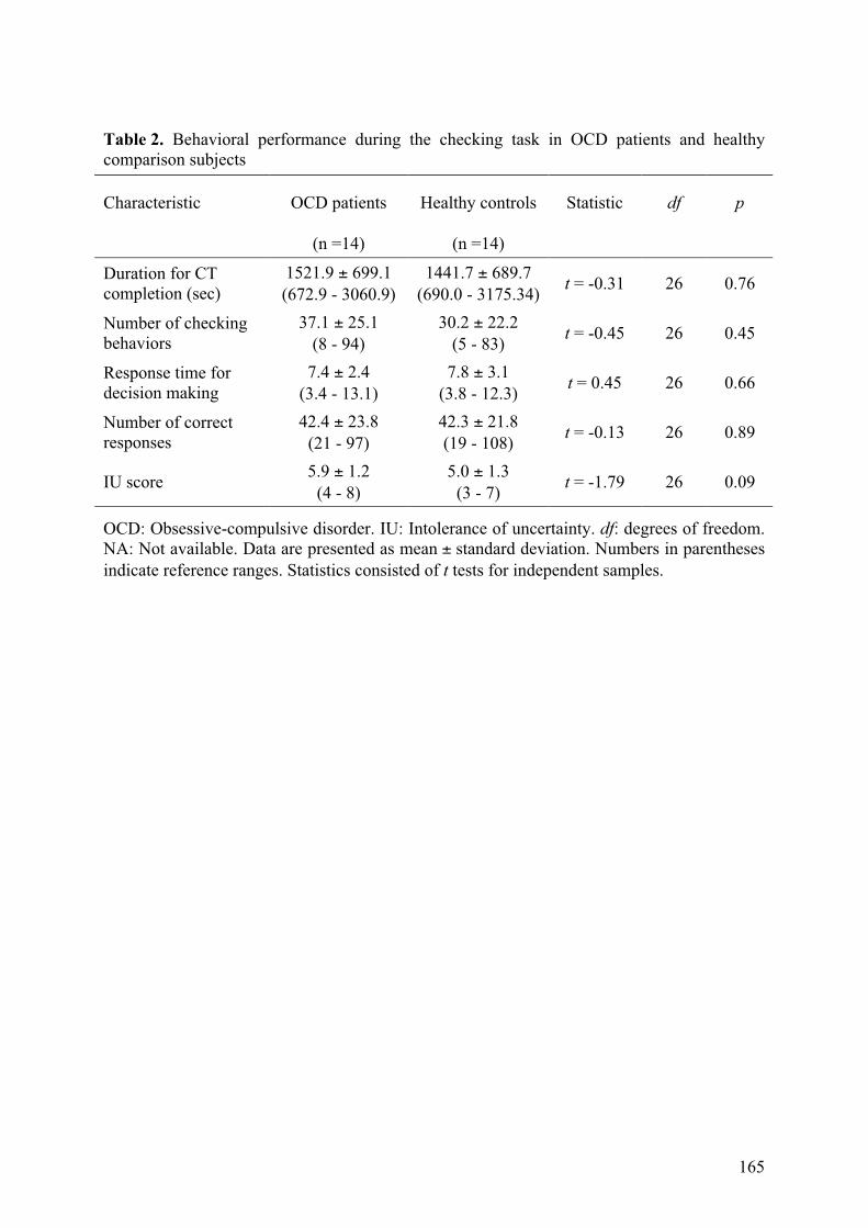
165
Table 2. Behavioral performance during the checking task in OCD patients and healthy comparison subjects
Characteristic OCD patients Healthy controls Statistic df p
(n =14) (n =14)
Duration for CT completion (sec)
1521.9 ± 699.1 (672.9 - 3060.9)
1441.7 ± 689.7 (690.0 - 3175.34)
t = -0.31 26 0.76
Number of checking behaviors
37.1 ± 25.1 (8 - 94)
30.2 ± 22.2 (5 - 83)
t = -0.45 26 0.45
Response time for decision making
7.4 ± 2.4 (3.4 - 13.1)
7.8 ± 3.1 (3.8 - 12.3)
t = 0.45 26 0.66
Number of correct responses
42.4 ± 23.8 (21 - 97)
42.3 ± 21.8 (19 - 108)
t = -0.13 26 0.89
IU score 5.9 ± 1.2
(4 - 8) 5.0 ± 1.3
(3 - 7) t = -1.79 26 0.09
OCD: Obsessive-compulsive disorder. IU: Intolerance of uncertainty. df: degrees of freedom. NA: Not available. Data are presented as mean ± standard deviation. Numbers in parentheses indicate reference ranges. Statistics consisted of t tests for independent samples.
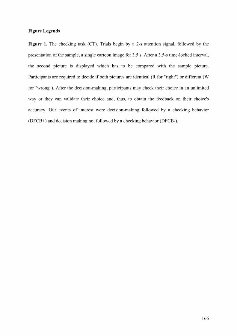
166
Figure Legends
Figure 1. The checking task (CT). Trials begin by a 2-s attention signal, followed by the
presentation of the sample, a single cartoon image for 3.5 s. After a 3.5-s time-locked interval,
the second picture is displayed which has to be compared with the sample picture.
Participants are required to decide if both pictures are identical (R for "right") or different (W
for "wrong"). After the decision-making, participants may check their choice in an unlimited
way or they can validate their choice and, thus, to obtain the feedback on their choice's
accuracy. Our events of interest were decision-making followed by a checking behavior
(DFCB+) and decision making not followed by a checking behavior (DFCB-).
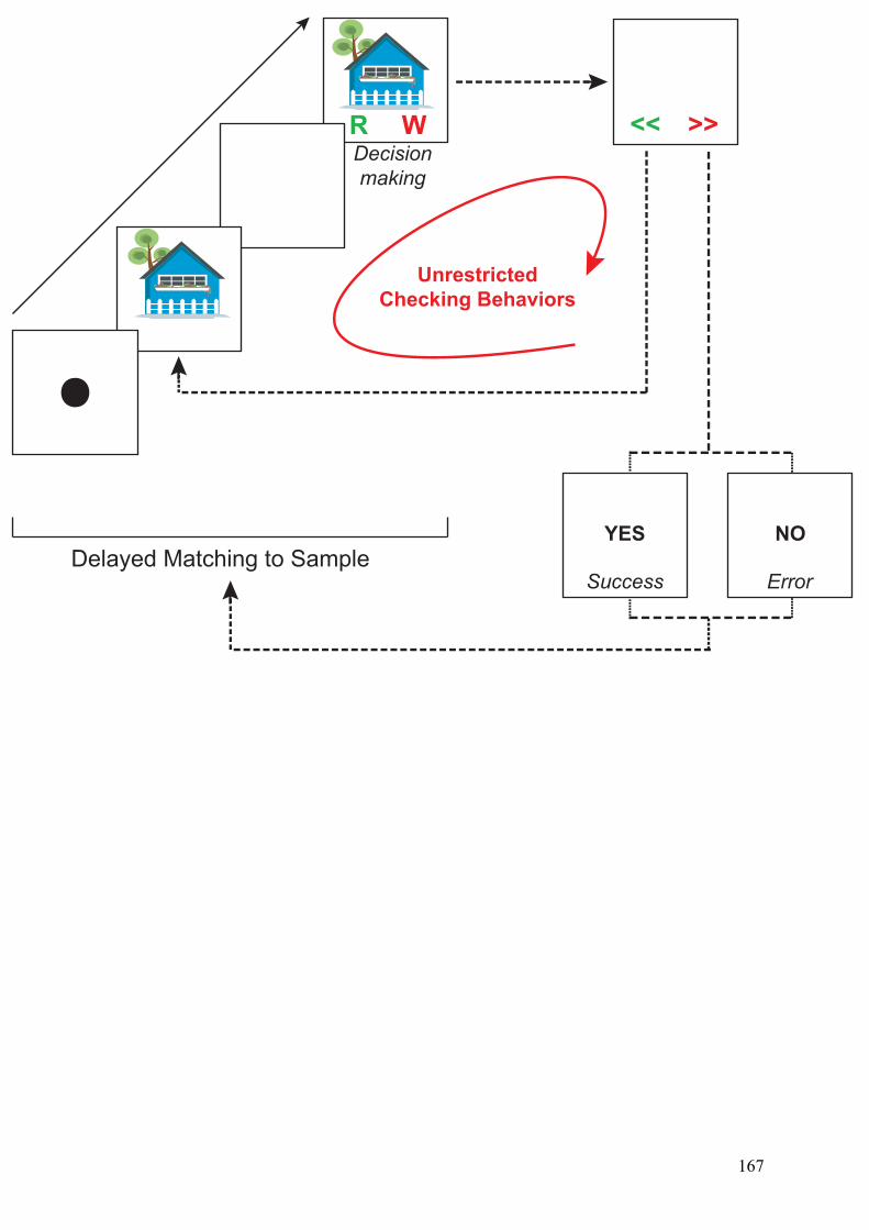
R W << >>
Unrestricted
Checking Behaviors
Delayed Matching to SampleYES NO
ErrorSuccess
Decision
making
167

168
169
Figure 2. Percentage of variation between checking behaviors during trials following an error
and trials following a success in OCD patients and healthy controls. Healthy controls
exhibited an increase of checking behaviors after an error (t = 2.66, p < 0.05 ) while checking
behaviors remain stable in OCD patients whatever the nature of the feedback (t = -0.11,
p = 0.92).
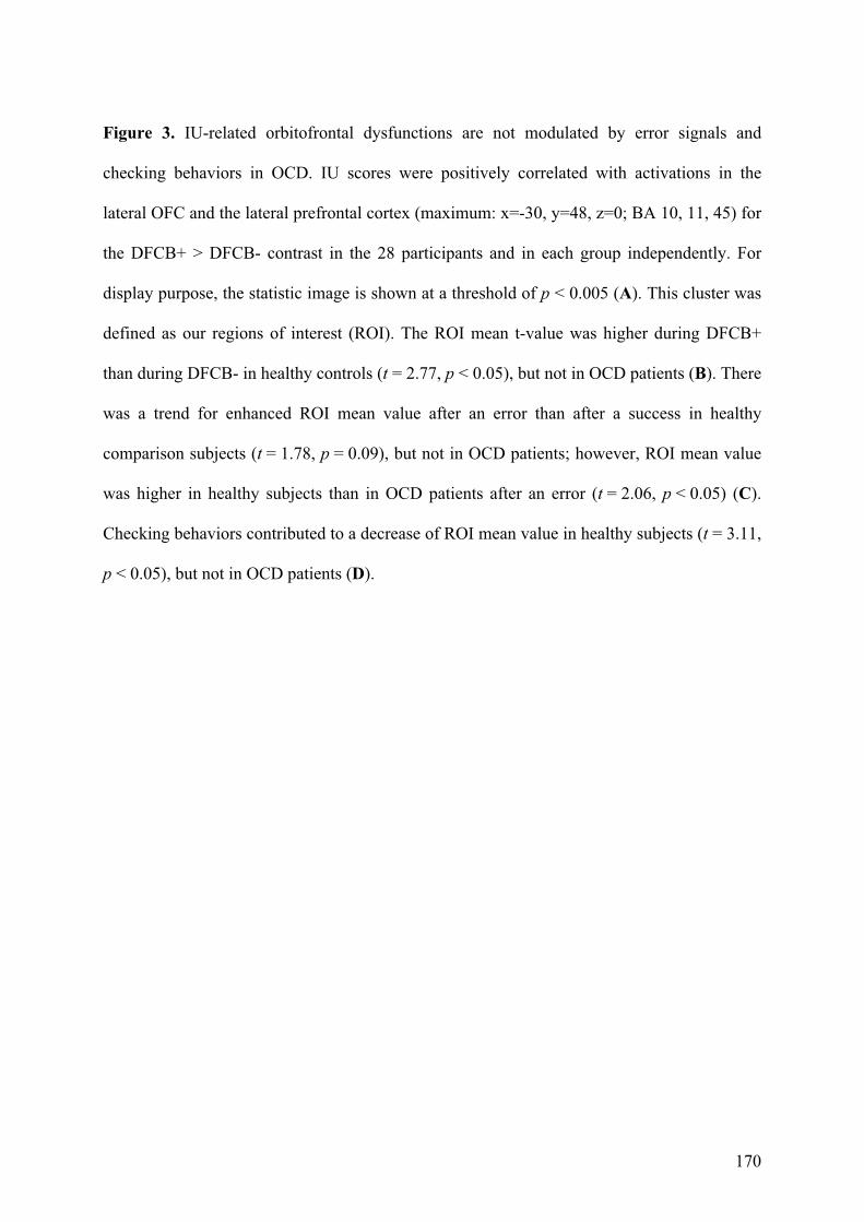
170
Figure 3. IU-related orbitofrontal dysfunctions are not modulated by error signals and
checking behaviors in OCD. IU scores were positively correlated with activations in the
lateral OFC and the lateral prefrontal cortex (maximum: x=-30, y=48, z=0; BA 10, 11, 45) for
the DFCB+ > DFCB- contrast in the 28 participants and in each group independently. For
display purpose, the statistic image is shown at a threshold of p < 0.005 (A). This cluster was
defined as our regions of interest (ROI). The ROI mean t-value was higher during DFCB+
than during DFCB- in healthy controls (t = 2.77, p < 0.05), but not in OCD patients (B). There
was a trend for enhanced ROI mean value after an error than after a success in healthy
comparison subjects (t = 1.78, p = 0.09), but not in OCD patients; however, ROI mean value
was higher in healthy subjects than in OCD patients after an error (t = 2.06, p < 0.05) (C).
Checking behaviors contributed to a decrease of ROI mean value in healthy subjects (t = 3.11,
p < 0.05), but not in OCD patients (D).
DFCB+ DFCB-
0
1
-1
Post success
DFCB+
Post error
DFCB+
0
1
-1
RO
I m
ea
n v
alu
eA B
RO
I m
ea
n v
alu
e
NO
Error R W
DFCB+ After CB+
0
1
-1
RO
I m
ea
n v
alu
e
R W<< >>
C D
Healthy controls
OCD patients
**
*
171

172
173
5. Relation entre les altérations anatomiques et fonctionnelles
5.1. Introduction
Les études de neuro-imagerie ont largement contribué à l’identification des altérations
anatomiques et fonctionnelles associées au TOC. On peut faire l’hypothèse que les altérations
anatomiques contribuent aux altérations fonctionnelles et inversement. Il s’agit d’une question
essentielle puisqu'y répondre permettrait de distinguer des modifications fonctionnelles
« primaires » sous-tendues par des anomalies anatomiques et des modifications fonctionnelles
« secondaires » pouvant relever davantage d’un processus adaptatif.
5.2. Méthodes
Pour apporter des éléments de réponse à cette question, nous avons utilisé les données
issues des méta-analyses précitées (morphométrie [page 57] et IRMf [page 81]) et nous avons
superposé les cartes obtenues pour chacune des deux techniques.
5.3. Résultats et discussion
Nous observons une superposition des modifications anatomiques et fonctionnelles au
sein d’une seule région cérébrale : la partie latérale du COF. De façon intéressante, cette
région cérébrale participe aux difficultés de flexibilité cognitive et d’inhibition
comportementale observées dans le TOC, récemment proposées comme endophénotypes de la
maladie (Roth et al., 2007 ; Chamberlain et al., 2008).
5.4. Conclusion
En montrant l’existence d’une relation entre anomalies anatomiques et fonctionnelles au
sein du COF, ce travail suggère un rôle de premier ordre de cette région corticale dans la
médiation des symptômes obsessionnels-compulsifs.
Rotgé JY, Langbour N, Jaafari N, Guehl D, Bioulac B, Aouizerate B, Allard M,
Burbaud P. Anatomical alterations and symptom-related functional activity in
obsessive-compulsive disorder are correlated in the lateral orbitofrontal cortex.
Biological Psychiatry 2010;67:e37-38.

174
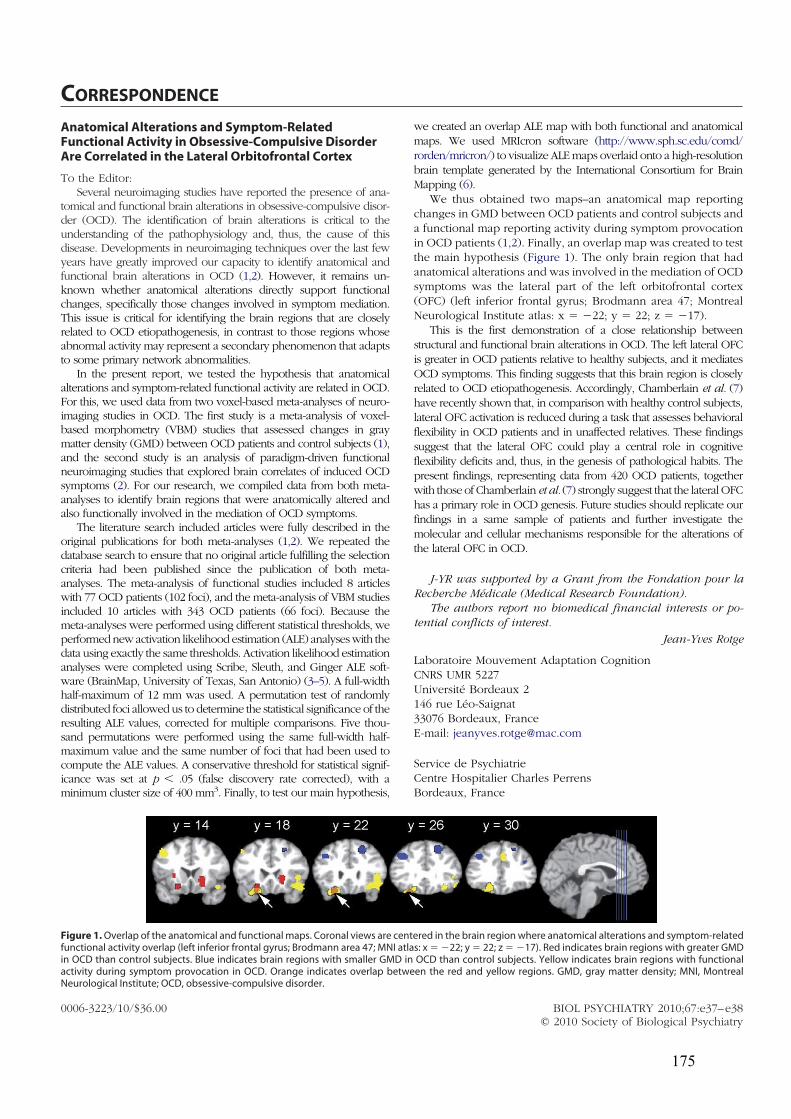
CORRESPONDENCE
Anatomical Alterations and Symptom-RelatedFunctional Activity in Obsessive-Compulsive DisorderAre Correlated in the Lateral Orbitofrontal Cortex
To the Editor:Several neuroimaging studies have reported the presence of ana-
tomical and functional brain alterations in obsessive-compulsive disor-der (OCD). The identification of brain alterations is critical to theunderstanding of the pathophysiology and, thus, the cause of thisdisease. Developments in neuroimaging techniques over the last fewyears have greatly improved our capacity to identify anatomical andfunctional brain alterations in OCD (1,2). However, it remains un-known whether anatomical alterations directly support functionalchanges, specifically those changes involved in symptom mediation.This issue is critical for identifying the brain regions that are closelyrelated to OCD etiopathogenesis, in contrast to those regions whoseabnormal activity may represent a secondary phenomenon that adaptsto some primary network abnormalities.
In the present report, we tested the hypothesis that anatomicalalterations and symptom-related functional activity are related in OCD.For this, we used data from two voxel-based meta-analyses of neuro-imaging studies in OCD. The first study is a meta-analysis of voxel-based morphometry (VBM) studies that assessed changes in graymatter density (GMD) between OCD patients and control subjects (1),and the second study is an analysis of paradigm-driven functionalneuroimaging studies that explored brain correlates of induced OCDsymptoms (2). For our research, we compiled data from both meta-analyses to identify brain regions that were anatomically altered andalso functionally involved in the mediation of OCD symptoms.
The literature search included articles were fully described in theoriginal publications for both meta-analyses (1,2). We repeated thedatabase search to ensure that no original article fulfilling the selectioncriteria had been published since the publication of both meta-analyses. The meta-analysis of functional studies included 8 articleswith 77 OCD patients (102 foci), and the meta-analysis of VBM studiesincluded 10 articles with 343 OCD patients (66 foci). Because themeta-analyses were performed using different statistical thresholds, weperformednewactivation likelihoodestimation (ALE) analyseswith thedata using exactly the same thresholds. Activation likelihood estimationanalyses were completed using Scribe, Sleuth, and Ginger ALE soft-ware (BrainMap, University of Texas, San Antonio) (3–5). A full-widthhalf-maximum of 12 mm was used. A permutation test of randomlydistributed foci allowed us to determine the statistical significance of theresulting ALE values, corrected for multiple comparisons. Five thou-sand permutations were performed using the same full-width half-maximum value and the same number of foci that had been used tocompute the ALE values. A conservative threshold for statistical signif-icance was set at p , .05 (false discovery rate corrected), with aminimum cluster size of 400 mm3. Finally, to test our main hypothesis,
we created an overlap ALE map with both functional and anatomicalmaps. We used MRIcron software (http://www.sph.sc.edu/comd/rorden/mricron/) to visualize ALE maps overlaid onto a high-resolutionbrain template generated by the International Consortium for BrainMapping (6).
We thus obtained two maps–an anatomical map reportingchanges in GMD between OCD patients and control subjects anda functional map reporting activity during symptom provocationin OCD patients (1,2). Finally, an overlap map was created to testthe main hypothesis (Figure 1). The only brain region that hadanatomical alterations and was involved in the mediation of OCDsymptoms was the lateral part of the left orbitofrontal cortex(OFC) (left inferior frontal gyrus; Brodmann area 47; MontrealNeurological Institute atlas: x 5 222; y 5 22; z 5 217).
This is the first demonstration of a close relationship betweenstructural and functional brain alterations in OCD. The left lateral OFCis greater in OCD patients relative to healthy subjects, and it mediatesOCD symptoms. This finding suggests that this brain region is closelyrelated to OCD etiopathogenesis. Accordingly, Chamberlain et al. (7)have recently shown that, in comparison with healthy control subjects,lateral OFC activation is reduced during a task that assesses behavioralflexibility in OCD patients and in unaffected relatives. These findingssuggest that the lateral OFC could play a central role in cognitiveflexibility deficits and, thus, in the genesis of pathological habits. Thepresent findings, representing data from 420 OCD patients, togetherwith those of Chamberlain et al. (7) strongly suggest that the lateralOFChas a primary role in OCD genesis. Future studies should replicate ourfindings in a same sample of patients and further investigate themolecular and cellular mechanisms responsible for the alterations ofthe lateral OFC in OCD.
J-YR was supported by a Grant from the Fondation pour la
Recherche Médicale (Medical Research Foundation).
The authors report no biomedical financial interests or po-
tential conflicts of interest.
Jean-Yves Rotge
Laboratoire Mouvement Adaptation CognitionCNRS UMR 5227Université Bordeaux 2146 rue Léo-Saignat33076 Bordeaux, FranceE-mail: [email protected]
Service de PsychiatrieCentre Hospitalier Charles PerrensBordeaux, France
Figure 1. Overlap of the anatomical and functional maps. Coronal views are centered in the brain region where anatomical alterations and symptom-relatedfunctional activity overlap (left inferior frontal gyrus; Brodmann area 47; MNI atlas: x 5 222; y 5 22; z 5 217). Red indicates brain regions with greater GMDin OCD than control subjects. Blue indicates brain regions with smaller GMD in OCD than control subjects. Yellow indicates brain regions with functionalactivity during symptom provocation in OCD. Orange indicates overlap between the red and yellow regions. GMD, gray matter density; MNI, MontrealNeurological Institute; OCD, obsessive-compulsive disorder.
BIOL PSYCHIATRY 2010;67:e37–e380006-3223/10/$36.00© 2010 Society of Biological Psychiatry
175
Nicolas Langbour
Laboratoire Mouvement Adaptation CognitionCNRS UMR 5227Université Bordeaux 2Bordeaux, France
Nematollah Jaafari
CIC-P U 802Service de PsychiatrieCentre Hospitalier Henri LaboritCentre Hospitalier La MillétrieUniversité de PoitiersPoitiers, France
Dominique Guehl
Laboratoire Mouvement Adaptation CognitionCNRS UMR 5227Université Bordeaux 2Bordeaux, France
Service de Neurophysiologie CliniqueCentre Hospitalier Universitaire PellegrinBordeaux, France
Bernard Bioulac
Laboratoire Mouvement Adaptation CognitionCNRS UMR 5227Université Bordeaux 2Bordeaux, France
Service de Neurophysiologie CliniqueCentre Hospitalier Universitaire PellegrinBordeaux, France
Bruno Aouizerate
Service de PsychiatrieCentre Hospitalier Charles PerrensBordeaux, France
Michele Allard
Laboratoire d’Imagerie Moléculaire et FonctionnelleCNRS UMR 5231
Université Bordeaux 2Bordeaux, France
Service de Médecine NucléaireCentre Hospitalier Universitaire PellegrinBordeaux, France
École Pratique des Hautes EtudesBordeaux, France
Pierre Burbaud
Laboratoire Mouvement Adaptation CognitionCNRS UMR 5227Université Bordeaux 2Bordeaux, France
Service de Neurophysiologie CliniqueCentre Hospitalier Universitaire PellegrinBordeaux, France
1. Rotge JY, Langbour N, Guehl D, Bioulac B, Jaafari N, Allard M, et al.(2009): Gray matter alterations in obsessive-compulsive disorder: Ananatomic likelihood estimation meta-analysis (published onlineahead of print October 25). Neuropsychopharmacology.
2. Rotge JY, Guehl D, Dilharreguy B, Cuny E, Tignol J, Bioulac B, et al. (2008):Provocation of obsessive-compulsive symptoms: A quantitative voxel-based meta-analysis of functional neuroimaging studies. J PsychiatryNeurosci 33:405– 412.
3. Turkeltaub PE, Eden GF, Jones KM, Zeffiro TA (2002): Meta-analysis of thefunctional neuroanatomy of single-word reading: Method and valida-tion. Neuroimage 16:765–780.
4. Laird AR, Lancaster JL, Fox PT (2005): BrainMap: The social evolution of afunctional neuroimaging database. Neuroinformatics 3:65–78.
5. Laird AR, Fox M, Price CJ, Glahn DC, Uecker AM, Lancaster JL, et al. (2005):ALE meta-analysis: Controlling the false discovery rate and performingstatistical contrasts. Hum Brain Mapp 25:155–164.
6. Kochunov P, Lancaster J, Thompson P, Toga AW, Brewer P, Hardies J, et al.(2002): An optimized individual target brain in the Talairach coordinatesystem. Neuroimage 17:922–927.
7. Chamberlain SR, Menzies L, Hampshire A, Suckling J, Fineberg NA, delCampo N, et al. (2008): Orbitofrontal dysfunction in patients with obses-sive-compulsive disorder and their unaffected relatives. Science 321:421–422.
doi:10.1016/j.biopsych.2009.10.007
e38 BIOL PSYCHIATRY 2010;67:e37–e38
www.sobp.org/journal
176
177
6. Discussion
Ces travaux issus d'approches méthodologiques complémentaires soulignent l'importance
des anomalies anatomiques et fonctionnelles intéressant des régions corticales comme le COF
et sous-corticales comme le striatum et le thalamus dans la médiation des symptômes
obsessionnels et compulsifs. Après une synthèse de ces résultats, nous proposons de les
intégrer dans une approche plus transversale de la maladie dans l’article qui suit. Le but de ce
travail était de mettre en relation les données issues de plusieurs champs de recherche
actuellement disponibles sur le TOC.
Les études génétiques se sont initialement intéressées aux systèmes sérotoninergiques et
dopaminergiques compte tenu de l’efficacité des modulateurs de ces systèmes. Cependant, les
résultats les plus concordants montrent une association entre le TOC et des variations du gène
SLC1A1 qui code pour le transporteur au glutamate EAAC-1. L’altération fonctionnelle de la
protéine EAAC-1 pourrait non seulement faciliter une hyperglutamatergie extra-synaptique et
donc la neurotoxicité glutamatergique, mais aussi affaiblir le système immun et favoriser une
infection virale au Borna Disease Virus (BDV), récemment incriminé dans le TOC. Le BDV
est lui-même un agent neurotrope susceptible d’entraîner des réactions neuro-immunes et une
hyperglutamatergie localement. Il pourrait ainsi contribuer à l’altération des voies thalamo-
corticales de façon relativement spécifique. Les phénomènes de neurotoxicité glutamatergique
pourraient expliquer les diminutions de volume observées alors que des réactions immuno-
inflammatoires induites par le BDV pourraient contribuer aux augmentations de densité
tissulaire. L’hyperglutamatergie locale pourrait contribuer aux altérations fonctionnelles
observées dans le TOC. Ainsi, ces éléments issus des recherches en génétique et en immuno-
infectiologie apportent des premières pistes explicatives quant aux anomalies cérébrales
contribuant aux altérations cognitives décrites dans le TOC.
Nous insistons sur le fait que cette hypothèse dont l’originalité tient à une approche
transversale de la maladie doit avant tout être considérée comme une hypothèse de travail
invitant à explorer les relations entre les facteurs intrinsèques et extrinsèques impliqués dans
le TOC.
Rotgé JY, Aouizerate B, Tignol J, Bioulac B, Burbaud P, Guehl D. The glutamate-
based genetic immune hypothesis in obsessive-compulsive disorder. An integrative
approach from genes to symptoms. Neuroscience 2010;35:686-691.

178
Author's personal copy
REVIEW
THE GLUTAMATE-BASED GENETIC IMMUNE HYPOTHESIS INOBSESSIVE-COMPULSIVE DISORDER. AN INTEGRATIVE APPROACHFROM GENES TO SYMPTOMS
J. Y. ROTGE,a,b* B. AOUIZERATE,b J. TIGNOL,b
B. BIOULAC,a,c P. BURBAUDa,c AND D. GUEHLa,c
aLaboratoire Mouvement Adaptation Cognition, CNRS UMR 5227,
Université Bordeaux 2, Bordeaux, France
bService de Psychiatrie Adulte, Centre Hospitalier Charles Perrens,
Bordeaux, France
cService de Neurophysiologie Clinique, Centre Hospitalier Universita-
ire Pellegrin, Bordeaux, France
Abstract—Recent advances in multiple areas of research
have contributed to the identification of several pathophysi-
ological factors underlying obsessive-compulsive disorder
(OCD). In particular, the glutamate transporter gene SLC1A1
has been associated with the diagnosis of OCD. Immunolog-
ical and infectious studies have reported alterations of the
immune system and the presence of immune complexes di-
rected against the Borna disease virus in OCD patients. In
addition, neuroimaging of OCD patients has demonstrated
abnormalities in the anterior cingulate cortex, orbitofrontal
cortex, thalamus, and the basal ganglia. Neuropsychological
assessments have found several cognitive disruptions that
have been identified in OCD, especially impairments in cog-
nitive flexibility. Here, we attempt to bridge the gap between
these remarkable findings through several previously unpre-
dicted pathophysiological mechanisms. We propose an inte-
grative hypothesis that indicates how genetic and environ-
mental factors may contribute to the structural and functional
alterations of cortico-subcortical circuits, leading to the char-
acteristic cognitive disruptions underlying OCD symptoms.
© 2010 IBRO. Published by Elsevier Ltd. All rights reserved.
Key words: anterior cingulate cortex, Borna disease virus,
glutamate, obsessive-compulsive disorder, orbitofrontal cor-
tex, thalamus.
ContentsGenetic alterations and their consequences on the
glutamatergic system in OCD 409Genetic factors in OCD 409
The glutamate transporter gene SLC1A1 in OCD 409Functional properties of the glutamate transporter EAAC-1 409Alterations of the glutamatergic system in OCD 409Glutamate-related cognitive dysfunctions 410
Infectious and immune factors in OCD 410Sydenham’s chorea, PANDAS, and OCD 410Immune factors in OCD 410Borna disease virus in OCD 411Cerebral immunopathological reactions induced by BDV 411
Brain alterations and cognitive disruptions in OCD 411Structural brain alterations in OCD 411Functional brain alterations in OCD 411Cognitive disruptions related to brain alterations in OCD 411
Toward an integrative view of OCD pathophysiology: theglutamate-based genetic immune hypothesis 412
Variations in the SLC1A1 gene may contribute to BDV-induced immunopathological reactions 412
Variations in the SLC1A1 gene may contribute to corticalalterations 412
BDV-induced immunopathological reactions may contribute tocortical alterations 413
BDV-induced immunopathological reactions may contribute tothalamic alterations 413
Thalamic alterations may contribute to cortical alterations 413Early-onset versus late-onset OCD: distinctpathophysiological mechanisms 414
Limitations of the glutamate-based genetic immune hypothesisin OCD 414
Conclusion 414Acknowledgments 414References 415
Obsessive-compulsive disorder (OCD) is a disabling and
chronic anxiety disorder with a lifetime prevalence of 3% in
the general population. It is clinically defined by the pres-
ence of obsessions and compulsions. Obsessions are un-
welcome, intrusive, and recurrent thoughts, impulses, or
images. Compulsions are behaviors or mental acts per-
formed in a repetitive or ritualistic way (Stein, 2002). Cog-
nitive and behavioral therapy and several pharmacological
agents, especially serotonin reuptake inhibitors (SRIs) and
antipsychotics, improve OCD symptoms and the quality of
life in about 70% of patients (Rasmussen et al., 1993;
Stein, 2002; Pallanti and Quercioli, 2006). However, up to
40–60% of OCD patients do not have a satisfactory out-
come and still have significant disability and morbidity after
treatment (Pallanti and Quercioli, 2006). The high rate of
non-responders to psychotherapy treatment or psycho-
tropic drugs has recently led to the development of neuro-
*Correspondence to: J. Y Rotge, Laboratoire Mouvement AdaptationCognition, CNRS UMR 5227, Université Bordeaux 2, 146 Rue Léo-Saignat, 33076 Bordeaux, France. Tel: 133-0-5-57-57-15-51; fax:133-0-5-56-90-14-21.E-mail address: [email protected] (J. Y. Rotge).Abbreviations: ACC, anterior cingulate cortex; BDV, Borna diseasevirus; EAAC-1, excitatory amino acid carrier-1; EAAT-3, excitatoryamino acid transporter-3; GABHS, Group A beta-hemolytic streptococ-cus; mGlu1R, metabotropic glutamatergic receptor 1; OCD, obses-sive-compulsive disorder; OFC, orbitofrontal cortex; PANDAS, pediat-ric autoimmune neuropsychiatric disorder associated with streptococcalinfection; SLC1A1, solute carrier family 1, member 1; SRI, serotoninreuptake inhibitor.
Neuroscience 165 (2010) 408–417
0306-4522/10 $ - see front matter © 2010 IBRO. Published by Elsevier Ltd. All rights reserved.doi:10.1016/j.neuroscience.2009.10.043
408179
Author's personal copy
surgical treatments such as deep brain stimulation. Al-though neurosurgical treatments allow for considerableimprovements in symptoms for some treatment-refractorypatients, full remission of OCD symptoms after surgery hasrarely been obtained (Greenberg et al., 2006).
Therefore, resolving the factors contributing to OCD’spathophysiology presents a major challenge for refiningcurrent therapeutic strategies or developing new therapeu-tics. During these last few decades, multiple efforts inseveral research areas have contributed to our under-standing of OCD pathophysiology (Stein, 2002; Aouizerateet al., 2004). Genetic investigations into OCD have high-lighted associations with gene polymorphisms, and infec-tious disease studies have identified a pathogenic agentand immunopathological reactions in OCD patients. Fur-thermore, neuroimaging studies have reported structuraland functional alterations in several brain regions in OCDpatients. Finally, neuropsychological studies have demon-strated impairments in several cognitive processes thatcontribute to OCD symptomatology. However, these find-ings have been considered independently. Here, an at-tempt has been made to bridge the gap between thesedifferent research areas on the basis of what is currentlyknown about OCD. The construction of a coherent andintegrative model is not expected to provide a cure, but toaddress numerous questions concerning possible relation-ships between genetic, infectious, immunological, cere-bral, and cognitive alterations. Therefore, this approachshould lead to new and exciting hypotheses to be tested inthe future.
GENETIC ALTERATIONS AND THEIR
CONSEQUENCES ON THE GLUTAMATERGIC
SYSTEM IN OCD
Genetic factors in OCD
Family and twin studies have contributed to elucidating theparticipation of genetic and environmental factors in OCD.The familial aggregation described in OCD patients sug-gests that a genetic factor may take part in this familialtransmission. Furthermore, twin studies have consistentlyreported greater concordance rates in monozygotic twinsthan in dizygotic twins, thereby supporting the importanceof a genetic component in OCD pathophysiology (Aouiz-erate et al., 2004; Pauls, 2008). Moreover, several geneshave been proposed as possible candidates for OCD. Onthe basis of the efficacy of SRIs and antipsychotics inOCD, research groups have primarily focused on sero-toninergic and dopaminergic systems. However, amongthe serotonin- or dopamine-related candidate genes stud-ied, “none have achieved genome-wide significance andwith the exception of the glutamate transporter gene, nonehave been reliably replicated” (see for review, Pauls,2008).
The glutamate transporter gene SLC1A1 in OCD
The glutamate transporter gene SLC1A1 (solute carrierfamily 1, member 1) has recently been proposed as apotential candidate gene for OCD. Three independent re-
search groups have reported a significant association be-tween SLC1A1 variability and the diagnosis of OCD (Ar-nold et al., 2006; Dickel et al., 2006; Stewart et al., 2007).Although two of these studies were ascertained throughchild probands (Dickel et al., 2006; Stewart et al., 2007),the largest study by Arnold et al. was conducted in adultswith OCD (Arnold et al., 2006). Secondary analyses re-ported by the authors suggested the importance of the ageof onset with regard to the relationship between SLC1A1variability and OCD. This finding was in accordance withthe idea that the genetic factors contributing to early-onsetOCD may differ from late-onset OCD (Eichstedt and Ar-nold, 2001; Arnold et al., 2006). Furthermore, a recentfamily-based association study showed a significant asso-ciation with one single nucleotide polymorphism that maybe a part of a regulator for SLC1A1, and therefore, mayinfluence the function of SLC1A1 coding for the glutamatetransporter EAAC-1 (excitatory amino acid carrier-1) (Shu-gart et al., 2009).
Functional properties of the glutamate transporter
EAAC-1
EAAC-1, also called EAAT-3 (excitatory amino acid trans-porter-3), is localized in the brain as well as several non-nervous tissues such as the heart, intestine, and kidney(see for review: Danbolt, 2001). In the brain, EAAC-1 ispredominately found in the cortex, thalamus, basal gan-glia, hippocampus, and cerebellum. In particular, it local-izes to the somas and dendrites of small and large pyra-midal neurons of the cortex (Rothstein et al., 1994; He etal., 2000; Danbolt, 2001). In general, glutamate transport-ers contribute to the regulation of glutamatergic neuro-transmission by maintaining low extracellular glutamatelevels, thereby influencing the kinetics of glutamate recep-tor activation (Fig. 1) (Bergles and Jahr, 1997; Diamondand Jahr, 1997).
Alterations of the glutamatergic system in OCD
There are several lines of evidence in the literature sup-porting the existence of glutamatergic hyperactivity asso-ciated with OCD, which may represent the consequence offunctional alterations of the glutamate transporter EAAC-1(see for review: Carlsson, 2000). For example, the level ofglutamate measured in the cerebrospinal fluid of OCDpatients is elevated in comparison with healthy subjects(Chakrabarty et al., 2005). Pharmacological studiesshowed that antiglutamatergic agents, such as riluzole ormemantine, may be helpful for decreasing the severity ofOCD symptoms (Coric et al., 2005; Grant et al., 2007).Furthermore, the established relationship between themetabolic rate of neurons and glutamatergic transmission(Sibson et al., 1998; Shen et al., 1999) suggests the exis-tence of glutamatergic hyperactivity in OCD, which wasindicated by the glucose metabolic rate changes reportedin positron emission tomography studies (Baxter et al.,1988; Swedo et al., 1989a; Aouizerate et al., 2004). In astudy using transcranial magnetic stimulation in OCD pa-tients, Greenberg et al. (2000) reported decreased intra-cortical inhibition, thereby indicating increased cortical
J. Y. Rotge et al. / Neuroscience 165 (2010) 408–417 409
180
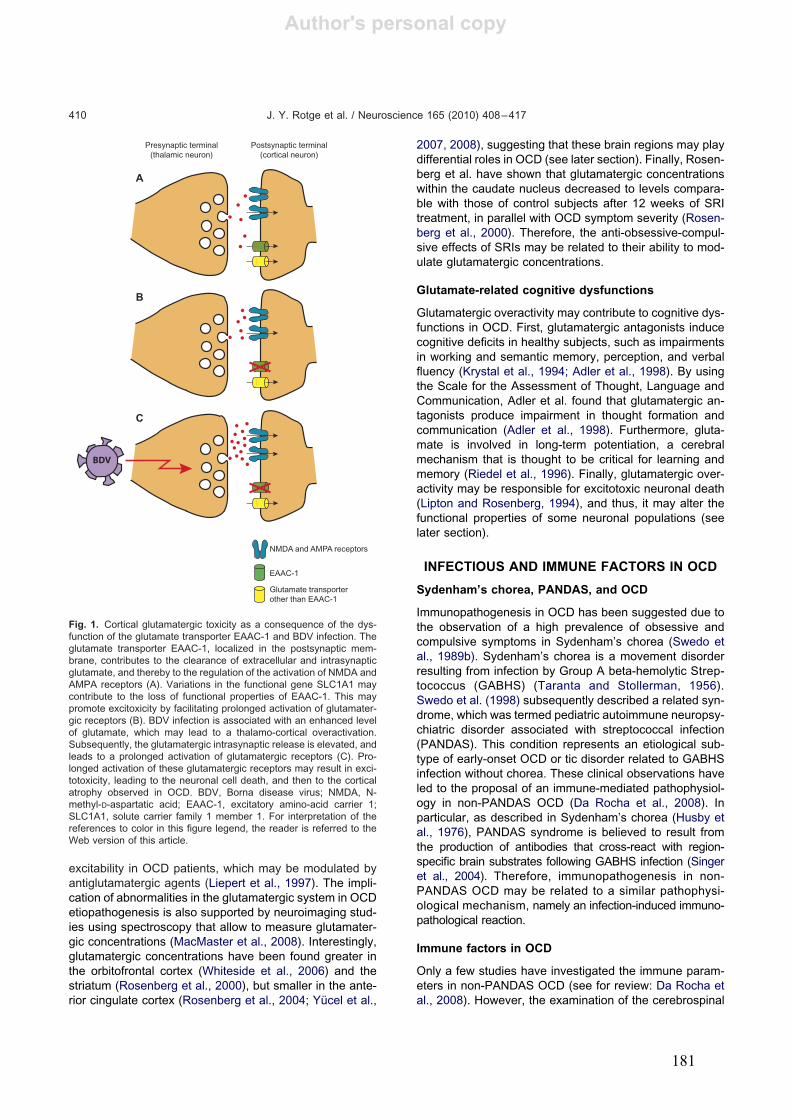
Author's personal copy
excitability in OCD patients, which may be modulated byantiglutamatergic agents (Liepert et al., 1997). The impli-cation of abnormalities in the glutamatergic system in OCDetiopathogenesis is also supported by neuroimaging stud-ies using spectroscopy that allow to measure glutamater-gic concentrations (MacMaster et al., 2008). Interestingly,glutamatergic concentrations have been found greater inthe orbitofrontal cortex (Whiteside et al., 2006) and thestriatum (Rosenberg et al., 2000), but smaller in the ante-rior cingulate cortex (Rosenberg et al., 2004; Yücel et al.,
2007, 2008), suggesting that these brain regions may playdifferential roles in OCD (see later section). Finally, Rosen-berg et al. have shown that glutamatergic concentrationswithin the caudate nucleus decreased to levels compara-ble with those of control subjects after 12 weeks of SRItreatment, in parallel with OCD symptom severity (Rosen-berg et al., 2000). Therefore, the anti-obsessive-compul-sive effects of SRIs may be related to their ability to mod-ulate glutamatergic concentrations.
Glutamate-related cognitive dysfunctions
Glutamatergic overactivity may contribute to cognitive dys-functions in OCD. First, glutamatergic antagonists inducecognitive deficits in healthy subjects, such as impairmentsin working and semantic memory, perception, and verbalfluency (Krystal et al., 1994; Adler et al., 1998). By usingthe Scale for the Assessment of Thought, Language andCommunication, Adler et al. found that glutamatergic an-tagonists produce impairment in thought formation andcommunication (Adler et al., 1998). Furthermore, gluta-mate is involved in long-term potentiation, a cerebralmechanism that is thought to be critical for learning andmemory (Riedel et al., 1996). Finally, glutamatergic over-activity may be responsible for excitotoxic neuronal death(Lipton and Rosenberg, 1994), and thus, it may alter thefunctional properties of some neuronal populations (seelater section).
INFECTIOUS AND IMMUNE FACTORS IN OCD
Sydenham’s chorea, PANDAS, and OCD
Immunopathogenesis in OCD has been suggested due tothe observation of a high prevalence of obsessive andcompulsive symptoms in Sydenham’s chorea (Swedo etal., 1989b). Sydenham’s chorea is a movement disorderresulting from infection by Group A beta-hemolytic Strep-tococcus (GABHS) (Taranta and Stollerman, 1956).Swedo et al. (1998) subsequently described a related syn-drome, which was termed pediatric autoimmune neuropsy-chiatric disorder associated with streptococcal infection(PANDAS). This condition represents an etiological sub-type of early-onset OCD or tic disorder related to GABHSinfection without chorea. These clinical observations haveled to the proposal of an immune-mediated pathophysiol-ogy in non-PANDAS OCD (Da Rocha et al., 2008). Inparticular, as described in Sydenham’s chorea (Husby etal., 1976), PANDAS syndrome is believed to result fromthe production of antibodies that cross-react with region-specific brain substrates following GABHS infection (Singeret al., 2004). Therefore, immunopathogenesis in non-PANDAS OCD may be related to a similar pathophysi-ological mechanism, namely an infection-induced immuno-pathological reaction.
Immune factors in OCD
Only a few studies have investigated the immune param-eters in non-PANDAS OCD (see for review: Da Rocha etal., 2008). However, the examination of the cerebrospinal
Presynaptic terminal(thalamic neuron)
Postsynaptic terminal(cortical neuron)
NMDA and AMPA receptors
EAAC-1
A
B
C
BDV
Glutamate transporterother than EAAC-1
Fig. 1. Cortical glutamatergic toxicity as a consequence of the dys-function of the glutamate transporter EAAC-1 and BDV infection. Theglutamate transporter EAAC-1, localized in the postsynaptic mem-brane, contributes to the clearance of extracellular and intrasynapticglutamate, and thereby to the regulation of the activation of NMDA andAMPA receptors (A). Variations in the functional gene SLC1A1 maycontribute to the loss of functional properties of EAAC-1. This maypromote excitoxicity by facilitating prolonged activation of glutamater-gic receptors (B). BDV infection is associated with an enhanced levelof glutamate, which may lead to a thalamo-cortical overactivation.Subsequently, the glutamatergic intrasynaptic release is elevated, andleads to a prolonged activation of glutamatergic receptors (C). Pro-longed activation of these glutamatergic receptors may result in exci-totoxicity, leading to the neuronal cell death, and then to the corticalatrophy observed in OCD. BDV, Borna disease virus; NMDA, N-methyl-D-aspartatic acid; EAAC-1, excitatory amino-acid carrier 1;SLC1A1, solute carrier family 1 member 1. For interpretation of thereferences to color in this figure legend, the reader is referred to theWeb version of this article.
J. Y. Rotge et al. / Neuroscience 165 (2010) 408–417410
181
Author's personal copy
fluid has revealed an enhanced rate of cell-mediated cyto-kines in pediatric OCD patients as compared with healthycontrol subjects (Mittleman et al., 1997). Furthermore, sev-eral studies argue for the alteration of cellular immuneparameters in OCD patients. For example, Marazziti et al.(1999) have reported an imbalance of CD81 and CD41
lymphocytes with an increased rate of CD81 and a de-creased rate of CD41 in OCD adults compared withhealthy controls (Marazziti et al., 1999). In parallel withclinical improvement, the CD41/CD81 ratio was revertedby SRIs (Marazziti et al., 2009). All these data were ob-tained in a small number of patients, but together theysuggest that cell-mediated immune processes may be in-volved in OCD etiopathogenesis.
Borna disease virus in OCD
Borna disease virus (BDV) is a RNA virus that persistentlyinfects a broad range of animal species. BDV infectioninduces neurological dysfunctions including behavioral ab-normalities such as stereotypies (Hornig et al., 2001). Re-cently, a German research group suggested that BDV mayplay a role in the etiology of some psychiatric disorders,especially OCD (Bode et al., 1996, 2001). The authorsisolated BDV in OCD patients and reported an associationbetween the BDV activity and dysfunction in the anteriorcingulate cortex in OCD patients (Bode et al., 1996, 2001;Dietrich et al., 2005). Indeed, the authors showed that theevent-related brain potential amplitude during a Go/NoGotask was higher in OCD patients than controls according tothe amount of BDV-specific circulating immune complexes(Dietrich et al., 2005). Although this unreplicated result wasobtained in a small number of patients, it gives strength tothe idea that BDV may contribute to the brain dysfunctionsobserved in OCD (see later section).
Cerebral immunopathological reactions induced by
BDV
Neurotropic BDV has some very specific characteristicsthat have been investigated through multiple animal exper-imental studies (see for review: De la Torre, 2002). Thevirus has a preferential neurotropism for the cortex, thala-mus, hippocampus, and the amygdala (Werner-Keiss etal., 2009). BDV-induced behavioral changes are not di-rectly related to its replication, but to the virus-specificT-cell immune responses (Narayan et al., 1983; Bilzer andStitz, 1996). Furthermore, increased levels of cytokines inthe brains of BDV-infected animals correlated with thedegree of inflammation (Stitz et al., 1995). These findingsare consistent with the cell- and cytokine-immune changesdescribed in OCD, suggesting the possible involvement ofa BDV-related cell-mediated immune dysfunctions in OCDpathophysiology (Mittleman et al., 1997; Marazziti et al.,1999, 2009). Although we discuss here cell-mediated im-mune dysfunctions in OCD, it is possible that humoral, thatis antibody-mediated, mechanisms may contribute to OCDpathophysiology (Whitney and McNamara, 1999). Autoan-tibodies to glutamatergic AMPA receptors that mediate fastexcitatory synaptic transmission are thought to be involvedin some neuropsychiatric disorders associated with chronic
hyperexcitability syndromes (Whitney andMcNamara, 1999).It has been shown that virus infection may result in theproduction of such autoantibodies, in particular autoanti-bodies directed against the GluR3 subunit of AMPA recep-tors (Koustova et al., 2001). Such a humoral mechanismcould occur in response to BDV infection and, therefore, itcould contribute to the glutamatergic overactivity observedin OCD. However, further studies are needed to test thehypothesis of an association between BDV, anti-GluR3antibodies and OCD.
BRAIN ALTERATIONS AND COGNITIVE
DISRUPTIONS IN OCD
Structural brain alterations in OCD
Several MRI studies have measured brain volumechanges associated with OCD. Recently, a meta-analysiscompiling data from volumetric MRI studies showed areduction in the anterior cingulate cortex (ACC) and orbito-frontal cortex (OFC) volumes, and an augmentation inthalamic volumes of OCD patients compared to healthysubjects (Rotge et al., 2009a). These findings are sup-ported by voxel-based morphometry studies showing vari-ations in the gray matter density in these cortical andsubcortical structures (Kim et al., 2001; Yoo et al., 2008).Within the thalamus, the ventral anterior and mediodorsalnuclei are of great interest in OCD pathophysiology be-cause they send vast projections to the ACC and OFC,which are also altered in OCD. Accordingly, an investiga-tion of thalamic shape deformities by Kang et al. (2008)supported this hypothesis. Indeed, the authors havemainly reported outward deformities in the anterior lateralportion of the thalamus in OCD patients, thereby suggest-ing that thalamic structural alterations may essentially in-volve the ventral anterior nucleus. Finally, structural alter-ations in the striatum, a subcortical structure that receivesprojections from the ACC and OFC have been described inOCD patients (Rotge et al., in press).
Functional brain alterations in OCD
The cortical and subcortical regions structurally altered inOCD are also overactive in patients during the restingstate. An initial voxel-based meta-analysis of OCD patientswith functional neuroimaging studies reported an in-creased activation in the ACC, OFC, striatum, and thethalamus (Menzies et al., 2008). Another voxel-basedmeta-analysis of functional neuroimaging studies showedthat the ACC, OFC and the thalamus are overactive whenOCD symptoms are provocated in patients (Rotge et al.,2008). Therefore, functional alterations within the anteriorcingulate-subcortical and the orbitofrontal-subcortical cir-cuits likely participate in mediating obsessive-compulsivesymptoms.
Cognitive disruptions related to brain alterations in
OCD
The ACC may be divided into the two following distinctfunctional regions: the most dorsal part is the cognitive
J. Y. Rotge et al. / Neuroscience 165 (2010) 408–417 411
182
Author's personal copy
division, which is involved in conflict monitoring and error
detection; the most ventral part is the affective division,
which is implicated in emotional processing, including the
assessment of emotional information and the regulation of
emotional responses (Bush et al., 2000). The overactivity
of the cognitive division in OCD has been shown to par-
ticipate in the dysfunctional monitoring of high-conflict sit-
uations, which places the subject in a potential position to
make an error (Maltby et al., 2005). The ACC also plays a
role in the increased evaluation that an action might be
mistaken, and in the impression the subject has of being in
error or at fault (Fitzgerald et al., 2005). Therefore, the
cognitive division of the ACC seems to be involved in the
generation of the internal high “error signal” that has been
proposed as the core of OCD symptomatology (Aouizerate
et al., 2004). On the other hand, due to its connections with
the limbic system, the affective division of the ACC might
mediate the clinical expression of anxiety symptoms asso-
ciated with the obsessional process (Aouizerate et al.,
2004).
The OFC plays a role in several cognitive processes,
such as the selection, comparison, and judgment of envi-
ronmental stimuli, in addition to the monitoring of error
recognition (Krawczyk, 2002; Wallis, 2007). It is highly
involved in encoding the representation of the value of an
expected outcome, which is used to anticipate the positive
and negative consequences likely to follow a given action
(Wallis, 2007; Fellows, 2007). The OFC may thus contrib-
ute to an increased evaluation of the occurrence of nega-
tive consequences following an action in OCD patients
(Aouizerate et al., 2004). This phenomenon may generate
persistent and distressing thoughts, leading the patient to
perform repetitive or ritualistic behaviors directed at pre-
venting the occurrence of such consequences or aimed to
ensuring that they do not occur. Consequently, this is the
only way to relieve the anxiety induced by obsessive
thoughts. Recent functional MRI studies have also shown
that OFC dysfunctions contribute to the occurrence of
some very characteristic cognitive disruptions observed in
OCD. In particular, cognitive inflexibility represents a cen-
tral feature of OCD and prevents patients from shifting
from one thought or behavior to another (Chamberlain et
al., 2006). OCD has been associated with a reduced or-
bitofrontal activation related to cognitive flexibility, as as-
sessed by reversal learning tasks or task switching exer-
cises (Gu et al., 2008; Chamberlain et al., 2008).
TOWARD AN INTEGRATIVE VIEW
OF OCD PATHOPHYSIOLOGY: THE
GLUTAMATE-BASED GENETIC
IMMUNE HYPOTHESIS
To date, we know the following: (i) variations in the functional
SLC1A1 gene, coding for a glutamate transporter, are asso-
ciated with OCD; (ii) cell-mediated immune reactions to BDV
infection possibly play a role in OCD etiopathogenesis; (iii)
the glutamatergic thalamo-cingulate and -orbitofrontal path-
ways are structurally and functionally altered in OCD; and (iv)
alterations of these thalamo-cortical pathways are associated
with some very characteristic cognitive disruptions in OCD.
The subsequent section will attempt to relate these remark-
able research advances.
Variations in the SLC1A1 gene may contribute to
BDV-induced immunopathological reactions
Variations in the SLC1A1 gene may affect the function of
the glutamate transporter EAAC-1. EAAC-1 is localized in
extracerebral tissues and therefore contributes to the
clearance of extracerebral extracellular glutamate, which
may accumulate when EAAC-1 loses its functional prop-
erties. There has been growing recognition in the literature
that glutamate acts as an immunomodulator, and more
particularly, as an important regulator of T-cell function
(see for review: Pacheco et al., 2007). Indeed, T-cells
express glutamatergic receptors on their surface (Kostan-
yan et al., 1997). These receptors may have different
functions according to the activation state of T-cells. In
particular, when T-cells are activated following a specific
antigen presentation, such as the BDV-specific antigen,
the expression of the metabotropic glutamatergic receptor
1 (mGlu1R) is induced (Pacheco et al., 2007). Then, its
activation by extracellular glutamate promotes enhanced
production of cytokines, which has been reported in the
brains of BDV-infected animals (Stitz et al., 1995) and in
the cerebrospinal fluid of patients with OCD (Mittleman et
al., 1997). Therefore, genetic alterations of the glutamater-
gic system may facilitate BDV-induced immunopathologi-
cal reactions in OCD patients.
Variations in the SLC1A1 gene may contribute to
cortical alterations
We know that the ACC and OFC receive widespread glu-
tamatergic thalamic inputs, but it is also known that both of
these cortical regions have a smaller volume (Rotge et al.,
2009a) and are overactive during the resting state in OCD
patients (Menzies et al., 2008; Rotge et al., 2008). There-
fore, it may be assumed that the cortical glutamatergic
hyperactivity observed in OCD patients is also responsible
for any associated neurotoxic insults. Indeed, sustained
glutamate excitatory activity contributes to atrophy in the
long term by promoting excitotoxic neuronal degeneration
(Lipton and Rosenberg, 1994). In the CNS, the glutamate
transporter EAAC-1 is localized in several cerebral tissues,
including the cortex. By clearing extracellular glutamate,
glutamate transporters contribute to the protection of neu-
rons against glutamatergic excitotoxicity as a result of
prolonged activation of NMDA and AMPA receptors (Ne-
dergaard et al., 2002), and thus, against excitotoxic cell
death (Danbolt, 2001). However, although the functional
alteration of EAAC-1 may represent a susceptibility factor
for cortical neuroexcitotoxicity, it is likely that other factors
are necessary to induce excitotoxic cell death since other
glutamate transporters remain functional and contribute to
the clearance of glutamate (Figs. 1 and 2).
J. Y. Rotge et al. / Neuroscience 165 (2010) 408–417412
183
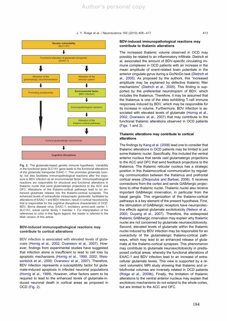
Author's personal copy
BDV-induced immunopathological reactions may
contribute to cortical alterations
BDV infection is associated with elevated levels of gluta-
mate (Hornig et al., 2002; Ovanesov et al., 2007). How-
ever, findings from experimental studies have suggested
that infection alone is insufficient to lead to cell loss by
apoptotic mechanisms (Hornig et al., 1999, 2002; Weis-
senböck et al., 2000; Ovanesov et al., 2007). Therefore,
BDV infection represents a susceptibility factor for gluta-
mate-induced apoptosis in infected neuronal populations
(Hornig et al., 1999). However, other factors seem to be
required to lead to the enhanced levels of glutamate-in-
duced neuronal death in cortical areas as proposed in
OCD (Fig. 2).
BDV-induced immunopathological reactions may
contribute to thalamic alterations
The increased thalamic volume observed in OCD may
possibly be related to an inflammatory infiltrate. Dietrich et
al. associated the amount of BDV-specific circulating im-
mune complexes in OCD patients with an increase in the
mean amplitude of event-related brain potentials in the
anterior cingulate gyrus during a Go/NoGo task (Dietrich et
al., 2005). As proposed by the authors, this “increased
amplitude may be explained by defective thalamic filter
mechanisms” (Dietrich et al., 2005). This finding is sup-
ported by the preferential neurotropism of BDV, which
includes the thalamus. Therefore, it may be assumed that
the thalamus is one of the sites exhibiting T-cell immune
responses induced by BDV, which may be responsible for
its increase in volume. Furthermore, BDV infection is as-
sociated with elevated levels of glutamate (Hornig et al.,
2002; Ovanesov et al., 2007) that may contribute to the
functional thalamic alterations observed in OCD patients
(Figs. 1 and 2).
Thalamic alterations may contribute to cortical
alterations
The findings by Kang et al. (2008) lead one to consider that
thalamic alterations in OCD patients may be limited to just
some thalamic nuclei. Specifically, this included the ventral
anterior nucleus that sends vast glutamatergic projections
to the ACC and OFC that send feedback projections to the
thalamus. The thalamic reticular nucleus has a strategic
position in this thalamocortical communication by regulat-
ing communication between the thalamus and prefrontal
cortical areas (Zikopoulos and Barbas, 2006). It receives
connections from the cortex and sends GABAergic projec-
tions to other thalamic nuclei. Thalamic nuclei also receive
important GABAergic innervation, in particular from the
basal ganglia. This organization of the thalamo-cortical
pathways is a key element of the present hypothesis. First,
the stimulation of GABAergic receptors have neuroprotec-
tive effects against glutamate excitotoxicity (Nelson et al.,
2000; Ouyang et al., 2007). Therefore, the widespread
thalamic GABAergic innervation may explain why thalamic
nuclei are not concerned by glutamate neuroexcitotoxicity.
Second, elevated levels of glutamate within the thalamic
nuclei induced by BDV infection may be responsible for an
overactivity of the glutamatergic thalamo-cortical path-
ways, which may lead to an enhanced release of gluta-
mate at the thalamo-cortical synapses. This phenomenon
may contribute to glutamate neuroexcitotoxicity in predis-
posed cortical areas, whereby the functional alterations of
EAAC-1 and BDV infection lead to an increase of extra-
cellular glutamate levels. This view is supported by a re-
cent volumetric MRI study showing that thalamic and or-
bitofrontal volumes are inversely related in OCD patients
(Rotge et al., 2009b). Finally, the limitation of thalamic
alterations to the ventral anterior nucleus may explain that
excitotoxic mechanisms do not extend to the whole cortex,
but are limited to the ACC and OFC.
Genetic vulnerability
(SLC1-A1)
Functional alteration of glutamate transporter(EAAC-1)
Alteration of theglutamatergic neurotransmission
Alteration of theimmune system
Promoting excitotoxicity
Immunopathological reactions
Alteration of thethalamo-cortical pathways
Cortical glutamatergic neurotoxicity
Cognitive disruptions
Environmental factor
(BDV infection)
Fig. 2. The glutamate-based genetic immune hypothesis. Variabilityin the functional gene SLC1A1 gene leads to the functional alterationsof the glutamate transporter EAAC-1. This promotes glutamate toxic-ity, but also facilitates immunopathological reactions after the expo-sure to BDV infection as an environmental factor. Immunopathologicalreactions are responsible for structural and functional alterations inthalamic nuclei that send glutamatergic projections to the ACC andOFC. Alterations of the thalamo-cortical pathways lead to an en-hanced glutamate release into the thalamo-cortical synapses. Theenhanced levels of extracellular intrasynaptic glutamate, facilitated byalterations of EAAC-1 and BDV infection, result in cortical neurotoxicitythat is responsible for the cognitive disruptions characteristic of OCD.BDV, Borna disease virus; EAAC-1, excitatory amino-acid carrier 1;SLC1A1, solute carrier family 1 member 1. For interpretation of thereferences to color in this figure legend, the reader is referred to theWeb version of this article.
J. Y. Rotge et al. / Neuroscience 165 (2010) 408–417 413
184
Author's personal copy
Early-onset versus late-onset OCD: distinct
pathophysiological mechanisms
Recent epidemiological studies have pointed out a bimodaldistribution for the age of OCD onset, thereby suggestingthe existence of age-related subtypes of OCD (Stein,2002). Age-related subtypes have distinguishing charac-teristics; for example, early-onset OCD has greater famil-iality and relation to tic disorders (Eichstedt and Arnold,2001). Furthermore, early-onset OCD is associated with aworse prognosis than late-onset OCD (Skoog and Skoog,1999; Stewart et al., 2004). Thus, the age-related subtypesof OCD may depend on distinct pathophysiological mech-anisms. For example, findings from Arnold et al. (2006)suggested that the association between variations in theSLC1A1 gene and OCD may be predominant in early-onset OCD, without formally excluding its role in late-onsetOCD. In the present hypothetical model, the description ofage-related subtypes of OCD is supported by infectiousand immunological investigations of BDV infection in ani-mals. Indeed, it has been shown that BDV-induced immu-nological reactions and associated clinical symptoms(Hirano et al., 1983), but also the tissue distribution of thevirus (Herzog et al., 1984) are dependent on the age atinfection. Furthermore, the neurotransmitter glutamateparticipates in controlling the programmed cell death thatoccurs during the normal development of the brain (Por-tera-Cailliau et al., 1997a, b). In addition, the glutamatergiccontrol of brain development appears to be age-dependent(Ikonomidou et al., 1999). Therefore, in genetically predis-posed individuals, a BDV infection in part of the glutama-tergic system at an early or late stage of development mayhave different consequences on the brain, particularly con-cerning the glutamatergic control of the normal pro-grammed cell death (Nelson et al., 2000). Interestingly, thiscould explain the clinical differences between early- andlate-onset OCD.
LIMITATIONS OF THE GLUTAMATE-BASED
GENETIC IMMUNE HYPOTHESIS IN OCD
Although the present pathophysiological model may ap-pear coherent and integrative, it is clearly oversimplifiedand speculative in regard to several aspects. First, animportant concern may lie in the localized alterations of thethalamus. A priori, there is no reason for BDV-inducedimmunopathological reactions to be limited to the ventralanterior nucleus without extensions to other thalamic nu-clei. However, if we assume that the increase of thalamicvolume is actually related to BDV infection, the two follow-ing explanations may be advanced: the expression of theSLC1A1 gene in the thalamus is limited to the ventralanterior nucleus, or BDV has a preferential neurotropismfor this nucleus within the thalamus. This point remainsunclear. Furthermore, it may be assumed that brain re-gions other than the thalamus and the cortex may beinfected by BDV in OCD according to the virus’s neurotro-pism, in particular the amygdalo-hippocampal complex.However, structural and functional alterations of thesebrain regions in OCD remain to be clarified (Menzies et al.,
2008; Rotge et al., 2009a). Second, the present article hasfocused on the glutamatergic system, and the role of otherneurotransmitters such as the serotonin and dopamine inOCD has not been discussed. Reciprocal modulationsbetween glutamate and serotonin, and between glutamateand dopamine, have been described. For example, resultsfrom animal studies show that there is a mutual influencebetween glutamate and the 5-HT2A serotoninergic recep-tor. Glutamatergic antagonists are responsible for an in-creased serotoninergic transmission through 5-HT2A re-ceptor and, conversely, the stimulation of 5-HT2A receptorinduces a decrease in glutamatergic transmission (Marekand Aghajanian, 1994; Carlsson, 1995, 2000; Waters etal., 1996). However, it seems us unlikely, but not impossi-ble, that serotoninergic and dopaminergic alterations aresecondary to the glutamatergic alterations in OCD, aspreviously proposed (Carlsson, 2000). Finally, we assumethat SLC1A1 variability associated with OCD is responsi-ble for alterations of the expression or function of theglutamate transporter EAAC-1. However, the functionalityof the polymorphism of SLC1A1 remains unresolved todate (Table 1). Further studies are needed to elucidate theconsequences of SLC1A1 polymorphism on the glutama-tergic system. Despite these limitations, the present ap-proach remains relevant and helpful, particularly whenused as a theoretical tool to ask new questions that may betested by future investigations that combine the variousaspects of different research areas (Table 1).
CONCLUSION
This article has proposed a pathophysiological hypothesisfor OCD that integrates genetic, infectious, immunological,neuroimaging, and neuropsychological data. Further stud-ies combining several approaches from these multiple re-search areas are warranted to test the numerous ques-tions generated by this glutamate-based genetic immunemodel.
Acknowledgments—JYR was supported by the “Fondation pour la
Recherche Médicale” [Medical Research Fundation].
Table 1. The glutamate-based genetic immune model: outstanding
questions and issues
Are genetic variations of SLC1A1 gene responsible for functionalalterations of the glutamate transporter EAAC-1?
Do functional alterations of EAAC-1 contribute to theglutamatergic overactivity observed in OCD?
Do functional alterations of EAAC-1 facilitate the BDV-inducedimmunopathological reactions?
Are BDV-induced immunopathological reactions responsible for thethalamic alterations observed in OCD?
Are BDV-induced immunopathological reactions limited to theventralanterior nucleus?
Are thalamic alterations responsible for an enhanced glutamaterelease at the thalamo-cortical synapse?
Do functional alterations of EAAC-1 and BDV infection promoteglutamatergic excitotoxicity?
Are excitotoxic mechanisms responsible for the reduction in corticalvolumes and cortical dysfunctions observed in OCD?
J. Y. Rotge et al. / Neuroscience 165 (2010) 408–417414
185
Author's personal copy
REFERENCES
Adler CM, Goldberg TE, Malhotra AK, Pickar D, Breier A (1998) Effects of
ketamine on thought disorder, working memory, and semantic mem-
ory in healthy volunteers. Biol Psychiatry 43:811–816.
Aouizerate B, Guehl D, Cuny E, Rougier A, Bioulac B, Tignol J, Burbaud
P (2004) Pathophysiology of obsessive-compulsive disorder: a nec-
essary link between phenomenology, neuropsychology, imagery and
physiology. Prog Neurobiol 72:195–221.
Arnold PD, Sicard T, Burroughs E, Richter MA, Kennedy JL (2006)
Glutamate transporter gene SLC1A1 associated with obsessive-
compulsive disorder. Arch Gen Psychiatry 63:769–776.
Baxter LR Jr, Schwartz JM, Mazziotta JC, Phelps ME, Pahl JJ, Guze
BH, Fairbanks L (1988) Cerebral glucose metabolic rates in non-
depressed patients with obsessive-compulsive disorder. Am J Psy-
chiatry 145:1560–1563.
Bergles DE, Jahr CE (1997) Synaptic activation of glutamate trans-
porters in hippocampal astrocytes. Neuron 19:1297–1308.
Bilzer T, Stitz L (1996) Immunopathogenesis of virus diseases affect-
ing the central nervous system. Crit Rev Immunol 16:145–222.
Bode L, Dürrwald R, Rantam FA, Ferszt R, Ludwig H (1996) First
isolates of infectious human Borna disease virus from patients with
mood disorders. Mol Psychiatry 1:200–212.
Bode L, Reckwald P, Severus WE, Stoyloff R, Ferszt R, Dietrich DE,
Ludwig H (2001) Borna disease virus-specific circulating immune
complexes, antigenemia, and free antibodies—the key marker trip-
let determining infection and prevailing in severe mood disorders.
Mol Psychiatry 6:481–491.
Bush G, Luu P, Posner MI (2000) Cognitive and emotional influences
in anterior cingulate cortex. Trends Cogn Sci 4:215–222.
Carlsson ML (1995) The selective 5-HT2A receptor antagonist MDL
100,907 counteracts the psychomotor stimulation ensuing manip-
ulations with monoaminergic, glutamatergic or muscarinic neuro-
transmission in the mouse—implications for psychosis. J Neural
Transm 100:225–237.
Carlsson ML (2000) On the role of cortical glutamate in obsessive-
compulsive disorder and attention-deficit hyperactivity disorder,
two phenomenologically antithetical conditions. Acta Psychiatr
Scand 102:401–413.
Chakrabarty K, Bhattacharyya S, Christopher R, Khanna S (2005)
Glutamatergic dysfunction in OCD. Neuropsychopharmacology
30:1735–1740.
Chamberlain SR, Fineberg NA, Blackwell AD, Robbins TW, Sahakian
BJ (2006) Motor inhibition and cognitive flexibility in obsessive-
compulsive disorder and trichotillomania. Am J Psychiatry 163:
1282–1284.
Chamberlain SR, Menzies L, Hampshire A, Suckling J, Fineberg NA,
del Campo N, Aitken M, Craig K, Owen AM, Bullmore ET, Robbins
TW, Sahakian BJ (2008) Orbitofrontal dysfunction in patients with
obsessive-compulsive disorder and their unaffected relatives. Sci-
ence 321:421–422.
Coric V, Taskiran S, Pittenger C, Wasylink S, Mathalon DH, Valentine
G, Saksa J, Wu YT, Gueorguieva R, Sanacora G, Malison RT,
Krystal JH (2005) Riluzole augmentation in treatment-resistant
obsessive-compulsive disorder: an open-label trial. Biol Psychiatry
58:424–428.
Danbolt NC (2001) Glutamate uptake. Prog Neurobiol 65:1–105.
Da Rocha FF, Correa H, Teixeira AL (2008) Obsessive-compulsive
disorder and immunology: a review. Prog Neuropsychopharmacol
Biol Psychiatry 32:1139–1146.
De la Torre JC (2002) Bornavirus and the brain. J Infect Dis 186:
241–247.
Diamond JS, Jahr CE (1997) Transporters buffer synaptically released
glutamate on a submillisecond time scale. J Neurosci 17:4672–
4687.
Dickel DE, Veenstra-VanderWeele J, Cox NJ, Wu X, Fischer DJ, Van
Etten-Lee M, Himle JA, Leventhal BL, Cook EH Jr, Hanna GL
(2006) Association testing of the positional and functional candi-
date gene SLC1A1/EAAC1 in early-onset obsessive-compulsive
disorder. Arch Gen Psychiatry 63:778–785.
Dietrich DE, Zhang Y, Bode L, Münte TF, Hauser U, Schmorl P,
Richter-Witte C, Gödecke-Koch T, Feutl S, Schramm J, Ludwig H,
Johannes S, Emrich HM (2005) Brain potential amplitude varies as
a function of Borna disease virus-specific immune complexes in
obsessive-compulsive disorder. Mol Psychiatry 10:519–520.
Eichstedt JA, Arnold SL (2001) Childhood-onset obsessive-compul-
sive disorder: a tic-related subtype of OCD? Clin Psychol Rev
21:137–157.
Fellows LK (2007) Advances in understanding ventromedial prefrontal
function: the accountant joins the executive. Neurology 68:991–
995.
Fitzgerald KD, Welsh RC, Gehring WJ, Abelson JL, Himle JA, Liber-
zon I, Taylor SF (2005) Error-related hyperactivity of the anterior
cingulate cortex in obsessive-compulsive disorder. Biol Psychiatry
57:287–294.
Grant P, Lougee L, Hirschtritt M, Swedo SE (2007) An open-label trial
of riluzole, a glutamate antagonist, in children with treatment-
resistant obsessive-compulsive disorder. J Child Adolesc Psycho-
pharmacol 17:761–767.
Greenberg BD, Ziemann U, Cora-Locatelli G, Harmon A, Murphy DL,
Keel JC, Wassermann EM (2000) Altered cortical excitability in
obsessive-compulsive disorder. Neurology 54:142–147.
Greenberg BD, Malone DA, Friehs GM, Rezai AR, Kubu CS, Malloy
PF, Salloway SP, Okun MS, Goodman WK, Rasmussen SA (2006)
Three-year outcomes in deep brain stimulation for highly resistant
obsessive-compulsive disorder. Neuropsychopharmacology 31:
2384–2393.
Gu BM, Park JY, Kang DH, Lee SJ, Yoo SY, Jo HJ, Choi CH, Lee JM,
Kwon JS (2008) Neural correlates of cognitive inflexibility during
task-switching in obsessive-compulsive disorder. Brain 131:155–
164.
He Y, Janssen WGM, Rothstein JD, Morrison JH (2000) Differential
synaptic localization of the glutamate transporter EAAC1 and glu-
tamate receptor subunit GluR2 in the rat hippocampus. J Comp
Neurol 418:255–269.
Herzog S, Kompter C, Frese K, Rott R (1984) Replication of Borna
disease virus in rats: age-dependent differences in tissue distribu-
tion. Med Microbiol Immunol 173:171–177.
Hirano N, Kao M, Ludwig H (1983) Persistent, tolerant or subacute
infection in Borna disease virus-infected rats. J Gen Virol 64:1521–
1530.
Hornig M, Weissenböck H, Horscroft N, Lipkin WI (1999) An infection-
based model of neurodevelopmental damage. Proc Natl Acad Sci
U S A 96:12102–12107.
Hornig M, Solbrig M, Horscroft N, Weissenböck H, Lipkin WI (2001)
Borna disease virus infection of adult and neonatal rats: models for
neuropsychiatric disease. Curr Top Microbiol Immunol 253:157–
177.
Hornig M, Mervis R, Hoffman K, Lipkin WI (2002) Infectious and
immune factors in neurodevelopmental damage. Mol Psychiatry
7:34–35.
Husby G, Van de Rijn I, Zabriskie JB, Abdin ZH, Williams RC Jr (1976)
Antibodies reacting with cytoplasm of subthalamic and caudate
nuclei neurons in chorea and acute rheumatic fever. J Exp Med
144:1094–1110.
Ikonomidou C, Bosch F, Miksa M, Bittigau P, Vöckler J, Dikranian K,
Tenkova TI, Stefovska V, Turski L, Olney JW (1999) Blockade of
NMDA receptors and apoptotic neurodegeneration in the develop-
ing brain. Science 283:70–74.
Kang DH, Kim SH, Kim CW, Choi JS, Jang JH, Jung MH, Lee JM, Kim
SI, Kwon JS (2008) Thalamus surface shape deformity in obses-
sive-compulsive disorder and schizophrenia. Neuroreport 19:
609–613.
Kim JJ, Lee MC, Kim J, Kim IY, Kim SI, Han MH, Chang KH, Kwon JS
(2001) Grey matter abnormalities in obsessive-compulsive disor-
J. Y. Rotge et al. / Neuroscience 165 (2010) 408–417 415
186
Author's personal copy
der: statistical parametric mapping of segmented magnetic
resonance images. Br J Psychiatry 179:330–334.
Kostanyan IA, Merkulova MI, Navolotskaya EV, Nurieva RI (1997)
Study of the interaction between L-glutamate and human blood
lymphocytes. Immunol Lett 58:177–180.
Koustova E, Sei Y, Fossom L, Wei ML, Usherwood PNR, Keele NB,
Rogawski MA, Basile AS (2001) LP-BM5 virus-infected mice pro-
duce activating autoantibodies to the AMPA receptor. J Clin Invest
107:737–744.
Krawczyk DC (2002) Contributions of the prefrontal cortex to the
neural basis of human decisions making. Neurosci Biobehav Rev
26:631–664.
Krystal JH, Karper LP, Seibyl JP, Freeman GK, Delaney R, Bremmer
JD, Heninger GR, Bowers M Jr, Charney DS (1994) Subanesthetic
effects of the noncompetitive NMDA antagonist, ketamine, in hu-
mans: psychotomimetic, perceptual, cognitive, and neuroendo-
crine responses. Arch Gen Psychiatry 51:199–214.
Liepert J, Schwenkreis P, Tegenthoff M, Malin JP (1997) The gluta-
mate antagonist riluzole suppresses intracortical facilitation. J Neu-
ral Transm 104:1207–1214.
Lipton SA, Rosenberg PA (1994) Excitatory amino acids as a final
common pathway for neurologic disorders. N Engl J Med 330:
613–622.
MacMaster FP, O’Neill J, Rosenberg DR (2008) Brain imaging in
pediatric obsessive compulsive disorder. J Am Acad Child Adolesc
Psychiatry 47:1262–1272.
Maltby N, Tolin DF, Worhunsky P, O’Keefe TM, Kiehl KA (2005)
Dysfunctional action monitoring hyperactivates frontal-striatal cir-
cuits in obsessive-compulsive disorder: an event-related fMRI
study. Neuroimage 24:495–503.
Marazziti D, Presta S, Pfanner C, Gemignani A, Rossi A, Sbrana S,
Rocchi V, Ambrogi F, Cassano GB (1999) Immunological alter-
ations in adult obsessive-compulsive disorder. Biol Psychiatry
46:810–814.
Marazziti D, Mungai F, Masala I, Baroni S, Vivarelli L, Ambrogi F,
Catena Dell’osso M, Consoli G, Massimetti G, Dell’osso L (2009)
Normalisation of immune cell imbalance after pharmacological
treatments of patients suffering from obsessive-compulsive disor-
der. J Psychopharmacol 23:567–573.
Marek GJ, Aghajanian GK (1994) Excitation of interneurons in piriform
cortex by 5-hydroxytryptamine (5-HT): blockade by MDL 100,907,
a highly selective 5-HT2A antagonist. Eur J Pharmacol 259:137–
141.
Menzies L, Chamberlain SR, Laird AR, Thelen SM, Sahakian BJ,
Bullmore ET (2008) Integrating evidence from neuroimaging and
neuropsychological studies of obsessive-compulsive disorder: the
orbitofronto-striatal model revisited. Neurosci Biobehav Rev 32:
525–549.
Mittleman BB, Castellanos FX, Jacobsen LK, Rapoport JL, Swedo SE,
Shearer GM (1997) Cerebrospinal fluid cytokines in pediatric neu-
ropsychiatric disease. J Immunol 159:2994–2999.
Narayan O, Herzog S, Frese K, Scheefers H, Rott R (1983) Behavioral
disease in rats caused by immunopathological responses to per-
sistent Borna virus in the brain. Science 220:1401–1403.
Nedergaard M, Takano T, Hansen AJ (2002) Beyond the role of
glutamate as a neurotransmitter. Nat Rev Neurosci 3:748–755.
Nelson RM, Green AR, Lambert DG, Hainsworth AH (2000) On the
regulation of ischaemia-induced glutamate efflux from rat cortex by
GABA; in vitro studies with GABA, clomethiazole and pentobarbi-
tone. Br J Pharmacol 130:1124–1130.
Ouyang C, Guo L, Lu Q, Xu X, Wang H (2007) Enhanced activity of
GABA receptors inhibits glutamate release induced by focal cere-
bral ischemia in rat striatum. Neurosci Lett 420:174–178.
Ovanesov MV, Vogel MW, Moran TH, Pletnikov MV (2007) Neonatal
Borna disease virus infection in rats is associated with increased
extracellular levels of glutamate and neurodegeneration in the
striatum. J Neurovirol 13:185–194.
Pacheco R, Gallart T, Lluis C, Franco R (2007) Role of glutamate on
T-cell mediated immunity. J Neuroimmunol 185:9–19.
Pallanti S, Quercioli L (2006) Treatment-refractory obsessive-compul-
sive disorder: methodological issues, operational definitions and
therapeutic lines. Prog Neuropsychopharmacol Biol Psychiatry
30:400–412.
Pauls DL (2008) The genetics of obsessive compulsive disorder: a
review of the evidence. Am J Med Genet C Semin Med Genet
148:133–139.
Portera-Cailliau C, Price DL, Martin LJ (1997a) Excitotoxic neuronal
death in the immature brain is an apoptosis-necrosis morphological
continuum. J Comp Neurol 378:70–87.
Portera-Cailliau C, Price DL, Martin LJ (1997b) Non-NMDA and NMDA
receptor-mediated excitotoxic neuronal deaths in adult brain are
morphologically distinct: further evidence for an apoptosis-necrosis
continuum. J Comp Neurol 378:88–104.
Rasmussen SA, Eisen JL, Pato MT (1993) Current issues in the
pharmacologic management of obsessive compulsive disorder.
J Clin Psychiatry 5:4–9.
Riedel G, Wetzel W, Reymann KG (1996) Comparing the role of
metabotropic glutamate receptors in long-term potentiation and in
learning and memory. Prog Neuropsychopharmacol Biol Psychia-
try 20:761–789.
Rosenberg DR, MacMaster FP, Keshavan MS, Fitzgerald KD, Stewart
CM, Moore GJ (2000) Decrease in caudate glutamatergic concen-
trations in pediatric obsessive-compulsive disorder patients taking
paroxetine. J Am Acad Child Adolesc Psychiatry 39:1096–1103.
Rosenberg DR, Mirza Y, Russell A, Tang J, Smith JM, Banerjee SP,
Bhandari R, Rose M, Ivey J, Boyd C, Moore GJ (2004) Reduced
anterior cingulate glutamatergic concentrations in childhood OCD
and major depression versus healthy controls. J Am Acad Child
Psychiatry 43:1146–1153.
Rotge JY, Guehl D, Dilharreguy B, Cuny E, Tignol J, Bioulac B, Allard
M, Burbaud P, Aouizerate B (2008) Provocation of obsessive-
compulsive symptoms: a quantitative voxel-based meta-analysis
of functional neuroimaging studies. J Psychiatry Neurosci 33:
405–412.
Rotge JY, Guehl D, Dilharreguy B, Tignol J, Bioulac B, Allard M,
Burbaud P, Aouizerate B (2009a) Meta-analysis of brain volume
changes in obsessive-compulsive disorder. Biol Psychiatry 65:
75–83.
Rotge JY, Dilharreguy B, Aouizerate B, Martin-Guehl C, Guehl D,
Jaafari N, Langbour N, Bioulac B, Tignol J, Allard M, Burbaud P
(2009b) Inverse relationship between thalamic and orbitofrontal
volumes in obsessive-compulsive disorder. Prog Neuropsychop-
harmacol Biol Psychiatry 33:682–687.
Rotge JY, Langbour N, Guehl D, Bioulac B, Jaafari N, Allard M,
Aouizerate B, Burbaud P (in press) Gray matter alterations in
obsessive-compulsive disorder: an anatomic likelihood estimation
meta-analysis. Neuropsychopharmacology, in press.
Rothstein JD, Martin L, Levey AI, Dykes-Hoberg M, Jin L, Wu D, Nash
N, Kuncl RW (1994) Localization of neuronal and glial glutamate
transporters. Neuron 13:713–725.
Shen J, Petersen KF, Behar KL, Brown P, Nixon TW, Mason GF,
Petroff OA, Shulan GI, Shulman RG, Rothman DL (1999) Deter-
mination of the rate of the glutamate/glutamine cycle in the human
brain by in vivo 13C NMR. Proc Natl Acad Sci U S A 96:
8235–8240.
Shugart YY, Wang Y, Samuels JF, Grados MA, Greenberg BD,
Knowles JA, McCracken JT, Rauch SL, Murphy DL, Rasmussen
SA, Cullen B, Hoehn-Saric R, Pinto A, Fyer AJ, Piacentini J, Pauls
DL, Bienvenu OJ, Riddle MA, Liang KY, Nestadt G (2009) A
family-based association study of the glutamate transporter gene
SLC1A1 in obsessive-compulsive disorder in 378 families. Am J
Med Genet B Neuropsychiatr Genet 150:886–892.
Skoog G, Skoog I (1999) A 40-year follow-up of patients with obses-
sive-compulsive disorder. Arch Gen Psychiatry 56:121–127.
J. Y. Rotge et al. / Neuroscience 165 (2010) 408–417416
187
Author's personal copy
Sibson NR, Dhankar A, Mason GF, Rothman DL, Behar KL, Shulman
RG (1998) Stoichiometric coupling of brain glucose metabolism
and glutamatergic neuronal activity. Proc Natl Acad Sci U S A
95:316–321.
Singer HS, Loiselle CR, Lee O, Minzer K, Swedo S, Grus FH (2004)
Anti-basal ganglia antibodies in PANDAS. Mov Disord 19:406–
415.
Stein DJ (2002) Obsessive-compulsive disorder. Lancet 360:397–
405.
Stewart SE, Geller DA, Jenike M, Pauls D, Shaw D, Mullin B, Faraone
SV (2004) Long-term outcome of pediatric obsessive-compulsive
disorder: a meta-analysis and qualitative review of the literature.
Acta Psychiatr Scand 110:4–13.
Stewart SE, Fagerness JA, Platko J, Smoller JW, Scharf JM, Illmann
C, Jenike E, Chabane N, Leboyer M, Delorme R, Jenike MA, Pauls
DL (2007) Association of the SLC1A1 glutamate transporter gene
and obsessive-compulsive disorder. Am J Med Genet B Neuropsy-
chiatr Genet 144:1027–1033.
Stitz L, Dietzschold B, Carbone KM (1995) Immunopathogenesis of
Borna disease. Curr Top Microbiol Immunol 190:75–92.
Swedo SE, Schapiro MB, Grady CL, Cheslow DL, Leonard HL, Kumar
A, Friedland R, Rapoport SI, Rapoport JL (1989a) Cerebral glu-
cose metabolism in childhood-onset obsessive-compulsive disor-
der. Arch Gen Psychiatry 46:518–523.
Swedo SE, Rapoport JL, Cheslow DL, Leonard HL, Ayoub EM, Hosier
DM, Wald ER (1989b) High prevalence of obsessive-compulsive
symptoms in patients with Sydenham’s chorea. Am J Psychiatry
146:246–249.
Swedo SE, Leonard HL, Garvey M, Mittleman B, Allen AJ, Perlmutter
S, Lougee L, Dow S, Zamkoff J, Dubbert BK (1998) Pediatric
autoimmune neuropsychiatric disorders associated with strepto-
coccal infections: clinical description of the first 50 cases. Am J
Psychiatry 155:264–271.
Taranta A, Stollerman GH (1956) The relationship of Sydenham’s chorea
to infection with group A streptococci. Am J Med 20:170–175.
Wallis JD (2007) Orbitofrontal cortex and its contribution to decision-
making. Annu Rev Neurosci 30:31–56.
Waters N, Lundgren C, Hansson LO, Carlsson ML (1996) Concurrent
locomotor stimulation and decrease in dopamine release in rats
and mice after treatment with the competitive NMDA receptor
antagonists D-CPPene and CGS 19755. J Neural Transm
103:117–129.
Weissenböck H, Hornig M, Hickey WF, Lipkin WI (2000) Microglial
activation and neuronal apoptosis in Bornavirus infected neonatal
Lewis rats. Brain Pathol 10:260–272.
Werner-Keišs N, Garten W, Richt JA, Porombka D, Algermissen D,
Herzog S, Baumgärtner W, Herden C (2009) Restricted expression
of Borna disease virus glycoprotein in brains of experimentally
infected Lewis rats. Neuropathol Appl Neurobiol 34:590–602.
Whiteside SP, Port JD, Deacon BJ, Abramowitz JS (2006) A magnetic
resonance spectroscopy investigation of obsessive-compulsive
disorder and anxiety. Psychiatry Res 146:137–147.
Whitney KD, McNamara JO (1999) Autoimmunity and neurological
disease: antibody modulation of synaptic transmission. Annu Rev
Neurosci 22:175–195.
Yoo SY, Roh MS, Choi JS, Kang DH, Ha TH, Lee JM, Kim IY, Kim SI,
Kwon JS (2008) Voxel-based morphometry study of gray matter
abnormalities in obsessive-compulsive disorder. J Korean Med Sci
23:24–30.
Yücel M, Harrison BJ, Wood SJ, Fornito A, Wellard RM, Pujol J, Clarke
K, Phillips ML, Kyrios M, Velakoulis D, Pantelis C (2007) Functional
and biochemical alterations of the medial frontal cortex in obses-
sive-compulsive disorder. Arch Gen Psychiatry 64:946–955.
Yücel M, Wood SJ, Wellard RM, Harrison BJ, Fornito A, Pujol J,
Velakoulis D, Pantelis C (2008) Anterior cingulate glutamate-glu-
tamine levels predict symptom severity in women with obsessive-
compulsive disorder. Aust N Z J Psychiatry 42:467–477.
Zikopoulos B, Barbas H (2006) Prefrontal projections to the thalamic
reticular nucleus form a unique circuit for attentional mechanisms.
J Neurosci 26:7348–7361.
(Accepted 21 October 2009)(Available online 25 October 2009)
J. Y. Rotge et al. / Neuroscience 165 (2010) 408–417 417
188
189
Conclusion
Dans ce travail, nous avons étudié les altérations anatomiques et fonctionnelles des voies
thalamo-corticales associées au TOC à l'aide de différents outils méthodologiques. Une
diminution du volume orbitofrontal, une augmentation du volume thalamique et une
relation entre ces modifications de volumes sont observées chez les patients avec TOC
comparativement aux témoins. Les modifications de densité de matière grise concernent le
COF et le putamen dans le sens d'une augmentation et les cortex pariétal et préfrontal
dorsolatéral dans le sens d'une diminution. Nous avons également montré la participation
fonctionnelle de régions comme le COF, le thalamus et le striatum lorsque des symptômes
obsessionnels et compulsifs sont provoqués chez des patients. Au niveau du thalamus, la
levée du frein GABAergique au niveau du noyau ventral-antérieur entraîne l'apparition
de comportements pseudo-compulsifs chez l'animal. A l'aide d'une tâche originale qui met
les sujets en situation de vérifier une décision, nous avons mis en évidence que les
dysfonctions orbitofrontales associées au doute lors de la prise de décision ne sont pas
modulées ni par les signaux d'erreur, ni par les comportements de type compulsif. Par
ailleurs, ces altérations anatomiques et fonctionnelles se rejoignent au sein du COF latéral et
pourraient ainsi rendre compte du déficit observé en flexibilité cognitive ou inhibition
comportementale.
L'ensemble de ces données issues de travaux de méta-analyses, de travaux expérimentaux
chez l'animal et chez l'homme suggère un rôle central des voies thalamo-orbitofrontales
dans l'émergence et la médiation des symptômes obsessionnels et compulsifs. Néanmoins,
l'étude de la physiopathologie du TOC ne saurait se limiter aux voies thalamo-corticales, c'est
pourquoi il nous paraissait tout à fait essentiel dans ce travail de replacer ces résultats dans
une vision physiopathologique plus élargie et plus intégrative de la maladie.

190
191
Références
American Psychiatric Association, Diagnostic and statistical manual of mental disorders :
DSM-IV. American Psychiatric Association, Washington, DC, 1994.
Ashburner J, Friston KJ. Voxel-based morphometry-the methods. Neuroimage 2000;11:805-
821.
Atmaca M, Yildirim H, Ozdemir H, Aydin A, Tezcan E, Ozler S. Volumetric MRI assessment
of brain regions in patients with refractory obsessive-compulsive disorer. Prog
Neuropsychoph 2006;30:1051-1057.
Atmaca M, Yildirim H, Ozdemir H, Tezcan E, Poyraz AK. Volumetric MRI study of key
brain regions implicated in obsessive-compulsive disorder. Prog Neuropsychoph 2007;31:46-
52.
Aylward EH, Harris GJ, Hoehn-Saric R, Barta PE, Machlin SR, Pearlson GD. Normal
caudate nucleus in obsessive-compulsive disorder assessed by quantitative neuroimaging.
Arch Gen Psychiatry 1996;53:577-584.
Bloch MH, Landeros-Weisenberger A, Rosario MC, Pittenger C, Leckman JF. Meta-analysis
of the symptom structure of obsessive-compulsive disorder. Am J Psychiatry 2008;165:1532-
1542.
Bush G, Luu P, Posner MI. Cognitive and emotional influences in anterior cingulate cortex.
Trends Cogn Sci 2000;4:215-222.
Chamberlain SR, Menzies L, Hampshire A, Suckling J, Fineberg NA, del Campo N, et al.
(2008): Orbitofrontal dysfunction in patients with obsessive-compulsive disorder and their
unaffected relatives. Science 321:421-422.
Davidson RJ, Irwin W. The functional neuroanatomy of emotion and affective style. Trends
Cogn Sci 1999;3:11-21.
Foa EB, Franklin ME, Moser J. Context in the clinic: how well do cognitive-behavioral
therapies and medications work in combination? Biol Psychiatry 2002;52:987-997.
192
Giedd JN, Rapoport JL, Garvey MA, Perlmutter S, Swedo SE. MRI assessment of children
with obsessive-compulsive disorder or tics associated with streptococcal infection. Am J
Psychiatry 2000;157:281-283.
Grabli D, McCairn K, Hirsch EC, Agid Y, Feger J, François C, Tremblay L. Behavioural
disorders induced by external globus pallidus dysfunction in primates: I. Behavioural study.
Brain 2004;127:2039-2054.
Greenberg BD, Gabriels LA, Malone DA Jr, Rezai AR, Friehs GM, Okun MS, et al. Deep
brain stimulation of the ventral internal capsule/ventral striatum for obsessive-compulsive
disorder: worldwide experience. Mol Psychiatry 2010;15:64-79.
Hassani OK, Cromwell HC, Schultz W. Influence of expectation of different rewards on
behavior-related neuronal activity in the striatum. J Neurophysiol 2001;85:2477-2489.
Holroyd CB, Nieuwenhuis S, Yeung N, Nystrom L, Mars R, Coles M, Cohen J. Dorsal
anterior cingulate cortex shows fMRI response to internal and external error signals. Nat
Neurosci 2004;7:497-498.
Jogs MS, Kubota Y, Connolly CI, Hillegaart V, Graybiel AM. Building neural representations
of habits. Science 1999;286:1745-1749.
Kang DH, Kim JJ, Choi JS, Kim YI, Kim CW, Youn T, et al. Volumetric investigation of the
frontal-subcortical circuitry in patients with obsessive-compulsive disorder.
J Neuropsychiatry Clin Neurosci 2004;16:342-349.
Karno M, Golding JM, Sorenson SB, Burnam MA. The epidemiology of obsessive–
compulsive disorder in five US communities. Arch Gen Psychiatry 1988;45:1094-1099.
Kerns JG, Cohen JD, MacDonald AW, Cho RY, Stenger VA, Carter CS. Anterior cingulate
conflict monitoring and adjustments in control. Science 2004;303:1023-1026.
Koran LM, Thienemann ML, Davenport R. Quality of life for patients with obsessive-
compulsive disorder. Am J Psychiatry 1996;153:783-788.
Krawczyk DC. Contributions of the prefrontal cortex to the neural basis of human decision
making. Neurosci Biobehav Rev 2002;26:631-664.
193
Kwon JS, Jang JH, Choi JS, Kang DH. Neuroimaging in obsessive-compulsive disorder.
Expert Rev Neurother 2009;9:255-269.
Leckman JF, Grice DE, Boardman J, Zhang H, Vitale A, Bondi C, et al. Symptoms of
obsessive-compulsive disorder. Am J Psychiatry 1997;154:911-917.
Mallet L, Polosan M, Jaafari N, Baup N, Welter ML, Fontaine D, et al. Subthalamic nucleus
stimulation in severe obsessive-compulsive disorder. N Engl J Med 2008;359:2121-2134.
Menzies L, Chamberlain SR, Laird AR, Thelen SM, Sahakian BJ, Bullmore ET. Integrating
evidence from neuroimaging and neuropsychological studies of obsessive-compulsive
disorder: the orbitofronto-striatal model revisited. Neurosci Biobehav Rev 2008;32:525-549.
Ogawa S, Lee TM, Kay AR, Tank DW. Brain magnetic resonance imaging with contrast
dependent on blood oxygenation. Proc Natl Acad Sci U S A 1990;87:9868-9872.
Pallanti S, Quercioli L. Treatment-refractory obsessive-compulsive disorder: methodological
issues, operational definitions and therapeutic lines. Prog Neuropsychopharmacol Biol
Psychiatry 2006;30:400-412.
Phillips ML, Drevets WC, Rauch SL, Lane R. Neurobiology of emotion perception I: The
neural basis of normal emotion perception. Biol Psychiatry 2003;54:504-514.
Pigott TA, L’Heureux F, Dubbert B, Bernstein S, Murphy DL. Obsessive compulsive
disorder: comorbid conditions. J Clin Psychiatry 1994;55:15-27.
Procyk E, Tanaka YL, Joseph JP. Anterior cingulate activity during routine and non-routine
sequential behaviors in macaques. Nat Neurosci 2000;3:502-508.
Ramnani N, Owen AM. Anterior prefrontal cortex: insights into function from anatomy and
neuroimaging. Nat Rev Neurosci 2004;5:184-194.
Rasmussen SA, Tsuang MT. The epidemiology of obsessive compulsive disorder. J Clin
Psychiatry 1984;45:450-457.
Rasmussen SA, Tsuang MT. Epidemiologic and clinical findings of significance to the design
of neuropharmacologic studies of obsessive–compulsive disorder. Psychopharmacology
Bulletin 1986;22:723–729.
194
Rasmussen SA, Eisen JL. The epidemiology and differential diagnosis of obsessive
compulsive disorder. J Clin Psychiatry 1992;53:4-10.
Riffkin J, Yücel M, Maruff P, Wood SJ, Soulsby B, Olver J, et al. A manual and automated
MRI study of anterior cingulate and orbitofrontal cortices, and caudate nucleus in obsessive-
compulsive disorder: Comparison with healthy controls and patients with schizophrenia.
Psychiatry Res 2005;138:99-113.
Robinson D,WuH, Munne RA, Ashtari M, Alvir JMJ, Lerner G, et al. Reduced caudate
nucleus volume in obsessive-compulsive disorder. Arch Gen Psychiatry 1995;52:393-398.
Rosenberg DR, Keshavan MS, O’Hearn KM, Dick EL, Bagwell WW, Seymour AB, et al.
Frontostriatal measurement in treatment-naïve children with obsessive-compulsive disorder.
Arch Gen Psychiatry 1997;54:824-830.
Rosenkilde CE, Bauer RH, Fuster JM. Single cell activity in ventral prefrontal cortex of
behaving monkeys. Brain Res 1981;209:375-394.
Roth R, Saykin A, Flashman L, Pixley H, West J, Mamourian A. Event-related functional
magnetic resonance imaging of response inhibition in obsessive-compulsive disorder. Biol
Psychiatry 2007;62:901-909.
Rubin RT, Villanueva-Meyer J, Ananth J, Trajmar PG, Mena I. Regional xenon 133 cerebral
blood flow and cerebral technetium 99m HMPAO uptake in unmedicated patients with
obsessive-compulsive disorder and matched normal control subjects. Determination by high-
resolution single-photon emission computed tomography. Arch Gen Psychiatry 1992;49:695-
702.
Ruscio AM, Stein DJ, Chiu WT, Kessler RC. The epidemiology of obsessive-compulsive
disorder in the National Comorbidity Survey Replication. Mol Psychiatry 2010;15:53-63.
Scarone S, Colombo C, Livian S, Abbruzzese M, Ronchi P, Locatelli M, et al. Increased right
caudate nucleus size in obsessive-compulsive disorder: Detection with magnetic resonance
imaging. Psychiatry Res 1992;45:115-121.
195
Schwartz JM. Neuroanatomical aspects of cognitive-behavioural therapy response in
obsessive-compulsive disorder. An evolving perspective on brain and behaviour. Br J
Psychiatry 1998;35:38-44.
Skoog G, Skoog I. A 40-year follow-up of patients with obsessive-compulsive disorder. Arch
Gen Psychiatry 1999;56:121-127.
Stein DJ. Obsessive compulsive disorder. Lancet 2002;360:397-405.
Sutton RS, Barto AG. Reinforcement learning: an introduction. Cambridge, Massachusetts.
MIT Press, 1998.
Swedo SE, Rapoport JL, Cheslow DL, Leonard HL, Ayoub EM, Hosier DM, Wald ER. High
prevalence of obsessive-compulsive symptoms in patients with Sydenham’s chorea. Am J
Psychiatry 1989;146:246-249.
Szeszko PR, MacMillan S, McMeniman M, Chen S, Baribault K, Lim KO, et al. Brain
structural abnormalities in psychotropic drug-naïve pediatric patients with obsessive-
compulsive disorder. Am J Psychiatry 2004;161:1049-1056.
Van den Heuvel OA, Remijnse PL, Mataix-Cols D, Vrenken H, Groenewegen HJ, Uylings
HB, et al. The major symptom dimensions of obsessive-compulsive disorder are mediated by
partially distinct neural systems. Brain 2009;132:853-868.
Wu KD, Aardema F, O'Connor KP. Inferential confusion, obsessive beliefs, and obsessive-
compulsive symptoms: a replication and extension. J Anxiety Disord 2009;23:746-752.

196
197
Annexe A : Critères diagnostiques du TOC
Critères diagnostiques (DSM-IV) du trouble obsessionnel-compulsif
A. Existence soit d'obsessions soit de compulsions:
Obsessions définies par (1), (2), (3) et (4):
(1) pensées, impulsions ou représentations récurrentes et persistantes qui, à certains moments de l'affection, sont ressenties comme intrusives et inappropriées et qui entraînent une anxiété ou une détresse importante.
(2) les pensées, impulsions ou représentations ne sont pas simplement des préoccupations excessives concernant les problèmes de la vie réelle.
(3) le sujet fait des efforts pour ignorer ou réprimer ces pensées, impulsions ou représentations ou pour neutraliser celles-ci par d'autres pensées ou actions.
(4) le sujet reconnaît que les pensées, impulsions ou représentations obsédantes proviennent de sa propre activité mentale, (elles ne sont pas imposées de l'extérieur comme dans le cas des pensées imposées).
Compulsions définies par (1) et (2):
(1) comportements répétitifs (p. ex., lavage des mains, ordonner, vérifier) ou actes mentaux (p. ex., prier, compter, répéter des mots silencieusement) que le sujet se sent poussé à accomplir en réponse à une obsession ou selon certaines règles qui doivent être appliquées de manière inflexible.
(2) les comportements ou les actes mentaux sont destinés à neutraliser ou à diminuer le sentiment de détresse ou à empêcher un événement ou une situation redoutés; cependant, ces comportements ou ces actes mentaux sont soit sans relation réaliste avec ce qu'ils proposent de neutraliser ou de prévenir, soit manifestement excessifs.
B. À un moment durant l'évolution du trouble, le sujet a reconnu que les obsessions ou les compulsions étaient excessives ou irraisonnées. N.B.: Ceci ne s'applique pas aux enfants.
C. Les obsessions ou compulsions sont à l'origine de sentiments marqués de détresse, d'une perte de temps considérable (prenant plus d'une heure par jour) ou interfèrent de façon significative avec les activités habituelles du sujet, son fonctionnement professionnel (ou scolaire) ou ses activités ou relations sociales habituelles.
D. Si un autre trouble de l'axe 1 est aussi présent, le thème des obsessions ou des compulsions n'est pas limité à ce dernier (p. ex., préoccupation liée à la nourriture quand il s'agit d'un trouble des conduites alimentaires; au fait de s'arracher les cheveux en cas de trichotillomanie; inquiétude concernant l'apparence en cas de peur d'une dysmorphie corporelle; préoccupations à propos de drogues quand il s'agit d'un trouble lié à l'utilisation d'une substance; crainte d'avoir une maladie sévère en cas d'hypocondrie; préoccupation à propos de besoins sexuels impulsifs ou de fantasmes en cas de paraphilie; ou ruminations de culpabilité quand il s'agit d'un trouble dépressif majeur).
E. La perturbation ne résulte pas des effets physiologiques directs d'une substance ni d'une affection médicale générale.

198
199
Annexe B : Traitements pharmacologiques du TOC
Rotgé JY. Traitements pharmacologiques du TOC. Dans : Aouizerate B, Rotgé JY.
Les traitements du trouble obsessionnel-compulsif. Elsevier Masson, 2007.
200
Les traitements pharmacologiques constituent un des versants essentiels dans la prise en
charge des patients souffrant d’un trouble obsessionnel-compulsif (TOC). Les antidépresseurs
capables de moduler la neurotransmission sérotoninergique ont pris une place de premier
choix dans l’arsenal thérapeutique actuellement à notre disposition pour lutter contre la
symptomatologie obsessionnelle et compulsive. En monothérapie, ils permettent d’obtenir
une amélioration clinique qui, malheureusement, n’est pas observée chez l’ensemble des
patients. Différentes stratégies d’association médicamenteuse peuvent alors s’avérer
pertinentes pour les sujets non-répondeurs au seul traitement antidépresseur. Nous nous
proposons dans ce chapitre de présenter les principaux travaux qui ont évalué l’efficacité et la
tolérance des psychotropes utilisés en monothérapie ou en association dans le TOC. Enfin,
nous évoquerons succinctement d’autres molécules étudiées pour lesquelles les données de la
littérature sont actuellement insuffisantes pour valider leur place au sein de la
pharmacothérapie du TOC.
201
1. MONOTHERAPIE
1.1. Clomipramine
En 1967 Fernandez-Cordoba et Lopez-Ibor ont rapporté pour la première fois
l’amélioration de la symptomatologie obsessionnelle et compulsive chez un patient après
l’administration d’un antidépresseur tricyclique, la clomipramine (Anafranil®) (Fernandez-
Cordoba et Lopez-Ibor, 1967). Cette observation originale a été confirmée, durant les vingt
années suivantes, par la publication d’une série d’études contrôlées montrant une efficacité,
d’une part, supérieure de la clomipramine au placebo (Karabanow, 1977 ; Thoren et al.,
1980 ; Katz et al., 1990 ; Montgomery et al., 1990) et d’autre part, indépendante de son action
antidépressive (Insel et al., 1983a ; Mavissakalian et al., 1985). La clomipramine est un
antidépresseur tricyclique particulier, en ce sens où la molécule native possède des propriétés
inhibitrices de la recapture de la sérotonine (IRS), alors que son métabolite, le N-
desméthylclomipramine, est davantage inhibiteur de la recapture de la noradrénaline (IRN).
La mise en évidence d’une corrélation entre les concentrations sanguines de la molécule
native, et non de son métabolite, et l’effet anti-obsessionnel-compulsif observé suggèrent que
ce sont les propriétés IRS, et non IRN, de la clomipramine qui contribuent à son efficacité
dans le TOC (Stern et al., 1980 ; Insel et al., 1983a ; Mavissakalian et al., 1990). Cette
constatation est corroborée par l’absence d’efficacité des autres antidépresseurs tricycliques,
davantage IRN qu’IRS, dans le TOC (Thoren, 1980 ; Ananth et al., 1981 ; Goodman et al.,
1990 ; Leonard et al., 1991 ; Barr et al., 1997).
Les résultats des premières études contrôlées en faveur d’une supériorité de la
clomipramine sur le placebo pouvaient difficilement être généralisés compte tenu de
l’absence d’échelle standardisée pour mesurer l’intensité de la symptomatologie
obsessionnelle et compulsive. Le développement puis la validation de la Yale–Brown
Obsessive-Compulsive Scale (Y–BOCS), un hétéro-questionnaire constitué de dix items, ont
202
permis d’uniformiser les évaluations du TOC en établissant un score de sévérité allant de 0 à
40 (Goodman et al., 1989a, b). A l’aide de cet outil de mesure, il a pu être défini plusieurs
critères d’efficacité que nous avons résumés dans le Tableau 1.
1.1.1. Clomipramine per os
Quatre études contrôlées versus placebo, réalisées en double insu, qui ont évalué
l’efficacité de la clomipramine dans le TOC comparativement au placebo ont trouvé des
résultats concordants et similaires (voir Tableau 2). Dans une première étude menée chez 27
sujets suivis pendant dix semaines, 54 % des patients traités par clomipramine à des doses
comprises entre 200 et 300 mg ont répondu au traitement (réduction du score initial de la Y–
BOCS ! 35%) alors qu’aucun sujet du groupe placebo n’a satisfait ce critère (Jenike et al.,
1989). Trois autres études ayant inclus respectivement 32, 239 et 281 participants suivis
pendant dix semaines ont rapporté une amélioration moyenne du score initial de la Y–BOCS
comprise entre 35 et 44 % pour des doses moyennes de clomipramine allant de 220 à 255 mg.
La réponse au traitement a été obtenue chez 51 à 60 % des patients traités par clomipramine,
mais chez moins de 8 % des sujets recevant le placebo (Greist et al., 1990 ; The
Clomipramine Collaborative Study Group, 1991).
Les principaux effets indésirables observés sous clomipramine sont directement liés à
son action IRS, ainsi qu’aux propriétés pharmacodynamiques communes aux antidépresseurs
tricycliques comprenant l’antagonisme des récepteurs histaminiques H1, des récepteurs
cholinergiques/muscariniques M1, et des récepteurs noradrénergiques alpha 1. Les effets
secondaires induits par les propriétés IRS de la clomipramine sont expliqués par la
stimulation aiguë des récepteurs sérotoninergiques (5-HT2, 5-HT3, 5-HT4). Dans les études
que nous venons de citer, ces effets ont principalement consisté en la survenue d’agitation
(12-14 %), de troubles du sommeil (12-17 %), de troubles sexuels (12-41 %), de baisse de la
libido (18 %) et de nausée (11-33 %). L’apparition d’une symptomatologie indésirable de
203
nature sérotoninergique est fréquente en début de traitement, puis tend à s’atténuer, voire à
disparaître quelques fois, suite à la désensibilisation des récepteurs post-synaptiques
secondaire à leurs stimulations répétées. Ceci souligne l’importance d’instaurer
progressivement le traitement pour éviter leur apparition, mais aussi de surveiller leur
survenue lors d’une augmentation de posologie. Les effets antihistaminiques correspondent
principalement à la prise de poids (12 %) et la somnolence (16-49 %) ; les effets
anticholinergiques/muscariniques consistent en la constipation (11-44 %), la vision floue (14-
18 %), la sécheresse buccale (39-81 %) et la somnolence (16-49 %) ; enfin, les effets
antiadrénergiques sont représentés par l’hypotension orthostatique, les vertiges (16-53 %) et
la somnolence (16-49 %). A cela s’ajoutent le risque de crise convulsive qui limite la
posologie maximale de la clomipramine à 250 mg (non décrit dans les études menées sur le
TOC), le risque d’une cytolyse hépatique qui nécessite une surveillance biologique, et enfin,
le risque de troubles du rythme qui justifie une surveillance électrocardiographique.
En conclusion, un peu plus de la moitié des patients voient la sévérité de leur
symptomatologie s’améliorer significativement avec des doses de clomipramine comprises
entre 150 et 250 mg avec une relative bonne tolérance dans les dix premières semaines de
traitement.
1.1.2. Clomipramine intra-veineuse
Lorsque la clomipramine est administrée par voie intra-veineuse (IV), elle permet
d’obtenir une amélioration du score de la Y–BOCS plus rapidement que la clomipramine
orale avec des posologies identiques (200 mg) (voir Tableau 2). Ainsi, dans une étude
contrôlée, six des sept sujets sous clomipramine IV et un seul des huit sujets sous
clomipramine orale ont répondu partiellement au quatrième jour de traitement (réduction du
score de la Y–BOCS ! 25 %). Après quatre jours d’administration intra-veineuse de
l’antidépresseur, un relais per os a été mis en place. A la fin des huit semaines suivantes, il n’a
204
pas été trouvé de différence entre les deux groupes (Koran et al., 1997, 1998). Par contre, la
clomipramine IV semble plus efficace que le placebo chez des patients avec une histoire de
résistance ou d’intolérance à la clomipramine orale (Fallon et al., 1998). Dans une étude
contrôlée versus placebo, l’administration de quatorze injections de clomipramine IV (avec
des doses débutant à 25 mg et atteignant 250 mg le 9ème jour) a permis l’obtention d’une
réponse partielle (réduction du score initial de la Y–BOCS ! 25 %) chez 4 des 21 patients
(19 %) une semaine après le traitement IV. Aucun des 18 sujets recevant le placebo n’a
répondu au traitement. Après la réalisation des quatorze injections de clomipramine IV et la
mise en place du relais per os, les patients ont été suivis en ouvert durant un mois. Parmi les
31 sujets résistants ou intolérants à la clomipramine orale, 58 % d’entre eux ont partiellement
répondu au traitement (réduction du score initial de la Y–BOCS ! 25 %) un mois après
l’instauration du relais per os. Parmi les cinq patients avec une histoire d’intolérance à la
clomipramine orale de cette étude, seuls trois ont toléré la clomipramine IV puis orale, les
deux autres ont rapporté des effets indésirables similaires à ceux survenus sous un précédent
traitement par clomipramine orale (Fallon et al., 1998).
Les effets secondaires observés sous clomipramine IV sont de la même nature que
ceux rapportés après administration orale du traitement. Les plus fréquemment observés dans
les études citées ont été la sécheresse buccale, la constipation, la somnolence et la nausée ; ils
ont pu concerner jusqu’à 100 % des patients inclus dans l’étude de Koran et al. (Koran et al.
1998). Le risque de survenue d’une hypotension artérielle et d’une bradycardie, décrites au
moment des injections et dans les heures qui suivent, nécessite une surveillance clinique et
électrocardiographique rapprochée (Koran et al., 1997, 1998 ; Fallon et al., 1998).
En conclusion, si la clomipramine IV permet une amélioration plus rapide de la
symptomatologie obsessionnelle et compulsive que l’administration orale, sa prescription ne
semble pertinente que chez les patients pour lesquels le traitement per os a été inefficace ou
205
mal toléré. En effet, la délivrance IV n’est pas prédictive à terme d’une plus grande efficacité
du traitement oral pris en relais chez des patients n’ayant jamais été traités par clomipramine
orale. Ces résultats, non dupliqués et observés au sein d’un faible effectif de patients, doivent
être considérés avec prudence, notamment en ce qui concerne l’utilisation de la clomipramine
IV chez des patients avec une histoire de mauvaise tolérance sous clomipramine orale.
1.2. Inhibiteurs sélectifs de la recapture de la sérotonine (ISRS)
Le lien retrouvé entre l’effet anti-obsessionnel-compulsif de la clomipramine et ses
propriétés IRS a conduit tout naturellement à étudier, puis proposer, les ISRS comme
traitement de cette affection psychiatrique. Les différents ISRS dont l’efficacité et la tolérance
ont été évalués lors d’essais contrôlés sont la fluoxétine (Prozac®), la paroxétine (Deroxat®),
la sertraline (Zoloft®), la fluvoxamine (Floxyfral®) et le citalopram (Seropram®).
L’escitalopram (Seroplex®), énantiomère actif du citalopram, n’a, pour le moment, pas fait
l’objet de telles études dans la prise en charge du TOC.
1.2.1. Fluoxétine
Quatre études contrôlées versus placebo, réalisées en double insu, ont évalué
l’efficacité de la fluoxétine dans le TOC à des posologies différentes (voir Tableau 3). La
première a inclus 214 patients répartis en quatre groupes selon le traitement pris : placebo,
20 mg, 40 mg ou 60 mg de fluoxétine. Après huit semaines de traitement, la réduction du
score initial de la Y–BOCS a été plus importante dans le groupe 60 mg comparativement aux
autres groupes. L’obtention d’une réponse partielle (réduction du score initial de la Y–BOCS
! 25 %) a davantage concerné les patients des groupes 40 mg (48 %) et 60 mg (47 %) que
ceux des groupes 20 mg (36 %) et placebo (26 %) (Montgomery et al., 1993). Ces résultats
ont été confirmés par une autre étude à la méthodologie similaire. Les taux de réponses au
traitement chez les 355 patients suivis pendant treize semaines sont allés croissant avec les
posologies de fluoxétine. La part de répondeurs (réduction du score initial de la Y–BOCS !
206
35 %) chez les sujets recevant le placebo, 20, 40 ou 60 mg de fluoxétine a été,
respectivement, de 8,5 %, 32,1 %, 32,4 % et 35,1 % (Tollefson et al., 1994a). Les 138 sujets
non-répondeurs de cette étude ont ensuite suivi, en ouvert, un traitement par 80 mg de
fluoxétine pendant 6 mois. Cette stratégie d’augmentation de la posologie a permis d’obtenir
une réponse partielle (réduction du score initial de la Y–BOCS ! 25 %) chez 57 % des non-
répondeurs (Tollefson et al., 1994b). Un autre essai contrôlé versus placebo a également
trouvé une efficacité supérieure de la fluoxétine, à la dose moyenne de 77 mg, chez un total
de 40 sujets. Les scores de Y–BOCS des sujets fluoxétine sont ainsi passés d’une moyenne de
19 à une moyenne de 16,2 à la fin de la dixième semaine, soit une réduction de l’ordre de
15% (Jenike et al., 1997). Enfin, il existe une dernière étude contrôlée dans laquelle 71
patients répondeurs à la fluoxétine ont été assignés à un traitement par fluoxétine ou placebo
d’une durée de cinquante-deux semaines. Si le nombre de rechutes a été plus faible dans le
groupe fluoxétine après un an, la différence avec le groupe placebo a été significative
uniquement après avoir considéré le sous-groupe composé des sujets recevant 60 mg de
fluoxétine (n = 27). Ce résultat suggère qu’à forte dose, la fluoxétine préviendrait la rechute
de la symptomatologie obsessionnelle et compulsive (Romano et al., 2001).
La fluoxétine est un traitement relativement bien toléré, y compris à fortes doses ; les
arrêts de traitement pour cause de survenue d’effets secondaires dans les essais cités a
concerné 3,3 à 9,6 % des sujets inclus (Montgomery et al., 1993 ; Tollefson et al., 1994a, b ;
Romano et al., 2001). Les principaux effets indésirables induits par la fluoxétine ont consisté
en la survenue d’une insomnie (24-39 %), de céphalée (20-54 %), d’anxiété (18-19 %), de
nausée (17-35 %), de diarrhée (13-15,4 %), de somnolence (15-21 %), de sécheresse buccale
(23 %), de tremblements (19 %), de vertige (30 %) ou encore de baisse de la libido (15 %).
Leur survenue a été plus fréquente chez les sujets recevant de fortes doses de fluoxétine (60 et
207
80 mg) comparativement à ceux prenant des doses plus faibles (20 et 40 mg) (Tollefson et al.,
1994a).
En conclusion, la fluoxétine est un traitement efficace dans le TOC pour près de la
moitié des patients. Les différentes études que nous avons passées en revue soulignent
l’importance d’aller rechercher la dose maximale tolérée tant à titre curatif qu’à titre préventif
de la rechute. L’augmentation de la fréquence de survenue des effets indésirables sous fortes
doses de fluoxétine (60 à 80 mg) impose une surveillance clinique étroite, a fortiori si le
résumé des caractéristiques du produit mentionne une posologie de 60 mg comme dose
maximale.
1.2.2. Paroxétine
Comme pour la fluoxétine, l’efficacité de la paroxétine sur la symptomatologie
obsessionnelle et compulsive semble dépendre de la posologie employée (voir Tableau 4).
Dans une étude contrôlée, réalisée en double insu, 348 patients ont été randomisés pour
recevoir un des quatre traitements suivants : placebo, 20 mg, 40 mg ou 60 mg de paroxétine
(Hollander et al., 2003a). La réduction moyenne du score initial de la Y–BOCS a été,
respectivement, de 13 %, 16 %, 25 % et 29 % après douze semaines dans chacun des groupes.
Les différences entre les groupes paroxétine et le groupe placebo ont été significatives
uniquement pour les posologies de 40 et 60 mg (Hollander et al., 2003a). Dans une autre
étude contrôlée, issue de la même publication, 104 patients ayant répondu (réduction du score
initial de la Y–BOCS ! 35 %) après six mois de traitement par paroxétine à la dose moyenne
de 52,5 mg ont été répartis en deux groupes, le premier recevant le placebo (n = 51), et le
second la paroxétine à la dose maximale tolérée (n = 53). Six mois après la randomisation, le
taux de rechute définie, ici, par un retour au score initial de la Y–BOCS a été
significativement plus important dans le groupe placebo (21,6 %) comparativement au groupe
208
paroxétine (9,4 %) ; de la même façon, le temps moyen de rechutes a été plus court avec le
placebo (28,5 versus 62,9 jours) (Hollander et al., 2003a).
Durant les douze premières semaines de traitement, les arrêts de traitement justifiés
par l’apparition d’effets indésirables ont été, respectivement, de 10,2 %, 9,3 % et 14,1 % chez
les patients traités par 20, 40 ou 60 mg de paroxétine alors qu’ils ont concerné 7,9 % des
sujets recevant le placebo. Les troubles de l’éjaculation ont constitué les effets secondaires les
plus fréquemment rapportés intéressant environ 30 % des hommes avec des posologies
atteignant 40 ou 60 mg. Les autres effets indésirables observés ont été la somnolence (33 %),
l’insomnie (27 %), l’asthénie (22 %), les céphalées (24 %) et les troubles digestifs (43 %)
(nausée, constipation ou diarrhée) (Hollander et al., 2003a). Contrairement à ce qui a été
décrit dans la dépression, la prise de poids sous paroxétine n’a pas été cliniquement
significative dans ces études (Fava et al., 2000). Dans les douze premières semaines de
traitement, elle a concerné 1 % des sujets traités aux différentes posologies ; après six mois de
traitement à une dose moyenne de 52,5 mg, elle a été légère à modérée chez 8,9 % des 263
patients inclus (Hollander et al., 2003a).
En conclusion, la paroxétine, aux doses de 40 ou 60 mg, est un traitement bien toléré
qui, non seulement, est efficace sur la symptomatologie obsessionnelle et compulsive, mais en
plus, permet de maintenir l’amélioration observée et de prévenir le risque de rechute.
1.2.3. Sertraline
Une première étude contrôlée de dix semaines conclut à une absence d’efficacité
supérieure de la sertraline (200 mg) au placebo dans le TOC (voir Tableau 5) (Jenike et al.,
1990). L’incapacité de mettre en évidence une différence statistiquement significative pourrait
être expliquée par le faible effectif de patients inclus ; en effet, seuls 10 sujets ont reçu la
molécule active et 9 le placebo (Jenike et al., 1990). A contrario, cinq autres études
contrôlées, réalisées chez un nombre plus élevé de sujets, ont trouvé que la sertraline est plus
209
efficace que le placebo, non seulement pour améliorer la symptomatologie obsessionnelle et
compulsive dans les premières semaines (Chouinard et al., 1990 ; Greist et al., 1995a ; Kronig
et al., 1999), mais aussi pour prévenir la rechute durant la première année de traitement
(Greist et al., 1995b ; Koran et al., 2002). Lorsque la sertraline a été administrée à des doses
différentes (50, 100 et 200 mg) dans une étude multicentrique à laquelle 324 patients ont
participé, la diminution moyenne des scores de la Y–BOCS a été proche de 25 % pour
l’ensemble des sujets recevant la sertraline. L’amélioration de la symptomatologie
obsessionnelle et compulsive a néanmoins été plus importante à douze semaines dans le
groupe avec 200 mg de sertraline (Greist et al., 1995a). A un an de suivi, les réductions
moyennes du score initial de la Y–BOCS ont été de 26 à 28 % pour les groupes 50 et 200 mg
et de 12 % pour les sujets placebo, la différence n’a pas été significative concernant le groupe
100 mg qui a connu une diminution moyenne de 18 % (Greist et al., 1995b). Dans une étude
contrôlée, réalisée en double insu, 66 patients non-répondeurs à 200 mg de sertraline ont été
randomisés en deux groupes, le premier a poursuivi la prise de l’IRS, et le second a bénéficié
d’une augmentation de la posologie jusqu’à 400 mg (dose moyenne : 357 mg). Cette stratégie
d’augmentation des doses a conduit à une plus importante amélioration de la
symptomatologie obsessionnelle et compulsive. Néanmoins, il n’a pas été trouvé de
différence significative concernant les taux de réponse partielle entre les deux groupes après
douze semaines de traitement (33 versus 40 %) (Ninan et al., 2006). La sertraline permet de
maintenir à long terme le bénéfice obtenu lors des premières semaines de traitement. Ainsi,
dans un essai contrôlé avec 223 patients suivis pendant 80 semaines, un quart des sujets
recevant le placebo ont stoppé leur participation à l’étude pour cause de rechute ou
d’amélioration insuffisante de la symptomatologie obsessionnelle et compulsive alors que
ceci a concerné moins de 10 % des sujets sertraline (187 mg en moyenne) (Koran et al.,
2002).
210
Bien que la survenue d’effets indésirables soit plus fréquente lors d’augmentation des
posologies, la sertraline est un traitement bien toléré à court et à long terme, y compris à
fortes doses. Les principaux effets secondaires recensés à 150-200 mg ont consisté en la
survenue de nausées (11-28 %), d’insomnie (14-34 %), de somnolence (3-18 %), de fatigue
(10-17 %), de diarrhée (11-29 %), de céphalée (10-38 %), de vertige (3-17 %), de sécheresse
buccale (13-16 %) et de troubles sexuels (10-15 %) (Chouinard et al., 1990 ; Jenike et al.,
1990 ; Greist et al., 1995a, b ; Kronig et al., 1999 ; Koran et al., 2002 ; Ninan et al., 2006).
En conclusion, la sertraline est un traitement ayant fait la preuve de son efficacité, tant
currative que préventive de la rechute, et de sa bonne tolérance dans le TOC pour des doses
comprises entre 150 et 200 mg. Des données supplémentaires apparaissent nécessaires avant
d’envisager l’utilisation de doses supérieures à 200 mg tant en terme d’efficacité que de
tolérance.
1.2.4. Fluvoxamine
Après une première étude avec un nombre limité de patients (n = 16) suggérant une
efficacité supérieure de la fluvoxamine au placebo, ce résultat a été confirmé par deux autres
essais randomisés utilisant la Y–BOCS comme outil de mesure (voir Tableau 6) (Perse et al.,
1987). La fluvoxamine a amélioré les scores de Y–BOCS de plus de 25 % chez 21 patients
traités par une dose moyenne de 255 mg durant 6-8 semaines de traitement alors que les
scores ont eu tendance à s’aggraver pour les 21 sujets recevant le placebo (Goodman et al.,
1989c, d). De la même façon, avec des doses de fluvoxamine comprises entre 100 et 300 mg
(dose moyenne : 271 mg), 45 % des 127 patients ont rempli les critères de réponse au
traitement (réduction du score initial de la Y–BOCS ! 35 %) à la fin des douze semaines du
suivi. La différence a été significative comparativement au groupe placebo (n = 126) où il a
tout de même été observé un taux de réponse au traitement de 30 % (Hollander et al., 2003b).
211
Les principaux effets secondaires survenus sous fluvoxamine ont été : la nausée (13-
34 %), l’insomnie (35 %), la somnolence (27 %), la diarrhée (18 %) et les troubles sexuels,
notamment troubles de l’éjaculation et anorgasmie. Le poids des sujets est resté stable
pendant les douze premières semaines de traitement. Les arrêts de traitement dûs à
l’apparition d’effets indésirables ont pu concerner jusqu’à 19 % des sujets (Hollander et al.,
2003b).
En conclusion, la fluvoxamine est un antidépresseur relativement bien toléré et
efficace dans le TOC pour des doses comprises entre 200 à 300 mg. Malheureusement, il
n’existe pas de suivi longitudinal susceptible d’évaluer sa capacité à prévenir la rechute de la
symptomatologie obsessionnelle et compulsive.
1.2.5. Citalopram
Le citalopram, le plus sélectif des ISRS, a été évalué à différentes posologies (20, 40
ou 60 mg) dans une étude contrôlée, réalisée en double insu, à laquelle ont participé 401
patients avec TOC (voir Tableau 7) (Montgomery et al., 2001). Après douze semaines de
traitement, les taux de réponse partielle (réduction du score initial à la Y–BOCS ! 25 %) ont
été de 36,6 % dans le groupe placebo, et, respectivement, de 57,4 %, 52 % et 65 % dans les
groupes avec 20, 40 et 60 mg de citalopram. Si la diminution du score de la Y–BOCS a été
significativement plus importante dans les trois groupes citalopram comparativement au
groupe placebo, il n’a pas été trouvé différence statistiquement significative entre les
différents groupes citalopram (Montgomery et al., 2001).
Parmi ces 300 patients traités pendant douze semaines à des doses différentes de
citalopram, moins de 5 % d’entre eux ont arrêté la prise du traitement pour cause d’effets
secondaires. La tolérance au citalopram a été sensiblement équivalente à faible ou à forte
posologie ; en effet, les arrêts de traitements ont été similaires dans les trois groupes et ne sont
pas allés croissants avec la posologie. La part de patients avec des effets secondaires a été,
212
respectivement, de 58 %, 73 %, 68 % et 72 % dans les groupes placebo, 20, 40 et 60 mg de
citalopram. Les effets secondaires les plus fréquemment induits par cet ISRS ont consisté en
la survenue de nausée (17-25 %), de diarrhée (5-11 %), de céphalées (15-19 %), des troubles
de l’éjaculation (7-13 %), une sécheresse buccale (6-10 %), d’une insomnie (12-19 %) et
d’une somnolence (5-10 %) (Montgomery et al., 2001).
En conclusion, un essai contrôlé suggère que le citalopram pourrait permettre
d’obtenir une amélioration cliniquement significative chez plus de 50% des patients. Ces
résultats mériteraient d’être répliqués pour préciser l’efficacité et la tolérance du citalopram
selon la posologie employée et d’être complétés par des études évaluant l’efficacité
préventive éventuelle du citalopram sur la rechute de la symptomatologie obsessionnelle et
compulsive.
1.3. Clomipramine versus ISRS
L’ensemble des essais contrôlés comparant les ISRS (fluoxétine, fluvoxamine et
paroxétine) à la clomipramine conclue à une efficacité similaire des deux traitements (voir
Tableau 8). Les taux de réponse retrouvés sont semblables dans les deux groupes allant de 38
à 67 % selon les études (Pigott et al., 1990 ; Freeman et al., 1994 ; Koran et al., 1996 ; Lopez-
Ibor et al., 1996 ; Zohar et al., 1996 ; Mundo et al., 2000). Si aucun essai comparatif n’a mis
en exergue une différence entre les deux traitements quant à leur capacité à réduire les scores
de Y–BOCS, deux méta-analyses concluent à une efficacité supérieure de la clomipramine
(Greist et al., 1995c ; Stein et al., 1995). D’autres analyses similaires suggèrent un effet
thérapeutique similaire pour les deux types de molécules corroborant ainsi les résultats des
sept études comparatives qui ne trouvent pas de différence en faveur de la clomipramine
(Picinelli et al., 1995 ; Abramowitz, 1997).
Par contre, en terme de tolérance, il est couramment retrouvé, dans les études
comparatives et dans certaines méta-analyses, une moindre survenue d’effets indésirables
213
sous ISRS. En plus de l’apparition d’une symptomatologie sérotoninergique que partagent les
ISRS, la clomipramine induit fréquemment des effets anticholinergiques, antihistaminiques et
antiadrénergiques que nous avons détaillés ci-dessus (voir Chapitre : 1.1. Clomipramine).
En conclusion, les ISRS et la clomipramine ont vraisemblablement une efficacité
comparable, et représentent des molécules de premier choix dans la prise en charge du TOC.
La meilleure tolérance des ISRS leur confèrent un avantage certain dans le choix du
traitement à administrer chez un patient vierge de toute prise médicamenteuse.
1.4. Autres antidépresseurs
1.4.1. Venlafaxine
La venlafaxine (Effexor®) est un antidépresseur qui inhibe la recapture de la
sérotonine et de la noradrénaline. Si la venlafaxine n’a pas fait l’objet d’études contrôlées
versus placebo, les résultats encourageants d’études ouvertes (Yaryura-Tobias et al., 1996 ;
Hollander et al., 2003c) ont conduit à la mise en place d’études comparatives à la
clomipramine et à la paroxétine (voir Tableau 9). Une étude contrôlée, réalisée en simple
aveugle, suggère que la venlafaxine, de 225 à 350 mg, est aussi efficace sur la
symptomatologie obsessionnelle et compulsive que la clomipramine, de 150 à 225 mg. Les
taux de réponse au traitement n’ont pas été significativement différents à douze semaines,
ainsi, 9 des 26 patients venlafaxine (34,6 %) et 20 des 47 sujets clomipramine (42,6 %) ont
connu une réduction du score initial de la Y–BOCS ! 35 % (Albert et al., 2002). Deux autres
publications dans lesquelles la venlafaxine a été comparée à la paroxétine proviennent d’une
même étude (Denys et al., 2003, 2004). Les 150 patients inclus dans cet essai ont été
randomisés dans deux groupes similaires qui ont reçu, après augmentation progressive de la
posologie, 60 mg de paroxétine ou 300 mg de venlafaxine. Après douze semaines, la
diminution moyenne du score de la Y–BOCS (26,7 versus 30,8 %) et la part de répondeurs
(37 versus 44 %) ont été comparables dans les deux groupes (Denys et al., 2003). Par contre,
214
la venlafaxine semble moins efficace que la paroxétine chez des patients non-répondeurs à un
traitement ISRS antérieur. Les taux de réponse à douze semaines ont, respectivement, été de
56 et de 19 % chez ces 43 patients non-répondeurs recevant la paroxétine ou la venlafaxine
(Denys et al., 2004). Ce dernier résultat doit être considéré avec une certaine prudence, dans
la mesure où le nombre de sujets non-répondeurs du groupe venlafaxine est faible (n = 16)
comparativement au groupe paroxétine (n = 27).
La venlafaxine a été mieux tolérée que la clomipramine avec une moindre survenue
d’effets indésirables, et elle a été aussi bien tolérée que la paroxétine. Les principaux effets
secondaires observés sous venlafaxine ont été les suivants : somnolence (15-41 %),
transpiration (28 %), insomnie (17-19 %), sécheresse buccale (11-16 %), nausée (19-27 %),
vertiges (13-23 %) et diminution de la libido (12 %). Seuls 5 % des patients ont interrompu
leur traitement à cause de l’apparition de tels effets (Albert et al., 2002 ; Denys et al., 2003).
En conclusion, la venlafaxine est aussi efficace que la clomipramine, et aussi bien
tolérée que la paroxétine, pour des doses comprises entre 225 et 300 mg dans les douze
premières semaines de traitement. Les données de Denys et al. suggèrent que chez les patients
non-répondeurs à un IRS, la venlafaxine s’avérerait moins efficace qu’un ISRS, mais ce
résultat se doit d’être répliqué, et notamment dans une plus large population de sujets non-
répondeurs. Enfin, des études menées à long terme auraient l’avantage d’évaluer l’efficacité
de la venlafaxine sur la prévention de la rechute.
1.4.2. Mirtazapine
La mirtazapine (Norset®) est un antidépresseur, dit sérotoninergique spécifique et
noradrénergique, qui permet d’augmenter la neurotransmission de ces deux monoamines par
antagonisme des récepteurs alpha 2. La mirtazapine possède par ailleurs des propriétés
antagonistes des récepteurs 5-HT2A, 5-HT2C et 5-HT3, ce qui est susceptible de limiter
l’apparition d’effets secondaires sérotoninergiques. Enfin, les propriétés anti-histaminiques
215
peuvent être responsables de la survenue d’effets indésirables que nous avons détaillés ci-
dessus (voir Chapitre : 1.1. Clomipramine).
En monothérapie, la mirtazapine semble efficace sur la symptomatologie
obsessionnelle et compulsive comme le suggère une étude ouverte (Koran et al., 2001,
2005a). La réponse au traitement (réduction du score initial de la Y–BOCS ! 35 %) a été
obtenue chez 40 % des patients après douze semaines de traitement à la dose moyenne de
56,3 mg. Ces résultats sont à considérer à la lumière des limitations que possèdent de telles
études ouvertes.
Les principaux effets secondaires rapportés sous mirtazapine sont expliqués par ses
propriétés sérotoninergiques et antihistaminiques. Il s’agit de sédation (60 %), de fatigue
(47 %), d’augmentation de l’appétit avec prise de poids (30 %), de céphalées (20 %), de
vertige (17 %), de sécheresse buccale (10 %) et de troubles sexuels (10 %) (Koran et al.,
2001, 2005a).
En conclusion, les données disponibles quant à l’efficacité de la mirtazapine, en
monothérapie dans le TOC, sont limitées à une seule étude ouverte dont les résultats
encourageants appellent à la réalisation d’essais de supériorité versus placebo ou d’essais
comparatifs à une molécule IRS.
216
2. ASSOCIATIONS MEDICAMENTEUSES
A l’exception de la mirtazapine pour laquelle les données restent clairement insuffisantes,
tous les antidépresseurs que nous venons de présenter ont fait preuve d’efficacité et d’une
relative bonne tolérance dans le TOC. Ils permettent d’obtenir une réponse au traitement
(réduction du score initial de la Y–BOCS ! 35 %) chez 40 à 60 % des patients. Chez les
patients répondeurs, l’obtention d’une rémission complète et durable est cependant rarement
atteinte. Il a ainsi été développé des stratégies d’association médicamenteuse qui consistent à
adjoindre au traitement IRS un second médicament, et qui ont pour but de permettre
l’obtention ou la poursuite de l’amélioration de la symptomatologie obsessionnelle et
compulsive. Les IRS peuvent être associés à des molécules susceptibles de potentialiser la
neurotransmission sérotoninergique ou à des substances modulant d’autres systèmes de
neurotransmission. Dans ce chapitre, nous nous proposons de passer en revue les données
actuellement disponibles sur ces différentes stratégies thérapeutiques pouvant être utilisées
lorsque la monothérapie s’avère insuffisamment efficace.
2.1. Association d’antidépresseurs
Parmi les stratégies de potentialisation sérotoninergique, figure la possibilité
d’associer deux antidépresseurs. Il s’agit d’adjoindre au traitement IRS en cours un second
IRS ou la mirtazapine. S’il a pu être un temps proposé l’adjonction d’un IRN tel que la
désipramine, ce type d’association médicamenteuse n’est plus utilisé compte tenu de son
faible intérêt dans le TOC (Barr et al., 1997).
2.1.1. Clomipramine et ISRS
Si des associations possibles entre la clomipramine et les ISRS ont pu faire l’objet
d’évaluation dans la dépression résistante, ce n’est pas le cas pour le TOC, à l’exception de
217
quelques rapports de cas pour la fluoxétine, d’un essai contrôlé pour le citalopram et d’une
étude à visée pharmacocinétique pour la fluvoxamine.
L’association clomipramine-citalopram a été comparée au citalopram en monothérapie
chez 16 patients n’ayant répondu ni à la fluoxétine, ni à la clomipramine, lors d’un essai
ouvert avec randomisation. Les doses de citalopram ont été de 40 mg dans les deux groupes,
celles de la clomipramine de 150 mg. La diminution moyenne des scores de Y–BOCS a été
significativement plus importante dans le groupe clomipramine-citalopram après 90 jours de
traitement comparativement au groupe citalopram seul (54,2 versus 20,1 %). La totalité des 9
sujets recevant l’association médicamenteuse ont répondu au traitement (réduction du score
initial de la Y–BOCS ! 35 %), alors que seul un sujet citalopram a satisfait à ce critère
clinique. Comme attendu, les effets secondaires ont été plus fréquemment observés dans le
groupe citalopram-clomipramine bien que cette association ait été relativement bien tolérée et
n’ait justifié aucun arrêt de la prise médicamenteuse. Parmi les principaux effets indésirables
observés, une sécheresse buccale (n = 8), une constipation (n = 5) et des troubles sexuels
(n = 3) sont les plus fréquemment survenus (Pallanti et al., 1999). En conclusion,
l’association du citalopram à la clomipramine chez des patients n’ayant pas répondu à un
traitement IRS dont la clomipramine, apparaît prometteuse. Ces résultats mériteraient
néanmoins d’être confirmés dans une étude contrôlée réalisée au sein d’une plus large
population.
Une étude a évalué la tolérance, mais pas l’efficacité, de l’association fluvoxamine et
clomipramine chez 22 patients avec TOC. Le but des auteurs a été d’évaluer la tolérance
clinique des interactions pharmacocinétiques de cette association médicamenteuse
principalement étudiée dans la prise en charge des troubles dépressifs résistants à la
monothérapie. En inhibant le cytochrome P450 1A2 dont la clomipramine est un substrat, la
fluvoxamine a entraîné une augmentation des concentrations sanguines de la molécule native,

218
et par là, favorisé l’action IRS de la clomipramine, mais aussi l’apparition d’effets
secondaires parmi lesquels les risques de crises convulsives ou de troubles du rythme
cardiaque imposent une vigilance toute particulière de la part du praticien (voir Chapitre :
1.1. Clomipramine). Une telle association médicamenteuse se doit donc d’être effectuée avec
prudence sous un monitoring électrocardiographique associé à des dosages sanguins réguliers
de clomipramine. Si l’association clomipramine-fluvoxamine nécessite une surveillance
clinique, biologique et électrocardiographique rapprochée, elle est restée relativement bien
tolérée chez les 22 patients inclus dans cette étude (Szegedi et al., 1996).
En conclusion, il existe un manque considérable de publications concernant l’intérêt
thérapeutique d’adjoindre un ISRS à la clomipramine pour la prise en charge des patients non-
répondeurs à la monothérapie. A l’heure actuelle, l’association citalopram-clomipramine est
la seule pour laquelle il existe des données suggérant son efficacité et sa bonne tolérance chez
les sujets avec un TOC résistant aux seuls IRS.
2.1.2. ISRS et Mirtazapine
L’originalité du profil pharmacologique de la mirtazapine que nous avons évoquée
précédemment laisse entrevoir la possibilité de potentialiser la neurotransmission
sérotoninergique en association avec les antidépresseurs (voir Chapitre 1.4.2. Mirtazapine)
IRS. Une étude contrôlée, réalisée en double insu, a comparé l’évolution de la
symptomatologie obsessionnelle et compulsive chez 49 patients non-répondeurs traités par
une association citalopram-placebo ou citalopram-mirtazapine. Les doses de citalopram ont
été comprises entre 20 et 80 mg, celles de mirtazapine entre 15 et 30 mg. Une réduction
significative du score de la Y–BOCS a été obtenue plus rapidement, de quatre semaines, dans
le groupe citalopram-mirtazapine ; mais, le taux de réponse au traitement (réduction du score
initial de la Y–BOCS ! 35 %) a été similaire dans les deux groupes à 12 semaines de suivi
(60,7 versus 61,9 %). Concernant la survenue d’effets secondaires, il a été retrouvé une
219
meilleure tolérance dans le groupe citalopram-mirtazapine comparativement aux sujets
citalopram-placebo, notamment en ce qui concerne l’apparition d’effets sérotoninergiques tels
que la nausée, l’anxiété, l’insomnie et les troubles sexuels. Cette association d’antidépresseurs
a par contre entraîné davantage d’effets imputables aux propriétés antihistaminiques de la
mirtazapine avec la somnolence (28,6 %), la sécheresse buccale (33,3 %), la prise de poids
(47,6 %) et l’asthénie (14,2 %) (Pallanti et al., 2004).
En conclusion, ces premiers résultats soulignent l’intérêt de la mirtazapine pour
obtenir une réponse au traitement plus rapidement, mais aussi pour limiter l’apparition
d’effets indésirables sérotoninergiques au prix d’effets antihistaminiques. De nouvelles études
comparatives pourraient préciser la place de la mirtazapine dans le cadre de stratégies
d’association médicamenteuse, notamment en terme d’efficacité.
2.2. Antipsychotiques
Les antipsychotiques, indiqués dans le syndrome Gilles de la Tourette, fréquemment
associé à un TOC, peuvent également être efficaces sur la symptomatologie psychiatrique
(Delgado et al., 1990 ; McDougle et al., 1990). Cette observation a conduit à leur emploi dans
le TOC, et plus particulièrement, en association à un traitement IRS en cas de résistance à la
monothérapie. L’efficacité et la tolérance de ces stratégies thérapeutiques sont résumées dans
les chapitres qui suivent pour chacun des antipsychotiques évalués.
2.2.1. Halopéridol
L’halopéridol (Haldol®) est un antagoniste des récepteurs dopaminergiques (D1, D2,
D3 et D4) pour lesquels il possède une forte affinité. Cet antipsychotique conventionnel
partage avec les molécules de la même classe médicamenteuse des propriétés
anticholinergiques, antihistaminiques et antiadrénergiques.
Dans une étude réalisée en double insu, 34 patients non-répondeurs à la fluvoxamine
ont été répartis en deux groupes, le premier a reçu l’association fluvoxamine-haloperidol, le
220
second fluvoxamine-placebo. La recherche de la posologie maximale tolérée de fluvoxamine
a permis d’atteindre une dose moyenne de 300 mg, celle de l’halopéridol a été de 6,2 mg.
Après quatre semaines de traitement, 11 des 17 patients (65 %) sous halopéridol ont répondu
au traitement (réduction du score initial de la Y–BOCS ! 35 %) alors qu’aucun sujet du
groupe placebo (0 %) n’a satisfait à ce critère. La totalité des huit patients (100 %) qui avaient
une comorbidité avec des tics moteurs ont répondu à l’association fluvoxamine-halopéridol
(McDougle et al., 1994). Cette association médicamenteuse a été bien tolérée. L’effet
secondaire le plus fréquemment rapporté a été l’akathisie (29 %) ; à l’exception de légers
effets anticholinergiques, aucun autre effet indésirable n’a été observé (McDougle et al.,
1994).
En conclusion, une seule étude contrôlée a évalué l’intérêt de l’adjonction de
l’haloperidol à un IRS en cas de non-reponse au traitement antidépresseur. Cette association
médicamenteuse semble efficace en cas de résistance aux antidépresseurs, et plus
particulièrement pour les patients avec des tics moteurs. L’haloperidol dans le cadre de
stratégies d’association médicamenteuse semble cependant avoir été supplanté par les
antipsychotiques atypiques qui possèdent classiquement un meilleur profil de tolérance.
2.2.2. Clozapine
La clozapine (Leponex®), premier antipsychotique atypique commercialisé, est
caractérisée par une faible affinité au récepteur D2 contrairement à l’haloperidol, mais par une
forte affinité à certains récepteurs sérotoninergiques (5-HT2a et 5-HT1c).
Dans une étude ouverte, la clozapine a été évaluée en monothérapie chez 10 patients
non-répondeurs aux IRS. Les critères de résistance définis pour l’inclusion des sujets ont
consisté en l’absence d’amélioration significative de la symptomatologie obsessionnelle et
compulsive après : 1) deux essais IRS, dont un avec la clomipramine, à la dose maximale
tolérée ; 2) un essai associant un IRS et un antipsychotique ; et, 3) un essai associant IRS et
221
thérapie cognitive et comportementale. Aucun des patients n’a répondu au traitement durant
les dix semaines de suivi. Les deux principaux effets indésirables observés dans cette étude
ont été la sédation (n = 6) et l’hypotension artérielle (n = 2) (McDougle et al., 1995a).
En conclusion, compte tenu de l’absence de données suggérant une efficacité de la
clozapine et de la nécessité d’une surveillance hématologique rapprochée qu’impose le risque
d’agranulocytose, l’utilisation de cet antipsychotique atypique dans le TOC reste fortement
déconseillée.
2.2.3. Olanzapine
L’olanzapine (Zyprexa®) est un antipsychotique atypique qui possède des propriétés
antagonistes des récepteurs dopaminergiques, avec cependant une plus faible affinité pour ces
récepteurs que les antipsychotiques conventionnels. L’olanzapine est également antagoniste
des récepteurs sérotoninergiques 5-HT2a et 5-HT2c. Une partie des effets secondaires
observés sous olanzapine peut être expliquée par ses propriétés anticholinergique,
antihistaminique et antiadrénergique (voir Chapitre 1.1. Clomipramine).
De nombreuses études ouvertes ont souligné l’intérêt d’adjoindre un traitement par
olanzapine chez des sujets n’ayant pas répondu à l’IRS (Weiss et al., 1999 ; Bogetto et al.,
2000 ; Koran et al., 2000 ; Poyurovsky et al., 2000 ; Francobandiera, 2001 ; Crocq et al.,
2002 ; Marazziti et al., 2005). Ces résultats ont été confirmés par une étude contrôlée versus
placebo dans laquelle 26 patients non-répondeurs après douze semaines de traitement IRS ont
été randomisés en deux groupes : IRS-olanzapine ou IRS-placebo. Après six semaines,
l’adjonction d’une dose moyenne de 11,2 mg d’olanzapine à l’antidépresseur a conduit à
l’obtention d’une réponse partielle au traitement (amélioration du score de la Y–BOCS !
25 %) chez 6 patients (46 %). Dans le groupe placebo, aucune amélioration cliniquement
significative n’a été constatée chez l’ensemble des sujets (Bystritsky et al., 2004). Une autre
étude contrôlée ménée chez 44 patients apporte des résultats apparemment contradictoires.
222
Dans les groupes IRS-olanzapine (6,1 mg en moyenne) et IRS-placebo, les scores de la Y–
BOCS se sont améliorés de façon similaire durant les six semaines de suivi. Cinq sujets
recevant l’olanzapine (23 %) et quatre le placebo (18 %) ont répondu au traitement (réduction
du score initial de la Y–BOCS ! 35 %). L’impossibilité de mettre en évidence une différence
liée au traitement tient à l’évolution favorable de la symptomatologie obsessionnelle et
compulsive observée dans le groupe placebo où neuf des 22 patients (41 %) ont connu une
réduction du score de la Y–BOCS ! 25 %. Ceci est vraisemblablement expliqué par la
poursuite de l’effet bénéfique de l’IRS puisque l’adjonction de l’olanzapine ou du placebo ne
s’est faite qu’après huit semaines de monothérapie, soit 6 semaines de traitement IRS à dose
stable (Shapira et al., 2004).
Dans ces deux études contrôlées, l’association IRS-olanzapine a été relativement bien
tolérée comparativement à l’association IRS-placebo (Bystritsky et al., 2004 ; Shapira et al.,
2004). Les principaux effets indésirables ont été représentés par la prise de poids ou la
sédation et ont justifié un arrêt de la prise médicamenteuse chez 9 à 15 % des sujets
(Bystritsky et al., 2004 ; Shapira et al., 2004). La prise de poids moyenne observée dans le
groupe olanzapine a été de 2,8 kgs alors qu’elle n’a été que de 0,5 kg pour les sujets placebo
après six semaines de traitement (Shapira et al., 2004).
En conclusion, la lecture de ces résultats suggère que l’adjonction de l’olanzapine à un
IRS chez les patients non-répondeurs à la monothérapie est efficace et cliniquement bien
tolérée. D’autres essais contrôlés sont tout de même nécessaires, non seulement, pour
confirmer ces résultats, mais aussi, pour préciser davantage le rapport bénéfice/risque à long
terme d’une telle association médicamenteuse. A l’heure actuelle, il apparaît
malheureusement difficile de définir une fourchette thérapeutique plus étroite que celle
utilisée par Bystritsky et al., à savoir 5-20 mg (11,2 mg en moyenne).
223
2.2.4. Risperidone
La risperidone (Risperdal®) est un antagoniste des récepteurs dopaminergiques D2 et
sérotoninergiques 5-HT2a. Cet antipsychotique atypique possède également des propriétés
antihistaminiques et antiadrénergiques, mais n’a qu’une très faible affinité pour les récepteurs
muscariniques.
Deux études contrôlées trouvent des résultats confirmant les données de trois essais
ouverts quant à l’efficacité de l’association IRS-risperidone chez les sujets non-répondeurs
(McDougle et al., 1995b ; Ravizza et al., 1996 ; Saxena et al., 1996). La risperidone (2,2 mg
en moyenne) a permis d’obtenir une réponse au traitement (réduction du score initial de la Y–
BOCS ! 35 %) après six semaines chez 50 % des 18 sujets non-répondeurs à la monothérapie
alors que dans le groupe placebo aucun patient n’a rempli ce critère d’efficacité (McDougle et
al., 2000). Dans une seconde étude à la méthodologie similaire, une réduction du score de la
Y–BOCS ! 25 % après 8 semaines de traitement est survenue chez 40% des 10 patients
risperidone (2,25 mg en moyenne) mais chez 0 % des 6 sujets placebo (Hollander et al.,
2003d). Enfin, dans une étude croisée, 16 patients non-répondeurs ont reçu, en association à
un IRS, tour à tour haloperidol (2 mg), risperidone (1mg) ou placebo pendant deux semaines.
Les deux antipsychotiques ont entraîné l’obtention d’une amélioration de la symptomatologie
obsessionnelle et compulsive plus importante que le placebo. Le bref temps d’évaluation a
l’avantage de souligner que l’adjonction d’un antipsychotique à l’antidépresseur a entraîné
une rapide réduction du score de la Y–BOCS, mais a le désavantage de ne pouvoir préciser le
bénéfice susceptible d’être observé pour chacune des molécules à moyen terme (Li et al.,
2005).
Aucun patient n’a arrêté la prise médicamenteuse pour cause d’effets secondaires dans
les trois études contrôlées que nous venons de voir. Les principaux effets secondaires décrits
ont consisté en la survenue d’une sédation (30-85 %), d’une sécheresse buccale (20-25 %),
224
d’une augmentation de l’appétit (30 %) et d’une akathisie (30 %) (McDougle et al., 2000 ;
Hollander et al., 2003d).
En conclusion, l’association IRS-risperidone permet d’obtenir une amélioration de la
symptomatologie obsessionnelle et compulsive chez 40 à 50 % des sujets non-répondeurs à la
monothérapie pour une dose moyenne comprise entre 2 et 2,5 mg. De faibles doses de
risperidone semblent suffisantes pour obtenir un bénéfice conséquent sur la symptomatologie
obsessionnelle et compulsive au prix d’une tolérance apparemment acceptable. L’usage de
faibles doses pourrait être expliqué, en partie, par une interaction pharmacocinétique dans la
mesure où la risperidone est un substrat du cytochrome P450 3A4 inhibé par certains IRS tels
que la fluoxétine, la fluvoxamine et à moindre degré la paroxétine. Des essais contrôlés avec
un suivi à long terme auraient le mérite de préciser les risques de cette association
médicamenteuse.
2.2.5. Amisulpride
Seule une étude ouverte a été menée pour évaluer la pertinence d’adjoindre
l’amisulpride (Solian®) à un antidépresseur IRS en cas de résistance à la monothérapie. Après
douze semaines, une dose moyenne de 325 mg d’amisulpride a permis d’améliorer d’un peu
plus de 50% les scores de Y–BOCS chez 20 patients. Les principaux effets indésirables
rapportés dans cette étude ont consisté en la survenue d’une prise de poids (70 %), d’une
sédation (65 %) et d’une asthénie (35 %) (Metin et al., 2003). Bien que ces données soient
prometteuses quant à l’intérêt de l’amisulpride chez les sujets non-répondeurs, elles sont pour
le moment limitées à cette seule étude menée chez un nombre restreint de patients.
2.2.6. Aripiprazole
L’aripiprazole (Abilify®) est un antipsychotique atypique au mécanisme d’action
original puisqu’il s’agit d’un agoniste partiel des récepteurs D2. Sa capacité à moduler la
neurotransmission sérotoninergique réside en ses propriétés d’agonisme partiel des récepteurs
225
5-HT1a et d’antagonisme des récepteurs 5-HT2a. Dans une étude ouverte qui a évalué
l’aripiprazole en monothérapie chez 8 patients, une réponse au traitement (réduction du score
initial de la Y–BOCS ! 35 %) a été obtenue chez 2 d’entre eux huit semaines après
l’instauration de l’antipsychotique. Un troisième patient a connu une réduction de 31 % du
score de la Y–BOCS. Deux sujets ont, par ailleurs, arrêté la prise médicamenteuse à cause de
l’apparition d’effets secondaires dont les plus fréquemment rapportés ont été la sédation, la
sécheresse buccale, la nausée, le tremblement, l’akathisie et la prise de poids (1,8 kgs en huit
semaines) (Connor et al., 2005). S’il n’existe pas de données quant aux possibles associations
IRS-aripiprazole dans le TOC, nous avons fait le choix de présenter rapidement au lecteur les
résultats encourageants de cette étude ouverte qui mériteraient d’être complétés par des études
contrôlées dans le cadre plus précis des stratégies d’association médicamenteuse lorsque la
monothérapie ne permet pas la réponse au traitement.
2.2.7. Quetiapine
La quetiapine est un antipsychotique atypique non commercialisé en France qui a une
affinité plus élevée pour certains récepteurs sérotoninergiques (5-HT2a, 5-HT1a et 5-HT1d)
que pour les récepteurs dopaminergiques D2.
Il existe trois essais ouverts et deux études contrôlées aux résultats apparemment
discordants quant à l’efficacité de la quetiapine en association avec un traitement IRS chez
des patients non-répondeurs à la monothérapie (Denys et al., 2002 ; Sevincok et Topuz,
2003 ; Bogan et al., 2005). Dans une première étude contrôlée, 27 sujets ont été répartis en
deux groupes, le premier a reçu l’association IRS-quetiapine (dose moyenne : 91 mg), le
second IRS-placebo. Après huit semaines de traitement, 9 des 14 patients (64,4 %) du groupe
quetiapine ont répondu au traitement (réduction du score initial de la Y–BOCS ! 35 %) alors
qu’aucun sujet du groupe placebo n’a été répondeur (Atmaca et al., 2002). Dans la seconde
étude contrôlée, 41 sujets sont également répartis en deux groupes : IRS-quetiapine (dose
226
moyenne : 168,7 mg) ou IRS-placebo. Après 6 semaines de traitement, chaque groupe
comprenait 8 patients avec une réduction du score de la Y–BOCS ! 35 %. Il n’a pas été
retrouvé de différence statistiquement significative entre les deux groupes (Carey et al.,
2005). Le taux de sujets répondeurs retrouvé (40 %) dans le groupe placebo dans cette
seconde étude est très inhabituel dans les essais pharmacologiques menés chez des patients
avec un TOC résistant à la monothérapie. Il pourrait être expliqué par la recherche de la dose
maximale tolérée des IRS seulement 6 semaines avant la randomisation, les patients assignés
au groupe placebo ont ainsi vraisemblablement répondu à l’IRS seul (Carey et al., 2005).
Les associations IRS-quetiapine ont été relativement bien tolérées dans les cinq études
que nous avons citées. Les principaux effets secondaires ont consisté en l’apparition de
nausée (5-42 %), de sédation (21-75 %), de sécheresse buccale (15-17 %), de troubles de la
concentration (10 %), d’augmentation de l’appétit (5-20 %), d’akathisie (10-20 %) et d’une
prise de poids moyenne de 1,9 kg à huit semaines (Atmaca et al., 2002 ; Denys et al., 2002 ;
Sevincod et Topuz, 2003 ; Bogan et al., 2005 ; Carey et al., 2005).
En conclusion, ces données tendent à suggérer que l’adjonction de quetiapine, à des
doses comprises entre 50 et 200 mg, serait efficace pour les patients non-répondeurs à l’IRS.
D’autres essais contrôlés avec un suivi plus long seraient particulièrement utiles pour préciser
l’efficacité et la tolérance de cette association médicamenteuse.
2.3. Lithium
L’effet thérapeutique de la stratégie antidépresseur-lithium dans la prise en charge de la
dépression résistante a conduit tout naturellement à son étude dans le TOC. A l’exception de
quatre rapports de cas dans les années 80, aucune étude, y compris en ouvert, n’a permis
d’établir l’efficacité d’une telle association médicamenteuse sur la symptomatologie
obsessionnelle et compulsive (Rasmussen, 1984 ; Feder, 1988 ; Golden et al., 1988). Dans un
essai contrôlé, 30 patients non-répondeurs, initialement traités par fluvoxamine, ont été
227
randomisés en deux groupes, dans le premier l’ISRS a été associé au lithium (lithiémie
comprise entre 0,5 et 1,2 mmol/l) et dans le deuxième au placebo. Les scores de Y–BOCS ont
connu une évolution comparable dans les deux groupes durant les deux semaines de
traitement. Parmi ces 30 patients, 10 ont été suivis pendant quatre semaines ; là encore, il n’a
pas été noté de différence significative entre les deux groupes, et aucun sujet n’a répondu au
traitement (réduction du score initial de la Y–BOCS ! 35 %) (McDougle et al., 1991). Des
résultats similaires ont également été trouvés chez 16 patients partiellement répondeurs à la
clomipramine. L’association clomipramine-lithium pendant quatre semaines n’a pas entraîné
de modification significative du score de la Y–BOCS si on considère l’ensemble des
participants. Une réponse partielle (réduction du score initial de la Y–BOCS ! 25 %) a
néanmoins été observée chez 2 d’entre eux (12,5 %). La symptomatologie dépressive s’est par
contre améliorée chez 44 % des 16 patients (Pigott et al., 1991). En association avec la
fluvoxamine ou clomipramine, le lithium a été bien toléré, le court suivi des patients n’a
cependant pas permis une évaluation de la tolérance de telles stratégies thérapeutiques à plus
long terme.
En conclusion, aucune étude, à l’exception de rapports de cas, ne trouve une efficacité
supérieure des associations IRS-lithium comparativement aux associations IRS-placebo chez
des sujets partiellement ou non-répondeurs aux IRS. Il apparaît important de documenter au
mieux l’intérêt d’une telle association sur la symptomatologie obsessionnelle et compulsive,
notamment en réalisant des essais contrôlés chez un plus grand nombre de patients afin de
limiter le risque de conclure à l’absence de différence alors que celle-ci existe, et avec un
suivi de plusieurs semaines, afin de s’assurer que la réponse clinique à cette stratégie
thérapeutique n’est pas retardée.
228
2.4. Buspirone
La buspirone (Buspar®) est un agoniste partiel des récepteurs somato-dendritiques 5-
HT1a dont la stimulation est susceptible d’augmenter la libération synaptique de sérotonine,
et ainsi de potentialiser l’action des IRS. Dans une étude comparative, réalisée en double insu,
9 patients traités en monothérapie par une dose moyenne de 58 mg de buspirone ont connu
une amélioration symptomatique similaire aux 9 sujets recevant 225 mg de clomipramine
avec une réduction moyenne de 27 % des scores de la Y–BOCS à six semaines (Pato et al.,
1991). Trois autres études contrôlées menées chez des patients partiellement ou non-
répondeurs à la clomipramine (n = 14), à la fluvoxamine (n = 33) ou à la fluoxétine (n = 13)
ont trouvé des résultats négatifs. L’amélioration moyenne du score de Y–BOCS n’a pas été
significativement plus importante sous IRS-buspirone (doses moyennes comprises entre 57 et
60 mg) que sous IRS-placebo dans ces trois essais contrôlés (Pigott et al., 1992a ; Grady et
al., 1993 ; McDougle et al., 1993). Ces résultats contrastent avec ceux suggérant l’efficacité
de la buspirone en monothérapie et ceux des études ouvertes (Markovitz et al., 1990 ; Jenike
et al., 1991). L’impossibilité dans ces trois études de mettre en évidence une différence
statistiquement significative pourrait, en partie, résulter des faibles effectifs de sujets inclus.
En association avec les IRS que nous avons cités, la buspirone a été bien tolérée. Il n’a
pas été rapporté une augmentation cliniquement significative d’effets secondaires après
adjonction de buspirone (Pigott et al., 1992a ; Grady et al., 1993 ; McDougle et al., 1993).
Néanmoins, la buspirone étant un substrat du cytochrome P450 3A4, il convient de surveiller
le risque d’apparition d’effets secondaires, notamment d’ordre sérotoninergique, lorsqu’un
inhibiteur de ce même cytochrome lui est associé. Les principales molécules IRS susceptibles
d’inhiber ainsi le métabolisme de la buspirone sont la fluoxétine, la fluvoxamine et de façon
moins importante la paroxétine.
229
En conclusion, si le mécanisme d’action de la buspirone laisse présager un intérêt
particulier de cette molécule en association avec les IRS à des fins de potentialisation
sérotoninergique chez des sujets partiellement ou non-répondeurs, aucune étude n’a rapporté
pour le moment une efficacité supérieure au placebo dans ce cadre précis. Cette association
médicamenteuse mériterait de faire l’objet d’études contrôlées chez un plus grand nombre de
sujets afin d’apporter des arguments clairs quant à sa possible efficacité et sa bonne tolérance.
2.5. Clonazepam
Le clonazepam (Rivotril®) est une benzodiazépine, modulateur allostérique des
récepteurs GABAA, pour laquelle des données expérimentales suggèrent une capacité à
moduler la neurotransmission sérotoninergique que ne semblent pas partager les autres
membres de la même classe médicamenteuse (Wagner et al., 1986 ; Lima, 1991 ; Lima et al.,
1995).
Dans une étude croisée, réalisée en double insu, 12 des 25 sujets (48 %) recevant une
dose moyenne de 6,85 mg de clonazepam en monothérapie ont partiellement répondu au
traitement (réduction du score initial de la Y–BOCS ! 25 %). L’amélioration de la
symptomatologie obsessionnelle et compulsive a rapidement été observée, dès la deuxième
semaine, et s’est maintenue pendant les six semaines de suivi (Hewlett et al., 1992). Une
seconde étude contrôlée a trouvé des résultats apparemment non concordants chez 27 patients.
La différence n’a pas été significative concernant l’évolution des scores de Y–BOCS durant
les dix semaines de suivi entre les 16 sujets recevant, en moyenne, 3,7 mg de clonazepam et
les 9 patients recevant le placebo (Hollander et al., 2003e). Il semble important de prendre en
considération la différence des posologies utilisées dans ces deux essais pour apprécier
correctement ces résultats.
Avec une dose moyenne de 6,85 mg, le clonazepam a induit 20 % d’arrêt de traitement
pour cause d’effets indésirables dont les plus fréquents ont consisté en la survenue d’une
230
ataxie, d’une dysarthrie, d’une désinhibition et d’une altération de l’humeur avec une légère
euphorie ou, à l’opposé, d’une symptomatologie dépressive (Hewlett et al., 1992).
En conclusion, bien que le clonazepam n’ait pas, à proprement parler, été étudié dans
le cadre de stratégies d’association médicamenteuse, il existe des données suggérant son
efficacité en monothérapie avec des doses comprises entre 4 et 10 mg (6,8 mg en moyenne). Il
s’agit, à ce titre, d’une benzodiazépine de premier choix chez les patients souffrant de TOC.
Des études contrôlées versus placebo mériteraient d’être effectuées pour valider les
associations IRS-clonazepam chez des patients partiellement ou non-répondeurs à la
monothérapie.
2.6. Pindolol
Il s’agit d’un béta-bloquant classiquement utilisé dans le traitement de l’hypertension
artérielle. Le pindolol (Visken®) possède, en outre, des propriétés antagonistes des récepteurs
5-HT1a, mécanisme d’action par lequel cette molécule permet de réduire la durée d’obtention
de la réponse au traitement dans l’épisode dépressif majeur (Artigas et al., 1994 ; Perez et al.,
1997 ; Tome et al., 1997 ; Zanardi et al., 1997). Deux essais contrôlés aux résultats
apparemment non concordants ont évalué l’intérêt du pindolol en association avec un ISRS
dans le TOC. Dans la première étude, 15 patients ont été répartis en deux groupes :
fluvoxamine-pindolol et fluvoxamine-placebo. La fluvoxamine (300 mg) et le pindolol
(7,5 mg en trois prises) ont été administrés au même moment. Après 8 semaines, il n’a pas été
noté de différence significative entre les deux groupes concernant les taux de réponses
observés (75 % pour le groupe fluvoxamine-pindolol versus 86% pour le groupe
fluvoxamine-placebo) ; de la même façon, le délai moyen pour obtenir la réponse au
traitement a été similaire au sein des deux populations (37,2 jours pour le groupe
fluvoxamine-pindolol versus 34,2 jours pour le groupe fluvoxamine-placebo) (Mundo et al.,
1998). Dans la seconde étude, après deux essais médicamenteux IRS inefficaces, 14 patients
231
non-répondeurs traités par dose maximale tolérée de paroxétine durant 17 semaines en
moyenne ont été randomisés en deux groupes. Le premier a reçu, en plus de la paroxétine, le
pindolol (7,5 mg en trois prises) et le second le placebo. Dès la quatrième semaine de
traitement est apparue une différence significative des scores de Y–BOCS en faveur du
groupe paroxétine-pindolol. Après six semaines, ces scores ont été améliorés de 25,7 % en
moyenne pour les sujets pindolol et de 7,7 % pour les sujets placebo (Dannon et al., 2000). La
principale différence entre ces deux études concerne les critères de sélection des patients,
puisque dans la première l’association fluvoxamine-pindolol a été instaurée chez des sujets ne
prenant aucun psychotrope depuis au moins trois semaines alors que dans la seconde il
s’agissait véritablement de patients non-répondeurs. L’incapacité des auteurs de la première
étude à mettre en évidence une différence entre les deux groupes de patients tient
vraisemblablement au fait que les sujets sous fluvoxamine-placebo ont pu répondre au
traitement IRS.
Les associations ISRS-pindolol ont été bien tolérées, aucun patient des deux études
citées n’a stoppé la prise médicamenteuse pour cause d’effets secondaires. Une surveillance
étroite des paramètres tensionnels et cardiaques n’a montré aucune différence entre les sujets
pindolol et les sujets placebo (Dannon et al., 2000).
En conclusion, le pindolol (7,5 mg en trois prises) pourrait s’avérer un traitement bien
toléré et efficace en association avec un ISRS sur la symptomatologie obsessionnelle et
compulsive chez des patients non-répondeurs. Il convient, d’une part, de dupliquer ce résultat
dans un essai contrôlé et randomisé, réalisé au sein d’une population à effectif plus important,
et d’autre part, d’évaluer les bénéfices et les risques potentiels à plus long terme de cette
association médicamenteuse.
232
3. MEDICAMENTS EN COURS D’ETUDE
Certains travaux se sont attachés à évaluer d’autres médicaments que ceux présentés
précédemment, en monothérapie ou en association avec un IRS, dans la prise en charge du
TOC. Guidé par le souci d’apporter une information complète au lecteur, nous les avons
brièvement présentés dans les chapitres suivants. Nous souhaitons néanmoins attirer son
attention sur le fait que ces différents traitements sont en cours d’étude et que le manque de
données actuellement disponibles les concernant explique qu’ils ne sont pas utilisés dans la
pratique clinique quotidienne. Leur prescription dans le TOC n’est, pour le moment, pas
recommandée, ni par les guidelines internationales, ni par les principales autorités sanitaires
telles que la Food and Drug Administration ou l’Agence Française de Sécurité Sanitaire des
Produits de Santé.
3.1. Molécules sérotoninergiques
3.1.1. Hypericum perforatum
Le millepertuis (Hypericum perforatum), plante médicinale dont l’efficacité est
discutée dans la dépression (Hypericum Depression Trial Study Group, 2002), possèderait des
propriétés inhibitrices de la recapture de la sérotonine, de la noradrénaline et de la dopamine
(Muller et al., 1997). Dans une étude ouverte à laquelle ont participé 12 patients, 450 mg
d’hypericine 0,3 % en monothérapie a conduit à une amélioration moyenne de 34,5 % du
score initial de la Y–BOCS après douze semaines. Relativement bien toléré, le millepertuis a
tout de même entraîné la survenue d’une diarrhée chez 25 % des patients (Taylor et al., 2000).
3.1.2. Ondansetron
L’ondansetron (Zophren®), un antagoniste des récepteurs sérotoninergiques 5-HT3, est
classiquement utilisé pour prévenir ou soulager les nausées et vomissements induits par
chimiothérapies anticancéreuses. Une étude ouverte avec 8 patients traités par 3 mg
233
d’ondansetron, en monothérapie, a rapporté une diminution moyenne du score initial de la Y–
BOCS de 28 % à la fin de la huitième semaine de traitement. Trois sujets (37 %) ont ainsi
satisfait au critère de réponse au traitement (réduction du score initial de la Y–BOCS ! 35 %).
Les principaux effets secondaires rapportés ont consisté en la survenue d’une constipation
(n = 5), d’une sécheresse buccale (n = 3) et de céphalées (n = 2). Une aggravation de
l’humeur a été observée chez un des huit patients inclus (Hewlett et al., 2003).
3.1.3. Trazodone
La trazodone est un antidépresseur, non commercialisé en France, dont le mécanisme
d’action repose davantage sur l’antagonisme des récepteurs 5-HT2a que sur ses capacités IRS.
Malgré les résultats prometteurs d’essais ouverts (Mattes, 1986 ; Hermesh et al., 1990), une
étude contrôlée, réalisée en double insu, a montré que l’évolution des scores de la Y–BOCS
sous trazodone (235 mg/j en moyenne), administrée en monothérapie, n’a pas été
significativement différente à celle observée sous placebo chez les 21 participants à la fin des
dix semaines de traitement (Pigott et al., 1992b).
3.2. Molécules noradrénergiques et dopaminergiques
3.2.1. Méthylphénidate et dextro-amphétamine
Le méthylphénidate (Ritaline®) est un dérivé amphétaminique indiqué dans le trouble
déficitaire de l’attention avec hyperactivité. Ces deux molécules, via leur action sur les
transporteurs monoaminergiques, entraînent une augmentation de la libération extra-cellulaire
de noradrénaline et de dopamine. Leur évaluation dans la prise en charge du TOC repose sur
l’observation d’une amélioration symptomatique par les patients consommateurs
d’amphétamines (Insel et al., 1983b).
Les trois essais ayant étudié ces deux molécules n’ont malheureusement pu utiliser la
Y–BOCS comme outil de mesure de la sévérité du TOC. La symptomatologie obsessionnelle
234
et compulsive a été mesurée avec différentes échelles qui ne sont pas communément
employées mais qui offrent l’avantage de quantifier l’intensité du TOC sur une courte durée
d’évaluation. Dans une première étude croisée, réalisée en double insu, l’administration d’une
seule dose de 30 mg de d-amphétamine semble avoir un effet anti-obsessionnel-compulsif
aigu (Insel et al., 1983b). Dans une étude ouverte, l’administration d’une dose de 40 mg de
méthylphénidate chez 13 patients n’a pas entraîné de modification de la sévérité du TOC dans
les heures qui suivent (Joffe et al., 1987). Devant ce résultat, a priori contradictoire, la même
équipe a effectué une étude croisée dans laquelle les doses de méthylphénidate, de d-
amphétamine et de placebo ont été administrées chez 11 patients. Les auteurs ont rapporté des
résultats similaires aux précédents, c’est-à-dire, il semble bien exister un effet bénéfique aigu
après l’administration d’une dose unique de d-amphétamine, mais que, par contre, il n’y a pas
de modifications de la symptomatologie obsessionnelle et compulsive après méthylphénidate
ou placebo (Joffe et al., 1991). Cette différence observée entre les deux molécules pourraient,
en partie, être expliquée par la relative faible dose employée de méthylphénidate
comparativement à celle de la d-amphétamine, ou encore, par les caractéristiques
pharmacocinétiques de ces deux molécules, la d-amphétamine possédant une demi-vie
plasmatique courte, celle du méthylphénidate étant plus longue.
3.2.2. Bupropion
Le bupropion (Zyban®) est un antidépresseur avec des propriétés inhibitrices de la
recapture de la noradrénaline et de la dopamine. Douze patients ont été inclus dans une étude
ouverte d’une durée de huit semaines. Une dose quotidienne de 300 mg de bupropion n’a pas
entraîné d’effet significatif sur la symptomatologie obsessionnelle et compulsive si l’on
considère l’ensemble de cette population clinique. Lorsque ces résultats ont été examinés plus
en détails par les auteurs, ils ont observé un effet bimodal du bupropion sur la
symptomatologie obsessionnelle et compulsive. Ainsi, le score de la Y–BOCS s’est amélioré
235
de 31% en moyenne chez quatre patients, et inversement, il s’est aggravé de 21 % en
moyenne chez huit d’entre eux (Vulink et al., 2005). Cette observation suggère fortement
l’implication du système dopaminergique dans la physiopathologie du TOC, comme déjà
évoquée dans le chapitre sur les antipsychotiques.
3.3. Molécules antiglutamatergiques et GABAergiques
3.3.1. Riluzole
Le riluzole (Rilutek®) est une molécule antiglutamatergique, classiquement utilisée dans la
sclérose latérale amyotrophique. Une étude ouverte a rapporté son efficacité en association
avec un traitement IRS chez des patients résistants. Ainsi, l’adjonction de 100 mg de riluzole,
en deux prises quotidiennes, a permis à 7 des 13 patients inclus (54 %) de remplir les critères
de réponse au traitement (réduction du score initial de la Y–BOCS ! 35 %). Aucun effet
secondaire sérieux imputable au riluzole n’a été décrit, seule une élévation asymptomatique
des enzymes hépatiques a été constatée chez quatre d’entre eux (Coric et al., 2005).
3.4.2. Topiramate
Le topiramate est un anticonvulsivant qui a la particularité d’être modullateur allostérique
des récepteurs GABAA et antagoniste des récepteurs glutamatergiques de type kaïnate. Dans
une étude ouverte, le topiramate a été utilisé en stratégie d’association médicamenteuse chez
16 patients n’ayant pas répondu aux doses maximales tolérées d’IRS. L’adjonction d’une dose
moyenne de 253,1 mg de topiramate, pendant au moins quatorze semaines, a conduit à une
« forte » ou « très forte » amélioration chez 11 patients (68,8 %). Cette évaluation a été faite
sur la base des scores de la CGI–I (Clinical Global Impression–Improvement) qui, comme son
nom l’indique, est une échelle aspécifique de la symptomatologie obsessionnelle et
compulsive. Malheureusement, aucune échelle standardisée n’a été utilisée pour mesurer
l’impact de l’association IRS-topiramate sur l’évolution de cette symptomatologie. Cette
236
association médicamenteuse a été bien tolérée dans cette étude, les principaux effets
secondaires décrits ont consisté en une perte de poids (25 %), une sédation (25 %), une
paresthésie (18,8 %) et des troubles de la mémoire (18,8 %). Aucun patient n’a cependant
arrêté la prise du topiramate durant les quatorze semaines de suivi (Van Ameringen et al.,
2006).
3.4.3. Gabapentine
La gabapentine (Neurontin®) est un anticonvulsivant modulant la neurotransmission
GABAergique. Ce traitement a été évalué chez 5 patients ayant partiellement répondu à 68
mg, en moyenne, de fluoxétine. En association à l’ISRS, la gabapentine, à la dose moyenne de
2520 mg, a permis une amélioration subjective de la symptomatologie obsessionnelle et
compulsive chez la totalité des patients. Si cet effet a été confirmé par une évaluation
clinique, il n’a pas été objectivé par l’utilisation d’échelle standardisée telle que la Y–BOCS.
Des effets gastro-intestinaux sont survenus chez un patient et ont empêché l’augmentation de
la posologie de l’anticonvulsivant (Cora-Locatelli et al., 1998).
3.4. Molécules opiacées
3.5.1. Naloxone
La naloxone (Narcan®) est un antagoniste des récepteurs opioïdes, classiquement
utilisé par voie intra-veineuse en cas de surdose aux opiacées. Dans une étude croisée en
double insu, 13 patients avec TOC ont reçu de façon randomisée et à 48 heures d’intervalle
une injection de placebo et une injection de naloxone (0,3 mg/kg). L’intensité de la
symptomatologie a été mesurée dans les heures suivants les injections. Une aggravation aiguë
et transitoire de la symptomatologie a été décrite chez trois des treize sujets (Keuler et al.,
1996). Ce résultat, précédemment observé dans deux rapports de cas (Insel et Pickar, 1983c),
pourrait suggérer l’implication du système opioïde dans la physiopathologie du TOC.
237
3.5.2. Morphine
La morphine, un agoniste des récepteurs opioïdes mu, est un puissant analgésique
opiacé. La description de cinq rapports de cas suggérant son intérêt chez les patients non-
répondeurs a contribué à la mise en place d’une étude croisée en double insu (Warnecke,
1997). Lors de cet essai, 23 sujets non-répondeurs aux IRS ont reçu, en plus de leur traitement
habituel et par périodes de deux semaines, des doses hebdomadaires de morphine comprises
entre 15 et 45 mg, de lorazepam comprises entre 0,5 et 2 mg, ou de placebo. Durant les
périodes morphine, lorazepam et placebo, une réponse partielle au traitement a été observée
chez, respectivement, 7 (30 %), 4 (17 %) et 0 (0 %) patients. L’effet induit par la morphine a
été significativement superieur au placebo, ceci n’a pas été retrouvé pour le lorazepam (Koran
et al., 2005b).
3.5.3. Tramadol
Le tramadol est un analgésique qui possède des propriétés agonistes des récepteurs
opioïdes (affinité moyenne pour les récepteurs mu, affinité faible pour les récepteurs delta et
kappa) ainsi que des propriétés IRS et IRN. Les scores de Y–BOCS de sept patients non-
répondeurs, inclus dans une étude ouverte, ont été améliorés de 25 % en moyenne après six
semaines de traitement par une dose moyenne de 254 mg de tramadol administré en
monothérapie (Shapira et al., 1997). Deux patients ont arrêté la prise médicamenteuse pour
cause d’effets indésirables (nausée et attaque de panique). Les principaux effets secondaires
rapportés dans cette étude ont consisté en la survenue d’une perte d’appétit (n = 5),
d’insomnie (n = 5), de sédation (n = 4), de démangeaison cutanée (n = 4), de vertige (n = 3),
de nausée (n = 3) et d’un syndrome grippal (n = 3). Il a également été décrit une exacerbation
du comportement de trichotillomanie (Shapira et al., 1997).
238
3.5. Autres molécules
3.6.1. Acides gras Oméga-3
Dans une étude croisée, contrôlée versus placebo, à laquelle ont participé 11 patients
non-répondeurs à la monothérapie, l’adjonction à un ISRS de 2 g d’acides gras oméga-3
(acide eicosapentaénoïque) pendant six semaines n’a pas été supérieurement efficace au
placebo sur la symptomatologie obsessionnelle et compulsive (Fux et al., 2004).
3.6.2. Hormones thyroïdiennes
Dans un essai croisé que nous avons présenté dans le Chapitre 3.2. Lithium et dans le
Tableau 11, l’administration de 25 "g de triiodothyronine (T3) a entraîné, à la fin de la
quatrième semaine, l’obtention d’une réponse partielle chez un seul des 16 patients (6,2%).
Ce résultat suggère que la T3 est inefficace comme traitement du TOC (Pigott et al., 1991).
3.6.3. Inositol
L’inositol est un précurseur du phosphatidylinositol qui participe à la synthèse de
second messager intracellulaire pour de nombreux neurotransmetteurs, dont la sérotonine.
Dans un essai ouvert qui a inclus dix patients non-répondeurs à au moins un IRS, 18 g
d’inositol ont été associés à l’IRS. Le score moyen de la Y–BOCS est passé de 23,6 à l’état de
base à 17,6 après 6 semaines de traitement, soit une amélioration moyenne de 25 %.
Malheureusement, les critères de réponse au traitement (réduction du score initial de la Y–
BOCS ! 35 %) n’ont pas été utilisés dans cette publication (Seedat et Stein, 1999). Dans une
essai croisé, 13 sujets ont reçu, tour à tour, 18 g d’inositol en monothérapie ou un placebo lors
de deux périodes de 6 semaines. Lors des périodes inositol, 5 sujets ont répondu au traitement
(réduction du score initial de la Y–BOCS ! 35 %) alors qu’aucun patient n’a satisfait à ce
critère durant les périodes placebo. Mais lorsqu’on regarde plus en détail ces résultats, on
s’aperçoit que les scores de la Y–BOCS à l’état de base des sujets répondeurs sont
239
relativement faibles puisque 4 d’entre eux ont un score initial # 17 (Fux et al., 1996).
L’inositol est un traitement très bien toléré, puisque dans l’une des deux études citées, aucun
effet indésirable n’a été rapporté (Fux et al., 1996). Dans l’autre, il s’agissait essentiellement
de troubles digestifs (diarrhée, flatulence, crampe abdominale et nausée) (Seedat et Stein,
1999).
3.6.4. Flutamide
Le flutamide (Flutamide®) est un antagoniste des récepteurs aux androgènes
classiquement utilisé dans le cancer ou l’hypertrophie de la prostate. L’hypothèse de son
intérêt dans le TOC repose sur des rapports de cas suggérant une possible efficacité des
inhibiteurs de la synthèse des androgènes, ou encore sur l’évolution spontanée de la
symptomatologie obsessionnelle et compulsive dans la dystrophie ovarienne polykystique
caractérisée par une augmentation de la synthèse d’androgènes. Dans une étude ouverte, huit
patients ont reçu 750 mg de flutamide pendant huit semaines. Aucun effet significatif sur la
symptomatologie obsessionnelle et compulsive n’a été observé (Altemus et al., 1999).
3.6.5. Ocytocine
L’ocytocine (Syntocinon®) est un peptide synthétisé au niveau des noyaux supra-
optiques et para-ventriculaires de l’hypothalamus. Transportée, puis stockée au niveau de la
post-hypophyse, elle peut être libérée dans la circulation sanguine pour agir sur l’utérus, les
glandes mammaires et le système nerveux central. Compte tenu d’une possible aggravation de
la symptomatologie obsessionnelle et compulsive durant la grossesse pendant laquelle il y a
une augmentation de la sécrétion d’ocytocine, il a été émis l’hypothèse selon laquelle ce
neuropeptide serait impliqué dans la physiopathologie du TOC. Dans une étude croisée menée
chez sept patients des deux sexes, l’administration intra-nasale de 160 à 320 UI d’ocytocine
n’a pas modifié de façon significative les scores de Y–BOCS après une semaine de traitement
(Epperson et al., 1996).
240
3.6. Conclusion et perspectives
Ce chapitre nous amène tout naturellement à un premier constat qui est celui de
l’avancée croissante de la psychopharmacologie dans le TOC, à l’image de l’évolution du
nombre de publications consacrées à ce sujet ces dernières années (voir Figure 1). Parmi les
différentes molécules que nous venons de présenter, nous retiendrons plus particulièrement le
millepertuis, l’ondansetron, le riluzole et le tramadol qui font l’objet de premiers résultats
encourageants ; bien évidemment, ceux-ci se doivent d’être confirmés lors de la réalisation
d’études contrôlées avant d’envisager leur emploi dans notre pratique clinique. Dans l’étude
ouverte que nous avons présentée, le riluzole, qui offre la possibilité d’agir sur le système
glutamatergique, a permis à un peu plus de la moitié des patients résistants de répondre à son
association avec l’IRS, et ce, avec une très bonne tolérance. Si un tel résultat laisse présager
un bon rapport bénéfice/risque, encore faut-il qu’il soit retrouvé dans une étude de supériorité
au placebo. La publication d’une telle étude porteuse de nouveaux espoirs thérapeutiques est
particulièrement attendue à ce jour.
241
4. REGLES GENERALES DE PRESCRIPTION
Cette revue de la littérature va dans le sens des principales recommandations
internationales en matière de prescription et de conduite à tenir dans le traitement
médicamenteux du TOC. Nous proposons de rappeler ici quelques règles générales de
prescription.
• En première intention, les ISRS sont particulièrement recommandés compte tenu de
leur efficacité et d’une meilleure tolérance relative comparativement à la
clomipramine (voir Chapitre 1. Monothérapie). Les doses des différents IRS
susceptibles d’être efficaces dans le TOC sont présentées dans le Tableau 13.
• En cas d’intolérance, il convient d’envisager la mise en place d’un autre traitement
IRS, y compris la clomipramine.
• En cas d’inefficacité, la dose maximale tolérée sera recherchée en augmentant très
progressivement la posologie. Si après 6-8 semaines de traitement à la dose maximale
tolérée, aucune amélioration de la symptomatologie obsessionnelle et compulsive
n’est survenue, alors on peut conclure à son inefficacité et considérer son
remplacement par un autre traitement IRS, y compris la clomipramine. La grande
variabilité individuelle concernant la réponse aux IRS justifie la conduite de nouveaux
essais IRS en cas d’inefficacité.
• En cas de réponse partielle ou d’inefficacité après plusieurs essais IRS, une stratégie
d’association médicamenteuse peut être indiquée. Parmi les différentes molécules que
nous avons présentées dans cette indication (voir Chapitre 2. Associations
médicamenteuses), certaines semblent particulièrement intéressantes, il s’agit des
antipsychotiques, du clonazepam et du pindolol. Si les stratégies d’associations
d’antidépresseurs et d’adjonctions de lithium ou de buspirone souffrent d’un manque
242
important de données quant à leur efficacité dans ce cadre précis, elles continuent
d’être fréquemment utilisées pour tenter d’améliorer la symptomatologie
obsessionnelle et compulsive. Le choix de la seconde molécule à adjoindre à l’IRS se
fera, notamment, en considérant les risques liés aux effets secondaires ou aux
interactions médicamenteuses, auxquels sera exposé le patient. Pour les sujets
souffrant de tics moteurs ou verbaux, les antipsychotiques peuvent s’avérer
particulièrement intéressants. Pour les sujets avec un trouble bipolaire de l’humeur, le
choix d’un thymorégulateur tel que le lithium paraît indiqué.
• En cas de réponse complète au traitement et de rémission (score de la Y–BOCS # 16),
le traitement sera poursuivi à dose efficace à long terme. En effet, le fait d’arrêter ou
de diminuer la prise médicamenteuse expose les patients, non seulement au risque de
rechute, mais aussi de résistance au traitement (Ravizza et al., 1996 ; Maina et al.,
2001 ; Kordon et al., 2005 ; Reddy et al., 2005).
En conclusion, la prise en charge et le pronostic des patients souffrant de TOC ont
considérablement évolué ces trois dernières décennies grâce à l’avancée constante des travaux
de psychopharmacologie. Si de nouveaux médicaments semblent très prometteurs pour venir
compléter l’arsenal thérapeutique du TOC, l’efficacité des différentes stratégies
médicamenteuses actuellement disponibles apparaît clairement insuffisante dans un nombre
non négligeable de cas. Les traitements psychothérapiques, et notamment les techniques de
thérapie cognitive et comportementale, constituent un deuxième pan, non exclusif de la
pharmacothérapie, et tout aussi essentiel, à la bonne prise en charge des patients.
DECLARATION D’INTERETS
La synthèse des données que nous venons d’effectuer sur les traitements pharmacologiques du
TOC a été réalisée de façon indépendante de l’industrie pharmaceutique.
243
REFERENCES
1. Abramowitz, J. S. (1997) Effectiveness of psychological and pharmacological
treatments for obsessive-compulsive disorder: a quantitative review. J Consult Clin
Psychol 65, 44-52.
2. Albert, U., Aguglia, E., Maina, G. and Bogetto, F. (2002) Venlafaxine versus
clomipramine in the treatment of obsessive-compulsive disorder: a preliminary single-
blind, 12-week, controlled study. J Clin Psychiatry 63, 1004-9.
3. Altemus, M., Greenberg, B. D., Keuler, D., Jacobson, K. R. and Murphy, D. L. (1999)
Open trial of flutamide for treatment of obsessive-compulsive disorder. J Clin
Psychiatry 60, 442-5.
4. Ananth, J., Pecknold, J. C., van den Steen, N. and Engelsmann, F. (1981) Double-
blind comparative study of clomipramine and amitriptyline in obsessive neurosis.
Prog Neuropsychopharmacol 5, 257-62.
5. Artigas, F., Perez, V. and Alvarez, E. (1994) Pindolol induces a rapid improvement of
depressed patients treated with serotonin reuptake inhibitors. Arch Gen Psychiatry 51,
248-51.
6. Atmaca, M., Kuloglu, M., Tezcan, E. and Gecici, O. (2002) Quetiapine augmentation
in patients with treatment resistant obsessive-compulsive disorder: a single-blind,
placebo-controlled study. Int Clin Psychopharmacol 17, 115-9.
7. Barr, L. C., Goodman, W. K., Anand, A., McDougle, C. J. and Price, L. H. (1997)
Addition of desipramine to serotonin reuptake inhibitors in treatment-resistant
obsessive-compulsive disorder. Am J Psychiatry 154, 1293-5.
8. Bogan, A. M., Koran, L. M., Chuong, H. W., Vapnik, T. and Bystritsky, A. (2005)
Quetiapine augmentation in obsessive-compulsive disorder resistant to serotonin
244
reuptake inhibitors: an open-label study. J Clin Psychiatry 66, 73-9.
9. Bogetto, F., Bellino, S., Vaschetto, P. and Ziero, S. (2000) Olanzapine augmentation
of fluvoxamine-refractory obsessive-compulsive disorder (OCD): a 12-week open
trial. Psychiatry Res 96, 91-8.
10. Bystritsky, A., Ackerman, D. L., Rosen, R. M., Vapnik, T., Gorbis, E., Maidment, K.
M. and Saxena, S. (2004) Augmentation of serotonin reuptake inhibitors in refractory
obsessive-compulsive disorder using adjunctive olanzapine: a placebo-controlled trial.
J Clin Psychiatry 65, 565-8.
11. Carey, P. D., Vythilingum, B., Seedat, S., Muller, J. E., van Ameringen, M. and Stein,
D. J. (2005) Quetiapine augmentation of SRIs in treatment refractory obsessive-
compulsive disorder: a double-blind, randomised, placebo-controlled study
[ISRCTN83050762]. BMC Psychiatry 5, 5.
12. Chouinard, G., Goodman, W., Greist, J., Jenike, M., Rasmussen, S., White, K.,
Hackett, E., Gaffney, M. and Bick, P. A. (1990) Results of a double-blind placebo
controlled trial of a new serotonin uptake inhibitor, sertraline, in the treatment of
obsessive-compulsive disorder. Psychopharmacol Bull 26, 279-84.
13. Connor, K. M., Payne, V. M., Gadde, K. M., Zhang, W. and Davidson, J. R. (2005)
The use of aripiprazole in obsessive-compulsive disorder: preliminary observations in
8 patients. J Clin Psychiatry 66, 49-51.
14. Cora-Locatelli, G., Greenberg, B. D., Martin, J. and Murphy, D. L. (1998) Gabapentin
augmentation for fluoxetine-treated patients with obsessive-compulsive disorder. J
Clin Psychiatry 59, 480-1.
15. Coric, V., Taskiran, S., Pittenger, C., Wasylink, S., Mathalon, D. H., Valentine, G.,
Saksa, J., Wu, Y. T., Gueorguieva, R., Sanacora, G., Malison, R. T. and Krystal, J. H.
245
(2005) Riluzole augmentation in treatment-resistant obsessive-compulsive disorder: an
open-label trial. Biol Psychiatry 58, 424-8.
16. Crocq, M. A., Leclercq, P., Guillon, M. S. and Bailey, P. E. (2002) Open-label
olanzapine in obsessive-compulsive disorder refractory to antidepressant treatment.
Eur Psychiatry 17, 296-7.
17. Dannon, P. N., Sasson, Y., Hirschmann, S., Iancu, I., Grunhaus, L. J. and Zohar, J.
(2000) Pindolol augmentation in treatment-resistant obsessive compulsive disorder: a
double-blind placebo controlled trial. Eur Neuropsychopharmacol 10, 165-9.
18. Delgado, P. L., Goodman, W. K., Price, L. H., Heninger, G. R. and Charney, D. S.
(1990) Fluvoxamine/pimozide treatment of concurrent Tourette's and obsessive-
compulsive disorder. Br J Psychiatry 157, 762-5.
19. Denys, D., van Megen, H. and Westenberg, H. (2002) Quetiapine addition to serotonin
reuptake inhibitor treatment in patients with treatment-refractory obsessive-
compulsive disorder: an open-label study. J Clin Psychiatry 63, 700-3.
20. Denys, D., van der Wee, N., van Megen, H. J. and Westenberg, H. G. (2003) A double
blind comparison of venlafaxine and paroxetine in obsessive-compulsive disorder. J
Clin Psychopharmacol 23, 568-75.
21. Denys, D., van Megen, H. J., van der Wee, N. and Westenberg, H. G. (2004) A
double-blind switch study of paroxetine and venlafaxine in obsessive-compulsive
disorder. J Clin Psychiatry 65, 37-43.
22. Epperson, C. N., McDougle, C. J. and Price, L. H. (1996) Intranasal oxytocin in
obsessive-compulsive disorder. Biol Psychiatry 40, 547-9.
23. Fallon, B. A., Liebowitz, M. R., Campeas, R., Schneier, F. R., Marshall, R., Davies,
S., Goetz, D. and Klein, D. F. (1998) Intravenous clomipramine for obsessive-
246
compulsive disorder refractory to oral clomipramine: a placebo-controlled study. Arch
Gen Psychiatry 55, 918-24.
24. Fava, M., Judge, R., Hoog, S. L., Nilsson, M. E. and Koke, S. C. (2000) Fluoxetine
versus sertraline and paroxetine in major depressive disorder: changes in weight with
long-term treatment. J Clin Psychiatry 61, 863-7.
25. Feder, R. (1988) Lithium augmentation of clomipramine. J Clin Psychiatry 49, 458.
26. Fernandez Cordoba, E. and Lopez-Ibor Alino, J. (1967) [Use of monochlorimipramine
in psychiatric patients who are resistant to other therapy]. Actas Luso Esp Neurol
Psiquiatr 26, 119-47.
27. Francobandiera, G. (2001) Olanzapine augmentation of serotonin uptake inhibitors in
obsessive-compulsive disorder: an open study. Can J Psychiatry 46, 356-8.
28. Freeman, C. P., Trimble, M. R., Deakin, J. F., Stokes, T. M. and Ashford, J. J. (1994)
Fluvoxamine versus clomipramine in the treatment of obsessive compulsive disorder:
a multicenter, randomized, double-blind, parallel group comparison. J Clin Psychiatry
55, 301-5.
29. Fux, M., Levine, J., Aviv, A. and Belmaker, R. H. (1996) Inositol treatment of
obsessive-compulsive disorder. Am J Psychiatry 153, 1219-21.
30. Fux, M., Benjamin, J. and Nemets, B. (2004) A placebo-controlled cross-over trial of
adjunctive EPA in OCD. J Psychiatr Res 38, 323-5.
31. Golden, R. N., Morris, J. E. and Sack, D. A. (1988) Combined lithium-tricyclic
treatment of obsessive-compulsive disorder. Biol Psychiatry 23, 181-5.
32. Goodman, W. K., Price, L. H., Rasmussen, S. A., Delgado, P. L., Heninger, G. R. and
Charney, D. S. (1989a) Efficacy of fluvoxamine in obsessive-compulsive disorder. A
247
double-blind comparison with placebo. Arch Gen Psychiatry 46, 36-44.
33. Goodman, W. K., Price, L. H., Rasmussen, S. A., Heninger, G. R. and Charney, D. S.
(1989b) Fluvoxamine as an antiobsessional agent. Psychopharmacol Bull 25, 31-5.
34. Goodman, W. K., Price, L. H., Rasmussen, S. A., Mazure, C., Delgado, P., Heninger,
G. R. and Charney, D. S. (1989c) The Yale-Brown Obsessive Compulsive Scale. II.
Validity. Arch Gen Psychiatry 46, 1012-6.
35. Goodman, W. K., Price, L. H., Rasmussen, S. A., Mazure, C., Fleischmann, R. L.,
Hill, C. L., Heninger, G. R. and Charney, D. S. (1989d) The Yale-Brown Obsessive
Compulsive Scale. I. Development, use, and reliability. Arch Gen Psychiatry 46,
1006-11.
36. Goodman, W. K., Price, L. H., Delgado, P. L., Palumbo, J., Krystal, J. H., Nagy, L.
M., Rasmussen, S. A., Heninger, G. R. and Charney, D. S. (1990) Specificity of
serotonin reuptake inhibitors in the treatment of obsessive-compulsive disorder.
Comparison of fluvoxamine and desipramine. Arch Gen Psychiatry 47, 577-85.
37. Grady, T. A., Pigott, T. A., L'Heureux, F., Hill, J. L., Bernstein, S. E. and Murphy, D.
L. (1993) Double-blind study of adjuvant buspirone for fluoxetine-treated patients
with obsessive-compulsive disorder. Am J Psychiatry 150, 819-21.
38. Greist, J. H., Jefferson, J. W., Rosenfeld, R., Gutzmann, L. D., March, J. S. and
Barklage, N. E. (1990) Clomipramine and obsessive compulsive disorder: a placebo-
controlled double-blind study of 32 patients. J Clin Psychiatry 51, 292-7.
39. Greist, J., Chouinard, G., DuBoff, E., Halaris, A., Kim, S. W., Koran, L., Liebowitz,
M., Lydiard, R. B., Rasmussen, S., White, K. and et al. (1995a) Double-blind parallel
comparison of three dosages of sertraline and placebo in outpatients with obsessive-
compulsive disorder. Arch Gen Psychiatry 52, 289-95.
248
40. Greist, J. H., Jefferson, J. W., Kobak, K. A., Chouinard, G., DuBoff, E., Halaris, A.,
Kim, S. W., Koran, L., Liebowtiz, M. R., Lydiard, B. and et al. (1995b) A 1 year
double-blind placebo-controlled fixed dose study of sertraline in the treatment of
obsessive-compulsive disorder. Int Clin Psychopharmacol 10, 57-65.
41. Greist, J. H., Jefferson, J. W., Kobak, K. A., Katzelnick, D. J. and Serlin, R. C.
(1995c) Efficacy and tolerability of serotonin transport inhibitors in obsessive-
compulsive disorder. A meta-analysis. Arch Gen Psychiatry 52, 53-60.
42. Hermesh, H., Aizenberg, D. and Munitz, H. (1990) Trazodone treatment in
clomipramine-resistant obsessive-compulsive disorder. Clin Neuropharmacol 13, 322-
8.
43. Hewlett, W. A., Schmid, S. P. and Salomon, R. M. (2003) Pilot trial of ondansetron in
the treatment of 8 patients with obsessive-compulsive disorder. J Clin Psychiatry 64,
1025-30.
44. Hewlett, W. A., Vinogradov, S. and Agras, W. S. (1992) Clomipramine, clonazepam,
and clonidine treatment of obsessive-compulsive disorder. J Clin Psychopharmacol
12, 420-30.
45. Hollander, E., Allen, A., Steiner, M., Wheadon, D. E., Oakes, R. and Burnham, D. B.
(2003a) Acute and long-term treatment and prevention of relapse of obsessive-
compulsive disorder with paroxetine. J Clin Psychiatry 64, 1113-21.
46. Hollander, E., Koran, L. M., Goodman, W. K., Greist, J. H., Ninan, P. T., Yang, H.,
Li, D. and Barbato, L. M. (2003b) A double-blind, placebo-controlled study of the
efficacy and safety of controlled-release fluvoxamine in patients with obsessive-
compulsive disorder. J Clin Psychiatry 64, 640-7.
47. Hollander, E., Friedberg, J., Wasserman, S., Allen, A., Birnbaum, M. and Koran, L.
249
M. (2003c) Venlafaxine in treatment-resistant obsessive-compulsive disorder. J Clin
Psychiatry 64, 546-50.
48. Hollander, E., Baldini Rossi, N., Sood, E. and Pallanti, S. (2003d) Risperidone
augmentation in treatment-resistant obsessive-compulsive disorder: a double-blind,
placebo-controlled study. Int J Neuropsychopharmacol 6, 397-401.
49. Hollander, E., Kaplan, A. and Stahl, S. M. (2003e) A double-blind, placebo-controlled
trial of clonazepam in obsessive-compulsive disorder. World J Biol Psychiatry 4, 30-4.
50. Hypericum Depression Trial Study Group (2002) Effect of Hypericum perforatum (St
John's wort) in major depressive disorder: a randomized controlled trial. Jama 287,
1807-14.
51. Insel, T. R., Murphy, D. L., Cohen, R. M., Alterman, I., Kilts, C. and Linnoila, M.
(1983a) Obsessive-compulsive disorder. A double-blind trial of clomipramine and
clorgyline. Arch Gen Psychiatry 40, 605-12.
52. Insel, T. R., Hamilton, J. A., Guttmacher, L. B. and Murphy, D. L. (1983b) D-
amphetamine in obsessive-compulsive disorder. Psychopharmacology (Berl) 80, 231-
5.
53. Insel, T. R. and Pickar, D. (1983c) Naloxone administration in obsessive-compulsive
disorder: report of two cases. Am J Psychiatry 140, 1219-20.
54. Jenike, M. A., Baer, L., Summergrad, P., Weilburg, J. B., Holland, A. and Seymour,
R. (1989) Obsessive-compulsive disorder: a double-blind, placebo-controlled trial of
clomipramine in 27 patients. Am J Psychiatry 146, 1328-30.
55. Jenike, M. A., Baer, L., Summergrad, P., Minichiello, W. E., Holland, A. and
Seymour, R. (1990) Sertraline in obsessive-compulsive disorder: a double-blind
comparison with placebo. Am J Psychiatry 147, 923-8.
250
56. Jenike, M. A., Baer, L. and Buttolph, L. (1991) Buspirone augmentation of fluoxetine
in patients with obsessive compulsive disorder. J Clin Psychiatry 52, 13-4.
57. Jenike, M. A., Baer, L., Minichiello, W. E., Rauch, S. L. and Buttolph, M. L. (1997)
Placebo-controlled trial of fluoxetine and phenelzine for obsessive-compulsive
disorder. Am J Psychiatry 154, 1261-4.
58. Joffe, R. T. and Swinson, R. P. (1987) Methylphenidate in primary obsessive-
compulsive disorder. J Clin Psychopharmacol 7, 420-2.
59. Joffe, R. T., Swinson, R. P. and Levitt, A. J. (1991) Acute psychostimulant challenge
in primary obsessive-compulsive disorder. J Clin Psychopharmacol 11, 237-41.
60. Karabanow, O. (1977) Double-blind controlled study in phobias and obsessions. J Int
Med Res 5 Suppl 5, 42-8.
61. Katz, R. J., DeVeaugh-Geiss, J. and Landau, P. (1990) Clomipramine in obsessive-
compulsive disorder. Biol Psychiatry 28, 401-14.
62. Keuler, D. J., Altemus, M., Michelson, D., Greenberg, B. and Murphy, D. L. (1996)
Behavioral effects of naloxone infusion in obsessive-compulsive disorder. Biol
Psychiatry 40, 154-6.
63. Koran, L. M., McElroy, S. L., Davidson, J. R., Rasmussen, S. A., Hollander, E. and
Jenike, M. A. (1996) Fluvoxamine versus clomipramine for obsessive-compulsive
disorder: a double-blind comparison. J Clin Psychopharmacol 16, 121-9.
64. Koran, L. M., Sallee, F. R. and Pallanti, S. (1997) Rapid benefit of intravenous pulse
loading of clomipramine in obsessive-compulsive disorder. Am J Psychiatry 154, 396-
401.
65. Koran, L. M., Pallanti, S., Paiva, R. S. and Quercioli, L. (1998) Pulse loading versus
251
gradual dosing of intravenous clomipramine in obsessive-compulsive disorder. Eur
Neuropsychopharmacol 8, 121-6.
66. Koran, L. M., Ringold, A. L. and Elliott, M. A. (2000) Olanzapine augmentation for
treatment-resistant obsessive-compulsive disorder. J Clin Psychiatry 61, 514-7.
67. Koran, L. M., Quirk, T., Lorberbaum, J. P. and Elliott, M. (2001) Mirtazapine
treatment of obsessive-compulsive disorder. J Clin Psychopharmacol 21, 537-9.
68. Koran, L. M., Hackett, E., Rubin, A., Wolkow, R. and Robinson, D. (2002) Efficacy
of sertraline in the long-term treatment of obsessive-compulsive disorder. Am J
Psychiatry 159, 88-95.
69. Koran, L. M., Gamel, N. N., Choung, H. W., Smith, E. H. and Aboujaoude, E. N.
(2005a) Mirtazapine for obsessive-compulsive disorder: an open trial followed by
double-blind discontinuation. J Clin Psychiatry 66, 515-20.
70. Koran, L. M., Aboujaoude, E., Bullock, K. D., Franz, B., Gamel, N. and Elliott, M.
(2005b) Double-blind treatment with oral morphine in treatment-resistant obsessive-
compulsive disorder. J Clin Psychiatry 66, 353-9.
71. Kordon, A., Kahl, K. G., Broocks, A., Voderholzer, U., Rasche-Rauchle, H. and
Hohagen, F. (2005c) Clinical outcome in patients with obsessive-compulsive disorder
after discontinuation of SRI treatment: results from a two-year follow-up. Eur Arch
Psychiatry Clin Neurosci 255, 48-50.
72. Kronig, M. H., Apter, J., Asnis, G., Bystritsky, A., Curtis, G., Ferguson, J.,
Landbloom, R., Munjack, D., Riesenberg, R., Robinson, D., Roy-Byrne, P., Phillips,
K. and Du Pont, I. J. (1999) Placebo-controlled, multicenter study of sertraline
treatment for obsessive-compulsive disorder. J Clin Psychopharmacol 19, 172-6.
73. Leonard, H. L., Swedo, S. E., Lenane, M. C., Rettew, D. C., Cheslow, D. L.,
252
Hamburger, S. D. and Rapoport, J. L. (1991) A double-blind desipramine substitution
during long-term clomipramine treatment in children and adolescents with obsessive-
compulsive disorder. Arch Gen Psychiatry 48, 922-7.
74. Li, X., May, R. S., Tolbert, L. C., Jackson, W. T., Flournoy, J. M. and Baxter, L. R.
(2005) Risperidone and haloperidol augmentation of serotonin reuptake inhibitors in
refractory obsessive-compulsive disorder: a crossover study. J Clin Psychiatry 66,
736-43.
75. Lima, L. (1991) Region-selective reduction of brain serotonin turnover rate and
serotonin agonist-induced behavior in mice treated with clonazepam. Pharmacol
Biochem Behav 39, 671-6.
76. Lima, L., Trejo, E. and Urbina, M. (1995) Serotonin turnover rate, [3H]paroxetine
binding sites, and 5-HT1A receptors in the hippocampus of rats subchronically treated
with clonazepam. Neuropharmacology 34, 1327-33.
77. Lopez-Ibor, J. J., Jr., Saiz, J., Cottraux, J., Note, I., Vinas, R., Bourgeois, M.,
Hernandez, M. and Gomez-Perez, J. C. (1996) Double-blind comparison of fluoxetine
versus clomipramine in the treatment of obsessive compulsive disorder. Eur
Neuropsychopharmacol 6, 111-8.
78. Maina, G., Albert, U. and Bogetto, F. (2001) Relapses after discontinuation of drug
associated with increased resistance to treatment in obsessive-compulsive disorder. Int
Clin Psychopharmacol 16, 33-8.
79. Marazziti, D., Pfanner, C., Dell'Osso, B., Ciapparelli, A., Presta, S., Corretti, G., Di
Nasso, E., Mungai, F. and Dell'Osso, L. (2005) Augmentation strategy with
olanzapine in resistant obsessive compulsive disorder: an Italian long-term open-label
study. J Psychopharmacol 19, 392-4.
253
80. Markovitz, P. J., Stagno, S. J. and Calabrese, J. R. (1990) Buspirone augmentation of
fluoxetine in obsessive-compulsive disorder. Am J Psychiatry 147, 798-800.
81. Mattes, J. A. (1986) A pilot study of combined trazodone and tryptophan in obsessive-
compulsive disorder. Int Clin Psychopharmacol 1, 170-3.
82. Mavissakalian, M., Turner, S. M., Michelson, L. and Jacob, R. (1985) Tricyclic
antidepressants in obsessive-compulsive disorder: antiobsessional or antidepressant
agents? II. Am J Psychiatry 142, 572-6.
83. McDougle, C. J., Goodman, W. K., Price, L. H., Delgado, P. L., Krystal, J. H.,
Charney, D. S. and Heninger, G. R. (1990) Neuroleptic addition in fluvoxamine-
refractory obsessive-compulsive disorder. Am J Psychiatry 147, 652-4.
84. McDougle, C. J., Price, L. H., Goodman, W. K., Charney, D. S. and Heninger, G. R.
(1991) A controlled trial of lithium augmentation in fluvoxamine-refractory obsessive-
compulsive disorder: lack of efficacy. J Clin Psychopharmacol 11, 175-84.
85. McDougle, C. J., Goodman, W. K., Leckman, J. F., Holzer, J. C., Barr, L. C.,
McCance-Katz, E., Heninger, G. R. and Price, L. H. (1993) Limited therapeutic effect
of addition of buspirone in fluvoxamine-refractory obsessive-compulsive disorder. Am
J Psychiatry 150, 647-9.
86. McDougle, C. J., Goodman, W. K., Leckman, J. F., Lee, N. C., Heninger, G. R. and
Price, L. H. (1994) Haloperidol addition in fluvoxamine-refractory obsessive-
compulsive disorder. A double-blind, placebo-controlled study in patients with and
without tics. Arch Gen Psychiatry 51, 302-8.
87. McDougle, C. J., Barr, L. C., Goodman, W. K., Pelton, G. H., Aronson, S. C., Anand,
A. and Price, L. H. (1995a) Lack of efficacy of clozapine monotherapy in refractory
obsessive-compulsive disorder. Am J Psychiatry 152, 1812-4.
254
88. McDougle, C. J., Fleischmann, R. L., Epperson, C. N., Wasylink, S., Leckman, J. F.
and Price, L. H. (1995b) Risperidone addition in fluvoxamine-refractory obsessive-
compulsive disorder: three cases. J Clin Psychiatry 56, 526-8.
89. McDougle, C. J., Epperson, C. N., Pelton, G. H., Wasylink, S. and Price, L. H. (2000)
A double-blind, placebo-controlled study of risperidone addition in serotonin reuptake
inhibitor-refractory obsessive-compulsive disorder. Arch Gen Psychiatry 57, 794-801.
90. Metin, O., Yazici, K., Tot, S. and Yazici, A. E. (2003) Amisulpiride augmentation in
treatment resistant obsessive-compulsive disorder: an open trial. Hum
Psychopharmacol 18, 463-7.
91. Montgomery, S. A., Montgomery, D. B. and Fineberg, N. (1990) Early response with
clomipramine in obsessive compulsive disorder--a placebo controlled study. Prog
Neuropsychopharmacol Biol Psychiatry 14, 719-27.
92. Montgomery, S. A., McIntyre, A., Osterheider, M., Sarteschi, P., Zitterl, W., Zohar, J.,
Birkett, M. and Wood, A. J. (1993) A double-blind, placebo-controlled study of
fluoxetine in patients with DSM-III-R obsessive-compulsive disorder. The Lilly
European OCD Study Group. Eur Neuropsychopharmacol 3, 143-52.
93. Montgomery, S. A., Kasper, S., Stein, D. J., Bang Hedegaard, K. and Lemming, O. M.
(2001) Citalopram 20 mg, 40 mg and 60 mg are all effective and well tolerated
compared with placebo in obsessive-compulsive disorder. Int Clin Psychopharmacol
16, 75-86.
94. Muller, W. E., Rolli, M., Schafer, C. and Hafner, U. (1997) Effects of hypericum
extract (LI 160) in biochemical models of antidepressant activity. Pharmacopsychiatry
30 Suppl 2, 102-7.
95. Mundo, E., Guglielmo, E. and Bellodi, L. (1998) Effect of adjuvant pindolol on the
255
antiobsessional response to fluvoxamine: a double-blind, placebo-controlled study. Int
Clin Psychopharmacol 13, 219-24.
96. Mundo, E., Maina, G. and Uslenghi, C. (2000) Multicentre, double-blind, comparison
of fluvoxamine and clomipramine in the treatment of obsessive-compulsive disorder.
Int Clin Psychopharmacol 15, 69-76.
97. Ninan, P. T., Koran, L. M., Kiev, A., Davidson, J. R., Rasmussen, S. A., Zajecka, J.
M., Robinson, D. G., Crits-Christoph, P., Mandel, F. S. and Austin, C. (2006) High-
dose sertraline strategy for nonresponders to acute treatment for obsessive-compulsive
disorder: a multicenter double-blind trial. J Clin Psychiatry 67, 15-22.
98. Pallanti, S., Quercioli, L., Paiva, R. S. and Koran, L. M. (1999) Citalopram for
treatment-resistant obsessive-compulsive disorder. Eur Psychiatry 14, 101-6.
99. Pallanti, S., Quercioli, L. and Bruscoli, M. (2004) Response acceleration with
mirtazapine augmentation of citalopram in obsessive-compulsive disorder patients
without comorbid depression: a pilot study. J Clin Psychiatry 65, 1394-9.
100. Pato, M. T., Pigott, T. A., Hill, J. L., Grover, G. N., Bernstein, S. and Murphy, D. L.
(1991) Controlled comparison of buspirone and clomipramine in obsessive-
compulsive disorder. Am J Psychiatry 148, 127-9.
101. Perez, V., Gilaberte, I., Faries, D., Alvarez, E. and Artigas, F. (1997) Randomised,
double-blind, placebo-controlled trial of pindolol in combination with fluoxetine
antidepressant treatment. Lancet 349, 1594-7.
102. Perse, T. L., Greist, J. H., Jefferson, J. W., Rosenfeld, R. and Dar, R. (1987)
Fluvoxamine treatment of obsessive-compulsive disorder. Am J Psychiatry 144, 1543-
8.
103. Piccinelli, M., Pini, S., Bellantuono, C. and Wilkinson, G. (1995) Efficacy of drug
256
treatment in obsessive-compulsive disorder. A meta-analytic review. Br J Psychiatry
166, 424-43.
104. Pigott, T. A., Pato, M. T., Bernstein, S. E., Grover, G. N., Hill, J. L., Tolliver, T. J. and
Murphy, D. L. (1990) Controlled comparisons of clomipramine and fluoxetine in the
treatment of obsessive-compulsive disorder. Behavioral and biological results. Arch
Gen Psychiatry 47, 926-32.
105. Pigott, T. A., Pato, M. T., L'Heureux, F., Hill, J. L., Grover, G. N., Bernstein, S. E. and
Murphy, D. L. (1991) A controlled comparison of adjuvant lithium carbonate or
thyroid hormone in clomipramine-treated patients with obsessive-compulsive disorder.
J Clin Psychopharmacol 11, 242-8.
106. Pigott, T. A., L'Heureux, F., Hill, J. L., Bihari, K., Bernstein, S. E. and Murphy, D. L.
(1992a) A double-blind study of adjuvant buspirone hydrochloride in clomipramine-
treated patients with obsessive-compulsive disorder. J Clin Psychopharmacol 12, 11-
8.
107. Pigott, T. A., L'Heureux, F., Rubenstein, C. S., Bernstein, S. E., Hill, J. L. and
Murphy, D. L. (1992b) A double-blind, placebo controlled study of trazodone in
patients with obsessive-compulsive disorder. J Clin Psychopharmacol 12, 156-62.
108. Poyurovsky, M., Dorfman-Etrog, P., Hermesh, H., Munitz, H., Tollefson, G. D. and
Weizman, A. (2000) Beneficial effect of olanzapine in schizophrenic patients with
obsessive-compulsive symptoms. Int Clin Psychopharmacol 15, 169-73.
109. Rasmussen, S. A. (1984) Lithium and tryptophan augmentation in clomipramine-
resistant obsessive-compulsive disorder. Am J Psychiatry 141, 1283-5.
110. Ravizza, L., Barzega, G., Bellino, S., Bogetto, F. and Maina, G. (1996a) Drug
treatment of obsessive-compulsive disorder (OCD): long-term trial with clomipramine
257
and selective serotonin reuptake inhibitors (SSRIs). Psychopharmacol Bull 32, 167-
73.
111. Ravizza, L., Barzega, G., Bellino, S., Bogetto, F. and Maina, G. (1996b) Therapeutic
effect and safety of adjunctive risperidone in refractory obsessive-compulsive disorder
(OCD). Psychopharmacol Bull 32, 677-82.
112. Reddy, Y. C., D'Souza, S. M., Shetti, C., Kandavel, T., Deshpande, S., Badamath, S.
and Singisetti, S. (2005) An 11- to 13-year follow-up of 75 subjects with obsessive-
compulsive disorder. J Clin Psychiatry 66, 744-9.
113. Romano, S., Goodman, W., Tamura, R. and Gonzales, J. (2001) Long-term treatment
of obsessive-compulsive disorder after an acute response: a comparison of fluoxetine
versus placebo. J Clin Psychopharmacol 21, 46-52.
114. Saxena, S., Wang, D., Bystritsky, A. and Baxter, L. R., Jr. (1996) Risperidone
augmentation of SRI treatment for refractory obsessive-compulsive disorder. J Clin
Psychiatry 57, 303-6.
115. Seedat, S. and Stein, D. J. (1999) Inositol augmentation of serotonin reuptake
inhibitors in treatment-refractory obsessive-compulsive disorder: an open trial. Int
Clin Psychopharmacol 14, 353-6.
116. Sevincok, L. and Topuz, A. (2003) Lack of efficacy of low doses of quetiapine
addition in refractory obsessive-compulsive disorder. J Clin Psychopharmacol 23,
448-50.
117. Shapira, N. A., Keck, P. E., Jr., Goldsmith, T. D., McConville, B. J., Eis, M. and
McElroy, S. L. (1997) Open-label pilot study of tramadol hydrochloride in treatment-
refractory obsessive-compulsive disorder. Depress Anxiety 6, 170-3.
118. Shapira, N. A., Ward, H. E., Mandoki, M., Murphy, T. K., Yang, M. C., Blier, P. and
258
Goodman, W. K. (2004) A double-blind, placebo-controlled trial of olanzapine
addition in fluoxetine-refractory obsessive-compulsive disorder. Biol Psychiatry 55,
553-5.
119. Stein, D. J., Spadaccini, E. and Hollander, E. (1995) Meta-analysis of
pharmacotherapy trials for obsessive-compulsive disorder. Int Clin Psychopharmacol
10, 11-8.
120. Stern, R. S., Marks, I. M., Wright, J. and Luscombe, D. K. (1980) Clomipramine:
plasma levels, side effects and outcome in obsessive-compulsive neurosis. Postgrad
Med J 56 Suppl 1, 134-9.
121. Szegedi, A., Wetzel, H., Leal, M., Hartter, S. and Hiemke, C. (1996) Combination
treatment with clomipramine and fluvoxamine: drug monitoring, safety, and
tolerability data. J Clin Psychiatry 57, 257-64.
122. Taylor, L. H. and Kobak, K. A. (2000) An open-label trial of St. John's Wort
(Hypericum perforatum) in obsessive-compulsive disorder. J Clin Psychiatry 61, 575-
8.
123. The Clomipramine Collaborative Study Group (1991) Clomipramine in the treatment
of patients with obsessive-compulsive disorder. The Clomipramine Collaborative
Study Group. Arch Gen Psychiatry 48, 730-8.
124. Thoren, P., Asberg, M., Cronholm, B., Jornestedt, L. and Traskman, L. (1980)
Clomipramine treatment of obsessive-compulsive disorder. I. A controlled clinical
trial. Arch Gen Psychiatry 37, 1281-5.
125. Tollefson, G. D., Rampey, A. H., Jr., Potvin, J. H., Jenike, M. A., Rush, A. J.,
kominguez, R. A., Koran, L. M., Shear, M. K., Goodman, W. and Genduso, L. A.
(1994a) A multicenter investigation of fixed-dose fluoxetine in the treatment of
259
obsessive-compulsive disorder. Arch Gen Psychiatry 51, 559-67.
126. Tollefson, G. D., Birkett, M., Koran, L. and Genduso, L. (1994b) Continuation
treatment of OCD: double-blind and open-label experience with fluoxetine. J Clin
Psychiatry 55 Suppl, 69-76; discussion 77-8.
127. Tome, M. B., Isaac, M. T., Harte, R. and Holland, C. (1997) Paroxetine and pindolol:
a randomized trial of serotonergic autoreceptor blockade in the reduction of
antidepressant latency. Int Clin Psychopharmacol 12, 81-9.
128. Van Ameringen, M., Mancini, C., Patterson, B. and Bennett, M. (2006) Topiramate
augmentation in treatment-resistant obsessive-compulsive disorder: a retrospective,
open-label case series. Depress Anxiety 23, 1-5.
129. Vulink, N. C., Denys, D. and Westenberg, H. G. (2005) Bupropion for patients with
obsessive-compulsive disorder: an open-label, fixed-dose study. J Clin Psychiatry 66,
228-30.
130. Wagner, H. R., Reches, A., Yablonskaya, E. and Fahn, S. (1986) Clonazepam-induced
up-regulation of serotonin1 and serotonin2 binding sites in rat frontal cortex. Adv
Neurol 43, 645-51.
131. Warneke, L. (1997) A possible new treatment approach to obsessive-compulsive
disorder. Can J Psychiatry 42, 667-8.
132. Weiss, E. L., Potenza, M. N., McDougle, C. J. and Epperson, C. N. (1999) Olanzapine
addition in obsessive-compulsive disorder refractory to selective serotonin reuptake
inhibitors: an open-label case series. J Clin Psychiatry 60, 524-7.
133. Yaryura-Tobias, J. A. and Neziroglu, F. A. (1996) Venlafaxine in obsessive-
compulsive disorder. Arch Gen Psychiatry 53, 653-4.

260
134. Zanardi, R., Artigas, F., Franchini, L., Sforzini, L., Gasperini, M., Smeraldi, E. and
Perez, J. (1997) How long should pindolol be associated with paroxetine to improve
the antidepressant response? J Clin Psychopharmacol 17, 446-50.
135. Zohar, J. and Judge, R. (1996) Paroxetine versus clomipramine in the treatment of
obsessive-compulsive disorder. OCD Paroxetine Study Investigators. Br J Psychiatry
169, 468-74.
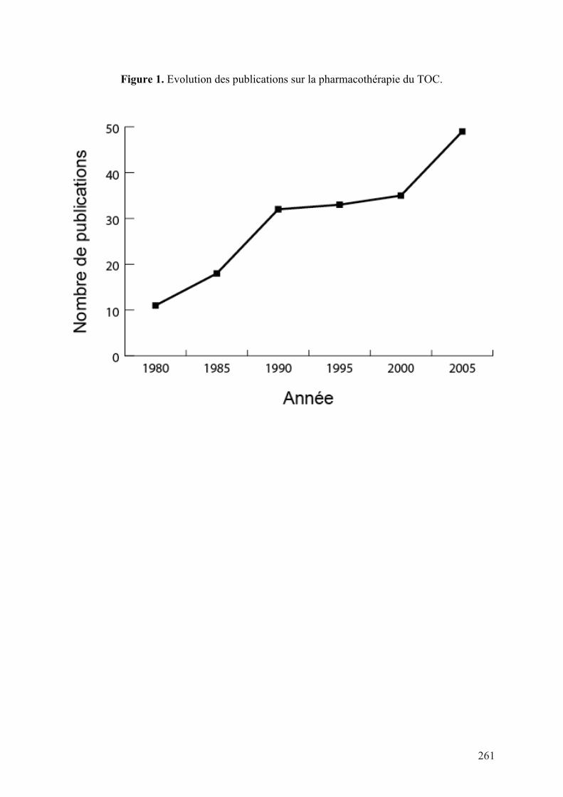
Figure 1. Evolution des publications sur la pharmacothérapie du TOC.
261

Tableau 1. Définition des critères d’efficacité
communément utilisés dans le TOC.
Critères d’efficacité Description
Guérison Y–BOCS ! 8
Rémission Y–BOCS ! 16
Réponse Réduction du score initial
de la Y–BOCS " 35%
Réponse partielle Réduction du score initial
de la Y–BOCS " 25%
Absence de réponse Réduction du score initial
de la Y–BOCS < 25%
Y–BOCS : Yale–Brown Obsessive-Compulsive Scale.
262
Ta
ble
au
2.
Réc
apit
ula
tif
des
ess
ais
con
trô
lés
éval
uan
t l’
effi
caci
té d
e la
clo
mip
ram
ine
dan
s le
TO
C.
Au
teu
rs
Na
ture
de
l’ét
ud
e
No
mb
re d
e
pa
tien
ts
Do
se m
oy
enn
e
(en
mg
/j)
Mo
lécu
les
de
com
pa
rais
on
D
uré
e d
u
suiv
i
Cri
tère
d’e
ffic
aci
té
uti
lisé
P
rin
cip
au
x r
ésu
lta
ts e
t co
mm
enta
ires
Clo
mip
ram
ine
ora
le
Jen
ike
et
al.
(19
89
) [5
4]
Etu
de
con
trô
lée,
ran
do
mis
ée,
en
do
ub
le i
nsu
27
2
00
-30
0 m
g
Pla
ceb
o
10
sem
ain
es
NP
A
mél
iora
tio
n m
oy
enn
e d
e la
Y–
BO
CS
de
28
,6%
so
us
clo
mip
ram
ine
et d
e 8
,4%
so
us
pla
ceb
o.
Gre
ist
et
al.
(19
90
) [3
8]
Etu
de
con
trô
lée,
ran
do
mis
ée,
en
do
ub
le i
nsu
32
2
55
P
lace
bo
1
0 s
emai
nes
N
P
Am
élio
rati
on
mo
yen
ne
de
la Y
–B
OC
S d
e
34
,8%
so
us
clo
mip
ram
ine
et d
e 6
,8%
so
us
pla
ceb
o.
Th
e C
CS
G
(19
91
) [1
23
]
Etu
de
con
trô
lée,
ran
do
mis
ée,
en
do
ub
le i
nsu
23
9
23
4,5
P
lace
bo
1
0 s
emai
nes
R
épo
nse
:
réd
uct
ion
! 3
5%
de
la
Y–
BO
CS
51
% d
es p
atie
nts
so
us
clo
mip
ram
ine
et 7
,5%
sou
s p
lace
bo
so
nt
rép
on
deu
rs.
Th
e C
CS
G
(19
91
) [1
23
]
Etu
de
con
trô
lée,
ran
do
mis
ée,
en
do
ub
le i
nsu
28
1
21
8,8
P
lace
bo
1
0 s
emai
nes
R
épo
nse
:
réd
uct
ion
! 3
5%
de
la
Y–
BO
CS
60
% d
es p
atie
nts
so
us
clo
mip
ram
ine
et 7
%
sou
s p
lace
bo
so
nt
rép
on
deu
rs.
Clo
mip
ram
ine
intr
a-v
ein
euse
Ko
ran
et
al.
(19
97
) [6
4]
Etu
de
con
trô
lée,
ran
do
mis
ée,
en
do
ub
le i
nsu
15
D
ose
max
imal
e
tolé
rée
(25
0 m
g).
Rel
ais
per
os
4,5
jou
rs a
prè
s la
2èm
e
inje
ctio
n.
Clo
mip
ram
ine
ora
le (
NP
)
8 s
emai
nes
R
épo
nse
par
tiel
le :
réd
uct
ion
! 2
5%
de
la
Y–
BO
CS
Rép
on
se o
bte
nu
e p
lus
rap
idem
ent
dan
s le
gro
up
e IV
qu
e d
ans
le g
rou
pe
ora
l (8
5 v
ersu
s
12
% c
inq
jo
urs
ap
rès
la 2
ème i
nje
ctio
n).
A
hu
it s
emai
nes
57
% d
es p
atie
nts
IV
et
50
%
des
pat
ien
ts o
ral
son
t p
arti
elle
men
t
rép
on
deu
rs (
dif
fére
nce
NS
).
Fal
lon
et
al.
(19
98
) [2
3]
Etu
de
con
trô
lée,
ran
do
mis
ée,
en
do
ub
le i
nsu
54
D
ose
max
imal
e
tolé
rée
(25
0 m
g)
Pla
ceb
o
3 s
emai
nes
(14
in
ject
ion
s)
Rép
on
se p
arti
elle
:
réd
uct
ion
! 2
5%
de
la
Y–
BO
CS
Les
54
pat
ien
ts s
on
t in
tolé
ran
ts o
u r
ésis
tan
ts
à la
clo
mip
ram
ine
ora
le. 2
1,4
% d
es p
atie
nts
sou
s cl
om
ipra
min
e IV
et
0%
so
us
pla
ceb
o
son
t p
arti
elle
men
t ré
po
nd
eurs
ap
rès
14
inje
ctio
ns.
CC
SG
: T
he
Clo
mip
ram
ine
Co
llab
ora
tiv
e S
tud
y G
rou
p ;
Y–
BO
CS
: Y
ale–
Bro
wn
Ob
sess
ive-
Co
mp
uls
ive
Sca
le ;
NP
: N
on
pré
cisé
; N
S :
No
n s
ign
ific
atif
; I
V :
In
tra-
vei
neu
x.
263
Ta
ble
au
3.
Réc
apit
ula
tif
des
ess
ais
contr
ôlé
s év
alu
ant
l’ef
fica
cité
de
la f
luo
xet
ine
dan
s le
TO
C.
Au
teu
rs
Na
ture
de
l’ét
ud
e
No
mb
re d
e
pa
tien
ts
Do
se m
oy
enn
e
en m
g/j
Mo
lécu
les
de
com
pa
rais
on
(do
se e
n m
g/j
)
Du
rée
du
suiv
i
Cri
tère
d’e
ffic
aci
té
uti
lisé
P
rin
cip
au
x r
ésu
lta
ts e
t co
mm
enta
ires
Mo
ntg
om
ery
et
al
(19
93
) [9
2]
Etu
de
con
trô
lée,
ran
do
mis
ée,
en
do
ub
le i
nsu
21
7
Do
ses
fix
ées
:
20
, 4
0 e
t 6
0 m
g
Pla
ceb
o
8 s
emai
nes
R
épo
nse
par
tiel
le :
réd
uct
ion
! 2
5%
de
la
Y–
BO
CS
36
% d
es p
atie
nts
so
us
20
mg
de
flu
ox
etin
e,
48
% s
ou
s 4
0 m
g d
e fl
uo
xet
ine,
47
% s
ou
s 6
0
mg
de
flu
ox
etin
e et
26
% s
ou
s p
lace
bo
so
nt
par
tiel
lem
ent
rép
on
deu
rs (
dif
fére
nce
NS
entr
e le
gro
up
e 2
0 m
g e
t le
gro
up
e p
lace
bo
).
To
llef
son
et
al.
1
(19
94
a) [
12
5]
To
llef
son
et
al.
1
(19
94
b)
[12
6]
Etu
de
con
trô
lée,
ran
do
mis
ée,
en
do
ub
le i
nsu
35
5
Do
ses
fix
ées
:
20
, 4
0 e
t 6
0 m
g
Pla
ceb
o
13
sem
ain
es
Rép
on
se :
réd
uct
ion
! 3
5%
de
la
Y–
BO
CS
32
,1%
des
pat
ien
ts s
ou
s 2
0 m
g d
e
flu
ox
etin
e, 3
2,4
% s
ou
s 4
0 m
g d
e fl
uo
xet
i ne,
35
,1%
so
us
60
mg
de
flu
ox
etin
e et
8,5
%
sou
s p
lace
bo
so
nt
rép
on
deu
rs.
Mai
nti
en e
t
po
urs
uit
e d
e l’
amél
iora
tio
n s
ym
pto
mat
iqu
e
chez
les
su
jets
rép
on
deu
rs s
ou
s d
ose
eff
icac
e
de
flu
ox
etin
e d
ura
nt
les
24
sem
ain
es
suiv
ante
s.
Jen
ike
et
al.
(19
97
) [5
7]
Etu
de
con
trô
lée,
ran
do
mis
ée,
en
do
ub
le i
nsu
64
7
7,9
P
lace
bo
Ph
énel
zin
e
(60
)
10
sem
ain
es
NP
A
mél
iora
tio
n m
oy
enn
e d
e la
Y–
BO
CS
de
14
,7%
so
us
flu
ox
etin
e, d
e 9
,4%
so
us
ph
énel
zin
e et
de
1%
so
us
pla
cebo
(dif
fére
nce
sig
nif
icat
ive
com
par
ativ
emen
t à
la p
hén
elzi
ne
et a
u p
lace
bo
).
Ro
man
o e
t a
l.
(20
01
) [1
13
]
Etu
de
con
trô
lée,
ran
do
mis
ée,
en
do
ub
le i
nsu
71
rép
on
deu
rs
Do
ses
fix
ées
:
20
, 4
0 e
t 6
0 m
g
Pla
ceb
o
52
sem
ain
es
Rec
hu
te :
Per
te !
50
% d
e
l’am
élio
rati
on
ob
s erv
ée
et Y
–B
OC
S !
19
17
,5%
des
pat
ien
ts s
ou
s 6
0 m
g d
e fl
uo
xet
ine
et 3
8%
so
us
pla
ceb
o o
nt
rech
uté
(d
iffé
ren
ce
NS
po
ur
des
do
ses
infé
rieu
res
à 6
0 m
g).
1 :
Do
nn
ées
issu
es d
e la
mêm
e ét
ud
e ;
Y–
BO
CS
: Y
ale–
Bro
wn
Ob
sess
ive-
Co
mp
uls
ive
Sca
le ;
NP
: N
on
pré
cisé
; N
S :
No
n s
ign
ific
atif
.
264
Ta
ble
au
4.
Réc
apit
ula
tif
des
ess
ais
con
trô
lés
éval
uan
t l’
effi
caci
té d
e la
par
ox
etin
e d
ans
le T
OC
.
Au
teu
rs
Na
ture
de
l’ét
ud
e
No
mb
re d
e
pa
tien
ts
Do
se m
oy
enn
e
en m
g/j
Mo
lécu
les
de
com
pa
rais
on
Du
rée
du
suiv
i
Cri
tère
d’e
ffic
aci
té
uti
lisé
P
rin
cip
au
x r
ésu
lta
ts e
t co
mm
enta
ires
Ho
llan
der
et
al.
(20
03
a) [
45
]1
Etu
de
con
trô
lée,
ran
do
mis
ée,
en
do
ub
le i
nsu
34
8
Do
ses
fix
ées
:
20
, 4
0 e
t 6
0 m
g
Pla
ceb
o
12
sem
ain
es
NP
A
mél
iora
tio
n m
oy
enn
e d
e la
Y–
BO
CS
de
16
% s
ou
s 2
0 m
g,
de
25
% s
ou
s 4
0 m
g,
de
29
%
sou
s 6
0 m
g d
e p
aro
xet
ine
et d
e 1
3%
so
us
pla
ceb
o (
dif
fére
nce
NS
en
tre
20
mg
et
le
pla
ceb
o).
Ho
llan
der
et
al.
(20
03
a) [
45
]1
Etu
de
con
trô
lée,
ran
do
mis
ée,
en
do
ub
le i
nsu
10
5 r
épo
nd
eurs
D
ose
s fi
xée
s :
20
, 4
0 e
t 6
0 m
g
Pla
ceb
o
6 m
ois
R
ech
ute
:
Ret
ou
r au
sco
re i
nit
ial
de
la Y
–B
OC
S
9,4
% d
es p
atie
nts
so
us
par
ox
etin
e et
21
,6%
sou
s p
lace
bo
on
t re
chu
té.
1 :
Do
nn
ées
issu
es d
e la
mêm
e ét
ud
e ;
Y–
BO
CS
: Y
ale–
Bro
wn
Ob
sess
ive-
Co
mp
uls
ive
Sca
le ;
NP
: N
on
pré
cisé
; N
S :
No
n s
ign
ific
atif
.
265
Ta
ble
au
5.
Réc
apit
ula
tif
des
ess
ais
con
trô
lés
éval
uan
t l’
effi
caci
té d
e la
ser
tral
ine
dan
s le
TO
C.
Au
teu
rs
Na
ture
de
l’ét
ud
e
No
mb
re d
e
pa
tien
ts
Do
se m
oy
enn
e
en m
g/j
Mo
lécu
les
de
com
pa
rais
on
D
uré
e d
u
suiv
i
Cri
tère
d’e
ffic
aci
té
uti
lisé
P
rin
cip
au
x r
ésu
lta
ts e
t co
mm
enta
ires
Jen
ike
et
al.
(19
90
) [5
5]
Etu
de
con
trô
lée,
ran
do
mis
ée,
en
do
ub
le i
nsu
19
2
00
P
lace
bo
1
0 s
emai
nes
N
P
Am
élio
rati
on
mo
yen
ne
de
la Y
–B
OC
S d
e
9,6
% s
ou
s se
rtra
lin
e et
de
2,1
% s
ou
s p
lace
bo
(dif
fére
nce
NS
).
Ch
ou
inar
d e
t a
l.
(19
90
) [1
2]
Etu
de
con
trô
lée,
ran
do
mis
ée,
en
do
ub
le i
nsu
87
1
80
P
lace
bo
8
sem
ain
es
NP
A
mél
iora
tio
n m
oy
enn
e d
e la
Y–
BO
CS
de
16
,1%
so
us
sert
rali
ne
et d
e 6
,5%
so
us
pla
ceb
o.
Gre
ist
et
al.
1
(19
95
a) [
39
]
Etu
de
con
trô
lée,
ran
do
mis
ée,
en
do
ub
le i
nsu
32
4
Do
ses
fix
ées
: 5
0,
10
0 e
t 2
00
mg
Pla
ceb
o
12
sem
ain
es
NP
A
mél
iora
tio
n m
oy
enn
e d
e la
Y–
BO
CS
de
23
,4%
so
us
sert
rali
ne
et d
e 1
4,6
% s
ou
s
pla
ceb
o (
dif
fére
nce
NS
po
ur
le g
rou
pe
10
0
mg
).
Gre
ist
et
al.
1
(19
95
b)
[40
]
Etu
de
con
trô
lée,
ran
do
mis
ée,
en
do
ub
le i
nsu
11
8
rép
on
deu
rs
Do
ses
fix
ées
: 5
0,
10
0 e
t 2
00
mg
Pla
ceb
o
1 a
n
NP
P
ou
rsu
ite
de
l’am
élio
rati
on
sy
mp
tom
atiq
ue
sou
s se
rtra
lin
e. A
mél
iora
tio
n m
oy
enn
e d
e la
Y–
BO
CS
de
28
,4%
so
us
50
mg
, d
e 1
7,8
%
sou
s 1
00
mg
, d
e 2
6,3
% s
ou
s 2
00
mg
de
sert
rali
ne
et d
e 1
1,9
% s
ou
s p
lace
bo
(dif
fére
nce
NS
po
ur
le g
rou
pe
10
0 m
g).
Kro
nig
et
al.
(19
99
) [7
2]
Etu
de
con
trô
lée,
ran
do
mis
ée,
en
do
ub
le i
nsu
16
7
16
5
Pla
ceb
o
12
sem
ain
es
NP
A
mél
iora
tio
n m
oy
enn
e d
e la
Y–
BO
CS
sig
nif
icat
ivem
ent
plu
s im
po
rtan
te s
ou
s
sert
rali
ne
qu
e so
us
pla
ce (
chif
fres
NP
).
Ko
ran
et
al.
(20
02
) [6
8]
Etu
de
con
trô
lée,
ran
do
mis
ée,
en
do
ub
le i
nsu
22
7
18
7
Pla
ceb
o
52
sem
ain
es
sou
s se
rtra
lin
e ;
pu
is 2
8
sem
ain
es s
ou
s
sert
rali
ne
ou
pla
ceb
o
Rec
hu
te :
Au
gm
enta
tio
n d
e 5
po
ints
du
sco
re d
e la
Y–
BO
CS
et
Y–
BO
CS
! 2
0
9%
des
pat
ien
ts s
ou
s se
rtra
lin
e et
24
% s
ou
s
pla
ceb
o o
nt
arrê
té l
eur
par
tici
pat
ion
po
ur
rech
ute
ou
rép
on
se i
nsu
ffis
ante
. A
gg
rav
atio
n
des
sco
res
de
Y–
BO
CS
so
us
pla
ceb
o,
mai
nti
en d
u b
énéf
ice
thér
apeu
tiq
ue
sou
s
sert
rali
ne.
1 :
Do
nn
ées
issu
es d
e la
mêm
e ét
ud
e ;
Y–
BO
CS
: Y
ale–
Bro
wn
Ob
sess
ive-
Co
mp
uls
ive
Sca
le ;
NP
: N
on
pré
cisé
; N
S :
No
n s
ign
ific
atif
.
266
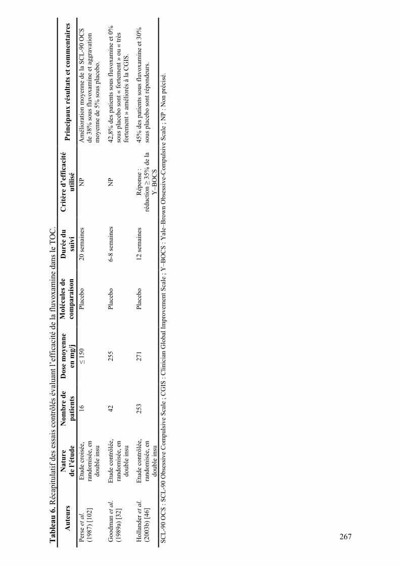
Ta
ble
au
6.
Réc
apit
ula
tif
des
ess
ais
con
trô
lés
éval
uan
t l’
effi
caci
té d
e la
flu
vo
xam
ine
dan
s le
TO
C.
Au
teu
rs
Na
ture
de
l’ét
ud
e
No
mb
re d
e
pa
tien
ts
Do
se m
oy
enn
e
en m
g/j
Mo
lécu
les
de
com
pa
rais
on
D
uré
e d
u
suiv
i
Cri
tère
d’e
ffic
aci
té
uti
lisé
P
rin
cip
au
x r
ésu
lta
ts e
t co
mm
enta
ires
Per
se e
t a
l.
(19
87
) [1
02
]
Etu
de
cro
isée
,
ran
do
mis
ée,
en
do
ub
le i
nsu
16
!
15
0
Pla
ceb
o
20
sem
ain
es
NP
A
mél
iora
tio
n m
oy
enn
e d
e la
SC
L-9
0 O
CS
de
38
% s
ou
s fl
uv
ox
amin
e et
ag
gra
vat
ion
mo
yen
ne
de
5%
so
us
pla
ceb
o.
Go
od
man
et
al.
(19
89
a) [
32
]
Etu
de
con
trô
lée,
ran
do
mis
ée,
en
do
ub
le i
nsu
42
2
55
P
lace
bo
6
-8 s
emai
nes
N
P
42
,8%
des
pat
ien
ts s
ou
s fl
uv
ox
amin
e et
0%
sou
s p
lace
bo
so
nt
« f
ort
emen
t »
ou
« t
rès
fort
emen
t »
am
élio
rés
à la
CG
IS.
Ho
llan
der
et
al.
(20
03
b)
[46
]
Etu
de
con
trô
lée,
ran
do
mis
ée,
en
do
ub
le i
nsu
25
3
27
1
Pla
ceb
o
12
sem
ain
es
Rép
on
se :
réd
uct
ion
" 3
5%
de
la
Y–
BO
CS
45
% d
es p
atie
nts
so
us
flu
vo
xam
ine
et 3
0%
sou
s p
lace
bo
so
nt
rép
on
deu
rs.
SC
L-9
0 O
CS
: S
CL
-90
Ob
sess
ive
Co
mp
uls
ive
Sca
le ;
CG
IS :
Cli
nic
ian
Glo
bal
Im
pro
vem
ent
Sca
le ;
Y–
BO
CS
: Y
ale–
Bro
wn
Ob
sess
ive-
Co
mp
uls
ive
Sca
le ;
NP
: N
on
pré
cisé
.
267
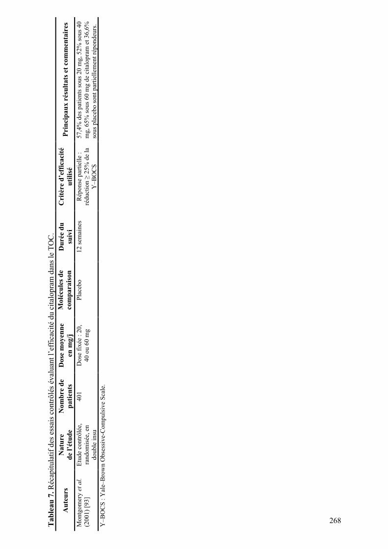
Ta
ble
au
7.
Réc
apit
ula
tif
des
ess
ais
con
trô
lés
éval
uan
t l’
effi
caci
té d
u c
ital
op
ram
dan
s le
TO
C.
Au
teu
rs
Na
ture
de
l’ét
ud
e
No
mb
re d
e
pa
tien
ts
Do
se m
oy
enn
e
en m
g/j
Mo
lécu
les
de
com
pa
rais
on
D
uré
e d
u
suiv
i
Cri
tère
d’e
ffic
aci
té
uti
lisé
P
rin
cip
au
x r
ésu
lta
ts e
t co
mm
enta
ires
Mo
ntg
om
ery
et
al.
(20
01
) [9
3]
Etu
de
con
trô
lée,
ran
do
mis
ée,
en
do
ub
le i
nsu
40
1
Do
se f
ixée
: 2
0,
40
ou
60
mg
Pla
ceb
o
12
sem
ain
es
Rép
on
se p
arti
elle
:
réd
uct
ion
! 2
5%
de
la
Y–
BO
CS
57
,4%
des
pat
ien
ts s
ou
s 2
0 m
g,
52
% s
ou
s 4
0
mg
, 6
5%
so
us
60
mg
de
cita
lop
ram
et
36
,6%
sou
s p
lace
bo
so
nt
par
tiel
lem
ent
rép
on
deu
rs.
Y–
BO
CS
: Y
ale–
Bro
wn
Ob
sess
ive-
Co
mp
uls
ive
Sca
le.
268
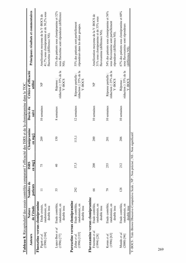
Ta
ble
au
8.
Réc
apit
ula
tif
des
ess
ais
con
trô
lés
com
par
ant
l’ef
fica
cité
des
IS
RS
et
de
la c
lom
ipra
min
e d
ans
le T
OC
.
Au
teu
rs
Na
ture
de
l’ét
ud
e
No
mb
re d
e
pa
tien
ts
ISR
S
en m
g/j
Clo
mip
ram
ine
en m
g/j
Du
rée
du
suiv
i
Cri
tère
d’e
ffic
aci
té
uti
lisé
P
rin
cip
au
x r
ésu
lta
ts e
t co
mm
enta
ires
Flu
ox
etin
e v
ersu
s cl
om
ipra
min
e P
igo
tt e
t a
l.
(19
90
) [1
04
]
Etu
de
cro
isée
,
ran
do
mis
ée,
en
do
ub
le i
nsu
11
7
5
20
9
10
sem
ain
es
NP
A
mél
iora
tio
n m
oy
enn
e d
e la
Y–
BO
CS
de
41
,7%
so
us
clo
mip
ram
ine
et d
e 3
0,2
% s
ou
s
flu
ox
etin
e (d
iffé
ren
ce N
S).
Lo
pez
-Ib
or
et
al
(19
96
) [7
7]
Etu
de
con
trô
lée,
ran
do
mis
ée,
en
do
ub
le i
nsu
55
4
0
15
0
8 s
emai
nes
R
épo
nse
:
réd
uct
ion
! 3
5%
de
la
Y–
BO
CS
55
% d
es p
atie
nts
so
us
clo
mip
ram
ine
et 3
2%
sou
s fl
uo
xet
ine
son
t ré
po
nd
eurs
(d
iffé
ren
ce
NS
).
Pa
rox
etin
e v
ersu
s cl
om
ipra
min
e Z
oh
ar e
t a
l.
(19
96
) [1
35
]
Etu
de
con
trô
lée,
ran
do
mis
ée,
en
do
ub
le i
nsu
29
2
37
,5
11
3,1
1
2 s
emai
nes
R
épo
nse
par
tiel
le :
réd
uct
ion
! 2
5%
de
la
Y–
BO
CS
55
% d
es p
atie
nts
so
nt
par
tiel
lem
ent
rép
on
deu
rs d
ans
les
deu
x g
rou
pes
.
Flu
vo
xa
min
e v
ersu
s cl
om
ipra
min
e F
reem
an e
t a
l.
(19
94
) [2
8]
Etu
de
con
trô
lée,
ran
do
mis
ée,
en
do
ub
le i
nsu
66
2
00
2
00
1
0 s
emai
nes
N
P
Am
élio
rati
on
mo
yen
ne
de
la Y
–B
OC
S d
e
31
% s
ou
s cl
om
ipra
min
e et
de
33
% s
ou
s
flu
vo
xam
ine
(dif
fére
nce
NS
).
Ko
ran
et
al.
(1
99
6)
[63
]
Etu
de
con
trô
lée,
ran
do
mis
ée,
en
do
ub
le i
nsu
79
2
55
2
01
1
0 s
emai
nes
R
épo
nse
par
tiel
le :
réd
uct
ion
! 2
5%
de
la
Y–
BO
CS
54
% d
es p
atie
nts
so
us
clo
mip
ram
ine
et 5
6%
sou
s fl
uv
ox
amin
e so
nt
par
tiel
lem
ent
rép
on
deu
rs (
dif
fére
nce
NS
).
Mu
nd
o e
t a
l.
(20
00
) [9
6]
Etu
de
con
trô
lée,
ran
do
mis
ée,
en
do
ub
le i
nsu
12
8
21
2
20
6
10
sem
ain
es
Rép
on
se :
réd
uct
ion
! 3
5%
de
la
Y–
BO
CS
67
% d
es p
atie
nts
so
us
clo
mip
ram
ine
et 6
0%
sou
s fl
uv
ox
amin
e so
nt
rép
on
deu
rs
(dif
fére
nce
NS
).
Y–
BO
CS
: Y
ale–
Bro
wn
Ob
sess
ive-
Co
mp
uls
ive
Sca
le ;
NP
: N
on
pré
cisé
; N
S :
No
n s
ign
ific
atif
.
269
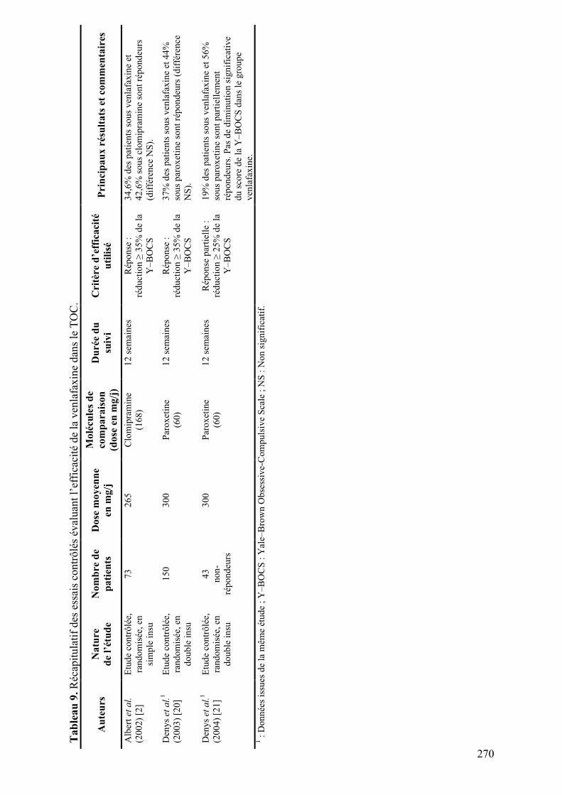
Ta
ble
au
9.
Réc
apit
ula
tif
des
ess
ais
con
trô
lés
éval
uan
t l’
effi
caci
té d
e la
ven
lafa
xin
e d
ans
le T
OC
.
Au
teu
rs
Na
ture
de
l’ét
ud
e
No
mb
re d
e
pa
tien
ts
Do
se m
oy
enn
e
en m
g/j
Mo
lécu
les
de
com
pa
rais
on
(do
se e
n m
g/j
)
Du
rée
du
suiv
i
Cri
tère
d’e
ffic
aci
té
uti
lisé
P
rin
cip
au
x r
ésu
lta
ts e
t co
mm
enta
ires
Alb
ert
et
al.
(20
02
) [2
]
Etu
de
con
trô
lée,
ran
do
mis
ée,
en
sim
ple
in
su
73
2
65
C
lom
ipra
min
e
(16
8)
12
sem
ain
es
Rép
on
se :
réd
uct
ion
! 3
5%
de
la
Y–
BO
CS
34
,6%
des
pat
ien
ts s
ou
s v
enla
fax
ine
et
42
,6%
so
us
clo
mip
ram
ine
son
t ré
po
nd
eurs
(dif
fére
nce
NS
).
Den
ys
et
al.
1
(20
03
) [2
0]
Etu
de
con
trô
lée,
ran
do
mis
ée,
en
do
ub
le i
nsu
15
0
30
0
Par
ox
etin
e
(60
)
12
sem
ain
es
Rép
on
se :
réd
uct
ion
! 3
5%
de
la
Y–
BO
CS
37
% d
es p
atie
nts
so
us
ven
lafa
xin
e et
44
%
sou
s p
aro
xet
ine
son
t ré
po
nd
eurs
(d
iffé
ren
ce
NS
).
Den
ys
et
al.
1
(20
04
) [2
1]
Etu
de
con
trô
lée,
ran
do
mis
ée,
en
do
ub
le i
nsu
43
no
n-
rép
on
deu
rs
30
0
Par
ox
etin
e
(60
)
12
sem
ain
es
Rép
on
se p
arti
elle
:
réd
uct
ion
! 2
5%
de
la
Y–
BO
CS
19
% d
es p
atie
nts
so
us
ven
lafa
xin
e et
56
%
sou
s p
aro
xet
ine
son
t p
arti
elle
men
t
rép
on
deu
rs.
Pas
de
dim
inu
tio
n s
ign
ific
ativ
e
du
sco
re d
e la
Y–
BO
CS
dan
s le
gro
up
e
ven
lafa
xin
e.
1 :
Do
nn
ées
issu
es d
e la
mêm
e ét
ud
e ;
Y–
BO
CS
: Y
ale–
Bro
wn
Ob
sess
ive-
Co
mp
uls
ive
Sca
le ;
NS
: N
on
sig
nif
icat
if.
270
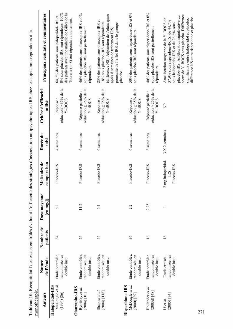
Ta
ble
au
10
. R
écap
itu
lati
f d
es e
ssai
s co
ntr
ôlé
s év
alu
ant
l’ef
fica
cité
des
str
atég
ies
d’a
sso
ciat
ion
an
tip
sych
oti
qu
es-I
RS
ch
ez l
es s
uje
ts n
on-r
épo
nd
eurs
à l
a
mo
no
thér
apie
.
Au
teu
rs
Na
ture
de
l’ét
ud
e
No
mb
re d
e
pa
tien
ts
Do
se m
oy
enn
e
(en
mg
/j)
Mo
lécu
les
de
com
pa
rais
on
Du
rée
du
suiv
i
Cri
tère
d’e
ffic
aci
té
uti
lisé
P
rin
cip
au
x r
ésu
lta
ts e
t co
mm
enta
ires
Ha
lop
erid
ol-
IRS
M
cDo
ug
le e
t a
l.
(19
94
) [8
6]
Etu
de
con
trô
lée,
ran
do
mis
ée,
en
do
ub
le i
nsu
34
6
,2
Pla
ceb
o-I
RS
4
sem
ain
es
Rép
on
se :
réd
uct
ion
! 3
5%
de
la
Y–
BO
CS
64
,7%
des
pat
ien
ts s
ou
s h
alo
per
ido
l-IR
S e
t
0%
so
us
pla
ceb
o-I
RS
so
nt
rép
on
deu
rs.
10
0%
des
pat
ien
ts a
vec
un
e m
alad
ie d
e G
ille
s d
e la
To
ure
tte
(n=
8)
on
t ré
po
nd
u a
u t
rait
emen
t.
Ola
nza
pin
e-IR
S
By
trit
sky
et
al.
(20
04
) [1
0]
Etu
de
con
trô
lée,
ran
do
mis
ée,
en
do
ub
le i
nsu
26
1
1,2
P
lace
bo
-IR
S
6 s
emai
nes
R
épo
nse
par
tiel
le :
réd
uct
ion
! 2
5%
de
la
Y–
BO
CS
46
% d
es p
atie
nts
so
us
ola
nza
pin
e-IR
S e
t 0
%
sou
s p
lace
bo
-IR
S s
on
t p
arti
elle
men
t
rép
on
deu
rs.
Sh
apir
a et
al.
(20
04
) [1
18
]
Etu
de
con
trô
lée,
ran
do
mis
ée,
en
do
ub
le i
nsu
44
6
,1
Pla
ceb
o-I
RS
6
sem
ain
es
Rép
on
se :
réd
uct
ion
! 3
5%
de
la
Y–
BO
CS
23
% d
es p
atie
nts
so
us
ola
nza
pin
e-IR
S e
t
18
% s
ou
s p
lace
bo
-IR
S s
on
t ré
po
nd
eurs
(dif
fére
nce
NS
). A
djo
nct
ion
de
l’o
lan
zap
ine
aprè
s 8
sem
ain
es d
e tr
aite
men
t IR
S,
po
urs
uit
e d
e l’
effe
t IR
S d
ans
le g
rou
pe
pla
ceb
o.
Ris
per
ido
ne-
IRS
M
cDo
ug
le e
t a
l.
(20
00
) [8
9]
Etu
de
con
trô
lée,
ran
do
mis
ée,
en
do
ub
le i
nsu
36
2
,2
Pla
ceb
o-I
RS
6
sem
ain
es
Rép
on
se :
réd
uct
ion
! 3
5%
de
la
Y–
BO
CS
50
% d
es p
atie
nts
so
us
risp
erid
on
e-IR
S e
t 0
%
sou
s p
lace
bo
-IR
S s
on
t ré
po
nd
eurs
.
Ho
llan
der
et
al.
(20
03
d)
[48
]
Etu
de
con
trô
lée,
ran
do
mis
ée,
en
do
ub
le i
nsu
16
2
,25
P
lace
bo
-IR
S
8 s
emai
nes
R
épo
nse
par
tiel
le :
réd
uct
ion
! 2
5%
de
la
Y–
BO
CS
40
% d
es p
atie
nts
so
us
risp
erid
on
e-IR
S e
t 0
%
sou
s p
lace
bo
-IR
S s
on
t p
arti
elle
men
t
rép
on
deu
rs.
Li
et
al.
(20
05
) [7
4]
Etu
de
cro
isée
,
ran
do
mis
ée,
en
do
ub
le i
nsu
16
1
2
mg
hal
op
erid
ol-
IRS
Pla
ceb
o-I
RS
3 X
2 s
emai
nes
N
P
Am
élio
rati
on
mo
yen
ne
de
la Y
–B
OC
S d
e
37
,9%
so
us
risp
erid
on
e-IR
TS
, d
e 4
4,7
%
sou
s h
alo
per
ido
l-IR
S e
t d
e 2
6,6
% s
ou
s
pla
ceb
o-I
RS
. A
mél
iora
tio
n s
ign
ific
ativ
e d
u
sco
re d
e Y
–B
OC
S s
ou
s p
lace
bo
. D
iffé
ren
ce
sig
nif
icat
ive
entr
e h
alo
per
ido
l et
pla
ceb
o,
dif
fére
nce
NS
en
tre
risp
erid
on
e et
pla
ceb
o.
271
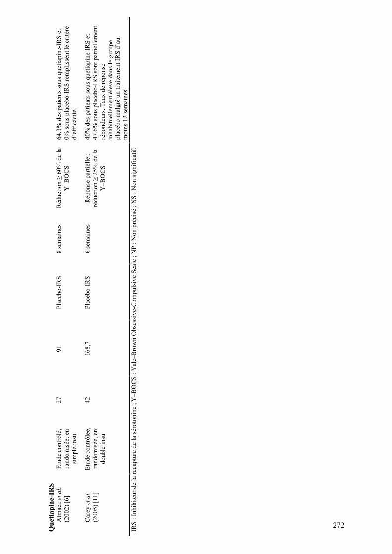
Qu
etia
pin
e-IR
S
Atm
aca
et
al.
(20
02
) [6
]
Etu
de
con
trô
lé,
ran
do
mis
ée,
en
sim
ple
in
su
27
9
1
Pla
ceb
o-I
RS
8
sem
ain
es
Réd
uct
ion
! 6
0%
de
la
Y–
BO
CS
64
,3%
des
pat
ien
ts s
ou
s q
uet
iap
ine-
IRS
et
0%
so
us
pla
ceb
o-I
RS
rem
pli
ssen
t le
cri
tère
d’e
ffic
acit
é.
Car
ey e
t a
l.
(20
05
) [1
1]
Etu
de
con
trô
lée,
ran
do
mis
ée,
en
do
ub
le i
nsu
42
1
68
,7
Pla
ceb
o-I
RS
6
sem
ain
es
Rép
on
se p
arti
elle
:
réd
uct
ion
! 2
5%
de
la
Y–
BO
CS
40
% d
es p
atie
nts
so
us
qu
etia
pin
e-IR
S e
t
47
,6%
so
us
pla
ceb
o-I
RS
so
nt
par
tiel
lem
ent
rép
on
deu
rs.
Tau
x d
e ré
po
nse
inh
abit
uel
lem
ent
élev
é d
ans
le g
rou
pe
pla
ceb
o m
alg
ré u
n t
rait
emen
t IR
S d
’au
mo
ins
12
sem
ain
es.
IRS
: I
nh
ibit
eur
de
la r
ecap
ture
de
la s
éro
ton
ine
; Y
–B
OC
S :
Yal
e–B
row
n O
bse
ssiv
e-C
om
pu
lsiv
e S
cale
; N
P :
No
n p
réci
sé ;
NS
: N
on
sig
nif
icat
if.
272
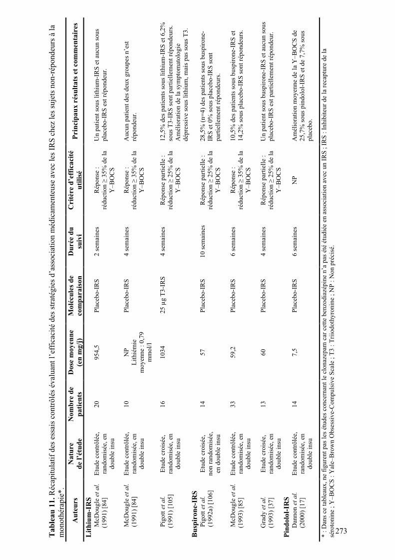
Ta
ble
au
11
. R
écap
itu
lati
f d
es e
ssai
s co
ntr
ôlé
s év
alu
ant
l’ef
fica
cité
des
str
atég
ies
d’a
sso
ciat
ion
méd
icam
ente
use
av
ec l
es I
RS
ch
ez l
es s
uje
ts n
on-r
épo
nd
eurs
à l
a
mo
no
thér
apie
*.
Au
teu
rs
Na
ture
de
l’ét
ud
e
No
mb
re d
e
pa
tien
ts
Do
se m
oy
enn
e
(en
mg
/j)
Mo
lécu
les
de
com
pa
rais
on
Du
rée
du
suiv
i
Cri
tère
d’e
ffic
aci
té
uti
lisé
P
rin
cip
au
x r
ésu
lta
ts e
t co
mm
enta
ires
Lit
hiu
m-I
RS
M
cDo
ug
le e
t a
l.
(19
91
) [8
4]
Etu
de
con
trô
lée,
ran
do
mis
ée,
en
do
ub
le i
nsu
20
9
54
,5
Pla
ceb
o-I
RS
2
sem
ain
es
Rép
on
se :
réd
uct
ion
! 3
5%
de
la
Y–
BO
CS
Un
pat
ien
t so
us
lith
ium
-IR
S e
t au
cun
so
us
pla
ceb
o-I
RS
est
rép
on
deu
r.
McD
ou
gle
et
al.
(19
91
) [8
4]
Etu
de
con
trô
lée,
ran
do
mis
ée,
en
do
ub
le i
nsu
10
N
P
Lit
hié
mie
mo
yen
ne
: 0
,79
mm
ol/
l
Pla
ceb
o-I
RS
4
sem
ain
es
Rép
on
se :
réd
uct
ion
! 3
5%
de
la
Y–
BO
CS
Au
cun
pat
ien
t d
es d
eux
gro
up
es n
’est
rép
on
deu
r.
Pig
ott
et
al.
(19
91
) [1
05
]
Etu
de
cro
isée
,
ran
do
mis
ée,
en
do
ub
le i
nsu
16
1
03
4
25
"g
T3
-IR
S
4 s
emai
nes
R
épo
nse
par
tiel
le :
réd
uct
ion
! 2
5%
de
la
Y–
BO
CS
12
,5%
des
pat
ien
ts s
ou
s li
thiu
m-I
RS
et
6,2
%
sou
s T
3-I
RS
so
nt
par
tiel
lem
ent
rép
on
deu
rs.
Am
élio
rati
on
de
la s
ym
pto
mat
olo
gie
dép
ress
ive
sou
s li
thiu
m,
mai
s p
as s
ou
s T
3.
Bu
spir
on
e-IR
S
Pig
ott
et
al.
(19
92
a) [
10
6]
Etu
de
cro
isée
,
no
n r
and
om
isée
,
en d
ou
ble
in
su
14
5
7
Pla
ceb
o-I
RS
1
0 s
emai
nes
R
épo
nse
par
tiel
le :
réd
uct
ion
! 2
5%
de
la
Y–
BO
CS
28
,5%
(n
=4
) d
es p
atie
nts
so
us
bu
spir
on
e-
IRS
et
0%
so
us
pla
ceb
o-I
RS
so
nt
par
tiel
lem
ent
rép
on
deu
rs.
McD
ou
gle
et
al.
(19
93
) [8
5]
Etu
de
con
trô
lée,
ran
do
mis
ée,
en
do
ub
le i
nsu
33
5
9,2
P
lace
bo
-IR
S
6 s
emai
nes
R
épo
nse
:
réd
uct
ion
! 3
5%
de
la
Y–
BO
CS
10
,5%
des
pat
ien
ts s
ou
s b
usp
iro
ne-
IRS
et
14
,2%
so
us
pla
ceb
o-I
RS
so
nt
rép
on
deu
rs.
Gra
dy
et
al.
(19
93
) [3
7]
Etu
de
cro
isée
,
ran
do
mis
ée,
en
do
ub
le i
nsu
13
6
0
Pla
ceb
o-I
RS
4
sem
ain
es
Rép
on
se p
arti
elle
:
réd
uct
ion
! 2
5%
de
la
Y–
BO
CS
Un
pat
ien
t so
us
bu
spir
on
e-IR
S e
t au
cun
so
us
pla
ceb
o-I
RS
est
par
tiel
lem
ent
rép
on
deu
r.
Pin
do
lol-
IRS
D
ann
on
et
al.
(20
00
) [1
7]
Etu
de
con
trô
lée,
ran
do
mis
ée,
en
do
ub
le i
nsu
14
7
,5
Pla
ceb
o-I
RS
6
sem
ain
es
NP
A
mél
iora
tio
n m
oy
enn
e d
e la
Y–
BO
CS
de
25
,7%
so
us
pin
do
lol-
IRS
et
de
7,7
% s
ou
s
pla
ceb
o.
* :
Dan
s ce
tab
leau
x,
ne
fig
ure
nt
pas
les
étu
des
co
nce
rnan
t le
clo
naz
epam
car
cet
te b
enzo
dia
zép
ine
n’a
pas
été
étu
dié
e en
ass
oci
atio
n a
vec
un
IR
S ;
IR
S :
In
hib
iteu
r d
e la
rec
aptu
re d
e la
séro
ton
ine
; Y
–B
OC
S :
Yal
e–B
row
n O
bse
ssiv
e-C
om
pu
lsiv
e S
cale
; T
3 :
Tri
iod
oth
yro
nin
e ;
NP
: N
on
pré
cisé
.
273

Tableau 12. Posologies efficaces des IRS
dans le TOC.
IRS Posologies (mg/j)
Clomipramine 150-250
Fluoxetine 40-80
Paroxetine 40-60
Sertraline 150-200
Fluvoxamine 200-300
Citalopram 20-60
IRS : inhibiteur de la recapture de la sérotonine.
274

275
Annexe C : Matériels supplémentaires de l’article « Meta-analysis of
brain volume changes in obsessive-compulsive disorder »
JY Rotge et al. 1
Meta-analysis of brain volume changes in obsessive-compulsive disorder
JY Rotge, D Guehl, B Dilharreguy, J Tignol, B Bioulac, M Allard, P Burbaud, B Aouizerate.
Supplementary Materials
Supplementary results
Meta-analyses are presented in detail for each brain volume, as follows: whole brain
volume (Figure S1), intra-cranial volume (Figure S2), gray matter volume (Figure S3),
prefrontal cortex volume (Figure S4), left and right ACC volumes (Figures S5 A, B), left and
right OFC volumes (Figures S6 A, B), left and right thalamus volumes (Figures S7 A, B), left
and right putamen volumes (Figures S8 A, B), total, left and right caudate nucleus volumes
(Figures S9 A, B, C).
Funnel plots were generated for the whole brain volume, intracranial volume, left and
right OFC volumes, left and right thalamus volumes, and the left and right caudate nucleus
volumes (Figures S10-18). No asymetry was detected in funnel plots.
276
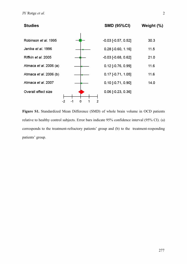
JY Rotge et al.
2
Figure S1. Standardized Mean Difference (SMD) of whole brain volume in OCD patients
relative to healthy control subjects. Error bars indicate 95% confidence interval (95% CI). (a)
corresponds to the treatment-refractory patients’ group and (b) to the treatment-responding
patients’ group.
277
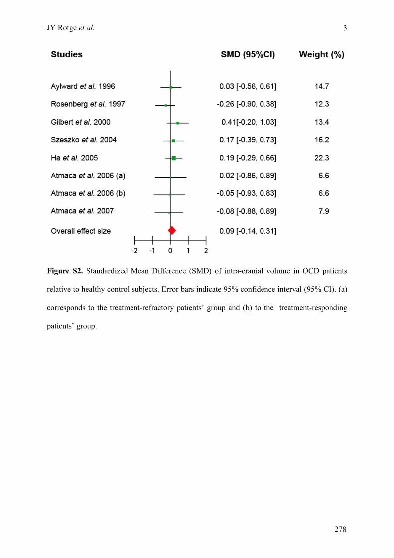
JY Rotge et al.
3
Figure S2. Standardized Mean Difference (SMD) of intra-cranial volume in OCD patients
relative to healthy control subjects. Error bars indicate 95% confidence interval (95% CI). (a)
corresponds to the treatment-refractory patients’ group and (b) to the treatment-responding
patients’ group.
278
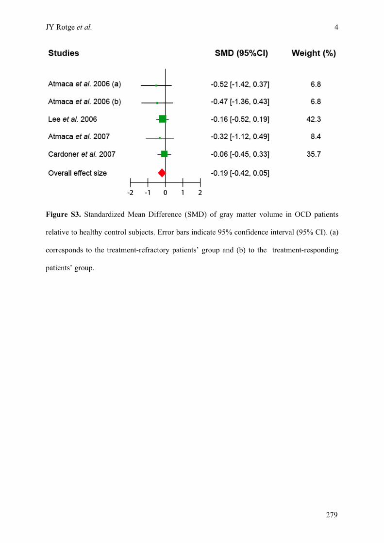
JY Rotge et al.
4
Figure S3. Standardized Mean Difference (SMD) of gray matter volume in OCD patients
relative to healthy control subjects. Error bars indicate 95% confidence interval (95% CI). (a)
corresponds to the treatment-refractory patients’ group and (b) to the treatment-responding
patients’ group.
279
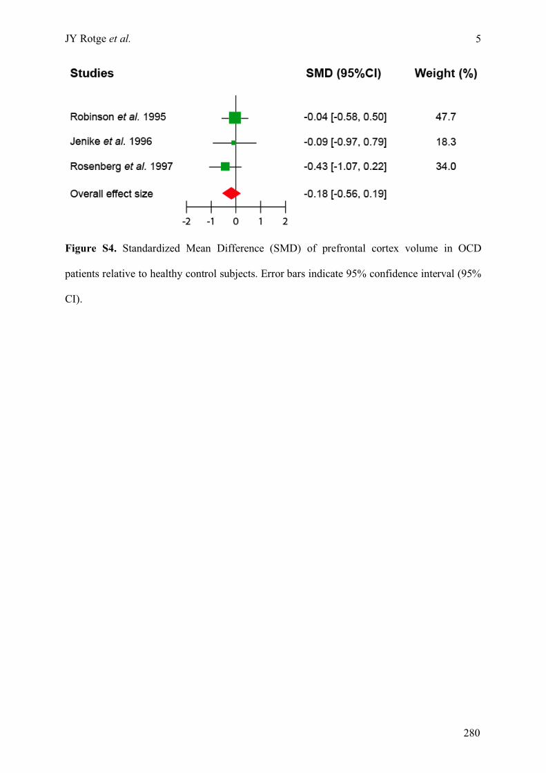
JY Rotge et al.
5
Figure S4. Standardized Mean Difference (SMD) of prefrontal cortex volume in OCD
patients relative to healthy control subjects. Error bars indicate 95% confidence interval (95%
CI).
280
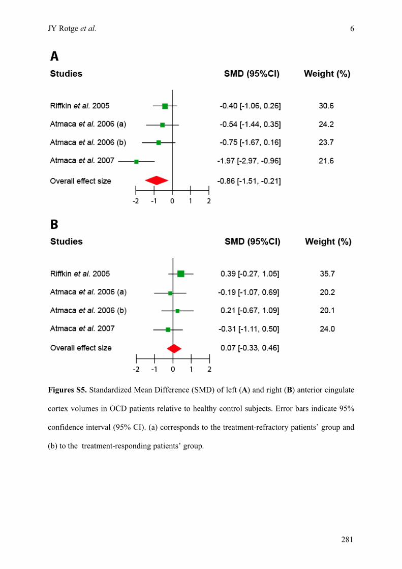
JY Rotge et al.
6
Figures S5. Standardized Mean Difference (SMD) of left (A) and right (B) anterior cingulate
cortex volumes in OCD patients relative to healthy control subjects. Error bars indicate 95%
confidence interval (95% CI). (a) corresponds to the treatment-refractory patients’ group and
(b) to the treatment-responding patients’ group.
281
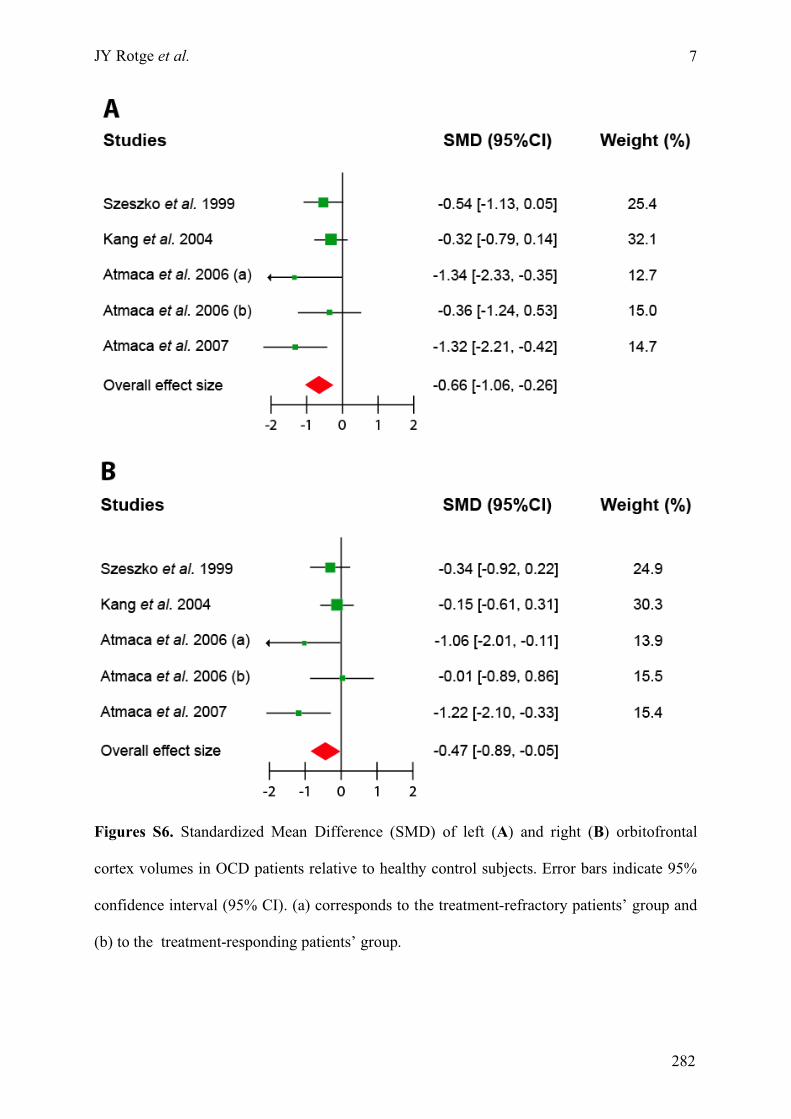
JY Rotge et al.
7
Figures S6. Standardized Mean Difference (SMD) of left (A) and right (B) orbitofrontal
cortex volumes in OCD patients relative to healthy control subjects. Error bars indicate 95%
confidence interval (95% CI). (a) corresponds to the treatment-refractory patients’ group and
(b) to the treatment-responding patients’ group.
282
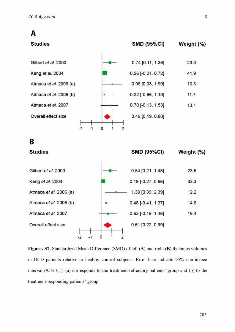
JY Rotge et al.
8
Figures S7. Standardized Mean Difference (SMD) of left (A) and right (B) thalamus volumes
in OCD patients relative to healthy control subjects. Error bars indicate 95% confidence
interval (95% CI). (a) corresponds to the treatment-refractory patients’ group and (b) to the
treatment-responding patients’ group.
283
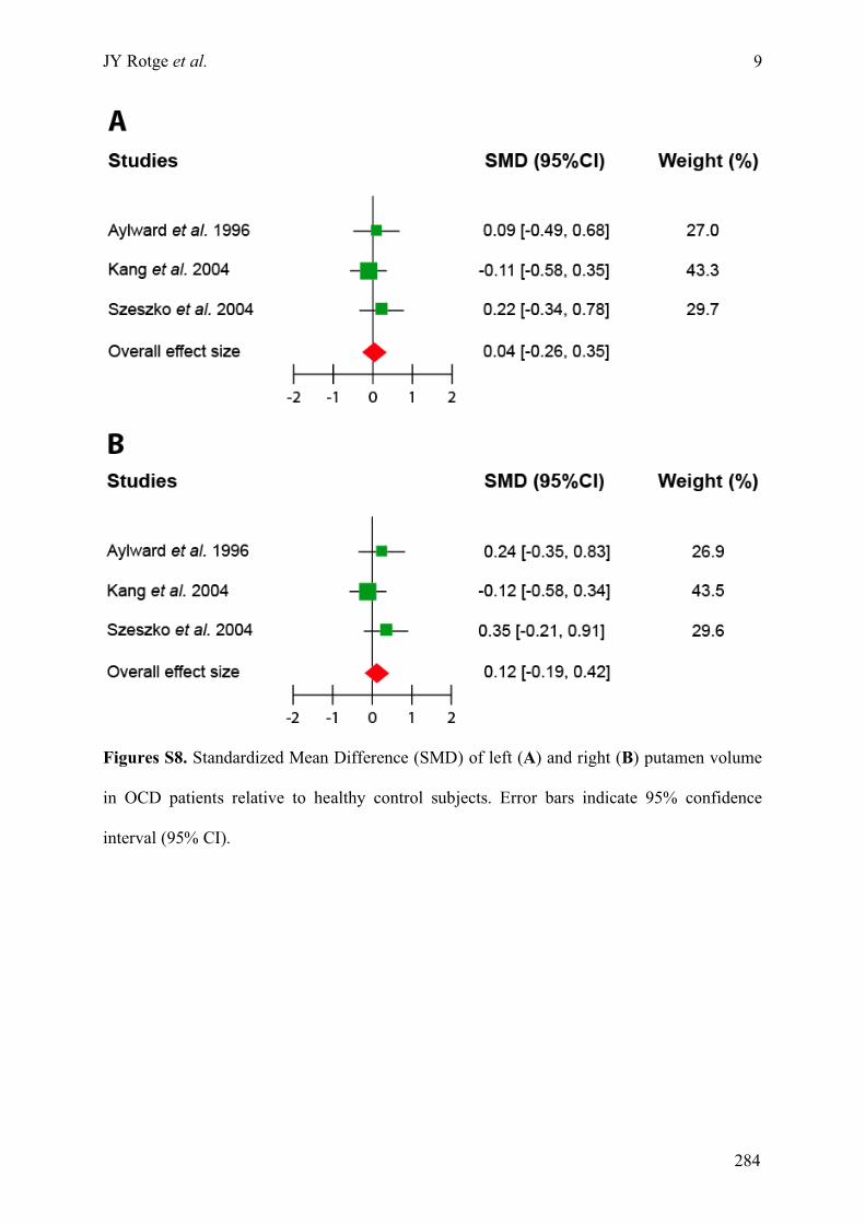
JY Rotge et al.
9
Figures S8. Standardized Mean Difference (SMD) of left (A) and right (B) putamen volume
in OCD patients relative to healthy control subjects. Error bars indicate 95% confidence
interval (95% CI).
284
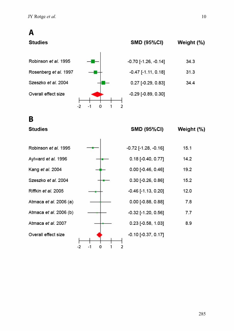
JY Rotge et al.
10
285
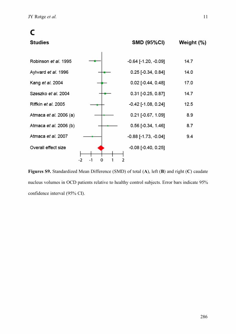
JY Rotge et al.
11
Figures S9. Standardized Mean Difference (SMD) of total (A), left (B) and right (C) caudate
nucleus volumes in OCD patients relative to healthy control subjects. Error bars indicate 95%
confidence interval (95% CI).
286
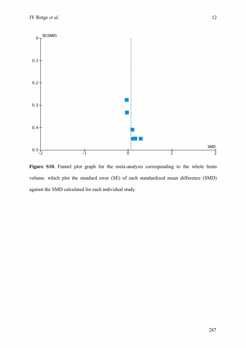
JY Rotge et al.
12
Figure S10. Funnel plot graph for the meta-analysis corresponding to the whole brain
volume. which plot the standard error (SE) of each standardized mean difference (SMD)
against the SMD calculated for each individual study.
287
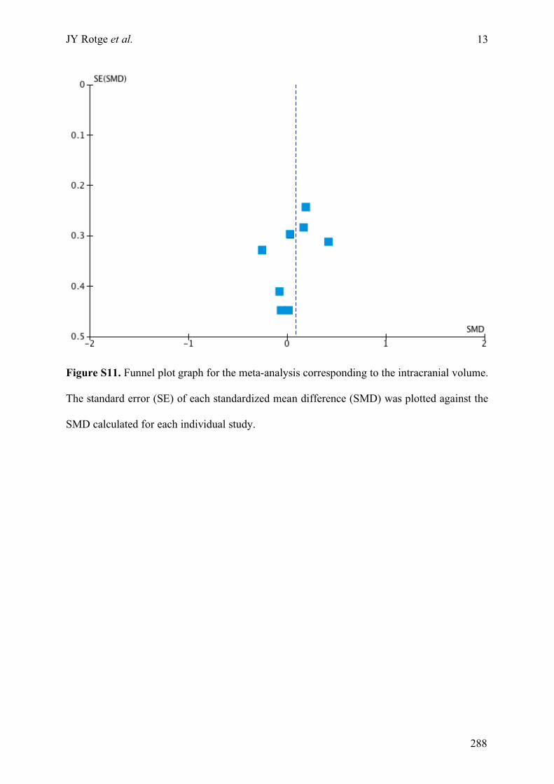
JY Rotge et al.
13
Figure S11. Funnel plot graph for the meta-analysis corresponding to the intracranial volume.
The standard error (SE) of each standardized mean difference (SMD) was plotted against the
SMD calculated for each individual study.
288
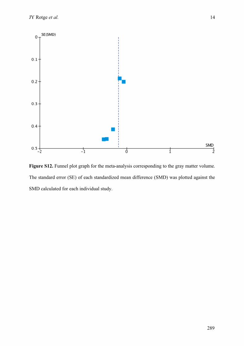
JY Rotge et al.
14
Figure S12. Funnel plot graph for the meta-analysis corresponding to the gray matter volume.
The standard error (SE) of each standardized mean difference (SMD) was plotted against the
SMD calculated for each individual study.
289
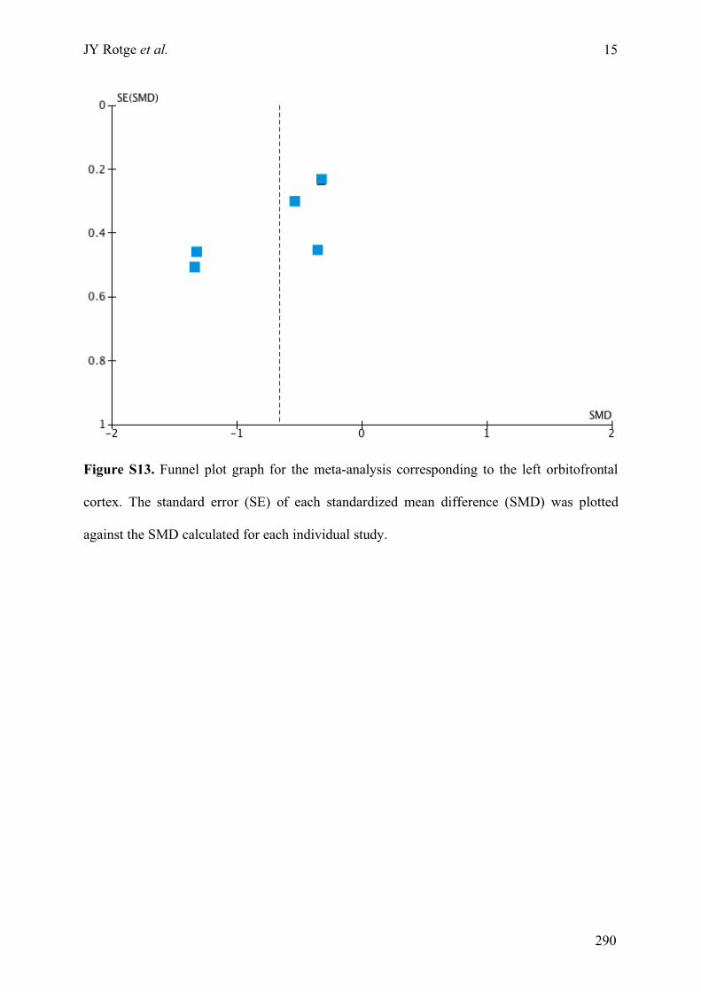
JY Rotge et al.
15
Figure S13. Funnel plot graph for the meta-analysis corresponding to the left orbitofrontal
cortex. The standard error (SE) of each standardized mean difference (SMD) was plotted
against the SMD calculated for each individual study.
290
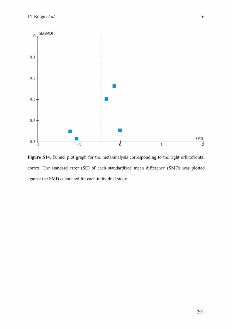
JY Rotge et al.
16
Figure S14. Funnel plot graph for the meta-analysis corresponding to the right orbitofrontal
cortex. The standard error (SE) of each standardized mean difference (SMD) was plotted
against the SMD calculated for each individual study.
291
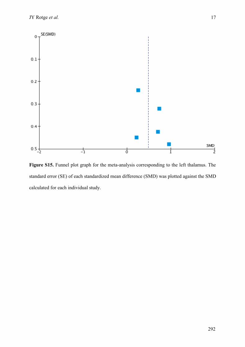
JY Rotge et al.
17
Figure S15. Funnel plot graph for the meta-analysis corresponding to the left thalamus. The
standard error (SE) of each standardized mean difference (SMD) was plotted against the SMD
calculated for each individual study.
292
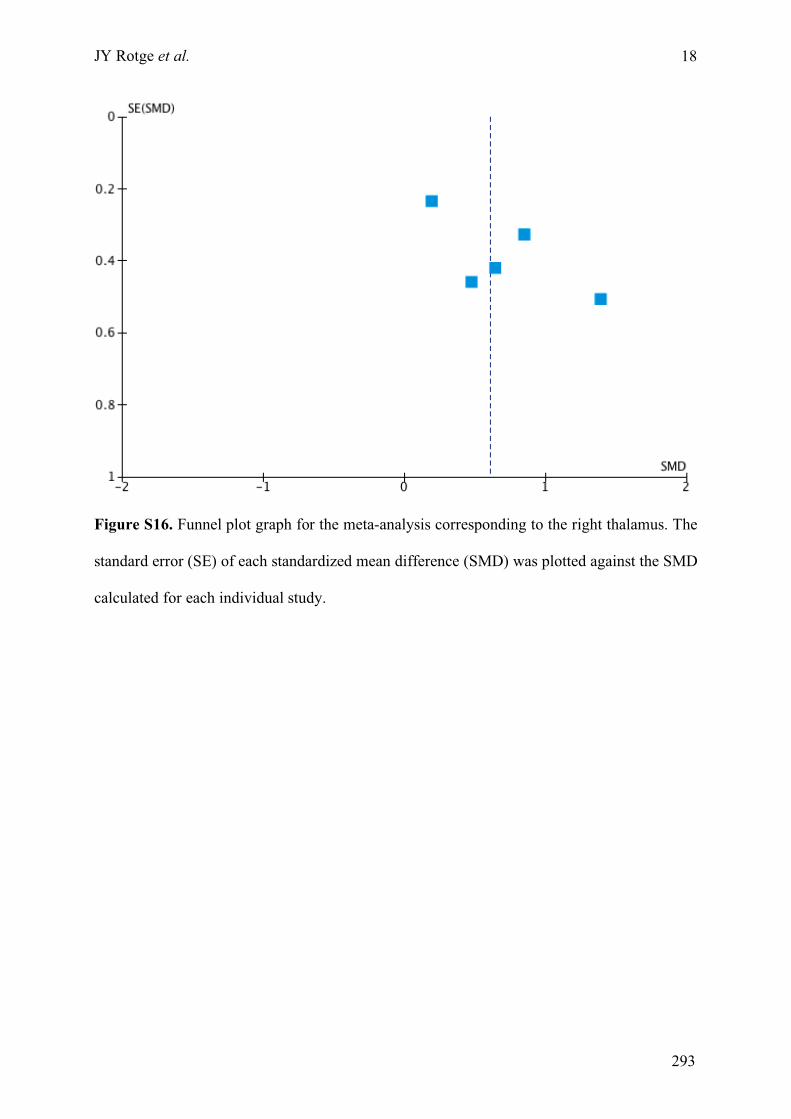
JY Rotge et al.
18
Figure S16. Funnel plot graph for the meta-analysis corresponding to the right thalamus. The
standard error (SE) of each standardized mean difference (SMD) was plotted against the SMD
calculated for each individual study.
293

JY Rotge et al.
19
Figure S17. Funnel plot graph for the meta-analysis corresponding to the left caudate
nucleus. The standard error (SE) of each standardized mean difference (SMD) was plotted
against the SMD calculated for each individual study.
294
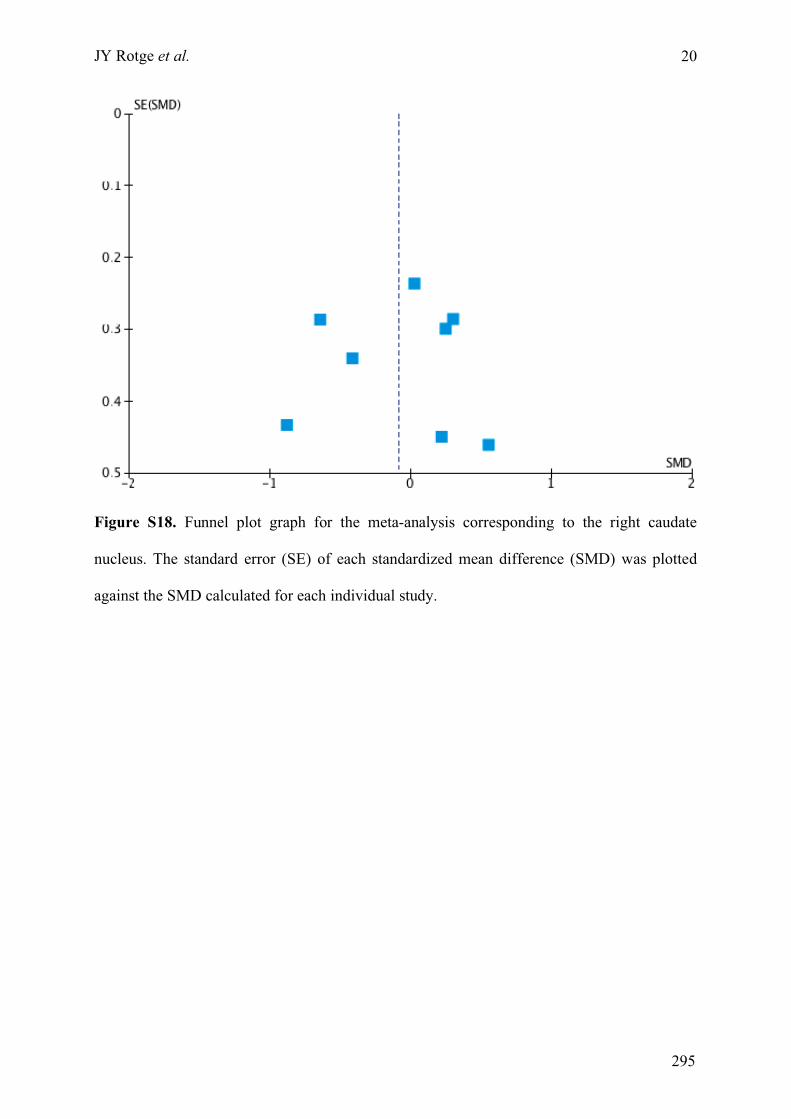
JY Rotge et al.
20
Figure S18. Funnel plot graph for the meta-analysis corresponding to the right caudate
nucleus. The standard error (SE) of each standardized mean difference (SMD) was plotted
against the SMD calculated for each individual study.
295


Top Related
Copyright © 2022 FDOKUMEN

