


Bahasa
Halaman
Hukum

PHYTOTAXA
ISSN 1179-3155 (print edition)
ISSN 1179-3163 (online edition)Copyright © 2014 Magnolia Press
Phytotaxa 164 (4): 255–264
www.mapress.com/phytotaxa/Article
http://dx.doi.org/10.11646/phytotaxa.164.4.4
Morphological and Molecular Characterization of Brasilonema roberti-lamii
(Cyanophyceae, Nostocales, Scytonemataceae), from Central Mexico
BEATRIZ RODARTE1†, ITZEL BECERRA-ABSALÓN2*, GUSTAVO A. MONTEJANO2, KARINA OSORIO-
SANTOS2, LUISA ALBA-LOIS1, HILDA LEÓN-TEJERA2 & CLAUDIA SEGAL-KISCHINEVZKY1
1 Departamento de Biología Celular.2Departamento de Biología Comparada. Facultad de Ciencias, Universidad Nacional Autónoma de México (UNAM). Colonia
Coyoacán, Código Postal 04510, P.O. Box 70-474, México, Distrito Federal, México. *e-mail: [email protected]
Abstract
This paper is a contribution to the morphological and molecular characterization of the cyanobacterium Brasilonema roberti-
lamii from populations found in Central Mexico. The general growth form and the morphological, morphometric and ecological characteristics of the populations studied clearly correspond to those described for Brasilonema roberti-lamii
(basionym: Tolypothrix roberti-lamii) from the French Antilles. Based on molecular data from DNA sequencing of the16S rRNA gene and the IGS of the cpcB-cpcA phycocyanin operon (cpcBA-IGS), we propose that the populations that we studied are closely related to those of other Brasilonema species, including B. octagenarum UFV-OR1, UFV-E1 and HA4187-MV1-p1F, Brasilonema sennae CENA 114, B. tolantongensis, B. terrestre CENA 116, B. angustatum HA4187-MV1-B2+p1F and HA4187-MV1-B2+p1H and B. bromeliae SPC951. Our findings support the transference of Tolypothrix roberti-lamii, which was made based exclusively on morphological criteria, to Brasilonema. The use of molecular analyses in addition to traditional morphological and ecological criteria, known as polyphasic approach, is a good alternative to describe taxa of cyanobacteria, mainly at the genus and species levels.
Keywords: cpcBA-IGS gene, Cyanobacteria, 16S rRNA gene, Tolypothrix roberti-lamii, polyphasic approach, systematics, taxonomy
Introduction
The use of several criteria, in addition to the morphological, such as molecular, ultrastructural, etc. (known as the “polyphasic approach”) has been proven to be adequate for the recognition and characterization of different taxa of cyanobacteria. The term “polyphasic” has been used in this sense by several researches (Flechtner et al. 2002, González-Resendiz et al. 2013, Komárek 2010, Vaccarino & Johansen 2011, 2012). The results reported by several authors (Wilmotte & Golubic 1991, Komárek 2006, 2010) show a clear relationship between the morphology and the DNA sequences of different genes within cyanobacteria, mainly the 16S rRNA. Several traditional cyanobacterial genera in the classification proposed by Geitler (1932) have been supported by DNA sequencing. Some recently described cyanobacteria have been separated from traditional genera to establish new genera such as Brasilonema Fiore, Sant’Anna, Azevedo, Komárek, Kaštovský, Sulek & Lorenzi (2007: 794), Coleofasciculus
Siegesmund, Johansen & Friedl in Siegesmund et al. (2008: 1575), Geminocystis Korelusová, Kaštovský & Komárek (2009: 933) or Phormidesmis Turicchia, Ventura, Komárková & Komárek (2009: 179). Some studies of extreme habitats have also led to the discovery of new genetically and morphologically well-defined genera, i.e. Spirirestis Fletchtner & Johansen in Fletcher et al. (2002: 6) or Rexia Cassamata, Gomez & Johansen (2006: 23).
The sequencing of the 16S rRNA gene has been used as a standard genetic approach to delimitate cyanobacterial genera. However, There are cases where some morphological and ecological different species. show a high percentage of DNA similarity, whereas other populations with similar morphology appear in different clades when their 16S rRNA gene sequences are compared (the so-called “cryptic species”, Komárek 2005, 2006, 2010).
The genus Brasilomena was erected based on both its morphological characters and DNA sequences, analyzed from the 16S rRNA gene and the IGS of the cpcB-cpcA phycocyanin operon sequences. At present, nine species
Accepted by Saúl Blanco Lanza: 5 Mar. 2014; published: 11 Apr. 2014
Licensed under a Creative Commons Attribution License http://creativecommons.org/licenses/by/3.0
255

are included in this genus, six of them have been erected based both on morphological and molecular criteria: Brasilonema bromeliae Fiore, Sant’Anna, Azevedo, Komárek, Kaštovský, Sulek & Lorenzi (2007: 796), B.octagenarum Aguiar, Fiore, Franco, Ventrella, Lorenzi, Vanetti & Alfenas (2008: 1325), B. sennae (Komárek 2003: 225) Sant’Anna & Komárek in Sant’Anna et al. (2011: 57), B. terrestre Sant’Anna & Komárek in Sant’Anna et al. (2011: 59), B. angustatum Vaccarino & Johansen (2012: 3) and B. tolantongensis Becerra-Absalón & Montejano in Becerra-Absalón et al. (2013: 27); and three other species based only on morphological characters: Brasilonema
ornatum Sant’Anna & Komárek in Sant’Anna et al. (2011: 54), B. epidendron Sant’Anna & Komárek in Sant’Anna et al. (2011: 52) and B. roberti-lamii (Bourrelly) Sant’Anna & Komárek in Sant’Anna et al. (2011: 56).
During the study of the cyanobacterial biota from Mexico’s central region, we found a population corresponding in morphology and ecology to B. roberti-lamii. This species was originally described from the French Antilles as Tolypothrix roberti-lamii Bourrelly & Manguin (1952: 151). Recently, this taxon has been moved to the genus Brasilonema based on its morphology and distinctive life cycle (Sant’Anna et al. 2011). Given our material and the taxonomic situation of the genus Brasilonema, we decided to perform a medium-term study (four years) of these cyanobacteria. The specific aim of this paper is to study the molecular, morphological and ecological correspondence (polyphasic approach) between our populations and those of other Brasilonema species, in order to corroborate the transfer from Tolypothrix Kützing ex Bornet & Flahault (1886–1888: 118) to Brasilonema roberti-lamii, as proposed by Sant’Anna et al. (2011).
Materials and Methods
Site description, sample collection and morphological analyses:—Samples were collected during the 2006–2010 period in the locality of Los Manantiales, Morelos, in Central Mexico (18° 55’ 39” N, 96° 00’ 37” W; 800 m a.s.l.). The climate of the locality is warm, sub-humid, with summer rain (Aw0 (w), Köppen modified by García (1988)) and the vegetation is a tropical deciduous forest. The populations were growing aerophytically on rocks and walls. Samples were transported dry to the laboratory. In the populations studied the following characteristics were analyzed: filament and trichome diameter, thallus and sheath morphology and coloration, cell color, length and width of heterocytes, branching, and young trichome form. The observations were carried out on an Olympus BX 51 microscope equipped with DIC (differential interferential contrast) and a DP 12 digital camera. The characteristics of our populations were compared to the information on Brasilonema populations reported in Fiore et al. (2007), Aguiar et al. (2008), Sant’Anna et al. (2011), Vaccarino & Johansen (2012), and Becerra-Absalón et
al. (2013).Genomic DNA extraction:—DNA was extracted according to Neilan et al. (1995) from ~30 mg of fresh field
material, which was studied and selected under an Olympus SZ2-ILST stereoscopic microscope to check for possible contaminations.
PCR amplification and sequencing:—PCR amplification was done using the primer set reported by Neilan et
al. (1995): PCßF and PCαR for the intergenic sequence of the cpcB and cpcA of the phycocyanin operon (cpcBA-IGS), and 27 F1 and 1494Rc for the 16S rRNA (Neilan et al. 1997). Amplification was performed in a Mastercycler Eppendorf. The reaction volume was 50 μL and contained 1X reaction buffer, 2.5 mM MgCl
2, 0.3
mM of each deoxynucleotide triphosphate (dNTPs), 0.5 µM of each primer and 1U of DNA polymerase Amplificasa (Biogénica, Mexico). The PCR program was the same for both amplifications and consisted of an initial denaturation at 94 °C for 7 minutes; 30 cycles at 94 °C for 30 seconds, 63 °C for 1 minute, 72 °C for 1 minute, and a final extension at 72 °C for 7 minutes. The amplification products were visualized on a 1.5% agarose electrophoresis gel and the amplification products of 16S rRNA were cloned into the pJet 1.2/blunt vector of the Clone Jet PCR Cloning Kit (Fermentas, Maryland, USA). The DNA from the positive samples were purified using
GeneClean®III Kit (Bio 101 Systems, California, USA) as recommended by the manufacturer. The obtained DNAs were submitted to “Unidad de Biología Molecular, Instituto de Fisiología Celular, UNAM (D.F., Mexico)”, to be sequenced. Sequences of both DNA strands were determined by using the same primers as those used for amplification. Fragments were assembled using the software Bioedit version 7.0.9.0 (Hall 1999). The GenBank/EMBL/DDBJ accession number for the cpcBA-IGS sequence determined in this study is EF676035, while the accession number for the 16S rRNA sequence determined in this study is GQ443308.1.
RODARTE ET AL.256 • Phytotaxa 164 (4) © 2014 Magnolia Press

Sequence alignment and phylogenetic analyses:—Phylogenetic analyses were based on two DNA sequences: the 16S rRNA gene (bp 106–1494), and the cpcBA-IGS region of the phycocyanin gene. The taxa used for 16S rRNA and cpcBA sequence analyses included a total of 124 and 25 OTUs, respectively, both with one novel sequence of B. roberti-lamii from Los Manantiales Morelos, Mexico, and OTUs from GenBank. We made a BLAST search to select the sequences most closely related to these of B. roberti-lamii. We also selected sequences based on morphological classification criteria, so we included sequences from all the Nostocalean families: Scytonemataceae, Microchaetaceae, Nostocaceae, Rivulariaceae, Symphyonemataceae, Hapalosiphonaceae and Stigonemataceae. In both analyses the external group was Gloeobacter violaceaus Rippka, Waterbury & Cohen-Bazire (1974: 436). The initial alignment was constructed using ClustalW (Larkin et al. 2007), and the sequences were manually reviewed as well using the program PhyDE-1 version 0.9971 (Müller et al. 2010). The trees were constructed using parsimony and maximum likelihood analyses in MEGA version 5 (Tamura et al. 2011). Parsimony analysis was performed using the Subtree Pruning Regrafting (SPR) search method with 10 random additions and 100 trees retain. Bootstrapping of 500 replicates was used. The best models for analysis with maximum likelihood were found with MEGA version 5. Maximum likelihood was performed using the GTR+I+G likelihood model for the 16S rRNA and Tamura 3-parameter likelihood model for the cpcBA-IGS. The heuristic search followed the Nearest Neighbor Interchange (NNI) method. Bootstrapping was conducted with 500 pseudoreplicates for the 16S rRNA and 1000 pseudoreplicates for the cpcBA-IGS.
Results
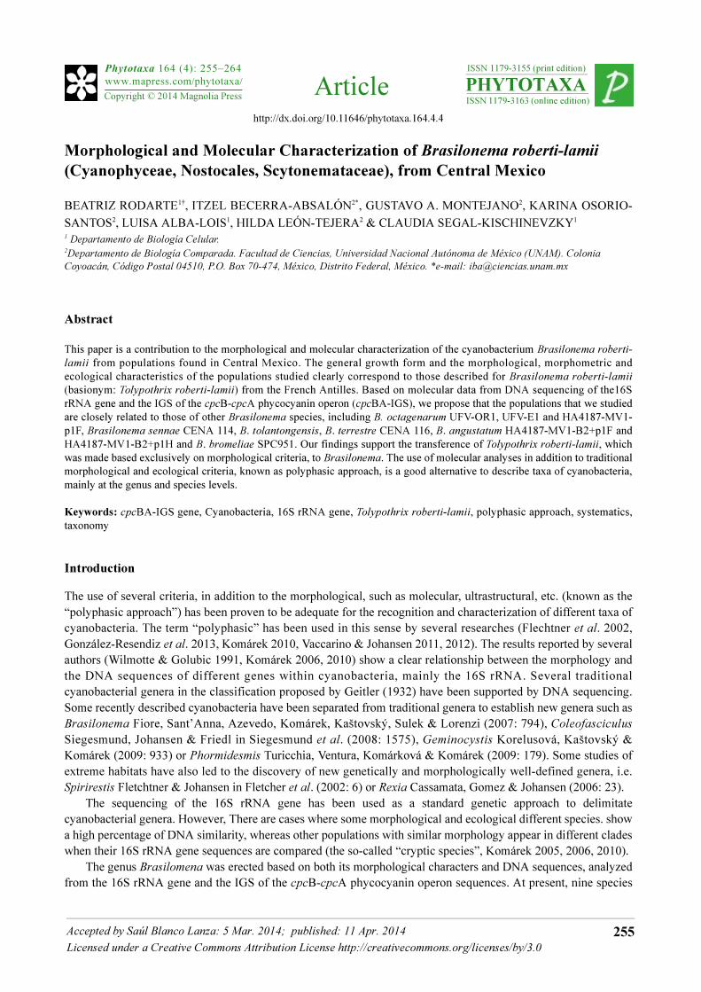
Morphological analysis:—Our material of B. roberti-lamii clearly belongs to that described for the Antilles Island by Bourrelly and Manguin (1952). The habit is heterotrichous with a postrated and erect portion (Fig. 1, a–e); this last one is organized in fascicles with parallel trichomes (Fig. 1, d–e, j). Filaments have a distinctive “C” or “J” form (Fig. 1, f–h), and the morphometry and ecology also correspond to the description of B. roberti-lamii (Table 1). Brasilonema roberti-lamii is quite different from other Brasilonema species: The filaments of Brasilonema
bromeliae, B. ornatum, B. sennae and B. tolantongensis are wider than B. roberti-lamii, B. epidendron and B. ornatum have ornamented sheaths which B. roberti-lammi lacks, B. epidendron has bright blue-green cells (whereas those of B. roberti-lamii are violet), and finally B. angustatum is tapering while B. roberti-lamii is not (Table 1).
Brasilonema roberti-lammi (Bourrelly & Manguin) Sant’Anna & Komárek
Thallus in erect fascicles, forming extended carpets 2–3 mm high, brownish and blackish in color (Fig. 1, a–e). Filaments parallel, closely packed, organized in fascicles, 11–14 µm in diameter (Table 1). Occasional false single branching, sometimes from hormogonia. Sheath thin, colorless or yellow brownish (Fig. 1, e). Trichomes isopolar, cylindrical, not constricted at the cell walls (Fig. 1, e–h), not attenuated toward the ends and rounded at the apex, ends 9.5–11.5 µm in diameter. Young trichomes with a characteristically “C” or “J” shape (Fig. 1, f–h). Cells isodiametric or slightly longer than wide, except in meristematic zones where they are disk-shaped. Cells present “vacuolization” frequently. Heterocytes intercalary, 11.0–13.5 µm wide × 10.5–13.0 µm long. Hormogonia develop in the apical extremes of filaments by formation of necridic cells (Fig. 1, i–l).
Sequences alignments and phylogenetic analyses:—The Maximum Likelihood (ML) and Maximum Parsimony (MP) algorithms produced similar tree topologies in the 16S rRNA and cpcBA-IGS genes analyses. Only ML trees are shown in figures 2 and 3. The phylogenetic tree resulting from the 16S rRNA gene analysis (Fig. 2) showed that B. roberti-lamii clusters with the other six recently reported Brasilonema taxa, with a high bootstrap value (92%). In this tree, B. roberti-lamii is a sister species of B. octagenarum and the cluster formed with both species is a sister with B. angustatum. The sister cluster of the Brasilonema group is formed with Symphyonemopsis Tiwari & Mitra (1969: 93) sequences and the next branch related to both clusters is formed with several sequences of Scytonema
Agardh ex Bornet & Flahault (1886: 85). We determined 1379 bp from the 16S rRNA gene sequence from Brasilonema roberti-lamii strain, this sequence was analyzed by the BLAST algorithm and the result showed that the percentage of similarity between B. roberti-lamii, B. octagenarum UFV-ORI, B. octagenarum UFV-E1 and B.
Phytotaxa 164 (4) © 2014 Magnolia Press • 257BRASILONEMA ROBERTI-LAMII FROM CENTRAL MEXICO
angustatum was 99%, the next similar strains were B. tolantongensis with 98%, and lastly B. terrestre, B. sennae
CENA 114 and B. bromeliae SPC951 with 97% similarity. The other taxa presented in Table 2 were less than 96% similar to B. roberti-lamii. The phylogenetic tree resulting from the cpcBA-IGS gene analysis (Fig. 3) showed that all six Brasilonema sequences were clustered together in one defined clade with high bootstrap values, the nearest (but in a different branch) was Tolypothrix sp, while Nostoc linckia Bornet ex Bornet & Flahault (1888: 192), Nostoc sp. and Scytonema hofmannii Agardh ex Bornet & Flahault (1887: 97) were found in a sister cluster. Using the phycocyanin cpcBA-IGS to determine the phylogenetic placement of our populations results in B. roberti-lamii
being a member of the Brasilonema genus and indeed constituting a distinctive species.
FIGURE 1. Brasilonema roberti-lamii. A. According to Bourrelly and Manguin (1952). B–L. Brasilonema roberti-lamii
from Los Manantiales. B–D. Habit. E. Detail of filament organization in fascicles. F–H. typical “C” or “J” shape of
young trichomes with heterocytes. I, K, L. Different stages of hormogonia development. Arrow: proheterocyte. J.
Parallel organization of filaments. Scale bars: C = 0.5 mm, D = 50 µm, E–J, L = 20 µm, K = 7 µm.
RODARTE ET AL.258 • Phytotaxa 164 (4) © 2014 Magnolia Press
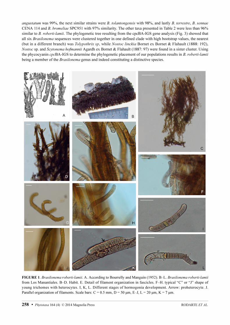
TABLE 1. Morphological characters of Brasilonema species. In bold, “Los Manantiales” population, Mexico. The information about the other species was obtained from Fiore et al. (2007), Aguiar et al. (2008), Santa’Anna et al. (2011), Vaccarino & Johansen (2012) and Becerra-Absalón et al. (2013).
Character B. brome-
liae
(Fiore et al.
2007)
B.
epidendron
(Sant’Ann
a et al.
2011)
B.
octogenaru
m
(Aguiar et
al. 2008)
B. ornatum
(Sant’Anna
et al. 2011)
B.
angustatum
(Vaccarino
& Johansen
2012)
B. sennae
(Sant’Anna
et al. 2011)
B. terrestre
(Sant’Anna
et al. 2011)
B. tolantongensis
(Becerra-
Absalón et al.
2013)
B. roberti-
lamii
(Sant’Anna
et al. 2011)
B. roberti-lamii
(Present study)
Filament
diameter
(µm)
10–14.8–21 (7)10.9–
12(14)
9.8–16–
18.5
20–23 9.8–17.8 10–20 12–17 17–25 12–15–18 11.5–13
Trichome
diameter
(µm)
8–13.2–18 (5.5)8.2–
10(11)
9.5–14.9–
18.4
17–18 (5.5)9.8–
17.0
6–12.5 9–15 12.5–20 8 8.5–11.5
Sheath
morpho-
logy
Thin, firm Thin, firm Thin, firm,
later
lamellated
Thick,
lamellated,
regularly
ornamented
Thin,
smooth
Thin, firm,
later
lamellated
Thin, firm,
orna-
mented
Thin, firm,
smooth
Thin firm
sometimes
lamellated
Thin, firm
Cell color Grayish
blue or
brownish,
olive-green,
or violet
Bright
blue-green
Brownish,
olive-green,
or rarely
violet
Dark blue-
green
Brownish
or purplish
gray
Blue-green
or olive-
green
Greyish-
green or
green-blue
Violet or brown - Violet
Thallus
morpho-
logy
Free
fascicles
Irregular
erect
fascicles
Mats of
irregular
fascicles,
creeping
fascicles
Irregular
fine mats
with
creeping,
partly
fasciculated
filaments
Free, erect
moderately
long
filaments
Regular
erect
fascicles
Forming
mats or
irregular
fascicles
Creeping
fascicles
Irregular
fascicles,
parallel
filaments
Erect fascicles,
parallel
filaments
Thallus
color
Blackish-
green to
blackish
violet
Dark green
to blackish
Dirty-green,
brownish,
or
blackish-
green
Greyish
green
Brown Dirty-
green,
brownish,
or
blackish-
green
Dirty green Blackish-violet Violet Brownish
blackish
Hetero-
cytes
(µm)
±Cylin-
drical
4–19 long ×
15–16.8
wide
Barrel-
shaped to
cylindrical
(7)8–
10(11.5)
long × 7–9
wide
Cylindrical
5.4–15.6 ×
10–17.6
Rounded,
17–18 long
× 3–6 wide
Intercalary,
flattened or
elongated
10–17 wide
×
2.8–17.4
long
Cylindrical
6.8–15.4
long ×
10.2–11.2
wide
Cylindrical
-barrel
shaped
6–17 long
× 13–14
wide
7–15.5 wide ×
12.5–16.5 long
Rectan-
gular
Cylindrical,
isodiametrical
Branch-
ing type
Double or
single
Simple Double or
single
- Double or
single
Double or
single
- Double, single or
from hormogonia
Single (like
Tolypothrix
?)
Double or
single from
hormogonia
Shape of
young
trichomes
“J”- and
“C”-shaped
- Lightly
curved
- Curved,
with
heterocytes
at one end
- - Curved,
attenuated at one
or both ends.
“J”- and
“C”-shaped
“J”- and “C”-
shaped
Hormo-
gonia
develop-
ment
- Short cells,
slightly
constricted
at cross
walls,
granulated
- - Crescent-
shaped,
isopolar
when first
released
- - Initially with one
or two
intercalary,
asymmetric
heterocytes, after
fragmentation,
with basal
heterocytes
Medium
heterocyst
Initially with
one or two
intercalary,
asymmetric
heterocytes,
after
fragmentation,
with basal
heterocytes
Ecology Subaerial,
epiphytic on
living and
dead leaves
of
bromeliads
(inside of
leaf
rosettes)
Subaerial,
corticolous
or on old
wooden
substrates
in Atlantic
Forest.
Epiphytic
on living
and dead
leaves,
stem, and
buds of
Eucalyptus
grandis
Subaerial,
on bark of
trees among
mosses and
lichens
Moss bank
along
Moleka
Trail
Subaerial,
edge of
springs on
wet
wooden
stony and
iron
substrates
Subaerial,
on concrete
Subaerial, on
walls near
runoffs
Subaerial,
on rocks
and roofs
Subaerial, on
rocks and
walls
Phytotaxa 164 (4) © 2014 Magnolia Press • 259BRASILONEMA ROBERTI-LAMII FROM CENTRAL MEXICO

FIGURE 2. Maximum Likelihood tree based on sequences of the 16S rRNA gene. The phylogenetic analysis showed the relationships between Brasilonema species and Cyanobacteria of the same order (Nostocales), using Gloeobacter violaceus as the external functional group. Bootstrap values at supported nodes (> 50%) are given for Maximum Likelihood, and Maximum Parsimony values only in the cluster with Brasilonema.
Discussion
Our populations showed the morphological diagnostic features of Brasilonema roberti-lamii described for the French Antilles populations (Bourrelly & Manguin 1952), including habit, morphology, morphometry and ecology, therefore we conclude that, according to our analyses, they correspond to this species (Table 1, Fig. 1, a–e). This species has the distinctive morphological and molecular characteristics of Brasilonema: growth in fascicles with parallel filaments; cylindrical trichomes with vacuolization; thin, colorless or yellow-brown sheaths, roundly closed at the ends; cells with the typical violet color; solitary, intercalary heterocytes lacking akinetes (Fiore et al. 2007) and hormogonia with asymmetric development (Becerra-Absalón et al. 2013. Table 1. Fig. 1, e–l). At the molecular (DNA sequence) level, the 16S RNA (Fig. 2) and cpcB-IGS (Fig. 3) analyses showed that the sequences
RODARTE ET AL.260 • Phytotaxa 164 (4) © 2014 Magnolia Press

of B. roberti-lamii belong to the same clade as the other Brasilonema species. Therefore, our results support the transfer of Tolypothrix roberti-lamii to Brasilonema, as Sant’Anna et al. (2011) proposed.
FIGURE 3. Maximum Likelihood tree based on sequences of the cpcBA-IGS gene. The analysis showed the
relationships among Brasilonema species and Cyanobacteria of the same order (Nostocales), using Gloeobacter
violaceus as the external functional group. Bootstrap values at supported nodes (> 50%) are given for Maximum
Likelihood, and Maximum Parsimony values only in the cluster with Brasilonema.
In the 16S rRNA analysis, the Brasilonema clade is separated from the most morphologically similar genus Scytonema. Brasilonema appeared also as a sister group to Symphyonemopsis (Fig. 2).
At the species level, B. roberti-lamii exhibits a high percentage of similarity (99%) to both B. octagenarum and B. angustatum (Table 2). Indeed, in the phylogenetic tree these three species are part of a single tight cluster. At this level it is difficult to decide, based exclusively on differences among the 16S rRNA sequences, whether these three populations belong to the same or different species, although the differences in morphology and ecology (Table 1) allow us to consider that B. roberti-lamii is actually a separate species from B. octagenarum and B. angustatum. The morphologically closest species (not coincident with the genetic closeness) is B. bromeliae, similar in habit and morphology with the young trichomes of B. roberti-lamii, and sharing the “C” and “J” shapes (Table 1). However, this latter species exhibits double branches and is epiphytic, whereas B. roberti lamii has single branches and is epilithic (Table 1).
In summary, our results support the proposal of Sant’Anna et al. (2011) to transfer Tolypothrix roberti-lamii to the genus Brasilonema since this species presents the diagnostic characteristics of the genus (Fiore et al. 2007) and belongs to the Brasilonema cluster. Additionally, B. roberti-lamii can be regarded as a species different from other taxa of the genus when applying morphological, genetic and ecological criteria. It was evident that, in the case of Brasilonema species, the 16S rRNA gene analysis (phylogeny and percentage of similarity) is not enough for species recognition and delimitation, thus stressing the necessity of a polyphasic approach with morphological, molecular and ecological data.
Phytotaxa 164 (4) © 2014 Magnolia Press • 261BRASILONEMA ROBERTI-LAMII FROM CENTRAL MEXICO

TABLE 2. Similarity matrix (percentages) for 16 strains comparing partial sequences of the 16S rRNA gene. Strain access numbers:
(1) GQ443308, (2) HQ847567, (3) EF150855, (4) HQ847562, (5) JN676147, (6) EF490447, (7) DQ486055, (8) EF117246, (9) AJ544085, (10) AJ544079, (11) AJ544084, (12) AJ544083, (13) AF334700, (14)AB075996, (15) AY069954, (16) AB093483.
Conclusions
The taxonomy of Cyanobacteria has changed considerably in recent years. The incorporation of molecular techniques has been of great help to delimit and characterize genera and higher-level taxa, allowing to understand the diversification processes within cyanobacteria, which in turn has affected their classification. At the species level, however, the use of the 16S rRNA gene is not generally sufficient by itself for species delimitation because the similarity values are often high (above 98%) and phylogenetic analyses allow already the recognition of significant differences. Nevertheless, the interpretation of such differences requires the use of other criteria (such as morphological and ecological) to provide sufficient data for species delimitation. For this reason, the best approach available to date is the use of complementary criteria (polyphasic approach).
Acknowledgements
The authors would like to thank “Programa de Apoyo a Proyectos de Investigación e Inovación Tecnológica (PAPIIT)” for supporting project No IN207709, to Dr. Michele Gold Morgan for her critical review and English revision and to the anonymous Reviewers for reviewing and commenting on the manuscript.
References
Aguiar, R., Fiore, M.F., Franco, M.W., Ventrella, M.C., Lorenzi, A.S., Vanetti, C. & Alfenas, A.C. (2008) A novel epiphytic cyanobacterial species from the genus Brasilonema causing damage to Eucalyptus leaves. Journal of Phycology 44: 1322–1334.http://dx.doi.org/10.1111/j.1529-8817.2008.00584.x
Becerra-Absalón, I., Rodarte, B., Osorio, K., Alba-Lois, L., Segal-Kischinevzky, C. & Montejano, G. (2013) A new species of Brasilonema (Scytonemataceae, Cyanoprokaryota) from Tolantongo, Hidalgo, Central Mexico. Fottea, Olomouc 13: 25–38.
Bornet, É. & Flahault, C. (1886–1888) Revision des Nostocacées hétérocystées contenues dans les principaux herbiers de France. Annales des Sciences Naturelles, Botanique, Trosième Série 7: 51–129.
2 3 4 5 6 7 8 9 10 11 12 13 14 15 16
1. Brasilonema roberti-lamii 99 99 99 98 97 97 97 96 94 94 95 95 93 93 93
2. B. angustatum HA4187-MV1 99 99 98 98 97 97 96 95 94 95 95 93 93 93
3. B. octagenarum UFV-OR1 100 99 98 97 97 96 95 94 95 95 93 93 93
4. B. octagenarum HA4186-MV1 99 98 97 97 96 95 94 95 95 93 93 93
5. B. tolantongensis 98 98 98 97 95 94 95 94 93 93 93
6. B. terrestre CENA116 97 97 97 95 94 95 94 93 93 93
7. B. bromeliae SPC951 99 96 95 94 95 94 93 92 92
8. B. sennae CENA114 96 95 95 95 94 93 92 93
9. Symphyonemopsis VAPOR 1 95 94 94 94 93 93 93
10. Mastigocladopsis repens MORA 96 96 95 93 92 92
11. Symphyonema sp. 1517 99 97 93 92 92
12. Symphyonema sp. 1269-1 96 93 92 92
13. Scytonema hyalinum 93 92 91
14. Scytonema hoffmannii PCC7110 95 95
15. Scytonema sp. U-3-3 98
16. Scytonema sp. IAM M-262 100
RODARTE ET AL.262 • Phytotaxa 164 (4) © 2014 Magnolia Press
Bourrelly, P. & Manguin, E. (1952) Algues d’eau douce de la Guadeloupe et dépendances. Recueillies par la Mission P. Allorge
en 1936. Société d’Edition d’Enseignement Supérieur, Paris, 281 pp.http://dx.doi.org/10.2307/2257052
Casamatta, D.A., Gomez, S.R & Johansen, J.R. (2006) Rexia erecta gen. et sp. nov. and Capsosira lowei sp. nov., two newly described cyanobacterial taxa from the Great Smoky Mountains National Park (USA). Hydrobiologia 561: 13–26.http://dx.doi.org/10.1007/s10750-005-1602-6
Fiore, M.F., Sant’Anna, C.L., Azevedo, M.T.P., Komarék, J., Kástowský, J., Sulek, J. & Lorenzi, A.S. (2007) The cyanobacterial genus Brasilonema, gen. nov., a molecular and phenotypic evaluation. Journal of Phycology 43: 789–798.http://dx.doi.org/10.1111/j.1529-8817.2007.00376.x
Flechtner, V.R., Boyer, S.L., Johansen, J.R. & DeNoble, M.L. (2002) Spirirestis rafaelensis gen. et sp. nov. (Cyanophyceae), a new cyanobacterial genus from arid soils. Nova Hedwigia 74: 1–24.http://dx.doi.org/10.1127/0029-5035/2002/0074-0001
García, E. (1988) Modificaciones al sistema de clasificación climático de Köppen. Adaptación a las condiciones de la República Mexicana. Cuarta Edición. Instituto de Geografía, UNAM,. México, D.F, 246 pp.
Geitler, L. (1932) Cyanophyceae. Akademische Verlagsgesellschaft, Leipzing, 1196 pp.http://dx.doi.org/10.1038/130292a0
González-Resendiz, L., León-Tejera H., Díaz-Larrea, J., Alba-Lois, L. & Segal-Kischinevzky, C. (2013) Hassallia littoralis sp. nov. (Cyanobacteria, Microchaetaceae) from Mexico’s marine supralittoral based on morphological and molecular evidence. Phytotaxa 137(1): 35–47.http://dx.doi.org/10.11646/phytotaxa.137.1.4
Hall, T.A. (1999) BioEdit: a user-friendly biological sequence alignment editor and analysis program for Windows 95/98/NT. Nucleic Acids. Symposium Series 41: 95–98.
Komárek, J. (2003) Two Camptylonemopsis species (cyanoprokaryotes) from “Mata Atlantica” in coastal Brazil. Preslia,
Praha 75: 223–232.Komárek, J. (2005) The modern classification of cyanoprokaryotes (Cyanobacteria). Oceanological and Hydrobiological
studies 34: 5–17.Komárek, J. (2006) Cyanobacterial taxonomy: Current Problems and prospects for the integration of traditional and molecular
approaches. Algae 21(4): 349–375.Komárek, J. (2010) Recent changes (2008) in cyanobacteria taxonomy based on a combination of molecular background with
phenotype and ecological consequences (genus and species concept). Hydrobiologia 639: 245–259.http://dx.doi.org/10.1007/s10750-009-0031-3
Korelusová, J., Kaštovský, J. & Komárek, J. (2009) Heterogeneity of the cyanobacterial genus Synechocystis and description of a new genus, Geminocystis. Journal of Phycology 45: 928–937.http://dx.doi.org/10.1111/j.1529-8817.2009.00701.x
Larkin, M.A., Blackshields, G., Brown. N.P., Chenna. R., McGettigan, P.A., McWilliam, H., Valentin, F., Wallace, I.M., Wilm, A., Lopez, R., Thompson, J.D., Gibson, T.J. & Higgins, D.G. (2007) Clustal W and Clustal X version 2.0. Bioinformatics
23: 2947-2948.Müller, J., Müller, K., Neinhuis, C. & Quandt, D. (2010) PhyDE-Phylogenetic Data Editor. Available from: http://www.
Phyde.de (accessed 30/1/2014).Neilan, B.A., Jacobs, D. & Goodman, A.E. (1995) Genetic diversity and phylogeny of toxic cyanobacteria determined by DNA
polymorphisms within the phycocyanin locus. Applied and Environmental Microbiology 61: 3875–3883. Neilan, B.A., Jacobs, D., Del Dot, T., Blackall, L.L., Hawkins, P.R., Cox, P.T. & Goodman, A.E. (1997) rRNA sequences and
evolutionary relationships among toxic and nontoxic cyanobacteria of the genus Microcystis. International Journal of
Systematic Bacteriology 47: 693–697.http://dx.doi.org/10.1099/00207713-47-3-693
Rippka, R., Waterbury, J. & Cohen-Bazire, G. (1974) A cyanobacterium which lacks thylakoids. Archives for Microbiology
100: 419–436.Sant’Anna, C.L., Azevedo, M.T.P., Fiore, M.F., Lorenzi, A.S., Kaštovský, J. & Komárek, J. (2011) Subgeneric diversity of
Brasilonema (Cyanobacteria, Scytonemataceae). Revista Brasileira de Botânica 34: 51–62.Siegesmund, M.A., Johansen, J.R., Karsten, U. & Friedl, T. (2008) Coleofasciculus gen. nov. (Cyanobacteria): morphological
and molecular criteria for revision of the genus Microcoleus Gomont. Journal of Phycology 44: 1572–1585.http://dx.doi.org/10.1111/j.1529-8817.2008.00604.x
Tamura, K., Peterson, D., Peterson, N., Stecher, G., Nei, M & Kumar, S. (2011) MEGA5: Molecular Evolutionary Genetics Analysis using Maximum Likelihood, Evolutionary Distance and Maximum Parsimony Methods. Molecular Biology and
Evolution 28: 2731–2739.http://dx.doi.org/10.1093/molbev/msr121
Tiwari, G.L., & Mitra, A.K. (1969) Symphyonemopsis katniensis gen. et sp. nov., a new member of the Myxophyceae. Phykos
7: 186–194.Turicchia, S., Ventura, S., Komárkova, J. & Komárek, J. (2009) Taxonomic evaluation of cyanobacterial microflora from
alkaline marshes of northern Belize. 2. Molecular and phenotype diversity of oscillatorialean genera. Nova Hedwigia 89: 165–200.
Phytotaxa 164 (4) © 2014 Magnolia Press • 263BRASILONEMA ROBERTI-LAMII FROM CENTRAL MEXICO

http://dx.doi.org/10.1127/0029-5035/2009/0089-0165Vaccarino, M.A. & Johansen, J.R. (2011) Scytonematopsis contorta sp. nov. (Nostocales), a new species from the Hawaiian
Islands. Fottea 11: 149–161.Vaccarino, M.A. & Johansen, J.R. (2012) Brasilonema angustatum sp. nov. (Nostocales), a new filamentous cyanobacterial
species from the Hawaiian Islands. Journal of Phycology 48: 1178–1186.http://dx.doi.org/10.1111/j.1529-8817.2012.01203.x
Wilmotte A. & Golubić S. (1991) Morphological and genetic criteria in the taxonomy of Cyanophyta/Cyanobacteria. Algological Studies 64: 1–24.
RODARTE ET AL.264 • Phytotaxa 164 (4) © 2014 Magnolia Press
Copyright © 2022 FDOKUMEN

