


Bahasa
Halaman
Hukum

Methods xxx (2010) xxx–xxx
Contents lists available at ScienceDirect
Methods
journal homepage: www.elsevier .com/locate /ymeth
Purification and characterization of HIV–human protein complexes
Stefanie Jäger a, Natali Gulbahce a, Peter Cimermancic a, Joshua Kane a, Nanhai He b, Seemay Chou b,Iván D’Orso c, Jason Fernandes c, Gwendolyn Jang c, Alan D. Frankel c, Tom Alber b, Qiang Zhou b,Nevan J. Krogan a,⇑a Department of Cellular and Molecular Pharmacology, University of California-San Francisco, 1700 4th Street, San Francisco, CA 94158, United Statesb Department of Biochemistry and Molecular Biology, University of California-Berkeley, 374 Stanley Hall, Berkeley, CA 94720-3220, United Statesc Department of Biochemistry and Biophysics, University of California-San Francisco, 600 16th Street, San Francisco, CA 94158, United States
a r t i c l e i n f o
Article history:Available online xxxx
Keywords:Virus-host interactionMass spectrometryProtein interactionHIVProteomics
1046-2023/$ - see front matter � 2010 Published bydoi:10.1016/j.ymeth.2010.08.007
⇑ Corresponding author.E-mail address: [email protected] (N.J. Krogan
Please cite this article in press as: S. Jäger et al
a b s t r a c t
To fully understand how pathogens infect their host and hijack key biological processes, systematicmapping of intra-pathogenic and pathogen–host protein–protein interactions (PPIs) is crucial. Due tothe relatively small size of viral genomes (usually around 10–100 proteins), generation of comprehensivehost–virus PPI maps using different experimental platforms, including affinity tag purification-massspectrometry (AP-MS) and yeast two-hybrid (Y2H) approaches, can be achieved. Global maps such asthese provide unbiased insight into the molecular mechanisms of viral entry, replication and assembly.However, to date, only two-hybrid methodology has been used in a systematic fashion to characterizeviral–host protein–protein interactions, although a deluge of data exists in databases that manuallycurate from the literature individual host–pathogen PPIs. We will summarize this work and also describean AP-MS platform that can be used to characterize viral-human protein complexes and discuss its appli-cation for the HIV genome.
� 2010 Published by Elsevier Inc.
1. Introduction
Protein–protein interaction networks have been generatedusing AP-MS and Y2H targeting various organisms, ranging frombacteria to humans. Yeast two-hybrid screenings consist of testingall pair wise combinations of proteins, which generates a collectionof binary interactions. High-throughput Y2H maps have been gen-erated for Saccharomyces cerevisiae [16,23,41,47], Caenorhabditiselegans [28,45], Drosophila [18] and humans [10,35,39]. Affinitytag purification-mass spectrometry approaches identify groups ofproteins that participate in complexes and these have been usedto study the cellular make-up of Escherichia coli [2,5], S. cerevisiae[17,22,27] and human cells [14]. While AP-MS assays have a higherpropensity of detecting stable, stoichiometric complexes, Y2Hscreens tend to detect transient protein interactions [47]. Thereforethe data from both approaches are complementary with respect torevealing physical connections between proteins, complexes andbiological processes.
These unbiased approaches have also been used to study PPIsbetween proteins that are derived from a specific virus. For exam-ple, an intraviral Hepatitis C (HCV) Y2H interaction map was builtusing a limited set of predefined coding segments, which revealed
Elsevier Inc.
).
., Methods (2010), doi:10.1016/
the functional interactions between the proteins in the viral lifecycle when a cell culture system is absent [15]. Also, using a Y2Happroach, intraviral protein–protein interaction networks havebeen generated for two herpesviruses, Kaposi sarcoma-associatedherpesvirus (KSHV) and Varicella-Zoster virus (VZV) [40]. Theresulting PPI networks appear as a single highly connected modulewhereas cellular networks (e.g. yeast and human) have been ob-served to be organized in functional modules. Despite a broadrange of pathogenicity, herpesviruses share a significant percent-age of common conserved genes and the authors attempted to de-fine a core set of interactions conserved among these viruses.Calderwood et al. proteomically interrogated another herpesvirus,Epstein-Barr virus (EBV), by classifying the genes into two evolu-tionary classes based on conservation and showed enrichmentfor interactions among proteins in the same evolutionary class[6]. Another example of an intraviral PPI network based yeast-two-hybrid matrix analysis was obtained for SARS coronavirus[43]. SARS-CoV has 14 ORFs, most of whose functions are un-known. Interestingly, one of the accessory proteins turned out tobe highly connected, and the authors propose that although notessential for viral replication in cell culture systems, it could en-hance the global stability of the SARS proteome network andpathogenicity.
These pair-wise interaction studies have also been extendedinto studying the interaction landscape between viral proteins
j.ymeth.2010.08.007

2 S. Jäger et al. / Methods xxx (2010) xxx–xxx
and host factors. For example, Lotteau and colleagues published aproteome-wide, Y2H-based mapping of interactions among HCVand human proteins. They reported 314 interactions (in additionto 170 literature curated interactions) and discovered that HCVCORE protein was a major perturbator of the insulin, Jak/STATand TGFß pathways [11]. More recently, another study targetedVaccinia virus, a large double stranded DNA virus with more than280 ORFs and a prototype of the Orthopoxvirus, which includesseveral pathogenic poxviruses such as variola virus, a lethal hu-man-specific pathogen that causes smallpox [49]. The authors re-ported a comprehensive yeast-two hybrid screening with 109protein–protein interactions between vaccinia proteins and humanproteins and provided functional insight into a number of unchar-acterized viral proteins. Finally, Shapira et al. introduced a multi-layered approach to uncover pathways in H1N1 infection bycombining yeast-two-hybrid analysis and genome-wide expres-sion profiling [38]. They found human factors mediating virus–hostinteractions, which were further studied via depletion analysis inprimary lung cells. These types of unbiased physical and regulatorymodels of virus–host interactions provide a promising direction forthe unveiling of new virus biology and development of new viraldrugs [31].
Collectively, the global network properties of human proteinstargeted by pathogens, including bacteria and viruses, were re-cently studied [13]. It was observed that pathogenic proteins pref-erentially interact with human hub proteins and ‘‘bottleneck”
Fig. 1. Literature derived HIV–human protein–protein interaction map. A network dispInfectious Diseases Division of AIDS (NIAID) HIV-1 Human Protein Interaction Database. Htotal, 1785 unique HIV-human interactions among 1175 human and 15 HIV proteins areor functionally relevant.
Please cite this article in press as: S. Jäger et al., Methods (2010), doi:10.1016/
factors in human pathways. Although 190 pathogens were ana-lyzed in this study, 98.3% of the interactions were obtained fromviruses and 77.9% of them were associated with HIV. Interactionsof each of its 18 proteins have been individually studied in numer-ous labs, mostly using Y2H, but also AP/MS, in vitro binding andother methodologies. In an attempt to catalog these data, the Na-tional Institute of Allergy and Infectious Diseases Division of AIDS(NIAID) has initiated the development of an HIV-1 Human ProteinInteraction database [33]. From HIV relevant publications, 2589unique HIV-human PPIs among 1448 human proteins were curated(Fig. 1); 32% of these interactions are reported to be direct, physicalinteractions. Surprisingly, 37% of the human proteins on this listinteract with more than one HIV-1 protein. For example, mito-gen-activated protein kinase 1 (MAPK1), a signaling protein, hasbeen described to interact with 10 HIV proteins.
Since the HIV-human interactions are mostly literature-curated[8], it is hard to know if the nature of the interactions are physio-logically relevant or due to the apparent bias in the literature to-wards highly studied proteins [48]. The fact that the number ofdirect interactions reported for each protein vary considerably,ranging from only one for polymerase or reverse transcriptaseand up to 219 for the 14 kDa protein Tat, suggests that this data-base includes false positives for some proteins while for othersthere still might be host interactors to be discovered. Therefore,although there have been a variety of excellent studies on HIV-1human interactions, providing invaluable information about host
laying HIV-human interactions derived from the National Institute of Allergy andIV proteins correspond to red nodes whereas yellow nodes represent host factors. Inpresented. Further work will be required to determine which interactions are direct
j.ymeth.2010.08.007
S. Jäger et al. / Methods xxx (2010) xxx–xxx 3
factors crucial for HIV pathogenicity, a systematic approach ofbuilding the HIV-1–host protein–protein interaction networkwould help to get a clearer picture of the interconnection of the dif-ferent virus components with the host cell. In this paper, we de-scribe such an approach, based on AP-MS methodology, anddescribe how it can be used to proteomically interrogate HIV aswell as other viruses.
HIVTAG1
TAG1HIV
HIVHIV TAG1
TAG1
TAG2
HIVTAG1
TAG1HIV
HIV TAG1
TAG2
HIV TAG1
TAG2
HIV TAG1
TAG2
HIV TAG1
TAG2
Purification using TAG1
Purification using TAG2
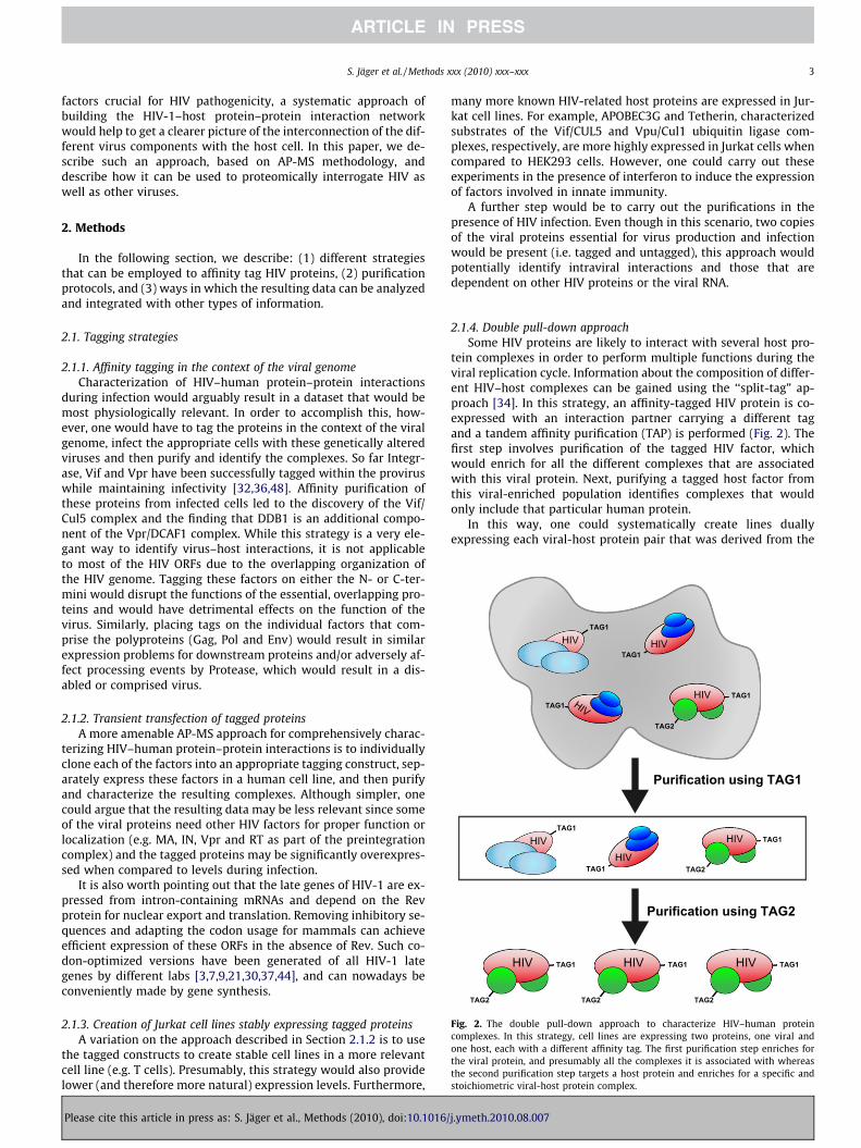
Fig. 2. The double pull-down approach to characterize HIV–human proteincomplexes. In this strategy, cell lines are expressing two proteins, one viral andone host, each with a different affinity tag. The first purification step enriches forthe viral protein, and presumably all the complexes it is associated with whereasthe second purification step targets a host protein and enriches for a specific andstoichiometric viral-host protein complex.
2. Methods
In the following section, we describe: (1) different strategiesthat can be employed to affinity tag HIV proteins, (2) purificationprotocols, and (3) ways in which the resulting data can be analyzedand integrated with other types of information.
2.1. Tagging strategies
2.1.1. Affinity tagging in the context of the viral genomeCharacterization of HIV–human protein–protein interactions
during infection would arguably result in a dataset that would bemost physiologically relevant. In order to accomplish this, how-ever, one would have to tag the proteins in the context of the viralgenome, infect the appropriate cells with these genetically alteredviruses and then purify and identify the complexes. So far Integr-ase, Vif and Vpr have been successfully tagged within the proviruswhile maintaining infectivity [32,36,48]. Affinity purification ofthese proteins from infected cells led to the discovery of the Vif/Cul5 complex and the finding that DDB1 is an additional compo-nent of the Vpr/DCAF1 complex. While this strategy is a very ele-gant way to identify virus–host interactions, it is not applicableto most of the HIV ORFs due to the overlapping organization ofthe HIV genome. Tagging these factors on either the N- or C-ter-mini would disrupt the functions of the essential, overlapping pro-teins and would have detrimental effects on the function of thevirus. Similarly, placing tags on the individual factors that com-prise the polyproteins (Gag, Pol and Env) would result in similarexpression problems for downstream proteins and/or adversely af-fect processing events by Protease, which would result in a dis-abled or comprised virus.
2.1.2. Transient transfection of tagged proteinsA more amenable AP-MS approach for comprehensively charac-
terizing HIV–human protein–protein interactions is to individuallyclone each of the factors into an appropriate tagging construct, sep-arately express these factors in a human cell line, and then purifyand characterize the resulting complexes. Although simpler, onecould argue that the resulting data may be less relevant since someof the viral proteins need other HIV factors for proper function orlocalization (e.g. MA, IN, Vpr and RT as part of the preintegrationcomplex) and the tagged proteins may be significantly overexpres-sed when compared to levels during infection.
It is also worth pointing out that the late genes of HIV-1 are ex-pressed from intron-containing mRNAs and depend on the Revprotein for nuclear export and translation. Removing inhibitory se-quences and adapting the codon usage for mammals can achieveefficient expression of these ORFs in the absence of Rev. Such co-don-optimized versions have been generated of all HIV-1 lategenes by different labs [3,7,9,21,30,37,44], and can nowadays beconveniently made by gene synthesis.
2.1.3. Creation of Jurkat cell lines stably expressing tagged proteinsA variation on the approach described in Section 2.1.2 is to use
the tagged constructs to create stable cell lines in a more relevantcell line (e.g. T cells). Presumably, this strategy would also providelower (and therefore more natural) expression levels. Furthermore,
Please cite this article in press as: S. Jäger et al., Methods (2010), doi:10.1016/
many more known HIV-related host proteins are expressed in Jur-kat cell lines. For example, APOBEC3G and Tetherin, characterizedsubstrates of the Vif/CUL5 and Vpu/Cul1 ubiquitin ligase com-plexes, respectively, are more highly expressed in Jurkat cells whencompared to HEK293 cells. However, one could carry out theseexperiments in the presence of interferon to induce the expressionof factors involved in innate immunity.
A further step would be to carry out the purifications in thepresence of HIV infection. Even though in this scenario, two copiesof the viral proteins essential for virus production and infectionwould be present (i.e. tagged and untagged), this approach wouldpotentially identify intraviral interactions and those that aredependent on other HIV proteins or the viral RNA.
2.1.4. Double pull-down approachSome HIV proteins are likely to interact with several host pro-
tein complexes in order to perform multiple functions during theviral replication cycle. Information about the composition of differ-ent HIV–host complexes can be gained using the ‘‘split-tag” ap-proach [34]. In this strategy, an affinity-tagged HIV protein is co-expressed with an interaction partner carrying a different tagand a tandem affinity purification (TAP) is performed (Fig. 2). Thefirst step involves purification of the tagged HIV factor, whichwould enrich for all the different complexes that are associatedwith this viral protein. Next, purifying a tagged host factor fromthis viral-enriched population identifies complexes that wouldonly include that particular human protein.
In this way, one could systematically create lines duallyexpressing each viral-host protein pair that was derived from the
j.ymeth.2010.08.007
4 S. Jäger et al. / Methods xxx (2010) xxx–xxx
single HIV purification experiments and subject the extract to this‘‘double pull-down” strategy. This approach would: (1) verify therelevance of individual interactions derived from single purifica-tion experiments, (2) place host factors into their respective com-plexes via the co-enrichment patterns and (3) identify morephysiological, stoichiometric HIV-human protein complexes,which would more likely be used for subsequent functional assaysor even structural studies. Of course, an additional, complementarystrategy would be to carry out single, reciprocal purifications of thetagged human proteins, alone and also in the presence of theappropriate viral protein, which would help functionally verifythe host-pathogen protein–protein interactions and expand thenetwork to include more host proteins and complexes.
2.2. Purification of affinity-tagged HIV proteins
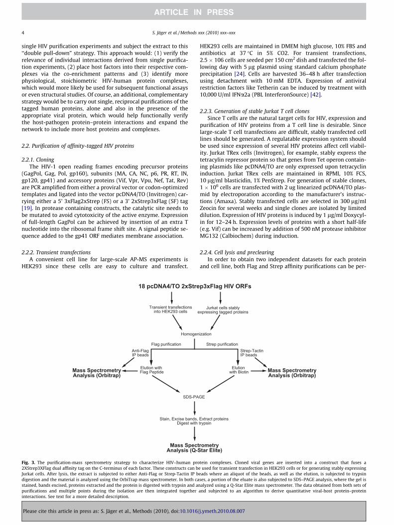
2.2.1. CloningThe HIV-1 open reading frames encoding precursor proteins
(GagPol, Gag, Pol, gp160), subunits (MA, CA, NC, p6, PR, RT, IN,gp120, gp41) and accessory proteins (Vif, Vpr, Vpu, Nef, Tat, Rev)are PCR amplified from either a proviral vector or codon-optimizedtemplates and ligated into the vector pcDNA4/TO (Invitrogen) car-rying either a 5’ 3xFlag2xStrep (FS) or a 3’ 2xStrep3xFlag (SF) tag[19]. In protease containing constructs, the catalytic site needs tobe mutated to avoid cytotoxicity of the active enzyme. Expressionof full-length GagPol can be achieved by insertion of an extra Tnucleotide into the ribosomal frame shift site. A signal peptide se-quence added to the gp41 ORF mediates membrane association.
2.2.2. Transient transfectionsA convenient cell line for large-scale AP-MS experiments is
HEK293 since these cells are easy to culture and transfect.
18 pcDNA4/TO 2xStre
Transient transfectionsinto HEK293 cells e
Homogen
SDS-PA
Stain, Excise bands, Digest with
Flag purification
Elution with Flag Peptide
Anti-FlagIP beads
Mass SpectrometryAnalysis (Orbitrap)
Mass SpectAnalysis (Q-S
Fig. 3. The purification-mass spectrometry strategy to characterize HIV–human pro2XStrep3XFlag dual affinity tag on the C-terminus of each factor. These constructs can bJurkat cells. After lysis, the extract is subjected to either Anti-Flag or Strep-Tactin IP bdigestion and the material is analyzed using the OrbiTrap mass spectrometer. In both castained, bands excised, proteins extracted and the protein is digested with trypsin and anpurifications and multiple points during the isolation are then integrated togetherinteractions. See text for a more detailed description.
Please cite this article in press as: S. Jäger et al., Methods (2010), doi:10.1016/
HEK293 cells are maintained in DMEM high glucose, 10% FBS andantibiotics at 37 �C in 5% CO2. For transient transfections,2.5 � 106 cells are seeded per 150 cm2 dish and transfected the fol-lowing day with 5 lg plasmid using standard calcium phosphateprecipitation [24]. Cells are harvested 36–48 h after transfectionusing detachment with 10 mM EDTA. Expression of antiviralrestriction factors like Tetherin can be induced by treatment with10,000 U/ml IFNa2a (PBL InterferonSource) [42].
2.2.3. Generation of stable Jurkat T cell clonesSince T cells are the natural target cells for HIV, expression and
purification of HIV proteins from a T cell line is desirable. Sincelarge-scale T cell transfections are difficult, stably transfected celllines should be generated. A regulatable expression system shouldbe used since expression of several HIV proteins affect cell viabil-ity. Jurkat TRex cells (Invitrogen), for example, stably express thetetracylin repressor protein so that genes from Tet operon contain-ing plasmids like pcDNA4/TO are only expressed upon tetracyclininduction. Jurkat TRex cells are maintained in RPMI, 10% FCS,10 lg/ml blasticidin, 1% PenStrep. For generation of stable clones,1 � 106 cells are transfected with 2 ug linearized pcDNA4/TO plas-mid by electroporation according to the manufacturer’s instruc-tions (Amaxa). Stably transfected cells are selected in 300 lg/mlZeocin for several weeks and single clones are isolated by limiteddilution. Expression of HIV proteins is induced by 1 lg/ml Doxycyl-in for 12–24 h. Expression levels of proteins with a short half-life(e.g. Vif) can be increased by addition of 500 nM protease inhibitorMG132 (Calbiochem) during induction.
2.2.4. Cell lysis and preclearingIn order to obtain two independent datasets for each protein
and cell line, both Flag and Strep affinity purifications can be per-
p3xFlag HIV ORFs
Jurkat cells stablyxpressing tagged proteins
ization
GE
Extract proteinstrypsin
Strep-TactinIP beads
Mass SpectrometryAnalysis (Orbitrap)
rometrytar Elite)
Elution with Biotin
Strep purification
tein complexes. Cloned viral genes are inserted into a construct that fuses ae used for transient transfection in HEK293 cells or for generating stably expressingeads where an aliquot of the beads, as well as the elution, is subjected to trypsinses, a portion of the eluate is also subjected to SDS–PAGE analysis, where the gel isalyzed using a Q-Star Elite mass spectrometer. The data obtained from both sets of
and subjected to an algorithm to derive quantitative viral-host protein–protein
j.ymeth.2010.08.007
S. Jäger et al. / Methods xxx (2010) xxx–xxx 5
formed (Fig. 3). Two plates of (3 � 107) HEK293 cells or 5 � 108 Jur-kat cells (500 ml culture), respectively, are washed with PBS, pel-leted and resuspended in 2 ml cold lysis buffer (0.5% NP40,50 mM Tris–HCl pH 7.4; 150 mM NaCl, 1 mM EDTA, protease andphosphatase inhibitors). The amount of detergent in the lysis buf-fer is always a compromise between protein solubilization, main-tenance of relevant interactions and elimination of unspecificbinding. Usually a concentration between 0.2% NP40 and 1%NP40 + 0.25% CHAPS is used. In addition, the cells can be mechan-ically disrupted by douncing, sonication or homogenization to en-hance protein extraction.
Insoluble material is then pelleted for 20 min at 2800g. Pre-clearing of the supernatant with unspecific beads significantly re-duces background binding. Depending on the beads used for thesubsequent AP, 60–100 ll of either mouse IgG Agarose (Sigma)or Sepharose 4FF (GE Healthcare) is added to the supernatantand incubated for 1 h on an overhead shaker at 4 �C. Mass spec-trometry (MS) analysis of these preclearing beads yields a back-ground binding dataset.
2.2.5. Affinity purificationFor purification of individual proteins, 30–50 ul IP beads (anti-
Flag M2 Affinity Gel, SIGMA or Strep-Tactin Sepharose, IBA) isadded to the precleared lysate and the immunprecipitation is per-formed in batch on an overhead shaker for at least 1 h at 4 �C.Beads are then washed extensively either in columns (Poly-Prep,BioRad) or in batch (2 ml dolphin tubes, BLD Science) with cold0.1% NP40, 50 mM Tris–HCl pH 7.4, 150 mM NaCl, 1 mM EDTA.In case of purification of proteins with RNA binding motifs (Gag,NC, Tat, Rev), unspecific association of RNA associated proteins likesplicing factors, RNA helicases, etc. can be reduced by incubation ofthe beads with 1500 U RNase A (Fermentas) for 30 min on ice, fol-lowed by washing. The last washing step is performed with deter-gent-free buffer to avoid interference with MS analysis. 10 ll of thebeads are directly analyzed by MS using on-bead trypsin digest,while the rest of the beads is eluted with 30–50 ll of either100 lg/ml 3xFLAG peptide (Elim Biopharmaceuticals) or D-desthi-obiotin solution (IBA), respectively (Fig. 3). Five microliters of theeluate is analyzed by SDS–PAGE and silver staining. The rest is ana-lyzed directly by MS as well as after fractionation on a SDS poly-acrylamide gradient gel (4–20%, BioRad) and staining withGelCode Blue Safe Protein Stain (Thermo Scientific).
2.2.6. Subcellular fractionationsSubcellular compartments may have to be enriched prior to
purification to identify functional HIV–human protein–proteininteractions. Nuclear proteins like Integrase, Rev, Tat, and Vprcan be extracted from the nuclear fraction using high salt bufferaccording to the standard protocol [1]. The remaining insolublematerial can further be treated with 1.5 U/ll benzonase nuclease(Merck) to solubilize chromatin-associated proteins like Integrase.
Membrane-associated proteins like Gag, Vpu, Nef and Env canbe affinity purified from membrane fractions enriched by flotationin a discontinuous iodixanol gradient. To this end, cells are hypo-tonically lysed and disrupted by dounce homogenization. The ly-sate is adjusted to 40% OptiPrep (SIGMA), overlaid with 28%Optiprep and TNE buffer (50 mM Tris, pH 7.4, 150 mM NaCl,5 mM EDTA) and centrifuged at 165,000g for 3 h at 4 �C. The lightmembranes and associated proteins will float to the top and appearas an opaque band at the OptiPrep-buffer interface, where it can becollected and applied to AP-MS.
2.2.7. Double pull-down experimentsFor the double pull-down experiment, 10 � 150 cm2 plates
HEK293 cells are co-transfected with vectors coding for thestrep-tagged viral protein and one or more Flag-tagged host pro-
Please cite this article in press as: S. Jäger et al., Methods (2010), doi:10.1016/
teins. The affinity purifications are performed as described above,with the first step being scaled up accordingly. The eluates afterboth steps are compared by SDS–PAGE and mass spectrometry.
2.3. Specificity and validation
An inherent problem of AP-MS experiments is the high numberof unspecific interactions that can be detected. Usually the data ob-tained from the purification of the affinity tagged protein of inter-est is compared to a negative control using untagged protein,untransfected cell lysate, tagged GFP or preimmune serum. Whilethis helps to identify a limited set of unspecific binders, proteinsoften have distinct background interactions depending on theirlocalization and nature, e.g. nuclear proteins have a different setof background interactors than membrane proteins. In general,the more unrelated proteins with similar characteristics are ana-lyzed, the easier it is to identify specific, and therefore physiologi-cally relevant interactions.
Since AP-MS studies reveal little information about whether theassociation is direct or indirect, interactions should be confirmedusing different methodologies, including in vitro binding studies.For a functional validation, the data can be compared to the pub-lished studies using genome-wide RNAi screens to identify hostfactors needed for HIV replication [4,25,46,50]. However, sincethe HIV accessory genes Vif, Vpu, Vpr, and Nef are often dispens-able for replication in cell culture systems [29], specific assaysneed to be applied in these cases.
2.3.1. Quantitative HIV–human protein–protein interaction scoringsystem
Once pull-down samples are acquired and analyzed by massspectrometry, proteomic information concerning host–pathogeninteractions can be ascertained. MS identification of a host proteinas a putative interactor allows for further investigation into thehost–pathogen interaction, employing methods such as yeasttwo-hybrid, biochemical assays, or viral infectivity assays, to verifyand establish the biological relevance of the viral-host interaction.In this regard, proper quantitative analysis of mass spectrometrydata derived from affinity purified viral or host proteins is essentialin identifying biologically meaningful PPIs in order to minimizetime and resources expended on false-positive MS identified inter-actors. However, one major caveat of reported PPIs obtained fromAP-MS experiments is often little information relating to proteinabundance or specificity is directly revealed. Also, the interactorabundance may not even be the best indicator of the interactionreliability, especially since some protein abundance strongly de-pend on interaction affinity as well as its concentration in the cellor in the final experimental sample. If the purified material from asingle affinity purification of one tagged protein bait is analyzed,even when contrasted with data from a non-tagged control, it re-mains incredibly difficult to ascertain specificity and reproducibil-ity with respect to putative interactors. Furthermore, shotgunsequencing approaches to protein identification suffer from poorsampling of IP proteins independent of precision of sample purifi-cation replicates. For example, shotgun sequencing of the samesample may only result in 30–40% overlap with respect to proteinsidentified and may require 5–10 separate runs to obtain 95% cov-erage [12,20,26].
To identify physiologically relevant PPIs, it helps to have infor-mation pertaining to protein abundance in the sample, some met-ric of bait-interactor specificity, and some metric of interactorreproducibility. As an example concerning specificity, RNA bindingproteins (e.g. Tat and Rev) will often be identified with ribosomalprotein subunits due to binding to an RNA molecule and not a di-rect PPI occurring in situ. Likewise, any highly abundant cellularproteins (i.e. cytoskeleton proteins) tend to be identified in high
j.ymeth.2010.08.007
6 S. Jäger et al. / Methods xxx (2010) xxx–xxx
abundance irrespective of the employed bait, indicating a problem-atic disconnect between protein abundance and interaction speci-ficity. On the other hand, such non-specific interactions may not bevery reproducible, which could in principle allow us to recognizethem and filter them out.
Each pull-down experiment can be represented as a vector ofabundance scores (defined below) for all of the unique interactorsfound in our approach. When the specific interactor is not pulleddown with particular bait, its abundance score is set to zero. Thevectors of different affinity purification experiments can then beorganized into a two-dimensional matrix, and the vectors of thereplicated experiments extending further into the third dimension.In order to quantify abundance of identified proteins within a sam-ple and to allow normalization of this data across different samplessuch that values between multiple datasets can be accurately com-pared, one can use the label-free SIN normalization method de-scribed previously [20]. The SIN formula takes into account thespectral intensities from each interactor, the total spectral intensi-ties observed in the MS run, and the length of the protein identi-fied, and has been shown to outperform other methods ofquantifying abundance such as spectral counts or number of inter-actors identified [20]. Assuming constant binding capacities for thesame baits in different experimental runs, we can define a new SIN
score (pSIN) for a particular interactor from a single pull-downexperiment as a proportion of the real SIN scores in that particularexperiment. The pSIN is now normalized and therefore equivalentacross all of the experiments, and is used to fill in the matrix cells.The first of the three proposed metrics, abundance, is then an aver-age of interactor pSIN scores in the third (replicates) dimension inour data matrix. The second metric, reproducibility, which is de-rived from the values in the third dimension as well, should be ameasure of how the interactor amounts (or pSIN scores) are repro-ducible among replicated experiments. The more reproduciblenumbers would be hence uniformly distributed, but less reproduc-ible numbers would form peak(s), which suggests using the entro-py as a measure of system organization. Before applying a standardentropy equation to the data, one has to normalize the scores inthis third dimension so that they add up to one. To avoid biasesdue to different number of replicates, we also normalize the entro-py, dividing it with the maximal entropy possible. The third metric,specificity, is defined similarly to abundance, except that instead ofusing the proportion of pSIN scores in the vector (or in first dimen-sion), we use the proportions of abundances in the second dimen-sion of the matrix. To avoid the inconvenience of dealing withinteractions described by three scores, one can compress the threemetrics into a single score using Principal Component Analysis(PCA), a statistical technique most commonly used to linearlytransform the original set of data into the set of uncorrelated vari-ables with the goal to reduce the dimensionality of this originaldata set.
3. Summary/outlook
Viruses, like HIV, are incredibly complicated, resourceful organ-isms that are involved in many diverse functions during infection.However, their genomes are surprisingly small considering thetasks they must carry out, and therefore they rely very heavilyon the cellular machinery in the host cells they infect. Based onthis, one might expect that one viral protein would be involvedin multiple processes and therefore would hijack several host com-plexes during infection. Using an approach like AP-MS, therefore,would be a powerful way to identify these relationships, especiallywhen it is conducted in an unbiased and systematic way. Overlay-ing the PPI network with genetic information derived from globalRNAi screens [4,25,46,50] would help identify which of these phys-
Please cite this article in press as: S. Jäger et al., Methods (2010), doi:10.1016/
ical interactions have functional consequences. Finally, comparinghost-pathogen protein–protein interaction data using a variety ofdifferent viruses will help identify commonalities with respect toinfection and should help develop new therapeutic strategies.Since vaccine and drug development targeting HIV proteins hasbeen problematic, identifying novel host targets that aid in infec-tion may represent the next step in combating HIV infection.
Acknowledgments
We thank members of the Krogan lab for helpful comments.This work was funded by NIH. NJK is a Searle Fellow and KeckYoung Investigator Fellow.
References
[1] S.M. Abmayr, T. Yao, T. Parmely, J.L. Workman, Curr. Protoc. Mol. Biol. (2006).Chapter 12: Unit 12.1.
[2] M. Arifuzzaman, M. Maeda, A. Itoh, K. Nishikata, C. Takita, R. Saito, T. Ara, K.Nakahigashi, H.C. Huang, A. Hirai, et al., Genome Res. 16 (2006) 686–691.
[3] D.L. Bolton, M.J. Lenardo, J. Virol. 81 (2007) 8878–8890.[4] A.L. Brass, D.M. Dykxhoorn, Y. Benita, N. Yan, A. Engelman, R.J. Xavier, J.
Lieberman, S.J. Elledge, Science 319 (2008) 921–926.[5] G. Butland, J.M. Peregrin-Alvarez, J. Li, W. Yang, X. Yang, V. Canadien, A.
Starostine, D. Richards, B. Beattie, N. Krogan, et al., Nature 433 (2005) 531–537.[6] M.A. Calderwood, K. Venkatesan, L. Xing, M.R. Chase, A. Vazquez, A.M.
Holthaus, A.E. Ewence, N. Li, T. Hirozane-Kishikawa, D.E. Hill, et al., Proc.Natl. Acad. Sci. USA 104 (2007) 7606–7611.
[7] B.K. Chakrabarti, W.P. Kong, B.Y. Wu, Z.Y. Yang, J. Friborg, X. Ling, S.R. King, D.C.Montefiori, G.J. Nabel, J. Virol. 76 (2002) 5357–5368.
[8] A. Chatr-aryamontri, A. Ceol, D. Peluso, A. Nardozza, S. Panni, F. Sacco, M. Tinti,A. Smolyar, L. Castagnoli, M. Vidal, et al., Nucleic Acids Res. 37 (2009) D669–673.
[9] P. Cherepanov, W. Pluymers, A. Claeys, P. Proost, E. De Clercq, Z. Debyser,FASEB J. 14 (2000) 1389–1399.
[10] F. Colland, X. Jacq, V. Trouplin, C. Mougin, C. Groizeleau, A. Hamburger, A. Meil,J. Wojcik, P. Legrain, J.M. Gauthier, Genome Res. 14 (2004) 1324–1332.
[11] B. de Chassey, V. Navratil, L. Tafforeau, M.S. Hiet, A. Aublin-Gex, S. Agaugue, G.Meiffren, F. Pradezynski, B.F. Faria, T. Chantier, et al., Mol. Syst. Biol. 4 (2008)230.
[12] E. Durr, J. Yu, K.M. Krasinska, L.A. Carver, J.R. Yates, J.E. Testa, P. Oh, J.E.Schnitzer, Nat. Biotechnol. 22 (2004) 985–992.
[13] M.D. Dyer, T.M. Murali, B.W. Sobral, PLoS Pathog. 4 (2008) e32.[14] R.M. Ewing, P. Chu, F. Elisma, H. Li, P. Taylor, S. Climie, L. McBroom-Cerajewski,
M.D. Robinson, L. O’Connor, M. Li, et al., Mol. Syst. Biol. 3 (2007) 89–90.[15] M. Flajolet, G. Rotondo, L. Daviet, F. Bergametti, G. Inchauspe, P. Tiollais, C.
Transy, P. Legrain, Gene 242 (2000) 369–379.[16] M. Fromont-Racine, J.C. Rain, P. Legrain, Nat. Genet. 16 (1997) 277–282.[17] A.C. Gavin, P. Aloy, P. Grandi, R. Krause, M. Boesche, M. Marzioch, C. Rau, L.J.
Jensen, S. Bastuck, B. Dumpelfeld, et al., Nature 440 (2006) 631–636.[18] L. Giot, J.S. Bader, C. Brouwer, A. Chaudhuri, B. Kuang, Y. Li, Y.L. Hao, C.E. Ooi, B.
Godwin, E. Vitols, et al., Science 302 (2003) 1727–1736.[19] C.J. Gloeckner, K. Boldt, A. Schumacher, R. Roepman, M. Ueffing, Proteomics 7
(2007) 4228–4234.[20] N.M. Griffin, J. Yu, F. Long, P. Oh, S. Shore, Y. Li, J.A. Koziol, J.E. Schnitzer, Nat.
Biotechnol. 28 (2010) 83–89.[21] L. Hermida-Matsumoto, M.D. Resh, J. Virol. 74 (2000) 8670–8679.[22] Y. Ho, A. Gruhler, A. Heilbut, G.D. Bader, L. Moore, S.L. Adams, A. Millar, P.
Taylor, K. Bennett, K. Boutilier, et al., Nature 415 (2002) 180–183.[23] T. Ito, T. Chiba, R. Ozawa, M. Yoshida, M. Hattori, Y. Sakaki, Proc. Natl. Acad. Sci.
USA 98 (2001) 4569–4574.[24] R.E. Kingston, C.A. Chen, H. Okayama, Curr. Protoc. Cell Biol. (2003). Chapter
20: Unit 20.3.[25] R. Konig, Y. Zhou, D. Elleder, T.L. Diamond, G.M. Bonamy, J.T. Irelan, C.Y. Chiang,
B.P. Tu, P.D. De Jesus, C.E. Lilley, et al., Cell 135 (2008) 49–60.[26] J.A. Koziol, A.C. Feng, J.E. Schnitzer, Anal. Chem. 78 (2006) 3203–3207.[27] N.J. Krogan, G. Cagney, H. Yu, G. Zhong, X. Guo, A. Ignatchenko, J. Li, S. Pu, N.
Datta, A.P. Tikuisis, et al., Nature 440 (2006) 637–643.[28] S. Li, C.M. Armstrong, N. Bertin, H. Ge, S. Milstein, M. Boxem, P.O. Vidalain, J.D.
Han, A. Chesneau, T. Hao, et al., Science 303 (2004) 540–543.[29] M.H. Malim, M. Emerman, Cell Host Microbe 3 (2008) 388–398.[30] K.L. Nguyen, M. llano, H. Akari, E. Miyagi, E.M. Poeschla, K. Strebel, S. Bour,
Virology 319 (2004) 163–175.[31] X. Peng, E.Y. Chan, Y. Li, D.L. Diamond, M.J. Korth, M.G. Katze, Curr. Opin.
Microbiol. 12 (2009) 432–438.[32] C. Petit, O. Schwartz, F. Mammano, J. Virol. 73 (1999) 5079–5088.[33] R.G. Ptak, W. Fu, B.E. Sanders-Beer, J.E. Dickerson, J.W. Pinney, D.L. Robertson,
M.N. Rozanov, K.S. Katz, D.R. Maglott, K.D. Pruitt, et al., AIDS Res. Hum.Retroviruses 24 (2008) 1497–1502.
[34] O. Puig, F. Caspary, G. Rigaut, B. Rutz, E. Bouveret, E. Bragado-Nilsson, M. Wilm,B. Seraphin, Methods 24 (2001) 218–229.
j.ymeth.2010.08.007
S. Jäger et al. / Methods xxx (2010) xxx–xxx 7
[35] J.F. Rual, K. Venkatesan, T. Hao, T. Hirozane-Kishikawa, A. Dricot, N. Li, G.F.Berriz, F.D. Gibbons, M. Dreze, N. Ayivi-Guedehoussou, et al., Nature 437(2005) 1173–1178.
[36] B. Schrofelbauer, Y. Hakata, N.R. Landau, Proc. Natl. Acad. Sci. USA 104 (2007)4130–4135.
[37] S. Schwartz, M. Campbell, G. Nasioulas, J. Harrison, B.K. Felber, G.N. Pavlakis, J.Virol. 66 (1992) 7176–7182.
[38] S.D. Shapira, I. Gat-Viks, B.O. Shum, A. Dricot, M.M. de Grace, L. Wu, P.B. Gupta,T. Hao, S.J. Silver, D.E. Root, et al., Cell 139 (2009) 1255–1267.
[39] U. Stelzl, U. Worm, M. Lalowski, C. Haenig, F.H. Brembeck, H. Goehler, M.Stroedicke, M. Zenkner, A. Schoenherr, S. Koeppen, et al., Cell 122 (2005) 957–968.
[40] P. Uetz, Y.A. Dong, C. Zeretzke, C. Atzler, A. Baiker, B. Berger, S.V. Rajagopala, M.Roupelieva, D. Rose, E. Fossum, et al., Science 311 (2006) 239–242.
[41] P. Uetz, L. Giot, G. Cagney, T.A. Mansfield, R.S. Judson, J.R. Knight, D. Lockshon,V. Narayan, M. Srinivasan, P. Pochart, et al., Nature 403 (2000) 623–627.
[42] N. Van Damme, D. Goff, C. Katsura, R.L. Jorgenson, R. Mitchell, M.C. Johnson,E.B. Stephens, J. Guatelli, Cell Host Microbe 3 (2008) 245–252.
Please cite this article in press as: S. Jäger et al., Methods (2010), doi:10.1016/
[43] A. von Brunn, C. Teepe, J.C. Simpson, R. Pepperkok, C.C. Friedel, R. Zimmer, R.Roberts, R. Baric, J. Haas, PLoS One 2 (2007) e459.
[44] R. Wagner, M. Graf, K. Bieler, H. Wolf, T. Grunwald, P. Foley, K. Uberla, Hum.Gene Ther. 11 (2000) 2403–2413.
[45] A.J. Walhout, R. Sordella, X. Lu, J.L. Hartley, G.F. Temple, M.A. Brasch, N.Thierry-Mieg, M. Vidal, Science 287 (2000) 116–122.
[46] M.L. Yeung, L. Houzet, V.S. Yedavalli, K.T. Jeang, J. Biol. Chem. 284 (2009)19463–19473.
[47] H. Yu, P. Braun, M.A. Yildirim, I. Lemmens, K. Venkatesan, J. Sahalie, T.Hirozane-Kishikawa, F. Gebreab, N. Li, N. Simonis, et al., Science 322 (2008)104–110.
[48] X. Yu, Y. Yu, B. Liu, K. Luo, W. Kong, P. Mao, X.F. Yu, Science 302 (2003) 1056–1060.
[49] L. Zhang, N.Y. Villa, M.M. Rahman, S. Smallwood, D. Shattuck, C. Neff, M.Dufford, J.S. Lanchbury, J. Labaer, G. McFadden, J. Proteome Res. 8 (2009)4311–4318.
[50] H. Zhou, M. Xu, Q. Huang, A.T. Gates, X.D. Zhang, J.C. Castle, E. Stec, M. Ferrer, B.Strulovici, D.J. Hazuda, et al., Cell Host Microbe 4 (2008) 495–504.
j.ymeth.2010.08.007
Copyright © 2022 FDOKUMEN

