


Bahasa
Halaman
Hukum

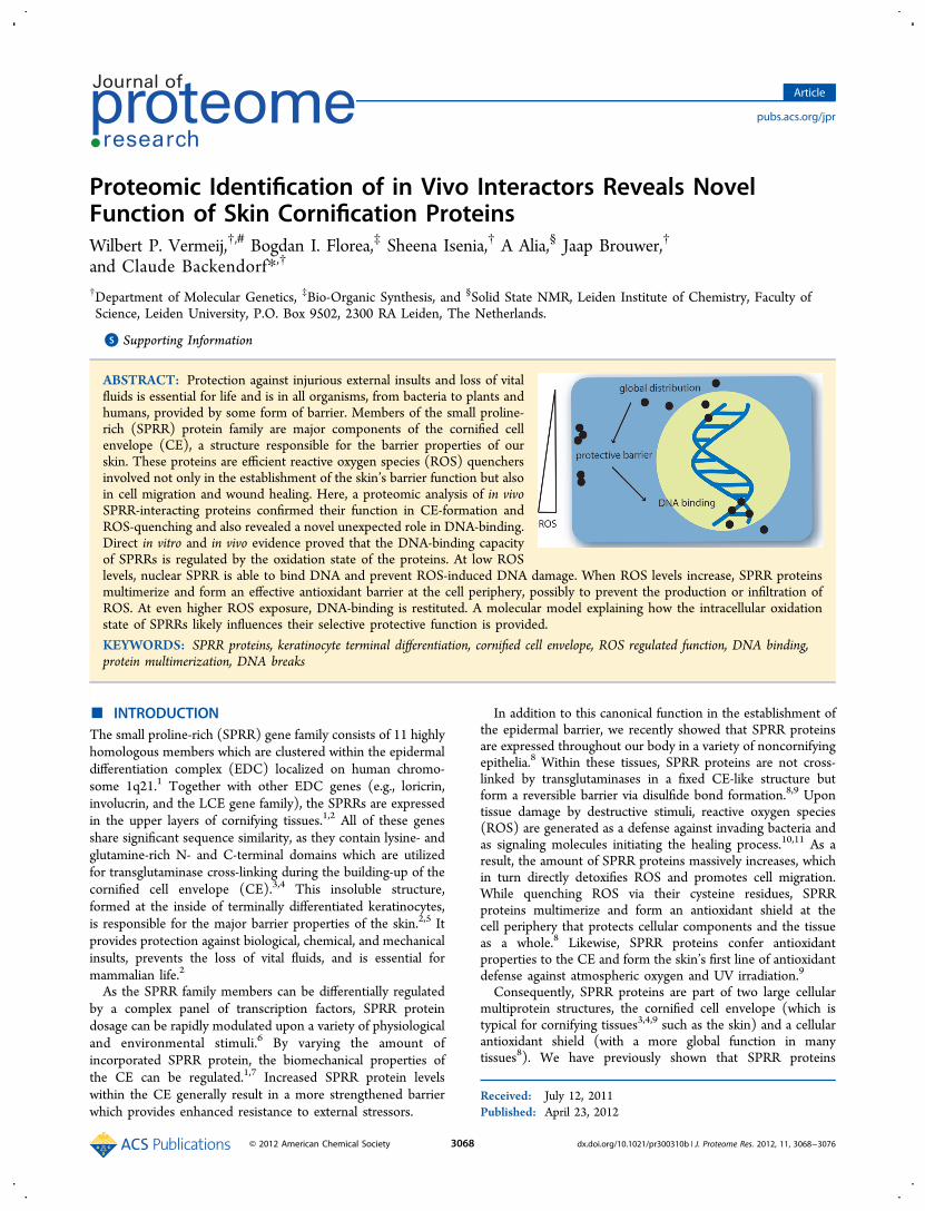
Proteomic Identification of in Vivo Interactors Reveals NovelFunction of Skin Cornification ProteinsWilbert P. Vermeij,†,# Bogdan I. Florea,‡ Sheena Isenia,† A Alia,§ Jaap Brouwer,†
and Claude Backendorf*,†
†Department of Molecular Genetics, ‡Bio-Organic Synthesis, and §Solid State NMR, Leiden Institute of Chemistry, Faculty ofScience, Leiden University, P.O. Box 9502, 2300 RA Leiden, The Netherlands.
*S Supporting Information
ABSTRACT: Protection against injurious external insults and loss of vitalfluids is essential for life and is in all organisms, from bacteria to plants andhumans, provided by some form of barrier. Members of the small proline-rich (SPRR) protein family are major components of the cornified cellenvelope (CE), a structure responsible for the barrier properties of ourskin. These proteins are efficient reactive oxygen species (ROS) quenchersinvolved not only in the establishment of the skin’s barrier function but alsoin cell migration and wound healing. Here, a proteomic analysis of in vivoSPRR-interacting proteins confirmed their function in CE-formation andROS-quenching and also revealed a novel unexpected role in DNA-binding.Direct in vitro and in vivo evidence proved that the DNA-binding capacityof SPRRs is regulated by the oxidation state of the proteins. At low ROSlevels, nuclear SPRR is able to bind DNA and prevent ROS-induced DNA damage. When ROS levels increase, SPRR proteinsmultimerize and form an effective antioxidant barrier at the cell periphery, possibly to prevent the production or infiltration ofROS. At even higher ROS exposure, DNA-binding is restituted. A molecular model explaining how the intracellular oxidationstate of SPRRs likely influences their selective protective function is provided.
KEYWORDS: SPRR proteins, keratinocyte terminal differentiation, cornified cell envelope, ROS regulated function, DNA binding,protein multimerization, DNA breaks
■ INTRODUCTIONThe small proline-rich (SPRR) gene family consists of 11 highlyhomologous members which are clustered within the epidermaldifferentiation complex (EDC) localized on human chromo-some 1q21.1 Together with other EDC genes (e.g., loricrin,involucrin, and the LCE gene family), the SPRRs are expressedin the upper layers of cornifying tissues.1,2 All of these genesshare significant sequence similarity, as they contain lysine- andglutamine-rich N- and C-terminal domains which are utilizedfor transglutaminase cross-linking during the building-up of thecornified cell envelope (CE).3,4 This insoluble structure,formed at the inside of terminally differentiated keratinocytes,is responsible for the major barrier properties of the skin.2,5 Itprovides protection against biological, chemical, and mechanicalinsults, prevents the loss of vital fluids, and is essential formammalian life.2
As the SPRR family members can be differentially regulatedby a complex panel of transcription factors, SPRR proteindosage can be rapidly modulated upon a variety of physiologicaland environmental stimuli.6 By varying the amount ofincorporated SPRR protein, the biomechanical properties ofthe CE can be regulated.1,7 Increased SPRR protein levelswithin the CE generally result in a more strengthened barrierwhich provides enhanced resistance to external stressors.
In addition to this canonical function in the establishment ofthe epidermal barrier, we recently showed that SPRR proteinsare expressed throughout our body in a variety of noncornifyingepithelia.8 Within these tissues, SPRR proteins are not cross-linked by transglutaminases in a fixed CE-like structure butform a reversible barrier via disulfide bond formation.8,9 Upontissue damage by destructive stimuli, reactive oxygen species(ROS) are generated as a defense against invading bacteria andas signaling molecules initiating the healing process.10,11 As aresult, the amount of SPRR proteins massively increases, whichin turn directly detoxifies ROS and promotes cell migration.While quenching ROS via their cysteine residues, SPRRproteins multimerize and form an antioxidant shield at thecell periphery that protects cellular components and the tissueas a whole.8 Likewise, SPRR proteins confer antioxidantproperties to the CE and form the skin’s first line of antioxidantdefense against atmospheric oxygen and UV irradiation.9
Consequently, SPRR proteins are part of two large cellularmultiprotein structures, the cornified cell envelope (which istypical for cornifying tissues3,4,9 such as the skin) and a cellularantioxidant shield (with a more global function in manytissues8). We have previously shown that SPRR proteins
Received: July 12, 2011Published: April 23, 2012
Article
pubs.acs.org/jpr
© 2012 American Chemical Society 3068 dx.doi.org/10.1021/pr300310b | J. Proteome Res. 2012, 11, 3068−3076
provide protection against ROS induced DNA damage.8,9 Herewe show that SPRR proteins are part of yet another (third)cellular structure which is in direct contact with DNA.
■ MATERIALS AND METHODSProtein Interaction Screen
For the isolation and identification of protein interactioncomplexes, SPRR1B and SPRR4 were provided with a Strep-tagand used as bait proteins. Full-length SPRR cDNA sequenceswere cloned in the pEXPR-IBA105-vector as described by themanufacturer (IBA, Gottingen, Germany). Immortal OKFkeratinocytes (OKF6/TERT-2) were kindly provided byDr. J. G. Rheinwald (Harvard Medical School, Boston) andcultured in defined Keratinocyte-SFM (KSFM; GIBCO) asdescribed by Dickson et al.12 Transfections with the above-mentionedStrep-SPRR1B, Strep-SPRR4, or an empty vector control,solely expressing the Strep-tag, were performed by using Amaxanucleofection according to the manufacturer (Lonza AG,Cologne, Germany). After 48 h both proliferating andmigrating cells were present and also the first signs ofdifferentiating cells were observed. Approximately 109 cellswere lysed in cold 50 mM Tris-HCl (pH 7.5), 5 mM EDTA,250 mM NaCl, 0.1% Triton X-100, 7 mM CaCl2 supplementedwith 5 mM NaF, 100 mM Na3VO4, 20 mM β-glycerolphos-phate, and 1 protease inhibitor cocktail tablet (Roche, Basel,Switzerland) per 10 mL of buffer, all freshly added before use.The soluble cell extracts, containing either Strep-SPRR1B,Strep-SPRR4 proteins, or only the Strep-tag (control), wereloaded on a 0.2 mL Strep-Tactin column (IBA, Gottingen,Germany), and the unbound proteins were washed away.Subsequently, a first elution step was performed with 0.6 mL ofwash-buffer (100 mM Tris-HCl (pH 8.0), 150 mM NaCl,1 mM EDTA) supplemented with 50 mM DTT. In this way,proteins interacting via or stabilized by disulfide bonds wereeluted. All other interaction partners or complexes (eithercovalently linked or not) were eluted with the Strep-taggedSPRR bait proteins by using biotin elution buffer: 100 mMTris-HCl (pH 8.0), 150 mM NaCl, 1 mM EDTA, 2 mM biotinas described by the manufacturer (IBA, Gottingen, Germany).
Mass Spectrometry Analysis and Database Searching
Proteins from the individual elution fractions were precipitatedby addition of equal volumes of 20% trichloroacetic acid.13
After three wash-steps with 0.2 mL of ice cold acetone, theproteins were dissolved in 25 μL of 8 M urea, 0.4 Mammonium bicarbonate. The cysteine residues were reducedwith 5 μL of 45 mM DTT for 15 min at 50 °C and alkylatedwith 5 μL of 100 mM iodoacetamide at room temperature inthe dark. Trypsin digestion was performed at 37 °Covernight,13 and the resultant peptides were purified usingStageTips.14 The LTQ-Orbitrap (Thermo Fisher Scientific,Waltham, MA) tandem mass spectrometry analysis wasperformed as previously described.15 Database searching withMascot (Matrix Science, Boston, MA) against all human entriesin Swiss-Prot was performed with the following parameters:peptide tolerance 2 ppm, MS/MS tolerance 0.5 Da, fixedcarbamidomethyl modification (C), variable oxidation (M), twomissed cleavages allowed, decoy database option on. TheMudPIT scoring algorithm was used with an ion score cutoff of20, with required bold red only. Two unique peptideassignments with a significance threshold value p < 0.05 wererequired per protein identification. The data sets were importedin Pipeline Pilot (Accelrys Inc., San Diego, CA) according to
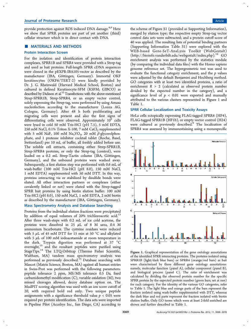
the scheme of Figure S1 (provided as Supporting Information),merged by elution type; the respective empty Strep-tag vectorcontrol data sets were subtracted; and a protein cutoff score of40 was applied. The resulting lists of potential binding partners(Supporting Information Table S1) were explored with theWEB-based Gene-SeT-AnaLysis Toolkit (WebGestalt)(http://bioinfo.vanderbilt.edu/webgestalt/index.php).16 GOenrichment analysis was performed by the statistics module(by comparing the individual data files) with the Homo sapiensgenome reference set. The hypergeometric test was used toevaluate the functional category enrichment, and the p valueswere adjusted by the default Benjamini and Hochberg method.GO categories with at least two identified proteins, a ratio ofenrichment R > 2 (calculated as observed protein numberdivided by the expected number in the category), and asignificance level of p < 0.01 were exported and manuallyattributed to the various clusters represented in Figure 1 andTable 1.
SPRR Cellular Localization and Toxicity Assays
HeLa cells ectopically expressing FLAG-tagged SPRR4 (HF4),FLAG-tagged SPRR1B (HF1b), or empty vector control (H24)were cultured as previously described.9 The localization ofSPRR4 was assessed by immunostaining using a monospecific
Figure 1. Graphical representation of the gene ontology annotationsof the identified SPRR interacting proteins. The proteins isolated usingSPRR1B (light/dark blue bars) or SPRR4 (orange/red bars) as baitwere characterized by three different gene ontology annotations:namely, molecular function (panel A), cellular component (panel B),and biological process (panel C). The ratio of enrichment wascalculated by dividing the observed protein number for the specificSPRR protein by the expected protein number (green bars; set at one)for each category. For the identity of the various GO categories, referto Table 1. The light blue and orange parts of the bars represent thefraction isolated using wash-buffer supplemented with DTT, whereasthe dark blue and red parts represent the fraction isolated with biotinelution buffer. Only GO terms which were at least 2-fold enriched areshown and further described in Table 1.
Journal of Proteome Research Article
dx.doi.org/10.1021/pr300310b | J. Proteome Res. 2012, 11, 3068−30763069
rabbit-antibody, obtained after immunization of rabbits with thepeptides DPCAPQVKKQCPPKG and CPSAQQASKSKQK(Eurogentec). The monospecificity of these antibodies wasassessed as previously described.17 Hoechst 33342 (Sigma, St.Louis, MO) was used as DNA stain. HF4-cells were pretreatedwith 50 μM H2O2 for the indicated time points and fixed withcold 80% acetone.Comet assays were performed as previously described8,18 and
quantified using ColorProc, an in-house software programkindly provided by Dr. H. Vrolijk (Department of MolecularCell Biology, Leiden University Medical Center, Leiden). TheH2O2 concentrations used ranged from 0 to 200 μM with atleast 1000 comets measured per cell line. From the linearincrease in DNA breaks, the calculated slopes were used as avalue for the Intracellular Quenching Activity (IQA) of thespecific cell lines. For intracellular protein quantification, thevarious ectopically expressed proteins were detected on
Western blot with a monoclonal anti-FLAG antibody (cloneM5, Sigma).
SPRR Protein Multimerization and DNA Binding Assays
SPRR proteins were produced and purified as previouslydescribed.8 For all DNA binding experiments, 0.12 pmol of alinear DNA fragment (3200 bp) was used with a 100 timesmolar excess of SPRR4 protein (12 pmol), unless notedotherwise. Prior to DNA binding, equal amounts of proteinstock solutions were oxidized using a serial dilution of H2O2
ranging from 0 to 100 mM. Excess H2O2 was removed by gelfiltration on Sephadex G10 spin columns or diluted out inbinding buffer. All binding reactions were performed on ice in10 mM sodium phosphate buffer (pH7) and analyzed on 1%agarose gels (for DNA detection) or 15% PAGE (for proteindetection), using loading buffer without β-mercaptoethanol.Binding of SPRR4 to DNA was stable until a salt concentration
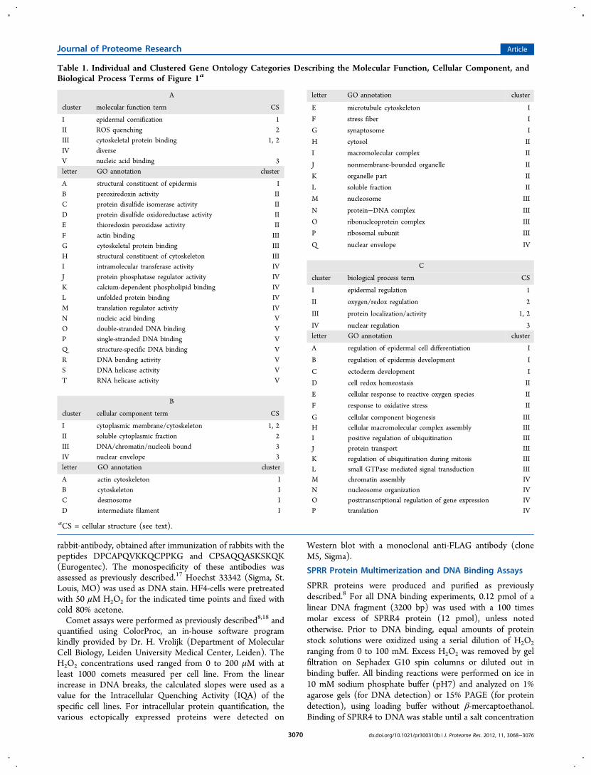
Table 1. Individual and Clustered Gene Ontology Categories Describing the Molecular Function, Cellular Component, andBiological Process Terms of Figure 1a
A
cluster molecular function term CS
I epidermal cornification 1II ROS quenching 2III cytoskeletal protein binding 1, 2IV diverseV nucleic acid binding 3letter GO annotation cluster
A structural constituent of epidermis IB peroxiredoxin activity IIC protein disulfide isomerase activity IID protein disulfide oxidoreductase activity IIE thioredoxin peroxidase activity IIF actin binding IIIG cytoskeletal protein binding IIIH structural constituent of cytoskeleton IIII intramolecular transferase activity IVJ protein phosphatase regulator activity IVK calcium-dependent phospholipid binding IVL unfolded protein binding IVM translation regulator activity IVN nucleic acid binding VO double-stranded DNA binding VP single-stranded DNA binding VQ structure-specific DNA binding VR DNA bending activity VS DNA helicase activity VT RNA helicase activity V
B
cluster cellular component term CS
I cytoplasmic membrane/cytoskeleton 1, 2II soluble cytoplasmic fraction 2III DNA/chromatin/nucleoli bound 3IV nuclear envelope 3letter GO annotation cluster
A actin cytoskeleton IB cytoskeleton IC desmosome ID intermediate filament I
letter GO annotation cluster
E microtubule cytoskeleton I
F stress fiber I
G synaptosome I
H cytosol II
I macromolecular complex II
J nonmembrane-bounded organelle II
K organelle part II
L soluble fraction II
M nucleosome III
N protein−DNA complex III
O ribonucleoprotein complex III
P ribosomal subunit III
Q nuclear envelope IV
C
cluster biological process term CS
I epidermal regulation 1
II oxygen/redox regulation 2
III protein localization/activity 1, 2
IV nuclear regulation 3letter GO annotation cluster
A regulation of epidermal cell differentiation I
B regulation of epidermis development I
C ectoderm development I
D cell redox homeostasis II
E cellular response to reactive oxygen species II
F response to oxidative stress II
G cellular component biogenesis IIIH cellular macromolecular complex assembly IIII positive regulation of ubiquitination IIIJ protein transport IIIK regulation of ubiquitination during mitosis IIIL small GTPase mediated signal transduction IIIM chromatin assembly IVN nucleosome organization IVO posttranscriptional regulation of gene expression IVP translation IV
aCS = cellular structure (see text).
Journal of Proteome Research Article
dx.doi.org/10.1021/pr300310b | J. Proteome Res. 2012, 11, 3068−30763070
of 100 mM NaCl (results not shown). For the analysis of theredox state of the cysteine residues within these diverselyoxidized SPRR samples, the different protein bands wereexcised from the gel and cut into small pieces. The free thiolgroups were first labeled with N-ethylmaleimide as previouslydescribed.9 Subsequently, the cysteines engaged in disulfidebonds were reduced by addition of DTT and labeled withiodoacetamide. In this way, free thiols can be recognized byN-ethylmaleimide labeling and cysteines originally engaged indisulfide bonds by iodoacetamide. Thiols, already oxidized tosulfenic, sulfinic, or sulfonic acid, are not affected by thesetreatments. The labeled SPRR4 peptides were extracted afterin-gel tryptic digestion19 and analyzed by OT-Orbitrap tandemmass spectrometry.9,15 Cysteine modifications of SPRR4peptides were manually identified in Xcalibur (Thermo FisherScientific, Waltham, MA) by using the SPRR4 protein sequence(NCBI accession no. AF335109) and the calculated peptidemasses. All identified peptides were validated by de novosequencing using PEAKS (Bioinformatics Solutions Inc.,Waterloo, ON, Canada).
■ RESULTS
Identification of SPRR Interacting Proteins
SPRR proteins are part of two ultralarge cellular structures, thecornified cell envelope2 (indicated here as cellular structure 1,CS1) and a still poorly defined cellular antioxidant shield8,9
(cellular structure 2, CS2) (see Introduction), which in the caseof the cornified cell envelope covers the whole inner side of thecytoplasmic membrane. These structures are held togetherrespectively via Nε-(γ-glutamyl)lysine bonds2 (CS1) or viacysteine disulfide bonds9 (CS2). Such large intracellularstructures are not amenable to classical protein−proteininteraction studies (e.g., coimmunoprecipitation or native gelelectrophoresis). Hence, we have designed a proteomicapproach to further analyze and characterize these structures.20
Strep-tagged SPRR proteins, used as bait, were expressed inhuman OKF keratinocytes, an immortalized cell line normallyexpressing these proteins upon epidermal differentiation or cellmigration.8 Soluble cell extracts, containing the Strep-taggedSPRR proteins, were loaded on a Strep-Tactin column, andafter removal of all noninteracting proteins, SPRR interactorswere isolated using a dual elution procedure to distinguishbetween different modes of interaction. In step 1 wash buffersupplemented with DTT was used to identify proteins whoseinteraction is mediated or stabilized via disulfide bonds (CS2).In step 2 biotin elution buffer was used to elute the remaininginteracting proteins (CS1 or possibly other cellular complexes).Eluted samples were analyzed by LTQ-Orbitrap tandem massspectrometry and the potential binding partners were identifiedusing the Mascot search engine.15,21 Peptide hits from theempty vector control were subtracted (Supporting InformationFigure S1), and the identified interactors (SupportingInformation Table S1) were characterized by the WebGestaltprogram.16
The gene ontology (GO) results, represented in Figure 1 andTable 1, were classified according to three different terms,namely molecular function (panel A), cellular component(panel B), and biological process (panel C). Interestingly, ineach of these GO terms, the identified proteins can besubdivided into three categories relating to three biologicalprocesses, namely “epidermal regulation”, “oxygen/redoxregulation”, and “nuclear regulation”. Whereas the first two
functions correspond respectively to the above-mentionedcellular structures CS1 and CS2, the third category ofinteractors was unexpected and appears to involve a thirdcellular structure in contact with DNA (CS3 in Table 1).Whereas all subcategories were enriched by more than 2-foldfor at least one of the SPRRs (Figure 1), the large majority ofGO categories were enriched for both SPRR1B (light/dark bluebars) and SPRR4 (orange/red bars), as compared to theexpected number of genes calculated by the program (greenbars; set at one). However, there appears to be a clearselectivity between SPRR1B and SPRR4 interactors. Thisdifference is especially visible within the biological processterms (Figure 1, panel C). Whereas SPRR1B interactors (light/dark blue bars) were more enriched within the epidermal andoxygen/redox biological process terms (clusters I and II),SPRR4 (orange/red bars) appeared to be more involved innuclear processes (cluster IV). This increased presence ofinteractors with a nuclear function was found in the fractionseluted with both DTT (orange bars) and biotin (red bars),suggesting that the presumed nuclear function of SPRR4 mightbe subjected to redox regulation. Furthermore, the diversemolecular functions of the interactors in cluster V (Table 1,panel A) suggest that the interaction between SPRRs and theidentified proteins within CS3 occurs indirectly via mutualDNA binding (see below). Besides, this nuclear link is visible inall three GO terms; two of these categories also uncover a linkwith the cellular cytoskeleton (GO molecular function clusterIII and GO cellular component cluster I; see Table 1).
Cellular Localization of SPRR Is Subjected to ROSTreatment
To ascertain whether SPRR proteins are actually involved in thecytoskeletal and nuclear GO categories identified above, theircellular localization was analyzed. Immunofluorescence stainingwith a SPRR4 monospecific antibody revealed mainlycytoplasmic localization in HeLa cells ectopically expressingFLAG-tagged SPRR4 (HF4) (Figure 2). Within the cytoplasm,ordered fiber-like structures can be observed (Figure 2A),indicating that SPRR proteins are at least in close proximity tosome cytoskeletal proteins. This supports the GO analysiswhere such proteins were detected both in the “molecularfunction” cluster III (F−H) and in the “cellular component”cluster I (A−G) (Table 1). In addition, minor but consistentnuclear SPRR4 staining was observed (Figure 2A−C).Immunofluorescence staining against the N-terminal FLAG-tag showed similar cytoplasmic and nuclear localization (datanot shown). Interestingly, the nuclear localization of SPRR4changed upon H2O2 treatment of the cells (Figure 2A−E).After a ROS challenge, SPRR proteins shifted toward thecytoplasm, where they preferentially localized to the cyto-plasmic membrane (GO cellular component clusters I and II).Mock treatment followed by a similar incubation period did notresult in this altered localization (data not shown).
SPRR4 Protects Cells from ROS Induced DNA Breaks
We have previously shown that SPRR1B protects chromatin inhuman cells against H2O2 induced DNA breaks.8 Hence, weinvestigated the effect of SPRR4 on ROS induced DNAbreakage and compared it to SPRR1B. As a control, theincrease in DNA breaks after addition of various concentrationsof H2O2 to empty vector control cells (named H24) wasanalyzed using a comet assay. From the linear increase in DNAbreaks after addition of H2O2, the calculated slope was used as avalue for the intracellular quenching activity (IQA) of a specific
Journal of Proteome Research Article
dx.doi.org/10.1021/pr300310b | J. Proteome Res. 2012, 11, 3068−30763071

cell line (Table 2). Ectopic expression of SPRR1B (HF1b)resulted in a decrease in the amount of DNA breaks andconsequently in a lower slope and thus a higher IQA. Althoughthe expression level of SPRR4 (HF4) was clearly lower ascompared to that of SPRR1B, it gave a similar reduction in theamount of DNA breaks, indicating its superior relativeintracellular quenching activity (RIQA) (almost 5-fold increase;Table 2, last column).SPRR Proteins Bind Directly to DNA
As the GO analysis (Figure 1) suggested that SPRR proteinsmight be part of a cellular structure which is in close contactwith DNA and as SPRRs can efficiently protect humanchromatin from ROS induced DNA breaks (see above), wehave evaluated a possible direct binding of SPRR proteins toDNA. For this purpose, electrophoretic mobility shift assayswere performed with purified SPRR proteins and isolated DNAfragments. Addition of increasing amounts of purified SPRR4protein to a linear DNA fragment resulted in a lower mobilityof the DNA molecules in agarose gel (Figure 3A). Similarelectrophoretic mobility shifts were observed when usingpurified SPRR1, SPRR2, or SPRR3 proteins, although a 10times higher molar excess of protein was required (data notshown). These data imply that SPRR proteins have the abilityto directly bind to double-stranded DNA. This ability wasfurther substantiated by using atomic force microscopy (AFM).Circular DNA was visualized in the absence (Figure 3B) andpresence (Figure 3C) of SPRR4. The analysis indicates thatSPRR4 has the ability to randomly coat the DNA double helix(coated and uncoated DNA regions are indicated respectivelyby white and black arrows). Both experiments constitute thefirst direct evidence that SPRR proteins have the ability to binddirectly to DNA. They further corroborate the protein
interatomic analysis (GO data) and the cellular localizationstudies described above.Oxidation State of SPRR Proteins Influences Their DNABinding Capacity
To investigate the effect of ROS on the DNA binding properties ofSPRR proteins, equal amounts of purified SPRR4 were pretreatedwith various concentrations of H2O2. At initial increasingconcentrations of H2O2, a clear gradual decrease in DNA bindingwas observed (Figure 3D, lanes 1−5). Analysis of the corre-sponding fractions via PAGE revealed an increase in the formationof SPRR multimers (Figure 3E, lanes 1−5), illustrated by adecrease of SPRR monomers and an increase of dimers, trimers,and tetramers (Figure 3F, bars 1−5). However, when higherH2O2 concentrations were used (lanes 6−7), a reversion of the
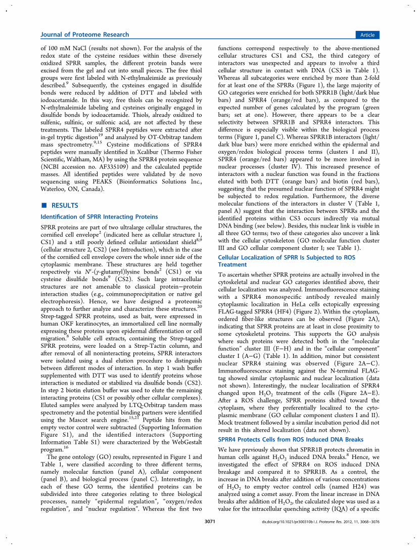
Figure 2. The cellular distribution of SPRR4 is affected by ROS. Immunofluorescence detection of SPRR4 expression (A−E) in HeLa cellsectopically expressing FLAG-tagged SPRR4 (HF4). DNA was counterstained with Hoechst (F−J). The cells were subjected to ROS treatment with50 μM H2O2 for a time period of 0 (A, F), 5 (B, G), 10 (C, H), 15 (D, I), or 20 (E, J) min.
Table 2. Quantification of ROS Induced DNA Breaks inHeLa Cells Ectopically Expressing SPRR1B, SPRR4, orEmpty Control Vector
ectopicprotein cell line
slope (ΔDNAbreaks/[H2O2]) IQAa
relativeexpr level RIQAb
none H24 0.089 ± 0.009 11.15 1.00SPRR1B HF1b 0.050 ± 0.002 19.84 1 1.78SPRR4 HF4 0.049 ± 0.001 20.08 0.21 8.58
aIntracellular quenching activity (IQA) is calculated as 1/slope.bRelative IQA (RIQA) is corrected for the relative expression level ofthe ectopically expressed SPRR proteins. At least 1000 comets wereanalyzed for each cell line.
Figure 3. DNA binding properties of SPRR proteins depend on theiroxidation state. (A) Addition of increasing amounts of purified SPRR4protein to 0.12 pmol of a linear DNA fragment (3200 bp) results in analtered migration in agarose gel. 0, 50, 100, and 150 times molar excessof SPRR4 proteins were respectively used in lanes 1−4. (B−C)Analysis of the SPRR4−DNA complexes with AFM. Circular DNA inthe absence (B) and presence of purified SPRR4 (C). Black and whitearrows, respectively, indicate native DNA regions or DNA regionsdecorated by SPRR proteins. (D−F) SPRR protein multimerizationand DNA binding are affected by ROS. DNA binding of SPRR4proteins, oxidized with increasing concentrations of H2O2 (1, 0 mM; 2,2.4 mM; 3, 4.7 mM; 4, 9.0 mM; 5, 17.5 mM; 6, 33 mM; 7, 64 mM; 8,124 mM), is shown in panel D; the same SPRR fractions were alsoanalyzed on PAGE (panel E). The relative amounts of monomers(blue), dimers (green), trimers (red), and tetramers (yellow) on theprotein gel of panel E are quantified in panel F.
Journal of Proteome Research Article
dx.doi.org/10.1021/pr300310b | J. Proteome Res. 2012, 11, 3068−30763072
above-mentioned effect was observed as the DNA binding capacityof SPRR4 increased again (Figure 3D, lanes 6−7) while theamount of SPRR multimers decreased (Figure 3E/F, lanes 6−7).Surprisingly, the highest H2O2 concentration showed a seconddrop in DNA binding (Figure 3D, lane 8), whereas the amount ofmultimers was still decreasing (Figure 3F, bar 8).Disulfide bonds can be broken either by reduction to free
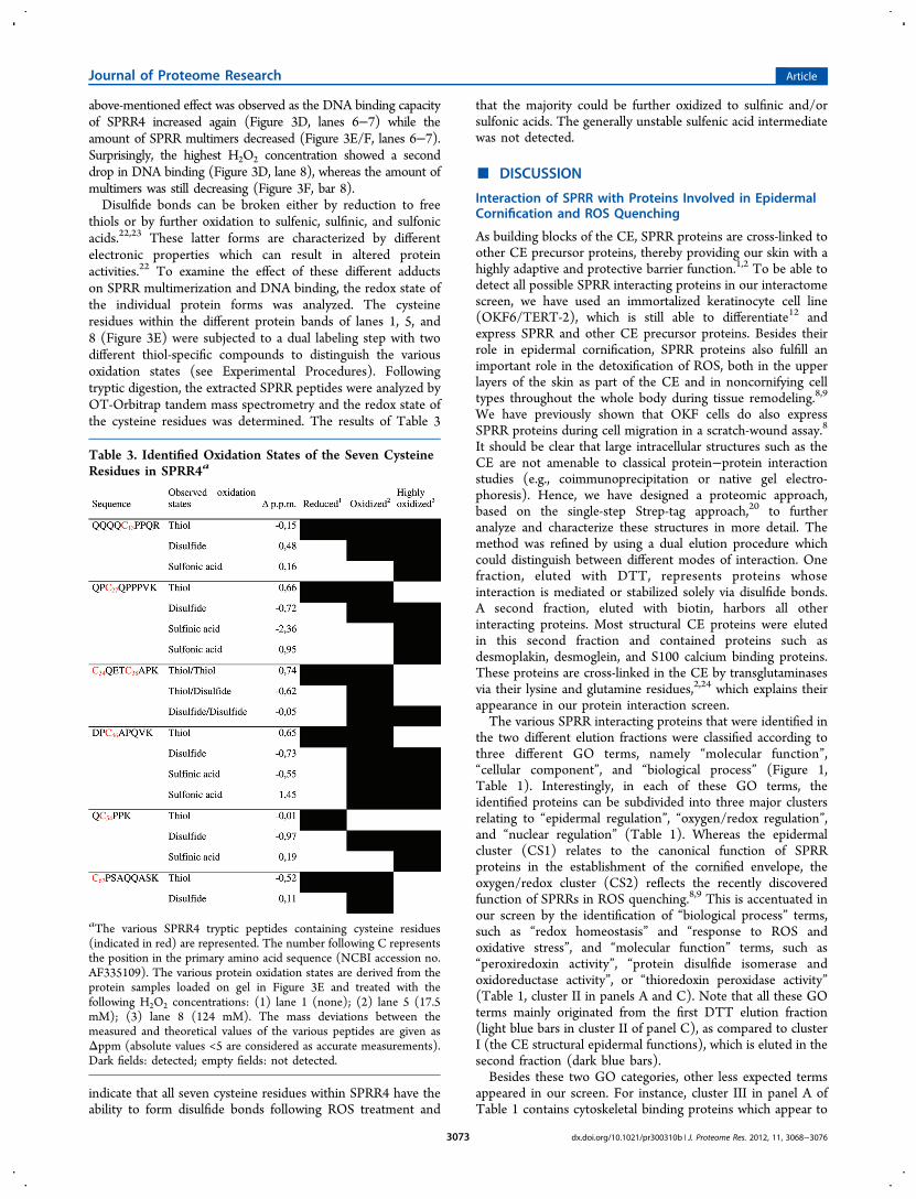
thiols or by further oxidation to sulfenic, sulfinic, and sulfonicacids.22,23 These latter forms are characterized by differentelectronic properties which can result in altered proteinactivities.22 To examine the effect of these different adductson SPRR multimerization and DNA binding, the redox state ofthe individual protein forms was analyzed. The cysteineresidues within the different protein bands of lanes 1, 5, and8 (Figure 3E) were subjected to a dual labeling step with twodifferent thiol-specific compounds to distinguish the variousoxidation states (see Experimental Procedures). Followingtryptic digestion, the extracted SPRR peptides were analyzed byOT-Orbitrap tandem mass spectrometry and the redox state ofthe cysteine residues was determined. The results of Table 3
indicate that all seven cysteine residues within SPRR4 have theability to form disulfide bonds following ROS treatment and
that the majority could be further oxidized to sulfinic and/orsulfonic acids. The generally unstable sulfenic acid intermediatewas not detected.
■ DISCUSSION
Interaction of SPRR with Proteins Involved in EpidermalCornification and ROS Quenching
As building blocks of the CE, SPRR proteins are cross-linked toother CE precursor proteins, thereby providing our skin with ahighly adaptive and protective barrier function.1,2 To be able todetect all possible SPRR interacting proteins in our interactomescreen, we have used an immortalized keratinocyte cell line(OKF6/TERT-2), which is still able to differentiate12 andexpress SPRR and other CE precursor proteins. Besides theirrole in epidermal cornification, SPRR proteins also fulfill animportant role in the detoxification of ROS, both in the upperlayers of the skin as part of the CE and in noncornifying celltypes throughout the whole body during tissue remodeling.8,9
We have previously shown that OKF cells do also expressSPRR proteins during cell migration in a scratch-wound assay.8
It should be clear that large intracellular structures such as theCE are not amenable to classical protein−protein interactionstudies (e.g., coimmunoprecipitation or native gel electro-phoresis). Hence, we have designed a proteomic approach,based on the single-step Strep-tag approach,20 to furtheranalyze and characterize these structures in more detail. Themethod was refined by using a dual elution procedure whichcould distinguish between different modes of interaction. Onefraction, eluted with DTT, represents proteins whoseinteraction is mediated or stabilized solely via disulfide bonds.A second fraction, eluted with biotin, harbors all otherinteracting proteins. Most structural CE proteins were elutedin this second fraction and contained proteins such asdesmoplakin, desmoglein, and S100 calcium binding proteins.These proteins are cross-linked in the CE by transglutaminasesvia their lysine and glutamine residues,2,24 which explains theirappearance in our protein interaction screen.The various SPRR interacting proteins that were identified in
the two different elution fractions were classified according tothree different GO terms, namely “molecular function”,“cellular component”, and “biological process” (Figure 1,Table 1). Interestingly, in each of these GO terms, theidentified proteins can be subdivided into three major clustersrelating to “epidermal regulation”, “oxygen/redox regulation”,and “nuclear regulation” (Table 1). Whereas the epidermalcluster (CS1) relates to the canonical function of SPRRproteins in the establishment of the cornified envelope, theoxygen/redox cluster (CS2) reflects the recently discoveredfunction of SPRRs in ROS quenching.8,9 This is accentuated inour screen by the identification of “biological process” terms,such as “redox homeostasis” and “response to ROS andoxidative stress”, and “molecular function” terms, such as“peroxiredoxin activity”, “protein disulfide isomerase andoxidoreductase activity”, or “thioredoxin peroxidase activity”(Table 1, cluster II in panels A and C). Note that all these GOterms mainly originated from the first DTT elution fraction(light blue bars in cluster II of panel C), as compared to clusterI (the CE structural epidermal functions), which is eluted in thesecond fraction (dark blue bars).Besides these two GO categories, other less expected terms
appeared in our screen. For instance, cluster III in panel A ofTable 1 contains cytoskeletal binding proteins which appear to
Table 3. Identified Oxidation States of the Seven CysteineResidues in SPRR4a
aThe various SPRR4 tryptic peptides containing cysteine residues(indicated in red) are represented. The number following C representsthe position in the primary amino acid sequence (NCBI accession no.AF335109). The various protein oxidation states are derived from theprotein samples loaded on gel in Figure 3E and treated with thefollowing H2O2 concentrations: (1) lane 1 (none); (2) lane 5 (17.5mM); (3) lane 8 (124 mM). The mass deviations between themeasured and theoretical values of the various peptides are given asΔppm (absolute values <5 are considered as accurate measurements).Dark fields: detected; empty fields: not detected.
Journal of Proteome Research Article
dx.doi.org/10.1021/pr300310b | J. Proteome Res. 2012, 11, 3068−30763073
interact with both SPRR1B and SPRR4 (see also cluster I inpanel B). These interactions are further substantiated by thefact that SPRR4 localizes to ordered fiber-like structures withinthe cytoplasm of HeLa cells (Figure 2A). Apparently, SPRRproteins are at least in close proximity to some cytoskeletalproteins. Both Pradervand et al.25 and Bonilla et al.26 havepreviously described the localization of SPRR1 along actinstructures. Although no direct physical interaction was detectedby these authors, it was inferred that SPRR proteins might altercytoskeletal functions and contribute to tissue remodeling.25,26
Consistent with this, we recently showed that SPRR proteinsindeed localize to membrane ruffles of migrating keratinocytesand play a major role in cell migration during wound healing.8
SPRR Proteins Function in DNA Binding
As mentioned above, many of the SPRR interacting proteinscan be classified under the denominator of nuclear function(molecular function cluster V; cellular component clusters IIIand IV; biological process cluster IV). Due to the large diversityin molecular functions of the various proteins in thesecategories, we assumed that the interaction between SPRRand these proteins within cellular structure CS3 might beindirect and mediated by mutual interaction of each of theseproteins with DNA. Besides, our screen indicated thatespecially SPRR4 might be involved in this nuclear function.For this reason we have monitored the cellular expression ofSPRR4 in living cells and tested its direct interaction with DNAin an in vitro binding assay.Immunofluorescence staining with a SPRR4 monospecific
antibody revealed minor but significant nuclear localization ofSPRR4 proteins (Figure 2A−C). In line with these results areearlier observations by other researchers who occasionallydetected SPRR1, SPRR2, and SPRR3 proteins in the nucleus ofcells by using various antibodies.17,27−30 More recently, this wassubstantiated by live-cell-imaging of pEGFP-SPRR1B trans-fected keratinocytes where SPRR was consistently found in thenucleus of migrating cells.8 This localization further highlights apotential nuclear function of SPRR proteins.If the interaction of SPRR4 with the various identified
proteins within CS3 is indirect and mediated by mutualinteraction of all proteins with DNA, then SPRRs should havethe ability to directly bind DNA. Consequently, we assessed theDNA binding properties of purified SPRR proteins directly inan in vitro assay with purified components. Addition ofincreasing amounts of purified SPRR4 protein to linear DNAfragments indeed resulted in a lower mobility of the DNAmolecules in agarose gel (Figure 3A), which is a measure fordirect interaction between proteins and nucleic acids. PurifiedSPRR1, SPRR2, and SPRR3 proteins showed similar electro-phoretic mobility shifts, although with lower affinity, as a 10times higher molar excess of protein was required (results notshown). Interaction of SPRR4 with plasmid DNA was alsodetected via AFM analysis. Here, multiple small protein dotsdecorating the DNA molecules can be observed (Figure 3C),suggesting that SPRR proteins bind DNA in a sequenceindependent way.As mentioned above, our Gene Ontology analysis has
indicated a clear selectivity between SPRR1B and SPRR4interactors as far as the “biological process” terms areconcerned. Whereas SPRR4 interactors are mainly found incluster IV (nuclear regulation), SPRR1B interactors areoverrepresented in clusters I and II (epidermal and oxygen/redox regulation, respectively). This selectivity is in complete
agreement with the higher DNA-binding affinity of SPPR4 anda more efficient protection against ROS induced DNA-breakage(Table 2). These data further highlight the specificity of theproteomic screen that we have performed.ROS Affects SPRR Protein Multimerization and DNABinding
Since the oxidation of SPRR proteins during ROS detox-ification results in the reversible formation of both inter- andintramolecular S−S bonds9 and since many GO “nuclearregulation” terms were identified from the first (oxidationspecific) elution fraction, we questioned whether these redoxmodifications can influence the DNA binding capacity ofSPRRs. Analysis of the DNA binding activity of equal amountsof SPRR4, pretreated with increasing concentrations of H2O2,revealed in a first instance a clear decrease in the DNA bindingpotential (Figure 3D, lanes 1−5) suggesting an inverserelationship between SPRR protein multimerization and DNAbinding. At higher H2O2 concentrations, less protein multimerswere observed on PAGE (Figure 3E and F), while the DNAbinding capacity increased. Mass spectrometric analysis of thedifferent cysteine oxidation states revealed that at higher ROSlevels disulfide bonds are broken by further oxidation to sulfinicand sulfonic acid (Table 3), which provides an explanation for areversion to monomeric forms at higher H2O2 concentrations.Apparently only monomeric SPRR4 can bind efficiently toDNA. At the highest ROS levels, however, DNA binding isagain reduced whereas the amount of monomers still increases.This is likely due to the generation of cysteine sulfinate/sulfonate adducts, which might counteract DNA bindingbecause of their higher negative charge.
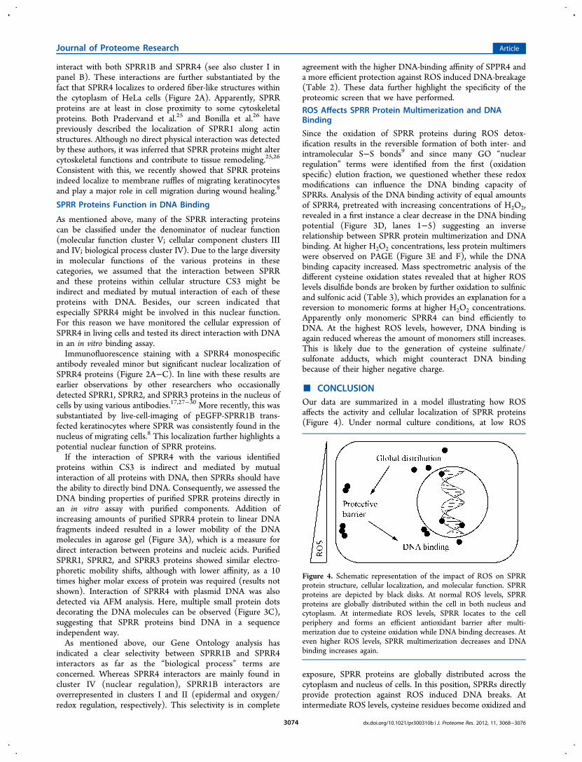
■ CONCLUSIONOur data are summarized in a model illustrating how ROSaffects the activity and cellular localization of SPRR proteins(Figure 4). Under normal culture conditions, at low ROS
exposure, SPRR proteins are globally distributed across thecytoplasm and nucleus of cells. In this position, SPRRs directlyprovide protection against ROS induced DNA breaks. Atintermediate ROS levels, cysteine residues become oxidized and
Figure 4. Schematic representation of the impact of ROS on SPRRprotein structure, cellular localization, and molecular function. SPRRproteins are depicted by black disks. At normal ROS levels, SPRRproteins are globally distributed within the cell in both nucleus andcytoplasm. At intermediate ROS levels, SPRR locates to the cellperiphery and forms an efficient antioxidant barrier after multi-merization due to cysteine oxidation while DNA binding decreases. Ateven higher ROS levels, SPRR multimerization decreases and DNAbinding increases again.
Journal of Proteome Research Article
dx.doi.org/10.1021/pr300310b | J. Proteome Res. 2012, 11, 3068−30763074

SPRR protein multimers are formed via disulfide bonding. Thecellular localization is then shifted toward the cytoplasmicmembrane and the DNA binding activity is reduced. Whilelocalized at the cell periphery, SPRR proteins form an efficientprotective barrier against cell-infiltrating ROS, which are oftenproduced at the cytoplasmic membrane via lipid peroxidationfollowing UV exposure.31 In this respect, it is important to notethat SPRRs were originally identified as UV inducible genes32
and that SPRR4, the best quencher (Table 2), is selectivelyexpressed in sun-exposed skin.33 If the ROS levels are too highand exceed the natural reducing potential of the cell, majoroxidative stress arises and the cysteine residues of SPRRproteins become further oxidized. As a result, SPRR convertsback to a monomeric form and the DNA binding activityincreases again. Such high ROS levels will eventually lead to celldeath, and many of the apoptotic signaling pathways willbecome activated by redox modifications.24,34 However, sinceDNA itself is a very efficient ROS quencher, DNA binding bySPRR might temporarily delay DNA breakdown to provideantioxidant protection to neighboring cells. This compares tothe normal situation in our skin, where a layer of dead flattenedcells protects the inner tissue against injurious external insults.2
Apparently, the modulation of the oxidation state of SPRRproteins constitutes the basis for their selective antioxidantperformance and fine-tunes their targeting to those cellularcomponents that are most threatened at a given moment intime.
■ ASSOCIATED CONTENT*S Supporting Information
Pipeline Pilot protocol used to identify SPRR interactingproteins, and raw data related to the mass spectrometricidentification of the various proteins described in this study.This material is available free of charge via the Internet athttp://pubs.acs.org.
■ AUTHOR INFORMATIONCorresponding Author
*Tel: +31 715274409. Fax: +31 715274340. E-mail: [email protected] Address#Erasmus MC, University Medical Center Rotterdam, 3015 GERotterdam, The Netherlands.
Notes
The authors declare no competing financial interest.
■ ACKNOWLEDGMENTSWe would like to thank Patrick Voskamp (LIC, Leiden) forhelping with the protein interaction screen. Dr. John van Noort(LION, Leiden) is acknowledged for technical assistance withAFM. We would also like to thank Steef de Valk and MaartenOvergaauw for their contribution to the SPRR multimerizationand DNA binding assays and Ivana Bagaric for antibody testing.This research was financed exclusively by the Leiden Instituteof Chemistry.
■ REFERENCES(1) Cabral, A.; Voskamp, P.; Cleton-Jansen, A. M.; South, A.; Nizetic,D.; Backendorf, C. Structural organization and regulation of the smallproline-rich family of cornified envelope precursors suggest a role inadaptive barrier function. J. Biol. Chem. 2001, 276 (22), 19231−7.
(2) Candi, E.; Schmidt, R.; Melino, G. The cornified envelope: amodel of cell death in the skin. Nat. Rev. Mol. Cell. Biol. 2005, 6 (4),328−40.(3) Backendorf, C.; Hohl, D. A common origin for cornified envelopeproteins? Nat. Genet. 1992, 2 (2), 91.(4) Kalinin, A. E.; Kajava, A. V.; Steinert, P. M. Epithelial barrierfunction: assembly and structural features of the cornified cellenvelope. Bioessays 2002, 24 (9), 789−800.(5) Rice, R. H.; Green, H. The cornified envelope of terminallydifferentiated human epidermal keratinocytes consists of cross-linkedprotein. Cell 1977, 11 (2), 417−22.(6) Fischer, D. F.; Backendorf, C. Promoter analysis in the humanSPRR gene family. Methods Mol. Biol. 2005, 289, 303−14.(7) Steinert, P. M.; Kartasova, T.; Marekov, L. N. Biochemicalevidence that small proline-rich proteins and trichohyalin function inepithelia by modulation of the biomechanical properties of theircornified cell envelopes. J. Biol. Chem. 1998, 273 (19), 11758−69.(8) Vermeij, W. P.; Backendorf, C. Skin Cornification ProteinsProvide Global Link between ROS Detoxification and Cell Migrationduring Wound Healing. PLoS One 2010, 5 (8), e11957.(9) Vermeij, W. P.; Alia, A.; Backendorf, C. ROS QuenchingPotential of the Epidermal Cornified Cell Envelope. J. Invest. Dermatol.2011, 131 (7), 1435−41.(10) Gurtner, G. C.; Werner, S.; Barrandon, Y.; Longaker, M. T.Wound repair and regeneration. Nature 2008, 453 (7193), 314−21.(11) auf dem Keller, U.; Kumin, A.; Braun, S.; Werner, S. Reactiveoxygen species and their detoxification in healing skin wounds. J. Invest.Dermatol. Symp. Proc. 2006, 11 (1), 106−11.(12) Dickson, M. A.; Hahn, W. C.; Ino, Y.; Ronfard, V.; Wu, J. Y.;Weinberg, R. A.; Louis, D. N.; Li, F. P.; Rheinwald, J. G. Humankeratinocytes that express hTERT and also bypass a p16(INK4a)-enforced mechanism that limits life span become immortal yet retainnormal growth and differentiation characteristics. Mol. Cell. Biol. 2000,20 (4), 1436−47.(13) Matsudaira, P. T. A Practical guide to protein and peptidepurification for microsequencing, 2nd ed.; Academic Press: San Diego,1993; p 184.(14) Rappsilber, J.; Mann, M.; Ishihama, Y. Protocol for micro-purification, enrichment, pre-fractionation and storage of peptides forproteomics using StageTips. Nat. Protoc. 2007, 2 (8), 1896−906.(15) Florea, B. I.; Verdoes, M.; Li, N.; van der Linden, W. A.;Geurink, P. P.; van den Elst, H.; Hofmann, T.; de Ru, A.; van Veelen,P. A.; Tanaka, K.; Sasaki, K.; Murata, S.; den Dulk, H.; Brouwer, J.;Ossendorp, F. A.; Kisselev, A. F.; Overkleeft, H. S. Activity-basedprofiling reveals reactivity of the murine thymoproteasome-specificsubunit beta5t. Chem. Biol. 2010, 17 (8), 795−801.(16) Zhang, B.; Kirov, S.; Snoddy, J. WebGestalt: an integratedsystem for exploring gene sets in various biological contexts. NucleicAcids Res. 2005, 33 (Web Server issue), W741−8.(17) Hohl, D.; de Viragh, P. A.; Amiguet-Barras, F.; Gibbs, S.;Backendorf, C.; Huber, M. The small proline-rich proteins constitute amultigene family of differentially regulated cornified cell envelopeprecursor proteins. J. Invest. Dermatol. 1995, 104 (6), 902−9.(18) Collins, A. R.; Dusinska, M.; Gedik, C. M.; Stetina, R. Oxidativedamage to DNA: do we have a reliable biomarker? Environ. HealthPerspect. 1996, 104 (Suppl 3), 465−9.(19) Shevchenko, A.; Tomas, H.; Havlis, J.; Olsen, J. V.; Mann, M.In-gel digestion for mass spectrometric characterization of proteinsand proteomes. Nat. Protoc. 2006, 1 (6), 2856−60.(20) Junttila, M. R.; Saarinen, S.; Schmidt, T; Kast, J.; Westermarck,J. Single-step Strep-tag purification for the isolation and identificationof protein complexes from mammalian cells. Proteomics 2005, 5,1199−203.(21) Perkins, D. N.; Pappin, D. J.; Creasy, D. M.; Cottrell, J. S.Probability-based protein identification by searching sequence data-bases using mass spectrometry data. Electrophoresis 1999, 20 (18),3551−67.(22) Poole, L. B.; Karplus, P. A.; Claiborne, A. Protein sulfenic acidsin redox signaling. Annu. Rev. Pharmacol. Toxicol. 2004, 44, 325−47.
Journal of Proteome Research Article
dx.doi.org/10.1021/pr300310b | J. Proteome Res. 2012, 11, 3068−30763075
(23) Winyard, P. G.; Moody, C. J.; Jacob, C. Oxidative activation ofantioxidant defence. Trends Biochem. Sci. 2005, 30 (8), 453−61.(24) Lorand, L.; Graham, R. M. Transglutaminases: crosslinkingenzymes with pleiotropic functions. Nat. Rev. Mol. Cell. Biol. 2003, 4(2), 140−56.(25) Pradervand, S.; Yasukawa, H.; Muller, O. G.; Kjekshus, H.;Nakamura, T.; St Amand, T. R.; Yajima, T.; Matsumura, K.; Duplain,H.; Iwatate, M.; Woodard, S.; Pedrazzini, T.; Ross, J.; Firsov, D.;Rossier, B. C.; Hoshijima, M.; Chien, K. R. Small proline-rich protein1A is a gp130 pathway- and stress-inducible cardioprotective protein.EMBO J. 2004, 23 (22), 4517−25.(26) Bonilla, I. E.; Tanabe, K.; Strittmatter, S. M. Small proline-richrepeat protein 1A is expressed by axotomized neurons and promotesaxonal outgrowth. J. Neurosci. 2002, 22 (4), 1303−15.(27) Jarnik, M.; Kartasova, T.; Steinert, P. M.; Lichti, U.; Steven, A.C. Differential expression and cell envelope incorporation of smallproline-rich protein 1 in different cornified epithelia. J. Cell. Sci. 1996,109 (Pt 6), 1381−91.(28) Koizumi, H.; Kartasova, T.; Tanaka, H.; Ohkawara, A.; Kuroki,T. Differentiation-associated localization of small proline-rich proteinin normal and diseased human skin. Br. J. Dermatol. 1996, 134 (4),686−92.(29) Morris, J. S.; Stein, T.; Pringle, M. A.; Davies, C. R.; Weber-Hall,S.; Ferrier, R. K.; Bell, A. K.; Heath, V. J.; Gusterson, B. A.Involvement of axonal guidance proteins and their signaling partnersin the developing mouse mammary gland. J. Cell. Physiol. 2006, 206(1), 16−24.(30) Zhang, Y.; Feng, Y. B.; Shen, X. M.; Chen, B. S.; Du, X. L.; Luo,M. L.; Cai, Y.; Han, Y. L.; Xu, X.; Zhan, Q. M.; Wang, M. R.Exogenous expression of Esophagin/SPRR3 attenuates the tumor-igenicity of esophageal squamous cell carcinoma cells via promotingapoptosis. Int. J. Cancer 2008, 122 (2), 260−6.(31) Evelson, P.; Ordonez, C. P.; Llesuy, S.; Boveris, A. Oxidativestress and in vivo chemiluminescence in mouse skin exposed to UVAradiation. J. Photochem. Photobiol. B 1997, 38 (2−3), 215−9.(32) Kartasova, T.; van de Putte, P. Isolation, characterization, andUV-stimulated expression of two families of genes encodingpolypeptides of related structure in human epidermal keratinocytes.Mol. Cell. Biol. 1988, 8 (5), 2195−203.(33) Cabral, A.; Sayin, A.; de Winter, S.; Fischer, D. F.; Pavel, S.;Backendorf, C. SPRR4, a novel cornified envelope precursor: UV-dependent epidermal expression and selective incorporation intofragile envelopes. J. Cell Sci. 2001, 114, 3837−3843.(34) Waster, P. K.; Ollinger, K. M. Redox-dependent translocation ofp53 to mitochondria or nucleus in human melanocytes after UVA- andUVB-induced apoptosis. J. Invest. Dermatol. 2009, 129 (7), 1769−81.
Journal of Proteome Research Article
dx.doi.org/10.1021/pr300310b | J. Proteome Res. 2012, 11, 3068−30763076
Top Related
Copyright © 2022 FDOKUMEN

