


Bahasa
Halaman
Hukum

Oxygen-Dependent Regulation of Bacterial Lipid Production
Kimberly C. Lemmer,a Alice C. Dohnalkova,d Daniel R. Noguera,a,c Timothy J. Donohuea,b
DOE Great Lakes Bioenergy Research Center,a Department of Bacteriology,b and Department of Civil and Environmental Engineering,c University of Wisconsin—Madison,Madison, Wisconsin, USA; Pacific Northwest National Laboratory, Environmental Molecular Sciences Laboratory, Richland, Washington, USAd
ABSTRACT
Understanding the mechanisms of lipid accumulation in microorganisms is important for several reasons. In addition to provid-ing insight into assembly of biological membranes, lipid accumulation has important applications in the production of renew-able fuels and chemicals. The photosynthetic bacterium Rhodobacter sphaeroides is an attractive organism to study lipid accu-mulation, as it has the ability to increase membrane production at low O2 tensions. Under these conditions, R. sphaeroidesdevelops invaginations of the cytoplasmic membrane to increase its membrane surface area for housing of the membrane-boundcomponents of its photosynthetic apparatus. Here we use fatty acid levels as a reporter of membrane lipid content. We showthat, under low-O2 and anaerobic conditions, the total fatty acid content per cell increases 3-fold. We also find that the increasesin the amount of fatty acid and photosynthetic pigment per cell are correlated as O2 tensions or light intensity are changed. Toask if lipid and pigment accumulation were genetically separable, we analyzed strains with mutations in known photosyntheticregulatory pathways. While a strain lacking AppA failed to induce photosynthetic pigment-protein complex accumulation, itincreased fatty acid content under low-O2 conditions. We also found that an intact PrrBA pathway is required for low-O2-in-duced fatty acid accumulation. Our findings suggest a previously unknown role of R. sphaeroides transcriptional regulators inincreasing fatty acid and phospholipid accumulation in response to decreased O2 tension.
IMPORTANCE
Lipids serve important functions in living systems, either as structural components of membranes or as a form of carbon stor-age. Understanding the mechanisms of lipid accumulation in microorganisms is important for providing insight into the assem-bly of biological membranes and additionally has important applications in the production of renewable fuels and chemicals. Inthis study, we investigate the ability of Rhodobacter sphaeroides to increase membrane production at low O2 tensions in order tohouse its photosynthetic apparatus. We demonstrate that this bacterium has a mechanism to increase lipid content in responseto decreased O2 tension and identify a transcription factor necessary for this response. This is significant because it identifies atranscriptional regulatory pathway that can increase microbial lipid content.
Lipids serve important functions in living systems, either asstructural components of membranes or as a form of carbon
storage. Lipids derived from oil-rich microorganisms (includingbacteria, yeasts, and microalgae) also offer a promising source ofrenewable fuels and chemicals (1, 2). However, the genetic andbiochemical mechanisms regulating lipid accumulation in micro-organisms are poorly understood, have been difficult to study, andare typically linked to stress conditions that hinder growth (3–5).Much effort has been directed at increasing lipid accumulationthrough alteration of enzymes involved in biosynthesis or catab-olism of fatty acids, but little is known about the endogenoussystems that regulate lipid accumulation (6, 7).
With an interest in understanding the molecular mechanismsregulating microbial lipid accumulation, we are analyzing thecontrol of lipid accumulation in purple bacteria, which have theability to increase their membrane content under low-O2 condi-tions. In this study, we investigate this process in Rhodobactersphaeroides, a facultative purple bacterium that can grow via aer-obic or anaerobic respiration or photosynthesis (8). Unlike manywell-studied facultative bacteria, changes in O2 tension cause sig-nificant morphological changes in R. sphaeroides. At high O2 ten-sions, its cell envelope resembles that of other Gram-negative bac-teria. In response to low O2 tension, R. sphaeroides increases itsmembrane surface area for assembly of a photosynthetic appara-tus by developing specialized intracytoplasmic membrane (ICM)invaginations (9–11). Three major types of integral membrane
pigment-protein complexes populate the ICM: the photochemi-cal reaction center (RC) and two distinct light-harvesting com-plexes (LH1 and LH2) that together collect and convert the energyof light into biological energy (10, 12).
The pathways that control transcription of many genes re-quired for the synthesis and function of the photosynthetic appa-ratus have been extensively studied at the genetic and genomiclevels (12–15). The photosynthesis response regulator (Prr) pro-teins comprise a two-component signal transduction system acti-vated by low O2 tension, in which PrrB is a membrane-spanningsensor histidine kinase and PrrA is a DNA-binding response reg-
Received 20 November 2014 Accepted 22 February 2015
Accepted manuscript posted online 2 March 2015
Citation Lemmer KC, Dohnalkova AC, Noguera DR, Donohue TJ. 2015. Oxygen-dependent regulation of bacterial lipid production. J Bacteriol 197:1649 –1658.doi:10.1128/JB.02510-14.
Editor: G. A. O’Toole
Address correspondence to Timothy J. Donohue, [email protected].
Supplemental material for this article may be found at http://dx.doi.org/10.1128/JB.02510-14.
Copyright © 2015, American Society for Microbiology. All Rights Reserved.
doi:10.1128/JB.02510-14
The authors have paid a fee to allow immediate free access to this article.
May 2015 Volume 197 Number 9 jb.asm.org 1649Journal of Bacteriology
ulator (16, 17). The AppA-PpsR regulatory system is composed ofa DNA-binding repressor, PpsR, and the light- and redox-sensi-tive antirepressor AppA (18). FnrL, a member of the FNR (fuma-rate and nitrate reductase) family of transcription factors, is aglobal regulator of anaerobic gene expression that is directly in-hibited by the presence of O2 (19, 20). These three pathways eachcontribute to increased expression of photosynthesis genes at lowO2 tension, including those in the puf and puc operons (encodingpolypeptides of the RC, LH1, and LH2 complexes), biosyntheticenzymes for bacteriochlorophyll (BChl) and carotenoid (Crt) pig-ments, and electron transport chain components (12, 13, 16,21–23).
Despite the extensive knowledge of the systems regulating syn-thesis of the pigment and protein components of photosyntheticmembranes, little is known about synthesis of phospholipids thatform the ICM bilayer. Cells that make ICM have a higher rate ofphospholipid synthesis (24, 25) and a greater proportion of lipidin cellular biomass (26) than cells grown at high O2, which aredevoid of ICM. Yet it is not known how much more membrane isneeded for ICM development or if there is a specific mechanism toincrease fatty acid and phospholipid levels in response to low O2.Observations of de novo ICM synthesis have shown that nascentICM invaginations can be seen before pigment-protein complexesare evident on their surface, suggesting that phospholipid accu-mulation may not be obligately linked with synthesis of this spe-cialized membrane (9).
In this study, we ask whether an increase in membrane lipidabundance is linked to ICM assembly. In wild-type cells, we findthat fatty acid content is correlated with the level of BChl. How-ever, accumulation of these two ICM components is geneticallyseparable, since we identify a mutant that is devoid of pigment-protein complexes but contains high, near-anaerobic levels offatty acid at low O2 tensions.
MATERIALS AND METHODSBacterial strains and growth conditions. The bacterial strains used in thisstudy are described in Table 1. R. sphaeroides 2.4.1 strains were grown at30°C in a succinate-based minimal medium (SIS) (27), unless otherwisenoted. For growth in the presence of O2, 500-ml cultures were bubbledwith 1% CO2, the indicated percentage of O2, and the balance in N2. Foranaerobic growth, 500-ml cultures were bubbled with 5% CO2 and 95%N2. In addition, anaerobic photosynthetic cultures were illuminated withan incandescent light with an intensity of 3, 10, or 100 W/m2 measuredthrough a red glass filter, and anaerobic dark cultures were grown in SISsupplemented with 10% Luria-Bertani medium and 0.3% dimethyl sul-foxide (DMSO). For analysis, cultures were grown to an optical density at600 nm (OD600) of �0.5 to 0.9; anaerobic photosynthetic cultures were
grown to an OD600 of �0.9 to 1.5 to achieve similar cell densities tohigh-O2 cultures at the lower OD range.
SC and BChl analyses. To assess spectral complex (SC) assembly, ali-quots of cell cultures were assayed by visible spectroscopy on an OlisDW-2/2000 spectrophotometer. All spectra were scaled to an absorbanceof 1 at 680 nm to normalize for cell density and then staggered verticallyfor presentation of multiple curves on one axis. BChl content was esti-mated by readings of absorption at 775 nm of material extracted from cellsinto 7:2 acetone-methanol, using an extinction coefficient of 75 mM�1
cm�1 (28).Fatty acid and lipid phosphorus analysis. Cell harvesting, extraction
of lipids with chloroform-methanol, esterification, and gas chromatogra-phy-mass spectrometry (GC-MS) analysis were performed as previouslydescribed (29). For lipid phosphorus measurements, dried lipid extrac-tions from 2.5-ml samples were digested with perchloric acid and assayedfor phosphorus content (30). Cell plating experiments were used to makea standard curve of OD600 versus CFU to normalize fatty acid, lipid phos-phorus, and BChl content per cell. For the experiment shown in Fig. 2A,insufficient cell plating data were collected to make a curve, so cell countsfrom plating within that experiment were used to normalize.
P values for statistical significance were calculated by unpaired t testusing Graph Pad QuickCalcs.
TEM. High-pressure freezing (HPF) and automatic freeze substitu-tion (AFS), followed by plastic embedding, were used to produce thinsections of respective samples of cell suspensions. Cells were pelleted bybrief centrifugation with a Quick-Spin minicentrifuge, and 5 �l of theconcentrated cell suspension was transferred into an HPF flat specimencarrier and frozen with a Leica EM PACT high-pressure freezer (LeicaMicrosystems, Inc., Bannockburn, IL) at a typical rate of 1,700°C/s. Thepods with compacted frozen cells were transferred under liquid nitrogento the precooled AFS (EM AFS; Leica), and a protocol for cell fixation,water substitution by acetone, and a gradual warm-up to room tempera-ture was followed (see Table S1 in the supplemental material). After 72 h,the samples were released from the pods, washed three times in acetone,gradually infiltrated with an ascending series of Spurr’s low-viscosity em-bedding medium (Electron Microscopy Sciences, Hatfield, PA) (with 25,50, and 75%, followed by three 100% washes for 120 min each), and curedat 60°C for 48 h. The polymerized blocks were sectioned to 70-nm thinsections with a Leica Ultracut UCT ultramicrotome, mounted on Form-var-coated 100 mesh Cu transmission electron microscopy (TEM) gridssputter coated with carbon, and poststained for 7 min with aqueous 2%uranyl acetate followed by 3 min of Reynolds’ lead citrate (31) prior toTEM imaging. Samples were examined with the Tecnai T-12 TEM (FEI)operating at 120 kV with a LaB6 filament. Images were collected digitallywith a 2�2K Ultrascan 1000 CCD (Gatan).
RESULTSO2 and light regulation of fatty acid and spectral complex con-tent in R. sphaeroides. Given that R. sphaeroides increases itsmembrane surface area to synthesize ICM under low-O2 and an-aerobic conditions, we wanted to test if these cells contain highermembrane lipid content than high-O2-grown cells. Thus, wesought to quantify membrane lipid content as a function of O2
tension and to determine how membrane content relates to thelevel of membrane-integral ICM spectral complexes (SCs). To dothis, R. sphaeroides 2.4.1 was grown in liquid cultures sparged withhigh (30%) O2, anaerobically at moderate light intensity (10W/m2) and in the dark using DMSO as an electron acceptor. Inorder to relate SC levels to membrane lipid content, we used totalextracted BChl as a measure of SC assembly and both fatty acidand lipid phosphorus levels to assess membrane lipid content. Wechose to express all of these measurements on a per cell basis inorder to gain insight into the physiological changes of an individ-ual cell under our experimental conditions. Since other cellular
TABLE 1 R. sphaeroides strains used in this study
Strain Relevant properties Reference
2.4.1 Wild-type strain 55PPS1 (�PpsR) 2.4.1 ppsR::�Kmr 40PRRB78 2.4.1 derivative encoding L78P substitution
in PrrB, with �SmrSpr at SmaI sitedownstream of prrB
39
PUFB1 2.4.1 �pufB::Knr 41PRRA1 (�PrrA) 2.4.1 prrA:: �SmrSpr 43APP11 (�AppA) 2.4.1 �appA::Tpr 42JZ1678 (�FnrL) 2.4.1 fnrL::�SmrSpr 19
Lemmer et al.
1650 jb.asm.org May 2015 Volume 197 Number 9Journal of Bacteriology

components of biomass also change in abundance as O2 and lightintensity changes (26), this method of reporting provides insightinto the physiology of lipid accumulation under the conditionsexamined.
As expected, the absorbance spectra of these cultures show thatSCs are absent in high-O2 cultures and present in anaerobic cul-tures under light or dark conditions, with characteristic absor-bance peaks at 800 and 850 nm (representing LH2) and a shoulderat 875 nm (representing LH1) (Fig. 1A). We also found that cellsgrown at 30% O2 tension lack detectable BChl, while cells grownunder anaerobic conditions either at moderate light intensity or inthe dark contain measurable levels of BChl (5 � 10�18 to 6 �10�18 mol/cell) (Fig. 1B). At high (30%) O2 tension, cells con-tained 3.5 � 10�17 mol/cell of fatty acid and 1.5 � 10�17 mol/cellof lipid phosphorus (Fig. 1C). The approximately 2:1 ratio of fattyacid to phosphorus supports the conclusion that both assays aremeasuring phospholipid levels. Anaerobically grown cells con-tained �3 times more fatty acid and lipid phosphorus per cell thanthose grown at high O2 tension (Fig. 2), consistent with the in-creased membrane content associated with ICM formation. Therelative amounts of the major fatty acid species were the sameunder all tested growth conditions (Fig. 1D) and were consistentwith the fatty acid composition observed in previous studies of R.sphaeroides (32, 33). Since fatty acid and lipid phosphorus levelsmirrored each other, we chose to use fatty acid levels to assess totalmembrane lipid content throughout the rest of this study.
ICM formation is induced by lowered O2 tension, while light
intensity also controls the number of ICM invaginations as well asthe level and ratio of individual SCs (10, 11, 34, 35). In order toexamine if the lipid content correlates with the SC component ofICM, we analyzed cells grown under a range of O2 concentrations(30 to 0%) in the dark. This analysis showed that both the cellularfatty acid and BChl contents began increasing at O2 concentra-tions below 10%, with a large increase occurring from 1.5% to0.5%, such that 0.5% O2-grown cells contained levels of bothcomponents similar to those in anaerobic cells (Fig. 2A). Averagedover at least 6 independent cultures, there was no statistical differ-ence between the fatty acid and BChl contents in 0.5% O2-growncells (1.2 � 10�16 mol/cell fatty acid, 5.0 � 10�18 mol/cell BChl)compared to those in cells grown anaerobically under light or darkconditions (Fig. 1B). At external O2 concentrations of between 0.5and 6%, the degree of increase in fatty acid content over high(30%)-O2 cells was proportional to the amount of BChl present.For example, at 1.5% O2, cells contained 29% of the BChl presentin anaerobic cells (1.5 � 10�18 mol/cell versus 5.2 � 10�18 mol/cell), while the increase in fatty acid compared to 30% O2 (2.1 �10�17 mol/cell) was 31% of the increase observed in anaerobiccells (6.8 � 10�17 mol/cell) (Fig. 2A). When we varied the incidentlight intensity under anaerobic conditions, we found that thesechanges also affected lipid and pigment content, with higher cel-lular lipid and BChl levels as the light intensity decreases (Fig. 2B).This is consistent with previous observations showing that high-light-grown cells contain less BChl and fewer ICM invaginationsthan low-light-grown cells (35–37). When BChl content was plot-
FIG 1 SC, BChl, and fatty acid contents of R. sphaeroides cells sparged with 30% O2 tension compared to those in cells grown anaerobically in the light oranaerobically in the dark with DMSO as an electron acceptor. (A) Absorbance scans of intact cells to show SC assembly. (B and C) Levels of BChl (B) and totalfatty acid (C) in cells grown anaerobically compared to those grown with 30% O2. (D) Percentages of composition of the four predominant fatty acid speciespalmitic acid (C16:0), palmitoleic acid (C16:1), stearic acid (C18:0), and vaccenic acid (C18:1). Data for panel A are from one representative experiment. Data inpanels B to D were averaged from six or more independent cultures; error bars represent standard deviation.
Control of Lipid Production
May 2015 Volume 197 Number 9 jb.asm.org 1651Journal of Bacteriology
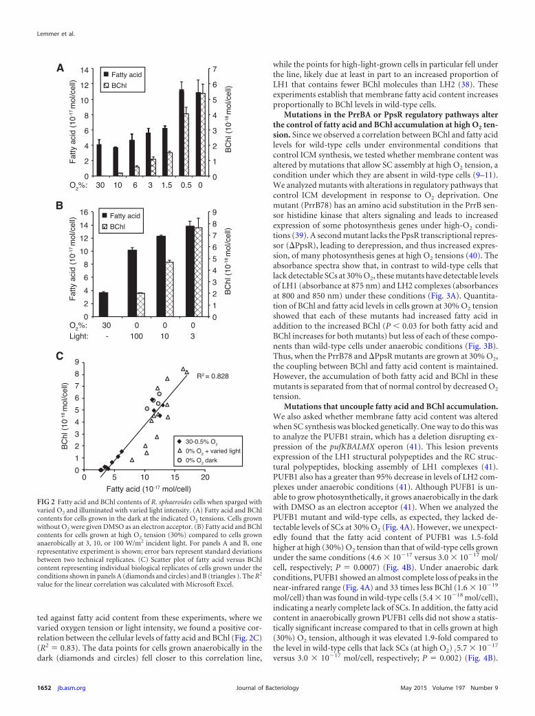
ted against fatty acid content from these experiments, where wevaried oxygen tension or light intensity, we found a positive cor-relation between the cellular levels of fatty acid and BChl (Fig. 2C)(R2 � 0.83). The data points for cells grown anaerobically in thedark (diamonds and circles) fell closer to this correlation line,
while the points for high-light-grown cells in particular fell underthe line, likely due at least in part to an increased proportion ofLH1 that contains fewer BChl molecules than LH2 (38). Theseexperiments establish that membrane fatty acid content increasesproportionally to BChl levels in wild-type cells.
Mutations in the PrrBA or PpsR regulatory pathways alterthe control of fatty acid and BChl accumulation at high O2 ten-sion. Since we observed a correlation between BChl and fatty acidlevels for wild-type cells under environmental conditions thatcontrol ICM synthesis, we tested whether membrane content wasaltered by mutations that allow SC assembly at high O2 tension, acondition under which they are absent in wild-type cells (9–11).We analyzed mutants with alterations in regulatory pathways thatcontrol ICM development in response to O2 deprivation. Onemutant (PrrB78) has an amino acid substitution in the PrrB sen-sor histidine kinase that alters signaling and leads to increasedexpression of some photosynthesis genes under high-O2 condi-tions (39). A second mutant lacks the PpsR transcriptional repres-sor (�PpsR), leading to derepression, and thus increased expres-sion, of many photosynthesis genes at high O2 tensions (40). Theabsorbance spectra show that, in contrast to wild-type cells thatlack detectable SCs at 30% O2, these mutants have detectable levelsof LH1 (absorbance at 875 nm) and LH2 complexes (absorbancesat 800 and 850 nm) under these conditions (Fig. 3A). Quantita-tion of BChl and fatty acid levels in cells grown at 30% O2 tensionshowed that each of these mutants had increased fatty acid inaddition to the increased BChl (P 0.03 for both fatty acid andBChl increases for both mutants) but less of each of these compo-nents than wild-type cells under anaerobic conditions (Fig. 3B).Thus, when the PrrB78 and �PpsR mutants are grown at 30% O2,the coupling between BChl and fatty acid content is maintained.However, the accumulation of both fatty acid and BChl in thesemutants is separated from that of normal control by decreased O2
tension.Mutations that uncouple fatty acid and BChl accumulation.
We also asked whether membrane fatty acid content was alteredwhen SC synthesis was blocked genetically. One way to do this wasto analyze the PUFB1 strain, which has a deletion disrupting ex-pression of the pufKBALMX operon (41). This lesion preventsexpression of the LH1 structural polypeptides and the RC struc-tural polypeptides, blocking assembly of LH1 complexes (41).PUFB1 also has a greater than 95% decrease in levels of LH2 com-plexes under anaerobic conditions (41). Although PUFB1 is un-able to grow photosynthetically, it grows anaerobically in the darkwith DMSO as an electron acceptor (41). When we analyzed thePUFB1 mutant and wild-type cells, as expected, they lacked de-tectable levels of SCs at 30% O2 (Fig. 4A). However, we unexpect-edly found that the fatty acid content of PUFB1 was 1.5-foldhigher at high (30%) O2 tension than that of wild-type cells grownunder the same conditions (4.6 � 10�17 versus 3.0 � 10�17 mol/cell, respectively; P � 0.0007) (Fig. 4B). Under anaerobic darkconditions, PUFB1 showed an almost complete loss of peaks in thenear-infrared range (Fig. 4A) and 33 times less BChl (1.6 � 10�19
mol/cell) than was found in wild-type cells (5.4 � 10�18 mol/cell),indicating a nearly complete lack of SCs. In addition, the fatty acidcontent in anaerobically grown PUFB1 cells did not show a statis-tically significant increase compared to that in cells grown at high(30%) O2 tension, although it was elevated 1.9-fold compared tothe level in wild-type cells that lack SCs (at high O2) (5.7 � 10�17
versus 3.0 � 10�17 mol/cell, respectively; P � 0.002) (Fig. 4B).
FIG 2 Fatty acid and BChl contents of R. sphaeroides cells when sparged withvaried O2 and illuminated with varied light intensity. (A) Fatty acid and BChlcontents for cells grown in the dark at the indicated O2 tensions. Cells grownwithout O2 were given DMSO as an electron acceptor. (B) Fatty acid and BChlcontents for cells grown at high O2 tension (30%) compared to cells grownanaerobically at 3, 10, or 100 W/m2 incident light. For panels A and B, onerepresentative experiment is shown; error bars represent standard deviationsbetween two technical replicates. (C) Scatter plot of fatty acid versus BChlcontent representing individual biological replicates of cells grown under theconditions shown in panels A (diamonds and circles) and B (triangles ). The R2
value for the linear correlation was calculated with Microsoft Excel.
Lemmer et al.
1652 jb.asm.org May 2015 Volume 197 Number 9Journal of Bacteriology

Thus, disruption of the pufKBALMX operon results in higher fattyacid content under both high-O2 and anaerobic conditions com-pared to the level in wild-type aerobic cells. This demonstrates thatPUFB1 is able to increase its lipid content relative to the wild typeeven though it does not assemble appreciable levels of SCs.
The properties of PUFB1 suggested that there were geneticalterations in which lipid accumulation could be separated fromassembly of ICM SCs. To test this hypothesis further, we examinedBChl and fatty acid levels in cells containing additional mutationsin known regulators of photosynthesis genes. We reasoned that ifO2-dependent increases in fatty acid levels require one or more ofthese regulatory pathways, then we should be able to identify amutant(s) in which SC assembly is disrupted but fatty acid accu-mulation is seen or vice versa. For these experiments, we analyzedpreviously characterized strains in which there is inactivation ofeither the PrrA transcriptional regulator (�PrrA), AppA, an anti-repressor that regulates PpsR transcriptional activity (�AppA), orthe FnrL transcriptional regulator (�FnrL) (Table 1) (19, 42, 43).The mutations in each of these strains reduce expression of somephotosynthesis genes and lower SC assembly at low O2: as a result,all of these mutants are unable to grow photosynthetically (19, 42,43). When grown anaerobically in the dark, unlike wild-type cells,the �AppA and �PrrA cells lack appreciable levels of SCs (42, 43).The �FnrL mutant is unable to grow anaerobically in the dark;when grown at low (2%) O2, it assembles SCs but to a lesser extentthan wild-type cells (19).
We used growth at low (0.5%) O2 to compare fatty acid andBChl levels in these mutants to those in wild-type cells, since allthree strains grow under these conditions, and our experimentsshowed that wild-type cells accumulated high levels of fatty acidand BChl under these conditions (Fig. 2A). We found that at 0.5%O2, both the �PrrA and �AppA mutants lacked detectable SCs(Fig. 5A) and had only trace amounts of BChl (Fig. 5B), similar towhat had previously been observed under anaerobic conditions(42, 43). The �FnrL mutant, however, assembled SCs at 0.5% O2
(Fig. 5A) with 71% of the total BChl (3.5 � 10�18 mol/cell versus5.4 � 10�18 mol/cell in the wild type) (Fig. 5B). When wild-typecells were grown at low (0.5%) O2, the increase in fatty acid con-tent was 3.1-fold relative to the level in cells grown at high (30%)
FIG 3 SC, BChl, and fatty acid contents of R. sphaeroides mutants that activatephotosynthesis gene expression in the presence of oxygen (PrrB78 and �PpsR)compared to wild-type (WT) cells. (A) Absorbance scans of intact cells to showSC assembly. (B) Fatty acid and BChl contents of wild-type, �PpsR, andPrrB78 cells grown in the presence of 30% O2 compared to wild-type cellsgrown anaerobically at 10 W/m2 light. Asterisks indicate that the increases infatty acid and BChl contents in both mutants relative to wild-type cells underthe same condition are statistically significant at P 0.03. Data represent theaverage from three biological replicates from independent experiments; errorbars indicate standard deviation.
FIG 4 Fatty acid and SC levels in the PUFB1 mutant. (A) Absorption spectra of wild-type (WT) and PUFB1 cells grown at 30% O2 tension (top) andanaerobically in the dark (bottom). (B) Fatty acid content of cells under the same conditions shown in panel A. Data represent the average from three biologicalreplicates; error bars indicate standard deviation. �, P � 0.0025; ��, P � 0.0035; NS, not significant.
Control of Lipid Production
May 2015 Volume 197 Number 9 jb.asm.org 1653Journal of Bacteriology
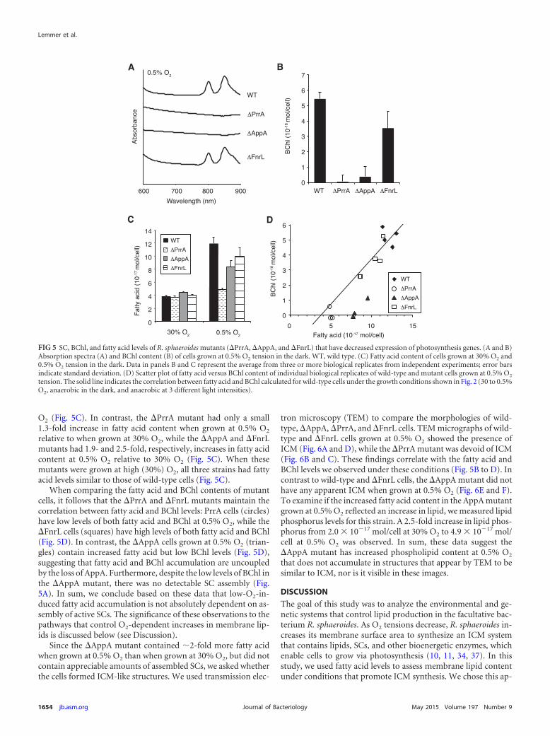
O2 (Fig. 5C). In contrast, the �PrrA mutant had only a small1.3-fold increase in fatty acid content when grown at 0.5% O2
relative to when grown at 30% O2, while the �AppA and �FnrLmutants had 1.9- and 2.5-fold, respectively, increases in fatty acidcontent at 0.5% O2 relative to 30% O2 (Fig. 5C). When thesemutants were grown at high (30%) O2, all three strains had fattyacid levels similar to those of wild-type cells (Fig. 5C).
When comparing the fatty acid and BChl contents of mutantcells, it follows that the �PrrA and �FnrL mutants maintain thecorrelation between fatty acid and BChl levels: PrrA cells (circles)have low levels of both fatty acid and BChl at 0.5% O2, while the�FnrL cells (squares) have high levels of both fatty acid and BChl(Fig. 5D). In contrast, the �AppA cells grown at 0.5% O2 (trian-gles) contain increased fatty acid but low BChl levels (Fig. 5D),suggesting that fatty acid and BChl accumulation are uncoupledby the loss of AppA. Furthermore, despite the low levels of BChl inthe �AppA mutant, there was no detectable SC assembly (Fig.5A). In sum, we conclude based on these data that low-O2-in-duced fatty acid accumulation is not absolutely dependent on as-sembly of active SCs. The significance of these observations to thepathways that control O2-dependent increases in membrane lip-ids is discussed below (see Discussion).
Since the �AppA mutant contained �2-fold more fatty acidwhen grown at 0.5% O2 than when grown at 30% O2, but did notcontain appreciable amounts of assembled SCs, we asked whetherthe cells formed ICM-like structures. We used transmission elec-
tron microscopy (TEM) to compare the morphologies of wild-type, �AppA, �PrrA, and �FnrL cells. TEM micrographs of wild-type and �FnrL cells grown at 0.5% O2 showed the presence ofICM (Fig. 6A and D), while the �PrrA mutant was devoid of ICM(Fig. 6B and C). These findings correlate with the fatty acid andBChl levels we observed under these conditions (Fig. 5B to D). Incontrast to wild-type and �FnrL cells, the �AppA mutant did nothave any apparent ICM when grown at 0.5% O2 (Fig. 6E and F).To examine if the increased fatty acid content in the AppA mutantgrown at 0.5% O2 reflected an increase in lipid, we measured lipidphosphorus levels for this strain. A 2.5-fold increase in lipid phos-phorus from 2.0 � 10�17 mol/cell at 30% O2 to 4.9 � 10�17 mol/cell at 0.5% O2 was observed. In sum, these data suggest the�AppA mutant has increased phospholipid content at 0.5% O2
that does not accumulate in structures that appear by TEM to besimilar to ICM, nor is it visible in these images.
DISCUSSION
The goal of this study was to analyze the environmental and ge-netic systems that control lipid production in the facultative bac-terium R. sphaeroides. As O2 tensions decrease, R. sphaeroides in-creases its membrane surface area to synthesize an ICM systemthat contains lipids, SCs, and other bioenergetic enzymes, whichenable cells to grow via photosynthesis (10, 11, 34, 37). In thisstudy, we used fatty acid levels to assess membrane lipid contentunder conditions that promote ICM synthesis. We chose this ap-
FIG 5 SC, BChl, and fatty acid levels of R. sphaeroides mutants (�PrrA, �AppA, and �FnrL) that have decreased expression of photosynthesis genes. (A and B)Absorption spectra (A) and BChl content (B) of cells grown at 0.5% O2 tension in the dark. WT, wild type. (C) Fatty acid content of cells grown at 30% O2 and0.5% O2 tension in the dark. Data in panels B and C represent the average from three or more biological replicates from independent experiments; error barsindicate standard deviation. (D) Scatter plot of fatty acid versus BChl content of individual biological replicates of wild-type and mutant cells grown at 0.5% O2
tension. The solid line indicates the correlation between fatty acid and BChl calculated for wild-type cells under the growth conditions shown in Fig. 2 (30 to 0.5%O2, anaerobic in the dark, and anaerobic at 3 different light intensities).
Lemmer et al.
1654 jb.asm.org May 2015 Volume 197 Number 9Journal of Bacteriology

proach since R. sphaeroides is not known to accumulate or containgenes annotated to encode enzymes for, neutral lipid synthesis(such as triacylglycerols [TAGs] or wax esters) (44). To furthervalidate this approach, we found the expected 2:1 ratio of fatty acidto lipid phosphorus. Previous studies have demonstrated thatthere are not significant differences in the phospholipid or fattyacid composition between aerobic and anaerobic cells (32, 45),therefore, we focused on the difference in absolute levels of fattyacids. Our fatty acid analysis method also allowed us to assess fattyacid composition, which was not found to vary significantly be-tween the conditions or strains used in this study.
We show here that conditions that promote ICM synthesis alsocause cells to increase their total lipid content. At low O2 tension(0.5%), as well as anaerobically in the dark or at moderate lightintensity, this increase in lipid content is approximately 3-fold com-pared to that in cells grown at high O2, which are devoid of ICM. Wealso found that a decrease in O2 tension is sufficient for this increasedlipid accumulation, indicating that light is not needed for this re-sponse. Below we summarize the new information we have obtainedby analyzing the environmental and genetic controls that allow thisbacterium to increase its membrane surface area.
Uncoupling of membrane lipid and SC accumulation. Ourdata demonstrate a correlation between fatty acid and BChl levels
under physiological conditions that control ICM synthesis (Fig.2C), indicating there is a direct relationship between the amountof membrane lipid and assembled SCs. This correlation is notsurprising, since the increased membrane surface area is thoughtto house the SCs and other integral membrane enzymes of theICM. However, the systems that link fatty acid levels to those ofICM SCs are still unresolved. For example, it is possible that in-creased expression of an ICM protein(s) is sufficient to drive in-creased membrane synthesis. This is illustrated by observations inEscherichia coli, where overexpression of individual integral mem-brane proteins, including fumarate reductase (46), the glycerolacyl transferase PlsB (47), or the ATP synthase b subunit (48), issufficient to induce formation of intracellular membranes. In-deed, we found that when synthesis of SCs was increased by mu-tation under high O2 tension, when they are not normally present,membrane lipid content also increased (Fig. 3A and B). Thus, it ispossible that increased membrane content at low O2 is an indirectconsequence of inserting a large amount of SCs or other integralmembrane proteins into the ICM.
However, when assembly of SCs was blocked at low O2 in the�AppA mutant, this strain contained �2-fold-higher fatty acidand lipid phosphorus levels than when grown at high O2 (Fig. 5Aand C). This observation suggests that there is a mechanism to
FIG 6 TEM micrographs of thin sections of the R. sphaeroides wild type (WT) and mutant (�PrrA, �AppA, and �FnrL) cells grown at 0.5% O2 tension. Thearrows in the wild-type and �FnrL panels indicate examples of the lighter-colored spherical ICM structures. The larger white cellular structures representpolyhydroxybutyrate granules, which are present in all strains but not in each cross-section of a cell. A representative image is shown for each strain (A, B, D, andE), with additional lower-magnification images included for �PrrA (C) and �AppA (F) to show more cells.
Control of Lipid Production
May 2015 Volume 197 Number 9 jb.asm.org 1655Journal of Bacteriology

increase membrane content in response to low O2 tension that isindependent of SC insertion into the ICM. This notion is alsosupported by observations of the de novo ICM assembly that oc-curs when cells are shifted from aerobic to anaerobic growth con-ditions (9). Under these conditions, new ICM invaginations formbefore BChl accumulation is detected. Furthermore, these nascentICM invaginations appear smooth when analyzed by freeze frac-ture electron microscopy, suggesting they are enriched in phos-pholipid, in contrast to mature photosynthetic membranes thatcontain a high level of particulate membrane-bound pigment-protein complexes (9).
Potential regulators of membrane lipid accumulation. TheR. sphaeroides transcriptional networks regulating gene expres-sion in response to decreased O2 tension contain many operonsencoding proteins that are necessary for the assembly of func-tional photosynthetic membranes (12). In this study, we used mu-tations in each of these pathways to test their role in the accumu-lation of membrane lipids at low O2 tension.
For some mutants, the normal linkage between fatty acidand BChl levels was maintained at 0.5% O2. Specifically, the�PrrA mutant had low levels of both lipid and pigment, whilethe �FnrL mutant has high levels of both lipid and pigmentunder these conditions. In contrast, the �AppA mutant has nodetectable SCs but high levels of fatty acid at 0.5% O2, suggest-ing that this lesion uncouples the linkage between SCs andmembrane lipid content. This observation suggests that whileAppA is necessary for SC assembly, it is not necessary for in-creased lipid levels at low O2 since �AppA cells still exhibit anincrease in fatty acid at 0.5% O2 compared to cells grown at30% O2. Since AppA acts as an antirepressor of the transcrip-tion factor PpsR (18), this suggests that depression of the tran-scriptional target genes of PpsR is not necessary for increasedfatty acid at low O2. It is also possible that deletion of AppAleads to derepression of a regulator of fatty acid biosynthesisindependent of PpsR. However, fatty acid levels are not signif-icantly increased in the �AppA mutant when it is sparged with30% O2. Thus, we do not think it is likely that the increase infatty acid levels seen in the �AppA mutant is due to directderepression of a regulator of fatty acid synthesis. Finally, ourdata suggest that FnrL is not required for either increasedmembrane lipid or SC synthesis at low O2, while the PrrBApathway is required for both. In sum, we conclude that tran-scriptional regulation by FnrL and the AppA-PpsR pathway aredispensable for low-O2-induced fatty acid accumulation, sincethe �AppA and �FnrL mutant strains still increased fatty acidcontent at least 2-fold under low O2 tension compared to highO2 tension (Fig. 5C), while the PrrBA pathway is required. Wecan infer from this that one or more genes within the PrrAregulon either directly or indirectly are necessary for cells toincrease membrane content for ICM development. To datethere are not any members of this regulon that are known orpredicted to participate in or regulate fatty acid and phospho-lipid biosynthesis.
Our finding that at 0.5% O2 the �FnrL mutant assembles SCs,has 71% of the BChl and 83% of the fatty acid as wild-type cells,and forms ICM was somewhat unexpected since previous analysisfound that this mutant has much lower SC levels than the wildtype at 2% O2 tension (49) and formed many fewer ICM invagi-nations than wild-type cells in poorly aerated cultures (50). Thesedifferences may be due to the different O2 tensions used between
our studies and previous experiments and may indicate that loss ofFnrL changes the sensitivity of cells to decreased O2 for inductionof ICM SC and lipid synthesis. However, from our data at 0.5%O2, we conclude that FnrL is not strictly essential for low-O2-induced lipid accumulation.
Relevance to lipid production in other microbes. Our datademonstrate that R. sphaeroides has a mechanism to increase lipidcontent in response to decreased O2 tension, which is at least par-tially dependent on an intact PrrBA pathway. This is significantand relevant to both ICM assembly and the potential engineeringof microbes for production of lipid biofuels and chemicals, be-cause it identifies a transcriptional regulatory pathway that canincrease microbial lipid content. In most microbes that accumu-late storage lipids, such as triacylglycerols (TAGs), accumulationis normally induced by nutrient limitation or other stresses (3–5).Under such conditions, the gene expression changes contributingto increased lipid storage have not been elucidated. For exam-ple, in the oleaginous microalgae Cytotella cryptica under siliconstarvation conditions that lead to TAG accumulation, the firstdedicated enzyme in fatty acid biosynthesis, acetyl coenzyme A(acetyl-CoA) carboxylase (ACCase), has elevated gene expressionand enzyme activity (51, 52). However, overexpression of ACCasealone, in this and other microalgae, does not lead to increasedTAG accumulation (52). Other studies in microalgae, yeast, andbacteria have used similar approaches with more success, target-ing deletion and/or overexpression of enzymes both singly and incombination to increase the yield of TAGs or other fatty-acid-derived fuels (7, 53). While the success of individual studies varies,this approach requires the manipulation of many different genesto maximize product yield. Instead, an approach that altered ac-tivity of a transcriptional regulator would be advantageous, as itcould potentially alter the expression of many relevant genes af-fecting a metabolic pathway with one genetic manipulation andalso be used in organisms where the enzymes of fatty acid biosyn-thesis and degradation are not well characterized. In support ofthis idea, a recent study demonstrated that in E. coli, cells engi-neered to produce free fatty acids, homologous expression of theFadR transcription factor, a negative regulator of fatty acid degra-dation and positive regulator of biosynthesis, increased fatty acidyield 7.5-fold (54). Elucidating additional regulatory networkscontrolling fatty acid and lipid accumulation in other microbeswill facilitate achieving of the increased product yields necessaryfor economical large-scale production of biofuels and chemicals.
Our studies in R. sphaeroides identify a novel oxygen-regulatedmechanism of lipid accumulation that is dependent, at least inpart, on the PrrA global transcriptional regulator. Future studieswill be directed at determining the relationship of the PrrBA path-way and other yet to be discovered pathways to increased lipidaccumulation and how the knowledge gained from studying thesesystems might be applied to microbial production of renewablefuels and chemicals.
ACKNOWLEDGMENTS
This work was supported by DOE Great Lakes Bioenergy Research Centergrant (DOE Office of Science BER DE-FC02-07ER64494) to T.J.D. andUSDA NIFA fellowship 2011-67012-30702 to K.C.L. Electron microscopywas performed at the Environmental Molecular Science Laboratory(EMSL), a DOE Office of Science user facility sponsored by the Office ofBiological and Environmental Research and located at PNNL.
We thank Mark Gomelsky for providing strains APP11 (�AppA) and
Lemmer et al.
1656 jb.asm.org May 2015 Volume 197 Number 9Journal of Bacteriology
PPS1 (�PpsR) and Jill Zeilstra-Ryalls for providing strain JZ1678(�FnrL).
REFERENCES1. Li Q, Du W, Liu D. 2008. Perspectives of microbial oils for biodiesel
production. Appl Microbiol Biotechnol 80:749 –756. http://dx.doi.org/10.1007/s00253-008-1625-9.
2. Meng X, Yang J, Xu X, Zhang L, Nie Q, Xian M. 2009. Biodieselproduction from oleaginous microorganisms. Renewable Energy 34:1–5.http://dx.doi.org/10.1016/j.renene.2008.04.014.
3. Blatti JL, Michaud J, Burkart MD. 2013. Engineering fatty acid biosyn-thesis in microalgae for sustainable biodiesel. Curr Opin Chem Biol 17:496 –505. http://dx.doi.org/10.1016/j.cbpa.2013.04.007.
4. Alvarez HM, Steinbuchel A. 2002. Triacylglycerols in prokaryotic micro-organisms. Appl Microbiol Biotechnol 60:367–376. http://dx.doi.org/10.1007/s00253-002-1135-0.
5. Beopoulos A, Nicaud JM, Gaillardin C. 2011. An overview of lipidmetabolism in yeasts and its impact on biotechnological processes. ApplMicrobiol Biotechnol 90:1193–1206. http://dx.doi.org/10.1007/s00253-011-3212-8.
6. Kosa M, Ragauskas AJ. 2011. Lipids from heterotrophic microbes: ad-vances in metabolism research. Trends Biotechnol 29:53– 61. http://dx.doi.org/10.1016/j.tibtech.2010.11.002.
7. Liang MH, Jiang JG. 2013. Advancing oleaginous microorganisms toproduce lipid via metabolic engineering technology. Prog Lipid Res 52:395– 408. http://dx.doi.org/10.1016/j.plipres.2013.05.002.
8. Melandri AB, Zannoni D. 1978. Photosynthetic and respiratory electronflow in the dual functional membrane of facultative photosynthetic bac-teria. J Bioenerg Biomembr 10:109 –138. http://dx.doi.org/10.1007/BF00743056.
9. Chory J, Donohue TJ, Varga AR, Staehelin LA, Kaplan S. 1984. Induc-tion of the photosynthetic membranes of Rhodopseudomonas sphaeroides:biochemical and morphological studies. J Bacteriol 159:540 –554.
10. Tavano CL, Donohue TJ. 2006. Development of the bacterial photosyn-thetic apparatus. Curr Opin Microbiol 9:625– 631. http://dx.doi.org/10.1016/j.mib.2006.10.005.
11. Kiley PJ, Kaplan S. 1988. Molecular genetics of photosynthetic mem-brane biosynthesis in Rhodobacter sphaeroides. Microbiol Rev 52:50 – 69.
12. Zeilstra-Ryalls JH, Kaplan S. 2004. Oxygen intervention in the regulationof gene expression: the photosynthetic bacterial paradigm. Cell Mol LifeSci 61:417– 436. http://dx.doi.org/10.1007/s00018-003-3242-1.
13. Zeilstra-Ryalls J, Gomelsky M, Eraso JM, Yeliseev A, O’Gara J, KaplanS. 1998. Control of photosystem formation in Rhodobacter sphaeroides. JBacteriol 180:2801–2809.
14. Arai H, Roh JH, Kaplan S. 2008. Transcriptome dynamics during thetransition from anaerobic photosynthesis to aerobic respiration in Rhodo-bacter sphaeroides 2.4.1. J Bacteriol 190:286 –299. http://dx.doi.org/10.1128/JB.01375-07.
15. Gomelsky L, Moskvin OV, Stenzel RA, Jones DF, Donohue TJ, Gomel-sky M. 2008. Hierarchical regulation of photosynthesis gene expression bythe oxygen-responsive PrrBA and AppA-PpsR systems of Rhodobactersphaeroides. J Bacteriol 190:8106 – 8114. http://dx.doi.org/10.1128/JB.01094-08.
16. Comolli JC, Carl AJ, Hall C, Donohue T. 2002. Transcriptional activa-tion of the Rhodobacter sphaeroides cytochrome c2 gene P2 promoter bythe response regulator PrrA. J Bacteriol 184:390 –399. http://dx.doi.org/10.1128/JB.184.2.390-399.2002.
17. Potter CA, Ward A, Laguri C, Williamson MP, Henderson PJ, Phillips-Jones MK. 2002. Expression, purification and characterisation of full-length histidine protein kinase RegB from Rhodobacter sphaeroides. J MolBiol 320:201–213. http://dx.doi.org/10.1016/S0022-2836(02)00424-2.
18. Elsen S, Jaubert M, Pignol D, Giraud E. 2005. PpsR: a multifacetedregulator of photosynthesis gene expression in purple bacteria. Mol Mi-crobiol 57:17–26. http://dx.doi.org/10.1111/j.1365-2958.2005.04655.x.
19. Zeilstra-Ryalls JH, Kaplan S. 1995. Aerobic and anaerobic regulation inRhodobacter sphaeroides 2.4.1: the role of the fnrL gene. J Bacteriol 177:6422– 6431.
20. Dufour YS, Imam S, Koo BM, Green HA, Donohue TJ. 2012. Conver-gence of the transcriptional responses to heat shock and singlet oxygenstresses. PLoS Genet 8:e1002929. http://dx.doi.org/10.1371/journal.pgen.1002929.
21. Dufour YS, Kiley PJ, Donohue TJ. 2010. Reconstruction of the core and
extended regulons of global transcription factors. PLoS Genet 6:e1001027.http://dx.doi.org/10.1371/journal.pgen.1001027.
22. Bruscella P, Eraso JM, Roh JH, Kaplan S. 2008. The use of chromatinimmunoprecipitation to define PpsR binding activity in Rhodobactersphaeroides 2.4.1. J Bacteriol 190:6817– 6828. http://dx.doi.org/10.1128/JB.00719-08.
23. Karls RK, Wolf JR, Donohue TJ. 1999. Activation of the cycA P2 pro-moter for the Rhodobacter sphaeroides cytochrome c2 gene by the photo-synthesis response regulator. Mol Microbiol 34:822– 835. http://dx.doi.org/10.1046/j.1365-2958.1999.01649.x.
24. Onishi JC, Niederman RA. 1982. Rhodopseudomonas sphaeroides mem-branes: alterations in phospholipid composition in aerobically and pho-totrophically grown cells. J Bacteriol 149:831– 839.
25. Lascelles J, Szilagyi JF. 1965. Phospholipid synthesis by Rhodopseudo-monas spheroides in relation to the formation of photosynthetic pigments.J Gen Microbiol 38:55– 64. http://dx.doi.org/10.1099/00221287-38-1-55.
26. Imam S, Yilmaz S, Sohmen U, Gorzalski AS, Reed JL, Noguera DR,Donohue TJ. 2011. iRsp1095: a genome-scale reconstruction of the Rho-dobacter sphaeroides metabolic network. BMC Syst Biol 5:116. http://dx.doi.org/10.1186/1752-0509-5-116.
27. Sistrom WR. 1960. A requirement for sodium in the growth of Rhodo-pseudomonas spheroides. J Gen Microbiol 22:778 –785. http://dx.doi.org/10.1099/00221287-22-3-778.
28. Clayton RK. 1966. Spectroscopic analysis of bacteriochlorophylls in vitroand in vivo. Photochem Photobiol 5:669 – 677. http://dx.doi.org/10.1111/j.1751-1097.1966.tb05813.x.
29. Lennon CW, Lemmer KC, Irons JL, Sellman MI, Donohue TJ, GourseRL, Ross W. 2014. A Rhodobacter sphaeroides protein mechanisticallysimilar to Escherichia coli DksA regulates photosynthetic growth. mBio5(3):e01105-14. http://dx.doi.org/10.1128/mBio.01105-14.
30. Rouser G, Fkeischer S, Yamamoto A. 1970. Two dimensional thin layerchromatographic separation of polar lipids and determination of phos-pholipids by phosphorus analysis of spots. Lipids 5:494 – 496. http://dx.doi.org/10.1007/BF02531316.
31. Reynolds ES. 1963. The use of lead citrate at high pH as an electron-opaque stain in electron microscopy. J Cell Biol 17:208 –212. http://dx.doi.org/10.1083/jcb.17.1.208.
32. Russell NJ, Harwood JL. 1979. Changes in the acyl lipid composition ofphotosynthetic bacteria grown under photosynthetic and non-photosynthetic conditions. Biochem J 181:339 –345.
33. Donohue TJ, Cain BD, Kaplan S. 1982. Purification and characterizationof an N-acylphosphatidylserine from Rhodopseudomonas sphaeroides.Biochemistry 21:2765–2773. http://dx.doi.org/10.1021/bi00540a029.
34. Niederman RA. 2013. Membrane development in purple photosyn-thetic bacteria in response to alterations in light intensity and oxygentension. Photosynth Res 116:333–348. http://dx.doi.org/10.1007/s11120-013-9851-0.
35. Chory J, Kaplan S. 1983. Light-dependent regulation of the synthesis ofsoluble and intracytoplasmic membrane proteins of Rhodopseudomonassphaeroides. J Bacteriol 153:465– 474.
36. Adams PG, Hunter CN. 2012. Adaptation of intracytoplasmic mem-branes to altered light intensity in Rhodobacter sphaeroides. Biochim Bio-phys Acta 1817:1616 –1627. http://dx.doi.org/10.1016/j.bbabio.2012.05.013.
37. Yen GS, Cain BD, Kaplan S. 1984. Cell-cycle-specific biosynthesis of thephotosynthetic membrane of Rhodopseudomonas sphaeroides. Structuralimplications. Biochim Biophys Acta 777:41–55. http://dx.doi.org/10.1016/0005-2736(84)90495-4.
38. Cogdell RJ, Gall A, Kohler J. 2006. The architecture and function of thelight-harvesting apparatus of purple bacteria: from single molecules to invivo membranes. Q Rev Biophys 39:227–324. http://dx.doi.org/10.1017/S0033583506004434.
39. Eraso JM, Kaplan S. 1995. Oxygen-insensitive synthesis of the photosyn-thetic membranes of Rhodobacter sphaeroides: a mutant histidine kinase. JBacteriol 177:2695–2706.
40. Gomelsky M, Kaplan S. 1997. Molecular genetic analysis suggestinginteractions between AppA and PpsR in regulation of photosynthesisgene expression in Rhodobacter sphaeroides 2.4.1. J Bacteriol 179:128 –134.
41. Davis J, Donohue TJ, Kaplan S. 1988. Construction, characterization,and complementation of a Puf� mutant of Rhodobacter sphaeroides. JBacteriol 170:320 –329.
42. Gomelsky M, Kaplan S. 1995. appA, a novel gene encoding a trans-acting
Control of Lipid Production
May 2015 Volume 197 Number 9 jb.asm.org 1657Journal of Bacteriology
factor involved in the regulation of photosynthesis gene expression inRhodobacter sphaeroides 2.4.1. J Bacteriol 177:4609 – 4618.
43. Eraso JM, Kaplan S. 1994. prrA, a putative response regulator involved inoxygen regulation of photosynthesis gene expression in Rhodobacter spha-eroides. J Bacteriol 176:32– 43.
44. Kontur WS, Schackwitz WS, Ivanova N, Martin J, Labutti K, Desh-pande S, Tice HN, Pennacchio C, Sodergren E, Weinstock GM, Nogu-era DR, Donohue TJ. 2012. Revised sequence and annotation of theRhodobacter sphaeroides 2.4.1 genome. J Bacteriol 194:7016 –7017. http://dx.doi.org/10.1128/JB.01214-12.
45. Donohue TJ, Cain BD, Kaplan S. 1982. Alterations in the phospholipidcomposition of Rhodopseudomonas sphaeroides and other bacteria in-duced by Tris. J Bacteriol 152:595– 606.
46. Weiner JH, Lemire BD, Elmes ML, Bradley RD, Scraba DG. 1984.Overproduction of fumarate reductase in Escherichia coli induces a novelintracellular lipid-protein organelle. J Bacteriol 158:590 –596.
47. Wilkison WO, Walsh JP, Corless JM, Bell RM. 1986. Crystalline arraysof the Escherichia coli sn-glycerol-3-phosphate acyltransferase, an integralmembrane protein. J Biol Chem 261:9951–9958.
48. Arechaga I, Miroux B, Karrasch S, Huijbregts R, de Kruijff B, RunswickMJ, Walker JE. 2000. Characterisation of new intracellular membranes inEscherichia coli accompanying large scale over-production of the b subunitof F1Fo ATP synthase. FEBS Lett 482:215–219. http://dx.doi.org/10.1016/S0014-5793(00)02054-8.
49. Zeilstra-Ryalls JH, Gabbert K, Mouncey NJ, Kaplan S, Kranz RG. 1997.
Analysis of the fnrL gene and its function in Rhodobacter capsulatus. JBacteriol 179:7264 –7273.
50. Fedotova Y, Zeilstra-Ryalls J. 2014. Analysis of the role of PrrA, PpsR,and FnrL in intracytoplasmic membrane differentiation of Rhodobactersphaeroides 241 using transmission electron microscopy. Photosynth Res119:283–290. http://dx.doi.org/10.1007/s11120-013-9944-9.
51. Roessler PG. 1988. Changes in the activities of various lipid and carbohy-drate biosynthetic enzymes in the diatom Cyclotella cryptica in response tosilicon deficiency. Arch Biochem Biophys 267:521–528. http://dx.doi.org/10.1016/0003-9861(88)90059-8.
52. Sheehan J, Dunahay T, Bennemann J, Roessler P. 1998. A look back atthe U.S. Department of Energy’s aquatic species program: biodiesel fromalgae; close-out report. Technical report. NREL/TP-580-24190. NationalRenewable Energy Laboratory, Golden, CO.
53. Janssen HJ, Steinbuchel A. 2014. Fatty acid synthesis in Escherichia coliand its applications towards the production of fatty acid based biofuels.Biotechnol Biofuels 7:7. http://dx.doi.org/10.1186/1754-6834-7-7.
54. Zhang F, Ouellet M, Batth TS, Adams PD, Petzold CJ, MukhopadhyayA, Keasling JD. 2012. Enhancing fatty acid production by the expressionof the regulatory transcription factor FadR. Metab Eng 14:653– 660. http://dx.doi.org/10.1016/j.ymben.2012.08.009.
55. van Niel CB. 1944. The culture, general physiology, morphology, andclassification of the non-sulfur purple and brown bacteria. Bacteriol Rev8:1–118.
Lemmer et al.
1658 jb.asm.org May 2015 Volume 197 Number 9Journal of Bacteriology
Top Related
Copyright © 2022 FDOKUMEN

