


Bahasa
Halaman
Hukum

ORIGINAL ARTICLE
Oculocutaneous albinism type 2 (OCA2) with homozygous 2.7-kbdeletion of the P gene and sickle cell disease in a Cameroonianfamily. Identification of a common TAG haplotype in the mutatedP gene
Robert Aquaron Æ Nadem Soufir ÆJean-Louis Berge-Lefranc Æ Catherine Badens ÆFrederic Austerlitz Æ Bernard Grandchamp
Received: 29 May 2007 / Accepted: 20 July 2007 / Published online: 1 September 2007
� The Japan Society of Human Genetics and Springer 2007
Abstract In this study, we report on a Cameroonian family
from the Ewondo ethnic group, presenting with three ocu-
locutaneous albinism type 2 (OCA2) patients homozygous
for the 2.7-kb deletion of the P gene. In one of these patients
OCA2 was associated with sickle cell anaemia and in two
with the sickle cell trait. We took this opportunity to deter-
mine single nucleotide polymorphism (SNP) haplotypes
within the P gene in this family in comparison with a group
of 53 OCA2 patients homozygous for the same mutation and
with a matched unrelated full-coloured control group of 49
subjects, originating from seven different ethnic groups of
Southern Cameroon including Ewondo. A combination of
five exonic and intronic SNPs in the OCA2 gene was
genotyped by sequencing PCR products. We found 3 dif-
ferent haplotypes (TAGCT, TAGTT and TAGCC with
frequencies of 0.66, 0.28 and 0.06, respectively) associated
with the mutation in the 53 OCA2 patients, while 11 dif-
ferent haplotypes were observed in the control group. These
observations suggest that the mutation appeared on the rel-
atively frequent haplotype TAGCT, and that the two other
haplotypes are derived from two independent recombination
events. These haplotypic data, associated with a value of 1/
15,000 for the prevalence of the 2.7-kb mutation, a present
effective population size of 10,000,000 for Cameroon and a
recombination rate of 0.0031, allowed us to estimate that this
mutation originated 4,100–5,645 years ago.
Keywords OCA2 � Sickle cell disease � P gene �bS-globin gene � SNPs � Cameroon
Introduction
Type 2 oculocutaneous albinism (OCA2; MIM#
2032000)—an autosomal recessive disorder in which the
biosynthesis of melanin pigment is reduced in the skin, hair
and eyes—is associated with the ocular features of nys-
tagmus, reduced visual acuity and misrouting of the optic
fibres at the chiasma. OCA2 results from mutations in the P
gene—the human homologue of the mouse pink-eye dilu-
tion (p) gene on chromosome segment 15q11.2–q12, which
contains 25 exons, 23 of which are coding. This region
encodes a 110-kDa protein integral to the melanosomal
membrane with a predicted 12-transmembrane domain
structure, resembling a channel or transporter (Rosemblat
et al. 1994). Despite the critical role played by this 838-
amino-acid P polypeptide in controlling tyrosinase pro-
cessing and melanosome biogenesis, its precise biological
function is still not defined. However, it is known to be
required for normal pigmentation (Chen et al. 2002). OCA2
R. Aquaron (&)
Laboratoire de Biochimie et Biologie Moleculaire,
Faculte de Medecine, Universite de la Mediterranee
Aix-Marseille II, 27 Boulevard Jean Moulin,
13385 Marseille, Cedex 5, France
e-mail: [email protected]
N. Soufir � B. Grandchamp
Biochimie hormonale et Genetique, AP-HP,
Hopital Bichat-Claude Bernard, Paris, France
J.-L. Berge-Lefranc
Laboratoire de Biochimie et Biologie Moleculaire,
Hopital de la Conception, Marseille, France
C. Badens
Faculte de Medecine, Centre d’Enseignement et de
Recherche en Genetique Medicale, Marseille, France
F. Austerlitz
Laboratoire Ecologie, Systematique et Evolution,
UMR CNRS/UPS/ENGREF 8079,
Universite Paris Sud, Orsay, France
123
J Hum Genet (2007) 52:771–780
DOI 10.1007/s10038-007-0181-y
is the most common form of albinism, especially among
Africans and African-Americans. There is a high incidence
of OCA2 among specific African populations: 1 in 1,100
among the Ibo of Nigeria (Okoro 1975), 1 in 3,900 in South
Africa (Kromberg and Jenkins 1982), 1 in 4,100 in Tanzania
(Luande et al. 1985), 1 in 4,882 in Zimbabwe (Lund 1996),
1 in 5,000 in Nigeria (Barnicot, 1952), 1 in 7,900 among the
Bamileke of Cameroon (Aquaron 1980, 1990) and 1 in
10,000 in African-Americans (Durham-Pierre et al. 1996).
The most common mutation in the P gene among Africans
and African-Americans is a 2.7-kb deletion that removes a
single exon (exon 7; amino acids 216–270) and results in a
frameshift mutation in the first luminal loop of the P poly-
peptide, producing a truncated and non-functional gene
product (Durham-Pierre et al. 1994). This mutation was first
detected in the homozygous state in a large family with
seven albinos from an inbred population of triracial (Black,
Caucasoid, and American-Indian) origin. This mutant P
allele was also detected, in the heterozygous state, in four
unrelated African-American OCA2 individuals, in a Zai-
rean patient and in a Cameroonian patient, but not in any
Caucasoids with OCA2, indicating an African origin for this
allele (Durham-Pierre et al. 1994). This deletion allele is
associated with a common haplotype, suggesting a founder
effect (Stevens et al. 1995, 1997), and represents a high
proportion of mutated P alleles in central, eastern and
southern African countries: 33% (4/12 chromosomes) in the
Central African Republic (Stevens et al. 1997), 65% (47/72
chromosomes) to 67% (233/346 chromosomes) in Camer-
oon (Puri et al. 1997; Aquaron and Berge-Lefranc 2002),
77% (20/26 chromosomes) in Tanzania (Spritz et al. 1995),
77% (131/170 chromosomes) in South Africa (Stevens et al.
1995), 79% (11/14 chromosomes) in Zambia (Stevens et al.
1995) and 94% (60/64 chromosomes) in Zimbabwe (Puri
et al. 1997). In contrast, this mutant P allele has never been
found in 30 OCA2 patients from west African countries: 1
albinism case in Nigeria (Spritz et al. 1995), and 29 cases in
Niger, Mali, Togo, Burkina Faso (R. Aquaron and J.-L.
Berge-Lefranc, unpublished results).
Sickle cell disease is an autosomal recessive disorder
(MIM # 603903) characterised by chronic anemia with a
haemoglobin concentration of around 80 g/l, painful
swelling of the hands and/or feet at between 6 and
18 months of age. Survivors may also suffer recurrent and
unpredictable severe painful crises as well as pneumonia or
pulmonary infarction, bone or joint necrosis, cerebrovas-
cular accidents or renal failure. Patients with sickle cell
anemia are homozygous (SS) for a missense mutation (c.19
A [ T ) in the b-globin gene located on chromosome
11p15.5 that leads to replacement of the hydrophilic neg-
atively charged amino acid (Glu) by the hydrophobic
neutral amino acid (Val) (p.E6V) in the adult haemoglobin
protein HbA, giving rise to the abnormal protein HbS.
Heterozygous individuals (AS) are not symptomatic and
have sickle-cell trait. The prevalence of sickle-cell trait
ranges from 9.3 to 28.2% in Cameroon depending on
ethnic group: 9.3% in albino Bamileke, 15.7% in black
Bamileke, 17.1% in Pygmies, 20.7% in Ewondo and 28.2%
in Eton (Gamet and Labes 1964; Nguematcha et al. 1973;
Juhan and Kaptue 1974; Aquaron et al. 1984).
We know of only one case of albinism associated with
sickle-cell anemia in an African-American family from
Nashville, Tennessee (Massie and Hartmann 1957). Here,
we report the study of a Cameronian family from the
Ewondo ethnic group presenting with three OCA2 patients
homozygous for the 2.7-kb deletion mutation in the P gene,
one associated with sickle-cell anemia and two with sickle-
cell trait. We took this opportunity to determine the single
nucleotide polymorphism (SNP) haplotypes of the P gene
in this family as well as in a group of 53 OCA2 patients
homozygous for the same mutation and in a matched
unrelated full-coloured control group of 48 subjects, all
from seven different ethnic groups of South Cameroon
including the Ewondo. We also identified haplotypes of the
bS-globin gene in this family.
Subjects and methods
Study population
Propositus (IV-3), was a Cameroonian girl born from non-
consanguineos parents in 1985 with clinical OCA2 (Fig. 1).
Her skin was virtually white, her hair was pale golden
yellow, and her irises were pale blue. Visual acuity was
decreased and she had nystagmus. Her parents had normal
pigmentation, originated from Yaounde and belong to the
Ewondo ethnic group. She had one affected sister, four
unaffected siblings, and an affected first cousin. Diagnosis
of sickle cell anemia was made at the age of 6 years. She
died at the age of 15 years from malaria. A total of 53
OCA2 patients homozygous for the 2.7-kb deletion allele
and 48 matched unrelated full-coloured controls belonging
to seven ethnic groups from South Cameroon was recruited:
Douala, Bakundu, Bassa, Central-Bantus (Yambassa),
Semi-Bantus (Bamileke), Beti-Pahouins (Boulou, Eton,
Ewondo, Fang), and Maka (Dugast 1949). All albino sub-
jects had been examined by one of us (R.A.) in main cities
of Cameroon (Yaounde, the Cameroon capital, Douala,
Bafoussam) and surroundings areas. The control subjects
were recruited in Yaounde. Informed consent was obtained
from all patients, or their legal guardian, and from control
subjects enrolled in the study. Genomic DNA was isolated
from peripheral blood leukocytes or from buccal cells using
an automated device (Biorobot M48, Qiagen, Hilden,
Germany).
772 J Hum Genet (2007) 52:771–780
123
Analysis of the 2.7-kb deletion allele of the P gene
PCR-based screening for the 2.7-kb deletion allele of the P
gene was performed as described by Durham-Pierre et al.
(1994). The method used primers located on both side of
the deletion (MHB107 and MHB71) and an internal primer
(MHB72) to discriminate between normal and mutated
alleles. Two PCR products are generated by the assay and
separated by gel electrophoresis; the larger (420 bp) frag-
ment is indicative of the deletion, and the smaller (240 bp)
fragment is amplified from the normal non-deleted allele.
DNA from obligate carriers gave both fragments after PCR
amplification.
OCA2 SNP genotyping and estimation
of the age of mutation
A combination of five exonic and intronic common SNPs of
the OCA2 gene (NCBI SNP ID: rs1800401, rs1800410,
rs1900758, rs1800419, rs8025804) were genotyped by
sequencing PCR products from exons 9, 13 and 22 and in-
tronic flanking sequences (Jannot et al. 2006) in 53 unrelated
OCA2 patients with the 2.7-kb deletion in the homozygous
state and 48 ethnically matched full-coloured unrelated
controls (Table 1). Haplotype identification was inferred
from unphased genotypes using Arlequin ver3.0 software
(Excoffier et al. 2005). Family members were genotyped for
the same SNPs and the phase was inferred from segregation
analysis assuming the absence of intragenic recombination.
These haplotypic data allowed us to estimate the age of
the mutation (i.e. the time since its appearance in the
population) as well as its growth rate, using the method
developed by Austerlitz et al. (2003). This method uses as
inputs the current number of carriers of the mutation in the
population and the level of linkage disequilibrium with the
surrounding haplotype. The number of carriers can be
deduced from the prevalence of the disease and the total
effective size of the present population. According to data
published in the literature concerning the prevalence of
OCA2 in African and African-American patients (1 in
3,900–1 in 10,000) with the 2.7-kb deletion mutation allele
(33–94%), the prevalence of the 2.7-kb deletion mutation
can be estimated at between 1/3,900 and 1/33,000. We thus
tried four possible values: 1/500, 1/5,000, 1/15,000 and 1/
30,000. For the effective population size, we assumed four
possible values: 100,000, 1,000000, 10,000,000 and
100,000,000.
The method also requires an independent knowledge of
the recombination rate of the whole haplotype. Two
recombination rates were used: 0.00477 and 0.0031. The
first was deduced from the physical length of the haplotype
(167.09 kb) and the ratio between the physical and genetic
length in the region (1 cM for 350 kb). The second
was estimated from the fine scale genetic map published
by Myers et al. (2005) between the two recombining
markers rs1900758 (intron 13) and rs1800419 (exon 22)
(http://www.stats.ox.ac.uk/mathgen/Recombination.html).
The age estimated in generations was converted into years
by assuming an average generation length of 25 years.
bS haplotypes analysis
Haplotype analysis was performed as described by Elion
et al. (1992). The following polymorphic sites were
431 2
21
III
I
II
IV
3 41 2
1 42 653
T C
A G
G G
T C
T T
T C
A A
G G
C C
C C
T C
A G
G G
T C
T T
T C
A A
G G
C C
C C
C C
G A
G G
C C
T C
T T
A A
G G
C T
T T
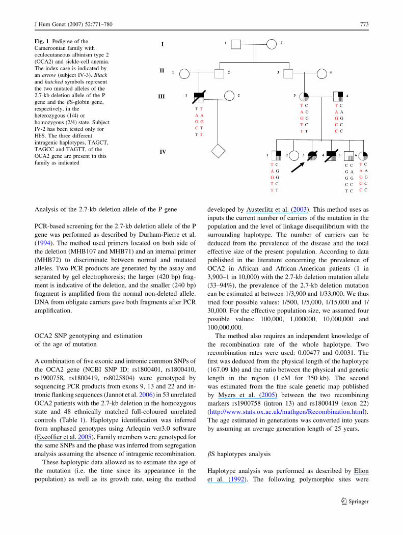
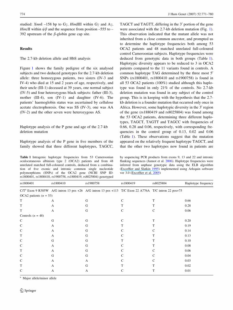
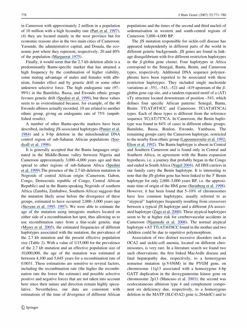
Fig. 1 Pedigree of the
Cameroonian family with
oculocutaneous albinism type 2
(OCA2) and sickle-cell anemia.
The index case is indicated by
an arrow (subject IV-3). Blackand hatched symbols represent
the two mutated alleles of the
2.7-kb deletion allele of the P
gene and the bS-globin gene,
respectively, in the
heterozygous (1/4) or
homozygous (2/4) state. Subject
IV-2 has been tested only for
HbS. The three different
intragenic haplotypes, TAGCT,
TAGCC and TAGTT, of the
OCA2 gene are present in this
family as indicated
J Hum Genet (2007) 52:771–780 773
123
studied: XmnI –158 bp to Gc, HindIII within Gc and Ac,
HincII within wb and the sequence from position –555 to –
392 upstream of the b-globin gene cap site.
Results
The 2.7-kb deletion allele and HbS analysis
Figure 1 shows the family pedigree of the six analysed
subjects and two deduced genotypes for the 2.7-kb deletion
allele: three homozygous patients, two sisters (IV-3 and
IV-4) who died at 15 and 2 years of age, respectively, and
their uncle (III-1) deceased at 39 years, one normal subject
(IV-5) and four heterozygous black subjects: father (III-3),
mother (III-4), son (IV-1) and daughter (IV-6). The
patients’ haemoglobin status was ascertained by cellulose
acetate electrophoresis. One was SS (IV-3), one was AA
(IV-2) and the other seven were heterozygous AS.
Haplotype analysis of the P gene and age of the 2.7-kb
deletion mutation
Haplotype analysis of the P gene in five members of the
family showed that three different haplotypes, TAGCC,
TAGCT and TAGTT, differing in the 30 portion of the gene
were associated with the 2.7-kb deletion mutation (Fig. 1).
This observation indicated that the mutant allele was not
inherited from a close common ancestor, and prompted us
to determine the haplotype frequencies both among 53
OCA2 patients and 48 matched unrelated full-coloured
control Cameroonian subjects. Haplotype frequencies were
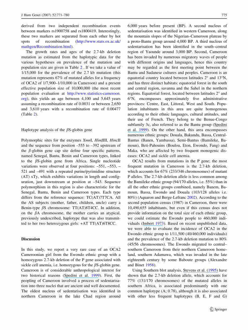
deduced from genotypic data in both groups (Table 1).
Haplotypic diversity appears to be reduced to 3 in OCA2
patients compared to the 11 variants found in controls. A
common haplotype TAG determined by the three most 50
SNPs (rs1800401, rs1800410 and rs1900758) is found in
all 53 OCA2 patients (100%) studied although this haplo-
type was found in only 21% of the controls. No 2.7-kb
deletion mutation was found in any subject of the control
group. This is in keeping with the hypothesis that the 2.7-
kb deletion is a founder mutation that occurred only once in
Africa. However, some haplotypic diversity in the 30 region
of the gene (rs1880419 and rs8025804) was found among
the 53 OCA2 patients, determining three different haplo-
types, TAGCT, TAGTT and TAGCC with frequencies of
0.66, 0.28 and 0.06, respectively, with corresponding fre-
quencies in the control group of 0.13, 0.02 and 0.06
(Table 1). These observations suggest that the mutation
appeared on the relatively frequent haplotype TAGCT, and
that the other two haplotypes now found in patients are
Table 1 Intragenic haplotype frequencies from 53 Cameroonian
oculocutaneous albinism type 2 (OCA2) patients and from 48
unrelated matched full-coloured controls, deduced from a combina-
tion of five exonic and intronic common single nucleotide
polymorphisms (SNPs) of the OCA2 gene (NCBI SNP ID:
rs1800401, rs1800410, rs1900758, rs1800419, rs8025804) genotyped
by sequencing PCR products from exons 9, 13 and 22 and intronic
flanking sequences (Jannot et al. 2006). Haplotype frequencies were
inferred from unphase genotypic data using the ELB algorithm
(Excoffier and Slatkin 1995) implemented using Arlequin software
ver 3.0 (Excoffier et al. 2005)
rs1800401 rs1800410 rs1900758 rs1800419 rs8025804 Haplotype frequency
C/Ta Exon 9 R305W A/G intron 13 pos +26 A/G intron 13 pos +113 T/C Exon 22 A776A T/C intron 22 pos+75
OCA2 patients (n = 53)
T A G C T 0.66
T A G T T 0.28
T A G C C 0.06
Controls (n = 48)
C G G C T 0.20
C A G T T 0.19
C A G C C 0.14
T A G C T 0.13
C G G T T 0.10
C A G C T 0.08
T A G C C 0.06
C G G C C 0.04
C A A C C 0.03
T A G T T 0.02
C A A C T 0.01
a Major allele/minor allele
774 J Hum Genet (2007) 52:771–780
123
derived from two independent recombination events
between markers rs1900758 and rs1800419. Interestingly,
these two markers are separated from each other by hot
spots of recombination (http://www.stats.ox.ac.uk/
mathgen/Recombination.html).
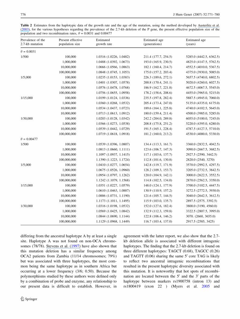
The growth rates and ages of the 2.7-kb deletion
mutation as estimated from the haplotypic data for the
various hypotheses on prevalence of the mutation and
population size are given in Table 2 . If we take a value of
1/15,000 for the prevalence of the 2.7 kb mutation (this
mutation represents 67% of mutated alleles for a frequency
of OCA2 of 1/7,900–1/10,000 in Cameroon) and a present
effective population size of 10,000,000 (the most recent
population evaluation at http://www.statistics-cameroon.
org), this yields an age between 4,100 and 5,645 years
assuming a recombination rate of 0.0031 or between 2,650
and 3,610 years with a recombination rate of 0.00477
(Table 2).
Haplotype analysis of the bS-globin gene
Polymorphic sites for the enzymes XmnI, HindIII, HincII
and the sequence from position –555 to –392 upstream of
the b-globin gene cap site define four specific patterns,
named Senegal, Bantu, Benin and Cameroon types, linked
to the bS-globin gene from Africa. Single nucleotide
variations were observed at four positions: –551, –553, –
521 and –491 with a repeated purine/pyrimidine structure
(AT) xTy, which exhibits variations in length and config-
uration, just downstream at position at –543. Sequence
polymorphism in this region is also characteristic for the
Senegal, Bantu, Benin and Cameroon types. Each type
differs from the reference sequence: TC(AT)7T7CA. All
the AS subjects (mother, father, children, uncle) carry a
Benin-type bS chromosome: TT(AT)8T4CC. In addition,
on the bA chromosome, the mother carries an atypical,
previously undescribed, haplotype that was also transmit-
ted to her two heterozygous girls: +AT TT(AT)6T8CC.
Discussion
In this study, we report a very rare case of an OCA2
Cameroonian girl from the Ewondo ethnic group with a
homozygous 2.7-kb deletion of the P gene associated with
sickle cell anemia, i.e. homozygous for the bS-globin gene.
Cameroon is of considerable anthropological interest for
two historical reasons (Spedini et al. 1999). First, the
peopling of Cameroon involved a process of sedentarisa-
tion into three nuclei that are ancient and well documented.
The oldest nucleus of sedentarisation was identified in
northern Cameroon in the lake Chad region around
6,000 years before present (BP). A second nucleus of
sedentarisation was identified in western Cameroon, along
the mountain slopes of the Nigerian–Cameroon plateau by
a proto-Bantu group around 4,000 BP. A third nucleus of
sedentarisation has been identified in the south–central
region of Yaounde around 3,000 BP. Second, Cameroon
has been invaded by numerous migratory waves of people
with different origins and languages, hence this country
may be regarded as the first meeting point between the
Bantu and Sudanese cultures and peoples. Cameroon is an
equatorial country located between latitudes 2� and 13�N,
and has three distinct habitats: equatorial forest in the south
and central region, savanna and the Sahel in the northern
regions. Equatorial forest, located between latitudes 2� and
6�N, encompasses approximately five administrative
provinces: Centre, East, Littoral, West and South. Popu-
lation inhabitants in this area are quite homogenous
according to their ethnic languages, cultural attitudes, and
their use of French. They belong to the Benue-Congo
subfamily 3c, also referred to as the Bantu group (Spedini
et al. 1999). On the other hand, this area encompasses
numerous ethnic groups: Douala, Bakundu, Bassa, Central-
Bantus (Banen, Yambassa), Semi-Bantus (Bamileke, Ba-
moun), Beti-Pahouins (Boulou, Eton, Ewondo, Fang) and
Maka, who are affected by two frequent monogenic dis-
eases: OCA2 and sickle cell anemia.
OCA2 results from mutations in the P gene; the most
frequent mutation in Cameroon is the 2.7-kb deletion,
which accounts for 67% (233/346 chromosomes) of mutant
P alleles. The 2.7-kb deletion allele is less common among
the Bamileke ethnic group (94/170 alleles, i.e. 55%) than in
all the other ethnic groups combined, namely Banem, Ba-
moun, Bassa, Ewondo and Douala (103/128 alleles i.e.
80%) (Aquaron and Berge-Lefranc 2002). According to the
second population census (1987) in Cameroon, there were
10,490,655 inhabitants, but even if this census does not
provide information on the total size of each ethnic group,
we could estimate the Ewondo people to 460,000 indi-
viduals (Imbert 1973). Based on recent unpublished data,
we were able to evaluate the incidence of OCA2 in the
Ewondo ethnic group to 1/11,500 (40/460,000 individuals)
and the prevalence of the 2.7-kb deletion mutation to 80%
(45/56 chromosomes). The Ewondo migrated to central–
southern Cameroon from their northern Cameroon home-
land, southern Adamawa, which was invaded in the late
eighteenth century by some Baboute groups (Alexandre
and Binet 1958).
Using Southern blot analysis, Stevens et al. (1995) have
shown that the 2.7-kb deletion allele, which accounts for
77% (131/170 chromosomes) of the mutated alleles in
southern Africa, is associated predominantly with one
common haplotype (A; 0.78), although it is also associated
with other less frequent haplotypes (B, E, F and G)
J Hum Genet (2007) 52:771–780 775
123
differing from the ancestral haplotype A by at least a single
site. Haplotype A was not found on non-OCA chromo-
somes (78/78). Stevens et al. (1997) have also shown that
this mutation deletion has a similar frequency among
OCA2 patients from Zambia (11/14 chromosomes; 79%)
but was associated with three haplotypes, the most com-
mon being the same haplotype as in southern Africa but
occurring at a lower frequency (3/6; 0.50). Because the
polymorphisms studied by these authors were defined only
by a combination of probe and enzyme, any relationship to
our present data is difficult to establish. However, in
agreement with the latter report, we also show that the 2.7-
kb deletion allele is associated with different intragenic
haplotypes. The finding that the 2.7-kb deletion is found on
three different haplotypes: TAGCT (0.68), TAGCC (0.26)
and TAGTT (0.06) sharing the same 50 core TAG is likely
to reflect two ancestral intragenic recombinations that
resulted in the present haplotypic diversity associated with
this mutation. It is noteworthy that hot spots of recombi-
nation are located between the 50 and the 30 parts of the
haplotype between markers rs1900758 (intron 13) and
rs1800419 (exon 22 ) (Myers et al. 2005 and
Table 2 Estimates from the haplotypic data of the growth rate and the age of the mutation, using the method developed by Austerlitz et al.
(2003), for the various hypotheses regarding the prevalence of the 2.7-kb deletion of the P gene, the present effective population size of the
population and two recombination rates, h = 0.0031 and 0.00477
Prevalence of the
2.7-kb mutation
Present effective
population size
Estimated
growth rate
Estimated age
(generations)
Estimated age
(years)
h = 0.0031
1/500 100,000 1.0316 (1.0226, 1.0482) 211.4 (177.7, 254.5) 5285.0 (4442.5, 6362.5)
1,000,000 1.0488 (1.0392, 1.0673) 193.0 (165.9, 230.5) 4825.0 (4147.5, 5762.5)
10,000,000 1.0666 (1.0566, 1.0863) 182.1 (160.4, 214.7) 4552.5 (4010.0, 5367.5)
100,000,000 1.0848 (1.0745, 1.1053) 175.0 (157.2, 203.4) 4375.0 (3930.0, 5085.0)
1/5,000 100,000 1.0235 (1.0153, 1.0383) 226.3 (189.6, 272.1) 5657.5 (4740.0, 6802.5)
1,000,000 1.0401 (1.0307, 1.0578) 200.8 (170.4, 241.1) 5020.0 (4260.0, 6027.5)
10,000,000 1.0576 (1.0478, 1.0768) 186.9 (162.7, 221.8) 4672.5 (4067.5, 5545.0)
100,000,000 1.0756 (1.0655, 1.0958) 178.2 (158.6, 208.6) 4455.0 (3965.0, 5215.0)
1/15,000 100,000 1.0199 (1.0124, 1.0336) 235.5 (197.8, 282.4) 5887.5 (4945.0, 7060.0)
1,000,000 1.0360 (1.0268, 1.0532) 205.4 (173.4, 247.0) 5135.0 (4335.0, 6175.0)
10,000,000 1.0534 (1.0437, 1.0723) 189.6 (164.1, 225.8) 4740.0 (4102.5, 5645.0)
10,000,000 1.0713 (1.0613, 1.0912) 180.0 (159.4, 211.4) 4500.0 (3985.0, 5285.0)
1/30,000 100,000 1.0203 (1.0128, 1.0342) 242.2 (204.0, 289.8) 6055.0 (5100.0, 7245.0)
1,000,000 1.0366 (1.0273, 1.0538) 208.8 (175.8, 251.2) 5220.0 (4395.0, 6280.0)
10,000,000 1.0539 (1.0442, 1.0729) 191.5 (165.1, 228.4) 4787.5 (4127.5, 5710.0)
100,000,000 1.0719 (1.0618, 1.0918) 181.2 (160.0, 213.2) 4530.0 (4000.0, 5330.0)
h = 0.00477
1/500 100,000 1.0539 (1.0396, 1.0807) 134.4 (113.3, 161.7) 3360.0 (2832.5, 4042.5)
1,000,000 1.0813 (1.0660, 1.1111) 123.6 (106.7, 147.3) 3090.0 (2667.5, 3682.5)
10,000,000 1.1097 (1.0937, 1.1415) 117.1 (103.6, 137.7) 2927.5 (2590, 3442.5)
100,000,000 1.1390 (1.1223, 1.1724) 112.8 (101.6, 130.8) 2820.0 (2540, 3270)
1/5,000 100,000 1.0410 (1.0277, 1.0654) 142.8 (119.7, 171.9) 3570.0 (2992.5, 4297.5)
1,000,000 1.0675 (1.0526, 1.0960) 128.2 (109.3, 153.7) 3205.0 (2732.5, 3842.5)
10,000,000 1.0954 (1.0797, 1.1262) 120.0 (104.9, 142.1) 3000.0 (2622.5, 3552.5)
100,000,000 1.1242 (1.1079, 1.1568) 114.8 (102.5, 134.0) 2870.0 (2562.5, 3350.0)
1/15,000 100,000 1.0351 (1.0227, 1.0579) 148.0 (124.1, 177.9) 3700.0 (3102.5, 4447.5)
1,000,000 1.0610 (1.0463, 1.0887) 130.9 (110.9, 157.2) 3272.5 (2772.5, 3930.0)
10,000,000 1.0886 (1.0731, 1.1190) 121.6 (105.7, 144.5) 3040.0 (2642.5, 3612.5)
100,000,000 1.1173 (1.1011, 1.1495) 115.9 (103.0, 135.7) 2897.5 (2575, 3392.5)
1/30,000 100,000 1.0316 (1.0198, 1.0532) 152.0 (127.6, 182.4) 3800.0 (3190, 4560.0)
1,000,000 1.0569 (1.0425, 1.0842) 132.9 (112.3, 159.8) 3322.5 (2807.5, 3995.0)
10,000,000 1.0844 (1.0690, 1.1144) 122.8 (106.4, 146.2) 3070. (2660, 3655.0)
100,000,000 1.1129 (1.0968, 1.1449) 116.7 (103.4, 137.0) 2917.5 (2585, 3425)
776 J Hum Genet (2007) 52:771–780
123
http://www.stats.ox.ac.uk/mathgen/Recombination.html).
These observations suggest that, within African populations
in Cameroon, the mutation appeared on the relatively fre-
quent haplotype TAGCT and that the two other haplotypes
are derived from two independent recombination events.
This may, at first, appear surprising, because albinism, a
detrimental autosomic trait, is expected to affect the Dar-
winian fitness of affected individuals. Darwinian fitness (w),
which reflects the ability of albinos to survive and repro-
duce, may have a lower value relative to non-albinos
because of death at an earlier age, resulting mainly from
solar radiation-induced skin cancer, or because of envi-
ronmental accidents like malaria for our two albino girls. As
skin cancer, which is the most frequent reason for death in
equatorial regions, occurs mainly during the second to
fourth decade of life, after reproductive age, we expect that
the Darwinian fitness of albinos will not be severely
decreased. Thus, natural selection has little or no influence
on the frequency of the OCA2 deletion allele. For example,
with a selection coefficient ‘‘s’’ of 0.5 measuring the degree
of selection against albinos, the Darwinian fitness will be
w = 1 – 0.5 = 0.5, and with an assumed mutation rate (l) at
the P locus of 2.5 · 10–5, the frequency of albinos at
equilibrium in a large population would be q2 = l/s =
5 · 10–5 or 1/20,000, which is the usual value found in the
world (Hedrick 2003). If s = 0.3 or 0.2, then the equilibrium
incidence of albinism is only 1/12,000 and 1/8,000, the
incidence values found in Cameroon. However, as the
mutation rate at the P locus and the selection coefficient are
unknown, the high frequency of the OCA2 deletion allele
requires other explanations such as heterozygote advantage
and/or cultural selection and/or chance processes: founder
effect, bottleneck effect or genetic drift (Stern 1960).
The classic example of heterozygote advantage is
resistance to infection by the malaria parasite (Plasmodium
falciparum) in heterozygotes for sickle-cell anemia (Alli-
son 1954), and more recently for G6PD deficiency, a+
thalassemia, and haemoglobin C (Kwiatkowski 2005). The
application of novel haplotype-based techniques has dem-
onstrated that malaria-protective genes have been subject
to recent positive selection (Kwiatkowski 2005). Oettle
(1963) speculated that the lighter skin colour of the OCA2
heterozygote, as demonstrated by skin reflectance (Roberts
et al. 1986), may confer a social advantage, as individuals
with lighter skins may be preferable as marriage partners.
This hypothesis seems not to be true from the longstanding
experience of one of us (R.A.) in Cameroon. If, as very
often, children of albinos who are obligate heterozygotes
have lighter skin than the full-coloured parent, we are not
aware of any such advantage for OCA2 heterozygotes. The
same conclusion about the absence of heterozygote
advantage at the P locus was drawn in Amerindian popu-
lations with OCA2 albinism (Woolf 2005), particularly
among the Navajo, which present a 122.5-kb deletion of
the P gene (Yi et al. 2003).
The additional factor of cultural selection for albinos
was suggested in Amerindians with OCA2. An intense
negative cultural selection against San Blas Cuna albinos,
culminating in infanticide, and almost complete marriage
discrimination against albino males, has been documented.
On the contrary, albinos were not ostracised or looked upon
as being inferior among the Hopi and Zuni Indians, and
they married full-coloured individuals and had healthy
offspring (Woolf 2005). Hopi males with albinism were
traditionally allowed to remain in the villages, thereby
avoiding bright sunlight and its detrimental effects on
them, and ‘‘had ample opportunity to engage in sexual
activity’’ (Hedrick 2003). In Cameroon, we know that the
average number of children, obligate heterozygotes, in
albino families (full-coloured male/albino female mating or
reverse, even though many were not married) is about the
same as in their black counterparts. We also heard that
females with albinism were preferred by full-coloured
individuals to engage in sexual activity because they are
white. In Cameroon traditional permissive sexual mores
was observed. On the other hand, mating between albinos
is not prohibited in Africa but is a very rare event. We had
the opportunity in Mali to observe such a couple with four
albino children (Aquaron 2000). In conclusion, it is diffi-
cult to establish if a mating advantage ‘‘m’’ of males and/or
females exists.
An interesting example of a founder effect is described
in the Bamileke society. The Bamileke society is organised
as small kingdoms each headed by a king. In the West
province, 131 kingdoms have been individualised and 88,
i.e. 67%, have less than 5,000 inhabitants (Barbier and
Nkwi 1977). The founder effect is illustrated in the
Balengou kingdom where the eighth and ninth kings, at the
beginning of the twentieth century had albinism. These
kings were polygamists, with 80–100 wives and 200–300
children, who were obligate heterozygotes; on the other
hand, in the Bamileke tribe, marriages tended to occur
within the same clan or town (Puri et al. 1997) with a high
endogamic rate of around 95% (Spedini et al. 1999). A
hypothetical scenario would be that the founder effect was
the cause of the initial high proportion of albinos in this
small endogamous population, which was then spread by
migrating groups to other geographic regions followed by
the rapid increase in size of that population (Woolf 2005).
This scenario could be possible because (1) Balengou had
less than 5,000 inhabitants around 1900, (2) albinism
occurs most frequently in the Bamileke group [70% (190/
273 registered albinos) even if the 2.7-kb deletion was less
common: 55% (94/170 alleles) than in other ethnic groups
(Aquaron 1990; Aquaron and Berge-Lefranc 2002)], (3)
the Bamileke population are the predominant ethnic group
J Hum Genet (2007) 52:771–780 777
123
in Cameroon with approximately 2 million in a population
of 10 million with a high fecundity rate (Puri et al. 1997),
(4) they are located mainly in the west province but for
economic reasons also in the two main cities of Cameroon:
Yaounde, the administrative capital, and Douala, the eco-
nomic port where they represent, respectively, 20 and 40%
of the population (Marguerat 1975).
Finally, it would seem that the 2.7-kb deletion allele is a
predominantly Bantu-specific marker that has attained a
high frequency by the combination of higher viability,
some mating advantage of males and females with albi-
nism, founder effect and by genetic drift or some other
unknown selective force. The high endogamic rate (97–
99%) in the Bamileke, Bassa, and Ewondo ethnic groups
favours genetic drift (Spedini et al. 1999), but these values
seem to us overestimated because, for example, of the 40
Ewondo albinos actually recorded, 10 are related to another
ethnic group, giving an endogamic rate of 75% (unpub-
lished results).
A number of other Bantu-specific markers have been
described, including bS-associated haplotypes (Panier et al.
1984) and a 9-bp deletion in the mitochondrial DNA
control region of sub-Saharan African populations (Soo-
dyall et al. 1996).
It is generally accepted that the Bantu languages origi-
nated in the Middle-Benue valley between Nigeria and
Cameroon approximately 3,000–4,000 years ago and then
spread to other regions of sub-Saharan Africa (Spedini
et al. 1999).The presence of the 2.7-kb deletion mutation in
Negroids of central African origin (Cameroon, Gabon,
Congo, Democratic republic of Congo, Central African
Republic) and in the Bantu-speaking Negroids of southern
Africa (Zambia, Zimbabwe, Southern-Africa) suggests that
the mutation likely arose before the divergence of these
groups, estimated to have occurred 2,000–3,000 years ago
(Stevens et al. 1995, 1997). We were able to estimate the
age of the mutation using intragenic markers located on
either side of a recombination hot spot, thus allowing us to
use recombination rates from a fine-scale genetic map
(Myers et al. 2005), the estimated frequencies of different
haplotypes associated with the mutation, the prevalence of
the 2.7 kb mutation and the present effective population
size (Table 2). With a value of 1/15,000 for the prevalence
of the 2.7 kb mutation and an effective population size of
10,000,000, the age of the mutation was estimated at
between 4,100 and 5,645 years for a recombination rate of
0.0031. These estimations are sensitive to several factors,
including the recombination rate (the higher the recombi-
nation rate the lower the estimate) and possible selective
positive and negative forces that are not taken into account
here since their nature and direction remain highly specu-
lative. Nevertheless, our data are consistent with
estimations of the time of divergence of different African
populations and the times of the second and third nucleii of
sedentarisation in western and south-central regions of
Cameroon 3,000–4,000 BP.
The bS mutation responsible for sickle-cell disease has
appeared independently in different parts of the world in
different genetic backgrounds. bS genes are found in link-
age disequilibrium with five different restriction haplotypes
in the b-globin gene cluster. Four haplotypes in Africa
correspond to the Senegal, Bantu, Benin, and Cameroon
types, respectively. Additional DNA sequence polymor-
phisms have been reported to be associated with these
restriction haplotypes. They included single nucleotide
variations at –551, –543, –521 and –419 upstream of the b-
globin gene cap site, and a tandem repeated motif of a (AT)
xTy structure located downstream of position –543, which
defines four specific African patterns: Senegal, Bantu,
Benin: TT(AT)8T4CC and Cameroon: TC(AT)8T5CA)
types. Each of these types is different from the reference
sequence TC(AT)7T7CA. In Cameroon, the Benin haplo-
type was found in 84% of cases in different ethnic groups:
Bamileke, Bassa, Boulou, Ewondo, Yambassa. The
remaining groups carry the Cameroon haplotype, restricted
to the nearby Eton ethnic group (Lapoumeroulie et al. 1992;
Elion et al. 1992). The Bantu haplotype is absent in Central
and Southern Cameroon and is found only in Central and
Southern Africa, in agreement with the Bantu expansion
hypothesis, i.e. a journey that probably began in the Congo
and ended in South Africa (Nagel 2004). All HbS carriers in
our family carry the Benin haplotype. It is interesting to
note that the bS-globin gene has been linked to the 50 Benin
haplotype for only 2,000–3,000 years BP, i.e. the approxi-
mate time of origin of the HbS gene (Steinberg et al. 1998).
However, it has been found that 5–10% of chromosomes
have less common haplotypes, usually referred to as
‘‘atypical’’ haplotypes frequently resulting from crossovers
between a typical bS haplotype and a different bA-associ-
ated haplotype (Zago et al. 2000). These atypical haplotypes
seem to be at higher risk for cerebrovascular accidents in
Cameroon (Njamnshi et al. 2006). The normal atypical
haplotype +AT TT(AT)6T8CC found in the mother and two
children could be due to repetitive polymorphism.
Association of two distinct recessive disorders such as
OCA2 and sickle-cell anemia, located on different chro-
mosomes, is very rare. In a literature search we found two
such observations: the first linked MacArdle disease and
fatal hepatopathy due, respectively, to a homozygous
nonsense mutation (p.V456M) in the PYGM gene, on
chromosome 11q13 associated with a homozygous 4-bp
GATT duplication in the deoxyguanosine kinase gene on
chromosome 2p13 (Mancuso et al. 2003); the second was
oculocutaneous albinism type 4 and complement compo-
nent six deficiency due, respectively, to a homozygous
deletion in the MATP (SLC45A2) gene (c.264delC) and to
778 J Hum Genet (2007) 52:771–780
123
a homozygous nonsense mutation (p.S91X) in the C6 gene,
located in close proximity (approximately 7 Mb), on the
short arm of chromosome 5 (Ikinciogullari et al. 2005).
The manifestation of two rare conditions results from the
respective high frequency of the two mutated alleles in a
given population.
Acknowledgements We wish to thank all the members of the
affected family, the albino patients and the control subjects for their
kind cooperation, Pr. P. Ndumbe, dean of the School of Medicine of
Yaounde and Mr. J.J. Ndoudoumou, president of the Cameroonian
albino association (ASMODISA) for their continual support, Mr. Luc
Kamdem for his skillful assistance, particularly for his help on field
trips in Cameroon, Sebastien Courrier for performing HbS and HbA
haplotype analysis, Chantal Bideau and Danielle Iniesta for per-
forming 2.7-kb P gene deletion mutation analysis, Claire Oudin for
sequencing OCA2, Pr. Laurent Gouya for helpful discussions, Liane
and Sam Lehrer, PhD, Boston Biomedical Research Institute, Boston,
for careful reading of the manuscript and Y. Mourayre for graphic
assistance.
References
Alexandre P, Binet J (1958) Le groupe dit Pahouin (Fang-Boulou-
Beti). Presses Universitaires de France, Paris
Allison AC (1954) Protection afforded by sickle-cell trait against
subtertian malareal infection. BMJ 4857:290–294
Aquaron R (1980) L’albinisme oculo-cutane au Cameroun. Rev
Epidemiol Sante Publique 28:81–88
Aquaron R (1990) Oculocutaneous albinism in Cameroon: a 15 year
follow-up study. Ophthalmic Paediatr Genet 4:255–263
Aquaron R (2000) L’albinisme humain: aspects cliniques, genetiques,
cellulaires, biochimiques et moleculaires. Med Trop 60:331–341
Aquaron R, Berge-Lefranc JL (2002) Type 2 oculocutaneous albinism
(OCA2) in Cameroon: distribution of the 2.7-kb deletion allele
of the P gene among various ethnic groups. Pigment Cell Res
15(Suppl 9):63
Aquaron R, Kamdem L, Menard JC, Bridonneau C, Battaglini PF
(1984) Etudes seroanthropologiques des populations albinos et
melanodermes Bamilekes (Cameroun): groupes erythrocytaires
ABO et rhesus, hemoglobine S et sensibilite gustative a la
phenylthiocarbamide. Med Trop 44:311–318
Austerlitz F, Kalaydjieva L, Heyer E (2003) Detecting population
growth, selection and inherited fertility from haplotypic data.
Genetics 165:1579–1586
Barbier JC, Nkwi PN (1977) Essai de definition de la chefferie en
pays Bamileke. Grassfield kings and chiefs and modern politics.
Documents de l’ Institut des Sciences Humaines, Yaounde
Barnicot NA (1952) Albinism in south-western Nigeria. Ann Eugen
17:38–73
Chen K, Manga P, Orlow SJ (2002) Pink-eyed dilution protein controls
the processing of tyrosinase. Mol Biol Cell 13:1953–1964
Dugast I (1949) Inventaire ethnique du Sud-Cameroun. Mem Inst
Franc Afr Noire, Centre du Cameroun, Douala, Cameroun
Durham-Pierre D, Gardner JM, Nakatsu Y, King RA, Francke U, Ching
A, Aquaron R, del Marmol V, Brilliant MH (1994) African origin
of an intragenic deletion of the P gene in tyrosinase-positive
oculocutaneous albinism. Nat Genet 7:176–179
Durham-Pierre D, King RA, Naber JM, Laken S, Brilliant MH (1996)
Estimation of carrier frequency of a 2.7-k deletion of the P gene
associated with OCA2 in African-Americans. Hum Mutat 7:370–
373
Elion J, Berg P, Lapoumeroulie C, Trabuchet G, Mittelman M,
Krishnmoorthy R, Schechter A, Labie D (1992) DNA sequence
variation in a negative control region 50 to the b-globin gene
correlates with the phenotypic expression of the bS mutation.
Blood 79:787–792
Excoffier L, Slatkin M (1995) Maximum-likelihood estimation of
molecular haplotype frequencies in a diploid population. Mol
Biol Evol 12:921–927
Excoffier L, Laval G, Schneider S (2005) Arlequin ver. 3.0: An
integrated software package for population genetics data anal-
ysis. Evol Bioinform Online 1:47–50
Gamet A, Labes A (1964) Premiere etude sur les hemoglobinoses au
Centre-Cameroun. Bull Soc Pathol Exot 57:1125–1133
Hedrick PW (2003) Hopi indians, ‘‘cultural’’ selection, and albinism.
Am J Phys Anthropol 121:151–156
Ikinciogullari A, Tekin M, Dogu F, Reisli I, Tanir G, Yi Z, Garrison
N, Brilliant MH, Babacan E (2005) Meningococcal meningitis
and complement component 6 deficiency associated with
oculocutaneous albinism. Eur J Pediatr 164:177–179
Imbert J (1973) Le Cameroun. Presses Universitaires de France, Paris
Jannot AS, Meziani R, Bertrand G, Gerard B, Descamps V,
Archimbaud A, Picard C, Ollivaud L, Basset-Seguin N, Kerob
D, Lanternier G, Lebbe C, Saiag P, Crickx B, Clerget-Darpoux
F, Grandchamp B, Soufir N, Melan-Cohort (2005) Allele
variations in the OCA2 gene (pink-eyed-dilution locus) are
associated with genetic susceptibility to melanoma. Eur J Hum
Genet 8:913–920
Juhan I, Kaptue L (1974) Epidemiologie et transfusion sanguine a
Yaounde. Med Afr Noire 21:947–949
Kromberg JGR, Jenkins T (1982) Prevalence of albinism in the South
African negro. S Afr Med J 61:383–386
Kwiatkowski DP (2005) How malaria has affected the human genome
and what human genetics can teach us about malaria. Am J Hum
Genet 77:171–192
Lapoumeroulie C, Dunda O, Ducrocq R, Trabuchet G, Mony-Lobe M,
Bodo JM, Carnevale P, Labie D, Elion J, Krishnamoorthy R
(1992) A novel sickle cell mutation of yet another origin in
Africa: the Cameroon type. Hum Genet 89:333–337
Luande J, Henschke CI, Mohammed N (1985) The Tanzanian human
albino skin. Cancer 55:1823–1828
Lund PM (1996) Distribution of oculocutaneous albinism in Zimba-
bwe. J Med Genet 33:641–644
Mancuso M, Filosto M, Tsujino S, Lamperti C, Shanske S, Coquet M,
Desnuelle C, DiMauro S(2003) Muscle glycogenosis and
mitochondrial hepatopathy in an infant with mutations in both
the myophosphorylase gene and deoxyguanosine kinase genes.
Arch Neurol 60:1445–1447
Marguerat Y (1975) Analyse numerique des migrations vers les villes
du Cameroun. Documents de l’ORSTOM, Paris
Massie RW, Hartmann RC (1957) Albinism and sicklemia in a negro
family. Am J Hum Genet 9:127–132
Myers S, Bottolo L, Freeman C, McVean G, Donnelly P (2005) A
fine-scale map of recombination rates and hotspots across the
human genome. Science 310:321–324
Nagel RL (2004) Beta-globin-gene haplotypes, mitochondrial DNA,
the Y chromosome: their impact on the genetic epidemiology of
the major structural hemoglobinopathies. Cell Mol Biol 50:5–21
Nguematcha R, Savina JF, Juhan I, Boche R, Ravisse P (1973)
Recherche de la tare drepanocytaire dans un groupe pygmee du
Sud Cameroun. Med Afr Noire 20:605–606
Njamnshi AK, Woankam A, Djientcheu VP, Ongolo-Zogo P, Obama
MT, Muna WFT, Sztajzel R (2006) Stroke may appear to be rare
in Saudi-Arabian and Nigerian children with sickle cell disease,
but not in Cameroonian sickle cell patients. Br J Haematol
133:210
J Hum Genet (2007) 52:771–780 779
123
Oettle AG (1963) Skin cancer in Africa. Natl Cancer Inst (USA)
Monograph 10:197–214
Okoro AN (1975) Albinism in Nigeria. Br J Dermatol 92:485–492
Panier J, Mears JG, Dunda-Belkhoya O, Schaefer-Rego KE, Beldjord
C, Nagel RL, Labie D (1984) Evidence for the multicentric
origin of the sickle cell hemoglobin gene in Africa. Proc Natl
Acad Sci USA 81:1771–1773
Puri N, Durham-Pierre D, Aquaron R, Lund PM, King RA, Brilliant
MH (1997) Type 2 oculocutaneous albinism (OCA2) in Zimba-
bwe and Cameroon: distribution of the 2.7-kb deletion allele of
the P gene. Hum Genet 100:651–656
Roberts DF, Kromberg JGR, Jenkins T (1986) Differentiation of
heterozygotes in recessive albinism. J Med Genet 23:323–327
Rosemblat S, Durham-Pierre D, Gardner JM, Nakatsu Y, Brilliant
MH, Orlow SJ (1994) Identification of a melanosomal mem-
brane protein encoded by the pink-eyed dilution (type II
oculocutaneous albinism) gene. Proc Natl Acad Sci USA
91:12071–12075
Soodyall H, Vigilant L, Hill AV, Stoneking M, Jenkins T (1996)
mtDNA control-region sequence variation suggests multiple
independent origin of an ‘‘Asian-specific’’ 9-bp deletion in sub-
Saharan Africans. Am J Hum Genet 58:595–608
Spedini G, Destro-Bisol G, Mondovi S, Kaptue L, Taglioli L, Paoli G
(1999) The peopling of sub-Saharan Africa; the case study of
Cameroon. Am J Phys Anthropol 110:143–162
Spritz RA, Fukai K, Holmes SA, Luande J (1995) Frequent intragenic
deletion of the P gene in Tanzanian patients in type II
oculocutaneous albinism (OCA2). Am J Hum Genet 56:1320–
1323 (1960)
Steinberg MH, Lu ZH, Nagel RL, Venkataramani S, Milner PF, Huey
L, Safaya S, Rieder RF (1998) Hematological effects of atypical
and Cameroon b-globin gene haplotypes in adult sickle cell
anemia. Al J Hematol 59:121–126
Stern C (1960) Principles of human genetics, 2nd edn. Freeman, San
Francisco
Stevens G, van Beukering J, Jenkins T, Ramsay M (1995) An
intragenic deletion of the P gene is the common mutation
causing tyrosinase-positive oculocutaneous albinism in southern
African negroids. Am J Hum Genet 56:586–591
Stevens G, Ramsay M, Jenkins T (1997) Oculocutaneous albinism
(OCA2) in sub-Saharan Africa: distribution of the common 2.7-
kb P gene deletion mutation. Hum Genet 99:523–527
Woolf CM (2005) Albinism (OCA2) in Amerindians. Am J Phys
Anthropol Suppl 41:118–140
Yi Z, Garrison N, Cohen-Barak O, Karafet TM, King RA, Erickson
RP, Hammer MF, Brilliant MH (2003) A 122.5-kilobase deletion
of the P gene underlies the high prevalence of oculocutaneous
albinism type 2 in the Navajo population. Am J Hum Genet
72:62–72
Zago MA, Silva WA, Dalle B, Gualandro S, Hutz MH, Lapoume-
roulie C, Tavella MH, Araujo AG, Krieger JE, Elion J,
Krishnamoorthy R (2000) Atypical bS haplotypes are generated
by diverse genetic mechanisms. Am J Hematol 63:79–84
780 J Hum Genet (2007) 52:771–780
123
Top Related
Copyright © 2022 FDOKUMEN

