


Bahasa
Halaman
Hukum

NOVEMBER, 1 969
THE NEW ZEAlAND JOURNAl OF
An Official PubllcaHon of the New Zealand Institute of Medical Laboratory Technology IncorpOrated ·
EDITOR: John Case
SUB-EDITORS: J. Rees, H. C. W. Shott
ADVERTISING MANAGER: F. C. Kershaw
PUBLICATIONS SUB-COMMITTEE Marilyn M. Eales R. T. Kennedy
Janet Marsland C. S. Shepherd VOLUME 23. No. 3
•
Directions for Contributors Adherence to the following instructions is necessary iu order t<..
ensure unilormity of presentation, and all contributors are urged to study them before submitting their manuscripts.
M anuscripts should be typewritten on one sid e o nly of good quality quarto paper, be double spaced and have a one inch margin all round. They should bear the author's name (male authors give initials and female authors one gi,·cn name), address and ( if th is is differeut) the address of the laboratory where the work was carried out. Carbon copies a re not acceptable, and nothing should be underlined unless it is to be printed in italics. T he use of ita lics to denote emphasis should be a\oided , if possible.
ILLUSTRATIONS The ./oumal will bear the cost of a reasonable number of illustrations.
but th ese should be u sed sparingly. Graphs, line drawings and photo~ graphs a re all referred to as " Figures " and should be numbered in the order of their appearance in the text, using Arl'lbic numerals. Drawings should be made in ludian in k on stout white paper, somewhat larger than required for reproduction. Legends should be typed on separate pieces of paper , and their approximate position in relation to the text should be noted in the typescript. Elaborate tables should be kept to a minimum, should be typed on separate pieces of paper and numbered in Roman numerals.
NOM ENCLATURE Scientific names of micro-organisms should be in conformity with the
style adopted in the latest edition of Bergey's M anual of Determi11ative BacteriologJ' and should be underlined to indicate that they are to be printed in italics. Abbreviations such as CSF for cerebro-spina l nuid are permissible, but their meaning must be clearly indicated when first introduced. Conventional abbreviations such as mi. for millilitre arc ::tcceptable wit hout explanation, but a uthors should note that the correct abbreviation fo r gram (or grams) is g. and not gm. or gms.
REFE RENCES Only papers closely related to the author's work should be quoted.
Authors should study past issues of the journal for examples of t he preferred method of making reference. All references are brought together a t the end in alphabetical order a nd numbered, the <tppropria te numerals being used in superscript within the text . [ n the list references should include the surna me of the author, fo llowed by initia ls {or by one given name if the author is a female) , the year of p ublicati on in brackets. the abbreviated ti tle of the pu blication ( underlin ed to denote ita lics ), the volume number and the page n umber. If th ere are three or more authors, the first author's name may be used in the t ext followed by t he words et al., but the names of the co-authors must be given in the list. The abbreviation of titles of periodicals may be copied from World L ist of Scientific Periodicals, but the derivation is largely common sense. Broadly, all prepositions and conj unctions arc omitted, nouns commence with a capi tal letter, adjectives lower case, and words are foreshortened consisten t with understan ding. Example: The A merican ] ournal of Clinical PathologJ' is A mer . ./. clin. Path. R eferences to books shou ld include name (s ) , year. title ( underlined ), edition. page number{s), name of publisher and place of publication in that order.
PROOFS When time permits a uthors will be given the opportunity to correct
galley proofs before public::ttion , but proofs must be re turned within three days of receipt.
REPRINTS Reprints arc available to a uthors a t their own expe nse on a cost basis.
Alternatively authors may purchase extra copies of the ./ournal a t half the normal subscription price. Orders for reprints or extra copies must be given when rPturning the galley p roofs.
JJ
RHEUMATEx·· ... A LATEX
SENSITIZED ANTIGEN
FOR rthritis
SERVING
SCIENCE
••• WHICH MAY BE USED AS A QUANTITATIVE IN AGGLUTINATION TEST PERFORMED EITHER IN THE TEST TUBE OR IN A WELLED TRAY THEORY
AND
••• OR WHICH MAY BE USED AS A RELIABLE SLIDESCREENING TEST ••• IN
PRACTICE
®
PRODUCTS FROM
Sole Agents & Distribwors: Diagnostic Dirision DOMINION DENTAL SUPPLIES LTD.
WELLINGTOJ\ AUCKLAND iv
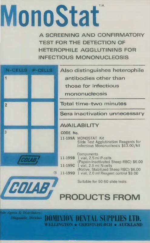
MonoStatTM
1
2
A SCREENING AND CONFIRMATORY
TEST FOR THE DETECTION OF
HETEROPHILE AGGLUTININS FOR
INFECTIOUS MONONUCLEOSIS
Also distinguishes heterophile
antibodies other than
those for infectious
mononucleosis
Total time-tvvo minutes
Sera inactivation unnecessary
AVAILABILITY
CODE No.
11·199A MONOSTAT Kit Slide Test Aggl ut inat ion Reagents for Infect ious Mononucleosis $13.00/kit
Components 11-1998 1 vial, 2.5 ml P-cells
(Papain-inactivated Sheep RBC) $6.00 ll-199C 1 vial, 2.5 ml N-cells
(Native, Stabilized Sheep RBC) $6.00 ® 11-1990 1 vial, 2.0 ml Reagent control $3.00 ...----· Suitable for 50-60 slide tests
PRODUCTS FROM
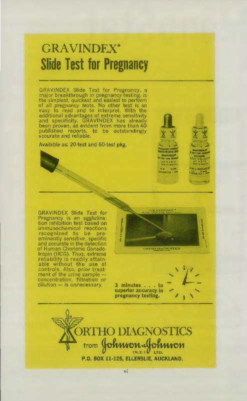
G·RA VINDEX* Sl~de Test for Pregnancy
GRAVINDEX Slide Test for Pregnancy, a major breakthrough in pregnancy testing, is the '';implest, quickest and easiest to perform of a: I pregnancy tests. No other test is so eas} to read and to interpret. With the add.tional advantages of extreme sensitivity and specificity, GRAVINDEX has already been proven, as evident from more than 40 published reports, to be outstandingly accurate and reliable.
Available as: 20-test and 50-test pkg. . ... fi.ftUMANCHoMi <llwlOTROJIICWf
GltA.VlNOtxt li'AttUrOfiHI_.
~'lT ;;, .& a Ti Hl'1l .. Al'
t:. ~··' .O::·.OIIT<CS•,...,..
GRAVINDEX Slide Test for Pregnancy is an agglutina · tion inhibition test based on immunochemical reactions recognized to be pre eminently sensitive, specific and accurate in the detection of Human Chorionic Gonado· tropin (HCG). Thus, extreme reliability is readily attain · able without the use of controls. Also, prior treat· ment of the urine sample -concentration , filtration or dilution - is unnecessary. 3 minutes . . . to
--t't' ••••• .. 110'1
'- :::li~A,~~,::~ ~oi1~N~!~IIlfJ ~r ,. "'~ ()(f"·:.l. MAY'
t"11i£l\,8(FOf'E ..
~~":a.r•~~:.~
superior accuracy in ,; ' pregnancy testing. / \
ORTHO l)lf\GNOSTICS from~~
P.O. BOX 11·125, ELLERSLIE, AUCKLAND.
VI
CURRE"'T IUU ... VOl.TAGI E
~ t.IAfN$
.lt{\'tii(Sf R(VERS{
POu.~tlh
NOJUUt NORM4t
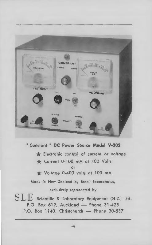
"Constant" DC Power Source Model V-202
* Electronic control of current or voltage * Current 0-1 00 mA at 400 Volts or * Voltage 0-400 volts at 1 00 mA
Made in New Zealand by Brook Laboratories,
exclusively represented by
S L E Scientific & laboratory Equipment (N.Z.) ltd.
P.O. Box 619, Auckland- Phone 31-425 P.O. Box 1140, Christchurch - Phone 30-537
vii
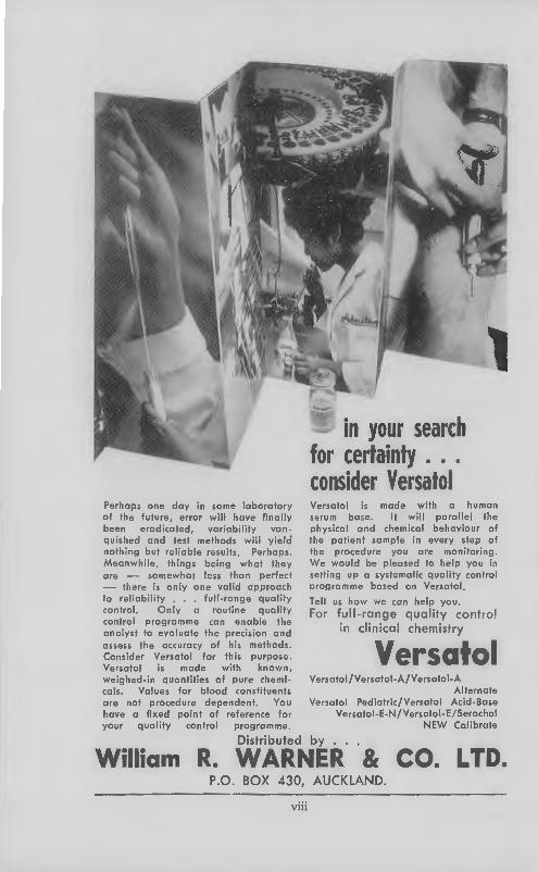
Perhaps one day in same laboratory of the future, error will have finally been eradicated, variability vanquished and test methods will yield nothin!l but reliable results. Perhaps. Meanwhile, things being what they are - somewhat less than perfect - there is only one valid approach to reliability ... full-range quality control. Only a routine quality control programme can enable the analyst to evaluate the precision and assess the accuracy of his methods. Consider Versatol for this purpose. Versatol is made with known, weiqhed-in quantities of pure chemicals. Values for blood constituents are not procedure dependent. You have a fixed point of reference for your quality control programme.
in your search for certainty . . . consider Versatol Versatol is made with a human serum base. It will parallel the physical and chemical behaviour of the patient sample in every step of the procedure you are monitoring. We would be pleased to help you in settinq up a systematic quality control orogramme based on Versatol.
Tell us how we can help you. For full-range quality control
in clinical chemistry
Versatol Versa tal I Versato 1-A /Versa to 1-A
Alternate Versatol PediatriciVersatol Acid-Base
Versa to I- E-N I Versatol- E I Seracho I NEW Ca librate
Distributed by . .
William R. WARNER & CO. LTD. P.O. BOX 430, AUCKLAND.
viii
:Jfte rJew ZafanJ Journal o/
medical el!atoralor'! :Jechnofofl'!
Volume 23, No. 3
The New Zealand Institute of Medical laboratory Technology (Inc.)
Office-bearers, 1969-70
PRESIDENT : H. E. Hutching s
Pathology Department Palmerston North Hospital
VICE-PRESIDENTS: J. R. Morgan D. J. Philip
SECRETARY: R. T. Kenned y
Centro I laboratory Auckland Hospital
TREASURER: E. K. Fletcher
Pathology Department New Plymouth Hospital
COUNCIL:
Marilyn M. Ea les R. T. Kennedy M. J. lynch
November, 1969
The JOURNAL is published three
tim es year ly (in March, July and
November), and is distributed, with
out charge, to all financial members
of th e N.Z.I.M.l.T. (Inc. ) .
Subscription to the JOURNAL for
non-members is ONE DOLLAR per
year, or FORTY CENTS per single
issue, postage paid. Overseas s'ub
scription rates on app lication .
Intending contributors should sub
mit their material to the Editor, at
the Blood Bank laboratory, Dunedin
Hospital. Acceptance is at the
discre tion of the Editor, and no
undertaking is given that any article
will be published in a particular
issue. The copy deadline for each
issue is the first of the month prior
to the month of publication .
Inquiries regarding advertising
rates and copy or blocks for advertis
ing should be addressed to the
Advertising Manager, at the Micro
biology Department, Medical School ,
Dunedin.
Communicatinns primari ly concern·
ing the Institute should be addressed
to the Secretary .
B. W. Main Changes of addre.s shou ld be C. S. Shepherd notified promptly to l he Editor.
Contributions to the JOURNAl do not necessarily reflect the views of the Ed itor, nor the policy of the Council of the Institute.
78 N.Z. ]. med. Lab. Techno/.
Electrophoresis of Hh Az on Cellulose Acetate M. JEANNETTE GREY, F.N.Z.I.M.L.T. Central Laboratory, Auckland Hospital.
Received /or Publication, March, 1969. Zone electrophoresis is now accepted as routine practice in
clinical laboratories, although its commonest usage is still limited to the separation of serum protein fractions. However, the importance of the electrophoretic separation of haemoglobin into its types is gradually becoming recognised. Within New Zealand, the chances of finding abnormal haemoglobins are becoming greater each year, resulting from increases in visitors and immigrants.
So many variable factors affect ionic movement in electrophoresis that it is often difficult to select a suitable combination. The variables from which a working method must be selected include the following:
(a) Buffers: (i) pH and ionic strength (ii) chemical constitution
( i.ii) continuous or discontinuous system (b) Suj;port medium: the commonest are starch gel, agar,
paper and cellulose acetate (c) Strength of electric current (d) Duration of electric current (e) Method of application of sample (f) Position of apj;lication of samjJle (g) Vapour content of sunounding atmosj;/wrc (h) T)'Pe of tank (i) Method of fraction quantitation. The choice depends largely upon exactly which fractions of
haemoglobin are of most interest, upon speed and case in use, and upon reliability under the conditions in a routine clinical labora·· tory.
It is just over ten years since Kohn" first described the usc of cellulose acetate for electrophoresis. As a support medium it offers many advantages. A very rapid separation of haemoglobins is achieved with well-defined zones that can be quantitated with remarkable accuracy and precision. This is of great importance for haemoglobin work, especially in detecting the slight increases of Hb A2 which lead to diagnosis of thalassaemia minor. Kunkel and Wallenius6 first isolated the normal haemoglobin A2 on starch block in 1955; three years later Gerald and Diamond3 suggested that an increase .in Hb A2 was a minimum criterion for the diagnosis of the thalassaemia trait. The method described here has been found reliable in routine use over a period of at least eighteen months and the results obtained in haemoglobin electrophoresis by this method are presented here, together w.ith the normal range of Hb A2 values established in the Auckland area of New Zealand.
N.Z. f. med. Lab. Techno/. 79
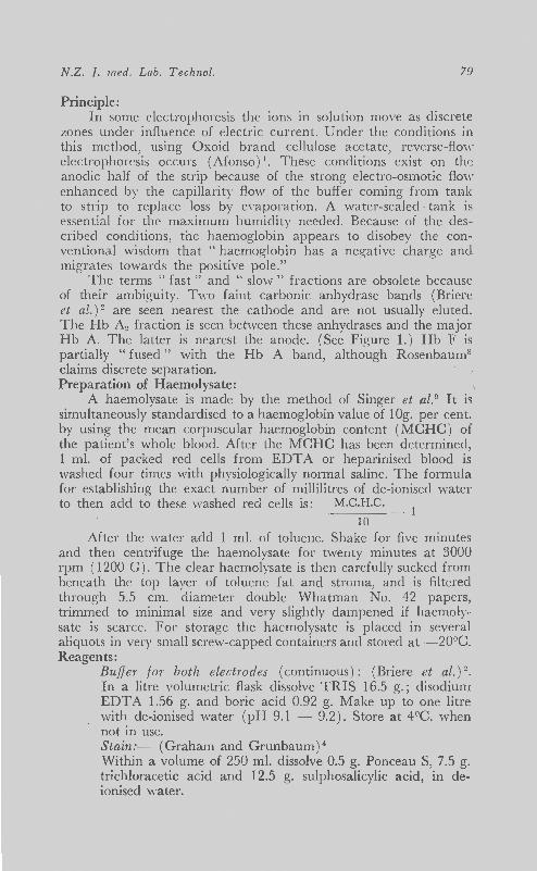
Principle: In some electrophoresis the ions in solution move as discrete
zones under influence of electric current. Under the conditions in this method, using Oxoid brand cellulose acetate, reverse-flow electrophoresis occurs (Afonso) 1 • These conditions exist on thl'! anodic half of the strip because of the strong electro-osmotic flow enhanced by the capillarity flow of the buffer coming from tank to strip to replace loss by evaporation. A water-sealed · tank is essential for the maximum humidity needed. Because of the described conditions, the haemoglobin appears to disobey the conventional wisdom that " haemoglobin has a negative charge and migrates towards the positive pole."
The terms " fast " and " slovv" fractions are obsolete because of their ambiguity. Two faint carbonic anhydrase bands (Briere et al. ) 2 are seen nearest the cathode and are not usually eluted. The Hb A2 fraction is seen between these anhydrases and the major Hb A. The latter is nearest the anode. (See Figure 1.) Hb F is partially " fused " with the Hb A band, although Rosenbaum8
claims discrete separation. Preparation of Haemolysate:
A haemolysate is made by the method of Singer et al. 9 It is simultaneously standardised to a haemoglobin value of lOg. per cent. by using the mean corpuscular haemoglobin content (MCHC) of the patient's whole blood. After the MCHC has been determined, 1 mi. of packed red cells from EDT A or heparinised blood is washed four times with physiologically normal saline. The formula for establishing the exact number of millilitres of de-ionised water to then add to these washed red cells is: M.C.H.C. _
1 10
After the water add 1 mi. of toluene. Shake for five minutes and then centrifuge the haemolysate for twenty minutes at 3000 rpm (1200 G). The clear haemolysate is then carefully sucked from beneath the top layer of toluene fat and stroma, and is filtered through 5.5 em. diameter double Whatman No. 42 papers, trimmed to minimal size and very slightly dampened if haemolysate is scarce. For storage the haemolysate is placed in several aliquots in very small screw-capped containers and stored at -20°C. Reagents:
Buffer for both electrodes (continuous): (Briere et al.) 2•
In a litre volumetric flask dissolve TRIS 16.5 g.; disodium EDT A 1.56 g. and boric acid 0.92 g. Make up to one litre with de-.ionised water (pH 9.1 - 9.2). Store at 4°C. when not in use. Stain:- (Graham and Grunbaum)~ Within a volume of 250 mi. dissolve 0.5 g. Ponceau S, 7.5g. trichloracetic acid and 12.5 g. sulphosalicylic acid, in deionised water.
80 N.Z. ]. med. Lab. Technol.
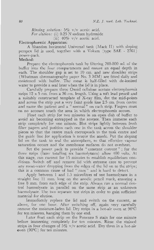
Rinsing solution: 5% v jv acetic acid. For elution: (i) 0.25 N sodium hydroxide
(ii) 40% v/v acetic acid. Electrophoretic Apparatus:
A Shandon horizontal Universal tank (Mark II) with sloping perspex lid .is used, together with a Vokam (type SAE - 2761) power-pack. Method:
Prepare the electrophoresis tank by filtering 700-800 mi. of the buffer into the four compartments and ensure an equal depth in each. The shoulder gap is set to 10 em. and new shoulder strips (Whatman chromatography paper No. 3 MM) are fitted daily and moistened with buffer. The moat is half-filled with de-ionised water to provide a seal later when the lid is in place.
Carefully prepare three Oxoid cellulose acetate electrophoresis strips 12 x 5 em. from a 36 em. length. Using a soft lead pencil and a suitably constructed template of X-ray film, dot the mid-points and across the strip put a very faint guide line 2.5 em. from centre and name the patient and a " normal " on each strip. Fingers must on no account touch the area in which electrophoresis occurs.
Float each strip for two minutes in an open dish of buffer to avoid air becoming entrapped in the acetate. Then immerse each strip completely for one minute. Blot strips lightly between fresh filter papers and position each one in the tank across the shoulder pieces so that the centre mark corresponds to the tank centre and the guide line for application is nearer the anode. Put the sloping lid on the tank to seal the atmosphere, so that effective vapour saturation occurs and the membrane surfaces do not overheat.
Set the power pack to provide "constant current" ; for the three strips (later totalling six haemolysates ) allow 400 volts. At this stage, run current for 15 minutes to establish equilibrium conditions. Switch off and remove lid with extreme care to prevent any moat-water dropping from the edges of the lid on to the strips; this is a common cause of bad " runs " and is hard to detect.
Apply between 1 and 1.5 microlitres of test haemolysate in a straight line 11 mm. long on the anodic pencil mark, ensuring a free 8 mm. from each edge of the strip. Always run a normal control haemolysate in parallel on the same strip as an unknown haemolysate. Use two separate test strips in order to gain sufficient material for elution.
Immediately replace the lid and switch on the current, as above, for one hour. After switching off, again very carefully remove the moisture-laden lid. Dry strips in a hot-air oven at 90°C for ten minutes, hanging them by one end.
Later float each strip on the Ponceau S stain for one minute before immersing completely for ten minutes. Rinse the stained strips ,in four changes of 5% v jv acetic acid. Dry them in a hot-air oven (90°C) for ten minutes.
N.Z. T. med. Lab . Technol. 81
Quantitation by Elution: Duplicate each test so that 2-3 microlitres of haemolysates are
electrophoresed and quantitated by pooling the appropriate fractions.
Cut out each stained haemoglobin band carefully into labelled plastic dishes and cut out blank acetate pieces of equal area to each band from the same acetate strip.
Float two Hb A bands on, and then soak in 9 ml. 0.25 N sodium hydroxide in wide 10 ml. beakers. Ag.itate for a minute to ensure complete elution and then add 0.6 ml. of 40% v / v acetic acid; mix and immediately read Absorbance at 515 mp. (Unicam SP600 ). It is probably advisable to remove the acetate pieces soon after elution, because sodium hydroxide degrades them and produces cloudiness.
Two Hb A2 bands are treated in the same manner, but volumes are 3 mi. of 0.25 N sodium hydroxide and 0.2 mi. of 40% v /v acetic acid per band.
All the corresponding blank acetate pieces are similarly treated and their optical density subtracted from that of the Hb band•.
Optical density A2 100
(3 x OD of A) + OD of A2 o/o Hb A2
If Gelman brand cellulose acetate is used, proceed as for Oxoid but apply haemolysate on the cathodic side. Elution and Normal range of Hb A2 From Oxoid Cellulose Acetate:
Haemolysate from thirty-two staff members were electrophoresed and eluted by the above method on Oxoid brand cellulose acetate. Their Hb A2 ranged from 0.4 to 3.1% with a mean of 1.7%.
Haemolysates from forty-four adult patients being tested for increases in Hb A2 and found to be within the normal range, gave results from 1.3 to 3.2% with a mean of 2.2%. Ten babies gave Hb A2 results from 0 to 1.2% with a mean of 0.6% . This conforms with the findings of Allison et al. as cited by Lehman and Huntsman.'
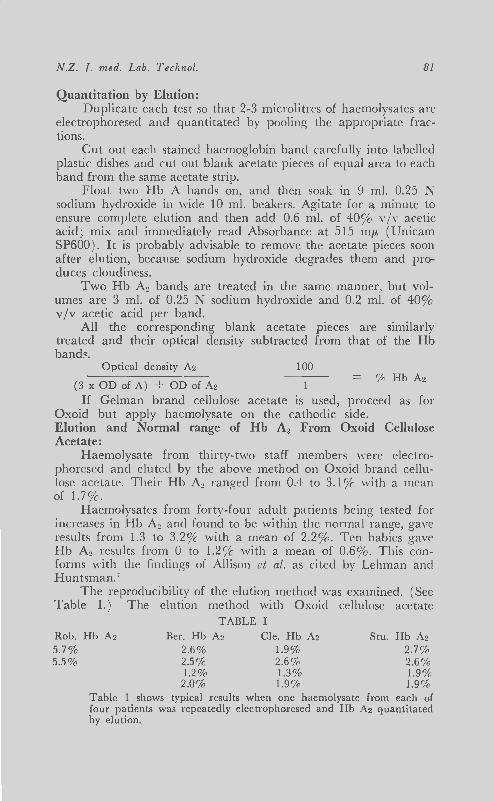
The reproducibility of the elution method was examined. (See Table I.) The elution method with Oxoid cellulose acetate
TABLE I Rob. Hb Az Ber. Hb A2 Cle. Hb A2 Stu. Hb A2 5.7% 5.5%
2.6% 1.9% 2.7% 2.5 o/o 2.6% 2.6% 1.2% 1.3% 1.9% 2.0% 1.9% 1.9%
Table 1 shows typical results when one haemolysate from each of four patients was repeatedly electrophoresed and Hb A2 quantitated by elution.
82
Dilution of 1 Og.% haemolysate.
3:3 of lOg%
2:3 of lOg%
1:2 of lOg%
Hb.
100 llg
66 llg
50 llg
TABLE II
N.Z. ]. med. Lab. Techno/.
Hb Az
5.4% 4.8 % 5.2%
Table II illustrates electrophoresis of varying amounts of haemoglobin from one patient; Hb Az quantitated by elu tion. (Same patient as in Table IV).
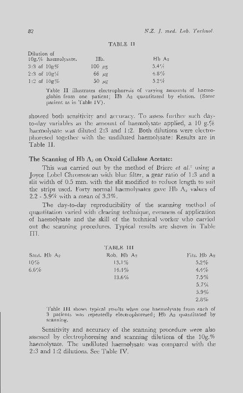
showed both sensitivity and accuracy. To assess further such clayto-day variables as the amount of haemolysate applied, a 10 g.% haemolysate was diluted 2:3 and 1 :2. Both dilutions were electrophoresed together with the undiluted haemolysate: Results arc in Table II.
The Scanning of Hb A2 on Oxoid Cellulose Acetate:
This was carried out by the method of Briere et a/.2 using a Joyce Lobel Chromoscan with blue fi lter, a gear ratio of 1 :3 and a slit width of 0.5 mm. with the slit modified to reduce length to suit the strips used. Forty normal haemolysates gave Hb A2 values of 2.2 - 5.9% with a mean of 3.3%.
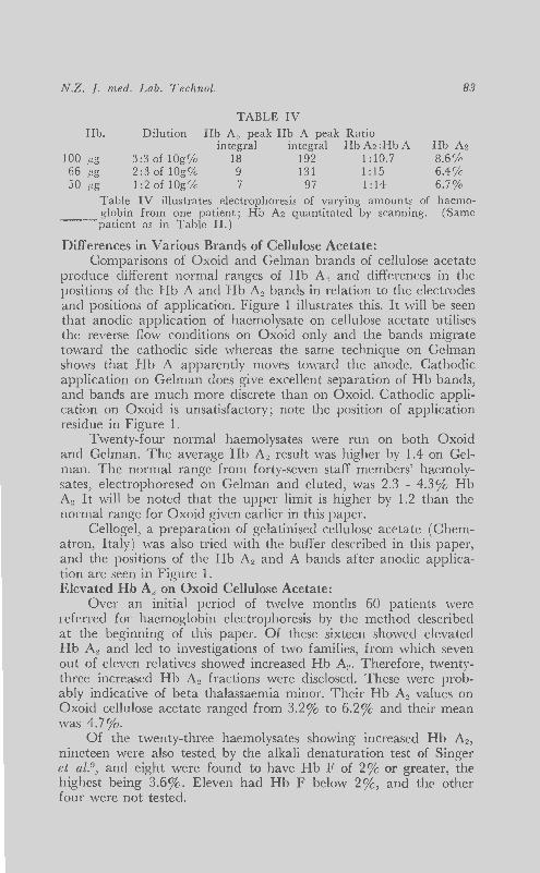
The day-to-day reproducibility of the scanning method of quantitation varied with clearing technique, evenness of application of haemolysate and the skill of the technical worker who carried out the scanning procedures. Typical results are shown in Table III.
Sant. Hb Az
10%
TABLE III
Rob. Hb A~
13.1% 14.4% 13.6%
Fitz. Hb Az
5.2% 4.4% 7.5% 5.7% 5.9% 2.8%
6.6%
Table III shows typical results when one haemolysate from each of 3 patients was repeatedly electrophoresed ; H b Az quantitated by scanning.
Sensitivity and accuracy of the scanning procedure were also assessed by electrophoresing and scanning dilutions of the lOg.% haemolysate. The undiluted haemolysate was compared with the 2:3 and 1 :2 dilutions. See Table IV.
N.Z. 1. med. Lab. Technol. 83
TABLE IV
Hb. Dilution Hb A0 peak Hb A peak Ratio integral integral HbA2:HbA Hb A2
100 /).g 3:3 of lOg% 18 192 1:10.7 8.6% 66 /).g 2:3 of lOg% 9 131 1:15 6.4% 50 /J.g 1:2 of lOg% 7 97 1 :14 6.7%
Table IV illustrates electrophoresis of varying amounts of haemo-globin from one patient; Hb A2 quantitated by scanning. (Same
--~ patient as in Table II.)
Differences in Various Brands of Cellulose Acetate: Comparisons of Oxoid and Gelman brands of cellulose acetate
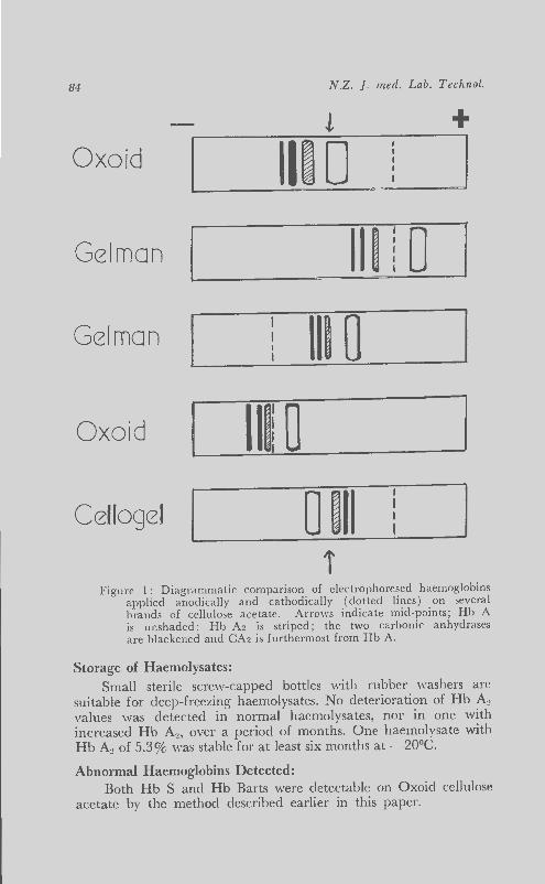
produce different normal ranges of Hb A2 and differences in the positions of the Hb A and Hb A2 bands in relation to the electrodes and positions of application. Figure 1 illustrates this. It will be seen that anodic application of haemolysate on cellulose acetate utilises the reverse flow conditions on Oxoid only and the bands migrate toward the cathodic side whereas the same technique on Gelman shows that Hb A apparently moves toward the anode. Cathodic application on Gelman does give excellent separation of Hb bands, and bands are much more discrete than on Oxoid. Cathodic application on Oxoid is unsatisfactory ; note the position of application residue in Figure 1.
Twenty-four normal haemolysates were run on both Oxoid and Gelman. The average Hb A2 result vvas higher by 1.4 on Gelman. The normal range from forty-seven staff members' haemolysates, electrophoresed on Gelman and eluted, was 2.3 - 4.3% Hb A2 It will be noted that the upper limit is higher by 1.2 than the normal range for Oxoid given earlier in this paper.
Cellogel, a preparation of gelatinised cellulose acetate (Chematron, Italy) was also tried with the buffer described in this paper, and the positions of the Hb A2 and A bands after anodic application are seen in Figure 1. Elevated Hb A" on Oxoid Cellulose Acetate:
Over an initial period of twelve months 60 patients were referred for haemoglobin electrophoresis by the method described at the beginning of this paper. Of these sixteen showed elevated Hb A2 and led to investigations of two families, from which seven out of eleven relatives showed increased Hb A2 • Therefore, twentythree increased Hb A2 fractions were disclosed. These were probably indicative of beta thalassaemia minor. Their Hb A2 values on Oxoid cellulose acetate ranged from 3.2% to 6.2% and their mean was 1.7%.
Of the twenty-three haemolysates showing increased Hb A2,
nineteen were also tested by the alkali denaturation test of Singer et al. 9
, and eight were found to have Hb F of 2% or greater, the highest being 3.6%. Eleven had Hb F below 2%, and the other four were not tested .
84 N.Z. ]. med. Lab. Techno/.
! + Oxoid Ill 0 I
Gelman Ill 0 I
Gelman Ill 0 Oxoid Ill! 0 Cellogel 0 Ill
i Figure 1: Diagrammatic comparison of electrophoresed haemoglobins
applied anodically and cathodically (dotted lines) on several brands of cellulose acetate. Arrows indicate mid-points; Hb A is unshaded; Hb A2 is striped; the two carbonic anhydrases are blackened and CA2 is furthermost from Hb A.
Storage of Haemolysates: Small sterile screw-capped bottles with rubber washers are
suitable for deep-freezing haemolysates. No deterioration of Hb A~ values was detected in normal haemolysates, nor in one with increased Hb A2, over a period of months. One haemolysate with Hb A2 of 5.3% was stable for at least six months at -20°C.
Abnormal Haemoglobins Detected: Both Hb S and Hb Barts were detectable on Oxoid cellulose
acetate by the method described earlier in this paper.
N.Z. T. med. Lab. Techno/. 85
The Hb Barts was separated and quantitated to 11.4% alongside an Hb A2 of 0.8%. The Hb Barts band was situated on the ano:lic side of the Hb A and F ( Oxoid; anodic application). The identity of the band was confirmed by ultra-violet spectrophotometry; the Hb Barts was also electrophoresed on starch gel at pH 7. 7 by the Auckland Blood Transfusion Service. The patient was a child of four weeks; foetal haemoglobin and Hb Barts by alkali denatura',;cli were 53%. The findings were consistent with alpha thalassaemia minor.
In another patient Hb S was clearly separated, situated between Hb A and Hb A2 • It was a heterozygous expression of the S gene and gave a Chromoscan quanti tation of 23% Hb S. This haemolysate had been deep-frozen for more than three years prior to electrophoresis on cellulose acetate. Recently a fresh haemolysate from this patient gave an Hb S of 27% by elution. Discussion:
Experience with cellulose acetate haemoglobin electrophoresis over many months and including many trials and modifications of methods has led to the following observations :
(a) T he method presented in this paper, using Oxoid cellulose acetate, is a reliable and reproducible method for routine daily use.
(b ) There are marked variations in the quality and properties of different batches within a brand of cellulose acetate.
(c ) Although Hb E occupies the site of Hb A2 in the method descr.ibed in this paper, it is unlikely to cause confusion o£ .identity because Hb E, even in the E heterozygote, amounts to 30-40% (Lehman)' whereas Hb A2 is usually below 7%.
(d) In our experience in a routine haematology laboratory, quantitation by scanning required much more expertise and care by technical staff than did quantitation by elution.
(e ) Hb F is better quantitated by the alkali denaturation test as it is extremely difficult to separate completely as a discrete band, although Rosenbaum8 claims to have done this without difficulty.
(f ) The continuous buffer system used in this paper, as described by Briere et al. 2, gave better results than a discontinuous system using buffers of barbital and Tris types. The pH of 8.8 given by Briere et aU for the buffer described in this paper is unattainable without adjustment with acid. However, this adjustment is not necessary and at the unadjusted pH of 9.1-9.2 the buffer functions efficiently.
(g ) The phenomenon whereby, under certain conditions,
86 N.Z. ]. med. Lab. Tech.nol.
haemoglobins applied anodically on Oxoid cellulose acetate migrate towards the cathode by reverse flow electrophoresis is particularly interesting, as this does not occur on Gelman cellulose acetate when haemolysate is applied anodically under similar conditions. Nor does it occur on Oxoid when application is cathodic. (Figure 1.)
(h) Cathodic application on Oxoid and anodic application on Gelman lead to inaccurate quantitations because the residue often present at the application line is situated and is stained in the middle of the haemoglobin fractions required for accurate elution and quantitation (Figure 1.)
Summary: 1. A modified method of haemoglobin electrophoresis on cellu
lose acetate is presented which .is both reliable and accurate. This method has been used to detect and to quantitate Hb A, Hb A2, Hb S and Hb Barts as well as the anhydrases CA1 and CA2 . It is reputed to detect other abnormal haemoglobins.
2. A normal range of Hb A~ from 76 adults is presented using Oxoid cellulose acetate and elution ; the range is 0.4-3.2% of the total Hb.
3. A normal range of Hb A2 from 40 adults is presented using Oxoid cellulose acetate and scanning; the range is 2.2-5.9% of the total Hb.
4. The range of Hb A2 from 23 cases of beta thalassaemia minor is presented together with results of 19 of the concomitant Hb F determinations by alkali denaturation.
5. The normal range of Hb A" on Gelman cellulose acetate was found to be higher than on Oxoid cellulose acetate.
6. Technical details and phenomena relating to Hb electrophoresis on cellulose acetate are discussed, with particular reference to differences between various brands of acetate.
Acknowledgement.~: The author wishes to thank Dr J. G. Buchanan for his per
mission to publish this work performed in the Haematology Department of the Auckland Hospital Laboratory and Mr A. Nixon, Charge Technologist, for his assistance.
REFERENCES: I. Afonso, E., (1962), Clin. chim. Acta, 7, 545. 2. Briere, R. 0., Golias, T. , and Batsakis, .J. G. ( 1965), A mer. ] . clin.
Path., 44, 695. 3. Gerald, P.S. and Diamond, L.K., (1958), Blood, 13, 61. 4. Graham, J.L. and Grunbaum, B.W., (1963) , Amer. ]. clin. Path., 39,
567. 5. Kohn, J., (1957) , Clin. chim. Acta, 2, 297. 6. Kunkel, H.G. and Wallenius, G., (1955), Science, 122, 288. 7. Lehman, H., and Huntsman, R.G., (1966), Man's Ha.emoglobins, 1st
edition p. 48 and p. 279, North-Holland Publishing Co., Amsterdam. 8. Rosenbaum, D.L., (1965) , Amn. ] . med. Sci., 252,726. 9. Singer, K., Chernoff, A.I., and Singer, L. (1951), Blood., 6, 4·13.
FIBROMETER SYSTEM ...
: the world's first • • preCISIOD
coagulation timer
ix
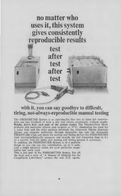
no matter who uses it, this system gives consistently
reproducible results test after test after test
with it, you can say goodbye to difficult, tiring, not-always-reproducible manual testing The FIBROMETER System is so reproducible that two or three lab technologists can run hundreds of tests a day and obtain consi;tently uniform results. Briefly, here's how each part of the system works: The Thermal-Prep Block preheats and maintains plasma and reagents at 37 °C, eliminating the need for a water bath and the extra motions involved; the Automatic Pipette dispenses plasma and reagents uniformly through disposable tips into the disposable FIBROTUBE Cups and starts the sensing and timing device; the FIBROMETER Unit electromechanically measures and records the clot formation time to 0.1 of a second. And the entire clot sensing and timing process is automatic. In addition, the FIBROMETER System is of a modular design so you can use any combination, up to 6 units, with a single electrical outlet, for your particular coagu-lation test work load. This is just part of the FIBROMETER System. For all the facts and a copy of "A Manual of Methods for the Coagulation Laboratory" contact the sole N.Z. agents.
X
Coagulation testing
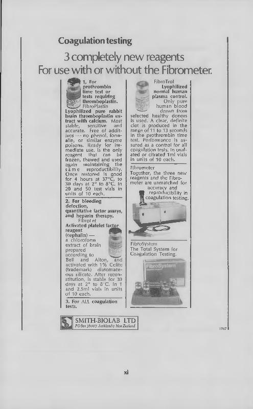
3 completely new reagents For use with or without the Fibrometer.
1 .• For prothrombin time test or tests requiring
' thromboplastin. FibroPiastin
Lyophil ized pure rabbit brain thromboplastin extract with calcium. Most stab le, sensit ive and accurate. Free of additives - no phenol , formalin , or similar enzyme poisons. Ready for immediate use. Is the only reagent that can be frozen, thawed and used again ma intaining the same reproduclibi li l y. Once res tored is good for 4 hours at 37°C, to 30 clays at 2° to 8°C. In 20 and 50 test via ls in units of 10 each.
2. For bleeding detection, quantitative factor assays, and heparin therapy.
Fibrolet Activated platelet factor reagent (cephalin) -a ch loroform extract of brain prepared according to Bel l and Alton, and activated w ith 1% Celi te (trademark) diatomaceous silicate. After reconstitution, is stab le for 30 days at r to 8 C. In 1 and 2.5ml vials in units of 10 each .
3. For ALL coagulation tests.
ribroTrol Lyophilized
normal human plasma control.
On ly pure human blood
...__. drawn from se lected hea lthy donors is used. A clear, definite clot is produced in the range of 11 to 13 seconds in the prothrombin time test. Performance is assured as a control for all coagulation tests. In oxalatecl or cit rated 1 ml vials in un its of 10 each.
Fibrometer Toge ther, the three new reagents and the Fibrometer are unmatched for
accuracy and 1 reproducibility in ~l coagulat ion test ing. ~
SMITH-BIOLAB LTD PQBo, ]600] lurklandg \'ell lMioncl
xi
1967
announcing
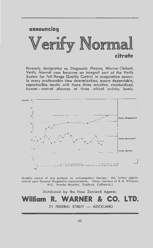
Verify Normal citrate
Formerly designated as Diagnostic Plasma, Worner-Chilcott, Verify Normal now becomes an integral part of the Verify System for Full-Range Quality Control in coagulation assays. In every prothrombin time determination, assure dependable, reproducible results with these three sensitive, standardised, human - control plasmas at three critical activity levels.
lecondt 4~~---
AO
" ------ ----------,-..----1
............... .. .... .............. ;.-·· ' ,,\ "
.~·-····
( ·-·
15, .. -····
"
1 2 3 4 5 6 7 8 9 10 11 12 13 14 15 16 17 18 li 20 21 22 23 24 25 26 27 28 ~ 30 .. ,
Verily Abnormal II
Verity Abnormal I
Graphic record of two patients on anticoagulant therapy. For further detoils, consult your General Diagnostics representative. (Data, courtesy of R. R. Williams,
M.D., Peralta Hospital, Oakland, California.)
Distributed by the New Zealand Agents:
William R. WARNER & CO. LTD. 21 FEDERAL STREET - AUCKLAND
xii
N.Z. f. med. Lab. Techno/.
The Occurrence of Microsporum nanum as a Human and Animal Pathogen
in New Zealand M. BAXTER M.Sc., Ph. D. and R. D. PEARSON
Department of Animal Health, Massey University, Palmerston North.
Received for Publication, March, 1969.
87
The intensive study of keratinophilic fungi which has occurred in recent years has led to the discovery of several previously unrecognised species of Microsporum. Of the fourteen species of this genus considered valid by Ajello .in 19681 only three were in Emmons' 1934 classification6
: At!. audouinii, M. canis and M. gyjJseum. Although these three are the most frequently isolated members of the genus from human and animal infections, certain of the other species may occasionally be encountered in the diagnostic laboratory. One of these .is M . nanum, a fungus first described by Fuentes et al8 as a dwarf form of M . gypseum and later given its specific name by Fuentes. 7 It is known as a cause of tinea capitis and tinea corporis in the Americas~ but has not previously been recorded as a human pathogen in New Zealand. This report concerns the isolation of M. nanum from a human infection apparently contracted from infected pigs. These animals arc frequent hosts of this fungus\ n and a reservoir of potential human infection which appears to be fairly widespread in New Zealand10• We give details of the cultural appearance of this dermatophyte so that it may be recognised in the laboratory should it occur during routine isolations from ringworm material. Case Reports: Human
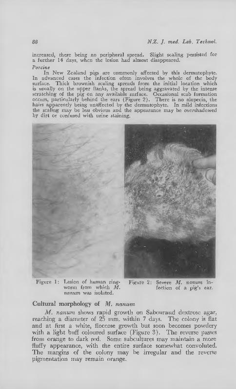
The human infection was of one of us. The lesion on the left forearm was first noticed as a circular, slightly erythematous area , 2 x 1.5 em. in diameter and I 0 em. above the wrist (Figure I ) . There was no associated scaling when first observed but the skin scrapings could be obtained without difficulty by running a scalpel blade over the affected area. Microscopically, in KOH, the skin scrapings contained abundant fungal hyphae but there was no apparent involvement of the hair, although, macroscopically on the skin, some hair follicles had appeared somewhat engorged. There was no fluorescence under Wood's light. (In some cases of tinea capitis caused by this fungus a green fluorescence has been described' and an endothrix type of hair invasion may occur' '.) When cultured on Sabouraud dextrose agar containing cycloheximide and chloramphenicol the skin scrapings yielded colonies identified as M. nanum.
Scaling of the lesion was first observed on the 5th day and treatment 3 times daily with " Tinaderm " ( tolnaftate sulphate)* was commenced The amount of scaling between applications of the Tinaderm, when the skin had become dry, increased over the subsequent 9 days. By this time the centre of the lesion had resolved but the total area involved had not
·Jt Schering Corporation, New Jersey.
88 N.Z. ]. med. Lab. Techno/.
increased, there being no peripheral spread. Slight scaling persisted for a further 14 days, when the lesion had almost disappeared.
Porcine In ::\'ew Zealand pigs are commonly affected by this dermatophyte.
In ad,·anced cases the infection often involves the whole of the body surface. Thick brownish scaling spreads from the initial location which is usually on the upper flanks, the spread being aggravated by the intense scratching of the pig on any a\'ailable surface. Occasional scab formation occurs, particularly behind the ears (Figure 2). There is no alopecia, thr hairs apparently being unaffected by the dermatophyte. In mild infections the scaling may be less obvious and the appearance may be m·ershaclowecl by dirt or confused with urine staining.
Figure 1 : Lesion of human ringworm from which M. nanum was isolated.
F igure 2: Senre M. nanum infection of a pig's car.
Cultural morphology of Jill. nctnum
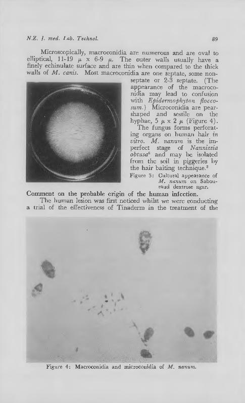
M. nanum shows rapid growth on Sabouraud dextrose agar, reaching a diameter of 25 mm. \\'ithin 7 da) s. The colony is flat and at first a white, floccose growth but soon becomes powdery with a light buff coloured surface (Figure 3). The reverse passes from orange to dark red . Some subcultures may maintain a more fluffy appearance, with the entire surface somewhat convoluted. T he margins of the colony may be .irregular and the reverse pigmentation may remain orange.
N.Z. T. med. Lab. Techno/. 89
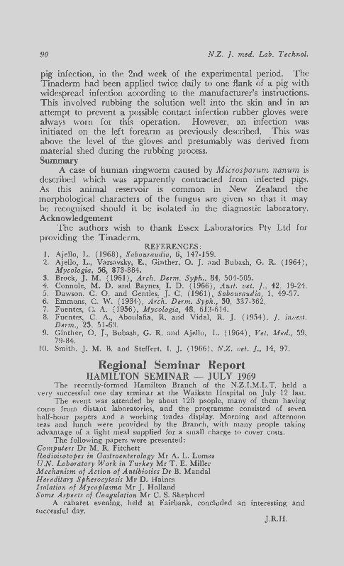
Microscopically, macroconidia are numerous and are oval to elliptical, 11-19 f.L x 6-9 f.L· The outer walls usually have a finely echinulate surface and arc thin when compared to the thick vvalls of M. canis. l'v1ost macroconidia are one septate, some non
septate or 2-3 septate. (The appearance of the macroconidia may lead to confusion \Nith Ej;idermojJh)"ton floccosum. ) Microconidia are pearshaped and sessile on the hyphae, 5 f.L x 2 f.L (Figure 4 ) .
The fungus forms perforating organs on human hair i11 vitro. M. nanum is the imperfect stage of N annizzia obtusa5 and may be isolated from the soil in piggeries by the hair baiting technique. 2
Figure 3: Cultural appearance of M. nanum on Sabouraud dextrose agar.
Comment on the probable cngm of the human infection. The human lesion was first noticed whilst we were conducting
a tr:al of the effectiveness of Tinaderm in the treatment of the
' , .... .. '
.,. I ., .. \
.1'\
, -.
Figure 4: Macroconidia and microconidia of M. nanum.
90 N.Z. ]. med. Lab. Techno{.
pig infection, in the 2nd week of the experimental period. The Tinaderm had been applied twice daily to one flank of a pig with widespread infection according to the manufacturer's instructions. This involved rubbing the solution well jnto the skin and in an attempt to prevent a possible contact infection rubber gloves were always worn for this operation. However, an infection was initiated on the left forearm as previously described. This was above the level of the gloves and presumably was derived from material shed durjng the rubbing process. Summary
A case of human ringworm caused by Microsporum nanum is described which was apparently contracted from infected pigs. As this animal reservoir is common in New Zealand the morphological characters of the fungus are given so that it may be recognised should it be isolated jn the diagnostic laboratory. Acknowledgement
The authors wish to thank Essex Laboratories Pty Ltd for providing the Tinaderm.
REFERENCES: 1. Ajello, L. (1968), Sabouraudia, 6, 147-159. 2. Ajello, L., Varsavsky, E., Ginther, 0. J. and Bubash, G. R. (1964),
Mycologia, 56, 873-884. 3. Brock, J. M. ( 1961), Arch. Derm. Syph., 84, 504-505. 4. Connole, M. D. and Baynes, I. D. (1966), Aust. vet. ]., 42, 19-24. 5. Dawson, C. 0. and Gentles, J. C. (1961), Sabouraudia, 1, 49-57. 6. Emmons, C. W. (1934), Arch. Derm. Syph., 30, 337-362. 7. Fuentes, C. A. (1956), Mycologia, 48, 613-614. 8. Fuentes, C. A., Aboulafia, R. an·d Vidal, R. J. ( 1954), ]. invest.
Derm., 23, 51-63. 9. Ginther, 0. ]., Bubash, G. R. and Ajello, L. (1964), Vet. Med., 59,
79-84. 10. Smith, J. M. B. and Steffcrt, I. J. (1966), N.Z. vet.]., 14, 97.
Regional Seminar Report HAMILTON SEMINAR- JULY 1969
The recently-formed Hamilton Branch of the N.Z.I.M.L.T. held a ve1y successful one day seminar at the Waikato Hospital on July 12 last.
The event was attended by about 120 people, many of them having come from distant laboratories, and the programme consisted of seven half-hour papers and a working trades display. Morning and afternoon teas and lunch were provided by the Branch, with many people taking advantage of a light meal supplied for a small charge to cover costs.
The following papers were presented: Computers Dr M. R. Fitchett Radioisotopes in Gastroenterology Mr A. L. Lomas U.N. Laboratory Work in Turkey Mr T. E. Miller Mechanism of Action of Antibiotics Dr B. Mandai Hereditary Spherocytosis Mr D. Haines Isolation of Mycoplasma Mr J. Holland Some Aspects of Coagulation Mr C. S. Shepherd
A cabaret evening, held at Fairbank, concluded an interesting and successful day.
.J.R.H.
Invest in 'Volucon'* volumetric concentrates for the simplest preparation of standard solutions
Profit from the speed and convenience of using plastic or glass ampoules
Security of standardized accuracy is as5ured (each ampoule makes one li tre of stated normality)
Save t ime and money
Bank on May & Baker Laboratory Chemicals
Deta il ed information is avai lable on request
'Volucon'
xiii
Volumetric Concentrates
MAY & BAKER (NEW ZEALAND) LTD and Kempthorne Prosser & Co's NZ Drug Co. Ltd. D1stnbutors of the products manufactured by MAY & BAKER LTD Dagenham Essex England and MAY & BAKER (NEW ZEALAND) LTD P.O. Box 35060 Naenae Tel 677-01 6
*trade mark LAI090
ASK YOURSELF:
DOES THE LDH PROCEDURE
YOU ARE NOW USING ... allow you to do 10 tests and contro ls in 10 min utes? Lac-Dehystrate does. give you straight line calibration through the entire useful c I i n i c a I range?
Lac-Dehystrate does. show 95% disease specifically? *
Lac-Dehystrate does. If you're not usin g Lac-De hystrate maybe you oug ht to be. We'd be p leased to tell you more about this u n i q u e reagent system a1 your convenience. Call on us. Additional information avail able on request .
Marymont, J. H., Jr.; Cawley, L. P. and Hoff· ma nn , R. G. : Am. J. Clin . Path. 49:431, 1968 .
TM
c-Dehystrate reagent system f or serum LDH
IIUiia.te.WARNER -'0.411 P.O. Box -430, Auckland
xiv
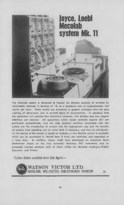
Joyce, loebl Mecolab system Mk. 11
The Mecolob sys te m is des ig ned to ha nd le the d iscrete a na lys is of samp les by
co lori me tric meth ods in botches of 15, at o maxi mu m rote of app roximate ly 165
results per ho ur. Those resu lt s ore presen ted in grap hic ana logue form for easy
sighti ng of obnormo ls, and in p rinted digits of concentration. In standard form
the equ ipme ni wi ll provide four analytica l channels, with di lution and two reagent
additions per channel. All operations which would normally require skill ore
performed automat ically, and the only physical activities associated with this
system ore the introduction of so:nple into the appropriate cups and the transfer
of carriers from sampling unit to water both if necessary, and then to colorimeter .
As the design of the system is based on modules, a very flexible system is available
which con be purchased in limited form if funds ore restricted, and expanded at
a later dote. In addition, Joyce, Loeb! hove developed an automatic flame
photometer based on the very successful Beckman 105 instrument, and on
automatic enzyme analyser both of which utilize the Mecolab Analogue/Digital
Converter and Printer.
Further details avai lable from Sole Agent:-
IAWATSON VICTOR LTD. -AUCKLAND, WELLINGTON, CHRISTCHURCH, DUNEDIN tG
XV
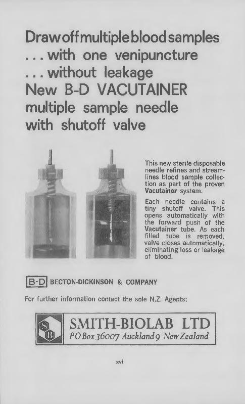
Draw off multiple blood samples ... with one venipuncture ... without leakage New B-D VA CUT AINER multiple sample needle with shutoff valve
This new sterile disposable needle refines and stream· lines blood sample collec· tion as part of the proven Vacutainer system.
Each needle contains a tiny shutoff valve. This opens automatically with the forward push of the Vacutainer tube. As each filled tube is removed, valve closes automatically, eliminating loss or leakage of blood.
IB·DI BECTON-DICKINSON & COMPANY
For further information contact the sole N.Z. Agents:
~t'l] SMITH-BIOLAB LTD ~ P 0 Box 36007 Auckland 9 New Zealand
xvi
N.Z. !. med. Lab. Technol.
A Simple Method of Hormonal Iodine Assay I. C. T. LYON, M.Sc., M.A.A.C.B., F.N.Z.I.M.L.T. C/ o Drs Alexander and McCafferty, Lower Hutt*.
RPceived /or Publication, February, 1969.
Introduction:
91
The evaluation of thyroid status by laboratory im·estigations has for some time depended on the serum protein bound iodine estimation (PBI) which, though presenting some technical difficulties, has become widely used. Contamination of the specimen both in vivo and in vitro is well known. and the organic iodinecontaining drugs have been a constant source of trouble. Lately the T 3 test4 and the simpler TBI ( thyrobinding index) r, which are a measure of the unsaturated thyroid binding globulin, have been used by laboratories possessing radio-isotope counting equipment; and these tests have provided a very useful adjunct, drawing attention to the effects of oestrogens, large doses of salicylates, diphenyl hydantoin and the nephrotic syndrome on thyroid function tests. Many authors have endeavoured to improve the PBI estimation to provide greater precision and accuracy, the aim being to determine only the serum thyroxine level. The butanol extractable iodine (BEI) has evolved from the work of Blau3 to provide for the removal of iodoproteins which are included in the PBI and the iodotyrosines and inorganic iodine, by washing the butanol extract. However, all exogenous organic compounds remain in the final reaction mixture.
An isotope dilution technique in the form "competitive protein binding analysis" has been introduced by Murphy & Jachan5
and produced commercially8 but this author has found the method both expensive and lacking in precision.
This paper deals with modifications to the method of Backer, Postmes and Wiener' which originally attracted attention because it offered a "simple and specific method for the determination of iodoamino acids (IAA) and hormonal iodine (HI) in serum." In a later paper, Wiener & Backer0 suggested the HI level was the most useful, and the presence of many chemically difTerent iodinecontaining compounds failed to give falsely elevated HI values. Two groups (Masen3 and Pileggi and Kessler6 ) have introduced non-incineration techniques for determining the iodine-containing compounds present after their respective isolation techniques, and this type of reaction has been applied by the present author to HI isolated by exchange resin.
*Present address:- c/o Human Genetics Research Unit of the M.R.C., Medical School, Dunedin.
92 N.Z. ]. med. Lab. Techno/.
The test consists of addition of 0.5ml. of serum to a cation column; removal of protein, inorganic and organic iodine. elution of Ta and T.1 by ammonia; evaporation of the ammonia; release of the iodine; and subsequent estimation of the same.
Equipment: Chromatography columns conSIStmg of "milk-testing"
pipettes 30cm. long and 7mm. internal diameter. Aluminium block electrically heated and simmerstat con
trolled, with 40 holes to accommodate 15 x 125 mm. heavy wall Pyrex test tubes (Boiling water bath could be used) .
Vortex test tube mixer. 37°C water bath. Spectrophotometer capable of working at 390 to 420 nM.
Reagents: Prepared using analytical grade chemicals, cleionised water
and acid-washed glassware. 1. R esin: Bio. Rae!* analytical grade cation exchange resin
AG (R ) 50 W- X2 200-400 mesh H + form. 2. Borate Buffer: 4·50 ml. 0.4M boric acid plus 550 mi. O.lM
sodium tetraborate. 3. H)1drochloric acid lN. 4. Ammonia 5 N. 5. Antibumj;ing granules (BDH) * * acid washed. 6. Sulphuric acid 3.5N. 7. Bromine wateT, saturated. Ensure a separate phase is
present. 8. Acid bromine solution: (Prepare fresh) 20 ml. glacial
acetic acid, 20 ml. 3.5N sulphuric acid and 2 ml. bromine water.
9. Arsenious acid reagent :-Dissolve with heat 1.44g. As20;. in 15 ml. of 0.5N NaOH and add to approximately 850 mi. of clcionisecl water in a 1 litre volumetric flask. Con-centrated HCl ( 28.0ml.) and concentrated H 2S0.1 ( 45.5 ml.) are added slowly with mixing. After cooling make up to volume with cleionisecl water.
10. Ceric reagent:- Slowly add 24.3 ml. concentrated H 2S0.1 to 350 ml. deionised water in a 500 ml. volumetric flask. Ceric ammonium sulphate 1 Og. is dissolved in the mixture and made up to 500 ml. \\'ith deionisecl water.
11. Standards. It is convenient to use a serum of known HI content because thyroxine solutions are relatively unstable.
T he sera used by the author contains 3.9 and 7.2 /Lgm/ 100 ml. of HI respectively.
* Bio-Rad Labs, 32nd and Griffin, Richmond, Calif., U.S.A. H· British D rug Houses Limited, Laboratory Chemicals Division,
Poole, England.
N.Z. T. med. Lab. Techno!. 93
Method: Preparation of columns: After Backer et aU A small plug of
glass wool is placed .in the column. A suspension of the resin in water is poured into the column to a depth (when settled) of about 2.5 em. After washing successively with 5 mi. of 1 N HCl and 5ml. water, the column is ready for use. Regeneration requires washing with 2 mi. water, 5 ml of 1N HCl and 5 mi. water. The same columns have been used some 43 times without apparent loss of usefulness.
Column chromatograph,y: On separate columns, place 0.5 mL water and 0.25, 0.5 and 0.75 ml. of the respective standards and 0.5 mi. of each unknown serum. Wash each column successively with 2 mi. water, 8 mi. borate buffer, 2 mi. water, 4ml. of lN HCl and 4 mi. water.
Elution of HI: Add 2 mi. of 5N ammonia and collect each eluate in a 16 x 125 mm. Pyrex test tube containing an antibumping granule.
Removal of ammonia: Tubes placed in the aluminium block preheated to 90°C, are allowed to dry at temperatures rising to 125°C. Time required is up to 45 minutes.
Iodine determination: After cooling tubes, 1 mi. of freshly prepared acid bromine solution is added to release iodine from tri-iodothyronine (T3 ) and thyroxine (T4 ). Mixing on a vortex mixer is followed by placing in a 37°C bath for 6 minutes. The tubes should all possess a yellow bromine colour. Addition of 4 mi. of arsenious acid reagent will discharge the colour and 5 minutes at 37°C are required to equilibrate the temperature of reaction. In sequence at 30 second intervals, 1.0 mi. of eerie reagent is added to each tube which is mixed and incubated for 20 minutes. In the same sequence each tube is read in a spectrophotometer at 395 nM (390-420 nM) using water as reference.
Calculation: A standard curve is plotted of optical density readings of the standards against the ttg/ 1 OOml. HI, and the results read from the graph.
Development of the Method: Modifications used are basically an increase .in washings to include the hydrochloric acid wash indicated by Wiener and Backer6, as this always removes any residual salts from the buffer wash.
Attempts to acidify the ammonia eluate before the bromination were unsuccessful, as the presence of ammonium ions inhibited the eerie-arsenate reaction. After removal of the ammonia by e\'ap01ation, the addition of only 50% acetic acid and saturated bromine water gave results lacking in precision; however, the addition of sulphuric acid solved this problem. The mixing of acetic acid, sulphuric acid and bromine water prior to addition to the
94 N.Z. ]. med. Lab. Techno/.
reaction tubes reduces the possible error clue to pipette manipulations.
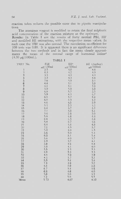
The arsenious reagent is modified to retain the final sulphuric acid concentration of the reaction mixture at the optimum. Results: In Table I are the results of forty normal PBI, HP. and modified HI estimations, with the respective mean values. In each case the TBI was also normal. The correlation co-efficient for 100 tests was 0.89. It is apparent there is no significant diflerence between the two methods and in fact the mean closely approximates the mean of the normal range of hormonal iocline1
(4.30 p.gflOOml.). TABLE I
TEST No. P:i3I HIG HI (Author) ug/ 100ml ug/100ml ug/100ml
1 5.5 4.0 4.6 2 6.3 4.2 4.9 3 5.3 4.3 4.3 4 5.3 4.3 4.4 5 7.6 3.9 5. 1 6 4.4 3. 1 3.9 7 4. 1 3.1 3.0 8 5.9 3.8 4.9 9 6.9 5.7 5.2
10 5.9 4.3 4.7 11 6.5 3.7 3.4 12 6.0 4.3 4.2 13 4.6 4.5 3.9 14 5.2 3.5 4.2 15 6.4 5.7 4.4 16 7.1 4.8 5.1 17 5.6 2.8 3.0 18 5.6 4.6 4.8 19 6.4 4.3 4.8 20 5.4 3.7 3.6 21 6.4 4.4 4.8 22 7.1 4.9 5.7 23 5.0 4.0 4.5 24 5.6 5.0 5.4 25 6.0 5.1 5.4 26 5.4 40 3.7 27 4.9 3.7 4.1 28 5.8 46 5.4 29 5.8 4.1 4.0 30 5.5 3 '3 3.5 31 4.9 3.3 3.3 32 4.8 '10 3.9 33 4.2 3.7 3.1 34 5.3 H 3.4 35 7.4 48 ::1.2 36 4.3 ?..9 2.8 37 67 ::1.4 36 38 6.8 4.6 4.0 39 3.9 30 3.2 40 7.2 ~ n ~-')
Mean 5 72 4.07 4.22
N.Z. f. med. Lab. Techno/. 95
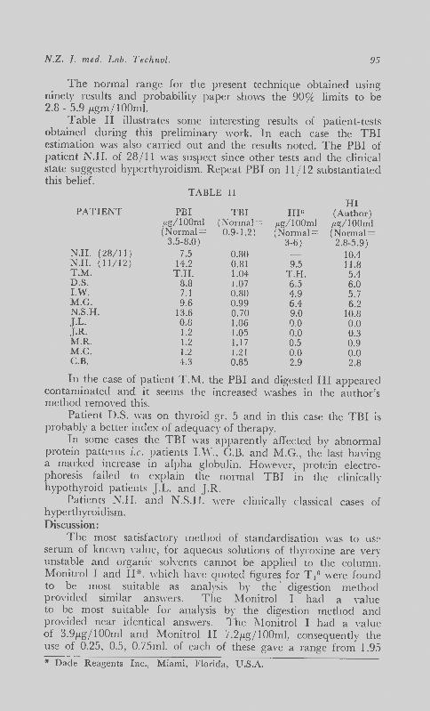
The normal range for the present technique obtained using ninety results and probability paper shows the 90% limits to be 2.8 - 5.9 /Lgm/ 1 OOml.
Table II illustrates some interesting results of patient-tests obtained during this preliminary \·vork. In each case the TBI estimation was also carried out and the results noted. The PBI of patient N.H. of 28/11 was suspect since other tests and the clinical state suggested hyperthyroidism. Repeat PBI on 11/12 substantiated this belief.
TABLE II HI
PATIENT PBI TBI J-IIG (Author) pg/100m1 (Normal = 1Lg/100ml JL~/100ml (Normal = 0.9-1.2) (Normal = (Normal=
3.5-8.0 ) 3-6) 2.8-5.9) N.H. (28/ 11 ) 7.5 0.80 10.4 N.H. ( 11 / 12) 14.2 0.8 1 9.5 11.8 T.M. T.I-I. 1.04 T.H. 5.4 D.S. 8.8 1.07 6.5 6.0 I.W. 7.1 0.80 4.9 5.7 M.G. 9.6 0.99 6.4 6.2 N.S.H. 13.6 0. 70 9.0 10.8 .J.L. 0.8 1.06 0.0 0.0 .J.R. 1.2 1.05 0.0 0.3 M.R. 1.2 l.l i 0.5 0.9 M.C. 1.2 1.21 0.0 0.0 C.B. 4.3 0.85 2.9 2.8
In the case of patient T.M. the PBI and digested HI appeared contaminated and it seems the increased washes in the author's method removed this.
Patient D.S. \\·as on thyroid gr. 5 and in this case the TBI is probably a better index of adequacy of therapy.
In some cases the TBI was apparently affected by abnormal protein patterns i.e. patients I.W., C.B. and M.G., the last having a marked increase in alpha globulin. However, protein electrophoresis failed to explain the normal TBI in the clinically hypothyroid patients J.L. and J.R.
Patients N.H. and N.S.I-1. wPre clinically classical cases of hyperthyroidism. Discussion:
The most satisfactory method of standardisation was to use serum of lmmm Yalue, for aquecus solutions of thyroxine are very unstable and organic solvents cannot be applied to the column. Monitrol I and n·:<-, which have quoted figure~ for T.j 6 were found to be most suitable as analysis by the· digestion method provided similar answers. The Monitrol I had a '·alue to be most suitable for analysis by the digestion method and provided ncar identical answers. The l\1onitrol I had a value of 3.91Lg/ 100ml and Monitrol II 7.2 /Lg/ lOOml, consequently the use of 0.25, 0.5, 0. 75ml. of each of these gave a range from 1.95
* Dade Reagents Inc., Miami, Florida, U.S.A.
96 N.Z . ]. med. Lab. Techno!.
to 1 0.8, with a double check in the mid normal range. Using a Shimadzu QV.50*·X· with deflection meter accessory
set and modified to take a tubular cell sequential sampler, the wave length of 395m,u provided an OD range for the standards of 1.45 to 0.25, which is read with considerable precision using the sensitivity selecting switch set xl, xy'lO, or xlO as required. No real evidence is presented here of the usefulness of this test in the presence of contaminants. One could expect that as the technique of isolation of the HI is virtually that of Backer et al' and Wiener and Backer9
, and since the latter authors' paper provides evidence of the lack of interference by many organic iodine compounds, little trouble should arise in this connection.
T he test correla tes well with the digestion method and the PBI, and provides an advantage in that exclusion of incineration or digestion removes the possibility of loss due to vaporisation or cross-contamination in the furnace.
The method is rapid and provides for batches of 33 patient tests w.ith 6 standards and a blank in 3 hours, provided adequate means of evaporation of the ammonium solvent is available.
Summary: A direct determination of hormonal iodine isola ted from
serum by cationic exchange resin provides a simple, rapid test of th yroid function. Hormonal iodine is separated by allowing 0.5ml. of serum to run through a column from which, after sui table washing to remove other iodine containing compounds, the hormone is elu ted. Removal of solvent is followed by release of the iodine from tr.i-iodothyronine and thyroxine by bromine. The " free " iodine is then estimated by the eerie-arsenious acid reaction.
Acknowledgement: The author wishes to thank Dr W. S. Alexander for his
continued encouragement to develop improved techniques, and Mesdames Phyllis Hampson and Jan Birt for the TBI and PBI estimations.
** Shimadzu Seikakusho Limited, Kyoto, Japan.
REFERENCES: 1. Backer, E. T., Postmes, T. J. and Wiener, J. D . (1967) , Clin. chim.
Acta, 15, 77. 2. Blau, N. F. ( 1935), ]. bioi. Chern., 110, 335. 3. Masen, J. M. (1967), Arner. ] . clin. Path., 48, 561. 4. Mitchell, M. L., Harden, A. B. and O'Rourke, M. E. ( 1960), ]. clin.
Endocr., 20, 1474. 5 Murphy, B. E. P. and Jachan, C. (1965), ]. Lab. clin. Med., 66, 165. 6. Pileggi, V. J. and Kessler, G. ( 1968 ), Clin. Chern., 14, 339. 7. Scholer, J. F. ( 1962), ] . nucl. Me d., 3, 4 1. 8. T etrasorb-125, T4 Diagnostic Kit. Abbott Labora tories, U.S.A. 9. Wiener, J. D. and Backer, E. T . (1968) , Clin . chirn. A cta, 20, 155.
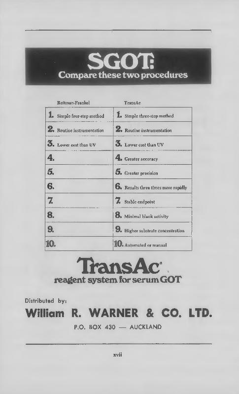
SGOT: Compare these two procedures
Reitman-Franke! TransAc
1 Simple four-step method 1 Simple three-step method
2. Routine instrumentation 2. Routine instrumentation
3. Lower cost than UV 3, Lower cost than UV
4. 4. Greater accuracy
5. 15, Greater precision
6. 6, Results three times more rapidly
7. 7. Stable endpoint
8. 8, Minimal blank activity
9. 9, lligher substrate concentration
10. 10, Automated or manual
TransAceo reagent system for serum GOT
Distributed by:
William R. WARNER & CO. LTD. P.O. BOX 430 - AUCKLAND
xvii
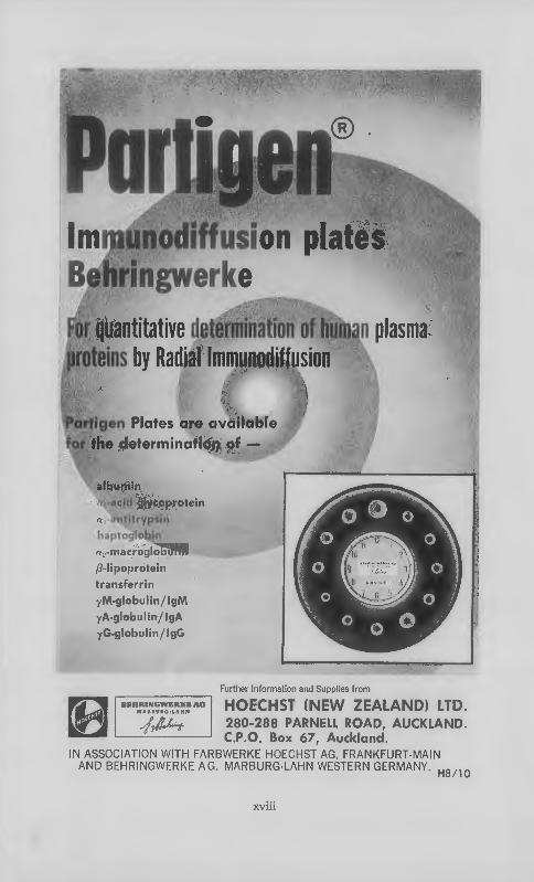
usion plates.;_ .. ngwerke
",-antitrypsin haptoglobin n,-macroglobufln {3-lipoprotein transferrin yM-globulin f igM yA-globulinflgA yG-globulin/lgG
BEHRJNGWERHE AG MAI\BUII.G·LAHN
-~~<f
Further Information and Supplies from
HOECHST (NEW ZEALAND! LTD. 280-288 PARNELL ROAD, AUCKLAND. C.P.O. Box 67, Auckland.
IN ASSOCIAT ION WITH FARBWERKE HOECHST AG, FRANKFURT-MAIN AND BEHRINGWERKE AG. MARBURG-LAHN WESTERN GERMANY. HB/lO
xviii
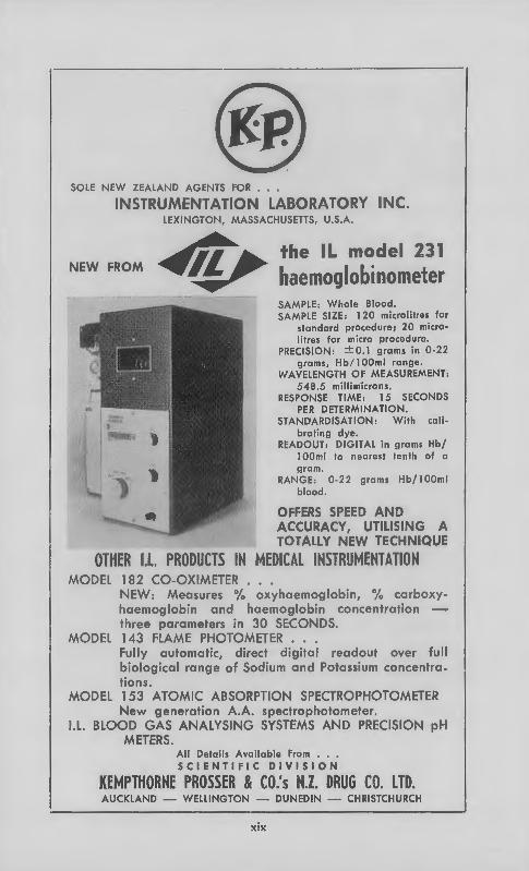
SOLE NEW ZEALAND AGENTS FOR •••
INSTRUMENTATION LABORATORY INC.
NEW FROM
LEXINGTON, MASSACHUSETTS, U.S.A.
the I L model 231 haemoglobinometer SAMPLE: Whole Blood. SAMPLE SIZE: 1 20 microlitres for
standard procedure; 20 microlitres for micro procedure.
PRECISION: ±0.1 grams in 0-22 grams, Hb/ 1 OOml range.
WAVELENGTH OF MEASUREMENT: 548.5 millimicrons.
RESPONSE TIME: 15 SECONDS PER DETERMINATION.
STANDARDISATION: With cali-brating dye.
READOUT: DIGITAL in grams Hb/ 1 OOml to nearest tenth of a gram.
RANGE: 0-22 grams Hb/1 OOml blood.
OFFERS SPEED AND ACCURACY, UTILISING A TOTALLY NEW TECHNIQUE
OTHER I.L. PRODUCTS IN MEDICAL INSTRUMENTATION MODEL 182 CO-OXIMETER . . .
NEW: Measures % oxyhaemoglobin, % carboxy haemoglobin and haemoglobin concentration three parameters in 30 SECONDS.
MODEL 143 FLAME PHOTOMETER .. . Fully automatic, direct digital readout over full biological range of Sodium and Potassium concentrations.
MODEL 153 ATOMIC ABSORPTION SPECTROPHOTOMETER New generation A.A. spectrophotometer.
I.L. BLOOD GAS ANALYSING SYSTEMS AND PRECISION pH METERS.
All Details Available Fram .•• SCIENTIFIC DIVISION
KEMPTHORNE PROSSER & CO.'s N.Z. DRUG CO. LTD. AUCKLAND - WELLINGTON - DUNEDIN - CHRISTCHURCH
xix
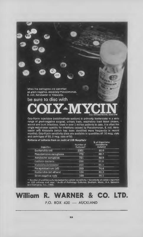
William R. WARNER & CO. LTD. P.O. BOX 430 - AUCKLAND
XX
~.
Wild
M20
T
he
hig
h s
tan
da
rd la
bo
rato
ry
an
d r
ese
arc
h m
icro
sc
op
e
A p
reci
sio
n i
nst
rum
en
t o
f tr
ad
itio
na
l a
SW
ISS
qu
alit
y w
ith
e
xce
llen
t o
pti
cs a
nd
nu
me
rou
s a
cce
sso
rie
s, f
or
all
mo
de
rn
me
tho
ds.
It
's s
o e
asy
to
bu
ild i
t u
p o
r to
co
mp
lete
it
for
an
y p
oss
ible
re
qu
ire
me
nts
. Yo
u w
ill p
rofi
t by
it
s p
erf
ect
inte
rch
an
ge
ab
ility
o
f p
art
s.
Up
pe
r ri
gh
t: W
ILD
M 2
0 w
ith
in
cid
en
t li
gh
t a
tta
ch
me
nt
for
bri
gh
t fi
eld
, d
ark
gro
un
d a
nd
po
rari
sati
on
eq
uip
pe
d w
ith
p
ho
tom
iCro
gra
ph
ic c
amer
a II
Ple
ase
ask
fo
r p
am
ph
lets
co
nta
inin
g f
ull
tech
nic
al
de
tails
. W
ild H
ee
rbru
gg
ltd
. H
ee
rbru
gg
I SG
Sw
itze
rla
nd
ILD
Ful
l g
ua
ran
tee
fo
r re
liab
le s
er
vice
th
rou
gh
wo
rld
re
no
wn
ed
a
SW
ISS
en
terp
rise
s.
ff::'•
Sol
e N
.Z.
Ag
en
ts:
DE
NT
AL
&
ME
DIC
AL
SU
PP
LY
CO
. LT
D.,
A
UC
KLA
ND
, W
EL
LIN
GT
ON
, C
HR
ISTC
HU
RC
H,
DU
NE
DIN
.
Introducing new Versatol Acid-Base serum calibration reference and control for monitoring acid-base analyses ...
at three clinically-significant levels .•.
NORMAL, ACIDOSIS, ALKALOSIS • First and only serum-based calibration reference
and control for acid-base analyses. • Make ii possible to monitor normal and pathological
ranges with a serum-based reference material.
William R. WARNER & CO. LTD. 21 FEDERAL STREET - AUCKLAND
xxii
N.Z . .f. med. Lab. Techno!. 97
The T. H. Pullar Memorial Address
Some Perspectives in Medical Technology
STEPHEN E. WILLIAMS, M.B., Ch.B., D.C.P., M.C.P.A .• M.C. Path.
School of Medical Laboratory Technology Auckland Hospital Board
Delivered at the 25th Annual Conference of the New Zealand Institute Medical Laboratory Technology, Artgust, 1969.
I am deeply conscious of the honour you have done me to-day in asking me to give this address to the Annual Conference of our Institute. To this feeling I must add the pleasure and very genuine pride which it gives me, by virtue of my Honorary Membership, to speak to you as a fellow member.
Each year we assemble on this occasion to pay tribute to Dr Thomas H. Pullar, whose memory is still alive in the minds of the senior members present, and whose contribution to both pathology and medical technology in this country have heartened and enriched us all. Last year Dr P. P. Lynch, when g.iving this address, recorded the important biographical details of Dr Pullar's career and I shall not repeat these to-day, except to remind you that he arrived in New Zealand, a young Scottish graduate, in 1938 and died at the relatively young age of fifty-nine in 1966. In the intervening years, while based at Palmerston North and latterly at Tauranga, he influenced all pathologists, all technologists, many medical students and the officers of our Health Department, in profound and lasting ways.
Tom Pullar was my personal friend. Although we only saw one another at infrequent intervals, usually at conferences and committe2 meetings, I took many problems to him and received in return the most helpful advice, encouragement and wisdom. On the last occasion on which he came to my home we watched a television programme known to many of you which relates to the Scottish \'illage of Tannochbrae. He mentioned that his youth was spent in just such a setting and that his father, a country medical practitioner, could have doubled for the part of Dr Cameron. In his own way, and in a very different setting, Tom Pullar brought to us this same attitude of professional good sense, warmth, humanity and high principle.
Of the many active contributions which he made, perhaps the most far-reaching and significant was his early recognition of the importance of the role of medical laboratory technology and his subsequent energetic participation in the upgrading of technological education and status in New Zealand. It therefore seemed to me appropriate today to take a brief look at some of the present pressures we are all experiencing in our laboratories,
98 N .Z. ]. med. Lab. Tech no/.
and to speculate on what the future holds for our profession; to examine, in fact, what our scientific members would refer to as "parameters and extrapolations."
In such an exercise it is useful to adopt the technique of historians and to lock back over a fevv years. For those of you who may question the validity of this view I can recommend a study of T hucydides's account of the Peloponnesian War in the year 431 B.C. In a technology of swords and spears you will find here all the problems of our communities today, brilliantly illuminated: the hopes, frustrations, passions, satisfactions and disappointments, the treachery, nobility and the violence of human endeavour. Had T hucydides seen fi t to record it, I have no doubt that he could have given us a most vivid description in terms we should all at once recognise, of the reactions of that great medical technologist, H ippocrates, on receiving his statement " original grading determined" from the Athenian Department of Health. My sober point is that in 2,400 years human behaviour has changed very little. But our technology, for better or worse, continues to alter at an ever increasing pace.
Laboratories of the Future To return, then, to the year 1949. In this year Green Lane
Hospital had virtually the same number of beds as it has to-day, and accurate records of its laboratory work are available. We can scan the range and volume of this work to note that in an average month some 1,500 tests were performed by a staff of three. In 1969, 25,000 tests are carried ou t on the same number of patients by a staff of 70. Analysis of the type of tests done is interesting, for it shows that apart from a diminution in one or two categories, such as red cell counts and the disappearance of a few rather infrequently performed tests, including the Weltman test and that majestically named but very simple measurement of adrenal function known as the Robinson-Keppler-Power test, a ll the tests done in 1949 are still being requested for patients, but at a greatly increased frequency. More significantly, it is to be noted that many of the tests which make up a large proportion of our work today were not practicable, and were barely conceivable, in 1949. Such categories, for example, as electrolyte estimations, many of the enzyme assays, the steroid tests, electrophoresis and cytology.
A simple projection forward to the year 1990, then, shows us that in this one hospital perhaps 300,000 tests will be done each mon th by a laboratory staff of 500, and that as well as doing all the tests which we carry out today, much of the work upon which they will be engaged has yet to be developed. Lest any of you should wish to dismiss this view as fan tasy, may I record that had the true facts of medical laboratory work today been presented to us in 1919 we should have rejected them out of hand as science fiction of an extravagant type. Contempla te, therefore, your
N.Z . ]. med. Lab. Techno[. 99
forward march to the tissue typing serv1ce m its six-storeyed Institute, the Department of Electron Microscopy (Routine Service Division), the routine chromosome profile on all admissions, the psychiatric stabilisation absorption test, the laserscope and. of course, the computer. Many of you here will no doubt be able to elaborate the theme in more detailed and descriptive terms of what we know as the "hardware." Such concepts are by no means fancifu l and, despite the limits of our present capacity for speculation, advances of this general type are inevitable.
My own interest at this point in time, however, is directed particularly and urgently to those precious components of laboratory practice whom we corporately term "the software." I mean, of course, the human beings who are going to staff these great Institutes. By returning to our two points on the graph, twenty years apart, we can make some deductions in this connection. In the first place, we observe that we are starting to run out of pathologists. I know that some of you may view this development with relative unconcern, feeling with justification, perhaps, that anything in clinical pathology that a pathologist can do a technologist can do better. In all laboratories, however, there is the important interface between the purely medical and the purely scientific aspects of a patient's illness and it is only by a properly regulated and continuous flow across this interface that the laboratory can perform its true functions of medical diagnostic support. T h e pathologist must place himself in this area and, by virtue of his medical qualification and experience, he must ensure that the vital communications are maintained. It seems probable that in the next twenty years we cannot possibly generate enough pathologists to stand at the proliferated interfaces which we arc now establishing. and there is a very real danger that laboratories and laboratory departments could suffer from an increasing degree of professional and spiritual isolation.
Secondly, vve note that medical technology, already well founded as a profession twenty years ago, is growing robustly and, like so many other facets of our world today, growing exponentially. The lag phase is over and we may expect to double our size every five or ten years. Without question the medical technologist has emerged at a categorical entity for all the world to note, a well respected paramedical scientist and colleague, the essential structural unit of our laboratory services. Unlike the pathologists, we are not yet experiencing serious shortages of technologists, nor do I feel that their sources of generation are likely to fail , at least in the near future. Perhaps the most significant chan,ge in this sector of the staff has been the clear emergence of two further groups of technical workers who have appeared much more recently, in this country only in the last ten years, namely the
100 N.Z. ]. me d. Lab. T echnol.
hospital scientific officers and the technical assistants. Both these groups arc now acting as buttresses for the central structure of medical technologists in such a way that, in the larger laboratories at least, we could not now contemplate operating our departments without them.
From time to time some associates have raised the question of the medical technologist having any place at all in the laboratories of the future, envisaging them being compressed out of existence by the newer categories of scientific officers and technical assistants. Such a view is, of course, quite unrealistic. Laboratories can only operate successfully if we continue to recruit boys and girls of high calibre who from the outset of their training will be exposed to the full range of service pressure of general laboratory practice, whose basis of training will almost certainly become a technical college or university diploma and who will remain an elite of leaders and managers of laboratory groups. This concept must be accepted as fundamental to all future planning of medical laboratory organisation.
Thirdly, we have the phenomenon of a proliferation of hospital laboratories and scientific services other than those which we now incorporate in the routine diagnostic laboratory services, employing in many instances technical staff closely akin to but not actual members of our own group. In this category I would include the medical, surgical and professional units, the nuclear physics, physiology, cardiothoracic, haemodialysis, cancer and other research laboratories.
By applying our method of extrapolation to 1990 we must recognise that in Green Lane Hospital the scientific staff could number around 1,000, working in 40 specialised fields and possibly belonging to 20 different professional organisations. While the medical technologists will remain the core of this group, hospital scientific officers will have increased in number and authority, so that they and the senior technologists will have taken oyer many of the functions and contributions at present expected from pathologists and other medically qualified scientists.
The Zuckerman Report I should now like to refer to some of the findings of the
Committee set up two years ago by the British Ministry of Health under the chairmanship of Sir Solly Zuckennan to report on Hospital Scientific and Technical Services. For those of you who may not have seen the report of this Committee, I may mention that its terms of reference were " to consider the future organisation and development of hospital scientific and technical services in the National Health Hospitals and the broad pattern of staffing required, and to make recommendations." After examining evidence presented by one hundred and fifty-seven individuals and organisations, the Committee has now made a number of general
N.Z. ]. med. Lab. Technol. 101
recommendations. In a consideration of the present position in the United Kingdom the Report has this to say:-
" In recent years there has been an unco-ordinated growth in the size and nature of se1entific services within hospitals. This growth primarily reflects a demand which emanates in the first instance from clinical stafl', although some of it has been generated by the scientific staff themselves. Some of the growth has been due to fashion . . . . . . .
" The scientific service in hospitals tends to be fragmented ; the staff who are classified in a large number of rigid grades (and this brings with it lack of flexibility), sometimes work in isolation. The various disciplines have not been pulled together, and the service, because of its lack of planned organisation, fails to provide good career prospects for all its staff. Furthermore graduate non-medical scientists who work in hospitals do not in general enjoy the opportunities for remuneration comparable to those of their clinical colleagues.
"All this recent growth has been taking place without any clear recognition of the fact that scientists and technicians are in short supply. Unless the growth of the hospital scientific service becomes better organised than it is now, many hospitals will continue to be dissatisfied with the services that can be provided.
"There are considerable variations in the training arrangements from class to class. The basic training of technicians ranges from three years fulltime training before entering employment or five years part-time training whilst in employment, to li ttle or no formal training at all. In some specialities the medical profession has played an active part in the promotion of training, while in others it has been left to the initiative of the technicians themselves, through the formation of staff associations which devise courses of training and award the1r own qualifications ... The general experience is that the establishment of a suitable training scheme follows long after the technical function for which it is required has developed. Further training and advanced qualifications are available in only a few of the classes, and there are no general arrangements by the hospital service for re-training or re-orientation to meet changing needs."
The Report, after considering all the classes of hospital scientific and technical workers, recommends that 21 of these classes should be associated together to form the basis of a Hospital Scientific Service, which while remaining an integral part of the whole hospital service would be subject to special administrative and organisational arrangements. These arrangements include the establishment of a National Hospital Scientific Council to advise the Ministry of Health, Regional Scientific Advisory Committees to work with each Regional Hospital Board, and a Regional Scientist, with senior administrative status, to be a member of each Board. Amongst many other duties this individual will have administrativP responsibilities for the training (including post-graduate training) of scientific and technical staff at all levels.
The future staffing pattern for this Service is visuali sed as consisting of four categories, very much as we operate the hospital laboratory services in this country today. These are to be Scientific Officers recruited from university graduates, Technical Officers who would include all our qualified technological staff,
102 N.Z. ]. med. Lab . Techno/.
Technical Assistants who would include all our trainee staff and Technical Aides, a group somewhat below our own technical assistants, and corresponding to earlier concepts of laboratory assistants. There is nothing very radical in this proposal, but I am interested to note a provision that in addition to entrants to any of these classes being able to make their career within that class, there should also be ample opportunity for advancement to the next higher class, either by obtaining the requisite qualifications or by demonstrating the necessary qualities to the satisfaction of an assessment panel. I interpret this to imply tha t a senior technologist, for example, could, simply by the determination of a grading committee, be advanced to the status of a senior scientific officer, provided that his capabilities justified this action. T he inclusion of this provision seems to me desirab!t: in relation to the very heavy scientific and managerial r esponsibili ties which even today are required of many senior technologists in this country. Certainly it is an objective a t which this Institute should aim.
Predictably, the Zuckerman report has received a mixed reception from the medical profession, including a guarded and, in parts, hostile leading article in the British M edical Journal. H owever, subsequent correspondence has indica ted that many, if not all, pathologists would give it enthusiasti c support. T he I.M.L.T. has also been guarded but admits that some process of unification within the ranks of hospital scientific workers is required. I believe that the Report makes a most significant contribution in focusing attention at this time on a confused situation in which we arc all more or less deeply involved . . Whethc1 its recommendations find acceptance in this country or not, it raises many urgent issues which this Institute cannot ignore . In the period of expansion which we can now c!Parly sec as our destiny an intelligent and realistic administrative basis is quite essc>ntial.
Education
However, the linchpin of a Scientific Service is, in my view, the organisation of its educational programme. If this is well constructed, intelligent, appropriate, imaginative and above all flexible, thf' Service will be secured. If it is badly oriented, rigid or in some respects non-existent, then no matter what efforts are made with rates of pay or conditions of work the wheels of the service will eventually come off. In this country we are fortunate in having a particular agency established by the Department of Health for this purpose, the Medical Laboratory Technologists Board. With this Board, composed jointly of pathologists and technologists, lies the responsibility of translating with speed and insight the changing needs of medical
N.Z. ]. med. Lab. Tec hnol. 103
science into the proper courses of trammg, examinations and qualifications. In its ten years of existence the Board, despite some acutely evident growing pains, has made notable advances, and is now operating from a position of some competence and confidence. If I have seemed in the past to be impatient with the Board this may be because in Auckland vve have encountered quantitative and qualitative problems of service and training which are only now becoming apparent in some other areas. '"'e have adjusted these difficulties in some measure by the formation of our own School and the establishment of special qualifications. More recently the Institute has entered this field with the formation of its Technical Assistants Examination Committee and its qualifications for this group. While this move has been made as the result of strong pressure on a nationa l scale, and is being a ttended by a fair measure of success, there is a growing awareness that within our professional family it may be sensible to have only one academic agency, which could be either this Institute or the M edical Laboratory Technologists Board.
As you are aware we are now moving, rather late in the day compared with most other countries, to pass some and eventually all of our academic technical training over to the Technical Institutes. T his will relieve the Technologists Board of some responsibilities and allow it to develop others. Its successful ventures into the field of "workshop" courses should encourage it to expand this concept not only in relation to educational techniques but into the whole range of post-graduate training. Regular refresher courses for regional and central groups, seminars on specialised topics, workshops for new techniques, the circulation of test slides and specimens, tours by specialist lecturers, brought if necessary from other countries, demonstrations of equipment and the distribution of technical literature should all be accepted by the Board as proper functions of its high office. The continuing education of scientific workers is now regarded as quite essential to the maintenance of proper standards of work, and indeed some countries are proposing a system of formal re-qualification after 20 years in the practice of certain special science fields. The Medical Laboratory Technologists Board is doubtless aware of the problem in this phase of rapid technological expansion and change. It is to be hoped that it will justify its unique position by establishing a sound system of quaternary education for our profession.
With these references to education I turn again to the part played by Dr Pullar, and his contribution to our growth and maturity. The real starting point of the Medical Laboratory Technologists Board was a meeting one Saturday afternoon in
104 N.Z. ]. med. Lab. Techno[.
1959 when Mr Whillans and I had travelled to Palmerston North to talk with Dr Pullar and Mr Hutchings. From this meeting came the Joint-Committee of Pathologists and Technologists, which led some years later to the formation of the Board. Dr Pullar's influence in these moves was paramount, as he sought to balance the enthusiasms, apathies and apprehensions of the pathologists, technologists and administrators. With slender resources and often with the most meagre encouragement he purposefully set about the crucial task of fashioning a linchpin for our service.
His conviction that the medical technologist must become a professional colleague of high scientific standing, and his continued support for this principle have placed us deeply in his debt. Unlike the architect or the engineer, Tom Pullar leaves no physical edifice to mark his contribution. His building materials were the hearts and minds of his fellow men, and the dimensions and design of his construction are to be gauged by the solidarity of our own ranks. Your presence here today in such numbers, strength and spirit, is, to me, the best evidence of the success with which he built. In all his endeavours he would surely have echoed the cry of that great Athenian Nicias:
"It is men, not walls, that make the city."
26th Annual Conference
DUNEDIN 30 and 31 July, 1970
Conference Secretary:
D. A. MacDuff,
Diagnostic Laboratories, Dunedin Hospital,
Dunedin.
25th Annual Conference, 1969 (Right) Delegates to the 25th (Silver Jubilee) Conference at Auckland,
August, 1969. P hoto by: Barry lllcKay Industrial Photography Ltd., P.O Box 5162, Auckland, from whom prints may be obtained at 85 cents each. ;(Quote: 69921-B).
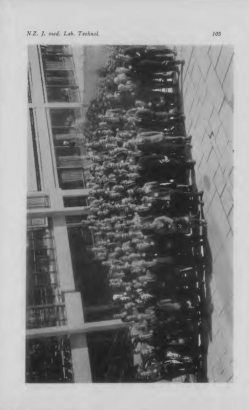
N.Z. ]. med. Lab. Tee /mol. 105
106 N.Z. ]. med. Lab. Technol.
A Changing Scene in Medical Laboratory Technology
J. R. SAAL, Queensland Institute of Technology, Brisbane.
A paper read to the 25th Annual Conference of the N.Z.l.M.L.T., August, 1969.
The Industrial Revolution created the need for tradesmen to use, maintain or manufacture the equipment necessary for industry. From this need there emerged a pattern of education now called technical education, whereby tradesmen were trained on the job in conjunction with studies at night in technical colleges or similar institutions. Along with this development there came a gradual raising of entrance and course standards in order for the tradesmen to cope with the increasing complexity of their trades. This trend is continuing still. In these times the entrance standards have risen to the tenth year of schooling, and the courses arc conducted not only by night but also by release from industry during the cl ay.
Wi th the impetus of two world wars and space exploration today, we are living in a technological revolution. Modern industrial needs of skilled manpower include, in addition to scientists and tradesmen, technologists and technicians. Unfortunately, just as in the previous century, educational programmes have followed rather than kept pace with industrial needs. The reason lies in the types of educational establishments available.
Outside the usual school system, the two traditional areas of training ha\ c been the universities and the technical colleges. Both have been found to be inadequate for the training of technologists.
The universities, to discharge their function, orientate courses to training students to seek knowledge for knowledge's sake. It should not be inferred that all courses in universities are of this type. Exceptions in most universities include engineering and medicine.
Technical colleges by virtue of staffing, facilities, and for other reasons, have not and cannot reach standards of technological excellence, although there have been exceptional technical colleges which have achieved high standards in technological courses.
On the European and North American continents new types of educational establishments have been developed over the past fifty years. They are variously described as Colleges of Advanced Education, Polytechnics, and Institutes of Technology. Development of such institutions has been rapid in a few countries, slower in some countries, and non-existent in other countries. Sir Ian
N.Z . .T. med. Lab. Techno!. 107
Wark, Chairman of the Commonwealth of Australia Advisory Committee on Advanced Education, in a recent address, stated that, on the one hand, Ne\\· Zealand had six universities and insufficiently developed technical institutes, whi le on the other hand Russia had ten times the number of institutes to universities. and ninety per cent. of the tertiary students were enrolled in these institutes.
In Australia, the development of a second tertiary area of education followed the publication of the Martin report in 196'k and has continued under the leadership of Sir Ian Wark's Committee. This has resulted in the establishment of systems of Colleges of Ad\'anced Education in each State. Certain of these colleges offer courses for both technologist and technician, others for the technologist solely. A range of awards are given from Certificate courses, commencing from the tenth year of schooling, to Associate Diploma courses with full professional acceptance equivalent to uniYersity degrees, commencing a t the end of the twelth year of schooling. T he Colleges a re awaiting implementation of two reports recently submitted to the H on. Mr M alcolm Fraser, Commonwealth M inister of Education and Science : T he Wiltshi re repor t, dealing wit!1 the types of awards issued by Colleges, including degrees; and the Sweeney report, dealing with a salary structure for colleges uniform throughou t Australia and suffi cien tly competitive with university salaries to attract staff of high calibre.
In 1966, in this atmosphere of change, the profession of medical laboratory technology in Queensland entrusted educational programmes to the Queensland Institute of Technology, Brisbane. Prior to this, the course had been conducted on a formal basis, by the first two ) ears of basic science being in the technological section of the Central Technical College, and the latter three years being conducted in university departments and major hospitals. The old course was controlled by a Council consisting of representatives of the AIMLT, the college of Pathologists of Australia, and the Department of Education. This council designed the ne, ... · course and continues to advise the Queensland. Institute of Technology in this field.
In designing a suitable course of the requisite standard a number of factors had to be determined. These included:
l. The standard of the course: technician or technologist. 2. Whether the course would be aimed at produ~ing a
graduate as a specialist or a general practitioner. 3. Whether the course would be offered as a full time course
or a part time course. As the p revious course had been of a professional standard and
successful- the course introduced was also one at professional level, an Associate Diploma. Because Q ueensland is a sparsely
JOB N.Z. ]. med. Lab. Techno/.
populated large area, with small hospitals distributed widely, the course was designed to include the major areas of medical laboratory technology. Specialisation was to be the subject of postgraduate training. The course was designed to be completed either in three years full time training or in six years part time training. Cognisance was taken of the grave disadvantage of part time training, in that the intake of such courses is based on today's needs while their output should be geared to the needs of six years hence. Thus, part-time courses never satisfy the needs of industry. Consequently a full time course was also offered.
In order to reach professional standards, to train students adequately in today's technology and to provide a graduate who could cope with tomorrow's technology, the new course contained heavy components of basic and applied science.
The academic year is of 32-33 \Neeks, each week consisting of 32 hours tuition. The first year is devoted to the basic disciplines cf chemistry, physics, biology, and mathematics.
The second year contains both basic science and applied science. To cope with the restrictions of three year full time courses as opposed to four year courses, the subjects of first and second year are orientated towards medical laboratory technology by, wherever possible, using illustrations of scientific principles from the field of medical laboratory technology instead of using classical academic examples. The standard of the first two years may be appreciated by stating that the levels reached are equi\'alent to the same or similar subjects within universities.
The third year deviates from what is usually seen in universities. The subjects taught form the major areas of medical laboratory technology. Although emphasis is p laced on routine practices. here again the lectures are directed towards underlying principles rather than a cookery book approach to tests used in routine work. The practical work of all years, but in particular of the third year, consists of the performance of the standard and adYanccd techniques of each subject.
To offset the lack of practical experience, the full time students are given additional practical training in routine testing in the second and third year, and vacational jobs in diagnostic laboratories are arranged at the end of first and second year.
The part-time students are given less practical work than full-time students, in recognition of their day-time experience. This experience is guaranteed by a regulation which demands that the student be employed in an approved capacity in a laboratory approved on the basis of its staff, workload, equipment and training programmes.
In briefly describing this course, I must also outline the organisation which carries out the programme. The course is one of two Diploma courses conducted in the Department of Para-
N.Z . .f. med. Lab. Techno/. 109
medical Studies in the School of Applied Science of the Brisbane Institute.
The students are taught in a building which contains large lecture theatres, special practical laboratories of biochemistry, haematology, microbiology, and stain technology, as well as general chemical laboratories.
Equipment ranges from an ultracentrifuge and auto-analyser to microtomes, pH meters and microscopes.
The staff consists of a blend of highly qualified university graduates and medical laboratory technologists. A large part-time staff of specialists is also employed from the University of Queensland, C.S.I.R.O., Research laboratories and diagnostic laboratories.
I have briefly described the course as it has been conducted for the past four years. In the light of this experience a number of changes, mainly to the full time course, will be implemented next year. No changes will be made in the academic content of the course, but formal practical work will be reduced. This reduction will allow in first and second year greater utilisation of library and assignment work, and will permit a longer period of time spent by the student on projects. The latter should develop in the students a greater independence of thought than was possible under the previous structure. No doubt in the near future further changes will be found necessary.
A laboratory service which is dependent entirely on the use of technologists and technologists in training is one which is not efficiently utilising labour. At the present time a certificate course is available for those who wish to enter the ranks of the technicians of the hospital diagnostic service. This certificate is a broadly based one, covering a range of basic subjects and basic biological techniques. It is not directed at the techniques of medicine, although many are covered. Thought is now being given to a course orientated more directly to the needs of medicine.
You, in New Zealand, arc also changing your educational needs to meet the demands of the ever-widening requirements of our profession. May I sound a warning to your considerations. Do not look at your present system to seek where the course may be modified. If you do you may perpetuate old ills and concepts. Start afresh. Begin by defining objectives in terms of responsibilities of the graduates, taking into account that the graduates will have some forty years of everchanging responsibilities. Once the level of responsibilities has been set, the type of course may be determined. At the technologist level, provide a substantial core of basic and applied Science. Around this core, set the subjects which impart the necessary ski lls. Above all remember that industry has a role to play in the training of graduates. It cannot expect fully trained graduates from formal courses of any
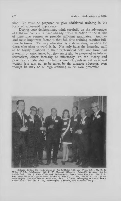
JIO N.Z . .T. med. Lab. Techno/.
kind. It must be prepared to give additional training in the form of supervised experience.
During your deliberations, think carefully on the advantages of full-time courses. I have already drawn attenticn to the failure of part-time courses to provide sufficient graduates. Another and most important factor is that full-time training requires fulltime lecturers. Tertiary education is a demanding vocation for those who elect to work in it. Not only have the lecturing staff to be highly qualified in their professional field, and have had a wealth of experience, but they must also be prepared to inform themselves, either formally or informally, on the theory and practices of education. The training of professional men and women is a task not to be taken by the amateur educator, even though he may be of high standing in his own profession.
Piccured during the celebrations at Smith-Biolab Ltd. are (from left) : Mr D. R. ~Iiller (C.S.L., ':\lelboume), '~IQ· P. W. l\Iaxwell (:\lanager, Scientific DiY·ision, Smithlliolab ~Ad.), :.\Ir I. Cole (Principal 1'echnologist, Green Lane Hospital), :\lr J. R. Saal (Vhe lnst.itu:te's gue.-;t from Brisbane) !:\Irs IJ), G. 'l'ill, ~Ir !D. G. '!'ill ('Chief Technologi:..t, )(a tional 'Health In'stitute), :.\I•r D. W. Hill (l~lanaging Director, SmitllRiolab lAd.) and l\l.r B . .}f. Prentlergast (•Director, tSmith-Biolllb iLtd.).
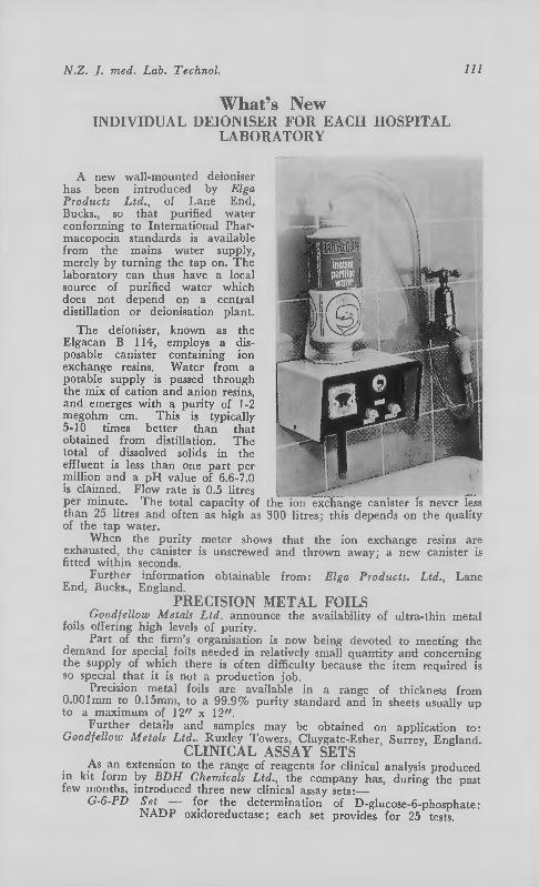
N.Z. f. med. Lab. Techno!.
What's New INDIVIDUAL DEIONISER FOR EACH HOSPITAL
LABORATORY
A new wall-mounted deioniser has been introduced by Elga Products Ltd., of Lane End, Bucks., so that purified water conforming to International Pharmacopoeia standards is available from the mains water supp!y, merely by turning the tap on. The laboratory can thus have a local source of purified water which does not depend on a central distillation or deionisation plant.
T h e deioniser, known as the Elgacan B 114, employs a disposable canister containing ion exchange resins. Water from a potable supply is passed through the mix of cation and anion resins, and emerges with a purity of 1-2 megohm em. T his is typically 5-10 times better than that obtained from distillation. The total of dissolved solids in the effluent is less than one part per million and a pH value of 6.6-7.0
Ill
is claimed. Flow rate is 0.5 litres . per minute. The total capacity of the ion exchange canister is ne,·er less than 25 litres and often as high as 300 litres; this depends on the quality of the tap water.
When the purity meter shows that the ion exchange resins arc exhausted. the canister is unscrewed and thrown away; a new canister is fitted \\"ithin seconds.
Further information obtainable from: Elga Products. Ltd., Lane End, Bucks., England.
PRECISION METAL FOILS Goodfellow Metals Ltd. announce the availability of ultra-thin metal
foils offering high levels of purity. Part of the firm's organisation is now being devoted to meeting the
demand for special foils needed in relatively small quantity and concerning the supply of which there is often difficulty because the item required is so special that it is not a production job.
Precision metal foils are available in a range of thickness from 0.001mm to 0.15mm, to a 99.9'7< purity standard and in sheets usually up to a maximum of 12" x 12".
Further details and samples may be obtained on application to: Goodfellow M eta.ls Ltd .. Ruxley Towers, Claygate-Esher, Surrey, England.
CLINICAL ASSAY SETS As an extension to the range of reagents for clinical analysis produced
in kit form by BDH Chemicals Ltd., the company has, during the past few months. introduced three new cl inical assay sets:-
G-6-P D S PI - for the determination of D-glucose-6-phosphate: NADP oxidoreductase; each set provides for 25 tests.
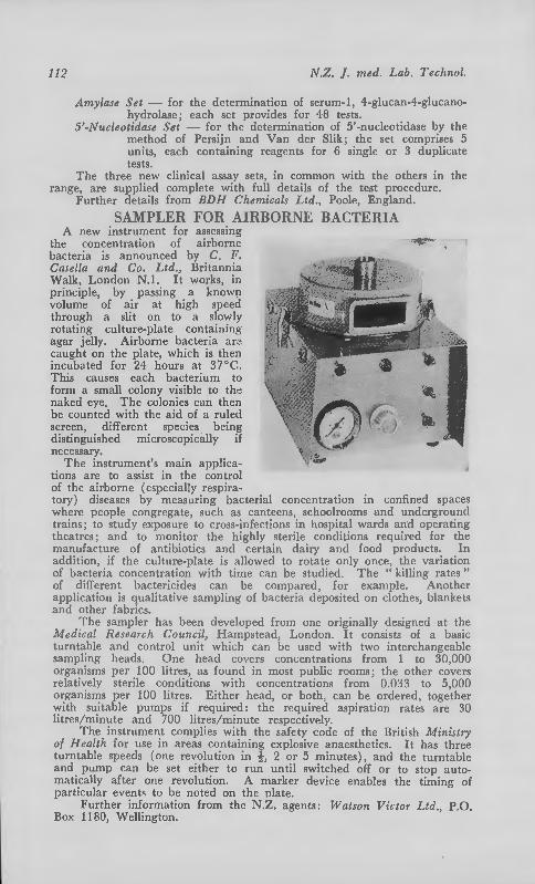
112 N.Z. ]. med. Lab. Techno/.
Amylase Set - for the determination of serum-1, 4-g1ucan-4-glucanohydrolase; each set provides for 48 tests.
5'-Nucleotidase Set - for the determination of 5'-nucleotidase by the method of Persijn and Van der Slik; the set comprises 5 units, each containing reagents for 6 single or 3 duplicate tests.
The three new clinical assay sets, in common with the others in the range, are supplied complete with full details of the test procedure.
Further details from BDH Chemicals Ltd., Poole, England.
SAMPLER FOR AIRBORNE BACTERIA A new instrument for assessing
the concentration of airborne bacteria is announced by C. F. Casella and Co. Ltd., Britannia Walk, London N.l. It works, in principle, by passing a known volume of a ir at high speed through a slit on to a slowly rotating culture-pla te containing agar j elly. Airborne bacteria ar2 caught on the pla te, which is then incubated for 24 hours a t 37°C. T his causes each ba cterium to form a small colony visible to the naked eye. The colonies can then be counted with the a id of a ruled screen , different species being d istinguished m icroscopically if necessary.
T h e instrument's main applications are to assist in the control of the airborne (especially respira-
-tory) diseases by measuring bacterial concentration in confined 5paces where people congregate, such as canteens, schoolrooms and underground trains; to study exposure to cross-infections in hospital wards and operating theatres; and to monitor the highly sterile conditions required for the manufacture of antibiotics and certain dairy and food products. In addition, if the culture-plate is allowed to rotate only once. the variation of bacteria concentration with time can be studied. The "killing rates" of diiTerent bactericides can be compared, for example. Another application is qualitative sampling of bacteria deposited on clothes, blankets and other fabrics.
The sampler has been developed from one originally designed at the Medical Research Council, Hampstead, London. It consists of a basic turntable and control unit which can be used with two interchangeable sampling heads. One head cO\·ers concentrations from I to 30.000 organisms per 100 litres, as found in most public rooms: the other covers relatively sterile conditions with concentrations from 0.033 to 5,000 organisms per 100 litres. Either head, or both, can be ordered, together with suitable pumps if required: the required aspiration rates are 30 litres/minutc and 700 litres/minute respectively.
The instrument complies with the safety code of the British Ministry of Health for use in areas containing explosive anaesthetics. It has three turntable speeds (one revolution in i, 2 or 5 minutes), and the turntable and pump can be set either to run until switched off or to stop automatically after one revolu tion. A marker device enables the timing of particular events to be noted on the pla te.
F urther information from the N.Z. agen ts: W atson V ictor Ltd., P.O. Box 1180, Wellington.
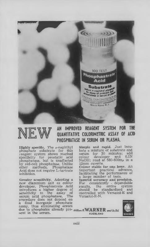
NJEW AN IMPROVED REAGENT SYSTEM FOR THE QUANTITATIVE COLORIMETRIC ASSAY Of ACID PHOSPHATASE IN SERUM OR PLASMA.
Highly specific. The a-naphthyl phosphate substrate for this reagent system shows marked specificity for prostatic acid phosphatase, but is unaffected by red-cell phosphatase. Unlike other methods, Phosphatase Acid does not require L-tartrate inhibition.
Greater sensitivity. Adopting a new diazonium salt as colour developer, Phosphastrate Acid introduces a higher degree of sensitivity to the assay of serum acid phosphatase. The procedure does not depend on a final inorganic phosphate assay, thus eliminating errors
Simple and rapid. Just incubate a mixture of substrate and serum for 30 minutes; add colour developer and O.lN NaOH; read at 580-600mll in a 12mm cuvette. Colour stable for one hour. An important practical advance, facilitating the performance of a large number of tests. Insured accuracy and precision. For consistently - dependable results, the entire system should be standardised and controlled with Versatol-E and Versatol-E-N.
due t? phosphates already pre- ftld&m.!ieWARNE R andG>LU sent m the serum. AUCKLAND
xxiii
SERVING THE REQUIREMENTS OF THE MEDICAL LABORATORY Available from New Zealand stock ...
SCIENTIFIC INDUSTRIES INC. Vortex Genie Mixers, Multi -purpose Sample Rotators, Tiny Timers, Automatic Syringes, Piggy-back Pipetters.
BRUNSWICK LABORATORIES Microhaematocrit Capi ll ary Tubes, C R IT 0 SEA L Capillary Tube Sea lant, DISPOSABLES - Tripour Beakers, Funnels, UNIMOUNT Mounting Medium, PARAPLAST Imbedding Medium, e liminates coo ling.
INSTRUMENTATION LABORATORY INC. Model 265 pH Meters, Accessories, Expendable
Supplies and Spares.
DISTILLATION PRODUCTS INDUSTRIES EASTMAN CHROMAGRAM T.L.C. De v e I oping
Apparatus and Prepared Sheets.
JENCONS (SCIENTIFIC) LTD. ZIPPETTES - - - - - - - 2m I, 1Om I, 30m I. REPETTES - - - - - - - Micro, 2m I, 1Om I.
Full Range of Spares.
COMPREHENSIVE RANGE Of LAB. GLASSWARE AND HARDWARE Major New Zea land Distributor for • ••
MAY AND BAKER LABORATORY CHEMICALS Inquiries to ••.
SCIENTIFIC DIVISION KEMPTHORNE PROSSER & CO.'s N.Z. DRUG CO. LTD. AUCKLAND - WELLINGTON - DUNEDIN - CHRISTCHURCH
xxiv
METROHM OF SWITZERLAND
pH Meters for All Purposes
Complete Titration Equipmen~ pH Electrodes, Piston Burettes
Magnetic and Propeller Stirrers Spectra-Photometers
and Spectro Colorimeters
Sole New Zealand Agents:
DENTAL AND MEDICAL SUPPLY CO. LTD. Auckland • Wellington • Christchurch • Dunedin
XXV
( MaJ· or Explorations cjthe
Golonies Gram-negative organisms can claj s. PathoTec rea~ent sysbe dentified more rapdly terns now inchde seven bio-\\·hcn PathoTec rca[!,ent sys- chemical test~: cytochrome tems arc part of your O\'erall r·xida~e 1 PathoTec - CO ). scheme. Impregnated 1\'ith pre- phen) !alanine de a 111 in as e mea~urNI , standardised re - ( p a t h 0 T e c _ PD ) . urease agents. PathoTec systems ( PathoTec-U ). lysine decarborliminate the need for manv unstable special reagents an~! xy la~e ( PathoTec-LD ); plus media. PathoTec test systems three ne\1' tests from the are reliable, stable, save valu- n1YiC group; indole (Pathoable storage space and provide Tee - I ) , Voges - Proskaucr sig~ifir~nt information "·ith a (PathoTec - VP), citra t c savmg m time of one to four (Pa·hoTer-C ) .
for rapid, reliable testing of certain Gram-negative bacteria
Pathotec reagent systems
tiAIYUMI!?. wARN E R a.na' (}c.Qq
AUCKLAND
xxvi
sharp decisive
end-point-
* Highly Active Thr·omboplastin * No detectable factor· V or· factor VII * Stable for· at least 21 DAYS in liquid state under r·efr•igeration * Reproducible from vial to vial and lot to lot * Pure rabbit brain 11/efl'i.l' fiii'OIIIIJoplttsfiu t•uldulll is a sllpt·rior twtg<'lll .for t/11• lrtll' rl••t••r· 1nination o.f prothrombin tim e ht•cuuse it C"Untuin s 110 demon strab!t• amounts of Jnctor T or .Jitctor V 11.
~~~ METRI)C
DIAGNOSTIC STANDARDS AND CONTROLS Man ufactured by
MICHAEL REESE RESEARCH FOUNDATION Ch1cago 16, 111mols, U.S A
xxvii
Information concerning the full range of Worner-Chilcott Diagnostic Reagents is available from the New Zealand Agents on request:
William R. WARNER & CO. LTD. P.O. BOX 430 - AUCKLAND
xxviii
N.Z. f. med. Lab. Techno/. 113
A Simple, Quick Method for the Estimation of Total Protein and Albumin in Serum
D. W. EVERARD, A.N.Z.I.M.L.T., F.I.M.L.T.
Introduction
Department of Chemical Pathology, Memorial Hospital, Hastings.
Received /or Publication, May, 1969.
The method outlined uses the "albumin error" of certain dyes to quantitate albumin, and the conventional biuret reaction for total protein.
The albumin method is that of Bartholomew and Delaney ( 1966 ) \ which is not affected by abnormal concentrations of globulins, bilirubin, haemoglobin, salicylates or heparins. The reagents are cheap, easy to make up and keep well at 4°C.
In setting up this method the author found it necessary to alter the procedure for making up the buffered dye solution, as when made up according to formula the graph was linear only to a concentration of 2 g./100 mi. of albumin.
It was found possible to rectify this by the addition of more dye to the buffer solution until the range became linear up to a concentration of 5 g./100 mi. of albumin. As it was felt that a higher top limit would be useful and it was not considered desirable to increase the optical density of the blank, more buffered dye solution was employed in the test, thus increasing the ratio of dye to albumin without further increasing the optical density of the blank.
The effect of protein on certain dyes in solution has been known for a number of years and was originally called the "protein error," but as this change can only be attributed to albumin, the term " albumin error " has been substituted.
In effect, when albumin is added to buffered dye solutions it appears that a substantial change of pH has taken place, but in fact this is not the case, as only a very minor pH change has occurred.
Many methods using this albumin error have been tried, but most failed in conditions of abnormal albumin globulin ratios or were too expensive.
Bromocresol green was found to give linear results in relation to albumin concentration, and is unaffected by abnormal albumin globulin ratios, bilirubin in excess of 40 mg/100 mi., haemoglobin up to 750 mg/100 ml. and salicylates.
114 N.Z . .f. med. Lab. Tachnol.
Materials and Methods Reagents:
(1) M. Sodium Citrate: dissolve 147 g. of sodium citrate (Na3C6H 50,-2H20) in water and make up to 500 mi. Store .in the refrigerator at 4°C.
(2) M. Citric Acid: dissolve 105 g. of citric acid in water and make up to 500 mi. Store in refrigerator at 4°C.
(3) 0.01M. bromocresol green: add 9.8 mi. of 0.1N NaOH to 0.968 g. of dye, dissolve as much as possible, then make up to 100 mi. with water. Store in a dark bottle at room temperature.
( 4) Buffered indicator solution: to about 400 mi. of water add 8.6 mi. of M. sodium citrate, 16.3 mi. of M. citric acid, and 3-6 mi. of dye solution·l:·. Check pH 3.8 and store in refrigerator at 4°C.
( 5) Biuret reagent. Solution A. Potassium Sodium tartrate - 4·5 g.
Potassium iodide 5 g. Cupric Sulphate - 15 g. Sodium hydroxide - 8 g. Water to make 1000 mi.
Solution B. Sodium hydroxide - 80 g. Potassium iodide - 50 g. Water to make 1000 mi.
For use take 100 mi. of Solution A. and 40 mi. of Solution B., then make up 500 mi. with vvater.
(6) 0.9% sodium chloride in water. ( 7) Standard protein solution: dilute concentrated human
albumin approximately 25 g% to give 7 g%. Check against commercial dried sera by the Biuret method.
Methods Total Protein To 1.9 mi. of 0.9% saline add 0.1 mi. of serum, then 8 mi. of
Biuret reagent. At the same time put up a reagent blank of 2 mi. of saline and 8 mi. of Biuret reagent, and a standard treated as the test.
Place all tubes in a 37° C water bath for 10 minutes, then measure optical densities of tests and standard against reagent blank, at 540 mf.L. Calculate results from standard or from a previously constructed calibration graph.
Albumin Due to the variation in batches of dye it is necessary to
standardise the buffered dye solu tion. Make up the buffered bromocresol green reagent and measure the optical density of the
.,. The amourit of dye solution needed changes from batch to batch of dye. See calibration of dye reagent .
N .Z. J. med. Lab. Technol. 115
solufon against \\·ater at 635 mp.. Keep a note of the value (on the instrument employed, and using 6 mi. of dye solution per 500 mi. of buffer, this gave a reading of 0. 230).
Prepare a series of albumin dilutions, ranging from 1 g% to 7 g'/o and treat them as for tests. Plot the opti:::al densities against albumin concentration. This should produce a straight line graph. If the line cur\'es at the higher concentrations more dye should be added to the indicator solution, (e.g. 0.5 to 1 mi. should be sufficient, but this will depend on original batch of dye and the amount of dye solution added to the reagent).
To 5 mi. of buffered indicator solu'.ion add 0.02 ml. of serum ; treat a standard albumin solution in the same manner and measure their optical densities against a reagent blank at 635 mp..
The colour may be read immediately or any time within three hollls . Calculate results against a known standard or from a previously calibrated graph.
The method has been used in the author's laboratory for the las t few months and found to be both quick and reliable. Day-toclay var,iations using commercial freeze-dried quality control sera are -+- 0.2 g/ 100 ml.
REFERENCE: 1. Bartholomew, R .J. and D elaney, A.M. ( 1966 ), Proc. Aust. Assoc. clin.
Biochem., 7, 214.
Technical Communications
Simple Colorimetric Estimation of Serum Calcium
Sir, The following method for estimating serum calcium,
which is that of Bellinger and CampbelP with minor modifications, may be of interest to readers.
Method: 1. Place 2.0ml. water in duplicate tubes for test (T) and
standard (S) and 2.1 ml. into a fifth tube for a blank. 2. Into (T) pipette 0.1 mi. serum and into (S) pipette 0.1
mi. of working calcium standard (10mg.j100ml.) and mix. 3. To all tubes add l.Oml. of G.B.H.A. reagent (see under
"Reagents" ) and mix, then add 0.2ml. of colour developer. Mix well, place in centrifuge and bring to about 3,000 r.p.m. then switch off and allow to coast to a gentle stop.
116 N.Z. ]. med. Lab. Techno[.
4. Add 5.0ml. chloroform to all tubes, stopper with polyethylene stoppers then shake vigorously for about 15 seconds.
5. Again centrifuge tubes at about 3,000 r.p.m. for 2-3 minutes to separate the chloroform layer containing the violet calcium complex.
6. Immediately read optical density of the chloroform layer at 535 mp. against the blar:lc Due to turbidity which begins to develop, particularly in T soon after the centrifuge has stopped, reading of optical densitie:~ should be made immediately. Reagents:
1. G.B.H.A. reagent - 0.4% Glyoxal bis - (2-hydroxyanil) (Hopkin and Williams ) ,in absolute ethanol. (I find this keeps at least a week in the refrigerator, in contrast to most authors who state that it must be prepared fresh daily).
2. Colour developer - 10% NaOH and 0.5 % Na2C03 in distilled water.
3. Stock calcium standard ( 1mg./ml.) [Modified] - Dry CaCb overnight at 105°C and cool in a desiccator. 2.768g are weighed and transferred to a litre flask. 200 ml. of distilled water and 50ml. 1.0N HCI are added, shake to dissolve and dilute to the mark with water. Keeps well at room temperature.
4. Working calcium standard ( 10mg./ 100ml. ) - Dilute stock standard 1 in 10 with distilled water. Keep in refrigerator.
5. Chloroform - B.P. or reagent grade is satisfactory. Discussion:
The above method depends on the ability of calcium m alkaline solution to react with glyoxal bis - (2-hydroxanil)" to form a red complex which is extractable into chloroform. Certain other metal ions (e.g. cobalt and nickel) will also react, but the colours produced do not extract into chloroform.
In this laboratory optical densities are read on a Coleman Jr. spectrophotometer and although this type of instrument does not have a narrow band-width, it was found that Beer's law is obeyed over the range 5-15 mgm Ca + + /1 OOml. Because of the turbidity which develops in the chloroform extract, and because of the difficulty of pipetting this extract so as to obtain a clear solution free from tiny water globules or protein particles, it is recommended that cuvettes or precalibrated test tubes be used for the entire procedure. In this \Nay colorimetry can be performed immediately after removal of tubes from the centrifuge without the need for additional manipulation. We have found a set of specially selected 4 x 5-8" test tubes to be most convenient. Because of the sensitivity of the method it is essential that all glassware be scrupulously clean.
T he standard is a modification of the original and uses calcium chloride instead of calcium carbonate, the reason being that
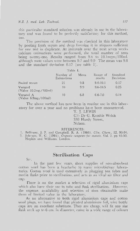
N.Z . .f. med. Lab. Techno[. 117
this particular standard solution was already in use in the laboratory and was found to be perfectly satis:actory for this method, too.
The precision of the method was checked in this laboratory by pooling fresh serum and deep freezing it in aliquots sufficient for one test in duplicate. At intervals over the next seven weeks calcium estimations were performed, the total number of tests being twenty-one. Results ranged from 9.4 to lO.lmgm j lOOml. although most values were between 9. 7 and 9.9 The mean ·was 9.8 and the standard deviation 0.17 (see table I).
Table I. Number of Mean Range of Standard Estimations results Deviation
Pooled serum 21 9.8 9.4-10 .1 0.1 7
Versatol 10 9.9 9.6-10.5 0.25 (Value 10.2mg./ 100ml )
Versatol A 10 6.8 6.6-7.0 0.14 (Value 6.9mg./ 100ml )
The above method has now been in routine use in this labor-atory for over a year and no problems have been encountered.
T. J. LEWIS C!- Dr G. Kemble Welch 330 Hardy Street, Nelson.
REFERENCES: 1. Bellinger, J. F. and Campbell, R. A. (1966), Clin. Chem., 12, 90-94. 2. Johnson, W. C. ( 1964) , Organic reagents for metals. Vol. 2, pp 48-50.
Hopkin and Williams. London.
Sterilisation Caps Sir,
In the past few years, short supplies of non-absorbent cotton wool has been a headache to many microbiology laboratories. Cotton wool is used extensively in plugging test tubes and media flasks prior to sterilisation, and acts as an ideal air fi lter and cap.
There is on the market a selection of r.igid aluminium caps which also have their use in tube and flask sterilisation. However, the expense, avai lability and selection of sizes obtainable make them of limited value in the laboratory.
As an alternative to both rigid aluminium caps and cotton wool plugs, we have found that pleated aluminium foil, wire bottle caps are an excellent substitute. They are cheap, will fit any size flask ne~k up to 6 em. in cEameter, come in a wide range of colours
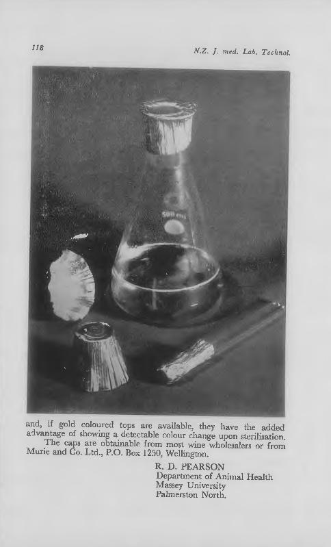
IJB N.Z . .T. med. Lab. Tech no/.
and, if gold coloured tops are available, they have the added advantage of showing a detectable colour change upon sterilisation.
The caps are obtainable from most wine wholesalers or from Murie and Co. Ltd., P.O. Box 1250, Wellington.
R. D. PEARSON Department of Animal Health Massey University Palmerston North.
IM<-~.WAR N E R am/C'a/.J'd.
21· 23 Federal Street, Auckland
xxix

*Hydatidiform Mole *Abortion !hreatened and
Incomplete
*Trophoblastic Tumour *Ectopic Pregnancy
'PREPUER!N' provides a rapid, easy and accurate method of measuring H.C.G. excretion, facilitating both diagnosis and management.
FOR DETAILS OF THE TECHN IQUES USED IN THESE TESTS WRITE TO,
BURROUGHS WELLCOME & CO. (New Zealand) LTD. BOX 22·258. AUCKLAND S.E.7.
XXX
Dextrostix will give YQU the inside storv on MrS. Jones ...
<I
in 60seoonds flat, for just 19cents. With Ames DEXTROSTIX* information system you can check for abnorma l blood-g lucose levels as quickly as you can take a temperature. Check blood-glucose levels in routine phys ical exam inations D pregnancy D obes ity D periphera l vascular diseaseD loss of v isual acu ity D recurrent infections D diabetes detection two hours after a glucose load D diabetic management and patients on 'thiazides' steroids and other potentially hyperglycaemic drugs. With DEXTROSTIX you can differentiate between hypo-, normo-, and hyperg lycaem ia, and re liably se lect those patients who will require more precise analytical laboratory methods.
Trade Mark
Ames Company - Division Miles Laboratories Australia Ply. Ltd. :;n 4 Derby Street, Collingwood, Vic. 3066
New Zealand Distributors: @ Early Brothers Dental & Surgical Supplies Ltd., Christchurch, Auckland, We llington & Dunedin.
Chemical and biological information systems serving medicine and Industry. '"" Af4B
TheY're all ia the BDB Catalogue ... a range of more than 5,000 chemicals
BDH can supply almost every reagent in regular demand m laboratory practice today. as well as many developed for special applications on research and industry reagents of the highest standards of purity and reliability.
Items of particular interest include: 'Aristar' ultra~pure chemicals: for analytical, research and industrial applications in which extreme ' purity is essential.
'AnalaR' analytical reagents: More than 300 guaranteed analytical reagents conforming to the recently revised·'AnalaR' specifications.
BDH Biochemicals: A comprehensive selection of h1gh quality biochemicals and reagents including Amino acids and derivatives, enzymes and enzyme substrates, nucleotides. nucleosides and nucleic acids. carbohydrates and sugar phosphates, highly pure fatty acids and methyl esters.
Materials for chromatography and electrophoresis Micro·analytical reagents Organic Analytical Standards General laboratory chemicals Clinical reagents Solvents for spectroscopy Microscopical stains and sundries lon exchange resins Volumetric solutions Indicators Test papers
If you do not already receive our monthly publication 'New Entries in the BDH Catalogue', p lease ask us to add your name to the mail ing list.
NEW ZEALAND LIMITED, PO. BOX 624, lr\LMERSlON NORTH.
xxxii
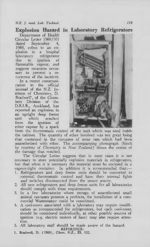
N.Z . .f. med. Lab. Technol. 119
Explosion Hazard in Laboratory Refrigerators Department of Health
Circular Letter 1969/101 dated September 4, 1969, refers to an explosion in a hospital laboratory refrigerator due to ignition of flammable vapour, and suggests measures necessary to prevent a recurrence of the incident.
In a recent communi-cation to the official , / journal of the N.Z. Institute of Chemistry, D. Bradwell\ of the Chemistry Division of the D.S.I.R., Auckland, has reported an explosion in an upright deep freeze unit which resulted from the igmtwn of ether vapour by a spark from the thermostatic control of the unit which vvas sited inside the cabinet. The quantity of ether involved was not great being that contained in the carcasses of some rats which had been anaesthetised with ether. The accompany.ing photograph (block by courtesy of Chemistl')' in New Zealand) shows the extent of the damage that resulted.
The Circular Letter suggests that in most cases it is not necessary to store potentially explosive materials in refrigerators, but that when it is necessary the material must be enclosed in a vapour-tight container. In addition it is recommen~led that:-1. Refrigerators and deep freeze units should be converted to
external thermostatic control and have their internal lights and switches disconnected from the power source.
2. All new refrigerators and deep freeze units for all laboratories should comply with these requirements.
3. In a few laboratories where storage of anaesthetised small animal carcasses presents a problem, the installation of a commercial Wastemaster could be considered.
4. A cool-room associated with a laboratory may require modification as recommended for refrigerators, but each cool-room should be considered individually, as other possible sources of ignition (e.g. electric motors of fans) may also require attention.
5. All laboratory staff should be made aware of the hazard. REFERENCE:
1. Bradwell, D. (1969), Chern. N.Z., 33, 122.
120 N.Z. ]. med. Lab. Techno[.
Selected Abstracts
BLOOD BANKING
A New Method for Detection of Red Blood Cell Antibodies. Lalezari, P. ( 1968), Tramfusion, Philad., 8, 372.
Suitable for use in automated procedures, this technique employs Polybrene, a positively charged polymer, to produce agglutination of red cells. This agglutination is reversible by the addition of hypertonic salt solution but persists in the presence of antibodies.
The method has proved to be highly sensitive and with a wide spectrum of usefulness for the detection of both complete and incomplete antibodies, including those which are normally inhibited by the use of enzymes. Anti-C as a Naturally-Occuning Antibody. Beck, M ., Dixon, J., Lawson, N. S. and Oberman, H. A. (1968), Transfusion, Philad., 8, 387.
Anti-C was detected in the serum of a 49-year-old manl who had no transfusion history. The facts that the antibody was characterised as an IgM globulin, and with a low thermal range of activity, were consistent with the behaviour of a natura lly occurring antibody. Transmission of Syphilis by Fresh Blood Components. Chambers, R. W., Foley, H. T. and Schmidt, P. J. (1969), Transfusion, Philad ., 9, 32.
This is the report of a case of secondary syphilis resulting from the transfusion of fresh pla telet-rich plasma from donors who were all serologically negative for syphilis. Agglutinins fo1· "Null" Red Blood Cells. McGinnis, M. H., Kaplan, H. S., Bowen. A. B. and Schmidt, P. J. ( 1969), Tmnsfusion, Philad., 9, 40 .
Sera from two anaemic patients gave a pattern of agglutination which was the reverse of that described for antibodies of Rh-lik e specificity associated with autoimmune haemolytic anaemia. They reacted not only with Rh null cells but also with U-negative, Oh and other cells lacking usual agglutinogens. They did not react with cells possessing the usual blood types and it is suggested that the sera contain antibodies directed against antigen sites which are normally either absent or concealed. Transfusion Re:tction: A Reappraisal of Surgical Incidence and Significance. Baker, R. J., Moinichen. S. L. and Nyhus, L. M. ( 1969) , Ann. Sur g., 169, 684.
A review of the incidence of immediate reactions to transfusion of 116,273 units of blood and blood products was undertaken. These were administered to 42 ,331 recipients. A reaction was recorded following 2,569 transfusions (2.21%). These reactions were seen in 2,293 recipients ( 5.42 C/c). with the following distribution of types: allergic, 45.6%, febrile, 43.5'/f, haemolytic, 9.9'/o and miscellaneous, 1.0%.
Serious, symptomatic haemolytic reactions occurred in I in 3,322 units infused or I in 1.209 recipients. Fourteen ( 40 % ) of these 35 recipients died. .J.H.
CHEMICAL PATHOLOGY Serum Triglyceride Levels of Normal Subjects. K udo, H. ( 1969), Tohoku J. exp. Med., 97, 35.
Using the method of Van Handel and Zilversmit, with modifications . fasting serum triglyceride levels of 263 normal subjects, 2 to 79 years of age, were determined. The overall mean level was 70 ± 1.3mg./100ml., with a range of 30-110 mg./lOOm!. It was concluded that levels over 120 mg./ 100 mi. may be regarded as indicating hypertriglyceridaemia.
J.H.
N.Z . .f. med. Lab. Techno/. 121
Observations on Diagnostic Kits for the Determination of Urea Nitrogen. Logan, J. E., Renton, H. M. and Eby, P. W. (1969), Clin. Biochem., 2, 189.
Fourteen diagnostic kits or reagent sets from 12 manufacturers were assessed for adequacy of literature supplied, reproducibility, accuracy compared to an automated diacetyl monoxime method, recovery of added urea and stability of reagents throughout the test period.
Three kits fa1led to show sufficient precision, i.e., within ± I 0 per cent. Although the precision was adequate, 3 kits yielded values signifi
cantly higher than those of the reference method , while 3 gave lower results.
While slight improvement has been shO\m recently in the adequacy of the literature enclosed with the kits . much more complete data could be given in many cases.
.J,H. A simple Method of Estimating Plasma Salicylate Levels. Burston, G. R. (1969), Scot. med. ]., 14, 55.
On addition of heparinised plasma to a co!our reagent (ferric nitrate-mercuric chloride-hydrochloric acid), a precipitate forms. The tube is centrifuged and the colour of the supernatant compared visually with a set of comparatively permanent standards. The concentration can be estimated to within ±5 mg. per cent. J.H. Normal Values for Cerebrospinal Fluid Protein Concentration in Children: What is the Upper Limit of Normal ? Abramowicz, M. ( 1969 ), Clin. Pediat., 8, 300.
Standard textbooks of paediatrics set the normal range of ceJCbrospinal fluid p rotein concentration in ch ild ~en at 15-40 mg. per cent. However, investigations by the author and studies by others suggest that the upper range should be set at 27 or at most 31 mg. per cent. Other workers have found that values obtained from infants less than 6 months of age were higher. .J.H. A Comparison Between Albustix, Hema-Combistix, Labstix, the Sulphosalicylic-Acid Test, Heller's Nitric-Acid Test, and a Biuret Method: Diagnosis of Proteinuria. Thysell, H. ( 1969 ), Acta me d. scand., 185, 401.
This comparison of the various tests for proteinuria showed unexpectedly great discrepancies. There is a great need for simple, rapid and reliable screening methods for detecting proteinuria.
It was concluded that none of the applied tests satisfied the demands that should be made on such screening tests. Until more reliable and objective methods are available, the older methods should be used, preferably the sulphosalicylic acid test and Heller's nitric acid test. It should be possible to screen out some of the false-positive results if the tests are repeated. However, the high frequency of false-positive results is a factor to be noted. .J.H.
HAEMATOLOGY A Comparison of Two Broad-Spectrum Coagulation Tests. Wolfe, W. T., Hunter, D. T. and Allred, L. ( 1969), Sth. me d. ]., Nashville, 62, 490.
There are two major differences between the two tests studied, the partial thromboplastin time (PTT) and the plasma recalcification test (PRT). In the former a synthetic platelet Factor 3 is added, while in the latter, the patient's own platelets contribute this factor. Hence, the PRT gives an evaluation of platelet number and function , while the PTT completely omits this vital parameter. Since Factor VII is intrinsic in the artificial PTT reagent, assay of this factor is a lso omitted in the PTT.
It was concluded that the PRT more nearly fulfils screening test criteria.
122 N.Z. ]. med. Lab. 1'echnol.
Evaluation Studies of Peripheral Blood Leukocyte Changes in Malignancy. Malmgren, R. A., Bennett, J. M., Del Vecchio, P. R., DeWitt, Sara H., Feld, Minna, Kazam, E. and Schneiderman, M. A. (1969), Acta cytol., 13, 149.
A study of " malignancy-related " changes in peripheral blood cells is described. These changes consisted of thin (or fine) excrescences in the polymorphonuclear leucocytes and halos in mononuclear cells (monocytes and lymphocytes) .
There was a significant difference between the average percentage of cells showing characteristic morphologic changes in 11 cancer patients as compared to II normal individuals. However, the overlap in the counts of positive cells, and the variability in counts within each group were too large to make the procedure useful either for the study of tumour-host interaction or as a cancer detection method. .J,H. Labor:~tory Control of Heparin Therapy with the Activated Partial Thromboplastin Time Test. Degnan, T. J., Karasik, S. and Lenahan, J. ( 1969), Czar. ther. Res., 11, 390.
To cla rify the equivocal literature reports, the authors have studied patients during the course of long-term heparin therapy. Their results suggest that the activated partial thromboplastin time offers a reproducible, practicable and sensitive guide for the administration of heparin.
J.H. Leucocyte Death in Generalised Virus Infection. Smith;, H. (1969), A ust. paediat. ]., 5, 56.
Dead leucocytes may be detected in increased numbers in blood films from patients with a variety of disorders. but in adults at least, gross increases are unusual. This paper describes a neonate with a generalised infection with a virus of herpes simplex type whose blood contained dead leucocytes in striking numbers.
Cells were regarded as necrobiotic only when there were obvious changes in the nucleus, such as loss of internal structure, conversion to a homogenous mass or masses, and shrinkage, fragmentation and dispersal of the nuclear substance. Increase in staining intensity, to a deep redbrown or almost black, usually accompanied these changes, but an increase in staining intensity alone, without structural alteration, was not accepted as evidence of cell death. Frequently there was a change to a globular or approaching a globular shape, and often, in multilobed cells such as neutrophils , interlobar connexions had disappeared.
Changes in the cytoplasm alone were not accepted as evidence of cell death although they were common in cells in which the nuclei appeared to be non-viable.
The infant died at 11 days of age. J.H. Platelet Adhesion: A Comparison of Four Methods. Sjogren, A. , Bottiger, L. E. and Biorck, G. ( 1969), Acta med. scand., 185, 127.
A comparison of Wright's, Hellem's whole-blood and platelet-rich plasma/adenosine diphosphate and Salzman's methods showed good correlation between Hellem's and Salzman's whole-blood methods, a weaker. yet significant correlation between Wright's and Salzman's methods, but no correlation between any of the three whole-blood methods and Hallem's platelet-rich plasma/ adenosine diphosphate method.
J.H. MICROBIOLOGY
A Comparative Study of the Pathogenic Criteria of Staphylococci and Pathogenicity of Coagulase Negative Staphylococci. Panda, G. K., Mohanty, D., Nanda, B. K. and Naik, U. P. (1969) , Indian]. med. Sci., 23, 7.
Of 262 strains of staphylococci isolated from various clinical materials,
HYLAND INTERLOCKED
BASIC ENZYMES SYSTEMS
PROGRAMME
HYLAND ENZYME TESTS
K its or separate components for determination of SGOT, SGPT, LDH, HBD and Amylase levels.
'We shall be pleased to supply you with further details on this Pro.e;ramme, as well as other HYLAND products.
HYLAND MULTI-ENZYME REFERENCE SERA Standardise at T H R E E levels without diluting for SGOT,SGPT, LDH, HBD . CPK, Amylase, Alkaline Phosphatase and Acid Phosphatase.
;: .·
HYLAND CLINICAL CHEMISTRY CONTROLS Efficient, time-saving and flexible e\·aluation of enzyme and other bio-chemical assays.
Available through N.Z. Agents:
DENTAL & MEDICAL SUPPLY CO. LTD. Auckland - Wellington - Christchurch· - Dunedin
xxxiii
May & Baker have broken with glass
The solid chemicals from their famous laboratory range have been encased in plastic
They have entered the silent world of the wide mouthed, unbreakable bottle
A. polythene conta iner of rectangular design which can be easi ly shelved
May & Baker have sealed this achievement with an airtight cap
May&Baker Laboratory Chemicals Manufactured by MAY & BAKER LTD Distributors MAY & BAKER (NEW ZEALAND) LTD P.O. Box 35060 Naenae Tel. 677-016 and Kempthorne Prosser & Co's NZ Drug Co. Ltd.
LAiot~/a~
xxxiv
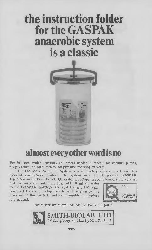
the instruction folder for the GASPAK anaerobic system
is a classic
almost every other word is no For instance, under accessory equipment needed it reads: "no vneuum pumps, no gas tanks, no manometers, no pressure reducing valves."
The GASPAK Anaerobic System is a completely self-contained unit. No external connections. Instead, the system uses the Disposable GASP AK Hydrogen + Carbon Dioxide Generator Envelope, a room temperature catalyst and an anaerobic indicator. Just add 10 ml of water to the GASPAK Envelope and seal the jar. Hydrogen produced by the Envelope reacts with oxygen in the presence of the catalyst, and an anaerobic atmosphere is produced.
For further ill/ormation COl/fact the sole N.Z. agents:
SMITH-BIOLAB LTD P 0 Box 36007 Auckland 9 New Zealand
XXXV
BBL
Division of BioOuest
TWO UNIQUE MEDIA FROM OXOID
BLOOD AGAR BASE No.2 The most advanced general-purpose medium yet devised. For the certain recovery of fastidious ]JRthogens such as H. inlluenzae, D. pneumoniae, staphy lococci, neisseriae a111l streptococci, use Oxoid Blood Agar l3use No. 2, preferably enriched with defibri-" na.ted horse blood.
T his new form ula tion represents a considerable advance on convent ional mecl ia ancl promotes growth of a wide range of patho
#~-__2:~'j::;;l'= gens. It is pa r ticula rly useful for ,4 15.., __ :; Jna:~ it'"..l llln rec·overy fromJninimal
DST AGAR BASE This unique cliagnost. i ~ seoh;iti ,·it~· tcstagarpermitss im••l taneonsc loterminat.ion of antibioti~ nncl s ul fc, .. ,,m ide Hll seeptib ili ty, with t he adcl itional advantage of pet~nit t, ing dircc·t, ob:-;orvation of h enlulytic· aC'ti,·ity. The performn.nce of the modi 11m is cons istent with the criteria laid down by an international eommittee 01; sensitivity testing.*
WJ/0 'l't•rlmil'ul Rrport ... Yo. :!JU.
Manufactured by Oxo ltd., london. Sole New Zea land Agents: Tasman Vaccine Laboratory Ltd., Private Bag, Upper Hutt. Telephone 87-139. Telegraphic address: " Blovax " Upper Hutt.
inocula.
xxxvi
N.Z . .f. med. Lab. Techno!. 123
3 7 were found to be coagulase negative. Six of these 3 7 strains were suspected of being pathogenic since they were isolated on repeated occasions' Two were isolated from post-operative wounds, 1 from otorrhoea and 3 from urine. Strains isolated from urine were in significant numbers ( > 1 0,000/ml.) and pus' cells were also found in the urine in these cases. Further support of pathogenicity was obtained from the clinical course of the disease following drugs given according to the sensitivity pattern. Mannitol fermentation, gelatin liquefaction and urea hydrolysis are taken as evidence of pathogenicity and these tests were positive in these 6 strains .
.J.H. Herellea vaginicola Dalton, H. P. , Allison, M. J and Escobar, M. R. (1969), Va med. Mon., 96, 412.
H erellea vaginicola is one member of a poorly-defined group of bacteria, the tribe Mimae. Clinical laboratories are increasingly isolating these agents and this necessitates some knowledge as to how these organisms m;g!-Jt ~~ contributing to the infectious st<:~ ~ .
During the last 9 months of 1967. 222 isolations were made in the authors' laboratory: 119 from wounds, 65 from the respiratory tract, 35 from urin e and 3 from blood. These results are in agreement with the literature which points to the fact that these organisms demonstra te a low degree of invasiveness and are often associated with underlying conditions such as wounds, burns, malignancy, and indwelling catheters; infections sometimes occur after p rolonged antimicrobial or steroid therapy.
Neomycin and colymycin appear to be effective against the majority of strains. T etracycline and kanamycin also show activity aga inst some strains. J.H. Recent Developments in the Laboratory Diagnosis of Gonococcal Infections. Reyn, Alice ( 1969), Bull. Wld Hlth Or g., 40, 245.
fhe most significant new developments are the use (a) of fluorescent antibo"dy techniques and (b) of selective media, particularly that of Thayer & Martin, for the isolation and identification of N eisse1·ia gonorrhoeae. The sensitivity and specificity of culture methods and of the direct "delayed" fluorescent antibody technique (FAT) are very similar, but the FAT allows more rapid reporting. In the " delayed " technique, specimens from patients are preincubated on a nutrient medium before smears of the culture are treated with fluorescent antibody.
Other new achievements are a complete, chemically defined, proteinfree liquid medium for the culture of fastidious Neisseriae; a commercially available, chemically defined enrichment supplement; and a new specimen-transport kit using Stuart's medium. .f.H. Identification of Pseudomonas aeruginosa in the Clinical Laboratory. Phillips, I. ( 1969), ]. med. Microbial., 2, 9.
The literature concerning the identification of Pseudomonas aeruginosa is large and increasing. Because of this, it is difficult to choose a series of tests that, while technically simple and suitable for use in a hospital diagnostic laboratory, will yield the most useful information in the least time. The purpose of this paper is to report an assessment of some of the methods in a hospital laboratory, and to suggest a simple scheme of identification. J.H. Control of Microbiological Hazards in the Laboratory. Phillips, G. B. ( 1969), A mer. ind. Hyg. Ass. ]., 30, 170.
Through the years, several hundred publications have mentioned approximately 6,000 laboratory infections, but reports and surveys do not ad equately illustrate the safety problem because only a fraction of the laboratorycacquired infections are ever. reported in the literature. However, where mfectious disease agents are handled to an appreciable extent a frequency rate between 1.0 and 5.0 per million m an-hours would be typical ; unsafe acts or conditions are difficult to identify in approximately 80 per cent. of the cases. The estimated case fatality rate for laboratory infections is 4.0 per cent.
124 N.Z . .f. med. Lab. Techno/.
The causes of laboratory infection are discussed as are approaches to hazard control. J.H. Flotation Hydi"ocarbon Technique in Isolation of Tubercle Bacilli from Cerebrospinal Fluid in Tuberculosis Meningitis. Thapar, R. K., Atal, P. R. and Dayal, R. S. ( 1969), Indian Pediat., 6, 172.
Equal parts of cerebrospinal fluid (CSF) and chloroform were mixed and centrifuged. A thick scum formed at the junction of chloroform and CSF which may be used for smear and/or culture.
By this technique, 50 per cent. of cases in early stages, 73 per cent. in the intermediate stage and 92 per cent. in the late stage showed tubercle bacilli in smears.
Other authors have demonstrated that non-acid-fast bacilli in the water phase of a water-hydrocarbon emulsion, on reaching the interphase either remain in the interphase or return to water but never pass into the hydrocarbon phase. They suggested that the difference of behaviour is due to the presence in acid-fast organisms of a mixture of fatty substances with many non-polar groups, which prevent their being wetted by water, but allow them to be wetted by non-polar liquids such as chloroform, xylol, petroleum and oils. J.H. Diagnosis of Pyelonephritis. Chaterjee, A. M. ( 1969), Antiseptic, 66, 330.
Kidney infection is remarkable for its silent course, often difficult to recognise. Kleeman ( 1960 ) noted in his survey that only 20 per cent. could be diagnosed during life. Similarly, Brod ( 1962 ) reported that of his 78 cases of pyelonephritis, only 24 ( 32' per cent.) were diagnosed during the illness. Pathologists claim pyelonephritis to be the commonest cause of death from uraemia. Brod (1962) noted that 36 per cent. of deaths from uraemia were due to chronic pyelonephritis. Such a silent and relentless progression of the disease necessitates early diagnosis.
Various laboratory tests to aid in the diagnosis are reveiwed, keeping in view the problems of the general physician. Simple tests are described in some detail. J.H.
MYCOLOGY Rapi:l Contrast Stain as a Diagnostic Aid for Fungous Infections. Swartz, J H. and Medrek, T. F. (1969) , Archs Derm., 99, 494.
Ink Blue PP and Rose Bengal are among the ingredients of th1s stain. preparation of which might be somewhat time-consuming for the average laboratory ; the stain is, however, available ready for use.
The suspected material (skin scales, nail scrapings, hairs, sputa, pus, exudate or a teased representative portion of a grown colony is placed in a drop of stain on a ~!ide. A coverslip is added and the slide warmed.
The staining procedure does not replace cultural and other studies for the identification of species. J.H. Modified Sabouraud Medium Containing Neomycin and Polymyxin. Cohen, S. N. ( 1969) , Appl. Microbial., 17, 486.
Addition of a neomycin-polymyxin B mixture to Sabouraud dextrose agar was found to be a useful adjunct in the isolation of fungi and a definite improvement over earlier modifications containing penicillin and streptomycin or chloramphenicol, since there was decreased overgrowth by the Gram-negative flora frequently found in the sputum of hospitalised patients. It is suggested that a similar modification of cycloheximidecontaining Sabouraud agar would be expected to improve the performance of that medium. J.H.
UNCLASSIFIED Icteric Serum: A Helpful Diagnostic Clue in Comatose Patients. Whang, R ., Saiki, J. H., Mansour, E. J. and Gehred, G. A. ( 1969), Sth. med. ]., Nashville, 62, 361.
Because of either the inability of the comatose patient to provide a
N.Z . .T. med. Lab. Technol. 125
history or a paucity of overt clinica l manifes tations of liver disease, the diagnosis of hepatic coma may be delaye'd,, This article documents experiences with two patients having no physical signs of hepatocellular d isease, and in whom detection of icteric serum led to prompt initiation of appropriate therapy.
In Case 1, although numerous laboratory examinations were performed, icteric serum was not noted until the physicians examined the serum.
In Case 2, many admission laboratory tests were done but the presence of icteric serum was not reported until it was noted the following day during a toxicological survey. J.H.
Book Reviews Chromatographic and Electrophoretic Techniques: Volume 1, Chromatography. 1 hi rd Edition. Edi ted by I. Smi th, B.Sc., Ph .D., F .R.I.C ., M .I. Bioi. 1080 pages with illustrations, 10 in colou r . William Heinemann , London. ( 1959 ), U.K. p rice 130s Od.
A new edition of " Ivor Smith " is a noteworthy event for the practising chroma tographist, for one is assured of finding the prac tical de ta ils and tricks of the trade which are necessary when a pplying new techniq ues. The fi rs t edi tion ( 1957), one thi rd of the cur ren t size, involved ten authors. T hirty-seven au thors were required for this thi rd edition, indicating the expansion of the subjec t.
There are many new topics of wide interes t, one group rela ted to the struc tu re of proteins. This describes sequence analysis through phenylthiohydantoin degrada tion and subsequent id entifica tion of lansyl amino acid compounds. Another deals simila rly with ("'P) labelled ribonucleotides and the last chapter, which can be obtained as a separa te pamphlet, describes a sys tem of plastic macro-molecules called "Bio Bits."
Specia list chapters include a spark chamber technique for e\·aluating radio-ch romatograms, ion exchange celluloses, p reparative layer ch romatography handling up to 100g. quantities, automated quantitative analysis, column chromatography, inorganic ion chromatography and steroid analysis. Incidentally, the only typographical error I detected was a heading on p:1ge 602, "Specific 'Steroid' Problems."
The chapters on amino acids are of particular interest to the clinical chemistry laboratory in regard to aminoaciduria and inborn errors of metabolism. All of these chapters, and indeed all of the book, has been extensively revised.
One finds corraboration of the advantages of paper over thin layer in certain instances; for example, the single run scrceening techniques for u~ine and serum without deproteinising. The chapter by the late Mary Efron on pathological aminoaciduria contains useful tables classifying disease, abnormal enzyme, amino acid, clinical feature and treatment. There is also an extensive classified list of references. How delightful it would have beeT\ to see colour plates of the urine amino acids of the same quality as those depicting pigments in inks! Without doubt meticulous technique is required to produce uniform and comparable results and considerable experience needed to interpret chromatograms. The comment that seventeen amino acids are present on the fingers is a warning against undue handling!
One of the later chapters outlines a rational approach to the investigation of new compounds and gives examples of the techniques employed.
T his book now contains such a wealth of diverse material one wonders whether it might not with advantage be made ava ilable as sep ara te part&. In any event, those involved in chromatography can scarcely do without it.
R .D .A.
126 N.Z. ]. med. Lab. Tee/mol.
Comprehensive Review for Medical Laboratory Technologists. F. E. Dolan Ph.D. Editor, B. L. Garrick M.T. (ASCP) B.S., S. P. Gottfried Ph.D., C. F. Spiltoir Ph.D., D . P. Stiff M .D., F .C.A.P. 181 Pages, C. V. Mosby Co. St. Louis (1968) U.S. price $6.95.
This slim volume in an attractive strong hard cover presents the information in a somewhat unusual manner.
Each section is introduced by a posed question heading, and the text which follows p resents the relevant facts woven into a practical explanation and answer. The emphasis is on the interpretation and relevance of inform::t ~ ion to an understanding of the practical significance and usefulness of knowledge; so that reasons, principles and evaluation form a good deal of the subject matte r.
The level of knowledge is primarily di rec ted to the first few years of training, but the fairly broad subject coverage could serve as useful recall material for more advanced students to enlarge further in detail.
There are ten chapters, the short first one gives some good sound advice to examination candidates. The chapter on Anatomy and Physiology (of 60 pages) presents useful materia l in palatable form, although some diagrams could help. However, a comprehensive review must necessarily omi t much minor and specific detail which can be consul ted elsewhere.
T he chap ter on general chemistry is brief and includes very elementary basic material, but is none- the-less per tinent. O ne would like to see this expanded with some specifi c applica tions of physico-chemical p roperties to biological states, such as osmolarity.
T he th ree page chapter on " special chemical tests" seems hardly worthy of separate identity, bu t could warrant i ts place if many of the newer tes ts available were included; for example, chromatography and various " function " tests which fi ll the gap between this book's arbitrary division between "Urine Chemistry" and "Blood Chemistry." This latter chapter is reasonably well done and includes most of the basic major areas in some twenty pages.
Two further chapters of the same length are devoted to Blood Transfusion Serology and Haemotology, plus one of half this length on haemostasis. These all appear to have more detail, coupled with tables and diagrams, than the other sections. Most of the important subject detail appears to be well covered and up to date. One notable omission could be any reference to the international haemoglobin standard. These three chapters, together with that on Anatomy and Physiology, are the highlights of this book. Microbiology, the final chapter, seemed rather skimpy and not in keeping with other sections, but in a wide subject with many specialised details, inclusion of these would be difficult. However, one would expect more on fundamental principles such as growth requiremen~s and environment as it affects the cultivation and isolation of organisms. Nevertheless there is some good basic but very first year stuff in this.
This is not a reference book full of detailed facts and procedures, but p:esents the more important aspect of the way in which factual knowledge is employed to build up understanding and evaluation. Today's facts may change, but the manner of their use and application will not. The authors would appear to be successful in conveying this idea, in contrast to the assimi lation of a mass of knowledge fo llowed by meaningless regurgitation.
This book should prove stimulating and useful to new trainees and serve as good revision material for others to part I level. G.R.G. A Guide to Practical Histochemistry. First Edition. J. Chayen Ph.D. D.Sc., Lucille Bitensky M.B., B.Ch., Ph .D. M.G. Path ., R. G . Bu tcher B.Sc., L. W. Poulter F.R. M.S. 261 pages. O liver and Boyd, Edinburgh 1969. Price in U.K. 63s Od.
N.Z. ]. med. Lab. Techno/. 127
This is a good book to place in the hands of junior technicians to enable them to appreciate the " art" in histochemical procedures. Advanced students of histology will find many of the techniques described in great detail wi th the rationale and biochemical data included where relevant. The references quoted are well selected and provide adequate coverage.
Chapter V is of particular interest in that it presents the problem of an:~lysis of chemical components of cells and tissues in a " sequence of tests " analogous to " group separation " fami liar to students of chemistry. It is a fascinating and highly commendable approach and should prove useful in creating an atmosphere of analytical approach to students of histochemistry.
The growing importance of enzyme histochemistry is well emphasised, as this subject utilises half the book. It is stimulating to read tne discussion on aspects dealing with the enzyme-substra te complex formation, their demonstration and interpretations. The usefulness of procedures for posi,ive and negative controls, substrate control and inhibitor control does not appear to have been stressed by the a uthors . They must have had good reasons for this. Since false reactions and artefacts can cause a great amount of confusion, descriptive and relevant photomicrographs (in colour where necessary) would have considerably enhanced the value of the book as a reference source. Even a companion volume of photographs would demonst rate the results outlined in th e book, especially in view of the elusiveness of enzyme histochemical reactions.
The guide is concise and comprehensive in i ts coverage of the rapid studies made in applied histochemistry, and is a very valuable addi tion to any laboratory bookshelf.
Students of histochemistry in all walks of biology would find it extremely rewarding from the point of view of practical application and as a springboard to further inte rest in the subject.
S.N.R. An Introduction to Virology. C. R. Goodheart. 432 pages, illustrated. W. B. Saunders Co., Philadelphia ( 1969). Obtainable from N. M. Peryer Ltd. at $10.00.
This new book provides an excellent introduction to the study of viruses without regard to the hosts in which they replicate or the effects which they have on their hosts. After an introductory chapter and a description of the size and morphology of viruses, the author deals with the thorny problem of virus classification. This is a question which has exercised the minds of virologists for a number of years and is still far from being answered unanimously. Dr Goodheart states his preference clearly, while conceding that it is not universally accepted. There are several chapters on the general characteristics of viruses, covering the assay of virus suspensions, the action of physical and chemical agents on viruses and virus serology.
The book then embarks on its main theme, with a series of chapters dealing with the replication of viruses and the associated molecular biology of infected cells. Viruses are considered in turn according to the type of nucleic acid which they contain, first double-stranded and singlestranded DNA and then the RNA viruses .
Later chapters deal with the interrelationships between viruses. Here we have the " satellite " or " associated " viruses which can only replicate in cells already infected with another virus ; and interference, where the growth of one virus can inhibit the growth of another.
The two final topics are viral genetics, dealt with under the headings of mutation and recombination, and tumour viruses.
The book is clearly and concisely written with relevant data drawn from recent articles used to illustrate the variom points. Of necessity most of the conclusions are based on the results of experiments using
128 N.Z . .T. med. Lab. Techno!.
bacterial and animal viruses because these are the groups in which reactions between virus and cell can be studied in vitro. However with the recent advances in insect tissue culture it is certain that the next few years will produce a lot of fascinating information about the replication and molecular biology of insect viruses.
The book is well documented, with over 1,400 references- many of them from the last two years, and the illustrations are clear and relevant. This is not a technical manual; it makes no claim to be. It is simply an excellent introductory work to the discipline of basic virology.
F.J.A. Medical Laboratory Technology and Clinical Pathology. Second Edition. M. J. Lynch, M.D., F.R.C.P., F.C. Path., F.C.A.P., M.R.C.P.; S. S. Raphael, M.B., F.R.C.P., F.C. Path.; Leslie D. Mellor, L.C.S.L.T., F.I.M.L.T.; P . D. Spare, F.I.M.L.T., M.R.S .H., A.C.I.C. and M . .J. H. Inwood, B.Sc .. L.C.S.L.T., F.I.M.L.T. 1,359 pages illustrated. W. B. Saunders Co., Philadelphia ( 1969). N .Z. price (from N. M. Peryer Ltd., P.O. Box 833, Ch ristchurch ) $23.00 .
T his second edition of " Lynch " has been considerably expanded and, as will be seen from the title the emphasis on the underlying clinical pathology stressed.
The author's stated intention is to produce a textbook in one volume, covering all aspects of clinical labora tory work.
Although the book collects into one volume the relevant informa tion on most routine clinical laboratory work the doubling in its size since the first edition in 1963 shows tha t it will be difficult to keep to one volume in futu re editions. It is a pparen t that, in spite of the expansion of the text, severe limita tions are apparent in parts, particularly microbiology. It is difficul t to see who the book is intended for. I t is in some aspects too advanced, and in others too sketchy for the student technologist, and its high price of $23 makes it unattractive. For the trained technologist single subject textbooks covering the ground in greater dep th are avai lable. I think this book will be most usefu l. to the tutor technologist, particularly for its wedding of theory to practice.
This edition, as mentioned above, places great emphasis on the theoretical background to laboratory work. To this end eight new chapters have been introduced; three of them in biochemistry, embracing immunoglobulins, enzymology and amino acid studies. The haematology section has been extensively revised and a new chapter on bone marrow and plasma studies added. Histopathology has been extensively revised and now includes cryostat processing and a greatly expanded chapter on cytological diagnosis.
Entirely new sections have been added on cell organisation and structure, virus and rickettsial diseases and cytogenetics.
The first section of the book, that on cell organisation and function, is a concise and lucid exposition of the current state of knowledge in this field. Chapter two contains an excellent section on laboratory safety, as did the first edition; however the discussion of laboratory records is inadequate.
The chapters covering biochemistry as mentioned above have been greatly expanded. They are of a uniformly high standard and this is one of the few textbooks of clinical biochemistry which gives adequate coverage of the underlying theory of the tests being used. The " Methods of Analysis " chapter has been revised, updated and enlarged by bringing together items wh ich in the previous edition were scattered throughout the text . In addition, a short section on automated chemistry has been added. Although far from comprehensible this chapter is a useful source of data for the student.
Haematology is given good coverage, particularly non-spherocytic haemolytic anaemias. T he descrip tion of the haemoglobinopathies is also
N.Z . .T. med. Lab. Tec hnol. 129
very sound. As in the first edition the description of blood coagulation is the most lucid generally available and is excellent for students. Two criticisms of the haematology section are the poor coverage of acquired auto-immune haemolytic anaemias and the advocacy of the use of Thoma pipettes for cell counting.
Blood Bank Serology is adequately covered although some of the techniques advocated are controversial, the reduction of the time of incubation of a routine compatibility test to 30 minutes for instance. The advocacy of the albumin replacement rather than displacement technique is also surprising. No mention is made of screening recipients for antibodies before transfusion and, although a quality control system in the blood bank is suggested, no concrete suggestions as to how to implement such a system are made. On the credit side the discussion of basic blood bank reagents is very good and fully covers the requirements of the nonreference, hospital laboratory.
The M icrobiology section is undoubtedly the weakest in the book. Culture media and sterilisation are very sketchily covered . It is refreshing, however, to come across a textbook dealing with clinical microbiology which uses up to date nomeclature. T he section on mycobacteria has been updated and includes a classifica tion of the anonymous mycobacteria. M ycoplasma are very cursorily dealt with. T he chapter on virus and rickettsial diseases is a good introduction to the subj ect.
The histology section gives good coverage and background information on all methods likely to be used in most laboratories. It includes a section on cryostat technique and a good description of cytological diagnosis. As only monochrome illustra tions are used throughout the book however, the illustrations in this section are not as valuable as they could be.
T he final chapter on cytogenetics I found most instructive to a nonspecialist such as myself.
T he printing and paper quality of this edi tion have been improved; however, some annoying transpositions and mis-spellings were evident in the review copy. D.G.B.
Textbook of Medical Virology. A. Cohen, M.A., D.M. (Oxon.), M.C.Path. 550pp., 98 illustrations. Blackwell, Oxford. U.K. price, 60s Od.
A manifestation of increasing interest in viruses and viral diseases is the recent publication of several new textbooks dealing with the subject aimed primarily at the non-virologist.
This book is based on the author's lectures at University College Hospital, London ; and although intended mainly for students Dr Cohen has covered his subject in sufficient detail to enable use during study for higher qualifications.
The text is grouped into three parts: General Virology, Virus Infections, and Rickettsial Infections.
The General Virology is well done as Part 1 covering techniques, structure, nomenclature, infection, replication and immunity. The recommendations of the Provisional Committee for Virus Nomenclature have been followed throughout with the use of the more familiar colloquial names as alternatives.
Part 2 covers Virus Infections which are arranged in broad clinical groups, for example- Respiratory Infections which covers Myxoviruses, Adenoviruses and Rhinovirus.
Each agent discussed is dealt with from the aspects of Morphology, Biological properties, Antigenic composition, Cultivation, Clinical features, Pathology, Epidemiology and Immunology. Excellent chapters on Antibiotics and Chemotherapy of Virus Infections and Oncogenic (tumour inducing) Viruses conclude Part 2.
T he R ickettsial D iseases are covered in Part 3.
130 N.Z. ]. med. Lab . Techno!
One criticism of Dr Cohen's book is that after having defined the Psittacosis-Lymphogranuloma-Trachoma group of organisms as being closely related to the Rickettsia and therefore not viruses; he has arranged the chapters dealing with these organisms in Part 2 between the Arboviruses and M iscellaneous infections, rather than with the Rickettsia les.
The illustrations include photographs and line diagrams. Many of the latter, particularly those drawn especially for the book, arc excellent.
The photographs are, in general, good but the omission of a scale would in some cases be confusing to a non-virologist.
" F eulgen " has been mis-spelled three times in the section on adenoviruses, elsewhere it is spelled correctly.
These minor criticisms aside, Dr Cohen has produced an excellent introduction to the sub ject which more than fulfi ls his aims. This is an excellent introduction to the subject for a newcomer to the field or to anyone wishing to bring themselves up to date with broader aspects of cu rren t knowledge in the subject, be they medical student, technologist, p hysician or trainee virologist .
An adequate bibliography is given at the end of the book fo r those who would like to read fu r ther. D etailed references have rightly been excluded as this book is not primarily intended for the professional virologist.
R.H.J .
B.ook Received Fundamentals of Mycology. J.H. Burnett, M.A., D . Phil. 546 pages,
illustrated. Edward A rnold, London (1969 ) . U .K. p rice £ 6 !Os. (Also available in paperback a t 65s Od. )
Papers Read at the 25th Annual Conference CHEMICAL PATHOLOGY FORUM (Cha irm an : D. A . McArthur)
Keynote Address Dr R. 0. Farrelly. Simultaneous Automated Method for S.G.O.T. and Alkaline Phosphatase
G. R. McLare11 Clinical Enzyme Units - An Unresolved Problem G. R. McLaren A Comparison of 5' Nucleotidase and Alkaline Phosphatase Levels in
Disease of Hepatic and Osseous Orif?in E. K. Fletcher A stud)' of the Heat Inactivation of Serum Alkaline Phosphatase by an
Automated Technique C. W. Small Some Observations on Spectrophotometric Methods for the Iodom.etric
Estimation of Serum Amylase, with Reference to thP. Method of King and Wootton Miss A. Buchanan
Automation of a Method for the Estimation of Pepsin G. C. Rimmer Analysis of Quality Control Results K. G. Couchman The Expression of Blood Acid-Ba.re Results G. R. George A Case Study Showing Clinical and Laborator;• Management of Acid-
Base Regulation R. MacKenzie Earthquakes - A Source of Error H . T. G. Olive Demonstration of Analgesic Agents m Serum b)' Thin Layer
Chromatograp hy W. J, Sloan Molecular Sieve Chromatography S. H. Drumgoole
HAEMATOLOGY FORUM (Chairman: J. Marr) Some Rare Blood Group Findings of Considerable Clinical Importance
J. W. Stinear Standardisation of R esults for the Laboratory Control of Oral A nticoagulant
Th erapy. E . .J. Montgomery H aemoglobin Standardisation K. Wilding Classification of Leukaemia Dr J, Buchanan The Laboratory Diagnosis of Haemolytic A naemia A. D . Nixon
N.Z. T. med. Lab. Techno/. 131
Two Case Reports of Phenacetin Induced Haemolvtic Anaemia C. S. Shepherd
J. Rees Mycosis Fungoides - Sezary Syndrome Haemoglobin Electrophoressis on Cellulose Some Interest:ng Findings Associated with
Acetate M. Jeannette Grey Hereditary Elliptocytosis
M. S. G. Clist The First New Zealand Example of a Rare Blood Croup Antibod)' J. GasP. Some Aspects of Laboratory Investigations in I-laemol·ytic Disease of the
Newbom Miss M . Beatie, Mrs B. Montgomery. Mrs M. Blood Volume Estimation in Pregnancy The Composition of Roma.nowsky Stains and
Play in the Romanowsk)' Effect
McDonald, R. J. Coleman R. J. Coleman
the Role the Components J. R. Saal
MICROBIOLOGY FORUM (Ch airman: Mr J. T. Holland) Disc Sensitivity Testing - Its Control and En·ors Some .ispects of Sulphonamide Sensitivity Testing Serum Antibiotic Assays Clinical and Laboratory Aspects of Transferable Drug
B. M. Cornere R. J. Holloway G. L . Cameron
Resistance H. C. W. Shott
Antibiotic SuscejJtibility of Urinary Tmct Pathogens Determined by Serial Tub e Dilution and Single Disc Tests T. E. Mill er
The Determination of Penicillinase Production by Staphylococci G. L. Cameron
Staphylococcal Deoxyribonuclease Production and Pathogenicity
Brucellosis Lance field Cr ouping of Streptococci Using
Antigen Extraction Some Recent Developments in Tuberculosis
If aemophilus Listeria monorytogenes - A Review A Case of Blastocystis hominis in Urine
G. Thorne Miss A. Robinson
an Autoclave Method for Mrs M. Hilbourne
Laboratol')' Methods Miss G. R. Bott
J. C. Beattie R. C. Sowden
G. Hill
HISTOLOGY FORUM (Chairman: Dr S. E. Williams) Tissue Reaction to Surgical Glove Powder Special Stains for the Identification of Connective
B. Glynn-Jones Tissue Components
Dr G. Hitchcock The Alcian Green-Saffron Stain in Lung Carcinoma P. Hickey Immunofluorescence Techniques in Histopathology K. G. Couchman Discussion on Future Deo•elopment in Histology and Cytology
Cytology in New Zealand The PAS Stain in Cytological Diagnosis Undecalcified Bone Sections
introduced by J. R. Saal Dr S. E. Williams
J. Taylor Dr R. Palmer
R. .T. Patterson Histological Staining of Islet Cell Tumows Rapid Tissue Processing and Cone Biopsies in a Gynaecological Laboratory
Identification of Lipofuscin with Particulcn Reference Johnson Syndrome
Dr M. McLean to the DubinB. Glynn-Jones
The Place of Histochemistr)' in the General Laboratory Histological ObseTvation of Wound Healin.f.!
E. Manns Dr M. Flint
PLENARY SESSION Technical Variation in Cytogenetics seen in Britain
Oral Examinations RepoT! on a Proposed New Zealand Certificate in
Medical Laboratory Technologists Changing Scene in Medical Laborator)' Technolog)'
and the U.S.A. H. E. Hutchings
R. T. Kennedy Science Course for
D. J. Philip J. R. Saal
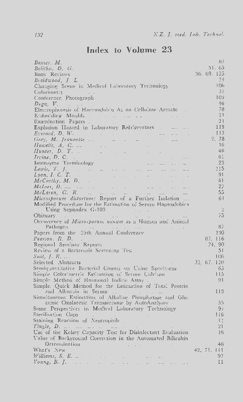
132 N.Z . .T. med. Lab. Technu!.
Baxter, M. Bolitho, D. C. Book Re\'ie"·s Braidwood, .J. L.
Index to Volume 23
Changing Scene in Medical Laboratory Technology Colorimetry Conference Photograph Degn, V. Electrophore&is of Haemoglobin A2 on Cellulose Acetate Embedding Moulds Examination Papers Explosion Hazard in Laboratory Refrigerators Everard, D . W. Grey, M. .T eannette Ho wells, A. C. Hunt n, D. T. Irvine, D. C. Isoenzyme Terminology Lewis, T. .J. Lyon, I. C. T. McCarth)', M. D. Mci ver, D. McLaren, C . R. Microsporum distortum: Report of a F urther Isolation Modified Procedure for the Estimation of Serum Haptoglobins
Using Sephadex G-100 Obituary Occurrence of Atficrosporum nanum as a Human and Animal
Pathogen Papers from the 25th Annual Conference Pearson, R. D . Regional Seminar Reports Re\'iew of a Bacteruria Screening Test Saal, ]. R. Selected Abstracts Semiquantitati\'e Bacterial Counts on Urine Specimens Simple Colorimetric Estimation of Serum Calcium Simple Method of Hormonal Iodine Assay Simple. Quick Method for the Estimation of Total Protein
and Albumin in Serum Simultaneous Estimation of Alkaline Phosphatase and Glu-
tamic Oxalacetic Transaminase by AutoAnalyzer Some Perspectives in Medical Laboratory Technology Sterilisation Caps Sta ining Reaction of Neutrophils Tingle, D. Use of the Kelsey Capacity Test for D isin fectant Evaluation Valu e of Background Correction in the Automated Bilirubin
Determination What's ~ew Williams, S. E. Yo ung, B . ].
87 51. 65
36. 69, 125 24
106 27
105 46 78 21 2+
119 11 3
2. 78 16 4fi 61 23
115 9 1 61 27 55 61
2 75
87 130
87. 116 74, 90
51 106
32, 67, 120 65
115 91
113
55 97
116 11 21 16
46 42, 71, 111
97 II
from diagnosis to therapy Biolab offers the complete allergy service.
( Biolab) Diagnostic Materials Screen sets: Consisting of nine se lected mixtures of po ll en, fungus, and inhalent all ergens for scratch testing.
Individual sets: Comprising the thirty-two individual allergens which make up the Scre-en Set.
Selected sets: Four sets of specific groups of all ergens - Shrub & Tree Pollens, Food Extracts, Flower Pol lens and Weed Pollens.
( Biolab) Treatment extracts Specific therapeutic mixtures for a graded dose regime, individually prepared from your patient's skin test results . Full S.S. charge.
XXX\'11
2046
,.I
~ •,
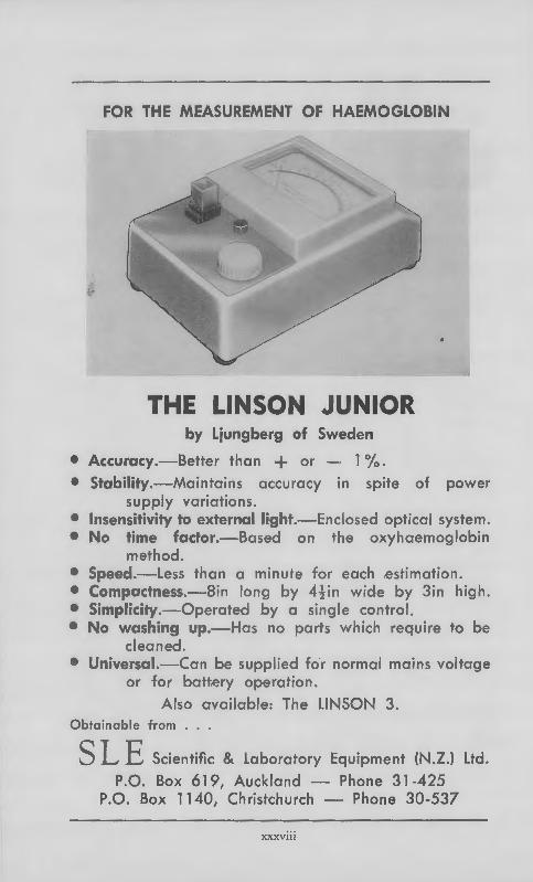
FOR THE MEASUREMENT OF HAEMOGLOBIN
..
THE LINSON JUNIOR by Ljungberg of Sweden
• Accuracy.-Better than + or - 1 %. • Stability.-Maintains accuracy in spite of power
supply variations. • Insensitivity to external light.-Enclosed optical system. • No time factor.-Based on the oxyhaemoglobin
method. • Speed.-Less than a minute for each .estimation. • Compactness.-8in long by 4tin wide by 3in high. • Simplicity.-Operated by a single control. • No washing up.-Has no parts which require to be
cleaned. • Universai.-Can be supplied for normal mains voltage
or for battery operation.
Also available: The LINSON 3. Obtainable from ...
S L E Scientific & Laboratory Equipment (N.Z.) Ltd.
P.O. Box 619, Auckland - Phone 31-425 P.O. Box 1140, Christchurch - Phone 30-537
XXXV III
S!1 .
What diffet~ence does a
of.O. lsi e error
make?
Balance errors can be avoided today. MeHler guarantees the reproducibility of the measurement results and the accuracy of the weight indication on its balances. In the ultra micro range up to •. 2/10.000,000 g.
Until MeHler constructed the first singlepan substitution balance in 1946, people had to accept (or had been accepting) the inherent errors of the two-pan balance lor 6,000 year~ Even the first MeHler balance had an accuracy of · 1110,000 g.
Then the MeHler research deportment (120 men) baHied against those errors which occur when weighing in the range of hundred thousandths and mill ionths of a gram.
In order to eliminate these insidiously tiny
SOLE AGENT •
errors, o few dozen inventions were necessary. For example: we found a way to produce scales on which the scale divisions have on accuracy of position in fractions of 111000 mm. We invented the ring weights which are supported completely symmetrically and do not a<cillate. We developed an electrochemical treatment for the surfaces of the weights so that dust particles cannot seHie on them. To our way of thinking, a few particles of dust weighing ant millionth of a gram would be intolerable.
The Mettler researchers hove also eliminated subjective weighing erro~ The Mettler level-malic compensates automatically slight changes in balance level. The
WATSON VICTOR LTD. Head Office: 4 Adelaide Road, Wellington.
Branches: Auckland, Christchurch and Dunedin.
xxxix
rapid and fine tanng simplifies operation. The compact digital indicator makes reading errors almost impassible. For those who are scared of reading errors, the printer, which prints out the result to riVe decimal plac.,. is available. Or they can combine a Mf>:HI ~r with a recording device or computer.
For a while, our researchers were some· what disturbed because they had mastered errors in determining weight.
However, they are very sotisr!Od again today becauSP. they have discovered many sources of error in the methods of thermal and volumetric measurement.
Alma<! as many as there were in 1946 with the old two-pan balance.
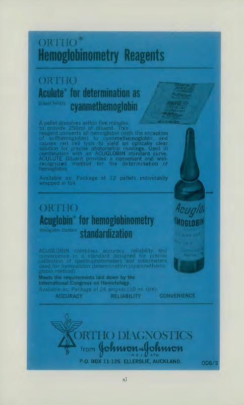
xl
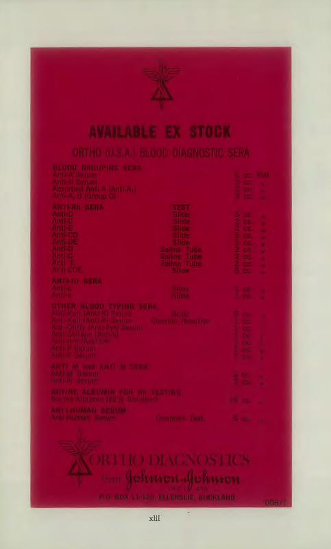
xlii
Index to Advertisers
Ames Company xxxi
British Drug Houses xxxu
Burroughs Wellcome & Co. Ltd. XXX
Dental & Medical Supply Co. Ltd. xxi, xxv, xxxm
Dominion Dental Supplies Ltd. iii, iv, v, xxvii, xli Hoechst New Zealand Ltd. xvu1
.Johnson & Johnson (N.Z.) Ltd. VI, xl, xlii
Kempthorne Prosser & Co.'s N.Z. Drug Co.
May & Baker (N.Z.) Ltd.
xix, xxiv
xiii.) xxxiv .. . ..
Vll, XXXVlll Scientific & Laboratory Equipment (N.Z.) Ltd.
Smith-Biolab Ltd. ix, X, xi, xvi, XXXV, XXXVll
Tasman Vaccine Laboratory Ltd.
Watson Victor Ltd .
William R . Warner & Co. Ltd .
XXXVI
xv, xxxix
viii, xii, xiv, xvii, xx, xxii , xxiii, xxvi, xxvii , xx1x
Insurance Facilities N .z. Medical Assurance Society Co-operative
The Medical Assurance Society Co-operative has now officially agreed to extend the privileges of membership to qualified medical laboratory technologists.
These include car, property and hou~eholders insurance at rates which are in general below those of tariff company rates and which are subject to a further 10'/r concession if all three classes are taken.
In addition, sickness and personal accident insurance (at an average premium of $12 per annum for a salary of $3,500-$4,000); life assurance; some other types of general assurance; and loans for motor car or property purchase are available.
Full details may be obtained from the Society at its Head Office: Aurora House, Mayfair Centre, 48-64 The Terrace, Wellington, Telephone 58-719, ur from the nearest District Office:
Wellington: Telephone 58-719 Auckland: Telephone 31-433
Christchurch: Telephone 69-329 Dunedin: Telephone PB0-697
Invercargill: Telephone 89-149
xliii
Contents
Original Articles ELECTROPHORESIS OF Hb A., ON CELLULOSE ACETATE
M. Jeannette Grey 78
THE OCCURRENCE OF MICROSPORUM NANUM AS A HUMAN AND ANIMAL PATHOGEN IN NEW ZEALAND
M. Baxter and R. D. Pearson
A SIMPLE METHOD OF HORMONAL IODINE ASSAY
87
I. C. T. Lyon .... . 91
The T. H. Pullar Memorial Address SOME PERSPECTIVES IN MEDICAL LABORATORY TECH
NOLOGY S. E. Williams . . 97
Sho1't Articles A SIMPLE, QUICK METHOD FOR THE ESTIMATION OF
TOTAL PROTEIN AND ALBUMIN IN SERUM D. W. Everard . 113
E:lucation CHANGING SCENE IN MEDICAL LABORATORY TECH
NOLOGY l R. Saal 106
Technic::tl Communications SIMPLE COLORIMET RIC ESTIMATION OF SERUM
CALCIUM T. J. Lewis 115
STERILISATION CAPS R. D. Pearson 116
EXPLOSION HAZARD IN Boak Reviews LABORATORY RE- Chromatographic and Elec-FRIGERATORS 119 trophoretic Techniques 1:25
What's New Ill Comprehensive Review for
Selected Abstracts 120 Medical Technologists 126
Conference Photograph 105 A Guide to Practical Histo-chemistry 126
Papers Read at 25 th Confer- An Introduction to Virology 127 ence 130 Medical Laboratorv Tec h-
Regional Seminar 90 nology 128 Books Received 130 T~xtbook of Medical V irology 128
PR I NTE D BY THE EVENIN G S T A R C O. L T D . , DUN E D I N
Top Related
Copyright © 2022 FDOKUMEN

