


Bahasa
Halaman
Hukum

Review Article
Received: 29 April 2014, Revised: 14 June 2014, Accepted: 16 June 2014 Published online in Wiley Online Library
(wileyonlinelibrary.com) DOI 10.1002/jat.3048
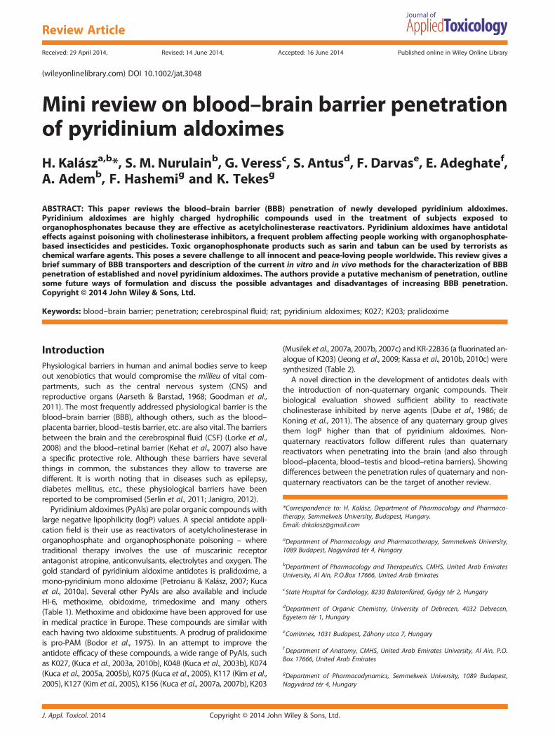
Mini review on blood–brain barrier penetrationof pyridinium aldoximesH. Kalásza,b*, S. M. Nurulainb, G. Veressc, S. Antusd, F. Darvase, E. Adeghatef,A. Ademb, F. Hashemig and K. Tekesg
ABSTRACT: This paper reviews the blood–brain barrier (BBB) penetration of newly developed pyridinium aldoximes.Pyridinium aldoximes are highly charged hydrophilic compounds used in the treatment of subjects exposed toorganophosphonates because they are effective as acetylcholinesterase reactivators. Pyridinium aldoximes have antidotaleffects against poisoning with cholinesterase inhibitors, a frequent problem affecting people working with organophosphate-based insecticides and pesticides. Toxic organophosphonate products such as sarin and tabun can be used by terrorists aschemical warfare agents. This poses a severe challenge to all innocent and peace-loving people worldwide. This review gives abrief summary of BBB transporters and description of the current in vitro and in vivo methods for the characterization of BBBpenetration of established and novel pyridinium aldoximes. The authors provide a putative mechanism of penetration, outlinesome future ways of formulation and discuss the possible advantages and disadvantages of increasing BBB penetration.Copyright © 2014 John Wiley & Sons, Ltd.
Keywords: blood–brain barrier; penetration; cerebrospinal fluid; rat; pyridinium aldoximes; K027; K203; pralidoxime
*Correspondence to: H. Kalász, Department of Pharmacology and Pharmaco-therapy, Semmelweis University, Budapest, Hungary.Email: [email protected]
aDepartment of Pharmacology and Pharmacotherapy, Semmelweis University,1089 Budapest, Nagyvárad tér 4, Hungary
bDepartment of Pharmacology and Therapeutics, CMHS, United Arab EmiratesUniversity, Al Ain, P.O.Box 17666, United Arab Emirates
cState Hospital for Cardiology, 8230 Balatonfüred, Gyógy tér 2, Hungary
dDepartment of Organic Chemistry, University of Debrecen, 4032 Debrecen,Egyetem tér 1, Hungary
eComInnex, 1031 Budapest, Záhony utca 7, Hungary
f Department of Anatomy, CMHS, United Arab Emirates University, Al Ain, P.O.Box 17666, United Arab Emirates
gDepartment of Pharmacodynamics, Semmelweis University, 1089 Budapest,Nagyvárad tér 4, Hungary
IntroductionPhysiological barriers in human and animal bodies serve to keepout xenobiotics that would compromise the millieu of vital com-partments, such as the central nervous system (CNS) andreproductive organs (Aarseth & Barstad, 1968; Goodman et al.,2011). The most frequently addressed physiological barrier is theblood–brain barrier (BBB), although others, such as the blood–placenta barrier, blood–testis barrier, etc. are also vital. The barriersbetween the brain and the cerebrospinal fluid (CSF) (Lorke et al.,2008) and the blood–retinal barrier (Kehat et al., 2007) also havea specific protective role. Although these barriers have severalthings in common, the substances they allow to traverse aredifferent. It is worth noting that in diseases such as epilepsy,diabetes mellitus, etc., these physiological barriers have beenreported to be compromised (Serlin et al., 2011; Janigro, 2012).
Pyridinium aldoximes (PyAls) are polar organic compounds withlarge negative lipophilicity (logP) values. A special antidote appli-cation field is their use as reactivators of acetylcholinesterase inorganophosphate and organophosphonate poisoning – wheretraditional therapy involves the use of muscarinic receptorantagonist atropine, anticonvulsants, electrolytes and oxygen. Thegold standard of pyridinium aldoxime antidotes is pralidoxime, amono-pyridinium mono aldoxime (Petroianu & Kalász, 2007; Kucaet al., 2010a). Several other PyAls are also available and includeHI-6, methoxime, obidoxime, trimedoxime and many others(Table 1). Methoxime and obidoxime have been approved for usein medical practice in Europe. These compounds are similar witheach having two aldoxime substituents. A prodrug of pralidoximeis pro-PAM (Bodor et al., 1975). In an attempt to improve theantidote efficacy of these compounds, a wide range of PyAls, suchas K027, (Kuca et al., 2003a, 2010b), K048 (Kuca et al., 2003b), K074(Kuca et al., 2005a, 2005b), K075 (Kuca et al., 2005), K117 (Kim et al.,2005), K127 (Kim et al., 2005), K156 (Kuca et al., 2007a, 2007b), K203
J. Appl. Toxicol. 2014 Copyright © 2014 John
(Musilek et al., 2007a, 2007b, 2007c) and KR-22836 (a fluorinated an-alogue of K203) (Jeong et al., 2009; Kassa et al., 2010b, 2010c) weresynthesized (Table 2).A novel direction in the development of antidotes deals with
the introduction of non-quaternary organic compounds. Theirbiological evaluation showed sufficient ability to reactivatecholinesterase inhibited by nerve agents (Dube et al., 1986; deKoning et al., 2011). The absence of any quaternary group givesthem logP higher than that of pyridinium aldoximes. Non-quaternary reactivators follow different rules than quaternaryreactivators when penetrating into the brain (and also throughblood–placenta, blood–testis and blood–retina barriers). Showingdifferences between the penetration rules of quaternary and non-quaternary reactivators can be the target of another review.
Wiley & Sons, Ltd.
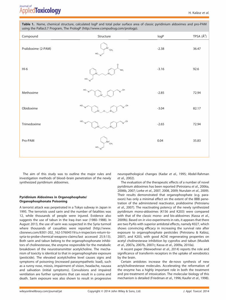
Table 1. Name, chemical structure, calculated logP and total polar surface area of classic pyridinium aldoximes and pro-PAMusing the Pallas3.7 Program, The PrologP (http://www.compudrug.com/prologp).
Compound Structure logP TPSA (Å2)
Pralidoxime (2-PAM) -2.38 36.47
HI-6 -3.16 92.6
Methoxime -2.85 72.94
Obidoxime -3.04 82.17
Trimedoxime -2.65 72.94
Pro-PAM 0.04 35.83
H. Kalász et al.
The aim of this study was to outline the major rules andinvestigation methods of blood–brain penetration of the newlysynthesized pyridinium aldoximes.
Pyridinium Aldoximes in Organophosphate/Organophosphonate Poisoning
A terrorist attack was perpetrated in a Tokyo subway in Japan in1995. The terrorists used sarin and the number of fatalities was12, while thousands of people were injured. Evidence alsosuggests the use of tabun in the Iraq–Iran war (1980–1988). InAugust 2013, the use of sarin was suspected in the Syria turmoilwhere thousands of casualties were reported (http://www.cbsnews.com/8301-202_162-57604519/u.n-inspectors-return-to-syria-to-probe-chemical-weapons-claims/last accessed 25.9.13).Both sarin and tabun belong to the organophosphonate inhibi-tors of cholinesterase, the enzyme responsible for the metabolicbreakdown of the neurotransmitter acetylcholine. The mecha-nism of toxicity is identical to that in organophosphate exposure(pesticide). The elevated acetylcholine level causes signs andsymptoms of poisoning (increased parasympathetic load), suchas a runny nose, miosis, impairment of vision, headache, nauseaand salivation (initial symptoms). Convulsions and impairedventilation are further symptoms that can result in a coma anddeath. Sarin exposure was also shown to result in progressive
Copyright © 2014 Johnwileyonlinelibrary.com/journal/jat
neuropathological changes (Kadar et al., 1995; Abdel-Rahmanet al., 2002).
The evaluation of the therapeutic effects of a number of novelpyridinium aldoximes has been reported (Petroianu et al., 2006a,2006b, 2007; Lorke et al., 2007, 2008, 2009; Nurulain et al., 2009).Their results demonstrated that organophosphate (e.g. para-oxon) has only a minimal effect on the extent of the BBB pene-tration of the administered reactivator, pralidoxime (Petroianuet al., 2007). The reactivating potency of the newly synthesizedpyridinium mono-aldoximes (K156 and K203) were comparedwith that of the classic mono- and bis-aldoximes (Kassa et al.,2009b). Based on in vivo experiments in rats, it appears that thereare two PyAls with superior antidotal effects, namely K027, whichshows convincing efficacy in increasing the survival rate afterexposure to organophosphate pesticides (Petroianu & Kalász,2007), and K203, with good AChE regenerating properties onacetyl cholinesterase inhibition by cyprofos and tabun (Musileket al., 2007a, 2007b, 2007c; Kassa et al., 2009a, 2010a).
A recent paper (Niewoehner et al., 2014) reports the role andsignificance of transferrin receptors in the uptake of xenobioticsby the brain.
Certain antidotes increase the de-novo synthesis of newactylcholinesterase molecules. Accelerating the reformation ofthe enzyme has a highly important role in both the treatmentand pre-treatment of intoxication. The molecular biology of thismechanism is detailed (Friedman et al., 1996; Kaufer et al., 1998).
J. Appl. Toxicol. 2014Wiley & Sons, Ltd.
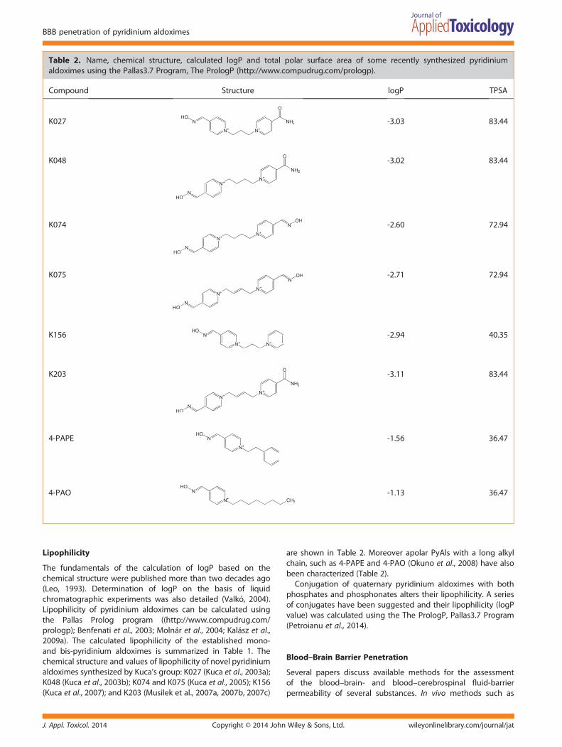
Table 2. Name, chemical structure, calculated logP and total polar surface area of some recently synthesized pyridiniumaldoximes using the Pallas3.7 Program, The PrologP (http://www.compudrug.com/prologp).
Compound Structure logP TPSA
K027 -3.03 83.44
K048 -3.02 83.44
K074 -2.60 72.94
K075 -2.71 72.94
K156 -2.94 40.35
K203 -3.11 83.44
4-PAPE -1.56 36.47
4-PAO -1.13 36.47
BBB penetration of pyridinium aldoximes
Lipophilicity
The fundamentals of the calculation of logP based on thechemical structure were published more than two decades ago(Leo, 1993). Determination of logP on the basis of liquidchromatographic experiments was also detailed (Valkó, 2004).Lipophilicity of pyridinium aldoximes can be calculated usingthe Pallas Prolog program ((http://www.compudrug.com/prologp); Benfenati et al., 2003; Molnár et al., 2004; Kalász et al.,2009a). The calculated lipophilicity of the established mono-and bis-pyridinium aldoximes is summarized in Table 1. Thechemical structure and values of lipophilicity of novel pyridiniumaldoximes synthesized by Kuca’s group: K027 (Kuca et al., 2003a);K048 (Kuca et al., 2003b); K074 and K075 (Kuca et al., 2005); K156(Kuca et al., 2007); and K203 (Musilek et al., 2007a, 2007b, 2007c)
J. Appl. Toxicol. 2014 Copyright © 2014 John
are shown in Table 2. Moreover apolar PyAls with a long alkylchain, such as 4-PAPE and 4-PAO (Okuno et al., 2008) have alsobeen characterized (Table 2).Conjugation of quaternary pyridinium aldoximes with both
phosphates and phosphonates alters their lipophilicity. A seriesof conjugates have been suggested and their lipophilicity (logPvalue) was calculated using the The PrologP, Pallas3.7 Program(Petroianu et al., 2014).
Blood–Brain Barrier Penetration
Several papers discuss available methods for the assessmentof the blood–brain- and blood–cerebrospinal fluid-barrierpermeability of several substances. In vivo methods such as
Wiley & Sons, Ltd. wileyonlinelibrary.com/journal/jat

H. Kalász et al.
histochemistry, pharmacokinetic studies, imaging techniques,indicator diffusion techniques, in situ brain perfusion andintracerebral microdialysis were used in BBB permeability ofsubstances (Dash & Elmquist, 2003). In addition to in vivoprocedures, in vitro methods can also be used.
Various mechanisms by which drugs cross the BBB wererecently summarized (Begley, 2004; Avdeef, 2012). A strongcorrelation was confirmed between lipid solubility and BBBpermeability, and the importance of good solubility in therespective compartments (plasma and CSF) was also empha-sized. Optimization of physico-chemical properties to avoidobstruction, and preparation of prodrugs to fit the compoundto the delivery system were also reported. The large polarsurface area of the drug was mentioned as one of the mostimportant properties hampering penetration. Prediction of BBBpenetration can be successfully performed on the basis ofcalculation of molecular cross-sectional parameters (Gerebtzoff& Seelig, 2006). Another publication also predicted that haloge-nation could be an effective tool to improve BBB penetration ofcertain organic compounds (Gerebtzoff et al., 2004).
A new series of bis-pyridinium compounds (bis-pyridiniumcyclophanes) with high affinity for the blood–brain barriercholine transporter were recently designed (Zhang et al., 2008).Choline transporters deliver the positively charged highly polarcholine from the periphery to the CNS where choline serves asprecursor of acetylcholine.
Indirect Proof of BBB Penetration
Evidence of BBB penetration can be inferred from achieving thedesired pharmacological effect in the brain (Bajgar et al., 1972;Shih, 1993; Petroianu & Kalász, 2007; Kuca et al., 2007a, 2007b;Okuno et al., 2008; Garcia et al., 2010; Shih et al., 2010). Evidenceof protection of neurons (in layers II and III of the cortex) wasfound when O-benzoyl derivatives of pralidoxime were co-administered before a lethal soman dose (Loke et al., 2005).
The extent of reactivation of the tabun-inhibited AChE enzymewas determined (Kassa et al., 2008) with atropine (as control),atropine+obidoxime, atropine+HI-6, atropine+ trimedoxime,atropine+K156 or atropine+K203 and also with pro-2-PAM(Demar et al., 2010). In blood, AChE activity was essentially restoredby atropine+obidoxime, atropine+ trimedoxime or atropine+K203, whereas in the brain, reactivation of the tabun-inhibitedenzyme was achieved solely with atropine+K203. Similarly,antidotal treatment showed the best protective effect (reducedlethality) in the case of atropine+ K203 administration.
Reactivation of AChE was not observed in guinea pig bypralidoxime after exposition of diisopropylfluorophosphate(DFP) (Demar et al., 2010). At the same time a lipid permeableprodrug, pro-pralidoxime reactivated AChE activity in the frontalcortex of guinea pigs.
In vitro Methods
Kuca et al. (2005b) compared the in vitro reactivation efficacy oftwo novel cholinesterase reactivators: K074 and K075 to that ofclassic cholinesterase reactivators such as HI-6, obidoxime,pralidoxime and trimedoxim on tabun-inhibited AChE in therat brain. HI-6, obidoxime and pralidoxime did not show any sig-nificant reactivating effects, but K074, K075 and trimedoximewere the most effective reactivators in tabun-inhibited AChE.
Copyright © 2014 Johnwileyonlinelibrary.com/journal/jat
An in vitro experiment was used to predict BBB penetration ofseveral reactivators of acetylcholinesterase using passive trans-port (Kuca et al., 2009; Karasova et al., 2010a). They followedthe method of Yoon et al. (2006). The basis of calculation wasto determine the capacity factor (kIAM) using chromatographyon a column of immobilized artificial membrane (IAM.PC.DD 2,150 × 4.6mm i.d., 12μm) of Regis Technologies (Morton Grove,IL, USA). The best penetration ability was predicted forpralidoxime, whereas the lowest was for HI-6 and obidoxime(Karasova et al., 2010b). On the basis of data obtained for 51compounds (30 pyridinium aldoximes and 19 standards)correlations were found between polar surface area (PSA) andkIAM/molecular weight. Their conclusions on the BBB penetrationof pyridinium aldoximes by passive transport were:
(1) simple and short linkers between the two pyridinium ringscan facilitate penetration
(2) para-positioned group(s) on pyridinium ring(s) are preferred(3) either carbamoyl or amidoxime substituents of the pyridinium
ring markedly decreases penetration into the CNS.
In vivo Methods
In 1969, 14C-obidoxime was used to determine BBB passage(Falb & Erdmann, 1969). Later on HI-6 was directly determinedin striatal dialysates after intamuscular administration both withand without previous soman treatment (Cassel & Fosbraey,1996). Pretreatment with soman slightly decreased tmax of HI-6in striatum and the extracellular level of both GABA anddopamine were also significantly increased. Other authors founddefinite BBB penetration of HI-6 after intravenous (i.v.) adminis-tration (Ligtenstein & Kossen, 1983; Ligtenstein et al., 1988).Radiolabeling was utilized to trace how 14C-HI-6 penetrates theCNS [brain and spinal cord] (Lundy et al., 1990).
The blood-to-brain penetration of HI-6 without and with 90 μgkg–1 subcutaneous (s.c.) soman pretreatment in rats was alsodetermined (Cassel et al., 1997). Their experiments were donetaking blood samples from the rat jugular vein and microdialysissamples from the corpus striatum. Concentrations of HI-6 weregraphically plotted (both in blood and in brain) and thenumerical values of area under the curve (AUC), volume of distri-bution (VD) and total clearance (Cltot) values were calculated.
An article on pralidoxime penetration across the BBB waspublished by Japanese authors (Sakurada et al., 2003). MaleWistar rats (250-400 g) were treated i.v. with pralidoxime at adose of 10, 50 and 100mg kg–1. Blood was taken from thetail vein, and dialysate was collected at three points in time(1-h intervals). Definite, dose-dependent penetration of pralidoximewas found in the region of the brain striatal segment. A constantdecrease of the 2-PAM level after the first hour followingadministration both in the blood and in the dialysate aroundthe striatum indicated first order elimination. They discussedthe apparent contradiction of BBB penetration of the hydro-philic pralidoxime phenomenon which cannot be explainedby a simple diffusion. Analogies between BBB penetration ofparaquat (Shimizu et al., 2001) and pralidoxime were found,such as competition for L-Valin transport and dependence onsodium ion concentration.
The brain entry of K027 and K048 was compared with that ofobidoxime (Lorke et al., 2007). AUC and Cmax values (both inplasma and in the brain) served for evaluation of the extent ofbrain entry. For obidoxime, Cmax,brain was 1.3% of Cmax,plasma,
J. Appl. Toxicol. 2014Wiley & Sons, Ltd.
BBB penetration of pyridinium aldoximes
and for K027 Cmax,brain it was only 0.6% of Cmax,plasma, whereasfor K048 Cmax,brain it was 1.4% of Cmax, plasma, when injected tointact rats. The AUC values in the brain were 6%, 2% and 5%compared with that of plasma, for obidoxime, K027 and K048,respectively. These limited entry values of K027 and K048 tothe brain are unable to explain the good reactivation potencyand in vivo efficacy of these compounds after intoxication withparaoxon.
The concentration of several pyridinium aldoximes in brainmicrodialysates were determined (Okuno et al., 2008). Numericalvalues were given for the ratio of blood to striatal extracellularconcentrations (that were between 1.9% and 30% dependingon the dose and time elapsed after i.v. injection) in male Wistarrats weighing 320–465 g. First order of elimination was found forboth 4-PAPE and 4-PAO compounds for serum when thedeterminations were done at 60, 120 and 180min after i.v.administration. The same (first order) elimination was character-istic for both the 4-PAPE and 4-PAO level in brain dialysatebetween 0 to 1, 1 to 2, and 2 to 3 h after i.v. administration.
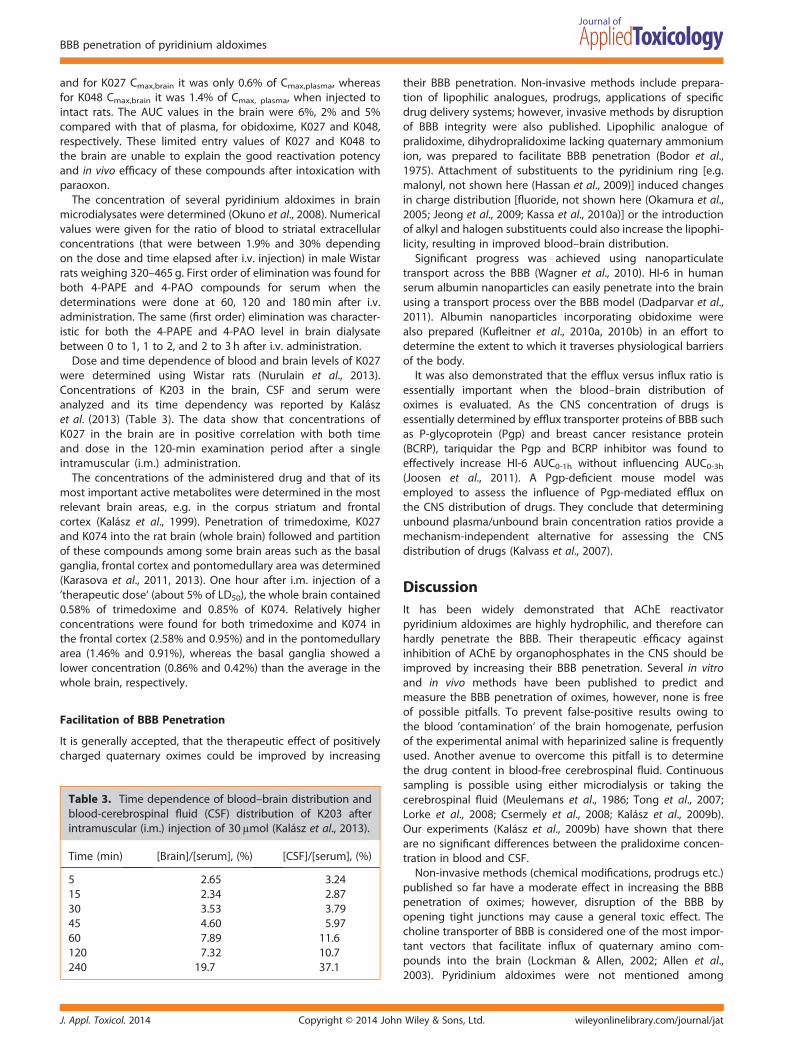
Dose and time dependence of blood and brain levels of K027were determined using Wistar rats (Nurulain et al., 2013).Concentrations of K203 in the brain, CSF and serum wereanalyzed and its time dependency was reported by Kalászet al. (2013) (Table 3). The data show that concentrations ofK027 in the brain are in positive correlation with both timeand dose in the 120-min examination period after a singleintramuscular (i.m.) administration.
The concentrations of the administered drug and that of itsmost important active metabolites were determined in the mostrelevant brain areas, e.g. in the corpus striatum and frontalcortex (Kalász et al., 1999). Penetration of trimedoxime, K027and K074 into the rat brain (whole brain) followed and partitionof these compounds among some brain areas such as the basalganglia, frontal cortex and pontomedullary area was determined(Karasova et al., 2011, 2013). One hour after i.m. injection of a’therapeutic dose‘ (about 5% of LD50), the whole brain contained0.58% of trimedoxime and 0.85% of K074. Relatively higherconcentrations were found for both trimedoxime and K074 inthe frontal cortex (2.58% and 0.95%) and in the pontomedullaryarea (1.46% and 0.91%), whereas the basal ganglia showed alower concentration (0.86% and 0.42%) than the average in thewhole brain, respectively.
Facilitation of BBB Penetration
It is generally accepted, that the therapeutic effect of positivelycharged quaternary oximes could be improved by increasing
Table 3. Time dependence of blood–brain distribution andblood-cerebrospinal fluid (CSF) distribution of K203 afterintramuscular (i.m.) injection of 30μmol (Kalász et al., 2013).
Time (min) [Brain]/[serum], (%) [CSF]/[serum], (%)
5 2.65 3.2415 2.34 2.8730 3.53 3.7945 4.60 5.9760 7.89 11.6120 7.32 10.7240 19.7 37.1
J. Appl. Toxicol. 2014 Copyright © 2014 John
their BBB penetration. Non-invasive methods include prepara-tion of lipophilic analogues, prodrugs, applications of specificdrug delivery systems; however, invasive methods by disruptionof BBB integrity were also published. Lipophilic analogue ofpralidoxime, dihydropralidoxime lacking quaternary ammoniumion, was prepared to facilitate BBB penetration (Bodor et al.,1975). Attachment of substituents to the pyridinium ring [e.g.malonyl, not shown here (Hassan et al., 2009)] induced changesin charge distribution [fluoride, not shown here (Okamura et al.,2005; Jeong et al., 2009; Kassa et al., 2010a)] or the introductionof alkyl and halogen substituents could also increase the lipophi-licity, resulting in improved blood–brain distribution.Significant progress was achieved using nanoparticulate
transport across the BBB (Wagner et al., 2010). HI-6 in humanserum albumin nanoparticles can easily penetrate into the brainusing a transport process over the BBB model (Dadparvar et al.,2011). Albumin nanoparticles incorporating obidoxime werealso prepared (Kufleitner et al., 2010a, 2010b) in an effort todetermine the extent to which it traverses physiological barriersof the body.It was also demonstrated that the efflux versus influx ratio is
essentially important when the blood–brain distribution ofoximes is evaluated. As the CNS concentration of drugs isessentially determined by efflux transporter proteins of BBB suchas P-glycoprotein (Pgp) and breast cancer resistance protein(BCRP), tariquidar the Pgp and BCRP inhibitor was found toeffectively increase HI-6 AUC0-1h without influencing AUC0-3h(Joosen et al., 2011). A Pgp-deficient mouse model wasemployed to assess the influence of Pgp-mediated efflux onthe CNS distribution of drugs. They conclude that determiningunbound plasma/unbound brain concentration ratios provide amechanism-independent alternative for assessing the CNSdistribution of drugs (Kalvass et al., 2007).
DiscussionIt has been widely demonstrated that AChE reactivatorpyridinium aldoximes are highly hydrophilic, and therefore canhardly penetrate the BBB. Their therapeutic efficacy againstinhibition of AChE by organophosphates in the CNS should beimproved by increasing their BBB penetration. Several in vitroand in vivo methods have been published to predict andmeasure the BBB penetration of oximes, however, none is freeof possible pitfalls. To prevent false-positive results owing tothe blood ’contamination‘ of the brain homogenate, perfusionof the experimental animal with heparinized saline is frequentlyused. Another avenue to overcome this pitfall is to determinethe drug content in blood-free cerebrospinal fluid. Continuoussampling is possible using either microdialysis or taking thecerebrospinal fluid (Meulemans et al., 1986; Tong et al., 2007;Lorke et al., 2008; Csermely et al., 2008; Kalász et al., 2009b).Our experiments (Kalász et al., 2009b) have shown that thereare no significant differences between the pralidoxime concen-tration in blood and CSF.Non-invasive methods (chemical modifications, prodrugs etc.)
published so far have a moderate effect in increasing the BBBpenetration of oximes; however, disruption of the BBB byopening tight junctions may cause a general toxic effect. Thecholine transporter of BBB is considered one of the most impor-tant vectors that facilitate influx of quaternary amino com-pounds into the brain (Lockman & Allen, 2002; Allen et al.,2003). Pyridinium aldoximes were not mentioned among
Wiley & Sons, Ltd. wileyonlinelibrary.com/journal/jat
H. Kalász et al.
substrates of the blood–brain barrier choline transporter(Geldenhuys et al., 2004). However, one of the organic cationtransporters (Bennett et al., 2011) may be the main candidateprotein that facilitates both the influx and the efflux of oximesthrough the BBB.
These findings suggest the existence of a very effective effluxtransporter pumping out the pyridinium aldoxime from thebrain against a significant concentration gradient. Specificinhibitors of the efflux proteins in the BBB (e.g. Pgp and BCRP)may offer a possible solution in increasing CNS concentrationsof oximes.
It is generally accepted, that increasing the BBB penetration ofoxime antidotes is advantageous; however, little attention ispaid to the metabolism and toxicity of the highly stable complexformed between the oxime and the reactivated AChE.
ConclusionReliable sources in the literature insist on the importance of lipidsolubility of non-ionized drug species when their BBB penetra-tion is considered. The classic examples are that an essentialdecrease in lipophilicity resulted in a diminishing concentrationand effect in the CNS (such as in the case of second generationantihistamines) (Goodman et al., 2011). At the same time, noveleditions of textbooks on pharmacology mirror the highlyappreciated role of drug transporters that have come into thelimelight. Influx and efflux transporters are able to facilitate drugpenetration into the CNS, and remove them even in oppositedirection against a deep concentration gradient.
Neither the use of an artificial membrane nor the calculationof logP can be a substitute for a properly performed in vivoexperiment. In animal experiments, dose-dependence, timeversus drug level parameters, age and maturity, as well as manyaspects of physiological and pathological statuses can also bemodeled and considered.
Acknowledgments
We would like to thank Professor Kamil Kuca and Dr KamilMusilek for their advice and to Mr Zoltán Demeter for histechnical help. This project is financially supported by the grantof the Hungarian National Science Fund (OTKA T100155).
Conflict of InterestThe authors did not report any conflict of interest.
ReferencesAarseth P, Barstad JA. 1968. Blood-brain barrier permeability in various
parts of the central nervous system. Arch. Int. Pharmacodyn. Ther.176: 434–442.
Abdel-Rahman A, Shetty AK, Abou-Donia MB. 2002. Acute exposure tosarin increases blood brain barrier permeability and induces neuro-pathological changes in the rat brain: dose-response relationships.Neuroscience 113: 721–741.
Allen DD, Lockman PR, Roder KE, Dwoskin LP, Crooks PA. 2003. Activetransport of high-affinity choline and nicotine analogs into thecentral nervous system by the blood-brain barrier choline trans-porter. J. Pharmacol. Exp. Ther. 304: 1268–1274.
Avdeef A. 2012. Absorption and Drug Development, 2nd edn. Wiley:Hoboken, NJ.
Bajgar J, Jakl A, Hrdina V. 1972. The influence of obidoxime on acetylcho-linesterase activity in different parts of the mouse brain followingisopropylmethyl phosphonofluoridate intoxication. Eur. J. Pharmacol.19: 199–202.
Copyright © 2014 Johnwileyonlinelibrary.com/journal/jat
Begley DJ. 2004. Delivery of therapeutic agents to the central nervoussystem: the problems and the possibilities. Pharmacol. Ther. 104:29–45.
Benfenati E, Gini G, Piclin N, Roncaglioni A, Vari MR. 2003. Predicting logP of pesticides using different software. Chemosphere 53: 1155–1164.
Bennett KM, Liu J, Hoelting C, Stoll J. 2011. Expression and analysis of twonovel rat organic cation transporter homologs, SLC22A17 andSLC22A23. Mol. Cell. Biochem. 352: 143–154.
Bodor N, Shek E, Higuchi T. 1975. Delivery of a quaternary pyridinium saltacross the blood-brain barrier by its dihydropyridine derivative.Science 190: 155–156.
Cassel G, Karlsson L, Waara L, Ang KW, Goransson-Nyberg A. 1997.Pharmacokinetics and effects of HI 6 in blood and brain of soman-intoxicated rats: a microdialysis study. Eur. J. Pharmacol. 332: 43–52.
Cassel GE, Fosbraey P. 1996. Measurement of the oxime HI-6 after periph-eral administration in tandem with neurotransmitter levels in striataldialysates: effects of soman intoxication. J. Pharmacol. Toxicol.Methods 35: 159–166.
Csermely T, Kalász H, Petroianu GA, Kuca K, Darvas F, Ludányi K,Mudhafar A, Tekes K. 2008. Analysis of pyridinium aldoximes - A chro-matographic approach. Curr. Med. Chem. 15: 2401–2418.
Dadparvar M, Wagner S, Wien S, Kufleitner J, Worek F, von Briesen H,Kreuter J. 2011. HI-6 human serum albumin nanoparticles-Development and transport over an in vitro blood-brain barriermodel. Toxicol. Lett. 206: 60–66.
Dash AK, Elmquist WF. 2003. Separation methods that are capable ofrevealing blood-brain barrier permeability. J. Chromatogr. B Analit.Technol. Biomed. Life Sci. 797: 241–254.
Demar JC, Clarkson ED, Ratcliffe RH, Campbell AJ, Thangavelu SG,Herdman A, Leader H, Schulz SM, Marek E, Medynets MA, Ku TC,Evans SA, Khan FA, Owens RR, Nambiar MP, Gordon RK. 2010.Pro-2-PAM therapy for central and peripheral cholinesterases.Chem. Biol. Interact. 187: 191–198.
Dube SN, Ghosh AK, Jeevarathinam K, Kumar D, Das Gupta S, Pant BP,Batra BS, Jaiswal DK. 1986. Studies on the efficacy of diethyxime asan antidote against organophosphorus intoxication in rats. Jpn. J.Pharmacol. 41: 267–271.
Falb A, Erdmann WD. 1969. [Penetration of 14C-obidoxime through theso-called blood-brain barrier of mice and rats] [Article in German].Arch. Toxikol. 24: 123–132.
Friedman A, Kaufer D, Shemer J, Hendler I, Soreq H, Tur-Kaspa I. 1996.Pyridostigmine brain penetration under stress enhances neuronalexcitability and induces early immediate transcriptional response.Nat. Med. 2: 1382–1385.
Garcia GE, Campbell AJ, Olson J, Moorad-Doctor JD, Morthole VI. 2010.Novel oximes as blood-brain barrier penetrating cholinesterasereactivators. Chem. Biol. Interact. 187: 199–206.
Geldenhuys WJ, Lockman PR, McAfee JH, Fitzpatrick KT, Van der Schyf CJ,Allen DD. 2004. Molecular modeling studies on the active bindingsite of the blood-brain barrier choline transporter. Bioorg. Med. Chem.Lett. 14: 3085–3092.
Gerebtzoff G, Li-Blatter X, Fischer H, Frentzel A, Seelig A. 2004. Halogena-tion of drugs enhances membrane binding and permeation.Chembiochem 5: 676–684.
Gerebtzoff G, Seelig A. 2006. In silico prediction of blood-brain barrierpermeation using the calculated molecular cross-sectional area asmain parameter. J. Chem. Inf. Model. 46: 2638–2650.
Goodman LS, Brunton LL, Chabner B, Knollmann BC. 2011. Goodman &Gilman’s Pharmacological Basis of Therapeutics, 12th edn. McGraw-Hill: New York, NY.
Hassan HA, Abdel-Aziz M, Abuo-Rahma G-D, Farag HH. 2009. 1-Malonyl-1,4-dihydropyridine as a novel carrier for specific delivery of drugs tothe brain. Bioorg.Med. Chem. 17: 1681–1692.
Janigro D. 2012. Are you in or out? Leukocyte, ion, and neurotransmitterpermeability across the epileptic blood-brain barrier. Epilepsia 53(Suppl 1): 26–34.
Jeong HC, Park NJ, Chae CH, Musilek K, Kassa J, Kuca K, Jung YS. 2009.Fluorinated pyridinium oximes as potential reactivators for acetylcho-linesterases inhibited by paraoxon organophosphorus agent. Bioorg.Med. Chem. 17: 6213–6217.
Joosen MJA, van der Schans MJ, van Dijk CG, Kuijpers WC, WortelboerHM, van Helden HP. 2011. Increasing oxime efficacy by blood-brainbarrier modulation. Toxicol. Lett. 206: 67–71.
Kadar T, Shapira S, Cohen G, Sahar R, Alkalay D, Raveh L. 1995. Sarin-induced neuropathology in rats. Hum. Exp. Toxicol. 14: 252–259.
J. Appl. Toxicol. 2014Wiley & Sons, Ltd.
BBB penetration of pyridinium aldoximes
Kalász H, Bartók T, Szökő É, Háberle D, Kiss JP, Hennings EC, Magyar K,Fürst S. 1999. Analysis of deprenyl metabolites in the rat brain usingHPLC-ES-MS. Curr. Med. Chem. 6: 271–278.
Kalász H, Fűrész J, Tekes K. 2009a. Monitoring the pharmacokinetics ofpyridinium aldoximes in the body. Mini Rev. Med. Chem. 9: 596–610.
Kalász H, Szegi P, Janoki G, Balogh L, Pöstényi Z, Musilek K, Petroianu GA,Siddiq A, Tekes K. 2013. Study on medicinal chemistry of K203 inwistar rats and beagle dogs. Curr. Med. Chem. 20: 2137–2144.
Kalász H, Szökő É, Tábi T, Petroianu GA, Lorke DE, Omar A, Alafifi S, JasemA, Tekes K. 2009b. Analysis of pralidoxime in serum, brain and CSF ofrats. Med. Chem. 5: 237–241.
Kalvass JC, Maurer TS, Pollack GM. 2007. Use of plasma and brainunbound fractions to assess the extent of brain distribution of34 drugs: Comparison of unbound concentration ratios to in vivoP-glycoprotein efflux ratios. Drug Metab. Dispos. 35: 660–666.
Karasova JZ, Pohanka M, Musilek K, Zemek F, Kuca K. 2010a. Passivediffusion of acetylcholinesterase oxime reactivators through theblood-brain barrier: influence of molecular structure. Toxicol. In Vitro24: 1838–1844.
Karasova JZ, Stodulka P, Kuca K. 2010b. In vitro screening of blood-brainbarrier penetration of clinically used acetylcholinesterasereactivators. J. Appl. Biomed. 8: 35–40.
Karasova JZ, Zemek F, Bajgar J, Vasatova M, Prochazka P, Novotny L, KucaK. 2011. Partition of bispyridinium oximes (trimedoxime and K074)administered in therapeutic doses into different parts of the rat brain.J. Pharm. Biomed. Anal. 54: 1082–1087.
Karasova JZ, Zemek F, Musilek K, Kuca K. 2013. Time-dependent changesof oxime K027 concentrations in different parts of rat central nervoussystem. Neurotox. Res. 23: 63–68.
Kassa J, Karasova J, Musilek K, Kuca K. 2008. An evaluation of therapeuticand reactivating effects of newly developed oximes (K156, K203) andcommonly used oximes (obidoxime, trimedoxime, HI-6) in tabun-poisoned rats and mice. Toxicology 243: 311–316.
Kassa J, Karasova J, Vasina L, Bajgar J, Kuca K, Musilek K. 2009a. AComparison of neuroprotective efficacy of newly developed oximes(K203, K206) and commonly used oximes (obidoxime, HI-6) intabun-poisoned rats. Drug Chem. Toxicol. 32: 128–138.
Kassa J, Karasova JY, Tesarova S, Kuca K, Musilek K. 2010a. A comparison ofthe ability of newly-developed bispyridinium oxime K203 and currentlyavailable oximes (trimedoxime, obidoxime, HI-6) to counteract theacute neurotoxicity of soman in rats. Toxicol. Mech. Meth. 20: 445–451.
Kassa J, Karasova JZ, Caisberger F, Musilek K, Kuca K, Jung YS. 2010b. Acomparison of reactivating and therapeutic efficacy of the oximeK203 and its fluorinated analog (KR-22836) with currently availableoximes (obidoxime, trimedoxime, HI-6) against tabun in rats andmice. J. Enzyme Inhib. Med. Chem. 25: 480–484.
Kassa J, Karasova JZ, Musilek K, Kuca K. 2009b. A comparison of reactivatingand therapeutic efficacy of newly-developed oximes (K156, K203) andcommonly used oximes (obidoxime, HI-6) in cyclosarin-poisoned ratsand mice. Toxicol. Mech. Methods 19: 346–350.
Kassa J, Karasova JZ, Tesarova S, Musilek K, Kuca K, Jung YS. 2010c. Acomparison of neuroprotective efficacy of the oxime K203 and itsfluorinated analogue (KR-22836) with obidoxime in Tabun-poisonedrats. Basic Clin. Pharmacol. Toxicol. 107: 861–867.
Kaufer D, Friedman A, Seidman S, Soreq H. 1998. Acute stress facilitateslong-lasting changes in cholinergic gene expression. Nature 393:373–377.
Kehat R, Zemel E, Cuenca N, Evron T, Toiber D, Loewenstein A, Soreq H,Perlman I. 2007. A novel isoform of acetylcholinesterase exacerbatesphotoreceptors death after photic stress. Invest. Ophthalmol. Vis. Sci.48: 1290–1297.
Kim TH, Kuca K, Jun D, Jung YS. 2005. Design and synthesis of new bis-pyridinium oximes as cyclosarin-inhibited acetylcholinesterasereactivators. Bioorg. Med. Chem. Lett. 15: 2914–2917.
de Koning MC, van Grol M, Noort D. 2011. Peripheral site ligand conjuga-tion to a non-quaternary oxime enhances reactivation of nerveagent-inhibited human acetylcholinesterase. Toxicol. Lett. 206: 54–59.
Kuca K, Bartosova L, Kassa J, Cabal J, Bajgar J, Kunesova G, Jun D. 2005a.Comparison of the potency of newly developed and currently avail-able oximes to reactivate nerve agent-inhibited acetylcholinesterasein vitro and in vivo. Chem. Biol. Interact. 157–158: 367–368.
Kuca K, Bielavsky J, Cabal J, Bielavska M. 2003a. Synthesis of a potentialreactivator of acetylcholinesterase 1-(4-hydroxyiminomethylpyridinium)-3-(carbamoylpyridinium)-propane dibromide. Tetrahedron Lett. 44:3123–3125.
J. Appl. Toxicol. 2014 Copyright © 2014 John
Kuca K, Bielavsky J, Cabal J, Kassa J. 2003b. Synthesis of a new reactivatorof tabun-inhibited acetylcholinesterase. Bioorg. Med. Chem. Lett. 13:3545–3547
Kuca K, Cabal J, Jung YS, Musilek K, Soukup O, Jun D, Pohanka M,Musilova L, Karasova J, Novotny L, Hrabinova M. 2009. Reactivationof human brain homogenate cholinesterases inhibited by Tabunusing newly developed oximes K117 and K127. Basic Clin. Pharmacol.Toxicol. 105: 207–210.
Kuca K, Cabal J, Musilek K, Jun D, Bajgar J. 2005b. Effective bisquaternaryreactivators of tabun-inhibited AChE. J. Appl. Toxicol. 25: 491–495.
Kuca K, Hrabinova M, Soukup O, Tobin G, Karasova J, Pohanka M. 2010a.Pralidoxime--the gold standard of acetylcholinesterase reactivators--reactivation in vitro efficacy. Bratisl. Lek. Listy 111: 502–504.
Kuca K, Jun D, Cabal J, Musilova L. 2007a. Bisquaternary oximes asreactivators of tabun-inhibited human brain cholinesterases: anin vitro study. Basic Clin. Pharmacol. Toxicol. 101: 25–28.
Kuca K, Musilek K, Jun D, Pohanka M, Ghosh K, Hrabinova M. 2010b. Ox-ime K027: novel low-toxic candidate for the universal reactivator ofnerve agent- and pesticide-inhibited acetylcholinesterase. J. EnzymeInhib. Med. Chem. 25: 509–512.
Kuca K, Musilek K, Paar M, Jun D, Stodulka P, Hrabinova M, Marek J.2007b. Targeted Synthesis of 1-(4-Hydroxyiminomethylpyridinium)-3-pyridiniumpropane Dibromide – A New Nerve Agent Reactivator.Molecules 12: 1964–1972.
Kufleitner J, Wagner S, Worek F, von Briesen H, Kreuter J. 2010a. Adsorp-tion of obidoxime onto human serum albumin nanoparticles: Drugloading, particle size and drug release. J. Microencaps. 27: 506–513.
Kufleitner J, Worek F, Kreuter J. 2010b. Incorporation of obidoximeinto human serum albumin nanoparticles: optimisation of prepa-ration parameters for the development of a stable formulation.J. Microencapsul. 27: 594–601.
Leo AJ. 1993. Calculating logP(Oct) from structures. Chem. Rev. 93:1281–1306.
Ligtenstein DA, Kossen SP. 1983. Kinetic profile in blood and brain of thecholinesterase reactivating oxime HI-6 after intravenous administra-tion to the rat. Toxicol. Appl. Pharmacol. 71: 177–183.
Ligtenstein DA, Moes GW, Kossen SP. 1988. In vivo distribution of organ-ophosphate antidotes: autoradiography of [14C]HI-6 in the rat.Toxicol. Appl. Pharmacol. 92: 324–329.
Lockman PR. Allen DD. 2002. The transport of choline. Drug Dev. Ind.Pharm. 28: 749–771.
Loke WK, Sim MK, Go ML. 2005. Novel neuroprotective effects with O-benzyl derivative of pralidoxime in soman-intoxicated rodents. Eur.J. Pharmacol. 521: 59–69.
Lorke DE, Hasan MY, Nurulain SM, Kuca K, Schmitt A, Petroianu GA. 2009.Efficacy of two new asymmetric bispyridinium oximes (K-27 and K-48)in rats exposed to diisopropylfluorophosphate: comparison withpralidoxime, obidoxime, trimedoxime, methoxime, and HI-6. Toxicol.Mech. Methods 19: 327–333.
Lorke DE, Hasan MY, Nurulain SM, Sheen R, Kuca K, Petroianu GA. 2007.Entry of two new asymmetric bispyridinium oximes (K-27 and K-48)into the rat brain: comparison with obidoxime. J. Appl. Toxicol. 27:482–490.
Lorke DE, Kalász H, Petroianu GA, Tekes K. 2008. Entry of oximes into thebrain: a review. Curr. Med. Chem. 15: 743–753.
Lundy PM, Hand BT, Broxup BR, Yipchuck G, Hamilton MG. 1990. Distribu-tion of the bispyridinium oxime [14C] HI-6 in male and female rats.Arch. Toxicol. 64: 377–382.
Meulemans A, Vicart P, Mohler J, Vulpillat M, Pocidalo JJ. 1986. Continu-ous sampling for determination of pharmacokinetics in ratCcrebrospinal fluid. Antimicrob. Agents Chemother. 30: 888–891.
Molnár L, Keserű GM.; Papp A, Gulyás Z, Darvas F. 2004. A neural networkbased prediction of octanol-water partition coefficients usingatomic5 fragmental descriptors. Bioorg. Med. Chem. Lett. 14: 851–853.
Musilek K, Holas O, Jun D, Dohnal V, Gunn-Moore F, Opletalova V, Dolezal M,Kuca K. 2007a. Monooxime reactivators of acetylcholinesterase with (E)-but-2-ene linker: preparation and reactivation of tabun- and paraoxon-inhibited acetylcholinesterase. Bioorg. Med. Chem. 15: 6733–6741.
Musilek K, Holas O, Kuca K, Jun D, Dohnal V, Opletalova V, Dolezal M.2007b. Novel series of bispyridinium compounds bearing a (Z)-but-2-ene linker -- synthesis and evaluation of their reactivation activityagainst tabun and paraoxon-inhibited acetylcholinesterase. Bioorg.Med. Chem. Lett. 17: 3172–3176.
Musilek K, Jun D, Cabal J, Kassa J, Gunn-Moore F, Kuca K. 2007c. Design of apotent reactivator of tabun-inhibited acetylcholinesterase--synthesis
Wiley & Sons, Ltd. wileyonlinelibrary.com/journal/jat
H. Kalász et al.
and evaluation of (E)-1-(4-carbamoylpyridinium)-4-(4-hydroxyiminome-thylpyridinium)-but-2-ene dibromide (K203). J. Med. Chem. 50: 5514–5518.
Niewoehner J, Bohrmann B, Collin L, Urich E, Sade H, Maier P, Rueger P,Stracke JO, Lau W, Tissot AC, Loetscher H, Ghosh A, Freskgård PO.2014. Increased brain penetration and potency of a therapeuticantibody using a monovalent molecular shuttle. Neuron 81: 49–60.
Nurulain SM, Kalász H, Szegi P, Kuca K, Adem A, Hasan AYB, Hashemi F,Tekes K. 2013. HPLC analysis in drug level monitoring of K027. ActaChromatogr. 25: 703–710.
Nurulain SM, Lorke DE, Hasan MY, Shafiullah M, Kuca K, Musilek K,Petroianu GA. 2009. Efficacy of eight experimental bispyridiniumoximes against paraoxon-induced mortality: comparison with the con-ventional oximes pralidoxime and obidoxime. Neurotox. Res. 16: 60–67.
Okamura T, Kikuchi T, Nagamine A, Fukushi K, Sekine T, Arano Y, Irie T. 2005.An approach for measuring in vivo cerebral redox states using theoxidative conversion of dihydropyridine to pyridinium ion and themetabolic trapping principle. Free Radical Biol. Med. 38: 1197–1205.
Okuno S, Sakurada K, Ohta H, Ikegaya H, Kazui Y, Akutsu T, Takatori T,Iwadate K. 2008. Blood-brain barrier penetration of novelpyridinealdoxime methiodide (PAM)-type oximes examined by brainmicrodialysis with LC-MS/MS. Toxicol. Appl. Pharmacol. 227: 8–15.
Petroianu GA, Arafat K, Kuca K, Kassa J. 2006a. Five oximes (K-27, K-33, K-48, BI-6 and methoxime) in comparison with pralidoxime: in vitroreactivation of red blood cell acetylcholinesterase inhibited byparaoxon. J. Appl. Toxicol. 26: 64–71.
Petroianu GA, Kalász H. 2007. Comparison of the ability of pyridiniumaldoximes to reactivate human RBC Cholinesterases inhibited byethyl- and methyl-paraoxon. Curr. Org. Chem. 11: 1624–1634.
Petroianu GA, Lorke DE, Hasan MY, Adem A, Sheen R, Nurulain SM, KalászH. 2007. Paraoxon has only a minimal effect on pralidoxime brainconcentration in rats. J. Appl. Toxicol. 27: 350–357.
Petroianu GA, Nurulain SM, Nagelkerke N, Al-Sultan MA, Kuca K, Kassa J.2006b. Five oximes (K-27, K-33, K-48, BI-6 and methoxime) in compar-ison with pralidoxime: survival in rats exposed to the organophos-phate paraoxon. J. Appl. Toxicol. 26: 262–268.
Petroianu GA, Athauda G, Darvas F, Kalasz H, Lorke DE. 2014. K-oxime (K-27):Phosphorylation-induced changes in logP. Mil. Med. Sci. Lett. 83: 1–7.
Copyright © 2014 Johnwileyonlinelibrary.com/journal/jat
Sakurada K, Matsubara K, Shimizu K, Shiono H, Seto Y, Tsuge K,Yoshino M, Sakai I, Mukoyama H, Takatori T. 2003. Pralidoximeiodide (2-PAM) penetrates across the blood-brain barrier. Neurochem.Res. 28: 1401–1407.
Serlin Y, Levy J, Shalev H. 2011. Vascular pathology and blood-brainbarrier disruption in cognitive and psychiatric complications oftype 2 diabetes mellitus. Cardiovasc. Psychiatry Neurol. 609202,DOI:10.1155/2011/609202
Shih TM, Skovira JW, O’Donnell JC, McDonough JH. 2010. In vivoreactivation by oximes of inhibited blood, brain and peripheral tissuecholinesterase activity following exposure to nerve agents in guineapigs. Chem. Biol. Interact. 187: 207–214.
Shih TM. 1993. Comparison of several oximes on reactivation of soman-inhibited blood, brain and tissue cholinesterase activity in rats. Arch.Toxicol. 67: 637–646.
Shimizu K, Ohtaki K, Matsubara K, Aoyama K, Uezono T, Saito O, Suno M,Ogawa K, Hayase N, Kimura K, Shiono H. 2001. Carrier-mediatedprocesses in blood--brain barrier penetration and neural uptake ofparaquat. Brain Res. 906: 135–142.
The http://www.compudrug.com/prologp [14 July 2014].Tong X, Ratnaraj N, Patsalos PN. 2007. The pharmacokinetics of
vigabatrin in rat blood and cerebrospinal fluid. Seizure 16: 43–49.Valkó K. 2004. Application of high-performance liquid chromatography
based measurements of lipophilicity to model biological distribution.J. Chromatogr. A 1037: 299–310.
Wagner S, Kufleitner J, Zensi A, Dadparvar M, Wien S, Bungert J, Vogel T,Worek F, Kreuter J, von Briesen H. 2010. Nanoparticulate transport ofoximes over an iun vitro blood-brain barrier model. PLoS One 5:e14213.
Yoon CH, Kim SJ, Shin BS, Lee KC, Yoo SD. 2006. Rapid screening ofblood-brain barrier penetration of drugs using the immobilized artifi-cial membrane phosphatidylcholine column chromatography.J. Biomol. Screen. 11: 13–20.
Zhang Z, Lockman PR, Mittapalli RK, Allen D, Dwoskin LP, Crooks PA.2008. bis-Pyridinium cyclophanes: novel ligands with high affinityfor the blood-brain barrier choline transporter. Bioorg. Med. Chem.Lett. 18: 5622–5625.
J. Appl. Toxicol. 2014Wiley & Sons, Ltd.
Top Related
Copyright © 2022 FDOKUMEN

