


Bahasa
Halaman
Hukum

Loss of immunological tolerance in Gimap5-deficient mice isassociated with loss of Foxo in CD4+ T cells
H. Ibrahim Aksoylar*, Kristin Lampe*, Michael J. Barnes†, David R. Plas‡, and KasperHoebe*,§
*Department of Molecular Immunology, Cincinnati Children’s Hospital Research Foundation,Cincinnati, Ohio, USA†The Scripps Research Institute, La Jolla, California, USA‡Department of Cancer and Cell Biology, University of Cincinnati Medical Center, Cincinnati,Ohio, USA
AbstractPreviously, we reported the abrogation of quiescence and reduced survival in lymphocytes fromGimap5sph/sph mice, an ENU germline mutant with a missense mutation in the GTPase ofimmunity-associated nucleotide binding protein 5 (Gimap5). These mice showed a progressiveloss of peripheral lymphocyte populations and developed spontaneous colitis, resulting in earlymortality. Here, we identify the molecular pathways that contribute to the onset of colitis inGimap5sph/sph mice. We show that CD4+ T cells become Th1/Th17-polarized and are criticallyimportant for the development of colitis. Concomitantly, Treg cells become reduced in frequencyin the peripheral tissues and their immune-suppressive capacity becomes impaired. Mostimportantly, these progressive changes in CD4+ T cells are associated with the loss of Foxo1,Foxo3 and Foxo4 expression. Our data establish a novel link between Gimap5 and Foxoexpression and provide evidence for a regulatory mechanism that controls Foxo protein expressionand may help maintain immunological tolerance.
INTRODUCTIONThe family of GTPase of immune-associated protein (Gimap) genes are predominantlyexpressed in lymphocytes and regulate lymphocyte survival during development, selectionand homeostasis(1). Members of this family share a GTP-binding AIG1 homology domain,which was originally identified in disease resistance genes in higher plants (2, 3). Recentcrystallographic studies revealed that GDP-bound or nucleotide-free GIMAP2 exists in amonomeric configuration with an exposed guanine nucleotide binding domain(4). In thepresence of GTP, GIMAP2 oligomerizes and shows similarities with the nucleotidecoordination and dimerization mode previously observed for dynamin GTPase. In addition,these studies showed that GIMAP2 localized at the surface of lipid droplets, where it isthought to act as a nucleotide regulated scaffolding protein(4). Other members of theGIMAP family appear to be localized to different subcellular compartments(5). Overall, thefunction of these proteins remains poorly defined.
Gimap5 was recently reported to localize in lysosomes, based on studies in human, mouseand rat lymphocytes(5). Genetic aberrancies in Gimap5 have been strongly linked to reduced
§Correspondence to: Cincinnati Children’s Hospital Research Foundation; MLC7021, Room S5.421; 3333 Burnet Avenue; Cincinnati,OH USA; phone: +1 (513) 803-1056; fax: +1 (513) 636-5355; [email protected].. .Present address: University of California, San Francisco, USA
NIH Public AccessAuthor ManuscriptJ Immunol. Author manuscript; available in PMC 2013 January 1.
Published in final edited form as:J Immunol. 2012 January 1; 188(1): 146–154. doi:10.4049/jimmunol.1101206.
NIH
-PA Author Manuscript
NIH
-PA Author Manuscript
NIH
-PA Author Manuscript
lymphocyte survival and homeostasis, but, importantly have also been associated withautoimmune diseases. In humans, polymorphisms in GIMAP5 were associated withincreased concentrations of IA2 auto-antibodies in type 1 diabetes (T1D) patients(6) and anincreased risk of systemic lupus erythematosus (SLE)(7, 8). Studies using biobreeding (BB)rats— carrying a mutation (lyp/lyp) in Gimap5— show marked lymphopenia andpredisposition to the development of T1D(9-11). In addition, BB rats are prone to developintestinal inflammation on certain genetic backgrounds(12). Together these observationssuggest that, beyond lymphocyte survival, Gimap5 is essential for maintainingimmunological tolerance. Interestingly, impaired lymphocyte survival and consequentlymphopenia may be linked to the loss of immunological tolerance. Specifically, CD4+ Tcells in a lymphopenic environment can undergo thymic-independent expansion in theperiphery. This process—also referred to as homeostatic or lymphopenia-inducedproliferation (LIP) — is accompanied by marked alterations in T cell phenotype and islinked to auto-immunity(13-15). Most notably, T cells undergoing LIP acquire a memory-like phenotype, exemplified by high surface expression of CD44 and low surface expressionof CD62L. In addition, under lymphopenic conditions CD4+ T cells more readily adopt aneffector phenotype, including the ability to robustly produce cytokines upon stimulationthrough the TCR. The downstream consequences can be severe and a number ofpathological conditions have been associated with CD4+ T cells undergoing LIP, includingcolitis. Classic studies involving the adoptive transfer of naïve CD45RBhigh CD4+ T cellsinto lymphopenic SCID mice resulted in T cells acquiring a LIP phenotype and rapidlydriving colitis when recipient mice were colonized by intestinal bacteria(16-18).Importantly, colitis could be prevented if CD4+ CD25+ regulatory T (Treg) cells were co-transferred, suggesting that the presence or absence of Treg cells is an important determinantof immune-mediated sequela induced by CD4+ T cells undergoing LIP.
Our laboratory previously described an ENU germline mutant, designated sphinx, whichcontained a recessive mutation in Gimap5 that disrupted both lymphocyte survival andnormal hematopoiesis(19). Similar to Gimap5 knockout mice, these mice lack peripheralNK cells and CD8+ T cells, and exhibit dynamic changes in immune homeostasis, markedby the progressive loss of CD4+ T cells and B cells and neutrophilia(19, 20). Following thecollapse of lymphocyte populations, CD4+ T cells in Gimap5sph/sph mice acquire a LIPphenotype similar to CD4+ T cells transferred into lymphopenic hosts(18). Around 10-12weeks of age, Gimap5sph/sph mice develop wasting disease and colitis, limiting theirsurvival(19). Interestingly, adoptive transfer of Rag-sufficient splenocytes intoGimap5sph/sph mice around 5 weeks of age could restore lymphocyte homeostasis andprevent colitis and wasting(19).
In this report, we show that CD4+ T cells are required for development of colitis inGimap5sph/sph mice. Whereas CD4+ T cells exhibited impaired proliferation, they remainedhighly capable of producing proinflammatory cytokines, including IL-17A and IFNγ.Importantly, CD4+ T cells in Gimap5sph/sph mice exhibited a LIP phenotype and exhibited aprogressive and complete loss of full-length Foxo1, -3 and -4 expression. This loss of Foxoexpression was associated with a progressive reduction in the numbers and suppressivecapacity of Foxp3+ Treg cells. The development of colitis in Gimap5sph/sph mice could beprevented by transferring wild-type Treg cells into 3-week-old Gimap5sph/sph mice. SinceFoxo-deficient mice exhibit many of the phenotypes observed in Gimap5sph/sph mice,including impaired Treg cell activity and colitis, our data suggest that the loss ofimmunological tolerance in Gimap5-deficient mice may be critically linked to the loss ofFoxo expression in CD4+ T cells.
Aksoylar et al. Page 2
J Immunol. Author manuscript; available in PMC 2013 January 1.
NIH
-PA Author Manuscript
NIH
-PA Author Manuscript
NIH
-PA Author Manuscript
MATERIALS AND METHODSMice and reagents
All experiments were performed according to US National Institutes of Health guidelinesand were approved by the IACUC of The Cincinnati Children’s Hospital. C57BL/6J,Rag1−/−, CD45.1 congenic and CD90.1 congenic mice were obtained from Jackson.Gimap5sph/sph mice were generated as previously described(19) and bred in the vivarium ofthe Cincinnati Children’s Hospital. All mice were maintained under specific pathogen-freeconditions.
All antibodies used for flow cytometry were purchased from eBioscience or Biolegend.Antibodies for western blotting [anti-Foxo3a (#2497), Foxo1 (2880), pFoxo1(Thr24)/pFox03a(Thr32) (9464), p27, p-pRB (S807,S811), p-pRb (S780) and pan-Actin antibody]were purchased from Cell Signaling. Purified CD3ε (145-2C11) and CD28 (37.51)antibodies (eBioscience) were used for T cell activation. PMA and ionomycin was obtainedfrom Sigma.
Real time PCRCD4+T cells were isolated from spleen and lymph nodes of 4 week-old and 7 week-oldGimap5sph/sph and C57BL/6 mice using L3T4 MicroBeads (Miltenyi Biotech). RNAisolation was done with RNeasy Micro Kit (Qiagen) and reverse transcription wasperformed using High-Capacity cDNA Reverse Transcription Kit (Applied Biosystems).cDNAs were amplified with Lightcycler 480 SYBR Green I Master (Roche) and quantifiedby Roche Light Cycler 480 II instrument using the following primer pairs: Foxo1: Fwd:TTCGGAATGACCTCATGGATG Rev TGGACTGCTCCTCAGTTCCTG Foxo3: Fwd;AGTGGATGGTGCGCTGTGT, Rev: TCTGAACGCGCATGAAGCFoxo4: Fwd:GAGAACCTGGAGTGCGACATG, Rev: TGTGTTGCCACCAAT
Flow cytometry and T cell analysesTo quantify T cell proliferation, MACS (Miltenyi Biotech)-purified CD4+ T cells werelabeled with CFSE (5 μM CFSE) in PBS with 0.1% FCS for 10 min. Cells were cultured insupplemented IMDM media containing 10% FCS and 1% Penicillin/Streptomycin and wereeither left unstimulated or stimulated with PMA(50ng/mL)/ionomycin(1μg/mL). After 3days of incubation, proliferation was measured by analyzing CFSE dilution using flowcytometry. To assess the capacity of T cells to produce cytokines, MACS (Miltenyi Biotech)purified CD4+ T cells were incubated for 6 hours with/without PMA(10ng/mL)/ionomycin(10μg/mL) and subsequently fixed and analyzed for intracellular IL-17A, IL-4 orIFNγ production using flow cytometry. To measure surface markers ex vivo, CD4+ T cellsfrom spleen, MLN or lamina propria were isolated and stained with fluorochrome-labeledantibodies specific for mouse CD44, CD62L and CD69. Foxp3 expression was analyzed byintracellular staining.
BrdU stainingT cell-specific BrdU incorporation was measured as follows: during a 24-hr interval,wildtype or Gimap5sph/sph mice received three i.p. injections with 100 μL of a 10 mg/mlBrdU solution in sterile PBS. Incorporation of BrdU in CD4+ T cells was measured 8 hoursafter the last injection using flow cytometry.
In vitro Treg cell suppressor assaysThe Treg cell suppressor assay was performed under conditions previously described(21,22). Briefly, spleens were isolated and Treg cells were MACS purified using the
Aksoylar et al. Page 3
J Immunol. Author manuscript; available in PMC 2013 January 1.
NIH
-PA Author Manuscript
NIH
-PA Author Manuscript
NIH
-PA Author Manuscript
CD4+CD25+ regulatory T cell isolation kit (Miltenyi). Subsequently, Treg cells wereharvested and co-cultured at indicated ratios with 5 × 10⁴ MACS purified CFSE-labeledCD8+ T cells or CD4+ T cells. Also included were 1 × 10⁵ T cell-depleted, γ-irradiated(1,500 rad) splenocytes as bystander cells and 0.5 μg/mL soluble CD3 antibody. CFSEdilution was assessed by flow cytometry after three days of co-culture.
HistologyColon tissue was collected and immediately fixed in 10% buffered formalin solutionovernight, followed by routine paraffin embedding. Hematoxylin and eosin staining wasperformed on 4 μm sections from the paraffin-embedded tissue blocks for conventional lightmicroscopy analysis. Histological scoring was performed as described before (23) Briefly,scoring parameters included quantitation of the area of distal colon involved, edema,erosion/ulceration of the epithelial monolayer, crypt loss/damage, and infiltration of immunecells into the mucosa. Severity for the area involved (erosion/ulceration and crypt loss) wasgraded on a scale from 0 (normal), 1 (0–10%), 2 (10–25%), 3 (25–50%), and 4 (>50%).Immune cell infiltration was scored as: 0, absent; 1, weak; 2, moderate; and 3, severe. Totaldisease score was expressed as the mean of all combined scores per genotype.
Adoptive transfer and survival assaysFor adoptive transfer studies, Gimap5sph/sph mice at 25-35 days of age were injected i.v.with 3 × 10⁵ Treg cells isolated from C57BL/6J mice using a Treg cell isolation kit(Miltenyi). Purity was confirmed by Foxp3 staining using flow cytometry and cells were>90% Foxp3+. Mice were monitored and weighed every week following cell transfer.
Statistical analysisData were analyzed using GraphPad Prism4® software (GraphPad Software, San Diego,CA). Unless indicated otherwise, statistical significance of the differences among groupswas determined from the mean and standard deviation by Student’s two-tailed test or byANOVA followed by Dunnett’s test for three or more groups. Data were consideredstatistically significant if P values were <0.05.
RESULTSGimap5sph/sph CD4+ T cells from MLN are Th1/17-polarized
In previous work, we determined that NK, NKT, CD8+, CD4+ and B lymphocyte survivalare impaired in Gimap5sph/sph mice. In addition, they developed spontaneous colitis thatrequired the presence of microbiota and survived poorly, with most mice succumbing by150 days of age(19). As several mouse models have linked impaired lymphocyte functionwith colitis development, we further explored the contribution of lymphocytes to theimmunopathology observed in Gimap5sph/sph mice. Firstly, we investigated the survival andfunctional capacity of CD4+ T cells at different ages. By 4 weeks of age, a reduced numberof CD4+ T cells were found in the spleen, and a further decline in T cell numbers wasobserved in 6- and 10-week old Gimap5sph/sph mice (Figure 1A). Six-week-oldGimap5sph/sph CD4+ T cells had a CD44highCD62Llow phenotype characteristic of T cellsundergoing LIP(19) and showed increased incorporation of BrdU (Figure 1B). To assesswhether the loss of CD4+ T cells in the spleen and lymph nodes was also observed in gut-associated lymphoid tissue, we isolated lamina propria (LP) cells from the colons of 6-week-old Gimap5sph/sph mice and quantified the number of CD4+ T cells. Similar to the spleen,reduced numbers of CD4+ T cells were observed in the LP (Figure 1C). Further analysisrevealed that close to 100% of the colonic CD4+ T cells were CD44highCD62Llow,resembling the LIP phenotype of the CD4+ T cells in the peripheral lymphoid tissues (Figure
Aksoylar et al. Page 4
J Immunol. Author manuscript; available in PMC 2013 January 1.
NIH
-PA Author Manuscript
NIH
-PA Author Manuscript
NIH
-PA Author Manuscript
1C). Together these data suggest that CD4+ T cells are present in the GALT and exhibit aLIP phenotype similar to what is observed in the spleen.
We next investigated the functional capacity of CD4+ T cells in Gimap5sph/sph mice, andtheir potential for contributing to the development of colitis. Our previous work indicatedthat 8-week-old Gimap5sph/sph CD4+ T cells were unable to proliferate ex vivo followingstimulation with PMA/ionomycin or αCD3, even though lymphocytes exhibited normalactivation of NF-κB and MAP kinase pathways(19). Because of the latter observation, weinvestigated whether CD4+ T cells were capable of producing cytokines after suchstimulation and, if so, were Th1-, Th2- or Th17-polarized. We isolated total lymphocytesfrom spleen and MLNs from C57BL/6J control or Gimap5sph/sph mice and incubated cellsfor 6 hours with or without PMA/ionomycin in the presence of brefeldin. Interestingly, ahigher percentage of CD4+ T cells derived from Gimap5sph/sph spleen or MLNs producedIFNγ or IL-17A, or both cytokines following PMA/ionomycin stimulation (Figure 1D).Notably, T cell cytokine production was observed even in the absence of PMA/ionomycin inGimap5sph/sph MLN cells (but not splenic leukocytes), suggesting constitutive activation ofT cells in gut lymphoid tissue in these mice. Overall, these data indicate that, despite theirinability to proliferate normally ex vivo, CD4+ T cells derived from Gimap5sph/sph micebecome Th1/17 polarized and effectively produce cytokines.
Colitis in Gimap5sph/sph mice is driven by CD4+ T cellsBecause of their LIP phenotype and spontaneous production of IL-17A and IFNγ, wehypothesized that MLN CD4+ T cells may support the development of colitis inGimap5sph/sph mice. We tested this hypothesis by depleting CD4+ T cells in Gimap5sph/sph
mice using weekly injections of anti-CD4 (GK1.5) antibodies, starting at 3 weeks of age—before the CD4+ T cells “collapse” and the subsequent intestinal inflammation normallyoccurs in Gimap5sph/sph mice. Importantly, GK1.5 treatment, but not isotype treatment,prevented wasting disease (Figure 2A) and significantly decreased intestinal inflammationas determined by histology in 15-week-old Gimap5sph/sph mice (Figure 2B-H). These datasupport our hypothesis that the development of colitis in Gimap5sph/sph mice requires CD4+
T cells.
Gimap5sph/sph mice fail to maintain a Treg cell population with normal immunosuppressivefunction
Colitis induced by naïve CD45RBhigh T cell transfer into SCID recipients does not occurwhen Treg cells are co-transferred. Therefore, even though Treg cell development in thethymus of Gimap5sph/sph mice appeared to occur normally(19), we considered that Treg cellfunction may be impaired in the peripheral tissues of these lymphopenic mice and contributeto the development of colitis. Thus, we examined the presence and immunosuppressivecapacity of Foxp3+ Treg cells in Gimap5sph/sph mice. Although relatively normal numbers ofFoxp3+ Treg cells were observed in 3-week-old mice (Supplemental figure 2A), Treg cellsbecame significantly reduced in the MLNs of 6- to 8-week old mice, both as a percentagewithin the CD4+ T cell compartment as well as in total numbers of cells (Figure 3A,B). Inthe spleen, the number of Treg cells were reduced but the percentage of Foxp3+ CD4+ Tcells within the CD4 T cell population remained similar to the percentage observed inwildtype mice (Figure 3A,B). To assess their functional capacity, we purified Treg cellsfrom 4- or 6-week-old C57BL/6J or Gimap5sph/sph spleens, and co-cultured Treg cells withCFSE-labeled wild-type CD8+ T cells that were stimulated with soluble anti-CD3-antibodies. Treg cells from 4-week-old Gimap5sph/sph mice showed a slight, but significantreduction in their ability to suppress CD8+ T cell proliferation in vitro, whereas Treg cellsisolated from 6-week-old Gimap5sph/sph mice were incapable of suppressing CD8+ T cellproliferation (Figure 3C). Similar results were obtained for suppression of wildtype CD4+ T
Aksoylar et al. Page 5
J Immunol. Author manuscript; available in PMC 2013 January 1.
NIH
-PA Author Manuscript
NIH
-PA Author Manuscript
NIH
-PA Author Manuscript
cell proliferation (Supplemental Figure 1A). These findings suggest that both Treg cellsurvival and functional capacity become impaired as Gimap5sph/sph mice age.
We next questioned whether reduced peripheral Treg cell accumulation in Gimap5sph/sph
mice resulted from a cell-intrinsic phenomenon. We injected CD4+ splenocytes from 3week-old wildtype and/or Gimap5sph/sph mice into Rag-deficient recipients, either as amixture, or alone, and quantified the presence of Foxp3+ Treg cells 5 weeks after injection(Supplemental Figure 2A). Whereas no differences in the percentage of Treg cells within theCD4+ T cell compartment were observed at the time of injection, after 5 weeks theGimap5sph/sph Treg population was lost, regardless of whether wild-type cells werecotransferred or not (Figure 3D). Overall, these data indicate that cell-intrinsic expression ofGimap5 is required to allow normal Treg cell survival.
Gimap5sph/sphCD4+ T cells exhibit progressive loss of Foxo1, -3 and -4 expressionOur data indicate that the Gimap5sph/sph CD4+ T cell population collapses around 5 weeksof age and that the remaining CD4+ T cells undergo LIP thereafter. At the same time, theyfail to maintain a functional Treg cell population. Interestingly, these T cell phenotypesshow striking similarities with those seen in mice with T cells deficient in the family ofForkheadbox group O (Foxo) transcription factors. The family of Foxo transcription factorscontain 4 members of which three (Foxo1, Foxo3 and Foxo4) have overlapping patterns ofexpression and transcriptional activities(24-26). They play an essential role in the regulationof cell cycle progression, apoptosis, glucose metabolism and the regulation of life span(27).Foxo1 expression is critical for maintaining naïve T cell quiescence. Foxo1-deficient CD4+
T cells exhibit a CD44highCD62Llow LIP or effector memory phenotype(28-30). In addition,Foxo expression has been reported to be essential for Treg cell development andfunction(29, 31). We therefore analyzed expression of Foxo1, Foxo3 and Foxo4 inGimap5sph/sph CD4+ T cells. Strikingly, immunoblot analysis of CD4+ T from 6-week-oldGimap5sph/sph mice revealed a near absence of full-length Foxo1, -3a and -4 protein (Figure4A). At the mRNA level, a reduction in Foxo1 but not Foxo3 and 4 could be observed inCD4+ T cells isolated from Gimap5sph/sph compared to wildtype mice (Figure 4B),suggesting that regulation of Foxo3 and Foxo4 protein expression occurred at the post-transcriptional level. Since many of the T cell-specific phenotypes observed inGimap5sph/sph occur after 4 weeks of age, we next quantified the temporal progression ofchanges in Foxo expression in lymphocytes. Immunoblot analyses revealed that Foxoexpression was normal at 3 weeks, somewhat reduced after 4 weeks, and almost absent after6 to 10 weeks of age (Figure 4C and supplementary Figure 1A). Concordant with the loss ofFoxo expression, we detected reductions in the abundance of the cyclin-dependent kinase(Cdk) inhibitor, p27kip1, a downstream target of Foxo proteins and an important regulator ofcell cycle entry (Figure 4C)(32, 33). As p27kip1 inhibits Cdk4, we measured Cdk4 activityand detected increased phosphorylation of its substrate, retinoblastoma protein (pRb), inGimap5sph/sph cells (Figure 4C). Due to the progressive nature of this phenotype, weconsidered that lymphocytes isolated from young Gimap5sph/sph mice with intact Foxoexpression might respond normally to mitogenic stimuli. Indeed, CD4+ T cells isolated from4 week-old, but not 8-week-old, Gimap5sph/sph mice were able to proliferate after TCRstimulation (Supplemental Figure 3A and(19)). Finally, we assessed whether the loss ofFoxo expression was also observed in regulatory T cells from Gimap5sph/sph mice. Indeed,Foxo1 and Foxo3 expression was mostly absent in CD4+CD25+ T cells from 6-week-oldGimap5sph/sph mice (Figure 4D).
Interestingly, the progressive loss of Foxo expression appeared to correlate with aprogressive increase in the number of CD4+ T cells undergoing LIP (CD44hi, CD62Llo) in4- to 10-week old Gimap5sph/sph mice as previously reported(19). Subsequent analysis ofCD44lo, CD62Lhi and CD44hi, CD62Llo CD4+ T cells from 5-week-old Gimap5sph/sph and
Aksoylar et al. Page 6
J Immunol. Author manuscript; available in PMC 2013 January 1.
NIH
-PA Author Manuscript
NIH
-PA Author Manuscript
NIH
-PA Author Manuscript
wildtype mice revealed loss of FoxO expression specifically in CD4+ T cells undergoingLIP but not naïve CD4+ T cells in Gimap5sph/sph mice (Figure 4E), suggesting that the lossof Foxo expression follows T cell activation. Overall, these data link the loss of full-lengthFoxo expression with the onset of lymphopenia in Gimap5sph/sph lymphocytes, the impairedcell cycle control and proliferative capacity in such lymphocytes, and the reduced Treg cellsurvival and function in Gimap5sph/sph mice.
Prevention of colitis in Gimap5sph/sph mice by the adoptive transfer of wildtype Treg cellsOur previous data show that colitis can be prevented in Gimap5sph/sph mice through adoptivetransfer of normal, but not Rag-deficient splenocytes(19), indicating that a lymphocytepopulation is responsible for the rescue. Given the impaired Treg cell survival and functionobserved in Gimap5sph/sph mice, we next examined whether adoptively transferred wild-typeTreg cells could prevent the development of colitis. Gimap5sph/sph recipients of 3×10⁵ wild-type CD4+CD25+ T cells showed prolonged survival, delayed wasting disease (Figure 5A)and, importantly, did not develop colitis (Figure 5B-D). Characterization of lymphocytepopulations 25 weeks after transfer of CD45.1 congenically marked CD4+CD25+ Treg cellsrevealed that Treg cell reconstitution of the spleen of Gimap5sph/sph mice achieved ~50% ofthe level observed in wild-type C57BL/6J mice (Figure 5E). Notably, the Foxp3+CD4+ Tregcell population constituted 40% of the overall CD4+ T cell population and was entirelycongenic, whereas the Foxp3−CD4+ population was predominantly Gimap5sph/sph–derived(Figure 5E). Functional analysis of isolated CD4+ T cell from spleen and MLN of 15-week-old treated Gimap5sph/sph mice revealed no background cytokine production and a similaractivation as observed for wildtype CD4+ T cells following stimulation with PMA/Ionomycin (supplementary Figure 2D). Around 25 weeks of age, Treg cell-recipient micestill developed wasting disease. Necropsy at this time revealed severe inflammation in thelung and infiltration of macrophages in a number of mice (Supplemental Figure 2C).Interestingly, colitis could be prevented by the transfer of Il10−/− splenocytes (data notshown), suggesting that IL-10-independent regulatory pathways are more perturbed byGimap5-deficienty. Together, these data link impaired Treg cell survival and function to thedevelopment of colitis in Gimap5sph/sph mice. In addition, they reveal that the Gimap5sph/sph
environment is capable of supporting a functional Treg cell population.
DISCUSSIONGenetic aberrancies in Gimap5 have been linked to lymphopenia and the loss ofimmunological tolerance(6, 7, 9, 10). Although we found no evidence of autoimmuneresponses in Gimap5sph/sph mice, we observed severe and spontaneous inflammation in thegut(19)—an environment where homeostasis critically depends upon maintaining toleranceto exogenous antigens and bacterial stimuli. Similar to Gimap5sph/sph mice, loss ofimmunological tolerance in the lyp/lyp rat has been associated with reduced Treg cellsurvival and function, as well as the polarization of T helper cells towards a Th17 pattern ofdifferentiation(34, 35). However, the molecular pathways underlying the loss ofimmunological tolerance in rat and mouse models of Gimap5-deficiency have remainedelusive. Here, we explored the pathways that contribute to the loss of tolerance observed inGimap5sph/sph mice. We show that the development of colitis in Gimap5sph/sph mice iscritically dependent on CD4+ T cells. Around 8 weeks of age, CD4+ T cells lose theircapacity to proliferate ex vivo, yet they remain capable of producing proinflammatorycytokines, including IL-17A and IFNγ, and contain a population of IL-17+IFNγ+ Th1/17cells that have been associated with IL-23 signaling and more severe colitis(36). At the sametime, Treg cell numbers and function decline. Most importantly, we found that thesephenotypes are directly associated with a progressive loss of protein expression of Foxo1, -3and -4—important transcription factors that regulate both quiescence and survival of
Aksoylar et al. Page 7
J Immunol. Author manuscript; available in PMC 2013 January 1.
NIH
-PA Author Manuscript
NIH
-PA Author Manuscript
NIH
-PA Author Manuscript
lymphocytes(37). Mice with T-cell-specific deletions of Foxo1 and/or Foxo3 mimic many ofthe immunological and pathological phenotypes observed in Gimap5sph/sph mice(28-31). Forexample, Foxo-deficient CD4+ T cells have impaired proliferative capacity and adoptCD44highCD62Llow LIP or memory-like phenotype(28, 30). In addition, reduced Treg cellnumbers and function were observed in mice lacking Foxo1 or both Foxo1 and -3 in T cells.Similar to Gimap5sph/sph mice, mice lacking Foxo1 and -3 in T cells develop spontaneouscolitis, and furthermore purified Foxo1−/−Foxo3−/− Treg cells were unable to prevent colitisin Rag1−/− mice when coinjected with naïve wildtype CD4+ T cells(31). Interestingly, theimpaired Treg cell development and function in T cell-specific Foxo1-deficient mice causedexaggerated T follicular helper cell accumulation, which contributed to B cell-mediatedauto-immunity(29). In Gimap5sph/sph mice we found no evidence of autoreactive Bcells(19), but it is important to note that, similar to CD4+ T cells, Gimap5sph/sph B cellsprogressively lost Foxo expression (Supplementary Figure 3B) and were unable toproliferate following stimulation with IgM(19). Thus, B cell expansion and differentiationmay be severely hampered in Gimap5sph/sph mice, preventing the development of auto-reactive antibody responses.
The mechanisms by which Foxo transcription factors control Treg cell development,homeostasis and function have been studied in some detail. Foxo proteins have been shownto serve as coactivators downstream of the TGFβ signaling pathway by interacting withSMAD proteins, ultimately fine tuning the TGFβ-induced transcriptional program(38, 39).This pathway is also critical for the development of inducible Treg (iTreg) cells(40), whichdevelop extra-thymically and have been suggested by many studies to comprise animportant population of regulatory T cells in the gut(21, 41-43). Indeed, the loss of Tregcells within the CD4+ T cell compartment is most evident in the MLN of Gimap5sph/sph
mice (Figure 3), suggesting the iTregs in particular are impaired. In addition, Foxo1 andFoxo3 can cooperatively control the differentiation of Foxp3+ Treg cells through theregulation of a number of Treg cell associated genes, including Foxp3 itself(31).Furthermore, conditional deletion of Foxo1 in T cells resulted in reduced surface expressionof CTLA-4 and CD25 in Foxp3+CD4+ T cells(29). Analysis of the Ctla-4 gene showed thatthe promoter region contained a conserved Foxo binding site 193 bp upstream of thetranscription start site(44). Thus, the impaired function of Foxp3+ Treg cells is likely theresult of an incomplete transcriptional program in the absence of Foxo expression. Insummary, the loss of Foxo expression affects multiple pathways that regulate Treg celldevelopment, homeostasis and function, as well as the generation of iTreg cells.
In Gimap5sph/sph mice, an absence of Foxo expression was observed in all lymphocytepopulations examined, including peripheral Foxp3+ Treg cells, conventional Foxp3−CD4+ Tcells and B cells. Although conventional Foxp3−CD4+ T cells lack Foxo expression, ourexperiments reveal that colitis can be prevented by treatment with competent wild-type Tregcells, suggesting that colitogenic CD4+ T cells remain capable of being regulated when theylack Gimap5. Although we link the sphinx mutation in Gimap5 to progressive loss of Foxoexpression in lymphocytes, it is unclear to what extent these genes directly interact witheach other. Given the progressive nature of the loss of Foxo expression, it is unlikely thatGimap5 directly interacts with Foxo proteins. One possibility that we considered is that theloss of Foxo expression may drive a secondary phenotype resulting from the constitutiveproliferation cues associated with LIP, something that may be driven by self-antigens orantigens derived from the microbiota. While we cannot exclude that LIP may contribute tothe loss of Foxo expression in Gimap5sph/sph CD4+RB45high T cells, wild-typeCD4+RB45high T cells transferred into a lymphopenic host retained normal levels of Foxoexpression (Supplemental Figure 3C), suggesting that LIP alone is insufficient to cause lossof Foxo expression.
Aksoylar et al. Page 8
J Immunol. Author manuscript; available in PMC 2013 January 1.
NIH
-PA Author Manuscript
NIH
-PA Author Manuscript
NIH
-PA Author Manuscript
Our data show that Gimap5-deficiency affects Foxo3 and Foxo4 expression at the proteinlevel, not at the mRNA level. Regulation of Foxo proteins has previously been reported tooccur via ubiquitination and proteosomal degradation(45). Moreover, loss of Foxo1expression has been observed in mouse lymphomas, which served as a mechanism toremove the tumor suppressor activity of Foxo1(46). Foxo degradation was inverselycorrelated with increased expression of S-phase kinase-associate protein-2 (Skp2)—an E3ubiquitin ligase that targets numerous cell cycle proteins. Foxo degradation could bereversed following down-regulation of Skp2 via shRNAs, and therefore increased Skp2expression could pose a potential mechanism by which loss of Foxo-expression inGimap5sph/sph T cells occurs. Alternatively, the localization of Gimap5 in the lysosomalcompartment(5) and the presumed scaffolding function of Gimap family members(4)suggests that Gimap5 may be necessary for optimal lysosomal function. Lysosomes areessential for the catabolic turnover of intra- and –extracellular macromolecules, but also canrelease lysosomal enzymes (such as cathepsins) that can initiate programmed cell death oncein the cytosol(47). Intriguingly, lymphocytes containing large numbers of cytotoxicgranules, such as CD8+ T cells and NK cells do not survive in Gimap5- deficient mice,perhaps supporting the hypothesis of a deregulated lysosomal compartment. Although thelink between loss of Gimap5 and Foxo protein expression remains to be established inhuman cells, understanding the molecular pathways that lead to the degradation of Foxoproteins could provide important therapeutic targets, not only in the context of tumorgrowth, but potentially in the context of autoimmune or chronic inflammatory disorders.
Our data provide evidence that Gimap5 is essential for maintaining lymphocyte quiescenceand immunological tolerance. In the absence of functional Gimap5, Foxo expression inlymphocytes is progressively lost, with loss of Foxo3 and Foxo4 most likely involving aproteolytic mechanism. This progressive loss of Foxo expression is associated withconcomitant decline in Treg numbers and function, which ultimately leads to the loss ofimmunological tolerance in the gut. Thus, not only do we establish a critical link betweenGimap5 and Foxo protein levels, we also provide evidence for a novel regulatorymechanism controlling Foxo protein expression that may be involved in the development ofimmune-mediated diseases such as SLE, T1D and colitis.
Supplementary MaterialRefer to Web version on PubMed Central for supplementary material.
AcknowledgmentsThis research was funded by grants from the NIH, including NIH/NCI R21 Grant 1R21CA133649, NIH/NIAIDRO1 Grant 1R01AI074743 and PHS Grant P30 DK078392 (Integrative Morphology Core of the CincinnatiDigestive Disease Research Core Center).
References1. Nitta T, Nasreen M, Seike T, Goji A, Ohigashi I, Miyazaki T, Ohta T, Kanno M, Takahama Y. IAN
family critically regulates survival and development of T lymphocytes. PLoS.Biol. 2006; 4:e103.[PubMed: 16509771]
2. Reuber TL, Ausubel FM. Isolation of Arabidopsis genes that differentiate between resistanceresponses mediated by the RPS2 and RPM1 disease resistance genes. Plant Cell. 1996; 8:241–249.[PubMed: 8742710]
3. Poirier GM, Anderson G, Huvar A, Wagaman PC, Shuttleworth J, Jenkinson E, Jackson MR,Peterson PA, Erlander MG. Immune-associated nucleotide-1 (IAN-1) is a thymic selection markerand defines a novel gene family conserved in plants. J.Immunol. 1999; 163:4960–4969. [PubMed:10528200]
Aksoylar et al. Page 9
J Immunol. Author manuscript; available in PMC 2013 January 1.
NIH
-PA Author Manuscript
NIH
-PA Author Manuscript
NIH
-PA Author Manuscript
4. Schwefel D, Frohlich C, Eichhorst J, Wiesner B, Behlke J, Aravind L, Daumke O. Structural basisof oligomerization in septin-like GTPase of immunity-associated protein 2 (GIMAP2). Proc NatlAcad Sci U S A. 2010; 107:20299–20304. [PubMed: 21059949]
5. Wong V, Saunders A, Hutchings A, Pascall J, Carter C, Bright N, Walker S, Ktistakis N, ButcherGW. The auto-immunity-related GIMAP5 GTPase is a lysosome-associated protein. self/Nonself.2010; 1:9.
6. Shin JH, Janer M, McNeney B, Blay S, Deutsch K, Sanjeevi CB, Kockum I, Lernmark A, Graham J,Arnqvist H, Bjorck E, Eriksson J, Nystrom L, Ohlson LO, Schersten B, Ostman J, Aili M, BaathLE, Carlsson E, Edenwall H, Forsander G, Granstrom BW, Gustavsson I, Hanas R, Hellenberg L,Hellgren H, Holmberg E, Hornell H, Ivarsson SA, Johansson C, Jonsell G, Kockum K, Lindblad B,Lindh A, Ludvigsson J, Myrdal U, Neiderud J, Segnestam K, Sjoblad S, Skogsberg L, Stromberg L,Stahle U, Thalme B, Tullus K, Tuvemo T, Wallensteen M, Westphal O, Aman J. IA-2autoantibodies in incident type I diabetes patients are associated with a polyadenylation signalpolymorphism in GIMAP5. Genes Immun. 2007; 8:503–512. [PubMed: 17641683]
7. Hellquist A, Zucchelli M, Kivinen K, Saarialho-Kere U, Koskenmies S, Widen E, Julkunen H,Wong A, Karjalainen-Lindsberg ML, Skoog T, Vendelin J, Cunninghame-Graham DS, Vyse TJ,Kere J, Lindgren CM. The human GIMAP5 gene has a common polyadenylation polymorphismincreasing risk to systemic lupus erythematosus. J Med Genet. 2007; 44:314–321. [PubMed:17220214]
8. Lim MK, Sheen DH, Kim SA, Won SK, Lee SS, Chae SC, Chung HT, Shim SC. IAN5polymorphisms are associated with systemic lupus erythematosus. Lupus. 2009; 18:1045–1052.[PubMed: 19762377]
9. Greiner DL, Mordes JP, Handler ES, Angelillo M, Nakamura N, Rossini AA. Depletion of RT6.1+T lymphocytes induces diabetes in resistant biobreeding/Worcester (BB/W) rats. J.Exp.Med. 1987;166:461–475. [PubMed: 3496416]
10. Jacob HJ, Pettersson A, Wilson D, Mao Y, Lernmark A, Lander ES. Genetic dissection ofautoimmune type I diabetes in the BB rat. Nat.Genet. 1992; 2:56–60. [PubMed: 1303251]
11. Hornum L, Romer J, Markholst H. The diabetes-prone BB rat carries a frameshift mutation in Ian4,a positional candidate of Iddm1. Diabetes. 2002; 51:1972–1979. [PubMed: 12031988]
12. Cousins L, Graham M, Tooze R, Carter C, Miller JR, Powrie FM, Macpherson GG, Butcher GW.Eosinophilic bowel disease controlled by the BB rat-derived lymphopenia/Gimap5 gene.Gastroenterology. 2006; 131:1475–1485. [PubMed: 17064701]
13. Khoruts A, Fraser JM. A causal link between lymphopenia and autoimmunity. Immunol Lett.2005; 98:23–31. [PubMed: 15790505]
14. King C, Ilic A, Koelsch K, Sarvetnick N. Homeostatic expansion of T cells during immuneinsufficiency generates autoimmunity. Cell. 2004; 117:265–277. [PubMed: 15084263]
15. Krupica T Jr. Fry TJ, Mackall CL. Autoimmunity during lymphopenia: a two-hit model. ClinImmunol. 2006; 120:121–128. [PubMed: 16766227]
16. Powrie F, Correa-Oliveira R, Mauze S, Coffman RL. Regulatory interactions betweenCD45RBhigh and CD45RBlow CD4+ T cells are important for the balance between protective andpathogenic cell-mediated immunity. J Exp Med. 1994; 179:589–600. [PubMed: 7905019]
17. Powrie F, Leach MW, Mauze S, Caddle LB, Coffman RL. Phenotypically distinct subsets of CD4+T cells induce or protect from chronic intestinal inflammation in C. B-17 scid mice. Int Immunol.1993; 5:1461–1471. [PubMed: 7903159]
18. Aranda R, Sydora BC, McAllister PL, Binder SW, Yang HY, Targan SR, Kronenberg M. Analysisof intestinal lymphocytes in mouse colitis mediated by transfer of CD4+, CD45RBhigh T cells toSCID recipients. J Immunol. 1997; 158:3464–3473. [PubMed: 9120308]
19. Barnes MJ, Aksoylar H, Krebs P, Bourdeau T, Arnold CN, Xia Y, Khovananth K, Engel I, SovathS, Lampe K, Laws E, Saunders A, Butcher GW, Kronenberg M, Steinbrecher K, Hildeman D,Grimes HL, Beutler B, Hoebe K. Loss of T cell and B cell quiescence precedes the onset ofmicrobial flora-dependent wasting disease and intestinal inflammation in Gimap5-deficient mice. JImmunol. 2010; 184:3743–3754. [PubMed: 20190135]
20. Schulteis RD, Chu H, Dai X, Chen Y, Edwards B, Haribhai D, Williams CB, Malarkannan S,Hessner MJ, Glisic-Milosavljevic S, Jana S, Kerschen EJ, Ghosh S, Wang D, Kwitek AE,
Aksoylar et al. Page 10
J Immunol. Author manuscript; available in PMC 2013 January 1.
NIH
-PA Author Manuscript
NIH
-PA Author Manuscript
NIH
-PA Author Manuscript
Lernmark A, Gorski J, Weiler H. Impaired survival of peripheral T cells, disrupted NK/NKT celldevelopment, and liver failure in mice lacking Gimap5. Blood. 2008; 112:4905–4914. [PubMed:18796632]
21. Barnes MJ, Krebs P, Harris N, Eidenschenk C, Gonzalez-Quintial R, Arnold CN, Crozat K, SovathS, Moresco EM, Theofilopoulos AN, Beutler B, Hoebe K. Commitment to the regulatory T celllineage requires CARMA1 in the thymus but not in the periphery. PLoS.Biol. 2009; 7:e51.[PubMed: 19260764]
22. Thornton AM, Shevach EM. CD4+CD25+ immunoregulatory T cells suppress polyclonal T cellactivation in vitro by inhibiting interleukin 2 production. J Exp Med. 1998; 188:287–296.[PubMed: 9670041]
23. Steinbrecher KA, Harmel-Laws E, Sitcheran R, Baldwin AS. Loss of epithelial RelA results inderegulated intestinal proliferative/apoptotic homeostasis and susceptibility to inflammation. JImmunol. 2008; 180:2588–2599. [PubMed: 18250470]
24. Anderson MJ, Viars CS, Czekay S, Cavenee WK, Arden KC. Cloning and characterization of threehuman forkhead genes that comprise an FKHR-like gene subfamily. Genomics. 1998; 47:187–199.[PubMed: 9479491]
25. Biggs WH 3rd, Cavenee WK, Arden KC. Identification and characterization of members of theFKHR (FOX O) subclass of winged-helix transcription factors in the mouse. Mamm Genome.2001; 12:416–425. [PubMed: 11353388]
26. Furuyama T, Kitayama K, Shimoda Y, Ogawa M, Sone K, Yoshida-Araki K, Hisatsune H,Nishikawa S, Nakayama K, Ikeda K, Motoyama N, Mori N. Abnormal angiogenesis in Foxo1(Fkhr)-deficient mice. J Biol Chem. 2004; 279:34741–34749. [PubMed: 15184386]
27. Greer EL, Brunet A. FOXO transcription factors at the interface between longevity and tumorsuppression. Oncogene. 2005; 24:7410–7425. [PubMed: 16288288]
28. Kerdiles YM, Beisner DR, Tinoco R, Dejean AS, Castrillon DH, DePinho RA, Hedrick SM. Foxo1links homing and survival of naive T cells by regulating L-selectin, CCR7 and interleukin 7receptor. Nat.Immunol. 2009; 10:176–184. [PubMed: 19136962]
29. Kerdiles YM, Stone EL, Beisner DL, McGargill MA, Ch’en IL, Stockmann C, Katayama CD,Hedrick SM. Foxo transcription factors control regulatory T cell development and function.Immunity. 2010; 33:890–904. [PubMed: 21167754]
30. Ouyang W, Beckett O, Flavell RA, Li MO. An essential role of the Forkhead-box transcriptionfactor Foxo1 in control of T cell homeostasis and tolerance. Immunity. 2009; 30:358–371.[PubMed: 19285438]
31. Ouyang W, Beckett O, Ma Q, Paik JH, DePinho RA, Li MO. Foxo proteins cooperatively controlthe differentiation of Foxp3+ regulatory T cells. Nat Immunol. 2010; 11:618–627. [PubMed:20467422]
32. Dijkers PF, Medema RH, Pals C, Banerji L, Thomas NS, Lam EW, Burgering BM, RaaijmakersJA, Lammers JW, Koenderman L, Coffer PJ. Forkhead transcription factor FKHR-L1 modulatescytokine-dependent transcriptional regulation of p27(KIP1). Mol.Cell Biol. 2000; 20:9138–9148.[PubMed: 11094066]
33. Medema RH, Kops GJ, Bos JL, Burgering BM. AFX-like Forkhead transcription factors mediatecell-cycle regulation by Ras and PKB through p27kip1. Nature. 2000; 404:782–787. [PubMed:10783894]
34. Poussier P, Ning T, Murphy T, Dabrowski D, Ramanathan S. Impaired post-thymic developmentof regulatory CD4+25+ T cells contributes to diabetes pathogenesis in BB rats. J.Immunol. 2005;174:4081–4089. [PubMed: 15778366]
35. van den Brandt J, Fischer HJ, Walter L, Hunig T, Kloting I, Reichardt HM. Type 1 diabetes inBioBreeding rats is critically linked to an imbalance between Th17 and regulatory T cells and analtered TCR repertoire. J Immunol. 2010; 185:2285–2294. [PubMed: 20644174]
36. Ahern PP, Schiering C, Buonocore S, McGeachy MJ, Cua DJ, Maloy KJ, Powrie F. Interleukin-23drives intestinal inflammation through direct activity on T cells. Immunity. 2010; 33:279–288.[PubMed: 20732640]
37. Tothova Z, Gilliland DG. FoxO transcription factors and stem cell homeostasis: insights from thehematopoietic system. Cell Stem Cell. 2007; 1:140–152. [PubMed: 18371346]
Aksoylar et al. Page 11
J Immunol. Author manuscript; available in PMC 2013 January 1.
NIH
-PA Author Manuscript
NIH
-PA Author Manuscript
NIH
-PA Author Manuscript
38. Gomis RR, Alarcon C, He W, Wang Q, Seoane J, Lash A, Massague J. A FoxO-Smadsynexpression group in human keratinocytes. Proc Natl Acad Sci U S A. 2006; 103:12747–12752.[PubMed: 16908841]
39. Seoane J, Le HV, Shen L, Anderson SA, Massague J. Integration of Smad and forkhead pathwaysin the control of neuroepithelial and glioblastoma cell proliferation. Cell. 2004; 117:211–223.[PubMed: 15084259]
40. Harada Y, Elly C, Ying G, Paik JH, DePinho RA, Liu YC. Transcription factors Foxo3a and Foxo1couple the E3 ligase Cbl-b to the induction of Foxp3 expression in induced regulatory T cells. JExp Med. 2010; 207:1381–1391. [PubMed: 20439537]
41. Atarashi K, Tanoue T, Shima T, Imaoka A, Kuwahara T, Momose Y, Cheng G, Yamasaki S, SaitoT, Ohba Y, Taniguchi T, Takeda K, Hori S, Ivanov II, Umesaki Y, Itoh K, Honda K. Induction ofcolonic regulatory T cells by indigenous Clostridium species. Science. 2011; 331:337–341.[PubMed: 21205640]
42. Izcue A, Hue S, Buonocore S, Arancibia-Carcamo CV, Ahern PP, Iwakura Y, Maloy KJ, Powrie F.Interleukin-23 restrains regulatory T cell activity to drive T cell-dependent colitis. Immunity.2008; 28:559–570. [PubMed: 18400195]
43. Zheng Y, Josefowicz S, Chaudhry A, Peng XP, Forbush K, Rudensky AY. Role of conserved non-coding DNA elements in the Foxp3 gene in regulatory T-cell fate. Nature. 2010; 463:808–812.[PubMed: 20072126]
44. Perkins D, Wang Z, Donovan C, He H, Mark D, Guan G, Wang Y, Walunas T, Bluestone J,Listman J, Finn PW. Regulation of CTLA-4 expression during T cell activation. J Immunol. 1996;156:4154–4159. [PubMed: 8666782]
45. Plas DR, Thompson CB. Akt activation promotes degradation of tuberin and FOXO3a via theproteasome. J Biol Chem. 2003; 278:12361–12366. [PubMed: 12517744]
46. Huang H, Regan KM, Wang F, Wang D, Smith DI, van Deursen JMA, Tindall DJ. Skp2 inhibitsFOXO1 in tumor suppression through ubiquitin-mediated degradation. Proceedings of theNational Academy of Sciences of the United States of America. 2005; 102:1649–1654. [PubMed:15668399]
47. Kirkegaard T, Jaattela M. Lysosomal involvement in cell death and cancer. Biochim Biophys Acta.2009; 1793:746–754. [PubMed: 18948147]
Aksoylar et al. Page 12
J Immunol. Author manuscript; available in PMC 2013 January 1.
NIH
-PA Author Manuscript
NIH
-PA Author Manuscript
NIH
-PA Author Manuscript
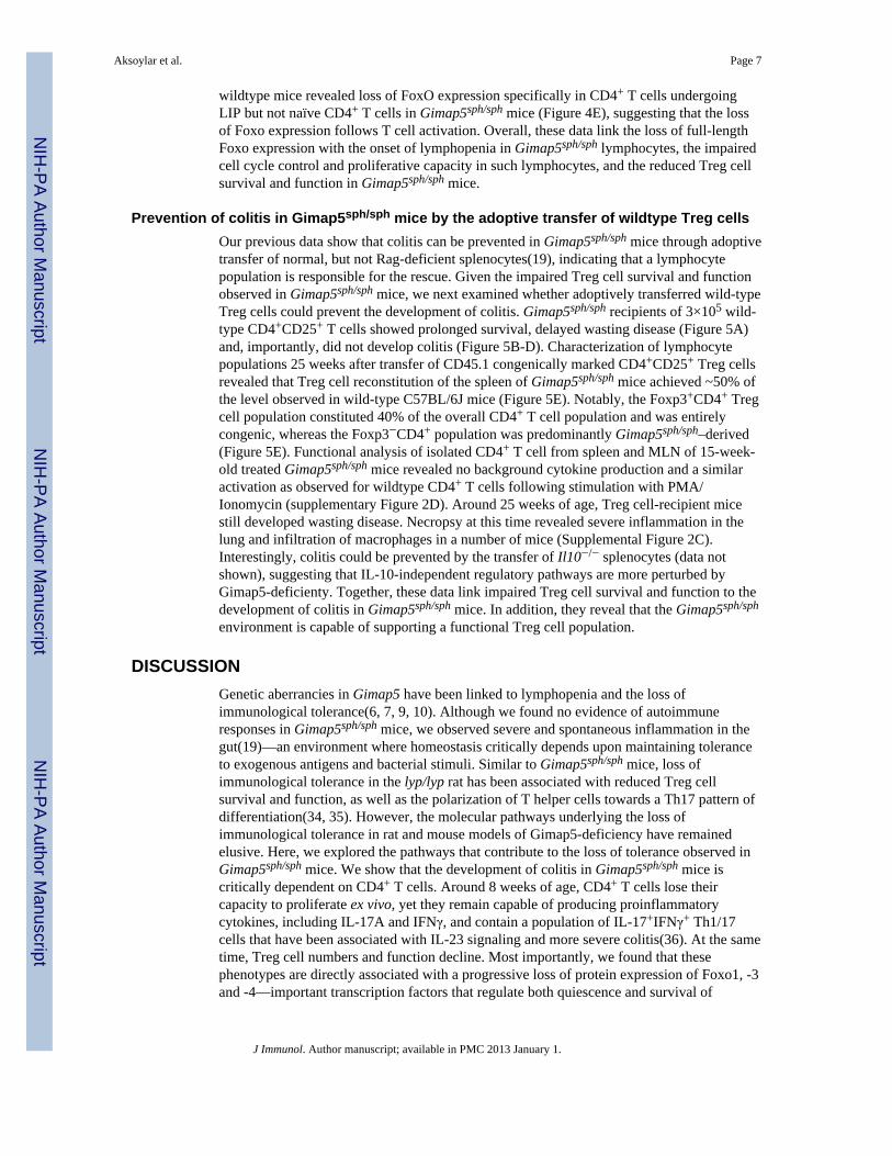
Figure 1. Phenotypic characterization of CD4+ T cells in Gimap5sph/sph mice(A) Splenic CD4+ T cell population collapse around 6 weeks of age in Gimap5sph/sph mice,at which time they exhibit a LIP phenotype with increased BrdU uptake (B). (C) Thenumber of CD4+ T cells and the percentage of CD44highCD62Llow CD4+ T cells in laminapropria of 6-week-old wildtype and Gimap5sph/sph mice. (D) Ex vivo cytokine production byCD4+ T cells isolated from wildtype or Gimap5sph/sph spleen and mesenteric lymph node(MLN), left unstimulated or following stimulation with PMA/ionomycin (100ng/ml) for sixhours (mean values ± SEM; n ≥ 4 mice per genotype from 2 independent experiments).(*P<0.05, **P<0.01, ***P<0.001)
Aksoylar et al. Page 13
J Immunol. Author manuscript; available in PMC 2013 January 1.
NIH
-PA Author Manuscript
NIH
-PA Author Manuscript
NIH
-PA Author Manuscript
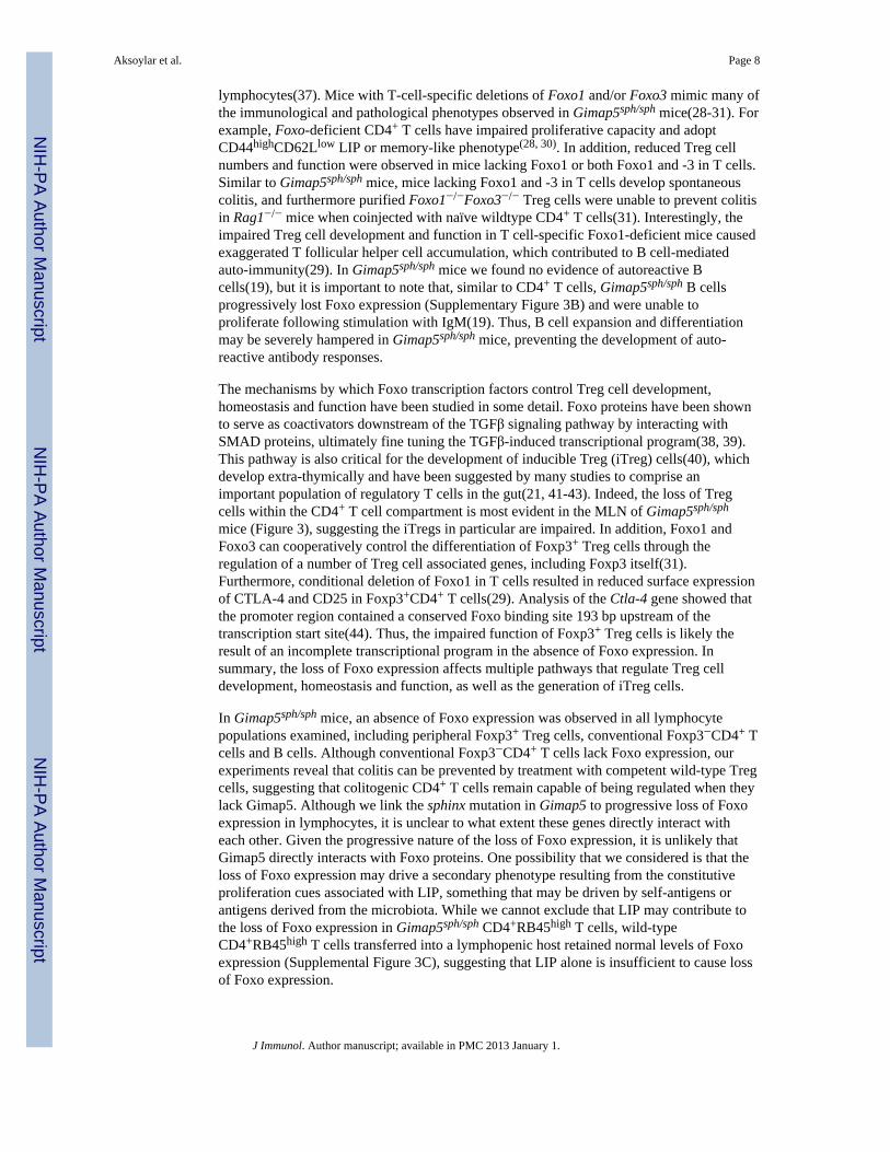
Figure 2. Depletion of CD4+ T cells prevents wasting disease and colitis in Gimap5sph/sph miceMale Gimap5sph/sph mice were given 200μg/mouse anti-CD4 (GK1.5) or isotype controlantibodies i.p. weekly, beginning at 3 weeks of age. (A) Weights of 10-week-old wild-type,isotype-treated or GK1.5 treated Gimap5sph/sph mice. (B) CD4-depletion significantlyreduces colitis in Gimap5sph/sph mice Data represents histological scoring as described in thematerial and methods. (C-H) At fifteen weeks of age, mice were sacrificed and the colonwas analyzed for signs of intestinal inflammation following H&E staining (C, wildtypeuntreated; D-F, Gimap5sph/sph-isotype treated; G-H, Gimap5sph/sph GK1.5-treated). Datarepresent mean weight percentage (wild type mice =100%) ± SEM from at least 3 animalsper group and histology is representative of 3 analyzed mice per group. (*P<0.05, **P<0.01,***P<0.001)
Aksoylar et al. Page 14
J Immunol. Author manuscript; available in PMC 2013 January 1.
NIH
-PA Author Manuscript
NIH
-PA Author Manuscript
NIH
-PA Author Manuscript
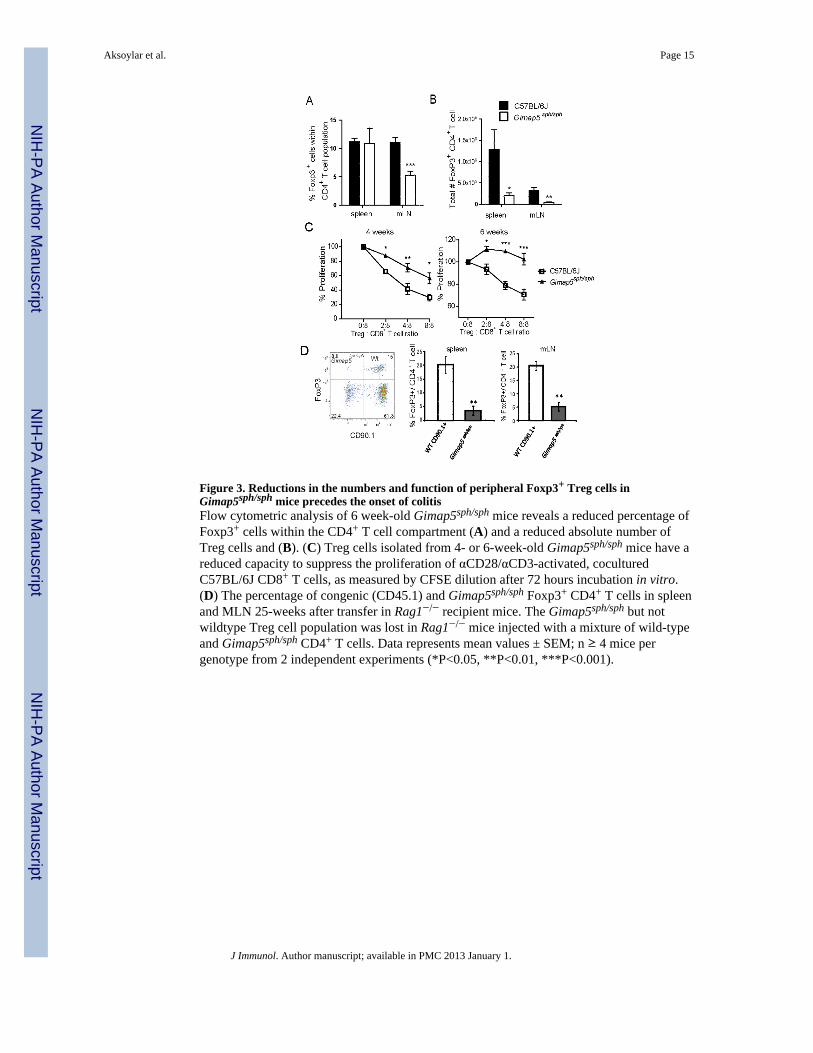
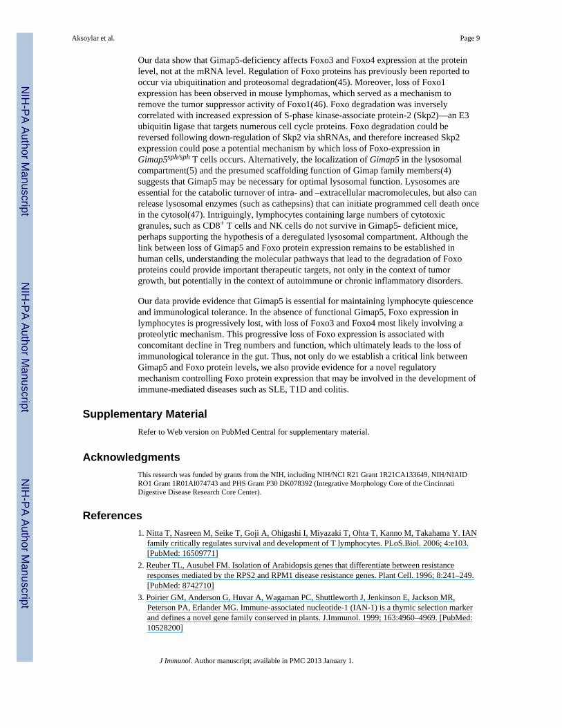
Figure 3. Reductions in the numbers and function of peripheral Foxp3+ Treg cells inGimap5sph/sph mice precedes the onset of colitisFlow cytometric analysis of 6 week-old Gimap5sph/sph mice reveals a reduced percentage ofFoxp3+ cells within the CD4+ T cell compartment (A) and a reduced absolute number ofTreg cells and (B). (C) Treg cells isolated from 4- or 6-week-old Gimap5sph/sph mice have areduced capacity to suppress the proliferation of αCD28/αCD3-activated, coculturedC57BL/6J CD8+ T cells, as measured by CFSE dilution after 72 hours incubation in vitro.(D) The percentage of congenic (CD45.1) and Gimap5sph/sph Foxp3+ CD4+ T cells in spleenand MLN 25-weeks after transfer in Rag1−/− recipient mice. The Gimap5sph/sph but notwildtype Treg cell population was lost in Rag1−/− mice injected with a mixture of wild-typeand Gimap5sph/sph CD4+ T cells. Data represents mean values ± SEM; n ≥ 4 mice pergenotype from 2 independent experiments (*P<0.05, **P<0.01, ***P<0.001).
Aksoylar et al. Page 15
J Immunol. Author manuscript; available in PMC 2013 January 1.
NIH
-PA Author Manuscript
NIH
-PA Author Manuscript
NIH
-PA Author Manuscript
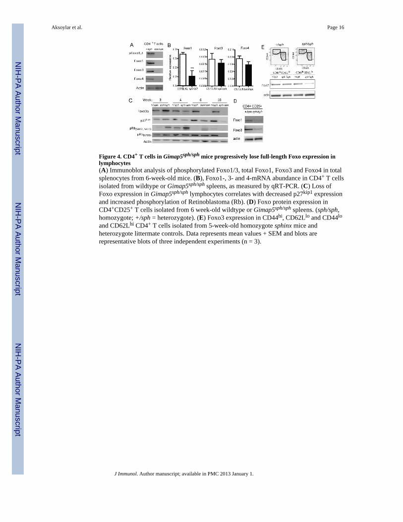
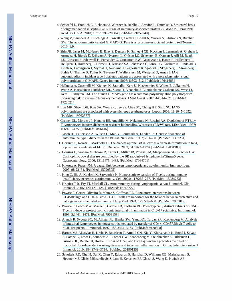
Figure 4. CD4+ T cells in Gimap5sph/sph mice progressively lose full-length Foxo expression inlymphocytes(A) Immunoblot analysis of phosphorylated Foxo1/3, total Foxo1, Foxo3 and Foxo4 in totalsplenocytes from 6-week-old mice. (B), Foxo1-, 3- and 4-mRNA abundance in CD4+ T cellsisolated from wildtype or Gimap5sph/sph spleens, as measured by qRT-PCR. (C) Loss ofFoxo expression in Gimap5sph/sph lymphocytes correlates with decreased p27kip1 expressionand increased phosphorylation of Retinoblastoma (Rb). (D) Foxo protein expression inCD4+CD25+ T cells isolated from 6 week-old wildtype or Gimap5sph/sph spleens. (sph/sph,homozygote; +/sph = heterozygote). (E) Foxo3 expression in CD44hi, CD62Llo and CD44lo
and CD62Lhi CD4+ T cells isolated from 5-week-old homozygote sphinx mice andheterozygote littermate controls. Data represents mean values + SEM and blots arerepresentative blots of three independent experiments (n = 3).
Aksoylar et al. Page 16
J Immunol. Author manuscript; available in PMC 2013 January 1.
NIH
-PA Author Manuscript
NIH
-PA Author Manuscript
NIH
-PA Author Manuscript
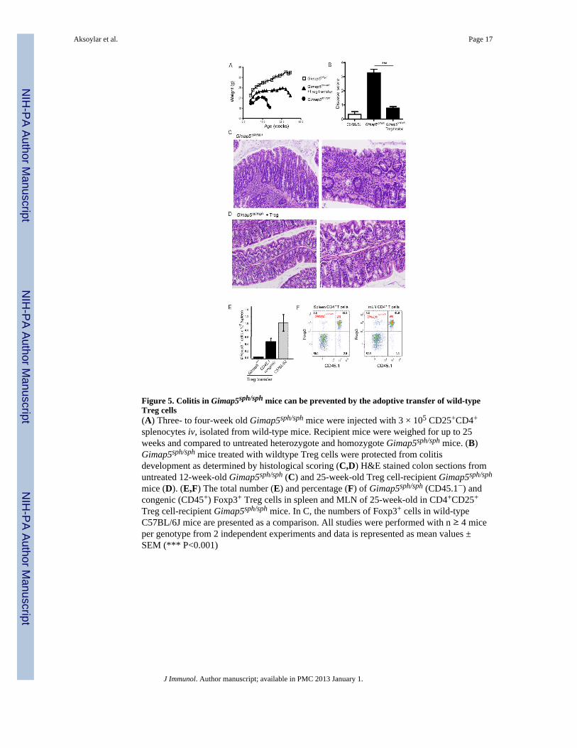
Figure 5. Colitis in Gimap5sph/sph mice can be prevented by the adoptive transfer of wild-typeTreg cells(A) Three- to four-week old Gimap5sph/sph mice were injected with 3 × 10⁵ CD25+CD4+
splenocytes iv, isolated from wild-type mice. Recipient mice were weighed for up to 25weeks and compared to untreated heterozygote and homozygote Gimap5sph/sph mice. (B)Gimap5sph/sph mice treated with wildtype Treg cells were protected from colitisdevelopment as determined by histological scoring (C,D) H&E stained colon sections fromuntreated 12-week-old Gimap5sph/sph (C) and 25-week-old Treg cell-recipient Gimap5sph/sph
mice (D). (E,F) The total number (E) and percentage (F) of Gimap5sph/sph (CD45.1−) andcongenic (CD45+) Foxp3+ Treg cells in spleen and MLN of 25-week-old in CD4+CD25+
Treg cell-recipient Gimap5sph/sph mice. In C, the numbers of Foxp3+ cells in wild-typeC57BL/6J mice are presented as a comparison. All studies were performed with n ≥ 4 miceper genotype from 2 independent experiments and data is represented as mean values ±SEM (*** P<0.001)
Aksoylar et al. Page 17
J Immunol. Author manuscript; available in PMC 2013 January 1.
NIH
-PA Author Manuscript
NIH
-PA Author Manuscript
NIH
-PA Author Manuscript
Top Related
Copyright © 2022 FDOKUMEN

