


Bahasa
Halaman
Hukum

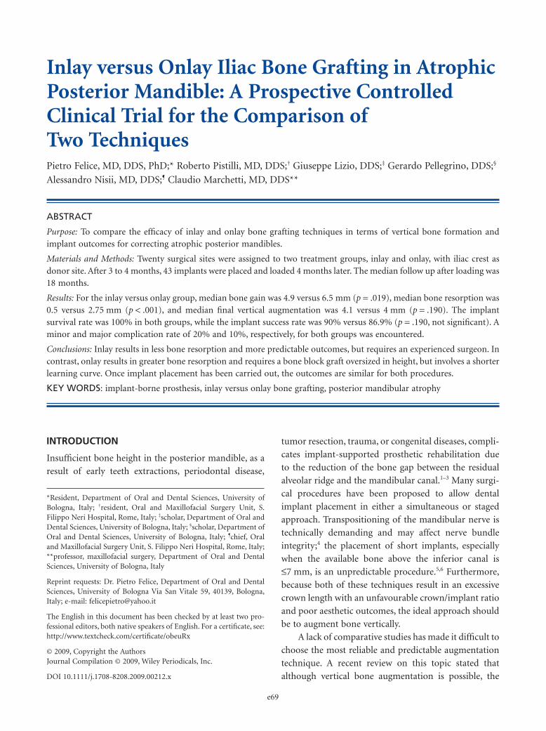
Inlay versus Onlay Iliac Bone Grafting in AtrophicPosterior Mandible: A Prospective ControlledClinical Trial for the Comparison ofTwo Techniquescid_212 69..82
Pietro Felice, MD, DDS, PhD;* Roberto Pistilli, MD, DDS;† Giuseppe Lizio, DDS;‡ Gerardo Pellegrino, DDS;§
Alessandro Nisii, MD, DDS;¶ Claudio Marchetti, MD, DDS**
ABSTRACT
Purpose: To compare the efficacy of inlay and onlay bone grafting techniques in terms of vertical bone formation andimplant outcomes for correcting atrophic posterior mandibles.
Materials and Methods: Twenty surgical sites were assigned to two treatment groups, inlay and onlay, with iliac crest asdonor site. After 3 to 4 months, 43 implants were placed and loaded 4 months later. The median follow up after loading was18 months.
Results: For the inlay versus onlay group, median bone gain was 4.9 versus 6.5 mm (p = .019), median bone resorption was0.5 versus 2.75 mm (p < .001), and median final vertical augmentation was 4.1 versus 4 mm (p = .190). The implantsurvival rate was 100% in both groups, while the implant success rate was 90% versus 86.9% (p = .190, not significant). Aminor and major complication rate of 20% and 10%, respectively, for both groups was encountered.
Conclusions: Inlay results in less bone resorption and more predictable outcomes, but requires an experienced surgeon. Incontrast, onlay results in greater bone resorption and requires a bone block graft oversized in height, but involves a shorterlearning curve. Once implant placement has been carried out, the outcomes are similar for both procedures.
KEY WORDS: implant-borne prosthesis, inlay versus onlay bone grafting, posterior mandibular atrophy
INTRODUCTION
Insufficient bone height in the posterior mandible, as a
result of early teeth extractions, periodontal disease,
tumor resection, trauma, or congenital diseases, compli-
cates implant-supported prosthetic rehabilitation due
to the reduction of the bone gap between the residual
alveolar ridge and the mandibular canal.1–3 Many surgi-
cal procedures have been proposed to allow dental
implant placement in either a simultaneous or staged
approach. Transpositioning of the mandibular nerve is
technically demanding and may affect nerve bundle
integrity;4 the placement of short implants, especially
when the available bone above the inferior canal is
27 mm, is an unpredictable procedure.5,6 Furthermore,
because both of these techniques result in an excessive
crown length with an unfavourable crown/implant ratio
and poor aesthetic outcomes, the ideal approach should
be to augment bone vertically.
A lack of comparative studies has made it difficult to
choose the most reliable and predictable augmentation
technique. A recent review on this topic stated that
although vertical bone augmentation is possible, the
*Resident, Department of Oral and Dental Sciences, University ofBologna, Italy; †resident, Oral and Maxillofacial Surgery Unit, S.Filippo Neri Hospital, Rome, Italy; ‡scholar, Department of Oral andDental Sciences, University of Bologna, Italy; §scholar, Department ofOral and Dental Sciences, University of Bologna, Italy; ¶chief, Oraland Maxillofacial Surgery Unit, S. Filippo Neri Hospital, Rome, Italy;**professor, maxillofacial surgery, Department of Oral and DentalSciences, University of Bologna, Italy
Reprint requests: Dr. Pietro Felice, Department of Oral and DentalSciences, University of Bologna Via San Vitale 59, 40139, Bologna,Italy; e-mail: [email protected]
The English in this document has been checked by at least two pro-fessional editors, both native speakers of English. For a certificate, see:http://www.textcheck.com/certificate/obeuRx
© 2009, Copyright the AuthorsJournal Compilation © 2009, Wiley Periodicals, Inc.
DOI 10.1111/j.1708-8208.2009.00212.x
e69
associated number of complications and failures with
the various described techniques remains unacceptably
high (>20%).6 In particular, insufficient data are avail-
able regarding vertical bone gain and stability over time
in atrophic posterior mandibles.
The inlay technique shows great potential for bone
graft incorporation, with a low resorption level and high
implant survival and success rates,7–9 and recent studies
into this technique have reported good outcomes in
atrophic posterior mandibles.1,10–13 However, this tech-
nique is not simple to perform and requires at least 4 to
6 mm of residual bone above the mandibular canal.11,12
Onlay grafting has been used successfully in the
correction of vertically deficient edentulous ridges,2,14
although the reported two-stage approach results in
considerable resorption of the bone graft before implant
insertion.2,14,15
The major difficulty in vertical reconstruction of
the posterior mandible is the management of soft
tissues, which have a high risk of dehiscence and subse-
quent infection and necrosis of the graft.
The use of an autogenous bone block harvested
from extra-oral donor sites has been reported to be
effective,16–18 and iliac bone grafts in particular have
been used successfully with inlay procedures in the pos-
terior mandible.1,12,13,19
The aim of this study was to compare the effective-
ness of the inlay and onlay techniques in terms of bone
gain, bone resorption, final vertical augmentation, peri-
implant bone resorption, implant survival, implant
success, and complication rate in implant-borne pros-
thetic rehabilitation of atrophic posterior mandibles.
PATIENTS AND METHODS
A total of 20 systemically healthy patients (six males and
14 females) between 30 and 75 years of age (mean,
53.9 years) with resorbed partially edentulous posterior
mandibles were recruited between 2003 and 2006. These
patients required correction of a vertical bone deficit to
provide adequate bone support for implant-borne pros-
thetic rehabilitation. The median value for preoperative
bone height over the mandibular canal was 7.5 mm
(range, 4.8–10.7 mm). All of the surgical sites were
affected by early tooth loss atrophy.
The inclusion criteria were: (1) age from 20 to 80
years; (2) desire for implant-borne fixed prosthesis reha-
bilitation; and (3) a minimum native bone height above
the mandibular canal of 4 to 5 mm (as determined via
computed tomography [CT]). The exclusion criteria
were: (1) reduced thickness (25 mm) of the edentulous
ridge; (2) any general contraindication to implant
surgery; (3) chemotherapy, irradiation, immunosup-
pressive therapy, or aminobisphosphonate treatment
in the previous 5 years; (4) pregnancy or lactation;
(5) uncontrolled diabetes; (6) previously subjected to
reconstructive procedures of the posterior mandible; (7)
active periodontal disease involving the residual denti-
tion; (8) mucosal disease such as lichen planus in the
areas to be treated; (9) poor oral hygiene; and (10)
noncompliance.
These 20 patients were randomly assigned to two
different groups: 10 patients (four males and six
females) with 10 monolateral surgical sites underwent
an interpositional bone graft procedure (the inlay
group), and the remaining 10 patients (two males and
eight females) with 10 monolateral surgical sites under-
went an appositional bone graft procedure (the onlay
group) for a total of 20 surgical sites. The surgeons were
blind to the group assignment until the surgical proce-
dure. The mean age of the patients in the inlay group
was 55.2 years (range, 30–75 years), whereas the mean
age of the onlay group was 52.6 years (range, 39–63
years).
Clinical records, casts, and radiographic evaluations
(periapical X-ray, orthopantomograms [OPT], and CT)
of the patients were assessed. Two experienced surgeons
performed all reconstructive and implant placement
procedures. Each patient provided written informed
consent for the treatment.
Operative Procedure for the Inlay Group
The mandible was augmented under general anesthesia
by interposing a monocortical iliac crest bone graft har-
vested from the medial surface of the anterior ilium. All
patients received prophylactic antibiotic therapy. Ceftri-
axone (Ceftriaxon, Tyrol Pharma, Bordon, UK) was
administered intravenously upon anesthetic induction
at a loading dose of 2 g, and local anesthesia was induced
with articain 4% and adrenaline 1:100,000.
After making a full-thickness vestibular incision in
the lower vestibule while avoiding the emergence of the
mental nerve, the subperiosteal tissue was carefully dis-
sected to obtain adequate visibility of the underlying
bone, with no tension on the ipsilateral mental nerve.
Mucoperiosteal dissection was not performed toward
the alveolar crest or on the lingual side to preserve an
e70 Clinical Implant Dentistry and Related Research, Volume 11, Supplement 1, 2009
adequate blood supply to the bone segment to be
osteotomized. Horizontal osteotomy and two oblique
cuts were made using a reciprocal saw and by piezosur-
gery (Mectron Piezosurgery Device™; Mectron,
Carasco, Italy) to obtain a cleaner osteotomy line, with
minor risk of transport segment fracture. The horizon-
tal osteotomy was 2 to 4 mm above the mandibular
canal; the mesial oblique cut was 2 mm distal to the last
tooth in the arch, and the distal oblique cut was relative
to the implant–graft treatment plane. The height of the
transport segment was at least 3 mm to allow for the
insertion of the stabilizing screws without fracturing.
The osteotomized segment was then raised in the
coronal direction, sparing the lingual periosteum.
At this point, the autogenous bone block was har-
vested from the iliac crest in a conventional manner.20
The iliac block was modeled to be properly fitted in the
recipient site, interposed between the raised fragment
and the mandibular basal bone (Figure 1), and fixed
with titanium miniplates and miniscrews (Gebrüder
Martin and Co., Tuttlingen, Germany) to both the basal
bone and the transport fragment (Figure 2). Gaps
between the graft and the recipient site were filled with
particulated iliac bone. The grafted areas were covered
with a resorbable barrier (Bio-Guide®; Geistlich
Pharma, Wolhusen, Switzerland). After releasing the ves-
tibular periosteum, the flap was closed with 4.0 vicryl
sutures (Ethicon FS-2, St-Stevens-Woluwe, Belgium).
The postsurgical therapy protocol consisted of
Ceftriaxone at a dose of 2 g/day, for 10 days after surgery,
together with a nonsteroidal analgesic drug (ketoprofen,
Orudis; Aventis Pharma, Bridgewater, UK) at a dose of
200 mg twice daily for 3 days and thereafter as required.
Cortisone (betametason; 4 mg) was administered twice
daily for 2 days and once a day on day 3. A soft diet and
appropriate oral hygiene were prescribed for 2 weeks,
including twice daily rinsing with 0.2% chlorhexidine
mouthwash and the application of 1% chlorhexidine
gel (Corsodyl gel; GlaxoSmithKline, Middlesex, UK).
Patients were instructed not to wear removable prosthe-
ses for 30 days postsurgery, and to avoid brushing or
otherwise traumatizing the surgical site.
Sutures in the oral cavity were removed 10 days
after the procedure, whereas the iliac crest sutures were
removed after 7 days. Additional postoperative check-
ups were carried out at 3 and 6 weeks and 3 months after
surgery.
After a 3- to 4-month waiting period, the aug-
mented site was exposed after elevation of a mucoperi-
osteal flap. The miniplates were removed (Figure 3), and
two endosseous implants were inserted under local
anesthesia in each treated site. The implant features
(type, diameter, and length) were decided according to
the anatomical situation. Twenty titanium screw-shaped
endosseous implants were positioned in the crestal aug-
mented region; nine patients received 18 Biomet 3i
implants (Palm Beach, FL, USA) and one patient
received two XiVe implants (Friadent-Dentsply, Man-
nheim, Germany; Figure 4). The fixtures were allowed to
heal for 4 months for osseointegration before prosthetic
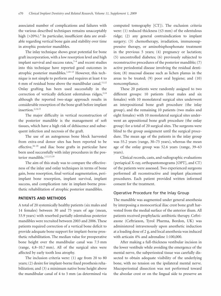
Figure 1 Inlay procedure: after vestibular periosteum elevationand exposure of the buccal surface of the mandibular bone, theiliac block is interposed between the raised fragment and themandibular basal bone.
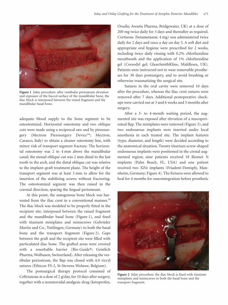
Figure 2 Inlay procedure: the iliac block is fixed with titaniumminiplates and miniscrews to both the basal bone and thetransport fragment.
Inlay and Onlay Grafting for the Treatment of Atrophic Posterior Mandibles e71
rehabilitation was initiated. Two grams of amoxicillin
were administered preoperatively and then 1 g twice a
day for 5 days. Ibuprofen 600 mg was prescribed to be
taken as required. The implants were uncovered 4
months later; abutments were placed, and a screw-
retained, acrylic-resin, temporary fixed prosthesis was
affixed for 4 to 5 months until the insertion of a screw-
retained definitive prosthesis. Patients were enrolled in
an oral hygiene program with evaluations every 3 to 4
months until the end of the follow-up period (Figure 5).
Table 1 reports in details the radiographic, clinical, and
treatment data for the inlay group.
Operative Procedure for the Onlay GroupPatients
The mandible was augmented under general anesthesia
by placing a monocortical iliac crest bone graft, which
was harvested from the medial surface of the anterior
ilium, on the ridge. The onlay group was administered
the same prophylactic antibiotic therapy as the inlay
group, and local anesthesia was induced with articain
4% and adrenaline 1:100,000.
After making a full-thickness crestal incision con-
tinuing into the adherent gingiva of the mesial teeth
without involving their periodontal attachment, the
subperiosteal tissue was carefully dissected to obtain
adequate visibility of the underlying bone without
applying tension to the ipsilateral mental nerve. Releas-
ing incisions were performed when required to improve
the mobility of the buccal flap. The bone recipient site
was flattened slightly to eliminate asperities and perfo-
rated with a 1-mm round bur under copious saline irri-
gation to increase the blood supply from endosseous
vessels. The autogenous bone block was harvested from
the iliac crest in a conventional manner.15 The iliac block
was modeled to be properly fitted in the recipient
site and rigidly fixed upon the mandibular ridge with
1.5-mm-diameter titanium miniscrews (Gebrüder
Martin and Co.; Figure 6). Gaps between the graft and
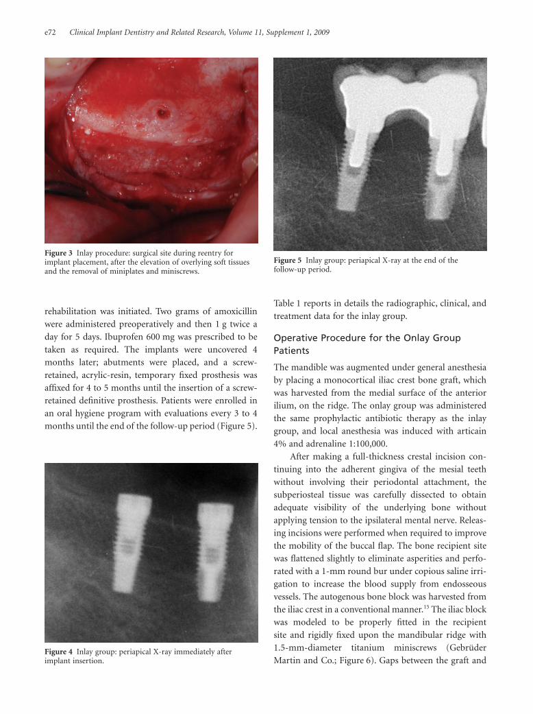
Figure 3 Inlay procedure: surgical site during reentry forimplant placement, after the elevation of overlying soft tissuesand the removal of miniplates and miniscrews.
Figure 4 Inlay group: periapical X-ray immediately afterimplant insertion.
Figure 5 Inlay group: periapical X-ray at the end of thefollow-up period.
e72 Clinical Implant Dentistry and Related Research, Volume 11, Supplement 1, 2009

the recipient site were filled with particulated iliac bone.
The grafted areas were covered with a resorbable barrier
(Bio-Guide®; Geistlich Pharma). After releasing the ves-
tibular periosteum, the flap was closed with 4.0 vicryl
sutures (Ethicon FS-2).
The postsurgical therapy protocol and postopera-
tive care instructions was identical as for the inlay group
such as the sutures removal and the postoperative check-
ups timing.
After a 3- to 4-month waiting period, the aug-
mented site was exposed after the elevation of a muco-
periosteal flap (Figures 7 and 8). The miniscrews were
removed, and two or three endosseous implants were
inserted under local anesthesia in each treated site.
The implant features (type, diameter, and length) were
decided according to the anatomical situation. Twenty-
three titanium screw-shaped endosseous implants were
positioned in the crestal augmented region (Figure 9).
Seven patients received 17 Astra implants (Astra Tech
AB, Mölndal, Sweden), two patients received four Biolok
implants (Biolok, Deerfield, FL, USA), and one patient
received two Alpha Bio implants (Alpha-Bio Tec Ltd.,
Petak-Tikva, Israel). The same antibiotic and analgesic
as the inlay group was prescribed. Four months later, the
implants were uncovered. Abutments were placed, and a
screw-retained, acrylic-resin, temporary fixed prosthesis
was placed for 4 to 5 months until the insertion of a
screw-retained definitive prosthesis. The patients were
TABLE 1 Inlay Group: Radiographic, Clinical, and Treatment Data
Patientno. Sex
Age(years)
Starting boneheight above
mandibular canal (mm)Treated
siteImplant
no.Implant location
and dimension (mm)Follow-up after
loading (months)
#1 F 67 5.7 left 2 36 = 4 ¥ 10 18
37 = 4 ¥ 10
#2 F 46 4.8 left 2 35 = 3.8 ¥ 8.75 18
37 = 3.8 ¥ 9.5
#3 F 50 8.6 right 2 46 = 4 ¥ 10 17
47 = 4 ¥ 11.5
#4 F 30 6.2 right 2 45 = 4 ¥ 10 20
46 = 4 ¥ 11.5
#5 M 69 7.6 left 2 35 = 4 ¥ 10 22
36 = 4 ¥ 10
#6 F 59 5.6 right 2 46 = 4 ¥ 10 18
47 = 4 ¥ 10
#7 M 61 8.5 left 2 36 = 4 ¥ 10 17
37 = 4 ¥ 11.5
#8 F 75 8.2 right 2 45 = 4 ¥ 10 19
47 = 4 ¥ 10
#9 M 43 8.5 right 2 46 = 4 ¥ 11.5 19
47 = 4 ¥ 11.5
#10 M 52 6.3 left 2 36 = 4 ¥ 10 17
37 = 4 ¥ 10
Figure 6 Onlay procedure: the iliac block superimposed on theresidual ridge and fixed with titanium miniscrews.
Inlay and Onlay Grafting for the Treatment of Atrophic Posterior Mandibles e73
enrolled in an oral hygiene program with evaluations
every 3 to 4 months until the end of the follow-up
period (Figure 10). Table 2 reports in details the radio-
graphic, clinical, and treatment data for the onlay group.
Assessment Timing
Every patient underwent a clinical examination 1 week
after surgery, twice in the first month, and monthly
in the subsequent months before implantation. The
patients were then evaluated in the first week and twice
monthly for the subsequent 4 months after implant
placement, and monthly after prosthetic loading until
the end of the follow-up period.
A subjective evaluation of neurosensory function
was carried out at each clinical check-up by asking the
patient if there were any areas of hypoesthesia, numb-
ness, tingling, or pain in the lip or chin region. The time
required to recover full lip and chin region sensitivity
after the augmentation procedures was recorded.
The following assessment surveys (clinical and
X-ray evaluations) were carried out for both groups:
• T0 = Preoperative phase: clinical records, casts,
OPT, and CT.
• T1 = Postoperative phase: OPT and CT.
� T1a = Immediately after surgery.
� T1b = Surgical reentry (3–4 months postopera-
tively), just before miniplate removal and
implant insertion.
• T2 = Immediately after implant insertion: periapi-
cal X-rays.
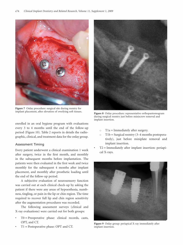
Figure 7 Onlay procedure: surgical site during reentry forimplant placement, after elevation of overlying soft tissues.
Figure 8 Onlay procedure: representative orthopantomogramduring surgical reentry just before miniscrew removal andimplant insertion.
Figure 9 Onlay group: periapical X-ray immediately afterimplant insertion.
e74 Clinical Implant Dentistry and Related Research, Volume 11, Supplement 1, 2009

• T3 = Prosthetic loading.
• T4 = End of the follow-up time (median, 18
months; range, 13–22 months).
Radiographic Parameters Evaluated
A qualified examiner performed all radiographic assess-
ments. The paraxial 1-mm-thick CT slices obtained at 6,
12, and 18 mm posterior to the mental foramina were
assessed for pre-implant placement phase investigation;
for all parameters taken into consideration, except ver-
tical resorption of the inlay graft, the distance from the
most coronal point of the mandibular canal to the inter-
mediate point of the crestal ridge was measured using
Autocad-Autodesk® Software (San Rafael, CA, USA)
and a mean value was considered. First, starting bone
height above the mandibular canal was assessed at T0 via
CT in both groups. Vertical bone gain was assessed by
comparing T0 and the immediate postoperative time
(T1a) via CT in both groups. Vertical bone resorption of
the augmented ridge was assessed by comparing T1a and
Figure 10 Onlay group: periapical X-ray at the end of thefollow-up period.
TABLE 2 Onlay Group: Radiographic, Clinical, and Treatment Data
Patientno. Sex
Age(years)
Starting boneheight above
mandibular canal (mm)Treated
siteImplant
no.Implant location
and dimension (mm)Follow-up after
loading (months)
#1 F 63 10.7 left 3 35 = 4.5 ¥ 13 22
36 = 4.5 ¥ 13
37 = 4.5 ¥ 13
#2 F 55 9.1 right 2 46 = 4 ¥ 10 18
47 = 5 ¥ 10
#3 F 55 6.8 left 3 34 = 4 ¥ 9 17
35 = 4 ¥ 8
36 = 4 ¥ 8
#4 F 58 7.8 right 2 46 = 4 ¥ 10 20
47 = 4 ¥ 10
#5 M 39 8.1 right 2 46 = 4 ¥ 11 14
47 = 5 ¥ 11
#6 F 40 8.1 left 2 36 = 4 ¥ 8 14
37 = 4 ¥ 9
#7 F 54 4.9 right 2 44 = 3.75 ¥ 10 15
47 = 5 ¥ 8
#8 F 54 5.6 left 3 34 = 3.5 ¥ 9 13
35 = 3.5 ¥ 10
37 = 4 ¥ 9
#9 F 57 7.5 left 2 34 = 4 ¥ 9 22
35 = 4 ¥ 13
#10 M 52 5.4 left 2 36 = 4 ¥ 10 20
37 = 4 ¥ 9
Inlay and Onlay Grafting for the Treatment of Atrophic Posterior Mandibles e75
T1b CT evaluations for both groups. This measurement
assessed the resorption of the osteotomized fragment in
the inlay group and the resorption of the graft in the
onlay group.
Final vertical augmentation was assessed by sub-
tracting bone resorption of augmented ridge values
from vertical bone gain values for each group; this com-
parison was required to evaluate the real effectiveness of
the two techniques in augmenting bone. Vertical resorp-
tion of the graft interposed between the transport
segment and the basal bone in the inlay group was
assessed by comparing T1a and T1b CT evaluations.
This parameter was investigated, measuring the distance
from the intermediate point of the upper border to the
intermediate point of the lower border of the graft.
Peri-implant bone resorption was assessed according
to the criteria of Albrektsson and colleagues21 by com-
paring periapical radiographs made perpendicular to
the long-axis of the implants, using conventional film
holders. Radiographs were taken at T2, T3, and T4; the
change in bone level was evaluated mesial and distal to
each implant using a transparent ruler to measure the
distance in millimeters between the top of the implant
head shoulder and the most coronal point of the direct
bone-to-implant contact. The bone level measured peri-
apically immediately after implant placement was
considered the baseline for further measurements. The
measurements were recorded to the nearest 0.5 mm.
Implant Success and Survival Rates
We evaluated implant survival and success according to
the criteria of Albrektsson and colleagues.21
Complication Assessment and SensitivityAlteration
Complications were assessed and classified as “minor” or
“major,” based on the criteria established by Enislidis
and colleagues22 and considering the treatment phase in
which they occurred.
Changes in the sensitivity of the area innervated by
the inferior alveolar nerve were considered separately.
A subjective evaluation was carried out by asking the
patient if there were any areas of hypoesthesia, numb-
ness, tingling, or pain in the lip or chin region.
Statistical Analysis
Data were analyzed using non-parametric statistical
methods. The two-tailed Mann–Whitney U-test and
chi-square test were used to detect significant differences
between groups (inlay vs onlay). Friedman’s test (non-
parametric analysis of variance) was used to detect dif-
ferences throughout the three time intervals within the
two groups (inlay and onlay).
RESULTS
Inlay Group Patients
The median value for starting bone height above the
mandibular canal were 6.9 mm (range, 4.8–8.6 mm),
with a residual ridge shape of IV/V according to the
Cawood/Howell classification.23 The median vertical
bone gain from T0 to T1a was 4.9 mm (range, 4–7 mm),
with a median bone resorption value of 0.5 mm (range,
0.10–2.9 mm) at T1b corresponding to a median bone
gain of 10.2% (range, 2.2–51.8%). The median final ver-
tical augmentation was 4.1 mm (range, 2.7–6.3 mm).
Vertical bone resorption of the graft was insignificant,
ranging from 0 to 0.1 mm.
Recovery of the surgical site was uneventful in all
but three patients. Two weeks after surgery, patients 4
and 8 developed buccal dehiscence at the treated site;
dehiscence was <1 cm in diameter and resulted in partial
exposure of the titanium miniplate and screw and signs
of inflammation. These complications were addressed
by local debridement and increased use of clorhexidine
for the entire healing period; the soft tissues closed
progressively until the implant placement phase.
Dehiscence was considered to be a minor complication,
yielding a 20% minor complication rate. However,
patient 2 developed a buccal dehiscence that was >2 cm
in the augmented site in the first postsurgical week,
resulting in greater exposure of the titanium miniplate,
inflammation, and resorption of the cranial segment;
this situation required surgical reentry 10 days after
the initial surgery to remove inflammatory tissues and
mobilize a new mucoperiosteal flap to obtain complete
closure of the bone graft. Considerable bone loss of the
cranial segment (51.8% at the time of implant place-
ment) necessitated the use of shorter implants to com-
plete the treatment. This problem was considered to be a
major complication because it resulted in the modifica-
tion of the original treatment plan; therefore, a 10%
major complication rate was obtained.
Six patients did not report any subjective sign of
impaired lip or chin sensation after the augmenta-
tion procedure. Four patients reported hypoesthesia
e76 Clinical Implant Dentistry and Related Research, Volume 11, Supplement 1, 2009

immediately after the augmentation procedure in their
ipsilateral inferior lip and chin region, and two of these
patients had progressively increasing numbness and tin-
gling. Altered sensitivity persisted for 2 weeks in three of
the four patients and 3 weeks in the remaining patient.
Recovery of the graft donor sites was uneventful in
all cases, with no complications. At the end of the
follow-up period, the cumulative implant survival rate
was 100%. Two of the 20 implants exhibited peri-
implant bone resorption >1.5 mm in the first year after
prosthetic loading, yielding a cumulative implant
success rate of 90%. Complications between implant
placement and implant loading occurred in only one
patient (patient 1, implant #36), in whom peri-
implantitis was observed and resolved with local debri-
dement before loading.
The median cumulative peri-implant bone resorp-
tion was 0.9 mm (range, 0.3–1.8 mm). The median
follow-up periods from the start of prosthetic loading
were 18 months (range, 17–22 months). All patients
showed acceptable function of the implant-borne pros-
thesis, and no complication occurred during implant
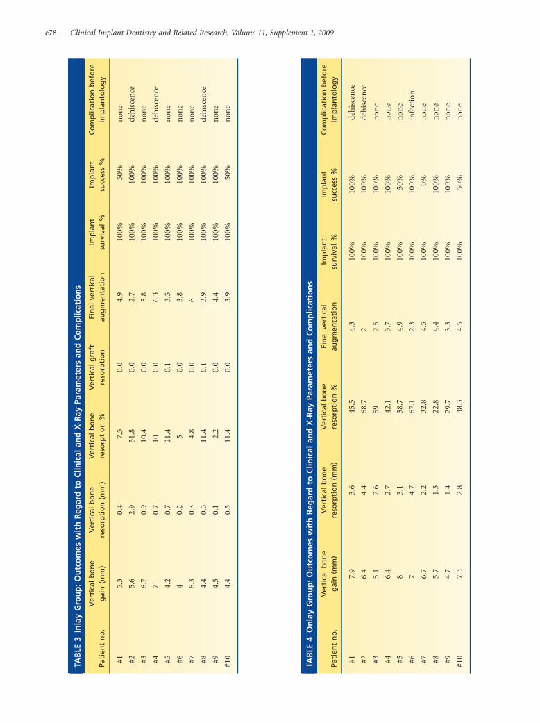
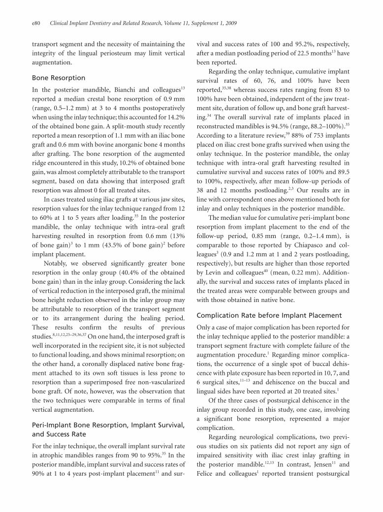
loading to the end of the follow-up period. Table 3
reports in details the clinical and radiographic outcomes
of the inlay group.
Onlay Group Patients
The median value for starting bone height above the
mandibular canal was 7.6 mm (range, 4.9–10.7 mm),
with a residual ridge shape of IV/V according to the
Cawood/Howell classification.23
The median value for vertical bone gain from T0 to
T1a was 6.5 mm (range, 4.7–8 mm), with a median
vertical bone resorption value of 2.7 mm (range, 1.3–
4.7 mm), at 4 months postoperatively (i.e., immediately
before implant placement). This corresponded to a
median value of 40.4% bone gain (range, 22.8–68.7%).
The median final vertical augmentation was 4 mm
(range, 2–4.9 mm).
Recovery of the surgical site was uneventful except
in three patients. Patients 1 and 2 developed a dehis-
cence, resulting in partial exposure of the graft at 13 and
21 days, respectively, after the reconstruction. In patient
2, this problem required surgical reentry to remove
inflammatory tissue, mobilize a new mucoperiosteal
flap, and completely cover the bone graft. In patient 1,
the exposed bone was ground with a round diamond
bur, and the site healed completely after 20 days of
increased use of chlorhexidine. These complications
were considered minor, yielding a 20% minor compli-
cation rate. Patient 6 reported an inflammatory acute
process involving the distal fixation screw in the second
week after augmentation. This patient was reoperated
with local anesthesia for the removal of screw and
inflammatory tissues. Considerable loss of the grafted
bone (68.7% at the time of implant placement) necessi-
tated the use of shorter implants to complete the treat-
ment. This was considered to be a major complication
because it modified the original treatment plan, produc-
ing a 10% major complication rate.
Eight patients did not report subjective signs of
impaired lip or chin sensation after the augmentation
procedure; however, patients 8 and 9 reported hypoes-
thesia immediately after the augmentation procedure in
the ipsilateral lip and chin region that was characterized
by progressively increasing numbness and tingling.
These sensitivity alterations persisted for 6 months after
the augmentation procedure in patient 8 and lasted until
the end of the follow-up period in patient 9.
The recovery of the graft donor sites was uneventful
in all cases and, at the end of the follow-up period, the
cumulative implant survival rate was 100%. Three of
the twenty-three implants exhibited peri-implant bone
resorption >1.5 mm in the first year after prosthetic
loading; therefore, the cumulative implant success rate
was 86.9%. Only patient 8 (implant #37) showed com-
plications between implant placement and implant
loading. In this patient, peri-implantitis was observed
and resolved with local debridement before loading.
The median cumulative peri-implant bone resorp-
tion was 0.85 mm (range, 0.2–2.8 mm), and the median
follow-up periods from the start of prosthetic loading
was 17.5 months (range, 13–22 months). All patients
showed acceptable function of the implant-borne pros-
thesis, and no complication occurred during the period
between implant loading and the end of follow up.
Table 4 reports in details the clinical and radiographic
outcomes of the onlay group.
The overall median follow-up period from the start
of prosthetic loading was 18 months (range, 13–22
months). No significant difference in the starting
bone height was observed between groups (p = .910);
however, the onlay group showed greater vertical bone
gain (p = .019) and greater vertical bone resorption
(p < .001). In terms of final vertical augmentation (sub-
tracting bone resorption from bone gain values for each
Inlay and Onlay Grafting for the Treatment of Atrophic Posterior Mandibles e77
TAB
LE3
Inla
yG
rou
p:
Ou
tco
mes
wit
hR
egar
dto
Clin
ical
and
X-R
ayPa
ram
eter
san
dC
om
plic
atio
ns
Pati
ent
no
.V
erti
cal
bo
ne
gai
n(m
m)
Ver
tica
lb
on
ere
sorp
tio
n(m
m)
Ver
tica
lb
on
ere
sorp
tio
n%
Ver
tica
lg
raft
reso
rpti
on
Fin
alve
rtic
alau
gm
enta
tio
nIm
pla
nt
surv
ival
%Im
pla
nt
succ
ess
%C
om
plic
atio
nb
efo
reim
pla
nto
log
y
#15.
30.
47.
50.
04.
910
0%50
%n
one
#25.
62.
951
.80.
02.
710
0%10
0%de
his
cen
ce
#36.
70.
910
.40.
05.
810
0%10
0%n
one
#47
0.7
100.
06.
310
0%10
0%de
his
cen
ce
#54.
20.
721
.40.
13.
510
0%10
0%n
one
#64
0.2
50.
03.
810
0%10
0%n
one
#76.
30.
34.
80.
06
100%
100%
non
e
#84.
40.
511
.40.
13.
910
0%10
0%de
his
cen
ce
#94.
50.
12.
20.
04.
410
0%10
0%n
one
#10
4.4
0.5
11.4
0.0
3.9
100%
50%
non
e
TAB
LE4
On
lay
Gro
up
:O
utc
om
esw
ith
Reg
ard
toC
linic
alan
dX
-Ray
Para
met
ers
and
Co
mp
licat
ion
s
Pati
ent
no
.V
erti
cal
bo
ne
gai
n(m
m)
Ver
tica
lb
on
ere
sorp
tio
n(m
m)
Ver
tica
lb
on
ere
sorp
tio
n%
Fin
alve
rtic
alau
gm
enta
tio
nIm
pla
nt
surv
ival
%Im
pla
nt
succ
ess
%C
om
plic
atio
nb
efo
reim
pla
nto
log
y
#17.
93.
645
.54.
310
0%10
0%de
his
cen
ce
#26.
44.
468
.72
100%
100%
deh
isce
nce
#35.
12.
659
2.5
100%
100%
non
e
#46.
42.
742
.13.
710
0%10
0%n
one
#58
3.1
38.7
4.9
100%
50%
non
e
#67
4.7
67.1
2.3
100%
100%
infe
ctio
n
#76.
72.
232
.84.
510
0%0%
non
e
#85.
71.
322
.84.
410
0%10
0%n
one
#94.
71.
429
.73.
310
0%10
0%n
one
#10
7.3
2.8
38.3
4.5
100%
50%
non
e
e78 Clinical Implant Dentistry and Related Research, Volume 11, Supplement 1, 2009
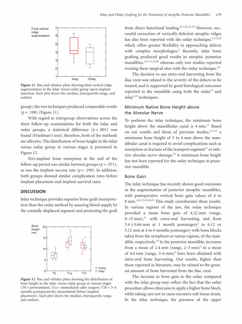
group), the two techniques produced comparable results
(p = .190) (Figure 11).
With regard to intergroup observations across the
three follow-up examinations for both the inlay and
onlay groups, a statistical difference (p < .001) was
found (Friedman’s test); therefore, both of the methods
are effective. The distribution of bone height in the inlay
versus onlay group at various stages is presented in
Figure 12.
Peri-implant bone resorption at the end of the
follow-up period was similar between groups (p = .971),
as was the implant success rate (p = .190). In addition,
both groups showed similar complication rates before
implant placement and implant survival rates.
DISCUSSION
Inlay technique provides superior bone graft incorpora-
tion than the onlay method by assuring blood supply by
the cranially displaced segment and protecting the graft
from direct functional loading.8,11,22,24–28 However, suc-
cessful correction of vertically deficient atrophic ridges
has also been reported with the onlay technique,2,3,14,29
which offers greater flexibility in approaching defects
with complex morphologies.3 Recently, inlay bone
grafting produced good results in atrophic posterior
mandibles,1,10–13,19,30 whereas only two studies reported
treating these surgical sites with the onlay technique.2,3
The decision to use extra-oral harvesting from the
iliac crest was related to the severity of the defects to be
treated, and is supported by good histological outcomes
reported in the mandible using both the onlay15 and
inlay1,19 techniques.
Minimum Native Bone Height abovethe Alveolar Nerve
To perform the inlay technique, the minimum bone
height above the mandibular canal is 4 mm.11 Based
on our results and those of previous studies,1,12,13 a
minimum bone height of 5 to 6 mm above the man-
dibular canal is required to avoid complications such as
resorption or fracture of the transport segment31 or infe-
rior alveolar nerve damage.32 A minimum bone height
has not been reported for the onlay technique in poste-
rior mandible.
Bone Gain
The inlay technique has recently shown good outcomes
in the augmentation of posterior atrophic mandibles,
with postoperative vertical bone gain values of 4 to
8 mm.1,10–13,19,30,33 This study corroborates these results.
In various regions of the jaw, the onlay technique
provided a mean bone gain of 4.22 mm (range,
0–15 mm),11 with extra-oral harvesting, and from
3.4 1 0.66 mm at 1 month postsurgery2 to 6.12 or
5.12 mm at 4 to 6 months postsurgery with bone blocks
taken from the symphysis or ramus regions, of the man-
dible, respectively.34 In the posterior mandible, increases
from a mean of 2.4 mm (range, 2–3 mm)2 to a mean
of 4.6 mm (range, 3–6 mm)3 have been obtained with
intra-oral bone harvesting. Our results, higher than
those reported in literature, may be related to the great-
est amount of bone harvested from the iliac crest.
The increase in bone gain in the onlay compared
with the inlay group may reflect the fact that the onlay
procedure allows clinicians to apply a higher bone block,
while taking care not to cause excessive soft tissue strain.
In the inlay technique, the presence of the upper
Final vertical ridge augmentation
Inlay Onlay1
2
3
4
5
6
7
Figure 11 Box and whisker plots showing final vertical ridgeaugmentation in the inlay versus onlay group upon implantinsertion. Each plot shows the median, interquartile range, andoutliers.
T0 T1a T1b0
5
10
15
20
InlayOnlay
Bone height mm
Figure 12 Box and whisker plots showing the distribution ofbone height in the inlay versus onlay group at various stages(T0 = pretreatment; T1a = immediately after surgery; T1b = 3–4months postoperatively, immediately before implantplacement). Each plot shows the median, interquartile range,and outliers.
Inlay and Onlay Grafting for the Treatment of Atrophic Posterior Mandibles e79
transport segment and the necessity of maintaining the
integrity of the lingual periosteum may limit vertical
augmentation.
Bone Resorption
In the posterior mandible, Bianchi and colleagues13
reported a median crestal bone resorption of 0.9 mm
(range, 0.5–1.2 mm) at 3 to 4 months postoperatively
when using the inlay technique; this accounted for 14.2%
of the obtained bone gain. A split-mouth study recently
reported a mean resorption of 1.1 mm with an iliac bone
graft and 0.6 mm with bovine anorganic bone 4 months
after grafting. The bone resorption of the augmented
ridge encountered in this study, 10.2% of obtained bone
gain, was almost completely attributable to the transport
segment, based on data showing that interposed graft
resorption was almost 0 for all treated sites.
In cases treated using iliac grafts at various jaw sites,
resorption values for the inlay technique ranged from 12
to 60% at 1 to 5 years after loading.35 In the posterior
mandible, the onlay technique with intra-oral graft
harvesting resulted in resorption from 0.6 mm (13%
of bone gain)3 to 1 mm (43.5% of bone gain)2 before
implant placement.
Notably, we observed significantly greater bone
resorption in the onlay group (40.4% of the obtained
bone gain) than in the inlay group. Considering the lack
of vertical reduction in the interposed graft, the minimal
bone height reduction observed in the inlay group may
be attributable to resorption of the transport segment
or to its arrangement during the healing period.
These results confirm the results of previous
studies.8,11,12,25–29,36,37 On one hand, the interposed graft is
well incorporated in the recipient site, it is not subjected
to functional loading, and shows minimal resorption; on
the other hand, a coronally displaced native bone frag-
ment attached to its own soft tissues is less prone to
resorption than a superimposed free non-vascularized
bone graft. Of note, however, was the observation that
the two techniques were comparable in terms of final
vertical augmentation.
Peri-Implant Bone Resorption, Implant Survival,and Success Rate
For the inlay technique, the overall implant survival rate
in atrophic mandibles ranges from 90 to 95%.35 In the
posterior mandible, implant survival and success rates of
90% at 1 to 4 years post-implant placement11 and sur-
vival and success rates of 100 and 95.2%, respectively,
after a median postloading period of 22.5 months13 have
been reported.
Regarding the onlay technique, cumulative implant
survival rates of 60, 76, and 100% have been
reported,35,38 whereas success rates ranging from 83 to
100% have been obtained, independent of the jaw treat-
ment site, duration of follow up, and bone graft harvest-
ing.34 The overall survival rate of implants placed in
reconstructed mandibles is 94.5% (range, 88.2–100%).35
According to a literature review,39 88% of 753 implants
placed on iliac crest bone grafts survived when using the
onlay technique. In the posterior mandible, the onlay
technique with intra-oral graft harvesting resulted in
cumulative survival and success rates of 100% and 89.5
to 100%, respectively, after mean follow-up periods of
38 and 12 months postloading.2,3 Our results are in
line with correspondent ones above mentioned both for
inlay and onlay techniques in the posterior mandible.
The median value for cumulative peri-implant bone
resorption from implant placement to the end of the
follow-up period, 0.85 mm (range, 0.2–1.4 mm), is
comparable to those reported by Chiapasco and col-
leagues3 (0.9 and 1.2 mm at 1 and 2 years postloading,
respectively), but results are higher than those reported
by Levin and colleagues40 (mean, 0.22 mm). Addition-
ally, the survival and success rates of implants placed in
the treated areas were comparable between groups and
with those obtained in native bone.
Complication Rate before Implant Placement
Only a case of major complication has been reported for
the inlay technique applied to the posterior mandible: a
transport segment fracture with complete failure of the
augmentation procedure.1 Regarding minor complica-
tions, the occurrence of a single spot of buccal dehis-
cence with plate exposure has been reported in 10, 7, and
6 surgical sites,11–13 and dehiscence on the buccal and
lingual sides have been reported at 20 treated sites.1
Of the three cases of postsurgical dehiscence in the
inlay group recorded in this study, one case, involving
a significant bone resorption, represented a major
complication.
Regarding neurological complications, two previ-
ous studies on six patients did not report any sign of
impaired sensitivity with iliac crest inlay grafting in
the posterior mandible.12,13 In contrast, Jensen11 and
Felice and colleagues1 reported transient postsurgical
e80 Clinical Implant Dentistry and Related Research, Volume 11, Supplement 1, 2009
paresthesia in all of their patients; impaired sensitivity
lasted up to 6 weeks11 and 13 days,1 respectively. Our
neurological impairments, limited to four patients, did
not persist longer than 3 weeks.
Generally, uneventful healing/consolidation has
been reported for the majority of patients (90–100%)
treated via the onlay technique, with dehiscence and
partial/total loss of the graft reported in only 3.3 and
1.4% of patients, respectively.35 Other authors have
reported a 38.5% complication rate in vertical bone aug-
mentation using an onlay bone graft.14 In the posterior
mandible, one case of eight reported showed graft expo-
sure and partial loss of the graft 2 months after recon-
struction, and implant placement was subsequently
performed using shorter implants.3 In contrast, other
authors did not report complications at this site.2
Paresthesia in the area innervated by the inferior
alveolar nerve was previously observed in three of eight
patients and persisted until the end of the follow-up
period in one case (3 years after surgery), but this com-
plication was attributed to the harvesting procedure
from the chin.3 In our study, two patients complained of
sensitivity impairment, lasting in one case till the end of
follow-up. The rate of complication in our study, the
same for inlay and onlay, may be considered low for both
groups.
CONCLUSIONS
This comparative study suggests that the inlay and onlay
techniques provide adequate correction of vertical defi-
cits in atrophic posterior mandibles, with acceptable
complication rate and implant results similar to those in
native bone.
The inlay technique is associated with lower bone
resorption values and produces more predictable out-
comes, but requires an experienced surgeon. The onlay
technique results in higher bone resorption values
before implant placement and requires a bone block
graft that is oversized in height, with respect to the
desired final vertical augmentation; however, it has a
shorter learning curve. Once implant placement has
been carried out, the outcomes are similar for both graft
procedures; this demonstrates that the type of augmen-
tation technique performed has no effect on the
maintenance of vertical bone after implant-loading.
Considering the lack of published data, we believe that
the personal experience of the surgeon should dictate
which technique is to be used in the posterior mandible.
CONFLICTS OF INTEREST STATEMENT
The authors have declared no conflicts of interest. [Cor-
rection added after online publication 23 October 2009:
Conflicts of Interest Statement added.]
REFERENCES
1. Felice P, Marchetti C, Piattelli A et al. Vertical ridge augmen-
tation of the atrophic posterior mandible with interposi-
tional block grafts: bone from the iliac crest versus bovine
anorganic bone. Results up to delivery of the final prostheses
from a split-mouth, randomised controlled trial. Eur J Oral
Implantol 2008; 1:193–198.
2. Cordaro L, Sarzi Amadè D, Cordaro M. Clinical results of
alveolar ridge augmentation with mandibular block bone
grafts in partially edentulous patients prior to implant place-
ment. Clin Oral Implants Res 2002; 13:103–111.
3. Chiapasco M, Zaniboni M, Rimondini L. Autogenous onlay
bone grafts vs. alveolar distraction osteogenesis for the cor-
rection of vertically deficient edentulous ridges: a 2–4-year
prospective study on humans. Clin Oral Implants Res 2007;
18:432–440.
4. Rosenquist B. Implant placement in combination with nerve
transpositioning: experiences with the first 100 cases. Int J
Oral Maxillofac Implants 1994; 9:522–531.
5. das Neves FD, Fones D, Bernardes SR, do Prado CJ, Neto AJ.
Short implants – an analysis of longitudinal studies. Int J
Oral Maxillofac Implants 2006; 21:86–93.
6. Esposito M, Grusovin MG, Kwan S, Worthington HV,
Coulthard P. Interventions for replacing missing teeth: bone
augmentation techniques for dental implant treatment. In:
Cochrane database of systematic reviews. Chichester, UK:
John Wiley & Sons, Ltd., 2008.
7. Stellingsma C, Raghoebar G, Meijer HJA, Batenburg RHK.
Reconstruction of the extremely resorbed mandible with
interposed bone grafts and placement of endosseous
implants. Br J Oral Maxillofac Surg 1998; 36:290–295.
8. Choi B-H, Lee S-HR, Huh J-H, Han S-G. Use of the sand-
wich osteotomy plus an interpositional allograft for vertical
augmentation of the alveolar ridge. J Craniomaxillofac Surg
2004; 32:51–54.
9. Stoelinga PJW, Blijdorp PA, Ross RR, De Koomen HA,
Huybers AJM. Augmentation of the atrophic mandible with
interposed bone grafts and particulate hydroxyapatite. J Oral
Maxillofac Surg 1986; 44:353–360.
10. Yeung R. Surgical management of the partially edentulous
atrophic mandibular ridge using a modified sandwich
osteotomy: a case report. Int J Oral Maxillofac Implants
2005; 20:799–803.
11. Jensen OT. Alveolar segmental “sandwich” osteotomies for
posterior edentulous mandibular sites for dental implants. J
Oral Maxillofac Surg 2006; 64:471–475.
12. Marchetti C, Trasarti S, Corinaldesi G, Felice P. Interposi-
tional bone grafts in the posterior mandibular region. A
Inlay and Onlay Grafting for the Treatment of Atrophic Posterior Mandibles e81
report on six patients. Int J Periodontics Restorative Dent
2007; 27:547–555.
13. Bianchi A, Felice P, Lizio G, Marchetti C. Alveolar distraction
osteogenesis versus inlay bone grafting in posterior man-
dibular atrophy. A prospective study. Oral Surg Oral Med
Oral Pathol Oral Radiol Endod 2008; 105:282–292.
14. Schwartz-Arad D, Levin L, Sigal L. Surgical success of
intraoral autogenous block onlay bone grafting for alveolar
ridge augmentation. Implant Dent 2005; 14:131–138.
15. Hodges NE, Perry M, Mohamed W, Hallmon WW, Rees T,
Opperman LA. Distraction osteogenesis versus autogenous
onlay grafting. Part II: biology of regenerate and onlay bone.
Int J Oral Maxillofac Implants 2006; 21:237–244.
16. Keller EE, Van Roekel NB, Desjardins RP, Tolman DE.
Prosthetic surgical reconstruction of the severely resorbed
maxilla with iliac bone grafting and tissue integrated pros-
theses. Int J Oral Maxillofac Implants 1987; 2:155–165.
17. McGrath CJ, Schepers SH, Blijdorp PA, Hoppenreijs TJ, Erbe
M. Simultaneous placement of endosteal implants and man-
dibular onlay grafting for treatment of the atrophic man-
dible. A preliminary report. Int J Oral Maxillofac Surg 1996;
25:184–188.
18. Bahat O, Fontanessi RV. Implant placement in three-
dimensional grafts in the anterior jaw. Int J Periodontics
Restorative Dent 2001; 21:357–365.
19. Felice P, Corinaldesi G, Lizio G, Piattelli A, Iezzi G, Marchetti
C. Implant prosthetic rehabilitation of posterior mandible
following tumor ablation with inferior alveolar nerve mobi-
lization and inlay bone grafting. A case report. J Oral Max-
illofac Surg 2009; 67(5):1104–1112.
20. Converse JM, Campbell RM. Bone grafts in surgery of the
face. Surg Clin North Am 1954; 375–401.
21. Albrektsson T, Zarb G, Worthington PMD, Eriksson AR. The
long term efficacy of currently used dental implants: a review
and proposed criteria of success. Int J Oral Maxillofac
Implants 1986; 1:11–25.
22. Enislidis G, Fock N, Millesi-Schobel G et al. Analysis of com-
plications following alveolar distraction osteogenesis and
implant placement in the partially edentulous mandible.
Oral Surg Oral Med Oral Pathol Oral Radiol Endod 2005;
100:25–30.
23. Cawood JI, Howell RA. A classification of the edentulous
jaws. Int J Oral Maxillofac Surg 1988; 17:232–236.
24. Garcia Garcia A, Somoza Martin M, Gandara Vila P, Lopez
Maceiras J. Alveolar ridge osteogenesis using 2 intraosseous
distractors: uniform and nonuniform distraction. J Oral
Maxillofac Surg 2002; 60:1510–1512.
25. Schettler D. Sandwich technique with cartilage transplant
for raising the alveolar process in the lower jaw. Fortschr
Kiefer Gesichtschir 1976; 20:61–63.
26. Stoelinga PJW, Tidemann JS, Berger H, de Koonen A. Inter-
positional bone graft augmentation of the atrophic man-
dible. J Oral Surg 1978; 36:30–32.
27. Blijdorp PA, Schoenaers JHA. Reconstruction of the severely
resorbed mandible with interposed bone grafts and hydroxy-
lapatite. Int J Oral Maxillofac Surg 1988; 17:157–160.
28. Stellingsma C, Raghoebar G, Meijer HJA, Batenburg RHK.
Reconstruction of the extremely resorbed mandible with
interposed bone grafts and placement of endosseous
implants. Br J Oral Maxillofac Surg 1998; 36:290–295.
29. Pikos MA. Block autografts for localized ridge augmenta-
tion. Part II. The posterior mandible. Implant Dent 2000;
9:67–75.
30. Felice P, Iezzi G, Lizio G, Piattelli A, Marchetti C. Recon-
struction of atrophied posterior mandible with inlay
technique and mandibular ramus block graft for implant
prosthetic rehabilitation. J Oral Maxillofac Surg 2009;
67:372–380.
31. Hwang S-J, Jung J-G, Jung J-U, Kyung S-H. Vertical alveolar
bone distraction at molar region using lag screw principle. J
Oral Maxillofac Surg 2004; 62:787–794.
32. Chiapasco M, Consolo U, Bianchi A, Ronchi P. Alveolar
distraction osteogenesis for the correction of vertically defi-
cient edentulous ridges: a multicenter prospective study on
humans. Int J Oral Maxillofac Implants 2004; 19:399–407.
33. Felice P, Piattelli A, Iezzi G, Degidi M, Marchetti C. Recon-
struction of atrophied posterior mandible with an inlay
technique and inorganic bovine bone block: a case report.
Int J Periodontics Restorative Dent (In press).
34. Proussaefs P, Lozada J, Kleinman A, et al. The use of ramus
autogenous block grafts for vertical alveolar ridge augmen-
tation and implant placement: a pilot study. Int J Oral
Maxillofac Implants 2002; 17:238–248.
35. Chiapasco M, Zaniboni M, Boisco M. Augmentation proce-
dures for the rehabilitation of deficient edentulous ridges
with oral implants. Clin Oral Implants Res 2006; 17:136–
159.
36. Binger T, Hell B. Resorption of microsurgically vascularized
bone grafts after augmentation of the mandible. J Craniom-
axillofac Surg 1999; 27:82–85.
37. Åstrand P, Nord PG, Brånemark PI. Titanium implants and
onlay bone graft to the atrophic edentulous maxilla. Int J
Oral Maxillofac Surg 1996; 25:25–29.
38. Rocchietta I, Fontana F, Simion M. Clinical outcomes of
vertical bone augmentation to enable dental implant place-
ment: a systematic review. J Clin Periodontol 2008; 35:203–
215.
39. Aghaloo TL, Moy PK. Which hard tissue augmentation tech-
niques are the most successful in furnishing bony support
for implant placement? Int J Oral Maxillofac Implants 2007;
22(Suppl):49–79.
40. Levin L, Nitzan D, Schwartz-Arad D. Success of dental
implants placed in intraoral block bone grafts. J Periodontol
2007; 78:18–21.
e82 Clinical Implant Dentistry and Related Research, Volume 11, Supplement 1, 2009
Top Related
Copyright © 2022 FDOKUMEN

