


Bahasa
Halaman
Hukum

Protein Science (1996), 5:2089-2094. Cambridge University Press. Printed in the USA. Copyright 0 1996 The Protein Society
In vitro renaturation of bovine P-lactoglobulin A leads to a biologically active but incompletely refolded state
VINOD SUBRAMANIAM,’*2*5 DUNCAN G. STEEL,*v3 AND ARI GAFN1234 ’ Applied Physics Program, The University of Michigan, Ann Arbor, Michigan 48109
Institute of Gerontology, The University of Michigan, Ann Arbor, Michigan 48109 Harrison M. Randall Laboratory of Physics, The University of Michigan, Ann Arbor, Michigan 48109 Department of Biological Chemistry, The University of Michigan Medical School, Ann Arbor, Michigan 48109
(RECEIVED June 25, 1996; ACCEPTED July 29, 1996)
Abstract
When bovine P-lactoglobulin (P-LC) was refolded after extensive denaturation in 4.8 M guanidine hydrochloride (GuHCI), the functional activity of the protein, retinol binding, as measured by the enhancement of this ligand’s fluorescence, was completely recovered. In contrast, the room-temperature tryptophan phosphorescence lifetime of the refolded protein, a local measure of the residue environment, was - I O ms, significantly shorter than the phosphores- cence lifetime of the untreated native protein (-20 ms). The lability of the freshly refolded protein, as monitored by following the time course of its unfolding when incubated in 2.5 M GuHCl through the change in fluorescence intensity at 385 nm, was also determined and found to be increased significantly relative to untreated native protein. In contrast to the long term postactivation conformational changes detected previously in Escherichia coli alkaline phosphatase (Subramaniam V, Bergenhem NCH, Gafni A, Steel DG, 1995, Biochemistry 341 133-1 136), we found no changes in either the lability or phosphorescence decays of P-LC during a period of 24 h. Our results are in agreement with the report by Hattori et al. (1993, J Biol Chem 268:22414-22419), using conformation-specific monoclonal antibodies to recognize native-like structure, that long-term changes occur in the protein conformation, compared with the native structure, on refolding.
Keywords: bovine P-lactoglobulin; protein folding; protein lability; protein structure; tryptophan phosphorescence; spectroscopy
During in vitro folding of a protein, the recovery of biological activity traditionally has been assumed to reflect the return of the protein structure to its “native” state, the end product of the in vivo folding process. However, several recent studies using especially sensitive techniques to detect subtle conformational changes in the protein suggest that the structure of a fully reactivated protein may differ from the original native state (Hattori et al., 1993; Subra- maniam et al., 1995). Using a combination of biophysical and biochemical methods, a recent report from our laboratory (Subra- maniam et al., 1995) demonstrated distinct differences between the structure of enzymatically active refolded Escherichia coli alkaline phosphatase (AP) and the native protein. Both the room-temperature tryptophan phosphorescence (RTP) lifetime, a local probe of the microenvironment of the emitting Trp, and the protein lability, a global parameter, exhibited a slow recovery to the initial, “native- like” value long after the return of enzymatic activity.
Reprint requests to: Ari Gafni, Institute of Gerontology, University of Michigan, 300 North Ingalls Bldg., Ann Arbor, Michigan 48109; e-mail: [email protected].
’Present address: Department of Molecular Biology, Max Planck Insti- tute for Biophysical Chemistry, Postfach 2841, D-37018 Gottingen, Germany.
It is interesting to note that there are other reported cases of pro- teins that possess the functional biological signatures of their native state, yet exhibit significantly different structural and thermodynamic properties. Phosphoglycerate kinase (PGK) has been shown to ex- ist in fully active, slowly interconverting conformations, leading to more stable forms of PGK without any effect on activity (Roth- stein, 1985). Hattori et al. (1993) showed that, although renatured j3-lactoglobulin (P-LC) exhibited the same retinol binding activity as the native protein, some specific epitopes in refolded P-LC were not recognized by conformation-specific monoclonal antibodies, sug- gesting that these epitopes did not return to the native conformation from the denatured state. Here we present biochemical and spec- troscopic evidence, including RTP data, that extends the initial ob- servation by Hattori et al. (1993) that the renatured protein does not completely refold. Our data indicates that these structural differ- ences affect the environment of Trp 19 in P-LC.
Results
Retinol binding
Figure 1 depicts the results of retinol binding assays of native and renatured P-LC assessed by determining the fluorescence from the
2089
2090 V Subrammiam et al.
120000 - -
40000 L /
"t Free Retinol in buffer -.IF Denatured "-t-- Natlve - - ~ --Renatured
0 5 0 100 150 200
FM Retinol
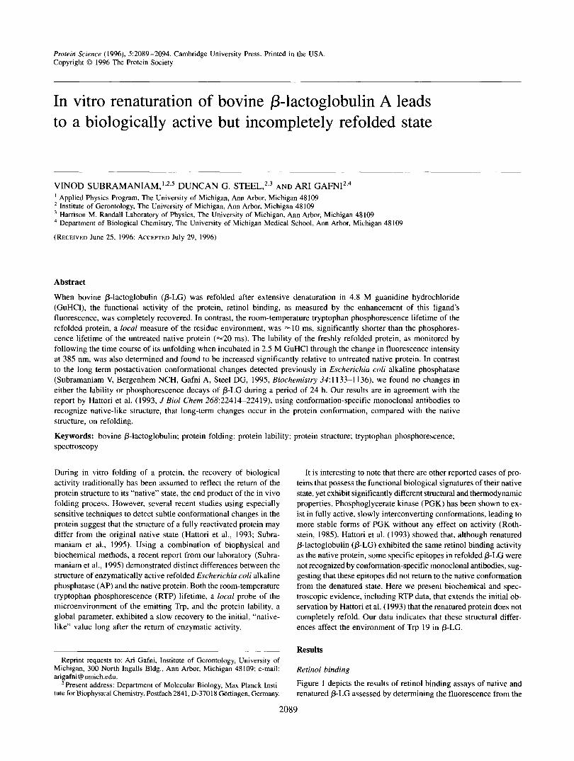
Fig. 1. Fluorescence titration curves of retinol binding to native and re- folded @-LG. A,, = 330 nm; A,, = 470 nm. @-LG was denatured in buffer (0.1 1 M phosphate, 0.04 M NaCI, pH 7.1) containing 4.8 M GuHCI, and renatured by rapid 100-fold dilution into denaturant-free buffer. Renatured protein shows essentially the same ability to bind retinol as the native protein.
p-ionone ring of the retinol molecule following excitation at 330 nm. Although the calculated fits to the data for native and rena- tured protein (produced by dilution of GuHCl denatured p-LC with excess buffer) indicate a small difference in the saturated value of the fluorescence intensity, possibly representing a small fraction of misfolded protein, the difference is within the experi- mental error (= 10%). We conclude that the binding curves clearly demonstrate that the retinol binding of renatured p-LC is nearly indistinguishable from that of native p-LC. In contrast, p-LC de- natured in 6 M GuHCl completely loses its retinol binding activity (Fig. I ) . These data indicate that, on refolding, denatured 0-LC regains its biological function of retinol binding.
RTP decays of native and refolded p-LG
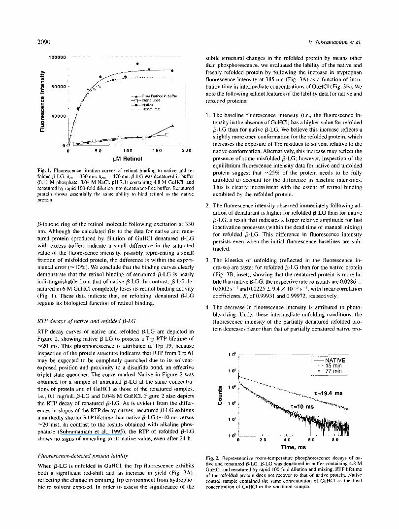
RTP decay curves of native and refolded P-LC are depicted in Figure 2, showing native p-LC to possess a Trp RTP lifetime of -20 ms. This phosphorescence is attributed to Trp 19, because inspection of the protein structure indicates that RTP from Trp 61 may be expected to be completely quenched due to its solvent- exposed position and proximity to a disulfide bond, an effective triplet state quencher. The curve marked Native in Figure 2 was obtained for a sample of untreated p-LC at the same concentra- tions of protein and of GuHCl as those of the renatured samples, i.e., 0.1 mg/mL 0-LC and 0.048 M GuHCI. Figure 2 also depicts the RTP decay of renatured 0-LC. As is evident from the differ- ences in slopes of the RTP decay curves, renatured p-LC exhibits a markedly shorter RTP lifetime than native P-LC (- I O ms versus -20 ms). In contrast to the results obtained with alkaline phos- phatase (Subramaniam et al., 1995), the RTP of refolded p-LC shows no signs of annealing to i ts native value, even after 24 h.
Fluorescence-detected protein lability
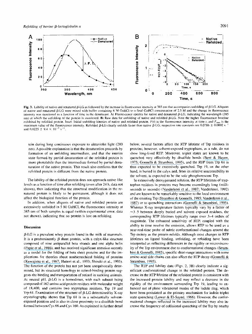
When p-LC is unfolded in GuHCI, the Trp fluorescence exhibits both a significant red-shift and an increase in yield (Fig. 3A), reflecting the change in emitting Trp environment from hydropho- bic to solvent exposed. In order to assess the significance of the
subtle structural changes in the refolded protein by means other than phosphorescence, we evaluated the lability of the native and freshly refolded protein by following the increase in tryptophan fluorescence intensity at 385 nm (Fig. 3A) as a function of incu- bation time in intermediate concentrations of GuHCl (Fig. 3B). We note the following salient features of the lability data for native and refolded proteins:
The baseline fluorescence intensity (i.e., the fluorescence in- tensity in the absence of GuHCI) has a higher value for refolded p-LC than for native p-LC. We believe this increase reflects a slightly more open conformation for the refolded protein, which increases the exposure of Trp residues to solvent relative to the native conformation. Alternatively, this increase may reflect the presence of some misfolded 0-LC; however, inspection of the equilibrium fluorescence intensity data for native and unfolded protein suggest that -25% of the protein needs to be fully unfolded to account for the difference in baseline intensities. This is clearly inconsistent with the extent of retinol binding exhibited by the refolded protein.
The fluorescence intensity observed immediately following ad- dition of denaturant is higher for refolded p-LC than for native 0-LC, a result that indicates a larger relative amplitude for fast inactivation processes (within the dead time of manual mixing) for refolded 0-LC. This difference in fluorescence intensity persists even when the initial fluorescence baselines are sub- tracted.
The kinetics of unfolding (reflected in the fluorescence in- crease) are faster for refolded p-LC than for the native protein (Fig. 3B, inset), showing that the renatured protein is more la- bile than native p-LC; the respective rate constants are 0.0286 ? 0.0002 s" and 0.0225 ? 9.4 X s - ' , with linear correlation coefficients, R, of 0.99931 and 0.99972, respectively.
The decrease in fluorescence intensity is attributed to photo- bleaching. Under these intermediate unfolding conditions, the fluorescence intensity of the partially denatured refolded pro- tein decreases faster than that of partially denatured native pro-
l * F - "
0 2 0 4 0 6 0 80
Time, ms
Fig. 2. Representative room-temperature phosphorescence decays of na- tive and renatured b-LG. @-LG was denatured in buffer containing 4.8 M GuHCl and renatured by rapid 100-fold dilution and mixing. RTP lifetime of the refolded protein does not recover to that of native protein. Native control sample contained the same concentration of GuHCl as the final concentration of GuHCl in the renatured sample.
Refolding of bovine P-luctoglobulin u 209 1
3 0 0 3 5 0 4 0 0 4 5 0
Wavelength, nm
t Lt. - 31
-x - 1 i 2 "
0 5 0 100 150 200 i Time, s o t - . , L ,A, 1 LL ,",~_ 1
0 200 400 600 800 Time, s
Fig. 3. Lability of native and renatured P-LC as followed by the increase in fluorescence intensity at 385 nm that accompanies unfolding of P-LC. Aliquots of native and renatured P-LC were mixed with buffer containing 6 M GuHCl to a final GuHCl concentration of 2.5 M and the change in fluorescence intensity was monitored as a function of time in the denaturant. A: Fluorescence spectra for native and denatured P-LC, indicating the wavelength (385 nm) at which the unfolding of the protein is monitored. B: Raw data for unfolding of native and refolded P-LC. Note the higher fluorescence baseline exhibited by refolded protein. Inset: Initial unfolding kinetics of native and refolded protein. F(t ) is the fluorescence intensity at time 1, and F,, is the maximum value of the fluorescence intensity. Refolded P-LC clearly unfolds faster than native P-LC; respective rate constants are 0.0286 2 0.0002 s" and 0.0225 +- 9.4 X I O -' s".
tein during long continuous exposure to ultraviolet light (280 nm). A possible explanation is that the denaturation proceeds by formation of an unfolding intermediate, and that the interim state formed by partial denaturation of the refolded protein is more photolabile than the intermediate formed by partial dena- turation of the native protein. This result also confirms that the refolded protein is different from the native protein.
The lability of the refolded protein does not approach native-like levels as a function of time after refolding (even after 24 h, data not shown), thus indicating that the structural modification in the re- natured protein is likely to be permanent, although it does not affect the biological function of the protein.
In addition, when aliquots of native and refolded protein are extensively unfolded in 5 M GuHCI, the fluorescence intensity at 385 nm of both samples is equal (within experimental error, data not shown), indicating that no protein is lost on refolding.
Discussion
P-LC is a prevalent whey protein found in the milk of mammals. It is a predominantly P-sheet protein, with a calyx-like structure composed of nine antiparallel beta strands and one alpha helix (Papiz et al., 1986), and has received significant attention recently as a model for the folding of beta sheet proteins and for the im- plications for theories about nonhierarchical folding of proteins (Kuwajima et al., 1987; Hattori et al., 1993; Shiraki et al., 1995). The function of the protein has not yet been unequivocally deter- mined, but its structural homology to retinol-binding protein sug- gests the binding and transportation of retinol in suckling animals. At neutral pH, P-LC is a homodimer, with each subunit being composed of 162 amino acid protein residues with molecular weight of 18,400, and contains two tryptophan residues, Trp 19 and Trp 6 1. Examination of the structure of P-LC determined by X-ray crystallography shows that Trp 61 is in a substantially solvent- exposed position and is also in close proximity to a disulfide bond formed between Cys 66 and Cys 160. As explained in further detail
below, several factors affect the RTP lifetime of Trp residues in proteins; however, solvent-exposed tryptophans, as a rule, do not show long-lived RTP. Moreover, triplet states are known to be quenched very effectively by disulfide bonds (Bent & Hayon, 1975; Gonnelli & Strambini, 1995), and the RTP from Trp 61 is thus expected to be extensively quenched. Trp 19, on the other hand, is buried in the calyx and, from its relative inaccessibility to the solvent, is expected to be the sole phosphorescent Trp.
In extensively deoxygenated solution, the RTP lifetimes of tryp- tophan residues in proteins may become exceedingly long (milli- seconds to seconds) (Vanderkooi et al., 1987; Vanderkooi, 1992) and are known to be remarkably sensitive to the local environment of the emitting Trp (Strambini & Gonnelli, 1985; Vanderkooi et al., 1987) or to quenching interactions (Gonnelli & Strambini, 1995). Whereas X-ray temperature factors typically vary by a factor of -3-5 between deeply buried and solvent exposed residues, the corresponding RTP lifetimes typically range over 3-4 orders of magnitude. The enhanced sensitivity of RTP, coupled with the ability to time resolve the emission, allows RTP to be used as a near-real-time probe of subtle conformational changes around the Trp moiety as the protein refolds. Although most changes in RTP lifetimes on ligand binding, unfolding, or refolding have been interpreted as reflecting differences in the rigidity or microviscos- ity of the Trp environment due to conformational changes (Stram- bini & Gonnelli, 1985), specific short-range interactions with some amino acid side chains can also affect the RTP decay (Gonnelli & Strambini, 1995).
The RTP and lability data (Figs. 2, 3B) clearly indicate a sig- nificant conformational change in the refolded protein. The de- crease in the RTP lifetime of the refolded protein is consistent with the increased protein lability and may reflect a decrease in the rigidity of the environment surrounding Trp 19, leading to en- hanced out-of-plane vibrational modes of the indole ring, which have been postulated as the primary mechanism for indole triplet state quenching (Lower & El-Sayed, 1966). However, the confor- mational changes reflected in the increased lability may also in- crease the frequency of collisional quenching of the Trp by nearby
2092 V Suhramaniam et al.
side chains, or may allow for a more favorable orientation between Trp 19 and nearby groups that exhibit a high propensity for intrin- sic quenching of the Trp phosphorescence, such as Tyr, Cys, or His (Gonnelli & Strambini, 1995). Two possible candidates are the residues Tyr 102 and Tyr 20. which are within 5 8, of Trp 19 in P-LG. Irrespective of the specific mechanism of quenching, fun- damentally the origin of the shorter RTP lifetime observed for the renatured form of P-LG lies in the local environmental changes in the vicinity of the emitting tryptophan (assumed to be Trp 19).
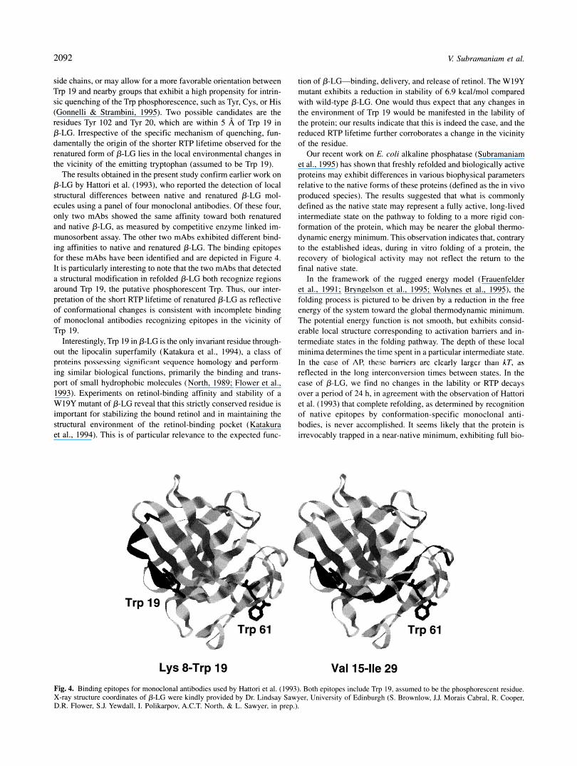
The results obtained in the present study confirm earlier work on P-LG by Hattori et al. (1993). who reported the detection of local structural differences between native and renatured 0-LG mol- ecules using a panel of four monoclonal antibodies. Of these four, only two mAbs showed the same affinity toward both renatured and native P-LG, as measured by competitive enzyme linked im- munosorbent assay. The other two mAbs exhibited different bind- ing affinities to native and renatured P-LG. The binding epitopes for these mAbs have been identified and are depicted in Figure 4. It is particularly interesting to note that the two mAbs that detected a structural modification in refolded P-LG both recognize regions around Trp 19, the putative phosphorescent Trp. Thus, our inter- pretation of the short RTP lifetime of renatured P-LG as reflective of conformational changes is consistent with incomplete binding of monoclonal antibodies recognizing epitopes in the vicinity of Trp 19.
Interestingly, Trp 19 in P-LG is the only invariant residue through- out the lipocalin superfamily (Katakura et a!., 1994), a class of proteins pnwmaing Pipiticant sequence homology and perform- ing similar biological functions, primarily the binding and trans- port of small hydrophobic molecules (North, 1989; Flower et al., 1993). Experiments on retinol-binding affinity and stability of a W19Y mutant of P-LG reveal that this strictly conserved residue is important for stabilizing the bound retinol and in maintaining the structural environment of the retinol-binding pocket (Katakura et al., 1994). This is of particular relevance to the expected func-
' ;&:.
Lys 8-Trp 19
tion of P-LG-binding, delivery, and release of retinol. The W19Y mutant exhibits a reduction in stability of 6.9 kcal/mol compared with wild-type P-LG. One would thus expect that any changes in the environment of Trp 19 would be manifested in the lability of the protein; our results indicate that this is indeed the case, and the reduced RTP lifetime further corroborates a change in the vicinity of the residue.
Our recent work on E. coli alkaline phosphatase (Subramaniam et al., 1995) has shown that freshly refolded and biologically active proteins may exhibit differences in various biophysical parameters relative to the native forms of these proteins (defined as the in vivo produced species). The results suggested that what is commonly defined as the native state may represent a fully active, long-lived intermediate state on the pathway to folding to a more rigid con- formation of the protein, which may be nearer the global thermo- dynamic energy minimum. This observation indicates that, contrary to the established ideas, during in vitro folding of a protein, the recovery of biological activity may not reflect the return to the final native state.
In the framework of the rugged energy model (Frauenfelder et al., 1991; Bryngelson et al., 1995; Wolynes et al., 1995), the folding process is pictured to be driven by a reduction in the free energy of the system toward the global thermodynamic minimum. The potential energy function is not smooth, but exhibits consid- erable local structure corresponding to activation barriers and in- termediate states in the folding pathway. The depth of these local minima determines the time spent in a particular intermediate state. In the casc of AI? thcsc barricrs arc clcarly lnrgcr than hT. as reflected in the long interconversion times between states. In the case of P-LG, we find no changes in the lability or RTP decays over a period of 24 h, in agreement with the observation of Hattori et al. (1993) that complete refolding, as determined by recognition of native epitopes by conformation-specific monoclonal anti- bodies, is never accomplished. It seems likely that the protein is irrevocably trapped in a near-native minimum, exhibiting full bio-
n
Val 15-lle 29 Fig. 4. Binding epitopes for monoclonal antibodies used by Hattori et al. (1993). Both epitopes include Trp 19, assumed to be the phosphorescent residue. X-ray structure coordinates of P-LG were kindly provided by Dr. Lindsay Sawyer, University of Edinburgh (S. Brownlow, J.J. Morais Cabral, R. Cooper, D.R. Flower. S.J. Yewdall, 1. Polikarpov, A C T . North, & L. Sawyer. in prep.).
Refolding of bovine P-lactoglobulin a 2093
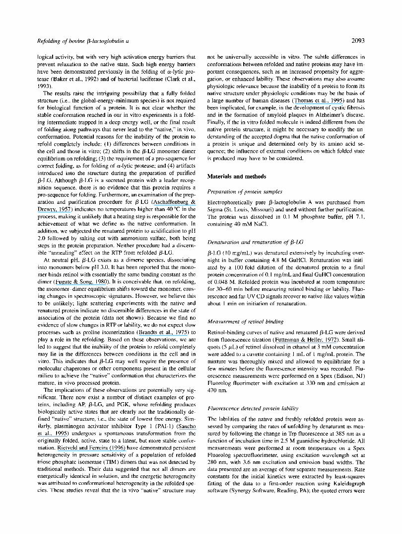
logical activity, but with very high activation energy bamers that prevent relaxation to the native state. Such high energy barriers have been demonstrated previously in the folding of a-lytic pro- tease (Baker et al., 1992) and of bacterial luciferase (Clark et al., 1993).
The results raise the intriguing possibility that a fully folded structure (i.e., the global-energy-minimum species) is not required for biological function of a protein. It is not clear whether the stable conformation reached in our in vitro experiments is a fold- ing intermediate trapped in a deep energy well, or the final result of folding along pathways that never lead to the “native,” in vivo, conformation. Potential reasons for the inability of the protein to refold completely include: (1) differences between conditions in the cell and those in vitro; (2) shifts in the P-LC monomer-dimer equilibrium on refolding; (3) the requirement of a pro-sequence for correct folding, as for folding of a-lytic protease; and (4) artifacts introduced into the structure during the preparation of purified P-LG. Although P-LG is a secreted protein with a leader recog- nition sequence, there is no evidence that this protein requires a pro-sequence for folding. Furthermore, an examination of the prep- aration and purification procedure for P-LG (Aschaffenburg & Drewry, 1957) indicates no temperatures higher than 40 “C in the process, making it unlikely that a heating step is responsible for the achievement of what we define as the native Conformation. In addition, we subjected the renatured protein to acidification to pH 2.0 followed by salting out with ammonium sulfate, both being steps in the protein preparation. Neither procedure had a discern- ible “annealing” effect on the RTP from refolded P-LG.
At neutral pH, P-LG exists as a dimeric species, dissociating into monomers below pH 3.0. It has been reported that the mono- mer binds retinol with essentially the same binding constant as the dimer (Fugate & Song, 1980). It is conceivable that, on refolding, the monomer-dimer equilibrium shifts toward the monomer, caus- ing changes in spectroscopic signatures. However, we believe this to be unlikely; light scattering experiments with the native and renatured protein indicate no discernible differences in the state of association of the protein (data not shown). Because we find no evidence of slow changes in RTP or lability, we do not expect slow processes such as proline isomerization (Brandts et al., 1975) to play a role in the refolding. Based on these observations, we are led to suggest that the inability of the protein to refold completely may lie in the differences between conditions in the cell and in vitro. This indicates that P-LG may well require the presence of molecular chaperones or other components present in the cellular milieu to achieve the “native” conformation that characterizes the mature, in vivo processed protein.
The implications of these observations are potentially very sig- nificant. There now exist a number of distinct examples of pro- teins, including AP, P-LG, and PGK, whose refolding produces biologically active states that are clearly not the traditionally de- fined “native” structure, Le., the state of lowest free energy. Sim- ilarly, plasminogen activator inhibitor Type 1 (PAI-I) (Sancho et al., 1995) undergoes a spontaneous transformation from the originally folded, active, state to a latent, but more stable confor- mation. Rietveld and Ferreira (1996) have demonstrated persistent heterogeneity in pressure sensitivity of a population of refolded triose phosphate isomerase (TIM) dimers that was not detected by traditional methods. Their data suggested that not all dimers are energetically identical in solution, and the energetic heterogeneity was attributed to conformational heterogeneity in the refolded spe- cies. These studies reveal that the in vivo “native” structure may
not be universally accessible in vitro. The subtle differences in conformations between refolded and native proteins may have im- portant consequences, such as an increased propensity for aggre- gation, or enhanced lability. These observations may also assume physiologic relevance because the inability of a protein to form its native structure under physiologic conditions may be the basis of a large number of human diseases (Thomas et al., 1995) and has been implicated, for example, in the development of cystic fibrosis and in the formation of amyloid plaques in Alzheimer’s disease. Finally, if the in vitro folded molecule i s indeed different from the native protein structure, it might be necessary to modify the un- derstanding of the accepted dogma that the native conformation of a protein is unique and determined only by its amino acid se- quence; the influence of external conditions on which folded state is produced may have to be considered.
Materials and methods
Preparation of protein samples
Electrophoretically pure P-lactoglobulin A was purchased from Sigma (St. Louis, Missouri) and used without further purification. The protein was dissolved in 0.1 M phosphate buffer, pH 7.1, containing 40 mM NaCl.
Denaturution and renaturution of P-LC
0-LG ( I O mglmL) was denatured extensively by incubating over- night in buffer containing 4.8 M GuHCl. Renaturation was init i- ated by a 100-fold dilution of the denatured protein to a final protein concentration of 0. I mg/mL and final GuHCl concentration of 0.048 M. Refolded protein was incubated at room temperature for 30-60 min before measuring retinol binding or lability. Fluo- rescence and far-UV CD signals recover to native-like values within about 1 min on initiation of renaturation.
Measurement of retinol binding
Retinol-binding curves of native and renatured P-LG were derived from fluorescence titration (Futterman & Heller, 1972). Small ali- quots ( 5 FL) of retinol dissolved in ethanol at 3 mM concentration were added to a cuvette containing 1 mL of I mg/mL protein. The mixture was thoroughly mixed and allowed to equilibriate for a few minutes before the fluorescence intensity was recorded. Flu- orescence measurements were performed on a Spex (Edison, NJ) Fluorolog fluorimeter with excitation at 330 nm and emission at 470 nm.
Fluorescence detected protein lability
The labilities of the native and freshly refolded protein were as- sessed by comparing the rates of unfolding by denaturant as mea- sured by following the change in Trp fluorescence at 385 nm as a function of incubation time in 2.5 M guanidine hydrochloride. All measurements were performed at room temperature on a Spex Fluorolog spectrofluorimeter, using excitation wavelength set at 280 nm, with 3.6 nm excitation and emission band widths. The data presented are an average of four separate measurements. Rate constants for the initial kinetics were extracted by least-squares fitting of the data to a first-order reaction using Kaleidagraph software (Synergy Software, Reading, PA); the quoted errors were
2094 K Subramaniarn et al.
determined from the least-squares fit. Photolability of the putative unfolding intermediate from native and refolded protein is inferred from the decrease in Trp fluorescence intensity with continuous exposure to UV excitation light (280 nm).
RTP
RTP measurements were performed using a laser-based system of our own design (Mersol et al., 1993; Subramaniam et al., 1995). Briefly, the experimental configuration consists of a Spectra- Physics model DCR-I 1 Nd:YAG laser emitting pulses at a wave- length of 532 nm to pump a Spectra-Physics model PDL-3 dye laser operating at 560 nm. The latter light is frequency-doubled externally in a nonlinear crystal (Inrad KDP) to produce 5-ns pulses of 280-nm light. Phosphorescence was detected at 440 nm using an Instruments SA HR 320 single monochromator; alterna- tively, broad-band detection was accomplished by directly close- coupling to the photomultiplier tube through a 0.5 M potassium nitrite solution serving to cut off fluorescence light. Data was collected using an EG&G Ortec ACEMCS multi-channel scaler card, with a minimum dwell time of 2 ps per channel, installed in an IBM personal computer. Phosphorescence decays were ana- lyzed using Photon Technology International fluorescence decay analysis software, which fits the decay curve to a discrete sum of exponential decays; the quality of the fit is determined by a Mar- quardt x* parameter. Deconvolution of the instrument response is not necessary because the laser pulse is contained entirely in the first channel of the data.
Because molecular oxygen is an extremely effective quencher of the room-temperature phosphorescence, great care was taken to deoxygenate the sample extensively by passing a stream of ultra pure argon gas over the sample for several hours, after which the sample was kept sealed at room temperature without further de- oxygenation for the remainder of the experiment. The phos- phorescence lifetime obtained in deoxygenated solution prior to denaturation was used to define the lifetime of the native state. All decays were collected for the same number of laser shots. Laser intensity variations were monitored by measuring the number of counts from a native 0-LC standard immediately following data acquisition.
Acknowledgments
This work was supported by the National Institute on Aging grant NIA AGO976 I and the Office of Naval Research grant ONR NOOO 14-9 1 -J- 1938. Coordinates for the p-LC X-ray crystal structure were provided by Dr. Lindsay Sawyer, University of Edinburgh.
References
Aschaffenburg R, Drewry J. 1957. Improved method for the preparation of crystalline 6-lactoglobulin and a-lactalbumin from cow's milk. Biochem J 65:273-277.
Baker D, Soh1 JL, Agard DA. 1992. A protein-folding reaction under kinetic control. Nature 356:263-265.
Bent DV, Hayon E. 1975. Excited state chemistry of aromatic amino acids and related peptides. 111. Tryptophan. J A m Chem Soc 972612-2619.
Brandts JF, Halvorson HR. Brennan M. 1975. Consideration of the possibility that the slow step in protein denaturation reactions is due to cis-trans isom- erism of proline residues. Biochemistry 144953-4963.
Bryngelson JD, Onuchic IN, Socci ND, Wolynes PC. 1995. Funnels, pathways, and the energy landscape of protein folding: A synthesis. Proteins Srruct Funcr Genet 21:167-195.
Clark AC, Sinclair JF, Baldwin TO. 1993. Folding of bacterial luciferase in-
tive enzyme and the unfolded subunits. J Biol Chem 268:10773-10779. volves a non-native heterodimeric intermediate in equilibrium with the na-
Flower DR, North AC, Attwood TK. 1993. Structure and sequence relationships in the lipocalins and related proteins. Protein Sei 2:753-761.
Frauenfelder H, Sligar SG, Wolynes PC. 1991. The energy landscapes and motions of proteins. Science 254: 1598-1603.
Fugate RD, Song PS. 1980. Spectroscopic characterization of beta-lactoglobulin- retinol complex. Biochim Biophvs Acta 625:28-42.
Futterman S, Heller J. 1972. The enhancement of fluorescence and the decreased susceptibility to enzymatic oxidation of retinol complexed with bovine se- rum albumin, 6-lactoglobulin, and the retinol-binding protein of human plasma. J Biol Chem 2475168-5172.
Gonnelli M, Strambini GB. 1995. Phosphorescence lifetime of tryptophan in
Hattori M, Ametani A, Katakura Y, Shimizu M, Kaminogawa S. 1993. Unfolding/ proteins. Biochemirtry 34: 13847-1 3857.
refolding studies on bovine /3-lactoglobulin with monoclonal antibodies as probes. J Biol Chem 268:22414-22419.
Katakura Y, Totsuka M, Ametani A, Kaminogawa S. 1994. Tryptophan-I9 of beta-lactoglobulin, the only residue completely conserved in the lipocalin superfamily, is not essential for binding retinol, but relevant to stabilizing bound retinol and maintaining its structure. Biochim Biophys Acta 1207 58-67.
Kuwajima K, Yamaya H, Miwa S, Sugai S, Nagamura T. 1987. Rapid formation of secondary structure framework in protein folding studied by stopped- flow circular dichroism. FEES Lett 221: 115-8.
Lower SK, El-Sayed MA. 1966. The triplet state and molecular electronic processes in organic molecules. Chemical Rev 66: 199-241.
Mer501 JV, Steel DG, Gafni A. 1993. Detection of intermediate protein confor- mations by room temperature tryptophan phosphorescence spectroscopy during denaturation of Escherichia coli alkaline phosphatase. Biophys Chem 48:28 1-29 I .
North AC. 1989. Three-dimensional arrangement of conserved amino acid res- idues in a superfamily of specific ligand-binding proteins. Int J Biol Mac- romol 11:56-58.
Papiz MZ, Sawyer L, Eliopoulos EE, North AC, Findlay JB, Sivaprasadarao R, Jones TA, Newcomer ME, Kraulis PJ. 1986. The structure of beta-lactoglobulin and its similarity to plasma retinol-binding protein. Nature 324:383-385.
Rietveld AWM, Ferreira ST. 1996. Deterministic pressure dissociation and un- folding of triose phosphate isomerase: Persistent heterogeneity of a protein dimer. Biochemistry 35:7743-775 I .
Rothstein M. 1985. Age-related changes in enzyme levels and enzyme proper- ties. In: Rothstein M, ed. Review of biological research in aging 2. New York: Alan R. Liss Inc. pp 421-433.
Sancho E, Declerck PJ, Price NC, Kelly SM, Booth NA. 1995. Conformational studies on plasminogen activator inhibitor (PAI-I) in active, latent, substrate
Shiraki K. Nishikawa K, Goto Y. 1995. Trifluoroethanol-induced stabilizatlon of and cleaved forms. Biochemistry 34: 1064-1069.
the alpha-helical structure of beta-lactoglobulin: Implication for non- hierarchical protein folding. J Mol Biol 245:180-194.
Strambini GB. Gonnelli M. 1985. The indole nucleus triplet-state lifetime and its dependence on solvent microviscosity. Chem Phys Lett 1/5:196-200.
Subramaniam V, Bergenhem NCH. Gafni A, Steel DG. 1995. Phosphorescence reveals a continued slow annealing of the protein core following reactiva- tion of Escherichia coli alkaline phosphatase. Biochemisfry 34: I 133-1 136.
Thomas PJ, Qu BH, Pedersen PL. 1995. Defective protein folding as a basis of human disease. Trends Biochem Sei 20:456-459.
Vanderkooi JM. 1992. Tryptophan phosphorescence from proteins at room tem- perature. In: Lakowicz JR, ed. 7i)pic.s in fluorescence spectroscopy. New York: Plenum Press. pp 1 13-136.
Vanderkooi JM, Calhoun DB, Englander WS. 1987. On the prevalence of room- temperature protein phosphorescence. Science 236568-569.
Wolynes P C , Onuchic JN, Thirumalai D. 1995. Navigating the folding routes. Science 267 I6 19- 1620.
Top Related
Copyright © 2022 FDOKUMEN

