


Bahasa
Halaman
Hukum

Sarkis et al. Head & Neck Oncology 2010, 2:13http://www.headandneckoncology.org/content/2/1/13
Open AccessR E S E A R C H
ResearchImmunohistochemical expression of epidermal growth factor receptor (EGFR) in oral squamous cell carcinoma in relation to proliferation, apoptosis, angiogenesis and lymphangiogenesisSeta A Sarkis1, Bashar H Abdullah1, Ban A Abdul Majeed2 and Nazar G Talabani*3
AbstractObjectives: Squamous cell carcinoma (SCC) is by far the most common malignant neoplasm of the oral cavity. A number of etiologic factors have been implicated in its development. During the past few decades, a particular focus has been placed on the investigation of valid biomarkers predictive of cancer behavior and cervical lymph node metastasis in head and neck Squamous cell carcinoma (HNSCC).The present study was designed to investigate the expression of epidermal growth factor in these tumors in relation to proliferation, apoptosis, angiogenesis and lymphangiogenesis.
Materials and methods: Immunohistochemical (IHC) evaluation of epidermal growth factor receptor (EGFR) expression in 40 retrospective OSCC specimens and its correlation with proliferating cell nuclear antigen (PCNA), antiapoptotic antibody (P53), vascular endothelial growth factor (VEGF), and D2-40 monoclonal antibodies (Mab), in relation to the clinicopathological parameters.
Results: Data revealed positive EGFR immunoreactivity in 35(87.5%) cases. There was a statistically significant correlation regarding EGFR extent score with respect to intratumoral lymphatic vessel density (ILVD) (r = 0.35) as well as EGFR intensity score with respect to ILVD and peritumoral lymphatic vessel density (PLVD) (r = 0.33, r = 0.36 respectively). EGFR expression was not correlated with the clinicopathological parameters. Conclusions: EGFR is expressed by most of the cases. EGFR correlation with D2- 40 positive lymphatic vessels suggests a higher tendency of OSCC for lymphatic dissemination. Lack of correlation among the studied markers suggests their independent effect on tumor behavior.
BackgroundOral squamous carcinogenesis is a multistep process inwhich multiple genetic events occur that alter the normalfunction of oncogenes and tumor suppressor genes (tsg).Cancer related genes have to be considered in the contextof six fundamental changes [1].
•Self sufficiency in growth signals•Insensitivity to growth inhibitory signals•Evasion of apoptosis•Limitless replicative potential•Sustained angiogenesis
•Ability to invade and metastasizeAll normal cells require stimulation on the basis of sig-
nals to undergo growth, differentiation and proliferation;many of which carried by growth factors [1,2]. EGFRplays an important role in the differentiation and mor-phogenesis of many organs and proliferation and survivalin mammalian cells [3,4]. EGFR has been reported to beexpressed in a variety of human tumors of epithelial ori-gin; over expression of EGFR has been documented in80% of SCC [1].
Angiogenesis is a crucial step in the successful growth,invasion and metastasis of tumors, without which tumorswill not grow more than 1-2 mm3 in diameter [5,6]. VEGFhas been considered as a leading candidate in the process
* Correspondence: [email protected] Department of Oral Pathology and Medicine, College of Dentistry, University of Sulaimani, IraqFull list of author information is available at the end of the article
© 2010 Sarkis et al; licensee BioMed Central Ltd. This is an Open Access article distributed under the terms of the Creative CommonsAttribution License (http://creativecommons.org/licenses/by/2.0), which permits unrestricted use, distribution, and reproduction inany medium, provided the original work is properly cited.
Sarkis et al. Head & Neck Oncology 2010, 2:13http://www.headandneckoncology.org/content/2/1/13
Page 2 of 8
of tumor angiogenesis. Various studies reported upregu-lation of VEGF in different malignancies [7,8].
Tissue growth depends on both cell proliferation andthe rate of cell death. PCNA is a 36 kd intra nuclear poly-peptide protein whose expression is associated with DNAsynthesis and cell proliferation. Many studies demon-strated an association of high expression rate of PCNAwith poor prognosis in solid tumors [2,9,10].
Apoptosis is a process of programmed cell death, it is asessential as cell growth for the maintenance of homeosta-sis [2,11]. P53 is a well known protein that regulates cellcycle check points and is responsible for maintaining theintegrity of genome. Mutation of p53 tsg is one of the bestknown and by far the most frequent genetic alterationidentified in malignant tumors [12].
Metastasis unequivocally signifies that a tumor ismalignant. Lymphangiogenesis which refers to thegrowth of new lymphatic vessels has long been regardedas a putative efficient pathway to neoplastic metastasiza-tion [13,14]. A new selective immunohistochemicalmarker is D2-40 which is specific for lymphatic endothe-lium since it doesn't stain vascular endothelium.
Tumors vary considerably in their behavior, notably inthe rate of their growth, the degree of their differentiationand the ability to invade and metastasize. Because of theobscure and variable behavior of cancer, this study con-cerned different aspects of tumor dynamics through theimmunohistochemcial evaluation of EGFR expression inOSCC and its correlation with proliferation, apoptosis,angiogenesis and lymphangiogensis via evaluatingPCNA, p53, VEGF and D2- 40 Mabs immunohistochem-ically.
MethodsThe study sample consisted of 40 retrospective OSCCspecimens from the department of oral pathology, collegeof Dentistry, Baghdad University. An immunoshitochem-cial staining with five types of Mabs was preformed: antiEGFR & VEGF (Dako Cytomation -Denmark), antiPCNA, anti P53 and anti D2-40 lymphatic endothelialmarker (Dako Cytomation - USA). Negative and positivecontrol slides were included in each IHC run (as recom-mended by the manufacturers).
Immunohistochemistry staining procedureAll tests were carried out on 5 μm formalin fixed paraffinembedded sections. Slides were baked in hot air oven at65°C overnight. Sections were sequentially dewaxedthrough a series of xylene, graded alcohol and waterimmersion steps. Antigen (Ag) retrieving was done asrecommended by the manufacturers using 500 mL of cit-rate buffer solution pH.6.0 for p53, pH 8.0 for VEGF and
D2-40, on a hot plate at temperature of (95-99°C), whilethis step was omitted for PCNA Ag. Whereas EGFR Agand target retrieval was performed by pretreating tissuesections using proteinase K proteolytic enzyme for 10minutes. Then endogenous peroxidase activity wasblocked followed by blocking the non- specific staining.Primary Abs (100 ml) was applied for each section. Adilution of 1:25 for both EGFR and VEGF was used; 1:100for D2-40, while PCNA and p53 Mabs were ready touse.The samples were then incubated at 4°C overnight ina humid chamber. After washing with PBS, secondaryAbs were applied to the sections, incubated and rinsedwith a stream of PBS. Primary Abs was visualized withDAB chromogen. Sections were counterstained withMayer's hematoxyline for 30 seconds, dehydrated andmounted.
Assessment of IHC resultsIn each tissue section five representative fields wereselected for EGFR, VEGF, PCNA and p53 Mabs and D2-40 positive tumor cells with an average of 1000 tumor cellper case and 200 tumor cells per field.
The immunoreactivity in tumor cells were classifiedand scored as follows: -(0-25%), + (26-50%), ++ (51-75%),+++(76-100%) for both PCNA and p53 [15]. ± (0-19%), +(20-39%), ++ (40-59%), +++ (60-100%) for VEGF [16].The extent of EGFR immunostaining was graded andscored as follows: 0 points for negative staining of theconsidered cells, (1) <10%, (2)10-50%, (3)51-80% and (4)≥ 80% positive staining of the considered cells. The inten-sity of staining was scored as 0, no staining; 1, weak; 2,moderate; 3, strong [4,17] -< (10%), +(10-25%), ++(26-50%), +++(51-100%) for D2-40 positive tumor cells [18].
D2-40 positivity was evaluated by selecting six "hotspots" intratumorally and peritumorally. Lymphatic vesseldensity (LVD) was expressed as the number of stainedvessels per optical field [18]. The average number of posi-tively stained vessels in each region was evaluated andrecorded separately as intratumoral and peritumoral lym-phatic vessel density (ILVD and PLVD). The averagecount of positive vessels of both regions was recordedtogether representing the total LVD (TLVD) for eachcase. In addition, intratumoral (It) and peritumoral (Pt)lymphatic vessel invasion (LVI) was considered evident ifat least one tumor cell cluster was clearly visible inside aD2-40 positive vessel [18,19].
Mann-whitney test was used to explore the statisticaldifference in median between two study groups. The sta-tistical significance, direction and strength of linear cor-relation were measured by spearman's rank linearcorrelation coefficient. P value less than the 0.05 level ofsignificance was considered statistically significant.
Sarkis et al. Head & Neck Oncology 2010, 2:13http://www.headandneckoncology.org/content/2/1/13
Page 3 of 8
ResultsClinicopathological dataThe study sample consisted of 27 males (67.5%) and 13females (32.5%) with an age range (24-86) years. Clini-cally, ulcer represented the most frequent clinical presen-tation (50%) of the cases. The majority of the cases (45%)were seen in the tongue. T1 and T2 were presented in12(31.6%) cases for each. Only 12 cases (30.8%) werenode positive. Most of the cases (43.2%) were TNM stageIII, and were histologically moderately differentiated car-cinoma (50%).
Evaluation of IHC resultsThe positivity rate of the selected immunostains EGFR(Fig.1), VEGF (Fig.2), PCNA (Fig.3), P53 (Fig.4); D2-40 inthe total sample is shown in (table 1) which reveals posi-tive immunostaining in most of the cases.
Assessment of EGFR expressionThirty five cases (87.5%) showed positive brown mem-branous and/or cytoplasmic EGFR immunostaining. InOSCC specimens the expression involved all the epithe-lial layers while in normal oral mucosa it was localized tothe basal cell layer. EGFR positive cells were often seenlocalized at the periphery of tumor nests. Besides EGFRextent score, the intensity score was also considered. Rel-ative frequency distribution revealed that (37.5%) of thecases showed low intensity (35%) moderate and only(15%) showed high intensity.
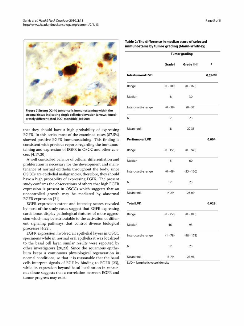
Assessment of D2-40 immunostainnigD2-40 brown staining of lymphatic endothelial cells wasobserved intratumorally, peritumorally or both. Positivelymphatic vessels were unevenly distributed throughoutthe tumor and their number in the (Pt) area was slightlyhigher 34(85%) than that in the (It) area 28(70%).The total
lymphatic vessel stain was seen in 35 (87.5%) cases. TheD2-40 stained lymphatic vessels, the adjacent blood ves-sels were always unstained (Fig.5).
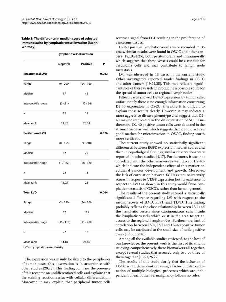
Moreover, cancer cells were occasionally observed in(It) lymphatic vessels and/or (Pt) lymphatic vessels(Fig.6), LVI was detected in 13 (37.1%) cases out of 35 D2-40 positive cases of which only five cases presented nodalmetastasis, eleven cases were grade II, one of the remain-ing two was grade I and the other was grade III.
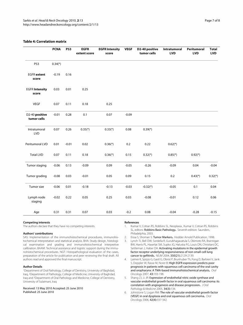
There was diffuse or strong granular cytoplasmic and/or membranous D2-40 expression in the malignant epi-thelial cells of 15 (37.5%) cases out of 35 D2-40 positivecases the remaining 28 cases were totally devoid of stain-ing. Eleven of the positive cases were grade II and theremaining four were grade I. In addition, 7 of D2-40 posi-tive tumor cell cases presented LVI. Furthermore, carefulexamination of D2-40 expressing sections under oilimmersion revealed microinvasion of positive malignantcells into the stromal tissue (Fig.7)
Figure 1 Positive brown membranous and or cytoplasmic EGFR immunostaining in well differentiated SCC (buccal mucosa, tongue and floor of mouth) (x100)
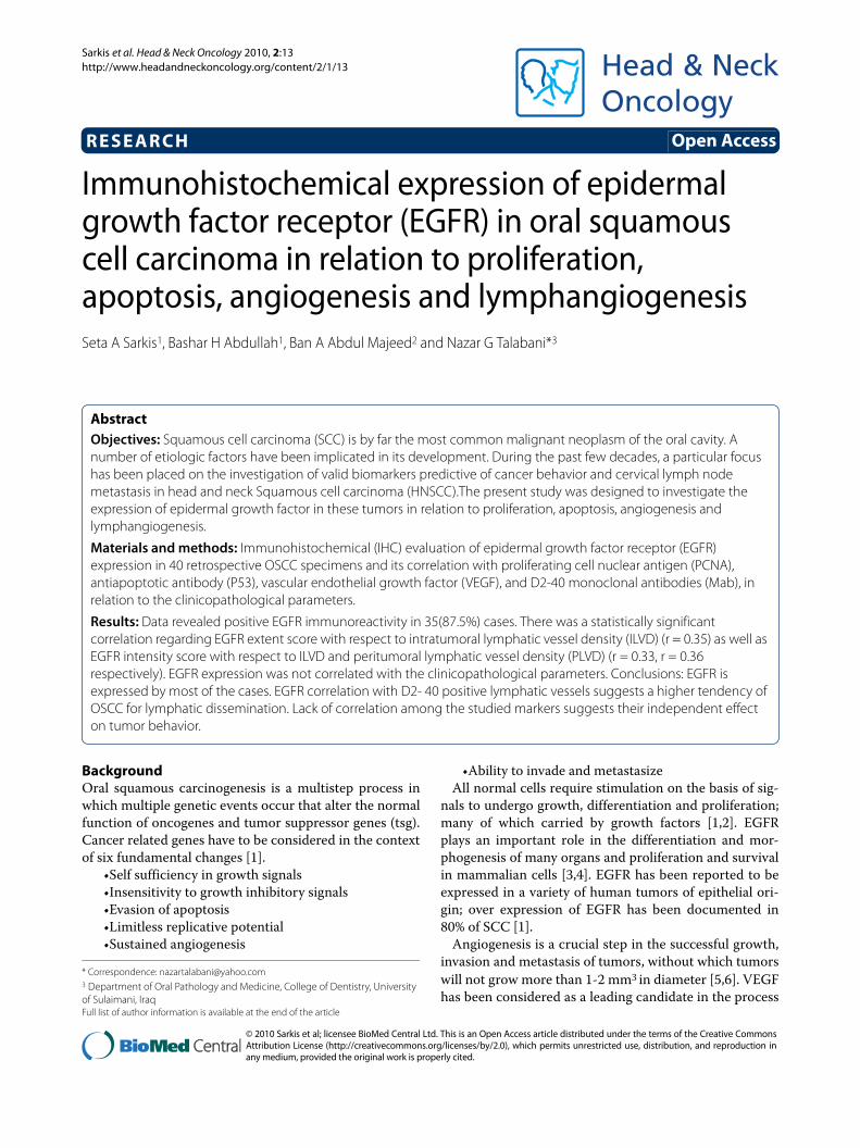
Figure 2 Strong granular cytoplasmic VEGF immunostaining in moderately differentiated SCC (x200)
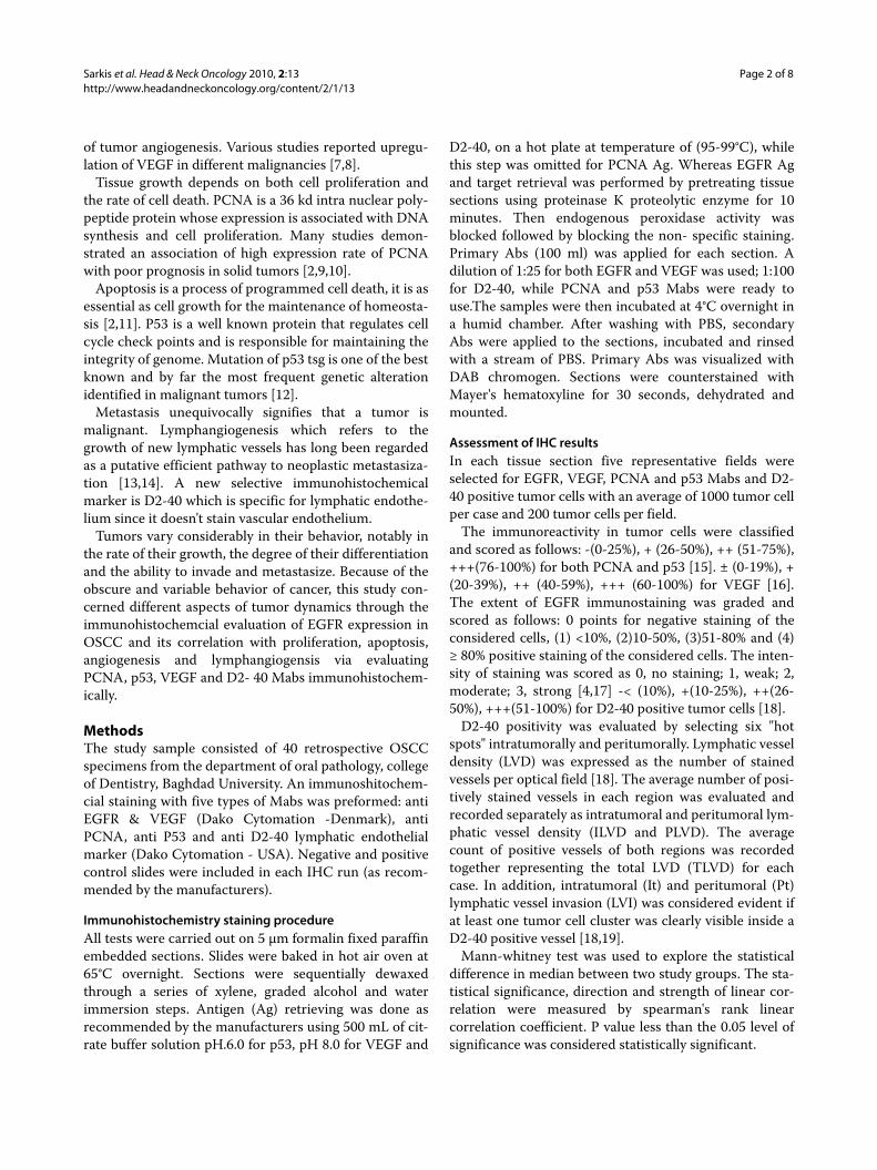
Figure 3 Strong PCNA nuclear immunostaining in moderately differentiated SCC (tongue and floor of mouth (x200)
Sarkis et al. Head & Neck Oncology 2010, 2:13http://www.headandneckoncology.org/content/2/1/13
Page 4 of 8
Assessment of the biological markers in relation to the clinicopathological parameters as well as to each otherMann-whitny's test showed a statistically non significantdifference regarding tumor stage, size, lymph nodemetastasis and grade with the median expression of allthe study markers. The only exception was a highly sig-nificant PLVD as well as a statistically significant TLVDdifferences were found with respect to tumor grade (p =0.002, p = 0.028) respectively (table 2). Moreover a highlysignificant ILVD and TLVD and a statistically significant
PLVD difference was found with respect to LVI (p =0.002, p = 0.004, p = 0.026) respectively (table 3).
On the other hand, spearman's rank linear correlationcoefficient test revealed a statistically significant correla-tion in PCNA vs p53 (r = 0.34), EGFR extent score vsILVD (p = 0.35), EGFR intensity score vs ILVD, PLVD andTLVD (p = 0.33, p = 0.36, p = 0.36) respectively. In addi-tion D2-40 positive tumor cells showed a statistically sig-nificant correlation vs ILVD and TLVD (p = 0.39, p =0.32) respectively, ILVD vs PLVD and TLVD (p = 0.62, p =0.85) respectively, PLVD vs TLVD and tumor grading (p =0.92, p = 0.43) respectively (p < 0.05) (Table 4).
DiscussionIt has been reported that the majority of head and neckcancer, including oral cancer express EGFR [20]. Sincemost oral cancers are epithelial in origin, it is reasonable
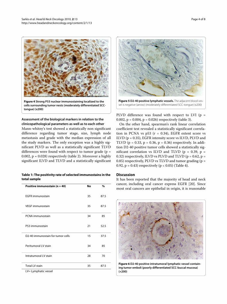
Figure 4 Strong P53 nuclear immunostaining localized to the cells surrounding tumor nests (moderately differentiated SCC-tongue) (x200)
Table 1: The positivity rate of selected immunostains in the total sample
Positive immunostain (n = 40) No %
EGFR immunostain 35 87.5
VEGF immunostain 35 87.5
PCNA immunostain 34 85
P53 immunostain 21 52.5
D2-40 immunostain for tumor cells 15 37.5
Peritumoral LV stain 34 85
Intratumoral LV stain 28 70
Total LV stain 35 87.5
LV= Lymphatic vessel
Figure 5 D2-40 positive lymphatic vessels. The adjacent blood ves-sel is negative (arrow) (moderately differentiated SCC-tongue) (x200)
Figure 6 D2-40 positive intratumoral lymphatic vessel contain-ing tumor emboli (poorly differentiated SCC-buccal mucosa) (×200)
Sarkis et al. Head & Neck Oncology 2010, 2:13http://www.headandneckoncology.org/content/2/1/13
Page 5 of 8
that they should have a high probability of expressingEGFR. In this series most of the examined cases (87.5%)showed positive EGFR immunostaining. This finding isconsistent with previous reports regarding the immunos-taining and expression of EGFR in OSCC and other can-cers [4,17,20].
A well controlled balance of cellular differentiation andproliferation is necessary for the development and main-tenance of normal epithelia throughout the body; sinceOSCCs are epithelial malignancies, therefore, they shouldhave a high probability of expressing EGFR. The presentstudy confirms the observations of others that high EGFRexpression is present in OSCCs which suggests that anuncontrolled growth may be mediated by abnormalEGFR expression [21].
EGFR expression extent and intensity scores revealedby most of the study cases suggest that EGFR expressingcarcinomas display pathological features of more aggres-sion which may be attributable to the activation of differ-ent signaling pathways that control diverse biologicalprocesses [4,22].
EGFR expression involved all epithelial layers in OSCCspecimens while in normal oral epithelia it was localizedto the basal cell layer, similar results were reported byother investigators [20,23]. Since the squamous epithe-lium keeps a continuous physiological regeneration innormal conditions, so that it is reasonable that the basalcells interpret signals of EGF by binding to EGFR [23],while its expression beyond basal localization in cancer-ous tissue suggests that a correlation between EGFR andtumor progress may exist.
Figure 7 Strong D2-40 tumor cells immunostaining within the stromal tissue indicating single cell microinvasion (arrows) (mod-erately differentiated SCC- mandible) (x1000)
Table 2: The difference in median score of selected immunostains by tumor grading (Mann-Whitney)
Tumor grading
Grade I Grade II-III P
Intratumoral LVD 0.24[NS]
Range (0 - 200) (0 - 160)
Median 18 30
Interquartile range (0 - 38) (8 - 57)
N 17 23
Mean rank 18 22.35
Peritumoral LVD 0.004
Range (0 - 155) (0 - 240)
Median 15 60
Interquartile range (0 - 48) (35 - 100)
N 17 23
Mean rank 14.29 25.09
Total LVD 0.028
Range (0 - 250) (0 - 300)
Median 46 93
Interquartile range (1 - 78) (48 - 173)
N 17 23
Mean rank 15.79 23.98
LVD = lymphatic vessel density
Sarkis et al. Head & Neck Oncology 2010, 2:13http://www.headandneckoncology.org/content/2/1/13
Page 6 of 8
The expression was mainly localized to the peripheriesof tumor nests, this observation is in accordance withother studies [20,23]. This finding confirms the presenceof this receptor on undifferentiated cells and explains thatthe staining reaction varies with cellular differentiation.Moreover, it may explain that peripheral tumor cells
receive a signal from EGF resulting in the proliferation ofcancerous tissues.
D2-40 positive lymphatic vessels were recorded in 35cases, similar results were found in OSCC and other can-cers [18,19,24,25], both peritumorally and intraumorallywhich suggests that these vessels could be a conduit forcarcinoma cells and may contribute to lymph nodemetastasis.
LVI was observed in 13 cases in the current study.Other investigators reported similar findings in OSCCand other cancers [19,24,25]. This may reflect a signifi-cant role of these vessels in producing a possible route forthe spread of tumor cells to regional lymph nodes.
Fifteen cases showed D2-40 expression by tumor cells,unfortunately there is no enough information concerningD2-40 expression in OSCC, therefore it is difficult toexplain these results clearly. However, it may indicate amore aggressive disease phenotype and suggest that D2-40 may be implicated in the differentiation of SCC. Fur-thermore, D2-40 positive tumor cells were detected in thestromal tissue as well which suggests that it could act as agood marker for microinvasion in OSCC, finding worthmore verification.
The current study showed no statistically significantdifferences between EGFR expression median scores andthe clinicopatholgical findings; similar observations werereported in other studies [4,17]. Furthermore, it was notcorrelated with the other markers as well (except D2-40)which indicate the independent effect of this marker onepithelial cancers development and growth. Moreover,the lack of correlation between EGFR extent or intensityscores in respect to VEGF expression but its existence inrespect to LVD as shown in this study would favor lym-phatic metastasis of OSCCs rather than hematogenous.
The results of the present study showed a statisticallysignificant difference regarding LVI with respect to themedian scores of ILVD, PLVD and TLVD. This findingprobably reflects the close relationship between LVI andthe lymphatic vessels since carcinomatous cells invadethe lymphatic vessels which exist in the area to get anaccess to the regional lymph nodes. Furthermore, lack ofcorrelation between LVD, LVI and D2-40 positive tumorcells may be attributed to the small size of node positivecases (12 out of 40).
Among all the available studies reviewed, to the best ofour knowledge, the present work is the first of its kind instudying comprehensively these biomarkers all together,except several studies that assessed only two or three ofthem together [15,21,26,27].
The results of this study clarify that the behavior ofOSCC is not dependent on a single factor but its combi-nation of multiple biological processes which are inde-pendent of each other i.e. malignancy follows no rules.
Table 3: The difference in median score of selected immunostains by lymphatic vessel invasion (Mann-Whitney)
Lymphatic vessel invasion
Negative Positive P
Intratumoral LVD 0.002
Range (0 - 200) (24 - 160)
Median 17 45
Interquartile range (0 - 31) (32 - 64)
N 22 13
Mean rank 13.82 25.08
Peritumoral LVD 0.026
Range (0 - 155) (9 - 240)
Median 42 72
Interquartile range (19 - 62) (48 - 120)
N 22 13
Mean rank 15.05 23
Total LVD 0.004
Range (2 - 250) (54 - 300)
Median 52 115
Interquartile range (36 - 110) (91 - 200)
N 22 13
Mean rank 14.18 24.46
LVD = Lymphatic vessel density
Sarkis et al. Head & Neck Oncology 2010, 2:13http://www.headandneckoncology.org/content/2/1/13
Page 7 of 8
Competing interestsThe authors declare that they have no competing interests.
Authors' contributionsSAS: Implementation of the immunohistochemical procedures, immunohis-tochemical interpretation and statistical analysis. BHA: Study design, histologi-cal examination and grading and immunohistochemical interpretivecalibration. BAAM: Technical assistance and logistic support during the immu-nohistochemical procedures. NGT: Histopathological evaluation of the cases,preparation of the article for publication and peer reviewing the final draft. Allauthors read and approved the final manuscript.
Author Details1Department of Oral Pathology, College of Dentistry, University of Baghdad, Iraq, 2Department of Pathology, College of Medicine, University of Baghdad, Iraq and 3Department of Oral Pathology and Medicine, College of Dentistry, University of Sulaimani, Iraq
References1. Kumar V, Cotran RS, Robbins SL, Neoplasia , Kumar V, Cotran RS, Robbins
SL, editors: Robbins Basic Pathology. Seventh edition. Saunders, Philadelphia; 2003.
2. Eissa S, Shoman S: Tumor Markers. Hodder Arnold Publication; 1998. 3. Lynch TJ, Bell DW, Sordella R, Gurubhagavatula S, Okimoto RA, Brannigan
BW, Harris PL, Haserlat SM, Supko JG, Haluska FG, Louis DN, Christiani DC, Settleman J, Haber DA: Activating mutations in the epidermal growth factor receptor underlying responsiveness of non-small-cell lung cancer to gefitinib. NEJM 2004, 350(21):2129-2139.
4. Laimer K, Spizzo G, Gastl G, Obrist P, Brunhuber Th, Fong D, Barbieri V, Jank S, Doppler W, Rasse M, Norer B: High EGFR expression predicts poor prognosis in patients with squamous cell carcinoma of the oral cavity and oropharynx: A TMA-based immunohistochemical analysis. Oral Oncology 2007, 43:193-198.
5. Shang ZJ, Li JR: Expression of endothelial nitric oxide synthase and vascular endothelial growth factor in oral squamous cell carcinoma: its correlation with angiogenesis and disease progression. J Oral Pathology & Medicine 2005, 34(3):134.
6. Johnstone S, Logan RM: The role of vascular endothelial growth factor (VEGF) in oral dysplasia and oral squamous cell carcinoma. Oral Oncology 2006, 42(4):337-342.
Received: 13 May 2010 Accepted: 25 June 2010 Published: 25 June 2010This article is available from: http://www.headandneckoncology.org/content/2/1/13© 2010 Sarkis et al; licensee BioMed Central Ltd. This is an Open Access article distributed under the terms of the Creative Commons Attribution License (http://creativecommons.org/licenses/by/2.0), which permits unrestricted use, distribution, and reproduction in any medium, provided the original work is properly cited.Head & Neck Oncology 2010, 2:13
Table 4: Correlation matrix
PCNA P53 EGFR extent score
EGFR Intensity score
VEGF D2-40 positive tumor cells
Intratumoral LVD
Peritumoral LVD
Total LVD
P53 0.34(*)
EGFR extent score
-0.19 0.16
EGFR Intensity score
0.03 0.01 0.25
VEGF 0.07 0.11 0.18 0.25
D2-40 positive tumor cells
-0.01 0.28 0.1 0.07 -0.09
Intratumoral LVD
0.07 0.26 0.35(*) 0.33(*) 0.08 0.39(*)
Peritumoral LVD 0.01 -0.01 0.02 0.36(*) 0.2 0.22 0.62(*)
Total LVD 0.07 0.11 0.18 0.36(*) 0.15 0.32(*) 0.85(*) 0.92(*)
Tumor staging -0.06 0.13 -0.09 0.09 -0.05 -0.26 -0.09 0.04 -0.04
Tumor grading -0.08 0.03 -0.01 0.05 0.09 0.15 0.2 0.43(*) 0.32(*)
Tumor size -0.06 0.01 -0.18 -0.13 -0.03 -0.32(*) -0.05 0.1 0.04
Lymph node staging
-0.02 0.22 0.05 0.25 0.03 -0.08 -0.01 0.12 0.06
Age 0.31 0.31 0.07 0.03 -0.2 0.08 -0.04 -0.28 -0.15
Sarkis et al. Head & Neck Oncology 2010, 2:13http://www.headandneckoncology.org/content/2/1/13
Page 8 of 8
7. Fukuda S, Shirahama T, Imazono Y: Expression of vascular endothelial growth factor in patients with testicular germ cell tumor as an indicator of metastatic disease. Cancer 1999, 85:1323-1330.
8. Costa C, Soares R, Schmitt F: Angiogenesis: Now and then. APMIS 2004:402-416.
9. Shin DM, Vararud N, Ro JY: Sequential increases in proliferative cell nuclear antigen expression in head and neck tumorigenesis: A potential biomarker. J Natl Cancer Inst 1993, 85:971-978.
10. Wang LF, Chai CY, Kuo WR, Tai CF, Lee KW, Ho KY: Correlation between proliferating cell nuclear antigen and p53 protein expression and 5-year survival rate in nasopharyngeal carcinoma. Am J of Otolaryngology 2006, 27:101-105.
11. Hall P: Cell proliferation. Journal of Pathology 1991, 165:349-354.12. Gasco M, Crook T: The p53 network in head and neck cancer. Oral
Oncology 2003, 39:222-231.13. Pepper MS: Lymphangiogenesis and tumor metastasis. Clinical Cancer
Research 2001, 7:462-468.14. Stacker S, Baldwin M, Achen M: Lymphangiogenesis lymphedema and
cancer. The role of tumor lymphangiogenesis in metastatic spread. The FASEB Journ 2002, 16:922-934.
15. Alves FA, Pires FR, de Almeida OP, Lopes MA, Kowalski LP: PCNA, Ki-67 and p53 expressions in submandibular salivary gland tumors. Int J Oral Maxillofac Surg 2004, 10:593-597.
16. Lim J, Kang S, Lee M, Pai H, Lee J, Hong S, et al.: Expression of vascular endothelial growth factor in salivary gland carcinomas and its relation to P53, Ki-67 and prognosis. J Oral Pathol Med 2003, 32:552-561.
17. Hiraishi Y, Wada T, Nakatani K, Negoro K, Fujita S: Immunohistochemical expression of EGFR and p-EGFR in oral squamous cell carcinomas. Pathology Oncology Research 2006, 12(2):.
18. Browning L, Bailey D, Parker A: D2-40 is a sensitive and specific marker in differentiating primary adrenal cortical tumors from both metastatic clear cell renal cell carcinoma and phaeochromocytoma. Journal of Clinical Pathology 2008, 61:293-296.
19. Kyzas PA, Stefanou D, Batistatou A, Agnantis NJ, Nakanishi Y, Hirohashi S, et al.: Dysadherin expression in head and neck squamous cell carcinoma association with lymphangiogenesis and prognostic significance. Am J Surg Pathol 2006, 30(2):185-193.
20. Christensen ME, Therkildsen MH, Hansen BL, Albeck H, Hensen GN, Bretlau P: Epidermal growth factor receptor expression on oral mucosa dysplastic epithelia and squamous cell carcinoma. Eur Arch Otorhinolaryngol 1992, 249(5):243-7.
21. Kręcicki T, Jeleń M, Zalesska-Kręcicka M, Rak J, Szkudlarek T, Jeleń-Kręcicka J: Epidermal growth factor receptor (EGFR), proliferating cell nuclear antigen (PCNA) and Ki-67 antigen in laryngeal epithelial lesions. Oral Oncology 1999, 35:180-186.
22. O- charoenrat P, Rhys-Evans PH, Archer DJ, Bccles SA: C- erb B recepotrs in squamous cell carcinomas of the head and neck clinical significance and correlation with matrix metalloproteinases and vascular endothelial growth factors. Oral Oncolgy 2002, 10(1):.
23. Sakai H, Kawano K, Hishimoto N: Immunohistochemical localization of c-myc oncogene product and EGF receptor in oral squamous cell carcinoma. J Oral Pathol Med 1990, 19:1-4.
24. Siriwardena BSMS, Kudo Y, Ogawa I, Udagama MNGPK, Tilakaratne WM, Takata T: VEGF-C is associated with lymphatic status and invasion in oral cancer. J Clin Pathol 2008, 61:103-108.
25. Gombos Z, Xu X, Chu CS, Zhang PJ, Acs G: Peritumoral lymphatic vessel density and vascular endothelial growth factor C expression in early- stage squamous cell carcinoma of the uterine cervix. Clinical Cancer Research 2005, 11:8364-8371.
26. Keum KC, Chung EJ, Koom WS, Cho JH, Cho SH, Choi EC, Lee CG, Suh CO, Kim GE: Predictive value of P53 and PCNA expression for occult neck metastases in patients with clinically node negative oral tongue cancer. Otolaryngology 2006, 135(6):858-864.
27. Lieto E, Ferraraccio F, Orditura M, Castellano P, Mura AL, Pinto M, Zamboli A, DeVita F, Galizia G: Expression of vascular endothelial growth factor (VEGF) and epidermal growth factor receptor (EGFR) is an independent prognostic indicator of worse outcome in gastric cancer patients. Ann Surg Oncol 2008, 15(1):69-79.
doi: 10.1186/1758-3284-2-13Cite this article as: Sarkis et al., Immunohistochemical expression of epider-mal growth factor receptor (EGFR) in oral squamous cell carcinoma in rela-tion to proliferation, apoptosis, angiogenesis and lymphangiogenesis Head & Neck Oncology 2010, 2:13
Top Related
Copyright © 2022 FDOKUMEN

