


Bahasa
Halaman
Hukum


High-Resolution Mass Spectrometry Driven Discovery ofPeptidic Danger Signals in Insect ImmunityArton Berisha1, Krishnendu Mukherjee2, Andreas Vilcinskas2, Bernhard Spengler1, Andreas Rompp1*
1 Institute of Inorganic and Analytical Chemistry, Justus Liebig University, Giessen, Germany, 2 Institute of Phytopathology and Applied Zoology, Justus Liebig University,
Giessen, Germany
Abstract
The ‘danger model’ is an alternative concept for immune response postulating that the immune system reacts to entitiesthat do damage (danger associated molecular patterns, DAMP) and not only to entities that are foreign (pathogen-associated molecular patterns, PAMP) as proposed by classical immunology concepts. In this study we used Galleriamellonella to validate the danger model in insects. Hemolymph of G. mellonella was digested with thermolysin (as arepresentative for virulence-associated metalloproteinases produced by humanpathogens) followed by chromatographicfractionation. Immune-stimulatory activity was tested by measuring lysozyme activity with the lytic zone assays againstMicrococcus luteus cell wall components. Peptides were analyzed by nano-scale liquid chromatography coupled to high-resolution Fourier transform mass spectrometers. Addressing the lack of a genome sequence we complemented therudimentary NCBI protein database with a recently established transcriptome and de novo sequencing methods for peptideidentification. This approach led to identification of 127 peptides, 9 of which were identified in bioactive fractions. DetailedMS/MS experiments in comparison with synthetic analogues confirmed the amino acid sequence of all 9 peptides. To testthe potential of these putative danger signals to induce immune responses we injected the synthetic analogues into G.mellonella and monitored the anti-bacterial activity against living Micrococcus luteus. Six out of 9 peptides identified in thebioactive fractions exhibited immune-stimulatory activity when injected. Hence, we provide evidence that small peptidesresulting from thermolysin-mediated digestion of hemolymph proteins function as endogenous danger signals which canset the immune system into alarm. Consequently, our study indicates that the danger model also plays a role in insectimmunity.
Citation: Berisha A, Mukherjee K, Vilcinskas A, Spengler B, Rompp A (2013) High-Resolution Mass Spectrometry Driven Discovery of Peptidic Danger Signals inInsect Immunity. PLoS ONE 8(11): e80406. doi:10.1371/journal.pone.0080406
Editor: Joy Sturtevant, Louisiana State University, United States of America
Received June 12, 2013; Accepted October 2, 2013; Published November 26, 2013
Copyright: � 2013 Berisha et al. This is an open-access article distributed under the terms of the Creative Commons Attribution License, which permitsunrestricted use, distribution, and reproduction in any medium, provided the original author and source are credited.
Funding: This work was supported by the Hessian ministry of Science and Arts (HMWK) through LOEWE focus "Ambiprobe" and LOEWE focus "Insectbiotechnology", by the Deutsche Forschungsgemeinschaft DFG through project Sp314/13-1, and by the European Research Council ERC-STG 210356. The fundershad no role in study design, data collection and analysis, decision to publish, or preparation of the manuscript.
Competing Interests: The authors have declared that no competing interests exist.
* E-mail: [email protected]
Introduction
A prerequisite for the evolution of Metazoa was the ability to
discriminate between self and nonself. This power of distinction is
mediated by a functioning immune system which encompasses all
endogenous mechanisms and molecules contributing to host
defense against microbes and parasites.
Vertebrate and invertebrate animals share the ancient innate
immunity mediating discrimination between infectious nonself and
noninfectious self by a limited number of germ line encoded
receptors which bind to molecules common among pathogens, but
absent from the host such as microbial cell wall components,
particularly, bacterial lipopolysaccharides (LPS), peptidoglycans,
and fungal ß-1,3-glucans [1]. This binding of receptor proteins to
pathogen-associated molecular patterns (PAMPs) differs from the
highly specific recognition of a wide range of antigens by a large
and diverse spectrum of somatically rearranged receptors of T and
B cells which represent an evolutionary novelty of vertebrates.
Their adaptive or acquired immunity relies on antibody-based
immunological memory of infection [2]. Janeways model claiming
that activation of the immune system requires recognition of
PAMPs has become a paradigm, particularly in insect immunity
[3]. Binding of PAMPs to corresponding pattern recognition
receptors results in activation of immune responses including both
cellular mechanisms such as phagocytosis or multicellular encap-
sulation of microbes, and humoral defences such as the rapid
synthesis of antimicrobial peptides and proteins [4].
Matzinger has challenged immunological research by introduc-
ing her danger model, which is based on the idea that the immune
system is more concerned with entities that do damage than with
those that are foreign [5]. Her alternative model explains
recognition of foreign threat by molecules released from damaged
cells or wounded tissues whose binding to corresponding pattern
recognition receptors results in activation of immune responses.
Such damage-associated molecular patterns (DAMPs) function as
danger signals which can set the immune system into alarm [6].
First evidence for the presence of immunity-related danger signals
has been reported from insects, particularly in the larvae of the
greater wax moth Galleria mellonella [7]. The latter have in between
emerged as powerful model hosts for pathogens infecting insects or
humans [8]. This study elucidated that activation of innate
immune responses in G. mellonella does not necessarily require
PAMPs, the presence of microbial enzymes, particularly metallo-
proteinases of the M4 family with thermolysin as the prototype, in
PLOS ONE | www.plosone.org 1 November 2013 | Volume 8 | Issue 11 | e80406

the hemolymph is sufficient to produce DAMPs [7]. Members of
the thermolysin-family encompass prominent virulence factors and
toxins of human pathogens such as aureolysin, bacillolysin and
pseudolysin which have been implicated to be responsible for
increase of vascular permeability, hemorrhagic edema, sepsis and
necrotic tissue destruction [9]. Thermolysin-mediated digestion of
hemolymph proteins in G. mellonella results in formation of small
peptidic fragments smaller than 3 kDa which elicit innate immune
responses that are qualitatively (spectrum of immune-related
proteins secreted within the hemolymph) and quantitatively
(expression levels of antimicrobial peptides) comparable with the
response to injected bacterial lipopolysaccharide (LPS), a widely
used microbial elicitor of innate immune responses [10].
Mass spectrometry (MS) is a powerful and universal tool for the
analysis and identification of proteins and peptides. Complex
biological samples are usually separated by liquid chromatography
or gel electrophoresis prior to MS analysis. The MS-based
identification of peptides and proteins from G. mellonella is hindered
by fact that the genome is not sequenced and thus protein
databases are only rudimentary. Traditional database search
approaches using SEQUEST [11] or Mascot [12] do not result in
satisfactory results. To compensate for this impediment we have
recently subjected the immunity-related transcriptome of G.
mellonella to next generation sequencing using the Roche 454-
FLX platform combined with traditional Sanger sequencing to
obtain a comprehensive immune gene repertoire [13]. In addition,
we have developed a method for de novo sequencing of peptides
isolated from G. mellonella hemolymph [14]. The reliability of
peptide identification can be significantly improved by using high
resolution and accurate measurements [15,16]. The highest mass
resolution is achieved by Fourier Transform mass spectrometers
based on orbital trapping [17] or ion cyclotron resonance [18,19].
A method that simplifies de novo sequencing is the composition
based sequencing (CBS) approach which takes advantage of
accurate mass measurements [20]. An example for CBS analysis of
the tree frog H. savigni with unknown genome sequence was
recently reported [21].
The application of high-resolution mass spectrometry enabled
the identification of a number of hemolymph protein fragments
resulting from thermolysin-mediated hydrolysis of hemolymph.
Their putative function as danger signals was verified using
synthetic analogues which were injected into G. mellonella larvae in
order to test their capacity to elicit humoral immune responses. As
in our previous studies [7,10] we used freeze-dried or living
Micrococcus luteus bacteria as indicator organisms in lytic zone or
inhibition zone assays, respectively.
Materials and Methods
Insect rearing and sample preparationG. mellonella larvae were reared on an artificial diet (22% maize
meal, 22% wheat germ, 11% dry yeast, 17.5% bee wax, 11%
honey and 11% glycerin) at 32uC in darkness. For collecting the
hemolymph the larvae were first cooled at 4uC for 15 min and the
prolegs were pierced with a sterile needle. Hemolymph melani-
sation was prevented by the use of few crystals of phenylthiourea.
Cell-free hemolymph was degraded with thermolysin (Sigma,
Taufkirchen, Germany) with an end concentration of 1 mg/mL
in sterile bidistilled water. The mixture was incubated for 1h at
36uC on a rotary shaker. These parameters (moderate enzyme
concentration and short incubation time) were chosen in order to
mimic the in vivo conditions. Thermolysin-degraded fragments
smaller than 3 kDa were obtained gradually by centrifugation at
7000 x g for 4-6h at 4uC using Centricon centrifugal concentrators
Centricon-30, Centricon-10 and Centricon-3 (Millipore, Billerica,
Massachusetts, USA), respectively.
Fractionation of hemolymph sampleSeparation of the thermolysin-digested hemolymph sample
(, 3 kDa) was realized by an Agilent 1200 RP HPLC system
(Agilent, Waldbronn, Germany) with a Symmetry C18 4.66250 mm
column at a flow-rate of 1 mL/min. The injection volume of the
sample was 100 mL. Separation was carried out using a 40 min
gradient starting at 5% acetonitrile in water (v/v). After 15 min the
solvent concentration of acetonitrile was raised up to 50% (v/v).
After 40 min the concentration of acetonitrile was increased to
95% (v/v). The resulting fractions were collected manually in
30sec intervals, lyophilized and dissolved in 50 mL H2O. The
fractions were injected in Galleria larvae and immune stimulation
was determined by lytic zone assay, subsequently.
Mass spectrometryNanoHPLC-ESI FTMS measurements. The measure-
ments of the samples were performed on a binary nanoHPLC
system consisting of the units Switchos, Famos and Ultimate
(LCPackings/Dionex, Idstein, Germany). Solvent A was water
(HPLC grade, Fluka, Neu-Ulm, Germany) containing 2% aceto-
nitrile (v/v) (UvasolH grade, Merck KGaA Darmstadt, Germany)
and 0.1% formic acid (v/v) (puriss p.a. for ms, Fluka Neu-Ulm,
Germany). Solvent B was acetonitrile containing 20% water (v/v)
and 0.08% formic acid (v/v). Separation of the hemolymph
sample was carried out using a 77 min gradient. First B was
increased from 0% to 10% in 10 min, subsequently increased to
30% in 30 min and then increased to 100% in 5 min and
maintained for 11 min. The injection volumes were 0.3 mL for the
hemolymph bulk sample and 1.2 to 5 mL for hemolymph fractions,
respectively. After pre-concentration on a C18 PepMap trap
column (5 mm6300 mm i.d.) the samples were separated on a
fused silica C18 PepMap100 capillary column (150 mm675 mm
i.d.) (Dionex, Idstein, Germany) at 200 nL/min. The separation
was monitored by a UV detector at 214 nm.
The nanoHPLC system was coupled to the mass spectrometer
by a nanospray source. Pico-TipH Emitter (New Objective, Woburn,
MS, USA) were used as nanospray needles. The separated peptides
were measured on a tandem mass spectrometer (LTQ FT Ultra,
Thermo Fisher Scientific GmbH, Bremen, Germany) consisting of a
linear ion trap (IT) and a Fourier Transform Ion Cyclotron
Resonance (FTICR) mass spectrometer. For database searches
high-resolution (R = 100.000 at m/z 400) survey spectra were
measured on the FTICR with high mass accuracy (, 2 ppm).
Fragment ion spectra were measured both with high resolution
and in the ion trap. For analyses with manual de novo and the
composition-based sequencing method precursor and fragment
ions were solely measured with high resolution and high mass
accuracy. Collision induced dissociation (CID) was used for fragmen-
tation. MS/MS data was obtained on the FT also using the wide
scan range (WSR) method with three microscans in order to
compensate the loss of ions due to the time-of-flight effect of the
FTICR mass spectrometer.
In addition, coupling of nanoHPLC with a linear ion trap /
Fourier transform orbital trapping (IT-FTOT MS) mass spec-
trometer (LTQ Orbitrap Discovery, Thermo Scientific GmbH, Bremen,
Germany) equipped with a nanospray ion source, was used. This
mass spectrometer allowed, in addition to CID-fragmentation, the
detection of low mass fragments by higher-energy collisional dissociation
(HCD), providing additional information for peptide sequencing.
Full scan and MS/MS spectra on the FTOT instrument were
acquired with high resolution (R = 30.000 at m/z 400) and high
MS Discovery of Peptidic Danger Signals
PLOS ONE | www.plosone.org 2 November 2013 | Volume 8 | Issue 11 | e80406

mass accuracy (, 4 ppm). External calibration of both FT
instruments was performed immediately before measurements
according to the instructions of the manufacturer. All MS
measurements were performed using the positive ion mode.
Offline ESI FTMS measurements. Offline nano ESI MS
measurements of synthetic peptides were performed for validation
of identified peptides using the orbitral trapping mass spectrom-
eter. Pico tip emitters (5 mm i.d., DNU-MS, Berlin, Germany) were
used for this task. The synthesized peptides were dissolved in
200 mL water/acetonitrile 50:50 (v/v) with 0.1% formic acid and
diluted 1:10 before measurement.
MALDI FTMS measurements. For matrix-assisted laser
desorption/ionization (MALDI) measurements the commercial
MALDI source of the orbital trapping mass spectrometer,
equipped with a N2-Laser (337 nm), was used. Alpha-cyano-4-
hydroxycinnamic acid (10 mg/mL) dissolved in acetonitrile/water
70:30 (v/v) with 0.1% trifluoroacetic acid was used as matrix.
Two mL matrix solution were mixed with 1 mL sample on a
stainless steel sample plate. The laser energy was set to 20 mJ and
the crystal positioning system (CPS) was activated. MS and MS/
MS experiments were performed with collision induced dissoci-
ation (CID) and higher-energy collisional dissociation (HCD) to
obtain low-mass fragment ions.
Data analysis / Identification of peptidesDatabase search with standard database. Data obtained
from HPLC-MS measurements were analyzed with SEQUEST
database search using the BioWorks V3.3.1 software (Thermo Fisher
Scientific, Bremen, Germany). For bulk samples 5 HPLC-MS
measurements were carried out and at least 2 measurements for
each bioactive fraction. Protein sequence database entries for G.
mellonella are very limited. Therefore all available database entries
for Galleria were downloaded from the server of the ‘‘National Center
of Biotechnology Information’’ NCBI (http://www.ncbi.nlm.nih.gov/).
This database contained 162 protein sequence database entries.
The database searches of all HPLC-MS measurements were
carried out with following settings: precursor ion tolerance 5 ppm;
fragment ion tolerance 1 Da. The following variable modifications
were selected: phosphorylation on serine, threonine, and tyrosine,
hydroxylation on proline, threonine and methionine and carbami-
domethylation on cysteine. These modifications are considered in
the software with a mass increment of the respective amino acid, for
hydroxylation Dm = +15.99492, for phosphorylation Dm =
+79.96633 and for carbamidomethylation Dm = +57.02146, re-
spectively. Thermolysin cleaves at the N-terminal side of amino
acids with hydrophobic or bulky side chains [22]. The amino acids
alanine, isoleucine, leucine, phenylalanine, valine and methionine
were selected as cleavage sites. The number of allowed missed
cleavage sites was set to 10. Peptides with a Peptide Probability below
0.001 were considered as identified. False discovery rates (FDR)
were calculated according to Nesvizhskii et al. [23].
Database search with experimental database. An
additional database search was applied with a newly implemented
G. mellonella database obtained from transcriptome analysis [13].
The transcriptomic data were translated into the single letter
amino acid code, manually implemented in a FASTA file and used
the same way as standard database entries for the SEQUEST
database searches with BioWorks. The database included 96756
entries, mainly short sequences (64% of all entries had a chain
length of less than 50 amino acids).
De novo sequencing. Manual de novo sequencing was
performed with accurately measured masses. Additionally, the
composition-based de novo sequencing approach was applied using
the computer program Peptide Composer 1.0 [20,24]. Calculations of
amino acid compositions of peptides were performed using
accurately measured mass spectra with a tolerance of 2 ppm for
FTICR and 4 ppm for FTOT mass spectra, both for precursor
ions and fragment ions. (The isobaric peptides leucine and
isoleucine could not be differentiated by exact mass measurements
due to their identical elemental composition.)
Synthesized peptidesAfter sequence determination, peptides were custom-synthe-
sized by GL Biochem (Shanghai Ltd., China) with a purity of over
85%.
Bioactivity testsLast instar G. mellonella larvae, weighing between 250–350 mg,
were used in this study for determining the immune stimulation.
TenmL of collected fractions or 20 mL of synthesized peptide
solutions (20 mM), respectively, were injected dorsolaterally into
the hemocoel of the larva using 1 mL disposable syringes and
0.4620 mm needles mounted on a microapplicator. After
injection, the larvae were incubated at 32uC in darkness. 24 hours
post injection, hemolymph was collected to investigate the immune
stimulation.
As a simple read-out system to determine the immune-
stimulatory activity of samples, we used the inhibition zone
assay using freeze-dried Micrococcus luteus [7,25]. The inhibition
zone assay against living bacteria such as Micrococcus luteus is a
standard assay which is widely used in microbiology to
qualitatively and quantitatively determine the activity of soluble
antimicrobial compounds. The latter are pipetted into wholes
which have been punched in the agar inoculated with bacteria.
The sample defund into the agar around thereby inhibiting the
growth of the bacteria. The diameter of the clear zones without
growing bacterial colonies can be measured and subsequently
quantified by using a calibration curve with a standard
antibiotic such as gentamycin. Accordingly, the anti-bacteria
activity in samples can by quantified in gentamycin equivalents.
Briefly, seven milliliter of M. luteus agar consisting of 1% high-
purity agar-agar (Carl Roth, Karlsruhe, Germany), 5 mg/mL
freeze-dried M. luteus (Sigma, Taufkirchen, Germany), and
67 mM potassium phosphate buffer (PBS) pH 6.4 were placed
onto petri dishes (Ø100 mm). Holes with a diameter of 4 mm
were filled with 3 mL hemolymph and incubated for 24h at
37uC. Lysozyme activity was quantitatively determined (units/
mL) by establishing a calibration curve from standard chicken
lysozyme (Sigma, Taufkirchen, Germany).
For validating the potency of identified peptidic danger signals
to elicit immune responses we used living M. luteus as an
indicator organism in the inhibition zone assay because
bacterial growth inhibition indicates the presence of induced
antibacterial peptides, whereas the lytic zone assay using freeze-
dried M. luteus indicates enzymatic digestion by lysozyme.
Synthetic peptides were dissolved in sterile saline solution
(120 mM NaCl) with an end concentration of 20 mM and
20 mL were injected into larvae. For each peptide 6 individual
larvae were used. The anti-M. luteus-activity was calculated to
gentamicin equivalents (inmg/mL) using a calibration curve with
gentamicin as previously described [10,26]. Saline solution
(peptide solvent) was used as control, and injected in the larvae
following the same procedure as for the tested peptides. Average
value and standard deviation for 6 individual animals were
calculated for each peptide. Statistically significant differences
between activities of larvae injected with peptides and control
were determined using Students t-test.
MS Discovery of Peptidic Danger Signals
PLOS ONE | www.plosone.org 3 November 2013 | Volume 8 | Issue 11 | e80406

Results and Discussion
Figure 1 shows the workflow for the combined approach used
for the identification of bioactive peptides. The thermolysin-
digested hemolymph sample was measured directly by nanoLC-
FTMS. Second, the hemolymph sample was fractionated and
resulting fractions showing lysozyme inducing activity were
analyzed in more detail by nanoLC-FTMS and MALDI MS.
Data analysis was performed by SEQUEST database search and
de novo sequencing. Identified peptides were tested individually for
immune stimulation by synthetic analogues.
Fractionation of hemolymph sample and initialbioactivity tests
Initial HPLC-MS measurements revealed that the thermolysin-
digested sample (, 3 kDa) was too complex for the direct
determination of bioactive compounds. For this reason the bulk
sample was fractionated by HPLC and collected fractions were
tested with a lytic zone assay to screen for elevated bioactivity.
Details on chromatographic fractionation and activity tests can be
found in the supporting information (File S1). Two fractions,
termed A and B, in which the highest immune-stimulatory activity
was detected were subjected to a second round of HPLC
fractionation in order to further decrease the complexity. The
resulting sub-fractions of these separation steps were tested again
for immune-stimulatory activity. This procedure resulted in 4 sub-
fractions for fraction A and 2 sub-fractions for fraction B. The tests
of these sub-fractions showed that sub-fraction A1 and B1 had the
highest activity and were therefore subjected to more detailed
analysis.
Peptide identification by mass spectrometryThe hemolymph samples were measured by nanoHPLC
coupled to FTMS instruments. The acquired data was analyzed
using standard database searches as well as experimental databases
derived from transcriptome data. Bioactive fractions were
analyzed in detail by nanoHPLC-FTMS and additionally by
MALDI Orbitrap measurements. Peptide sequence analysis was
finally extended to database-independent de novo sequencing
methods. Identification results are reported for the bulk sample
and for bioactive fractions separately, in the following. A list of all
identified peptides can be found in the supporting information
(Table S1).
Database search with standard database. One of the
standard procedures in proteomics for characterization of peptides
is high performance liquid chromatography coupled to mass
spectrometry followed by protein database searches. In this work
HPLC-FTMS measurements of the thermolysin-digested hemo-
lymph sample were performed with database search using
SEQUEST. For this purpose all rudimentary available protein
sequence entries from the public database NCBI (162 protein
entries) of G. mellonella were used.
Database search of the bulk sample with the NCBI database led
to the identification of 75 peptides. The majority of these identified
peptides were obtained by HPLC-MS/MS measurements where
the precursor ion was measured in the FT cell with high
resolution, and the five most intense ions of this survey scan were
fragmented and detected in the ion trap.
Most of the identified peptides could be assigned to abundant
hemolymph proteins such as hexamerin, apolipophorin and
transferrin precursor. The number of identified peptides was
Figure 1. Workflow for the identification of bioactive peptides. On the one hand the thermolysin-digested hemolymph sample was analyzeddirectly by nanoHPLC-FTMS. On the other hand the thermolysin-digested sample was pre-fractionated using a HPLC-system and the collectedsamples were tested for bioactivity for a more detailed analysis of immuno-relevant peptides.doi:10.1371/journal.pone.0080406.g001
MS Discovery of Peptidic Danger Signals
PLOS ONE | www.plosone.org 4 November 2013 | Volume 8 | Issue 11 | e80406

relatively low, especially when compared with database searches of
fully-sequenced species where in one run several hundred peptides
can be identified. This was due to the limited number of protein
entries in the database for G. mellonella and showed the necessity of
further identification methods. The determination of a false discovery
rate (FDR) that is common in database search studies was only
partly possible, because of the small size of the used databases. An
FDR of 2,7% was determined by searching a decoy database that
was generated by reversing the sequences of the original NCBI
database [27].
SEQUEST database search was also performed with na-
noHPLC-MS measurements of the bioactive fractions A, B and
the sub-fractions A1 and B1 (which had the highest bioactivity in
the initial activity tests). Five peptides could be identified in this
manner in the fraction B. These peptides were VV-9, IE-8, FN-9,
IN-10 and LY-11 (see Table 1 for sequence information). Of these
peptides IE-8 and LY-11 were also identified in the hemolymph
bulk measurements discussed above. The NCBI database searches
of fraction A and sub-fractions A1 and B1 showed no peptide
identification.
Database search with experimental database. A
recently established comprehensive transcriptome of G. mellonella
[13] was used as a database in order to identify additional
peptides. Database search of the bulk sample with this experi-
mental database resulted in 38 identified peptides. The decoy
database searches of three HPLC-MS runs using the reversed
database resulted in a FDR rate of 29%. These high values are
most likely due to the fact that most of the database entries in the
experimental database were short and therefore tended to lead to
more hits with the decoy database.
The HPLC-MS measurements of the bioactive fractions were
also analyzed by database search using the experimental database.
In fraction B1 only one peptide could be identified by this
approach: APPSGPAAPPAKTP (AP-14). In fraction B peptide
VV-9 could be identified that was also identified with the standard
database approach. No peptides were identified in fraction A and
sub-fraction A1. The total number of peptides identified with the
experimental database was 40.
De novo sequencing. De novo sequencing of peptides allows
identifying peptides without prior knowledge about possible amino
acid sequences. Therefore this approach was used in order to
identify peptides which are not included in the NCBI or
transcriptome database. Data analysis by de novo sequencing was
always performed using high resolution and high mass accuracy
measurements, both for precursor and fragment ions. In doing so,
it was possible to determine amino acid-specific increment masses
within the MS/MS spectra. For example lysine and glutamine
have the same nominal mass, but can easily be differentiated by
accurate mass measurements due to their mass difference of 36
mDa.
Twenty-two peptides were identified using the de novo sequenc-
ing approach in the bulk sample. Nine of this set were identified by
manual de novo sequencing of non-significant SEQUEST database
search hits (Peptide Probability above threshold of 0.001).
The de novo sequencing approach led to no identification of
peptides in the bioactive fractions. Therefore additional experi-
ments were performed that implemented matrix-assisted laser
desorption/ionization (MALDI) and the higher-energy collisional
dissociation device (HCD) as a fragmentation technique. This
fragmentation method allows the fragmentation in a dedicated
quadrupolar collision cell, which results in a lower mass cut-off for
fragmentation ions. An example of a peptide identified by this
approach is discussed in the following. In sub-fraction A1 two
peaks, m/z = 517.28360 and m/z = 631.38722, could be se-
quenced by the composition based sequencing approach (CBS)
(see [20,24] for more details). The MS/MS spectrum of precursor
ion m/z = 631.38722 using HCD fragmentation is shown in
Figure 2. For the characterization of this peptide the first step was
the determination of the amino acid composition. The detected
mass of m/z = 631.38722 corresponds to 11 possible amino acid
compositions within an accuracy of 3 ppm. By checking against
the fragment ions this value could be reduced to 4 possible amino
acid compositions (A,A,S,V,K,R), (G,A,S,L,K,R), (A,A,V,T,K,R)
and (A,K,K,E,R). In the MS/MS spectrum two neutral mass
losses indicated the loss of glutamic acid (single letter code: E) with
Dm/z = 129.0425 Da (between fragment ions m/z = 432.2558 and
m/z = 303.2133) and Dm/z = 129.0421 Da (between m/
z = 329.1812 and m/z = 200.1391), respectively. On the other
hand, there was no indication for a loss of serine, threonine, valine
or leucine. This information was used to determine the amino acid
composition, as only one of the 4 remaining possibilities contains
glutamic acid. The only remaining amino acid composition could
Table 1. Peptides identified in the bioactive fractions.
Fraction Sequence [M+H]+exp. Abbr. Used method Ionization/
Fragmentation
B IYHKPTTE 988.50713 IE-8 NCBI DB ESI/CID
B IKANAPQAEN 1055.54729 IN-10 NCBI DB ESI/CID
B FQATNDNKN 1051.47905 FN-9 NCBI DB ESI/CID
B LKTKNPSPDTY 1263.65666 LY-11 NCBI DB ESI/CID
B VDGKSAPNV 886.46198 VV-9 NCBI & transcriptome DB ESI/CID
B1 APPSGPAAPPAKTP 1258.67646 AP-14 transcriptome DB ESI/CID
B1 SRPSPNYP 917.44540 SP-8 de novo ESI/HCD
A1 ERRG 517.28360 EG-4 de novo MALDI/HCD
A1 KAERK 631.38722 KK-5 de novo MALDI/HCD
Listed are: the amino acid sequence of the peptides, the fraction in which the peptide was identified, and the method that was used for identification.CID: Collision induced dissociation.HCD: Higher-energy collisional induced dissociation.DB: Database.doi:10.1371/journal.pone.0080406.t001
MS Discovery of Peptidic Danger Signals
PLOS ONE | www.plosone.org 5 November 2013 | Volume 8 | Issue 11 | e80406
therefore be determined as (A,K,K,E,R). In the second step of the
composition-based de novo sequencing the peptide sequence was
determined by scoring the agreement between observed and
expected fragment ions of permuted sequence propositions. For
that all fragment ion signals of the MS/MS spectrum (Figure 2)
were used. The results of the de novo (CBS) sequencing showed that
the sequence KAERK (abbreviation ’KK-5’ in Table 1) was
matched with the highest CBS score value. The peptide sequence
of precursor ion m/z = 517.28360 was identified using the same
approach as ERRG (EG-4). Three peptides were identified by de
novo sequencing in the bioactive sub-fractions. Peptide SP-8 was
identified in sub-fraction B1 in addition to the two peptides EG-4
and KK-5 discussed above. In total 25 peptides were identified by
de novo sequencing.
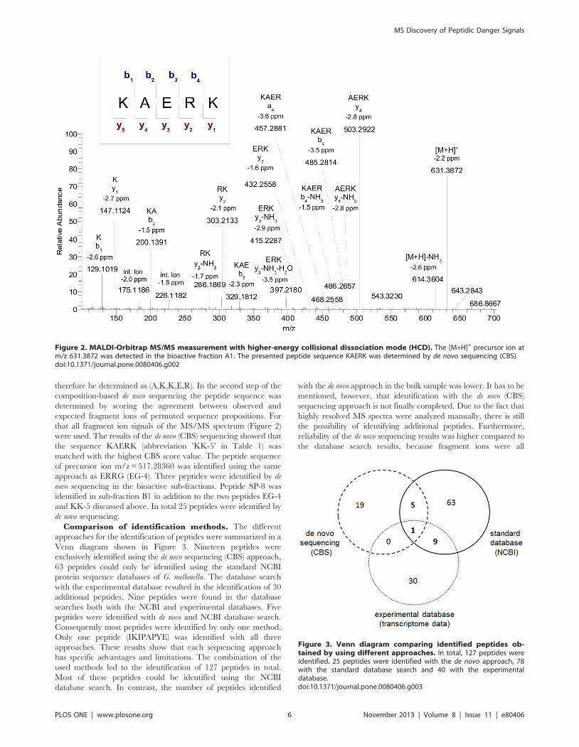
Comparison of identification methods. The different
approaches for the identification of peptides were summarized in a
Venn diagram shown in Figure 3. Nineteen peptides were
exclusively identified using the de novo sequencing (CBS) approach,
63 peptides could only be identified using the standard NCBI
protein sequence databases of G. mellonella. The database search
with the experimental database resulted in the identification of 30
additional peptides. Nine peptides were found in the database
searches both with the NCBI and experimental databases. Five
peptides were identified with de novo and NCBI database search.
Consequently most peptides were identified by only one method.
Only one peptide (IKIPAPYE) was identified with all three
approaches. These results show that each sequencing approach
has specific advantages and limitations. The combination of the
used methods led to the identification of 127 peptides in total.
Most of these peptides could be identified using the NCBI
database search. In contrast, the number of peptides identified
with the de novo approach in the bulk sample was lower. It has to be
mentioned, however, that identification with the de novo (CBS)
sequencing approach is not finally completed. Due to the fact that
highly resolved MS spectra were analyzed manually, there is still
the possibility of identifying additional peptides. Furthermore,
reliability of the de novo sequencing results was higher compared to
the database search results, because fragment ions were all
Figure 2. MALDI-Orbitrap MS/MS measurement with higher-energy collisional dissociation mode (HCD). The [M+H]+ precursor ion atm/z 631.3872 was detected in the bioactive fraction A1. The presented peptide sequence KAERK was determined by de novo sequencing (CBS).doi:10.1371/journal.pone.0080406.g002
Figure 3. Venn diagram comparing identified peptides ob-tained by using different approaches. In total, 127 peptides wereidentified. 25 peptides were identified with the de novo approach, 78with the standard database search and 40 with the experimentaldatabase.doi:10.1371/journal.pone.0080406.g003
MS Discovery of Peptidic Danger Signals
PLOS ONE | www.plosone.org 6 November 2013 | Volume 8 | Issue 11 | e80406

assigned based on accurate mass measurements. The FDR rate of
the search with the experimental database was high, indicating
that there were also false positives among these.
Of the 127 peptides that were identified in total, 9 peptides were
identified in the fractions with increased bioactivity. These
peptides are summarized in Table 1. Within this set, 4 peptides
were identified in fraction B using the standard NCBI database
search, whereas one peptide (VV-9) was identified both with the
standard and experimental database. Two peptides each were
identified in sub-fraction A1 and B1. Three peptides (EG-4, KK-5
and SP-8) were identified by the de novo approach, whereas one
peptide (AP-14) was determined by database search with the
experimental database. Using the standard database search alone
would have led to no results in the sub-fractions. Inclusion of
MALDI and HCD was also necessary in order to identify the
peptides in the sub-fractions. These results show that different
ionization techniques and different approaches for peptide analysis
had to be used for the identification of a broad set of peptides. All
of the identified peptides in the bioactive fractions were of
particular interest and had to be validated and tested for
bioactivity as discussed in the following section.
Confirmation by synthetic peptides. To validate the
sequence of the peptides identified in the bioactive fractions
summarized in Table 1, synthetic analogs were synthesized. Figure
4 shows the comparison of the FT-MS/MS spectra of peptide VV-
9 detected in the hemolymph fraction and its synthetic analogue.
Fragmentation experiments for validation were performed the
same way as for the hemolymph sample. The fragment ions of the
identified peptide and of the synthetic analogue, matched all with
high accuracy thus confirming the initial identification.
Seven peptides could be validated by accurate mass measure-
ments for precursor ion and fragment ions. Two peptides
identified by the database search, FN-9 and AP-14, were
confirmed by accurate precursor ion mass and fragment ions
measured in the ion trap. MS/MS spectra of these peptides can be
found in the supporting information (File S2). Consequently all
peptide sequences identified in bioactive fractions shown in Table
1 were confirmed by MS/MS measurements of synthesized
peptides.
Bioactivity tests of identified peptidesThe peptides identified by our MS approaches have been
postulated to represent putative danger signals resulting from
digestion of hemolymph by microbial metalloproteinases. In order
to validate their immune-stimulatory activity we produced
synthetic analogues which were injected into last instar larvae of
G. mellonella to determine their capacity to induce immune
response in vivo. As a simple read-out system we used the
inhibition zone assay against living M. luteus to determine
antibacterial activity in the hemolymph [25].
Interestingly, not all synthetic peptides turned out to exhibit
immune-stimulatory activity when injected (Figure 5). In compar-
ison with control injections with saline alone, the peptides VV-9,
IE-8, FN-9, IN-10, LY-11 and AP-14 induced significantly higher
anti-M. luteus-activity. On the other hand peptides KK-5, EG-4
and SP-8 did not induce a significant response. Therefore, we
conclude that only particular peptidic hemolymph protein
fragments can function as danger signals when released upon
metalloproteinase-mediated hydrolysis.
The larvae of the lepidopteran G. mellonella have been
established as a powerful model host for pathogens of insects or
humans and as a source for novel anti-infective therapeutics [28].
In addition, they were used to demonstrate for the first time that
the presence of microbial metalloproteinases alone is sufficient to
induce potent innate immune responses [7]. Thermolysin is the
prototype metalloproteinase belonging to the M4 family which
encompasses many virulence factors of human pathogens [9].
Thermolysin-mediated hydrolysis of hemolymph proteins from G.
mellonella results in formation of peptidic fragments which, when
injected, can elicit immune responses that are qualitatively and
quantitatively comparable with those observed upon injection of
bacterial cell wall components such as LPS [10].
Peptide analysis and identification in our study was more
complicated in comparison to standard proteomics experiments
due to several factors. Firstly, lacking a sequenced genome of G.
mellonella it was difficult to characterize these putative danger
signals by standard proteomics approaches. However, fully
sequenced insect systems such as Drosophila and Tribolium are very
small and therefore it is difficult to obtain hemolymph samples in
sufficient quantities (for mass spectrometric analysis and bioactivity
tests). Furthermore we have discovered the first specific peptidic
inhibitor of metalloproteinases in G. mellonella. This so-called IMPI
is currently being developed as new second generation antibiotic to
cure symptoms caused by thermolysin-like metalloproteinases
during infections in humans [28]. Secondly, peptides produced by
thermolysin are more difficult to ionize than those resulting from
other enzymes. Trypsin, as a digestion enzyme which produces
ions which are much easier to ionize, would not be representative
for the activity of microbial pathogens. It was shown previously
that trypsin does not induce immune response in Galleria mellonella
[7]. Finally, thermolysin digestion was performed at moderate
enzyme concentration and for 1 hour only, which results in
incomplete digestion of hemolymph proteins. A longer incubation
time and/or higher thermolysin concentrations would result in a
higher number of peptides. But this situation would not be
comparable with in vivo conditions during infection of G. mellonella
where only limited amounts of thermolysin-like metalloproteinases
are produced, plausibly to avoid activation of host immune
responses [29]. In addition it is known from our previous studies
that stronger or total digestion of hemolymph proteins does not
necessarily increases their immune-stimulatory activity [7].
To account for these specific challenges, we applied in this study
high resolution mass spectrometry combined with de novo
sequencing of peptides generated by metalloproteinase-mediated
digestion of hemolymph proteins from G. mellonella. This approach
led to the identification of several peptides in bioactive fractions.
Following a manual inspection of LC-MS/MS data, we estimate
that the two sub-fractions A1 and B1 contain about 10 peptides
each (see supporting information File S3 for details). The full
sequence of 2 peptides each was identified in these fractions and
partial sequences were obtained for other peptides. In order to
validate the postulated function of the de novo sequenced peptides
as danger signals we produced synthetic analogues which were
injected into G. mellonella larvae to assess their capacity to induce
innate immune responses. The latter are characterized by the
release of antimicrobial peptides among which some exhibit
activity against living Micrococcus luteus [28]. Strikingly but in
accordance with our expectation, it turned out that not all peptidic
fragments exhibit immune-stimulatory activity. Only the peptides
designated as VV-9, IE-8, FN-9, IN-10, LY-11 and AP-14 were
confirmed as elicitors of innate immune responses when injected.
We therefore postulate that the release of metalloproteinases
produced by invading pathogens and parasites results in genera-
tion of these danger signals which, in turn, induce innate immune
responses [29]. The formation of these peptidic danger signals can
only be detected by direct (mass spectrometric) analysis on the
peptide level. Transcriptomic approaches would only detect the
endogenous precursor peptides/proteins of these digestion prod-
MS Discovery of Peptidic Danger Signals
PLOS ONE | www.plosone.org 7 November 2013 | Volume 8 | Issue 11 | e80406

ucts. In this context it is important to note that G. mellonella is the
only known animal which is capable to produce a specific peptidic
inhibitor against microbial metalloproteinases [30]. This insect
metalloproteinase inhibitor (IMPI) was originally discovered in
and purified from hemolymph of immune-stimulated larvae [31],
and mediates reportedly feedback-loop regulation of harmful
microbial metalloproteinases [32].
Consequently, we have identified peptides which are generated
in G. mellonella under presence of microbial metalloproteinases, and
which mediate the activation of immune responses encompassing
the synthesis and release into the hemolymph of both antimicro-
bial peptides and the IMPI. Regarding the high in vivo toxicity of
thermolysin-like metalloproteinases, the characterized peptidic
products of their activity can be considered as danger signals
because they can set the immune system into alarm. This efficient
mechanism providing the ability to sense invading pathogens or
parasites by the activity of their virulence-associated enzymes [29]
adds to the sophisticated strategies determining the tremendous
evolutionary plasticity of insect immunity [33].
Conclusions
In this study we used high-resolution mass spectrometry to
identify peptidic hemolymph protein fragments which are
generated in the hemolymph of the model host G. mellonella when
harmful microbial metalloproteinases belonging to the M4 family,
with thermolysin as the prototype, are present. Addressing the lack
of a genome sequence we complemented the rudimentary NCBI
database with a transcriptome database and de novo sequencing
approaches for peptide identification. This approach led to the
identification of 9 potentially bioactive peptides. These (tentative)
results were validated by studying synthesized peptide analogues.
Detailed MS/MS experiments confirmed the amino acid sequence
of all 9 peptides. Six out of 9 peptides identified in the bioactive
Figure 4. Validation of the identified peptide VV-9 in hemolymph fraction B. The MS/MS spectrum of the precursor ion m/z 886.46 wasacquired with accurate mass using CID(35) as fragmentation technique. The sequence of this peptide was determined as VDGKSAPNV by databasesearch. The fragmentation pattern of the synthetic peptide is in good accordance with the MS/MS spectrum of the identified peptide in the bioactivefraction.doi:10.1371/journal.pone.0080406.g004
MS Discovery of Peptidic Danger Signals
PLOS ONE | www.plosone.org 8 November 2013 | Volume 8 | Issue 11 | e80406

fractions exhibited immune-stimulatory activity when injected in
Galleria larvae. These six peptidic elicitors of immune responses are
postulated to function as danger signals indicating the presence of
microbial metalloproteinases and mediating their regulation by
specific inducible metalloproteinase inhibitors. Our results suggest
that the validity of the immunity-related danger model proposed
by Matzinger can be expanded beyond mammals to include
insects. The detailed molecular pathways and importance of this
process will have to be investigated in follow-up studies.
Supporting Information
Table S1 List of identified peptides.
(DOC)
File S1 Chromatographic fractionation and activitytests of hemolymph.
(DOC)
File S2 MS/MS spectra of peptides identified inhemolymph and of the corresponding synthetic peptidestandards.(DOC)
File S3 Estimating number of peptides in bioactivefractions.(DOC)
Acknowledgments
We thank Hala Mahfoud for HPLC separation of the hemolymph bulk
sample and for the lytic zone assays of the fractions.
Author Contributions
Conceived and designed the experiments: AR AV BS. Performed the
experiments: AB KM. Analyzed the data: AB AR KM. Contributed
reagents/materials/analysis tools: BS AV AR. Wrote the paper: AR AB
AV. Provided unique instrumentation and data analysis capabilities: BS.
References
1. Janeway CA, Medzhitov R (2002) Innate immune recognition. Annual Review
of Immunology 20: 197–216.
2. Akira S, Uematsu S, Takeuchi O (2006) Pathogen recognition and innate
immunity. Cell 124: 783–801.
3. Lazzaro BP, Rolff J (2011) Immunology. Danger, microbes, and homeostasis.
Science 332: 43–44.
4. Wiesner J, Vilcinskas A (2010) Antimicrobial peptides The ancient arm of the
human immune system. Virulence 1: 440–464.
5. Matzinger P (2002) The danger model: A renewed sense of self. Science 296:
301–305.
6. Matzinger P (2007) Friendly and dangerous signals: is the tissue in control?
Nature Immunology 8: 11–13.
7. Griesch J, Wedde M, Vilcinskas A (2000) Recognition and regulation of
metalloproteinase activity in the haemolymph of Galleria mellonella: a new
pathway mediating induction of humoral immune responses. Insect Biochem-
istry and Molecular Biology 30: 461–472.
8. Mylonakis E, Casadevall A, Ausubel FM (2007) Exploiting amoeboid and non-
vertebrate animal model systems to study the virulence of human pathogenic
fungi. Plos Pathogens 3: 859–865.
9. Adekoya OA, Sylte I (2009) The Thermolysin Family (M4) of Enzymes:
Therapeutic and Biotechnological Potential. Chemical Biology & Drug Design
73: 7–16.
10. Altincicek B, Linder M, Linder D, Preissner KT, Vilcinskas A (2007) Microbial
metalloproteinases mediate sensing of invading pathogens and activate innate
immune responses in the lepidopteran model host Galleria mellonella. Infection
and Immunity 75: 175–183.
11. Eng JK, McCormack AL, Yates JR (1994) An Approach to Correlate Tandem
Mass-Spectral Data of Peptides with Amino-Acid-Sequences in a Protein
Database. Journal of the American Society for Mass Spectrometry 5: 976–989.
12. Perkins DN, Pappin DJC, Creasy DM, Cottrell JS (1999) Probability-based
protein identification by searching sequence databases using mass spectrometry
data. Electrophoresis 20: 3551–3567.
Figure 5. Bioactivity test of the identified peptides against (living) Micrococcus bacteria. The activity is shown in Gentamycin equivalentsinmg/mL. Peptides VV-9, IE-8, FN-9, IN-10, LY-11 and AP-14 exhibit significantly higher immune-stimulatory activity than the used solvent control.Peptide KK-5 shows no activity compared to the solvent control, in contrast. EG-4 and SP-8 show slightly elevated immune activity. Statisticallysignificant differences between activities of larvae injected with peptides and control were determined using Students t-test and are indicated by *(p,0.05) and ** (p,0.005), (n = 4-6 for each peptide).doi:10.1371/journal.pone.0080406.g005
MS Discovery of Peptidic Danger Signals
PLOS ONE | www.plosone.org 9 November 2013 | Volume 8 | Issue 11 | e80406

13. Vogel H, Altincicek B, Gloeckner G, Vilcinskas A (2011) A comprehensive
transcriptome and immune-gene repertoire of the lepidopteran model hostGalleria mellonella. Bmc Genomics 12: 1–19.
14. Altincicek B, Berisha A, Mukherjee K, Spengler B, Roempp A, et al. (2009)
Identification of collagen IV derived danger/alarm signals in insect immunity bynanoLC-FTICR MS. Biological Chemistry 390: 1303–1311.
15. Rompp A, Dekker L, Taban I, Jenster G, Boogerd W, et al. (2007) Identificationof leptomeningeal metastasis-related proteins in cerebrospinal fluid of patients
with breast cancer by a combination of MALDI-TOF, MALDI-FTICR and
nanoLC-FTICR MS. Proteomics 7: 474–481.16. Schober Y, Schramm T, Spengler B, Rompp A (2011) Protein identification by
accurate mass matrix-assisted laser desorption/ionization imaging of trypticpeptides. Rapid Communications in Mass Spectrometry 25: 2475–2483.
17. Scigelova M, Makarov A (2006) Orbitrap mass analyzer—overview andapplications in proteomics. Proteomics 6 Suppl 2: 16–21.
18. Marshall AG, Hendrickson CL (2002) Fourier transform ion cyclotron
resonance detection: principles and experimental configurations. InternationalJournal of Mass Spectrometry 215: 59–75.
19. Rompp A, Taban IM, Mihalca R, Duursma MC, Mize TH, et al. (2005)Examples of Fourier transform ion cyclotron resonance mass spectrometry
developments: from ion physics to remote access biochemical mass spectrometry.
European Journal of Mass Spectrometry 11: 443–456.20. Spengler B (2004) De novo sequencing, peptide composition analysis, and
composition-based sequencing: A new strategy employing accurate massdetermination by Fourier transform ion cyclotron resonance mass spectrometry.
Journal of the American Society for Mass Spectrometry 15: 703–714.21. Langsdorf M, Ghassempour A, Rompp A, Spengler B (2010) Characterization
of a peptide family from the skin secretion of the Middle East Tree Frog Hyla
savignyi by composition-based de novo sequencing. Rapid Communications inMass Spectrometry 24: 2885–2899.
22. Keil B (1992) Specificity of Proteolysis. Berlin-Heidelberg-New York: Springer.336 p.
23. Nesvizhskii AI, Vitek O, Aebersold R (2007) Analysis and validation of
proteomic data generated by tandem mass spectrometry. Nature Methods 4:
787–797.
24. Spengler B (2007) Accurate mass as a bioinformatic parameter in data-to-
knowledge conversion: Fourier transform ion cyclotron resonance mass
spectrometry for peptide de novo sequencing. European Journal of Mass
Spectrometry 13: 83–87.
25. Vilcinskas A, Matha V (1997) Effect of the entomopathogenic fungus Beauveria
bassiana on the humoral immune response of Galleria mellonella larvae
(Lepidoptera: Pyralidae). European Journal of Entomology 94: 461–472.
26. Altincicek B, Vilcinskas A (2006) Metamorphosis and collagen-IV-fragments
stimulate innate immune response in the greater wax moth, Galleria mellonella.
Developmental and Comparative Immunology 30: 1108–1118.
27. Reidegeld KA, Eisenacher M, Kohl M, Chamrad D, Korting G, et al. (2008) An
easy-to-use Decoy Database Builder software tool, implementing different decoy
strategies for false discovery rate calculation in automated MS/MS protein
identifications. Proteomics 8: 1129–1137.
28. Vilcinskas A (2011) ANTI-infective Therapeutics from the Lepidopteran Model
Host Galleria mellonella. Current Pharmaceutical Design 17: 1240–1245.
29. Vilcinskas A (2010) Coevolution between pathogen-derived proteinases and
proteinase inhibitors of host insects. Virulence 1: 206–214.
30. Vilcinskas A, Wedde M (2002) Insect inhibitors of metalloproteinases. IUBMB
Life 54: 339–343.
31. Wedde M, Weise C, Nuck R, Altincicek B, Vilcinskas A (2007) The insect
metalloproteinase inhibitor gene of the lepidopteran Galleria mellonella encodes
two distinct inhibitors. Biological Chemistry 388: 119–127.
32. Clermont A, Wedde M, Seitz V, Podsiadlowski L, Lenze D, et al. (2004) Cloning
and expression of an inhibitor of microbial metalloproteinases from insects
contributing to innate immunity. Biochemical Journal 382: 315–322.
33. Vilcinskas A (2013) Evolutionary plasticity of insect immunity. Journal of Insect
Physiology 59: 123–129.
MS Discovery of Peptidic Danger Signals
PLOS ONE | www.plosone.org 10 November 2013 | Volume 8 | Issue 11 | e80406
Top Related
Copyright © 2022 FDOKUMEN

