


Bahasa
Halaman
Hukum

Instructions for use
Title Hertwig's epithelial root sheath cell behavior during initial acellular cementogenesis in rat molars
Author(s) Yamamoto, Tsuneyuki; Yamamoto, Tomomaya; Yamada, Tamaki; Hasegawa, Tomoka; Hongo, Hiromi; Oda,Kimimitsu; Amizuka, Norio
Citation Histochemistry and cell biology, 142(5), 489-496https://doi.org/10.1007/s00418-014-1230-1
Issue Date 2014-11
Doc URL http://hdl.handle.net/2115/60279
Rights The final publication is available at link.springer.com
Type article (author version)
File Information Histochem Cell Biol_142(5).pdf
Hokkaido University Collection of Scholarly and Academic Papers : HUSCAP
1
Hertwig’s epithelial root sheath cell behavior during initial acellular cementogenesis in rat molars
Tsuneyuki Yamamoto1, Tomomaya Yamamoto
1, Tamaki Yamada
1, Tomoka Hasegawa
1, Hiromi Hongo
1,
Kimimitsu Oda2, Norio Amizuka
1
1 Department of Developmental Biology of Hard Tissue, Hokkaido University Graduate School of Dental
Medicine
2 Division of Biochemistry, Niigata University Graduate School of Medical and Dental Sciences
Corresponding author:
Tsuneyuki Yamamoto
Department of Developmental Biology of Hard Tissue, Hokkaido University Graduate School of Dental
Medicine, Kita 13 Nishi 7, Kita-ku, Sapporo 060-8586, Japan
E-mail: [email protected]
Tel: +81-11-706-4224
Fax: +81-11-706-4225
2
Abstract
This study was designed to examine developing acellular cementum in rat molars by
immunohistochemistry, to elucidate (1) how Hertwig’s epithelial root sheath disintegrates and (2) whether
epithelial sheath cells transform into cementoblasts through epithelial–mesenchymal transition (EMT).
Initial acellular cementogenesis was divided into three developmental stages, which can be seen in three
different portions of the root: portion 1, where the epithelial sheath is intact; portion 2, where the epithelial
sheath becomes fragmented; and portion 3, where acellular cementogenesis begins. Antibodies against
three kinds of matrix proteinases, which degrade epithelial sheath-maintaining factors, including basement
membrane and desmosomes, were used to investigate proteolytic activity of the epithelial sheath. Tissue
non-specific alkaline phosphatase (TNALP) and keratin were used to investigate EMT. Epithelial sheath
cells showed immunoreactivity for all three enzymes at fragmentation, which suggests that epithelial sheath
disintegration is enzymatically mediated. Dental follicle cells and cementoblasts showed intense
immunoreactivity for TNALP, and from portion 1 through to 3, the reaction extended from the alveolar
bone-related zone to the root-related zone. Cells possessing keratin/TNALP double immunoreactivity were
virtually absent. Keratin-positive epithelial sheath cells showed negligible immunoreactivity for TNALP,
and epithelial cells did not appear to migrate to the dental follicle. Together, these findings suggest that a
transition phenotype between epithelial cells and cementoblasts does not exist in the developing dental
follicle, and hence that epithelial sheath cells do not undergo EMT during initial acellular cementogenesis.
In brief, this study supports the notion that cementoblasts derive from the dental follicle.
246 words
Key words: Hertwig’s epithelial root sheath, Cementoblasts, Epithelial–mesenchymal transition, Acellular
cementum, Rat molars

3
Introduction
Cementum is a dental hard tissue that functions as a tooth-supporting structure, along with the periodontal
ligament and the alveolar bone. Its formation is regulated by Hertwig’s epithelial root sheath and
cementoblasts. The prevailing notion on initial cementogenesis is that the epithelial root sheath covers the
developing root edge and grows apically with cell proliferation. As root dentinogenesis advances, the
epithelial sheath becomes fenestrated and fragmented. Dental follicle cells then approach the root surface
through the gaps formed by sheath fragmentation, differentiate into cementoblasts, and secrete cementum
matrices, including collagen fibrils and non-collagenous matrices. Epithelial cell clusters derived from the
fragmented sheath survive in the periodontal ligament as the epithelial rests of Malassez.
However, questions remain in this theory, one of which concerns epithelial sheath fragmentation.
It is known that the structural integrity of the epithelial sheath is maintained by the basement membrane
and desmosomes. E-cadherins are also important for intercellular adhesion (Obara et al. 1999). During
sheath fragmentation, however, these epithelial sheath-maintaining factors must suffer some sort of
degradation. Two mechanisms have been proposed for the degradation. First, external mechanical damage
or invasion of dental follicle cells into the epithelial sheath causes degradation (Cho and Garant 1988;
Suzuki et al. 2002). Alternatively, epithelial sheath cells secrete matrix-degrading enzymes that disintegrate
the factors (Hirata and Nakamura 2006). Which one of these phenomena is the actual degradation
mechanism is presently unknown.
Another indistinct point in the theory, concerns the fate of the epithelial sheath cells. Many
reports have emphasized that some epithelial sheath cells transform into cementoblasts through epithelial–
mesenchymal transition, or EMT (Thomas 1995; Webb et al. 1996; Bosshardt et al. 1998; Bosshardt and
Nanci 1998, 2004; Lésot et al. 2000; Zeichner-David et al. 2003; Bosshardt 2005; Huang et al. 2009;
Akimoto et al. 2011). This novel idea can explain why the number of epithelial cells decreases immediately
after epithelial sheath fragmentation, even though only a few epithelial cells die by apoptosis (Kaneko et al.
1999; Suzuki et al. 2002). Some investigators, however, have disputed EMT and support the original idea
of cementogenesis (Diekwisch 2001; Suzuki et al. 2002; Yamamoto et al. 2007; Yamamoto and Takahashi
2009).
This study was designed to examine developing acellular cementum in rat molars by
immunohistochemistry (IHC), to thereby elucidate (1) how the epithelial sheath disintegrates, or how the
epithelial sheath-maintaining factors are degraded, and (2) whether the epithelial sheath cells undergo EMT.
For point (1), antibodies against three kinds of matrix proteinases[kallikrein7(KLK7)], a disintegrin and
metalloproteinase 10(ADAM10), and matrix metalloproteinase7(MMP7) ), which are associated with the
degradation of the epithelial sheath-maintaining factors, were used; KLK7 degrades desmosomal cadherins
(Caubet et al. 2004), ADAM10 degrades desmosomal cadherins, type IV collagen and E-cadherins (White
2003; Edwards et al. 2008), and MMP7 degrades laminin and type IV collagen (Birkedal-Hansen et al.
1993). For point (2), antibodies against tissue non-specific alkaline phosphatase (TNALP) and keratin
were used to verify whether epithelial sheath cells acquire mineralization-inducing activity. It has been
established that cementoblasts, like other mineralization-inducing cells, such as odontoblasts and
osteoblasts, show intense TNALP activity (Iwamatsu 1993; Yamamoto et al. 2007).
4
Materials and Methods
Twenty 20-day-old male Wistar rats weighing about 50 g were used in this study. The animals and tissue
specimens were treated in accordance with the guidelines of Hokkaido University’s Experimental Animal
Committee (No.10-0081).
After anesthesia with an intraperitoneal injection of sodium pentobarbital, animals were perfused
with 4% paraformaldehyde in 0.1 M phosphate buffer (pH 7.4) for 15 min. The maxillae were removed,
freed of soft tissues and demineralized in 5% ethylene-diaminetetraacetic acid. Specimens were dehydrated
in a graded series of ethanol and embedded in paraffin. Sagittal serial sections of the first maxillary molar
were then cut at 5 μm thickness. Some sections were stained with hematoxylin and eosin (HE) for general
histological examination and others were used for IHC as described below.
IHC for keratin
Deparaffinized sections were immersed in methanol containing 0.3% hydrogen peroxide to inhibit
endogeneous peroxidase and treated with 0.5% trypsin in 0.01M Tris-HCl buffer (pH7.6) for 20 min at
37°C. Pre-treated sections were incubated with anti-pan keratin mouse monoclonal antibody (Abcam,
Tokyo, Japan) and then with anti-mouse IgG goat polyclonal antibody conjugating horseradish peroxidase
(HRP) (Histofine Simple Stain rat MAX-PO (M): Nichirei, Tokyo, Japan). The immunoreaction was
visualized using 3, 3’-diaminobenzidine as a substrate. Immunostained sections were counter-stained with
methyl green.
IHC for proteinases (MMP7, KLK7 and ADAM10) and double IHC for proteinase-keratin
Rabbit polyclonal antibodies against MMP7 (Bioss Inc., Woburn, MA, USA), KLK7 (Bioss Inc.) and
ADAM10 (Sigma, St. Louis, MO, USA) were used. After inhibition of endogeneous peroxidase, sections
were incubated with the antibodies and then with anti-rabbit IgG goat polyclonal antibody conjugating HRP
(Histofine Simple Stain rat MAX-PO (R): Nichirei), and visualized by the 3, 3’-diaminobenzidine method.
Sections were counter-stained with methyl green, mounted with glycerin and photographed. Sections were
then processed for proteinase-keratin double staining.
After removal of glycerin, sections were treated with trypsin and incubated with anti-pan keratin
antibody (Abcam), followed by incubation with anti-mouse IgG secondary antibody. Keratin
immunoreaction was visualized with the Vector® VIP substrate Kit (Vector Laboratories, Burlingame, CA,
USA). Double-immunostained sections were again counter-stained with methyl green.
IHC for TNALP staining and double IHC for TNALP-keratin
After inhibition of endogeneous peroxidase, sections were incubated with anti-TNALP rabbit polyclonal
antibody (Oda et al. 1999), and then with anti-rabbit IgG secondary antibody, for posterior visualization by
the 3, 3’-diaminobenzidine method. Sections were counter-stained with methyl green, mounted with
5
glycerin and photographed for successive TNALP-keratin double staining. After removal of glycerin,
sections were treated for keratin staining as described in the proteinase-keratin double staining method.
For all sets of IHC experiments, controls were obtained by substitution of normal rabbit or mouse
serum for the primary antibodies. These control sections did not show any specific immunoreactivity.
6
Results
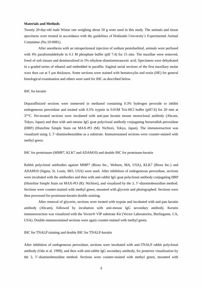
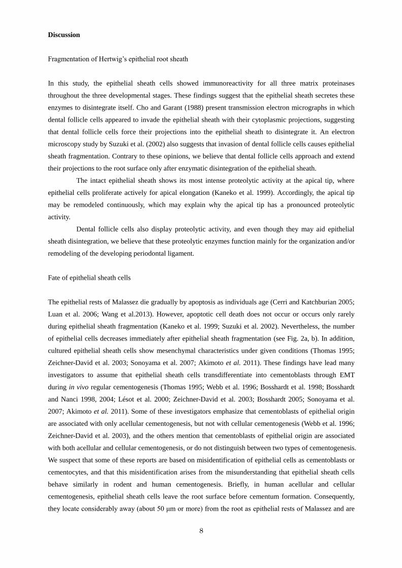
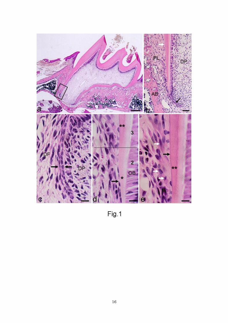
The mesial side of the mesial root of the maxillary first molars displayed the full range of stages of
acellular cementogenesis (Fig. 1a, b). Hence, the apical portion of this side was examined for investigation
of initial acellular cementogenesis. For descriptive convenience, initial acellular cementogenesis was
divided into three stages, which can be seen in three different portions: portion 1, where the Hertwig’s
epithelial root sheath is intact; portion 2, where the epithelial sheath becomes fragmented; and portion 3,
where acellular cementogenesis begins.
Histology
In portion 1, Hertwig’s epithelial root sheath consisted of two cell layers, namely the inner and the outer
enamel epithelial cells (Fig. 1c). Dental follicle cells were small and slender, and arranged in parallel with
the epithelial sheath. In portion 2, dental follicle cells became large and plump; in contrast, the epithelial
sheath cells turned into small, cytoplasm-poor cells. Dental papilla cells differentiated into odontoblasts and
started to form predentin (Fig. 1d). With the onset of predentin formation, the epithelial sheath began to
fragment. At this point, cytoplasm-poor cells adhering on the root surface could be recognized as epithelial
cells. However, it was difficult to make a strict distinction between dental follicle cells and epithelial cells
in HE-stained sections in portion 2. In portion 3, with the onset of dentin mineralization,
hematoxylin-stainable initial acellular cementum started to form on the mineralized dentin, and
cytoplasm-rich large cells, generally referred to as cementoblasts, appeared on the root surface (Fig. 1e).
The cementoblasts located about 15-20 μm apart from the root surface.
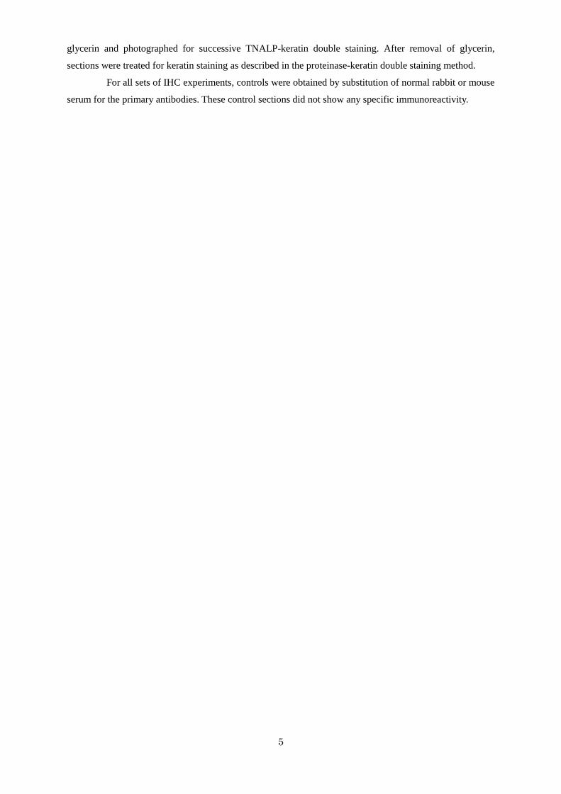
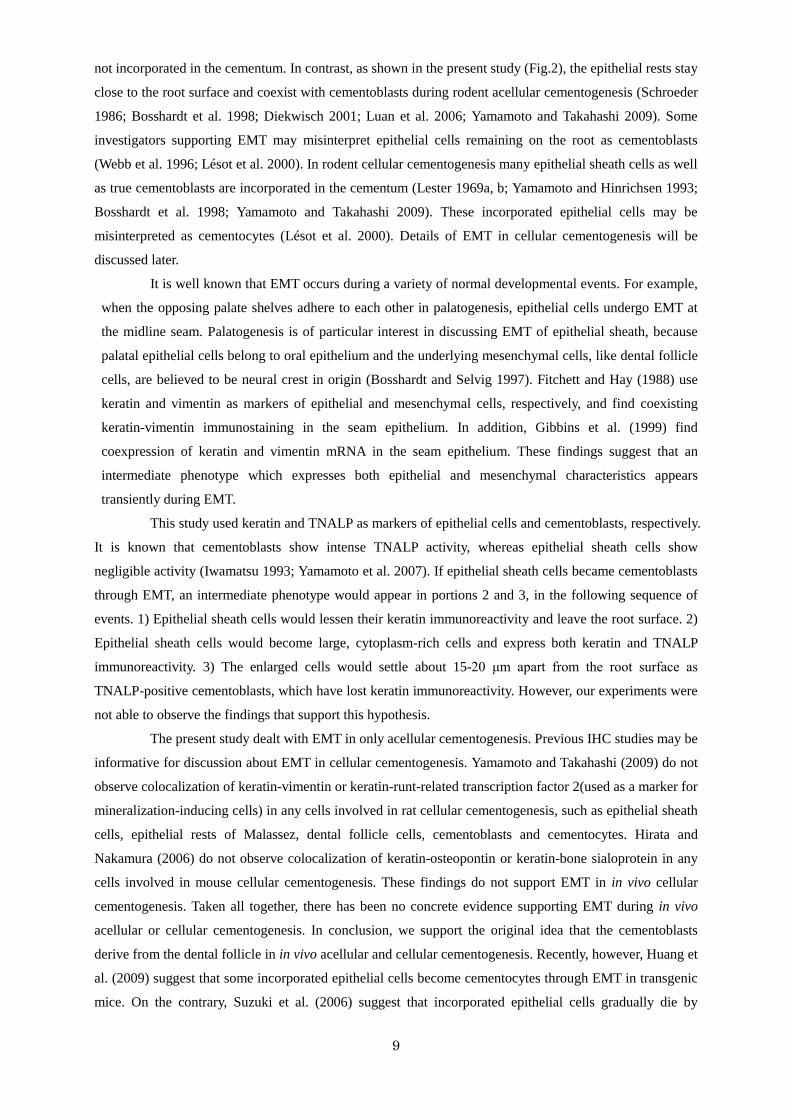
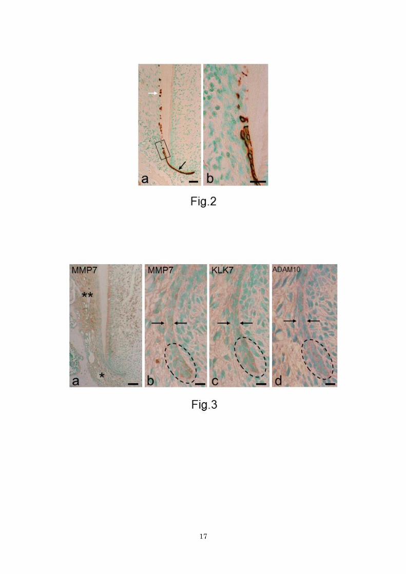
IHC for keratin
In portion 1, the intact epithelial sheath was consistently immunoreactive for keratin (Fig. 2a). In portions 2
and 3, this consistency was lost with fragmentation of the epithelial sheath and epithelial rests of Malassez
appeared. The epithelial rests were located in close proximity to the root surface. Keratin-positive epithelial
cells decreased in number after fragmentation (Fig. 2b). Cementoblasts were not immunoreactive for
keratin (see Fig. 1e).
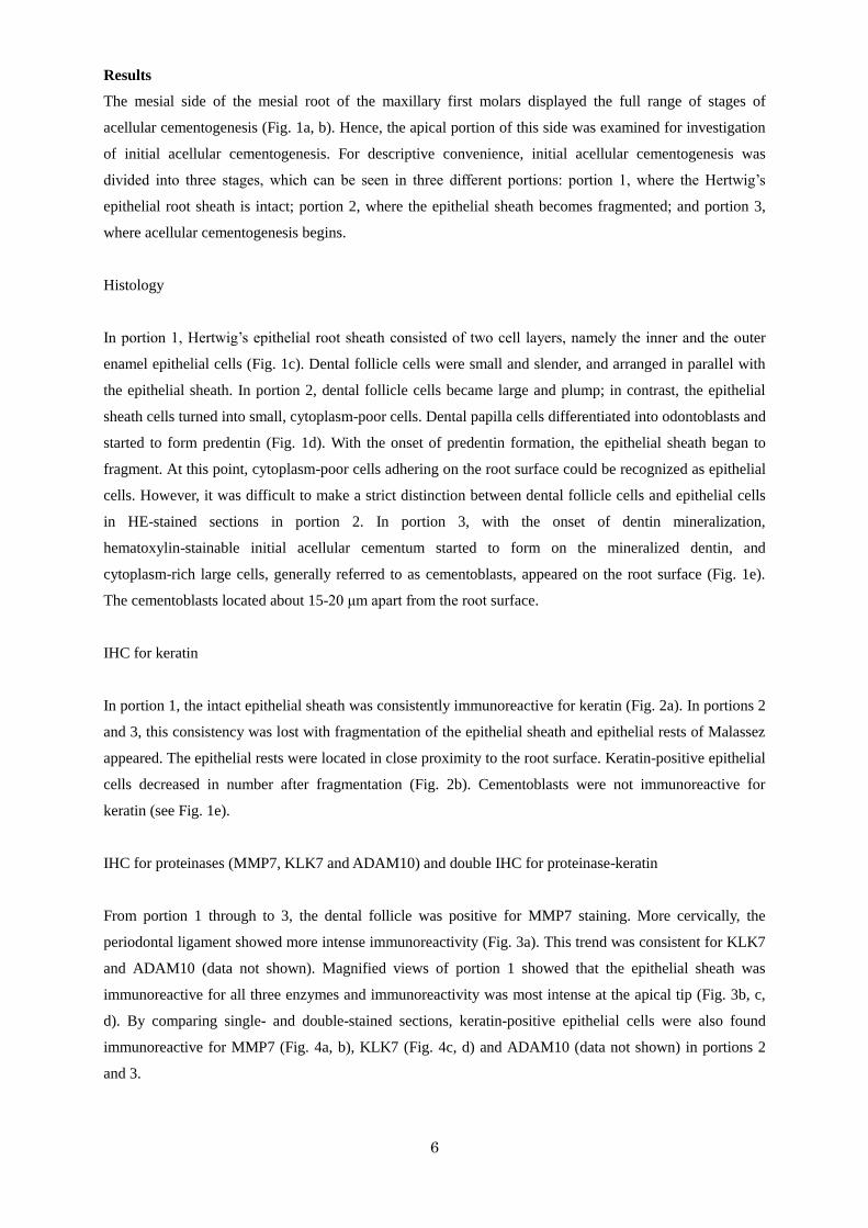
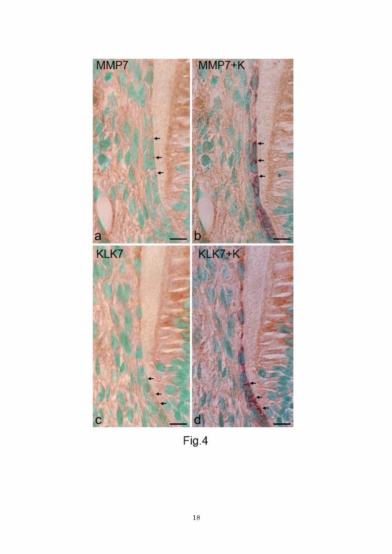
IHC for proteinases (MMP7, KLK7 and ADAM10) and double IHC for proteinase-keratin
From portion 1 through to 3, the dental follicle was positive for MMP7 staining. More cervically, the
periodontal ligament showed more intense immunoreactivity (Fig. 3a). This trend was consistent for KLK7
and ADAM10 (data not shown). Magnified views of portion 1 showed that the epithelial sheath was
immunoreactive for all three enzymes and immunoreactivity was most intense at the apical tip (Fig. 3b, c,
d). By comparing single- and double-stained sections, keratin-positive epithelial cells were also found
immunoreactive for MMP7 (Fig. 4a, b), KLK7 (Fig. 4c, d) and ADAM10 (data not shown) in portions 2
and 3.
7
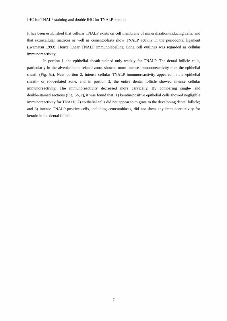
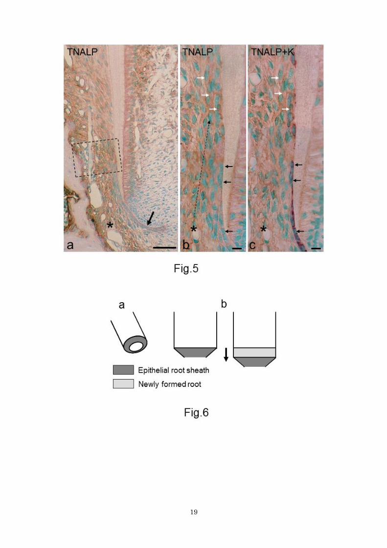
IHC for TNALP staining and double IHC for TNALP-keratin
It has been established that cellular TNALP exists on cell membrane of mineralization-inducing cells, and
that extracellular matrices as well as cementoblasts show TNALP activity in the periodontal ligament
(Iwamatsu 1993). Hence linear TNALP immunolabelling along cell outlines was regarded as cellular
immunoreactivity.
In portion 1, the epithelial sheath stained only weakly for TNALP. The dental follicle cells,
particularly in the alveolar bone-related zone, showed more intense immunoreactivity than the epithelial
sheath (Fig. 5a). Near portion 2, intense cellular TNALP immunoreactivity appeared in the epithelial
sheath- or root-related zone, and in portion 3, the entire dental follicle showed intense cellular
immunoreactivity. The immunoreactivity decreased more cervically. By comparing single- and
double-stained sections (Fig. 5b, c), it was found that: 1) keratin-positive epithelial cells showed negligible
immunoreactivity for TNALP; 2) epithelial cells did not appear to migrate to the developing dental follicle;
and 3) intense TNALP-positive cells, including cementoblasts, did not show any immunoreactivity for
keratin in the dental follicle.
8
Discussion
Fragmentation of Hertwig’s epithelial root sheath
In this study, the epithelial sheath cells showed immunoreactivity for all three matrix proteinases
throughout the three developmental stages. These findings suggest that the epithelial sheath secretes these
enzymes to disintegrate itself. Cho and Garant (1988) present transmission electron micrographs in which
dental follicle cells appeared to invade the epithelial sheath with their cytoplasmic projections, suggesting
that dental follicle cells force their projections into the epithelial sheath to disintegrate it. An electron
microscopy study by Suzuki et al. (2002) also suggests that invasion of dental follicle cells causes epithelial
sheath fragmentation. Contrary to these opinions, we believe that dental follicle cells approach and extend
their projections to the root surface only after enzymatic disintegration of the epithelial sheath.
The intact epithelial sheath shows its most intense proteolytic activity at the apical tip, where
epithelial cells proliferate actively for apical elongation (Kaneko et al. 1999). Accordingly, the apical tip
may be remodeled continuously, which may explain why the apical tip has a pronounced proteolytic
activity.
Dental follicle cells also display proteolytic activity, and even though they may aid epithelial
sheath disintegration, we believe that these proteolytic enzymes function mainly for the organization and/or
remodeling of the developing periodontal ligament.
Fate of epithelial sheath cells
The epithelial rests of Malassez die gradually by apoptosis as individuals age (Cerri and Katchburian 2005;
Luan et al. 2006; Wang et al.2013). However, apoptotic cell death does not occur or occurs only rarely
during epithelial sheath fragmentation (Kaneko et al. 1999; Suzuki et al. 2002). Nevertheless, the number
of epithelial cells decreases immediately after epithelial sheath fragmentation (see Fig. 2a, b). In addition,
cultured epithelial sheath cells show mesenchymal characteristics under given conditions (Thomas 1995;
Zeichner-David et al. 2003; Sonoyama et al. 2007; Akimoto et al. 2011). These findings have lead many
investigators to assume that epithelial sheath cells transdifferentiate into cementoblasts through EMT
during in vivo regular cementogenesis (Thomas 1995; Webb et al. 1996; Bosshardt et al. 1998; Bosshardt
and Nanci 1998, 2004; Lésot et al. 2000; Zeichner-David et al. 2003; Bosshardt 2005; Sonoyama et al.
2007; Akimoto et al. 2011). Some of these investigators emphasize that cementoblasts of epithelial origin
are associated with only acellular cementogenesis, but not with cellular cementogenesis (Webb et al. 1996;
Zeichner-David et al. 2003), and the others mention that cementoblasts of epithelial origin are associated
with both acellular and cellular cementogenesis, or do not distinguish between two types of cementogenesis.
We suspect that some of these reports are based on misidentification of epithelial cells as cementoblasts or
cementocytes, and that this misidentification arises from the misunderstanding that epithelial sheath cells
behave similarly in rodent and human cementogenesis. Briefly, in human acellular and cellular
cementogenesis, epithelial sheath cells leave the root surface before cementum formation. Consequently,
they locate considerably away (about 50 μm or more) from the root as epithelial rests of Malassez and are
9
not incorporated in the cementum. In contrast, as shown in the present study (Fig.2), the epithelial rests stay
close to the root surface and coexist with cementoblasts during rodent acellular cementogenesis (Schroeder
1986; Bosshardt et al. 1998; Diekwisch 2001; Luan et al. 2006; Yamamoto and Takahashi 2009). Some
investigators supporting EMT may misinterpret epithelial cells remaining on the root as cementoblasts
(Webb et al. 1996; Lésot et al. 2000). In rodent cellular cementogenesis many epithelial sheath cells as well
as true cementoblasts are incorporated in the cementum (Lester 1969a, b; Yamamoto and Hinrichsen 1993;
Bosshardt et al. 1998; Yamamoto and Takahashi 2009). These incorporated epithelial cells may be
misinterpreted as cementocytes (Lésot et al. 2000). Details of EMT in cellular cementogenesis will be
discussed later.
It is well known that EMT occurs during a variety of normal developmental events. For example,
when the opposing palate shelves adhere to each other in palatogenesis, epithelial cells undergo EMT at
the midline seam. Palatogenesis is of particular interest in discussing EMT of epithelial sheath, because
palatal epithelial cells belong to oral epithelium and the underlying mesenchymal cells, like dental follicle
cells, are believed to be neural crest in origin (Bosshardt and Selvig 1997). Fitchett and Hay (1988) use
keratin and vimentin as markers of epithelial and mesenchymal cells, respectively, and find coexisting
keratin-vimentin immunostaining in the seam epithelium. In addition, Gibbins et al. (1999) find
coexpression of keratin and vimentin mRNA in the seam epithelium. These findings suggest that an
intermediate phenotype which expresses both epithelial and mesenchymal characteristics appears
transiently during EMT.
This study used keratin and TNALP as markers of epithelial cells and cementoblasts, respectively.
It is known that cementoblasts show intense TNALP activity, whereas epithelial sheath cells show
negligible activity (Iwamatsu 1993; Yamamoto et al. 2007). If epithelial sheath cells became cementoblasts
through EMT, an intermediate phenotype would appear in portions 2 and 3, in the following sequence of
events. 1) Epithelial sheath cells would lessen their keratin immunoreactivity and leave the root surface. 2)
Epithelial sheath cells would become large, cytoplasm-rich cells and express both keratin and TNALP
immunoreactivity. 3) The enlarged cells would settle about 15-20 μm apart from the root surface as
TNALP-positive cementoblasts, which have lost keratin immunoreactivity. However, our experiments were
not able to observe the findings that support this hypothesis.
The present study dealt with EMT in only acellular cementogenesis. Previous IHC studies may be
informative for discussion about EMT in cellular cementogenesis. Yamamoto and Takahashi (2009) do not
observe colocalization of keratin-vimentin or keratin-runt-related transcription factor 2(used as a marker for
mineralization-inducing cells) in any cells involved in rat cellular cementogenesis, such as epithelial sheath
cells, epithelial rests of Malassez, dental follicle cells, cementoblasts and cementocytes. Hirata and
Nakamura (2006) do not observe colocalization of keratin-osteopontin or keratin-bone sialoprotein in any
cells involved in mouse cellular cementogenesis. These findings do not support EMT in in vivo cellular
cementogenesis. Taken all together, there has been no concrete evidence supporting EMT during in vivo
acellular or cellular cementogenesis. In conclusion, we support the original idea that the cementoblasts
derive from the dental follicle in in vivo acellular and cellular cementogenesis. Recently, however, Huang et
al. (2009) suggest that some incorporated epithelial cells become cementocytes through EMT in transgenic
mice. On the contrary, Suzuki et al. (2006) suggest that incorporated epithelial cells gradually die by
10
apoptosis. We will investigate EMT in cellular cementogenesis further.
But still, why do the epithelial cells decrease in number immediately after epithelial sheath
fragmentation? Regarding this question, Diekwisch (2001) has an interesting insight: “The epithelial root
sheath cells proliferate only at the apical end of the sheath while the entire root surface grows considerably.
As a result, the epithelial sheath only covers small portions of the developmentally advanced tooth root.
The disproportionate growth rate may explain why the root surface is only covered by very few epithelial
cells.” We agree with this comment, and consider that the shape of the intact epithelial sheath is also
associated with the disproportionate growth rate. The intact epithelial sheath bends toward the dental
papilla and forms a tapering cylinder (Fig. 6). The root grows somewhat straight, whereas the epithelial
sheath maintains the tapering shape during root elongation (Kaneko et al. 1999; Luan et al. 2006). In this
situation, the epithelial sheath could not cover the entire surface of the developing root owing to the
discrepancy of surface area, even though epithelial cells proliferate actively at the apical tip. Therefore, the
epithelial sheath may be enzymatically disintegrated and dispersed over the root surface. This may help
cementoblasts approach the root surface and explain the decreased epithelial cell number in histological
sections.
Acknowledgment
This study was supported by a grant from the Japanese Society for the Promotion of Science to T.
Yamamoto (No. 22592028).
11
References
Akimoto T, Fujiwara N, Kagiya T, Otsu K, Ishizeki K, Harada H (2011) Establishment of Hertwig’s
epithelial root sheath cell line from cells involved in epithelial-mesenchymal transition. Biochem Biophys
Res Commun 404:308-312.
Birkedal-Hansen H, Moore WGI, Bodden MK , Windsor LJ, Birkedal-Hansen B, DeCarlo A, Engler
JA(1993) Matrix metalloproteinases: a review. Crit Rev Oral Biol Med 4: 197-250.
Bosshardt DD (2005) Are cementoblasts a subpopulation of osteoblasts or a unique phenotype? J Dent Res
84: 390-406.
Bosshardt DD, Nanci A (1998) Immunolocalization of epithelial and mesenchymal matrix constituents in
association with inner enamel epithelial cells. J Histochem Cytochem 46: 135-142.
Bosshardt DD, Nanci A (2004) Hertwig’s epithelial root sheath, enamel matrix proteins, and initiation of
cementogenesis in porcine teeth. J Clin Periodontol 31: 184-192.
Bosshardt DD, Selvig KA(1997) Dental cementum: the dynamic tissue covering of the root. Periodontol
2000 13:41-75.
Bosshardt DD, Zalzal S, McKee MD, Nanci A(1998) Developmental appearance and distribution of bone
sialoprotein and osteopontin in human and rat cementum. Anat Rec 250: 13-33.
Caubet C, Jonca N, Brattsand M, Guerrin M, Bernard D, Schmidt R, Egelrud T, Simon M, Serre G(2004)
Degradation of corneodesmosome proteins by two serine proteases of the Kallikrein family,
SCTE/KLK5/hK5 and SCCE/KLK7/hK7. J Invest Dermatol 122:1235-1244.
Cerri PS, Katchburian E (2005) Apoptosis in the epithelial cells of the rests of Malassez of the
periodontium of rat molars. J Periodont Res 40:365-372.
Cho MI, Garant PR (1988) Ultrastructural evidence of directed cell migration during initial cementoblast
differentiation in root formation. J Periodont Res 23:268-276.
Diekwisch TSH (2001) The developmental biology of cementum. Int J Dev Biol 45: 695–706.
Edwards DR, Handsley MM, Pennington CJ (2008) The ADAM metalloproteinases. Mol Aspects Med 29:
258-289.
Fitchett JE, Hay ED(1989) Medial edge transform to mesenchyme after embryonic palatal shelves fuse.
12
Dev Biol 131:455-474.
Gibbins JR, Manthey A, Tazawa YM, Scott B, Bloch-Zupan A, Hunter N(1999) Midline fusion in the
formation of secondary palate anticipated by upregulation of keratinK5/6 and localized expression of
vimentin mRNA in medial edge epithelium. Int J Dev Biol 43:237-244.
Hirata A, Nakamura H (2006) Localization of perlecan and heparanase in Hertwig’s epithelial root sheath
during root formation in mouse molars. J Histochem Cytochem 54: 1105-1113.
Huang X, Bringas JrP, Slavkin HC, Chai Y (2009) Fate of HERS during tooth root development. Dev Biol
334:22-30.
Iwamatsu Y (1993) Histochemical and electron microscopical study during the development of the mouse
molar periodontal ligament. Jpn J Conserv Dent (in Japanese) 36:252-270.
Kaneko H, Hashimoto S, Enokiya Y, Ogiuchi H, Shimono M (1999) Cell proliferation and death of
Hertwig’s epithelial root sheath in the rat. Cell Tissue Res 298:95-103.
Lésot F, Davideau J-L, Thomas B, Sharpe P, Forest N, (2000) Epithelial Dlx-2 homeogene expression and
cementogenesis. J Histochem Cytochem 48:277-283.
Lester KS (1969a) The incorporation of epithelial cells by cementum. J Ultrastruct Res 27: 63-87.
Lester KS (1969b) The unusual nature of root formation in molar teeth of the laboratory rat. J Ultrastruct
Res 28: 481-506.
Luan X, Ito Y, Diekwisch TGH (2006) Evolution and development of Hertwig’s epithelial root sheath. Dev
Dyn 235: 1167-1180.
Obara N, Suzuki Y, Nagai Y, Takeda M (1999) Immunofluorescence detection of cadherins in mouse tooth
germs during root development. Arch Oral Biol 44:415-421.
Oda K, Amaya Y, Fukushi-Irie M, Kinameri Y, Ohsuye K, Kubota I, Fujimura S, Kobayashi J(1999) A
general method foe rapid purification of soluble versions of glycosylphosphatidylinositol-anchoered
proteins expressed in insect cells: an application for human tissue-non specific alkaline phosphatase. J
Biochem 126:694-699.
Schroeder HE (1986) Cementum. In: Schroeder HE(ed) The periodontium. Springer, Berlin Heidelberg
New York, pp23-127.
Sonoyama W, Seo BM, Yamaza T, Shi S (2007) Human Hertwig’s epithelial root sheath cells play crucial
roles in cementum formation. J Dent Res 86:594-599.
Suzuki M, Inoue T, Shimono M, Yamada S (2002) Behavior of epithelial root sheath during tooth root
formation in porcine molars: TUNEL, TEM, and immunohistochemical studies. Anat Embryol 206:13-20.
13
Suzuki M, Matsuzaka K, Yamada S, Shimono M, Abiko Y, Inoue T (2006) Morphology of Marassez’s
epithelial rest-like cells in the cementum: transmission electron microscopy, immunohistochemical, and
TdT-mediated dUTP-biotin nick end labeling studies. J Periodont Res 41:280-287.
Thomas HF (1995) Root formation. Int J Dev Biol 39: 231-237.
Wang Y, Lv L, Yu X, Zhang t, Li S(2013) The characteristics of epithelial cell rests of Malassez during
tooth eruption of development mice. J Mol Hist.doi:10.1007/s10735-013-9527-2
Webb PP, Moxham BJ, Benjamin M, Ralphs JR(1996) Changing expression of intermediate filaments in
fibroblasts and cementoblasts of the developing periodontal ligament of the rat molar teeth. J Anat 188:
529-539.
White JM (2003) ADAMs: modulator of cell-cell and cell-matrix interactions. Curr Opn Cell Biol
15:598-606.
Yamamoto T, Hinrichsen KV (1993) The development of cellular cementum in rat molars, with special
reference to the fiber arrangement. Anat Embryol 188: 537-549.
Yamamoto T, Domon T, Takahashi S, Anjuman KAY, Fukushima C, Wakita M (2007) Mineralization
process during acellular cementogenesis in rat molars: a histochemical and immunohistochemical study
using fresh-frozen sections. Histochem Cell Biol 127: 303-311.
Yamamoto T, Takahashi S (2009) Hertwig’s epithelial root sheath cells do not transform into cementoblasts
in rat molar cementogenesis. Ann Anat 191:547-555.
Zeichner-David M, Oishi K, Su Z, Zakartchenco V, Chen LS, Arzate H, Bringas Jr P (2003) Role of
Hertwig’s epithelial root sheath cells in tooth root development. Dev Dyn 228: 651-663.
14
Figure Legends
Figure 1. HE-stained sections. a Whole view of 20-day-old rat maxillary first molar. Developing mesial
root is observed. Bar 200 μm. b Magnification of boxed area in a. Black and white arrows indicate
Hertwig’s epithelial root sheath and hematoxylin-stained acellular cementum, respectively. PL periodontal
ligament, AB alveolar bone, DP dental pulp. Bar 50 μm. c Portion 1. Intact epithelial root sheath (between
arrows) demarcates dental follicle (DF) and dental papilla (DP). Bar 10 μm (common in c-e). d Portion 2
and 3 partitioned by line. In portion 2 the epithelial sheath (arrow) is fragmented with predentin formation
(asterisk). In portion 3, dentin mineralization (double asterisks) starts. OB odontoblasts. e Portion 3. With
the onset of dentin mineralization (double asterisks), the initial acellular cementum (black arrow) appears
on the dentin surface. Large and cytoplasm-rich cells suggestive of cementoblasts (white arrows) are
observed.
Figure 2. Sections stained for keratin. a Apical half of the root. Black and white arrows indicate epithelial
root sheath and epithelial rests of Malassez, respectively. Note that the epithelial rests lie close to the root
surface. Bar 50 μm. b Magnification of boxed area in a. With epithelial sheath fragmentation,
keratin-positive epithelial cells decrease in number. Bar 10 μm.
Figure 3. Sections stained for MMP7 (a, b), KLK7 (c), and ADAM10 (d). a Apical half of the root. Dental
follicle (asterisk) stains moderately. Developing periodontal ligament stains more intensely (double
asterisks). Bar 50 μm. b, c, d In portion 1, the intact epithelial root sheath (between arrows) shows
immunoreactivity for all three enzymes. The apical tip (enclosed area) is most immunoreactive. Bar 10
μm.
Figure 4. Sections of portions 2 and 3, stained for MMP7 (a), double-stained for MMP7 and keratin (b),
stained for KLK7 (c), and double-stained for KLK7 and keratin (d). a and b, and c and d pairs are the same
section. Epithelial cells (arrows) are positive for proteinases (brown) and keratin (purple). Bars 10 μm.
Figure 5. Sections stained for TNALP (a, b) and double-stained for TNALP and keratin (c). TNALP and
keratin stain brown and purple, respectively. b and c Figures were taken from the same section. a Apical
half of the root. The intact epithelial sheath (arrow) stains weakly for TNALP. The dental follicle cells,
particularly in the alveolar bone-related zone (asterisk), is more intensely immunoreactive than the
epithelial sheath cells. Near portion 2, intense immunoreactivity encompasses the root-related zone. In the
boxed area, which corresponds to portion 3, the entire dental follicle shows intense immunoreactivity. Bar
50 μm. b and c Magnification of portion 2 and 3. Linear immunolabelling corresponds to cellular TNALP
immunoreactivity. In portion 2, epithelial cells (black arrows) are keratin-positive and show no or only
negligible immunoreactivity for TNALP. Keratin-positive epithelial cells do not appear to move away from
15
the root surface. Dotted line indicates the extension of intense cellular TNALP immunoreactivity from the
alveolar bone-related zone (asterisk) to the root surface. In portion 3, cementoblasts (white arrows) show
intense cellular TNALP immunoreactivity drawing cell outlines and do not show any immunoreactivity for
keratin. Note that there are no cells showing double immunostaining of TNALP and keratin in portion 2 or
3. Bars 10 μm.
Figure 6. Schematic diagram explaining the disproportionate growth rate between the epithelial sheath and
the forming root. a Tapering epithelial sheath covers the root tip. b During root elongation the root grows
straight (arrow) and the epithelial sheath maintains the tapering shape. Consequently, the surface area of
newly formed root exceeds that of the epithelial sheath.
16
17
18
19
Top Related
Copyright © 2022 FDOKUMEN

