


Bahasa
Halaman
Hukum

GPR30 Predicts Poor Survival for Ovarian Cancer
Harriet O. Smith, M.D [Professor],Department of Obstetrics and Gynecology, Division of Gynecologic Oncology, Albert EinsteinCollege of Medicine and Montefiore Medical Center, 1695 Eastchester Road #601, Bronx, NY10461-2376, PN 718-405-8079, FAX 718-405-8087
Hugo Arias-Pulido, PhD [Research Assistant Professor],Department of Internal Medicine, 1 University of New Mexico, MSC 08 4630, Albuquerque, NM87131-0001
Clifford R. Qualls, PhD [Professor Emeritus],Mathematics and Statistics, and Research Professor of Medicine, University of New MexicoHealth Sciences Center, 1 University of New Mexico, MSC 09 5240, Albuquerque, NM87131-0001
Sang-Joon Lee, PhD [Assistant Professor],Division of Epidemiology and Biostatistics Department of Internal Medicine, 1 University of NewMexico, MSC 10 5550, Albuquerque, NM 87131-0001
Dennis Y. Kuo, M.D [Associate Professor],Department of Obstetrics and Gynecology and Women’s Health, Division of GynecologicOncology, Albert Einstein College of Medicine and Montefiore Medical Center, 1695 EastchesterRoad, #722, Bronx, NY 10461-2374
Tamara Howard, M.S [Research Scientist],Department of Cell Biology and Physiology, University of New Mexico Health Sciences Center, 1University of New Mexico, MSC 08 4750, Albuquerque, NM 87131-0001
Claire F. Verschraegen, M.D [Professor],Internal Medicine Hematology Oncology, Director, Clinical Research Translational Therapeutics,University of New Mexico, Cancer Center, 900 Camino de Salud NE, MSC 08 4630,Albuquerque, NM 87131-0001
Helen Hathaway, Ph D [Associate Professor],Department of Cell Biology and Physiology, University of New Mexico Health Sciences Center, 1University of New Mexico, MSC 08 4750, Albuquerque, NM 87131-0001
Nancy E. Joste, M.D [Professor], andDirector of Anatomic Pathology, Department of Pathology, UNM SOM, MSC08 4640, 1 Universityof New Mexico, Albuquerque, New Mexico 87131-0001
Correspondence to: Harriet O. Smith, [email protected]. Smith and Arias-Pulido equally contributed as first authors, and Drs. Smith and Prossnitz equally contributed as seniorinvestigators on this study.From the Department of Obstetrics and Gynecology, Albert Einstein College of Medicine and Montefiore Medical Center;Departments of Mathematics and Statistics, Cell Biology and Physiology, and Pathology; and the Cancer Research and TreatmentCenter, University of New Mexico Health Sciences Center, Albuquerque, NM.PRESENTATIONS: Presented at the 2008 American Association for Cancer Research, San Diego, April 16, 2008 (#5499).Publisher's Disclaimer: This is a PDF file of an unedited manuscript that has been accepted for publication. As a service to ourcustomers we are providing this early version of the manuscript. The manuscript will undergo copyediting, typesetting, and review ofthe resulting proof before it is published in its final citable form. Please note that during the production process errors may bediscovered which could affect the content, and all legal disclaimers that apply to the journal pertain.
NIH Public AccessAuthor ManuscriptGynecol Oncol. Author manuscript; available in PMC 2010 September 1.
Published in final edited form as:Gynecol Oncol. 2009 September ; 114(3): 465–471. doi:10.1016/j.ygyno.2009.05.015.
NIH
-PA Author Manuscript
NIH
-PA Author Manuscript
NIH
-PA Author Manuscript
Eric R. Prossnitz, PhD [Professor]Department of Cell Biology and Physiology, Department of Cell Biology and Physiology,University of New Mexico Health Sciences Center, 1 University of New Mexico, MSC 08 4750,Albuquerque, NM 87131-0001Harriet O. Smith: [email protected]
AbstractObjectives—GPR30 is a 7-transmembrane G protein-coupled estrogen receptor that functionsalongside traditional estrogen receptors to regulate cellular responses to estrogen. Recent studiessuggest that GPR30 expression is linked to lower survival rates in endometrial and breast cancer.This study was conducted to evaluate GPR30 expression in ovarian tumors.
Methods—GPR30 expression was analyzed using immunohistochemistry and archivalspecimens from 45 patients with ovarian tumors of low malignant potential (LMP) and 89 patientswith epithelial ovarian cancer (EOC). Expression, defined as above or below the median (intensitytimes the percentage of positive epithelial cells) was correlated with predictors of adverse outcomeand survival.
Results—GPR30 expression above the median was observed more frequently in EOC than inLMP tumors (48.3% vs. 20%, p= 0.002), and in EOC was associated with lower 5-yr survivalrates (44.2% vs. 82.6%, Log rank p < 0.001). Tumor grade and FIGO stage, the other significantpredictors of survival, were used to stratify cases into “high-risk” and “low risk” groups. The 5-yrsurvival rate for “low risk” EOC (all grade 1 and stage I/II, grade 2) was 100%. In “high risk”EOC (all grade 3 and stage III/IV, grade 2), the difference in 5-year survival by GPR 30expression was significant (33.3% vs. 72.4%, p = 0.001).
Conclusions—The novel estrogen-responsive receptor GPR30 is preferentially expressed in“high risk” EOC and is associated with lower survival rates. Further investigation of GPR30 as apotential target for therapeutic intervention in high risk EOC is warranted.
KeywordsGPR30; epithelial ovarian carcinoma; low malignant potential; overall survival
INTRODUCTIONIn the United States, epithelial ovarian cancer (EOC) is the 8th most common malignancy inwomen, with an estimated 22,430 new cases and 15,530 deaths expected in 2008 [1].Although trends analyses using Surveillance, Epidemiology, and End Results (SEER) dataindicate a steady improvement in survival rates [37% (1975–1997) vs. 45% (1996–2003)],the majority of patients present with advanced stage and ultimately die from the disease[1,2].
Recent data support a 2-tier grading system for serous ovarian cancers. Grade 1 tumors,which comprise the low-risk group, appear to arise within ovarian tumors of low malignantpotential (LMP) in up to 60% of cases, occur in younger women, and have a more favorableprognosis [3–5]. The largest series to date comparing EOC by tumor grade was performedusing SEER data. In this study, those with grade 1 tumors had 24- and 60-month survivalrates of 88% and 70%, respectively, whereas those with grade 2 tumors had correspondingsurvival rates of 75% and 45%, and those with grade 3 tumors, 70% and 37%, respectively[4].
LMP tumors account for 10–15% of all EOC, are characterized by atypical nuclear features,an absence of destructive stromal invasion, and have an excellent survival rate. Whether or
Smith et al. Page 2
Gynecol Oncol. Author manuscript; available in PMC 2010 September 1.
NIH
-PA Author Manuscript
NIH
-PA Author Manuscript
NIH
-PA Author Manuscript
not LMP tumors are a precursor to EOC is controversial. However, recent molecularprofiing and genetic studies support a dualistic model for the development of serous tumors,where serous LMP tumors represent precursors of low grade serous carcinomas but highgrade tumors arise “de novo” from in situ alterations in epithelial inclusion cysts [3–5].Gene expression profiling studies indicate that the majority of low grade EOC tumors clusterwith LMP tumors [6–8]. LMP and low-grade EOC both exhibit a high frequency ofmutations in KRAS and BRAF, which are uncommon in high grade tumors [9], whereashigh grade lesions exhibit greater chromosomal instability, activation of genes associatedwith proliferation, and down-regulation of genes that inhibit cell migration and invasion[6,10]. Thus, clinical and genetic studies support the hypothesis that LMP tumors, or at leastan intermediate risk subset, termed micropapillary serous carcinoma, give rise to low-gradeinvasive ovarian cancer [11].
Estrogen plays a critical role in breast, endometrial, and ovarian physiology. Although therole of estrogen in ovarian carcinogenesis is less clear, tissue expression profiles of ERαshow prognostic value in EOC. In a recent study of 773 ovarian tumors, ERα positivity wasobserved in 16% of LMP and 36% of EOC tumors; occurred more often in serous thanmucinous tumors, and conferred more favorable prognosis [12]. Low grade tumors expresssignificantly higher levels of ER, PR, and E-cadherin than do high grade tumors, suggestingtheir involvement in low grade EOC and LMP tumors [13]. However, although up to 60% ofovarian cancers are ERα positive, the concordance between receptor status and response toantiestrogen therapy is substantially lower for ovarian compared to breast cancer [14–16].These findings suggest that estrogen mediates its effects on ovarian tumors at least in partthrough mechanisms independent of classical estrogen receptors.
We and others have described a novel intracellular 7-transmembrane G protein-coupledestrogen receptor (GPR30) that functions alongside the traditional estrogen receptors (ERαand ERβ) to regulate physiological responsiveness to estrogen [17,18]. GPR30 is stimulatedby estrogen, tamoxifen, and fulvestrant to activate numerous cell signaling pathwaysincluding calcium mobilization, adenylyl cyclase, MAP kinase and phosphatidyl inositol 3-kinase, in large part via the transactivation of epidermal growth factor receptors (EGFRs)[18–21]. GPR30 is widely expressed in numerous tissues throughout the body and is oftenhighly expressed in cancer cell lines, particularly those from aggressive tumors [18,22,23],and has been shown to be an important prognostic factor in breast and endometrial cancers[22,23]. In endometrial cancer, we reported that high levels of GPR30 expression wereobserved more frequently in tumors associated with lower overall survival rates whenstratified by stage of disease. Moreover, GPR30 expression was inversely correlated withER and PR expression, yet correlated positively with EGFR expression [22]. From theseobservations, we hypothesized that GPR30 is an alternative estrogen receptor that isoverexpressed in high-risk endometrial carcinomas where expression of ER and PR isdownregulated.
In ovarian cancer cell lines, it has been shown that GPR30 is able to mediate geneexpression changes and proliferation in response to 17β-estradiol and the selective GPR30agonist G-1 [24]. These observations led to the hypothesis that GPR30 activation mayrepresent an alternative pathway for estrogen-mediated activity in high grade and advancedstage epithelial ovarian tumors that are more often ER negative, whereas LMP tumors andgrade 1 tumors, which are associated with good prognosis, exhibit low levels of GPR30expression. The purpose of the current study was to evaluate GPR30 expression usingimmunohistochemistry in a variety of epithelial ovarian tumors in order to elucidatepotential relationships between GPR30 expression, clinical/pathological findings, andoverall survival.
Smith et al. Page 3
Gynecol Oncol. Author manuscript; available in PMC 2010 September 1.
NIH
-PA Author Manuscript
NIH
-PA Author Manuscript
NIH
-PA Author Manuscript
Material and MethodsSpecimens
This study was reviewed and approved by the Institutional Review Board of the Universityof New Mexico Health Science Center. Paraffin blocks from 134 patients with either EOC(89) or LMP tumors (45) who underwent consultation and/or treatment at the University ofNew Mexico Health Sciences Center between March, 1996 and June, 2005 were retrievedfrom the University of New Mexico Human Tissue Repository. Available clinical data,survival time in months, and cause of death were abstracted from the University of NewMexico Hospital and Cancer Center Tumor Registry database, and these data wereultimately linked in a blinded fashion to the IHC results. The medical records were alsoreviewed to confirm stage of disease, survival time, and cause of death. Trained abstractorshad previously assigned International Classification of Diseases for Oncology (ICD-O)codes to each case [25]. The SEER definitions of race and ethnicity were used to classifypatients. A secondary pathology review was also performed in all cases by two independentobservers (NEJ, HOS), and where there was discordance in the classification (11 cases),joint review was performed to reach a consensus regarding the final pathological diagnosis.In 13 cases with mixed epithelial histology but where a single histological type accountedfor 75% of the morphologic appearance, the predominating histology was used to assign celltype. LMP tumors were assigned a grade of 0.
ImmunohistochemistryFive-micron sections from paraffin-embedded tumor tissues were prepared forimmunohistochemistry (IHC), which was performed as previously described using a rabbitpolyclonal affinity purified antibody directed against the C-terminus of GPR30 [22]. Inaddition, 8 cases were evaluated for GPR30 expression using a rabbit polyclonal antibodydirected against the N-terminus. In brief, sections were deparaffinized in CitriSolv clearingagent (Fisher, Pittsburgh, PA) followed by dehydration in ethanol. Antigen retrieval wasaccomplished by microwaving slides in 0.01 M citrate buffer (pH 6.0) for 25 minutes,followed by incubation of cooled slides in fresh 2% H2O2 for 10 minutes. Permeabilizationand blocking were performed by incubating the slides for 30 minutes in 200 μL of 0.1%Triton X-100 in PBS with 3% bovine serum albumin in a humid chamber. Slides wereincubated with the GPR30 C-terminal antibody diluted 1:400 (for a final proteinconcentration of 2 μg/mL) or isotype control (diluted to the same final antibody proteinconcentration) in 3% normal goat serum for 1 hour. Following multiple washes, boundantibody was detected using the immunoperoxidase system by incubating with goat anti-rabbit IgG conjugated to horseradish peroxidase (diluted 1:250 in 3% normal goat serum)for 45 minutes. Bound peroxidase was detected by staining with the enzyme substrate 3′, 3-diaminobenzidine tetrahydrochloride (DAB; Sigma, St Louis, MO). All series ofimmunostaining assays included endometrial tissue known to express GPR30 as a positivecontrol, and tissues that express GPR30 incubated with matched isotypic antibody as thenegative control.
Grading of epithelial cell cytoplasmic staining was performed using an H-scoring system,obtained by multiplying the epithelial cell intensity (graded as 0 negative, 1+ weak, 2+moderate, or 3+ strong) by the percentage of positive epithelial cells (0 –100%). Forstatistical analysis as well as to reduce intra-observer variability, the samples were groupedinto low-expressing (H values below or equal to the median, GPR30-Low) and high-expressing (H values greater than the median, GPR30-High). In 37 cases, slides werereviewed and analyses by two examiners (HOS, NEJ). A strong positive correlation(Spearman r = 0.8, p < 0.001), and good agreement between their scores (κ = 0.69 ± 0.13)was observed when the H-score of 105 (the median of EOC) was used. Those individuals
Smith et al. Page 4
Gynecol Oncol. Author manuscript; available in PMC 2010 September 1.
NIH
-PA Author Manuscript
NIH
-PA Author Manuscript
NIH
-PA Author Manuscript
involved in staining and interpretation were blinded to clinical information until thestatistical analysis.
Statistical analysisThe clinical/pathological data and immunohistochemistry results were transferred into SAS(SAS Institute Inc, SAS/STAT User’s Guide version 9.1, Cary NC SAS Institute Inc., 2003).Fisher’s exact tests were used to compare demographic, clinical and pathological data, andtrends were analyzed using the Jonckheere-Terpstra (JT) test. Since there were no cancer-related deaths in patients with LMP tumors, only EOC cases were used in these studies. Inorder to define a “high risk” category of EOC, we considered known factors implicated inEOC survival (age at diagnosis, race/ethnicity, tumor grade, histology, stage), and in ourdata only grade and stage were predictive. These variables were used to stratify patients into“high risk” EOC (all grade 3 and stage III/IV, grade 2) and “low risk” (all grade 1 and stageI/II, grade 2) groups. The LIFETEST procedure was used to calculate survival curves inEOC and in “high risk” EOC, and differences in survival were compared using the Log-ranktest [26]. Two-tailed p values less than 0.05 were considered statistically significant.
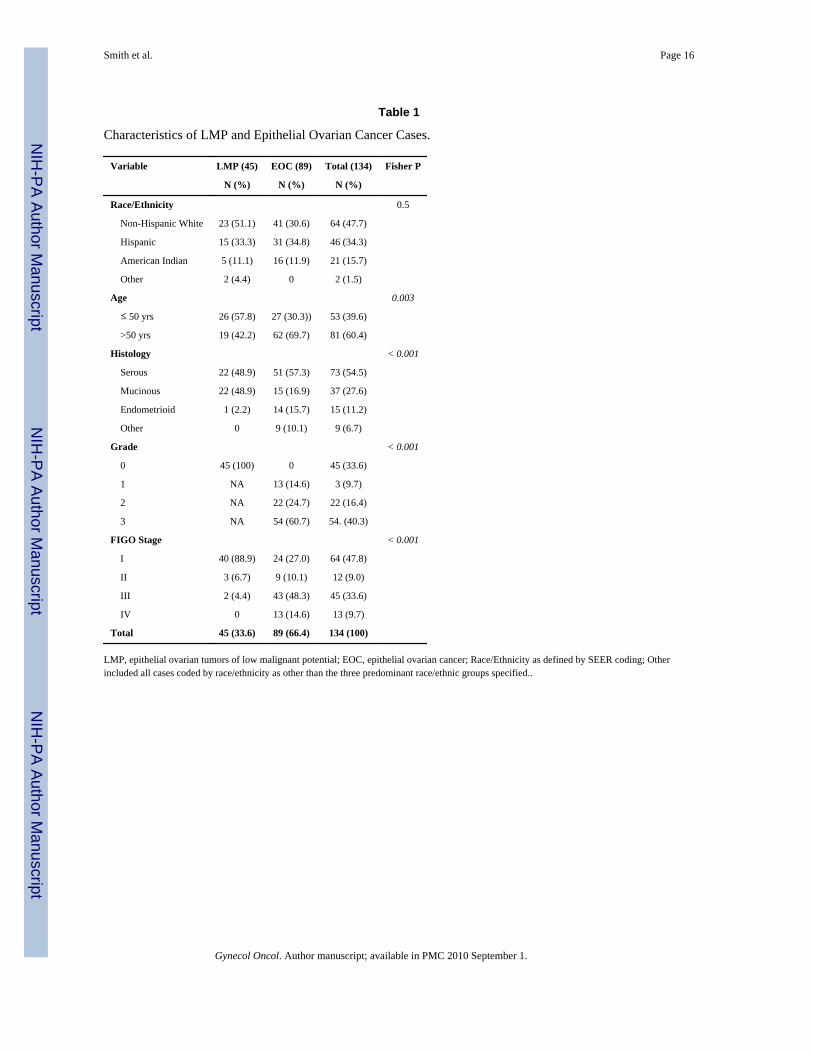
ResultsTable 1 depicts the distribution of patients with LMP tumors (45, 33.6%) and EOC (89,66.4%) by race/ethnicity, age at diagnosis, histological subtype, tumor grade, and FIGOstage. Patients with LMP tumors were significantly younger (p = 0.003), had earlier(predominantly stage I) disease (p < 0.001), and the tumors were more frequently ofmucinous histology (p < 0.001), in approximately 50% of the cases. The median H-score of105 for ovarian cancer was used to divide patients into two groups: H-scores greater than105 were defined as GPR30-High and H-scores up to 105 were defined as GPR30-Low..
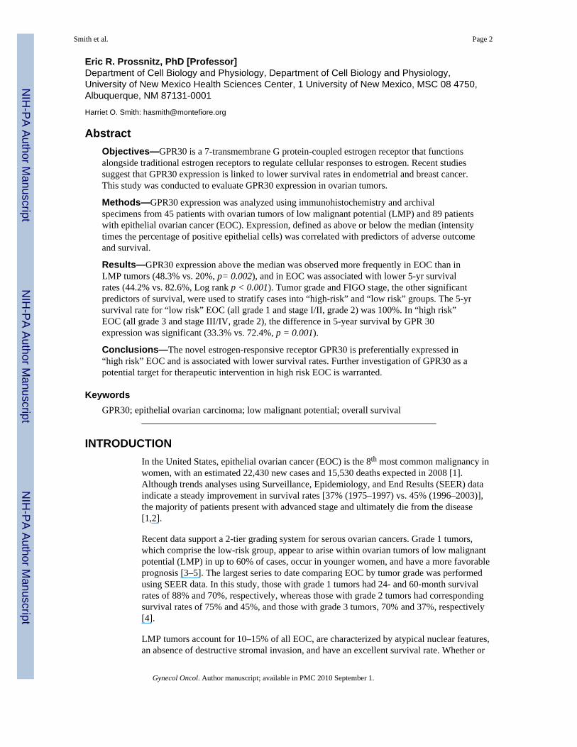
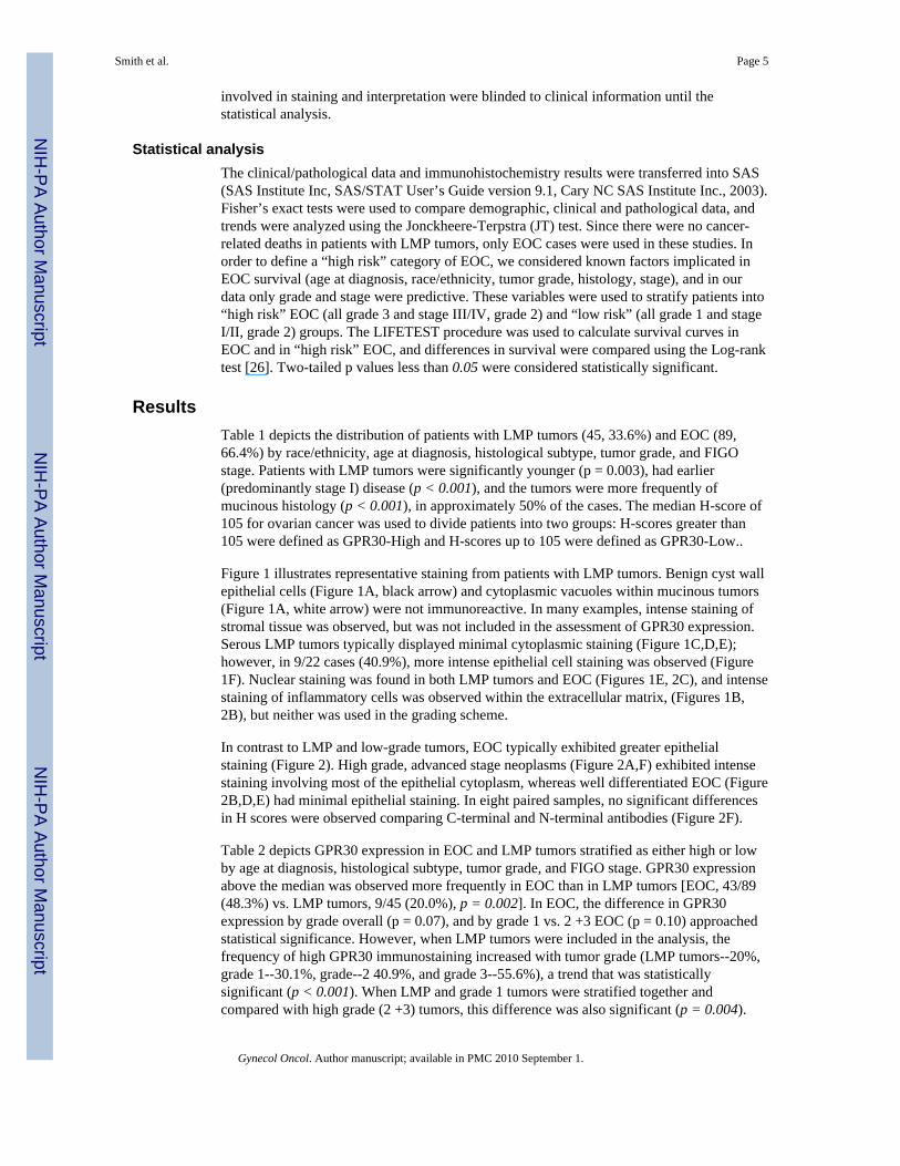
Figure 1 illustrates representative staining from patients with LMP tumors. Benign cyst wallepithelial cells (Figure 1A, black arrow) and cytoplasmic vacuoles within mucinous tumors(Figure 1A, white arrow) were not immunoreactive. In many examples, intense staining ofstromal tissue was observed, but was not included in the assessment of GPR30 expression.Serous LMP tumors typically displayed minimal cytoplasmic staining (Figure 1C,D,E);however, in 9/22 cases (40.9%), more intense epithelial cell staining was observed (Figure1F). Nuclear staining was found in both LMP tumors and EOC (Figures 1E, 2C), and intensestaining of inflammatory cells was observed within the extracellular matrix, (Figures 1B,2B), but neither was used in the grading scheme.
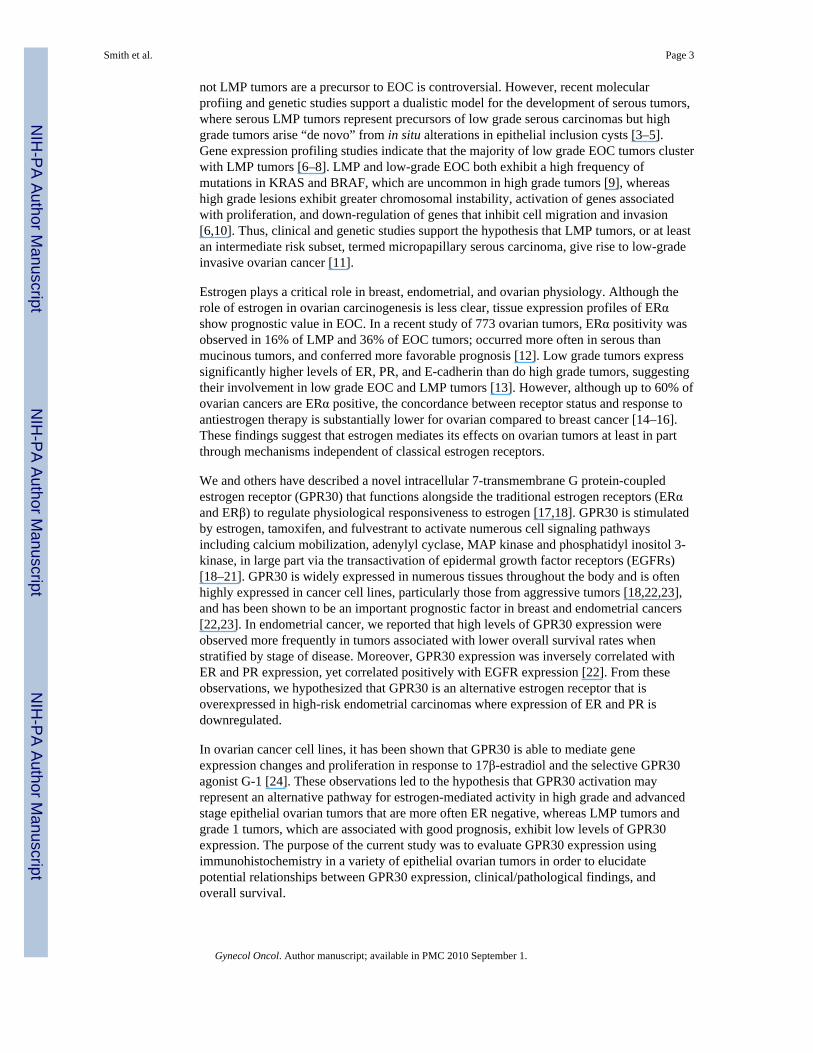
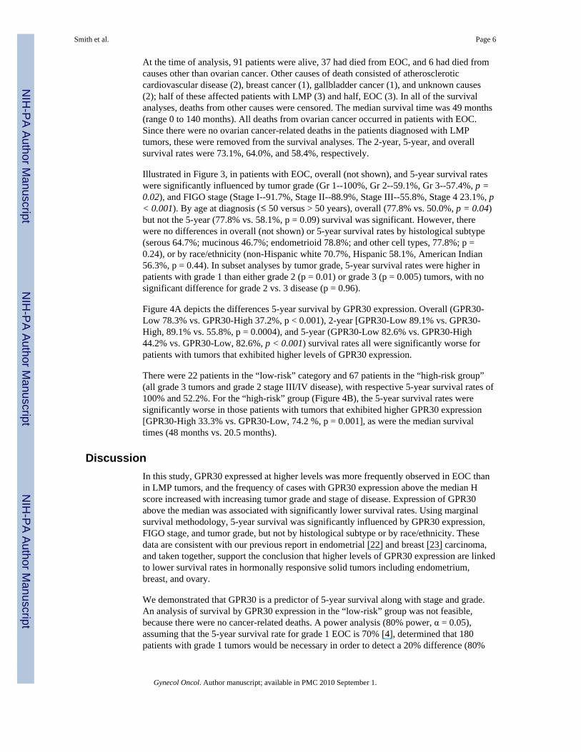
In contrast to LMP and low-grade tumors, EOC typically exhibited greater epithelialstaining (Figure 2). High grade, advanced stage neoplasms (Figure 2A,F) exhibited intensestaining involving most of the epithelial cytoplasm, whereas well differentiated EOC (Figure2B,D,E) had minimal epithelial staining. In eight paired samples, no significant differencesin H scores were observed comparing C-terminal and N-terminal antibodies (Figure 2F).
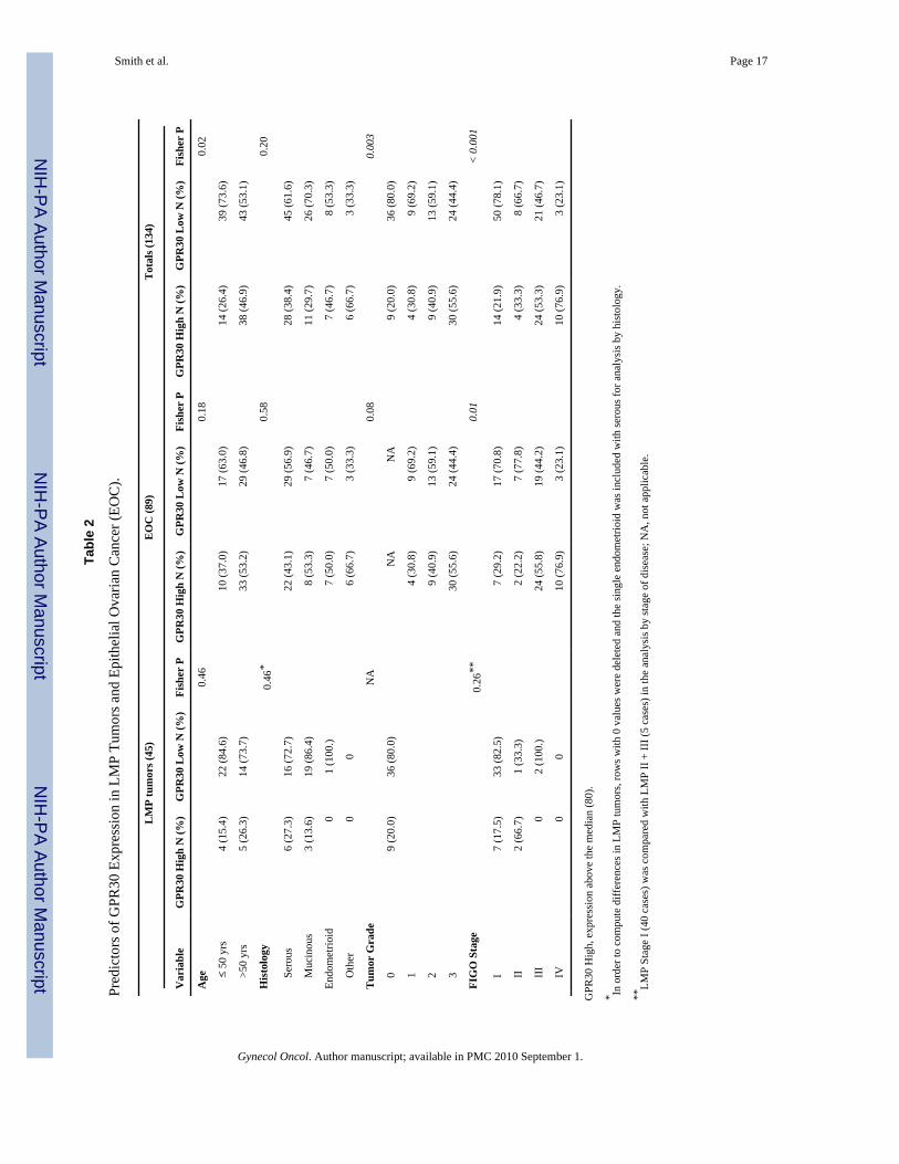
Table 2 depicts GPR30 expression in EOC and LMP tumors stratified as either high or lowby age at diagnosis, histological subtype, tumor grade, and FIGO stage. GPR30 expressionabove the median was observed more frequently in EOC than in LMP tumors [EOC, 43/89(48.3%) vs. LMP tumors, 9/45 (20.0%), p = 0.002]. In EOC, the difference in GPR30expression by grade overall (p = 0.07), and by grade 1 vs. 2 +3 EOC (p = 0.10) approachedstatistical significance. However, when LMP tumors were included in the analysis, thefrequency of high GPR30 immunostaining increased with tumor grade (LMP tumors--20%,grade 1--30.1%, grade--2 40.9%, and grade 3--55.6%), a trend that was statisticallysignificant (p < 0.001). When LMP and grade 1 tumors were stratified together andcompared with high grade (2 +3) tumors, this difference was also significant (p = 0.004).
Smith et al. Page 5
Gynecol Oncol. Author manuscript; available in PMC 2010 September 1.
NIH
-PA Author Manuscript
NIH
-PA Author Manuscript
NIH
-PA Author Manuscript
At the time of analysis, 91 patients were alive, 37 had died from EOC, and 6 had died fromcauses other than ovarian cancer. Other causes of death consisted of atheroscleroticcardiovascular disease (2), breast cancer (1), gallbladder cancer (1), and unknown causes(2); half of these affected patients with LMP (3) and half, EOC (3). In all of the survivalanalyses, deaths from other causes were censored. The median survival time was 49 months(range 0 to 140 months). All deaths from ovarian cancer occurred in patients with EOC.Since there were no ovarian cancer-related deaths in the patients diagnosed with LMPtumors, these were removed from the survival analyses. The 2-year, 5-year, and overallsurvival rates were 73.1%, 64.0%, and 58.4%, respectively.
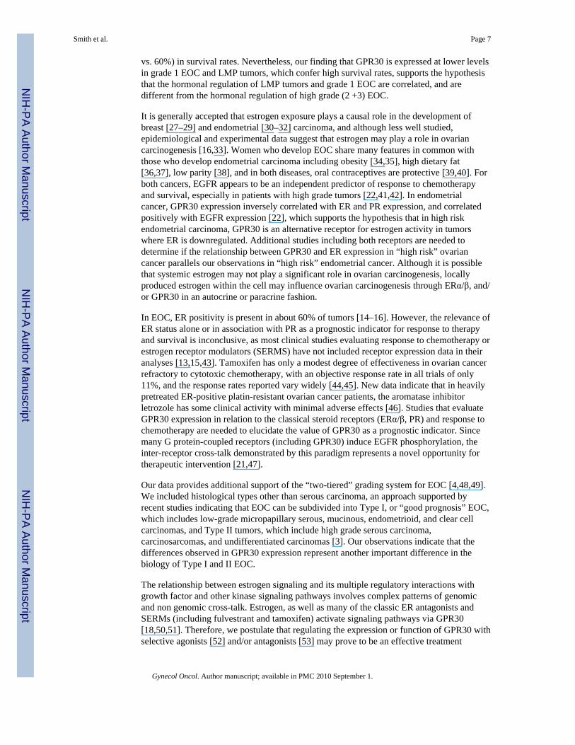
Illustrated in Figure 3, in patients with EOC, overall (not shown), and 5-year survival rateswere significantly influenced by tumor grade (Gr 1--100%, Gr 2--59.1%, Gr 3--57.4%, p =0.02), and FIGO stage (Stage I--91.7%, Stage II--88.9%, Stage III--55.8%, Stage 4 23.1%, p< 0.001). By age at diagnosis (≤ 50 versus > 50 years), overall (77.8% vs. 50.0%, p = 0.04)but not the 5-year (77.8% vs. 58.1%, p = 0.09) survival was significant. However, therewere no differences in overall (not shown) or 5-year survival rates by histological subtype(serous 64.7%; mucinous 46.7%; endometrioid 78.8%; and other cell types, 77.8%; p =0.24), or by race/ethnicity (non-Hispanic white 70.7%, Hispanic 58.1%, American Indian56.3%, p = 0.44). In subset analyses by tumor grade, 5-year survival rates were higher inpatients with grade 1 than either grade 2 (p = 0.01) or grade 3 (p = 0.005) tumors, with nosignificant difference for grade 2 vs. 3 disease (p = 0.96).
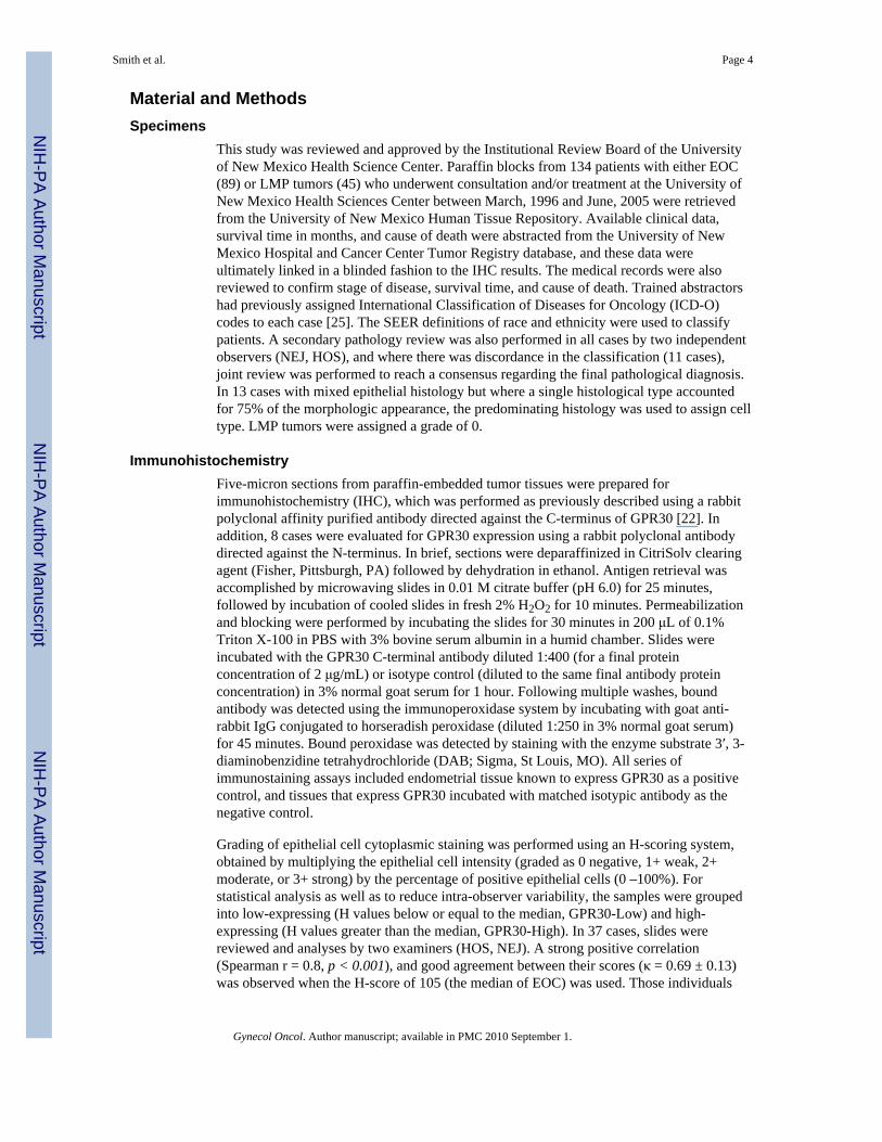
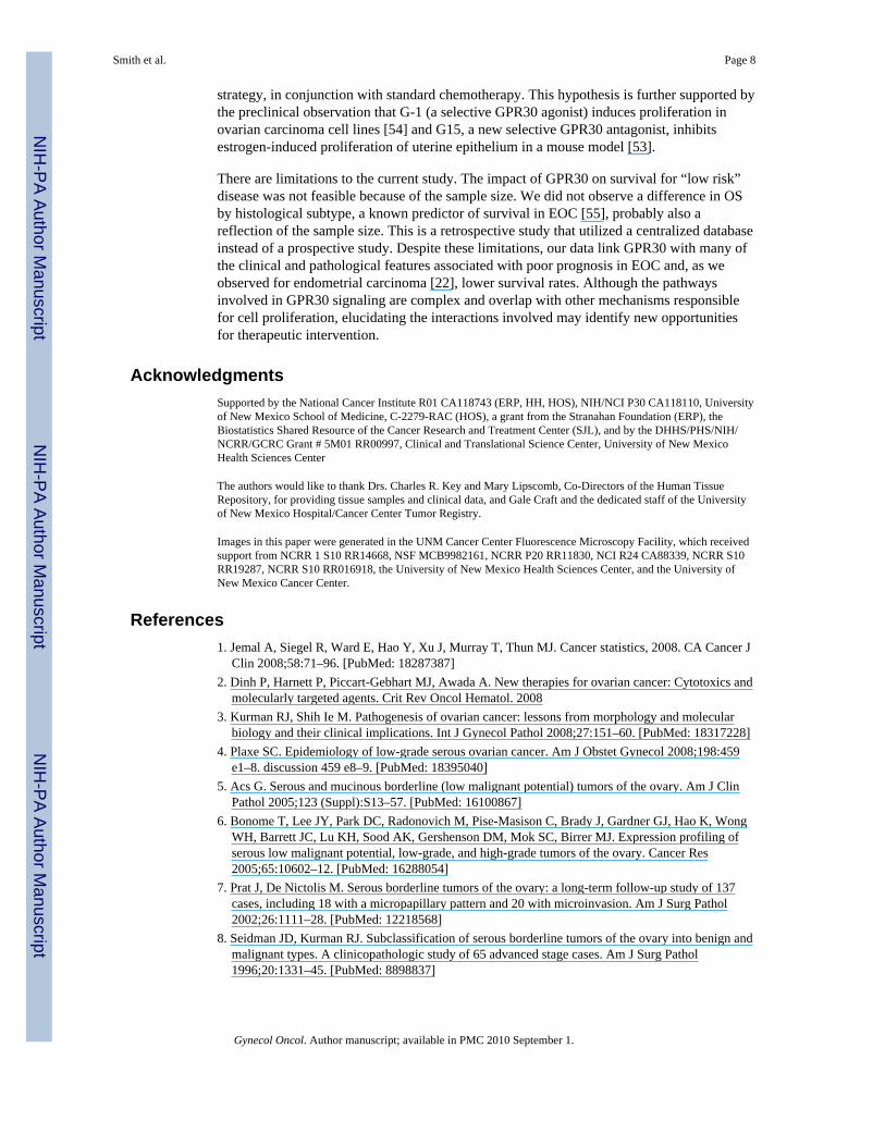
Figure 4A depicts the differences 5-year survival by GPR30 expression. Overall (GPR30-Low 78.3% vs. GPR30-High 37.2%, p < 0.001), 2-year [GPR30-Low 89.1% vs. GPR30-High, 89.1% vs. 55.8%, p = 0.0004), and 5-year (GPR30-Low 82.6% vs. GPR30-High44.2% vs. GPR30-Low, 82.6%, p < 0.001) survival rates all were significantly worse forpatients with tumors that exhibited higher levels of GPR30 expression.
There were 22 patients in the “low-risk” category and 67 patients in the “high-risk group”(all grade 3 tumors and grade 2 stage III/IV disease), with respective 5-year survival rates of100% and 52.2%. For the “high-risk” group (Figure 4B), the 5-year survival rates weresignificantly worse in those patients with tumors that exhibited higher GPR30 expression[GPR30-High 33.3% vs. GPR30-Low, 74.2 %, p = 0.001], as were the median survivaltimes (48 months vs. 20.5 months).
DiscussionIn this study, GPR30 expressed at higher levels was more frequently observed in EOC thanin LMP tumors, and the frequency of cases with GPR30 expression above the median Hscore increased with increasing tumor grade and stage of disease. Expression of GPR30above the median was associated with significantly lower survival rates. Using marginalsurvival methodology, 5-year survival was significantly influenced by GPR30 expression,FIGO stage, and tumor grade, but not by histological subtype or by race/ethnicity. Thesedata are consistent with our previous report in endometrial [22] and breast [23] carcinoma,and taken together, support the conclusion that higher levels of GPR30 expression are linkedto lower survival rates in hormonally responsive solid tumors including endometrium,breast, and ovary.
We demonstrated that GPR30 is a predictor of 5-year survival along with stage and grade.An analysis of survival by GPR30 expression in the “low-risk” group was not feasible,because there were no cancer-related deaths. A power analysis (80% power, α = 0.05),assuming that the 5-year survival rate for grade 1 EOC is 70% [4], determined that 180patients with grade 1 tumors would be necessary in order to detect a 20% difference (80%
Smith et al. Page 6
Gynecol Oncol. Author manuscript; available in PMC 2010 September 1.
NIH
-PA Author Manuscript
NIH
-PA Author Manuscript
NIH
-PA Author Manuscript
vs. 60%) in survival rates. Nevertheless, our finding that GPR30 is expressed at lower levelsin grade 1 EOC and LMP tumors, which confer high survival rates, supports the hypothesisthat the hormonal regulation of LMP tumors and grade 1 EOC are correlated, and aredifferent from the hormonal regulation of high grade (2 +3) EOC.
It is generally accepted that estrogen exposure plays a causal role in the development ofbreast [27–29] and endometrial [30–32] carcinoma, and although less well studied,epidemiological and experimental data suggest that estrogen may play a role in ovariancarcinogenesis [16,33]. Women who develop EOC share many features in common withthose who develop endometrial carcinoma including obesity [34,35], high dietary fat[36,37], low parity [38], and in both diseases, oral contraceptives are protective [39,40]. Forboth cancers, EGFR appears to be an independent predictor of response to chemotherapyand survival, especially in patients with high grade tumors [22,41,42]. In endometrialcancer, GPR30 expression inversely correlated with ER and PR expression, and correlatedpositively with EGFR expression [22], which supports the hypothesis that in high riskendometrial carcinoma, GPR30 is an alternative receptor for estrogen activity in tumorswhere ER is downregulated. Additional studies including both receptors are needed todetermine if the relationship between GPR30 and ER expression in “high risk” ovariancancer parallels our observations in “high risk” endometrial cancer. Although it is possiblethat systemic estrogen may not play a significant role in ovarian carcinogenesis, locallyproduced estrogen within the cell may influence ovarian carcinogenesis through ERα/β, and/or GPR30 in an autocrine or paracrine fashion.
In EOC, ER positivity is present in about 60% of tumors [14–16]. However, the relevance ofER status alone or in association with PR as a prognostic indicator for response to therapyand survival is inconclusive, as most clinical studies evaluating response to chemotherapy orestrogen receptor modulators (SERMS) have not included receptor expression data in theiranalyses [13,15,43]. Tamoxifen has only a modest degree of effectiveness in ovarian cancerrefractory to cytotoxic chemotherapy, with an objective response rate in all trials of only11%, and the response rates reported vary widely [44,45]. New data indicate that in heavilypretreated ER-positive platin-resistant ovarian cancer patients, the aromatase inhibitorletrozole has some clinical activity with minimal adverse effects [46]. Studies that evaluateGPR30 expression in relation to the classical steroid receptors (ERα/β, PR) and response tochemotherapy are needed to elucidate the value of GPR30 as a prognostic indicator. Sincemany G protein-coupled receptors (including GPR30) induce EGFR phosphorylation, theinter-receptor cross-talk demonstrated by this paradigm represents a novel opportunity fortherapeutic intervention [21,47].
Our data provides additional support of the “two-tiered” grading system for EOC [4,48,49].We included histological types other than serous carcinoma, an approach supported byrecent studies indicating that EOC can be subdivided into Type I, or “good prognosis” EOC,which includes low-grade micropapillary serous, mucinous, endometrioid, and clear cellcarcinomas, and Type II tumors, which include high grade serous carcinoma,carcinosarcomas, and undifferentiated carcinomas [3]. Our observations indicate that thedifferences observed in GPR30 expression represent another important difference in thebiology of Type I and II EOC.
The relationship between estrogen signaling and its multiple regulatory interactions withgrowth factor and other kinase signaling pathways involves complex patterns of genomicand non genomic cross-talk. Estrogen, as well as many of the classic ER antagonists andSERMs (including fulvestrant and tamoxifen) activate signaling pathways via GPR30[18,50,51]. Therefore, we postulate that regulating the expression or function of GPR30 withselective agonists [52] and/or antagonists [53] may prove to be an effective treatment
Smith et al. Page 7
Gynecol Oncol. Author manuscript; available in PMC 2010 September 1.
NIH
-PA Author Manuscript
NIH
-PA Author Manuscript
NIH
-PA Author Manuscript
strategy, in conjunction with standard chemotherapy. This hypothesis is further supported bythe preclinical observation that G-1 (a selective GPR30 agonist) induces proliferation inovarian carcinoma cell lines [54] and G15, a new selective GPR30 antagonist, inhibitsestrogen-induced proliferation of uterine epithelium in a mouse model [53].
There are limitations to the current study. The impact of GPR30 on survival for “low risk”disease was not feasible because of the sample size. We did not observe a difference in OSby histological subtype, a known predictor of survival in EOC [55], probably also areflection of the sample size. This is a retrospective study that utilized a centralized databaseinstead of a prospective study. Despite these limitations, our data link GPR30 with many ofthe clinical and pathological features associated with poor prognosis in EOC and, as weobserved for endometrial carcinoma [22], lower survival rates. Although the pathwaysinvolved in GPR30 signaling are complex and overlap with other mechanisms responsiblefor cell proliferation, elucidating the interactions involved may identify new opportunitiesfor therapeutic intervention.
AcknowledgmentsSupported by the National Cancer Institute R01 CA118743 (ERP, HH, HOS), NIH/NCI P30 CA118110, Universityof New Mexico School of Medicine, C-2279-RAC (HOS), a grant from the Stranahan Foundation (ERP), theBiostatistics Shared Resource of the Cancer Research and Treatment Center (SJL), and by the DHHS/PHS/NIH/NCRR/GCRC Grant # 5M01 RR00997, Clinical and Translational Science Center, University of New MexicoHealth Sciences Center
The authors would like to thank Drs. Charles R. Key and Mary Lipscomb, Co-Directors of the Human TissueRepository, for providing tissue samples and clinical data, and Gale Craft and the dedicated staff of the Universityof New Mexico Hospital/Cancer Center Tumor Registry.
Images in this paper were generated in the UNM Cancer Center Fluorescence Microscopy Facility, which receivedsupport from NCRR 1 S10 RR14668, NSF MCB9982161, NCRR P20 RR11830, NCI R24 CA88339, NCRR S10RR19287, NCRR S10 RR016918, the University of New Mexico Health Sciences Center, and the University ofNew Mexico Cancer Center.
References1. Jemal A, Siegel R, Ward E, Hao Y, Xu J, Murray T, Thun MJ. Cancer statistics, 2008. CA Cancer J
Clin 2008;58:71–96. [PubMed: 18287387]2. Dinh P, Harnett P, Piccart-Gebhart MJ, Awada A. New therapies for ovarian cancer: Cytotoxics and
molecularly targeted agents. Crit Rev Oncol Hematol. 20083. Kurman RJ, Shih Ie M. Pathogenesis of ovarian cancer: lessons from morphology and molecular
biology and their clinical implications. Int J Gynecol Pathol 2008;27:151–60. [PubMed: 18317228]4. Plaxe SC. Epidemiology of low-grade serous ovarian cancer. Am J Obstet Gynecol 2008;198:459
e1–8. discussion 459 e8–9. [PubMed: 18395040]5. Acs G. Serous and mucinous borderline (low malignant potential) tumors of the ovary. Am J Clin
Pathol 2005;123 (Suppl):S13–57. [PubMed: 16100867]6. Bonome T, Lee JY, Park DC, Radonovich M, Pise-Masison C, Brady J, Gardner GJ, Hao K, Wong
WH, Barrett JC, Lu KH, Sood AK, Gershenson DM, Mok SC, Birrer MJ. Expression profiling ofserous low malignant potential, low-grade, and high-grade tumors of the ovary. Cancer Res2005;65:10602–12. [PubMed: 16288054]
7. Prat J, De Nictolis M. Serous borderline tumors of the ovary: a long-term follow-up study of 137cases, including 18 with a micropapillary pattern and 20 with microinvasion. Am J Surg Pathol2002;26:1111–28. [PubMed: 12218568]
8. Seidman JD, Kurman RJ. Subclassification of serous borderline tumors of the ovary into benign andmalignant types. A clinicopathologic study of 65 advanced stage cases. Am J Surg Pathol1996;20:1331–45. [PubMed: 8898837]
Smith et al. Page 8
Gynecol Oncol. Author manuscript; available in PMC 2010 September 1.
NIH
-PA Author Manuscript
NIH
-PA Author Manuscript
NIH
-PA Author Manuscript
9. Singer G, Oldt R 3rd, Cohen Y, Wang BG, Sidransky D, Kurman RJ, Shih Ie M. Mutations inBRAF and KRAS characterize the development of low-grade ovarian serous carcinoma. J NatlCancer Inst 2003;95:484–6. [PubMed: 12644542]
10. Meinhold-Heerlein I, Bauerschlag D, Hilpert F, Dimitrov P, Sapinoso LM, Orlowska-Volk M,Bauknecht T, Park TW, Jonat W, Jacobsen A, Sehouli J, Luttges J, Krajewski M, Krajewski S,Reed JC, Arnold N, Hampton GM. Molecular and prognostic distinction between serous ovariancarcinomas of varying grade and malignant potential. Oncogene 2005;24:1053–65. [PubMed:15558012]
11. Shih Ie M, Kurman RJ. Ovarian tumorigenesis: a proposed model based on morphological andmolecular genetic analysis. Am J Pathol 2004;164:1511–8. [PubMed: 15111296]
12. Hogdall EV, Christensen L, Hogdall CK, Blaakaer J, Gayther S, Jacobs IJ, Christensen IJ, KjaerSK. Prognostic value of estrogen receptor and progesterone receptor tumor expression in Danishovarian cancer patients: from the ‘MALOVA’ ovarian cancer study. Oncol Rep 2007;18:1051–9.[PubMed: 17914554]
13. Wong KK, Lu KH, Malpica A, Bodurka DC, Shvartsman HS, Schmandt RE, Thornton AD,Deavers MT, Silva EG, Gershenson DM. Significantly greater expression of ER, PR, and ECADin advanced-stage low-grade ovarian serous carcinoma as revealed by immunohistochemicalanalysis. Int J Gynecol Pathol 2007;26:404–9. [PubMed: 17885490]
14. Bizzi A, Codegoni AM, Landoni F, Marelli G, Marsoni S, Spina AM, Torri W, Mangioni C.Steroid receptors in epithelial ovarian carcinoma: relation to clinical parameters and survival.Cancer Res 1988;48:6222–6. [PubMed: 3167868]
15. al-Timimi A, Buckley CH, Fox H. An immunohistochemical study of the incidence andsignificance of sex steroid hormone binding sites in normal and neoplastic human ovarian tissue.Int J Gynecol Pathol 1985;4:24–41. [PubMed: 3880152]
16. Ho SM. Estrogen, progesterone and epithelial ovarian cancer. Reprod Biol Endocrinol 2003;1:73.[PubMed: 14577831]
17. Filardo EJ, Quinn JA, Bland KI, Frackelton AR Jr. Estrogen-induced activation of Erk-1 and Erk-2requires the G protein-coupled receptor homolog, GPR30, and occurs via trans-activation of theepidermal growth factor receptor through release of HB-EGF. Mol Endocrinol 2000;14:1649–60.[PubMed: 11043579]
18. Revankar CM, Cimino DF, Sklar LA, Arterburn JB, Prossnitz ER. A transmembrane intracellularestrogen receptor mediates rapid cell signaling. Science 2005;307:1625–30. [PubMed: 15705806]
19. Filardo EJ, Thomas P. GPR30: a seven-transmembrane-spanning estrogen receptor that triggersEGF release. Trends Endocrinol Metab 2005;16:362–7. [PubMed: 16125968]
20. Prossnitz ER, Arterburn JB, Smith HO, Oprea TI, Sklar LA, Hathaway HJ. Estrogen Signalingthrough the Transmembrane G Protein-Coupled Receptor GPR30. Annu Rev Physiol2008;70:165–190. [PubMed: 18271749]
21. Prossnitz ER, Sklar LA, Oprea TI, Arterburn JB. GPR30: a novel therapeutic target in estrogen-related disease. Trends Pharmacol Sci 2008;29:116–23. [PubMed: 18262661]
22. Smith HO, Leslie KK, Singh M, Qualls CR, Revankar CM, Joste NE, Prossnitz ER. GPR30: anovel indicator of poor survival for endometrial carcinoma. Am J Obstet Gynecol 2007;196:386e1–9. discussion 386 e9–11. [PubMed: 17403429]
23. Filardo EJ, Graeber CT, Quinn JA, Resnick MB, Giri D, DeLellis RA, Steinhoff MM, Sabo E.Distribution of GPR30, a seven membrane-spanning estrogen receptor, in primary breast cancerand its association with clinicopathologic determinants of tumor progression. Clin Cancer Res2006;12:6359–66. [PubMed: 17085646]
24. Albanito L, Madeo A, Lappano R, Vivacqua A, Rago V, Carpino A, Oprea TI, Prossnitz ER, MustiAM, Ando S, Maggiolini M. G protein-coupled receptor 30 (GPR30) mediates gene expressionchanges and growth response to 17beta-estradiol and selective GPR30 ligand G-1 in ovariancancer cells. Cancer Res 2007;67:1859–66. [PubMed: 17308128]
25. Fritz, A.; Percy, C.; Jack, A.; Shanmugaratnam, K.; Sobin, L.; Porkin, D.; Whelan, S.; Van Holten,V.; Muir, CS. International classification of diseases for oncology (ICD-0). 3. Geneva: WorldHealth Organization; 2000.
26. Collett, D. Modeling survival data in medical research. London: Chapman & Hall; 1994.
Smith et al. Page 9
Gynecol Oncol. Author manuscript; available in PMC 2010 September 1.
NIH
-PA Author Manuscript
NIH
-PA Author Manuscript
NIH
-PA Author Manuscript
27. Ali S, Coombes RC. Estrogen receptor alpha in human breast cancer: occurrence and significance.J Mammary Gland Biol Neoplasia 2000;5:271–81. [PubMed: 14973389]
28. Anderson E. The role of oestrogen and progesterone receptors in human mammary developmentand tumorigenesis. Breast Cancer Res 2002;4:197–201. [PubMed: 12223124]
29. Conner P, Lundstrom E, von Schoultz B. Breast cancer and hormonal therapy. Clin Obstet Gynecol2008;51:592–606. [PubMed: 18677153]
30. Hormone replacement therapy and cancer. Gynecol Endocrinol 2001;15:453–65. [PubMed:11826770]
31. Allen NE, Key TJ, Dossus L, Rinaldi S, Cust A, Lukanova A, Peeters PH, Onland-Moret NC,Lahmann PH, Berrino F, Panico S, Larranaga N, Pera G, Tormo MJ, Sanchez MJ, Ramon QuirosJ, Ardanaz E, Tjonneland A, Olsen A, Chang-Claude J, Linseisen J, Schulz M, Boeing H, LundinE, Palli D, Overvad K, Clavel-Chapelon F, Boutron-Ruault MC, Bingham S, Khaw KT, BasBueno-de-Mesquita H, Trichopoulou A, Trichopoulos D, Naska A, Tumino R, Riboli E, Kaaks R.Endogenous sex hormones and endometrial cancer risk in women in the European ProspectiveInvestigation into Cancer and Nutrition (EPIC). Endocr Relat Cancer 2008;15:485–97. [PubMed:18509001]
32. Cohen I, Beyth Y, Altaras MM, Shapira J, Tepper R, Cardoba M, Yigael D, Figer A, Fishman A,Berenhein J. Estrogen and progesterone receptor expression in postmenopausal tamoxifen-exposedendometrial pathologies. Gynecol Oncol 1997;67:8–15. [PubMed: 9345349]
33. Lukanova A, Kaaks R. Endogenous hormones and ovarian cancer: epidemiology and currenthypotheses. Cancer Epidemiol Biomarkers Prev 2005;14:98–107. [PubMed: 15668482]
34. Reeves GK, Pirie K, Beral V, Green J, Spencer E, Bull D. Cancer incidence and mortality inrelation to body mass index in the Million Women Study: cohort study. BMJ 2007;335:1134.[PubMed: 17986716]
35. Modesitt SC, van Nagell JR Jr. The impact of obesity on the incidence and treatment ofgynecologic cancers: a review. Obstet Gynecol Surv 2005;60:683–92. [PubMed: 16186785]
36. Kiani F, Knutsen S, Singh P, Ursin G, Fraser G. Dietary risk factors for ovarian cancer: theAdventist Health Study (United States). Cancer Causes Control 2006;17:137–46. [PubMed:16425091]
37. Huncharek M, Kupelnick B. Dietary fat intake and risk of epithelial ovarian cancer: a meta-analysis of 6,689 subjects from 8 observational studies. Nutr Cancer 2001;40:87–91. [PubMed:11962260]
38. La Vecchia C. Epidemiology of ovarian cancer: a summary review. Eur J Cancer Prev2001;10:125–9. [PubMed: 11330452]
39. Woutersz TB. Benefits of oral contraception: thirty years’ experience. Int J Fertil 1991;36 (Suppl3):26–31. [PubMed: 1687401]
40. Medard ML, Ostrowska L. Combined oral contraception and the risk of reproductive organs cancerin women. Ginekol Pol 2007;78:637–41. [PubMed: 18050614]
41. Scambia G, Benedetti-Panici P, Ferrandina G, Distefano M, Salerno G, Romanini ME, Fagotti A,Mancuso S. Epidermal growth factor, oestrogen and progesterone receptor expression in primaryovarian cancer: correlation with clinical outcome and response to chemotherapy. Br J Cancer1995;72:361–6. [PubMed: 7640219]
42. Vaidya AP, Parnes AD, Seiden MV. Rationale and clinical experience with epidermal growthfactor receptor inhibitors in gynecologic malignancies. Curr Treat Options Oncol 2005;6:103–14.[PubMed: 15717992]
43. Langdon SP, Hawkes MM, Lawrie SS, Hawkins RA, Tesdale AL, Crew AJ, Miller WR, Smyth JF.Oestrogen receptor expression and the effects of oestrogen and tamoxifen on the growth of humanovarian carcinoma cell lines. Br J Cancer 1990;62:213–6. [PubMed: 2386737]
44. Karagol H, Saip P, Uygun K, Caloglu M, Eralp Y, Tas F, Aydiner A, Topuz E. The efficacy oftamoxifen in patients with advanced epithelial ovarian cancer. Med Oncol 2007;24:39–43.[PubMed: 17673810]
45. Trope C, Marth C, Kaern J. Tamoxifen in the treatment of recurrent ovarian carcinoma. Eur JCancer 2000;36 (Suppl 4):S59–61. [PubMed: 11056321]
Smith et al. Page 10
Gynecol Oncol. Author manuscript; available in PMC 2010 September 1.
NIH
-PA Author Manuscript
NIH
-PA Author Manuscript
NIH
-PA Author Manuscript
46. Ramirez PT, Schmeler KM, Milam MR, Slomovitz BM, Smith JA, Kavanagh JJ, Deavers M,Levenback C, Coleman RL, Gershenson DM. Efficacy of letrozole in the treatment of recurrentplatinum- and taxane-resistant high-grade cancer of the ovary or peritoneum. Gynecol Oncol2008;110:56–9. [PubMed: 18457865]
47. Fischer OM, Hart S, Gschwind A, Ullrich A. EGFR signal transactivation in cancer cells. BiochemSoc Trans 2003;31:1203–8. [PubMed: 14641026]
48. Farley J, Ozbun LL, Birrer MJ. Genomic analysis of epithelial ovarian cancer. Cell Res2008;18:538–48. [PubMed: 18427574]
49. Gershenson DM, Sun CC, Lu KH, Coleman RL, Sood AK, Malpica A, Deavers MT, Silva EG,Bodurka DC. Clinical behavior of stage II-IV low-grade serous carcinoma of the ovary. ObstetGynecol 2006;108:361–8. [PubMed: 16880307]
50. Vivacqua A, Bonofiglio D, Recchia AG, Musti AM, Picard D, Ando S, Maggiolini M. The Gprotein-coupled receptor GPR30 mediates the proliferative effects induced by 17beta-estradiol andhydroxytamoxifen in endometrial cancer cells. Molecular Endocrinology 2006;20:631–46.[PubMed: 16239258]
51. Filardo EJ, Quinn JA, Frackelton AR Jr, Bland KI. Estrogen action via the G protein-coupledreceptor, GPR30: stimulation of adenylyl cyclase and cAMP-mediated attenuation of theepidermal growth factor receptor-to-MAPK signaling axis. Mol Endocrinol 2002;16:70–84.[PubMed: 11773440]
52. Bologa CG, Revankar CM, Young SM, Edwards BS, Arterburn JB, Kiselyov AS, Parker MA,Tkachenko SE, Savchuck NP, Sklar LA, Oprea TI, Prossnitz ER. Virtual and biomolecularscreening converge on a selective agonist for GPR30. Nat Chem Biol 2006;2:207–12. [PubMed:16520733]
53. Dennis MK, Burai R, Ramesh C, Petrie WK, Alcon SN, Nayak T, Bologa C, Leitao A, Brailoiu E,Deliu E, Dun NJ, Sklar LA, Hathaway HJ, Arterburn JB, Oprea TO, Prossnitz ER. In vivo effectsof a GPR30 antagonist. Nature Chemical Biology. 2009 (in press).
54. Albanito L, Lappano R, Madeo A, Chimento A, Prossnitz ER, Cappello AR, Dolce V, Abonante S,Pezzi V, Maggiolini M. G-protein-coupled receptor 30 and estrogen receptor-alpha are involved inthe proliferative effects induced by atrazine in ovarian cancer cells. Environ Health Perspect2008;116:1648–55. [PubMed: 19079715]
55. Skirnisdottir I, Sorbe B. Prognostic factors for surgical outcome and survival in 447 women treatedfor advanced (FIGO-stages III-IV) epithelial ovarian carcinoma. Int J Oncol 2007;30:727–34.[PubMed: 17273775]
Smith et al. Page 11
Gynecol Oncol. Author manuscript; available in PMC 2010 September 1.
NIH
-PA Author Manuscript
NIH
-PA Author Manuscript
NIH
-PA Author Manuscript
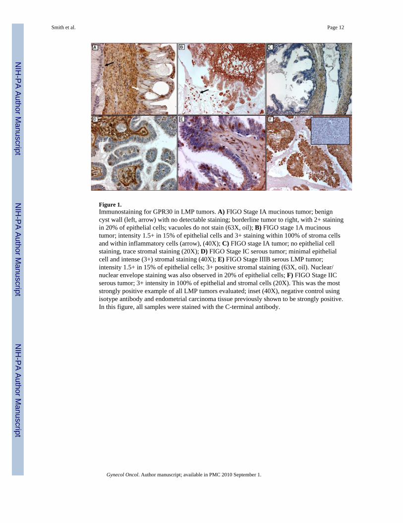
Figure 1.Immunostaining for GPR30 in LMP tumors. A) FIGO Stage IA mucinous tumor; benigncyst wall (left, arrow) with no detectable staining; borderline tumor to right, with 2+ stainingin 20% of epithelial cells; vacuoles do not stain (63X, oil); B) FIGO stage 1A mucinoustumor; intensity 1.5+ in 15% of epithelial cells and 3+ staining within 100% of stroma cellsand within inflammatory cells (arrow), (40X); C) FIGO stage IA tumor; no epithelial cellstaining, trace stromal staining (20X); D) FIGO Stage IC serous tumor; minimal epithelialcell and intense (3+) stromal staining (40X); E) FIGO Stage IIIB serous LMP tumor;intensity 1.5+ in 15% of epithelial cells; 3+ positive stromal staining (63X, oil). Nuclear/nuclear envelope staining was also observed in 20% of epithelial cells; F) FIGO Stage IICserous tumor; 3+ intensity in 100% of epithelial and stromal cells (20X). This was the moststrongly positive example of all LMP tumors evaluated; inset (40X), negative control usingisotype antibody and endometrial carcinoma tissue previously shown to be strongly positive.In this figure, all samples were stained with the C-terminal antibody.
Smith et al. Page 12
Gynecol Oncol. Author manuscript; available in PMC 2010 September 1.
NIH
-PA Author Manuscript
NIH
-PA Author Manuscript
NIH
-PA Author Manuscript
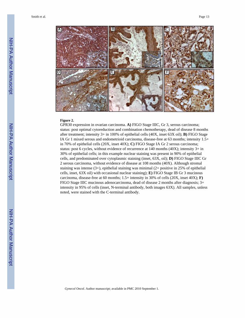
Figure 2.GPR30 expression in ovarian carcinoma. A) FIGO Stage IIIC, Gr 3, serous carcinoma;status: post optimal cytoreduction and combination chemotherapy, dead of disease 8 monthsafter treatment; intensity 3+ in 100% of epithelial cells (40X, inset 63X oil); B) FIGO StageIA Gr 1 mixed serous and endometrioid carcinoma, disease-free at 63 months; intensity 1.5+in 70% of epithelial cells (20X, inset 40X); C) FIGO Stage IA Gr 2 serous carcinoma;status: post 6 cycles, without evidence of recurrence at 140 months (40X); intensity 3+ in30% of epithelial cells; in this example nuclear staining was present in 90% of epithelialcells, and predominated over cytoplasmic staining (inset, 63X, oil); D) FIGO Stage IIIC Gr2 serous carcinoma, without evidence of disease at 108 months (40X). Although stromalstaining was intense (3+), epithelial staining was minimal (2+ positive in 25% of epithelialcells, inset, 63X oil) with occasional nuclear staining); E) FIGO Stage IB Gr 3 mucinouscarcinoma, disease-free at 60 months; 1.5+ intensity in 30% of cells (20X, inset 40X); F)FIGO Stage IIIC mucinous adenocarcinoma, dead of disease 2 months after diagnosis; 3+intensity in 95% of cells (inset, N-terminal antibody, both images 63X). All samples, unlessnoted, were stained with the C-terminal antibody.
Smith et al. Page 13
Gynecol Oncol. Author manuscript; available in PMC 2010 September 1.
NIH
-PA Author Manuscript
NIH
-PA Author Manuscript
NIH
-PA Author Manuscript
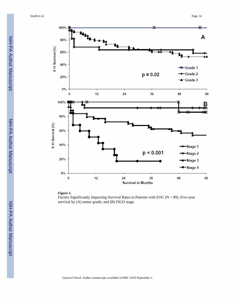
Figure 3.Factors Significantly Impacting Survival Rates in Patients with EOC (N = 89). Five-yearsurvival by (A) tumor grade; and (B) FIGO stage.
Smith et al. Page 14
Gynecol Oncol. Author manuscript; available in PMC 2010 September 1.
NIH
-PA Author Manuscript
NIH
-PA Author Manuscript
NIH
-PA Author Manuscript
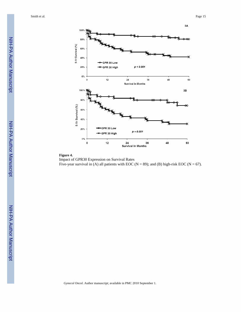
Figure 4.Impact of GPR30 Expression on Survival RatesFive-year survival in (A) all patients with EOC (N = 89); and (B) high-risk EOC (N = 67).
Smith et al. Page 15
Gynecol Oncol. Author manuscript; available in PMC 2010 September 1.
NIH
-PA Author Manuscript
NIH
-PA Author Manuscript
NIH
-PA Author Manuscript
NIH
-PA Author Manuscript
NIH
-PA Author Manuscript
NIH
-PA Author Manuscript
Smith et al. Page 16
Table 1
Characteristics of LMP and Epithelial Ovarian Cancer Cases.
Variable LMP (45) EOC (89) Total (134) Fisher P
N (%) N (%) N (%)
Race/Ethnicity 0.5
Non-Hispanic White 23 (51.1) 41 (30.6) 64 (47.7)
Hispanic 15 (33.3) 31 (34.8) 46 (34.3)
American Indian 5 (11.1) 16 (11.9) 21 (15.7)
Other 2 (4.4) 0 2 (1.5)
Age 0.003
≤ 50 yrs 26 (57.8) 27 (30.3)) 53 (39.6)
>50 yrs 19 (42.2) 62 (69.7) 81 (60.4)
Histology < 0.001
Serous 22 (48.9) 51 (57.3) 73 (54.5)
Mucinous 22 (48.9) 15 (16.9) 37 (27.6)
Endometrioid 1 (2.2) 14 (15.7) 15 (11.2)
Other 0 9 (10.1) 9 (6.7)
Grade < 0.001
0 45 (100) 0 45 (33.6)
1 NA 13 (14.6) 3 (9.7)
2 NA 22 (24.7) 22 (16.4)
3 NA 54 (60.7) 54. (40.3)
FIGO Stage < 0.001
I 40 (88.9) 24 (27.0) 64 (47.8)
II 3 (6.7) 9 (10.1) 12 (9.0)
III 2 (4.4) 43 (48.3) 45 (33.6)
IV 0 13 (14.6) 13 (9.7)
Total 45 (33.6) 89 (66.4) 134 (100)
LMP, epithelial ovarian tumors of low malignant potential; EOC, epithelial ovarian cancer; Race/Ethnicity as defined by SEER coding; Otherincluded all cases coded by race/ethnicity as other than the three predominant race/ethnic groups specified..
Gynecol Oncol. Author manuscript; available in PMC 2010 September 1.
NIH
-PA Author Manuscript
NIH
-PA Author Manuscript
NIH
-PA Author Manuscript
Smith et al. Page 17
Tabl
e 2
Pred
icto
rs o
f GPR
30 E
xpre
ssio
n in
LM
P Tu
mor
s and
Epi
thel
ial O
varia
n C
ance
r (EO
C).
LM
P tu
mor
s (45
)E
OC
(89)
Tot
als (
134)
Var
iabl
eG
PR30
Hig
h N
(%)
GPR
30 L
ow N
(%)
Fish
er P
GPR
30 H
igh
N (%
)G
PR30
Low
N (%
)Fi
sher
PG
PR30
Hig
h N
(%)
GPR
30 L
ow N
(%)
Fish
er P
Age
0.46
0.18
0.02
≤
50 y
rs4
(15.
4)22
(84.
6)10
(37.
0)17
(63.
0)14
(26.
4)39
(73.
6)
>5
0 yr
s5
(26.
3)14
(73.
7)33
(53.
2)29
(46.
8)38
(46.
9)43
(53.
1)
His
tolo
gy0.
46*
0.58
0.20
Se
rous
6 (2
7.3)
16 (7
2.7)
22 (4
3.1)
29 (5
6.9)
28 (3
8.4)
45 (6
1.6)
M
ucin
ous
3 (1
3.6)
19 (8
6.4)
8 (5
3.3)
7 (4
6.7)
11 (2
9.7)
26 (7
0.3)
Endo
met
rioid
01
(100
.)7
(50.
0)7
(50.
0)7
(46.
7)8
(53.
3)
O
ther
00
6 (6
6.7)
3 (3
3.3)
6 (6
6.7)
3 (3
3.3)
Tum
or G
rade
NA
0.08
0.00
3
0
9 (2
0.0)
36 (8
0.0)
NA
NA
9 (2
0.0)
36 (8
0.0)
1
4 (3
0.8)
9 (6
9.2)
4 (3
0.8)
9 (6
9.2)
2
9 (4
0.9)
13 (5
9.1)
9 (4
0.9)
13 (5
9.1)
3
30 (5
5.6)
24 (4
4.4)
30 (5
5.6)
24 (4
4.4)
FIG
O S
tage
0.26
**0.
01<
0.0
01
I
7 (1
7.5)
33 (8
2.5)
7 (2
9.2)
17 (7
0.8)
14 (2
1.9)
50 (7
8.1)
II
2 (6
6.7)
1 (3
3.3)
2 (2
2.2)
7 (7
7.8)
4 (3
3.3)
8 (6
6.7)
II
I0
2 (1
00.)
24 (5
5.8)
19 (4
4.2)
24 (5
3.3)
21 (4
6.7)
IV
00
10 (7
6.9)
3 (2
3.1)
10 (7
6.9)
3 (2
3.1)
GPR
30 H
igh,
exp
ress
ion
abov
e th
e m
edia
n (8
0).
* In o
rder
to c
ompu
te d
iffer
ence
s in
LMP
tum
ors,
row
s with
0 v
alue
s wer
e de
lete
d an
d th
e si
ngle
end
omet
rioid
was
incl
uded
with
sero
us fo
r ana
lysi
s by
hist
olog
y.
**LM
P St
age
I (40
cas
es) w
as c
ompa
red
with
LM
P II
+ II
I (5
case
s) in
the
anal
ysis
by
stag
e of
dis
ease
; NA
, not
app
licab
le.
Gynecol Oncol. Author manuscript; available in PMC 2010 September 1.
Top Related
Copyright © 2022 FDOKUMEN

