


Bahasa
Halaman
Hukum


FTY720 induces apoptosis in B16F10-NEX2 murinemelanoma cells, limits metastatic development invivo, and modulates the immune systemFelipe V. Pereira,I Denise C. Arruda,II Carlos R. Figueiredo,II Mariana H. Massaoka,II Alisson L. Matsuo,II
Valquiria Bueno,III Elaine G. RodriguesI
I Universidade Federal de Sao Paulo, Escola Paulista de Medicina (EPM-UNIFESP), Departamento de Microbiologia, Imunologia e Parasitologia, Laboratorio
de Imunobiologia do Cancer, Sao Paulo/SP, Brazil. II Universidade Federal de Sao Paulo, Escola Paulista de Medicina (EPM-UNIFESP), Departamento de
Microbiologia, Imunologia e Parasitologia, Unidade de Oncologia Experimental, Sao Paulo/SP, Brazil. III Universidade Federal de Sao Paulo, Escola Paulista
de Medicina (EPM-UNIFESP), Departamento de Microbiologia, Imunologia e Parasitologia, Disciplina de Imunologia, Sao Paulo/SP, Brazil.
OBJECTIVE: Available chemotherapy presents poor control over the development of metastatic melanoma.FTY720 is a compound already approved by the Food and Drug Administration for the treatment of patientswith multiple sclerosis. It has also been observed that FTY720 inhibits tumor growth in vivo (experimentalmodels) and in vitro (animal and human tumor cells). The aim of this study was to evaluate the effects ofFTY720 on a metastatic melanoma model and in tumor cell lines.
METHODS: We analyzed FTY720 efficacy in vivo in a syngeneic murine metastatic melanoma model, in whichwe injected tumor cells intravenously into C57BL/6 mice and then treated the mice orally with the compoundfor 7 days. We also treated mice and human tumor cell lines with FTY720 in vitro, and cell viability and deathpathways were analyzed.
RESULTS: FTY720 treatment limited metastatic melanoma growth in vivo and promoted a dose-dependentdecrease in the viability of murine and human tumor cells in vitro. Melanoma cells treated with FTY720exhibited characteristics of programmed cell death, reactive oxygen species generation, and increased b-catenin expression. In addition, FTY720 treatment resulted in an immunomodulatory effect in vivo bydecreasing the percentage of Foxp3+ cells, without interfering with CD8+ T cells or lymphocyte-producinginterferon-gamma.
CONCLUSION: Further studies are needed using FTY720 as a monotherapy or in combined therapy, as differenttypes of cancer cells would require a variety of signaling pathways to be extinguished.
KEYWORDS: FTY720; Murine Melanoma B16F10; Apoptosis; Metastasis; Reactive Oxygen Species; b-Catenin;Immunomodulation.
Pereira FV, Arruda DC, Figueiredo CR, Massaoka MH, Matsuo AL, Bueno V, et al. FTY720 induces apoptosis in B16F10-NEX2 murine melanomacells, limits metastatic development in vivo, and modulates the immune system. Clinics. 2013;68(7):1018-1027.
Received for publication on January 16, 2013; First review completed on February 11, 2013; Accepted for publication on March 14, 2013
E-mail: [email protected]
Tel.: 55 11 5549-6073
& INTRODUCTION
FTY720 (2-amino-2-[2-(4-octylphenyl)ethyl]propane-1,3-diol; Fingolimod, Novartis) is a synthetic analogue ofmyoricin, originally isolated from the extract of the fungusIsaria sinclairii. The compound is a substrate for sphingosinekinases (SphK1 and SphK2); its phosphorylated form,FTY720-P, acts as an agonist of sphingosine 1-phosphate
(S1P) receptors (S1P1, S1P3, S1P4, S1P5), binding to them withhigh affinity at sub-nano-molar concentrations (1). Severalgroups have shown that FTY720 increases allograft survival(2,3), promotes earlier recovery from ischemia and reperfu-sion injury (4), and attenuates autoimmune diseases (5,6).The mechanisms underlying the effects of FTY720 were firstdescribed as the sequestration of lymphocytes in the thymusand secondary lymphoid organs, thus preventing theirmigration to inflammatory sites. However, other mechanismshave been proposed for FTY720, such as immunomodulation.Using experimental models of transplantation and autoim-mune disease, our group demonstrated that FTY720 pre-vented the increase in Th17 (T helper 17) cells, instead ofpromoting Treg (T regulatory) expansion (3).
Another important effect of FTY720 has been observed intumor cells. The compound caused dose-dependent cell
Copyright � 2013 CLINICS – This is an Open Access article distributed underthe terms of the Creative Commons Attribution Non-Commercial License (http://creativecommons.org/licenses/by-nc/3.0/) which permits unrestricted non-commercial use, distribution, and reproduction in any medium, provided theoriginal work is properly cited.
No potential conflict of interest was reported.
DOI: 10.6061/clinics/2013(07)21
BASIC RESEARCH
1018

death in several human tumor cell lines, such as prostatecancer (DU145), bladder cancer (T24, UMUC3, HT1197), andhepatoma cells (HepG2, Huh-7, Hep3N) and a cisplatin-resistant renal lineage (ACHN) (7-10). Several mechanismshave been proposed to explain these antitumor effects,which depend on the cell line analyzed. Wallington-Beddoeet al. (11) reported that FTY720 induced autophagy in acutelymphoblastic leukemia cells; this effect was not mediatedby the S1P1 receptor or by protein phosphatase 2a (PP2A)activation. Liao et al. (12) demonstrated autophagy-inducedcaspase 3-dependent apoptosis in U266 cells, a multiplemyeloma cell line. Azuma et al. (13) showed that FTY720induced apoptosis in vitro and metastasis reduction in vivoby inducing cytoskeletal changes and reduced integrinexpression in a syngeneic breast cancer model. FTY720interaction with the S1P1 receptor in activated B cell-likediffuse large B-cell lymphoma (ABC-DLBCL) blockedSTAT3 (Signal transducer and activator of transcription 3)signaling and reduced lymphoma cell growth in vitro and invivo (14).
For this reason, FTY720 has been proposed as an enhancerof the efficacy of anticancer therapies. Here, we show thatFTY720 is effective in vivo against the B16F10-Nex2 model ofsyngeneic murine metastatic melanoma, and to betterunderstand the pathways associated with the antitumoreffects in our model, we evaluated the influence of thecompound on human and murine melanoma cell lines invitro.
& MATERIALS AND METHODS
In vivo evaluation of pulmonary metastasisMale C57BL/6 mice, 6-8 weeks old, were purchased from
CEDEME-UNIFESP (Centro de Desenvolvimento deModelos Experimentais - UNIFESP). The guidelines in"Principles of laboratory animal care" (NIH publicationNo. 85-23, revised 1985) were followed, and all of the animalexperiments were performed using protocols approved bythe Ethics Committee for animal experimentation of FederalUniversity of Sao Paulo, Brazil.
The animals were inoculated via the tail vein with 36105
viable B16F10-Nex2 melanoma cells. They were then treatedby gavage with 5 mg/kg/day of FTY720 (Novartis,Switzerland) or 0.2 mL of PBS for 7 days, starting on thesame day of tumor cell inoculation. Eight days after the lastdose of the drug, the mice were anesthetized, their lungswere harvested, and melanotic pulmonary nodules werecounted using a stereomicroscope.
Cells and culture conditionsB16F10, a syngeneic murine melanoma cell line in C57BL/
6 mice, was originally obtained from the Ludwig Institutefor Cancer Research, Sao Paulo branch. B16F10-Nex2, asubline isolated at the Experimental Oncology Unit (FederalUniversity of Sao Paulo (UNIFESP), Paulista School ofMedicine, EPM-UNIFESP), retains the principal character-istics of the original tumor cell line, namely, low immuno-genicity and moderate virulence in vivo. It forms lethalsubcutaneous tumors without spontaneous metastasis,which can be obtained by endovenous injection. Humanmelanoma cell line A2058 was originally obtained from theMemorial Sloan Kettering Cancer Center, New York.Human cervical carcinoma (HeLa) cells were acquired fromDr. Hugo P. Monteiro, Federal University of Sao Paulo
(UNIFESP), Paulista School of Medicine. The MCF-7 humanbreast carcinoma cell line was originally obtained from theLudwig Institute for Cancer Research. The cells werecultivated in RPMI 1640 medium (pH 7.2; Gibco/Invitrogen,Minneapolis, MN) supplemented with 10 mM HEPES (N-2-hydroxyethylpiperazine-N-2-ethanesulfonic acid), 24 mMsodium bicarbonate, and 10% fetal calf serum, all fromGibco/Invitrogen, and 40 mg/mL of gentamycin (HipolaborFarmaceutica, Sabara, MG, Brazil). The cells were maintainedin culture flasks at 37 C in a humidified atmosphere with5% CO2.
Cell viability assayB16F10-Nex2 or human tumor cells were seeded in a 96-
well plate (103 cells/well/100 mL of complete medium) andwere treated 12 h later with increasing concentrations ofFTY720 (Novartis, Switzerland). Viable adherent cells werecounted after 24 or 48 h using Trypan blue dye. For some ofthe experiments, the cells were previously incubated for 2 hwith 10 mM N-acetyl-L-cysteine or 15 mM L-cysteine (bothfrom Sigma-Aldrich, St. Louis, MO) and were then washedwith PBS and treated with FTY720 for 12 h. Alternatively,B16F10-Nex2 cells were co-incubated with 12 mM FTY720and 100 or 150 mM necrostatin-1 (Sigma-Aldrich) for 24 h.
Caspase-3 activityThe enzymatic activity of caspase-3 in B16F10-Nex2 cells
induced by FTY720 treatment was assayed using theApoTargetTM Caspase Colorimetric Protease Assay Kit(Invitrogen, Carlsbad, CA), according to the manufacturer’sinstructions. Briefly, the cells were treated with 6 mMFTY720 for 6 h and were then harvested and incubatedwith chilled lysis buffer for 10 min in an ice bath. The lysatewas centrifuged at 10,000 g for 1 min, and the proteinconcentration in the supernatant was measured using theBradford method. The extract was diluted to 4 mg/mL ofprotein in lysis buffer, and 200 mg of the protein wasincubated with 50 mL of DTT-containing reaction buffer and200 mM enzyme substrate (DEVD-pNA) at 37 C for 2 h in a96-well plate. The reaction was read at 400 nm in amicroplate reader (SpectraMax-M2 software).
Transmission electron microscopyB16F10-Nex2 cells (56104) were cultivated on plastic disks
made from Aclar film, incubated with 12 mM FTY720 for3 h, and then fixed in a mixture of 2.5% glutaraldehyde in0.1 M sodium cacodylate buffer (pH 7.4) for 20 h. Afterwashing with sodium cacodylate buffer for 15 min, the cellswere fixed with 1% osmium tetroxide in sodium cacodylatebuffer for 2 h and were then incubated in an aqueoussolution of 0.4% uranyl acetate for 30 min. After eachtreatment, the cells were washed with water for 10 min, andreactions were run at room temperature. All of the reagentswere acquired from Sigma-Aldrich. The cells were dehy-drated in graded ethanol solutions (70%, 95%, and 100%),treated with propylene oxide twice for 15 min, embedded inSPURR for 5 h at room temperature, and then embedded inSPURR for 72 h at 70 C, with suitable regions carefullyselected for final trimming of the blocks. Ultra-thin sectionsfrom the selected regions were collected on grids and werestained in alcoholic 1% uranyl acetate and in lead citrateprior to examination using a Jeol 100 CX ElectronMicroscope (Tokyo, Japan).
CLINICS 2013;68(7):1018-1027 FTY720 and cancer cellsPereira FV et al.
1019

TUNEL assayDNA fragmentation was analyzed by TUNEL assay (In
Situ Cell Death Detection kit, Roche Molecular Biochemicals,Mannheim, Germany). Briefly, B16F10-Nex2 cells (16104)were cultivated on round glass coverslips for 24 h at 37 C.The cells were then treated with 6 mM FTY720 for 6 h,washed with PBS, and fixed for 30 min at room temperaturewith 2% formaldehyde (Sigma-Aldrich). After fixation, thecells were washed with PBS and were permeabilized with0.1% Triton X-100 (Sigma Aldrich) for 30 min; then, theywere washed again with PBS and incubated with TdT(terminal deoxynucleotidyltransferase) in reaction bufferwith dUTP-fluorescein at 37 C for 1 h. Finally, the cells werewashed and stained with 10 mg/mL of DAPI (Invitrogen) for10 minutes. As a positive control, cells were incubated with1 mg/mL of actinomycin D (Sigma-Aldrich) for 2 h. Imageswere processed using ImageJ software (http://rsbweb.nih.gov/ij/). The cells were analyzed using an Olympus BX-51fluorescent microscope (Olympus, Center Valley, PA, USA).
Chromatin condensation analysisB16F10-Nex2 cells (16104) were cultivated on round glass
coverslips for 24 h at 37 C and were then treated with 6 mMFTY720 for 4 h. The cells were washed with PBS and fixedfor 30 min at room temperature with 2% formaldehyde(Sigma-Aldrich). After fixation, the cells were washed withPBS and then incubated with 2 mM Hoechst 33342 dye(Invitrogen) for 10 min. Images were processed with ImageJ software. Fluorescent cells were analyzed using anOlympus BX-51 microscope.
Superoxide anion productionThe superoxide anion production was analyzed by
dihydroethidium (DHE) assay (Invitrogen). Briefly, B16F10-Nex2 (16104) cells were cultivated on round glass coverslipsfor 24 h and were treated with 6 mM FTY720 for 4 h. Thecells were then incubated with 5 mM DHE at 37 C for30 min. As a positive control, cells were incubated with5 mM H2O2 (Sigma-Aldrich) for 20 min. Images wereprocessed with Image J software. Fluorescent cells wereanalyzed using an Olympus BX-51 microscope.
Western blottingB16F10-Nex2 cells (36107) were treated with 6 mM
FTY720 for 1 h. Cytoplasmic and nuclear extracts wereprepared using the Cellytic NuCLEAR Extraction Kit (Sigma-Aldrich), according to the manufacturer’s instructions.Briefly, the cells were pelleted and swollen with lysis bufferfor 15 min on ice. IGEPAL CA-630 was added to a finalconcentration of 0.6%, and the mixture was immediatelycentrifuged at 10,0006g for 30 sec. The supernatant at thisstage constituted the cytoplasmic extract. The pellet wasresuspended in extraction buffer and was then vortexed atmedium speed for 30 min and centrifuged at 20,0006g for5 min. The supernatant at this stage constituted the nuclearextract. The protein concentration was measured using theBradford method, and 40 mg of protein from each extract waselectrophoretically separated on a 10% SDS-PAGE gel andwas then transferred to a nitrocellulose membrane (Millipore,Billerica, MA). The membranes were washed in washingbuffer (10 mM Tris-HCl, pH 8.0, 150 nM NaCl, and 0.05%Tween 20, all from Sigma-Aldrich) and blocked with 5% skimmilk (Molico; Nestle, Sao Paulo, Brazil) in washing buffer for
12 h at 4 C with shaking. The membranes were thenincubated for 16 h at 4 C with mouse monoclonal antibodies(1:500) for the detection of murine b-catenin (Cell Signaling,MA, USA). After 1 h of incubation with 1:1000 rabbitanti-mouse peroxidase-conjugated antibody (Invitrogen),immunoreactive proteins were detected with enhancedchemiluminescence using an ECL detection system (GEHealthcare).
Flow cytometryThe spleens were removed from C57BL/6 mice, and cell
suspensions were prepared by pressing the organs througha 400-mm sterile nylon mesh. Single-cell suspensions wereplaced in individual tubes and were incubated with lysisbuffer for 1 min to cause hemolysis. For surface markers,16106 spleen cells were incubated for 20 min with rat anti-mouse antibody (BD Biosciences Pharmingen), CD4PerCP,CD3APC, and CD8PE. The cells were washed and fixedwith 4% paraformaldehyde in PBS for 20 min at roomtemperature. For Foxp3 intracellular staining, 16106 spleencells were permeabilized with PBS containing 0.5 BSA and0.1% saponin and were then incubated with anti-mouseFoxp3FITC (eBioscience – San Diego, CA) for 30 min atroom temperature. The cells were washed with PBScontaining BSA and 0.2% saponin, fixed with 4% parafor-maldehyde, and analyzed using flow cytometry.
IFN-c-producing cells were identified ex vivo using spleencells (16106) suspended in RPMI 1640 medium (Sigma-Aldrich Brasil Ltda), ionomycin (1 mg/mL), and the proteintransport inhibitor brefeldin A (1 mg/ml) in a humidifiedincubator containing 5% of CO2 at 37 C for 4 h. The cells werewashed twice in ice-cold PBS (Sigma-Aldrich Brasil LTDA),fixed overnight, and permeabilized with 0.2% Triton X-100(6 min at 37 C). Then, the cells were washed twice in ice-coldPBS and were pelleted and stained (30 min at 4 C) forintracellular cytokines using anti-mouse IFN-c Alexa-Fluor(eBioscience – San Diego, CA) flow cytometry analysis. Thecells were analyzed in a FACSCalibur Cell Cytometer usingCell Quest Pro software (BD Biosciences). At least 10,000events were evaluated.
Statistical analysisStudent’s two-tailed t-test was used for statistical analysis,
with p-values ,0.05 considered statistically significant.
& RESULTS
FTY720 reduces the development of metastaticmelanoma in vivo
Male C57BL/6 mice were injected i.v. with 36105 B16F10-Nex2 melanoma cells. The mice were treated by gavage withPBS (Control) or FTY720 (5 mg/kg/day) for 7 days, startingon the day of tumor cell injection, and the number of lungnodules was evaluated 8 days after the last dose of FTY720.The median number of lung nodules in the FTY720-treatedgroup (125 nodules) decreased significantly when comparedto the control C57BL/6 mice (more than 300 nodules)(Figure 1).
FTY720 has a cytotoxic effect on B16F10-Nex2murine melanoma and human tumor cells in vitro
B16F10-Nex2 murine melanoma cells showed morpholo-gical alterations and a reduction in the number of adherent
FTY720 and cancer cellsPereira FV et al.
CLINICS 2013;68(7):1018-1027
1020

cells after treatment with FTY720, as shown by lightmicroscopy in Figure 2A. When these cells were collectedwith trypsin and were counted using a vital stain, a time-and dose-dependent cytotoxic effect was observed onB16F10-Nex2 cells (Figure 2B); additionally dose-dependentcytotoxic effects were observed on A2058 human melanoma,HeLa human uterine cervix carcinoma and MCF-7 humanbreast carcinoma cells (Figure 2C) incubated with FTY720for 24 h. A2058 melanoma was the most resistant cell line toFTY720 treatment, followed by breast carcinoma MCF-7. Allof the tumor cells evaluated, except for A2058 melanoma,showed IC50 values less than 10 mM.
FTY720 induces apoptosis in tumor cellsNext, we evaluated the cell death pathway induced by
FTY720 in B16F10-Nex2 murine melanoma cells.After incubation with 6 mM FTY720 for 4 h, a significant
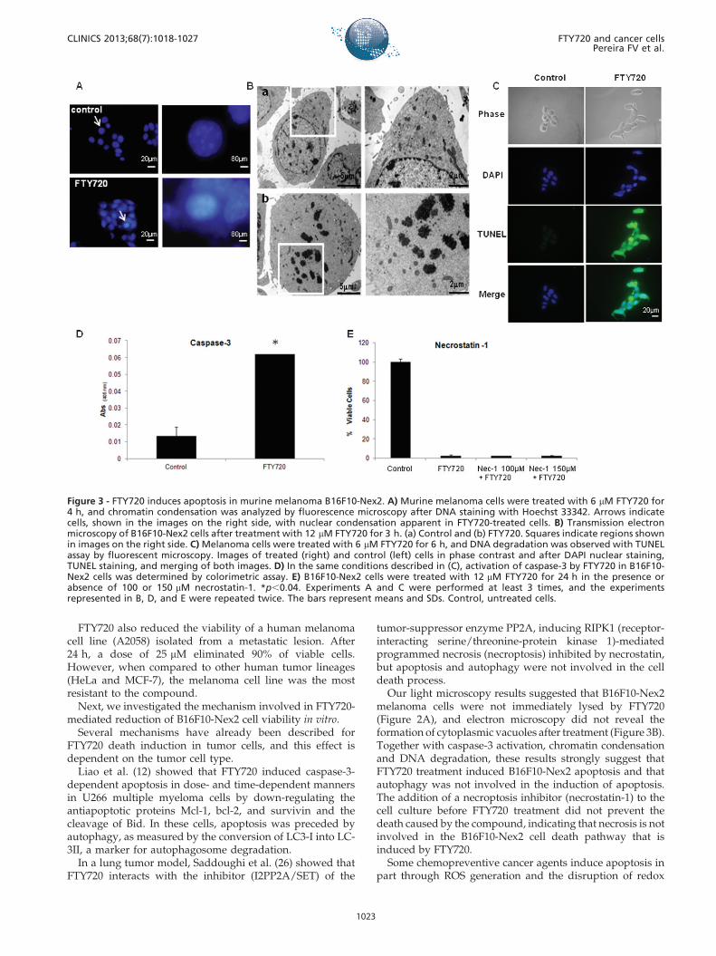
increase in tumor cells showing chromatin condensationwas observed after cell staining with Hoechst 33342. Fortypercent of FTY720-treated cells, but only 7% of control cells,showed evident chromatin condensation (Figure 3A). Bytransmission electron microscopy, important nuclear altera-tions were also observed after the compound treatment,
mainly early chromatin aggregation (after 3 h) along withcomplete nuclear membrane disruption (Figure 3B).
FTY720 treatment also induced strong DNA degradationin B16F10-Nex2 cells detected by TUNEL assay. While 87%of treated cells showed DNA degradation, only 2.5% ofcontrol cells were positive on TUNEL assay (Figure 3C).Strong activation of caspase-3, an important hallmark ofapoptosis, was observed in FTY720-treated cells (3D). Theaddition of necrostatin, a necroptosis inhibitor (15), to thecell culture before FTY720 treatment did not preventFTY720-induced death, indicating that necrosis is notinvolved in the B16F10-Nex2 cell death pathway that isinduced by FTY720 (Figure 3E).
Accumulation of reactive oxygen species andnuclear/cytoplasmic b-catenin in B16F10-Nex2 cellsafter FTY720 treatment
Reactive oxygen species (ROS) are important regulators ofapoptosis (16). To evaluate the participation of ROS inFTY720-induced cell death, intracellular anion superoxideproduction was detected using DHE after short (4 h)incubation with a low dose (6 mM) of FTY720. Thefluorescence intensity of cells treated with FTY720 was ashigh as that of the positive control treated with 5 mM ofH2O2, with the majority of cells being positive in bothtreatments, while untreated cells showed a few positive cellswith low fluorescence (Figure 4A). The role of ROS inFTY720-induced apoptosis was confirmed by measuring cellviability after pre-incubation of B16F10-Nex2 cells with theROS scavenger N-acetyl-l-cysteine (NAC) or L-cysteine,following treatment with the compound. It was observedthat both antioxidants partially inhibited cell death in thepresence of FTY720 at the doses used (10 mM NAC and15 mM L-cysteine) (Figures 4B-C). This result suggests thatROS are among the apoptosis effector molecules induced byFTY720.
Decreased expression of b-catenin has been reported inadvanced and metastatic prostate cancer, and Chua et al.(17) reported that FTY720 restored b-catenin expression andsuppressed the growth of human prostate cancer xenograftsin nude mice. In agreement with this observation, B16F10-Nex2 cells exhibited significantly increased expression of b-catenin in their nuclei and cytoplasms after FTY720treatment for 2 h compared to untreated cells, as observedby immunoblotting (Figure 4D).
Immunomodulation by FTY720To assess other possible roles played by FTY720 in the
melanoma model, we removed splenocytes from C57BL/6mice 15 days after B16F10-Nex2 injections and investigatedex vivo the percentages of T lymphocytes (CD4+ and CD8+),Treg cells (CD4+Foxp3+), and IFN-producing lymphocytesby flow cytometry. Mice treated with FTY720 evidenced asignificant decrease in the percentage of CD4+T cells,whereas no significant change was observed for CD8+ Tcells (Figure 5A and 5C). Additionally, Treg cells decreasedin mice treated with FTY720, but no change was observed inlymphocytes producing IFN-c (Figure 5B and 5D).
& DISCUSSION
Patients with advanced melanoma have a poor prognosis,with 1-year survival rates as low as 33% and a medianoverall survival (OS) of approximately 9 months (18).
Figure 1 - In vivo evaluation of pulmonary melanoma metastasisafter treatment with FTY720. A) C57BL/6 male mice were injectedi.v. with 36105 B16F10-Nex2 melanoma cells. The mice weretreated by gavage with PBS (Control) or with FTY720 (5 mg/kg/day) for 7 days starting on the day of tumor cell inoculation, andthe number of metastatic nodules in the lungs was evaluated 8days after the last dose of the compound (n = 5 animals pergroup). *p,0.04. B) Representative images of C57BL/6 mouselungs 15 days after melanoma cell inoculation.
CLINICS 2013;68(7):1018-1027 FTY720 and cancer cellsPereira FV et al.
1021

Systemic treatments are ineffective in general becausemetastatic melanoma is largely refractory to the availabletherapies. Dacarbazine (DTIC) remains the most commonlyused systemic agent, and when used alone or in combina-tion with other agents, the response rates are less than 20%,with an OS of less than 9 months (19). Interleukin-2 trialsreported durable complete responses in less than 10% ofpatients, with significant toxicity (20).
FTY720 has been approved by the FDA for the treatmentof patients with refractory multiple sclerosis (21).Additionally, this compound has been shown to exertanti-cancer functions against chronic myelogenous leuke-mia (22) and gastrointestinal tumors via activation of PP2A(23) by unknown mechanisms.
Our results showed that FTY720 could limit B16F10-Nex2metastatic melanoma development in vivo. Mice treatedwith 5 mg/kg/day of FTY720 for 7 days, starting on thesame day as B16F10-Nex2 melanoma cell inoculationevidenced a significant reduction in the number of meta-static lung nodules when evaluated 15 days later. Inagreement with this finding, in a model of lung cancerinduction by a chemical carcinogen (urethane), it wasrecently shown that FTY720 decreases tumor growth, whichis associated with reduced cell proliferation and VEGF(endothelial growth factor) expression, in addition toincreased expression of activated caspase-3 (24).
LaMontagne et al. (25) reported a reduction in tumor cellgrowth in vivo after the inoculation of B16/BL6 melanomacells in mouse ears and treatment with 3 mg/kg/day ofFTY720 for 14 days. Both the volumes and weights ofprimary and cervical lymph node metastases were signifi-cantly reduced after FTY720 treatment. In vitro, thecompound presented no cytotoxic or antiproliferative effectson melanoma cells at a dose of 1000 nM (1 mM), and thein vivo effects were attributed to the inhibition of tumorangiogenesis. In agreement with this observation, we foundthat 1 mM FTY720 had no effects on tumor cell apoptosisafter 24 h of culture. In contrast, 5 mM FTY720 caused a 20%decrease in cell viability, whereas 8 mM FTY720 caused 70%cell death in 24 h and complete extermination of cells in48 h. A dose of 12 mM was able to eliminate all viable cellsafter 24 h. Additionally, LaMontagne used B16/BL6 cellspossessing an aggressive metastatic capability, whereas weevaluated B16F10-Nex2 cells, which presented moderatevirulence. In LaMontagne’s research, experimental cellscould not even be subjected to prolonged incubation(.48 h) due to their rapid proliferative rate. Our resultsshowed that FTY720 inhibition of cancer development isdose-dependent. For human therapy, FTY720 should beused in higher doses or in combination with classicchemotherapy, as human tumor cells likely possess moreaggressive metastatic capabilities.
Figure 2 - In vitro effects of FTY720 on murine melanoma and human tumor cell lines. A) B16F10-Nex2 morphological alterations, aftertreatment with 1, 6, and 12 mM FTY720, were analyzed by light microscopy after 24 h. Control, untreated cells. Magnification, 40X. B)Viable B16F10-Nex2 cells were counted after 24 or 48 h of treatment with increasing doses of FTY720. C) The A2058, HeLa, and MCF-7human tumor cell lines were incubated with increasing doses of FTY720, and cell viability was evaluated after 24 h. Each experimentwas performed at least twice. The bars represent means and SDs.
FTY720 and cancer cellsPereira FV et al.
CLINICS 2013;68(7):1018-1027
1022
FTY720 also reduced the viability of a human melanomacell line (A2058) isolated from a metastatic lesion. After24 h, a dose of 25 mM eliminated 90% of viable cells.However, when compared to other human tumor lineages(HeLa and MCF-7), the melanoma cell line was the mostresistant to the compound.
Next, we investigated the mechanism involved in FTY720-mediated reduction of B16F10-Nex2 cell viability in vitro.
Several mechanisms have already been described forFTY720 death induction in tumor cells, and this effect isdependent on the tumor cell type.
Liao et al. (12) showed that FTY720 induced caspase-3-dependent apoptosis in dose- and time-dependent mannersin U266 multiple myeloma cells by down-regulating theantiapoptotic proteins Mcl-1, bcl-2, and survivin and thecleavage of Bid. In these cells, apoptosis was preceded byautophagy, as measured by the conversion of LC3-I into LC-3II, a marker for autophagosome degradation.
In a lung tumor model, Saddoughi et al. (26) showed thatFTY720 interacts with the inhibitor (I2PP2A/SET) of the
tumor-suppressor enzyme PP2A, inducing RIPK1 (receptor-interacting serine/threonine-protein kinase 1)-mediatedprogrammed necrosis (necroptosis) inhibited by necrostatin,but apoptosis and autophagy were not involved in the celldeath process.
Our light microscopy results suggested that B16F10-Nex2melanoma cells were not immediately lysed by FTY720(Figure 2A), and electron microscopy did not reveal theformation of cytoplasmic vacuoles after treatment (Figure 3B).Together with caspase-3 activation, chromatin condensationand DNA degradation, these results strongly suggest thatFTY720 treatment induced B16F10-Nex2 apoptosis and thatautophagy was not involved in the induction of apoptosis.The addition of a necroptosis inhibitor (necrostatin-1) to thecell culture before FTY720 treatment did not prevent thedeath caused by the compound, indicating that necrosis is notinvolved in the B16F10-Nex2 cell death pathway that isinduced by FTY720.
Some chemopreventive cancer agents induce apoptosis inpart through ROS generation and the disruption of redox
Figure 3 - FTY720 induces apoptosis in murine melanoma B16F10-Nex2. A) Murine melanoma cells were treated with 6 mM FTY720 for4 h, and chromatin condensation was analyzed by fluorescence microscopy after DNA staining with Hoechst 33342. Arrows indicatecells, shown in the images on the right side, with nuclear condensation apparent in FTY720-treated cells. B) Transmission electronmicroscopy of B16F10-Nex2 cells after treatment with 12 mM FTY720 for 3 h. (a) Control and (b) FTY720. Squares indicate regions shownin images on the right side. C) Melanoma cells were treated with 6 mM FTY720 for 6 h, and DNA degradation was observed with TUNELassay by fluorescent microscopy. Images of treated (right) and control (left) cells in phase contrast and after DAPI nuclear staining,TUNEL staining, and merging of both images. D) In the same conditions described in (C), activation of caspase-3 by FTY720 in B16F10-Nex2 cells was determined by colorimetric assay. E) B16F10-Nex2 cells were treated with 12 mM FTY720 for 24 h in the presence orabsence of 100 or 150 mM necrostatin-1. *p,0.04. Experiments A and C were performed at least 3 times, and the experimentsrepresented in B, D, and E were repeated twice. The bars represent means and SDs. Control, untreated cells.
CLINICS 2013;68(7):1018-1027 FTY720 and cancer cellsPereira FV et al.
1023

homeostasis (27). In addition to byproducts of mitochon-drial respiration, ROS are also key signaling molecules thatregulate mitochondrial dysfunction and apoptotic events(28). As secondary messengers, ROS can trigger the releaseof pro-apoptotic proteins from the mitochondrial intermem-brane space into the cytosol, such as cytochrome c, caspase-9, apoptosis-inducing factor, and Smac/DIABLO (secondmitochondria-derived activator of caspases) (29), elevatingintracellular Ca2+ concentrations and activating the caspasecascade (30). In acute lymphoblastic leukemia cells, the
inhibition of ROS production by 10 mM NAC treatmentprevented caspase-independent cell death (11).
Our results indicated that ROS are implicated in FTY720-induced cell death. B16F10-Nex2 melanoma cells treated witha low dose (6 mM) of FTY720 greatly increased their produc-tion of superoxide anions, as analyzed by DHE staining. Inaddition, the presence of the antioxidants NAC and L-cysteinepartially inhibited the cell death induced by FTY720, suggest-ing that there are other regulating factors, in addition to ROS,in the FTY720-induced cell death of B16F10-Nex2 cells.
Figure 4 - ROS production and b-catenin expression by B16F10-Nex2 cells after FTY720 treatment. A) B16F10Nex-2 cells were incubatedor not with 6 mM FTY720 for 4 h, and ROS production was determined by DHE assay and fluorescence microscopy. H2O2, positivecontrol treated with 5 mM hydrogen peroxide. B) B16F10Nex-2 cells were treated with 3.25-30 mM FTY720 after 2 h of pre-incubationwith 10 mM NAC or C) 15 mM FTY720 and pre-incubation with 15 mM L-cysteine, and cell viability was evaluated after 12 h. D)Expression of b-catenin was analyzed by western blot of nuclear and cytoplasmic extracts of B16F10Nex-2 cells incubated with 6 mMFTY720 for 1 h. *p,0.04.
FTY720 and cancer cellsPereira FV et al.
CLINICS 2013;68(7):1018-1027
1024

b-catenin is a key component of the Wnt/b-cateninsignaling pathway and a mediator of the Ras/phosphatidy-linositol 3-kinase (PI3K) pathways (31). Active b-catenininteracts with transcription factors, such as T cell factor/lymphoid enhancer (TCF/LEF), CBP and p300, resulting intarget gene transcription, and it also binds to cadherins in thecell membrane to provide structural support for adhesion(32,33). The b-catenin pathway is well known as an enhancerof proliferation and survival in tumor cells. However, itsoverexpression or accumulation has also been reported toinduce apoptosis in several tumor cell lines. Raab et al. (34)demonstrated that inhibition of PKC (protein kinase C)resulted in the accumulation of active b-catenin, whichcontributes to enzastaurin-induced cell death in multiplemyeloma cells. Overexpression of a stable form of b-cateninor inhibition of endogenous b-catenin degradation has beenreported to lead to G2 cell cycle arrest and apoptosis inepidermal keratinocytes (35). In malignant prostate cancer,reduced expression of nuclear b-catenin was associated withpoorer prognosis (36), and restored functions of b-cateninand E-cadherin were associated with decreased microvesseldensity and VEGF expression in a mouse model of prostatecancer (17). It has recently been shown that the activation ofWnt/b-catenin signaling in vitro, in vivo (murine melanomamodel), or in patient tumors results in increased levels ofnuclear b-catenin, and this finding has been correlated with abeneficial prognosis (37,38).
In our study, there was increased expression of b-cateninin the nuclei and cytoplasms of B16F10-Nex2 cells treatedwith FTY720, suggesting that this molecule could beinvolved in the FTY720-induced cell death observed inthese murine melanoma cells.
In addition to the reduced development of metastaticmelanoma in vivo and increased melanoma cell death invitro caused by FTY720, our results ex vivo showed adecrease in CD4+Foxp3+ cells in mice injected with B16F10-Nex2 melanoma cells and treated with FTY720. Treg cellshave been shown to facilitate tumor development (39); thus,FTY720 could act as an immunomodulator by decreasingthe numbers of these cells. Additionally, the capability oflymphocytes to produce IFN-c was not changed by FTY720treatment.
This study demonstrated that a low dose of orallyadministered FTY720 was effective in limiting murinemetastatic melanoma development in vivo and that thecompound induced apoptosis in these tumor cells in vitro,without indications of autophagy or necroptosis. Apoptoticcell death was regulated by ROS and by increasedexpression of b-catenin. FTY720 efficacy was obtained onlyin higher doses when added in vitro to A2058 humanmelanoma cells isolated from a metastatic lesion. In additionto the cancer cell death induced by FTY720, we alsoobserved immunomodulation in the host immune system
Figure 5 - Ex vivo evaluation of C57BL/6 mouse spleen cells by flow cytometry, showing the percentages of CD4+T cells, CD4+Foxp3+cells, CD8+ T cells, and lymphocytes producing IFN-c. Mice injected with B16F10Nex-2 cells and treated (FTY720) or not (Control) hadtheir spleens harvested 15 days later for the evaluation of A) CD4+ T cells, *p = 0.0021; B) CD4+Foxp3+ cells; C) CD8+ T cells; D) IFN-c-producing lymphocytes.
CLINICS 2013;68(7):1018-1027 FTY720 and cancer cellsPereira FV et al.
1025

caused by the compound, with a significant reduction inTreg cell numbers.
It has already been shown that FTY720, administered inassociation with chemotherapy, improves anticancer effectsmainly by inducing cell death (40). Further studies areneeded using FTY720 as a monotherapy or in combinedtherapy, as different types of cancer cells could require theinvolvement of a variety of signaling pathways to beeliminated completely.
& ACKNOWLEDGMENTS
We thank Fundacao de Amparo a Pesquisa do Estado de Sao Paulo
(FAPESP) and Conselho Nacional de Pesquisa e Desenvolvimento (CNPq)
for their financial support. EGR and VB are recipients of fellowships from
CNPq. DCA is a recipient of a postdoctoral fellowship from FAPESP
(2008/51256-7).
& AUTHOR CONTRIBUTIONS
Pereira FV cell viability assays, in vivo experiment, participation in the
manuscript design, statistical analysis, and helped to draft the manuscript.
Arruda DC analysis of superoxide anion, chromatin condensation,
transmission electron microscopy analysis and TUNEL assay. Figueiredo
CR caspase-3 activation. Massaoka MH b-catenin detection. Matsuo AL
cell viability assays. Bueno V in vivo treatment with FTY720, FACS
analysis, participation in the manuscript design, and draft of the
manuscript. Rodrigues EG conceived of the study, coordination and
manuscript design, draft of the manuscript. All authors read and approved
the final version of the manuscript.
& REFERENCES
1. Kiuchi M, Adachi K, Tomatsu A, Chino M, Takeda S, Tanaka Y, et al.Asymmetric synthesis and biological evaluation of the enantiomericisomers of the immunosuppressive FTY720-phosphate. Bioorg MedChem. 2005;13(2):425-32, http://dx.doi.org/10.1016/j.bmc.2004.10.008.
2. Tedesco-Silva H, Szakaly P, Shoker A, Sommerer C, Yoshimura N,Schena FP, et al. FTY720 versus mycophenolate mofetil in de novorenal transplantation: six-month results of a double-bind study.Transplantation. 2007;84(7):885-92, http://dx.doi.org/10.1097/01.tp.0000281385.26500.3b.
3. Commodaro AG, Pedregosa JF, Peron JPS, Brandao WN, Rizzo LV,Bueno V. The imbalance between Treg/Th17 cells caused by FTY720treatment in skin allograft rejection. Clinics. 2012;67(7):805-13, http://dx.doi.org/10.6061/clinics/2012(07)17.
4. Pedregosa JF, Haidar AA, Hirata AE, Franco M, Gomes GN, Bueno V.TLR2 and TLR4 expression after kidney ischemia and reperfusion injuryin mice treated with FTY720. Int Immunopharmacol. 2011;11(9):1311-8,http://dx.doi.org/10.1016/j.intimp.2011.04.014.
5. Papadoulos D, Rundle J, Patel R, Marshall I, Stretton J, Eaton R, et al.FTY720 ameliorates MOG-induced experimental autoimmune encepha-lomyelitis by suppressing both cellular and humoral immune responses.J Neurosci Res. 2010;88(2):346-59, http://dx.doi.org/10.1002/jnr.22196.
6. Commodaro AG, Perons JPS, Lopes CT, Arslanian C, Belfort R, Bueno V.Evaluation of experimental autoimmune uveitis in mice treated withFTY720. Invest Ophthalmol Vis Sci. 2010;51(5):2568-74, http://dx.doi.org/10.1167/iovs.09-4769.
7. Wang JD, Takahara S, Nonomura N, Ichimaru N, Toki K, Azuma H, et al.Early induction of apoptosis in androgen-independent prostate cancercell line by FTY720 requires caspase-3 activation. Prostate. 1999;40(1):50-5, http://dx.doi.org/10.1002/(SICI)1097-0045(19990615)40:1,50::AID-PROS6.3.0.CO;2-N.
8. Azuma H, Takahara S, Horie S, Muto S, Otsuki Y, Katsuoka Y. Inductionof apoptosis in human bladder cancer cells in vitro and in vivo caused byFTY720 treatment. J Urol. 2003;169(6):2372-7.
9. Lee TK, Man K, Ho JW, Sun CK, NgKT, Wang XH, et al. FTY720 inducesapoptosis of human hepatoma cell lines through PI2-K-mediated Aktdephosphorylation Carcinogenesis. 2004;25(12):2397-405.
10. Ubai T, Azuma H, Kotake Y, Inamoto T, Takahara K, Ito Y, et al. FTY720induced Bcl-associated and Fas-independent apoptosis in human renalcancer cell in vitro and significantly reduced in vivo tumor growth inmouse xenograft. Anticancer Res. 2007;27(1A):75-88.
11. Wallington-Beddoe CT, Hewson J, Bradstock KF, Bedall LJ. FTY720produces caspase-independent cell death of acute lymphoblasticleukemia cells. Autophagy. 2011;7(7):707-15, http://dx.doi.org/10.4161/auto.7.7.15154
12. Liao A, Hu R, Zhao Q, Li J, Li Y, Yao K, et al. Autophagy induced byFTY720 promotes apoptosis in U266 cells. Eur J Pharm Sci.2012;45(5):600-5.
13. Azuma H, Takahara S, Ichimaru N, Wang JD, Itoh Y, Otsuki Y, et al.Marked prevention of tumor growth and metastasis by a novelimmunosuppressive agent, FTY720, in mouse breast cancer models.Cancer Res. 2002;62:1410-9.
14. Liu Y, Deng J, Wang L, Lee H, Armstrong B, Scuto A, et al. S1PR1 is aneffective target to block STAT3 signaling in activated B cell-like diffuselarge B-cell lymphoma. Blood. 2012;120(7):1458-65, http://dx.doi.org/10.1182/blood-2011-12-399030.
15. Zhang N, Chen Y, Jiang R, Li E, Chen X, Xi Z, et al. PARP and RIP 1 arerequired for autophagy induced by 11’-deoxyverticillin A, whichprecedes caspase-dependent apoptosis. Autophagy. 2011;7(6):598-612,http://dx.doi.org/10.4161/auto.7.6.15103.
16. Mates JM, Segura JA, Alonso FJ, Marquez J. Intracellular redox statusand oxidative stress: implications for cell proliferation, apoptosis, andcarcinogenesis. Arch Toxicol. 2008;82(5):273-99, http://dx.doi.org/10.1007/s00204-008-0304-z.
17. Chua CW, Lee DT, Ling MT, Zhou C, Man K, Ho J, et al. FTY720, afungus metabolite, inhibits in vivo growth of androgen-independentprostate cancer. Int J Cancer. 2005;117(6):1039-48.
18. Balch CM, Gershenwald JE, Soong SJ, Thompson JF, Atkins MB, ByrdDR, et al. Final version of 2009 AJCC melanoma staging andclassification. J Clin Oncol. 2009;27(36):6199-06, http://dx.doi.org/10.1200/JCO.2009.23.4799.
19. Patel PM, Suciu S, Mortier L, Kruit WH, Robert C, Schadendorf D, et al.Extended schedule, escalated dose temozolomide versus dacarbazine instage IV melanoma: final results of a randomised phase III study(EORTC 18032) EORTC Melanoma Group.. Eur J Cancer. 2011;47(10):1476-83.
20. Atkins MB, Lotze MT, Dutcher JP, Fisher RI, Weiss G, Margolin K, et al.High-dose recombinant interleukin 2 therapy for patients with metastaticmelanoma: analysis of 270 patients treated between 1985 and 1993. J ClinOncol 1999;17(7):2105-16.
21. Cohen JA, Barkhof F, Comi G, Hartung HP, Khatri BO, Montalban X,et al. Oral fingolimod or intramuscular interferon for relapsing multiplesclerosis.TRANSFORMS Study Group. N Engl J Med. 2010 4;362(5):402-15.
22. Neviani P, Santhanam R, Oaks JJ, Eiring AM, Notari M, Blaser BW, et al.FTY720, a new alternative for treating blast crisis chronic myelogenousleukemia and Philadelphia chromosome-positive acute lymphocyticleukemia. J Clin Invest. 2007;117(9):2408-21, http://dx.doi.org/10.1172/JCI31095.
23. Roberts KG, Smith AM, McDougall F, Carpenter H, Horan M, Neviani P,et al. Essential requirement for PP2A inhibition by the oncogenicreceptor c-KIT suggests PP2A reactivation as a strategy to treat c-KIT+cancers. Cancer Res. 2010;70(13):5438-47, http://dx.doi.org/10.1158/0008-5472.CAN-09-2544.
24. Salinas NR, Oshima CT, Cury PM, Cordeiro JA, Bueno V. FTY720 andlung tumor development. Int Immunopharmacol. 2009;9(6):689-93,http://dx.doi.org/10.1016/j.intimp.2008.12.007.
25. LaMontagne K, Littlewood-Evans A, Schnell C, O’Reilly T, Wyder L,Sanchez T, et al. Antagonism of sphingosine-1-phosphate receptors byFTY720 inhibits angiogenesis and tumor vascularization. Cancer Res.2006;66(1):221-31, http://dx.doi.org/10.1158/0008-5472.CAN-05-2001.
26. Saddoughi SA, Gencer S, Peterson YK, Ward KE, Mukhopadhyay A,Oaks J, et al. Sphingosine analogue drug FTY720 targets I2PP2A/SETand mediates lung tumour suppression via activation of PP2A-RIPK1-dependent necroptosis. EMBO Mol Med. 2012;4(1):1-17.
27. Ling YH, Liebes L, Zou Y, Perez-Soler R. Reactive oxygen speciesgeneration and mitochondrial dysfunction in the apoptotic response toBortezomib, a novel proteasome inhibitor, in human H460 non-small celllung cancer cells. J Biol Chem. 2003;278(36):33714-23, http://dx.doi.org/10.1074/jbc.M302559200.
28. Liu MJ, Wang Z, Li HX, Wu RC, Liu YZ, Wu QY. Mitochondrialdysfunction as an early event in the process of apoptosis induced bywoodfordin I in human leukemia K562 cells. Toxicol Appl Pharmacol.2004;194(2):141-55, http://dx.doi.org/10.1016/j.taap.2003.08.017.
29. Kroemer G, Dallaporta B, Resche-Rigon M. The mitochondrial death/liferegulator in apoptosis and necrosis. Annu Rev Physiol. 1998;60:619-42,http://dx.doi.org/10.1146/annurev.physiol.60.1.619.
30. Paradies G, Petrosillo G, Pistolese M, Ruggiero FM. Reactive oxygenspecies affect mitochondrial electron transport complex I activitythrough oxidative cardiolipin damage. Gene. 2002;286(1):135-41,http://dx.doi.org/10.1016/S0378-1119(01)00814-9.
31. Herencia C, Martınez-Moreno JM, Herrera C, Corrales F, Santiago-MoraR, Espejo I, et al. Nuclear translocation of b-catenin during mesenchymalstem cells differentiation into hepatocytes is associated with a tumoralphenotype. PLoS One. 2012;7(4):e34656, http://dx.doi.org/10.1371/journal.pone.0034656.
32. Miller JR, Moon RT. Signal transduction through beta-catenin andspecification of cell fate during embryogenesis. Genes Dev. 1996;10(20):2527-39, http://dx.doi.org/10.1101/gad.10.20.2527.
FTY720 and cancer cellsPereira FV et al.
CLINICS 2013;68(7):1018-1027
1026

33. Moon RT, Brown JD, Torres M. WNTs modulate cell fate and behaviorduring vertebrate development. Trends Genet. 1997;13(4):157-62, http://dx.doi.org/10.1016/S0168-9525(97)01093-7.
34. Raab MS, Breitkreutz I, Tonon G, Zhang J, Hayden PJ, Nguyen T, et al.Targeting PKC: a novel role for beta-catenin in ER stress and apoptoticsignaling. Blood. 2009;113(7):1513-21.
35. Olmeda D, Castel S, Vilaro S, Cano A. Beta-catenin regulation during thecell cycle: implications in G2/M and apoptosis. Mol Biol Cell.2003;14(7):2844-60, http://dx.doi.org/10.1091/mbc.e03",-1,"xxx/mbc.e03-01-0865.
36. Horvath LG, Henshall SM, Lee CS, Kench JG, Golovsky D, Brenner PC,et al. Lower levels of nuclear beta-catenin predict for a poorer prognosisin localized prostate cancer. Int J Cancer. 2005;113(3):415-22.
37. Chien AJ, Moore EC, Lonsdorf AS, Kulikauskas RM, Rothberg BG,Berger AJ, et al. Activated Wnt/beta-catenin signaling in melanoma is
associated with decreased proliferation in patient tumors and a murinemelanoma model. Proc Natl Acad Sci U S A. 2009;106(4):1193-8, http://dx.doi.org/10.1073/pnas.0811902106.
38. Da Forno PD, Pringle JH, Hutchinson P, Osborn J, Huang Q, Potter L,et al. WNT5A expression increases during melanoma progression andcorrelates with outcome. Clin Cancer Res. 2008;14(18):5825-32, http://dx.doi.org/10.1158/1078-0432.CCR-07-5104.
39. Hansen W, Hutzler M, Abel S, Alter C, Stochmann C, Kliche S, et al.Nueropilin 1 deficienty on CD4+Foxp3+ regulatory T cells impairsmouse melanoma growth. J Exp Med. 2012;209(11):2001-16, http://dx.doi.org/10.1084/jem.20111497.
40. Alinari L, Mahoney E, Patton J, Zhang X, Huynh L, Earl CT, et al. FTY720increases CD74 expression and sensitizes mantle cell lymphoma cells tomilatuzumab-mediated cell death. Blood. 2011;118(26):6893-903, http://dx.doi.org/10.1182/blood-2011-06-363879.
CLINICS 2013;68(7):1018-1027 FTY720 and cancer cellsPereira FV et al.
1027
Top Related
Copyright © 2022 FDOKUMEN

