


Bahasa
Halaman
Hukum

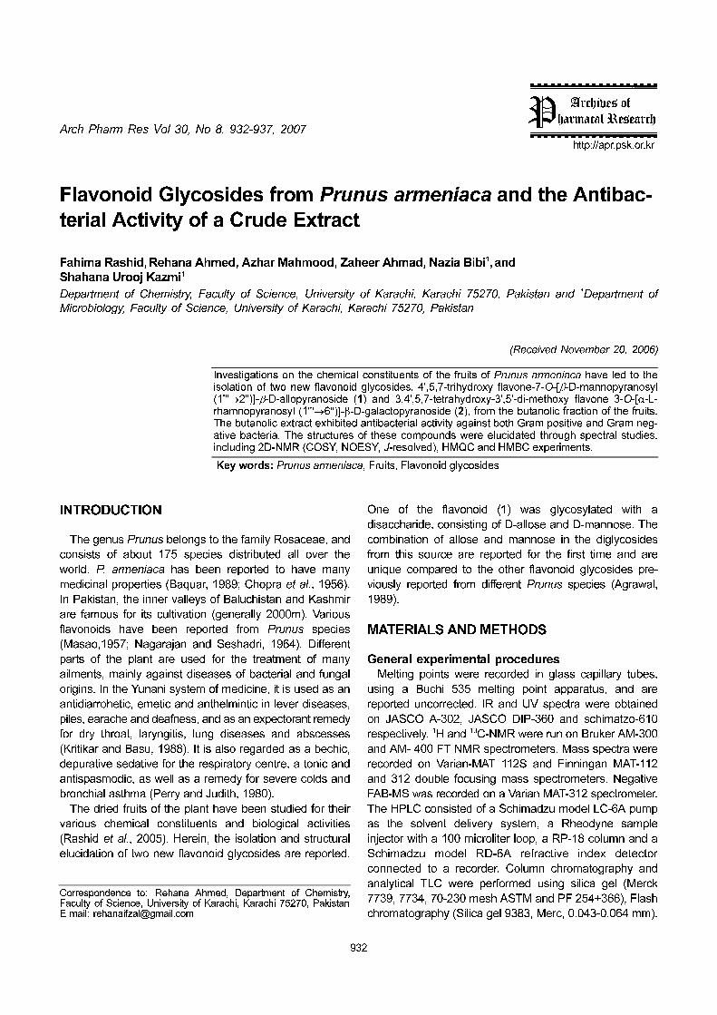
Arch Pharm Res Vol 30, No 8, 932-937, 2007
932
http://apr.psk.or.kr
Flavonoid Glycosides from Prunus armeniaca and the Antibac-
terial Activity of a Crude Extract
Fahima Rashid, Rehana Ahmed, Azhar Mahmood, Zaheer Ahmad, Nazia Bibi1, and
Shahana Urooj Kazmi1
Department of Chemistry, Faculty of Science, University of Karachi, Karachi 75270, Pakistan and 1Department of
Microbiology, Faculty of Science, University of Karachi, Karachi 75270, Pakistan
(Received November 20, 2006)
Investigations on the chemical constituents of the fruits of Prunus armeniaca have led to theisolation of two new flavonoid glycosides, 4',5,7-trihydroxy flavone-7-O-[β-D-mannopyranosyl(1'''→2'')]-β-D-allopyranoside (1) and 3,4',5,7-tetrahydroxy-3',5'-di-methoxy flavone 3-O-[α-L-rhamnopyranosyl (1'''→6'')]-β-D-galactopyranoside (2), from the butanolic fraction of the fruits.The butanolic extract exhibited antibacterial activity against both Gram positive and Gram neg-ative bacteria. The structures of these compounds were elucidated through spectral studies,including 2D-NMR (COSY, NOESY, J-resolved), HMQC and HMBC experiments.
Key words: Prunus armeniaca, Fruits, Flavonoid glycosides
INTRODUCTION
The genus Prunus belongs to the family Rosaceae, and
consists of about 175 species distributed all over the
world. P. armeniaca has been reported to have many
medicinal properties (Baquar, 1989; Chopra et al., 1956).
In Pakistan, the inner valleys of Baluchistan and Kashmir
are famous for its cultivation (generally 2000m). Various
flavonoids have been reported from Prunus species
(Masao,1957; Nagarajan and Seshadri, 1964). Different
parts of the plant are used for the treatment of many
ailments, mainly against diseases of bacterial and fungal
origins. In the Yunani system of medicine, it is used as an
antidiarrohetic, emetic and anthelmintic in lever diseases,
piles, earache and deafness, and as an expectorant remedy
for dry throat, laryngitis, lung diseases and abscesses
(Kritikar and Basu, 1988). It is also regarded as a bechic,
depurative sedative for the respiratory centre, a tonic and
antispasmodic, as well as a remedy for severe colds and
bronchial asthma (Perry and Judith, 1980).
The dried fruits of the plant have been studied for their
various chemical constituents and biological activities
(Rashid et al., 2005). Herein, the isolation and structural
elucidation of two new flavonoid glycosides are reported.
One of the flavonoid (1) was glycosylated with a
disaccharide, consisting of D-allose and D-mannose. The
combination of allose and mannose in the diglycosides
from this source are reported for the first time and are
unique compared to the other flavonoid glycosides pre-
viously reported from different Prunus species (Agrawal,
1989).
MATERIALS AND METHODS
General experimental procedures
Melting points were recorded in glass capillary tubes,
using a Buchi 535 melting point apparatus, and are
reported uncorrected. IR and UV spectra were obtained
on JASCO A-302, JASCO DIP-360 and schimatzo-610
respectively. 1H and 13C-NMR were run on Bruker AM-300
and AM- 400 FT NMR spectrometers. Mass spectra were
recorded on Varian-MAT 112S and Finningan MAT-112
and 312 double focusing mass spectrometers. Negative
FAB-MS was recorded on a Varian MAT-312 spectrometer.
The HPLC consisted of a Schimadzu model LC-6A pump
as the solvent delivery system, a Rheodyne sample
injector with a 100 microliter loop, a RP-18 column and a
Schimadzu model RD-6A refractive index detector
connected to a recorder. Column chromatography and
analytical TLC were performed using silica gel (Merck
7739, 7734, 70-230 mesh ASTM and PF 254+366), Flash
chromatography (Silica gel 9383, Merc, 0.043-0.064 mm).
Correspondence to: Rehana Ahmed, Department of Chemistry,Faculty of Science, University of Karachi, Karachi 75270, PakistanE-mail: [email protected]
Flavonoid Glycosides from Prunus armeniaca and its Antibacterial Activity 933
HPTLC was conducted on Silica gel GF254 and RP-18 F254
(Merc plates).
Plant materialDried fruits were purchased in October 2004 from a fruit
market in Karachi. A voucher specimen (Ch-2059-2004)
was deposited in the herbarium of the Department of
Botany, University of Karachi.
Extraction and isolationDried fruits (10 kg) were soaked in ethanol for 15 days
(5 L × 3 batches) at room temperature. The dark brown
colored extract was concentrated on a rotary evaporator
with the resulting brown gummy material (375 g) then
successively fractionated with n-hexane, chloroform, ethyl-
acetate and saturated butanol.
The butanolic fraction of the ethanolic extract was
concentrated, to a dark brown sticky mass (85 g). This
concentrated fraction was subjected to Silica gel column
chromatography using chloroform-methanol (5%, 10%,
15%, 20%, 30%, 40% and 1:1) and chloroform-methanol-
water (65:35:1) as solvent systems. The fractions of similar
composition were combined, with two main fractions
obtained; F (50 mg) and G (40 mg). TLC of the two
different fractions was conducted with BuOH:AcOH:H2O
(12:3:7, 12:3:5, 3:1:1) and EtOAc:CHCl3:H2O (17:3:1) as
the mobile phases. Fraction G was obtained with
chloroform-methanol (70:30), and indicated the presence
of two compounds with distinct Rf, which gave, prominent
colored spots with ferric chloride reagent. Further separa-
tion was achieved by HPLC employing an RP-18 column,
eluted using an isocratic methanol-water system (65:35),
followed by HPTLC and fractional crystallization to afford
compounds 1 (15 mg) and 2 (17 mg).
4',5,7-Trihydroxy-flavone-7-O-[β-D-mannopyranosyl-
(1'''→2'')]-β-D-allopyranoside (1)
Yellow powder, 15 mg, mp: 162C, UV (MeOH) λmax( nm):
272, and 333 nm; IR (KBr) Vmax (Cm-1): 3700-3310 (OH),
2920 (CH-str), 1722 (α, β unsaturated carbonyl group),
1662, 1600 (C=C), 1180 (C-O), 1070 and 1032; HR-FABMS
(negative) m/z: 593.5179 [M-H]+ (Calcd. for C27H29O15,
593.5171); 1H-NMR (DMSO-d6, 500 MHz) δ: 7.75 (1H, d,
J=2.5 Hz, H-6'), 7.66 (1H, dd, J=8.2, 2.5 Hz, H-2') 7.28
(1H, d, J=8.4 Hz, H-3', H-5'), 7.26 (1H, d, J=2.5 Hz, H-8),
7.06 (1H, d, J=2.4 Hz, H-6), 6.99 (1H, s, H-3) 4.89 (1H, d,
J=5.2 Hz, mannosyl H-1'''), 5.77 (1H, d, J=7.5 Hz, allosyl
H-1''), 3.10-3.55 (12H, m, sugar proton). 3.55-3.64 (m, H-
6'''); 13C-NMR (DMSO-d6, 125 MHz) δ: 182.1 (C-4), 164.1
(C-2), 162.8 (C-7), 161.2 (C-9), 156.8 (C-5), 148.4 (C-4'),
121.5 (C-6'), 120.5 (C-1'), 116.3 (C-5', 3'), 110.9 (C-2'),
105.4 (C-10), 103.4 (C-3), 102.1 (C-1''), 99.7 (C-6'), 98.1
(C-1''), 95.4 (C-8), 79.7 (C-2'''), 77.0 (C-5'''), 74.4 (C-5''),
73.7 (C-3''), 71.8 (C-3''), 70.1 (C-2'''), 68.2 (C-4'''), 67.2 (C-
4''), 61.8 (C-6'''), 60.6 (C-6'').
Acid hydrolysis of compound 1Compound 1 (5 mg) was subjected to acid hydrolysis
with 2% H2SO4 in methanol (5 mL), by refluxing for three
hours, cooled and then extracted with ethyl-acetate. The
ethyl acetate fraction and the aqueous fraction were
obtained. Ethyl acetate and water fractions were worked
up in the usual manner to afford a dihydroxy aglycone
(Table I) yellow powder, m.p 165-166oC, the aqueous
fraction containing two sugar moieties mannose and
allose. Hydrolysis of compound 1 showed initial removal
of one mannose unit followed by one of allose unit. D-
mannose and D-allose were mainly identified by paper
chromatography by comparison with authentic samples
which were further confirmed by comparing the Rf values
with standards using descending techniques, with BuOH:
AcOH: H2O 4:1:5 and 3:1:1 as the mobile phases, the
spots were detected by spraying with aniline phthalate
and heating at 110oC.
3,4',5,7-Tetrahydroxy-3',5'-dimethoxy flavone-3-O-[-β-L-
rhamnopyranosyl (1→6)]-β-D-galactopyranoside (2)
Yellow powder, 17 mg, mp: 176C, UV (MeOH) λmax( nm):
252 (band II) & 357 (band I); IR (KBr) Vmax (cm-1): 3620-
3560 (OH), 1650 (C=O), 1605 (C=C), 1508, 1450 & 1150
(C-O); HR-FABMS (negative) m/z: 654.6321 [M-H]+ (Calcd.
for C29H33O17; 654.5774); 1H-NMR (DMSO-d6, 400 MHz)
δ: 7.61 (2H, s, H-2' and H-6'), 6.45 (1H, d, J=2Hz H-8),
6.24 (1H, d, J=2 Hz H-6), 5.41 (d, J=7.8 Hz), 4.55 (1H, d,
J=1.4 Hz) and 3.95 (6H, s, 3'-OMe & 5'-OMe); 13C-NMR
(DMSO-d6, 100 MHz) δ: 179.1 (C-4), 166.1 (C-7), 163.0
(C-5), 158.6 (C-9), 158.4 (C-2), 149.1 (C-5'), 149.0 (C-3'),
140.4 (C-4'), 135.2 (C-3), 121.7 (C-1'), 108.5 (C-2', 6'),
105.9 (C-10), 104.3 (C-1''), 102.1 (C-1'''), 100.3 (C-6),
95.0 (C-8), 75.5 (C-5''), 75.2 (C-3''), 73.5 (C-4'''), 73.3 (C-
2''), 72.5 (C-3'''), 72.3 (C-2'''), 70.1 (C-4''), 69.9 (C-5'''),
67.5 (C-6''), 57.5 (C-OCH3, 3' and 5'), 18.5 (C-6''').
Acid hydrolysis of compound 2A solution of compound 2 (5 mg) in 5% HCl/methanol (5
mL) was refluxed for three hrs. The reaction mixture was
extracted with ethyl acetate. The ethyl acetate fraction con-
taining aglycone (Table I) yellow amorphous powder, m.p.
280-285oC and the aqueous fraction containing two sugar
units (galactose and glucose) were concentrated to dryness.
The sugars were identified by comparison with standards
using descending paper chromatography, with BuOH:
AcOH: H2O 4:1:5 as the mobile phase, the spots were
detected by spraying with aniline phthalate followed by
heating at 110 oC.

934 F. Rashid et al.
Antibacterial assayThe antibacterial activity of the butanolic extract of
Prunus armeniaca was determined against 20 gram-
positive bacteria, including 10 MRSA isolates, 2 non
tuberculous Mycobacteria (NTM) and 13 gram-negative
bacteria. All the environmental and clinical isolates were
obtained from the Immunology and Infectious Disease
Research Laboratory (IIDRL) Culture Collection Unit,
Department of Microbiology, University of Karachi and
were identified and characterized using conventional
microbiological methods (Marie, 1985). A stock solution of
the butanolic extract (10 mg/mL) was prepared by
dissolving in 100% DMSO (Merk). The stock solution was
filtered and sterilized by passing through a 0.22 µm pore
size syringe filter (Nalgen).
The antibacterial activity of the extract was determined
by evaluating the minimum inhibitory concentrations (MICs)
using the Micro Broth Dilution method, as described in the
National Committee for Clinical Laboratory Standards
(NCCLS) (Wayne, 1998). Briefly, serial dilutions of the
butanolic extract were prepared directly into Mueller Hinton
broth (Oxoid-UK) in a round bottom 96-well Microtitre plate.
The wells were seeded with cultures at 2 × 10⁵ cfu/mL.
The plates were incubated at 37oC for 24 h, except for the
M. fortuitum and M. smegmatis, which were incubated for
2 and 3 days, respectively. The results were recorded in
the form of MICs, defined as the minimum amounts of the
extract and pure compound that inhibited the visible
growth of the organisms.
RESULTS AND DISCUSSION
Compound 1 was isolated as a light yellow powder from
the butanol soluble fraction of the ethanolic fruit extract via
repeated column chromatography, flash chromatography
and finally by HPLC. The IR spectrum showed a strong
absorption band between 3700-3450 cm-1, indicating the
presence of a hydroxyl group, 2920 (CH-stretching), α, β
unsaturated carbonyl absorption at 1720 cm-1 (C-4
flavonoid), 1660, 1600 due to an olefinic double bond in
an aromatic system. A broad carbonyl stretching band in
the region of 1100-1600 cm-1 suggested the glycosidic
nature of a flavonoid. The UV spectrum showed λmax at
272 and 333 nm giving a very strong indication of the
presence of a flavonoid (Vorin, 1983). The compound was
assigned the molecular formula; C27H30O15, with the help
of a peak observed in the HR-FABMS (negative mode) at
m/z 593.5179 [M-H]+ (Calcd. for C27 H29 O15 593.5171).
The 1H-NMR spectrum showed signals for phenyl ring B
at δ 7.75, 7.66 and 7.28 corresponded to hydrogens on
the C-6', 2', and 3',5' respectively. The aromatic protons of
ring A appeared in the 1H-NMR spectrum at δ 7.26 (d, 1H,
J=2.5 Hz) and 7.06 (d, 1H, J=2.4 Hz), which could be due
to the hydrogens on the C-8 and C-6 of a flavone
derivative.
The compound was subjected to acid hydrolysis with
2% H2SO4, which afforded an aglycone with a fragment at
m/z 270 [C15H9O5, M-allose - mannose + H]+, and other
prominent fragments 152 [A1-C7H4O4]+, 124 [A1-CO]+ and
118 [B1-C8H6O]+, as well as two saugar units D-mannose
and D-allose were mainly identified by paper chromato-
graphy compared with authentic samples (Gupta and
Bahar, 1985). All the 1H- and 13C-NMR data of the two
sugars moieties were also confirmed by 2D-NMR spectro-
scopic techniques. The [α]D20+15o and +14o values of
allose and mannose respectively, confirmed their D-
configuration due to their δ-shifts (Whistler and Wolform,
1962). The 1H- and 13C-NMR spectra also confirmed the
presence of aglycone and two sugar residues. A signal at
δ 162.9 in the 13C-NMR spectrum was assigned to the C-7
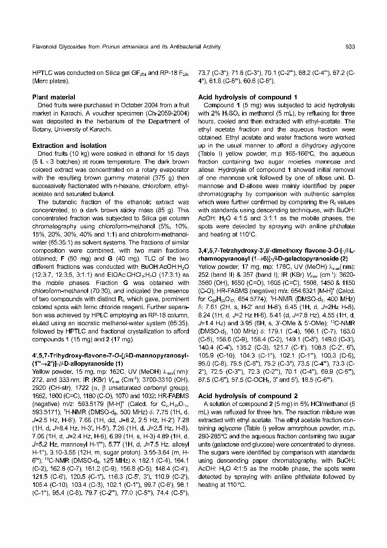
on the basis of the long range 13C-1H correlation observed
in HMBC experiments with the two protons signals at δ
7.26 (1H, d, J=2.5 Hz, H-8) and 7.06 (1H, d, J=2.4 Hz, H-
6) confirming the site of glycosidation at the C-7 position
of the aglycone. The C-7 signals showed three-band
correlations with the anomeric proton of the allosyl unit at
δ 5.77 (d, 1H, J=7.5 Hz). The anomeric proton of the
mannosyl residue at δ 4.89 (1H, d, J=5.2 Hz) also showed
long range correlations with the 13C-NMR signal at δ 79.8,
corresponding to a proton at δ 3.54 (H-2'') in HMQC
spectrum. The later signal showed a 1H-1H correlation, as
observed in a Cosy experiment with the anomeric proton
at δ 5.77 which was assigned as the H-1'' of the allosyl
moiety. Therefore glucosidation of allosyl at the C-2''
position was evident. The b-configuration of two anomeric
carbons was also confirmed from the coupling constant of
the H-1'' of allose (J=7.5 Hz) with that of the H-1''' of
mannose (J=5.2 Hz) (Agrawal, 1989). 2D-NMR allowed
the assignment of all 1H- and 13C-NMR signals of the
allosyl and mannosyl moieties.
The 13C-NMR spectrum, broad band and DEPT of the
Fig. 1. Compounds 1 and 2
Flavonoid Glycosides from Prunus armeniaca and its Antibacterial Activity 935
above compound revealed the presence of two methy-
lene, seventeen methine and eight quaternary carbon
atoms. On the basis of above discussions and spectral
data; compound 1 was assigned the structure 4',5,7-
trihydroxy flavone-7-O-[β-D-mannopyranosyl-(1'''→2'')]-β-D-
allopyranoside.
Compound 2 was obtained as yellow powder from the
butanolic fraction through column and flash chromatog-
raphy and finally purified by HPLC using an RP-18
column, with an isocratic methanol water system (65:35),
to afford 17 mg of compound 2. The IR spectrum of
compound 2 showed absorption at 3620-3560 cm-1
indicating the presence of a hydroxyl group (OH), other
prominent peaks were observed at 1650 (α, β unsaturated
carbonyl group), 1605 (C=C aromatic), 1508 and 1454
cm-1. The UV spectrum showed λmax at 252 (band II) and
357 (band I) nm, strongly indicating the presence of a
flavonoid system (Voirin, 1983). The compound was
assigned the molecular formula C29H34O17, from the HR-
FABMS (negative mode) at m/z 654.6321 [M-H]+ (Calcd.
for C29H33O17, 654.5774).
Complete acid hydrolysis of the compound afforded
aglycone, galactose and rhamnose. These sugars were
identified by comparison with authentic samples by TLC
(in acetone, isopropanol and 0.2 M acetic acid, 3.5: 3.5:
2), detection was achieved with aniline phthalate.
Aglycone showed fragments at m/z 347 [M-rhamnose and
galactose + H]+, 152 [A1-C7H4O4]+, 194 [B1-C10H10O4]
+ and
124 [A1, C7H4O4-CO]+, and identified from the data available
in the literature as 5,7-dihydroxy flavonoid (Wagner and
Mohnchari, 1976). The 1H-NMR spectrum at 400 MHz in
DMSO-d6, indicated a methoxyl signal at δ 3.95 (6H, s, 3'-
OMe and 5'-OMe) (Table I). Aromatic protons signals
were observed at δ 6.24 (1H, d, H-6'), 6.45 (1H, d, H-8)
and 7.61 (2H, s, H-2' and H-6'), which confirmed the 3-O-
glycoside structure. The 1H-NMR also supported the
presence of one rhamnose and one galactose unit, with a
rhamnose signal at δ 4.55 (1H, d, J=1.4 Hz, H-1''') and a
galactose signal at δ 5.41 (1H, d, J=7.8 Hz, H-1''),
indicating a β-linkage of the sugars, with a large coupling
constant.
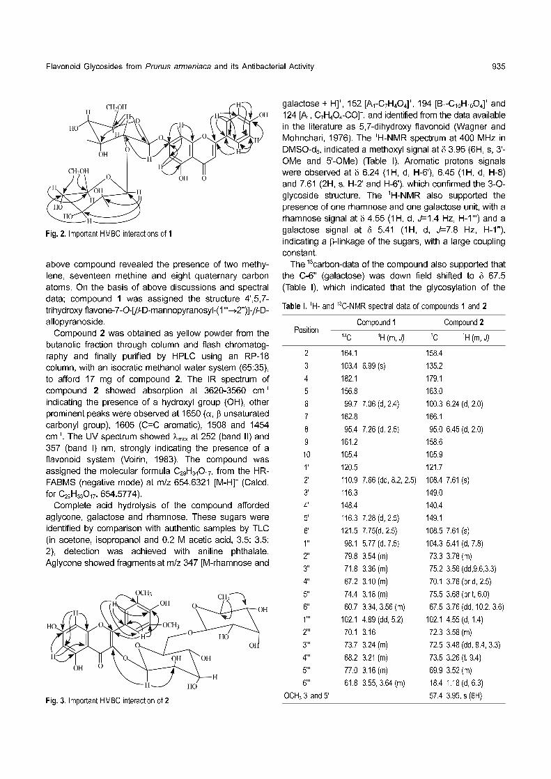
The 13carbon-data of the compound also supported that
the C-6'' (galactose) was down field shifted to δ 67.5
(Table I), which indicated that the glycosylation of the
Table I. 1H- and 13C-NMR spectral data of compounds 1 and 2
Position Compound 1 Compound 2
13C 1H (m, J) 13C 1H (m, J)
2 164.1 158.4
3 103.4 6.99 (s) 135.2
4 182.1 179.1
5 156.8 163.0
6 99.7 7.06 (d, 2.4) 100.3 6.24 (d, 2.0)
7 162.8 166.1
8 95.4 7.26 (d, 2.5) 95.0 6.45 (d, 2.0)
9 161.2 158.6
10 105.4 105.9
1' 120.5 121.7
2' 110.9 7.66 (dd, 8.2, 2.5) 108.4 7.61 (s)
3' 116.3 149.0
4' 148.4 140.4
5' 116.3 7.28 (d, 2.5) 149.1
6' 121.5 7.75(d, 2.5) 108.5 7.61 (s)
1'' 98.1 5.77 (d, 7.5) 104.3 5.41 (d, 7.8)
2'' 79.8 3.54 (m) 73.3 3.78 (m)
3'' 71.8 3.36 (m) 75.2 3.56 (dd,9.6,3.3)
4'' 67.2 3.10 (m) 70.1 3.76 (br d, 2.5)
5'' 74.4 3.18 (m) 75.5 3.68 (br t, 6.0)
6'' 60.7 3.34, 3.56 (m) 67.5 3.76 (dd, 10.2, 3.6)
1''' 102.1 4.89 (dd, 5.2) 102.1 4.55 (d, 1.4)
2''' 70.1 3.16 72.3 3.56 (m)
3''' 73.7 3.24 (m) 72.5 3.48 (dd, 9.4, 3.3)
4''' 68.2 3.21 (m) 73.5 3.26 (t, 9.4)
5''' 77.0 3.16 (m) 69.9 3.52 (m)
6''' 61.8 3.55, 3.64 (m) 18.4 1.18 (d, 6.3)
OCH3 3' and 5' 57.4 3.95, s (6H)
Fig. 2. Important HMBC interactions of 1
Fig. 3. Important HMBC interaction of 2

936 F. Rashid et al.
galactose moiety with the rhamnosyl was via 6-hydroxyl.
The HMBC spectrum also showed a cross peak between
the C-6'' of galactose and the H-1''' of rhamnose, and also
confirmed a linkage point between the two sugar moieties.
In addition, down field shielding of the C-2 also indicated
that position-3 was substituted by the glucosyl chain,
which was further confirmed by a cross peak between the
H-1'' of galactose and the C-3 of aglycone.
On the basis of above data and that available in the
literature (Tang et al., 2003), the compound was cha-
racterized as 3,4',5,7-tetrahydroxy-3',5'-dimethoxy flavon 3-
O-[-α-L-rhamnopyranosyl-(1→6)]-β-D- galactopyranoside.
The antibacterial activity of the butanolic extract was
determined against gram positive, gram negative and
non-tuberculous Mycobacteria (NTM). According to the
results, the extract was found to possess significant
antibacterial activity with MICs ranging between 31.25-
500 µg/mL (Tables II and III).
The butanolic extract was found to be more effective at
inhibiting the growth of Gram-positive bacteria (MIC
values 31.25-250 µg/mL), with the highest activity
exhibited against Micrococcus luteus (MIC 31.25 µg/mL)
(Table II). The MICs of the extract were found to be 125
and 250 µg/mL against almost all Gram-negative bacteria,
with the exception of the lowest activity against the
Enteropathogenic E. coli (EPEC), which had an MIC
value of 500 µg/mL (Table III).
In the case of MRSA, 10 isolates of clinical origin were
screened, with the MICs of the extract given as the average
of the collective MICs. According to the observation the
average MICs of the butanolic extract was found to be
78.125 µg/mL (Table II).
The present study was connected with our previous
work on the fruit extracts (ethanolic and ethyl acetate),
essential oils and pure compounds isolated from P.
armeniaca (Ahmed et al., 2004 Rashid et al 2005). This
study on the butanolic extract further strengthens the
evidence for the presence of antibacterial constituents in
apricots.
ACKNOWLEDGEMENTS
The authors are very thankful to the Department of
Botany, University of Karachi, for the plant identification,
the Dean of the Faculty of Science, University of Karachi,
for providing the research grant for carrying out this work
and HEJ Research Institute of Chemistry for the spectral
analyses of the compounds.
REFERENCES
Agrawal, P. K., 13C-NMR of Flavonoids,. Central Institute of
Medicinal and Aromatic Plants Luckhnow, India, Elsevier
(1989).
Ahmed, R., Rashid, F., Bibi, N., Kazmi, S. U., and Ansar, N.,
Phytochemical studies on Prunus Armeniaca and anti-
bacterial effects of fruit extracts., J. Trop. Med. Plants. 5, 37-
41 (2004).
Baquar, S. R., Medicinal and Poisonous Plants of Pakistan.
Printas Karachi, Pakistan, p.364 (1989).
Chopra, R. N., Nayar, L., and Chopra, I. C., Glossary of Indian
Medicinal Plants. CSIR, New Dehli, p.204 (1956).
Gupta, D. R. and Bahar, A., Asplenetin, a flavone and its
glycoside from Launaea asplenifolia. Phytochemistry., 24,
873-875 (1985).
Kritikar, K. R. and Basu, B. D., Indian medicinal plants., New
Dehli, M/S Periodical Experts, II, p.957 (1988).
Marie, B., Manual of Clinical Microbiology. Section: Aerobic
bacteria. 4th edition, American Society for Microbiology.
Table II. Antibacterial Activity of the ButanolicExtract Against GramPositive Bacteria
S No. Gram-positive bacteriaMinimum Inhibitory
Concentration(MICsµg/mL)
1 Staphylococcus aureus ATCC 6538 250
2 MRSA 78.125
3 Staphylococcus epidermidis 125
4 Streptococcus faecalis 250
5 Streptococcus pyogenes 250
6 Micrococcus luteus 31.25
7 Bacillus subtilis ATCC 6633 62.5
8 Corynebacterium diphtheriae 62.5
9 Mycobacterium fortuitum 250
10 Mycobacterium smegmatis 125
Table III. Antibacterial activity of the butanolic extract against gramnegative bacteria
S No. Gram-negative bacteriaMinimum Inhibitory
Concentration(MICs µg/mL)
1 Escherichia coli ATCC 25922 125
2 ETEC 125
3 EPEC 500
4 Salmonella typhi 125
5 MDR Salmonella typhi 125
6 Salmonella Paratyphi A 125
7 Salmonella Paratyphi B 125
8 Shigella dysenteriae 125
9 Enterobacter aerogenes 250
10 Klebseilla pneumoniae 125
11 Pseudomonas aeruginosa ATCC 9027 125
12 Proteus mirabilis 125
13 Proteus vulgaris 125
Flavonoid Glycosides from Prunus armeniaca and its Antibacterial Activity 937
Washington D.C. (1985).
Markham, K. R., Ternai, B., Stanley, R., Geiger, H., and Mabry,
T. J., Carbon-13 NMR studies of flavonoids-III, naturally
occurring flavonoid, glycoside and their acylated derivatives.
Tetrahedron., 34, 1389-1397 (1978).
Masao, H., Flavonoids of various Prunus species. VI. The
flavonoids in the wood of P. aequinoctialis, P. nipponica,
P.maximowiczii and P.avium. J. Am. Chem. Soc., 79, 1738-
1740 (1957).
Nagarajan., G. R. and Seshadri., T. R., Flavonoid components
of the heartwood of Prunus domestica linn. Phytochemistry.,
3, 477-484 (1964).
Perry, L. M. and Judith, M., Medicinal Plants of East & South
East Asia;Attributed Properties and Uses. The MIT press
Cambridge, Massachusetts, p.344 (1980).
Rashid, F., Ahmed, R., Bibi, N., and Kazmi, S. U., Triterpene
acid and its glycoside from Prunus armeniaca and
antibacterial and antioxidant activities of fruit extracts. J. Trop.
Med. Plants., 6, 31-35 (2005).
Tang, Yu. P., Liu, X., and Biao, Yu., Two new flavone glycosides
from Valeriana Jatamansi. Journal of Asian Natural Products
Research., 5, 257-260 (2003).
Voirin, B., UV spectral differentiation of 5-hydroxy- and- 5-
hydroxy-3-methoxy flavonoids with mono (4'), di (3',4') or tri-
(3',4',5')-substituted B-rings. Phytochemistry., 22 (10), 2107-
2145 (1983).
Wagner, H. and Mohnchari, V., 13C-NMR spectrum of
flavonoids. Tetrahedron Letters., 21,1799-1802 (1976).
Wayne, Pa., National Committee for Clinical Laboratory
Standards., Methods for dilution antimicrobial susceptibility
tests for bacteria that grow
aerobically M7-A3. National Committee for Clinical Laboratory
Standards (1998).
Whistler, R. L. and Wolfrom, M. L., Methods Carbohydr. Chem.,
1, 102 (1962).
Top Related
Copyright © 2022 FDOKUMEN

