


Bahasa
Halaman
Hukum

RESEARCH ARTICLE
Evaluation of the comparative accuracy of the
complement fixation test, Western blot and
five enzyme-linked immunosorbent assays for
serodiagnosis of glanders
Mandy Carolina ElschnerID1*, Karine Laroucau2, Harisankar Singha3, Bhupendra
Nath Tripathi3, Muhammad Saqib4, Ian Gardner5, Sheetal Saini3, Subodh Kumar6,
Hosny El-Adawy1,7, Falk Melzer1, Iahtasham Khan8, Praveen Malik9, Carola Sauter-
Louis10, Heinrich Neubauer1
1 Institute of Bacterial Infections and Zoonoses, Friedrich-Loeffler-Institut (FLI), Federal Research Institute
for Animal Health, Jena, Germany, 2 Paris Est University, Animal Health Laboratory, EU-Reference
Laboratory for Glanders, Maisons Alfort Cedex, France, 3 Indian Council of Agricultural Research—National
Research Centre on Equines, Hisar, India, 4 Department of Clinical Medicine and Surgery, University of
Agriculture Faisalabad, Faisalabad, Pakistan, 5 Department of Health Management, Atlantic Veterinary
College, UPEI, Prince Edward Island, Canada, 6 Defence Research and Development Establishment,
Microbiology Division, Gwalior, India, 7 Faculty of Veterinary Medicine, Kafrelsheikh University, Kafr El-
Sheikh, Egypt, 8 Section of Epidemiology and Public Health, College of Veterinary and Animal Sciences,
Jhang, Pakistan, 9 Chaudhary Charan Singh, National Institute of Animal Health, Department of Animal
Husbandry, Dairying and Fisheries, Baghpat, India, 10 Institute of Epidemiology, Friedrich-Loeffler-Institut,
Federal Research Institute for Animal Health, Greifswald-Insel Riems, Germany
Abstract
Glanders is a zoonotic contagious disease of equids caused by Burkholderia (B.) mallei.
Serodiagnosis of the disease is challenging because of false-positive and false-negative
test results. The accuracy of the complement fixation test (CFT) which is prescribed for inter-
national trade by the World Organisation for Animal Health (OIE), five ELISAs and a West-
ern blot (WB) were compared for serodiagnosis of glanders using sera from 3,000 glanders-
free and 254 glanderous equids. Four ELISA tests are based on recombinant antigens
(TssA, TssB, BimA and Hcp1), the IDVet ELISA is based on a semi-purified fraction of B.
mallei and WB makes use of a purified LPS-containing B. mallei-antigen. Sensitivity and
specificity of tests were estimated using cut-off values recommended by the test develop-
ers. The WB and all ELISAs, except BimA, were significantly more specific than the CFT.
ELISAs based on TssA, TssB, and BimA antigens had significantly lower sensitivity com-
pared to CFT while the sensitivities of the Hcp1-ELISA, the IDVet-ELISA and the WB did not
differ significantly from that of the CFT. Given their comparable sensitivities and specificities,
the CFT (98.0%, 96.4%), the WB (96.8%, 99.4%), the Hcp1-ELISA (95.3%, 99.6%) and the
IDVet-ELISA (92.5%, 99.5%) should be further developed to meet OIE requirements.
PLOS ONE | https://doi.org/10.1371/journal.pone.0214963 April 5, 2019 1 / 12
a1111111111
a1111111111
a1111111111
a1111111111
a1111111111
OPEN ACCESS
Citation: Elschner MC, Laroucau K, Singha H,
Tripathi BN, Saqib M, Gardner I, et al. (2019)
Evaluation of the comparative accuracy of the
complement fixation test, Western blot and five
enzyme-linked immunosorbent assays for
serodiagnosis of glanders. PLoS ONE 14(4):
e0214963. https://doi.org/10.1371/journal.
pone.0214963
Editor: Axel Cloeckaert, Institut National de la
Recherche Agronomique, FRANCE
Received: January 14, 2019
Accepted: March 23, 2019
Published: April 5, 2019
Copyright: © 2019 Elschner et al. This is an open
access article distributed under the terms of the
Creative Commons Attribution License, which
permits unrestricted use, distribution, and
reproduction in any medium, provided the original
author and source are credited.
Data Availability Statement: All relevant data are
within the manuscript and its Supporting
Information files.
Funding: This work was primarily funded by the
International Horse Sports Confederation and
World Organization of Animal Health (OIE) (Tender
Ref.: AD/SR/2015/1885), grant to MCE, HS, KL,
SM, IK, PM. The funder had no role in study
Introduction
Glanders is an infectious zoonotic disease of equids, caused by the gram-negative bacterium
Burkholderia (B.) mallei. Although the disease has been eradicated in most European and
North American countries in the last century, there are still outbreaks throughout the South
American and Asian continents [1–5]. The disease is mainly chronic in horses and latently-
infected animals pose a high risk of reintroduction of the infection into glanders-free countries
[6]. Therefore, trade restrictions with animals and products from endemic regions or outbreak
areas are mandatory.
Bacterial isolation and identification of B. mallei from cutaneous lesions and nasal exudates
are considered to be the “gold” (perfect reference) standard for diagnosis of glanders. As
approximately 90% of early infections occur in a non-clinical or latent form, clinical and bacte-
riological diagnosis are difficult [7].
Currently, mallein (allergic hypersensitivity test) and complement fixation test (CFT) are
used for indirect diagnosis [8]. The CFT is the World Organisation for Animal Health (OIE)
prescribed serodiagnostic method for international trade purposes and is also recommended
for surveillance investigations [7]. This test is known to have high sensitivity, but it gives a con-
siderable number of false-positive results [8, 9]. These false-positive results cause unnecessary
restrictions on international trade of animals and products thereof and result in financial losses
for owners and the horse industry. Test antigen used and the assay protocol have a significant
impact on CFT sensitivity [10–12]. Antigens used for CFT are not standardized and they have
different diagnostic accuracies. Moreover, the CFT is a labour intensive test and takes about
24h to perform. Recently, a CFT antigen based on a mixture of antigens from different histori-
cal serogroups has been extensively used [13]. Thus, the selection of antigens used for CFT has
also to be updated and standardized [12]. Various immunodiagnostic tests such as indirect
hemagglutination test, counter immunoelectrophoresis test, indirect fluorescent antibody test
and enzyme-linked immunosorbent assays (ELISA) have also been previously used but have
limitations [14].
Recently, new serological tests have been developed to overcome the disadvantages of the
CFT i.e. complexity, anti-complementary reactions and poor standardizability [15]. Alterna-
tive methods such as the Western blot (WB) and ELISAs have been applied but have not been
fully validated in large-scale studies [5, 9, 16–18] according to the principles and methods of
validation of diagnostic assays for infectious diseases [19]. The Western blot, using a LPS-prep-
aration, showed a markedly higher diagnostic specificity when compared to the CFT and was
used as a confirmatory test for glanders [9].
ELISAs developed include a competitive ELISA with a high sensitivity and specificity that
can be used in various host species [20]. Several purified recombinant proteins of B. malleiwere also evaluated for use in ELISA, e.g. the intracellular motility A protein (BimA) [14], the
type 6 secreted protein TssB [18] and a heat shock protein GroEL [21]. A new indirect ELISA
based on a semi-purified fraction of B. mallei was recently developed [22].
In regions where B. pseudomallei occurs endemically, all currently-available serological
diagnostic tests are unable to differentiate between B. mallei and B. pseudomallei, because of
their close phylogenetic relationship.
However, to our knowledge, there has not been a comprehensive comparative evaluation
study on these diagnostic tests using a suitable panel of well-characterized true-positive and
true-negative field sera from different geographical origins. Therefore, this prospective study
compared the diagnostic accuracy (sensitivity and specificity) of the WB technique, five indi-
rect ELISAs (iELISA) and the OIE-prescribed CFT for the serological detection of B. malleiantibodies in equids. Data were analysed based on the assumption that the true infection status
Evaluation of the comparative accuracy of seven assays for serodiagnosis of glanders
PLOS ONE | https://doi.org/10.1371/journal.pone.0214963 April 5, 2019 2 / 12
design, data collection and analysis, decision to
publish, or preparation of the manuscript.
Competing interests: The authors have declared
that no competing interests exist.
of animals was known with certainty and also in a Bayesian framework, where infection status
was considered not known.
Materials and methods
Sera
Samples from glanderous animals. True positive serum samples (n = 254) were collected
from equids in which the infection was confirmed by clinical signs (n = 112) or by B. malleiisolation or molecular detection of B. mallei by real-time PCR (n = 142) as recommended in
the OIE manual [7]. The animals were considered to be “clinically positive” (n = 112) on the
basis of the presence of at least one clinical sign consistent with glanders and the fact that they
were detected during a culture-confirmed glanders outbreak in this population including close
contact to infected animals. The 112 clinically positive samples were collected in 2016 and
2017 during glanders outbreaks in India (n = 111) and Pakistan (n = 1). The 142 culture or
PCR positive samples were collected during glanders outbreaks in Pakistan in 2007 and 2016
(n = 124) and from India (n = 16) in 2016 and 2017. Two samples originated from horses test-
ing positive in Germany in 2006 and 2014. The whole collection consisted of 226, 23 and 5 sera
from horse, mule and donkeys, respectively. No further demographic data were available.
Samples from animals not infected with B. mallei. True negative samples were collected
in countries officially free from glanders, to exclude exposure to B. mallei as much as possible.
The risk that these animals were in contact with B. pseudomallei is negligible, because the
regions of origin of the sampled animals do not belong to the known B. pseudomallei endemic
regions [23]. In total, 3,000 true negative samples from three different geographical areas were
used. The “Asia” batch (n = 1,000) included sera from Qatar, Saudi Arabia, Egypt, Bahrain,
Jordan, UAE, Kuwait, and Oman, provided in 2015. The “South-America” batch (n = 1,000)
was collected mostly from Buenos Aires, south of Santa Fe and south of Cordoba provinces in
2015. The “Europe” batch (n = 1,000) included samples from Germany and Ukraine was col-
lected from 2011 to 2014. All samples from the “Asia” and “South-America” batches and most
samples from Germany were unsystematically collected during routine testing for trade or
movement. Some German samples were collected during equine infectious anemia surveil-
lance. Samples from Ukraine were collected during a scientific surveillance study conducted
on glanders. No further demographic data are available.
There were no adverse reactions recorded for blood collection. All serum samples were dis-
pensed in 96 deep-well plates for each test and stored at -20˚C to avoid multiple freezing-thaw-
ing cycles. Serological testing, as described in the following sections, was done independent of
knowledge of infection status of the donor.
Ethics statement
For this study no ethical approvals were required. All blood samples were routinely collected
for prescribed diagnostic purposes or official monitoring studies and subsequently made avail-
able to the study.
Description of the tests
The characteristics of the tests compared in this study are given in Table 1.
Complement fixation test (CFT). CFT was performed as described in the OIE manual
[7]. Briefly, serum samples were diluted 1:5 in CFT buffer (Institute Virion/ Serion GmbH),
inactivated and two fold dilutions of them were mixed with Malleus CFT antigen (Ccpro
GmbH) and 5 complement haemolytic units-50% of guinea pig complement (Institute Virion/
Evaluation of the comparative accuracy of seven assays for serodiagnosis of glanders
PLOS ONE | https://doi.org/10.1371/journal.pone.0214963 April 5, 2019 3 / 12
Serion GmbH). Sera, complement and antigen were mixed in the plates and incubated over-
night at 4˚C. A 2% suspension of sensitized (amboceptor from Institute Virion/ Serion
GmbH) sheep red blood cells (Labor Dr. Merk & Kollegen) were added and plates were incu-
bated for 45 minutes at 37˚C and then centrifuged for 5 minutes at 600 g. Samples with 100%
hemolysis in a dilution of 1:5 were categorized as negative, samples showing 25–75% hemolysis
in a dilution of 1:5 were classified as suspicious and samples showing 100% inhibition of
hemolysis in a dilution of 1:5 were classified as positive. All suspicious test results were classi-
fied as positive.
Western blot (WB). The antigen preparation and WB were performed as described previ-
ously [9]. In the aforementioned study on the establishment of Western blot, the B. mallei iso-
lates Bogor, Mukteshwar and Zagreb, known for several decades, were used. For the study
described here, the strain Zagreb was replaced by a more recent isolate from Bahrain. Briefly,
the B. mallei strains Bogor (originating from Indonesia), Mukteswar (originating from India)
and Bahrain1 (isolated from a horse from Bahrain in 2011) were cultivated on blood agar
plates. For LPS purification, B. mallei colonies were re-suspended thoroughly in 6 ml saline
(0.9% NaCl, pH 7.0) up to a density comparable to McFarland scale 4.0. The cells were treated
with formaldehyde in a final concentration of 12.3% by overnight shaking. The cells were pel-
letized by centrifugation at 3,500g for 15 min and washed 3 times using PBS. The formalde-
hyde treatment and the subsequent 3 washings were repeated twice. After the last washing step
the pellet was resuspended in PBS and the correct formation of a typical LPS ladder was shown
by SDS-PAGE and silver staining before blotting. The batches of the three antigens were made
available in a lyophilized form. For the immunoblot analysis the antigens were mixed equally
and blotted onto a 0.45 μm nitrocellulose membrane (Invitrogen, Germany). Five millimeter
strips of the membrane were used to analyze the serum samples at a dilution of 1:50. Anti-
horse conjugate labelled with alkaline phosphatase (Sigma, Munich, Germany) was used in a
dilution of 1:5,000. Detection was made by the ready-to-use NBT-BCIP staining (Sigma). The
WB was scored positive, if the banding pattern of the LPS ladder was clearly visible, scored sus-
picious if a reaction was seen within the region of 20 to 60 kilodalton (kDa), and scored nega-
tive if no reactions were seen in the area between 20 and 60 kDa. All suspicious (intermediate)
test results were classified as positive.
Recombinant BimA-, Hcp1-, TssA- and TssB-ELISA. The four recombinant ELISAs
were performed according to the developer’s instructions. Expression and purification of
recombinant BimA and TssB antigens were described previously [14, 18], codon optimized
Table 1. Name of the test, test developer and used antigens of the 7 compared assays.
Test Developer/ protocol used Antigen used Abbreviation
Complement fixation test OIE-Manual [7] Malleus-CFT antigen; Ccpro GmbH, prepared from B. mallei strain Ivan-
NCTC 10230
CFT
Western blot FLI, Germany [9] LPS containing antigen consisting of 3 different B. mallei strains (Bogor,
Mukteswar, Bahrain1)
WB
ID Screen Glanders Indirect
ELISA
EU-Reference laboratory/ ID.Vet,
France
Semi-purified antigen fraction prepared from B. mallei strain ATCC 23344 IDVet
Indirect TssA ELISA ICAR-NRCE, India Recombinant protein TssA of Type 6 secretory system of B. mallei (Type strain) TssA
Indirect TssB ELISA ICAR-NRCE, India [18] Recombinant protein TssB of Type 6 secretory system of B. mallei (Type strain) TssB
Indirect Hcp1 ELISA ICAR-NRCE, India Recombinant protein Hcp1 of Type 6 secretory system of B. mallei (Type strain) Hcp1
Indirect BimA ELISA ICAR-NRCE, India [14] Recombinant protein BimA of B. mallei (Type strain) BimA
https://doi.org/10.1371/journal.pone.0214963.t001
Evaluation of the comparative accuracy of seven assays for serodiagnosis of glanders
PLOS ONE | https://doi.org/10.1371/journal.pone.0214963 April 5, 2019 4 / 12
and synthetic gene sequences of B. mallei loci BMAA0742 and BMAA0744, respectively, were
expressed in pQE30 vector (Qiagen). Recombinant Hcp1 and TssA antigens were purified by
Ni+-NTA agarose (Qiagen) column chromatography according to manufacturer’s instruction.
Optimization of ELISA protocol and diagnostic cut offs were determined using a panel of glan-
ders positive and negative sera (unpublished data).
Briefly, the ELISA plates (Maxisorp, Nunc) were coated with 100 μl of the antigen diluted in
coating buffer (Sigma-Aldrich) in a concentration of 75 ng/well (Hcp1, BimA) or 150 ng/well
(TssA, TssB) and incubated at 4˚C overnight. On the second day, the plates were washed three
times (each 200 μl/well) using washing buffer (PBS with 0,05% Tween-20 (Sigma-Aldrich)),
blocked for one hour at 37˚C using 100 μl/ well blocking buffer (washing buffer with 6% skim
milk powder, Merck-Millipore) and washed five times. The serum samples were tested in
duplicates pre-diluted 1:200 in dilution buffer (PBS with 3% skim milk powder, 100 μl/well)
and after incubation of 1 hour at 37˚C washed 5 times. Subsequently, the anti-horse horse rad-
ish peroxidase (HRPO) conjugate (Sigma-Aldrich) diluted 1:10,000 in dilution buffer was
added (100 μl/well) and the plates were incubated at 37˚ for one hour. After five washing steps,
each well was filled with 50 μl substrate solution (Ready to use 1-Step ultra TMB ELISA sub-
strate; Thermo Scientific). After processing in a dark place (Hcp1 and BimA for 5 minutes,
TssA and TssB for 10 minutes), the reaction was stopped by addition of 1M sulfuric acid solu-
tion (Carl Roth) OD 450 nm was recorded. Sample/Positive ratio (S/P%) was calculated for
each sample (OD Sample—OD Negative Control) / (OD Positive Control–OD Negative Con-
trol) x 100. Sera with S/P% values� 10 (BimA),� 15 (TssA) and� 20 (TssB, Hcp1) were con-
sidered as positive.
ID Screen Glanders ELISA (IDVet). The IDVet test (ID Screen Glanders ELISA, IDVet,
Grabels, France) was provided as a ready-to-use kit and was performed according to the man-
ufacturer’s instructions. The kit contained B. mallei antigen-coated plates with individual
8-well strips, positive and negative controls, 10x concentrated multi-species horseradish conju-
gate, dilution buffer, 20x wash concentrate, TMB substrate solution and stop solution. Briefly,
200 μl of the serum samples in a pre-dilution of 1:20 were added to an antigen-coated and
non-coated well, respectively. After 45 ± 5 min incubation at 21 ± 5˚C, wells were emptied,
washed 3 times and incubated with the conjugate for 30 ± 3 min at 21 ± 5˚C. Washing was
repeated 3 times. Then the substrate was added and after incubation for 15 ± 2 min reaction
was stopped and OD 450 nm was recorded. S/P% values were calculated and samples with S/P
%� 40% were considered as negative, between 40 and 50% as suspicious and� 50% as posi-
tive. All suspicious (intermediate) test results were classified as positive.
Statistical analysis
Ninety-five percent confidence intervals (CI) for sensitivity, specificity and likelihood ratios
were based on standard formulas [24] and done using MedCalc version 13.1.0.0 (https://www.
medcalc.org/).
Sensitivity and specificity covariances for pairs of tests (a measure of conditional depen-
dence or correlation) were calculated as described in Gardner and others [25] using an Excel
template and assuming the true infection status was known with certainty, as described in the
sample description paragraph. Results were expressed as the percentage of maximum value
and the obtained value is similar to a correlation coefficient. To account for the possibility of
misclassification of the true infection status, data were reanalyzed in a Bayesian framework
that accounted for uncertainty about true status and incorporated necessary covariance terms
in a latent class model (LCM). The model was based on 4 tests (CFT, WB and the two most
promising ELISAs) in two populations (infected and non-infected) and used code adapted
Evaluation of the comparative accuracy of seven assays for serodiagnosis of glanders
PLOS ONE | https://doi.org/10.1371/journal.pone.0214963 April 5, 2019 5 / 12
from Branscum and others [26]. The model initially assumed conditional independence of
tests, which is reasonable if the true status is known with certainty, constant sensitivity and
specificity in both populations, and distinct prevalences. Flat priors (beta 1,1) were used for
the sensitivity and specificity of all tests. Prevalence of infection in true-positive cases was
assumed to have a mode of 100% with 95% probability that the true value was greater than
98% (modelled as a beta 148.3,1) and prevalence in the true-negative cases (pooled across the 3
regions) was assumed to have a mode of 0% with 95% probability that the true value was less
than 2% (modelled as beta 1,148.3). Two sensitivity covariance terms were added to the model
to allow for conditional dependence that was evident in the sensitivity data. Models were run
in OpenBUGS version 3.2.3 [27] and model code is available from the authors on request.
Median and 95% probability intervals were used for posterior inferences about sensitivity and
specificity.
Results
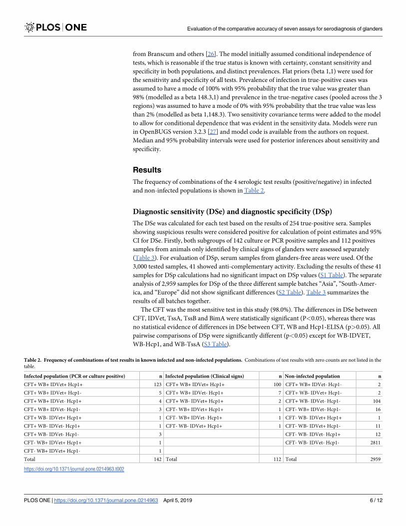
The frequency of combinations of the 4 serologic test results (positive/negative) in infected
and non-infected populations is shown in Table 2.
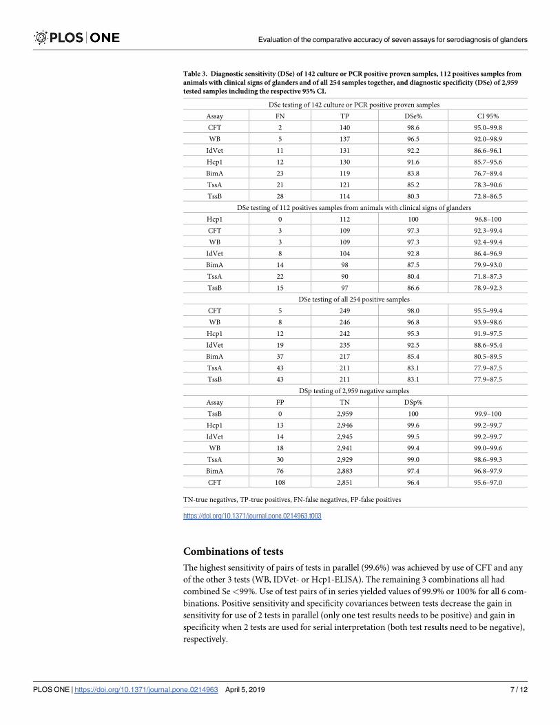
Diagnostic sensitivity (DSe) and diagnostic specificity (DSp)
The DSe was calculated for each test based on the results of 254 true-positive sera. Samples
showing suspicious results were considered positive for calculation of point estimates and 95%
CI for DSe. Firstly, both subgroups of 142 culture or PCR positive samples and 112 positives
samples from animals only identified by clinical signs of glanders were assessed separately
(Table 3). For evaluation of DSp, serum samples from glanders-free areas were used. Of the
3,000 tested samples, 41 showed anti-complementary activity. Excluding the results of these 41
samples for DSp calculations had no significant impact on DSp values (S1 Table). The separate
analysis of 2,959 samples for DSp of the three different sample batches “Asia”, “South-Amer-
ica, and “Europe” did not show significant differences (S2 Table). Table 3 summarizes the
results of all batches together.
The CFT was the most sensitive test in this study (98.0%). The differences in DSe between
CFT, IDVet, TssA, TssB and BimA were statistically significant (P<0.05), whereas there was
no statistical evidence of differences in DSe between CFT, WB and Hcp1-ELISA (p>0.05). All
pairwise comparisons of DSp were significantly different (p<0.05) except for WB-IDVET,
WB-Hcp1, and WB-TssA (S3 Table).
Table 2. Frequency of combinations of test results in known infected and non-infected populations. Combinations of test results with zero counts are not listed in the
table.
Infected population (PCR or culture positive) n Infected population (Clinical signs) n Non-infected population n
CFT+ WB+ IDVet+ Hcp1+ 123 CFT+ WB+ IDVet+ Hcp1+ 100 CFT+ WB+ IDVet- Hcp1- 2
CFT+ WB+ IDVet+ Hcp1- 5 CFT+ WB+ IDVet- Hcp1+ 7 CFT+ WB- IDVet+ Hcp1- 2
CFT+ WB+ IDVet- Hcp1+ 4 CFT+ WB- IDVet+ Hcp1+ 2 CFT+ WB- IDVet- Hcp1- 104
CFT+ WB+ IDVet- Hcp1- 3 CFT- WB+ IDVet+ Hcp1+ 1 CFT- WB+ IDVet- Hcp1- 16
CFT+ WB- IDVet+ Hcp1+ 1 CFT- WB+ IDVet- Hcp1+ 1 CFT- WB- IDVet+ Hcp1+ 1
CFT+ WB- IDVet- Hcp1+ 1 CFT- WB- IDVet+ Hcp1+ 1 CFT- WB- IDVet+ Hcp1- 11
CFT+ WB- IDVet- Hcp1- 3 CFT- WB- IDVet- Hcp1+ 12
CFT- WB+ IDVet+ Hcp1+ 1 CFT- WB- IDVet- Hcp1- 2811
CFT- WB+ IDVet+ Hcp1- 1
Total 142 Total 112 Total 2959
https://doi.org/10.1371/journal.pone.0214963.t002
Evaluation of the comparative accuracy of seven assays for serodiagnosis of glanders
PLOS ONE | https://doi.org/10.1371/journal.pone.0214963 April 5, 2019 6 / 12
Combinations of tests
The highest sensitivity of pairs of tests in parallel (99.6%) was achieved by use of CFT and any
of the other 3 tests (WB, IDVet- or Hcp1-ELISA). The remaining 3 combinations all had
combined Se <99%. Use of test pairs of in series yielded values of 99.9% or 100% for all 6 com-
binations. Positive sensitivity and specificity covariances between tests decrease the gain in
sensitivity for use of 2 tests in parallel (only one test results needs to be positive) and gain in
specificity when 2 tests are used for serial interpretation (both test results need to be negative),
respectively.
Table 3. Diagnostic sensitivity (DSe) of 142 culture or PCR positive proven samples, 112 positives samples from
animals with clinical signs of glanders and of all 254 samples together, and diagnostic specificity (DSe) of 2,959
tested samples including the respective 95% CI.
DSe testing of 142 culture or PCR positive proven samples
Assay FN TP DSe% CI 95%
CFT 2 140 98.6 95.0–99.8
WB 5 137 96.5 92.0–98.9
IdVet 11 131 92.2 86.6–96.1
Hcp1 12 130 91.6 85.7–95.6
BimA 23 119 83.8 76.7–89.4
TssA 21 121 85.2 78.3–90.6
TssB 28 114 80.3 72.8–86.5
DSe testing of 112 positives samples from animals with clinical signs of glanders
Hcp1 0 112 100 96.8–100
CFT 3 109 97.3 92.3–99.4
WB 3 109 97.3 92.4–99.4
IdVet 8 104 92.8 86.4–96.9
BimA 14 98 87.5 79.9–93.0
TssA 22 90 80.4 71.8–87.3
TssB 15 97 86.6 78.9–92.3
DSe testing of all 254 positive samples
CFT 5 249 98.0 95.5–99.4
WB 8 246 96.8 93.9–98.6
Hcp1 12 242 95.3 91.9–97.5
IdVet 19 235 92.5 88.6–95.4
BimA 37 217 85.4 80.5–89.5
TssA 43 211 83.1 77.9–87.5
TssB 43 211 83.1 77.9–87.5
DSp testing of 2,959 negative samples
Assay FP TN DSp%
TssB 0 2,959 100 99.9–100
Hcp1 13 2,946 99.6 99.2–99.7
IdVet 14 2,945 99.5 99.2–99.7
WB 18 2,941 99.4 99.0–99.6
TssA 30 2,929 99.0 98.6–99.3
BimA 76 2,883 97.4 96.8–97.9
CFT 108 2,851 96.4 95.6–97.0
TN-true negatives, TP-true positives, FN-false negatives, FP-false positives
https://doi.org/10.1371/journal.pone.0214963.t003
Evaluation of the comparative accuracy of seven assays for serodiagnosis of glanders
PLOS ONE | https://doi.org/10.1371/journal.pone.0214963 April 5, 2019 7 / 12
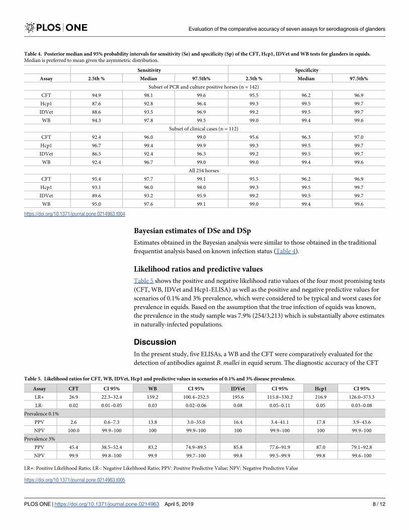
Bayesian estimates of DSe and DSp
Estimates obtained in the Bayesian analysis were similar to those obtained in the traditional
frequentist analysis based on known infection status (Table 4).
Likelihood ratios and predictive values
Table 5 shows the positive and negative likelihood ratio values of the four most promising tests
(CFT, WB, IDVet and Hcp1-ELISA) as well as the positive and negative predictive values for
scenarios of 0.1% and 3% prevalence, which were considered to be typical and worst cases for
prevalence in equids. Based on the assumption that the true infection of equids was known,
the prevalence in the study sample was 7.9% (254/3,213) which is substantially above estimates
in naturally-infected populations.
Discussion
In the present study, five ELISAs, a WB and the CFT were comparatively evaluated for the
detection of antibodies against B. mallei in equid serum. The diagnostic accuracy of the CFT
Table 4. Posterior median and 95% probability intervals for sensitivity (Se) and specificity (Sp) of the CFT, Hcp1, IDVet and WB tests for glanders in equids.
Median is preferred to mean given the asymmetric distribution.
Sensitivity Specificity
Assay 2.5th % Median 97.5th% 2.5th % Median 97.5th%
Subset of PCR and culture positive horses (n = 142)
CFT 94.9 98.1 99.6 95.5 96.2 96.9
Hcp1 87.6 92.8 96.4 99.3 99.5 99.7
IDVet 88.6 93.5 96.9 99.2 99.5 99.7
WB 94.3 97.8 99.5 99.0 99.4 99.6
Subset of clinical cases (n = 112)
CFT 92.4 96.0 99.0 95.6 96.3 97.0
Hcp1 96.7 99.4 99.9 99.3 99.5 99.7
IDVet 86.5 92.4 96.3 99.2 99.5 99.7
WB 92.4 96.7 99.0 99.0 99.4 99.6
All 254 horses
CFT 95.4 97.7 99.1 95.5 96.2 96.9
Hcp1 93.1 96.0 98.0 99.3 99.5 99.7
IDVet 89.6 93.2 95.9 99.2 99.5 99.7
WB 95.0 97.6 99.1 99.0 99.4 99.6
https://doi.org/10.1371/journal.pone.0214963.t004
Table 5. Likelihood ratios for CFT, WB, IDVet, Hcp1 and predictive values in scenarios of 0.1% and 3% disease prevalence.
Assay CFT CI 95% WB CI 95% IDVet CI 95% Hcp1 CI 95%
LR+ 26.9 22.3–32.4 159.2 100.4–252.5 195.6 115.8–330.2 216.9 126.0–373.3
LR- 0.02 0.01–0.05 0.03 0.02–0.06 0.08 0.05–0.11 0.05 0.03–0.08
Prevalence 0.1%
PPV 2.6 0.6–7.3 13.8 3.0–35.0 16.4 3.4–41.1 17.8 3.9–43.6
NPV 100.0 99.9–100 100 99.9–100 100 99.9–100 100 99.9–100
Prevalence 3%
PPV 45.4 38.5–52.4 83.2 74.9–89.5 85.8 77.6–91.9 87.0 79.1–92.8
NPV 99.9 99.8–100 99.9 99.7–100 99.8 99.5–99.9 99.8 99.6–100
LR+: Positive Likelihood Ratio; LR-: Negative Likelihood Ratio; PPV: Positive Predictive Value; NPV: Negative Predictive Value
https://doi.org/10.1371/journal.pone.0214963.t005
Evaluation of the comparative accuracy of seven assays for serodiagnosis of glanders
PLOS ONE | https://doi.org/10.1371/journal.pone.0214963 April 5, 2019 8 / 12
was analyzed following the guidance in the OIE manual [7]. The specificity of tests depends on
cross-reacting organisms which can vary from biotope to biotope [28]. Therefore, serum sam-
ples were analyzed separately from 3 different geographical regions (“South-America” batch,
“Europe” batch, “Asia” batch), but no significant differences in specificity were found. Based
on all 3,000-tested serum samples in this study, it was shown, that the five ELISAs and the WB
were more specific than the CFT. Previously published reports showed the utility of ELISA
techniques regarding the improvement of specificity in comparison to CFT. The specificity of
ELISA using either recombinant BimA antigen or TssB protein were 98.9% and 100%, respec-
tively [14, 18]. The WB was previously shown to have a specificity of 100% [9]. Another study
showed the IDVet-ELISA to have excellent specificity (98.9%) in a preliminary validation [22].
However, the estimates were based on lower sample numbers than considered adequate by
OIE.
The evaluation of test sensitivity showed the best results for the CFT followed by WB,
Hcp1-, and IDVet-ELISAs. Because of the outstanding sensitivity of the CFT, alternative meth-
ods must be benchmarked against it. Hence, OIE requires a confirmatory test for glanders
with equal or higher sensitivity and higher specificity [7]. Especially in areas where the number
of false positives is very high, the lack of specificity of the CFT results in constant obstacles to
trade in equidae and poses a constant challenge for veterinary authorities [6]. In international
trade, the most sensitive test for highly transmissible and pathogenic agents has to be applied
to avoid an introduction of infected animals into disease free populations [29].
The biggest challenge in validation of serological tests for glanders is the lack of availability
of a sufficient number of true-positive serum samples to statistically compare sensitivity across
multiple tests with high confidence. Positive sera, with positive isolation of B. mallei, are
extremely difficult to collect. The horses tested positive are considered to pose a zoonotic risk
or a source for future spread of the disease in the equine population and are destroyed imme-
diately before samples can be collected. The areas of endemicity are often in remote regions
and cooling chains cannot be maintained. In various countries serum collection is restricted
by regulations and laws. Our study therefore had to use a hybrid design in which cases were
selected based on clinical presentation and positive PCR or culture results. Non-infected sam-
ples were collected from countries officially free of infection. This design may have led to selec-
tion bias in estimates as glanders-positive cases may have been the most severely affected in
source populations [30]. However, because sample inclusion was not based on positive results
by any serologic tests, we believe this bias was not substantial and was unlikely to impact the
sensitivity and specificity rankings of the tests. This study showed that a serum panel well char-
acterized by confirmative clinical observations of experienced veterinarians could be useful to
increase case sample numbers and thus increase statistical certainty.
Considering both sensitivity and specificity, the WB (96.8%, 99.4%), the Hcp1-ELISA
(95.3%, 99.6%) and the IDVet-ELISA (92.5%, 99.5%) should be further developed to meet OIE
demands in the near future. These three tests were available for the study in very different
stages of development. The WB, as performed in the OIE reference laboratory at the FLI, was
based on a semi-purified antigen and reading of WB results needs experienced operators. The
in-house production of this test is expensive and thus this test is not appropriate for screening
large numbers of samples in surveillance programs. Hence, it is suitable as a confirmatory test.
The Hcp1-ELISA was available as a ‘semi-ready’ non-commercialized format. The ELISA
plates had to be coated with antigens, blocked and washed before the test could be used. If this
ELISA would be produced commercially, this test could be a promising tool for the future. The
IDVet-ELISA was provided as a ready-to-use commercialized kit. The cut offs for the ELISAs
and the WB were based on those recommended by the developer or manufacturer of the tests.
It is obvious that other results will be obtained with different cut off values. Whether a cut off
Evaluation of the comparative accuracy of seven assays for serodiagnosis of glanders
PLOS ONE | https://doi.org/10.1371/journal.pone.0214963 April 5, 2019 9 / 12
value shift is sufficient for the needs of OIE to improve the test properties of the ELISAs has to
be proven by testing more positive samples and under consideration of the important preva-
lence-independent test characteristics of LR- and LR+.
In Germany, the method used to exclude false-positive CFT results is to run a WB as a con-
firmatory test on CFT-positive sera [31]. The results from the present study confirm that use
of a combination of tests can enhance the accuracy of serological diagnosis.
However, taking into account the performance characteristics of assays based on the pre-
determined cut off values used in this study, replacement of the CFT with the WB or any of the
ELISAs cannot be recommended for testing equids for trade yet.
Presently, the CFT for glanders is still the prescribed technique for serological investigation
of equids for trade purposes [7] to certify individual animal freedom from disease. The test is
difficult to standardize, as there are no standardized sera available [12]. Furthermore, the CFT
requires experienced operators, ongoing training and quality management systems to be
implemented in the laboratory. The performance of the test is demanding and the results
depend on the antigen used and methods, such as incubation conditions [10, 11]. Therefore,
efforts to further improve and optimize the WB and ELISAs, e.g. combination of antigens,
should be continued.
• Ullrich Wernery, Central Veterinary Research Institute, Dubai, United Arab Emirates
• Teotimo Becu, Clinica equina SRL, Laboratorio de Diagnostico, Laboratorio de Produccion
de Biologicos, Buenos Aires, Argentina
• Roman Koziy, State Scientific Control Institute of Biotechnology and Strains of Microorgan-
isms, Kiev, Ukraine
Supporting information
S1 Table. DSp values with and without 41 anti-complementary sera.
(DOCX)
S2 Table. Separate analysis of 2,959 samples for DSp of the three different sample batches
“Asia”, “South-America, and “Europe”.
(DOCX)
S3 Table. Significance (p values) of differences in DSp for test pairs based on McNemar’s
test for correlated proportions.
(DOCX)
Acknowledgments
Special thanks go to Johannes Solle for his skilled technical work at the Friedrich-Loeffler-
Institut.
Thanks also go to the following institutes for collecting and providing the samples:
Author Contributions
Conceptualization: Mandy Carolina Elschner, Karine Laroucau, Harisankar Singha, Muham-
mad Saqib, Ian Gardner, Subodh Kumar, Hosny El-Adawy, Falk Melzer, Iahtasham Khan,
Praveen Malik, Carola Sauter-Louis, Heinrich Neubauer.
Data curation: Mandy Carolina Elschner.
Evaluation of the comparative accuracy of seven assays for serodiagnosis of glanders
PLOS ONE | https://doi.org/10.1371/journal.pone.0214963 April 5, 2019 10 / 12
Funding acquisition: Mandy Carolina Elschner, Heinrich Neubauer.
Investigation: Mandy Carolina Elschner, Ian Gardner, Sheetal Saini, Falk Melzer.
Methodology: Mandy Carolina Elschner, Karine Laroucau, Harisankar Singha, Ian Gardner,
Sheetal Saini, Subodh Kumar, Praveen Malik, Carola Sauter-Louis.
Project administration: Mandy Carolina Elschner.
Resources: Mandy Carolina Elschner, Harisankar Singha, Bhupendra Nath Tripathi, Muham-
mad Saqib, Iahtasham Khan, Praveen Malik, Heinrich Neubauer.
Software: Ian Gardner, Carola Sauter-Louis.
Supervision: Bhupendra Nath Tripathi, Heinrich Neubauer.
Validation: Mandy Carolina Elschner.
Writing – original draft: Mandy Carolina Elschner, Hosny El-Adawy.
Writing – review & editing: Karine Laroucau, Harisankar Singha, Ian Gardner, Falk Melzer,
Iahtasham Khan, Carola Sauter-Louis, Heinrich Neubauer.
References
1. Mota RA, da Fonseca Oliveira AA, da Silva AM, Junior JW, da Silva LB, de Farias Brito M, et al. Glan-
ders in donkeys (Equus Asinus) in the state of pernambuco, Brazil: A case report. Braz J Microbiol.
2010; 41(1):146–9. Epub 2010/01/01. https://doi.org/10.1590/S1517-838220100001000021 S1517-
838220100001000021 [pii]. PMID: 24031474; PubMed Central PMCID: PMC3768622.
2. Malik P, Singha H, Khurana SK, Kumar R, Kumar S, Raut AA, et al. Emergence and re-emergence of
glanders in India: a description of outbreaks from 2006 to 2011. Vet Ital. 2012; 48(2):167–78. Epub
2012/06/22. PMID: 22718333.
3. Malik P, Singha H, Goyal SK, Khurana SK, Tripathi BN, Dutt A, et al. Incidence of Burkholderia mallei
infection among indigenous equines in India. Vet Rec. 2015; 2(2):e000129. https://doi.org/10.1136/
vetreco-2015-000129 PMID: 26457190; PubMed Central PMCID: PMC4594314.
4. Arun S, Neubauer H, Gurel A, Ayyildiz G, Kuscu B, Yesildere T, et al. Equine glanders in Turkey. Vet
Rec. 1999; 144(10):255–8. Epub 1999/04/21. PMID: 10209817.
5. Ghori MT, Khan MS, Khan JA, Rabbani M, Shabbir MZ, Chaudhry HR, et al. Seroprevalence and risk
factors of glanders in working equines—Findings of a cross-sectional study in Punjab province of Paki-
stan. Acta tropica. 2017; 176:134–9. https://doi.org/10.1016/j.actatropica.2017.07.031 PMID:
28760480.
6. Kettle AN, Wernery U. Glanders and the risk for its introduction through the international movement of
horses. Equine Vet J. 2016; 48(5):654–8. https://doi.org/10.1111/evj.12599 PMID: 27288893.
7. Glanders and melioidosis [Internet]. OIE. 2018. Available from: http://www.oie.int/fileadmin/Home/eng/
Health_standards/tahm/3.05.11_GLANDERS.pdf.
8. Neubauer H, Sprague LD, Zacharia R, Tomaso H, Al Dahouk S, Wernery R, et al. Serodiagnosis of Bur-
kholderia mallei infections in horses: State-of-the-art and perspectives. J Vet Med. 2005; 52(5):201–5.
https://doi.org/10.1111/j.1439-0450.2005.00855.x PMID: 16115091
9. Elschner MC, Scholz HC, Melzer F, Saqib M, Marten P, Rassbach A, et al. Use of a Western blot tech-
nique for the serodiagnosis of glanders. BMC Vet Res. 2011; 7:4. Epub 2011/01/21. https://doi.org/10.
1186/1746-6148-7-4 1746-6148-7-4 [pii]. PMID: 21247488; PubMed Central PMCID: PMC3034690.
10. Khan I, Wieler LH, Melzer F, Gwida M, Santana VL, de Souza MM, et al. Comparative evaluation of
three commercially available complement fixation test antigens for the diagnosis of glanders. Vet Rec.
2011; 169(19):495. Epub 2011/09/08. https://doi.org/10.1136/vr.d5410 [pii]. PMID: 21896565.
11. Khan I, Wieler LH, Saqib M, Melzer F, Santana VL, Neubauer H, et al. Effect of incubation temperature
on the diagnostic sensitivity of the glanders complement fixation test. Rev Sci Tech. 2014; 33(3):869–
75. Epub 2015/03/31. PMID: 25812210.
12. Laroucau K, Colaneri C, Jay M, Corde Y, Drapeau A, Durand B, et al. Interlaboratory ring trial to evalu-
ate CFT proficiency of European laboratories for diagnosis of glanders in equids. Vet Rec. 2016;
178:632. https://doi.org/10.1136/vr.103617 PMID: 27122499
Evaluation of the comparative accuracy of seven assays for serodiagnosis of glanders
PLOS ONE | https://doi.org/10.1371/journal.pone.0214963 April 5, 2019 11 / 12
13. Malik P. Harmonising diagnostic testing for glanders in equids. Vet Rec. 2016; 178(25):630–1. https://
doi.org/10.1136/vr.i3093 PMID: 27313253
14. Kumar S, Malik P, Verma SK, Pal V, Gautam V, Mukhopadhyay C, et al. Use of a recombinant Burkhol-
deria intracellular motility a protein for immunodiagnosis of glanders. Clin Vaccine Immunol. 2011; 18
(9):1456–61. Epub 2011/07/15. https://doi.org/10.1128/CVI.05185-11 PMID: 21752949; PubMed Cen-
tral PMCID: PMCPMC3165212.
15. Raghavan R, Syriac G, Wernery R, Elschner M, Mawhinney I, Wernery U. Comparative test perfor-
mance of different serological tests for glanders. J Equine Vet Sci. 2016; 39:S18–S9. https://doi.org/10.
1016/j.jevs.2016.02.038
16. Elschner MC, Laroucau K, Zientara S, Singha H, Praveen M, Kumar S, et al. OIE-Project-Validation
study of a western blot technique and ELISAs for serological diagnosis of glanders in equids for the pur-
pose of certifying freedom from infection in individual animals for trade or movement. J Equine Vet Sci.
2016; 39:S16–S7. https://doi.org/10.1016/j.jevs.2016.02.033
17. Katz JB, Chieves LP, Hennager SG, Nicholson JM, Fisher TA, Byers PE. Serodiagnosis of equine piro-
plasmosis, dourine, and glanders using an arrayed immunoblotting method. J Vet Diagn Invest. 1999;
11(3):292–4. Epub 1999/06/03. https://doi.org/10.1177/104063879901100316 PMID: 10353365.
18. Singha H, Malik P, Goyal SK, Khurana SK, Mukhopadhyay C, Eshwara VK, et al. Optimization and vali-
dation of indirect ELISA using truncated TssB protein for the serodiagnosis of glanders amongst
equines. Sci World J. 2014; 2014:469–07. Epub 2014/03/29. https://doi.org/10.1155/2014/469407
PMID: 24672321; PubMed Central PMCID: PMC3932216.
19. Principles and methods of validation of diagnostic assays for infectious diseases [Internet]. OIE. 2018.
Available from: http://www.oie.int/fileadmin/Home/eng/Health_standards/aahm/current/chapitre_
validation_diagnostics_assays.pdf.
20. Sprague LD, Zachariah R, Neubauer H, Wernery R, Joseph M, Scholz HC, et al. Prevalence-dependent
use of serological tests for diagnosing glanders in horses. BMC Vet Res. 2009; 5:32. Epub 2009/09/03.
https://doi.org/10.1186/1746-6148-5-32 PMID: 19723336; PubMed Central PMCID:
PMCPMC2745380.
21. Dohre SK, Kamthan A, Singh S, Alam SI, Kumar S. Identification of a new diagnostic antigen for glan-
ders using immunoproteome analysis. Come Immunol Microbiol Infect Dis. 2017; 53:26–32. https://doi.
org/10.1016/j.cimid.2017.06.007.
22. Laroucau K, Bertin C, Roche M, Colaneri C, Madani N, Pourquier P, et al., editors. A new ELISA assay
for glanders diagnosis. 36 Arbeits- und Fortbildungstagung der DVG-Fachgruppe AVID; 2017 13.09.-
15.09.2017; Bad Staffelstein/ Kloster Banz, Germany.
23. Perumal SR, Stiles BG, Sethi G, Lim LHK. Melioidosis: Clinical impact and public health threat in the
tropics. PLoS Negl Trop Dis. 2017; 11 (5):e0004738. https://doi.org/10.1371/journal.pntd.0004738
PMID: 28493905
24. Dohoo I, Martin W, Stryhn H. Veterinary Epidemiologic Research: VER Inc., Charlottetown, Prince
Edward Island, Canada; 2009.
25. Gardner IA, Stryhn H, Lind P, Collins MT. Conditional dependence between tests affects the diagnosis
and surveillance of animal diseases. Prev Vet Med. 2000; 45(1–2):107–22. PMID: 10802336.
26. Branscum AJ, Gardner IA, Johnson WO. Estimation of diagnostic-test sensitivity and specificity through
Bayesian modeling. Prev Vet Med. 2005; 68(2–4):145–63. https://doi.org/10.1016/j.prevetmed.2004.
12.005 PMID: 15820113.
27. Lunn D, Spiegelhalter D, Thomas A, Best N. The BUGS project: Evolution, critique and future directions.
Stat Med. 2009; 28(25):3049–67. https://doi.org/10.1002/sim.3680 PMID: 19630097.
28. Greiner M, Gardner IA. Epidemiologic issues in the validation of veterinary diagnostic tests. Prev Vet
Med. 2000; 45(1–2):3–22. PMID: 10802331.
29. Jacobson RH. Validation of serological assays for diagnosis of infectious diseases. Rev sci tech Off int
Epiz. 1998; 17(2):469–86.
30. Gardner IA, Colling A, Greiner M. Design, statistical analysis and reporting standards for test accuracy
studies for infectious diseases in animals: progress, challenges and recommendations. Prev Vet Med.
2019; 162:46–55. https://doi.org/10.1016/j.prevetmed.2018.10.023 Epub 2018 Nov 2. PMID: 30621898
31. Elschner MC, Liebler-Tenorio E, Neubauer H. The diagnostic approach to equine glanders in Germany
Equine Dis Quart. 2016; 25(1).
Evaluation of the comparative accuracy of seven assays for serodiagnosis of glanders
PLOS ONE | https://doi.org/10.1371/journal.pone.0214963 April 5, 2019 12 / 12
Top Related
Copyright © 2022 FDOKUMEN

