


Bahasa
Halaman
Hukum

ORIGINAL ARTICLE
Dextransucrase from the mutant of Pediococcus pentosaceus(PPm) is more stable than the wild type
Damini Kothari • Ankur Tyagi • Seema Patel •
Arun Goyal
Received: 20 June 2011 / Accepted: 13 July 2011 / Published online: 30 July 2011
� The Author(s) 2011. This article is published with open access at Springerlink.com
Abstract A comparative study on both wild type and
mutant of Pediococcus pentosaceus for dextransucrase
activity, its stability, dextran synthesizing activity, antibi-
otic sensitivity and carbohydrate utilization was performed.
The wild type P. pentosaceus had specific activity of
0.58 U/mg whereas the mutant showed that of 1.0 U/mg
with 72% enhancement. The antibiogram of 27 antibiotics
tested against mutant showed significant differences with 9
antibiotics when compared to wild type. In carbohydrate
fermentation profile, trehalose, galactose, maltose, lactose
and fructose are metabolized by both the strains, but
weakly in case of mutant. Stabilization of purified dex-
transucrase from wild type and mutant with various stabi-
lizers was studied at 30 and 4 �C. Both enzymes were more
stable at 4 �C. Among various stabilizers such as dextran
(100 kDa, 10 lg/ml), glycerol (0.5%, v/v), PEG 8000
(10 lg/ml) and Tween 80 (0.5%, v/v), Tween 80 provided
maximum stabilization at 4 and 30 �C. The mutant showed
better stabilization than that of the wild type at both 30 and
4 �C. The loss of activity at 30 �C after 24 h in wild type
and mutant in the presence of Tween 80 was only 34 and
32%, respectively, whereas the loss of activity in control of
wild type and mutant was 76 and 59%, respectively. After
15 days at 4 �C, the loss of activity in control of wild type
and mutant in the presence of Tween 80 was only 15 and
8%, respectively, whereas at 30 �C, the loss of activity in
control of wild type and mutant was 49 and 42% respec-
tively. Half-life of the enzyme with Tween 80 was 28.5 and
33.5 h for wild type and mutant, respectively, at 30 �C and
52.1 and 106.6 days for wild type and mutant respectively,
at 4 �C.
Keywords Pediococcus � Dextransucrase � Antibiogram �Carbohydrate � Fermentation � Stabilization
Introduction
Lactic acid bacteria (LAB) have a long history of safe use
by man for food production and preservation. The LAB are
widely used as starter cultures for fermentation in the dairy,
meat and other food industries (Mugula et al. 2003). LAB
can be used as cell factories for the production of an array
of food additives and aroma compounds. Certain strains of
Lactococcus lactis through their surface physicochemical
properties interact and retain aroma compounds in food (Ly
et al. 2008). Fermentation of lupin protein extracts using
several LAB improve their aroma (Schindler et al. 2011).
The LAB may also function as probiotics and contribute to
the general health of the consumer (Sybesma et al. 2006).
Moreover, the LAB are known to synthesize enzymes,
vitamins, antioxidants, bioactive peptides and bacteriocins
(Fernandes et al. 1987; Knorr 1998). Several non-starter
LAB produce bioactive peptides, generate gamma-amino-
butyric acid and inactivate antigenotoxins, thus implicated
in cheese-making (Settanni and Moschetti 2010). Many
strains of Lactobacillus produce antifungal compounds
acetic and phenyllactic acids to inhibit bread mold spoilage
(Gerez et al. 2009). Antibacterial bacteriocin producing
Pediococcus pentosaceus have been isolated from
D. Kothari � A. Tyagi � A. Goyal (&)
Department of Biotechnology, Indian Institute of Technology
Guwahati, Guwahati 781 039, Assam, India
e-mail: [email protected]
S. Patel
Department of Biotechnology, Lovely School of Sciences,
Lovely Professional University, Jalandhar 144402, India
123
3 Biotech (2011) 1:199–205
DOI 10.1007/s13205-011-0018-4
fermented sausages (Abrams et al. 2011). Enterococcus
faecium has been reported as a potential producer of
pediocin-like bacteriocin with antiviral activity (Todorov
et al. 2010). The LAB have attracted immense commer-
cial interests, for their capacity to secrete a wide range of
exo-polysaccharides having industrially useful physico-
chemical properties (Sidebotham 1974; De Vuyst and
Degeest 1999; Ricciardi and Clementi 2000). The genera
of LAB that produce dextrans using dextransucrase
enzyme include Streptococcus, Leuconostoc, Weissella
and Lactobacillus (Kralj et al. 2004; Tieking et al. 2005;
Majumder and Goyal 2008). Smitinont et al. (1999) had
emphasized on dextran synthesizing ability of the Ped-
iococcus genus. Patel et al. (2010) reported the dextran
production ability of P. pentosaceus for the first time.
Dextrans are employed as blood plasma substitutes,
plasminogen activators, antithrombogenic agents, in
treatment of iron deficiency anaemia and in the matrix
preparation of chromatography columns (Naessens et al.
2005; Purama and Goyal 2005). Dextrans have major use
in food formulations as stabilizing, emulsifying, textur-
izing and gelling agent. Dextran can be used as a stabil-
ising coating to protect metal nanoparticles from
oxidation and improve biocompatibility of biomaterials
(Sengupta et al. 2006). The increase in the use of exo-
polysaccharides in food, pharmaceutical and cosmetics
industries emphasizes the importance of exploration of
the new species and characterization of their traits. It has
been reported that the genetic alterations of LAB that
occur during random mutagenesis may lead to strains
with improved traits (Sybesma et al. 2006). Mutants of
Leuconostoc strains NRRL B-512F (Kim and Robyt
1994), B-742 (Kim and Robyt 1995a, b), B-1299 (Kim
and Robyt 1995a, b) and 512FMC (Kitaoka and Robyt
1998), are presently being used in the industry for their
novel traits. Singh et al. (2009) conducted mutagenesis of
Leuconostoc dextranicum NRRL B-1146 by UV irradia-
tion and generated mutant strains with enhanced glucan
production. Patel and Goyal (2010a) carried out UV
mutagenesis on the natural isolate P. pentosaceus (Gen-
bank Accession Number EU569832) and screened a novel
mutant exhibiting higher dextransucrase activity. The
wild type P. pentosaceus had an enzyme activity of
3.4 U/ml whereas the mutant showed 4.9 U/ml with 40%
enhanced activity. The wild type P. pentosaceus had an
enzyme activity of 3.4 U/ml whereas the mutant showed
4.9 U/ml with 40% enhanced activity. The wild type
P. pentosaceus had specific activity of 0.58 U/mg
whereas the mutant gave 1.0 U/mg showing 72%
enhancement. The present study reports the comparative
study of antibiotic resistance, carbohydrate fermentation,
dextran synthesizing activity and stability of dextransu-
crase of wild type P. pentosaceus and its mutant.
Material and methods
Bacterial strains
The P. pentosaceus (PP) (Genbank Accession Number
EU569832) isolate was screened from the soil sample
collected from a sugarcane field of Assam (near Guwahati)
(Patel and Goyal 2010a). Mutagenesis of P. pentosaceus
was performed using UV irradiation. The colonies
appeared on the UV-irradiated Petri plates were screened
for their enzyme activity and specific activity. The mutant
colony producing significantly higher dextransucrase than
the wild type was selected for further study (Patel and
Goyal 2010b). The stock cultures of wild type and mutant
were maintained as MRS-S agar stab cultures at 4 �C and
sub-cultured every 2 weeks (Goyal and Katiyar 1996).
Antibiotic sensitivity
The mutant of P. pentosaceus was tested for susceptibility
to twenty seven antibiotics using agar disc diffusion test
(Barry and Thornsberry 1980). The antibiotic tests were
performed using commercially available antibiotic octo-
discs containing Amoxyclav (Ac), Cephalexin (Cp),
Ciprofloxacin (Cf), Clindamycin (Cd), Erythromycin (E),
Ampicillin (A), Carbenicillin (Cb), Cephotaxime (Ce),
Chloramphenicol (C), Co-Trimazine (Cm), Oxacillin (Ox),
Amikacin (Ak), Amoxycillin (Am), Bacitracin (B),
Cephalothin (Ch), Novobiocin (Nv), Oxytetracycline (O),
Vancomycin (Va), Cephaloridine (Cr), Kanamycin (K),
Lincomycin (L), Methicillin (M), Norfloxacin (Nx),
Olaendomycin (Ol), PenicillinG (P), Tetracycline (T) and
Gentamicin (G), from Hi-media Pvt. Ltd. India. MRS
medium containing 2% glucose as carbohydrate source
with 1.8% (w/v) agar and 0.8% (w/v) agar were used. The
petri-plates were first prepared with MRS medium con-
taining 1.8% (w/v) agar. The test strain was seeded in
MRS-soft agar (0.8%, w/v) and overlaid in the Petri-plate
having a bottom layer of above MRS agar (1.8%, w/v). The
octodiscs were then gently placed over the surface of the
seeded plate. The Petri plates were incubated in inverted
position overnight in an incubator at 28 �C and were
observed next day for zone of inhibition around the discs.
Carbohydrate fermentation
The wild type and mutant of P. pentosaceus were tested to
13 different carbohydrates for their ability to ferment using
the method of (Kandler and Weiss 1986). From the over-
night grown MRS broth containing 2% glucose as carbo-
hydrate source, 50 ll was inoculated in 5 ml liquid MRS
medium lacking glucose but containing phenol red (0.04 g/L)
as pH indicator and other test carbohydrates to give a final
200 3 Biotech (2011) 1:199–205
123
inoculum to medium ratio of 1% (v/v). The test media were
incubated at static condition, for 48 h at 28 �C (Purama
et al. 2008). The acid production was observed between 24
and 48 h. The acid production as a result of carbohydrate
fermentation was indicated by a change in colour of phenol
red to yellow.
Culture conditions for dextransucrase production
For the development of inoculum, a loopful of culture from
modified MRS agar stab was transferred to 5 ml of enzyme
production medium as described by Tsuchiya et al. 1952.
This enzyme production medium consists of (%, w/v)
sucrose, 2; yeast extract, 2; K2HPO4, 2; MgSO4�7H2O,
0.02; MnSO4�4H2O, 0.001; FeSO4�7H2O, 0.001; CaCl2,
0.001; NaCl, 0.001 and the pH was adjusted to 6.9
(Tsuchiya et al. 1952). The wild type and mutant of
P. pentosaceus cultures were incubated at 25 �C at
180 rpm for 12 h.
Dextransucrase production from wild type and mutant
of P. pentosaceus
One percent of the above 5 ml broth was again inoculated
to 100 ml enzyme production medium contained in a
250 ml Erlenmeyer flask and incubated for 16 h at 25 �C
under shaking at 180 rpm. One milliliter of broth sample
was withdrawn and centrifuged at 10,000g for 10 min at
4 �C. The cell-free supernatant was analyzed for enzyme
activity and protein concentration. All experiments were
performed in duplicates for accuracy of results.
Enzyme activity and protein concentration assay
The assay of dextransucrase activity was carried out in
1.0 ml of a reaction mixture in 20 mM sodium acetate
buffer, pH 5.4, containing 146 mM (5%, w/v) sucrose and
20 ll cell-free supernatant as the enzyme source. The
reaction mixture was incubated at 30 �C for 15 min.
Aliquots (0.1 ml), from the reaction mixture were ana-
lyzed for reducing sugar concentration. The enzyme
activity was determined by estimating the liberated
reducing sugar by Nelson–Somogyi method (Nelson
1944; Somogyi 1945). The absorbance was measured at
500 nm using a UV-visible spectrophotometer (Cary 100
Bio, Varian, Inc., USA) against a blank using D-fructose
as standard. One unit (U) of dextransucrase activity is
defined as the amount of enzyme that liberates 1 lmol of
reducing sugar per min at 30 �C in 20 mM sodium acetate
buffer, pH 5.4. The protein concentration of the cell-free
supernatant and other purified protein samples were
estimated by the method of Lowry et al. (1951) using
BSA as standard.
Purification of dextransucrase with PEG fractionation
Dextransucrase used in the present study was purified by a
single step fractionation method using polyethylene glycol
(PEG) 400 (Purama and Goyal 2008). An ice cold PEG-
400 solution was added to 100 ml cell-free extract at 4 �C,
to get the final concentration 25% (v/v). The mixture was
incubated at 4 �C for 12–16 h to allow dextransucrase to
fractionate. The mixture was centrifuged at 13,000g at 4 �C
for 30 min to separate the precipitated dextransucrase. The
enzyme pellet was resuspended in 20 mM sodium acetate
buffer (pH 5.4). The dextransucrase was subjected to
dialysis using the same buffer and 5 kDa cutoff membrane
to remove any trace of PEG-400. The purified dextransu-
crase obtained was analyzed for enzyme activity, protein
concentration and purity by SDS-PAGE analysis.
In situ detection of dextransucrase activity
For the detection of dextransucrase activity, periodic acid
staining (PAS) of sucrose incubated gel on 7.5% non-
denaturing SDS-PAGE (Holt et al. 2001) was done. Non-
denaturing SDS-polyacrylamide gel electrophoresis was
performed with a vertical slab mini gel unit (BioRad) using
1.5-mm thick gels (Laemmli 1970). After the run, the gel
was treated thrice for 20 min with 20 mM sodium acetate
buffer, pH 5.4 containing 0.005% (w/v) CaCl2 and 0.1%
(v/v) Triton X-100 to remove the SDS at room temperature.
The gel was then incubated in 10% sucrose solution in
20 mM sodium acetate buffer pH 5.4 for 6–8 h at 30 �C.
Following incubation, the gel was washed twice with 70%
(v/v) ethanol for 20 min and incubated in a solution con-
taining 0.7% (w/v) periodic acid and 5% (v/v) acetic acid
for 60 min at room temperature. The gel was again washed
thrice with a solution containing 0.2% (w/v) sodium
metabisulphite and 5% (v/v) acetic acid and was stained
finally with Schiff’s reagent (0.5%, w/v Fuchsin basic, 1%,
sodium bisulphite and 0.1 N HCl) until the discrete magenta
bands within the gel matrix appeared, which confirmed
dextransucrase activity. The other gel was stained with
Coomassie Brilliant Blue for location of activity bands.
Molecular mass marker proteins (myosin from rabbit muscle
205,000, phosphorylase b 97,400, bovine serum albumin
66,000, ovalbumin 43,000, carbonic anhydrase 29,000 Da)
purchased from Genei, India, were used as standard for
SDS-PAGE.
Effect of stabilizers on stability of dextransucrase
Effect of stabilizers on stability of dextansucrase was
studied by incubating the dextransucrase at different tem-
peratures (30 and 4 �C). Aqueous solutions of dextran
(100 kDa), PEG-8000, glycerol, Tween-80 were added to
3 Biotech (2011) 1:199–205 201
123
dextansucrase solution of wild type and mutant (0.24 mg/ml,
18 U/mg specific activity and 0.3 mg/ml, 18.2 U/mg) in
sodium acetate buffer, pH 5.4 to obtain the final concentra-
tions of dextran (100 kDa, 10 lg/ml), glycerol (0.5%, v/v),
PEG 8000 (10 lg/ml) and Tween 80 (0.5%, v/v). The
enzyme with or without any stabilizers was incubated at
30 �C for 24 h and 4 �C for 15 days (Purama et al. 2010).
The aliquots (20 ll) were taken at indicated time intervals
for the enzyme assay.
Results and discussion
Antibiotic susceptibility
A standardized filter-paper disc-agar diffusion assay allows
a rapid determination of the efficacy of the drug by mea-
suring the diameter of the zone of inhibition. The mutant of
P. pentosaceus was tested for susceptibility to 27 antibi-
otics that represent the major antibiotic groups. Out of 27
antibiotics tested, the mutant displayed the significant dif-
ferences in sensitivity and susceptibility to 9 antibiotics
when compared with wild type as reported earlier (Patel
and Goyal 2010b). In contrast to wild type, the mutant
showed high or moderate sensitivity towards clindamycin,
cephotaxime, amikacin, bacitracin, cephalothin, novobio-
cin, oxacillin and resistance against cephalexin, methicillin
(Table 1).
Carbohydrate fermentation
The mutant was tested for its ability to ferment 13 car-
bohydrates and compared with wild type as reported
earlier (Patel and Goyal 2010b). The critical nature of the
fermentation and the activity of the indicator make it
essential that all cultures should be observed within 48 h.
Extended incubation may mask acid producing reactions
by production of alkali because of enzymatic action on
substrates other than the carbohydrate (Purama et al.
2008). The carbohydrate fermentation profile of both the
wild type and mutant of P. pentosaceus was 62% similar.
In carbohydrate fermentation profiling the mutant
metabolized trehalose, galactose, maltose, lactose, and
fructose with reduced efficiency as compared to wild type
(Table 2).
In situ detection of dextransucrase activity
Non-denaturing SDS-PAGE was used for in situ detection
of enzyme activity to characterize dextransucrase pro-
duction by wild type and mutant of P. pentosaceus. This
study, however, was carried out to see if both the wild
type and mutant of P. pentosaceus produce a similar or
different dextran pattern that could be used to distinguish
among the dextransucrase producing species (Purama
et al. 2008). The results showed the presence of single
protein band of approximately 180 kDa molecular size
from both the wild type and mutant. The white bands
were observed on the gels incubated in sucrose after
6–8 h. These white bands turn to magenta color after PAS
staining, which confirmed the presence of dextran formed
on polyacrylamide gels (Fig. 1b). The PAS staining of the
sucrose incubated gels showed that the activity bands
corresponded to the native and active form of the purified
enzyme of approximately 180 kDa molecular size appearing
on the denaturing gels stained with Coomassie brilliant blue
(Fig. 1a).
Table 1 Antibiogram of wild type P. pentosaceus and its mutant of
using antibiotic octodiscs on MRS agar
S. no. Antibiotics Conc. PP wild type* PP mutant
1. Amoxyclav (Ac) 10 lg M M
2. Cephalexin (Cp) 10 lg M R
3. Ciprofloxacin (Cf) 10 lg R R
4. Clindamycin (Cd) 2 lg M S
5. Erythromycin (E) 15 lg S S
6. Ampicillin (A) 10 lg R R
7. Carbenicillin (Cb) 100 lg S S
8. Cephotaxime (Ce) 30 lg M S
9. Chloramphenicol (C) 30 lg S S
10. Co-Trimazine (Cm) 25 lg R R
11. Oxacillin (Ox) 5 lg R M
12. Amikacin (Ak) 10 lg R S
13. Amoxycillin (Am) 10 lg S S
14. Bacitracin(B) 10 U M S
15. Cephalothin (Ch) 30 lg M S
16. Novobiocin (Nv) 30 lg M S
17. Oxytetracycline (O) 30 lg S S
18. Vancomycin (Va) 30 lg R R
19. Cephaloridine (Cr) 30 lg R R
20. Kanamycin (K) 30 lg R R
21. Lincomycin (L) 2 lg S S
22. Methicillin (M) 5 lg S R
23. Norfloxacin (Nx) 10 lg R R
24. Olaendomycin (Ol) 15 lg S S
25. PenicillinG (P) 10 U S S
26. Tetracycline (T) 30 lg S S
27. Gentamicin (G) 10 lg R R
R resistant (0–0.1 cm), M moderate (0.2–0.8 cm), S sensitive
(0.9–2.5 cm)
Values in centimeter are the distances of zone of inhibition of growth
of microorganisms
* Patel and Goyal (2010a)
202 3 Biotech (2011) 1:199–205
123
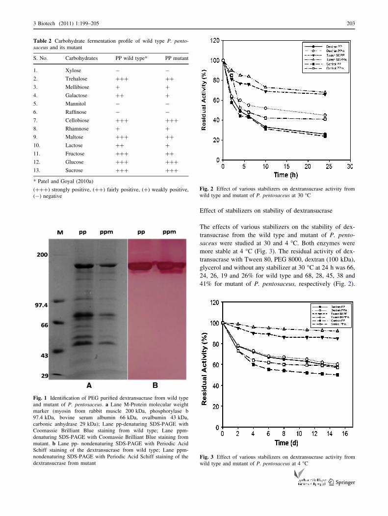
Effect of stabilizers on stability of dextransucrase
The effects of various stabilizers on the stability of dex-
transucrase from the wild type and mutant of P. pento-
saceus were studied at 30 and 4 �C. Both enzymes were
more stable at 4 �C (Fig. 3). The residual activity of dex-
transucrase with Tween 80, PEG 8000, dextran (100 kDa),
glycerol and without any stabilizer at 30 �C at 24 h was 66,
24, 26, 19 and 26% for wild type and 68, 28, 45, 38 and
41% for mutant of P. pentosaceus, respectively (Fig. 2).
Table 2 Carbohydrate fermentation profile of wild type P. pento-saceus and its mutant
S. No. Carbohydrates PP wild type* PP mutant
1. Xylose - -
2. Trehalose ??? ??
3. Mellibiose ? ?
4. Galactose ?? ?
5. Mannitol - -
6. Raffinose - -
7. Cellobiose ??? ???
8. Rhamnose ? ?
9. Maltose ??? ??
10. Lactose ?? ?
11. Fructose ??? ??
12. Glucose ??? ???
13. Sucrose ??? ???
* Patel and Goyal (2010a)
(???) strongly positive, (??) fairly positive, (?) weakly positive,
(-) negative
Fig. 1 Identification of PEG purified dextransucrase from wild type
and mutant of P. pentosaceus. a Lane M-Protein molecular weight
marker (myosin from rabbit muscle 200 kDa, phosphorylase b
97.4 kDa, bovine serum albumin 66 kDa, ovalbumin 43 kDa,
carbonic anhydrase 29 kDa); Lane pp-denaturing SDS-PAGE with
Coomassie Brilliant Blue staining from wild type; Lane ppm-
denaturing SDS-PAGE with Coomassie Brilliant Blue staining from
mutant. b Lane pp- nondenaturing SDS-PAGE with Periodic Acid
Schiff staining of the dextransucrase from wild type; Lane ppm-
nondenaturing SDS-PAGE with Periodic Acid Schiff staining of the
dextransucrase from mutant
Fig. 2 Effect of various stabilizers on dextransucrase activity from
wild type and mutant of P. pentosaceus at 30 �C
Fig. 3 Effect of various stabilizers on dextransucrase activity from
wild type and mutant of P. pentosaceus at 4 �C
3 Biotech (2011) 1:199–205 203
123

The residual activity of dextransucrase with Tween 80,
PEG 8000, dextran (100 kDa), glycerol and without any
stabilizer at 4 �C on 15th day was 85, 47, 58, 40 and 50%
and 92, 48, 60, 53 and 57% for wild type and mutant of
P. pentosaceus respectively (Fig. 3). The other stabilizers
dextran (100 kDa), glycerol and PEG-8000 did not show
any significant effect at both the temperatures on the
enzyme. The data for glycerol and PEG-8000 are not
shown in Figs. 2 and 3. It has been reported earlier that
dextransucrase is stable at lower temperatures (10–30 �C)
and loses rapidly the enzyme activity at temperatures
higher than 30 �C (Purama et al. 2010). Our results are
similar to those reported earlier for L. mesenteroides NRRL
B-640 (Purama et al. 2010). The results clearly indicate
that the mutant enzyme is more stable than the wild
type enzyme and Tween 80 was the best stabilizer for
dextransucrase of both the wild type and mutant of
P. pentosaceus.
Half-life of stabilizer treated dextransucrase
The residual activity of dextransucrase was measured at
various temperatures with respect to time with and without
stabilizers. The enzyme deactivation followed first-order
rate kinetics. The half-life (t1/2) of dextransucrase and
stabilizers treated dextransucrase was calculated by
assuming that the decay followed first-order kinetics
(Purama et al. 2010). Amongst all the stabilizers Tween 80
displayed maximum stabilization of dextransucrase with
t1/2 of 28.5 and 33.5 h for wild type and mutant, respec-
tively, at 30 �C and t1/2 of 52.1 and 106.6 days for wild
type and mutant respectively, at 4 �C. The addition of
Tween 80 to dextransucrase, incubated at both the tem-
peratures (30 and 4 �C) resulted in higher t1/2 than that of
with no Tween 80 (Table 3). The t1/2 of mutant enzyme
was 49% higher and 24% higher than that of wild type at
30 and 4 �C, respectively. The t1/2 of dextransucrase from
mutant with Tween was 17.5% higher and 104.6% higher
than that of wild type at 30 and 4 �C, respectively. Taken
together all these results, it can be summarized that Tween
80 provided the maximum stabilization at 30 and 4 �C and
the mutant showed better stabilization than that of the wild
type at both the temperatures.
Conclusion
The comparison of antibiotic resistance, carbohydrate
utilization pattern, dextransucrase activity and dextran-
sucrase stabilization of wild type and mutant of
P. pentosaceus was reported. The results of antibiotic
resistance, carbohydrate utilization pattern, dextransu-
crase activity and dextransucrase stabilization will
enhance understanding of these industrially significant
species and will aid in distinguishing between physio-
logically similar species. The data will be useful for
industrial applications where the strains are required with
higher enzyme stability. Both dextransucrase of wild type
and mutant were more stable at 4 �C than at 30 �C.
Amongst various stabilizers Tween 80 provided the
maximum stabilization to dextransucrase against activity
loss at 30 and 4 �C. The addition of Tween 80 to dex-
transucrase at 30 and 4 �C resulted in higher t1/2 than that
of without Tween 80. The residual activity and t1/2 were
higher for mutant enzyme than that of wild type. The
results suggested that dextransucrase from the mutant
showed better stabilization than that of the wild type and
therefore have greater importance.
Acknowledgments The research work was supported by a project
grant from Department of Biotechnology, Ministry of Science and
Technology, New Delhi, India to AG.
Open Access This article is distributed under the terms of the
Creative Commons Attribution License which permits any use, dis-
tribution and reproduction in any medium, provided the original
author(s) and source are credited.
References
Abrams D, Barbosa J, Albano H, Silva J, Gibbs PA, Teixeira P (2011)
Characterization of bacPPK34 a bacteriocin produced by Ped-iococcus pentosaceus strain K34 isolated from ‘‘Alheira’’. Food
Control 22:940–946
Barry AL, Thornsberry C (1980) Susceptibility testing: diffusion
procedures. In: Lennette EH, Balows A, Hausler WJ Jr, Truant
JP (eds) Manual of clinical microbiology. Am Soc Microbiol,
Washington, D.C., pp 463–474
De Vuyst L, Degeest B (1999) Heteropolysaccharides from lactic acid
bacteria. FEMS Microbiol Rev 23:153–177
Table 3 Half-life of
dextransucrase from wild type
P. pentosaceus and mutant of at
30 and 4 �C
Stabilizers 30 �C 4 �C
PP wild type t1/2 (h) PP mutant t1/2 (h) PP wild type t1/2 (days) PP mutant t1/2 (days)
Control 8.50 12.70 11.76 14.53
Glycerol 7.50 12.25 10.40 14.00
PEG 8000 8.36 10.22 11.03 11.21
Tween 80 28.52 33.50 52.10 106.61
Dextran 8.51 15.23 16.04 17.11
204 3 Biotech (2011) 1:199–205
123
Fernandes CF, Shahani KM, Amer MA (1987) Therapeutic role of
dietary lactobacilli and lactobacillic fermentated dairy products.
FEMS Microbiol Rev 46:343–356
Gerez CL, Torino MI, Rollan G, de Valdez GF (2009) Prevention of
bread mould spoilage by using lactic acid bacteria with
antifungal properties. Food Control 20:144–148
Goyal A, Katiyar SS (1996) Regulation of dextransucrase productiv-
ity of Leuconostoc mesenteroides B-512F by the maintenance
media. J Gen Appl Microbiol 42:81–85
Holt SM, Al-Sheikh H, Shin KJ (2001) Characterization of dextran-
producing Leuconostoc strains. Lett Appl Microbiol 32:185–189
Kandler O, Weiss N (1986) Regular, nonsporing gram-positive rods.
In: Sneath PHA, Mair NS, Sharpe ME, Holt JG (eds) Bergey’s
manual of systematic bacteriology. Williams & Wilkins, Balti-
more, pp 1208–1219
Kim D, Robyt JF (1994) Production and selection of mutants of
Leuconostoc mesenteroides constitutive for glucansucrases.
Enzym Microb Technol 16:659–654
Kim D, Robyt JF (1995a) Production, selection, and characteristics of
mutants of Leuconostoc mesenteroides B-742 constitutive for
dextransucrases. Enzym Microb Technol 17:689–695
Kim D, Robyt JF (1995b) Dextransucrase constitutive mutants of
Leuconostoc mesenteroides B-1299. Enzym Microb Technol
17:1050–1056
Kitaoka M, Robyt JF (1998) Large-scale preparation of highly
purified dextransucrase from a high-producing constitutive
mutant of Leuconostoc mesenteroides B-512FMC. Enzym
Microbiol Technol 23:386–391
Knorr D (1998) Technology aspects related to microorganisms in
functional foods. Trends Food Sci Technol 9:295–306
Kralj S, Van Geel-Schutten GH, Dondroff MG, Kirsanovs S, Van Der
Maarel MJEC, Dijkhuizen L (2004) Glucan synthesis in the
genus Lactobacillus: isolation and characterization of glucan-
sucrase genes, enzymes and glucan products from six different
strains. Microbiology 150:3681–3690
Laemmli UK (1970) Cleavage of structural proteins during the
assembly of the head of bacteriophage T4. Nature 227:680–685
Lowry OH, Rosebrough NJ, Farr AL, Randall RJ (1951) Protein
measurement with the Folin phenol reagent. J Biol Chem
193:265–275
Ly MH, Covarrubias-Cervantes M, Dury-Brun C, Bordet S, Voilley A,
Le TM, Belin JM, Wache Y (2008) Retention of aroma compounds
by lactic acid bacteria in model food media. Food Hydrocoll
22:211–217
Majumder A, Goyal A (2008) Enhanced production of exocellular
glucansucrase from Leuconostoc dextranicum NRRL B-1146 using
response surface method. Bioresour Technol 99:3685–3691
Mugula JK, Narvhus JA, Sorhaug T (2003) Use of starter cultures of
lactic acid bacteria and yeasts in the preparation of togwa, a
Tanzanian fermented food. Int J Food Microbiol 3:307–318
Naessens M, Cerdobbel A, Soetaert W, Vandamme EJ (2005)
Leuconostoc dextransucrase and dextran: production, properties
and applications. J Chem Technol Biotechnol 80:845–860
Nelson N (1944) A photometric adaptation of the Somogyi method
for the determination of glucose. J Biol Chem 153:375–380
Patel S, Kasoju N, Bora U, Goyal A (2010) Structural analysis and
biomedical applications of dextran produced by a new isolate
Pediococcus pentosaceus screened from biodiversity hot spot
Assam. Bioresour Technol 101(17):6852–6855
Patel S, Goyal A (2010a) 16S rRNA based identification and
phylogenetic analysis of a novel dextran producing Pediococcus
pentosaceus isolated from north-east Indian microbial diversity.
Curr Trends Biotechnol Pharm 4:746–754
Patel S, Goyal A (2010b) Isolation, characterization and mutagenesis
of exopolysaccharide synthesizing new strains of lactic acid
bacteria. Internet J Microbiol 8(1):3–4
Purama RK, Agrawal M, Majumder A, Ahmed S, Goyal A (2008)
Antibiotic sensitivity, carbohydrate fermentation characteristics
and plasmid profiles of glucansucrase producing four Leuconos-toc strains. J Pure Appl Microbiol 2:139–146
Purama RK, Goyal A (2005) Dextransucrase production by Leuco-nostoc mesenteroides. Ind J Microbiol 2:89–101
Purama RK, Goyal A (2008) Identification, effective purification and
functional characterization of dextransucrase from Leuconostocmesenteroides NRRL B-640. Bioresour Technol 99:3635–3642
Purama RK, Agrawal M, Goyal A (2010) Stabilization of dextran-
sucrase from Leuconostoc mesenteroides NRRL B-640. Ind J
Microbiol 50:57–61
Ricciardi A, Clementi F (2000) Exopolysaccharide from lactic acid
bacteria: structure, production and technological applications.
Ital J Food Sci 12:23–45
Schindler S, Wittig M, Zelena K, Krings U, Bez J, Eisner P, Berger
RG (2011) Lactic fermentation to improve the aroma of protein
extracts of sweet lupin (Lupinus angustifolius). Food Chem
128:330–337
Sengupta A, Wang S, Link E, Anderson EH, Hofmann C, Lewan-
dowski J, Kottke-Marchant K, Marchant RE (2006) Glycocalyx-
mimetic dextran modified poly (vinyl amine) surfactant coating
reduces platelet adhesion on medical-grade polycarbonate sur-
face. Biomaterials 27:3084–3095
Settanni L, Moschetti G (2010) Non-starter lactic acid bacteria used
to improve cheese quality and provide health benefits. Food
Microbiol 27:691–697
Sidebotham RL (1974) Dextrans. Adv Carbohydr Chem Biochem
30:371–444
Singh A, Majumder A, Goyal A (2009) Mutagenesis of Leuconostocdextranicum NRRL B-1146 for higher glucan production. Inter J
Microbiol 7(1):1–7
Smitinont T, Tansakul C, Tanasupawat S, Keeratipibul S, Navarini L,
Bosco M, Cescutti P (1999) Exopolysaccharide-producing lactic
acid bacteria strains from traditional Thai fermented foods:
isolation, identification and exopolysaccharide characterization.
Int J Food Microbiol 51:105–111
Somogyi M (1945) A new reagent for the determination of sugars.
J Biol Chem 160:61–68
Sybesma W, Hugenholtz J, de Vos Willem M, Smid EJ (2006) Safe
use of genetically modified lactic acid bacteria in food: bridging
the gap between consumers, green groups, and industry. Electron
J Biotechnol 9((4 ISSN)):0717–3458
Tieking M, Kaditzky S, Valcheva R, Korakli M, Vogel RF, Ganzle
MG (2005) Extracellular homopolysaccharides and oligosaccha-
rides from intestinal Lactobacilli. J Appl Microbiol 99:692–702
Todorov SD, Wachsman M, Tome E, Dousset X, Destro MT, Dicks
LMT, de Melo Franco BDG, Vaz-Velho M, Drider D (2010)
Characterisation of an antiviral pediocin-like bacteriocin pro-
duced by Enterococcus faecium. Food Microbiol 27:869–879
Tsuchiya HM, Koepsell HJ, Corman J, Bryant G, Bogard MO, Feger
VH, Jackson RW (1952) The effect of certain cultural factors on
production of dextransucrase by Leuconostoc mesenteroides.
J Bact 64:521–526
3 Biotech (2011) 1:199–205 205
123
Top Related
Copyright © 2022 FDOKUMEN

