


Bahasa
Halaman
Hukum


doi:10.1016/j.jmb.2007.12.014 J. Mol. Biol. (2008) 376, 1282–1304
Available online at www.sciencedirect.com
Designed Armadillo Repeat Proteins as GeneralPeptide-Binding Scaffolds: Consensus Design andComputational Optimization of the Hydrophobic Core
Fabio Parmeggiani1, Riccardo Pellarin1, Anders Peter Larsen1,Gautham Varadamsetty1, Michael T. Stumpp1, Oliver Zerbe2,Amedeo Caflisch1 and Andreas Plückthun1⁎
1Department of Biochemistry,University of Zürich,Winterthurerstrasse 190,CH-8057 Zürich, Switzerland2Department of OrganicChemistry, University ofZürich, Winterthurerstrasse190, CH-8057 Zürich,Switzerland
Received 3 July 2007;received in revised form13 November 2007;accepted 5 December 2007Available online14 December 2007
*Corresponding author. E-mail addrPresent addresses: A.P. Larsen, D
DK-2200 Copenhagen, Denmark; MSwitzerland.Abbreviations used: αArm, Arma
mouse β-catenin (residues 150–665)circular dichroism; HA, hemaggluticonsensus armadillo repeat; IMAC,obtained by computational approaclocalization sequence; NOE, nuclearSDS-PAGE, sodium dodecyl sulfatecatenin/plakoglobin-derived consen
0022-2836/$ - see front matter © 2007 E
Armadillo repeat proteins are abundant eukaryotic proteins involved inseveral cellular processes, including signaling, transport, and cytoskeletalregulation. They are characterized by an armadillo domain, composed oftandem armadillo repeats of approximately 42 amino acids, which mediatesinteractionswith peptides or parts of proteins in extended conformation. Theconserved binding mode of the peptide in extended form, observed fordifferent targets, makes armadillo repeat proteins attractive candidates forthe generation of modular peptide-binding scaffolds. Taking advantage ofthe large number of repeat sequences available, a consensus-based approachcombinedwith a force field-based optimization of the hydrophobic core wasused to derive soluble, highly expressed, stable, monomeric designed pro-teins with improved characteristics compared to natural armadillo proteins.These sequences constitute the starting point for the generation of designedarmadillo repeat protein libraries for the selection of peptide binders,exploiting their modular structure and their conserved binding mode.
© 2007 Elsevier Ltd. All rights reserved.
Keywords: consensus design; armadillo repeat; hydrophobic core; moltenglobule; molecular dynamics and minimization
Edited by F. E. CohenIntroduction
In recent years, as an alternative to raisingmonoclonal antibodies by immunization, recombi-nant antibodies1 and an increasing number of otherprotein scaffolds2 have been investigated as novelbinding molecules. However, neither antibodiesthemselves nor any of these alternative protein
ess: [email protected] of Biomedical S.T. Stumpp, Molecular Pa
dillo domain of human im; ANS, 1-anilino-naphthalenin tag; HSQC, heteronucimmobilized metal-ion affh; MALS, multiangle lightOverhauser enhancemen
–polyacrylamide gel electrsus armadillo repeat.
lsevier Ltd. All rights reserve
scaffolds were specifically designed to bind pep-tides. Target-specific binding molecules are, ingeneral, obtained from large protein libraries by invitro selection or, in the case of monoclonal anti-bodies, through traditional immunization proce-dures. Both approaches require that, for eachtarget, each new binding molecule is individuallygenerated and characterized for specificity and
.ch.ciences, University of Copenhagen, Blegdamsvej 3,rtners AG, Grabenstrasse 11a, CH-8952 Schlieren,
portin-α1 (residues 83–505); βArm, Armadillo domain ofne-8-sulfonate; C-type, overall consensus repeat; CD,lear single quantum coherence; I-type, importin-derivedinity chromatography; M-type, mutated armadillo repeatscattering; MRE, mean residue ellipticity; NLS, nuclear
t; pD, phage lambda protein D; PDB, Protein Data Bank;ophoresis; SEC, size-exclusion chromatography; T-type,
d.

Fig. 1. (a) Structure of S. cerevisiae importin-α incomplex with nucleoplasmin NLS (PDB ID 1EE5), show-ing the right-handed superhelical structure typical forarmadillo repeat proteins. The cylinders represent theα-helices. The N-terminal repeat is indicated in green, andthe C-terminal repeat is shown in orange. The boundpeptide is depicted in red in a stick representation. (b)Detail of repeat 6 from 1EE5. The α-helices are representedas ribbons. (c) Detail of the peptide-binding mode. Theconserved asparagine residues (in orange) contact viahydrogen bonds (purple) the backbone of the peptide,depicted in green. The residues that are responsible for theinteractions with the side chains of the target peptide areshown in yellow. In all panels, helix 1 (H1) is indicated inblue, helix 2 (H2) in red, and helix 3 (H3) in gray.
1283Designed Armadillo Repeat Proteins as Scaffolds
cross-reactivity, making the generation of bindersagainst a large number of peptide targets (e.g.,representing a full proteome) an almost prohibitivetask.The aim of the present study was to develop a
scaffold for the generation of peptide-specific bind-ing proteins. In more detail, we wanted to developproteins that were stable under various conditionsand with the intrinsic ability to bind peptides in aconserved fashion. To recognize peptides in asequence-selective manner the specificity of bindingshould ideally be conferred through specific inter-actions with the peptide side chains.Natural peptide-binding scaffolds can be grouped
in different classes. Antibodies are known to be ableto bind peptides and have been well character-ized.3–6 Although peptide-binding antibodies havecertain structural features in common, the mode ofbinding is not conserved. Thus, the informationacquired through studies of antibody–peptide com-plexes cannot easily be applied to the generaldesign of peptide-binding antibodies or extendedto other proteins.Small adaptor domains (e.g., SH2, SH3, and PDZ)7
show specific binding to their targets, usually in aconserved bindingmodewithin one family, but theiraffinity is generally low. The recognition sequence isvery short and biased toward certain amino acidtypes, posttranslational modifications, or free N- orC-termini. While several such domains could belinked together by flexible peptides to recognizelonger peptide sequences, a coverage of any arbi-trary sequence would still be very difficult sincethese small domains might not be adaptable to therecognition of any arbitrary sequence. Furthermore,the entropy loss upon binding of such flexibly linkedconstructs would not necessarily lead to highaffinities.The major histocompatibility complex proteins
(MHC I and MHC II)8 possess a higher intrinsicvariability and the ability to recognize a broad rangeof peptides, but the difficulties in their handlingreduce their attractiveness as a scaffold candidate.Repeat proteins, in particular tetratricopeptide
repeats (TPRs),9 armadillo,10 and WD4011 proteins,have been shown to possess an intrinsic ability tobind peptides, taking advantage of their repetitivestructure. Thus, for our purpose, a scaffold based onrepeat proteins seemed to constitute a promisingcandidate. For reasons outlined below, we chose thearmadillo repeat protein family as the basis for ourscaffold candidate.Armadillo repeat proteins12,13 are abundant in
eukaryotes, where they are involved in a broadrange of biological processes (e.g., transcriptionregulation,14 cell adhesion,15 tumor suppressoractivity,16 and nucleocytoplasmic transport17).These proteins are characterized by tandem repeatsof approximately 42 amino acids that were firstdiscovered in the product of the Drosophila melano-gaster segmentation polarity gene Armadillo, whichis homologous to mammalian β-catenin.18,19 Arma-dillo repeat proteins participate in protein–protein
interactions, and the armadillo domain is usuallyinvolved in the recognition process. The domainforms a right-handed superhelix20,21 (Fig. 1a), asshown by the crystal structures of β-catenin22 andimportin-α.23 Every repeat is composed of three α-helices, named H1, H2, and H3 (Fig. 1b), and severalrepeats stack to form the compact domain. Specia-lized repeats are present at the N- and C-termini ofthe protein, protecting the hydrophobic core fromsolvent exposure (Fig. 1a).Armadillo repeat proteins are able to bind different
types of peptides, yet relying on a conserved bindingmode of the peptide backbone. Reported dissocia-tion constant (Kd) values as low as 10–20 nM24

1284 Designed Armadillo Repeat Proteins as Scaffolds
indicate that high affinities can be achieved. Crystalstructures of armadillo repeat proteins in complexwith bound peptides have revealed that mostpeptide targets are bound in an extended conforma-tion along the surface, inside the groove formed bythe H3 helices. The superhelical armadillo domainwinds around the peptide, oriented in the oppositeN- to C-terminal direction (Fig. 1a), thus forming adouble-helical complex, topologically similar to theDNA double strand. An asparagine residue, con-served in almost every repeat at the C-terminal partof H3, makes hydrogen bonds to the main chain ofthe target peptide, thereby keeping it in an extendedconformation. Additional interactions to the targetside chains are provided by neighboring residues,mostly in H3 (Fig. 1c). In a first approximation, eachdipeptide unit of the target peptide is specificallyrecognized by one repeat in the armadillo domain(Fig. 2a).In theory, the possibility of developing individual
repeats that specifically bind a two-amino-acidsequence unit is very attractive. Given that theindividual repeats are based on the same optimizedscaffold and, thus, compatible with each other, any
Fig. 2. Binding of target peptides. (a) Schematicdrawing of an armadillo repeat protein binding to anextended peptide. The target peptide is bound in anantiparallel orientation to the protein. N and C indicateN- and C-termini of the peptide, which is depicted in red,with the amino acid side chains shown in blue. Theresidues of armadillo repeats involved in binding occupyspecific positions within the single repeat sequences,mostly on helix 3. The position indicated in orange (aconserved Asn) is responsible for the binding of thepeptide main chain; the positions in yellow are involved inrecognition of the peptide side chains. (b) Designedarmadillo repeat proteins potentially allow the selectionof single repeats that specifically recognize short se-quences. The selected peptide-specific repeats can bethen combined to recognize longer peptides withoutperforming additional selections.
given number of repeats can be directly stacked toextend the recognition to much longer peptidesequences. In contrast to flexibly linked smalladaptor domains mentioned above, armadillorepeats directly stack on each other in a rather rigidmanner, allowing binding to uninterrupted longerpeptides. This would exploit the specificity of theindividual repeats to provide a peptide-bindingdesigned armadillo protein with high and predeter-mined specificity, governed by the individualrepeats. Such an approach (Fig. 2b), using armadilloproteins assembled from previously selected “build-ing blocks,” could effectively bypass the current invitro selection procedures for individual peptides.However, this requires such individual peptide-specific repeats first to be developed, using alibrary-based approach.In the present study, we have, as a first step,
designed armadillo repeat modules based on con-sensus sequences. Proteins containing different typesof modules have been assembled and characterized,initially only leading to stable dimeric proteins ormonomeric molten-globule-like proteins. We subse-quently used a combination of molecular dynamicsand minimization to improve the hydrophobic corepacking and convert the consensus-designed arma-dillo repeat proteinwithmolten-globule-like proper-ties to a monomeric, stable folded protein. Finally,the protein characteristics were evaluated for explor-ing the possibility of generating a modular peptide-binding scaffold. We succeeded in developing astable, monomeric consensus protein that can beused now in the generation of peptide-specificindividual armadillo repeat proteins.
Results
Armadillo repeat protein design
A consensus design strategy25 has been applied inorder to generate armadillo repeat proteins withhigh expression levels of soluble protein in Escher-ichia coli, monomeric state, high thermodynamicstability, and absence of cysteines for convenientexpression and handling.This design procedure was aimed at the genera-
tion of self-compatible repeat modules; therefore,consensus sequences were derived from multiplealignments of single armadillo repeats from theSwiss-Prot database.26 A consensus design strategyhas been successfully applied previously to otherdesigned repeat proteins,27–30 and it is based onconsensus design of internal repeats (or internalmodules). Special terminal capping repeats (term-inal modules) have been generated to protect thehydrophobic core from solvent exposure. Thecrucial role of capping repeats has been previouslyshown in studies with designed ankyrin repeatproteins.31
The numbering used here to define the positionswithin the repeats was based on the family align-
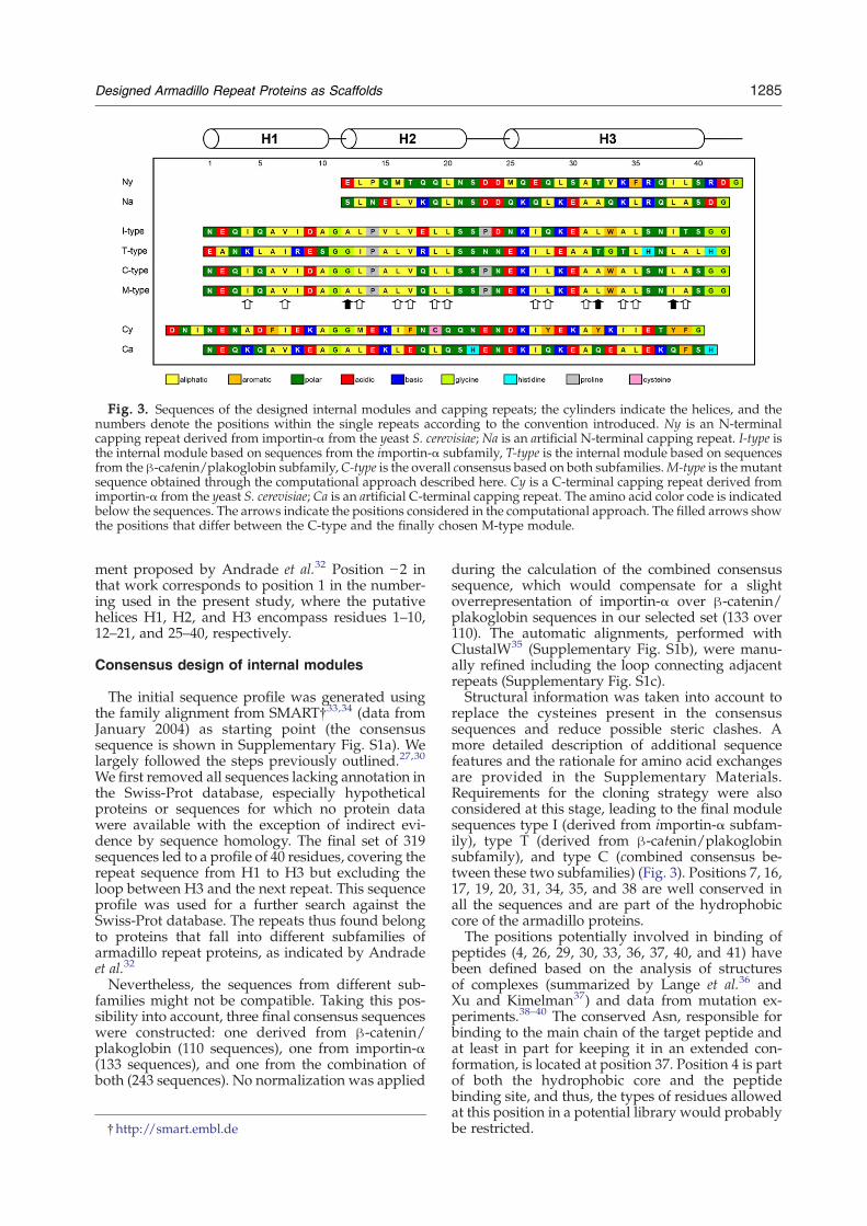
Fig. 3. Sequences of the designed internal modules and capping repeats; the cylinders indicate the helices, and thenumbers denote the positions within the single repeats according to the convention introduced. Ny is an N-terminalcapping repeat derived from importin-α from the yeast S. cerevisiae; Na is an artificial N-terminal capping repeat. I-type isthe internal module based on sequences from the importin-α subfamily, T-type is the internal module based on sequencesfrom the β-catenin/plakoglobin subfamily,C-type is the overall consensus based on both subfamilies.M-type is the mutantsequence obtained through the computational approach described here. Cy is a C-terminal capping repeat derived fromimportin-α from the yeast S. cerevisiae; Ca is an artificial C-terminal capping repeat. The amino acid color code is indicatedbelow the sequences. The arrows indicate the positions considered in the computational approach. The filled arrows showthe positions that differ between the C-type and the finally chosen M-type module.
1285Designed Armadillo Repeat Proteins as Scaffolds
ment proposed by Andrade et al.32 Position −2 inthat work corresponds to position 1 in the number-ing used in the present study, where the putativehelices H1, H2, and H3 encompass residues 1–10,12–21, and 25–40, respectively.
Consensus design of internal modules
The initial sequence profile was generated usingthe family alignment from SMART†33,34 (data fromJanuary 2004) as starting point (the consensussequence is shown in Supplementary Fig. S1a). Welargely followed the steps previously outlined.27,30
We first removed all sequences lacking annotation inthe Swiss-Prot database, especially hypotheticalproteins or sequences for which no protein datawere available with the exception of indirect evi-dence by sequence homology. The final set of 319sequences led to a profile of 40 residues, covering therepeat sequence from H1 to H3 but excluding theloop between H3 and the next repeat. This sequenceprofile was used for a further search against theSwiss-Prot database. The repeats thus found belongto proteins that fall into different subfamilies ofarmadillo repeat proteins, as indicated by Andradeet al.32
Nevertheless, the sequences from different sub-families might not be compatible. Taking this pos-sibility into account, three final consensus sequenceswere constructed: one derived from β-catenin/plakoglobin (110 sequences), one from importin-α(133 sequences), and one from the combination ofboth (243 sequences). No normalization was applied
†http://smart.embl.de
during the calculation of the combined consensussequence, which would compensate for a slightoverrepresentation of importin-α over β-catenin/plakoglobin sequences in our selected set (133 over110). The automatic alignments, performed withClustalW35 (Supplementary Fig. S1b), were manu-ally refined including the loop connecting adjacentrepeats (Supplementary Fig. S1c).Structural information was taken into account to
replace the cysteines present in the consensussequences and reduce possible steric clashes. Amore detailed description of additional sequencefeatures and the rationale for amino acid exchangesare provided in the Supplementary Materials.Requirements for the cloning strategy were alsoconsidered at this stage, leading to the final modulesequences type I (derived from importin-α subfam-ily), type T (derived from β-catenin/plakoglobinsubfamily), and type C (combined consensus be-tween these two subfamilies) (Fig. 3). Positions 7, 16,17, 19, 20, 31, 34, 35, and 38 are well conserved inall the sequences and are part of the hydrophobiccore of the armadillo proteins.The positions potentially involved in binding of
peptides (4, 26, 29, 30, 33, 36, 37, 40, and 41) havebeen defined based on the analysis of structuresof complexes (summarized by Lange et al.36 andXu and Kimelman37) and data from mutation ex-periments.38–40 The conserved Asn, responsible forbinding to the main chain of the target peptide andat least in part for keeping it in an extended con-formation, is located at position 37. Position 4 is partof both the hydrophobic core and the peptidebinding site, and thus, the types of residues allowedat this position in a potential library would probablybe restricted.

1286 Designed Armadillo Repeat Proteins as Scaffolds
Design of capping repeats
N- and C-terminal capping repeats, found innatural armadillo domains, protect the hydrophobiccore, as they present a hydrophobic surface to theinternal repeat side but a hydrophilic surface tothe solvent. Capping sequences have also beenconsidered in the previous design of other repeatproteins.27,29–31
The boundaries of armadillo domains have beenestimated by limited proteolysis.22,23 However, theyare not clearly defined, partly due to the weaksimilarity of the terminal repeats to the internalones. In addition, not all the residues are visible inthe crystal structures of importin-α and β-catenin. Itis likely that only the visible residues contribute tothe armadillo domain, and the additional parts areunstructured and do not strictly belong to thedomain. We have defined the N-terminal cappingrepeat as starting from position 12 (the beginning ofH2). In contrast, the C-terminal capping repeat iscompletely resolved in the x-ray structures, and wedefined it to comprise position 1 to position 41, thusincluding H1 to H3.The capping repeats have been designed by using
two different approaches. In the first, naturalcapping repeats were adapted to our designedinternal repeats. Structural information to ensurecompatibility between the capping repeats and thedesigned internal repeat is a fundamental prerequi-site. The importin-α from Saccharomyces cerevisiaewas considered to be the best candidate for a generalcapping repeat donor: all our designed modulespresent a flat surface that can interact with the innersurface of yeast importin-α capping repeats, asjudged from molecular models. The yeast impor-tin-α-derived N-terminal and C-terminal cappingrepeats were named Ny and Cy, respectively.The N-terminal capping repeat covers the residues
from Glu88 to His119 of yeast importin-α. However,the two residues Glu118–His119 were replaced byAsp–Gly (Fig. 3, positions 42 and 43 of Ny) to adaptthe terminal loop to the designedmodules: glycine isused for assembly of the modules (as its codonoverlaps a restriction site) and aspartate keeps anegative charge, which is frequently present at thisposition in natural proteins, reducing at the sametime the helical propensity in the turn region.The C-terminal capping repeat covers the region
from Asn471 to Gly510 in yeast importin-α. How-ever, the loop connecting the last internal repeatwith the C-terminal repeat contains additional resi-dues in yeast importin-α, compared to other naturalimportins. A modified version of this C-terminalcapping repeat has thus been generated by introdu-cing three residues (Asp-Asn-Ile) before H1 (Fig. 3,first three residues of Cy). Asn and Ile are naturallypresent at these positions; Asp has been includedto keep a negatively charged loop as observed inseveral natural sequences while reducing the helicalpropensity.In the second approach, two completely artificial
N- and C-terminal capping repeats were designed
(named Na and Ca, respectively, and shown in Fig.3), starting from the type C consensus andsubstituting the exposed hydrophobic residueswith hydrophilic ones. Positions 12, 19, 27, and34 of the N-terminal capping repeat are occupiedby hydrophobic residues in the consensus se-quence and were replaced by hydrophilic residuesbased on structures and frequently occurringresidues obtained from alignment of N-terminalcapping sequences. In a similar way, positions 8,13, 17, 20, 28, 32, 35, 38, and 39 of the C-terminalcapping repeat were replaced by hydrophilicresidues based on structures and frequentlyoccurring residues obtained from alignment ofC-terminal capping sequences. A detailed descrip-tion of the residues introduced in the designedcapping repeats is provided in the SupplementaryMaterials. A second version of the Cy cappingrepeat was also designed without the three initialresidues and with Ala replacing Cys at position19; no change was observed, compared to Cy, inthe level of expression and in the amount ofsoluble protein, and it will thus not be discussedfurther.
Assembly, cloning, and expression of designedarmadillo repeat proteins
The amino acid sequences of all modules wereback-translated to DNA sequences, optimizing thecodon usage for expression in E. coli. Each modulewas synthesized, starting from overlapping oligo-nucleotides (Supplementary Table S1).The modules were assembled stepwise using type
IIS restriction enzymes (Supplementary Fig. S2),following the approach reported by Binz et al.27The final proteins were named according to themodules that they contain: the name indicates, inorder, the type of N-terminal repeat (A for Artificialor Na, Y for Yeast derived or Ny), the type of internalrepeats (type I, type T, or type C) with the numberof modules used as a subscript, and the type ofC-terminal repeat (A for Artificial or Ca, Y for Yeastderived or Cy): for example, YI4A contains a Yeast-derived N-terminal repeat (Ny), four internal re-peats based on Importin consensus (type I), and anArtificial C-terminal repeat (Ca).Thus, Na or Ny as N-capping modules were
combined with T-, I-, or C-type internal modules andCa or Cy C-terminal modules, leading to 12 possiblecombinations. The proteins contain only one type ofinternal module to avoid incompatible surfaces atthe interface between repeats. The influence ofcapping and internal repeats was evaluated byanalyzing the expression properties of all theconstructs, containing two or four internal repeats.The proteins were expressed in E. coli XL1-blueusing a pQE30-based expression plasmid, providingan N-terminal MRGSH6 tag for purification. Theinsert was constructed with a double stop codon(Supplementary Fig. S3). As an example, the DNAand protein sequences of YC2A are provided inSupplementary Fig. S3.

1287Designed Armadillo Repeat Proteins as Scaffolds
The highest level of soluble protein expressionwas obtained when the internal modules werecombined with Ny and Ca (Fig. 4a). The Na capleads to almost undetectable expression in Coomas-sie-stained polyacrylamide gels, and the presence ofCy resulted in a substantial portion of the proteinfound in the insoluble fraction after cell lysis. Theobserved effects of terminal capping repeats wereindependent of the type and the number of internalmodules. However, increasing the number of inter-nal modules enhanced the amount of soluble proteinand the absolute amount of protein produced.Remarkably, type T proteins are characterized by alower apparent mobility in sodium dodecyl sulfate–polyacrylamide gel electrophoresis (SDS-PAGE),compared to type I and type C proteins (Fig. 4b).
Fig. 4. (a) Influence of capping repeats on expression.Soluble and insoluble fractions of E. coli cell extracts areshown in a Coomassie-stained SDS polyacrylamide gel.The proteins contain two internal C-type modules withdifferent combinations of capping repeats. (b) Whole-cellextracts of consensus proteins. The constructs contain Nyand Ca as capping repeats. Cells transformed with theempty vector or with the vector containing the armadillodomain of mouse β-catenin (Arm) were used as control.The proteins can be easily purified in a single step byIMAC, as shown for YM4A. The expected size is 18 and27 kDa for proteins containing two or four internalmodules, respectively, and 56 kDa for Arm. The triangleindicates the band corresponding to the armadillo domainof β-catenin, which is expressed at much lower yield thanthe designed proteins. The molecular mass of the marker(M) is indicated in kilodaltons on the left.
Protein purification and characterization:Comparison with natural armadillo domains
Proteins containing the combination of Ny, Ca,and two, four, or eight internal repeats have beenchosen for biophysical characterization and evalua-tion of the properties of type I, type T, and type Cmodules. The results are summarized in Table 1.The purification by immobilized metal-ion affinity
chromatography (IMAC) in a single step providedup to 100 mg of pure protein from 1 l of bacterialculture (Fig. 4b). No sign of precipitation ordegradation was detected by spectrophotometryand SDS-PAGE in protein solutions stored for up to1 month at 4 °C in the IMAC elution buffer.The natural human importin-α1 (Swiss-Prot P52294)
and the mouse β-catenin (Swiss-Prot Q02248) werealso expressed using the same pQE30-based plas-mid. The importin contains 10 armadillo repeatsand the catenin 12, including the capping repeats inthe count in both cases. Human importin-α1 gavethe highest yield among the importin-α familymembers tested (data not shown) after a two-stepcoupled IMAC–ion exchange purification and,together with mouse β-catenin, was used for thecomparison with the designed armadillo repeatproteins.Importin-α1 and β-catenin, despite their elon-
gated shape, elute at the volume expected from theirmolecular weight in gel filtration on a Superdex 200column, and the monomeric state was confirmed forboth proteins by multiangle light scattering (MALS)measurements (Table 1).On the other hand, the designed proteins show
elution volumes corresponding to higher-than-expected apparent molecular masses in size-exclu-sion chromatography (SEC) (Table 1). MALS indi-cates that the I- and T-type proteins are probablypresent as a mixture of dimers and monomers insolution. The main peak (Fig. 5a) corresponds to thedimeric form, and this value is reported in Table 1.At high concentration (2–4 mg/ml), I- and T-typeproteins are present as a mixture of oligomers.In contrast, monomeric and oligomeric fractions ofC-type proteins YC4A (Fig. 5a) and YC8A (data notshown) can be separated, up to the highestconcentration tested (4 mg/ml). However, thefractions of YC4A and YC8A, shown by MALS tobe monomeric, elute earlier than expected forproteins of comparable size. The smaller YC2Arepresents the only exception: independent of theconcentration, the MALS-calculated mass values arealways intermediate between monomer and dimer.A decrease in pH to 7 favors the formation ofoligomeric species of I- and T-type proteins. C-typeproteins are, in contrast, unaffected by pH (data notshown).The circular dichroism (CD) spectra (Fig. 5b)
indicate the presence of significant α-helical second-ary structure content for all proteins, particularly forthe I-type proteins. For I- and C-type consensusrepeats, the absolute value of mean residue ellipti-city (MRE) and the helical content generally increase

Table 1. Biophysical properties of designed and natural armadillo repeat proteins
ConstructResidues(repeats)a pIb MWcalc (kDa)b
Oligomericstatec MWobs (kDa)d MWobs/calc
eCD222(MRE)f
Helical content(%)g
Observed Tm(°C)h
YI2A 169 (4) 5.2 18.6 Dimer 64.6 1.7 −13,000 63 ∼55YI4A 253 (6) 4.8 27.4 Dimer 116.1 2.1 −19,500 80 ∼69YI8A 421 (10) 4.6 44.9 Dimer 148.8 1.7 −22,600 85 N85YT2A 169 (4) 6.3 18.6 Dimer 141.2 3.8 −7100 23 ∼56YT4A 253 (6) 6.5 27.3 Dimer 219.6 4.0 −10,100 40 ∼75YT8A 421 (10) 6.7 44.8 Dimer 229.7 2.6 −9400 35 ∼83YC2A 169 (4) 5.4 18.4 Mixture 59.1 n.d. −9100 45 n.d.YC4A 253 (6) 5.1 26.9 Monomer 50.0 1.9 −12,100 49 n.d.YC8A 421 (10) 4.8 44.0 Monomer 76.7 1.7 −20,000 62 n.d.YM4A 253 (6) 5.1 27.1 Monomer 32.2 1.2 −18,800 87 ∼70αArmi 435 (10) 5.5 48.2 Monomer 42.9 0.9 −14,300 54 ∼43βArmj 528 (12) 8.7 57.6 Monomer 52.6 0.9 −16,800 60 ∼58
n.d. indicates that the value has not been determined due to either an inhomogeneous sample (oligomeric state of YC2A) or lack ofcooperative transition in thermal denaturation (YC2A, YC4A, and YC8A).
a The number of residues includes the MRGSH6 tag; the number of repeats includes capping repeats.b pI and molecular weight calculated from the sequence; masses were confirmed by mass spectrometry.c Oligomeric state as indicated by multiangle static light scattering.d Observed molecular weight as determined in SEC.e Ratio between observed and calculated molecular weight, taking into account the oligomeric state (Os): MWobs/calc=MWobs/
(Os·MWcalc).f Mean residue ellipticity at 222 nm expressed as deg·cm2/dmol.g Helical content estimated with the program CDpro.41h Tm observed in thermal denaturation by CD.i Armadillo domain of human importin-α1.j Armadillo domain of mouse β-catenin.
1288 Designed Armadillo Repeat Proteins as Scaffolds
with the number of internal repeats; in contrast,the helical content is almost constant for T-type pro-teins (Supplementary Fig. S4). The values of helicalcontent were calculated using the program CDpro41
and are indicated in Table 1.The CD signal at 222 nm was chosen to monitor
stability against thermal and denaturant-induced
Fig. 5. Biophysical characterization of designed and naturaarmadillo repeat proteins containing four internal modules anmolecular weights higher than the globular proteins with theshown by YI4A and YT4A are due to a mixture of dimers and mby light scattering. The highest point of the peak correspondseluted contains probably a mixture of oligomers with highremains monomeric and was further characterized. The imporat the expected volume. The data were obtained with a Superd280 nm for YC4A, YI4A and αArm; YT4A does not possess anywas followed at 230 nm. V0 indicates the void volume of thebovine serum albumin (BSA; MW=66 kDa), carbonic anhydrawere used as molecular weight markers, and the correspondspectra of I-type, T-type, and C-type proteins containing four inimportin-α1 (αArm) and mouse β-catenin (βArm) are indicatreported as MRE. (c) Thermal denaturation curves. A comparisfour or eight internal modules is shown, from the top, for Idisplayed in the bottom panel. The denaturation was followdenaturation and renaturation of designed armadillo repeat procomparison, the bottom graph shows the irreversible dendenaturation (data not shown). The denaturation was followedsetting the initial and the final values of the denaturation cuinduced denaturation of armadillo repeat proteins containingYC8Awith αArm. The denaturation was followed by CD. Theinitial and the final values of the denaturation curves as 0 andof designed armadillo repeat proteins. YI4A, YT4A, and YC4Ashow fluorescence levels in the same range as natural proteins;significantly higher and increases with the number of repeats.was measured in a separate experiment and scaled accordingSimilar results were obtained with proteins containing two or
unfolding. I- and T-type proteins show a cooperativetransition, while no transition was observed inC-type proteins (Fig. 5c). The midpoint of transitionduring thermal denaturation (Tm) increases with thenumber of repeats, for example, from approximately70 °C for YI4A to more than 80 °C for YI8A (Table 1).Importin-α1 and β-catenin, containing 8 and 10
l armadillo repeat proteins. (a) SEC and MALS of designedd of importin-α1. YI4A, YT4A, and YC4A show apparentsame calculated mass (about 27 kDa). The broad peaksonomers, as indicated by the molecular mass determinedto the dimeric fraction. In the case of YC4A, the first peakmolecular masses. The monomeric peak after separationtin-α1 (αArm) is a monomer as indicated by LS and elutesex 200 column. The elution was followed by absorbance atresidue absorbing significantly at 280 nm; thus, the elutioncolumn. Alcohol dehydrogenase (ADH; MW=150 kDa),se (CA; MW=29 kDa), and aprotinin (Apr; MW=6.5 kDa)ing elution volumes are indicated by the arrows. (b) CDternal modules. The natural armadillo domains of humaned by open and filled circles, respectively. The values areon between designed armadillo repeat proteins containing-type, T-type, and C-type proteins. αArm and βArm areed by CD. The MRE at 222 nm is reported. (d) Thermalteins. From the top, YI4A, YT4A, and YC4A are shown. Foraturation of αArm. βArm shows a similar irreversibleby CD. The values of MRE at 222 nmwere normalized byrves as 0 and 1, respectively. (e) Guanidinium-chloride-eight internal modules. Comparison of YI8A, YT8A, andvalues of MRE at 222 nm were normalized by setting the1, respectively. (f) Emission spectra of ANS in the presenceare compared to αArm and βArm. I- and T-type proteinsin contrast, the fluorescence emission for C-type proteins isThe values without buffer subtractions are shown. αArmto the values of YC4A present in both sets of experiments.eight internal repeats.

1289Designed Armadillo Repeat Proteins as Scaffolds
internal repeats, respectively, have lower midpointsof transition, even when compared with designedproteins with only 4 internal repeats (Table 1). It
Fig. 5 (legend on
should be noted that the designed proteins retain asignificant percentage of secondary structure at95 °C and that the thermal unfolding is almost
previous page)

1290 Designed Armadillo Repeat Proteins as Scaffolds
completely reversible, in contrast to natural arma-dillo proteins that cannot refold after thermalunfolding (Fig. 5d); YT8A is the only designedarmadillo repeat protein whose thermal unfolding isirreversible (data not shown).We also investigated unfolding induced by gua-
nidinium chloride. A direct comparison betweennatural and designed armadillo repeat proteinscomposed of 10 repeats (Fig. 5e) reveals forimportin-α1 (αArm), with a midpoint of transitionof 1.4 M guanidinium chloride, a lower stability thanthat for YI8A and YT8A, with approximately 4.8 and3.2 M as midpoints of transition, respectively. YC8Ashows a gradual loss of secondary structure,especially at low concentrations of denaturant,apparently similar to αArm. Data from urea-induced unfolding experiments confirm the gradualloss of secondary structure for C-type proteins withincreasing denaturant concentration. Natural arma-dillo domains show a stable pretransition baseline inunfolding induced by the weaker denaturant urea(data not shown).The three types of consensus proteins (C-, I-,
and T- type) also show a different behavior in 1-anilino-naphthalene-8-sulfonate (ANS) bindingexperiments. ANS is a fluorescent dye sensitiveto the hydrophobic environment.42 C-type pro-teins bind ANS strongly, suggesting the presenceof an accessible hydrophobic core, while I- and T-type proteins show ANS binding in the same lowrange as the natural armadillo repeat proteins(Fig. 5f).Thermal and guanidinium-induced denaturation
and ANS results indicate that I- and T-type pro-teins share many characteristics with proteins withstable folds. Based on MALS data, however, I-typeproteins are mainly present as dimers. T-typeproteins show even higher deviations of elutionbehavior in SEC, and remarkably, the helicalcontent does not seem to be significantly affectedby the number of internal repeats, in contrastto the Tm value. C-type proteins, though mono-meric, are characterized by strong ANS binding,an elution volume smaller than expected for amonomeric protein in SEC, and lack of coopera-tivity in thermal and chemical denaturation. Thesefeatures, similar to some extent to the propertiesof molten globules,43 indicate that the C-type pro-teins are probably not folded in a well-packed con-formation, even though the expected secondarystructure is detected by CD. Nonetheless, we chosethe C-type proteins as the basis for our furtherinvestigations.
Consensus design improvement: Substitutionsin the hydrophobic core
Due to the lack of conserved interrepeat hydrogenbonds and salt bridges, the tertiary structure ofnatural armadillo repeat proteins holds togethermainly through nonpolar interactions. If the packingis not ideal, alternative conformations may becomeaccessible. As a consequence, the molten-globule-
like features of C-type proteins could be due tononoptimal packing of the hydrophobic core.The modular architecture of designed armadillo
repeat proteins suggests that the computationalsearch for a sequence leading to stable packing ofthe hydrophobic core might be achievable by con-sidering a single repeat. However, the repeat canassume its correct conformation only in the contextof a complete protein. It was, therefore, necessary touse the known structures of natural armadillo do-mains (comprising 400 to 500 residues) as templatesfor the sequence search.The use of available algorithms (self-consistent
mean field, dead-end elimination, genetic algo-rithm, and Monte Carlo search) for structures aslarge as armadillo domains has so far not beenreported, despite recent achievements (reviewedby Butterfoss and Kuhlman44); such approacheswould be, however, seriously compromised by thecomputational load and probably not even bepossible in the case of dead-end elimination, assuggested by Voigt et al.45 Therefore, we used herea different approach to treat a system of such size:information from sequence alignments was used toreduce the complexity in terms of variable posi-tions and allowed residue types. The selectedmutants were ranked according to energy valuesobtained by rotamer sampling. The method allows,in a simple way, to identify a number ofhydrophobic core mutant sequences, which arelikely to represent an improvement of the originalC-type sequence.The 16 positions contributing to the hydrophobic
core in each repeat (Figs. 3 and 6a) were defined byhaving a solvent-accessible surface corresponding toless than 5% of the total residue surface, asdetermined by a probe with 1.4 Å radius. The finalchoice was made after visual inspection of thestructures. The number of mutations was restrictedto the most frequently occurring aliphatic aminoacids at each position, based on the sequencealignment, while keeping the most conservedpositions constant. Using these criteria, only 7positions out of the 16 forming the hydrophobiccore of a single repeat were allowed to vary and tohost two or three different residue types (Fig. 6b).Mutants were modeled starting from three differentbackbones to average the influence of singlestructures out. Therefore, the structures of threedifferent proteins {mouse β-catenin [Protein DataBank (PDB) ID 2BCT],22 yeast importin-α (PDB ID1EE4),46 and mouse importin-α (PDB ID 1Q1T47)}were chosen to generate all the mutants (Fig. 6c).Model structures were constructed by substitutingthe core positions of every internal repeat with eitherthe residues present in the C-type consensus or theaforementioned mutations (Fig. 6b). The initialrotamer conformations were randomly assigned.The noncore residues of the original structures werekept. In each structure, every repeat of the proteincarries the same mutations. Structures correspond-ing to all the 432 combinations of allowedmutations,including also the set of residues of the original

Fig. 6. (a) Hydrophobic core positions in a single repeat are indicated with orange spheres and the correspondingnumbers. (b) Amino acids, in a single-letter code, allowed at the core positions during the calculations. The original aminoacids present in the type C consensus are highlighted in gray. The total number of different combinations in each repeat is432 (24×33). The number of mutants is also 432 because the same mutation pattern was applied to all repeats in eachprotein. (c) Armadillo domains used as starting structures for the models of the mutants: murine β-catenin (PDB ID 2BCT)and importin-α from mouse (PDB ID 1Q1T) and S. cerevisiae (PDB ID 1EE4). The backbone trace is shown in red, and theprotein surface is indicated in gray. The side chains belonging to the hydrophobic core residues, which correspond to theparts allowed to move freely during the simulation, are depicted in blue.
1291Designed Armadillo Repeat Proteins as Scaffolds
C-type consensus, were generated and subjected toenergy minimization.A sequence of heating–quench cycles (Fig. 7),
followed by energy minimization, resulted in aseries of structures and corresponding energyvalues that were used to generate the final rankingof the mutants (Supplementary Table S3). Adetailed description of the rotamer samplingprocedure is provided in Materials and Methods.Mutants with a hydrophobic core volume lowerthan the original consensus, calculated with valuesreported by Chothia,48 were not included in thefinal ranking to reduce the number of falsepositives that might arise due to underpacking ofthe core (see Discussion).
Gene assembly, expression, andcharacterization of selected hydrophobic coremutants
Among the 30 top-ranked single repeat mutantsequences, 18 were selected for experimental valida-tion. The best-ranking mutant sequence with lowcore volume was also selected to challenge the initialchoice of a core volume filter during the rankingprocess (Table 2 and Supplementary Table S3). Theinfluence of mutant repeats on the protein proper-ties was experimentally evaluated in the format of
proteins containing four identical internal repeatsand Ny and Ca as capping repeats (Fig. 3). Theoriginal reference consensus sequence is thus YC4A.The proteins were named with a progressive num-ber, from mut1 to mut18; mut19 contains thesequence with low core volume.The assembly of single repeats from oligonucleo-
tides and the stepwise ligations were performed asdescribed above, and the proteins were expressedand purified by IMAC in a single step with yieldscomparable to those obtained for YC4A, that is, up to100 mg/l of bacterial culture.The experimental comparison was carried out by
using CD, SEC, and binding of ANS. All the mutantsshare a similar CD spectrum with the originalconsensus but are characterized by a general increasein MRE at 222 nm, indicating a higher percentage ofα-helical secondary structure. The increased elutionvolume of the mutants indicates a higher compact-ness of the proteins (Fig. 8) and correlates well with adecreased ANS binding. The mutant mut1, being adimer, represents the only outlier, while all the othermutants aremonomers, as indicated byMALS. Someof the core mutants carry additional mutations(indicated in Table 2), which were unintentionallyintroduced during the gene synthesis. Most of thesemutations are located in the loops or at the surface ofthe helices and, thus, have probably only a small

Fig. 7. Schematic diagram of the computational procedure for the evaluation of the hydrophobic core mutants. mindicates a particular mutant, and i is 1 of the 100 conformations of the mutantm obtained after each minimization step inthe recursive sampling procedure. VdW, van der Waals interactions.
1292 Designed Armadillo Repeat Proteins as Scaffolds
influence, if any, on the stability of the hydrophobiccore; furthermore, they are present only in a singlerepeat out of four, reducing their overall contributionto protein properties.Mutants mut2, mut3, mut4, mut7, mut11, mut12,
and mut13 showed the best combination of lowANS binding and compactness, as judged by SEC,and were thus selected for further characterizationby thermal denaturation. The mutant mut7 shows asignificantly increased cooperativity during unfold-ing, compared to YC4A and the other mutants(Supplementary Fig. S5).The internal module corresponding to mut7,
which was named M-type, contains three point mu-
Table 2. Hydrophobic core of the selected mutants
Hydroph
4 7 12 13 16 17 19 20
C-type I V G L L V L Lmut1 – – – – – – – –mut2 – – A – – – – –mut3 – – A – – – – –mut4 – – A – – – – –mut5 – – A – – – – –mut6 – – A – – – – –mut7 – – A – – – – –mut8 – – A V – – – –mut9 – – A I – – – –mut10 – – A – – – – –mut11 – – A – – – – –mut12 – – A – – – – –mut13 – – A I – – – –mut14 – – A – – – – –mut15 – – A – – – – –mut16 – – – – – – – –mut17 – – A V – – – –mut18 – – A I – – – –mut19 – – – – – – – –I-type – – A – – – – –
The numbers indicate the positions in the single repeat (cf., Fig. 3). Th32, 34, and 38) are indicated in boldface. The amino acids present atdifference with respect to C-type consensus. As a comparison, in theshown. An Ala→Thr mutation occurs in mut6 at position 15 in repearepeat 1, and in mut17 at position 15 in repeat 1. mut8 has a mutation
tations compared to the initial consensus sequence(Fig. 3). Themutant proteinmut7, renamed YM4A, isa stable monomer at several salt and protein con-centrations, such as YC4A; however, dimer forma-tion of YM4Awas observed at pH 7 at high proteinconcentrations (5 mg/ml). No sign of precipitationor degradation was detected in protein solutionsstored for up to 1 month at 4 °C in the IMAC elutionbuffer. The values for the biophysical propertiesexamined are reported in Table 1.The direct comparison of YC4A and YM4A is
shown in Fig. 9. The [15N,1H]-heteronuclear singlequantum coherence (HSQC) NMR spectra of YM4Awere recorded at pH 7, 8, 9, 10, and 11. YC4A spectra
obic core residues
27 28 31 32 34 35 38 39
I L A A A L L AL – – L – – – –V – – L – – – –L – – L – – – –L – – – – – I –L – – – V – – –L – – – – – – –– – – L – – I –L – – L – – – –L – – L – – – –L – – V – – – –V – – L V – – –L – – L V – – –L – – L V – – –L – – V V – – –– – – – – – – –L – – L – – I –L – – L V – I –– – – – – – – –L – – – – – – –– Q – L – – I T
e hydrophobic core positions subjected to mutation (12, 13, 27, 28,each position are reported as single-letter code. “–” indicates nolast row, the sequence corresponding to the I-type consensus ist 3, in mut9 at position 31 in repeat 4, in mut10 at position 12 inGly→Val at position 42 in repeat 4.

Fig. 8. Experimental evaluation ofhydrophobic core mutants: elutionvolumes in SEC and fluorescenceemission upon ANS binding. Thenumbers refer to the mutantsreported in Table 1. Consensus indi-cates the protein containing fourC-type internal repeats (YC4A). Allthe proteins have a molecular mass ofapproximately 27 kDa. mut1 (in par-entheses) elutes before the consensusand the other mutants because of itsdimeric state. All other mutants wereshown to be monomeric by MALS.Peak values from absorbance at280 nm in SEC and from fluorescenceintensity are plotted. Errors in themeasurements have been estimatedwith a subset of six proteins and twodifferent preparations, leading to anaverage standard deviation of 0.01 mlfor SEC and an average percentage
error of 4% for ANS fluorescence intensity. As reference, carbonic anhydrase (CA; MW=29 kDa) and bovine serumalbumin (BSA; MW=66 kDa) elute at 1.73 and 1.55 ml, respectively. The mutants depicted in red were selected for furthercharacterization.
1293Designed Armadillo Repeat Proteins as Scaffolds
were collected at pH 6, 7, and 8. An increase in pHincreases the line broadening of YC4A but decreasesit for YM4A. Nevertheless, the overall dispersion isconserved for each protein at different pH values(data not shown). The YM4A spectrum recorded atpH 11 and the YC4A spectrum recorded at pH 6 areshown in Fig. 10. Amide proton frequencies of YC4Aare generally limited to the random-coil range (7.5–8.5 ppm), whereas many cross peaks of YM4A arelocated outside this range. Moreover, the line widthsfrom signals of YC4A are slightly larger than thosefrom signals of YM4A. Increased line widths due toconformational exchange processes as well aslimited signal dispersion are characteristic featuresof molten globule states of proteins.49,50 Althoughno attempts have been made to assign the 15N,1Hcorrelation map, 15N{1H}-nuclear Overhauser en-hancement (NOE) data were recorded to character-ize internal backbone dynamics51 and to probe forincreased rigidity of YM4A (data not shown). Alldetected amide moieties of YM4A are characterizedby 15N{1H}-NOEs larger than 0.6, indicating well-folded segments, whereas for YC4A, all the valuesare smaller than 0.3, many of which have negativeNOEs, indicating a large flexibility. Thus, the NMRmeasurements confirm the molten-globule-likecharacteristics of YC4A and the folded state proper-ties of YM4A.
Binding assay as functionality test
YM4A and YC4A share with natural importinsa considerable number of residues critical forbinding to nuclear localization sequences (NLSs),which are the natural ligands of importin-αproteins. Therefore, the designed proteins mightretain some binding properties toward NLS. TheNLS from the SV40 large T antigen52 is con-
sidered a prototype sequence: it has beenextensively studied in the literature and constitu-tes the reference point for the evaluation of NLSbinding.47
TheNLS from SV40 large Tantigen (SPKKKRKVE)was expressed as a fusion protein with phagelambda protein D (pD), biotinylated, and immobi-lized on NeutrAvidin-coated plates. Being of similarsize, the hemagglutinin tag (YPYDVPDYA, herereferred to as HA), also fused to protein D, wasused as a negative control. ELISA experiments (Fig.11) reveal that both YM4A and YC4A bind specifi-cally to the NLS and that the binding can be com-peted by a free NLS peptide in solution. However,the unspecific binding of YM4A to HA and Neu-trAvidin is reduced in comparison to YC4A.In summary, even though the high concentra-
tions of protein and competing peptide indicate arather weak affinity, YM4A was able to specificallyrecognize the same target as the natural armadillorepeat proteins and to reduce the unspecific bindingobserved for YC4A, further validating the designprocess.
Discussion
Consensus design
Consensus design has been successfully applied inthis work to generate designed armadillo repeatproteins. Similar to leucine-rich repeat proteins,30
but in contrast to ankyrin repeat proteins27 andtetratricopeptide repeats,29 different subfamilies canbe clearly defined in the case of armadillo repeatproteins, based on sequences and available struc-tures. Out of 42 signature positions, 12 are char-

Fig. 9. Comparison between YC4A, in blue, and YM4A, in red. SEC (a) was performed with samples directly afterIMAC purification. MALS data are also shown. The chromatogram of YC4A displays one peak corresponding to themonomer (on the right) and one corresponding to oligomeric fractions (on the left). CD spectroscopy (b) shows an increasein ellipticity for YM4A. ANS binding (c) is drastically reduced for YM4A to levels typical of natural armadillo repeatproteins; the data shown refer to values after buffer subtraction. Thermal denaturation (d) and guanidinium-induceddenaturation (e) indicate the presence of a cooperative unfolding transition, characteristic for native-like proteins, forYM4A. V0 indicates the column void volume. Alcohol dehydrogenase (ADH; MW=150 kDa), bovine serum albumin(BSA; MW=66 kDa), carbonic anhydrase (CA; MW=29 kDa), and aprotinin (Apr; MW=6.5 kDa) were used as molecularweight markers, and the corresponding elution volumes are indicated by arrows.
1294 Designed Armadillo Repeat Proteins as Scaffolds
acteristic for armadillo repeats, but the conservationat other positions is relatively low.32 To obtain amore reliable and informative consensus, wedeemed it necessary to analyze the subfamiliesindependently. The use of closely related sequencesshould also improve the self-compatibility betweendesigned repeats. At the time of the initial sequencedesign, only members of importin-α and β-catenin/plakoglobin subfamilies were known to be peptide
binders and had crystal structures available. As aconsequence, only the repeats from proteins belong-ing to these subfamilies were thus chosen for thecalculation of the consensus, to avoid interferencefrom other subfamilies of unknown structure thatcould negatively affect the final sequences. Indeed,the later publication of the structure of plakophilin53
(a member of the p120 subfamily) revealed anunexpected shape with a pronounced bend in the

Fig. 10. [15N,1H]-HSQC spectra of designed armadillo repeat proteins: YC4A (a) at pH 6 and YM4A (b) at pH 11. Bothspectra were recorded at a temperature of 310 K in 20 mM Tris–HCl and 30 mM NaCl. The protein concentration was0.6 mM.
1295Designed Armadillo Repeat Proteins as Scaffolds
middle of the domain, supporting, a posteriori, theinitial choice of sequence restriction to the subfami-lies mentioned above.An overall consensus was, however, also realized
to take into account the possible combination ofsequences belonging to importin-α and β-catenin/plakoglobin subfamilies. An obvious concernregarding the combination of these two subfamilieswas that the overall consensus (type C) might be toosimilar to the importin consensus (type I) due to theslight overrepresentation of importin sequences inthe original set. After the exclusion of the positionsinvolved in binding, highly conserved for func-tional reasons especially in the importin subfamilyand thus preserved also in the overall consensus,the C- and I-type repeats share 74% identity and82% similarity, while C- and T-types have corre-sponding values of 70% identity and 87% similarity.The values indicate that the overall consensus isthus not significantly biased toward the importinconsensus in the “framework” positions, that is, thepositions not responsible for binding. The positions
Fig. 11. ELISA of YM4A and YC4A. YM4A bindsspecifically to immobilized SV40 large T antigen NLS.Immobilized hemagglutinin tag peptide (HA) and Neu-trAvidin (NA) are negative controls. Binding to NLS canbe competed by addition of NLS peptide (SPKKKRKVE)in solution at a concentration of 10 μM (NLS+comp).Experiments were performed in duplicate with YM4A andYC4A at a concentration of 1 μM.
involved in peptide binding will be randomized inthe library design and thus do not play a role inthese considerations. Nevertheless, despite thesimilarity between I-, T-, and C-type modules, wealways used only one type of consensus modules inevery repeat protein tested, to provide a constantinterface between the repeats and to be able tocorrelate the protein properties with the types ofmodules.The capping repeats represented a second key
point in the protein design. As observed for de-signed ankyrin repeat proteins,31,54 capping repeatscan dramatically increase in vivo folding yield andprevent aggregation. We found that an N-terminalcapping repeat derived from yeast importin-α (Ny)and an artificial C-terminal capping repeat (Ca),designed by replacing exposed hydrophobic resi-dues, give the highest expression yield of solubleprotein in E. coli. Remarkably, we could find a singlecombination of capping repeats that allowed us toanalyze the properties connected to the types ofinternal modules.
Protein properties
Data from the artificial repeat proteins previouslydesigned28–30 indicate that biophysical propertiesoften correlate with the number of internal repeats.Indeed, this behavior was also observed for de-signed armadillo repeat proteins.
I-type proteins
I-type proteins show that helical secondary struc-ture content, thermal stability, and resistance toguanidinium-induced denaturation increasewith the number of repeats, pointing in the samedirection as data from other artificial repeatproteins.27,29,30,55 A helical content of approximately80% for YI4A and YI8A (Table 1) corresponds to theexpected theoretical value from the design and iseven higher than the values observed for naturalarmadillo domains. Low ANS binding and clearlydefined transitions in thermal and guanidinium-
1296 Designed Armadillo Repeat Proteins as Scaffolds
induced denaturation indicate that the I-type mod-ule can lead to native-like molecules, and theelevated midpoint of denaturation points toward asuperior thermodynamic stability compared to na-tural proteins. At the same time, thermal denatura-tion is almost completely reversible. The thermaldenaturation was employed here as a qualitativemethod to assess the stability of the designedproteins and to compare them to their naturalcounterparts. A detailed thermodynamic analysisrequires, however, further investigation of thefolding mechanism, which is probably more com-plex than a simple two-state transition and possiblyalso described by the Ising model as in the case ofother designed repeat proteins.56,57
I-type proteins could, thus, be good candidatesas scaffold for peptide-binding molecules. How-ever, their predominant dimeric state constitutes adisadvantage during selection and characterizationof binding properties due to possible avidityeffects. Even considering that the I-type proteinsare dimers, the SEC data indicate an elutionvolume still larger than expected, which could beinterpreted as a result of an elongated shape. It isnoteworthy that natural armadillo proteins do notshow a higher-than-expected apparent mass in gelfiltration (Table 1).
T-type proteins
T-type proteins share several native-like charac-teristics with I-type proteins, such as the presenceof a compact hydrophobic core inaccessible tosolvent, as suggested by ANS binding levels thatare as low as those of natural armadillo repeatproteins, and the transitions observed in thermaland guanidinium-induced denaturation. The rever-sibility of thermal denaturation in T-type proteinsis, however, less complete than that in I-typeproteins and completely lost in the case of YT8A.The helical content of T-type proteins is approxi-mately independent of the number of repeats andgenerally lower than that in natural armadillorepeat proteins. The gel filtration results are,however, similar to I-type proteins, with apparentmolecular masses higher than expected by morethan a factor of 3 on average, already taking thedimeric state into account (Table 1). Due to thenative-like properties of T-type proteins, theincrease in hydrodynamic radius could be inter-preted as an effect of a rodlike shape. Despite thedifferent behavior in gel filtration, T-type proteinsare therefore more similar in their biophysicalcharacteristics to native armadillo proteins than toan idealized scaffold. When applying a strategy ofprotein assembly from preselected modules, whichrepresents one of the aims of a general modularpeptide binder, the scaffold properties shouldideally change in a regular and predictable way,when adding modules, without altering the gen-eral characteristics. However, this is not observedfor YT8A, where the reversibility in thermalunfolding is completely lost.
C-type proteins
C-type proteins, in contrast to the other designedarmadillo proteins, do not show a clear transition inthermal or guanidinium-induced denaturation buta gradual loss of secondary structure, and ANSbinding results indicate the presence of an accessiblehydrophobic core. Thus, C-type proteins are prob-ably not completely folded but rather are in amolten-globule-like state. The secondary structure is present,as indicated by CD, but the proteins retain a highlevel of flexibility due to the lack of a fixed tertiarystructure. The high apparent molecular masses ob-served in gel filtration, where MALS indicate mono-meric states, might then be interpreted as a con-sequence of the intrinsic flexibility of the polypeptidechain. The molten-globule-like characteristics ofC-type proteins represent a serious limitation inlibrary generation, where framework stability andtolerance to mutations are desired. From the point ofview of the design, however, the observation of amolten-globule-like state for armadillo repeat pro-teins built from overall consensus (type C) modulescould suggest either an insufficient stability of eachrepeat or an inadequate interaction between them,supporting the initial choice of restricting the designto specific subfamilies.
Molten globule stabilization
The initial consensus-based approach led to stabledimers (I- and T-types) or molten-globule-likemonomeric proteins (C-type). The further possibledesign steps to obtain a stable monomer were eitherthe disruption of the interaction in the dimer or thestabilization of the C-type proteins. However, noinformation was available concerning the dimeriza-tion interface and the residues involved; bothsurface interaction and domain swap were concei-vable as mechanisms of dimer formation. The im-provement strategy would thus have to involvesystematic point mutations of several single residuesand combination of residues, with the risk that thedisruption of the dimer will simply lead to a stabilityloss or even a molten globule state. For disrupting adimer, the improvement strategy would have toconsist of systematic mutations of single residues orcombination of residues without a structural hint toselect mutations.We chose instead an alternative approach, focused
on the stabilization of the hydrophobic core of themolten-globule-like C-type proteins, using a com-putational approach. As mentioned above, themolten-globule-like state suggested inadequateinter- or intra-repeat interactions at the hydrophobiccore level and, hence, insufficient packing of thecore. Our results show that the introduction of onlythree point mutations in the hydrophobic core ofC-type repeats was sufficient to convert a molten-globule-like protein with four internal repeats to astable conformation. This strongly argues that thepacking of the hydrophobic core was indeed thecritical parameter for obtaining a stable fold.
1297Designed Armadillo Repeat Proteins as Scaffolds
The underpacking of the core may be one of thereasons for the molten globule behavior58 of theC-type proteins. Two of the mutations (Gly to Alaat position 12 of the repeat and Ala to Leu atposition 32) increase the calculated volume of thehydrophobic core, bringing it close to the averagevalue of natural repeats. These residues are alsothe most common among the 50 highest-rankingsequences with a frequency of 72% for Ala12 and50% for Leu32. The third mutation (Leu to Ile atposition 38) probably reduces the local flexibilityby limiting the number of available rotamers. Sucha restriction can help to lock the hydrophobic corein a unique conformation, and this positive effectcould overcome the disadvantage of having aresidue with low helical propensity, such asisoleucine. However, as observed for severalmutants, the contributions from the single residuesare not additive and the core packing is the resultof a particular combination of residues.Strikingly, the M-type repeat has, among the 432
mutants screened in silico, the core sequence closestto the I-type repeat (Table 2). The only two coreresidues that differ between M- and I-type repeats(Gln28 and Thr39) were not included in the set ofpossible mutations in our computational approach.The protein YI4A, derived from I-type modules,shows characteristics very similar to YM4A, apartfrom its dimeric state. Therefore, the particularcore sequence obtained for both types of repeatsrepresents a reliable solution for core packing,considering that it has been obtained by consensusdesign (for I-type) and simulation (for M-type).The hydrophobic core is probably rather stable,and we may speculate that the dimerizationobserved for I-type proteins takes place mostlikely via surface interaction instead of domainswap. The introduction of surface point mutationscould then possibly lead to the formation of stablemonomers.
YM4A
The observed biophysical characteristics indicatethat YM4A represents a significant improvement ofthe original consensus sequence. YM4A is almost ascompact as globular proteins with similar molecularweight, as judged from elution volumes in SEC,and only marginally binds ANS, with values in therange observed for natural armadillo repeat pro-teins. The thermal and guanidinium-induced dena-turation curves have sigmoidal profiles, indicatingthe presence of a cooperative unfolding, a hallmarkof natural globular proteins.
NMR
NMR spectra provide further indications of thefolded structure of YM4A. Due to the repetitivenature of the sequence, it is a priori not clear howmany peaks should be expected, but, even in theabsence of specific assignments and consideringthe effects of symmetry, most of the peaks are
probably present. However, Gly residues, usuallyobserved in a characteristic region of the corre-lation map, are missing, most likely due to thehighly accelerated amide proton exchange atpH 11 for residues outside the regions of secon-dary structure. Nevertheless, the presence of mostpeaks at the elevated pH indicates that the ma-jority of amide moieties are protected from solventaccess.Although it was not possible to assign the spec-
tra, the 15N{1H}-NOE data indicate that almost allpeaks in the proton–nitrogen correlation spectrumcorrespond to amide moieties with motionalproperties similar to those of residues from stablyfolded secondary structural elements. Hence, theNMR measurements suggest that YM4A at pH 11can be considered as a well-folded globular protein,whereas YC4A shows characteristics of a moltenglobule. YM4A, at pH lower than 10, displaysbroader lines, without affecting the signal disper-sion, indicating that under those conditions, goodside-chain packing is probably disturbed by thepresence of an ionized group. A large range ofpH values was also tested for YC4A, yet withoutleading to any improvement in the dispersion ofthe signals in [15N,1H]-HSQC spectra or narrowingof the line width. Hence, electrostatic interactionsare not dominating the molten globule propertiesof YC4A. These observations rather indicate thatsubtle effects of side-chain packing are involvedand that proper side-chain packing is achieved onlyin YM4A, which presumably requires a neutralstate of one group that is charged at neutral pH butuncharged at pH 11. As the lines are sharper atbasic pH, lysine residues are the candidates forcausing this effect, because of the possible repulsiveinteraction with the lysines in the neighboringrepeats when both are charged, as observed in themolecular models.
Peptide binding
The binding to the SV40 large T antigen NLSobserved in ELISA confirms the interpretation of thebiophysical data. Unspecific binding has beenreduced in YM4A, compared to the original mol-ten-globule-like YC4A, as observed in binding toNeutrAvidin and to the hemagglutinin tag and inthe competition experiment.Even though no design effort was made in the
present work for binding to a target peptide, YM4Aand YC4A do show a weak but specific binding,indicating a correct disposition of the residuesinvolved in interactions with the peptide. Glu30,Trp33, and Asn37 in the M-type repeat correspondto the residues responsible for binding to NLS innatural importin-α proteins. These residues arepresent in YC4A and YM4A due to the high con-servation in the importin-α sequences, which wereused in the original consensus design. The competi-tion with soluble peptide strongly suggests thepresence of specific interactions rather than a merelyelectrostatic binding phenomenon.

1298 Designed Armadillo Repeat Proteins as Scaffolds
Further experiments will be needed to clarify thebinding of consensus-designed armadillo repeatproteins. Nevertheless, the results already achievedindicate a correct structure. Armadillo repeat pro-teins based on M-type repeats can thus be used asscaffold for library generation and selection.
Evaluation of the computational method
A rotamer sampling method was chosen toidentify, from a large pool of candidates, armadillorepeat proteins with improved core packing. Theapproach was devised for use with large proteins,up to 500 residues in our case. Despite recentadvances,59 such complexity is still not easilytreatable by the available methods for core repack-ing, which proceed through a cycle of mutation,selection of residue conformation, and energyminimization. In terms of computational load, thesearch for a sequence with minimal energy is highlydemanding, or even not affordable at all, for largeproteins. In contrast, a simple evaluation of thepotential energy of protein models after energyminimization is rather unreliable. The introductionof point mutations in the hydrophobic core requiresthe rearrangement of the core side chains tooptimize the core packing. This task is, however,not fulfilled by a simple energy minimization,especially when the energy barrier between rota-mers is too high to be overcome and only thenearest local minima for the side chains arereached (e.g., for tightly packed side chains). How-ever, our aim was not to find the conformation atthe global minimum but to estimate the packingefficiency of given mutants. A random sampling,helped by the partial removal of the energy barriersand followed by statistical analysis, is thus a feasibleprocedure for evaluating the packing of each mutantprotein.Though being a simplified approach, it was still
necessary to reduce the complexity of the system.The choice of candidate sequences was based oninformation derived from sequence alignment andinteractions in the structures. This approach is notexhaustive, but it restricts the search space to themost promising mutants according to criteria thatare independent of the computational method.Nevertheless, some further restrictions were
required to keep the computational load withinreasonable boundaries. Calculations without solva-tion terms are computationally less expensive andcan be applied in our case, where the mutationscorrespond only to aliphatic–aliphatic substitutionsin the hydrophobic core and are not expected toinfluence solute–solvent energy contributions.Whenrestraining backbone atoms, the influence of the coremutations on the surface electrostatics is negligibleand the electrostatic contribution to the potentialenergy does not vary upon mutation.A second crucial issue was the choice of three
different armadillo structures as starting pointsfor the generation of the mutant models to avoida result biased by the use of a single structure.
Additionally, the introduction of multiple back-bone templates and backbone flexibility has alreadybeen shown to improve the quality of the sequencesearch.60,61 In the present work, we did not attemptto build a new backbone including the informationfrom multiple structures, but we allowed “flexibil-ity” by using three starting structures and fixing thecoordinates of the backbone atoms using harmonicrestraints. The use of three structures per mutantalso helped to enhance the signal-to-noise ratio ofthe ranking, as a high rank for all three armadillostructural backgrounds is generally required to ob-tain a high overall rank.Sequences with low core volume were excluded
from the final ranking to decrease the number offalse positives in the pool selected for experi-mental characterization (Supplementary Table S3).One example of such a low core volume sequenceis the original C-type consensus, which is highlyranked; its core volume was set as the lowervolume threshold for the discrimination of themutants. On a fixed backbone, a reduced corevolume allows side chains to assume nearly idealvalues of bond lengths, angles, and dihedrals,leading to a significant reduction of the totalenergy and to an artificially high rank. An increasein flexibility of the backbone could also be thesource of artifacts: the cavities in the hydrophobiccore of low volume mutants can be compensatedby compressing the backbone structure and bring-ing the side chains close enough to take advantageof the van der Waals interactions. However, areduction of the backbone flexibility could bedetrimental for mutants slightly more overpackedthan the natural structures, which would not beable to reach low energy values without backboneadjustments.Homology models of C-type proteins, based on
the armadillo crystal structures, indicated the likelypresence of small cavities in the hydrophobic core,suggesting that underpacking is one of the possiblereasons for the molten globule state. It is thusunlikely that proteins with core volume lower thanthe original C-type consensus can provide betterpacking. Furthermore, mut19, the highest-rankedmutant with low core volume, displays ANSbinding and SEC properties closest to the originaloverall consensus sequence, confirming the validityof our selection filter based on core volume. Nothreshold level was set for possible overpackingcases: the maximal value of core volume among theconsidered mutants was still in the range of theaverage repeat volume calculated for the referencestructure of murine importin-α (PDB ID 1Q1T;Supplementary Table S3).On the experimental side, the use of ANS binding
and SEC to discriminate mutants is rather qualita-tive but can represent an efficient and relativelyfast method for screening. A good overall indicationof the quality of our method is given by the factthat all the monomeric mutants analyzed showan improvement compared to the original overallconsensus.
1299Designed Armadillo Repeat Proteins as Scaffolds
Conclusions
This work focused on the generation of designedarmadillo repeat proteins for the construction of ageneral modular peptide-binding scaffold. An initialconsensus-based design led to well-expressed andstable but dimeric proteins or molten globules. Astable, well-expressed monomeric protein wasobtained using a force field-based approach for thestabilization of the hydrophobic core of the moltenglobule variant.In a library perspective, a monomeric protein
allows a better evaluation of the binding properties,without the influence of possible avidity effects,which can be critical in the discrimination betweensimilar target peptides. The mutations to be intro-duced to generate a library will only affect surfaceresidues, leaving the hydrophobic core untouchedexcept for one position (position 4 may contribute toboth the hydrophobic core and the binding site).Therefore, the favorable characteristics of thedesigned proteins will probably be kept for mostlibrary members and selected specific binders.
Materials and Methods
Sequence analysis and modeling
SMART†,33,34 Swiss-Prot‡,26 and PDB§62 were used asthe starting databases for our analysis. GCG (WisconsinPackage Version 10.3, Accelrys Inc., San Diego, CA),BLAST∥,63,64 and ClustalW¶35 were used for sequenceretrieval and alignment. Structure analysis and modelingwere performed with Swiss-Pdb Viewera,65 MOLMOLb,66
PyMOLc (DeLano Scientific LLC, San Francisco, SA), andINSIGHT II (Accelrys Inc.). Vector NTI (Invitrogen) wasused for vector and oligonucleotide design.
General molecular biology methods
Unless stated otherwise, experiments were performedaccording to Sambrook and Russell.67 Vent Polymerase(New England Biolabs, USA) was used for all DNAamplifications. Enzymes and buffers were from NewEngland Biolabs or Fermentas (Lithuania). The cloningand production strain was E. coli XL1-blue (Stratagene,USA). Competent cells were prepared according to Inoueet al.68 The E. coli strain M15 (Qiagen, Germany), contain-ing the plasmid pREP4, was used for the production of15N-labeled proteins for NMR experiments. The cloningand protein expression vectors were pQE30 (Qiagen,Switzerland) and pPANK (GenBank accession number
‡http://www.expasy.org§http://www.pdb.org∥http://www.ncbi.nlm.nih.gov/blast¶ http://www.ebi.ac.uk/clustalwa http://www.expasy.org/spdbv/b http://hugin.ethz.ch/wuthrich/software/molmol/c http://pymol.sourceforge.net
AY327140). From this, the vector pPANK-NyCa wasconstructed by cloning of the capping repeats Ny andCa. pPANK-NyCa contains the BsaI and BpiI restrictionsites between the capping repeats for cloning purposes.Note that the inserts were constructed with a double stopcodon (see Supplementary Fig. S3). pPANK-NyCa wasused to clone the internal repeats for N8C and core mutantproteins. pQE30 and derivatives such as pPANK carry anMRGSH6 tag at the N-terminus of the proteins. The DNAsequences corresponding to the NLS and HA peptideswere inserted in the vector pAT223 (GenBank accessionnumber AY327138) and expressed as fusion proteins topD. The produced proteins consist of N-terminal Avi tag,pD, His6 tag, and the peptide at the C-terminus. Theplasmid pBirAcm (Avidity, USA), encoding E. coli biotin-protein ligase BirA, was used for in vivo biotinylation ofpD peptides.
Cloning of designed armadillo repeat proteins
Oligonucleotides were purchased from Microsynth AG(Balgach, Switzerland). A complete list of all oligonucleo-tides used is given in Supplementary Table S1. Anapproach similar to the one described by Binz et al.27 wasadopted for gene assembly (Supplementary Fig. S2). Allsingle repeat modules were assembled from oligonucleo-tides by assembly PCR. The single modules of the coremutants were assembled using the combinations ofoligonucleotides indicated in Supplementary Table S2.As an example, for the C-type consensus, pairs of partiallyoverlapping oligonucleotides (1–2, 3–4, and 5–6) wereannealed and the double strand was completed by PCR.Then, 2 μl from these PCR reactionmixtureswas combinedas template for a second assembly reaction in the presenceof oligonucleotides 1 and 6. All the oligonucleotides wereused at a final concentration of 1 μM. The annealingtemperature was 47 °C for the first reaction and 50 °C forthe second. Thirty PCR cycles were performed with anextension time of 30 s. The same procedure was applied forthe other internal and capping repeats. Only fouroligonucleotides were used for the N-terminal cappingrepeats. BamHI andKpnI restriction siteswere used for thedirect insertion of the modules into the plasmid pQE30.The single modules were PCR amplified from the vectors,using external primers pQE_f_1 and pQE_r_1 (Qiagen,Switzerland). Neighboring modules were digested withthe type IIS restriction enzymes BpiI and BsaI and directlyligated together. The genes coding for the whole proteinswere assembled by stepwise ligation of the internal andcapping modules. BamHI and KpnI restriction sites wereused for insertion of thewhole genes into the vector pQE30and the plasmids were sequenced. For pD-peptide fusions,oligonucleotides encoding both strands of the peptidesequences and containing the restriction sites for BamHIand HindIII were mixed and heated to 95 °C for 10 minand then cooled to 4 °C to allow annealing of the twostrands. The double-stranded DNA fragments weresubsequently digested with BamHI and HindIII andligated into the plasmid pAT223.
Natural armadillo domain constructs
The armadillo domain of mouse β-catenin (βArm;residues 150–665) was amplified from the cloned β-cateningene22 (a generous gift fromW.I. Weis, Stanford University,USA) using oligonucleotides AcatFOR and AcatREV,digested with BamHI and KpnI, and inserted into pQE30.The armadillo domain of human importin-α1 (αArm;

1300 Designed Armadillo Repeat Proteins as Scaffolds
residues 83–505)was amplified froma vector containing theimportin gene (named importin-α5 in the originalpublication,69 a generous gift from M. Köhler, OstseeklinikDamp, Germany) using oligonucleotides IMAF5 andIMAR5, digested with BamHI and KpnI, and inserted intopQE30. Both proteins carry an N-terminal MRGSH6 tag, asdo the designed armadillo repeat proteins.
Protein expression and purification
E. coli XL1-blue cells were transformed with therespective plasmid and grown in LB medium containing1% (w/v) glucose and 50 μg/ml of ampicillin at 37 °Cwithvigorous shaking. Expression was induced by IPTG (finalconcentration of 0.5 mM) when the culture reachedOD600=0.6. After 3 h of expression, cells were harvestedby centrifugation. For in vivo biotinylation of pD peptidesthat contain an N-terminal Avi tag, cells were cotrans-formed with pBirAcm and pAT223 (carrying the pD-peptide constructs) and grown in medium containing30 μg/ml of chloramphenicol and 50 μg/ml of ampicillin.Before induction with IPTG, biotin was added to themedium to a final concentration of 50 μM, according toCull and Schatz.70
Protein purification was performed at 4 °C. Cells wereresuspended in 50 mM Tris–HCl and 500 mM NaCl(pH 8.0) and lysed in a French pressure cell (SLMInstruments, USA) at a pressure of 1200 psi. The lysismixture was further homogenized by sonication (Branson,USA). Insoluble material was pelleted by centrifugation at20,000g for 30 min. The supernatant was purified byIMAC with Ni–NTA material (Qiagen), equilibrated withbuffer containing 50 mM Tris–HCl, 500 mM NaCl, 10%(v/v) glycerol, and 20 mM imidazole (pH 8.0). Columnswere washed extensively with the equilibration buffer andthen proteins were eluted with an elution buffer identicalwith the equilibration buffer but also containing 250 mMimidazole. β-Catenin was expressed and purified underthe same conditions.For importin-α1, the expression was carried out at 25 °C
for 6 h and the cell pellet was resuspended in lysis buffercontaining 50 mM Tris–HCl, 500 mM NaCl, 10% glycerol,5 mM β-mercaptoethanol, and 10 mM imidazole (pH 8.0).IMAC purification was performed as indicated aboveusing the same buffers with the addition of 5 mM β-mercaptoethanol. Samples were then dialyzed overnightagainst 50 mM Tris–HCl and 2 mM DTT (pH 8.0) andapplied to a POROS HQ anion-exchange column, equili-brated with running buffer (50 mM Tris–HCl, pH 8.0),using the BioCAD 700 E Perfusion ChromatographyWorkstation (Applied Biosystems, Germany). The columnwas then washed with 50 mM Tris–HCl and 20 mM NaCl(pH 8.0), and the samples eluted with a gradient from20 mM to 1 MNaCl. Protein size and purity were assessedby 15% SDS-PAGE, stained with Coomassie PhastGel BlueR-350 (GE Healthcare, Switzerland).The expected mass of all the studied proteins was
confirmed by mass spectrometry. Protein concentrationswere determined by absorbance at 235 and 280 nm usingmolecular masses and extinction coefficients calculatedwith the tools available at the ExPASy proteomics server‡
and by the bicinchoninic acid assay (Pierce).
SEC and MALS
Analytical SEC was carried out either on an Ettan LCsystem using a Superdex 200 PC 3.2/30 column (flow rate
70 μl/min) or on an ÄKTA explorer chromatographysystem using a Superdex 200 10/30 GL column (flow rate,0.5 ml/min) (GE Healthcare). Phosphate buffer (50 mMphosphate and 150 mM NaCl, pH 7.4) and two Tris-basedbuffers (20 mM Tris–HCl and 50 mM NaCl, pH 8.0, or50 mM Tris–HCl and 500 mM NaCl, pH 8.0) were used.The armadillo domain of β-catenin was soluble only at 150or 500 mM salt concentration. The armadillo domain ofimportin-α1 was analyzed in phosphate buffer (50 mMphosphate, 500 mM NaCl, and 5 mM DTT, pH 7.4). Thecore mutants were analyzed in buffer containing 20 mMTris–HCl and 50 mM NaCl, pH 8.0. MALS measurementswere performed with a miniDAWN light-scatteringdetector and an Optilab refractometer (Wyatt Technolo-gies, USA) coupled to the ÄKTA system. Molecular weightestimates were calculated using the ASTRA 4.73.04 soft-ware package (Wyatt Technologies).
CD spectroscopy
CD measurements were performed on a Jasco J-810spectropolarimeter (Jasco, Japan) using a 0.5-mm cylind-rical thermocuvette. CD spectra were recorded from 190 to250 nm with a data pitch of 1 nm, a scan speed of 20 nm/min, a response time of 4 s, and a bandwidth of 1 nm. Eachspectrum was recorded three times and averaged.Measurements were performed at 20 °C. The CD signalwas corrected by buffer subtraction and converted toMRE. Heat denaturation curves were obtained bymeasuring the CD signal at 222 nm with temperatureincreasing from 20 to 95 °C (data pitch, 1 nm; heating rate,1 °C/min; response time, 4 s; bandwidth, 1 nm). Data wereprocessed as described above. Guanidinium-induceddenaturation measurements were performed after over-night incubation at 20 °C with increasing concentrations ofguanidinium chloride (99.5% purity, Fluka), and the datawere collected and processed as described above. Mea-surements of designed armadillo repeat proteins wereperformed in 20 mM Tris–HCl and 50 mM NaCl (pH 8.0).CD spectra and denaturation curves of the armadillodomain of β-catenin were measured in 50 mM Tris–HCland 500 mM NaCl (pH 8.0). CD spectra and denaturationcurves of importin-α1 were measured in 50 mM phos-phate, 500 mMNaCl, and 5 mMDTT (pH 7.4). CD spectrawere analyzed using CDpro.41 Among the algorithmsavailable in CDpro, CDSSTR was chosen for the analysis,with the reference protein set SDP48 (IBasis=7).
ANS binding
ANS fluorescence was measured using a PTI QM-2000-7 fluorimeter (Photon Technology International, USA).The measurements were performed at 20 °C in 20 mMTris–HCl, 50 mM NaCl, and 100 μM ANS (pH 8.0) usingpurified proteins at a final concentration of 10 μM. ANSbinding to the armadillo domain of β-catenin wasmeasured in 50 mM Tris–HCl, 500 mM NaCl, and100 μM ANS (pH 8.0) to avoid possible aggregationproblems. The emission spectrum from 400 to 650 nm(1 nm/s) was recorded with an excitation wavelength of350 nm. For each sample, three spectra were recorded andaveraged.
Rotamer sampling of hydrophobic core mutants
A computational approach at the atomic level of detailwas used to optimize the hydrophobic core. The approach

1301Designed Armadillo Repeat Proteins as Scaffolds
uses cycles of energy minimization and heating bymolecular dynamics to sample favorable arrangementsof the buried side chains and estimate the packingefficiency of residues in the hydrophobic core of a givenmutant. The number of possible mutations in each repeatis 432 (Fig. 6b). Three x-ray structures were chosen asstarting models to improve sampling: importin-α from S.cerevisiae (PDB ID 1EE446) and mouse (PDB ID 1Q1T47),consisting each of 8 internal repeats and 2 capping repeats,and murine β-catenin (PDB ID 2BCT22), which consists of10 internal repeats and 2 capping repeats. The originalcapping repeats of the three structures were substitutedwith Ny and Ca capping repeats (Fig. 3) in the models.Each mutation was modeled by deleting the side chains atthe core positions of each repeat and substituting themwith the new side chains with random rotamer conforma-tions; the resulting structure was minimized to eliminateclashes. Three models (from the three initial structures)were prepared for each of the 432 combinations of allowedmutations. All the repeats in each model were designed tohave the same mutation pattern.The extended atom approximation (param19) of the
CHARMM force field71 with a distance-dependent dielec-tric function was used for both energy minimization andheating by short molecular dynamics runs. All the side-chain atoms not directly in contact with core residueatoms (i.e., those more than 5 Å away, in the initialconformation, from any atom of the 16 core residues ofeach repeat) and all the backbone atoms were restrainedusing a harmonic potential with a force constant of1.0 kcal·mol−1·Å−2. As a consequence, only the side-chain rotatable bonds of the core residues were fullyflexible. The system was further minimized in thepresence of the harmonic potential. A heating–quenchprotocol was iterated 100 times for each of the 432 mutantsand each of the three protein models (Fig. 7). The first stepwas a 10-ps heating to 400 K, omitting the nonbondingenergy terms (i.e., van der Waals and electrostatics). Thesecond step was a minimization including all energyterms. The aim of the heating phase was to shuffle theflexible side-chain rotamers. The absence of nonbondingenergy terms and the high temperature granted a moreefficient exploration of the energy landscape. After theheating step, the minimization was used to reach thenearest minimum of the total potential energy. Thecoordinates were stored at the end of each minimization,and a total of 100 conformations were generated for eachmutant. These conformations were further minimizedwithout the aforementioned restraints, and the potentialenergy was evaluated for ranking.The CHARMM potential energy is:
E ¼ Ebonding þ Evdw þ Eelec ð1Þ
where Ebonding is the sum of bond, angle, improper, anddihedral potential terms; Evdw is the van derWaals energy;and Eelec is the coulombic energy. The Eelec term wasneglected for ranking, because it is insensitive to aliphatic-to-aliphatic mutations in the extended atom representa-tion. Moreover, because of the restraints, the restrictedflexibility of the backbone polar groups results in a noisycoulombic energy. Therefore, a reduced potential energywas used for ranking. For each starting structure, theenergy value for the conformation i of mutant m is:
Emi ¼ Em
i,bonding þ Emi,vdw ð2Þ
As each conformation has a different potential energy,median, first percentile (of most favorable values), and
minimum energies were extracted from the energy seriesof the 100 conformers to characterize each mutant: thesevalues were used to make three independent ranks. Atthe end of this procedure, for each of the three initialstructures, three rank numbers (corresponding to median,first percentile, and minimum ranking) were assignedto each mutant, and these nine rank numbers weresummed. Finally, this sum was used for the overall rankof the mutant (Supplementary Table S3). The combina-tion of multiple structures and different scoring criteria(i.e., median, first percentile, and minimum) was usedto take into account, in an approximate way, thelimited sampling. The central processing unit time re-quired for each starting model of a mutant was approxi-mately 5 h for importin structures and 7 h for cate-nin structures on a single processor of a 2800-MHzOpteron dual core. The total calculation time of approxi-mately 8000 h was distributed over 150 central processingunits.
NMR
Proteins for NMR studies were produced using E. colistrain M15 (Qiagen) containing the plasmid pREP4growing in minimal medium with 15N-labeled ammo-nium chloride as the only nitrogen source. The mediumwas supplemented with trace metals, 150 μM thiamin, and30 μg/ml kanamycin. Expression and purification byIMAC and gel filtration were performed as described. Thebuffers used for NMR measurements contained 20 mMdeuterated Tris–HCl and 30 mM NaCl (pH values of 6, 7,8, 9, 10, or 11). YC4A and YM4A were concentrated to0.6 mM for NMR measurements.Proton–nitrogen correlation maps were derived from
[15N,1H]-HSQC experiments72 utilizing pulsed-field gra-dients for coherence selection and quadrature detection73
and incorporating the sensitivity enhancement element ofRance and Palmer.51,74 The 15N{1H}-NOE data weremeasured using a proton-detected version of the 15N{1H}steady-state heteronuclear Overhauser effect.75 All experi-ments were recorded on a Bruker AV 700-MHz spectro-meter equipped with a triple-resonance cryoprobe at310 K. Spectra were processed and analyzed in thespectrometer software TOPSPIN 1.3 and calibrated rela-tive to the water resonance at 4.63 ppm proton frequency,from which the 15N scale was calculated indirectly.
ELISA
Biotinylated pD-peptide fusion proteins were immobi-lized on NeutrAvidin-coated plates after IMAC purifica-tion using 200 μl of 10-μM protein solutions and 1 hincubation time. One hundred microliters of 1 μMarmadillo repeat proteins was incubated for 1 h. Bindingwas detected with an anti-MRGSH4 antibody (Qiagen), asecondary anti-mouse immunoglobulin G alkaline phos-phatase conjugate (Sigma), and p-nitrophenylphosphate(Fluka). Absorbance at 405 nm was measured using aPerkin Elmer HTS 7000 Plus plate reader. A buffersolution containing 50 mM Tris–HCl, 150 mM NaCl, and0.5% bovine serum albumin (pH 7.4) was used for all theproteins and for the blocking steps. Washing after eachstep was carried out with TBST150 (Tris–HCl 50 mM,NaCl 150 mM, and 0.05% Tween 20, pH 7.4). All stepswere carried out at 4 °C. Development with 4-nitrophe-nylphosphate and readout were performed at roomtemperature.

1302 Designed Armadillo Repeat Proteins as Scaffolds
Acknowledgements
The authors want to thank W.I. Weis, M. Köhler,and E. Conti for kindly providing the plasmidscontaining the natural armadillo repeat proteingenes. We thank Dr. P. Kolb for valuable sugges-tions, Dr. A. Honegger for EXCEL macros, and theother members of the Plückthun laboratory forfruitful discussions. The calculations were per-formed on Matterhorn, a Beowulf Linux cluster atthe Informatikdienste of the University of Zürich.We thank C. Bolliger, Dr. T. Steenbock, and Dr. A.Godknecht for installing and maintaining the Linuxcluster. F. Parmeggiani was the recipient of apredoctoral fellowship from the Roche ResearchFoundation. F.P. and G.V. are members of theMolecular Life Science Ph.D. program. This workwas supported by the Swiss National Center ofCompetence in Research (NCCR) in StructuralBiology and in part by a Discovery grant from theKommission für Technologie und Innovation (KTI).
Supplementary Data
Supplementary data associated with this articlecan be found, in the online version, at doi:10.1016/j.jmb.2007.12.014
References
1. Hoogenboom, H. R. (2005). Selecting and screeningrecombinant antibody libraries. Nat. Biotechnol. 23,1105–1116.
2. Binz, H. K., Amstutz, P. & Plückthun, A. (2005).Engineering novel binding proteins from nonimmu-noglobulin domains. Nat. Biotechnol. 23, 1257–1268.
3. Almagro, J. C. (2004). Identification of differences inthe specificity-determining residues of antibodies thatrecognize antigens of different size: implications forthe rational design of antibody repertoires. J. Mol.Recognit. 17, 132–143.
4. MacCallum, R. M., Martin, A. C. & Thornton, J. M.(1996). Antibody–antigen interactions: contact analy-sis and binding site topography. J. Mol. Biol. 262,732–745.
5. Marchalonis, J. J., Adelman, M. K., Robey, I. F.,Schluter, S. F. & Edmundson, A. B. (2001). Exquisitespecificity and peptide epitope recognition promiscui-ty, properties shared by antibodies from sharks tohumans. J. Mol. Recognit. 14, 110–121.
6. Wilson, I. A., Ghiara, J. B. & Stanfield, R. L. (1994).Structure of anti-peptide antibody complexes. Res.Immunol. 145, 73–78.
7. Kuriyan, J. & Cowburn, D. (1997). Modular peptiderecognition domains in eukaryotic signaling. Annu.Rev. Biophys. Biomol. Struct. 26, 259–288.
8. Esteban, O. & Zhao, H. (2004). Directed evolution ofsoluble single-chain human class II MHC molecules.J. Mol. Biol. 340, 81–95.
9. Blatch, G. L. & Lässle, M. (1999). The tetratricopeptiderepeat: a structural motif mediating protein–proteininteractions. BioEssays, 21, 932–939.
10. Coates, J. C. (2003). Armadillo repeat proteins: beyondthe animal kingdom. Trends Cell Biol. 13, 463–471.
11. Smith, T. F., Gaitatzes, C., Saxena, K. & Neer, E. J.(1999). The WD repeat: a common architecture fordiverse functions. Trends Biochem. Sci. 24, 181–185.
12. Peifer, M., Berg, S. & Reynolds, A. B. (1994). Arepeating amino acid motif shared by proteins withdiverse cellular roles. Cell, 76, 789–791.
13. Hatzfeld, M. (1999). The armadillo family of structuralproteins. Int. Rev. Cytol. 186, 179–224.
14. Harris, T. J. & Peifer, M. (2005). Decisions, decisions:beta-catenin chooses between adhesion and transcrip-tion. Trends Cell Biol. 15, 234–237.
15. Anastasiadis, P. Z. & Reynolds, A. B. (2000). The p120catenin family: complex roles in adhesion, signalingand cancer. J. Cell Sci. 113, 1319–1334.
16. Nathke, I. S. (2004). The adenomatous polyposis coliprotein: the Achilles heel of the gut epithelium. Annu.Rev. Cell Dev. Biol. 20, 337–366.
17. Goldfarb,D. S., Corbett,A.H.,Mason,D.A.,Harreman,M. T. & Adam, S. A. (2004). Importin alpha: a multi-purpose nuclear-transport receptor. Trends Cell Biol. 14,505–514.
18. Wieschaus, E., Nüsslein-Volhard, C. & Jürgens, G.(1984). Mutations affecting the pattern of the larvalcuticle in Drosophila melanogaster. 3. Zygotic loci on theX-chromosome and 4th chromosome. Wilhelm Roux'sArch. Dev. Biol. 193, 296–307.
19. Riggleman, B., Wieschaus, E. & Schedl, P. (1989).Molecular analysis of the armadillo locus: uniformlydistributed transcripts and a protein with novelinternal repeats are associated with a Drosophilasegment polarity gene. Genes Dev. 3, 96–113.
20. Groves, M. R. & Barford, D. (1999). Topologicalcharacteristics of helical repeat proteins. Curr. Opin.Struct. Biol. 9, 383–389.
21. Kobe, B. & Kajava, A. V. (2000). When protein foldingis simplified to protein coiling: the continuum ofsolenoid protein structures. Trends Biochem. Sci. 25,509–515.
22. Huber, A. H., Nelson, W. J. &Weis, W. I. (1997). Three-dimensional structure of the armadillo repeat regionof beta-catenin. Cell, 90, 871–882.
23. Conti, E., Uy, M., Leighton, L., Blobel, G. & Kuriyan, J.(1998). Crystallographic analysis of the recognition ofa nuclear localization signal by the nuclear importfactor karyopherin alpha. Cell, 94, 193–204.
24. Catimel, B., Teh, T., Fontes,M.R., Jennings, I.G., Jans,D.A., Howlett, G. J. et al. (2001). Biophysical characteriza-tion of interactions involving importin-alpha duringnuclear import. J. Biol. Chem. 276, 34189–34198.
25. Forrer, P., Stumpp, M. T., Binz, H. K. & Plückthun, A.(2003). A novel strategy to design binding moleculesharnessing the modular nature of repeat proteins.FEBS Lett. 539, 2–6.
26. Gasteiger, E., Gattiker, A., Hoogland, C., Ivanyi, I.,Appel, R. D. & Bairoch, A. (2003). ExPASy: theproteomics server for in-depth protein knowledgeand analysis. Nucleic Acids Res. 31, 3784–3788.
27. Binz, H. K., Stumpp, M. T., Forrer, P., Amstutz, P. &Plückthun, A. (2003). Designing repeat proteins: well-expressed, soluble and stable proteins from combina-torial libraries of consensus ankyrin repeat proteins.J. Mol. Biol. 332, 489–503.
28. Mosavi, L. K., Minor, D. L., Jr & Peng, Z. Y. (2002).Consensus-derived structural determinants of theankyrin repeat motif. Proc. Natl Acad. Sci. USA, 99,16029–16034.
29. Main, E. R., Xiong, Y., Cocco, M. J., D'Andrea, L. &Regan, L. (2003). Design of stable alpha-helical arraysfrom an idealized TPR motif. Structure, 11, 497–508.
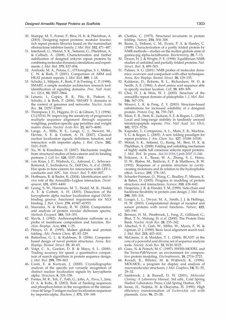
1303Designed Armadillo Repeat Proteins as Scaffolds
30. Stumpp, M. T., Forrer, P., Binz, H. K. & Plückthun, A.(2003). Designing repeat proteins: modular leucine-rich repeat protein libraries based on the mammalianribonuclease inhibitor family. J. Mol. Biol. 332, 471–487.
31. Interlandi, G., Wetzel, S. K., Settanni, G., Plückthun, A.& Caflisch, A. (2008). Characterization and furtherstabilization of designed ankyrin repeat proteins bycombiningmolecular dynamics simulations and experi-ments. J. Mol. Biol. 375, 837–854.
32. Andrade, M. A., Petosa, C., O'Donoghue, S. I., Müller,C. W. & Bork, P. (2001). Comparison of ARM andHEAT protein repeats. J. Mol. Biol. 309, 1–18.
33. Schultz, J., Milpetz, F., Bork, P. & Ponting, C. P. (1998).SMART, a simple modular architecture research tool:identification of signaling domains. Proc. Natl Acad.Sci. USA, 95, 5857–5864.
34. Letunic, I., Copley, R. R., Pils, B., Pinkert, S.,Schultz, J. & Bork, P. (2006). SMART 5: domains inthe context of genomes and networks. Nucleic AcidsRes. 34, D257–D260.
35. Thompson, J. D., Higgins, D. G. & Gibson, T. J. (1994).CLUSTALW: improving the sensitivity of progressivemultiple sequence alignment through sequenceweighting, position-specific gap penalties and weightmatrix choice. Nucleic Acids Res. 22, 4673–4680.
36. Lange, A., Mills, R. E., Lange, C. J., Stewart, M.,Devine, S. E. & Corbett, A. H. (2007). Classicalnuclear localization signals: definition, function, andinteraction with importin alpha. J. Biol. Chem. 282,5101–5105.
37. Xu, W. & Kimelman, D. (2007). Mechanistic insightsfrom structural studies of beta-catenin and its bindingpartners. J. Cell Sci. 120, 3337–3344.
38. von Kries, J. P., Winbeck, G., Asbrand, C., Schwarz-Romond, T., Sochnikova, N., Dell'Oro, A. et al. (2000).Hot spots in beta-catenin for interactions with LEF-1,conductin and APC. Nat. Struct. Biol. 7, 800–807.
39. Hoffmans, R. & Basler, K. (2004). Identification and invivo role of the Armadillo–Legless interaction. Devel-opment, 131, 4393–4400.
40. Leung, S. W., Harreman, M. T., Hodel, M. R., Hodel,A. E. & Corbett, A. H. (2003). Dissection of thekaryopherin alpha nuclear localization signal (NLS)-binding groove: functional requirements for NLSbinding. J. Biol. Chem. 278, 41947–41953.
41. Sreerama, N. & Woody, R. W. (2004). Computationand analysis of protein circular dichroism spectra.Methods Enzymol. 383, 318–351.
42. Slavik, J. (1982). Anilinonaphthalene sulfonate as aprobe of membrane composition and function. Bio-chim. Biophys. Acta, 694, 1–25.
43. Ptitsyn, O. B. (1995). Molten globule and proteinfolding. Adv. Protein Chem. 47, 83–229.
44. Butterfoss, G. L. & Kuhlman, B. (2006). Computer-based design of novel protein structures. Annu. Rev.Biophys. Biomol. Struct. 35, 49–65.
45. Voigt, C. A., Gordon, D. B. & Mayo, S. L. (2000).Trading accuracy for speed: a quantitative compar-ison of search algorithms in protein sequence design.J. Mol. Biol. 299, 789–803.
46. Conti, E. & Kuriyan, J. (2000). Crystallographicanalysis of the specific yet versatile recognition ofdistinct nuclear localization signals by karyopherinalpha. Structure, 8, 329–338.
47. Fontes, M. R., Teh, T., Toth, G., John, A., Pavo, I., Jans,D. A. & Kobe, B. (2003). Role of flanking sequencesand phosphorylation in the recognition of the simian-virus-40 large T-antigen nuclear localization sequencesby importin-alpha. Biochem. J. 375, 339–349.
48. Chothia, C. (1975). Structural invariants in proteinfolding. Nature, 254, 304–308.
49. Baum, J., Dobson, C. M., Evans, P. A. & Hanley, C.(1989). Characterization of a partly folded protein byNMRmethods—studies on the molten globule state ofguinea-pig alpha-lactalbumin. Biochemistry, 28, 7–13.
50. Dyson, H. J. & Wright, P. E. (1998). Equilibrium NMRstudies of unfolded and partially folded proteins. Nat.Struct. Biol. 5, 499–503.
51. Palmer, A. G. (2001). NMR probes of molecular dyna-mics: overview and comparison with other techniques.Annu. Rev. Biophys. Biomol. Struct. 30, 129–155.
52. Kalderon, D., Roberts, B. L., Richardson, W. D. &Smith, A. E. (1984). A short amino acid sequence ableto specify nuclear location. Cell, 39, 499–509.
53. Choi, H. J. & Weis, W. I. (2005). Structure of thearmadillo repeat domain of plakophilin 1. J. Mol. Biol.346, 367–376.
54. Mosavi, L. K. & Peng, Z. Y. (2003). Structure-basedsubstitutions for increased solubility of a designedprotein. Protein Eng. 16, 739–745.
55. Main, E. R., Stott, K., Jackson, S. E. & Regan, L. (2005).Local and long-range stability in tandemly arrayedtetratricopeptide repeats. Proc. Natl Acad. Sci. USA,102, 5721–5726.
56. Kajander, T., Cortajarena, A. L., Main, E. R., Mochrie,S. G. & Regan, L. (2005). A new folding paradigm forrepeat proteins. J. Am. Chem. Soc. 127, 10188–10190.
57. Wetzel, S. K., Settanni, G., Kenig, M., Binz, H. K. &Plückthun, A. (2008). Folding and unfolding mechanismof highly stable full consensus ankyrin repeat proteins.J. Mol. Biol. In press. doi:10.1016/j.jmb.2007.11.046
58. Eriksson, A. E., Baase, W. A., Zhang, X. J., Heinz,D. W., Blaber, M., Baldwin, E. P. & Matthews, B. W.(1992). Response of a protein structure to cavity-creating mutations and its relation to the hydrophobiceffect. Science, 255, 178–183.
59. Schueler-Furman, O., Wang, C., Bradley, P., Misura, K.& Baker, D. (2005). Progress in modeling of proteinstructures and interactions. Science, 310, 638–642.
60. Desjarlais, J. R. & Handel, T. M. (1999). Side-chain andbackbone flexibility in protein core design. J. Mol. Biol.290, 305–318.
61. Looger, L. L., Dwyer, M. A., Smith, J. J. & Hellinga,H. W. (2003). Computational design of receptor andsensor proteins with novel functions. Nature, 423,185–190.
62. Berman, H. M., Westbrook, J., Feng, Z., Gilliland, G.,Bhat, T. N., Weissig, H. et al. (2000). The Protein DataBank. Nucleic Acids Res. 28, 235–242.
63. Altschul, S. F., Gish, W., Miller, W., Myers, E. W. &Lipman, D. J. (1990). Basic local alignment search tool.J. Mol. Biol. 215, 403–410.
64. McGinnis, S. & Madden, T. L. (2004). BLAST: at thecore of a powerful and diverse set of sequence analysistools. Nucleic Acids Res. 32, W20–W25.
65. Guex, N. & Peitsch, M. C. (1997). SWISS-MODEL andthe Swiss-PdbViewer: an environment for compara-tive protein modeling. Electrophoresis, 18, 2714–2723.
66. Koradi, R., Billeter, M. & Wüthrich, K. (1996).MOLMOL: a program for display and analysis ofmacromolecular structures. J. Mol. Graphics, 14, 51–55,29–32.
67. Sambrook, J. & Russell, D. W. (2001). MolecularCloning: A Laboratory Manual, 3rd edit., Cold SpringHarbor Laboratory Press, Cold Spring Harbor, NY.
68. Inoue, H., Nojima, H. & Okayama, H. (1990). Highefficiency transformation of Escherichia coli withplasmids. Gene, 96, 23–28.

1304 Designed Armadillo Repeat Proteins as Scaffolds
69. Köhler, M., Speck, C., Christiansen, M., Bischoff, F. R.,Prehn, S., Haller, H. et al. (1999). Evidence for distinctsubstrate specificities of importin alpha family mem-bers in nuclear protein import. Mol. Cell. Biol. 19,7782–7791.
70. Cull, M. G. & Schatz, P. J. (2000). Biotinylation ofproteins in vivo and in vitro using small peptide tags.Methods Enzymol. 326, 430–440.
71. Brooks, B. R., Bruccoleri, R. E., Olafson, B. D., States,D. J., Swaminathan, S. & Karplus, M. (1983).CHARMM—a program for macromolecular energy,minimization, and dynamics calculations. J. Comput.Chem. 4, 187–217.
72. Bodenhausen, G. & Ruben, D. J. (1980). Naturalabundance N-15 NMR by enhanced heteronuclearspectroscopy. Chem. Phys. Lett. 69, 185–189.
73. Keeler, J., Clowes, R. T., Davis, A. L. & Laue, E. D.(1994). Pulsed-field gradients: theory and practice.Methods Enzymol. 239, 145–207.
74. Kay, L. E., Keifer, P. & Saarien, T. (1992). Pureabsorption gradient enhanced heteronuclear single-quantum correlation spectroscopy with improvedsensitivity. J. Am. Chem. Soc. 114, 10663–10665.
75. Noggle, J. H. & Schirmer, R. E. (1971). The NuclearOverhauser Effect: Chemical Applications, AcademicPress, New York.
Top Related
Copyright © 2022 FDOKUMEN

