


Bahasa
Halaman
Hukum

Diagnostics 2021, 11, 1373. https://doi.org/10.3390/diagnostics11081373 www.mdpi.com/journal/diagnostics
Review
Deep Learning Application for Analyzing of Constituents
and Their Correlations in the Interpretations of Medical Images
Tudor Florin Ursuleanu 1,2,3, Andreea Roxana Luca 1,4,*, Liliana Gheorghe 1,5,*, Roxana Grigorovici 1, Stefan Iancu 1,
Maria Hlusneac 1, Cristina Preda 1,6 and Alexandru Grigorovici 1,2
1 Faculty of General Medicine, “Grigore T. Popa” University of Medicine and Pharmacy,
700115 Iasi, Romania; [email protected] (T.F.U.); [email protected] (R.G.);
[email protected] (S.I.); [email protected] (M.H.); [email protected] (C.P.);
[email protected] (A.G.) 2 Department of Surgery VI, “Sf. Spiridon” Hospital, 700111 Iasi, Romania 3 Department of Surgery I, Regional Institute of Oncology, 700483 Iasi, Romania 4 Department Obstetrics and Gynecology, Integrated Ambulatory of Hospital “Sf. Spiridon”,
700106 Iasi, Romania 5 Department of Radiology, “Sf. Spiridon” Hospital, 700111 Iasi, Romania 6 Department of Endocrinology, “Sf. Spiridon” Hospital, 700111 Iasi, Romania
* Correspondence: [email protected] (A.R.L.); [email protected] (L.G.)
Abstract: The need for time and attention, given by the doctor to the patient, due to the increased
volume of medical data to be interpreted and filtered for diagnostic and therapeutic purposes has
encouraged the development of the option to support, constructively and effectively, deep learning
models. Deep learning (DL) has experienced an exponential development in recent years, with a
major impact on interpretations of the medical image. This has influenced the development, diver-
sification and increase of the quality of scientific data, the development of knowledge construction
methods and the improvement of DL models used in medical applications. All research papers fo-
cus on description, highlighting, classification of one of the constituent elements of deep learning
models (DL), used in the interpretation of medical images and do not provide a unified picture of
the importance and impact of each constituent in the performance of DL models. The novelty in our
paper consists primarily in the unitary approach, of the constituent elements of DL models, namely,
data, tools used by DL architectures or specifically constructed DL architecture combinations and
highlighting their “key” features, for completion of tasks in current applications in the interpreta-
tion of medical images. The use of “key” characteristics specific to each constituent of DL models
and the correct determination of their correlations, may be the subject of future research, with the
aim of increasing the performance of DL models in the interpretation of medical images.
Keywords: medical image analysis; types of data and datasets; methods of incorporating
knowledge; deep learning models; applications in medicine
1. Introduction
The performance of deep learning architectures (DL) has a continuously improved
by increasing the number and quality, respectively diversification data resources similar
to medical data, developing specific methods of integrating data into DL models accord-
ing to the objectives for which they were built and perfecting the construction of DL mod-
els used in medical applications.
Deep learning (DL) has experienced an exponential development of medicine, but
applications in interpretations of medical imaging are in continuous development. DL has
managed to achieve performance in diagnosis, classification, detection, segmentation, re-
construction of medical images [1] but also in achieving the correlation between image
diagnosis and patient survival, predicting new directions of development [2].
Citation: Ursuleanu, T.F.; Luca, A.R.;
Gheorghe, L.; Grigorovici, R.;
Iancu, S.; Hlusneac, M.; Preda, C.;
Grigorovici, A. Deep Learning
Application for Analyzing
of Constituents and Their
Correlations in the Interpretations
of Medical Images. Diagnostics 2021,
11, 1373. https://doi.org/10.3390/
diagnostics11081373
Academic Editor: Sameer Antani
Received: 7 July 2021
Accepted: 27 July 2021
Published: 30 July 2021
Publisher’s Note: MDPI stays neu-
tral with regard to jurisdictional
claims in published maps and institu-
tional affiliations.
Copyright: © 2021 by the authors. Li-
censee MDPI, Basel, Switzerland.
This article is an open access article
distributed under the terms and con-
ditions of the Creative Commons At-
tribution (CC BY) license (http://crea-
tivecommons.org/licenses/by/4.0/).
Diagnostics 2021, 11, 1373 2 of 50
The novelty in our paper consists in the unitary approach, of the constituent elements
of DL models, namely, data, tools used by DL architectures or specifically constructed DL
architecture combinations and highlighting their “key” features, for completion of tasks
in current applications in the interpretation of medical images.
In this article we present in primarily, a unitary, complete, up-to-date analysis of sci-
entific data, methods of knowledge incorporation, a classification and description of DL
models according to the structure and objectives for which they were designed and
presentation of medical applications according to these tasks. Secondly, it describes the
specific correlations between data, data integration methods, deep learning models used
in the interpretation of diagnostic medical images and their applications in medicine. Fi-
nally presents problems and future challenges.
The structure is composed of Section 2 describes types of images, medical data used
by deep learning architectures, Section 3 describes DL models according to the objectives
for which they were created, medical application, associating the types of data, Section 4
methods of incorporating images, information and medical data, in addition to the objec-
tive of DL. Section 5 contributions of the methods of incorporating images, information
and medical data in medical applications, Section 6 research issues and future challenges.
Methodology:
We have identified and selected significant research papers published in 2009–2020,
mainly from 2016 and 2020, with some papers from 2021. We focus on papers from the
most reputable publishers, such as IEEE, Elsevier, Springer, MDPI, Nature, SPIE, PLOS,
Wiley, RSNA, SCIRP. Some works have been selected from arXiv. I have reviewed more
than 273 papers on different DL topics. There are 17 works from 2021, 56 works from 2020,
56 works from 2019, 38 works from 2018, 58 works from 2017, 25 works from 2016 and 10
work from 2015. This indicates that this review focus on the latest publications in the field
of DL. The selected papers have been analyzed and reviewed for: descriptions types of
images, medical data used by deep learning architectures (Section 2), descriptions DL
models according to the objectives for which they were created, medical application, as-
sociating the types of data (Section 3), methods of incorporating images, information and
medical data, in addition to the objective of DL (Section 4), contributions of the methods
of incorporating images, information and medical data in medical applications (Section
5), research issues and future challenges (Section 6). Most keywords used for search crite-
ria for this review work are (Deep Learning and Data types), (Deep Learning and Data
Sets), (Deep Learning and Methods of Incorporation of Medical Knowledge and Data),
(Deep Learning Models and Models), (Deep Learning and Architectures), ((Deep Learn-
ing) and (Medical Image Analysis) and (Detection/Classification/Segmentation/Localiza-
tion/Reconstruction/Recovery)), (Deep Learning and Detection/Classification/Segmenta-
tion/Localization/Reconstruction), (Deep Learning and Images and Applications in Med-
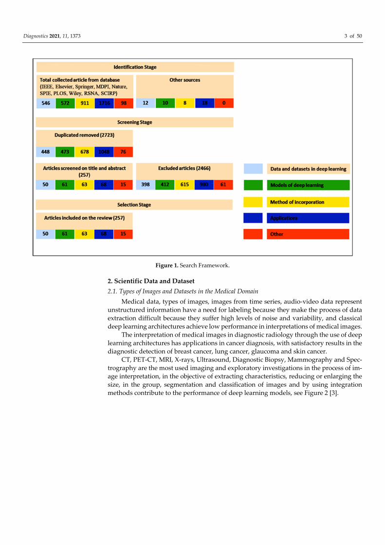
icine), (Deep Learning and Interpretation Medical Images). Figure 1 shows our search
structure of the survey paper.
Diagnostics 2021, 11, 1373 3 of 50
Figure 1. Search Framework.
2. Scientific Data and Dataset
2.1. Types of Images and Datasets in the Medical Domain
Medical data, types of images, images from time series, audio-video data represent
unstructured information have a need for labeling because they make the process of data
extraction difficult because they suffer high levels of noise and variability, and classical
deep learning architectures achieve low performance in interpretations of medical images.
The interpretation of medical images in diagnostic radiology through the use of deep
learning architectures has applications in cancer diagnosis, with satisfactory results in the
diagnostic detection of breast cancer, lung cancer, glaucoma and skin cancer.
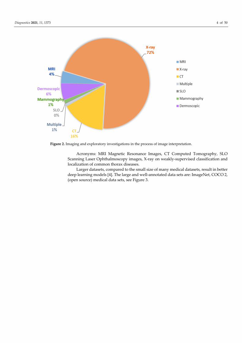
CT, PET-CT, MRI, X-rays, Ultrasound, Diagnostic Biopsy, Mammography and Spec-
trography are the most used imaging and exploratory investigations in the process of im-
age interpretation, in the objective of extracting characteristics, reducing or enlarging the
size, in the group, segmentation and classification of images and by using integration
methods contribute to the performance of deep learning models, see Figure 2 [3].
Diagnostics 2021, 11, 1373 4 of 50
Figure 2. Imaging and exploratory investigations in the process of image interpretation.
Acronyms: MRI Magnetic Resonance Images, CT Computed Tomography, SLO
Scanning Laser Ophthalmoscopy images, X-ray on weakly-supervised classification and
localization of common thorax diseases.
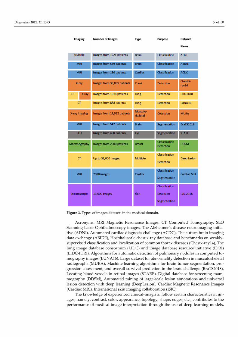
Larger datasets, compared to the small size of many medical datasets, result in better
deep learning models [4]. The large and well-annotated data sets are: ImageNet, COCO 2,
(open source) medical data sets, see Figure 3.
Diagnostics 2021, 11, 1373 5 of 50
Figure 3. Types of images datasets in the medical domain.
Acronyms: MRI Magnetic Resonance Images, CT Computed Tomography, SLO
Scanning Laser Ophthalmoscopy images, The Alzheimer’s disease neuroimaging initia-
tive (ADNI), Automated cardiac diagnosis challenge (ACDC), The autism brain imaging
data exchange (ABIDE), Hospital-scale chest x-ray database and benchmarks on weakly-
supervised classification and localization of common thorax diseases (Chestx-ray14), The
lung image database consortium (LIDC) and image database resource initiative (IDRI)
(LIDC-IDRI), Algorithms for automatic detection of pulmonary nodules in computed to-
mography images (LUNA16), Large dataset for abnormality detection in musculoskeletal
radiographs (MURA), Machine learning algorithms for brain tumor segmentation, pro-
gression assessment, and overall survival prediction in the brats challenge (BraTS2018),
Locating blood vessels in retinal images (STARE), Digital database for screening mam-
mography (DDSM), Automated mining of large-scale lesion annotations and universal
lesion detection with deep learning (DeepLesion), Cardiac Magnetic Resonance Images
(Cardiac MRI), International skin imaging collaboration (ISIC).
The knowledge of experienced clinical-imagists, follow certain characteristics in im-
ages, namely, contrast, color, appearance, topology, shape, edges, etc., contributes to the
performance of medical image interpretation through the use of deep learning models,

Diagnostics 2021, 11, 1373 6 of 50
namely, anomaly detection by identifying the characteristics in the image; image segmen-
tation; image reconstruction; combining two different images into one [5].
The knowledge of imaging doctors can be classified as follows:
1. Low-level medical data
Areas of attention of physicians in medical images [6],
Disease characteristics [7],
2. High-level medical data
Labels–Diagnostic pattern [8],
3. Diagnostic training model that represents specific data identified by doctors [9].
The type and volume of medical data, the labels, the category of field knowledge and
the methods of their integration into the DL architectures implicitly determine their per-
formance in medical applications.
2.2. Types of Images and Medical Data Used for Diagnosis–Classification of Diseases in Medical
Images
We will further expose, the types of medical images and data used in diagnosis-clas-
sification, segmentation, detection, reconstruction, recovery and, respectively, the gener-
ation of medical reports.
Natural images–from natural datasets, ImageNet 1 (over 14 million images tagged in
20 k categories) and COCO 2 (with over 200 images annotated in 80 categories).
Medical images-from medical datasets of the same diseases in similar and different
ways or from different diseases [10].
High-level medical data (diagnostic pattern), low-level medical data (areas of images,
disease characteristics).
Specific data identified by doctors (attention maps, hand-highlighted features) in-
crease the diagnostic performance of deep learning networks (no comparative studies
have been conducted).
2.3. Types of Images and Medical Data Used for Diagnosis Detection of Lesions and
Abnormalities in Medical Images
Large natural images (ImageNet) are incorporated for the detection of characteristics
in the medical images. Natural images are used in multiple applications.
Medical images are used in multiple applications. Multi-modal medical images, PET
images are incorporated for the detection of lesions in CT scans.
High-level medical data (diagnostic pattern), low-level medical data (areas of images,
disease characteristics).
Specific data identified by doctors (attention maps, hand-highlighted features) in-
crease the diagnostic performance of deep learning networks (no comparative studies
have been carried out).
2.4. Types of Images and Medical Data Used for Diagnosis–Segmentation into Medical Images
Natural Images, ImageNet, PASCAL VOC “static data” set, Sports-1M video datasets
[11].
Medical images, (CT, MRI, Angio-CT, butt eye images, annotated retinal images)
used in multiple applications.
External medical data and images of other diseases, dataset 3DSeg-8 [12].
High-level and low-level medical data, e.g., anatomical aspects of the image, shape,
position, typology of lesions integrated into segmentation tasks, example of the ISBI 2017
dataset used in skin injury segmentation. Many applications use additional data with sat-
isfactory results to improve CT image segmentation tasks in order to improve applications
for MRI use [13].
Diagnostics 2021, 11, 1373 7 of 50
Medical data from doctors, hand-made features, hand-highlighted features, are first
processed from the reference images. These features are used in the BRATS2015 dataset
in input-level merging image segmentation applications.
2.5. Medical Data and Manual Features Used for Image Reconstruction
X-ray projections in CT or spatial frequency information in MRI) [14,15], image re-
construction with optical diffuse tomography (DOT), reconstruction of magnetic reso-
nance imaging by compressed detection (CS-MRI) [16], reconstruction of the image with
diffuse optical tomography (DOT) of limited-angle breast cancer and limited sources in a
strong scattering environment [17,18], recovery of brain MRI images, target contrast using
GAN [19] are methods based on deep learning have been widely applied in this area.
2.6. Medical Data and Manual Features Used for Image Recovery
Knowledge from natural images, medical datasets for example, age and sex of pa-
tients, characteristics extracted from health areas.
2.7. Medical Data Used to Generate Medical Reports
Subtitling medical images, templates from radiologist reports, visual characteristics
of medical images, generating reports using the IU-RR dataset.
3. DL Models Description and Classification According to the Tasks in Medical
Images Analyses
We will describe the deep learning architectures in relation to the purpose and tasks
for which they were designed, namely, diagnosis-classification, detection, segmentation,
reconstruction.
3.1. DL Architectures Designed for Diagnosis–Classification in Medical Images
CNN, AlexNet, GoogLeNet, VGGNet, ResNet, DenseNet are used for diagnosis, clas-
sification, diseases.
GoogLeNet, VGGNet, ResNet are used for diagnosis, classification of superficial and
deep corneal ulcers with accuracy of over 90%.
DenseNet [20] used for diagnostic classification of lung nodules on X-Rey with accu-
racy of over 90% Architectures designed to detect objects in natural images used to detect
objects in medical images.
3.2. DL Architectures Designed for Diagnosis Detection of Lesions, Abnormalities in Medical
Images
Two-stage models for injury and organ detection consist of a network of regional
proposals (RPN) that involves the locations of candidate objects and a detection network
that selects regional proposals are Faster R-CNN [21] and Mask R-CNN [18,22].
Models with a faster and simpler stage, which go over the stage of the proposal of
the region and run the detection directly, taking into account the probability that the object
will appear at every point in the image such as YOLO (You Only Look Once) [23], SSD
(Single Shot MultiBox Detector) [9] and RetinaNet [24].
Combined FCN and GAN architectures, through PET images are generated first from
CT scans then synthesized PET images are used in a false positive reduction layer [18,25].
3.3. DL Architectures Designed for Diagnosis Segmentation of Medical Images
Three categories can be exemplified: FCN-based models [26]; U-Net-based models
[27]; GAN-based models [28].
Diagnostics 2021, 11, 1373 8 of 50
3.3.1. FCN Achieves Goals of Segmenting the Medical Image with Good Results
Types of FCN: Cascading FCN [29,30], parallel FCN [31]and recurrent FCN [32] also
achieve medical image segmentation goals with good results.
3.3.2. U-Net-Based Models
U-Net [27] and its derivatives segment the medical image with good results. U-Net
is based on the FCN structure, consisting of a series of convolutional and devolutionary
layers and with short connections between equal resolution layers. U-Net and its variants
such as UNet ++ [33] and recurrent U-Net [34] perform well in many medical image seg-
mentation tasks [18,35].
3.3.3. GAN-Based Models
GAN is a type of mixed architecture (supervised and unsupervised) called semi-su-
pervised architecture, an architecture composed of two neural networks, a generator and
a discriminator or classifier, which compete with each other in a contradictory formation
process [28]. In models, the generator is used to predict the target mask based on encoder-
decoder structures (such as FCN or U-Net) [18]. The discriminator serves as a form regu-
lator that helps the generator achieve satisfactory segmentation results [16,33]. GAN has
use in the generation of synthetic instances of different classes.
3.4. DL Architectures Designed for Diagnosis, Classification, Segmentation, Detection and
Reconstruction of Medical Images
Deep auto-encoders (AUD) are included in the type of unsupervised learning that
uses unlabeled input data, there is no a priori knowledge, and the results to be obtained
from the processing of input data are unknown, and can learn to organize information
without providing an error calculation to evaluate the possible solution [36,37]. The main
feature of the autoencoder is represented by the input and output layers have the same
size, and the output must reproduce the input, while the hidden layers are smaller in size
because the input patterns are progressively encoded and decoded throughout the pro-
cess, and has the ability to extract the fundamental characteristics of the input, being used
to reduce the size of the data, but also to reduce noise in input data (such as images). They
are often used for data reconstruction (image and signal), denoising or augmentation
[37,38].
3.5. Medical Applications of DL Models According to the Scope for Which They Were Used,
Classification, Segmentation, Detection and Reconstruction of Medical Images
DL architectures, e.g., CNN, U-Net, ResNet, VGGNet, AlexNet, RNN, GAN, DBN,
YOLO and respectively, the types of combined architectures VGGNet + CNN, CNN +
LSTM, GAN + U-Net, VGGNet + U-Net, RCC + U-Net which have as tasks classification,
segmentation, detectionand reconstruction of medical images are the most used and have
the best performance and contribution to medical applications (see Table 1) [39].
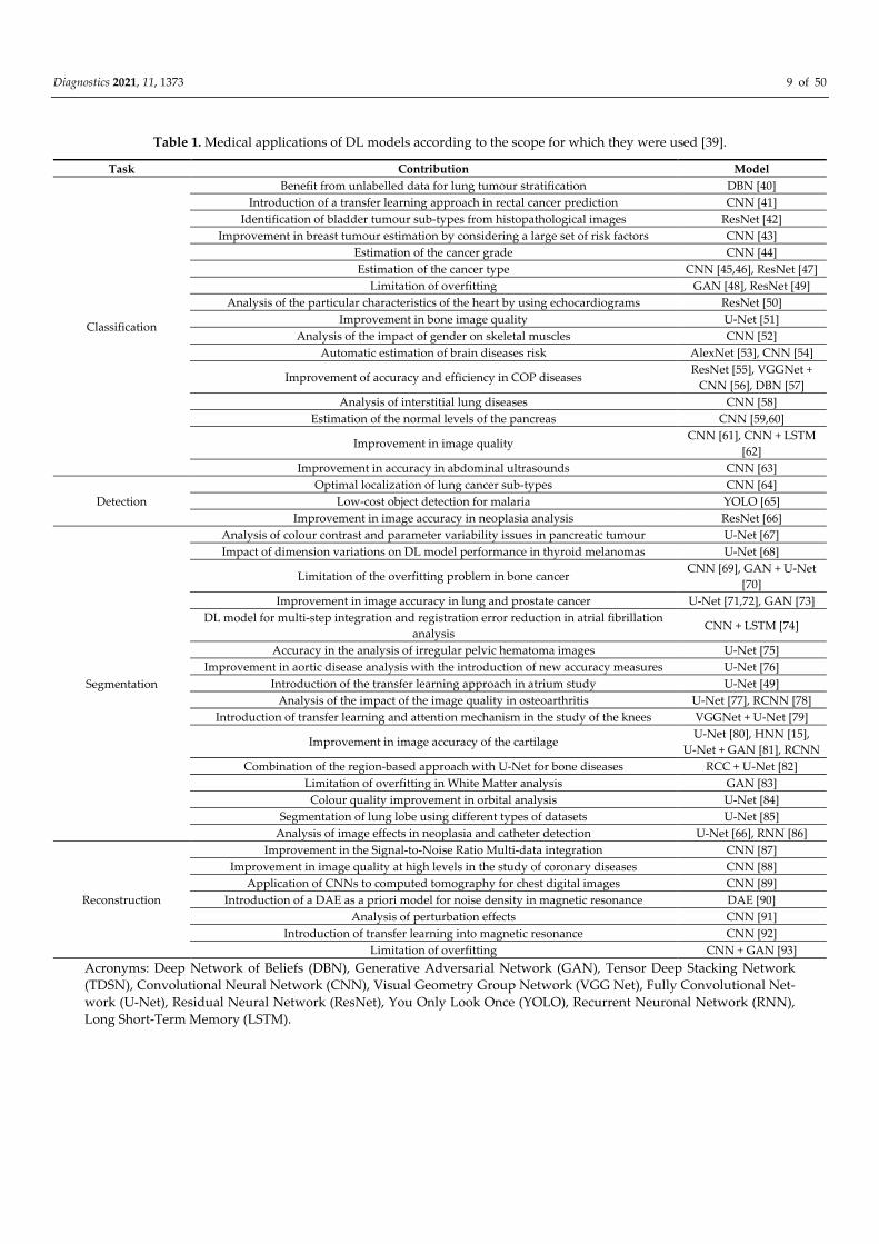
Diagnostics 2021, 11, 1373 9 of 50
Table 1. Medical applications of DL models according to the scope for which they were used [39].
Task Contribution Model
Classification
Benefit from unlabelled data for lung tumour stratification DBN [40]
Introduction of a transfer learning approach in rectal cancer prediction CNN [41]
Identification of bladder tumour sub-types from histopathological images ResNet [42]
Improvement in breast tumour estimation by considering a large set of risk factors CNN [43]
Estimation of the cancer grade CNN [44]
Estimation of the cancer type CNN [45,46], ResNet [47]
Limitation of overfitting GAN [48], ResNet [49]
Analysis of the particular characteristics of the heart by using echocardiograms ResNet [50]
Improvement in bone image quality U-Net [51]
Analysis of the impact of gender on skeletal muscles CNN [52]
Automatic estimation of brain diseases risk AlexNet [53], CNN [54]
Improvement of accuracy and efficiency in COP diseases ResNet [55], VGGNet +
CNN [56], DBN [57]
Analysis of interstitial lung diseases CNN [58]
Estimation of the normal levels of the pancreas CNN [59,60]
Improvement in image quality CNN [61], CNN + LSTM
[62]
Improvement in accuracy in abdominal ultrasounds CNN [63]
Detection
Optimal localization of lung cancer sub-types CNN [64]
Low-cost object detection for malaria YOLO [65]
Improvement in image accuracy in neoplasia analysis ResNet [66]
Segmentation
Analysis of colour contrast and parameter variability issues in pancreatic tumour U-Net [67]
Impact of dimension variations on DL model performance in thyroid melanomas U-Net [68]
Limitation of the overfitting problem in bone cancer CNN [69], GAN + U-Net
[70]
Improvement in image accuracy in lung and prostate cancer U-Net [71,72], GAN [73]
DL model for multi-step integration and registration error reduction in atrial fibrillation
analysis CNN + LSTM [74]
Accuracy in the analysis of irregular pelvic hematoma images U-Net [75]
Improvement in aortic disease analysis with the introduction of new accuracy measures U-Net [76]
Introduction of the transfer learning approach in atrium study U-Net [49]
Analysis of the impact of the image quality in osteoarthritis U-Net [77], RCNN [78]
Introduction of transfer learning and attention mechanism in the study of the knees VGGNet + U-Net [79]
Improvement in image accuracy of the cartilage U-Net [80], HNN [15],
U-Net + GAN [81], RCNN
Combination of the region-based approach with U-Net for bone diseases RCC + U-Net [82]
Limitation of overfitting in White Matter analysis GAN [83]
Colour quality improvement in orbital analysis U-Net [84]
Segmentation of lung lobe using different types of datasets U-Net [85]
Analysis of image effects in neoplasia and catheter detection U-Net [66], RNN [86]
Reconstruction
Improvement in the Signal-to-Noise Ratio Multi-data integration CNN [87]
Improvement in image quality at high levels in the study of coronary diseases CNN [88]
Application of CNNs to computed tomography for chest digital images CNN [89]
Introduction of a DAE as a priori model for noise density in magnetic resonance DAE [90]
Analysis of perturbation effects CNN [91]
Introduction of transfer learning into magnetic resonance CNN [92]
Limitation of overfitting CNN + GAN [93]
Acronyms: Deep Network of Beliefs (DBN), Generative Adversarial Network (GAN), Tensor Deep Stacking Network
(TDSN), Convolutional Neural Network (CNN), Visual Geometry Group Network (VGG Net), Fully Convolutional Net-
work (U-Net), Residual Neural Network (ResNet), You Only Look Once (YOLO), Recurrent Neuronal Network (RNN),
Long Short-Term Memory (LSTM).
Diagnostics 2021, 11, 1373 10 of 50
4. DL Model Description and Classification According to Medical Data Types Used,
Objectives and Performances in Medical Applications
4.1. DL Models According to the Characteristics and Tasks for Which They Were Designed
CNN (convolutional neural network) are popular in areas where the shape of an ob-
ject is an important feature, such as image analysis [5,39,94,95], particularly in the study
of cancers and bodily injuries in the medical sector [96,97] and video analysis [39,98].
CNN contains convolutive layers, grouping layers, dropout layers, and an output
layer, hierarchically positioned that each learn stun specific characteristics in the image
[99].
CNN in image analysis has low performance when high-resolution datasets are con-
sidered [100] and when localization over large patches is required, especially in medical
images [101,102].
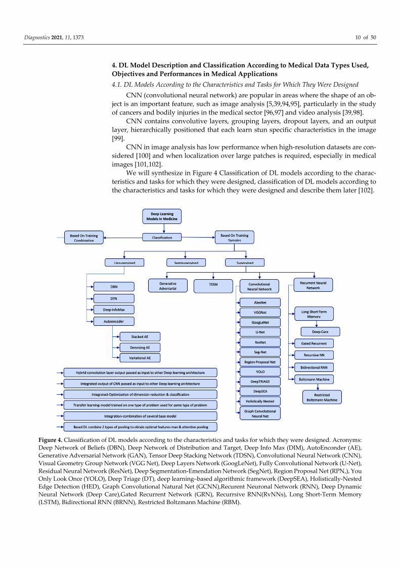
We will synthesize in Figure 4 Classification of DL models according to the charac-
teristics and tasks for which they were designed, classification of DL models according to
the characteristics and tasks for which they were designed and describe them later [102].
Figure 4. Classification of DL models according to the characteristics and tasks for which they were designed. Acronyms:
Deep Network of Beliefs (DBN), Deep Network of Distribution and Target, Deep Info Max (DIM), AutoEnconder (AE),
Generative Adversarial Network (GAN), Tensor Deep Stacking Network (TDSN), Convolutional Neural Network (CNN),
Visual Geometry Group Network (VGG Net), Deep Layers Network (GoogLeNet), Fully Convolutional Network (U-Net),
Residual Neural Network (ResNet), Deep Segmentation-Emendation Network (SegNet), Region Proposal Net (RPN,), You
Only Look Once (YOLO), Deep Triage (DT), deep learning–based algorithmic framework (DeepSEA), Holistically-Nested
Edge Detection (HED), Graph Convolutional Natural Net (GCNN),Recurent Neuronal Network (RNN), Deep Dynamic
Neural Network (Deep Care),Gated Recurrent Network (GRN), Recurrsive RNN(RvNNs), Long Short-Term Memory
(LSTM), Bidirectional RNN (BRNN), Restricted Boltzmann Machine (RBM).

Diagnostics 2021, 11, 1373 11 of 50
DL architectures classification [103]:
Supervised DL models:
Recurrent Neural Networks (RNN), Long short-term memory (LSTM), Gated Recur-
rent Unit (GRU),
Convolutional Neural Network (CNN)
Generative Adversarial Network (GAN).
Unsupervised deep learning models:
Deep Network of Beliefs (DBN),
Deep Transfer Network (DTN),
Tensor Deep Stack Networks (TDSN),
Autoencoders (AE).
CNN’s performance is strongly influenced by the selection of hyper-parameters. Any
small changes in hyper-parameters will affect CNN’s overall performance. Therefore,
careful selection of parameters is an extremely significant problem that should be taken
into account during the development of the optimisation scheme.
Impressive and robust hardware resources, such as GPs, are needed for an effective
CNN workout. Moreover, they are also needed to explore the effectiveness of using CNN
in intelligent and embedded systems.
Exploitation of depth and various structural adaptations is significantly improved in
CNN’s learning capacity. Replacing the traditional layer configuration with blocks leads
to significant progress in CNN’s performance, as shown in recent literature. Today, the
development of new and efficient block architectures is the main trend in the new research
models of CNN architectures. HRNet is just one example that shows that there are always
ways to improve the architecture. Cloud-based platforms are expected to play a key role
in the future development of DL computing applications [104].
Several deep learning, computer assisted diagnosis (CAD) systems for digital breast
tomosynthesis (DBT) are currently available and many new systems will be developed.
However, there are still many challenges to overcome. As Wang et al., [105] have recently
demonstrated, published models for the full-field digital mammography (FFDM) classifi-
cation fail when applied to different datasets, even when these data sets include purchases
using similar equipment. For FFDMs, deep learning-based detection models have proven
to be performing with almost human precision [106]. As more studies and data become
available, there is no reason to believe that this should be different for DBT. However, the
trained radiologist can adapt when analyzing different data sets, indicating that high-per-
formance deep learning models still lack the “key” characteristics that differentiate the
disease from normal [107].
Image analysis performance is enhanced by the use of the following architectures:
AlexNet, VGGNet and ResNet, YOLO or U-net that we describe below:
AlexNet was proposed by Krizhevsky et al. [97] for the ImageNet Large Scale Visual
Recognition Challenge (ILSVRC) in 2012 [39].
AlexNet [103] consists of 8 layers, 5 layers of convolution and 3 dense, fully connected
layers, overlapping overlay, abandonment, data augmentation, ReLU activations after
each convolutive layer and fully connected, SGD with impulse [97]. AlexNet is used for
image recognition in image analysis and is usually applied to issues involving semantic
segmentation and high-resolution data classification tasks [39,70,73].
VGG (Visual Geometry Group): Consists of 13 convolution layers (in VGG16) & 16
convolution layers (in VGG19), 3 dense layers, pooling and three RELU units, very small
responsive fields [108]. VGG is used for object recognition, classification of medical im-
ages [109,110] and image segmentation [18,36] VGG loses accuracy when the depth be-
comes too high.
ResNet (Residual Neural Network): Contains closed units or closed recurring units
and has a strong similarity to recent successful elements applied in RNNs [103]. ResNet is
characterized by: residual mapping, identity function, and a two-layer residual block, one
Diagnostics 2021, 11, 1373 12 of 50
layer learns from the residue, the other layer learns from the same function and has high
level of performance in image classification [111] and audio analysis tasks [39,112].
GoogLeNet is built from 22 deep LAYERS CNN and 4 million parameters and con-
tains several layer filters and stacked convolution layers [113]. It was used for batch nor-
malization, image distortions, and RMSprop [103].
U-Net, developed by Ronneberger [101], addresses the problem of locating images of
a standard CNN by extracting data features followed by reconstruction of the original
dimension through an upsampling operation. U-Net is a type of Enconder-Decoder net-
work in which the codification output belongs to the input space. U-Net is used in single-
stage segmentation and classification [114], specifically in the location of cancerous lesions
[38,115,116]. SegNet [39,117] is a U-Net variant that uses maximum grouping indices in
the upsampling step that reduces the complexity of U-Net space [118].
RNNs were developed by Rumelhart et al. [119] using with efficiency the correlations
existing between input data of a prediction problem, through which they process sequen-
tial data in relation to text analysis [84,119,120] in electronic medical records to predict
diseases [121,122] and speech recognition [123]. RnN variants are: one-way, learning from
the past and predicting the future and bidirectional that uses the future to restore the past.
RNN has the following variants: LSTM, GRU, Recursive NNs and two-way RNNs
(BiRNN). LSTMs were introduced by Hochreiter and Schmidhuber [39,103,124] and con-
sist of: the gate of oblivion that alleviates the escape and explosion gradient, the entrance
gate and the exit gate, the last two track the flow of data coming in and out of the cell.
They were used in speech recognition [45], path prediction [46] and medical diagnosis
[64], in which the authors proposed an LSTM network, called DeepCare, combining dif-
ferent types of data to identify clinical diseases.
GURs (recurrent unit gated) created by Kyunghyun Cho et al. in 2014 [48], solve the
problem of increasing the time complexity of LSTM, when large amounts of data are used.
The GRU consists of a reset gate in which it is decided how much information from the
past is transmitted in the future, and an update gate that decides how much information
from the past can be forgotten. GRU and LSTMs have similar applications especially in
speech recognition [39,125].
The two-way recurring neural network and the Boltzmann BRNNs introduced by
Schuster and Paliwal [44] are characterized by the fact that the hidden state is updated by
using past information, as in a classic RNN, and by using information related to future
moments. They were applied in handwriting and speech recognition, where they are used
to detect missing parts of a sentence in a knowledge of the other words [41,126]. BM mod-
els are a family of RNNs that are easy to implement and that reproduce many probability
distributions, BMs are used in image classification. BMs combined with other models are
used to locate objects [39,40,127]. In the classification of images, BMs are used to identify
the presence of a tumor [128]. BM models are slow and ineffective when the data size
increases exponentially due to the complete connection between neurons [129]. A re-
stricted BM was proposed in which relaxing the connections between neurons of the same
or one-way connection between neurons would solve the problem of the classic BM model
[5].
AEs, developed by Rumelhart et al. [119], consisting of encoder and decoder, with
the aim of reducing the size of the data through significant representations and learning
data characteristics for the reconstruction of outputs. They are used in applications in
medical image analysis [72,130], natural language processing [67] and video analysis [68].
Additional variants of AE that can be found in the literature are variational AE
(VAE). In a VAE, the encoder is represented by the probability density function of the
input into the feature space and, after the encoding stage, a sampling of the new data
using the PDF is added. Differently from the DAE and the SAE, a VAE is not a regularized
AE, but is part of the generation class [39].
Diagnostics 2021, 11, 1373 13 of 50
GAN it is used to generate synthetic training data from original data using latent
distribution [131]. It consisted of two networks, a generator estimates false data from in-
put data, and a discriminator, which differentiates fake data from real data and separates
it in order to increase the quality of the data generated. GAN has two problems: the prob-
lem of the collapse of the mode, and the fact that, can become very unstable [103].
DBN: The DBN (Deep Network of Beliefs), created by Hinton [132], consists of two
networks that build each other: of beliefs represented by an acyclic graph composed of
layers of stochastic binary units with weighted and respectively weighted connections,
restricted Boltzmann Machines which is a stochastic. DBNs are applied in image recogni-
tion and speech recognition, in classification to detect lesions in medical diagnosis and, in
video recognition to identify the presence of persons [133], in speech recognition to un-
derstand missing words in a sentence [134] and in application on physiological signals to
recognize human emotion [39,135,136].
DTN contains a characteristic extraction layer, which teaches a shared feature sub-
space in which marginal source distributions and target samples are drawn close and a
layer of discrimination that match conditional distributions by classified transduction
[103,106].
TDSN contains two parallel hidden representations that are combined using a bilin-
ear mapping [137]. This arrangement provides better generalization compared to the ar-
chitecture of a single module. The prejudices of the generalizers with regard to the learn-
ing set shall be inferred. It works effectively and better than an eco-validation strategy
when used with multiple generalizers compared to individual generalizers.
DIM maximizes mutual information between an input and output of a highly flexible
convolutive encoder [103,138] by forming another neural network that maximizes a lower
limit on a divergence between the marginal product of encoder input and output. Esti-
mates obtained by another network can be used to maximize the reciprocal information
of the features in the input encoder. The memory requirement of the DIM is lower because
it requires only encoder not decoder.
4.2. Combinations of Different DL Models Depending on the Type of Data Involved in the
Problem to Be Solved
DL models can be combined in five different ways depending on the type of data
involved in the problem to be solved. Of these, three types of HA (hybrid architectures),
namely the integrated model, the built-in model and the whole model.
In the integrated model, the output of the convolution layer is transmitted directly as
input to other architectures to the residual attention network, the recurrent convolutive
neural network (RCNN) and the model of the recurrent residual convolutive neural net-
work (IRRCNN) [103,139].
In the built-in model (the improved common hybrid CNN-BiLSTM), the size reduc-
tion model and the classification model perform together, the results of one represent the
inputs for the other model. In the model (EJH-CNN-BiLTM), several basic models are
combined.
In the transfer learning model (TL) is trained and uses the same type of problem.
CNN models that use the TL model are VGG (e.g., VGG16 or VGG19), GoogLeNet (e.g.,
InceptionV3), Inception Network (Inception-v4), Repiuled Neural Network (e.g., Res-
Net50), AlexNet. Joint AB based DL combines max pooling, and careful sharing [103].
Diagnostics 2021, 11, 1373 14 of 50
4.3. Combinations of Different DL Models to Benefit from the Characteristics of Each Model with
Medical Applications Are: CNN + RNN, AE + CNN and GAN + CNN
CNN + RNN are used for the capabilities of the CNN feature extraction model and
the RNNs [140]. Because the result of a CNN is a 3D value and an RNN works with 2D-
data, a remodeling layer is, associated between CNN and RNN, to convert production of
CNN into an array. CNN + RNN have been successfully applied in text analysis to identify
missing words [141] and image analysis to increase the speed of magnetic resonance im-
age storage [49,50]. CNN + RNN variants are obtained by replacing the Standard RNN
component with an LSTM component [39,48,65].
AE + CNN architecture combines AE as a pre-training model when using data with
high noise levels, and a CNN as a feature extractor model. AE + NVs have an application
in image analysis to classify noisy medical images [76] and in the reconstruction of medi-
cal images [86,130].
GAN + CNN combines GAN as a pre-workout model to moderate the problem of
over-mounting, and a CNN, used as a feature extractor. It has applications in image anal-
ysis [39,88,142].
The DL architectures applied especially in image analysis are CNN, AE and GAN.
NVs preserve the spatial structure of the data, and are used as feature extractors (espe-
cially U-Net), AEs reduce the characteristics of complex images in the analysis process,
and GANs are pre-training architectures that select input categories to control overfitting.
U-Net + Kite-Net + Attention U-Net + HarDNet-MSEG ahitecture, the DL model im-
agined by Luca, A.R. & all [143], combined model it designed takes into account the key
features of the architectures involved: U-Net will be enhanced with a block context aggre-
gation encoder and still retains the low-level image features that result from U-Net, but
will generate slightly finer segmentation without adding costs due to context aggregation
blocks; Kite-Net will contain a unit with attention gates and a Kite-Net decoder, in this
way add a benefit of attention to the details of Kite-Net; a partial decoder like the one in
the HarDNet-MSEG architecture used as the new U-Net decoder to reduce training time;
U-Net Attention that suppresses irrelevant regions, key features, does not add significant
computing costs, with a slightly smoother segmentation of image features. This combined
DL model is not demonstrated in practice being a project [143,144].
4.4. Applications in Medicine and the Performance of DL Models Depending on the Therapeutic
Areas in Which They Were Used
We further highlight the acquisitions in the study of deep learning and its applica-
tions in the analysis of the medical image [41]. You can easily identify references to image
labeling and annotation, developing new deep learning models with increased perfor-
mance, and new approaches to medical image processing:
diagnosis of cancer by using CNN with different number of layers [145],
studying deep learning optimization methods and applying in the analysis of medi-
cal images [146],
development of techniques used for endoscopic navigation [147],
highlighting the importance of data labelling and annotation and knowledge of
model performance [148,149],
perfecting the layer-wise architecture of convolution networks [103], lesson the cost
and calculation time for processor training [150],
description of the use of AI and its applications in the analysis [103] of medical im-
ages [151],
diagnosis in degenerative disorder using deep learning techniques [152] and,
detection of cancer by processing medical images using the medium change filter
technique [153],
classification of cancer using histopathological images and highlighting the rapidity
of Theano, superior tensor flow [153],
Diagnostics 2021, 11, 1373 15 of 50
development of two-channel computational algorithms using DL (segmentation, ex-
traction of characteristics, selection of characteristics and classification and classifica-
tion, extraction of high-level captures respectively) [154],
malaria detection using a deep neural network (MM-ResNet) [155].
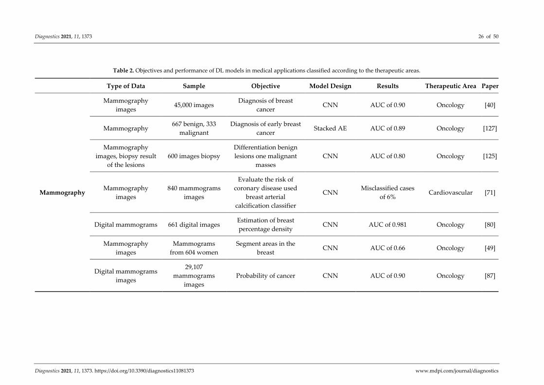
We will exemplify in Table 2 [37] applications in medicine and the performance of
DL models depending on types of medical images and the therapeutic areas in which they
were used.
Diagnostics 2021, 11, 1373 26 of 50
Diagnostics 2021, 11, 1373. https://doi.org/10.3390/diagnostics11081373 www.mdpi.com/journal/diagnostics
Table 2. Objectives and performance of DL models in medical applications classified according to the therapeutic areas.
Type of Data Sample Objective Model Design Results Therapeutic Area Paper
Mammography
Mammography
images 45,000 images
Diagnosis of breast
cancer CNN AUC of 0.90 Oncology [40]
Mammography 667 benign, 333
malignant
Diagnosis of early breast
cancer Stacked AE AUC of 0.89 Oncology [127]
Mammography
images, biopsy result
of the lesions
600 images biopsy
Differentiation benign
lesions one malignant
masses
CNN AUC of 0.80 Oncology [125]
Mammography
images
840 mammograms
images
Evaluate the risk of
coronary disease used
breast arterial
calcification classifier
CNN Misclassified cases
of 6% Cardiovascular [71]
Digital mammograms 661 digital images Estimation of breast
percentage density CNN AUC of 0.981 Oncology [80]
Mammography
images
Mammograms
from 604 women
Segment areas in the
breast CNN AUC of 0.66 Oncology [49]
Digital mammograms
images
29,107
mammograms
images
Probability of cancer CNN AUC of 0.90 Oncology [87]
Diagnostics 2021, 11, 1373 27 of 50
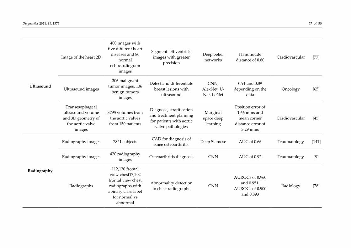
Ultrasound
Image of the heart 2D
400 images with
five different heart
diseases and 80
normal
echocardiogram
images
Segment left ventricle
images with greater
precision
Deep belief
networks
Hammoude
distance of 0.80 Cardiovascular [77]
Ultrasound images
306 malignant
tumor images, 136
benign tumors
images
Detect and differentiate
breast lesions with
ultrasound
CNN,
AlexNet, U-
Net, LeNet
0.91 and 0.89
depending on the
data
Oncology [65]
Transesophageal
ultrasound volume
and 3D geometry of
the aortic valve
images
3795 volumes from
the aortic valves
from 150 patients
Diagnose, stratification
and treatment planning
for patients with aortic
valve pathologies
Marginal
space deep
learning
Position error of
1.66 mms and
mean corner
distance error of
3.29 mms
Cardiovascular [45]
Radiography
Radiography images 7821 subjects CAD for diagnosis of
knee osteoarthritis Deep Siamese AUC of 0.66 Traumatology [141]
Radiography images 420 radiography
images Osteoarthritis diagnosis CNN AUC of 0.92 Traumatology [81
Radiographs
112,120 frontal
view chest17,202
frontal view chest
radiographs with
abinary class label
for normal vs
abnormal
Abnormality detection
in chest radiographs CNN
AUROCs of 0.960
and 0.951.
AUROCs of 0.900
and 0.893
Radiology [78]
Diagnostics 2021, 11, 1373 28 of 50
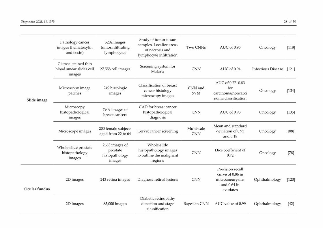
Slide image
Pathology cancer
images (hematoxylin
and eosin)
5202 images
tumorinfiltrating
lymphocytes
Study of tumor tissue
samples. Localize areas
of necrosis and
lymphocyte infiltration
Two CNNs AUC of 0.95 Oncology [118]
Giemsa-stained thin
blood smear slides cell
images
27,558 cell images Screening system for
Malaria CNN AUC of 0.94 Infectious Disease [121]
Microscopy image
patches
249 histologic
images
Classification of breast
cancer histology
microscopy images
CNN and
SVM
AUC of 0.77–0.83
for
carcinoma/noncarci
noma classification
Oncology [134]
Microscopy
histopathological
images
7909 images of
breast cancers
CAD for breast cancer
histopathological
diagnosis
CNN AUC of 0.93 Oncology [135]
Microscope images 200 female subjects
aged from 22 to 64 Cervix cancer screening
Multiscale
CNN
Mean and standard
deviation of 0.95
and 0.18
Oncology [88]
Whole-slide prostate
histopathology
images
2663 images of
prostate
histopathology
images
Whole-slide
histopathology images
to outline the malignant
regions
CNN Dice coefficient of
0.72 Oncology [78]
Ocular fundus
2D images 243 retina images Diagnose retinal lesions CNN
Precision recall
curve of 0.86 in
microaneurysms
and 0.64 in
exudates
Ophthalmology [120]
2D images 85,000 images
Diabetic retinopathy
detection and stage
classification
Bayesian CNN AUC value of 0.99 Ophthalmology [42]

Diagnostics 2021, 11, 1373 29 of 50
Images
6679 images from
Kaggle’s Diabetic
Retinopathy
Detection
Detect retinal
hemorrhages CNN
AUC of 0.894 and
0.972 Ophthalmology [47]
Images
168 images with
glaucoma and 428
control
Detect and evaluate
glaucoma
CNN: ResNet
and U-Net
AUC of 0.91 and
0.84 respectively Ophthalmology [128]
Images 90,000 images with
their diagnoses
Predict the evolution of
diabetic retinopathy CNN AUC of 0.95 Ophthalmology [51]
Images 7000 colour fundus
images
Image quality of diabetic
retinopathy CNN Accuracy of 100 % Ophthalmology [52]
AREDS (age related
eye disease study)
image
130,000 fundus
images
Diagnosis of Age-related
Macular Degeneration CNN
94.97 sensitivity
and 98.32 %
specificity
Ophthalmology [156]
Fundus images
219,302 from
normal participants
without
hypertension,
diabetes mellitus
(DM), and any
smoking history
Predict age and sex from
retinal fundus images CNN AUC 0.96 Ophthalmology [157]
Dermoscopy
Images
350 images of
melanomas and
374 benign nevi
Acral lentiginous
melanoma diagnosis CNN AUC of over 0.80 Oncology [129]
Clinical images 49,567 images Recognize nails
nychomycosis lesions
Region-based-
CNN
AUC of 0.98, AUC
of 0.95, AUC of
0.93, AUC of 0.82
Dermatology [130]
Diagnostics 2021, 11, 1373 30 of 50
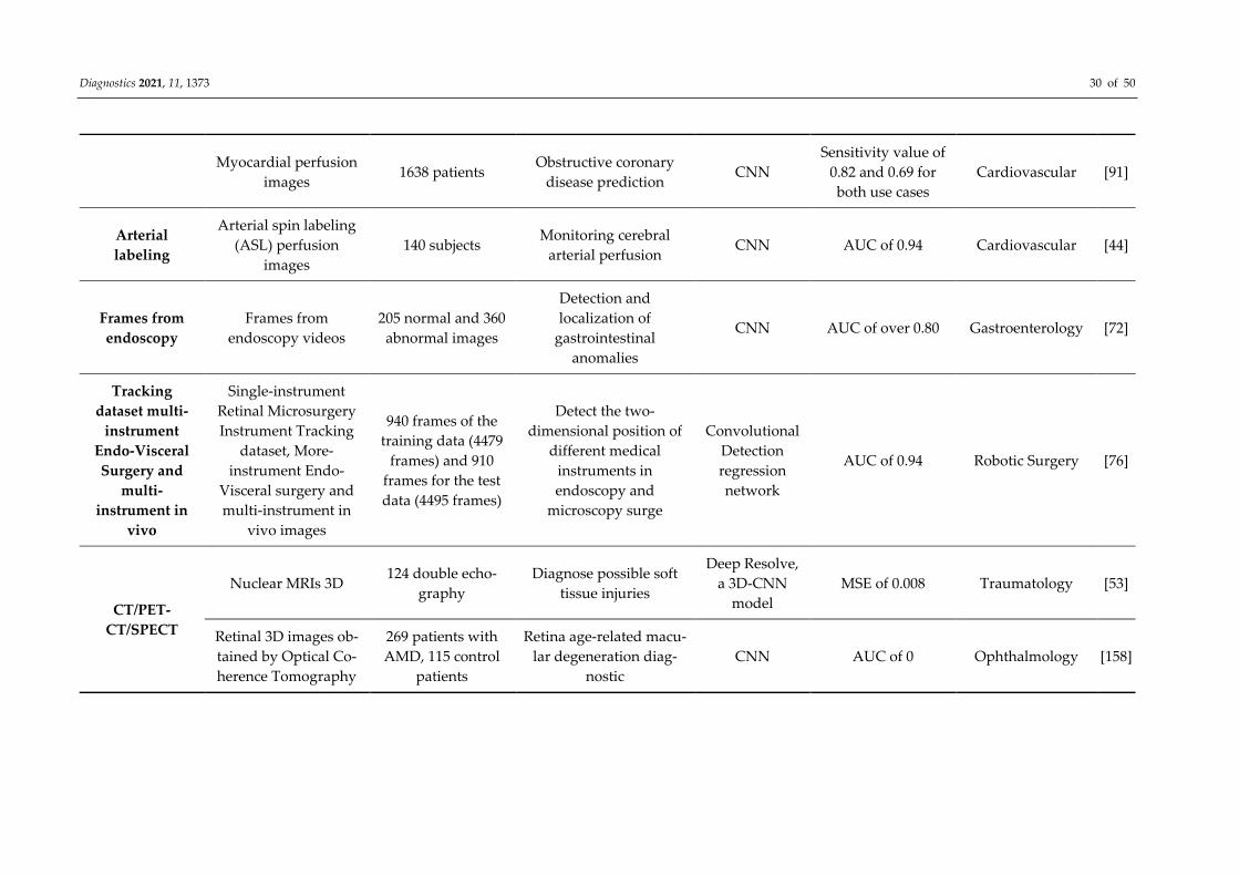
Myocardial perfusion
images 1638 patients
Obstructive coronary
disease prediction CNN
Sensitivity value of
0.82 and 0.69 for
both use cases
Cardiovascular [91]
Arterial
labeling
Arterial spin labeling
(ASL) perfusion
images
140 subjects Monitoring cerebral
arterial perfusion CNN AUC of 0.94 Cardiovascular [44]
Frames from
endoscopy
Frames from
endoscopy videos
205 normal and 360
abnormal images
Detection and
localization of
gastrointestinal
anomalies
CNN AUC of over 0.80 Gastroenterology [72]
Tracking
dataset multi-
instrument
Endo-Visceral
Surgery and
multi-
instrument in
vivo
Single-instrument
Retinal Microsurgery
Instrument Tracking
dataset, More-
instrument Endo-
Visceral surgery and
multi-instrument in
vivo images
940 frames of the
training data (4479
frames) and 910
frames for the test
data (4495 frames)
Detect the two-
dimensional position of
different medical
instruments in
endoscopy and
microscopy surge
Convolutional
Detection
regression
network
AUC of 0.94 Robotic Surgery [76]
CT/PET-
CT/SPECT
Nuclear MRIs 3D 124 double echo-
graphy
Diagnose possible soft
tissue injuries
Deep Resolve,
a 3D-CNN
model
MSE of 0.008 Traumatology [53]
Retinal 3D images ob-
tained by Optical Co-
herence Tomography
269 patients with
AMD, 115 control
patients
Retina age-related macu-
lar degeneration diag-
nostic
CNN AUC of 0 Ophthalmology [158]
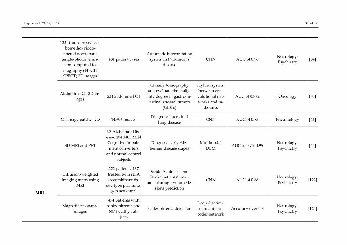
Diagnostics 2021, 11, 1373 31 of 50
123I-fluoropropyl car-
bomethoxyiodo-
phenyl nortropane
single-photon emis-
sion computed to-
mography (FP-CIT
SPECT) 2D images
431 patient cases
Automatic interpretation
system in Parkinson’s
disease
CNN AUC of 0.96 Neurology-
Psychiatry [84]
Abdominal CT 3D im-
ages 231 abdominal CT
Classify tomography
and evaluate the malig-
nity degree in gastro-in-
testinal stromal tumors
(GISTs)
Hybrid system
between con-
volutional net-
works and ra-
diomics
AUC of 0.882 Oncology [83]
CT image patches 2D 14,696 images Diagnose interstitial
lung disease CNN AUC of 0.85 Pneumology [46]
3D MRI and PET
93 Alzheimer Dis-
ease, 204 MCI Mild
Cognitive Impair-
ment converters
and normal control
subjects
Diagnose early Alz-
heimer disease stages
Multimodal
DBM AUC of 0.75–0.95
Neurology-
Psychiatry [41]
MRI
Diffusion-weighted
imaging maps using
MRI
222 patients. 187
treated with rtPA
(recombinant tis-
sue-type plasmino-
gen activator)
Decide Acute Ischemic
Stroke patients’ treat-
ment through volume le-
sions prediction
CNN AUC of 0.88 Neurology-
Psychiatry [122]
Magnetic resonance
images
474 patients with
schizophrenia and
607 healthy sub-
jects
Schizophrenia detection
Deep discrimi-
nant autoen-
coder network
Accuracy over 0.8 Neurology-
Psychiatry [124]
Diagnostics 2021, 11, 1373 32 of 50
Gadoxetic acid–en-
hanced 2D MRI
144,180 images
from 634 patients
Staging liver fibrosis
through MR CNN
AUC values of 0.84,
0.84, and 0.85 for
each stage
Gastroenterology [64]
Resting state func-
tional magnetic reso-
nance imaging (rs-
fMRI), T1 structural
cerebral images and
phenotypic infor-
mation
505 individuals
with autism and
520 matched typi-
cal controls
Identify different autism
spectrum disorders Denoising AE Accuracy of 0.70
Neurology-
Psychiatry [126]
3D MRI and PET
93 Alzheimer Dis-
ease, 204 MCI Mild
Cognitive Impair-
ment converters
and normal control
subjects
CAD for early Alz-
heimer disease stages
Multimodal
DBM
Accuracy of 0.95,
0.85 and 0.75 for
the three use cases
Neurology-
Psychiatry [41]
CT/PET-
CT/SPECT
CT images, MRI im-
ages and PET images 6776 images
Classify medical diag-
nostic images according
to the modality they
were produced and clas-
sify illustrations accord-
ing to their production
attributes
CNN and a
synergic signal
system
AUC of 0.86 Various [159]
CT image 2D
63,890 patients
with cancer and
171,345 healthy
Discriminate lung cancer
lesions in adenocarci-
noma, squamous and
small cell carcinoma
CNN
Log-Loss error of
0.66 with a sensitiv-
ity of 0.87
Oncology [160]
CT 2D images
3059 images from
several parts of hu-
man body
Speed up CT images col-
lection and rebuild the
data
Dense-Net
and CNN RMSE of 0.00048 Various [142]
Diagnostics 2021, 11, 1373 33 of 50
CT images 3D
6960 lung nodule
regions, 3480 of
which were posi-
tive samples and
rest were negative
samples
(nonnodule)
Diagnose lung cancer in
low-dosage CT
Eye-tracking
sparse atten-
tional model
and CNN
Accuracy of 0.97 Oncology [90]
CT images 2D and
text (reports)
9000 training and
1000 testing images
Processing text from CT
reports in order to clas-
sify their respective im-
ages
CNN AUC of 0,58, 0,70–
0.95 Various [92]
Computed tomogra-
phy (CT)
Three datasets:
224,316, 112,120
and 15,783
Binary classification of
posteroanterior chest x-
ray
CNN 92% accuracy Radiology [161]
MRI
Clinical characteristics
and MRI 3D
135 patients with
short-, medium-
and long-term sur-
vival
Predict the survival of
patients with amyo-
trophic lateral sclerosis
CNN Accuracy of 0.84 Neurology-
Psychiatry [67]
Optical coherence to-
mography images
52,690 AMD pa-
tients’ images and
48,312 control
Differentiate Age-Re-
lated Macular Degenera-
tion lesions in optical co-
herence tomography
Modification
of VGG16
CNN
AUC of 0.92, AUC
of 0.93 and AUC of
0.97 for the differ-
ent use cases
Ophthalmology [68]
Diagnostics 2021, 11, 1373 34 of 50
Lung computed axial
tomography 2D im-
ages and breast ultra-
sound lesions
520 breast sono-
grams from
520 patients (275
benign and 245 ma-
lignant lesions) and
lung CT image data
from 1010 patients
(700 malignant and
700 benign nod-
ules)
CAD system to classify
breast ultrasound lesions
and lung CT nodules
Stacked de-
noising AE AUC of 0.94 Oncology [58]
MRI 2D
444 images from
195 patients with
prostate cancer
Prevent errors in diag-
nosing prostate CNN AUC of 0.94 Oncology [88]
MRI 2D
MICCAI 2009 left
ventricle segmenta-
tion challenge data-
base
Determinate limits be-
tween the endocardium
and epicardium of the
left ventricle
RNN with au-
tomatc seg-
mentation
techniqes
AUC of 1.0 in the
best case Cardiovascular [132]
MRI
CT images, MRI im-
ages and PET images
6776 images for
training and 4166
for tests
Classify medical diag-
nostic images according
to the modality they
were produced and clas-
sify illustrations accord-
ing to their production
attributes
CNN and a
synergic signal
system
AUC of 0.86 Various [159]
Functional MRI
68 subjects perform
7 activities, and a
state of rest
Analyze cerebral cogni-
tive functions
3D CNN, rest-
ing state net-
works
AUC of 0.94 Neurology-
Psychiatry [140]
Diagnostics 2021, 11, 1373 35 of 50
Liver MRIs
522 liver MRI cases
with and without
contrast for known
or suspected liver
cirrhosis or focal
liver lesion
Screening system for un-
diagnosed hepatic mag-
netic resonance
images
CNN
Reduces negative
predictive value
and leads to greater
precision
Gastroenterology [50]
MRI images
1064 brain images
of autism patients
and healthy con-
trols. MRI data
from 110 multiple
sclerosis patient
Evaluate the quality of
multicenter structural
brain MRI
images
CNN AUC 0.90 and 0.71 Radiology [55]
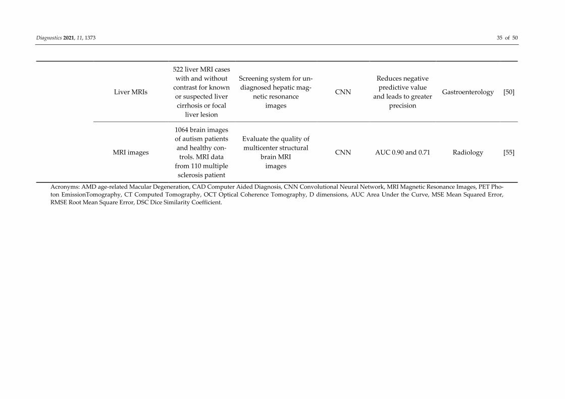
Acronyms: AMD age-related Macular Degeneration, CAD Computer Aided Diagnosis, CNN Convolutional Neural Network, MRI Magnetic Resonance Images, PET Pho-
ton EmissionTomography, CT Computed Tomography, OCT Optical Coherence Tomography, D dimensions, AUC Area Under the Curve, MSE Mean Squared Error,
RMSE Root Mean Square Error, DSC Dice Similarity Coefficient.
Diagnostics 2021, 11, 1373 36 of 50
Diagnostics 2021, 11, 1373. https://doi.org/10.3390/diagnostics11081373 www.mdpi.com/journal/diagnostics
5. Description of Methods for Incorporating Data Types and the Applications in
Which They Are Used
5.1. Schematically Present the Methods of Knowledge Incorporation and the Types of Data Used
for DL Objectives in the Interpretation of Medical Images
We will exemplify the methods of incorporation of medical knowledge and data ac-
cording to the purpose of DL models in medical applications, namely, diagnosis-classifi-
cation, detection, segmentation, reconstruction and recovery of medical images, genera-
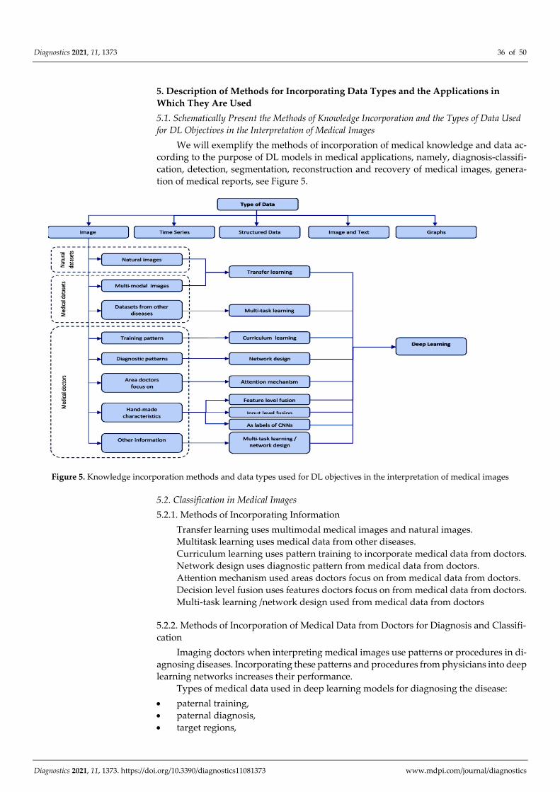
tion of medical reports, see Figure 5.
Figure 5. Knowledge incorporation methods and data types used for DL objectives in the interpretation of medical images
5.2. Classification in Medical Images
5.2.1. Methods of Incorporating Information
Transfer learning uses multimodal medical images and natural images.
Multitask learning uses medical data from other diseases.
Curriculum learning uses pattern training to incorporate medical data from doctors.
Network design uses diagnostic pattern from medical data from doctors.
Attention mechanism used areas doctors focus on from medical data from doctors.
Decision level fusion uses features doctors focus on from medical data from doctors.
Multi-task learning /network design used from medical data from doctors
5.2.2. Methods of Incorporation of Medical Data from Doctors for Diagnosis and Classifi-
cation
Imaging doctors when interpreting medical images use patterns or procedures in di-
agnosing diseases. Incorporating these patterns and procedures from physicians into deep
learning networks increases their performance.
Types of medical data used in deep learning models for diagnosing the disease:
paternal training,
paternal diagnosis,
target regions,
Diagnostics 2021, 11, 1373 37 of 50
hand crafted features (appearance, structures, shapes),
related diagnostic information
other types of diagnostic-related information
(1). The training model consists in the curricular learning through which tasks, im-
ages evolve from simple to complex in the training process. The curriculum involves a
suite of training samples classified in ascending order of learning difficulty. The training
model through curricular learning introduced into the deep learning network is devel-
oped by [162].
(2). General models of diagnosis of doctors, namely, the patterns and procedures
used by imaging doctors when interpreting medical images. Radiologists diagnose imag-
ing in three stages in the interpretation of X-ray images of the chest: overview, local lesion
regions and subsequently combine general data [163].
(3). The use of the diagnostic pattern of radiologists for the diagnosis of thoracic dis-
ease) by extracting and combining global and local traits is carried out in [163] Target
regions or “attention maps”. Imaging doctors focus on specific areas in the diagnosis of
diseases, “warning maps”, which indicates the target areas when interpreting images.
(4). Attention features (appearance, structure, shapes), “handcrafted characteristics”,
as they are made by doctors, can be described characteristics, asymmetry, edge, color,
margin, shape, micro-calcification and echo pattern, acoustic attenuation, side acoustic
shade, and also benign-malignant risk of pulmonary nodules is classified by six charac-
teristics of nodules: calcification, sphericality, edge, spiculation and texture and other.
(5). Related Diagnostic Information (Merger at Decision Level, Characteristics Level
Fusion, Imput-Level Fusion, Features as Labels).
Merger at decision-level. The CNN classifier model automatically extracts and com-
bines by merger at the decision-making level of handcrafted characteristics and extracted
characteristics (contrast, texture, spiculation of the image) from CNN, by merger-level de-
cision-level results from two classifiers [164].
Characteristic-level fusion. Feature-level fusion model combines two handcrafted
features, parameter less threshold adhesion statistics and gray-level co-occurrence matrix,
with the five groups of deep learning features extracted from five different deep models
[18,37].
Input-level fusion. Input-level fusion is achieved by the fact that handmade features
are used as patches that describe specific features and are used as input for CNN followed
by combination in solving the problem. In some models these patches are used as input
into DScGAN to increase diagnostic performance.
Using features as labels of CNN. Image classification labels and labels of handmade
features are included into deep learning patterns through the multi-task learning arhitec-
ture to increase their performance.
(6). Other Types of Diagnostic-Related Information (Additional Labels, Additional
Clinical Diagnostic Reports).
These are represented by additional labels and clinical diagnostic reports. Type of
additional category labels for medical images, normal, malignant or benign, condition of
the lesions is incorporated into a multi-task learning structure can improve the perfor-
mance of the diagnosis of major classification load [18].
Additional clinical diagnostic reports. The clinical report is a summary of descrip-
tions of the doctor made during the imaging examination.
5.3. Detection in Medical Images
We can exemplify four categories:
paternal training,
paternal diagnosis,
target regions,
hand crafted features (appearance, structures, shapes).
Diagnostics 2021, 11, 1373 38 of 50
5.3.1. Paternal Training Is the Resolution of Tasks with Increasing Difficulties That Use
Curricular Learning to Identify and Locate Lesions in Medical Images
CASED performs adaptive curriculum sampling to solve the problem of highly data
imbalance and makes it possible for the model to distinguish nodules from immediate
proximity and subsequently enlarges the hard-declassified global context, up to uniform
categories in the empirical data pool. In this way, CASED is the most performant and is
used in the detection of pulmonary nodules in thoracic CT [165].
LUNA16 also based on curricular learning is used in the detection of cardiac [166].
5.3.2. Paternal Diagnosis
Radiologists use patterns to locate lesions in medical images, namely:
1. Combine images in different settings (brightness and contrast),
2. Uses bilateral, transverse, adjacent images,
3. Radiologists combine collected images in different settings (brightness and contrast)
to locate lesions by visual interpretation of CT images. In the same way is built a
model with multi-viewing features (FPN) brightness and contrast, combined later
using an attention module that identifies the position with an increase in accuracy
compared to NIH DeepLesion [167].
4. Bilateral information is compared by radiologists when interpreting images.
5.3.3. Handmade Characteristics
Handmade characteristics, e.g., locations, structures, shapes are represented by
“Hand-Crafted Characteristics” for Identifying target objects, nodules or lesions in medi-
cal images.
5.3.4. Target Regions
The description of the target regions, e.g., information, radiological reports, addi-
tional labels is extracted from the radiological information and coupled with the curricular
learning and the results are used by the network in the ascending order of the difficulties.
5.4. Segmentation of Lesions and Organs into Medical Images
5.4.1. Incorporation of Data from Natural Datasets or Medical Data Sets
Transfer learning uses data from natural images for performance in the segmentation
of the medical image. The transfer of the acquired data of a CNN arhitectures originally
trained for segmenting WM hyper-intensity on old low-resolution data to new data from
the same scanner, but with good image resolution is studied by [168].
Multimodal learning in which MRI, CT, are used simultaneously by pre-trained ar-
chitecture deep learning.
5.4.2. Incorporation of Knowledge from Doctors
Training pattern. For the segmentation of lesions into medical images deep learning
models used curriculum learning.
Diagnostic pattern. Specific patterns used by doctors and embedded in the network.
Characteristics of the image (shape, location, topology).
Radiologists rely on certain characteristics of the image, shape, position, typological
lesions, when interpreting medical images.
There are three types of incorporation of features injuries from medical imaging in
deep learning architectures:
1. incorporating the characteristics of the lesions in the post-processing stage,
2. incorporating the characteristics of the lesions as elements of regularization in the
loss function,
3. learning the characteristics of the lesion through generational models.
Diagnostics 2021, 11, 1373 39 of 50
5.4.3. Incorporation Handmade Characteristics from Doctors
For input fusion, handmade characteristics are transformed into input patches, sub-
sequently, the original image patches and the tagged patches are inserted into a deep seg-
mentation network [18].
5.5. Reconstruction of Medical Image
The objective is to reconstruct a diagnostic image from a series of measurements.
5.6. Recovery of Medical Image
Deep learning architecture use knowledge from natural images (pre-trained VGG
model based on ImageNet) or medical data.
5.7. Generating Medical Reports
The deep learning models for image subtitles have been successfully applied for the
automatic generation of medical reports [169,170]. Some templates in radiologist reports
are used during the sentence generation process [80,167].
Model-agnostic method attempts to learn the short description of the text to explain
this decision-making process [171] and transfer the visual characteristics of medical im-
ages to a graph of anomalies [18].
Module to incorporate the pre-built graph on multiple findings of the disease to help
generate reports by using the IU-RR dataset [18,172].
5.8. Applications in Medicine, Methods of Incorporation of Types of Data, Datasets and Their
Correlation
Imaging doctors combine data from different stages and experiences as opposed to
DL models that incorporate the same types and modes of handcrafted features. Data qual-
ity and volume, annotations and labels, identification and automatic extraction of specific
medical terms can help deep learning models perform in the tasks of image analysis [18]
Simultaneous incorporation of different medical knowledge types features, labels, into DL
architectures increases their performance (see Table 3) [102].
Table 3. Applications in medicine, methods of incorporation of types of data, datasets and their correlation.
Dataset Images Methods of Incorporating Infor-
mation Application in Medicine
Data doctors focus on
Training pattern
high-level medical data,
curriculum learning
Training model
Images with increasing complex-
ity
diagnosis-classification of breast screening in DCE-RMN
[61]
application - the attention-based curriculum, used in
CNN, derived from radiology reports [173]
diagnosis of the proximal femoral fracture in X-ray im-
ages [18,174]
diagnosing of disease [18,175,176]
Diagnostic pattern, low-
level medical data, areas of
images, characteristics of
diseases
General models of diagnosis of
doctors
thoracic disease diagnosis [163]
final prediction of the disease [177]
diagnosis chest X-ray [178]
dermoscopic diagnosis of the lesion [63]
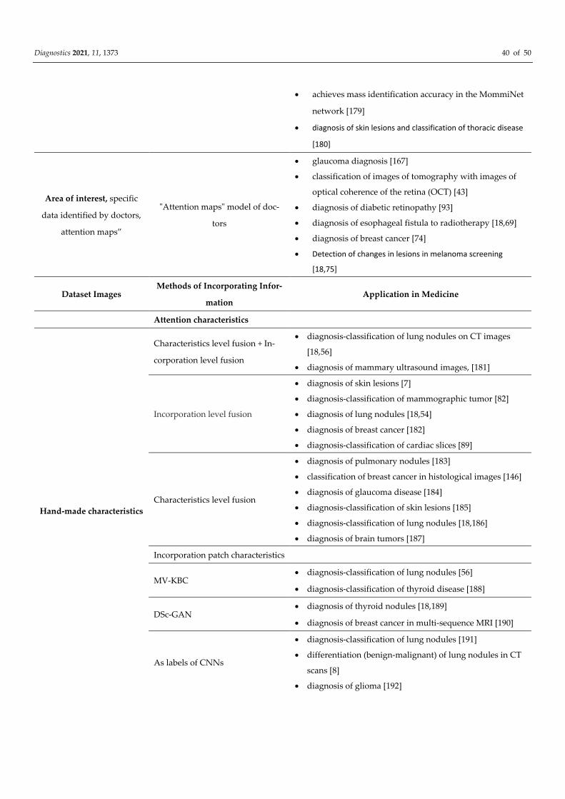
Diagnostics 2021, 11, 1373 40 of 50
achieves mass identification accuracy in the MommiNet
network [179]
diagnosis of skin lesions and classification of thoracic disease
[180]
Area of interest, specific
data identified by doctors,
attention maps”
"Attention maps" model of doc-
tors
glaucoma diagnosis [167]
classification of images of tomography with images of
optical coherence of the retina (OCT) [43]
diagnosis of diabetic retinopathy [93]
diagnosis of esophageal fistula to radiotherapy [18,69]
diagnosis of breast cancer [74]
Detection of changes in lesions in melanoma screening
[18,75]
Dataset Images Methods of Incorporating Infor-
mation Application in Medicine
Attention characteristics
Hand-made characteristics
Characteristics level fusion + In-
corporation level fusion
diagnosis-classification of lung nodules on CT images
[18,56]
diagnosis of mammary ultrasound images, [181]
Incorporation level fusion
diagnosis of skin lesions [7]
diagnosis-classification of mammographic tumor [82]
diagnosis of lung nodules [18,54]
diagnosis of breast cancer [182]
diagnosis-classification of cardiac slices [89]
Characteristics level fusion
diagnosis of pulmonary nodules [183]
classification of breast cancer in histological images [146]
diagnosis of glaucoma disease [184]
diagnosis-classification of skin lesions [185]
diagnosis-classification of lung nodules [18,186]
diagnosis of brain tumors [187]
Incorporation patch characteristics
MV-KBC diagnosis-classification of lung nodules [56]
diagnosis-classification of thyroid disease [188]
DSc-GAN diagnosis of thyroid nodules [18,189]
diagnosis of breast cancer in multi-sequence MRI [190]
As labels of CNNs
diagnosis-classification of lung nodules [191]
differentiation (benign-malignant) of lung nodules in CT
scans [8]
diagnosis of glioma [192]
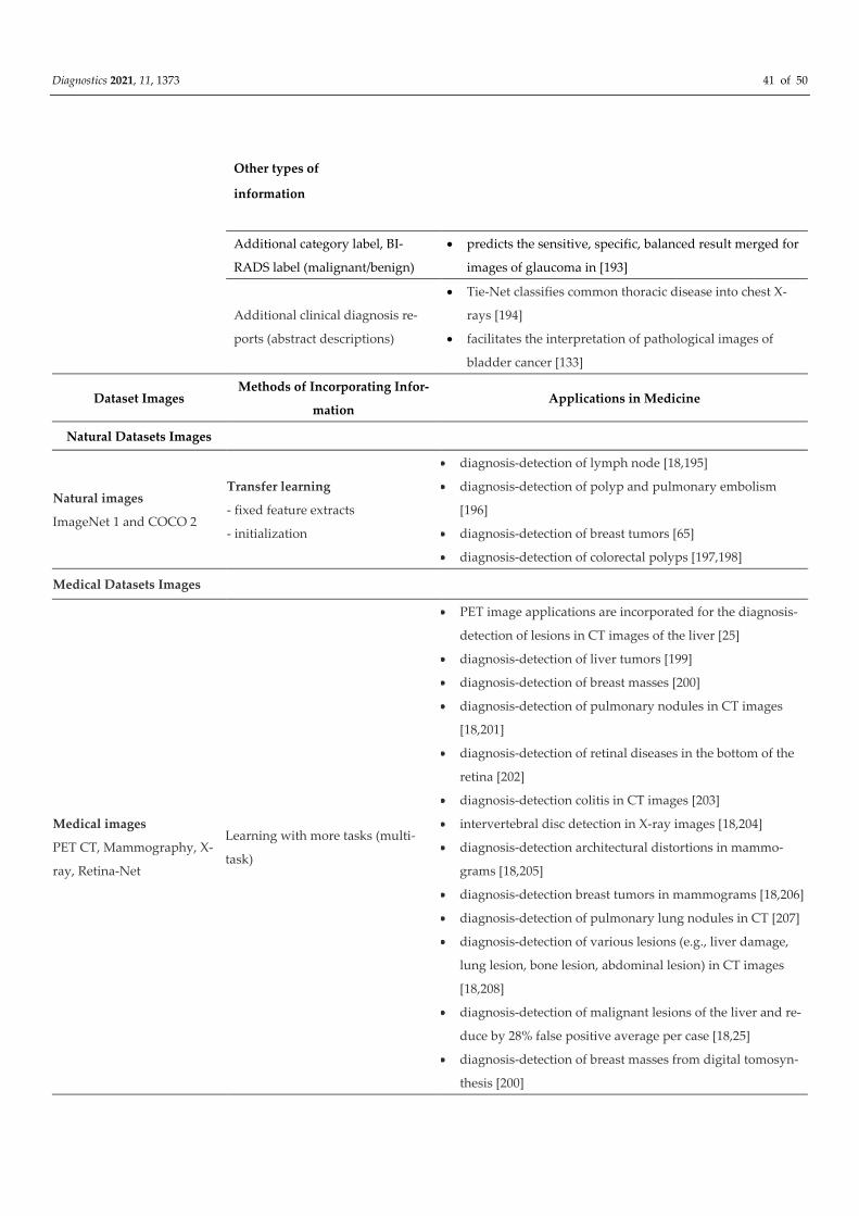
Diagnostics 2021, 11, 1373 41 of 50
Other types of
information
Additional category label, BI-
RADS label (malignant/benign)
predicts the sensitive, specific, balanced result merged for
images of glaucoma in [193]
Additional clinical diagnosis re-
ports (abstract descriptions)
Tie-Net classifies common thoracic disease into chest X-
rays [194]
facilitates the interpretation of pathological images of
bladder cancer [133]
Dataset Images Methods of Incorporating Infor-
mation Applications in Medicine
Natural Datasets Images
Natural images
ImageNet 1 and COCO 2
Transfer learning
- fixed feature extracts
- initialization
diagnosis-detection of lymph node [18,195]
diagnosis-detection of polyp and pulmonary embolism
[196]
diagnosis-detection of breast tumors [65]
diagnosis-detection of colorectal polyps [197,198]
Medical Datasets Images
Medical images
PET CT, Mammography, X-
ray, Retina-Net
Learning with more tasks (multi-
task)
PET image applications are incorporated for the diagnosis-
detection of lesions in CT images of the liver [25]
diagnosis-detection of liver tumors [199]
diagnosis-detection of breast masses [200]
diagnosis-detection of pulmonary nodules in CT images
[18,201]
diagnosis-detection of retinal diseases in the bottom of the
retina [202]
diagnosis-detection colitis in CT images [203]
intervertebral disc detection in X-ray images [18,204]
diagnosis-detection architectural distortions in mammo-
grams [18,205]
diagnosis-detection breast tumors in mammograms [18,206]
diagnosis-detection of pulmonary lung nodules in CT [207]
diagnosis-detection of various lesions (e.g., liver damage,
lung lesion, bone lesion, abdominal lesion) in CT images
[18,208]
diagnosis-detection of malignant lesions of the liver and re-
duce by 28% false positive average per case [18,25]
diagnosis-detection of breast masses from digital tomosyn-
thesis [200]
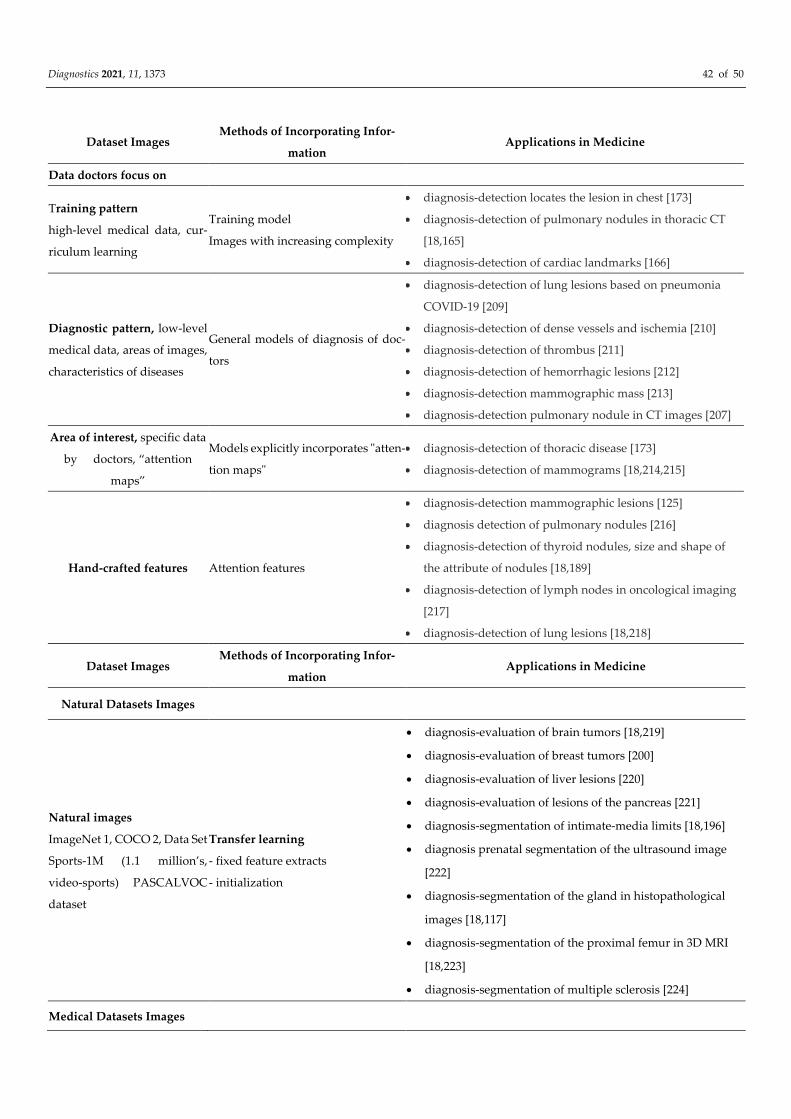
Diagnostics 2021, 11, 1373 42 of 50
Dataset Images Methods of Incorporating Infor-
mation Applications in Medicine
Data doctors focus on
Training pattern
high-level medical data, cur-
riculum learning
Training model
Images with increasing complexity
diagnosis-detection locates the lesion in chest [173]
diagnosis-detection of pulmonary nodules in thoracic CT
[18,165]
diagnosis-detection of cardiac landmarks [166]
Diagnostic pattern, low-level
medical data, areas of images,
characteristics of diseases
General models of diagnosis of doc-
tors
diagnosis-detection of lung lesions based on pneumonia
COVID-19 [209]
diagnosis-detection of dense vessels and ischemia [210]
diagnosis-detection of thrombus [211]
diagnosis-detection of hemorrhagic lesions [212]
diagnosis-detection mammographic mass [213]
diagnosis-detection pulmonary nodule in CT images [207]
Area of interest, specific data
by doctors, “attention
maps”
Models explicitly incorporates "atten-
tion maps"
diagnosis-detection of thoracic disease [173]
diagnosis-detection of mammograms [18,214,215]
Hand-crafted features Attention features
diagnosis-detection mammographic lesions [125]
diagnosis detection of pulmonary nodules [216]
diagnosis-detection of thyroid nodules, size and shape of
the attribute of nodules [18,189]
diagnosis-detection of lymph nodes in oncological imaging
[217]
diagnosis-detection of lung lesions [18,218]
Dataset Images Methods of Incorporating Infor-
mation Applications in Medicine
Natural Datasets Images
Natural images
ImageNet 1, COCO 2, Data Set
Sports-1M (1.1 million’s,
video-sports) PASCALVOC
dataset
Transfer learning
- fixed feature extracts
- initialization
diagnosis-evaluation of brain tumors [18,219]
diagnosis-evaluation of breast tumors [200]
diagnosis-evaluation of liver lesions [220]
diagnosis-evaluation of lesions of the pancreas [221]
diagnosis-segmentation of intimate-media limits [18,196]
diagnosis prenatal segmentation of the ultrasound image
[222]
diagnosis-segmentation of the gland in histopathological
images [18,117]
diagnosis-segmentation of the proximal femur in 3D MRI
[18,223]
diagnosis-segmentation of multiple sclerosis [224]
Medical Datasets Images
Diagnostics 2021, 11, 1373 43 of 50
Medical images
MRI data, CT angiography,
3DSeg-8 dataset
Learning with more tasks (multi-
task)
diagnosis-segmentation of the left/ right lung [18,225]
diagnosis of cerebral disease into MRI [18,226,227]
diagnosis-segmentation of heart vessel without annotations
used annotated retinal images [228]
diagnosis-segmentation of coronary artery, with high accu-
racy [18]
Data doctors focus on
Deep learning:
FCN
U-Net
GAN
diagnosis segmentation of brain [18,227]
diagnosis-segmentation of skin lesions [229]
diagnosis-segmentation of vessels [230]
diagnosis-segmentation of anomalies in the retina fundus
images [231]
Dataset Images Methods of Incorporating
Information Applications in Medicine
Data doctors focus on
Training pattern
high-level medical data,
curriculum learning
Training model
Images with increasing complexity
Self-paced learning (SPL)
SPL + active learning
diagnosis-segmentation of CT images with multiple organs
[232]
diagnosis-segmentation in 3D pulmonary images [194]
diagnosis-segmentation of liver tumors [18,212]
diagnosis-segmentation of the left ventricle [18,233]
diagnosis-segmenting of the finger bones [18,234]
diagnosis-segmentation of vessels [18,235]
Diagnostic pattern
LIDC-IDRI dataset, BraTS
2018 dataset
General models of diagnosis of
doctors
diagnosis on uncertain nodules [236]
diagnosis-segmentation of heart [237]
diagnosis-segmentation of the liver [238]
diagnosis-segmentation of raw tumors and clinical target
volume [18,239]
Area of interest
BRATS2015 dataset,
(ImageNet, video datasets
Used for 3D image
segmentation)
The fusion at the feature level +
concatenate
diagnosis-segmentation in histopathological images [57]
diagnosis-segmentation of brain [18,240]
diagnosis-segmentation of tumor brain-MRI images [18,241]
diagnosis-segmentation of cellular nuclei [18,242]
Specific characteristics
(shape, location, topology)
In the post-processing stage
diagnosis-segmentation identifying locations of breast
tumors [18,243]
diagnosis-segmentation anatomically [244]
diagnosis-representation of anatomical cardiac form [245]
In the loss function
diagnosis-segmentation of cardiac RM images [246]
diagnostic-segmentation skin lesions [247]
diagnosis-segmentation of kidney [18,248]
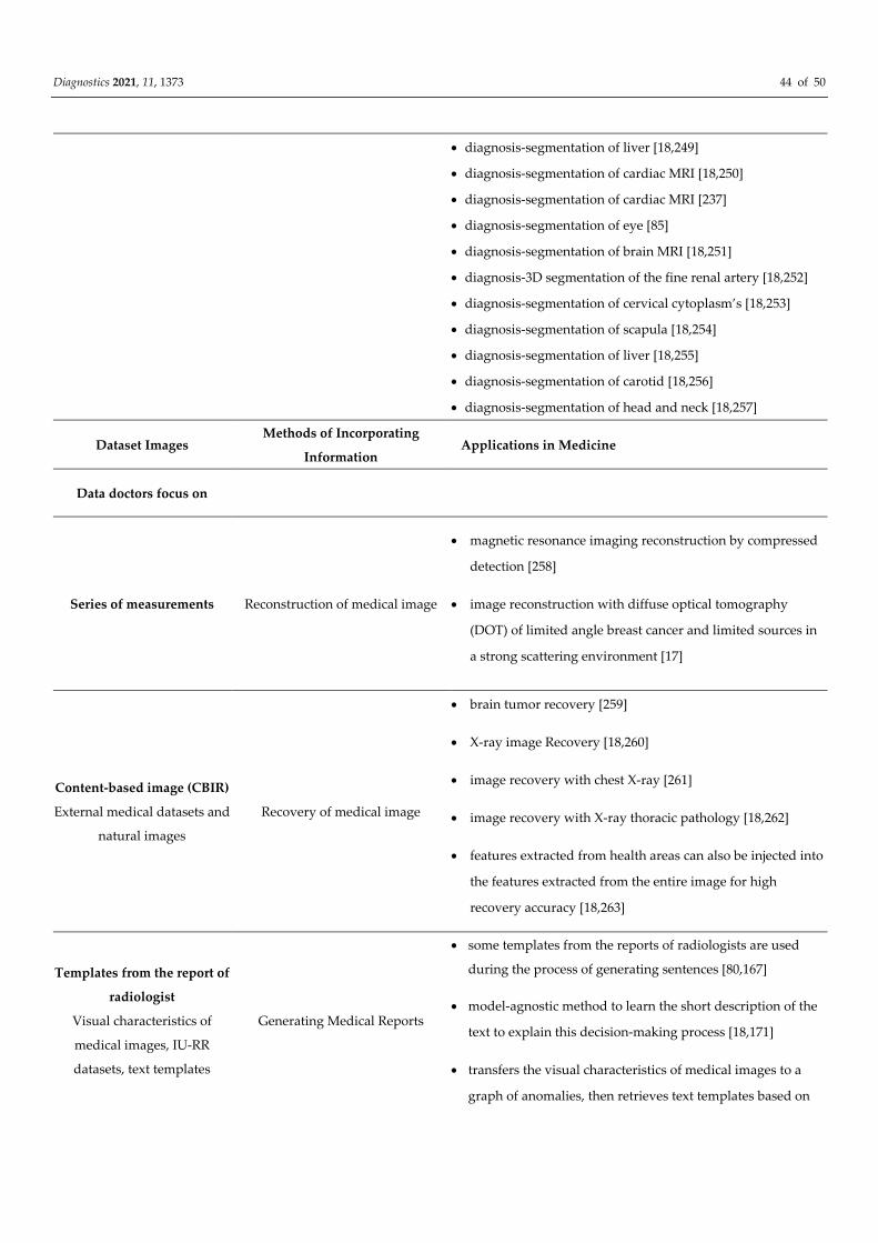
Diagnostics 2021, 11, 1373 44 of 50
diagnosis-segmentation of liver [18,249]
diagnosis-segmentation of cardiac MRI [18,250]
diagnosis-segmentation of cardiac MRI [237]
diagnosis-segmentation of eye [85]
diagnosis-segmentation of brain MRI [18,251]
diagnosis-3D segmentation of the fine renal artery [18,252]
diagnosis-segmentation of cervical cytoplasm’s [18,253]
diagnosis-segmentation of scapula [18,254]
diagnosis-segmentation of liver [18,255]
diagnosis-segmentation of carotid [18,256]
diagnosis-segmentation of head and neck [18,257]
Dataset Images Methods of Incorporating
Information Applications in Medicine
Data doctors focus on
Series of measurements Reconstruction of medical image
magnetic resonance imaging reconstruction by compressed
detection [258]
image reconstruction with diffuse optical tomography
(DOT) of limited angle breast cancer and limited sources in
a strong scattering environment [17]
Content-based image (CBIR)
External medical datasets and
natural images
Recovery of medical image
brain tumor recovery [259]
X-ray image Recovery [18,260]
image recovery with chest X-ray [261]
image recovery with X-ray thoracic pathology [18,262]
features extracted from health areas can also be injected into
the features extracted from the entire image for high
recovery accuracy [18,263]
Templates from the report of
radiologist
Visual characteristics of
medical images, IU-RR
datasets, text templates
Generating Medical Reports
some templates from the reports of radiologists are used
during the process of generating sentences [80,167]
model-agnostic method to learn the short description of the
text to explain this decision-making process [18,171]
transfers the visual characteristics of medical images to a
graph of anomalies, then retrieves text templates based on
Diagnostics 2021, 11, 1373 45 of 50
anomalies and their attributes for thoracic X-ray images
[18,167]
incorporate the pre-built graph (modeled with a CNN
graph) on multiple findings of the disease to help generate
reports by using the IU-RR dataset [18,172]
6. Conclusions
In this paper, as a research novelty, we approached in a unitary way, the constituent
elements of DL models:
Updated presentation of data types, DL models used in medical image analysis;
Correlation and contribution to the performance of DL models of the constituent el-
ements: data type, incorporation methods and DL architectures;
Features and “key” tasks of DL models for the successful completion of tasks in ap-
plications in the interpretation of medical images.
The quality of the data and their volume, annotations and labels, the identification
and automatic extraction of specific terms, from reports, guides, books in the medical
field, can increase the diagnostic accuracy of doctors and help deep learning models per-
form in the tasks of image analysis. Doctors use a descriptive language, namely, contour,
contrast, appearance, localization, topology, etc., or compare bilateral images. The incor-
poration of these representations, attributes from images, in DL architectures increase
their performance.
Imaging doctors combine data from different stages and experiences as opposed to
DL models that incorporate the same types and modes of handcrafted features. Data qual-
ity and volume, annotations and labels, identification and automatic extraction of specific
medical terms can help deep learning models perform in the tasks of image analysis [18].
Incorporating these features, labels, into DL architectures increases their performance
[102].
The diagnostic model, the training model simultaneously incorporates high-level
and low-level knowledge (handcrafted features, anatomical priorities). High-level medi-
cal data is incorporated as input images, and low-level medical data is learned using spe-
cific network structures [18,237] and along with direct networking, information from low-
level medical data can also be used to design training commands when combined with
the easy-to-use training model [18,173]. Simultaneous incorporation of different medical
knowledge types can increase performance of deep learning patterns in medical applica-
tions.
DL can be a support in solving complex problems, with uncertainties of options in
investigations and therapy and could help medically and by filtering, providing data from
literature. This aspect leads to a personalized medicine of the patient’s disease with diag-
nostic and therapeutic options based on scientific evidence. Another aspect is represented
by the time encoded by the doctor in patient care, time gained by the constructive and
effective support of DL in medical decision-making and synthesis activities.
The use of “key” characteristics specific to each constituent of DL models and the
correct determination of their correlations, may be the subject of future research, with the
aim of increasing the performance of DL models in the interpretation of medical images.
7. Research Problems
Problems in medical image analysis can be categorized as follows:
identification and automatic extraction and standardization of specific medical
terms,
representation of medical knowledge,
Diagnostics 2021, 11, 1373 46 of 50
incorporation of medical knowledge.
Problems in medical image analysis are related to:
medical images provided as data for deep-street models require: quality, volume,
specificity, labelling.
providing data from doctors, descriptive data, labels are ambiguous for the same
medical and non-standard references.
laborious time in data processing are problems to solve in the future.
lack of clinical trials demonstrating the benefits of using DL medical applications in
reducing morbidity and mortality and improving patient quality of life
[39,102,264,265].
Full analysis of the mechanism of realization of medical applications, from data, da-
tabases, methods of incorporation of knowledge into DL models and improvement of DL
models to their performance transposed into medical applications lead to the following
problems to be solved: identification and automatic extraction of specific terms from med-
ical documents, representation of medical knowledge, incorporation of medical
knowledge.
Specific medical terms and descriptive attributes corresponding to diseases in medi-
cal images, by incorporating in DL models improve their performance and therefore in-
volve solving problems related to the identification and automatic extraction of specific
terms from medical documents, the presentation of medical knowledge, the incorporation
of medical knowledge.
Problems in medical image analysis are related to quality, volume, specificity and
data labelling in medical images used for a particular action by DL. Also, the provision of
data from doctors, handmade, ambiguous expressions for the same medical references,
uncertain limits of segments in images, low resolution of images, annotations, labels and
laborious time in data processing are problems to solve in the future.
Another problem is the lack of clinical trials demonstrating the benefits of using DL’s
medical applications in reducing morbidity and mortality and improving the quality of
life of patients.
8. Future Challenges
These consist of domain adaptation, knowledge graph, generational models, and net-
work architecture search techniques.
The adaptation of the domain consisted of transferring information from a source
domain to a target domain, such as adversarial learning [266], makes it narrow the domain
change between source and target domain in input space [267], feature space [268,269]
and output space [270,271]. It can be used to transfer knowledge of one set of medical data
to another [212] even when they have different modes of imaging or belong to different
diseases [18,168,272]. UDA (unsupervised adaptation of the field) that uses medical labels
has demonstrated performance in disease diagnosis and organ segmentation [18],
[81,188,255,273].
The knowledge graph, which has the specificity of incorporating multimodal medical
data achieves performance in the analysis of the medical image and the creation of medi-
cal reports [167]. The graphs of medical data describing, the relationship between different
types of knowledge, the relationship between different diseases, the relationship between
medical datasets and a type of medical data, help the models of deep learning to perform
[274].
Generative models, GAN and AE are used for segmentation tasks in particular. GAN
uses MRI datasets for CT image segmentation [18,225,272]. GAN is a type of unsupervised
deep learning network used in medical image analysis. AE are used in extracting charac-
teristics, shape priorities in objects such as organs or lesions, completely unsupervised
and are easily incorporated into the process of network training [18,85,237].
Diagnostics 2021, 11, 1373 47 of 50
In traditional machine learning, the common learning process is separated and is car-
ried out only on certain models, data sets and tasks. Therefore, knowledge is not retained
or transferred to each other models. Instead, in deep learning, transfer learning can use
knowledge such as the weights and characteristics of a pre-trained model to prepare a
new model, as well as to address problems in the task that has a smaller amount of data.
Transfer learning with deep learning patterns is faster, has improved accuracy and/or
needs less training data [275].
A new approach to transfer learning, to address the problem of lack of data training
in medical imaging tasks is represented by the technique of learning by transfer called
dual transfer learning. Using the characteristics learned to improve the performance of
other tasks by, such as classification in skin lesions, such us, benign and malignant or in
the case of breast lesions to classify histological mammary images into four classes: inva-
sive carcinoma, in situ carcinoma, benign tumor and normal tissue [276].
Using cloud computing provides a solution for managing the enormous amount of
data. It also helps to increase efficiency and reduce costs. In addition, it offers the flexibil-
ity to train DL architectures [104]
With the recent development of computing tools, including a chip for neural net-
works and a mobile GPU, we will see more deep learning applications on mobile devices.
It will be easier for users to use DL [104].
Network Architecture Search Technique (NAS) can automatically identify a certain
network architecture in computer vision tasks [277] and promises that use and perfor-
mance in the medical field [18,278].
With audacity, hope and confidence in the realization of our scientific desires we,
authors, we launch an appeal to the international scientific forum with the aim that the
following ideas will be put into practice at the initiative of some standard researchers in
the field, “voices heard and heard” and who have the power to flesh them out:
the establishment of a federation institution integrating scientific data and products
specific to the field;
value categorization of industry-specific achievements;
launching challenges to be developed and completed;
facilitating the free circulation of discoveries, methods, formulas of scientific prod-
ucts within this federation institution;
establishing the board of the federation institution through the input and integration
of “consequential brains” in the field;
the creation of a Hub of Ideas under coordination within the federation board with
assignment of themes for development on specific teams;
joint effort for an idea launched within the federation institution;
an inventory of functional applications and methods, performing in the specific field;
the creation of a financing system to support and implement ideas specific to the
field;
integration of researchers with notable ideas and performance limited funding or ac-
cess to knowledge by belonging to geographical areas or institutions under repre-
sented internationally in the specific field.
Funding: Scientific research funded by the University of Medicine and Pharmacy “Gr. T. Popa” of
Iasi, based on contract number 4714.
Conflicts of Interest: The authors declare no conflict of interest. The funders had no role in the
design of the study; in the collection, analyses, or interpretation of data; in the writing of the manu-
script, or in the decision to publish the results.
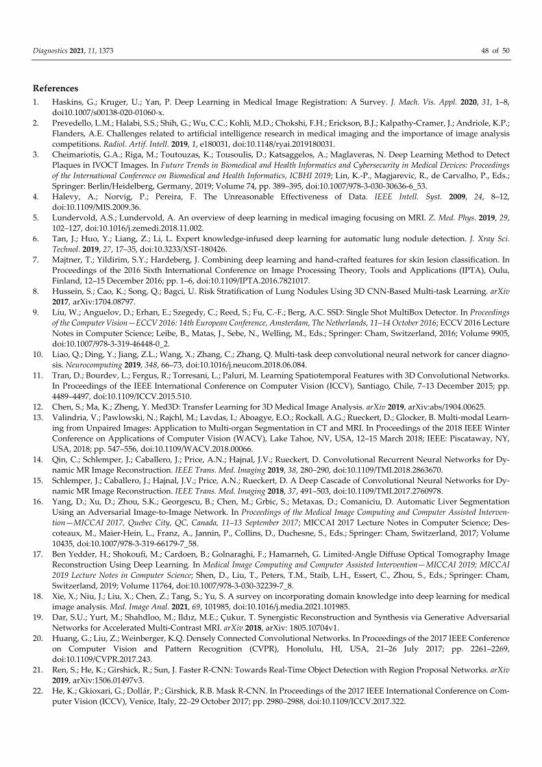
Diagnostics 2021, 11, 1373 48 of 50
References
1. Haskins, G.; Kruger, U.; Yan, P. Deep Learning in Medical Image Registration: A Survey. J. Mach. Vis. Appl. 2020, 31, 1–8,
doi10.1007/s00138-020-01060-x.
2. Prevedello, L.M.; Halabi, S.S.; Shih, G.; Wu, C.C.; Kohli, M.D.; Chokshi, F.H.; Erickson, B.J.; Kalpathy-Cramer, J.; Andriole, K.P.;
Flanders, A.E. Challenges related to artificial intelligence research in medical imaging and the importance of image analysis
competitions. Radiol. Artif. Intell. 2019, 1, e180031, doi:10.1148/ryai.2019180031.
3. Cheimariotis, G.A.; Riga, M.; Toutouzas, K.; Tousoulis, D.; Katsaggelos, A.; Maglaveras, N. Deep Learning Method to Detect
Plaques in IVOCT Images. In Future Trends in Biomedical and Health Informatics and Cybersecurity in Medical Devices: Proceedings
of the International Conference on Biomedical and Health Informatics, ICBHI 2019; Lin, K.-P., Magjarevic, R., de Carvalho, P., Eds.;
Springer: Berlin/Heidelberg, Germany, 2019; Volume 74, pp. 389–395, doi:10.1007/978-3-030-30636-6_53.
4. Halevy, A.; Norvig, P.; Pereira, F. The Unreasonable Effectiveness of Data. IEEE Intell. Syst. 2009, 24, 8–12,
doi:10.1109/MIS.2009.36.
5. Lundervold, A.S.; Lundervold, A. An overview of deep learning in medical imaging focusing on MRI. Z. Med. Phys. 2019, 29,
102–127, doi:10.1016/j.zemedi.2018.11.002.
6. Tan, J.; Huo, Y.; Liang, Z.; Li, L. Expert knowledge-infused deep learning for automatic lung nodule detection. J. Xray Sci.
Technol. 2019, 27, 17–35, doi:10.3233/XST-180426.
7. Majtner, T.; Yildirim, S.Y.; Hardeberg, J. Combining deep learning and hand-crafted features for skin lesion classification. In
Proceedings of the 2016 Sixth International Conference on Image Processing Theory, Tools and Applications (IPTA), Oulu,
Finland, 12–15 December 2016; pp. 1–6, doi:10.1109/IPTA.2016.7821017.
8. Hussein, S.; Cao, K.; Song, Q.; Bagci, U. Risk Stratification of Lung Nodules Using 3D CNN-Based Multi-task Learning. arXiv
2017, arXiv:1704.08797.
9. Liu, W.; Anguelov, D.; Erhan, E.; Szegedy, C.; Reed, S.; Fu, C.-F.; Berg, A.C. SSD: Single Shot MultiBox Detector. In Proceedings
of the Computer Vision—ECCV 2016: 14th European Conference, Amsterdam, The Netherlands, 11–14 October 2016; ECCV 2016 Lecture
Notes in Computer Science; Leibe, B., Matas, J., Sebe, N., Welling, M., Eds.; Springer: Cham, Switzerland, 2016; Volume 9905,
doi:10.1007/978-3-319-46448-0_2.
10. Liao, Q.; Ding, Y.; Jiang, Z.L.; Wang, X.; Zhang, C.; Zhang, Q. Multi-task deep convolutional neural network for cancer diagno-
sis. Neurocomputing 2019, 348, 66–73, doi:10.1016/j.neucom.2018.06.084.
11. Tran, D.; Bourdev, L.; Fergus, R.; Torresani, L.; Paluri, M. Learning Spatiotemporal Features with 3D Convolutional Networks.
In Proceedings of the IEEE International Conference on Computer Vision (ICCV), Santiago, Chile, 7–13 December 2015; pp.
4489–4497, doi:10.1109/ICCV.2015.510.
12. Chen, S.; Ma, K.; Zheng, Y. Med3D: Transfer Learning for 3D Medical Image Analysis. arXiv 2019, arXiv:abs/1904.00625.
13. Valindria, V.; Pawlowski, N.; Rajchl, M.; Lavdas, I.; Aboagye, E.O.; Rockall, A.G.; Rueckert, D.; Glocker, B. Multi-modal Learn-
ing from Unpaired Images: Application to Multi-organ Segmentation in CT and MRI. In Proceedings of the 2018 IEEE Winter
Conference on Applications of Computer Vision (WACV), Lake Tahoe, NV, USA, 12–15 March 2018; IEEE: Piscataway, NY,
USA, 2018; pp. 547–556, doi:10.1109/WACV.2018.00066.
14. Qin, C.; Schlemper, J.; Caballero, J.; Price, A.N.; Hajnal, J.V.; Rueckert, D. Convolutional Recurrent Neural Networks for Dy-
namic MR Image Reconstruction. IEEE Trans. Med. Imaging 2019, 38, 280–290, doi:10.1109/TMI.2018.2863670.
15. Schlemper, J.; Caballero, J.; Hajnal, J.V.; Price, A.N.; Rueckert, D. A Deep Cascade of Convolutional Neural Networks for Dy-
namic MR Image Reconstruction. IEEE Trans. Med. Imaging 2018, 37, 491–503, doi:10.1109/TMI.2017.2760978.
16. Yang, D.; Xu, D.; Zhou, S.K.; Georgescu, B.; Chen, M.; Grbic, S.; Metaxas, D.; Comaniciu, D. Automatic Liver Segmentation
Using an Adversarial Image-to-Image Network. In Proceedings of the Medical Image Computing and Computer Assisted Interven-
tion—MICCAI 2017, Quebec City, QC, Canada, 11–13 September 2017; MICCAI 2017 Lecture Notes in Computer Science; Des-
coteaux, M., Maier-Hein, L., Franz, A., Jannin, P., Collins, D., Duchesne, S., Eds.; Springer: Cham, Switzerland, 2017; Volume
10435, doi:10.1007/978-3-319-66179-7_58.
17. Ben Yedder, H.; Shokoufi, M.; Cardoen, B.; Golnaraghi, F.; Hamarneh, G. Limited-Angle Diffuse Optical Tomography Image
Reconstruction Using Deep Learning. In Medical Image Computing and Computer Assisted Intervention—MICCAI 2019; MICCAI
2019 Lecture Notes in Computer Science; Shen, D., Liu, T., Peters, T.M., Staib, L.H., Essert, C., Zhou, S., Eds.; Springer: Cham,
Switzerland, 2019; Volume 11764, doi:10.1007/978-3-030-32239-7_8.
18. Xie, X.; Niu, J.; Liu, X.; Chen, Z.; Tang, S.; Yu, S. A survey on incorporating domain knowledge into deep learning for medical
image analysis. Med. Image Anal. 2021, 69, 101985, doi:10.1016/j.media.2021.101985.
19. Dar, S.U.; Yurt, M.; Shahdloo, M.; Ildız, M.E.; Çukur, T. Synergistic Reconstruction and Synthesis via Generative Adversarial
Networks for Accelerated Multi-Contrast MRI. arXiv 2018, arXiv: 1805.10704v1.
20. Huang, G.; Liu, Z.; Weinberger, K.Q. Densely Connected Convolutional Networks. In Proceedings of the 2017 IEEE Conference
on Computer Vision and Pattern Recognition (CVPR), Honolulu, HI, USA, 21–26 July 2017; pp. 2261–2269,
doi:10.1109/CVPR.2017.243.
21. Ren, S.; He, K.; Girshick, R.; Sun, J. Faster R-CNN: Towards Real-Time Object Detection with Region Proposal Networks. arXiv
2019, arXiv:1506.01497v3.
22. He, K.; Gkioxari, G.; Dollár, P.; Girshick, R.B. Mask R-CNN. In Proceedings of the 2017 IEEE International Conference on Com-
puter Vision (ICCV), Venice, Italy, 22–29 October 2017; pp. 2980–2988, doi:10.1109/ICCV.2017.322.
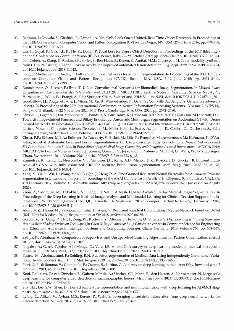
Diagnostics 2021, 11, 1373 49 of 50
23. Redmon, J.; Divvala, S.; Girshick, R.; Farhadi, A. You Only Look Once: Unified, Real-Time Object Detection. In Proceedings of
the IEEE Conference on Computer Vision and Pattern Recognition (CVPR), Las Vegas, NV, USA, 27–30 June 2016; pp. 779–788,
doi:10.1109/CVPR.2016.91.
24. Lin, T.; Goyal, P.; Girshick, R.; He, K.; Dollár, P. Focal Loss for Dense Object Detection. In Proceedings of the 2017 IEEE Inter-
national Conference on Computer Vision (ICCV), Venice, Italy, 22–29 October 2017; pp. 2999–3007, doi:10.1109/ICCV.2017.324.
25. Ben-Cohen, A.; Klang, E.; Raskin, S.P.; Soffer, S.; Ben-Haim, S.; Konen, E.; Amitai, M.M.; Greenspan, H. Cross-modality synthesis
from CT to PET using FCN and GAN networks for improved automated lesion detection. Eng. Appl. Artif. Intell. 2019, 186–194,
doi:10.1016/j.engappai.2018.11.013.
26. Long, J.; Shelhamer, E.; Darrell, T. Fully convolutional networks for semantic segmentation. In Proceedings of the IEEE Confer-
ence on Computer Vision and Pattern Recognition (CVPR), Boston, MA, USA, 7–12 June 2015; pp. 3431–3440.,
doi:10.1109/CVPR.2015.7298965.
27. Ronneberger, O.; Fischer, P.; Brox, T. U-Net: Convolutional Networks for Biomedical Image Segmentation. In Medical Image
Computing and Computer-Assisted Intervention—MICCAI 2015; MICCAI 2015 Lecture Notes in Computer Science; Navab, N.,
Hornegger, J., Wells, W., Frangi, A. Eds.; Springer: Cham, Switzerland, 2015; Volume 9351, doi:10.1007/978-3-319-24574-4_28.
28. Goodfellow, I.J.; Pouget-Abadie, J.; Mirza, M.; Xu, B.; Warde-Farley, D.; Ozair, S.; Courville, A.; Bengio, Y. Generative adversar-
ial nets. In Proceedings of the 27th International Conference on Neural Information Processing Systems—Volume 2 (NIPS'14),
Bangkok, Thailand, 18–22 November 2020; MIT Press: Cambridge, MA, USA, 2020; pp. 2672–2680.
29. Gibson, E.; Giganti, F.; Hu, Y.; Bonmati, E.; Bandula, S.; Gurusamy, K.; Davidson, B.R.; Pereira, S.P.; Clarkson, M.J.; Barratt, D.C.
Towards Image-Guided Pancreas and Biliary Endoscopy: Automatic Multi-organ Segmentation on Abdominal CT with Dense
Dilated Networks. In Proceedings of the Medical Image Computing and Computer Assisted Intervention—MICCAI 2017; MICCAI 2017
Lecture Notes in Computer Science; Descoteaux, M., Maier-Hein, L., Franz, A., Jannin, P., Collins, D., Duchesne, S., Eds.;
Springer: Cham, Switzerland, 2017; Volume 10433, doi:10.1007/978-3-319-66182-7_83.
30. Christ, P.F.; Elshaer, M.E.A.; Ettlinger, F.; Tatavarty, S.; Bickel, M.; Bilic, P.; Rempfler, M.; Armbruster, M.; Hofmann, F.; D’An-
astasi, M.; et al. Automatic Liver and Lesion Segmentation in CT Using Cascaded Fully Convolutional Neural Networks and
3D Conditional Random Fields. In Proceedings of the Medical Image Computing and Computer-Assisted Intervention—MICCAI 2016;
MICCAI 2016 Lecture Notes in Computer Science; Ourselin, S., Joskowicz, L., Sabuncu, M., Unal, G., Wells, W., Eds.; Springer:
Cham, Switzerland, 2016; Volume 9901, doi:10.1007/978-3-319-46723-8_48.
31. Kamnitsas, K.; Ledig, C., Newcombe, V.F.; Simpson, J.P.; Kane, A.D.; Menon, D.K.; Rueckert, D.; Glocker, B. Efficient multi-
scale 3D CNN with fully connected CRF for accurate brain lesion segmentation. Med. Image Anal. 2017, 36, 61–78,
doi:10.1016/j.media.2016.10.004.
32. Yang, X.; Yu, L.; Wu, L.; Wang, Y.; Ni, D.; Qin, J.; Heng, P.-A. Fine-Grained Recurrent Neural Networks for Automatic Prostate
Segmentation in Ultrasound Images. In Proceedings of the AAAI Conference on Artificial Intelligence, San Francisco, CA, USA,
4–9 February 2017; Volume 31. Available online: https://ojs.aaai.org/index.php/AAAI/article/view/10761 (accessed on 29 July
2021).
33. Zhou, Z.; Siddiquee, M.; Tajbakhsh, N.; Liang, J. UNet++: A Nested U-Net Architecture for Medical Image Segmentation. In
Proceedings of the Deep Learning in Medical Image Analysis and Multimodal Learning for Clinical Decision Support: 4th In-
ternational Workshop, Québec City, QC, Canada, 14 September 2017; Springer: Berlin/Heidelberg, Germany, 2018;
doi:10.1007/978-3-030-00889-5_1.
34. Alom, M.Z.; Hasan, M.; Yakopcic, C.; Taha, T.; Asari, V. Recurrent Residual Convolutional Neural Network based on U-Net
(R2U-Net) for Medical Image Segmentation. arXiv 2018, arXiv:abs/1802.06955.
35. Gordienko, Y.; Gang, P.; Hui, J.; Zeng, W.; Kochura, Y.; Alienin, O.; Rokovyi, O.; Stirenko, S. Deep Learning with Lung Segmenta-
tion and Bone Shadow Exclusion Techniques for Chest X-Ray Analysis of Lung Cancer; Advances in Computer Science for Engineering
and Education, Advances in Intelligent Systems and Computing; Springer: Cham, Germany, 2018; Volume 754, pp. 638–647,
doi:10.1007/978-3-319-91008-6_63.
36. Sathya, R.; Abraham, A. Comparison of Supervised and Unsupervised Learning Algorithms for Pattern Classification. IJARAI
2013, 2, doi:10.14569/IJARAI.2013.020206.
37. Nogales, A.; García-Tejedor, Á.J.; Monge, D.; Vara, J.S.; Antón, C. A survey of deep learning models in medical therapeutic
areas. Artif. Intell. Med. 2021, 112, 102020, doi:10.1016/j.artmed.2021.102020 PMid:33581832.
38. Pesteie, M.; Abolmaesumi, P.; Rohling, R.N. Adaptive Augmentation of Medical Data Using Independently Conditional Varia-
tional Auto-Encoders. IEEE Trans. Med. Imaging 2019, 38, 2807–2820, doi:10.1109/TMI.2019.2914656.
39. Piccialli, F.; di Somma, V.; Giampaolo, F.; Cuomo, S.; Fortino, G. A survey on deep learning in medicine: Why, how and when?
Inf. Fusion 2021, 66, 111–137, doi:10.1016/j.inffus.2020.09.006.
40. Kooi, T.; Litjens, G.; van Ginneken, B.; Gubern-Mérida, A.; Sánchez, C.I.; Mann, R.; den Heeten, A.; Karssemeijer, N. Large scale
deep learning for computer aided detection of mammographic lesions. Med. Image Anal. 2017, 35, 303–312, doi:10.1016/j.me-
dia.2016.07.007 PMid:27497072.
41. Suk, H.I.; Lee, S.W.; Shen, D. Hierarchical feature representation and multimodal fusion with deep learning for AD/MCI diag-
nosis. Neuroimage 2014, 101, 569–582, doi:10.1016/j.neuroimage.2014.06.077.
42. Leibig, C.; Allken, V.; Ayhan, M.S.; Berens, P.; Wahl, S. Leveraging uncertainty information from deep neural networks for
disease detection. Sci. Rep. 2017, 7, 17816, doi:10.1038/s41598-017-17876-z.
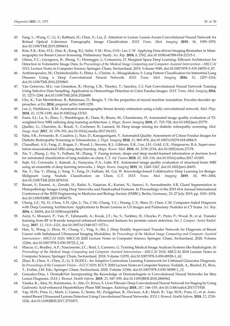
Diagnostics 2021, 11, 1373 50 of 50
43. Fang, L.; Wang, C.; Li, S.; Rabbani, H.; Chen, X.; Liu, Z. Attention to Lesion: Lesion-Aware Convolutional Neural Network for
Retinal Optical Coherence Tomography Image Classification. IEEE Trans. Med. Imaging 2019, 38, 1959–1970,
doi:10.1109/TMI.2019.2898414.
44. Kim, E.K.; Kim, H.E.; Han, K.; Kang, B.J.; Sohn, Y.M.; Woo, O.H.; Lee, C.W. Applying Data-driven Imaging Biomarker in Mam-
mography for Breast Cancer Screening: Preliminary Study. Sci. Rep. 2018, 8, 2762, doi:10.1038/s41598-018-21215-1.
45. Ghesu, F.C.; Georgescu, B.; Zheng, Y.; Hornegger, J.; Comaniciu, D. Marginal Space Deep Learning: Efficient Architecture for
Detection in Volumetric Image Data. In Proceedings of the Medical Image Computing and Computer-Assisted Intervention—MICCAI
2015; Lecture Notes in Computer Science; Springer: Cham, Switzerland, 2015; Volume 9349, doi:10.1007/978-3-319-24553-9_87.
46. Anthimopoulos, M.; Christodoulidis, S.; Ebner, L.; Christe, A.; Mougiakakou, S. Lung Pattern Classification for Interstitial Lung
Diseases Using a Deep Convolutional Neural Network. IEEE Trans. Med. Imaging 2016, 35, 1207–1216,
doi:10.1109/TMI.2016.2535865.
47. Van Grinsven, M.J.; van Ginneken, B.; Hoyng, C.B.; Theelen, T.; Sanchez, C.I. Fast Convolutional Neural Network Training
Using Selective Data Sampling: Application to Hemorrhage Detection in Color Fundus Images. IEEE Trans. Med. Imaging 2016,
35, 1273–1284, doi:10.1109/TMI.2016.2526689.
48. Cho, K.; Van Merriënboer, B.; Bahdanau, D.; Bengio, Y. On the properties of neural machine translation: Encoder-decoder ap-
proaches. arXiv 2014, preprint arXiv:1409.1259.
49. Lee, J.; Nishikawa, R.M. Automated mammographic breast density estimation using a fully convolutional network. Med. Phys.
2018, 45, 1178–1190, doi:10.1002/mp.12763.
50. Esses, S.J.; Lu, X.; Zhao, T.; Shanbhogue, K.; Dane, B.; Bruno, M.; Chandarana, H. Automated image quality evaluation of T2-
weighted liver MRI utilizing deep learning architecture. J. Magn. Reson. Imaging 2018, 47, 723–728, doi:10.1002/jmri.25779.
51. Quellec, G.; Charrière, K.; Boudi, Y.; Cochener, B.; Lamard, M. Deep image mining for diabetic retinopathy screening. Med.
Image Anal. 2017, 39, 178–193, doi:10.1016/j.media.2017.04.012.
52. Saha, S.K.; Fernando, B.; Cuadros, J.; Xiao, D.; Kanagasingam, Y. Automated Quality Assessment of Colour Fundus Images for
Diabetic Retinopathy Screening in Telemedicine. J. Digit. Imaging 2018, 31, 869–878, doi:10.1007/s10278-018-0084-9.
53. Chaudhari, A.S.; Fang, Z.; Kogan, F.; Wood, J.; Stevens, K.J.; Gibbons, E.K.; Lee, J.H.; Gold, G.E.; Hargreaves, B.A. Super-reso-
lution musculoskeletal MRI using deep learning. Magn. Reson. Med. 2018, 80, 2139–2154, doi:10.1002/mrm.27178.
54. Xie, Y.; Zhang, J.; Xia, Y.; Fulham, M.; Zhang, Y. Fusing texture, shape and deep model-learned information at decision level
for automated classification of lung nodules on chest, C.T. Inf. Fusion 2018, 42, 102–110, doi:10.1016/j.inffus.2017.10.005.
55. Sujit, S.J.; Coronado, I.; Kamali, A.; Narayana, P.A.; Gabr, R.E. Automated image quality evaluation of structural brain MRI
using an ensemble of deep learning networks. J. Magn. Reson. Imaging 2019, 50, 1260–1267, doi:10.1002/jmri.26693.
56. Xie, Y.; Xia, Y.; Zhang, J.; Song, Y.; Feng, D.; Fulham, M.; Cai, W. Knowledge-based Collaborative Deep Learning for Benign-
Malignant Lung Nodule Classification on Chest, C.T. IEEE Trans. Med. Imaging 2019, 38, 991–1004,
doi:10.1109/TMI.2018.2876510.
57. Rezaei, S.; Emami, A.; Zarrabi, H.; Rafiei, S.; Najarian, K.; Karimi, N.; Samavi, S.; Soroushmehr, S.R. Gland Segmentation in
Histopathology Images Using Deep Networks and Handcrafted Features. In Proceedings of the 2019 41st Annual International
Conference of the IEEE Engineering in Medicine and Biology Society (EMBC), Berlin, Germany, 23–27 July 2019; pp. 1031–1034,
doi:10.1109/EMBC.2019.8856776.
58. Cheng, J.Z.; Ni, D.; Chou, Y.H.; Qin, J.; Tiu, C.M.; Chang, Y.C.; Huang, C.S.; Shen, D.; Chen, C.M. Computer-Aided Diagnosis
with Deep Learning Architecture: Applications to Breast Lesions in US Images and Pulmonary Nodules in CT Scans. Sci. Rep.
2016, 6, 24454, doi:10.1038/srep24454.
59. Azizi, S.; Mousavi, P.; Yan, P.; Tahmasebi, A.; Kwak, J.T.; Xu, S.; Turkbey, B.; Choyke, P.; Pinto, P.; Wood, B.; et al. Transfer
learning from RF to B-mode temporal enhanced ultrasound features for prostate cancer detection. Int. J. Comput. Assist Radiol.
Surg. 2017, 12, 1111–1121, doi:10.1007/s11548-017-1573-x.
60. Han, X.; Wang, J.; Zhou, W.; Chang, C.; Ying, S.; Shi, J. Deep Doubly Supervised Transfer Network for Diagnosis of Breast
Cancer with Imbalanced Ultrasound Imaging Modalities. In Proceedings of the Medical Image Computing and Computer Assisted
Intervention—MICCAI 2020; MICCAI 2020 Lecture Notes in Computer Science; Springer: Cham, Switzerland, 2020; Volume
12266, doi:10.1007/978-3-030-59725-2_14.
61. Maicas, G.; Bradley, A.P.; Nascimento, J.C.; Reid, I.; Carneiro, G. Training Medical Image Analysis Systems like Radiologists. In
Proceedings of the Medical Image Computing and Computer Assisted Intervention—MICCAI 2018; MICCAI 2018 Lecture Notes in
Computer Science; Springer: Cham, Switzerland, 2018; Volume 11070, doi:10.1007/978-3-030-00928-1_62.
62. Zhao, R.; Chen, X.; Chen, Z.; Li, S. EGDCL: An Adaptive Curriculum Learning Framework for Unbiased Glaucoma Diagnosis.
In Proceedings of the Computer Vision—ECCV 2020; ECCV 2020 Lecture Notes in Computer Science; Vedaldi, A., Bischof, H., Brox,
T., Frahm, J.M. Eds.; Springer: Cham, Switzerland, 2020; Volume 12366, doi:10.1007/978-3-030-58589-1_12.
63. Gonzalez-Diaz, I. DermaKNet: Incorporating the Knowledge of Dermatologists to Convolutional Neural Networks for Skin
Lesion Diagnosis. IEEE J. Biomed. Health Inform. 2019, 23, 547–559, doi:10.1109/JBHI.2018.2806962.
64. Yasaka, K.; Akai, H.; Kunimatsu, A.; Abe, O.; Kiryu, S. Liver Fibrosis: Deep Convolutional Neural Network for Staging by Using
Gadoxetic Acid-enhanced Hepatobiliary Phase MR Images. Radiology 2018, 287, 146–155, doi:10.1148/radiol.2017171928.
65. Yap, M.H.; Pons, G.; Marti, J.; Ganau, S.; Sentis, M.; Zwiggelaar, R.; Davison, A.K.; Marti, R.; Yap, M.H.; Pons, G.; et al. Auto-
mated Breast Ultrasound Lesions Detection Using Convolutional Neural Networks. IEEE J. Biomed. Health Inform. 2018, 22, 1218–
1226, doi:10.1109/JBHI.2017.2731873.
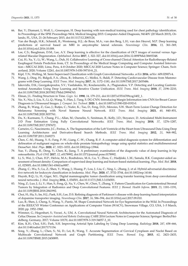
Diagnostics 2021, 11, 1373 51 of 50
66. Bar, Y.; Diamant, I.; Wolf, L.; Greenspan, H. Deep learning with non-medical training used for chest pathology identification.
In Proceedings of the SPIE Proceedings 9414, Medical Imaging 2015: Computer-Aided Diagnosis, 94140V (20 March 2015), Or-
lando, FL, USA, 21–26 February 2015, doi:10.1117/12.2083124.
67. Van der Burgh, H.K.; Schmidt, R.; Westeneng, H.J.; de Reus, M.A.; van den Berg, L.H.; van den Heuvel, M.P. Deep learning
predictions of survival based on MRI in amyotrophic lateral sclerosis. Neuroimage Clin. 2016, 13, 361–369,
doi:10.1016/j.nicl.2016.10.008.
68. Lee, C.S.; Baughman, D.M.; Lee, A.Y. Deep learning is effective for the classification of OCT images of normal versus Age-
related Macular Degeneration. Ophthalmol. Retina 2017, 1, 322–327, doi:10.1016/j.oret.2016.12.009PMid:30693348.
69. Cui, H.; Xu, Y.; Li, W.; Wang, L.; Duh, H. Collaborative Learning of Cross-channel Clinical Attention for Radiotherapy-Related
Esophageal Fistula Prediction from, CT. In Proceedings of the Medical Image Computing and Computer Assisted Interven-
tion—MICCAI 2020, Lima, Peru, 4–8 October 2020; MICCAI 2020 Lecture Notes in Computer Science; Springer: Cham, Swit-
zerland, 2020; Volume 12261, doi:10.1007/978-3-030-59710-8_21.
70. Kipf, T.N.; Welling, M. Semi-Supervised Classification with Graph Convolutional Networks. arXiv 2016, arXiv: 609.02907v4.
71. Wang, J.; Ding, H.; Bidgoli, F.A.; Zhou, B.; Iribarren, C.; Molloi, S.; Baldi, P. Detecting Cardiovascular Disease from Mammo-
grams with Deep Learning. IEEE Trans. Med. Imaging 2017, 36, 1172–1181, doi:10.1109/TMI.2017.2655486.
72. Iakovidis, D.K.; Georgakopoulos, S.V.; Vasilakakis, M.; Koulaouzidis, A.; Plagianakos, V.P. Detecting and Locating Gastroin-
testinal Anomalies Using Deep Learning and Iterative Cluster Unification. IEEE Trans. Med. Imaging 2018, 37, 2196–2210,
doi:10.1109/TMI.2018.2837002 PMid:29994763.
73. Elman, J.L. Finding Structure in Time. Cogn. Sci. 1990, 14, 179–211, doi:10.1207/s15516709cog1402_1.
74. Xie, X.; Niu, J.; Liu, X.; Li, Q.; Wang, Y.; Han, J.; Tang, S. DG-CNN: Introducing Margin Information into CNN for Breast Cancer
Diagnosis in Ultrasound Images. J. Comput. Sci. Technol. 2020, 1, doi:10.1007/s11390-020-0192-0.
75. Zhang, B.; Wang, Z.; Gao, J.; Rutjes, C.; Nufer, K.; Tao, D.; Feng, D.D.; Menzies, S.W. Short-Term Lesion Change Detection for
Melanoma Screening with Novel Siamese Neural Network. IEEE Trans. Med. Imaging 2021, 40, 840–851,
doi:10.1109/TMI.2020.3037761.
76. Du, X.; Kurmann, T.; Chang, P.L.; Allan, M.; Ourselin, S.; Sznitman, R.; Kelly, J.D.; Stoyanov, D. Articulated Multi-Instrument
2-D Pose Estimation Using Fully Convolutional Networks. IEEE Trans. Med. Imaging 2018, 37, 1276–1287,
doi:10.1109/TMI.2017.2787672.
77. Carneiro, G.; Nascimento, J.C.; Freitas, A. The Segmentation of the Left Ventricle of the Heart from Ultrasound Data Using Deep
Learning Architectures and Derivative-Based Search Methods. IEEE Trans. Med. Imaging 2012, 21, 968–982,
doi:10.1109/TIP.2011.2169273.
78. Chen, C.-M.; Huang, Y.-S.; Fang, P.-W.; Liang, C.-W.; Chang, R.-F. A computer-aided diagnosis system for differentiation and
delineation of malignant regions on whole-slide prostate histopathology image using spatial statistics and multidimensional
DenseNet. Med. Phys. 2020, 47, 1021–1033, doi:10.1002/mp.13964.
79. Xue, Y.; Zhang, R.; Deng, Y.; Chen, K.; Jiang, T. A preliminary examination of the diagnostic value of deep learning in hip
osteoarthritis. PLoS ONE 2017, 12, e0178992, doi:10.1371/journal.pone.0178992.
80. Li, S.; Wei, J.; Chan, H.P.; Helvie, M.A.; Roubidoux, M.A.; Lu, Y.; Zhou, C.; Hadjiiski, L.M.; Samala, R.K. Computer-aided as-
sessment of breast density: Comparison of supervised deep learning and feature-based statistical learning. Phys. Med. Biol. 2018,
63, 025005, doi:10.1088/1361-6560/aa9f87.
81. Zhang, C.; Wu, S.; Lu, Z.; Shen, Y.; Wang, J.; Huang, P.; Lou, J.; Liu, C.; Xing, L.; Zhang, J.; et al. Hybrid adversarial-discrimina-
tive network for leukocyte classification in leukemia. Med. Phys. 2020, 47, 3732–3744, doi:10.1002/mp.14144.
82. Huynh, B.Q.; Li, H.; Giger, M.L. Digital mammographic tumor classification using transfer learning from deep convolutional
neural networks. J. Med. Imaging 2016, 3, 034501, doi:10.1117/1.JMI.3.3.034501.
83. Ning, Z.; Luo, J.; Li, Y.; Han, S.; Feng, Q.; Xu, Y.; Chen, W.; Chen, T.; Zhang, Y. Pattern Classification for Gastrointestinal Stromal
Tumors by Integration of Radiomics and Deep Convolutional Features. IEEE J. Biomed. Health Inform. 2019, 23, 1181–1191,
doi:10.1109/JBHI.2018.2841992.
84. Choi, H.; Ha, S.; Im, H.J.; Paek, S.H.; Lee, D.S. Refining diagnosis of Parkinson’s disease with deep learning-based interpretation
of dopamine transporter imaging. NeuroImage Clin. 2017, 16, 586–594, doi:10.1016/j.nicl.2017.09.010.
85. Luo, B.; Shen, J.; Cheng, S.; Wang, Y.; Pantic, M. Shape Constrained Network for Eye Segmentation in the Wild. In Proceedings
of the IEEE/CVF Winter Conference on Applications of Computer Vision (WACV), Snowmass Village, CO, USA, 1–5 March,
2020; pp. 1952–1960.
86. Wimmer, G.; Hegenbart, S.; Vecsei, A.; Uhl, A. Convolutional Neural Network Architectures for the Automated Diagnosis of
Celiac Disease. In Computer-Assisted and Robotic Endoscopy; CARE 2016 Lecture Notes in Computer Science; Springer: Berlin/Hei-
delberg, Germany, 2017; Volume 10170, doi:10.1007/978-3-319-54057-3_10.
87. Kim, K.H.; Choi, S.H.; Park, S.H. Improving Arterial Spin Labeling by Using Deep Learning. Radiology 2018, 287, 658–666,
doi:10.1148/radiol.2017171154.
88. Song, Y.; Zhang, L.; Chen, S.; Ni, D.; Lei, B.; Wang, T. Accurate Segmentation of Cervical Cytoplasm and Nuclei Based on
Multiscale Convolutional Network and Graph Partitioning. IEEE Trans. Biomed. Eng. 2015, 62, 2421–2433,
doi:10.1109/TBME.2015.2430895.
Diagnostics 2021, 11, 1373 52 of 50
89. Moradi, M.; Gur, Y.; Wang, H.; Prasanna, P.; Syeda-Mahmood, T. A hybrid learning approach for semantic labeling of cardiac
CT slices and recognition of body position. In Proceedings of the 2016 IEEE 13th International Symposium on Biomedical Im-
aging (ISBI), Prague, Czech Republic, 13–16 April 2016; pp. 1418–1421, doi:10.1109/ISBI.2016.7493533.
90. Khosravan, N.; Celik, H.; Turkbey, B.; Jones, E.C.; Wood, B.; Bagci, U. A collaborative computer aided diagnosis (C-CAD) sys-
tem with eye-tracking, sparse attentional model, and deep learning. Med. Image Anal. 2019, 51, 101–115, doi:10.1016/j.me-
dia.2018.10.010.
91. Betancur, J.; Commandeur, F.; Motlagh, M.; Sharir, T.; Einstein, A.J.; Bokhari, S.; Fish, M.B.; Ruddy, T.D.; Kaufmann, P.; Sinusas,
A.J.; et al. Deep Learning for Prediction of Obstructive Disease From Fast Myocardial Perfusion SPECT: A Multicenter Study.
JACC Cardiovasc. Imaging 2018, 11, 1654–1663, doi:10.1016/j.jcmg.2018.01.020.
92. Shin, H.C.; Roth, H.R.; Gao, M.; Lu, L.; Xu, Z.; Nogues, I.; Yao, J.; Mollura, D.; Summers, R.M. Deep Convolutional Neural
Networks for Computer-Aided Detection: CNN Architectures, Dataset Characteristics and Transfer Learning. IEEE Trans. Med.
Imaging 2016, 35, 1285–1298, doi:10.1109/TMI.2016.2528162.
93. Mitsuhara, M.; Fukui, H.; Sakashita, Y.; Ogata, T.; Hirakawa, T.; Yamashita, T.; Fujiyoshi, H. Embedding Human Knowledge
into Deep Neural Network via Attention Map. arXiv 2019, arXiv:1905.03540v4.
94. Jiang, F.; Jiang, Y,; Zhi, H.; Dong, Y.; Li, H.; Ma, S.; Wang, Y.; Dong, Q.; Shen, H.; Wang, Y. Artificial intelligence in healthcare:
Past, present and future. Stroke Vasc. Neurol. 2017, 2, 230–243.
95. Bakator, M.; Radosav, D. Deep Learning and Medical Diagnosis: A Review of Literature. Multimodal Technol. Interact. 2018, 2,
47, doi:10.3390/mti2030047.
96. Hecht-Nielsen, R. Neurocomputing: Picking the human brain. IEEE Spectr. 1988, 25, 36–41, doi:10.1109/6.4520.
97. Krizhevsky, A.; Sutskever, I.; Hinton, G.E. ImageNet Classification with Deep Convolutional Neural Networks. In Advances in
Neural Information Processing Systems 25; Pereira, F., Burges, C.J.C., Bottou, L., Weinberger, K.Q., Eds.; 2012; pp. 1097–1105.
98. Arasu, A.; Garcia-Molina, H. Extracting Structured Data from Web Pages. In Proceedings of the 2003 ACM SIGMOD Interna-
tional Conference on Management of Data, San Diego, CA, USA, 9–12 June 2003; pp. 337–348, doi:10.1145/872757.872799.
99. Vizcarra, J.; Place, R.; Tong, L.; Gutman, D.; Wang, M.D. Fusion in Breast Cancer Histology Classification. ACM BCB 2019, 2019,
485–493, doi:10.1145/3307339.3342166.
100. Velicer, W.F.; Molenaar, P.C. Time Series Analysis for Psychological Research. In Handbook of Psychology, 2nd ed.; Weiner, I.,
Schinka, J.A., Velicer, W.F., Eds.; Wiley: Hoboken, NJ, USA, 2012; doi:10.1002/9781118133880.hop202022.
101. LeCun, Y.; Boser, B.; Denker, J.S.; Henderson, D.; Howard, R.E.; Hubbard, W.; Jackel, L.D. Backpropagation Applied to Hand-
written Zip Code Recognition. Neural Comput. 1989, 1, 541–551, doi:10.1162/neco.1989.1.4.541.
102. Ursuleanu, T.F.; Luca, A.R.; Gheorghe, L.; Grigorovici, R.; Iancu, S.; Hlusneac, M.; Preda, C.; Grigorovici, A. Unified Analysis
Specific to the Medical Field in the Interpretation of Medical Images through the Use of Deep Learning. E-Health Telecommun.
Syst. Netw. 2021, 10, 41–74, doi:10.4236/etsn.2021.102003.
103. Pandey, B.; Pandey, D.K.; Mishra, B.P.; Rhmann, W. A comprehensive survey of deep learning in the field of medical imaging
and medical natural language processing: Challenges and research directions. J. King Saud Univ. Comput. Inf. Sci. 2021,
doi:10.1016/j.jksuci.2021.01.007.
104. Alzubaidi, L.; Zhang, J.; Humaidi, A.J.; Al-Dujaili, A.; Duan, Y.; Al-Shamma, O.; Santamaría, J.; Fadhel, M.A.; Al-Amidie, M.;
Farhan, L. Review of deep learning: Concepts, CNN architectures, challenges, applications, future directions. J. Big Data 2021,
8, 53, doi:10.1186/s40537-021-00444-8.
105. Wang, X.; Liang, G.; Zhang, Y.; Blanton, H.; Bessinger, Z.; Jacobs, N. Inconsistent Performance of Deep Learning Models on
Mammogram Classification. J. Am. Coll. Radiol. 2020, 17, 796–803, doi:10.1016/j.jacr.2020.01.006.
106. McKinney, S.M.; Sieniek, M.; Godbole, V.; Godwin, J.; Antropova, N.; Ashrafian, H.; Back, T.; Chesus, M.; Corrado, G.S.; Darzi,
A.; et al. International evaluation of an AI system for breast cancer screening. Nat. Cell Biol. 2020, 577, 89–94, doi:10.1038/s41586-
019-1799-6.
107. Bai, J.; Posner, R.; Wang, T.; Yang, C.; Nabavi, S. Applying deep learning in digital breast tomosynthesis for automatic breast
cancer detection: A review. Med. Image Anal. 2021, 71, 102049, doi:10.1016/j.media.2021.102049.
108. Simonyan, K.; Zisserman, A. Very Deep Convolutional Networks for Large-Scale Image Recognition. arXiv 2014,
arXiv:1409.1556v6.
109. León, J.; Escobar, J.J.; Ortiz, A.; Ortega, J.; González, J.; Martín-Smith, P.; Gan, J.Q.; Damas, M. Deep learning for EEG-based
Motor Imagery classification: Accuracy-cost trade-off. PLoS ONE 2020, 15, e0234178, doi:10.1371/journal.pone.0234178.
110. Hochreiter, S.; Schmidhuber, J. Long short-term memory. Neural Comput. 1997, 9, 1735–1780, doi:10.1162/neco.1997.9.8.1735.
111. Saravanan, S.; Juliet, S. Deep Medical Image Reconstruction with Autoencoders using Deep Boltzmann Machine Training. EAI
Endorsed Trans. Pervasive Health Technol. 2020, 6, e2, doi:10.4108/eai.24-9-2020.166360.
112. Gondara, L. Medical Image Denoising Using Convolutional Denoising Autoencoders. In Proceedings of the 2016 IEEE 16th
International Conference on Data Mining Workshops (ICDMW), Barcelona, Spain, 12–15 December 2016; pp. 241–246.,
doi:10.1109/ICDMW.2016.0041.
113. Zhou, B.; Khosla, A.; Lapedriza, A.; Torralba, A.; Oliva, A. Places: An Image Database for Deep Scene Understanding. arXiv
2016, arXiv: 1610.02055v1.
114. Nowling, R.J.; Bukowy, J.; McGarry, S.D.; Nencka, A.S.; Blasko, O.; Urbain, J.; Lowman, A.; Barrington, A.; Banerjee, A.; Icz-
kowski, K.A.; et al. Classification before Segmentation: Improved U-Net Prostate Segmentation. In Proceedings of the 2019 IEEE
Diagnostics 2021, 11, 1373 53 of 50
EMBS International Conference on Biomedical & Health Informatics (BHI), Chicago, IL, USA ,19–22 May 2019; pp. 1–4,
doi:10.1109/BHI.2019.8834494.z.
115. Yu, E.M.; Iglesias, J.E.; Dalca, A.V.; Sabuncu, M.R. An Auto-Encoder Strategy for Adaptive Image Segmentation. In Proceedings
of the Third Conference on Medical Imaging with Deep Learning (PMLR), Montreal, QC, Canada, 6–8 July 2020; Volume 121,
pp. 881–891.
116. Uzunova, H.; Schultz, S.; Handels, H.; Ehrhardt, J. Unsupervised pathology detection in medical images using conditional var-
iational autoencoders. Int. J. CARS 2019, 14, 451–461, doi:10.1007/s11548-018-1898-0.
117. Chen, M.; Shi, X.; Zhang, Y.; Wu, D.; Guizani, M. Deep Features Learning for Medical Image Analysis with Convolutional
Autoencoder Neural Network. IEEE Trans. Big Data 2017, 1, doi:10.1109/tbdata.2017.2717439.
118. Saltz, J.; Gupta, R.; Hou, L.; Kurc, T.; Singh, P.; Nguyen, V.; Samaras, D.; Shroyer, K.R.; Zhao, T.; Batiste, R.; et al. Spatial Organ-
ization and Molecular Correlation of Tumor-Infiltrating Lymphocytes Using Deep Learning on Pathology Images. Cell Rep.
2018, 23, 181–193.e7, doi:10.1016/j.celrep.2018.03.086.
119. Rumelhart, D.; Hinton, G.; Williams, R. Learning representations by back-propagating errors. Nature 1986, 323, 533–536.
120. Lam, C.; Yu, C.; Huang, L.; Rubin, D. Retinal Lesion Detection with Deep Learning Using Image Patches. Investig. Opthalmol.
Vis. Sci. 2018, 59, 590–596, doi:10.1167/iovs.17-22721.
121. Rajaraman, S.; Antani, S.; Poostchi, M.; Silamut, K.; Hossain, A.; Maude, R.; Jaeger, S.; Thoma, G.R. Pre-trained convolutional
neural networks as feature extractors toward improved malaria parasite detection in thin blood smear images. PeerJ 2018, 6,
e4568, doi:10.7717/peerj.4568.
122. Nielsen, A.; Hansen, M.B.; Tietze, A.; Mouridsen, K. Prediction of Tissue Outcome and Assessment of Treatment Effect in Acute
Ischemic Stroke Using Deep Learning. Stroke 2018, 49, 1394–1401, doi:10.1161/strokeaha.117.019740.
123. Lee, H.C.; Ryu, H.G.; Chung, E.J.; Jung, C.W. Prediction of Bispectral Index during Target-controlled Infusion of Propofol and
Remifentanil: A Deep Learning Approach. Anesthesiology 2018, 128, 492–501., doi:10.1097/ALN.0000000000001892.
124. Zeng, L.L.; Wang, H.; Hu, P.; Yang, B.; Pu, W.; Shen, H.; Chen, X.; Liu, Z.; Yin, H.; Tan, Q.; et al. Multi-Site Diagnostic Classifi-
cation of Schizophrenia Using Discriminant Deep Learning with Functional Connectivity MRI. EBioMedicine 2018, 30, 74–85,
doi:10.1016/j.ebiom.2018.03.017.
125. Kooi, T.; Van Ginneken, B.; Karssemeijer, N.; den Heeten, A. Discriminating solitary cysts from soft tissue lesions in mammog-
raphy using a pretrained deep convolutional neural network. Med. Phys. 2017, 44, 1017–1027, doi:10.1002/mp.12110.
126. Heinsfeld, A.S.; Franco, A.R.; Craddock, R.C.; Buchweitz, A.; Meneguzzi, F. Identification of autism spectrum disorder using
deep learning and the ABIDE dataset. NeuroImage Clin. 2018, 17, 16–23, doi:10.1016/j.nicl.2017.08.017.
127. Wang, J.; Yang, X.; Cai, H.; Tan, W.; Jin, C.; Li, L. Discrimination of Breast Cancer with Microcalcifications on Mammography
by Deep Learning. Sci. Rep. 2016, 6, 27327, doi:10.1038/srep27327.
128. Fu, H.; Cheng, J.; Xu, Y.; Zhang, C.; Wong, D.W.K.; Liu, J.; Cao, X. Disc-Aware Ensemble Network for Glaucoma Screening
From Fundus Image. IEEE Trans. Med. Imaging 2018, 37, 2493–2501, doi:10.1109/tmi.2018.2837012.
129. Yu, C.; Yang, S.; Kim, W.; Jung, J.; Chung, K.-Y.; Lee, S.W.; Oh, B. Correction: Acral melanoma detection using a convolutional
neural network for dermoscopy images. PLoS ONE 2018, 13, e0196621, doi:10.1371/journal.pone.0196621.
130. Han, S.S.; Park, G.H.; Lim, W.; Kim, M.S.; Na, J.I.; Park, I.; Chang, S.E. Deep neural networks show an equivalent and often
superior performance to dermatologists in onychomycosis diagnosis: Automatic construction of onychomycosis datasets by
region-based convolutional deep neural network. PLoS ONE 2018, 13, e0191493, doi:10.1371/journal.pone.0191493.
131. Hsieh, Y.J.; Tseng, H.C.; Chin, C.L.; Shao, Y.H.; Tsai, T.Y. Based on DICOM RT Structure and Multiple Loss Function Deep
Learning Algorithm in Organ Segmentation of Head and Neck Image. In Proceedings of the Future Trends in Biomedical and
Health Informatics and Cybersecurity in Medical Devices: Proceedings of the International Conference on Biomedical and
Health Informatics, ICBHI 2019, Taipei, Taiwan, 17–20 April 2019; Springer: Berlin/Heidelberg, Germany, 2020; Volume 74, pp.
428–435, doi:10.1007/978-3-030-30636-6_58.
132. Ngo, T.A.; Lu, Z.; Carneiro, G. Combining deep learning and level set for the automated segmentation of the left ventricle of
the heart from cardiac cine magnetic resonance. Med. Image Anal. 2017, 35, 159–171, doi:10.1016/j.media.2016.05.009.
133. Hinton, G.; Vinyals, O.; Dean, J. Distilling the knowledge in a neural network. arXiv 2015, arXiv:1503.02531.
134. Araújo, T.; Aresta, G.; Castro, E.M.; Rouco, J.; Aguiar, P.; Eloy, C.; Polónia, A.; Campilho, A. Classification of breast cancer
histology images using Convolutional Neural Networks. PLoS ONE 2017, 12, e0177544, doi:10.1371/journal.pone.0177544.
135. Han, Z.; Wei, B.; Zheng, Y.; Yin, Y.; Li, K.; Li, S. Breast Cancer Multi-classification from Histopathological Images with Struc-
tured Deep Learning Model. Sci. Rep. 2017, 7, 4172, doi:10.1038/s41598-017-04075-z.
136. Zhang, X.; Yu, F.X.; Chang, S.; Wang, S. Deep Transfer Network: Unsupervised Domain Adaptation. arXiv 2015,
arXiv:1503.00591v1.
137. Hutchinson, B.; Deng, L.; Yu, D. Tensor deep stacking networks. IEEE Trans. Pattern Anal. Mach. Intell. 2012, 35, 1944–1957,
doi:10.1109/TPAMI.2012.268.
138. Hjelm, R.D.; Fedorov, A.; Lavoie-Marchildon, S.; Grewal, K.; Bachman, P.; Trischler, A.; Bengio, Y. Learning deep representa-
tions by mutual information estimation and maximization. arXiv 2018, arXiv: 1808.06670.
139. Alom, M.Z.; Yakopcic, C.; Nasrin, M.S.; Taha, T.M.; Asari, V.K. Breast Cancer Classification from Histopathological Images with
Inception Recurrent Residual Convolutional Neural Network. J. Digit. Imaging 2019, 32, 605–617, doi:10.1007/s10278-019-00182-
7.
Diagnostics 2021, 11, 1373 54 of 50
140. Zhao, Y.; Dong, Q.; Zhang, S.; Zhang, W.; Chen, H.; Jiang, X.; Guo, L.; Hu, X.; Han, J.; Liu, T. Automatic Recognition of fMRI-
Derived Functional Networks Using 3-D Convolutional Neural Networks. IEEE Trans. Biomed. Eng. 2018, 65, 1975–1984,
doi:10.1109/tbme.2017.2715281.
141. Tiulpin, A.; Thevenot, J.; Rahtu, E.; Lehenkari, P.; Saarakkala, S. Automatic Knee Osteoarthritis Diagnosis from Plain Radio-
graphs: A Deep Learning-Based Approach. Sci. Rep. 2018, 8, 1727, doi:10.1038/s41598-018-20132-7.
142. Zhang, Z.; Liang, X.; Dong, X.; Xie, Y.; Cao, G. A Sparse-View CT Reconstruction Method Based on Combination of DenseNet
and Deconvolution. IEEE Trans. Med. Imaging 2018, 37, 1407–1417, doi:10.1109/tmi.2018.2823338.
143. Luca, A.R.; Ursuleanu, T.F.; Gheorghe, L.; Grigorovici1, R.; Iancu, S.; Hlusneac, M.; Preda, C.; Grigorovici, A. Designing a High-
Performance Deep Learning Theoretical Model for Biomedical Image Segmentation by Using Key Elements of the Latest U-Net-
Based Architectures. J. Comput. Commun. 2021, 9, 8–20., doi:10.4236/jcc.2021.97002.
144. Huang, C.H.; Wu, H.Y.; Lin, Y.L. HarDNet-MSEG: A Simple Encoder-Decoder Polyp Segmentation Neural Network that
Achieves Over 0.9 Mean Dice and 86 FPS. arXiv 2021, arXiv:2101.07172.
145. Haryanto, T.; Wasito, I.; Suhartanto, H. Convolutional Neural Network (CNN) for gland images classification. In Proceedings
of the 2017 11th International Conference on Information & Communication Technology and System (ICTS), Surabaya, Indone-
sia, 31 October 2017; pp. 55–60, doi:10.1109/ICTS.2017.8265646.
146. Cao, H.; Bernard, S.; Heutte, L.; Sabourin, R. Improve the performance of transfer learning without fine-tuning using dissimi-
larity-based multi-view learning for breast cancer histology images. In Image Analysis and Recognition; Springer: Cham, Switzer-
land, 2018; pp. 779–787, doi:10.1007/978-3-319-93000-8_88.
147. Luo, X.; Mori, K.; Peters, T.M. Advanced Endoscopic Navigation: Surgical Big Data, Methodology, and Applications. Annu. Rev.
Biomed. Eng. 2018, 20, 221–251, doi:10.1146/annurev-bioeng-062117-120917.
148. Xiao, C.; Choi, E.; Sun, J. Opportunities and challenges in developing deep learning models using electronic health records data:
A systematic review. J. Am. Med. Inform. Assoc. 2018, 25, 1419–1428, doi:10.1093/jamia/ocy068.
149. Shickel, B.; Tighe, P.J.; Bihorac, A.; Rashidi, P. Deep EHR: A Survey of Recent Advances in Deep Learning Techniques for
Electronic Health Record (EHR) Analysis. IEEE J. Biomed. Heal. Inform. 2018, 22, 1589–1604, doi:10.1109/jbhi.2017.2767063.
150. Karkra, S.; Singh, P.; Kaur, K. Convolution Neural Network: A Shallow Dive in to Deep Neural Net Technology. Int. J. Recent
Technol. Eng. 2019, 8, 487–495, doi:10.35940/ijrte.b1092.0782s719.
151. Ranschaert, E.R.; Morozov, S.; Algra, P.R. Artificial Intelligence in Medical Imaging: Opportunities, Applications and Risks; Springer:
Berlin, Germany, 2019; doi:10.1007/978-3-319-94878-2.
152. Tsang, G.; Xie, X.; Zhou, S.M. Harnessing the Power of Machine Learning in Dementia Informatics Research: Issues, Opportu-
nities, and Challenges. IEEE Rev. Biomed. Eng. 2019, 13, 113–129, doi:10.1109/rbme.2019.2904488.
153. Haryanto, T.; Suhartanto, H.; Murni, A.; Kusmardi, K. Strategies to Improve Performance of Convolutional Neural Network on
Histopathological Images Classification. In Proceedings of the 2019 International Conference on Advanced Computer Science
and information Systems (ICACSIS), Bali, Indonesia, 12–13 October 2019; pp. 125–132, doi:10.1109/ICACSIS47736.2019.8979740.
154. Das, A.; Nair, M.S.; Peter, S.D. Computer-Aided Histopathological Image Analysis Techniques for Automated Nuclear Atypia
Scoring of Breast Cancer: A Review. J. Digit. Imaging 2020, 33, 1–31, doi:10.1007/s10278-019-00295-z.
155. Pattanaik, P.; Mittal, M.; Khan, M.Z.; Panda, S. Malaria detection using deep residual networks with mobile microscopy. J. King
Saud Univ. Comput. Inf. Sci. 2020, doi:10.1016/j.jksuci.2020.07.003.
156. Das, A.; Rad, P.; Choo, K.-K.R.; Nouhi, B.; Lish, J.; Martel, J. Distributed machine learning cloud teleophthalmology IoT for
predicting AMD disease progression. Futur. Gener. Comput. Syst. 2019, 93, 486–498, doi:10.1016/j.future.2018.10.050.
157. Kim, Y.D.; Noh, K.J.; Byun, S.J.; Lee, S.; Kim, T.; Sunwoo, L.; Lee, K.J.; Kang, S.-H.; Park, K.H.; Park, S.J. Effects of Hypertension,
Diabetes, and Smoking on Age and Sex Prediction from Retinal Fundus Images. Sci. Rep. 2020, 10, 4623, doi:10.1038/s41598-020-
61519-9.
158. Apostolopoulos, S.; Ciller, C.; De Zanet, S.; Wolf, S.; Sznitman, R. RetiNet: Automatic AMD identification in OCT volumetric
data. Invest. Ophthalmol. Vis. Sci. 2017, 58, 387.
159. Zhang, J.; Xia, Y.; Wu, Q.; Xie, Y. Classification of Medical Images and Illustrations in the Biomedical Literature Using Synergic
Deep Learning. arXiv 2017, arXiv:1706.09092.
160. Serj, M.F.; Lavi, B.; Hoff, G.; Valls, D.P. A Deep Convolutional Neural Network for Lung Cancer Diagnostic. arXiv 2018,
arXiv:1804.08170.
161. Jang, R.; Kim, N.; Jang, M.; Lee, K.H.; Lee, S.M.; Na Noh, H.; Seo, J.B. Assessment of the Robustness of Convolutional Neural
Networks in Labeling Noise by Using Chest X-Ray Images from Multiple Centers. JMIR Med. Inform. 2020, 8, e18089,
doi:10.2196/18089.
162. Bengio, Y.; Louradour, J.; Collobert, R.; Weston, J. Curriculum learning. In Proceedings of the 26th Annual International Con-
ference on Machine Learning—ICML 2009, Montreal, QC, Canada, 14–18 June 2009; pp. 41–48, doi:10.1145/1553374.1553380.
163. Guan, Q.; Huang, Y.; Zhong, Z.; Zheng, Z.; Zheng, L.; Yang, Y. Diagnose like a Radiologist: Attention Guided Convolutional
Neural Network for Thorax Disease Classification. arXiv 2018, arXiv:1801.09927v1.
164. Xia, X.; Gong, J.; Hao, W.; Yang, T.; Lin, Y.; Wang, S.; Peng, W. Comparison and Fusion of Deep Learning and Radiomics Fea-
tures of Ground-Glass Nodules to Predict the Invasiveness Risk of Stage-I Lung Adenocarcinomas in CT Scan. Front. Oncol.
2020, 10, 418, doi:10.3389/fonc.2020.00418.
Diagnostics 2021, 11, 1373 55 of 50
165. Jesson, A.; Guizard, N.; Ghalehjegh, S.H.; Goblot, D.; Soudan, F.; Chapados, N. CASED: Curriculum Adaptive Sampling for
Extreme Data Imbalance. In Proceedings of the Medical Image Computing and Computer Assisted Intervention—MICCAI 2017; MIC-
CAI 2017 Lecture Notes in Computer Science; Springer: Berlin/Heidelberg, Germany, 2017; Volume 10435, doi:10.1007/978-3-
319-66179-7_73.
166. Astudillo, P.; Mortier, P.; De Beule, M.; Wyffels, F. Curriculum Deep Reinforcement Learning with Different Exploration Strat-
egies: A Feasibility Study on Cardiac Landmark Detection. In Proceedings of the 13th International Joint Conference on Bio-
medical Engineering Systems and Technologies—Bioimaging, Valetta, Malta, 24–26 February 2020; pp. 37–45,
doi:10.5220/0008948900370045.
167. Li, C.Y.; Liang, X.; Hu, Z.; Xing, E.P. Knowledge-Driven Encode, Retrieve, Paraphrase for Medical Image Report Generation.
Proc. Conf. AAAI Artif. Intell. 2019, 33, 6666–6673, doi:10.1609/aaai.v33i01.33016666.
168. Ghafoorian, M.; Mehrtash, A.; Kapur, T.; Karssemeijer, N.; Marchiori, E.; Pesteie, M.; Guttmann, C.R.G.; De Leeuw, F.-E.; Tem-
pany, C.M.; Van Ginneken, B.; et al. Transfer Learning for Domain Adaptation in MRI: Application in Brain Lesion Segmenta-
tion. In Proceedings of the Medical Image Computing and Computer Assisted Intervention—MICCAI 2017: 20th International Conference,
Quebec City, QC, Canada, 11–13 September 2017; Lecture Notes in Computer Science; Springer: Berlin/Heidelberg, Germany, 2017;
Volume 10435, doi:10.1007/978-3-319-66179-7_59.
169. Jing, B.; Xie, P.; Xing, E. On the Automatic Generation of Medical Imaging Reports. arXiv 2017, arXiv:1711.08195.
170. Liu, G.; Hsu, T.-M.H.; McDermott, M.; Boag, W.; Weng, We.; Szolovits, P.; Ghassemi, M. Clinically Accurate Chest X-Ray Report
Generation. arXiv 2019, arXiv:1904.02633v2.
171. Gale, W.; Oakden-Rayner, L.; Carneiro, G.; Bradley, A.P.; Palmer, L.J. Producing radiologist-quality reports for interpretable
artificial intelligence. arXiv 2018, arXiv:1806.00340v1.
172. Zhang, Y.; Wei, Y.; Wu, Q.; Zhao, P.; Niu, S.; Huang, J.; Tan, M. Collaborative Unsupervised Domain Adaptation for Medical
Image Diagnosis. IEEE Trans. Image Process. 2020, 29, 7834–7844, doi:10.1109/tip.2020.3006377.
173. Tang, Y.; Wang, X.; Harrison, A.P.; Lu, L.; Xiao, J.; Summers, R.M. Attention-Guided Curriculum Learning for Weakly Super-
vised Classification and Localization of Thoracic Diseases on Chest Radiographs. In Proceedings of the Machine Learning in Medical
Imaging, MLMI 2018; Lecture Notes in Computer Science; Springer: Berlin/Heidelberg, Germany, 2018; Volume 11046,
doi:10.1007/978-3-030-00919-9_29.
174. Jiménez-Sánchez, A.; Mateus, D.; Kirchhoff, S.; Kirchhoff, C.; Biberthaler, P.; Navab, N.; Ballester, M.A.G.; Piella, G. Medical-
based Deep Curriculum Learning for Improved Fracture Classification. In Medical Image Computing and Computer Assisted Inter-
vention—MICCAI 2019; MICCAI 2019 Lecture Notes in Computer Science; Springer: Berlin/Heidelberg, Germany, 2019; Volume
11769, doi:10.1007/978-3-030-32226-7_77.
175. Jiménez-Sánchez, A.; Mateus, D.; Kirchhoff, S.; Kirchhoff, C.; Biberthaler, P.; Navab, N., Ballester, M.A.; Piella, G. Curriculum
learning for annotation-efficient medical image analysis: Scheduling data with prior knowledge and uncertainty. arXiv 2007,
arXiv:2007.16102v1.
176. Wei, J.; Suriawinata, A.; Ren, B.; Liu, X.; Lisovsky, M.; Vaickus, L.; Brown, C.; Baker, M.; Nasir-Moin, M.; Tomita, N.; et al. Learn
Like a Pathologist: Curriculum Learning by Annotator Agreement for Histopathology Image Classification. In Proceedings of
the IEEE/CVF Winter Conference on Applications of Computer Vision (WACV), Waikola, HI, USA, 5–9 January 2021; pp. 2473-
2483, doi:10.1109/WACV48630.2021.00252.
177. Wang, K.; Zhang, X.; Huang, S.; Chen, F.; Zhang, X.; Huangfu, L. Learning to Recognize Thoracic Disease in Chest X-Rays With
Knowledge-Guided Deep Zoom Neural Networks. IEEE Access 2020, 8, 159790–159805, doi:10.1109/access.2020.3020579.
178. Huang, X.; Fang, Y.; Lu, M.; Yan, F.; Yang, J.; Xu, Y. Dual-Ray Net: Automatic Diagnosis of Thoracic Diseases Using Frontal and
Lateral Chest X-rays. J. Med. Imaging Heal. Inform. 2020, 10, 348–355, doi:10.1166/jmihi.2020.2901.
179. Yang, Z.; Cao, Z.; Zhang, Y.; Han, M.; Xiao, J.; Huang, L.; Wu, S.; Ma, J.; Chang, P. MommiNet: Mammographic Multi-view
Mass Identification Networks. In Proceedings of the Medical Image Computing and Computer Assisted Intervention—MICCAI 2020;
MICCAI 2020 Lecture Notes in Computer Science; Springer: Berlin/Heidelberg, Germany, 2020; Volume 12266, doi:10.1007/978-
3-030-59725-2_20.
180. Liu, Q.; Yu, L.; Luo, L.; Dou, Q.; Heng, P.A. Semi-Supervised Medical Image Classification With Relation-Driven Self-Ensem-
bling Model. IEEE Trans. Med. Imaging 2020, 39, 3429–3440, doi:10.1109/tmi.2020.2995518.
181. Hsu, S.-M.; Kuo, W.-H.; Kuo, F.-C.; Liao, Y.-Y. Breast tumor classification using different features of quantitative ultrasound
parametric images. Int. J. Comput. Assist. Radiol. Surg. 2019, 14, 623–633, doi:10.1007/s11548-018-01908-8.
182. Antropova, N.; Huynh, B.Q.; Giger, M.L. A deep feature fusion methodology for breast cancer diagnosis demonstrated on three
imaging modality datasets. Med. Phys. 2017, 44, 5162–5171, doi:10.1002/mp.12453.
183. Xie, Y.; Zhang, J.; Liu, S.; Cai, W.; Xia, Y. Lung nodule classification by jointly using visual descriptors and deep features. In
Medical Computer Vision and Bayesian and Graphical Models for Biomedical Imaging; Springer, Cham, Germany, 2016; pp. 116–125,
doi:10.1007/978-3-319-61188-4_11.
184. Chai, Y.; Liu, H.; Xu, J. Glaucoma diagnosis based on both hidden features and domain knowledge through deep learning
models. Knowledge-Based Syst. 2018, 161, 147–156, doi:10.1016/j.knosys.2018.07.043.
185. Hagerty, J.R.; Stanley, R.J.; Almubarak, H.A.; Lama, N.; Kasmi, R.; Guo, P.; Drugge, R.J.; Rabinovitz, H.S.; Oliviero, M.; Stoecker,
W.V. Deep Learning and Handcrafted Method Fusion: Higher Diagnostic Accuracy for Melanoma Dermoscopy Images. IEEE
J. Biomed. Heal. Inform. 2019, 23, 1385–1391, doi:10.1109/jbhi.2019.2891049.
Diagnostics 2021, 11, 1373 56 of 50
186. Buty, M.; Xu, Z.; Gao, M.; Bagci, U.; Wu, A.; Mollura, D.J. Characterization of Lung Nodule Malignancy Using Hybrid Shape
and Appearance Features. In International Conference on Medical Image Computing and Computer-Assisted Intervention; Springer:
Cham, Switzerland, 2016; pp. 662–670, doi:10.1007/978-3-319-46720-7_77.
187. Saba, T.; Mohamed, A.S.; El-Affendi, M.; Amin, J.; Sharif, M. Brain tumor detection using fusion of hand crafted and deep
learning features. Cogn. Syst. Res. 2020, 59, 221–230, doi:10.1016/j.cogsys.2019.09.007.
188. Yang, J.; Dvornek, N.C.; Zhang, F.; Chapiro, J.; Lin, M.; Duncan, J.S. Unsupervised domain adaptation via disentangled repre-
sentations: Application to cross-modality liver segmentation. In International Conference on Medical Image Computing and Com-
puter-Assisted Intervention; Springer: Cham, Germany, 2019; pp. 255–263, doi:10.1007/978-3-030-32245-8_29.
189. Liu, T.; Guo, Q.; Lian, C.; Ren, X.; Liang, S.; Yu, J.; Niu, L.; Sun, W.; Shen, D. Automated detection and classification of thyroid
nodules in ultrasound images using clinical-knowledge-guided convolutional neural networks. Med. Image Anal. 2019, 58,
101555, doi:10.1016/j.media.2019.101555.
190. Feng, H.; Cao, J.; Wang, H.; Xie, Y.; Yang, D.; Feng, J.; Chen, B. A knowledge-driven feature learning and integration method
for breast cancer diagnosis on multi-sequence MRI. Magn. Reson. Imaging 2020, 69, 40–48, doi:10.1016/j.mri.2020.03.001.
191. Chen, S.; Qin, J.; Ji, X.; Lei, B.; Wang, T.; Ni, D.; Cheng, J.-Z. Automatic Scoring of Multiple Semantic Attributes With Multi-Task
Feature Leverage: A Study on Pulmonary Nodules in CT Images. IEEE Trans. Med. Imaging 2017, 36, 802–814,
doi:10.1109/tmi.2016.2629462.
192. Murthy, V.; Hou, L.; Samaras, D.; Kurc, T.M.; Saltz, J.H. Center-focusing multi-task CNN with injected features for classification
of glioma nuclear images. In Proceedings of the 2017 IEEE Winter Conference on Applications of Computer Vision (WACV),
Santa Rosa, CA, USA, 24–31 March 2017; pp. 834–841, doi:10.1109/WACV.2017.98.
193. Yu, S.; Zhou, H.Y.; Ma, K.; Bian, C.; Chu, C.; Liu, H.; Zheng, Y. Difficulty-Aware Glaucoma Classification with Multi-rater
Consensus Modeling. In International Conference on Medical Image Computing and Computer-Assisted Intervention; Springer: Cham,
Germany, 2020; pp. 741–750, doi:10.1007/978-3-030-59710-8_72.
194. Wang, W.; Lu, Y.; Wu, B.; Chen, T.; Chen, D.Z.; Wu, J. Deep active self-paced learning for accurate pulmonary nodule segmen-
tation. In International Conference on Medical Image Computing and Computer-Assisted Intervention; Springer: Cham, Switzerland,
2018; pp. 723–731, doi:10.1007/978-3-030-00934-2_80.
195. Roth, H.R.; Lu, L.; Seff, A.; Cherry, K.M.; Hoffman, J.; Wang, S.; Liu, J.; Turkbey, E.; Summers, R.M. A new 2.5 D representation
for lymph node detection using random sets of deep convolutional neural network observations. In International Conference on
Medical Image Computing and Computer-Assisted Intervention; Springer: Cham, Switzerland, 2014; pp. 520–527.
196. Tajbakhsh, N.; Shin, J.Y.; Gurudu, S.R.; Hurst, R.T.; Kendall, C.B.; Gotway, M.B.; Liang, J. Convolutional Neural Networks for
Medical Image Analysis: Full Training or Fine Tuning? IEEE Trans. Med. Imaging 2016, 35, 1299–1312,
doi:10.1109/tmi.2016.2535302.
197. Näppi, J.J.; Hironaka, T.; Regge, D.; Yoshida, H. Deep transfer learning of virtual endoluminal views for the detection of polyps
in CT colonography. In Medical Imaging 2016: Computer-Aided Diagnosis; International Society for Optics and Photonics: Belling-
ham, WA, USA, 2016; Volume 9785, p.97852B, doi:10.1117/12.2217260.
198. Zhang, R.; Zheng, Y.; Mak, T.W.C.; Yu, R.; Wong, S.H.; Lau, J.Y.W.; Poon, C.C.Y. Automatic Detection and Classification of
Colorectal Polyps by Transferring Low-Level CNN Features From Nonmedical Domain. IEEE J. Biomed. Heal. Inform. 2017, 21,
41–47, doi:10.1109/jbhi.2016.2635662.
199. Zhao, J.; Li, D.; Kassam, Z.; Howey, J.; Chong, J.; Chen, B.; Li, S. Tripartite-GAN: Synthesizing liver contrast-enhanced MRI to
improve tumor detection. Med. Image Anal. 2020, 63, 101667, doi:10.1016/j.media.2020.101667.
200. Zhang, J.; Saha, A.; Zhu, Z.; Mazurowski, M.A. Hierarchical Convolutional Neural Networks for Segmentation of Breast Tumors
in MRI With Application to Radiogenomics. IEEE Trans. Med. Imaging 2019, 38, 435–447, doi:10.1109/tmi.2018.2865671.
201. Setio, A.A.A.; Ciompi, F.; Litjens, G.; Gerke, P.; Jacobs, C.; van Riel, S.J.; Wille, M.M.W.; Naqibullah, M.; Sanchez, C.I.; van
Ginneken, B. Pulmonary Nodule Detection in CT Images: False Positive Reduction Using Multi-View Convolutional Networks.
IEEE Trans. Med. Imaging 2016, 35, 1160–1169, doi:10.1109/tmi.2016.2536809.
202. Gulshan, V.; Peng, L.; Coram, M.; Stumpe, M.C.; Wu, D.; Narayanaswamy, A.; Venugopalan, S.; Widner, K.; Madams, T.; Cuad-
ros, J.; et al. Development and Validation of a Deep Learning Algorithm for Detection of Diabetic Retinopathy in Retinal Fundus
Photographs. JAMA 2016, 316, 2402–2410, doi:10.1001/jama.2016.17216.
203. Liu, J.; Wang, D.; Lu, L.; Wei, Z.; Kim, L.; Turkbey, E.B.; Sahiner, B.; Petrick, N.A.; Summers, R.M. Detection and diagnosis of
colitis on computed tomography using deep convolutional neural networks. Med. Phys. 2017, 44, 4630–4642,
doi:10.1002/mp.12399.
204. Ruhan, S.; Owens, W.; Wiegand, R.; Studin, M.; Capoferri, D.; Barooha, K.; Greaux, A.; Rattray, R.; Hutton, A.; Cintineo, J.; et
al. Intervertebral disc detection in X-ray images using faster R-CNN. Annu. Int. Conf. IEEE Eng. Med. Biol. Soc. 2017, 2017, 564–
567, doi:10.1109/EMBC.2017.8036887.
205. Ben-Ari, R.; Akselrod-Ballin, A.; Karlinsky, L.; Hashoul, S. Domain specific convolutional neural nets for detection of architec-
tural distortion in mammograms. In Proceedings of the 2017 IEEE 14th International Symposium on Biomedical Imaging (ISBI
2017), Melbourne, VIC, Australia, 18–21 April 2017; pp. 552–556, doi:10.1109/isbi.2017.7950581.
206. Platania, R.; Shams, S.; Yang, S.; Zhang, J.; Lee, K.; Park, S.J. Automated breast cancer diagnosis using deep learning and region
of interest detection (BC-Droid). In Proceedings of the 8th ACM international conference on bioinformatics, computational bi-
ology, and health informatics, Boston, MA, USA, 20-23 August 2017; pp. 536–543, doi:10.1145/3107411.3107484.
Diagnostics 2021, 11, 1373 57 of 50
207. Li, N.; Liu, H.; Qiu, B.; Guo, W.; Zhao, S.; Li, K.; He, J. Detection and attention: Diagnosing pulmonary lung cancer from CT by
imitating physicians. arXiv 2017, arXiv:1712.05114v1.
208. Cai, G.; Chen, J.; Wu, Z.; Tang, H.; Liu, Y.; Wang, S.; Su, S. One stage lesion detection based on 3D context convolutional neural
networks. Comput. Electr. Eng. 2019, 79, 106449, doi:10.1016/j.compeleceng.2019.106449.
209. Ni, Q.; Sun, Z.Y.; Qi, L.; Chen, W.; Yang, Y.; Wang, L.; Zhang, X.; Yang, L.; Fang, Y.; Xing, Z.; et al. A deep learning approach to
characterize 2019 coronavirus disease (COVID-19) pneumonia in chest CT images. Eur. Radiol. 2020, 30, 6517–6527,
doi:10.1007/s00330-020-07044-9.
210. Lisowska, A.; Beveridge, E.; Muir, K.; Poole, I. Thrombus detection in ct brain scans using a convolutional neural network. In
Proceedings of the 10th International Joint Conference on Biomedical Engineering Systems and Technologies—BIOIMAGING,
Porto, Portugal, 21–23 February 2017; Volume 3, pp.24–33, doi:10.5220/0006114600240033.
211. Lisowska, A.; O’Neil, A.; Dilys, V.; Daykin, M.; Beveridge, E.; Muir, K.; Mclaughlin, S.; Poole, I. Context-aware convolutional
neural networks for stroke sign detection in non-contrast CT scans. In Proceedings of the Annual Conference on Medical Image
Understanding and Analysis, Edinburgh, UK, 11–13 July 2017; Springer: Cham, Switzerland, 2017; pp. 494–505,
doi.:10.1007/978-3-319-60964-5_43.
212. Li, H.; Liu, X.; Boumaraf, S.; Liu, W.; Gong, X.; Ma, X. A New Three-stage Curriculum Learning Approach for Deep Network
Based Liver Tumor Segmentation. In 2020 International Joint Conference on Neural Networks (IJCNN), Glasgow, UK, 19–24
July 2020; IEEE: Piscataway, NJ, USA, 2020; pp. 1–6, doi:10.1109/IJCNN48605.2020.9206789.
213. Li, X.; Qin, G.; He, Q.; Sun, L.; Zeng, H.; He, Z.; Chen, W.; Zhen, X.; Zhou, L. Digital breast tomosynthesis versus digital mam-
mography: Integration of image modalities enhances deep learning-based breast mass classification. Eur. Radiol. 2020, 30, 778–
788, doi:10.1007/s00330-019-06457-5.
214. Bakalo, R.; Ben-Ari, R.; Goldberger, J. Classification and Detection in Mammograms with Weak Supervision via Dual Branch
Deep Neural Net. In Proceedings of the 2019 IEEE 16th International Symposium on Biomedical Imaging (ISBI 2019), Venice,
Italy, 8–11 April 2019; IEEE: Piscataway, NJ, USA,2019; pp. 1905–1909, doi:10.1109/ISBI.2019.8759458.
215. Liang, G.; Wang, X.; Zhang, Y.; Jacobs, N. Weakly-Supervised Self-Training for Breast Cancer Localization. In Proceedings of
the 2020 42nd Annual International Conference of the IEEE Engineering in Medicine & Biology Society (EMBC), Montreal, QC,
Canada, 20–24 July 2020; IEEE: Piscataway, NJ, USA,2019; pp. 1124–1127, doi:10.1109/EMBC44109.2020.9176617.
216. Fu, L.; Ma, J.; Ren, Y.; Han, Y.S.; Zhao, J. Automatic detection of lung nodules: False positive reduction using convolution neural
networks and handcrafted features. In Medical Imaging 2017: Computer-Aided Diagnosis; International Society for Optics and
Photonics: Bellingham, WA, USA, 2017; Volume 10134, p. 101340A, doi:10.1117/12.2253995.
217. Chao, C.H.; Zhu, Z.; Guo, D.; Yan, K.; Ho, T.Y.; Cai, J.; Harrison, A.P.; Ye, X.; Xiao, J.; Yuille, A.; et al. Lymph Node Gross Tumor
Volume Detection in Oncology Imaging via Relationship Learning Using Graph Neural Network. In Proceedings of the Inter-
national Conference on Medical Image Computing and Computer-Assisted Intervention; Springer: Cham, Switzerland, 2020;
pp. 772–782, doi:10.1007/978-3-030-59728-3_75.
218. Sóñora-Mengan, A.; Gonidakis, P.; Jansen, B.; Garcı́a-Naranjo, J.; Vandemeulebroucke, J. Evaluating several ways to combine
handcrafted features-based system with a deep learning system using the LUNA16 Challenge framework. In Medical Imaging
2020: Computer-Aided Diagnosis; International Society for Optics and Photonics: Bellingham, WA, USA, 2020; Volume 11314, p.
113143T, doi:10.1117/12.2549778.
219. Havaei, M.; Davy, A.; Warde-Farley, D.; Biard, A.; Courville, A.; Bengio, Y.; Pal, C.; Jodoin, P.-M.; Larochelle, H. Brain tumor
segmentation with Deep Neural Networks. Med. Image Anal. 2017, 35, 18–31, doi:10.1016/j.media.2016.05.004.
220. Christ, P.F.; Ettlinger, F.; Grün, F.; Elshaera, M.E.A.; Lipkova, J.; Schlecht, S.; Ahmaddy, F.; Tatavarty, S.; Bickel, M.; Bilic, P.; et
al. Automatic liver and tumor segmentation of CT and MRI volumes using cascaded fully convolutional neural networks. arXiv
2017, arxiv:1702.05970v2.
221. Roth, H.R.; Farag, A.; Lu, L.; Turkbey, E.B.; Summers, R.M. Deep convolutional networks for pancreas segmentation in CT
imaging. In Medical Imaging 2015: Image Processing; International Society for Optics and Photonics: Bellingham, WA, USA, 2015;
Volume 9413, p. 94131G, doi:10.1117/12.2081420.
222. Wu, L.; Xin, Y.; Li, S.; Wang, T.; Heng, P.A.; Ni, D. Cascaded fully convolutional networks for automatic prenatal ultrasound
image segmentation. In Proceedings of the 2017 IEEE 14th International Symposium on Biomedical Imaging (ISBI 2017), Mel-
bourne, VIC, Australia, 18–21 April 2017; IEEE: Piscataway, NJ, USA 2017; pp. 663–666, doi:10.1109/ISBI.2017.7950607.
223. Zeng, G.; Yang, X.; Li, J.; Yu, L.; Heng, P.A.; Zheng, G. 3D U-net with multi-level deep supervision: Fully automatic segmenta-
tion of proximal femur in 3D MR images. In International Workshop on Machine Learning in Medical Imaging; Springer: Cham,
Switzerland, 2017; pp. 274–282, doi:10.1007/978-3-319-67389-9_32.
224. Valverde, S.; Salem, M.; Cabezas, M.; Pareto, D.; Vilanova, J.C.; Ramió-Torrentà, L.; Rovira, À.; Salvi, J.; Oliver, A.; Llado, X.
One-shot domain adaptation in multiple sclerosis lesion segmentation using convolutional neural networks. NeuroImage Clin.
2019, 21, 101638, doi:10.1016/j.nicl.2018.101638.
225. Chen, C.; Dou, Q.; Chen, H.; Heng, P.A. Semantic-aware generative adversarial nets for unsupervised domain adaptation in
chest x-ray segmentation. In International Workshop on Machine Learning in Medical Imaging; Springer: Cham, Switzerland, 2018;
pp. 143-151, doi:10.1007/978-3-030-00919-9_17.
226. Hu, M.; Maillard, M.; Zhang, Y.; Ciceri, T.; La Barbera, G.; Bloch, I.; Gori, P. Knowledge distillation from multi-modal to mono-
modal segmentation networks. In International Conference on Medical Image Computing and Computer-Assisted Intervention;
Springer: Cham, Germany, 2020; pp. 772–781, doi:10.1007/978-3-030-59710-8_75.
Diagnostics 2021, 11, 1373 58 of 50
227. Kamnitsas, K.; Baumgartner, C.; Ledig, C.; Newcombe, V.; Simpson, J.; Kane, A.; Menon, D.; Nori, A.; Criminisi, A.; Rueckert,
D.; et al. Unsupervised domain adaptation in brain lesion segmentation with adversarial networks. In International Conference
on Information Processing in Medical Imaging; Springer: Cham, Switzerland, 2017; pp. 597-609, doi:10.1007/978-3-319-59050-9_47.
228. Yu, F.; Zhao, J.; Gong, Y.; Wang, Z.; Li, Y.; Yang, F.; Dong, B.; Li, Q.; Zhang, L. Annotation-free cardiac vessel segmentation via
knowledge transfer from retinal images. In International Conference on Medical Image Computing and Computer-Assisted Interven-
tion; Springer: Cham, Germany, 2019; pp. 714–722, doi:10.1007/978-3-030-32245-8_79.
229. Izadi, S.; Mirikharaji, Z.; Kawahara, J.; Hamarneh, G. Generative adversarial networks to segment skin lesions. In Proceedings
of the 2018 IEEE 15th International Symposium on Biomedical Imaging (ISBI 2018), Washington, DC, USA, 4–7 April 2018; pp.
881–884, doi:10.1109/ISBI.2018.8363712.
230. Lahiri, A.; Ayush, K.; Kumar Biswas, P.; Mitra, P. Generative adversarial learning for reducing manual annotation in semantic
segmentation on large scale miscroscopy images: Automated vessel segmentation in retinal fundus image as test case. In Pro-
ceedings of the IEEE Conference on Computer Vision and Pattern Recognition Workshops, Honolulu, HI, USA, 21–26 July 2017;
pp. 42-48, doi:10.1109/cvprw.2017.110.
231. Schlegl, T.; Seeböck, P.; Waldstein, S.M.; Schmidt-Erfurth, U.; Langs, G. Unsupervised anomaly detection with generative ad-
versarial networks to guide marker discovery. In International Conference on Information Processing in Medical Imaging; Springer:
Cham, Switzerland, 2017; pp. 146–157, doi:10.1007/978-3-319-59050-9_12.
232. Berger, L.; Eoin, H.; Cardoso, M.J.; Ourselin, S. An adaptive sampling scheme to efficiently train fully convolutional networks
for semantic segmentation. In Medical Image Understanding and Analysis; Springer: Cham, Switzerland, 2018; pp. 277-286,
doi:10.1007/978-3-319-95921-4_26.
233. Kervadec, H.; Dolz, J.; Granger, É.; Ayed, I.B. Curriculum semi-supervised segmentation. In International Conference on Medical
Image Computing and Computer-Assisted Intervention; Springer: Cham, Germany, 2019; pp. 568–575, doi:10.1007/978-3-030-32245-
8_63.
234. Zhao, Z.; Zhang, X.; Chen, C.; Li, W.; Peng, S.; Wang, J.; Yang, X.; Zhang, L.; Zeng, Z. Semi-supervised self-taught deep learning
for finger bones segmentation. In 2019 IEEE EMBS International Conference on Biomedical & Health Informatics (BHI), Chicago,
IL, USA,19–22 May 2019; pp. 1–4, doi:10.1109/BHI.2019.8834460.
235. Zhang, J.; Wang, G.; Xie, H.; Zhang, S.; Huang, N.; Zhang, S.; Gu, L. Weakly supervised vessel segmentation in X-ray angio-
grams by self-paced learning from noisy labels with suggestive annotation. Neurocomputing 2020, 417, 114–127,
doi:10.1016/j.neucom.2020.06.122.
236. Wu, B.; Zhou, Z.; Wang, J.; Wang, Y. Joint learning for pulmonary nodule segmentation, attributes and malignancy prediction.
In 2018 IEEE 15th International Symposium on Biomedical Imaging (ISBI 2018), Washington, DC, USA, 4–7 April 2018; pp.
1109–1113, doi:10.1109/ISBI.2018.8363765.
237. Chen, C.; Biffi, C.; Tarroni, G.; Petersen, S.; Bai, W.; Rueckert, D. (2019). Learning Shape Priors for Robust Cardiac MR Segmen-
tation from Multi-view Images. In Proceedings of the Medical Image Computing and Computer Assisted Intervention—MICCAI 2019;
MICCAI 2019 Lecture Notes in Computer Science; Springer: Berlin/Heidelberg, Germany, 2019; Volume 11765, doi:10.1007/978-
3-030-32245-8_58.
238. Hatamizadeh, A.; Terzopoulos, D.; Myronenko, A. End-to-end boundary aware networks for medical image segmentation. In
International Workshop on Machine Learning in Medical Imaging; Springer: Cham, Switzerland, 2019; pp. 187–194, doi:10.1007/978-
3-030-32692-0_22.
239. Jin, D.; Guo, D.; Ho, T.-Y.; Harrison, A.P.; Xiao, J.; Tseng, C.-K.; Lu, L. DeepTarget: Gross tumor and clinical target volume
segmentation in esophageal cancer radiotherapy. Med. Image Anal. 2021, 68, 101909, doi:10.1016/j.media.2020.101909.
240. Kushibar, K.; Valverde, S.; González-Villà, S.; Bernal, J.; Cabezas, M.; Oliver, A.; Lladó, X. Automated sub-cortical brain struc-
ture segmentation combining spatial and deep convolutional features. Med. Image Anal. 2018, 48, 177–186, doi:10.1016/j.me-
dia.2018.06.006.
241. Khan, H.; Shah, P.M.; Shah, M.A.; Islam, S.U.; Rodrigues, J. Cascading handcrafted features and Convolutional Neural Network
for IoT-enabled brain tumor segmentation. Comput. Commun. 2020, 153, 196–207, doi:10.1016/j.comcom.2020.01.013.
242. Narotamo, H.; Sanches, J.M.; Silveira, M. Combining Deep Learning with Handcrafted Features for Cell Nuclei Segmentation.
Annu. Int. Conf. IEEE Eng. Med. Biol. Soc. 2020, 2020, 1428–1431, doi:10.1109/EMBC44109.2020.9175258.
243. Huang, K.; Cheng, H.D.; Zhang, Y.; Zhang, B.; Xing, P.; Ning, C. Medical knowledge constrained semantic breast ultrasound
image segmentation. In Proceedings of the 2018 24th International Conference on Pattern Recognition (ICPR), Beijing, China,
20–24 August 2018; pp. 1193–1198, doi:10.1109/ICPR.2018.8545272.
244. Painchaud, N.; Skandarani, Y.; Judge, T.; Bernard, O.; Lalande, A.; Jodoin, P.-M. Cardiac Segmentation With Strong Anatomical
Guarantees. IEEE Trans. Med. Imaging 2020, 39, 3703–3713, doi:10.1109/tmi.2020.3003240.
245. Painchaud, N.; Skandarani, Y.; Judge, T.; Bernard, O.; Lalande, A.; Jodoin, P.M. Cardiac MRI segmentation with strong anatom-
ical guarantees. In Proceedings of the International Conference on Medical Image Computing and Computer-Assisted Inter-
vention; Springer: Cham, Germany, 2019; pp. 632–640, doi:10.1007/978-3-030-32245-8_70.
246. Yue, Q.; Luo, X.; Ye, Q.; Xu, L.; Zhuang, X. Cardiac segmentation from LGE MRI using deep neural network incorporating
shape and spatial priors. In Proceedings of the International Conference on Medical Image Computing and Computer-Assisted
Intervention; Springer: Cham, Germany, 2019; pp. 559–567, doi:10.1007/978-3-030-32245-8_62.

Diagnostics 2021, 11, 1373 59 of 50
247. Mirikharaji, Z.; Hamarneh, G. Star shape prior in fully convolutional networks for skin lesion segmentation. In Proceedings of
the International Conference on Medical Image Computing and Computer-Assisted Intervention; Springer: Cham, Germany,
2018; pp. 737–745, doi:10.1007/978-3-030-00937-3_84.
248. Ravishankar, H.; Venkataramani, R.; Thiruvenkadam, S.; Sudhakar, P.; Vaidya, V. Learning and incorporating shape models
for semantic segmentation. In International Conference on Medical Image Computing and Computer-Assisted Intervention; Springer:
Cham, Germany, 2017; pp. 203–211, doi:10.1007/978-3-319-66182-7_24.
249. Zheng, H.; Lin, L.; Hu, H.; Zhang, Q.; Chen, Q.; Iwamoto, Y.; Han, X.; Chen, Y.W.; Tong, R.; Wu, J. Semi-supervised segmenta-
tion of liver using adversarial learning with deep atlas prior. In International Conference on Medical Image Computing and Computer-
Assisted Intervention; Springer: Cham, Germany, 2019; pp. 148–156, doi:10.1007/978-3-030-32226-7_17.
250. Oktay, O.; Ferrante, E.; Kamnitsas, K.; Heinrich, M.P.; Bai, W.; Caballero, J.; Cook, S.A.; De Marvao, A.; Dawes, T.; O’Regan,
D.P.; et al. Anatomically Constrained Neural Networks (ACNNs): Application to Cardiac Image Enhancement and Segmenta-
tion. IEEE Trans. Med. Imaging 2018, 37, 384–395, doi:10.1109/tmi.2017.2743464.
251. Dalca, A.V.; Guttag, J.; Sabuncu, M.R. Anatomical priors in convolutional networks for unsupervised biomedical segmentation.
In Proceedings of the 2018 IEEE/CVF Conference on Computer Vision and Pattern Recognition, Salt Lake City, UT, USA, 18–23
June 2018; pp. 9290–9299, doi:10.1109/CVPR.2018.00968.
252. He, Y.; Yang, G.; Chen, Y.; Kong, Y.; Wu, J.; Tang, L.; Zhu, X.; Dillenseger, J.L.; Shao, P.; Zhang, S.; et al. Dpa-densebiasnet: Semi-
supervised 3d fine renal artery segmentation with dense biased network and deep priori anatomy. In International Conference on
Medical Image Computing and Computer-Assisted Interventio; Springer: Cham, Switzerland, 2019; pp. 139–147, doi:10.1007/978-3-
030-32226-7_16.
253. Song, Y.; Zhu, L.; Lei, B.; Sheng, B.; Dou, Q.; Qin, J.; Choi, K.S. Shape Mask Generator: Learning to Refine Shape Priors for
Segmenting Overlapping Cervical Cytoplasms. In International Conference on Medical Image Computing and Computer-Assisted
Intervention; Springer: Cham, Switzerland, 2020; pp. 639–649, doi:10.1007/978-3-030-59719-1_62.
254. Boutillon, A.; Borotikar, B.; Burdin, V.; Conze, P.H. Combining shape priors with conditional adversarial networks for improved
scapula segmentation in MR images. In Proceedings of the 2020 IEEE 17th International Symposium on Biomedical Imaging
(ISBI), Iowa City, IA, USA, 3–7 April 2020; pp. 1164–1167, doi:10.1109/ISBI45749.2020.9098360.
255. Pham, D.D.; Dovletov, G.; Pauli, J. Liver Segmentation in CT with MRI Data: Zero-Shot Domain Adaptation by Contour Extrac-
tion and Shape Priors. In Proceedings of the 2020 IEEE 17th International Symposium on Biomedical Imaging (ISBI), Iowa City,
IA, USA, 3–7 April 2020; pp. 1538–1542, doi:10.1109/ISBI45749.2020.9098615.
256. Engin, M.; Lange, R.; Nemes, A.; Monajemi, S.; Mohammadzadeh, M.; Goh, C.K.; Tu, T.M.; Tan, B.Y.; Paliwal, P.; Yeo, L.L. Agan:
An anatomy corrector conditional generative adversarial network. In International Conference on Medical Image Computing and
Computer-Assisted Intervention; Springer: Cham, Switzerland, 2020; pp. 708–717, doi:10.1007/978-3-030-59713-9_68.
257. Gao, Y.; Huang, R.; Yang, Y.; Zhang, J.; Shao, K.; Tao, C.; Chen, Y.; Metaxas, D.N.; Li, H.; Chen, M. FocusNetv2: Imbalanced
large and small organ segmentation with adversarial shape constraint for head and neck CT images. Med. Image Anal. 2021, 67,
101831, doi:10.1016/j.media.2020.101831.
258. Yang, G.; Yu, S.; Dong, H.; Slabaugh, G.G.; Dragotti, P.L.; Ye, X.; Liu, F.; Arridge, S.R.; Keegan, J.; Guo, Y.; et al. DAGAN: Deep
De-Aliasing Generative Adversarial Networks for Fast Compressed Sensing MRI Reconstruction. IEEE Trans. Med. Imaging
2018, 37, 1310–1321, doi:10.1109/tmi.2017.2785879.
259. Swati, Z.N.K.; Zhao, Q.; Kabir, M.; Ali, F.; Ali, Z.; Ahmed, S.; Lu, J. Content-Based Brain Tumor Retrieval for MR Images Using
Transfer Learning. IEEE Access 2019, 7, 17809–17822, doi:10.1109/access.2019.2892455.
260. Khatami, A.; Babaie, M.; Tizhoosh, H.; Khosravi, A.; Nguyen, T.; Nahavandi, S. A sequential search-space shrinking using CNN
transfer learning and a Radon projection pool for medical image retrieval. Expert Syst. Appl. 2018, 100, 224–233,
doi:10.1016/j.eswa.2018.01.056.
261. Anavi, Y.; Kogan, I.; Gelbart, E.; Geva, O.; Greenspan, H. A comparative study for chest radiograph image retrieval using binary
texture and deep learning classification. In Proceedings of the 2015 37th Annual International Conference of the IEEE Engineer-
ing in Medicine and Biology Society (EMBC), Milan, Italy, 25–29 August 2015; pp. 2940–2943, doi:10.1109/EMBC.2015.7319008.
262. Anavi, Y.; Kogan, I.; Gelbart, E.; Geva, O.; Greenspan, H. Visualizing and enhancing a deep learning framework using patients
age and gender for chest x-ray image retrieval. In Medical Imaging 2016: Computer-Aided Diagnosis; International Society for
Optics and Photonics: Bellingham, WA, USA, 2020; Volume 9785, p. 978510, doi:10.1117/12.2217587.
263. Ahmad, J.; Sajjad, M.; Mehmood, I.; Baik, S.W. SiNC: Saliency-injected neural codes for representation and efficient retrieval of
medical radiographs. PLoS ONE 2017, 12, e0181707, doi:10.1371/journal.pone.0181707.
264. Ursuleanu, T.F.; Luca, A.R.; Gheorghe, L.; Grigorovici, R.; Iancu, S.; Hlusneac, M.; Preda, C.; Grigorovici, A. The Use of Artificial
Intelligence on Segmental Volumes, Constructed from MRI and CT Images, in the Diagnosis and Staging of Cervical Cancers
and Thyroid Cancers: A Study Protocol for a Randomized Controlled Trial. J. Biomed. Sci. Eng. 2021, 14, 300–304,
doi:10.4236/jbise.2021.146025.
265. Luca, A.; Ursuleanu, T.; Gheorghe, L.; Grigorovici, R.; Iancu, S.; Hlusneac, M.; Preda, C.; Grigorovici, A. The Use of Artificial
Intelligence on Colposcopy Images, in the Diagnosis and Staging of Cervical Precancers: A Study Protocol for a Randomized
Controlled Trial. J. Biomed. Sci. Eng. 2021, 14, 266–270, doi:10.4236/jbise.2021.146022.
266. Goodfellow, I.; Pouget-Abadie, J.; Mirza, M.; Xu, B.; Warde-Farley, D.; Ozair, S.; Courville, A.; Bengio, Y. Generative Adversarial
Networks. Adv. Neural Inf. Process. Syst. 2014, 3, doi:10.1145/3422622.
Diagnostics 2021, 11, 1373 60 of 50
267. Ching, T.; Himmelstein, D.S.; Beaulieu-Jones, B.K.; Kalinin, A.A.; Do, B.T.; Way, G.P.; Ferrero, E.; Agapow, P.-M.; Zietz, M.;
Hoffman, M.M.; et al. Opportunities and obstacles for deep learning in biology and medicine. J. R. Soc. Interface 2018, 15,
20170387, doi:10.1098/rsif.2017.0387.
268. Long, M.; Zhu, H.; Wang, J.; Jordan, M.I. Unsupervised domain adaptation with residual transfer networks. arXiv 2016,
arXiv:1602.04433v2.
269. Tzeng, E.; Hoffman, J.; Saenko, K.; Darrell, T. Adversarial discriminative domain adaptation. In Proceedings of the 2017 IEEE
Conference on Computer Vision and Pattern Recognition, Honolulu, HI, USA, 21–26 July 2017; pp. 7167–7176,
doi:10.1109/CVPR.2017.316.
270. Luo, Y.; Zheng, L.; Guan, T.; Yu, J.; Yang, Y. Taking a closer look at domain shift: Category-level adversaries for semantics
consistent domain adaptation. In Proceedings of the IEEE/CVF Conference on Computer Vision and Pattern Recognition, Long
Beach, CA, USA, 16–17 June 2019; pp. 2507–2516, doi:10.1109/CVPR.2019.00261.
271. Tsai, Y.H.; Hung, W.C.; Schulter, S.; Sohn, K.; Yang, M.H.; Chandraker, M. Learning to adapt structured output space for se-
mantic segmentation. In Proceedings of the 2018 IEEE/CVF Conference on Computer Vision and Pattern Recognition, Salt Lake
City, UT, USA, 18–23 June 2018; pp. 7472–7481, doi:10.1109/CVPR.2018.00780.
272. Jiang, J.; Hu, Y.-C.; Tyagi, N.; Zhang, P.; Rimner, A.; Mageras, G.S.; Deasy, J.O.; Veeraraghavan, H. Tumor-Aware, Adversarial
Domain Adaptation from CT to MRI for Lung Cancer Segmentation. Med. Image Comput. Comput. Assist Interv. 2018, 11071, 777–
785, doi:10.1007/978-3-030-00934-2_86.
273. Liu, J.; Li, W.; Zhao, N.; Cao, K.; Yin, Y.; Song, Q.; Chen, H.; Gong, X. Integrate domain knowledge in training CNN for ultra-
sonography breast cancer diagnosis. In Proceedings of the International Conference on Medical Image Computing and Com-
puter-Assisted Intervention; Springer, Cham, Switzerland, 2018; pp. 868–875, doi:10.1007/978-3-030-00934-2_96.
274. Wang, Z.; Zhang, J.; Feng, J.; Chen, Z. Knowledge graph and text jointly embedding. In Proceedings of the 2014 Conference on
Empirical Methods in Natural Language Processing (EMNLP), Doha, Qatar, 25–29 October 2014; pp. 1591–1601,
doi:10.3115/v1/D14-1167.
275. Alzubaidi, L.; Fadhel, M.A.; Al-Shamma, O.; Zhang, J.; Santamaría, J.; Duan, Y.; Oleiwi, S.R. Towards a Better Understanding
of Transfer Learning for Medical Imaging: A Case Study. Appl. Sci. 2020, 10, 4523, doi:10.3390/app10134523.
276. Alzubaidi, L.; Al-Amidie, M.; Al-Asadi, A.; Humaidi, A.; Al-Shamma, O.; Fadhel, M.; Zhang, J.; Santamaría, J.; Duan, Y. Novel
Transfer Learning Approach for Medical Imaging with Limited Labeled Data. Cancers 2021, 13, 1590, doi:10.3390/can-
cers13071590.
277. Wistuba, M.; Rawat, A.; Pedapati, T. A survey on neural architecture search. arXiv 2019, arXiv: 1905.01392v2.
278. Guo, D.; Jin, D.; Zhu, Z.; Ho, T.Y.; Harrison, A.P.; Chao, C.H.; Xiao, J.; Lu, L. Organ at risk segmentation for head and neck
cancer using stratified learning and neural architecture search. In Proceedings of the IEEE/CVF Conference on Computer Vision
and Pattern Recognition, Seattle, WA, USA, 14–19 June 2020; pp. 4223–4232, doi:10.1109/CVPR42600.2020.00428.
Copyright © 2022 FDOKUMEN

