


Bahasa
Halaman
Hukum

Crystal Structure of Cardiotoxin V from Taiwan Cobra Venom: pH-DependentConformational Change and a Novel Membrane-Binding Motif Identified in the
Three-Finger Loops of P-Type Cardiotoxin†,‡
Yuh-Ju Sun,§ Wen-guey Wu,| Chien-Min Chiang,| A-Yen Hsin,§ and Chwan-Deng Hsiao*,§
Crystallography Laboratory, Institute of Molecular Biology, Academia Sinica, Taipei, Taiwan 11529, and Structural BiologyGroup, Department of Life Sciences, National Tsing Hua UniVersity, Hsinchu, Taiwan 30043
ReceiVed October 16, 1996; ReVised Manuscript ReceiVed December 23, 1996X
ABSTRACT: The crystal structure of cardiotoxin V from Taiwan cobra venom (CTX A5) has been solvedat pH 8.5 and refined to anR-factor of 20.7% for 7013 reflections [>2σ(F)] between 8- and 2.19-Åresolution. The refined model shows that CTX A5 exists as a dimer. The assembly consists of 974non-hydrogen atoms from 124 residues and 73 water molecules. The global monomeric structure is similarto that determined by NMR at pH 3.7, characterized by a core formed by twoâ-sheets connected withthree-finger loops. However, local conformational differences are detected in two functionally importantregions, loops I and II. A disparity between the NMR and X-ray structure of CTX A5 is detected nearthe tip of loop I and can be attributed to the difference in the protonation state of His4 at different pH,resulting in a reorientation of the His4 imidazole ring. A concerted motion of amino acid side chainslocated near His4 is detected and possibly contributes to the pH-dependent binding ability of CTX A5 tophospholipid model membranes. The second difference, detected at the tip of loop II, is due to thehydrophobic contact between CTX dimers in the crystal packing and the interaction of water moleculeswith amino acid residues in the loop II region of the CTX containing Pro31 (P-type CTX). This interactionforces loop II into a more rigidΩ shape bridging the main chain at positions 27 and 34, contradictory tothe flexible, tapering shape detected by NMR. Thus, a novel continuous hydrophobic column capable ofbinding to and possibly penetrating the membrane lipid bilayer is formed by the tips of the three-fingerloops. In this respect, the X-ray crystal structure of CTX A5 may represent the CTX structure in themembrane-binding mode.
The three-dimensional structures of snake cardiotoxins(CTXs) have been extensively studied. Rees and co-workershave elucidated two crystal structures (Rees et al., 1990;Bilwes et al., 1994). In addition, four solution structuresfor various CTXs have been determined by NMR (Table 1).The reported structures of CTX monomers are similar. Theyconsist of a large triple-stranded, antiparallelâ-sheet con-nected by loops II and III and a short double-strandedâ-sheetconnected by loop I. A core is formed by four disulfidebonds in the bottom region of the CTX molecules with thethree loops protruding in the opposite direction.
The most significant conformational differences detectedamong these structures are located at the tips of loops I andII, probably due to the flexibility at these regions. However,whether the observed conformational differences have anyrelevance on the functional roles of various CTX homologueshas not been addressed. More interestingly, two distincttypes of CTXs, i.e., P- and S-type, can be distinguished by
the presence of Pro31 and Ser29, respectively, near the tipof loop II (Chien et al., 1994). P-type CTXs interact morestrongly with membrane than S-type CTXs, but the structuraland functional roles of these two residues remain to bedetermined.Fluorescence and NMR studies indicate that loops I, II,
and III are perturbed upon micelle association (Chien et al.,1994; Dauplais et al., 1995). Results from infrared, circulardichroism, and NMR spectroscopic studies have suggestedthat lipid association can induce an increase in theâ-sheetcontent of CTXs at the expense of the random-coil confor-mation (Surewicz et al., 1988; Chien et al., 1994; Dauplaiset al., 1995; Chiang et al., 1996a). This raises an interestingquestion of whether the conformation of CTX in solution orin the crystal is comparable to that of CTX in the cellmembranes.Cardiotoxin V from Taiwan cobra (CTX A5)1 is a P-type
CTX. It can cause vesicle aggregation/fusion around thephospholipid transition temperatures (Chien et al., 1991). TheNMR solution structure of this toxin at pH 3.7 has beenreported (Singhal et al., 1993). CTX A5 binds to phospho-lipid in a pH-dependent manner and an apparent pKa of 6.0has been assigned (Chiang et al., 1996a). Recently, Chianget al. (1996a,b) suggested that His4 on CTX A5 is protonated,resulting in a local conformational change. In addition, thereis a differential in surface charge distribution on CTX A5.
† This research was supported by Academia Sinica and in part byGrants NSC 85-2311-B001-096 (to C.-D.H.) and NSC 85-2113-M007-035Y (to W.-g.W.) from National Science Council, Taiwan.
‡ The coordinates have been deposited in the Brookhaven ProteinData Bank (reference number 1KXI).* Author to whom correspondence should be addressed. Tel: 886-
02-788-2743. Fax: 886-02-782-6085. Email: [email protected].
§ Academia Sinica.| National Tsing Hua University.X Abstract published inAdVance ACS Abstracts,February 15, 1997.
1 Abbreviations: CTX A5, cardiotoxin V fromNaja naja atra; NMR,nuclear magnetic resonance; PC, phosphatidylcholine.
2403Biochemistry1997,36, 2403-2413
S0006-2960(96)02594-9 CCC: $14.00 © 1997 American Chemical Society
Together, these two factors ensure the localization of theprotonated His4 at the membrane-water interface, with theimplication that this residue plays a role in the binding ofCTX A5 to the phosphatidylcholine (PC) monolayers. Themolecular details of this conformational change, however,are unknown. Out of 46 CTXs with known sequences,approximately 19 of them have a histidine at position 4(Dufton & Hider, 1991; Chien et al., 1994). Determinationof the X-ray crystal structure of CTX A5 near physiologicalpH should provide additional structural information on thisclass of CTX molecules.
We report here the X-ray crystal structure of CTX A5 at2.19-Å resolution. The structure shows dimeric assemblyat pH 8.5. Conformational differences between the structuredetermined by NMR at pH 3.7 and by crystallography atpH 8.5 are localized at two functionally important regionsin loops I and II. Due to the crystal packing and physi-ological pH condition, the X-ray structure represents a goodmodel for the membrane-binding state of the P-type CTXs.The results also provide a structural explanation for the pH-dependent binding of CTX A5 to membrane phospholipidsand suggest a novel membrane-spanning motif formed bythe three-finger loops.
MATERIALS AND METHODS
Isolation and Crystallization.CTX A5 was purified aspreviously reported (Chien et al., 1991, 1994). Briefly, CTXA5 was isolated from the venom ofNaja naja atraon aSP-Sephadex C-25 (Sigma) ion-exchange column and furtheron an Ultrapore reverse-phase C-8 (5-mm) column using aphosphate buffer system (10 mM NaH2PO4 and 10 mM Na2-SO4, pH 4) and eluted with an acetonitrile gradient.
The crystals of CTX A5 were grown by the hanging dropvapor diffusion method at 18°C from 3-µL droplets ofprotein solution (10 mg/mL) containing 10% PEG 4000, 0.15M Li 2SO4, and 0.1 M Tris-HCl (pH 8.5). The proteinsolution was placed on the wall of a siliconized cover slideagainst a reservoir of the above buffer containing 20% PEG4000. Small plate-shape crystals appeared in a day and grewto a size of 0.4 mm× 0.3 mm× 0.1 mm within 3 weeks.
The crystals were found to have unit cell dimensionsa ) b) 43.3 Å, c ) 147.8 Å with R ) â ) γ ) 90.0°. Thesystematic absences (00l except forl ) 4n andh00 exceptfor h ) 2n) and Laue symmetry of the diffraction pattern(4/mmm) indicate that the space group isP41212 orP43212.The crystals diffract to a 2.19-Å resolution and contain 2
molecules (one dimer) per asymmetric unit. The value ofVm (Matthews, 1968) is calculated to be 2.47 Å3/Da and thesolvent content is estimated to be 50%. The self-rotationsearch (Fitzgerald, 1988) (10-4 Å, Patterson cutoff of 23.5Å) showed only peaks corresponding to the directions ofcrystallographic axes. However, a large peak (weighingabout 50% of the origin peak) appeared in one of the Harkersections atU ) 0.0, V ) 0.0, andW ) 0.5 in the nativePatterson map. This peak suggests the possibility thatmolecules related by a translation of half a unit cell alongthe z axis with a corresponding unit cell dimension half ofthe reported value of 147.8 Å may be present. We thereforeanalyzed carefully the entire data set and found that most ofthe l ) 2n + 1 reflections are still present with intensitiesoccasionally even stronger than those of thel ) 2nreflections. In addition, the reduction of the z axis from147.8 to 73.9 Å makes it difficult to index many reflections.We conclude that the unit cell dimensions ofa ) b ) 43.3Å, c) 147.8 Å are indeed correct. This result also indicatesthat the dimeric local 2-fold axis is parallel to the 4-foldcrystallographic axis.Data Collection and Processing.X-ray intensity data were
collected from a single crystal using CuKR radiation on aRigaku R-AXIS II imaging plate system operated at 50 kVand 80 mA. The program DENZO (Otwinowski, 1993) wasused to index images, integrate intensities, and scale data.A total of 38 418 observations were recorded and reducedto 7280 unique reflections with anRmerge of 8% based onintensity between symmetry-related reflections. The finaldata set used for the refinement is summarized in Table 2.The merged data set corresponds to 91.7% of the completepossible data up to 2.19-Å resolution. The outermost shellof resolution is 82.2% complete.Structure Determination and Refinement.The 3D struc-
ture of CTX A5 was determined by the molecular replace-
Table 1: Amino Acid Sequences and Three-Dimensional Structures of Snake Cardiotoxinsa
aCTX Tg represents toxinγ fromNaja nigricollisvenom. CTX A and CTX M represent cardiotoxins fromNaja naja atraandNaja mossambicamossambicavenom, respectively. Tg differs from M1 by only one amino acid residue, at position 49. A5 consists of titratable His-4 with pKa*near 5.6. In general, P-type CTXs (CTXs with Pro31) exhibit greater phospholipid binding activity than S-type CTXs (CTXs with Ser29). Thesequence listed for CTX A1 is that used for NMR determination but differs from the sequence originally determined. There is an amino acidinversion between Asn47 and Ser48.
2404 Biochemistry, Vol. 36, No. 9, 1997 Sun et al.
ment method. In initial studies, the NMR solution structureof CTX A5 determined at pH 3.7 (Singhal et al., 1993) wasused as a search model with several molecular replacementsoftware packages without success. Therefore, a homologousprotein, CTX M3, whose structure has been resolved at 2.5Å (Rees et al., 1990) was used as template. The probe modelwas constructed by substituting the side chains of CTX M3with those from the CTX A5 sequence, and the flexible loopsI and II (residues 6-12 and 28-34) were excluded fromthe molecular replacement calculation. Molecular replace-ment was carried out using the AMoRe package (Navaza,1994). The search model was placed in aP1 cell with a )b ) c ) 60 Å andR ) â ) γ ) 90°. The shell of databetween 8.0 and 4.0 Å and a 17-Å radius sphere were usedfor all the rotation and translation function calculations. Todetermine the correct enantiomorph, bothP41212 andP43212space groups were used for molecular replacement calcula-tions. However, only theP41212 space group showed asignificant translation solution.Structure refinement was carried out using the X-PLOR
program package (Brunger, 1990). The first step was a rigid-body refinement. The probe model was used as a group tooptimize the orientational and translational parameters. TheR-factor after rigid-body refinement was 42%. The secondstep was an atomic position refinement by conjugate gradientminimization. The two molecules of the asymmetric unitwere treated by noncrystallographic symmetry restraints. TheR-factor was reduced to 34% in the 8-2.7-Å resolution rangeafter these two steps.A 2|Fo| - |Fc| electron density map was then calculated
to include the contribution of residues omitted from loops Iand II. After these loops were rebuilt, the simulatedannealing (SA) protocol was used with initial temperatureof 3000 K. The final temperature of 300 K was reached bygradually lowering the temperature by 25 K/50 dynamicssteps. The two molecules of the asymmetric unit were thenrefined independently. Only half of the relative weight (WA)was used to reduce the possibility of overfitting the modelduring the SA refinement. TheR-factor was dropped to23.3% after SA refinement.The entire molecule was rebuilt by examining the “omit”
difference Fourier maps. The annealed omit map protocolwas used initially to generate the omit maps. Approximately5% of the molecules (equivalent to three successive residuesfrom both monomers) were deleted in generating the an-nealed omit maps. The remaining atoms were used to cal-culate the structure factors (Fc) and phases (Rc). Difference
Fourier maps were then computed using (2|Fo| - |Fc|, Rc)and (|Fo| - |Fc|, Rc) as coefficients. The process wassystematically continued until sufficient omit maps weregenerated to account for the entire molecule. Refinementwas continued by gradually increasing the resolution of thedata from 2.7 to 2.19 Å, and theR-factor remained at 24%.The water molecules were located by searching the|Fo|
- |Fc| difference Fourier map for positive density with peakheight above 2.5σ and within H-bonding distance (2.5-3.5Å) of potential donor or acceptor atoms (O and N atoms) ofthe protein. During the refinement, the occupancies of thesewater molecules (oxygen atoms) were fixed at 1.0 while thelocations and the temperature factors were refined. Solventmolecules exhibiting temperature factors above 50 Å2 afterseveral cycles of refinement were excluded from the model.After this calculation, theφ, ψ angles of the polypeptidic
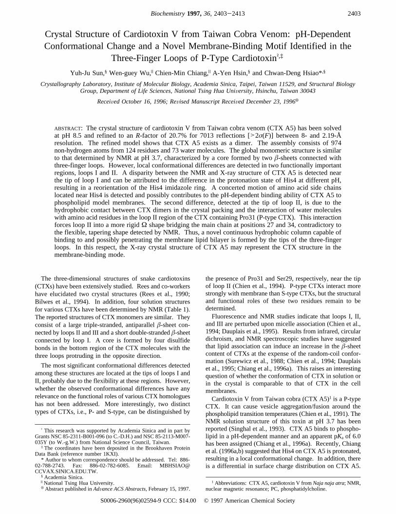
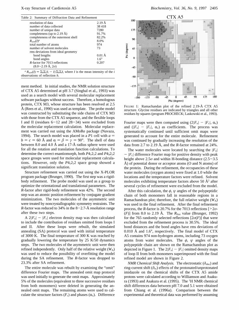
chain of both monomers fall in reasonable regions in aRamachandran plot; therefore, the full relative weight (WA)was used in the final refinement. After the final refinementprocess, theR-factor is 20.7% for the 7013 reflections [>2σ-(F)] from 8.0 to 2.19 Å. TheRfree value (Brunger, 1992)for the 765 randomly selected reflections [2σ(F)] that wereexcluded from the refinement process is 30.5%. The idealbond distances and the bond angles have rms deviations of0.010 Å and 1.6°, respectively. The final model of CTXA5 contains 974 non-hydrogen atoms, including 73 oxygenatoms from water molecules. Theφ, ψ angles of thepolypeptide chain are shown on the Ramachandran plot asdepicted in Figure 1. The 2|Fo| - |Fc| omitted density mapsof loop II from both monomers superimposed with the finalrefined model are shown in Figure 2.NMR Chemical Shift Analysis.The electrostatic (δelec) and
ring-current shift (δrc) effects of the protonated/unprotonatedimidazole on the chemical shifts of the CTX A5 amideprotons were calculated according to Williamson and Asaku-ra (1993) and Asakura et al. (1995). The1H NMR chemicalshift differences data between pH 7.0 and 5.1 were obtainedfrom Chiang et al. (1996a). Comparison between theexperimental and theoretical data was performed by assuming
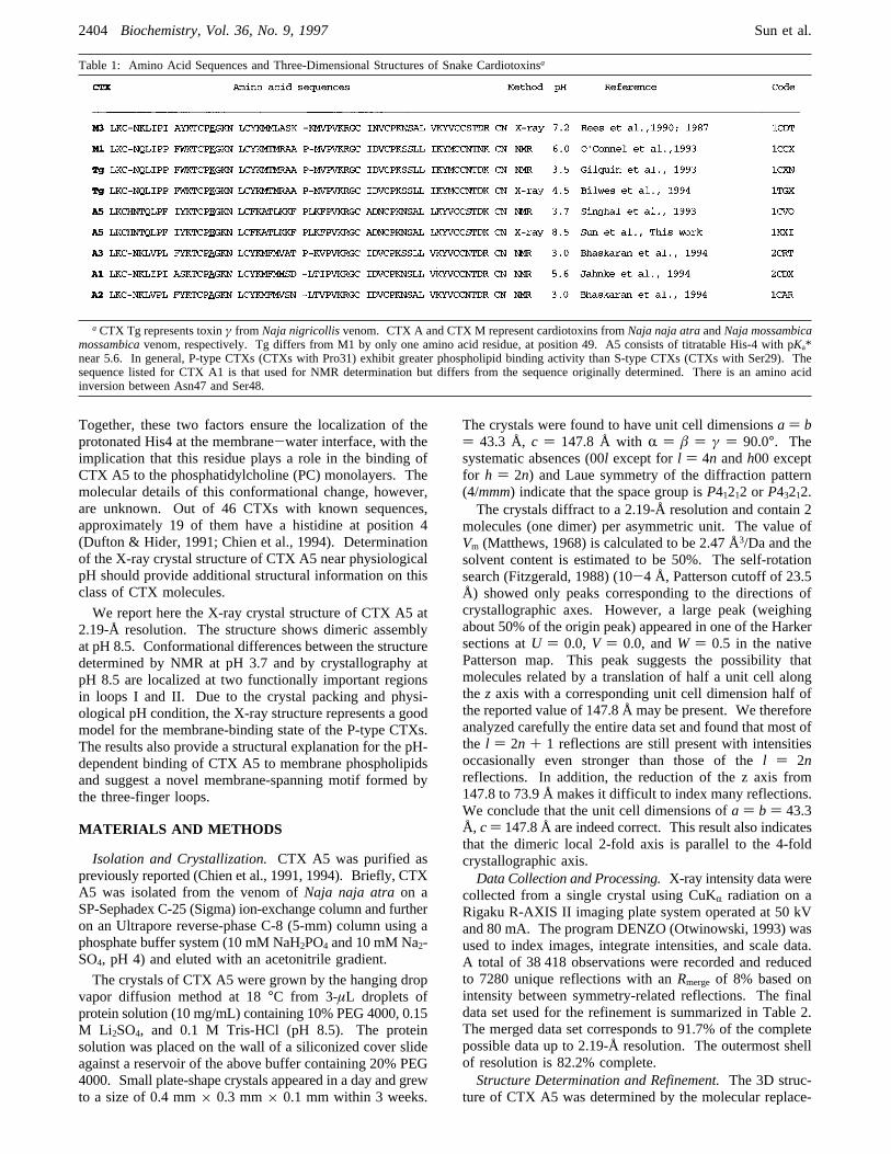
Table 2: Summary of Diffraction Data and Refinement
resolution of data 2.19 Ånumber of data collected 38 418number of unique data 7280completeness (up to 2.19 Å) 91.7%completeness of the outermost shell 82.2%Rsym(I)a 8.0%total number of atoms 974number of solvent molecules 73rms deviations from ideal geometrybond lengths 0.01 Åbond angles 1.6°
R-factor for 7013 reflections(8.0∼2.19 Å, 2σ)
0.207
a Rsym(I) ) ΣhΣi|Ii - I|/ΣhΣiI, whereI is the mean intensity of theiobservations of reflectionh.
FIGURE 1: Ramachandan plot of the refined 2.19-Å CTX A5structure. Glycine residues are indicated by triangles and all otherresidues by squares (program PROCHECK; Laskowski et al., 1993).
X-ray Structure of Cardiotoxin A5 Biochemistry, Vol. 36, No. 9, 19972405
that the imidazole ring may rotate by 90° during theprotonation/unprotonation process. Briefly, the electrostaticcontribution to the chemical shift differences of the studiedNH protons was calculated using the equationδelec )ε1Ez+ε2E2. The values ofε1 and ε2 were estimated to beabout 3.3 and 0 on the basis of the chemical shift differencedetected upon protonation/deprotonation of Glu17 andAsp42. The electric charge of the imidazole ring wasassumed to distribute equally at the N(ε2) and H(ε2) atomicpositions [see Creighton (1993) for notation of atomicpositions]. The ring-current effect of the imidazole ring onthe chemical shift of NH protons was estimated from theequationδrc ) iBG(r), whereG(r) was calculated accordingto the Haigh-Mallion model. The coefficient ofBwas fixedat 0.88, conforming with Osapay and Case (1991). Thedistances between the imidazole ring and the studied amideprotons were obtained from the present study. Only therotation of the ø2 angle (-Câ-Cγ-) was considered inestimating the reorientational effect of the imidazole ringon the chemical shift of the NH protons.
RESULTS
CTX A5 Has a Three-Finger Motif with a Water BindingCentral Loop. CTX A5 shares the common three-fingermotif and molecular shape with other snake toxins such asneurotoxins or CTXs (Figure 3). CTX A5 assumes a flatshape with dimensions of approximately 34 Å× 24 Å× 15Å. The secondary structure is exclusively ofâ-sheet type.
It consists of three loops termed loops I (residues 5-13), II(residues 27-36), and III (residues 48-52) that protrudefrom a central core, tightened by four disulfide linkages(Cys3-Cys22, Cys15-Cys40, Cys44-Cys55, and Cys56-Cys61).CTX A5 forms dimers in the X-ray crystal structure with
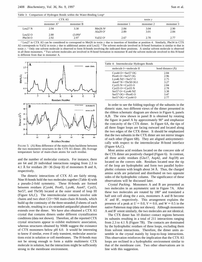
an important solvent site near the tip of loop II. Solvationstretches this loop into anΩ shape that can be observed inboth monomers. Monomer A has a single water moleculethat forms three H-bonds with Leu27, Leu32, and Phe34 inthis loop (Figure 4A). Two water molecules were detectedin this region for monomer B to form three H-bonds withthree residues as in monomer A (Figure 4B). Consequently,the water molecules stretch the loop II of monomer B to afarther extent than monomer A.NMR studies have not detected the existence of water
molecules within loop II. However, a similar water bindingsite has been observed in the crystal structure of the toxinγcentral loop, a P-type CTX (Bilwes et al., 1994; Table 3).There are three structural features regarding the water bindingsite of CTX molecule that warrant further attention. First,most of the amino acid residues wrapping around the watermolecule are hydrophobic. The formation of an extendedhydrophobic loop is more pronounced in CTX A5 than intoxin γ due to the insertion of a hydrophobic leucine atposition 32. Second, the rigid ring structure of Pro31apparently has significant effects on the polypeptide back-bone conformation that allow the interaction of water with
FIGURE 2: Stereo views of the residue-deleted (residues 30-34) 2|Fo| - |Fc| map with the final refined model superimposed. The mapsare contoured at 1σ. Panels A and B correspond to monomers A and B, respectively.
2406 Biochemistry, Vol. 36, No. 9, 1997 Sun et al.
the carbonyl group of the main chain. Third, examinationof the crystal packing suggests that the CTX dimers alsocontact each other via hydrophobic regions in loop II.Apparently, maximization of the hydrophobic contact playsa role in the formation of theΩ loop structure. Details ofthe crystal packing and the dimeric structure are presentedin the next section.The Dimer. The CTX A5 dimer is composed of two
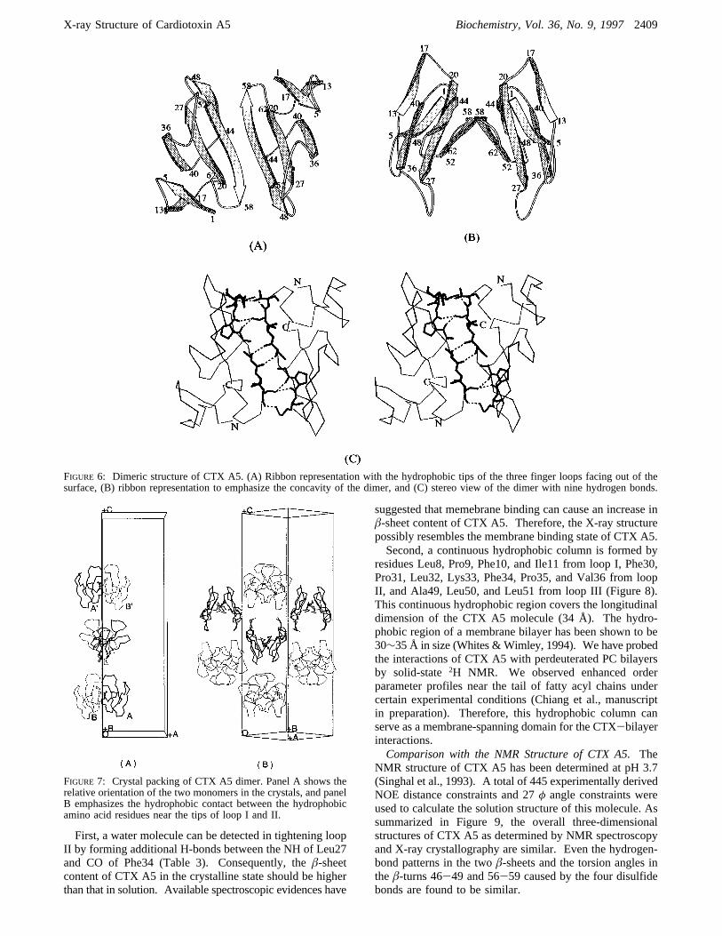
crystallographically independent molecules with essentiallyidentical folding, but the polypeptide chain configurationdiffers significantly in the external loops. The superimposed
CR positions of the two CTX monomers show an rmsdifference of 0.54 Å. The greatest conformational differ-ences between these two molecules occur at residues 28-36 (Figure 5A), which are located at the tip of loop II.Interestingly, the most significant structural difference isdetected at Lys33, which is located in the water binding loop(Figure 4). Smaller differences are observed around residues7-10 of loop I, residues 47-49 of loop III, and residues17-19. These differences arise partially from chain flex-ibility and the crystal packing environment, as indicated bythe higher thermal parameters for these residues (Figure 5B)
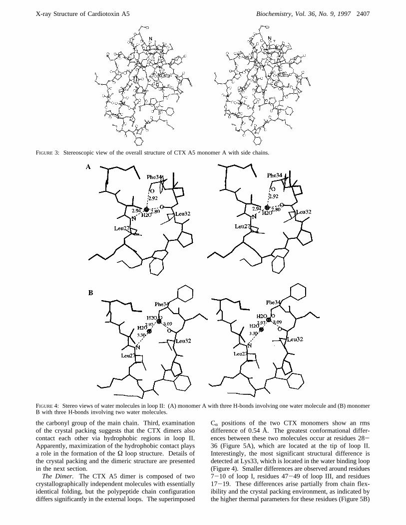
FIGURE 3: Stereoscopic view of the overall structure of CTX A5 monomer A with side chains.
FIGURE 4: Stereo views of water molecules in loop II: (A) monomer A with three H-bonds involving one water molecule and (B) monomerB with three H-bonds involving two water molecules.
X-ray Structure of Cardiotoxin A5 Biochemistry, Vol. 36, No. 9, 19972407
and the number of molecular contacts. For instance, thereare 64 and 20 individual interactions ranging from 2.3 to4.1 Å for residues 28-36 (loop II) of monomers B and A,respectively.
The dimeric interactions of CTX A5 are fairly strong.Nine H-bonds hold the two molecules together (Table 4) witha pseudo-2-fold symmetry. These H-bonds are formedbetween residues (Cys44, Pro45, Lys46, Asn47, Cys55,Ser57, and Thr58) located at the outer strand of loop III(Figure 6A,C). The intermolecular contacts involve sidechains and two short CO‚‚‚NH main-chain H-bonds, whichbuild up the continuity of the three-strandedâ-sheets of eachmolecule, resulting in a six-stranded antiparallel pleated sheetextends over the dimer. We have also obtained a CTX A5crystal that contains dimers under different crystallizationconditions (data not shown). Therefore, all the reported CTXcrystal structures appear to exhibit multimeric association,whereas structures obtained by NMR suggest the existanceof CTX monomers below pH 6.0. It would be interestingto know if similar, even if only transient, molecular associa-tions exist in solution or cell membranes. The H-bonds maynot be strong enough to form a stable multimeric CTXmolecule in solution, but the interactions might be sufficientlystrong in the membrane environment.
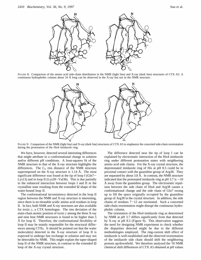
In order to see the folding topology of the subunits in thedimeric state, two different views of the dimer presented inthe ribbon schematic diagram are shown in Figure 6, panelsA,B. The view shown in panel B is obtained by rotatingthe figure in panel A by approximately 90° and emphasizethe concavity of the CTX dimer. In Figure 6A, the tips ofall three finger loops are facing outward and located alongthe two edges of the CTX dimer. It should be emphasizedthat the two subunits in the CTX dimer are not mirror imagesof each other (Figure 6B). They are aligned antisymmetri-cally with respect to the intermolecular H-bond interface(Figure 6A,C).Most amino acid residues located on the concave side of
the CTX dimer are positively charged (Figure 6). In contrast,all three acidic residues (Glu17, Asp42, and Asp59) arelocated on the convex side. Residues located near the tipof the loop are hydrophobic and form two parallel hydro-phobic columns with length about 34 Å. Thus, the chargedamino acids are polarized and distributed on two oppositesides of the hydrophobic column. The significance of theseobservations will be discussed later.Crystal Packing. Monomers A and B are presented as
two molecules in an asymmetric unit in Figure 7A. Afterthese two molecules are rotated by 180° and translated ahalf unit cell along thez axis, monomers A and B becomeA′ and B′, respectively. This arrangement explains thepresence of a peak atU ) 0.0,V ) 0.0, andW) 0.5 in thenative Patterson map (data not shown). Although monomersA and B′ orient similarly, the two molecules are not identical.The CTX dimer has 10 distinct contact regions between
its subunits resulting in a total of 211 interactions rangingfrom 2.3 to 4.1 Å (Figure 7B). The contacts are dominatedby the hydrophobic residues of three loops, excluding themfrom solvent interactions. Therefore, the dimer units as-semble in the crystal mainly by loop-to-loop interactions.This observation reinforces the implication that the CTX A5loops are reclined in a hydrophobic environment similar tothat of the membrane core. Two other observations are inline with this suggestion.
Table 3: Comparison of Hydrogen Bonds within the Water-Binding Loopa
CTX A5 toxin γ
monomer Ab monomer B monomer 1 monomer 2 monomer 3
Leu27 N 2.94 3.30 Met26 Nc 2.84 3.04 2.90Ala29 Oc 2.89 3.01 2.84
Leu32 O 2.80 (3.09)d
Phe34 O 2.92 2.97 Val32 Oc 2.67 2.70 2.77a Leu27 in CTX A5 can be considered to correspond to Met26 in toxinγ due to insertion of histidine at position 4. Similarly, Phe34 in CTX
A5 corresponds to Val32 in toxinγ due to additional amino acid Leu32.b The solvent molecule involved in H-bond formation is similar to that intoxin γ. cOnly one solvent molecule is observed to form H-bonds involving the indicated three positions. A similar solvent molecule is observedin all three monomers.d Two solvent molecules are involved in H-bond formation in monomer B and the solvent molecule involved in this H-bondis different from that in monomer A.
FIGURE5: (A) Rms difference of the main-chain backbone betweenthe two monomeric structures in the CTX A5 dimer. (B) Averagetemparature factor of main-chain atoms for each residue.
Table 4: Intermolecular Hydrogen Bonds
molecule I‚‚‚molecule II bond distance (Å)
Cys44 O‚‚‚Ser57 OG 2.84Pro45 O‚‚‚Ser57 OG 2.96Lys46 NZ‚‚‚Ser57 O 2.75Asn47 N‚‚‚Thr58 OG1 3.27Cys55 N‚‚‚Cys55 O 2.84Cys55 O‚‚‚Cys55 N 2.78Ser57 O‚‚‚Lys46 NZ 3.38Ser57 OG‚‚‚Pro45 O 3.47Ser57 OG‚‚‚Cys44 O 2.77
2408 Biochemistry, Vol. 36, No. 9, 1997 Sun et al.
First, a water molecule can be detected in tightening loopII by forming additional H-bonds between the NH of Leu27and CO of Phe34 (Table 3). Consequently, theâ-sheetcontent of CTX A5 in the crystalline state should be higherthan that in solution. Available spectroscopic evidences have
suggested that memebrane binding can cause an increase inâ-sheet content of CTX A5. Therefore, the X-ray structurepossibly resembles the membrane binding state of CTX A5.Second, a continuous hydrophobic column is formed by
residues Leu8, Pro9, Phe10, and Ile11 from loop I, Phe30,Pro31, Leu32, Lys33, Phe34, Pro35, and Val36 from loopII, and Ala49, Leu50, and Leu51 from loop III (Figure 8).This continuous hydrophobic region covers the longitudinaldimension of the CTX A5 molecule (34 Å). The hydro-phobic region of a membrane bilayer has been shown to be30∼35 Å in size (Whites &Wimley, 1994). We have probedthe interactions of CTX A5 with perdeuterated PC bilayersby solid-state2H NMR. We observed enhanced orderparameter profiles near the tail of fatty acyl chains undercertain experimental conditions (Chiang et al., manuscriptin preparation). Therefore, this hydrophobic column canserve as a membrane-spanning domain for the CTX-bilayerinteractions.Comparison with the NMR Structure of CTX A5.The
NMR structure of CTX A5 has been determined at pH 3.7(Singhal et al., 1993). A total of 445 experimentally derivedNOE distance constraints and 27φ angle constraints wereused to calculate the solution structure of this molecule. Assummarized in Figure 9, the overall three-dimensionalstructures of CTX A5 as determined by NMR spectroscopyand X-ray crystallography are similar. Even the hydrogen-bond patterns in the twoâ-sheets and the torsion angles intheâ-turns 46-49 and 56-59 caused by the four disulfidebonds are found to be similar.
FIGURE 6: Dimeric structure of CTX A5. (A) Ribbon representation with the hydrophobic tips of the three finger loops facing out of thesurface, (B) ribbon representation to emphasize the concavity of the dimer, and (C) stereo view of the dimer with nine hydrogen bonds.
FIGURE 7: Crystal packing of CTX A5 dimer. Panel A shows therelative orientation of the two monomers in the crystals, and panelB emphasizes the hydrophobic contact between the hydrophobicamino acid residues near the tips of loop I and II.
X-ray Structure of Cardiotoxin A5 Biochemistry, Vol. 36, No. 9, 19972409
We have, however, detected several interesting differencesthat might attribute to a conformational change in solutionand/or different pH conditions. A least-squares fit of theNMR structure to that of the X-ray structure highlights thedifferences. The CR rms distance of the NMR structuresuperimposed on the X-ray structure is 1.14 Å. The mostsignificant difference was found at the tip of loop I (Gln7-Lys13) and in loop II (Lys28-Val36). This is due partiallyto the enhanced interaction between loops I and II in thecrystalline state resulting from the extendedΩ shape of thewater-bound loop II.The conformational inconsistency detected in the loop II
region between the NMR and X-ray structure is interesting,since there is no titratable acidic amino acid residues in loopII. In fact, both NMR and X-ray structures are also availablefor toxin γ, a CTX homologue. The rms deviation of themain-chain atomic position of toxinγ among the three X-rayand nine best NMR structures is found to be higher than 5Å for loop II. Therefore, the conformational flexibility ofloop II may be mainly responsible for the structural differ-ences among CTXs. It should be pointed out that the watermolecule(s) detected in the X-ray structure of loop II isexpected to undergo fast exchange in solution and may notbe detectable by NMR. This might explain the taper-shapedloop II of the NMR structure, in contrast to the extendedΩloop of the X-ray crystal structure.
The difference detected near the tip of loop I can beexplained by electrostatic interaction of the His4 imidazolering under different protonation states with neighboringamino acid side chains. For the X-ray crystal structure, thedeprotonated imidazole ring of His at pH 8.5 could be inproximal contact with the guanidino group of Arg38. Theyare separated by about 3.8 Å. In contrast, the NMR structureindicated that the protonated imidazole ring at pH 3.7 is∼10Å away from the guanidino group. The electrostatic repul-sion between the side chain of His4 and Arg38 causes aconformational change and the side chain of Gln7 swungup to fill the space originally occupied by the guanidinogroup of Arg38 in the crystal structure. In addition, the sidechains of residues 7-12 are reoriented. Such a concertedside-chain reorientation might disrupt the continuous hydro-phobic column.The orientation of the His4 imidazole ring as determined
by NMR at pH 3.7 differs significantly from that detectedby X-ray at pH 8.5 (Figure 9). This observation suggeststhe need for designing NMR experiment to check whetherthe disparities detected might be due to the differentmethodologies employed. The ring-current shift effect ofimidazole is well-established and the observed reorientationof the imidazole side chain should shift the neighboringprotons up/downfield. We therefore analyzed the1H NMRchemical shift differences of CTX A5 obtained at pH values
FIGURE 8: Comparison of the amino acid side-chain distribution in the NMR (light line) and X-ray (dark line) structures of CTX A5. Acontinuous hydrophobic column about 34 Å long can be observed in the X-ray but not in the NMR structure.
FIGURE9: Comparison of the NMR (light line) and X-ray (dark line) structures of CTX A5 to emphasize the concerted side-chain reorientationduring the protonation of the His4 imidazole ring.
2410 Biochemistry, Vol. 36, No. 9, 1997 Sun et al.
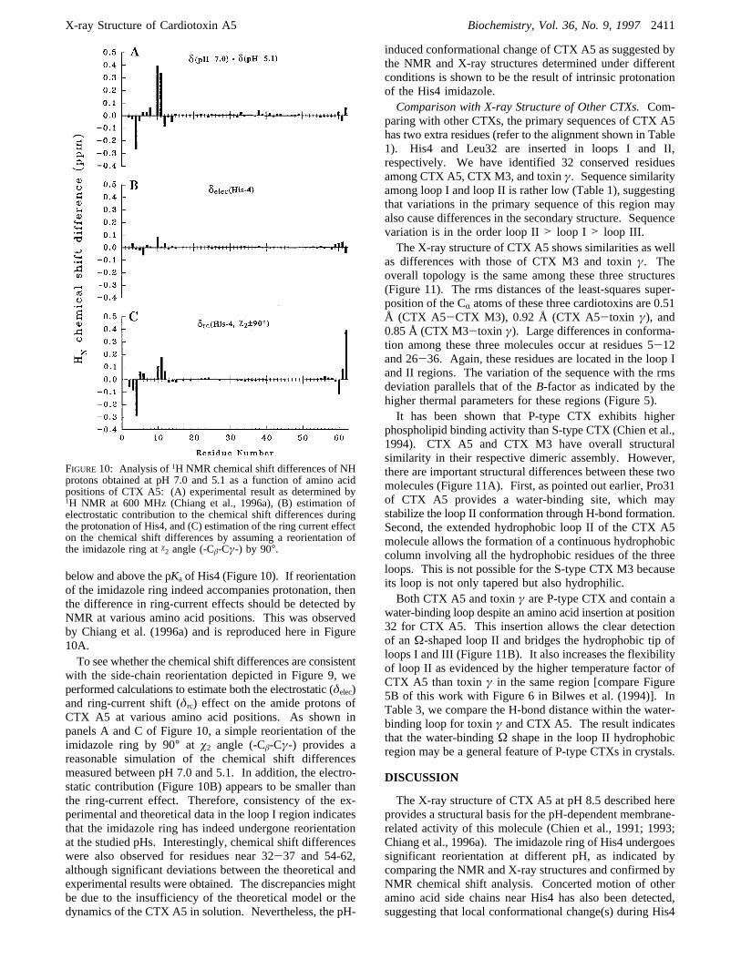
below and above the pKa of His4 (Figure 10). If reorientationof the imidazole ring indeed accompanies protonation, thenthe difference in ring-current effects should be detected byNMR at various amino acid positions. This was observedby Chiang et al. (1996a) and is reproduced here in Figure10A.To see whether the chemical shift differences are consistent
with the side-chain reorientation depicted in Figure 9, weperformed calculations to estimate both the electrostatic (δelec)and ring-current shift (δrc) effect on the amide protons ofCTX A5 at various amino acid positions. As shown inpanels A and C of Figure 10, a simple reorientation of theimidazole ring by 90° at ø2 angle (-Câ-Cγ-) provides areasonable simulation of the chemical shift differencesmeasured between pH 7.0 and 5.1. In addition, the electro-static contribution (Figure 10B) appears to be smaller thanthe ring-current effect. Therefore, consistency of the ex-perimental and theoretical data in the loop I region indicatesthat the imidazole ring has indeed undergone reorientationat the studied pHs. Interestingly, chemical shift differenceswere also observed for residues near 32-37 and 54-62,although significant deviations between the theoretical andexperimental results were obtained. The discrepancies mightbe due to the insufficiency of the theoretical model or thedynamics of the CTX A5 in solution. Nevertheless, the pH-
induced conformational change of CTX A5 as suggested bythe NMR and X-ray structures determined under differentconditions is shown to be the result of intrinsic protonationof the His4 imidazole.Comparison with X-ray Structure of Other CTXs.Com-
paring with other CTXs, the primary sequences of CTX A5has two extra residues (refer to the alignment shown in Table1). His4 and Leu32 are inserted in loops I and II,respectively. We have identified 32 conserved residuesamong CTX A5, CTX M3, and toxinγ. Sequence similarityamong loop I and loop II is rather low (Table 1), suggestingthat variations in the primary sequence of this region mayalso cause differences in the secondary structure. Sequencevariation is in the order loop II> loop I > loop III.The X-ray structure of CTX A5 shows similarities as well
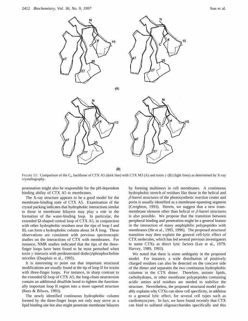
as differences with those of CTX M3 and toxinγ. Theoverall topology is the same among these three structures(Figure 11). The rms distances of the least-squares super-position of the CR atoms of these three cardiotoxins are 0.51Å (CTX A5-CTX M3), 0.92 Å (CTX A5-toxin γ), and0.85 Å (CTX M3-toxin γ). Large differences in conforma-tion among these three molecules occur at residues 5-12and 26-36. Again, these residues are located in the loop Iand II regions. The variation of the sequence with the rmsdeviation parallels that of theB-factor as indicated by thehigher thermal parameters for these regions (Figure 5).It has been shown that P-type CTX exhibits higher
phospholipid binding activity than S-type CTX (Chien et al.,1994). CTX A5 and CTX M3 have overall structuralsimilarity in their respective dimeric assembly. However,there are important structural differences between these twomolecules (Figure 11A). First, as pointed out earlier, Pro31of CTX A5 provides a water-binding site, which maystabilize the loop II conformation through H-bond formation.Second, the extended hydrophobic loop II of the CTX A5molecule allows the formation of a continuous hydrophobiccolumn involving all the hydrophobic residues of the threeloops. This is not possible for the S-type CTX M3 becauseits loop is not only tapered but also hydrophilic.Both CTX A5 and toxinγ are P-type CTX and contain a
water-binding loop despite an amino acid insertion at position32 for CTX A5. This insertion allows the clear detectionof anΩ-shaped loop II and bridges the hydrophobic tip ofloops I and III (Figure 11B). It also increases the flexibilityof loop II as evidenced by the higher temperature factor ofCTX A5 than toxinγ in the same region [compare Figure5B of this work with Figure 6 in Bilwes et al. (1994)]. InTable 3, we compare the H-bond distance within the water-binding loop for toxinγ and CTX A5. The result indicatesthat the water-bindingΩ shape in the loop II hydrophobicregion may be a general feature of P-type CTXs in crystals.
DISCUSSION
The X-ray structure of CTX A5 at pH 8.5 described hereprovides a structural basis for the pH-dependent membrane-related activity of this molecule (Chien et al., 1991; 1993;Chiang et al., 1996a). The imidazole ring of His4 undergoessignificant reorientation at different pH, as indicated bycomparing the NMR and X-ray structures and confirmed byNMR chemical shift analysis. Concerted motion of otheramino acid side chains near His4 has also been detected,suggesting that local conformational change(s) during His4
FIGURE 10: Analysis of1H NMR chemical shift differences of NHprotons obtained at pH 7.0 and 5.1 as a function of amino acidpositions of CTX A5: (A) experimental result as determined by1H NMR at 600 MHz (Chiang et al., 1996a), (B) estimation ofelectrostatic contribution to the chemical shift differences duringthe protonation of His4, and (C) estimation of the ring current effecton the chemical shift differences by assuming a reorientation ofthe imidazole ring atø2 angle (-Câ-Cγ-) by 90°.
X-ray Structure of Cardiotoxin A5 Biochemistry, Vol. 36, No. 9, 19972411
protonation might also be responsible for the pH-dependentbinding ability of CTX A5 to membranes.The X-ray structure appears to be a good model for the
membrane-binding state of CTX A5. Examination of thecrystal packing indicates that hydrophobic interactions similarto those in membrane bilayers may play a role in theformation of the water-binding loop. In particular, theextendedΩ-shaped central loop of CTX A5, in conjunctionwith other hydrophobic residues near the tips of loop I andIII, can form a hydrophobic column about 34 Å long. Theseobservations are consistent with previous spectroscopicstudies on the interactions of CTX with membranes. Forinstance, NMR studies indicated that the tips of the three-finger loops have been found to be most perturbed whentoxin γ interacts with perdeuterated dodecylphosphocholinemicelles (Dauplais et al., 1995).It is interesting to point out that important structural
modifications are usually found at the tip of loop II for toxinswith three-finger loops. For instance, in sharp contrast tothe extendedΩ loop of CTX A5, the long-chain neurotoxinscontain an additional disulfide bond to tighten the function-ally important loop II region into a more tapered structure(Rees & Bilwes, 1993).The newly identified continuous hydrophobic column
formed by the three-finger loops not only may serve as alipid binding site but also might penetrate membrane bilayers
by forming multimers in cell membranes. A continuoushydrophobic stretch of residues like those in the helical andâ-barrel structures of the photosynthetic reaction center andporin is usually identified as a membrane-spanning segment(Creighton, 1993). Herein, we suggest that a new trans-membrane element other than helical orâ-barrel structuresis also possible. We propose that the transition betweenperipheral binding and penetration might be a general featurein the interaction of many amphiphilic polypeptides withmembranes (He et al., 1995, 1996). The proposed structuraltransition may then explain the general cell-lytic effect ofCTX molecules, which has led several previous investigatorsto name CTXs as direct lytic factors (Lee et al., 1979;Harvey, 1989, 1993).We noted that there is some ambiguity in the proposed
model. For instance, a wide distribution of positivelycharged residues can also be detected on the concave sideof the dimer and separates the two continuous hydrophobiccolumns in the CTX dimer. Therefore, anionic lipids,carbohydrates, or other membrane polypeptides containingacidic amino acid residues are needed to stabilize thestructure. Nevertheless, the proposed structural model prob-ably explains why CTXs can show cell specificity, in additionto a general lytic effect, for several cell types such ascardiomyocytes. In fact, we have found recently that CTXcan bind to sulfated oligosaccharides specifically and this
FIGURE 11: Comparison of the CR backbone of CTX A5 (dark line) with CTX M3 (A) and toxinγ (B) (light lines) as determined by X-raycrystallography.
2412 Biochemistry, Vol. 36, No. 9, 1997 Sun et al.
binding further enhances the lipid binding ability of snakeCTXs as studied by Langmuir monolayer (Patel et al., 1997).We are currently investigating the detailed molecular interac-tions between sulfated oligosaccharide and CTX by usingfluorescence, NMR, and X-ray techniques. The X-raystructure of CTX A5 should be a useful good starting pointfor research in this direction.
ACKNOWLEDGMENT
We thank Professor B. C. Wang for the initiation of thepresent work. Suggestions and editing from reviewers andMs. Kavita Vyas are also deeply appreciated.
REFERENCES
Asakura, T., Taoka, K., Demura, M., & Williamson, M. P. (1995)J. Biomol. NMR 6, 227-236.
Bhaskaran, R., Huang, C.-C., Tsai, Y.-C., Jayaraman, G., Chang,D.-K., & Yu, C. (1994a)J. Biol. Chem. 38, 23500-23508.
Bhaskaran, R., Huang, C. C., Chang, D. K., & Yu, C. (1994b)J.Mol. Biol. 235, 1291-1301.
Bilwes, A., Rees, B., Moras, D., Me´nez, R., & Menez, A. (1994)J. Mol. Biol. 239, 122-136.
Brunger, A. T. (1990)Acta Crystallogr., Sect. A 46, 46-57.Chiang, C.-M., Chien, K.-Y., Lin, H.-J., Lin, J.-F., Yeh, H.-C., Ho,P.-L., & Wu, W. (1996a)Biochemistry 35, 9167-9176.
Chiang, C.-M., Chang, S.-L., Lin, H.-j., & Wu, W (1996b)Biochemistry 35, 9177-9186.
Chien, K.-Y., Huang, W.-N., Jean, J.-H., & Wu, W. (1991)J. Biol.Chem. 266, 3252-3259.
Chien, K.-Y., Chiang, C.-M., Hseu, Y.-C., Vyas, A. A., Rule, G.S., & Wu, W. (1994)J. Biol. Chem. 269, 14473-14483.
Creighton, T. E. (1993) inProteins: structures and molecularproperties, W. H. Freeman and Co., New York.
Dauplais, M., Neumann, J. M., Pinkasfeld, S., Menez, A., &Roumestana, C. (1995)Eur. J. Biochem. 230, 213-220.
Dufton, M. J., & Hider, R. C. (1991) inSnakeVenom(Harvey, A.L., Ed.) pp 259-302, Pergamon Press, Inc., New York.
Fitzgerald, P. A. M. (1988) MERLOT, an integrated package ofcomputer programs for the determination of crystal structures
by molecular replacement,J. Appl. Crystallogr. 21, 273.Gilquin, B., Roumestand, C., Zinn-Justin, S., Me´nez, A., & Toma,F. (1993)Biopolymers 33, 1659-1675.
Harvey, A. L. (1985)J. Toxicol. Toxin ReV. 4, 41-69.Harvey, A. L. (1991) inHandbook of natural toxins(Tu, A. T.,Ed.) Vol. 5, pp 85-103, Marcel Dekker, Inc., New York.
He, K., Ludtke, S. J., Huang, H. W., & Worcester, D. L. (1995)Biochemistry 34, 15614-15618.
He, K., Ludtke, S. J., Heller, W. T., & Huang, H. W. (1996)Biophys. J. (in press).
Jahnke, W., Mierke, D. F., Beress, L., & Kessler, H. (1994)J. Mol.Biol. 240, 445-458.
Lee, C. Y., Ed. (1979) Snake Venom,Handbook of ExperimentalPharmacology, Vol. 52, Springer-Verlag AG, Heidelberg, Ger-many.
Laskowski, R. A., MacArthur, M. W., Moss, D. S., & Thornton, J.M. (1993)J. Appl. Crystallogr. 26, 283-291.
Matthews, B. W. (1968)J. Mol. Biol. 33, 491-497.Navaza, J. (1994)Acta Crystallogr., Sect. A 50, 157-163.O’Connell, J. F., Bougis, P. E., & Wu¨thrich, K. (1993)Eur. J.Biochem. 213, 891-900.
Osapay, K., & Case, D. A. (1991)J. Am. Chem. Soc. 113, 9436-9444.
Otwinowski, Z. (1993) in Data Collection and Processing: Pro-ceedings of the CCP4 Study Weekend (Sawyer, L., Isaacs, N.,& Bailey, S., Eds.) pp 56-62, SERC, Daresbury Laboratory,Warrington, U.K.
Patel, H. V., Vyas, A. A., Vyas, K. A., Liu, Y.-S., Chiang, C.-M.,Chi, L.-M., & Wu, W. (1997)J. Biol. Chem. (in press).
Rees, B., & Bilwes, A. (1993)Chem. Res. Toxicol. 6, 385-406.Rees, B., Bilwes, A., Samama, J. P., & Moras, D. (1990)J. Mol.Biol. 214, 281-297.
Singhal, A. K., Chien, K.-Y., Wu, W., & Rule, G. S. (1993)Biochemistry 32, 8036-8044.
White, S. H., & Wimley, W. C. (1994)Curr. Opin. Struct. Biol. 4,79-86.
Williamson, M. P., & Asakura, T. (1993)J. Magn. Reson., Ser. B101, 63-71.
BI962594H
X-ray Structure of Cardiotoxin A5 Biochemistry, Vol. 36, No. 9, 19972413
Top Related
Copyright © 2022 FDOKUMEN

