


Bahasa
Halaman
Hukum


CONGENITAL ABNORMALITIES OF GENITAL TRACT -
UTERINE MALFORMATION
Pages with reference to book, From 261 To 266 Asif Zia Akhtar ( Department of Obstetric and Gynaecology, Abbasi Shaheed Hospital, Karachi. )
Abstract
Thirteen cases of congenital malformations of uterus were discovered during past ten years at Abbasi
Shaheed Hospital, KMC, Karachi. They varied from uterus dideiphys to uterus unicornis. Similar
malformations presented different clinical pictures in different individuals. However, most of the
congenital malformations encountered did not interfere with fertility and eight cases had conceived
within a year of marriage though three ended in abortions. Intravenous pyelograms were also found to
be normal in eight out of the nine cases, on whom this investigation was carried out. In conclusion it is
pertinent to state that early diagnosis is important to state that immediate operative treatment is very
effective in alleviating symptoms and preventing complications (JPMA 36: 261, 1986).
INTRODUCTION
Two mullerian ducts appear as buds in the outer part of each intermediate cell mass in the 5th and 6th
weeks of intrauterine life. The ducts from each side fuse together and are canalised to form two
fallopian tubes, the uterus and upper portion of vagina. Thus varying degrees of structural
abnormalities of uterus and tubes can occur due to imperfect fusion and canalisation of mullenan ducts.
The anomalies of mullerian ducts are genetically determined and are linked up with chromosomal make
up of the individual, but they are also dependent on intra-uterine environment. Wilaon and Harris1
believed that two out of every thousand women have a sufficiently severe degree of uterine
malformation to interfere with pregnancy.
Greiss and Mauzy2 estimated the incidence to be 3.3% in their cases. Many a times the congenital
malformations pass un-noticed as they do not always produce symptoms. Hence the incidence may
even be still higher. In the present series, therefore, an attempt is made to discuss all the cases of
congenital malformations encountered during a period of ten years.
MATERiAL AND METHOD
Thirteen cases with ages ranging from 16 to 55 years were detected to have congenital malformations
of the uterus amongst 11,574 total operations performed (both major and minor) from 1974 to 1983 at
Abbasi Shaheed Hospital, Karachi. Amongst these cases, eight were incidentally discovered at
laparatomies carried out for various gynaecological indications and five were picked up at diagnostic
curettages, through exploration of uterine cavities during evacuations and diagnostic curettage is
routine practice inthe department for detection of uterine abnormalities. Eight cases had symptoms
related to abnormality while other five did not relate symptoms pertaining to malformation detected.
All cases were questioned to establish the relationship of a particular anomaly with its relevant
symptomatology.
Four cases (1, 2, 3 and 4) belonged to complete failure of fusion of mullerian ducts with didelphys
uterus, 2 cases (5 and 6) had bifid uterus, 2 cases (7 and 8) were of sub septate uterus, 3 cases (9, 10
and 11) of bicornuate uterus, and case (12) of hypoplasia of one mullerian duct with undescended
gonad of the same side and one case (13) was of unicornuate uterus.

Various operative techniques employed included dilatation and curettage, excision of vaginal septa,
salpingectomy, utriculoplasty and enucleation and removal of ovarian cysts. All these cases are serially
summarised in a table.

RESULT AND DISCUSSION
Different authors have reported varied frequency of malformations of genital tract atiomalies, In our
study, 13 cases of significant malformations were detected over a period of 10 years during which
11,574 total operations were performed in the department. A frequency of 0.13% was recorded in our
cases.
Though eight of the cases were detected incidentally, a sound relationship with symptoms could be
established in retrospect on taking careful obstetrics and family history.
The variation in the anomalies of mullerian ducts depends upon the level at which canalization is
interfered with, so the problems of individual depends upon the type of malformation inherited. In case
No. 1 of uterus dideiphys, profuse menorrhagia was seen at menarche due to large bleeding area, and in
case 4 ectopic pregnancy was due to abnormally long tube and in later pregnancies in-co-ordinate
uterine action was diagnosed necessitating lower segment caesarean section. Non pregnant horn of
uterus dideiphys enlarged upto 12-14 weeks size of pregnancy under the effect of hormones and did not
cause obstruction to the presenting part, although it may do so in some cases. Jones3 described uterus
didelphys, obstetrically a bad uterus, but MonroKerr4 stated that more complete the malformation the
lesser is the likeithood of dystocia.
Bifid uterus may have rudimentary horn and implantation of pregnancy sac in rudimentary horn of
uterus may result in rupture and produce profound shock like that of rupture tubal pregnancy, some
time rudimentary horn may be so small that it is only recognised when ruptured ectopic tissue is
subjected to microscopic examination.
Buell5 and Perkin and Rubovitz6 have described diverticulum of uterus as a true sac connected to the
uterine cavity by a small aperture. The diverticulum of uterus may be due to improper fusion of
mullerian ducts in the mid-line and bulges out during pregnancy.


Bifjd uterus has a depression on its fundus with a single cavity and cervix, such malformations often
are asymptomatic and hence pass un.noticed (Case No 7 & 8).
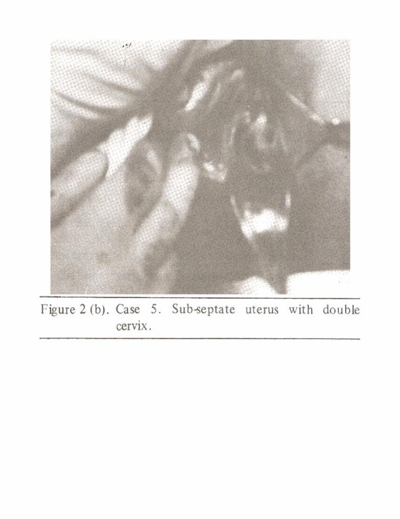
In sub-septate uterus (Case No. 5 & 6) the fundus of uterus has a smooth curve as opposed to
bidomuate uterus where two horn of uterus exist separately.
Uterus sub-septate (Case No. 6) should be strongly suspected if repeated attempts of external cephalic
versions fail in transverse or breach presentation in primigravida. Hunter7 reckons that 12% of
transverse lie are associated with this malformation and are more liable to cause inertia during labour.
There is also an increased likelihood of placenta previa, as the uterus does not have enough space
superiorly. Resection of septum is advised in cases of habitual abortions.

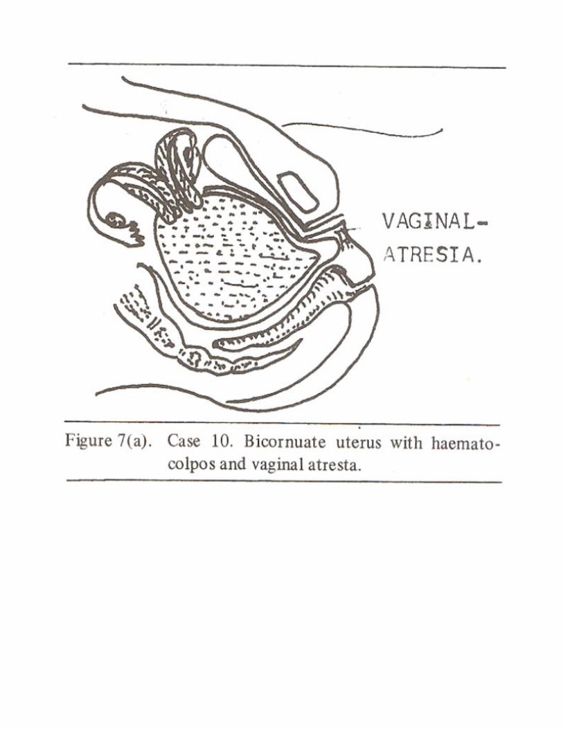


Bicornuate uterus may cause abortions, premature labour, dysmenorrhoea, dystocia, retention of gravid
horn; utriculo-plasty was performed in case No. 10 and 11 which is standard technique and may have
desired result. Patients should be kept under supervision in labour. Patients should be kept under
supervision in labour.
Absence of one mullerian duct direct resulting in unicornuate uterus (Case No. 12) is an extremely rare
condition. She had unicornuate uterus with benign cyst in the left ovary. She had brief history of
menstruation earlier on, but was amenorrhoic for past 12 years and did not respond to hormonal or
clomid therapy by withdrawal bleeding. Her intravenous pyelogram was essentially normal. Her
younger sister also suffers from a similar menstrual disorder thus suggesting a familial predisposition.
Non-descent of gonad as in case 13, is a rare condition too, and undescended gonads either function for
a short while or remain functionless. Hypoplasia of right mullerian duct resulted in poor development
of right tube and probably non canalization of entire right wall of uterus and thickened posterior lip

cervix presenting as polyp. Non-descent of right ovary is explained partly by poorly development
gubernaculum and eventually rudimentary right round ligament which is derived from it. Intravenous
pyelogram showed hypoplasia of right kidney.
Abnormalities of mullerian ducts are closely associated with woilfian duct (Case No.13). Therefore all
such cases should be scrutinised for congenital abnormalities of urinary tract.
CONCLUSION
It becomes apparent from this study that the recognition of congenital uterine malformations is not
always possible unless a clue is provided by double cervix or vagina detected incidentally during minor
or major gynaecological operations. This is because malformations of uterus may not always produce
typical symptoms but their possibility has to be borne in mind in the cases of abortions, premature
labours, cervical dystocia, unexplained severe dysmenorrhoea and shock in pregnant woman.
It is to be emphasised here that defects of external genitalia cannot provide a clue towards defects of
genital organs above the hymen.
More recently ultrasound8 real time scanners with superior resolution have been found helpful for
detection of congenital malformations of genital and renal tracts.
It is essential to realise that diagnosing female genital tract defects well in time can provide
symptomatic relief, improve gestational performance of the affected individual and save lives in
complicated cases.
Uterine malformations do not interfere with fertility of a woman.
REFERENCES
1. Wilson, D.C. and Harris, G.H. Congenital abnormalities. Gynaec. Brit. Comm., 1961; 68: 841.
2. Greiss, F.CJr. and Mauzy, C.H. Genital anomalies in women; an evaluation of diagnosis, incidence,
and obstetric performance. Am. J. Obstet. Gynecol., 1961;82: 330.
3. Jones, W.S. Obstetric significance of genital anomalies. Obstet. Gynecxl., 1957; 10: 113.
4. Moir, J.C. Munro-Kerr’s operative obstetrics. 9th ed. London, Bailliere Tindall, 1977; P. 316.
5. Buell, J.l. and Perkin, M.B. Diverticulum of the Uterus. Am. J. Obstet. Gynecol., 1962; 84: 244.
6. Rubovitz, W. U. True sacculation of the contractile portion of the pregnant uterus. Am. J. Obstet.
GynecoL, 1951; 62:1044.
7. Hunter, W. Practical significance of uterine deformities during pregnancy and labour. Br. Med. J.,
1960; 2: 1124.
8. Campbell, S. and Pearce, J.M. Ultrasound visualization of congenital malformations. Br. Med. Bull.,
1983; 39: 322.
Top Related
Copyright © 2022 FDOKUMEN

