


Bahasa
Halaman
Hukum

Comparative Anatomy
Circulatory System
13/19/2020 By: Dr Somayeh Esmaeili Rineh
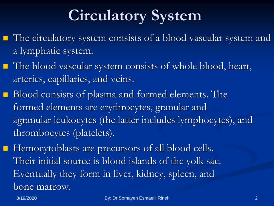
Circulatory System
The circulatory system consists of a blood vascular system and
a lymphatic system.
The blood vascular system consists of whole blood, heart,
arteries, capillaries, and veins.
Blood consists of plasma and formed elements. The
formed elements are erythrocytes, granular and
agranular leukocytes (the latter includes lymphocytes), and
thrombocytes (platelets).
Hemocytoblasts are precursors of all blood cells.
Their initial source is blood islands of the yolk sac.
Eventually they form in liver, kidney, spleen, and
bone marrow.23/19/2020 By: Dr Somayeh Esmaeili Rineh

Circulatory System
Aortic arches- within pharyngeal arches
Arteries
Carries blood away from heart
Muscular, elastic fibrous walls
Regulates blood pressure
Terminate in capillary bed
Veins
Carry blood toward heart, fibrouse tissue
Heart
Modified blood vessel
Figure 13.1: Cross section of
artery and vein.
33/19/2020 By: Dr Somayeh Esmaeili Rineh

Portal Systems
Veins drain organ and dump blood into other
organ instead of heart
Figure 13.4: Portal systems.43/19/2020 By: Dr Somayeh Esmaeili Rineh

Portal Systems (cont.)
Hepatic
Drains intestine into liver
Renal
Drains venous channels of
tail into kidneys
Hypophyseal
Drains hypothalamus into
sinusoids of anterior pituitary
Figure 13.5: Hepatic and renal portal systems. 53/19/2020 By: Dr Somayeh Esmaeili Rineh
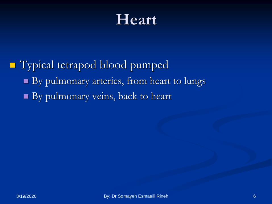
Typical tetrapod blood pumped
By pulmonary arteries, from heart to lungs
By pulmonary veins, back to heart
Heart
63/19/2020 By: Dr Somayeh Esmaeili Rineh

Heart (cont.)
Figure 13.7: Chambers of the primitive vertebrate heart. 73/19/2020 By: Dr Somayeh Esmaeili Rineh
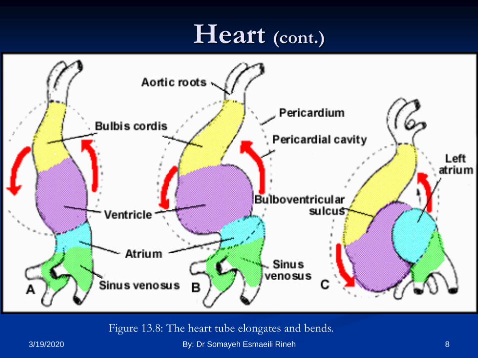
Heart (cont.)
Figure 13.8: The heart tube elongates and bends.
83/19/2020 By: Dr Somayeh Esmaeili Rineh

Fish Heart
Fish heart- tube like
4 chambers:
Sinus venosus
Atrium
Ventricle
Conus arteriosus
Figure 13.9: Four chambered heart.
93/19/2020 By: Dr Somayeh Esmaeili Rineh

single-circuit heart.
Fishes
Venous blood enters a sinus
venosus. traverses an atrium,
ventricle, and conus
arteriosus.
The last two are pumps
that discharge into a ventral
aorta.
The latter carries blood to
aortic arches that supply gills,
where blood is oxygenated.
It then passes via arteries
to
capillaries everywhere in
the body, gives up
oxygen, takes on carbon
dioxide, and returns to
the sinus venosus.
103/19/2020 By: Dr Somayeh Esmaeili Rineh

double-circuit heart
In craniates that breathe solely
by lungs, a pulmonary circuit
carries blood to the lungs and
back
and a systemic circuit carries
oxygenated blood
elsewhere and returns
deoxygenated blood to the
heart. This is a double-circuit
heart.
Double-circuit hearts
have two atria and one or
two ventricles.
The right atrium receives
deoxygenated
blood from the systemic
circuit; the left receives
oxygenated blood from
the pulmonary circuit.
113/19/2020 By: Dr Somayeh Esmaeili Rineh

Figure 13.10: Heart chambers, oxygenated blood flow (red), and septum modification.
Heart
123/19/2020 By: Dr Somayeh Esmaeili Rineh
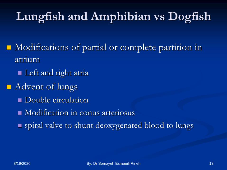
Lungfish and Amphibian vs Dogfish
Modifications of partial or complete partition in
atrium
Left and right atria
Advent of lungs
Double circulation
Modification in conus arteriosus
spiral valve to shunt deoxygenated blood to lungs
133/19/2020 By: Dr Somayeh Esmaeili Rineh

Spiral Valve
Figure 13.11: Spiral valve in
dipnoans; longitudinal folds
of conus lining.
Figure 13.12: Spiral valve in anurans; single flap.
143/19/2020 By: Dr Somayeh Esmaeili Rineh

Amphibian Heart
Spiral valve directs oxy. blood
entering ventricle from left
atrium
Conus (truncus) arteriosus;
Bulbous arteriosus
Swelling of ventral aorta
Smooth muscle
Figure 13.13: Three-chambered frog heart.
153/19/2020 By: Dr Somayeh Esmaeili Rineh

Urodele- partially
divided circulation
Right and left atrium
Sinus venosus dumps
into right atrium
Pulmonary veins leave
left ventricle
Reptile - fully divided
circulation, but
additional chamber (as
in turtle)
Figure 13.14: Turtle heart chambers and
circulation path.
Amphibian Heart (cont.)
163/19/2020 By: Dr Somayeh Esmaeili Rineh

Reptile Heart
Cavum venosum (CV)- internal pocket; e.g., turtle
Blood collected from post cava through sinus venosus
to precava
To right atrium
Venous blood to CV
Cavum pulmonale
Into pulmonary artery to lungs
Oxy. blood returns through pulmonary veins in left atrium
Back to CV
To left and right aortic trunk
173/19/2020 By: Dr Somayeh Esmaeili Rineh

Blood Circulation
Figure 13.15: Single loop fish and double loop amphibian and mammal circulation.
183/19/2020 By: Dr Somayeh Esmaeili Rineh

Amniote Heart
4 chambered heart
2 atria and 2 ventricles
Birds and mammals
Sinus venosus- 5th chamber in reptile heart
Becomes sino-atrial node
In embryo, right and left atria are not separated
Auricle - flap on side of atrium
193/19/2020 By: Dr Somayeh Esmaeili Rineh
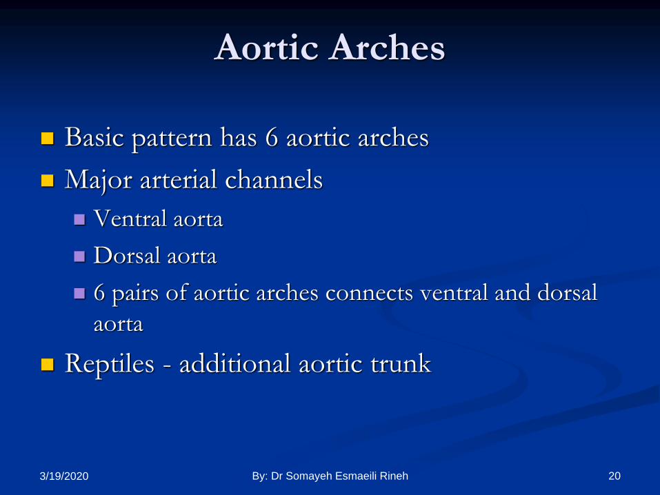
Aortic Arches
Basic pattern has 6 aortic arches
Major arterial channels
Ventral aorta
Dorsal aorta
6 pairs of aortic arches connects ventral and dorsal
aorta
Reptiles - additional aortic trunk
203/19/2020 By: Dr Somayeh Esmaeili Rineh

Aortic Arches (cont.)
Figure 13.21: Ventral perspective of aortic
arches.
Figure 13.20: Basic pattern of
aortic arches and dorsal aortae.
213/19/2020 By: Dr Somayeh Esmaeili Rineh

223/19/2020 By: Dr Somayeh Esmaeili Rineh
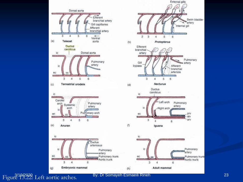
Figure 13.22: Left aortic arches. 233/19/2020 By: Dr Somayeh Esmaeili Rineh

Aortic Arches (cont.)
Teleost
1st and 2nd arches lost
Dorsal aortae become internal
carotids
Lung fish
Pulmonary artery from 6th arch
Tetrapods
Pulmonary artery from 6th arch
5th arch lost Figure 13.23: Aortic arches, internal
carotids (ic) and pulmonary artery.
243/19/2020 By: Dr Somayeh Esmaeili Rineh

Tetrapod Aortic Arches
1st and 2nd arches lost
Dorsal segment dropped
between 3rd and 4th arches
Ductus caroticus
Figure 13.24: Adult aortic arches
(book figure 14.19).
253/19/2020 By: Dr Somayeh Esmaeili Rineh
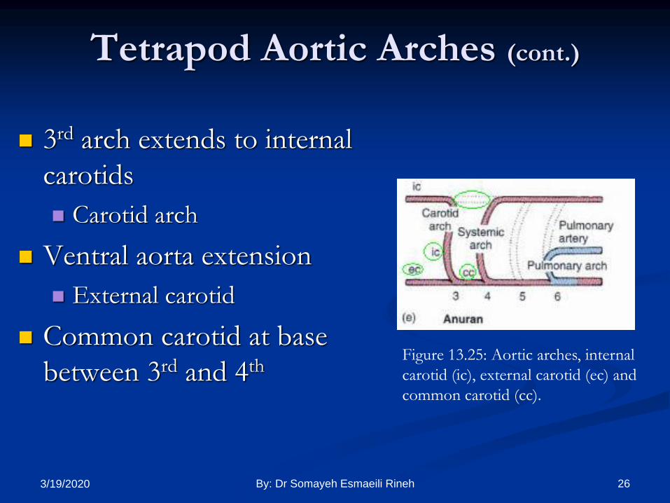
Tetrapod Aortic Arches (cont.)
3rd arch extends to internal
carotids
Carotid arch
Ventral aorta extension
External carotid
Common carotid at base
between 3rd and 4thFigure 13.25: Aortic arches, internal
carotid (ic), external carotid (ec) and
common carotid (cc).
263/19/2020 By: Dr Somayeh Esmaeili Rineh

Tetrapod Aortic Arches (cont.)
5th arch lost
Dorsal segment of 6th arch lost
4th arch- no anterior connection
Aortic arch
6th arch
Pulmonary arch
Ex: adult anuran
Figure 13.26: Adult aortic
arches.
273/19/2020 By: Dr Somayeh Esmaeili Rineh
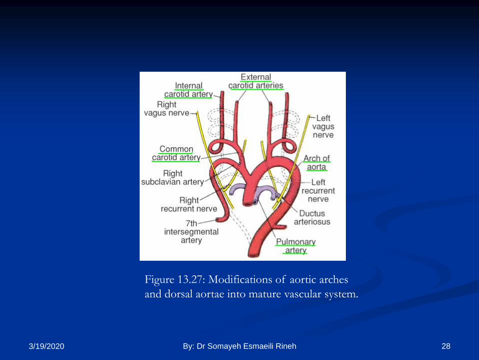
Figure 13.27: Modifications of aortic arches
and dorsal aortae into mature vascular system.
283/19/2020 By: Dr Somayeh Esmaeili Rineh

Aortic Arches
Urodele Ductus caroticus
Ductus arteriosus- dorsal segment of 6th arch
Reptiles 1st and 2nd arches lost
Ductus caroticus lost
5th arch lost
Ductus arteriosus lost
Additional aortic arch introduced
Arch from left side loops right
Arch from right side loops left
293/19/2020 By: Dr Somayeh Esmaeili Rineh

Mammalian Aortic Arches
3rd, 4th, 5th, & 6th retained
embryonically
Adults- 1st and 2nd dropped
3rd carotid arch
4th systemic arch
5th lost
Dorsal segment of 6th lost
Retained embryonically- ductus
arteriosus (becomes ligamentum
arteriosum)
Figure 13.28: Adult aortic
arches.
Figure 13.29: Left aortic arches.303/19/2020 By: Dr Somayeh Esmaeili Rineh

Bird Aortic Arches
Right portion of
aortic arch is
retained and left is
lost (opposite to
mammals)
Birds have right
aortic arch
Mammals have left
aortic arch313/19/2020 By: Dr Somayeh Esmaeili Rineh

A sinus venosus is present in fishes, amphibians,
and reptiles.
It becomes partially incorporated into
the wall of the right atrium in crocodilians.
In birds and mammals, it is not a sinus but a
local collection of cells in the right atrium
known as the sinoatrial (SA) node.
323/19/2020 By: Dr Somayeh Esmaeili Rineh

Comparative Anatomy
Digestive System
By: Vahid Akmali
Email:[email protected]
333/19/2020 By: Dr Somayeh Esmaeili Rineh

Digestive System
Six major subdivisions
Oral cavity
Pharynx
Esophagus
Stomach
Small & large intestine
Rectum
343/19/2020 By: Dr Somayeh Esmaeili Rineh
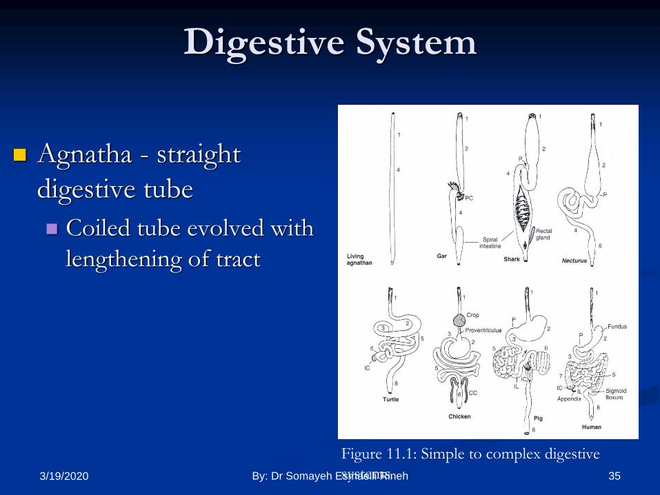
Digestive System
Agnatha - straight
digestive tube
Coiled tube evolved with
lengthening of tract
Figure 11.1: Simple to complex digestive
systems. 353/19/2020 By: Dr Somayeh Esmaeili Rineh

1- Esophagus
2- Stomach
3- Duodenum
4- intestine
5- Small intestine
6- large intestine
7- Colon
8- Rectum
363/19/2020 By: Dr Somayeh Esmaeili Rineh
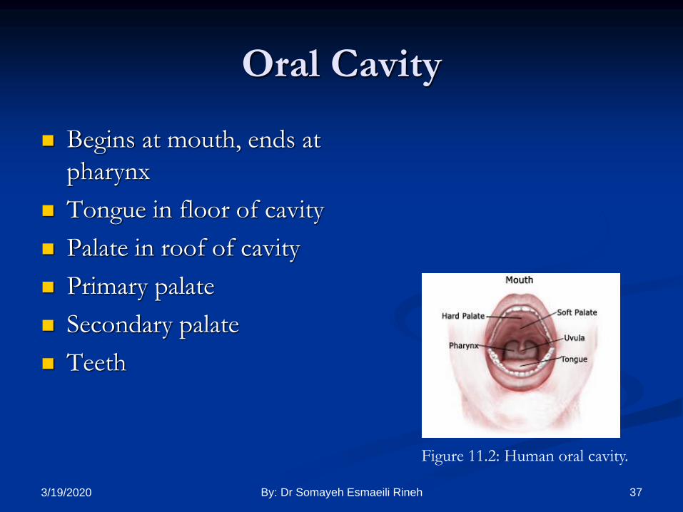
Oral Cavity
Begins at mouth, ends at
pharynx
Tongue in floor of cavity
Palate in roof of cavity
Primary palate
Secondary palate
Teeth
Figure 11.2: Human oral cavity.
373/19/2020 By: Dr Somayeh Esmaeili Rineh
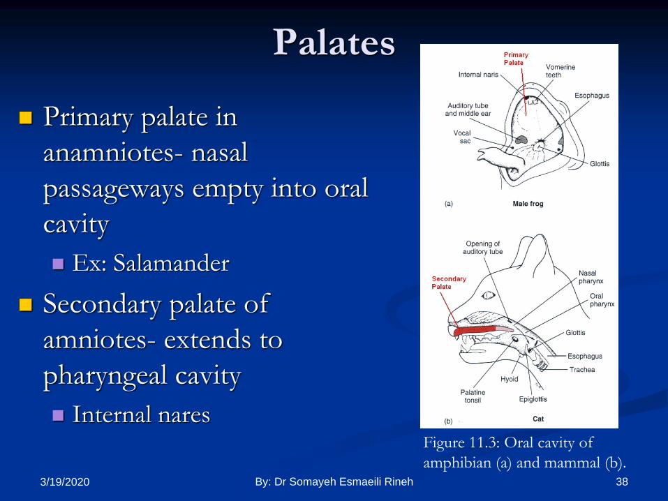
Palates
Primary palate in
anamniotes- nasal
passageways empty into oral
cavity
Ex: Salamander
Secondary palate of
amniotes- extends to
pharyngeal cavity
Internal naresFigure 11.3: Oral cavity of
amphibian (a) and mammal (b).383/19/2020 By: Dr Somayeh Esmaeili Rineh

Teeth On jaws normally
Cheeks in mammals form pocket
Acrodont teeth- fish and snakes
Bicuspid- amphibians
Tricuspid- lizards
Pleurodont teeth- snakes
Thecodont teeth-
crocodilians
Figure 11.4- Cross section of jaw.Figure 11.5- Types of cusps.
393/19/2020 By: Dr Somayeh Esmaeili Rineh

403/19/2020 By: Dr Somayeh Esmaeili Rineh

Jaw Teeth and Cheek
Modified placoid scales- sharks
Polyhyodont- permanent replacement of teeth
Diphyodont- two sets of teeth
Monophyodont- one set of teeth
413/19/2020 By: Dr Somayeh Esmaeili Rineh

Epidermal Teeth
Egg teeth all egg layers
Not actual tooth
Structure epidermal, horny,
keratinized
On tip of snout
To penetrate egg shell
Bird, turtle, crocodile, Sphenodon
& monotrems
Figure 11.6: Egg caruncle of 15 day
old owlet. 423/19/2020 By: Dr Somayeh Esmaeili Rineh
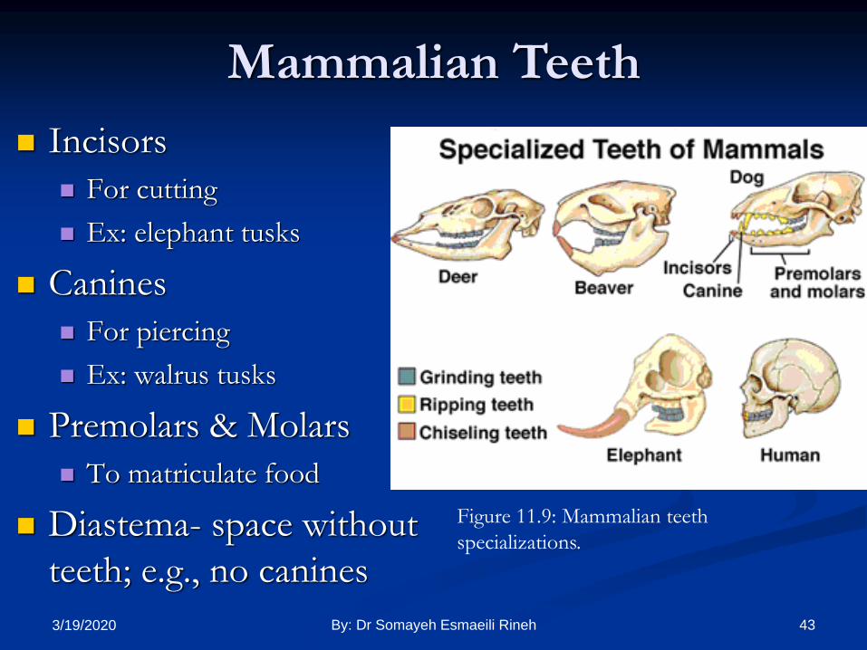
Incisors
For cutting
Ex: elephant tusks
Canines
For piercing
Ex: walrus tusks
Premolars & Molars
To matriculate food
Diastema- space without
teeth; e.g., no canines
Figure 11.9: Mammalian teeth
specializations.
Mammalian Teeth
433/19/2020 By: Dr Somayeh Esmaeili Rineh
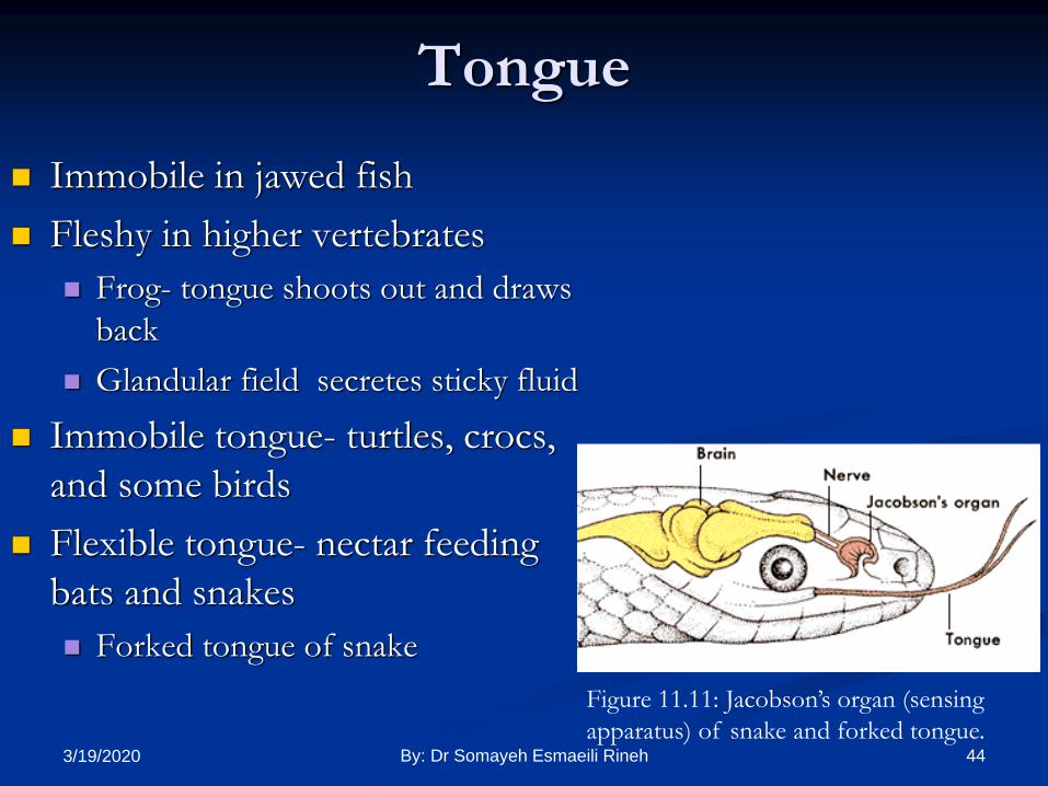
Tongue
Immobile in jawed fish
Fleshy in higher vertebrates
Frog- tongue shoots out and draws
back
Glandular field secretes sticky fluid
Immobile tongue- turtles, crocs,
and some birds
Flexible tongue- nectar feeding
bats and snakes
Forked tongue of snake
Figure 11.11: Jacobson’s organ (sensing
apparatus) of snake and forked tongue.443/19/2020 By: Dr Somayeh Esmaeili Rineh
Oral Glands
Named based on location
Labial- near the lips
Palatal- near palate
Internasal
Sublingual- releases venom
Parotid- salivary gland
Submaxillary
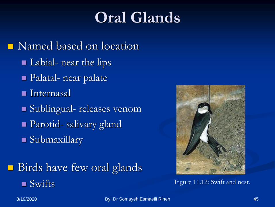
Birds have few oral glands
Swifts Figure 11.12: Swift and nest.
453/19/2020 By: Dr Somayeh Esmaeili Rineh
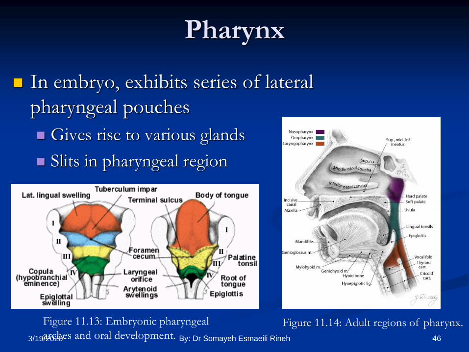
Pharynx
In embryo, exhibits series of lateral
pharyngeal pouches
Gives rise to various glands
Slits in pharyngeal region
Figure 11.13: Embryonic pharyngeal
arches and oral development.Figure 11.14: Adult regions of pharynx.
463/19/2020 By: Dr Somayeh Esmaeili Rineh

473/19/2020 By: Dr Somayeh Esmaeili Rineh
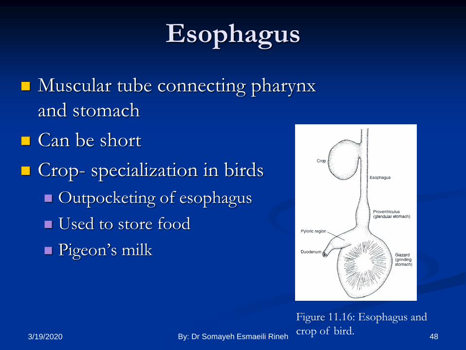
Esophagus
Muscular tube connecting pharynx
and stomach
Can be short
Crop- specialization in birds
Outpocketing of esophagus
Used to store food
Pigeon’s milk
Figure 11.16: Esophagus and
crop of bird.483/19/2020 By: Dr Somayeh Esmaeili Rineh

Stomach Muscular chamber
Secretes gastric juices
Different lining of stomachs
Esophageal-like epithelia
Glandular epithelia
Ruminant stomach
4 chambers: rumen, reticulum,
omasum, abomasum
Human stomach
Cardiac sphincter- esophagus
meets stomach
Mostly lined with gastric epithelium
Figure 11.17: Stomach of mammals with
esophageal-like epithelia in gray and glandular
epithelia in red.
493/19/2020 By: Dr Somayeh Esmaeili Rineh

Stomach Structure
Greater and lesser curvature
Messentaries
Greater omentum – attaches along greater curvature
Lesser omentum – attaches along lesser curvature
Cecum- increases surface area
2 parts in bird and crocodile stomach
Proventiculus-glandular
Gizzard- grinding mill (gastroliths)
503/19/2020 By: Dr Somayeh Esmaeili Rineh

4-Chambered Stomachs
Figure 11.18: Stomach of calf.
Rumen- food enters
Bacterial action
Reticulum- fermentation-
forms a bolus
Omasum- reswallowed grass
Salivary action
Abomasum- food worked out
by gastric glands
513/19/2020 By: Dr Somayeh Esmaeili Rineh
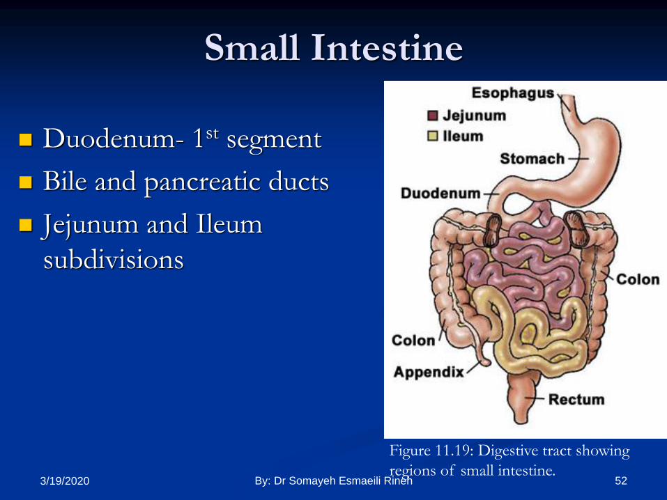
Small Intestine
Duodenum- 1st segment
Bile and pancreatic ducts
Jejunum and Ileum
subdivisions
Figure 11.19: Digestive tract showing
regions of small intestine.523/19/2020 By: Dr Somayeh Esmaeili Rineh

Brunner’s Glands- mucous glands in duodenum
and jejunum
Peyer’s Patches- lymphatic nodules in ileum
Crypts of Lieberkühns- intestinal glands at base
of villi
Lacteals- within villi—interior lymphatic vessels
Transport fat molecules to circulatory system
Valve of Kirckring- increases surface area
Small Intestine
533/19/2020 By: Dr Somayeh Esmaeili Rineh

Small Intestine
Figure 11.20: Histology of alimentary canal of a mammal
showing various glands of small intestine.
543/19/2020 By: Dr Somayeh Esmaeili Rineh
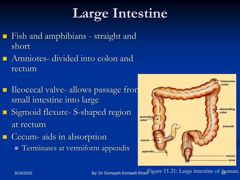
Large Intestine
Fish and amphibians - straight and short
Amniotes- divided into colon and rectum
Ileocecal valve- allows passage from small intestine into large
Sigmoid flexure- S-shaped region
at rectum
Cecum- aids in absorption
Terminates at vermiform appendix
Figure 11.21: Large intestine of human.553/19/2020 By: Dr Somayeh Esmaeili Rineh

Liver
Liver is diverticulum of primitive gut
Liver produces bile
Bile stored in gallbladder
Common bile duct
Ampulla of Vater- terminal portion
Figure 11.22: Development of liver and pancreas. 563/19/2020 By: Dr Somayeh Esmaeili Rineh

Pancreas
Pancreas – diverticulum of gut
Duct of Santorini- small, dorsal pancreas
Duct of Wirsung- large, ventral pancreas
Accessory duct- large duct after small, dorsal
duct disappears
Exocrine and endocrine glands
Islets of Langerhans- endocrine glands
573/19/2020 By: Dr Somayeh Esmaeili Rineh
Top Related
Copyright © 2022 FDOKUMEN

