


Bahasa
Halaman
Hukum


Blood-Brain Barrier Shuttles: From Design to Application
Pol Arranz Gibert
Aquesta tesi doctoral està subjecta a la llicència Reconeixement- NoComercial – SenseObraDerivada 3.0. Espanya de Creative Commons. Esta tesis doctoral está sujeta a la licencia Reconocimiento - NoComercial – SinObraDerivada 3.0. España de Creative Commons. This doctoral thesis is licensed under the Creative Commons Attribution-NonCommercial-NoDerivs 3.0. Spain License.

2016
Tes
idoc
tora
l P
ol A
rran
z G
iber
t
Blood-Brain Barrier Shuttles:From Design to Application
Pol Arranz Gibert


Programa de doctorat de química orgànica
Blood-Brain Barrier Shuttles:
From Design to Application
Pol Arranz Gibert
Tesi doctoral dirigida per:
Prof. Ernest Giralt LledóUniversitat de Barcelona
Facultat de QuímicaDepartament de Química Orgànica
Dra. Meritxell Teixidó
IRB BarcelonaPrograma de Química i
Farmacologia Molecular
Barcelona, 2016.


CONTENTS


ABBREVIATIONS i
INTRODUCTION 1
Origins of the Gatekeepers of the Brain—Evolution of Brain Barriers 3
Understanding the Door—Physiology of the Blood-Brain Barrier 8
Reasons to Look Inside—Physiology and Disease of the CNS: Social Impact 14
Devising a Key—Peptides as Therapeutics and for Drug Delivery to the Brain 16
OBJECTIVES 21
RESULTS AND DISCUSSION 25
Chapter 1: Study of Passive Diffusion BBB Shuttles 27
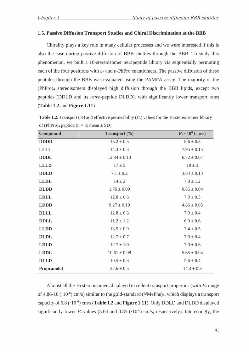
1.1. Peptide-Shuttle Design 321.2. Transport Ability of (PhPro)4 Shuttle Using the PAMPA Assay 331.3. Design and Synthesis of a 16-Steroisomer Library of (PhPro)4 371.4. Physicochemical Characterization of Pro4 and (PhPro)4 Shuttle 381.5. Passive Diffusion Transport Studies and Chiral Discrimination at the BBB 41
Chapter 2: Study of Actively-Transported BBB Shuttles throughReceptor-Mediated Transcytosis 45
2.1. Previous Studies with HAI Peptide 482.2. Amino Acid Replacement Effect on Transport Study Using a Novel
Method for Transport Quantification Based on MALDI-TOF MS 55
Chapter 3: Study of Immunogenic Responses to BBB Shuttle Peptides 71
Chapter 4: Attempts to Develop a Therapy for FRDA at the CNS 83
4.1. Protein Replacement Therapy for Friedreich’s Ataxia at the CNS—Chemistry with Proteins 92
4.2. Gene Therapy for Friedreich’s Ataxia at the CNS—Chemistry with Enveloped Viral Particles 101
CONCLUSIONS 115
EXPERIMENTAL SECTION 119 Materials and Methods 121 Solid-Phase Synthesis of Compounds 123 Peptide and Amino Acid Purification and Characterization 128 Structural Data 130 In Vitro Assays 132 Protein Expression, Purification, Bioconjugation and Characterization 137 HSV-1 Bioconjugation and Characterization 140 In Vivo Experiments 143 Product Characterization 145 Amino Acids 147 Peptides 148 Biologics 164 REFERENCES 167 SUMMARY IN CATALAN 187

ABBREVIATIONS

Abbreviations
iii
AAA amino acid analysis AAV adeno-associated virus ABC ATP binding cassette Ac acetyl ACH α-cyano-4-hydroxycinnamic acid AcOH acetic acid AJs adherens junctions AM amino methyl AME adsorptive-mediated endocytosis AMT adsorptive-mediated transcytosis amu atomic mass unit AO aminooxy moiety, i.e. aminooxyacetyl aPhe 4-amino-ʟ-phenylalanine apoB100 apolipoprotein B100 apoE apolipoprotein E apoTf apotransferrin ASMS affinity selection of proteins coupled to MS AtFH Arabidopsis thaliana frataxin homolog ATP adenosine triphosphate AU arbitrary units AuNPs gold nanoparticles BAC bacterial artificial chromosome BBB blood-brain barrier BBECs bovine brain endothelial cells BBMVEC bovine brain microvascular endothelial cells BBs brain barriers BMECs brain microvasculature endothelial cells Boc tert-butoxycarbonyl bp base pair BPLE brain polar lipid extract BSA bovine serum albumin C. elegans Caenorhabditis elegans Caco-2 human colorectal adenocarcinoma cell line CBMSO centro de biología molecular Severo Ochoa CD circular dichroism CDX candoxin Cf 5(6)-carboxyfluorescein CFA complete Freund’s adjuvant Cl-HOBt 6-chloro-1-hydroxybenzotriazole CLSM confocal laser scanning microscopy CNS central nervous system COMU 1-cyano-2-ethoxy-2-oxoethylidenaminooxy)dimethylamino-morpholino
carbenium hexafluorophosphate Conc. concentration CoQ10 coenzyme Q10 COSY correlation spectroscopy CPP cell-penetrating peptide CPs choroid plexuses CSD chemical shift deviation CSF cerebrospinal fluid CyaY Escherichia coli frataxin CYPs cytochrome P450s DBU 1,8-diazabicyclo[5.4.0]undec-7-ene Dbz diaminobenzoic acid

Abbreviations
iv
DCC N,N'-dicyclohexylcarbodiimide DCM dichloromethane De enantiomeric discrimination DHAP 2,6,-dihydroxyacetophenone DIPEA N,N-diisopropylethylamine DIPCDI N,N'-diisopropylcarbodiimide DLS dynamic light scattering DM-I diabetes mellitus type I DMAP 4-dimethylaminopyridine DMEM Dulbecco's modified Eagle medium DMF N,N-dimethylformamide DNA deoxyribonucleic acid Dpr diaminopropionic acid DSS sodium-3-(trimethylsilyl)propanesulfonate DTT dithiothreitol E. coli Escherichia coli ECL enhanced chemiluminescence ECM endothelial cell medium ECs endothelial cells EDC 1-ethyl-3-(3-dimethylaminopropyl)-carbodiimide EDT 1,2-ethanedithiol EDTA ethylenediamine tetraacetic acid EFACTS European Friedreich’s Ataxia consortium for translational studies EGFR epidermal growth factor receptor ELISA enzyme-linked immunosorbent assay EPO erythropoietin eq. equivalent Eq. equation ESI-MS electrospray ionization-mass spectrometry FARA Friedreich’s Ataxia Research Alliance FDA food and drug administration FGE formylglycine-generating enzyme fGly formylglycine Fmoc 9-fluorenylmethoxycarbonyl FPhe 4-fluoro-ʟ-phenylalanine FPLC fast protein liquid chromatography FRDA Friedreich’s Ataxia FXN frataxin GABA γ-aminobutyric acid GFP green fluorescent protein GSH glutathione HBSS Hanks’ balanced salt solution HBTU O-(benzotriazol-l-yl)-N,N,N’,N’-tetramethyluronium hexafluorophosphate hCha homocyclohexyl-ʟ-alanine HDAC histone deactylase HeNe helium-neon HEPES 4-(2-hydroxyethyl)-1-piperazineethanesulfonic acid HFIP hexafluoro-2-propanol HIV-1 human immunodeficiency virus 1 HOAt 1-hydroxy-7-azabenzotriazole HOBt 1-hydroxybenzotriazole holoTf holotransferrin HPLC high-performance liquid chromatography HPLC-MS high-performance liquid chromatography coupled to mass spectrometry

Abbreviations
v
HRMS high-resolution mass spectrometryHRP horseradish peroxidaseHsFtx human frataxinHSQC heteronuclear single quantum coherenceHSV-1 herpes simplex virus type 1hTf human transferrinIAA iodoacetic acidICP-MS inductively coupled plasma mass spectrometryIDL intermediate-density lipoproteinIFA incomplete Freund’s adjuvantIFN-γ interferon gammaIGF-1 insulin/insulin-like growth factor 1IgG immunoglobulin GIgM immunoglobulin MIMAC immobilized metal affinity chromatographyINAA instrumental neutron activation analysisi.p. intraperitonealiPSCs induced pluripotent stem cellsISCU iron-sulfur cluster assembly enzymeJAMs junctional adhesion moleculesKLH keyhole limpet hemocyaninʟ-DOPA ʟ-3,4-dihydroxyphenylalanineLDLR low-density lipoprotein receptorLOD limit of detectionLOQ limit of quantificationLY lucifer yellow lithium saltMAL maleimideMALDI matrix-assisted laser desorption/ionizationMES 2-(N-morpholino)ethanesulfonic acidMetAPs methionine aminopeptidasesMHC major histocompatibility complexMLS mitochondrial localization signalMPP mitochondrial processing peptidaseMS mass spectrometryMTBE methyl tert-butyl etherMTT 3-(4,5-dimethylthiazol-2-yl)-2,5-diphenyltetrazolium bromideMW molecular weightnAChR nicotinic acetylcholine receptorNADH nicotinamide adenine dinucleotideNbz N-acyl-benzimidazolinoneNCL native chemical ligationneg. negativeNFS1•ISD11 sulfur donor complexNHS N-hydroxysuccinimideNIP (R)-piperidine-3-carboxylic acid, or nipecotic acidNMePhe N-methyl phenylalanineNMR nuclear magnetic resonanceNOESY nuclear Overhauser spectroscopyNP nanoparticleo/n overnightOxyma ethyl (hydroxyimino)cyanoacetateP-gp P-glycoproteinpAbs polyclonal antibodiesPAMPA parallel artificial membrane permeability assay

Abbreviations
vi
pAb polyclonal antibody Papp apparent permeability Pbf 2,2,4,6,7-pentamethyldihydrobenzofuran-5-sulfonyl PBS phosphate-buffered saline PC phosphatidylcholine PDA photodiode array detector PDB protein data bank Pe effective permeability PE phosphatidylethanolamine PEG polyethylene glycol PhPro cis-3-phenylpyrrolidine-2-carboxylic acid PI phosphatidylinositol Pip pipecolic acid PLP pyridoxal-5’-phosphate PMP pyridoxamine-5’-phosphate PPII polyproline II PS phosphatidylserine PUFA polyunsaturated fatty acid PyBOP benzotriazol-1-yl-oxytripyrrolidinophosphonium hexafluorophosphate QD quantum dot RC random coil ref. reference REMD replica exchange molecular dynamics rD retro-ᴅ-version/ peptide made of ᴅ-amino acids and with the reversed sequence of
a parent peptide RME receptor-mediated endocytosis RMSD root-mean-square deviation RMT receptor-mediated transcytosis RNA ribonucleic acid RNAPII RNA polymerase II ROS reactive oxygen species RP-HPLC reversed phase-high-performance liquid chromatography RSV Rous sarcoma virus r.t. room temperature RVG rabies virus glycoprotein SD standard deviation SDS sodium dodecyl sulfate SDS-PAGE sodium dodecyl sulfate-polyacrylamide gel electrophoresis SEC size-exclusion chromatography SPPS solid-phase peptide synthesis stain. staining SUMO small ubiquitin-like modifier T transport TALE transcription activator-like effector TAT human immunodeficiency virus-1 trans-acting activator of transcription TBST tris-buffered saline with Tween 20 tBu tert-butyl TCEP tris(2-carboxyethyl)phosphine TEER transendothelial electrical resistance TEM transmission electron microscopy TFA trifluoroacetic acid Tf transferrin TfR transferrin receptor TGN trans Golgi network
Abbreviations
vii
Tha 4-thiazoyl-alanineTic 7-hydroxy-(S)-1.2.3.4-tetrahydroisoquinoline-3-carboxylic acidTIS triisopropylsilaneTJs tight-junctionsTMB 3,3',5,5'-tetramethylbenzidineTNR trinucleotide (triplet) repeatTOCSY total correlation spectroscopyTOF time-of-flighttR retention timeTrt trityl or triphenylmethylUV/Vis ultraviolet/visibleUWL unstirred water layerv/v volume/volumeVLDL very low-density lipoproteinVMD visual molecular dynamicsvol. volumevs. versusw/v weight/volumew/w weight/weightWB western blotWT wild typeX-Gal 5-bromo-4-chloro-3-indolyl-β-ᴅ-galactopyranosideYAC yeast artificial chromosomeYfh1 Saccharomyces cerevisiae frataxin

Abbreviations
viii
Proteinogenic Amino Acidsa
a ʟ-configurations.

Abbreviations
ix
Resins
Coupling Reagents and Additives
Activating and Protecting Groups


INTRODUCTION


Introduction
3
Origins of the Gatekeepers of the Brain—Evolution of Brain Barriers
Evolution of life led to the emergence of the Metazoa kingdom (animals) more than
600 milion years ago,1 comprising a group of heterotroph pluricellular eukaryote
organisms, with differentiated tissues and an embryonic development. Nervous and mussel
tissues are present in all organisms with the exception of the subkingdom Parazoa, i.e. phyla
Porifera (sponges) and Placozoa.2,3 Recent studies on comparative genomics have shown
that Ctenophora (comb jellies), which have both complex nervous and mesoderm-derived
muscular systems, are the most basal animal lineage instead of the subkingdom classically
assigned, the Parazoa (Figure I.1).4-6
Figure I.1. Relationships between major animal clades. Adapted from Moroz et al.6
During the embryonic development of most animals (Eumetazoa, including the phyla
Ctenophora and Cnidaria, and the clade Bilateria—and therefore the class Mammalia), an
early stage known as blastula—a single-layered structure—becomes a three-layered
structure, the so-called gastrula (Figure I.2), after a process named gastrulation.7-10 These
three layers—the three germ layers—are known as ectoderm, mesoderm and endoderm. By
means of organogenesis each one gives rise respectively to epidermis, neural crest and
nervous system;11,12 to notochord, cartilage and bone, as well as hematopoietic, endothelial,
and vascular smooth and skeletal muscle cells;13-15 and to the epithelium of the respiratory
and digestive systems, as well as associated organs such as the liver and pancreas.16

Introduction
4
Figure I.2. Developmental steps of embryogenesis. Adapted from Wozniak et al.10
Therefore, the central nervous system (CNS) in humans is the result of millions of
years of evolution from a common worm-like ancestor belonging to the clade Bilateria—
animals with bilateral symmetry, i.e. each side of the organism is the mirror image of the
other. Among Bilateria, worms include diverse phyla, such as Platyhelminthes (flatworms),
Nematoda (round worms), and Annelida (segmented worms). Flatworms, the simplest
bilaterian animals, do not have developed sensory organs or a nervous system but they do
have photoreceptors and ganglia—nerve centers—instead.17-19 The evolutionary stage of
the nervous system of flatworms, together with their simplicity, led to use of
Caenorhabditis elegans (Figure I.3), an organism belonging to the phylum Nematoda, to
study development in animals but more specifically the nervous system.20-24 The evolution
of sensory organs and these ganglia led to the formation of a center for integrating and
processing the information, namely the CNS.
Figure I.3. Neuron-specific transgene (fusion between mec-4 and GFP) in C. elegans in larval
stage L2. Adapted from Rankin.20

Introduction
5
Although the eyes of vertebrates (phylum Chordata) and cephalopods (phylum
Mollusca, e.g. squid) are similar (convergent camera-like eyes), they derive from a parallel
evolution with a common origin shared with arthropods, although the eye-type does not
share structural similarity (Figure I.4).25 Similarly, the CNS of these organisms may differ
considerably: protostomes (containing the phylum Mollusca) and deuterostomes
(containing the phylum Chordata),26,27 which differ mainly in embryonic development,
where the blastopore becomes the mouth or anus in each case—evolved from a common
origin (a diffuse nervous system).11
Figure I.4. Eye structures of an arthropod, squid and vertebrate, which arise from a parallel
evolution based on a shared history of generative mechanisms and cell types. Adapted from
Shubin et al.25
The CNS is therefore more than simply a rudimentary system to guide the organism
through the environment but also a learning machine with a complex neuronal network that
is vital for surveillance. The relevance of the CNS ensured the development of a system to
protect it. Mechanical protection of the CNS appeared with the clade Craniata, giving rise
to a skull covering the external surface of the encephalon (brain). The subphylum
Vertebrata has an additional feature, namely a vertebral column, which replaces the
notochord—found throughout the phylum Chordata—and protects the spinal cord.28
However, it is not only mechanical forces that are an issue for the integrity of the CNS, but
also metabolites and biohazards. Thus, a cellular structure gave rise to the formation of the
blood-brain barrier (BBB) to protect the CNS and regulate influxes and effluxes through it.

Introduction
6
Among vertebrates, the subclass Elasmobranchii (sharks and rays) has a BBB formed by
perivascular glial end-feet (glial barrier) and not by the endothelium (Figure I.5). The glial
barrier is a primitive feature shared by some invertebrates that have a BBB (subphylum
Crustacea, and classes Insecta), whereas others do not have a BBB (e.g. phylum Annelida
or lower Mollusca). An intermediate condition is observed in cephalopods, where the
barrier is located at the level of the pericyte layer.29-31
Figure I.5. The blood-brain barrier and the localization of the barrier layer in diverse animals:
glial in most invertebrates (e.g. Drosophila), at the pericytes as observed in Sepia (cephalopod),
and endothelial in mammals. Designed using Abbott30 and created with Adobe InDesign.
During evolution, the barrier shifted from being glial to endothelial. Thus, mammals
have a specialized endothelium that regulates molecular transport through it. This
endothelial barrier is also present in other classes of the subphylum Vertebrata, such as
Aves, Reptilia and Amphibia.30 In the BBB, diverse levels of regulation are found: (1)
specialized structures, so-called tight-junctions (TJs),32,33 which tighten the joints between
cells, thus greatly reducing gaps between cells and hydrophilic diffusion through them; (2)
transporters for specific metabolites, enabling the control of such molecules and buffering
the composition of the plasma in diverse situations (e.g. meal/digestion, exercise); (3) the
ATP binding cassette (ABC) transporter P-glycoprotein (P-gp) and other efflux transporters
that pump out lipophilic molecules that are potentially hazardous, such as toxins; (4) the
maintenance of low permeability to neurotransmitters and ionic homeostasis, thereby
reducing the noise and improving the efficiency of the synapses and thus that of CNS
functionality.29,34,35
In addition to the BBB, other cellular barriers appeared (Figure I.6): (1) the blood-
cerebrospinal fluid (CSF) barrier at the choroid plexuses36 (CPs)—one in each of the four
ventricles of the brain—made up of modified ependymal cells that have TJs and adherens
junctions (AJs),36,37 which produces CSF;38 (2) the ependymal,39 which is an epithelial layer
located in the ventricular system of the brain and in the central canal of the spinal cord and,

Introduction
7
like CPs, comprises ependymal cells; and (3) the meninges, which are formed by three
membranes and two inter-membrane spaces that cover the encephalon and spinal cord—
dura mater (external layer), subdural space (thin layer with CSF), arachnoid mater
(avascular layer), subarachnoid space (contains CSF), and pia mater (thin vascular
layer).40,41
Figure I.6. Brain barriers: (a) blood-brain barrier, (b) arachnoid barrier, (c) choroid plexus and
(d) ependyma. Abbreviations: Endo, endothelial cell; Peri, pericyte; bm, basement membrane;
As, astrocyte; Ep, epithelial cells; bv, blood vessels; Dura, dura mater; Arach, arachnoid
membrane; SAS, subarachnoid space; PIA, pial surface. Adapted from Saunders et al.41

Introduction
8
Understanding the Door—Physiology of the Blood-Brain Barrier
Although the BBB exerts greater and more strict regulation of access to the brain than
the other three brain barriers (BBs), it accounts for a large surface of exchange (20 m2)42
with blood and thus has been envisioned as an important route by which to deliver drugs
into the CNS. In recent decades, diverse approaches have been addressed to replace the
aggressive invasive or pseudo-invasive treatments used to date to overcome the BBs. These
treatments include surgical and non-surgical strategies like intracerebral injections or
temporal disruption of the BBB (solvent-43 or ultrasound-mediated),44 respectively, both
involving a physical disruption of a BB—meninges and BBB, respectively—and thus the
risks of permanent tissue damage and infection.
On the other hand, achieving drug delivery by means of the cell transport machinery
requires a complete understanding of cell metabolism, mechanics, and transport
mechanisms. Since the BBB is mainly an endothelial (non-fenestrated)45 barrier, the
molecular basis of such a structure is described in detail after a short summary of the
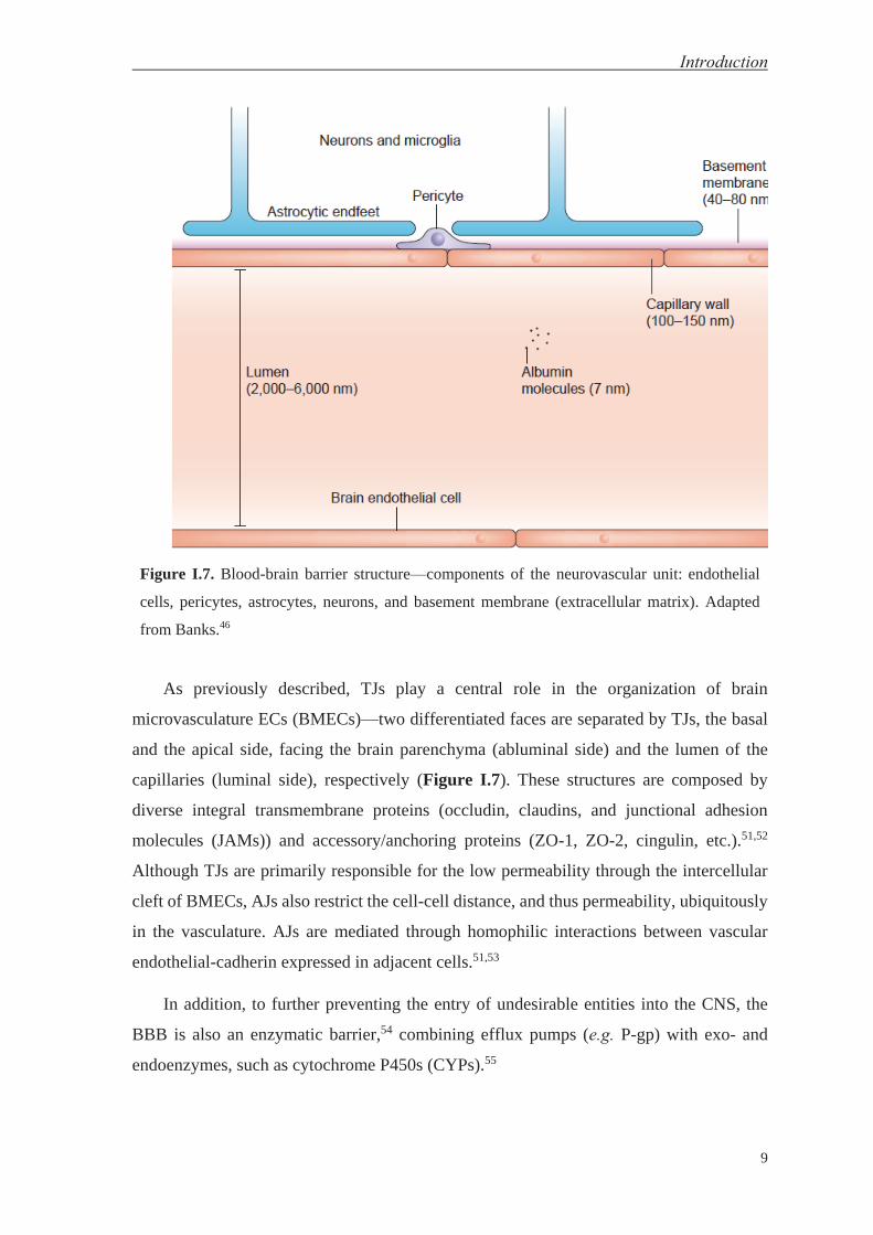
principal structural components, the so-called neurovascular unit (Figure I.7).46 These
components include several cell types—endothelial cells (ECs, addressed later on),
pericytes, astrocytes, and neurons, as well as the extracellular matrix. Pericytes are in close-
contact with ECs and have recently been demonstrated to play a key role in BBB
differentiation by regulating BBB-specific gene expression in ECs and inducing the
polarization of astrocyte end-feet.47,48 Astrocytes regulates at the same time the BBB
features of ECs, such as by modulating the tightness of TJs and the expression of
transporters and enzymes.49 It is known that there is a relationship between regional
neuronal activity and blood flow, whereas it has been purposed the regulation BBB
permeability by neurons—i.e. neurons can regulate the BBB function.50,51 Finally, the
extracellular matrix helps to preserve the integrity of the BBB by providing an anchor point
for ECs, mainly through the interaction of integrins with laminin and the regulation of
intercellular communication. ECs, pericytes and astrocytes secrete extracellular matrix
molecules thereby contributing to the formation of this matrix, the major components of
which are collagen type IV, laminin and fibronectin.45,51
Introduction
9
Figure I.7. Blood-brain barrier structure—components of the neurovascular unit: endothelial
cells, pericytes, astrocytes, neurons, and basement membrane (extracellular matrix). Adapted
from Banks.46
As previously described, TJs play a central role in the organization of brain
microvasculature ECs (BMECs)—two differentiated faces are separated by TJs, the basal
and the apical side, facing the brain parenchyma (abluminal side) and the lumen of the
capillaries (luminal side), respectively (Figure I.7). These structures are composed by
diverse integral transmembrane proteins (occludin, claudins, and junctional adhesion
molecules (JAMs)) and accessory/anchoring proteins (ZO-1, ZO-2, cingulin, etc.).51,52
Although TJs are primarily responsible for the low permeability through the intercellular
cleft of BMECs, AJs also restrict the cell-cell distance, and thus permeability, ubiquitously
in the vasculature. AJs are mediated through homophilic interactions between vascular
endothelial-cadherin expressed in adjacent cells.51,53
In addition, to further preventing the entry of undesirable entities into the CNS, the
BBB is also an enzymatic barrier,54 combining efflux pumps (e.g. P-gp) with exo- and
endoenzymes, such as cytochrome P450s (CYPs).55

Introduction
10
Hydrophilic (diffusion) transport is highly impeded by TJs, which can be crossed by
only small hydrophilic entities like ions and water molecules, and by cell membranes,
which account for most of the surface of the BBB (20 m2),42 blocking most compounds.
Therefore, entry of molecules into the brain across the BBB would be minimal unless
specific mechanisms were available for this purpose.
In this regard, more than 100 years ago, Goldmann reported that intrathecal (1913) and
parenteral (1909) injections of water-soluble dyes—trypan blue—in adult rats did and did
not stain the brain, respectively.56 He also realized that the CP exerted a protective function.
Previously, in 1885, Ehrlich did similar experiments but did not coin the term “blood-brain
barrier” or describe it appropriately—even saying “I am unable to accept that the vascular
endothelium, as such, exercises different functions in different organs, so that, for example
a liver capillary is permeable for certain substances that will not pass through other
capillaries”.56 Nonetheless, gases and compounds with certain characteristics, namely
those showing relatively high lipophilicity, a small size and low number of H-bond donors
or acceptors—better defined by the Lipinski “rule of five”—,57 are able to diffuse through
the lipid bilayer of cell membranes in a process known as lipophilic passive diffusion. This
is one of the classical routes to overcome the BBB and deliver drugs into the CNS, although
it is limited by the aforementioned rules.
In addition to the previously mentioned transport (diffusion) pathways, cells also have
mechanisms that require energy, such as that used in the hydrolysis of ATP or exchanged
from a positive electrochemical gradient. ECs at the BBB display much lower rates of
endocytosis and transcytosis compared to those at the peripheral endothelium.49 These
mechanisms can be divided in those that use the following: (1) transport proteins, and
endocytic mechanisms including (2) adsorptive-mediated (AME) and (3) receptor-
mediated endocytosis (RME). Transport proteins can be classified on the basis of
stoichiometry and type of energy used: transporters which favor the movement of specific
small molecules or ions down its concentration gradient—uniport, included as facilitated
diffusion, which does not imply the use of energy—, or against it but favored by the
simultaneous transport—cotransport—of other species (at the same (symport) or opposite
(antiport) side of the cell membrane) down their gradient. Additionally, pumps utilize ATP
hydrolysis as source of energy to move small molecules or ions against electrochemical
gradient.58

Introduction
11
Figure I.8. Transport protein mechanisms: uniport, symport, antiport, and ATP-coupled pumps.
Created using ChemBioDraw.
AME and RME are endocytic mechanisms classified on the basis of the nature of their
interaction with the membrane entity that recognizes the molecule that uses each
mechanism. The former entails positively charged molecules that are attracted
electrostatically by cell membranes—glycocalyx—, which are negatively charged.59 Thus,
its nature conditions the lack of selectivity to differentiate between tissues or cell types.
Conversely, RME is based on the internalization of an extracellular receptor, which
recognizes a ligand that is likely to be co-transported with the receptor, such as transferrin
receptor (TfR) and its ligand transferrin (Tf).60
Classifying endocytic mechanisms into these two groups (AME and RME) is
interesting for drug delivery purposes since it highlights the type of delivery system;
however, it does not account for the cellular mechanism of internalization involved. For
further understanding, the cellular basis of endocytosis and the diverse mechanisms
involved are described—phagocytosis, macropinocytosis, clathrin-dependent, cavoelin-
dependent or independent (of these two mechanisms) endocytosis.61,62
Phagocytosis is an actin-dependent and receptor-mediated process that entails the
engulfment of large particles (usually over 0.5 μM in diameter)—a process that includes
Fc- and complement-receptors, but also integrins, lectins and lipopolysaccharide-
receptor.63 Frequent in phagocytic cells such as monocytes, macrophages and tissue
dendritic cells, phagocytosis has also been observed in pericytes64,65 and astrocytes.66
Pinocytic activity—actin-dependent uptake of solutes in the fluid phase or adsorbed to the
cell membrane—67,68 has been reported to be very low in BMECs.69
Consistent with its name, clathrin-dependent internalization is dependent on the
recruitment and formation of a clathrin-coated pit, which contributes to the formation of a

Introduction
12
vesicle. Finally, vesicle scission is produced by the mechanochemical enzyme dynamin.
Although the induction of the mechanism is fostered in some cases by interaction with the
ligand receptor, for example epidermal growth factor receptor (EGFR), in others the
internalization is constitutive, such as in the case of TfR.70
Figure I.9. Endocytic mechanisms: phagocytosis, macropinnocytosis, and clathrin- and
caveolin-dependent and -independent endocytosis. Adapted from Mayor et al.61
In contrast, the other endocytic mechanisms are not clathrin-dependent and are
classified on the basis of the proteins involved in the internalization process. Caveolin-
dependent endocytosis recruits caveolin-1 protein to trigger vesicle formation; and again
dynamin is responsible for scission. This mechanism might be involved in the
internalization of some receptors that do internalize by clathrin-dependent endocytosis—
EGFR is internalized via the caveolin-dependent route after ubiquitination. The remaining
mechanisms, some dynamin-dependent and others not, are still not well understood.61
Although the BBB is an immunological barrier, leukocytes can cross it through a
process called diapedesis, a process defined as the transmigration of these cells from one
side of ECs to the other. Diapedesis is known to occur through either paracellular or
trancellular transmigration. Both have the same initiation process comprising the following
steps: rolling of leukocytes through the EC surface, while interacting with selectins; G
protein-mediated signaling, leading to arrest leukocytes with the help of integrins; and
finally, leukocytes on the surface of ECs can proceed with diapedesis (Figure I.10).71-73 In
the case of paracellular transmigration, JAMs have been reported to play a role in the
regulation of the paracellular migration of leukocytes.74 On the other hand, the

Introduction
13
transendothelial process requires the membrane-associated signaling protein caveolin-1.75
In this regard, viruses and bacteria can infect leukocytes and be transported to the
basolateral side of the BBB via the so-called “Trojan horse” mechanism; other pathogens
cross by themselves using the paracellular or transcellular pathways.76,77
Figure I.10. Leukocyte “Trojan horse” mechanism. Adapted from Ley et al.78
Introduction
14
Reasons to Look Inside—Physiology and Disease of the CNS: Social Impact
The transport logistics at the BBB entails high restriction. In this regard, it has been
reported that >98% of small molecule drugs and ~100% of large therapeutics do not cross
this barrier.43 The increase in research efforts devoted to drug delivery, especially to the
brain, reflects scientific and social interest in this field. In this regard, there has been an
exponential trend in the number of publications addressing drug delivery or drug delivery
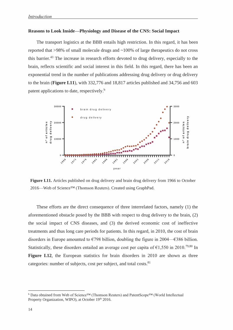
to the brain (Figure I.11), with 332,776 and 18,817 articles published and 34,756 and 603
patent applications to date, respectively.b
y e a r
nº
of
art
icle
s
dru
gd
eli
ve
ry
nº
of
art
icle
s
bra
ind
rug
de
liv
ery
1 9 6 6
1 9 7 1
1 9 7 6
1 9 8 1
1 9 8 6
1 9 9 1
1 9 9 6
2 0 0 1
2 0 0 6
2 0 1 1
2 0 1 6
0
10000
20000
30000
0
1000
2000
3000
d r u g d e l iv e r y
b r a in d r u g d e l iv e r y
Figure I.11. Articles published on drug delivery and brain drug delivery from 1966 to October
2016—Web of Science™ (Thomson Reuters). Created using GraphPad.
These efforts are the direct consequence of three interrelated factors, namely (1) the
aforementioned obstacle posed by the BBB with respect to drug delivery to the brain, (2)
the social impact of CNS diseases, and (3) the derived economic cost of ineffective
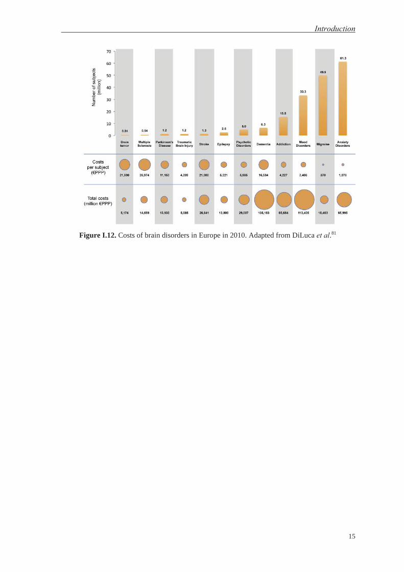
treatments and thus long care periods for patients. In this regard, in 2010, the cost of brain
disorders in Europe amounted to €798 billion, doubling the figure in 2004—€386 billion.
Statistically, these disorders entailed an average cost per capita of €1,550 in 2010.79,80 In
Figure I.12, the European statistics for brain disorders in 2010 are shown as three
categories: number of subjects, cost per subject, and total costs.81
b Data obtained from Web of Science™ (Thomson Reuters) and PatentScope™ (World Intellectual Property Organization, WIPO), at October 19th 2016.
Introduction
15
Figure I.12. Costs of brain disorders in Europe in 2010. Adapted from DiLuca et al.81

Introduction
16
Devising a Key—Peptides as Therapeutics and for Drug Delivery to the Brain
Peptides are sequences of concatenated amino acids through an amide bond. They are
distinguished from proteins classically by their size—considering peptides to be sequences
shorter than 50 residues. Nevertheless, sometimes the same molecule is considered both
peptide and protein. Rather than size, a more appropriate way to classify these polymers
may be on the basis of structure—i.e. primary, secondary, tertiary and quaternary
structures. Primary and secondary structures equate to the amino acid sequence and the first
homogeneous three-dimensional arrangement, respectively. Peptides could be considered
sequences that adopt a secondary structure while proteins could be defined as having a
tertiary structure, namely the second three-dimensional arrangement (homo- or
heterogeneous). Nevertheless, the quaternary structure, defining the assembly of diverse
molecules, can be adopted by both peptides and proteins (Figure I.13). Therefore, proteins
are more complex than peptides when analyzed as single molecules, but this may not be
the case when quaternary structure is taken into account.
Figure I.13. Peptide and protein structures: primary, secondary, tertiary (proteins) and
quaternary. Created using ChemBioDraw.

Introduction
17
The properties of peptides make them unique therapeutic molecules. They are well-
characterized thanks to the synthetic strategy called solid-phase peptide synthesis (SPPS).82
This methodology significantly reduces the production costs. With respect to the biological
properties of peptides, these molecules generally show better specificity and affinity for
therapeutic targets than small molecules and broader administration routes compared with
biologics. However, the (proteolytic) stability of peptides is their weak point. Nevertheless,
some peptides have a highly resistant profile conferred by their three-dimensional
rearrangement,83 and diverse strategies can be applied to improve the stability of labile
peptides.84 Thus, in most cases, peptides combine the best properties of two worlds, small
molecules and biologics (Table I.1), except for a controversial property, namely immune
response, where the performance of peptides is likely to fall between the two other drug
categories.
Table I.1. Comparison of small molecules, peptides and biologics.
Compound Small Molecule Peptide Biologic
s
Synthesis chemical chemical/SPPS biologica
l production
Cost low low high
Characterization well-defined well-defined hard
Specificity/Affinit
y limited medium/high high
Administration oral/cutaneous/injecte
d
cutaneous/injecte
d injected
Stability/
Metabolism
cytochrome
metabolism
enzyme
proteolysis
(highly labile)
enzyme
proteolysis
(medium
)
Immunogenicity low low-medium high
Like proteins, peptides abound in nature as effector molecules. A number of peptides
can cross the BBB using the transport mechanisms previously mentioned. However not all
BBB crossing peptides use the same mechanism of uptake. In this regard, several relatively
long neuropeptides (e.g. neuropeptide Y85 and orexin A,86 with a 36 and 33 amino acid
sequence, respectively) have been shown to cross the BBB by simple diffusion. On the

Introduction
18
other hand, glutathione (GSH), composed by three amino acids, namely γ-ʟ-glutamyl-ʟ-
Cys-ʟ-Gly, is transported into the CNS by a carrier-mediated saturable transport
mechanism.87,88 Similarly, opiate peptides such as enkephalins and antiopiates such as Tyr-
MIF-1 are also transported by carrier-mediated systems, in this case the so-called peptide
transport system 1 (PTS-1).89-91 Nevertheless, enkephalins also cross the BBB through non-
saturable mechanisms.91
Endocytic mechanisms are also used by a range of peptides and proteins. Protegrin 1
(PG-1), an 18-amino acid antimicrobial peptide isolated from leukocytes, is able to form
pores in bacterial membranes, and several authors have reported the use of diverse analogs
as agents for brain delivery via AME.92 Discovered in 1988, HIV TAT 48–57 peptide
derives from the transcriptional trans-activator protein of the human immunodeficiency
virus 1 (HIV-1).93,94 The peptide crosses cell membranes, probably through an adsorptive-
mediated mechanism, and has been used for drug delivery to the CNS.92,95 A recent study
using TAT-conjugated quantum dots (QDs) revealed new insights of the mechanism
underlying the capacity of this peptide to transport nanoparticles.96 Deriving from the third
helix of the Antennapedia homeodomain of Drosophila,97,98 penetratin has been used for
similar purposes. These peptides, rich in positive residues, are known as cell-penetrating
peptides (CPPs),99-101 a name that reflects their capacity to cross biological membranes.
Peptides and proteins internalized by RME have a key advantage over those
internalized by AME, in that they interact with a particular membrane receptor which might
be over- or singularly-expressed in a specific cell-type or tissue. Endogenous ligands of
each receptor, which are endocytosed, are susceptible to applications in drug delivery.
Among these, the low-density lipoprotein receptor (LDLR) family recognizes various
ligands like apolipoprotein B100 (apoB100) and E (apoE), which are embedded in the outer
lipid-membrane of variousc lipoprotein particles.102 The latter, apoE (as a full-length
protein or analogs), has been extensively used for drug delivery to the brain.103-105 Hepatitis
C virus uses this mechanism (LDLR-mediated) to enter cells.106 Nicotinic acetylcholine
receptor (nAChR) is an ionotropic receptor that is highly expressed in the brain, including
in BMECs.107 A 16-amino acid peptide derived from a toxin from Bungarus candidus,
Candoxin (CDX), was used as ligand of nAChR for drug delivery to the CNS.108 In
c Low-, very low-, intermediate-density lipoproteins (LDL, VLDL, IDL, respectively) and chylomicrons.
Introduction
19
addition, peptides derived from a rabies virus109 glycoprotein (RVG) have been extensively
used for the same purpose.110,111
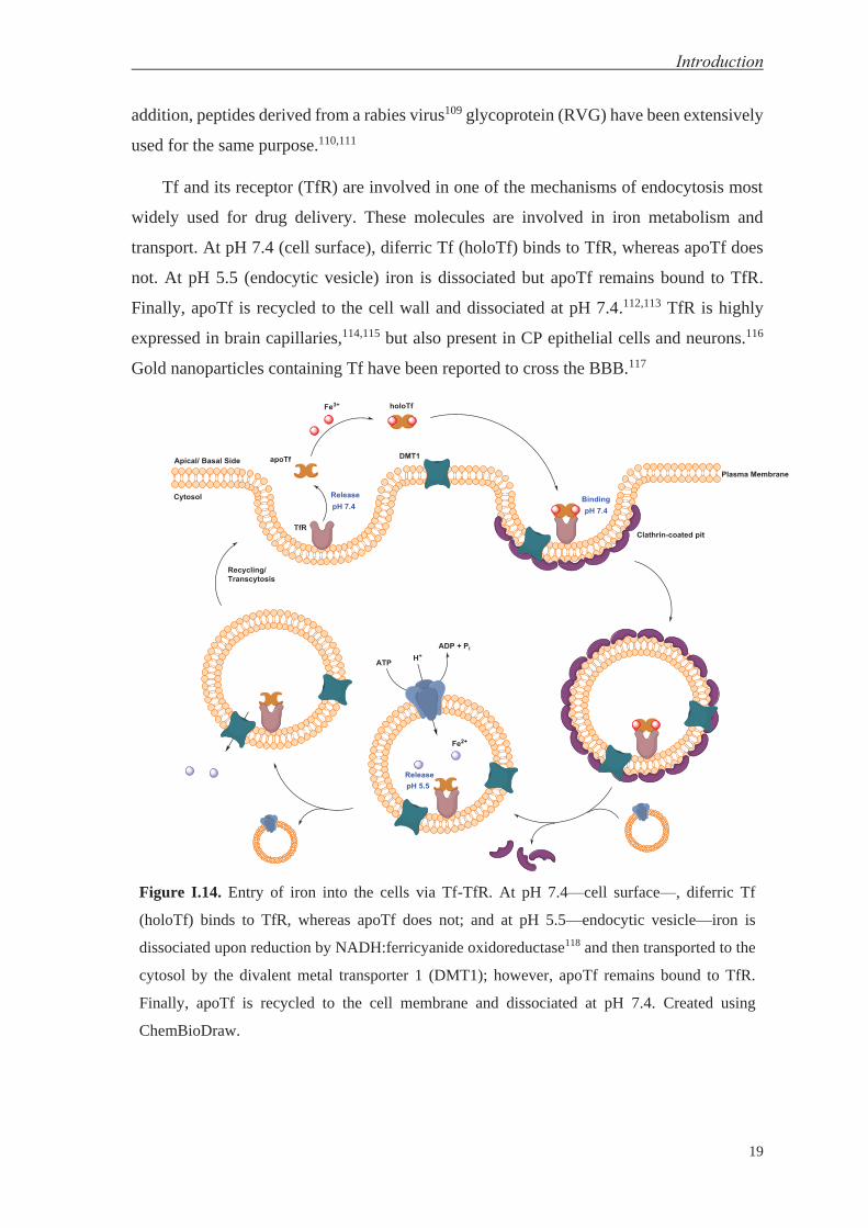
Tf and its receptor (TfR) are involved in one of the mechanisms of endocytosis most
widely used for drug delivery. These molecules are involved in iron metabolism and
transport. At pH 7.4 (cell surface), diferric Tf (holoTf) binds to TfR, whereas apoTf does
not. At pH 5.5 (endocytic vesicle) iron is dissociated but apoTf remains bound to TfR.
Finally, apoTf is recycled to the cell wall and dissociated at pH 7.4.112,113 TfR is highly
expressed in brain capillaries,114,115 but also present in CP epithelial cells and neurons.116
Gold nanoparticles containing Tf have been reported to cross the BBB.117
Figure I.14. Entry of iron into the cells via Tf-TfR. At pH 7.4—cell surface—, diferric Tf
(holoTf) binds to TfR, whereas apoTf does not; and at pH 5.5—endocytic vesicle—iron is
dissociated upon reduction by NADH:ferricyanide oxidoreductase118 and then transported to the
cytosol by the divalent metal transporter 1 (DMT1); however, apoTf remains bound to TfR.
Finally, apoTf is recycled to the cell membrane and dissociated at pH 7.4. Created using
ChemBioDraw.

Introduction
20
The term “Molecular Trojan horse” has been coined for a range of brain vectors
including endogenous peptides, modified proteins, and peptidomimetic monoclonal
antibodies.119,120 In this regard, monoclonal antibodies targeting TfR have been used for the
delivery of drugs to the brain.121 Nevertheless, “Molecular Trojan horse” would be a more
suitable description for the aforementioned “Trojan horse” mechanism used by viruses and
bacteria, which, after infecting leukocytes, are transported to the basolateral side of the
BBB.76,77 In this regard, the brain delivery of serotonin by monocytes following
phagocytosis of liposomes has been reported.122
In this thesis, the peptides used as molecular shuttles for brain delivery are referred to
as “BBB shuttles”. These shuttles are peptides with the ability to carry cargos across the
BBB either through active123-125 or passive transport mechanisms.126,127 The great
advantage of BBB shuttles is that they can confer the ability to cross the BBB to a wide
range of molecules through simple chemical conjugation strategies. Thus they are able to
significantly expand the therapeutic space of potential CNS drugs. BBB shuttles can
therefore serve as powerful facilitators of drug delivery to the brain.

OBJECTIVES


Objectives
23
This thesis addresses four main objectives, one per chapter. The first three objectives
are focused on basic research on BBB shuttles, whereas the last one has a more applied
character.
Objective 1: To design, synthesize and evaluate a new family of passive diffusion BBB
shuttles.
1.1.To improve the low solubility of the “gold standard” (NMePhe)-based BBB shuttle
peptides.
1.2.To enhance their shuttle capacity (maintaining the transport after cargo attachment).
1.3.To study the role of the stereochemistry in passive diffusion.
Objective 2: To study a new family of BBB shuttle peptides working through receptor-
mediated transcytosis (RMT), and to develop a methodology based on the combined use of
MALDI-TOF MS with in vitro cell-based models of the BBB to assess the transport
through the BBB.
Objective 3: To study and compare the immunological responses elicited by BBB shuttles
made by ʟ-amino acids and their retro-ᴅ-versions, made by ᴅ-amino acids.
Objective 4: To perform a series of preliminary studies with the final goal of developing a
therapy for Friedreich’s Ataxia (FRDA) at the central nervous system (CNS).
4.1.To study the viability of a protein replacement therapy for FRDA based on direct
conjugation of BBB shuttles to frataxin (FXN).
4.2.To improve bioconjugation methods to modify HSV-1 particles with BBB shuttles to
develop a gene therapy for FRDA at the CNS.
4.3.To characterize the physicochemical and biological properties of these viral particles
before and after bioconjugation.


RESULTS AND DISCUSSION


Chapter 1
Study of Passive Diffusion BBB Shuttles

This chapter is partially based on the following article:
Arranz-Gibert, P.; Guixer, B.; Malakoutikhah, M.; Muttenthaler, M.; Guzmán, F.; Teixidó, M.;
Giralt, E. Lipid Bilayer Crossing—The Gate of Symmetry. Water-Soluble Phenylproline-Based
Blood-Brain Barrier Shuttles. J. Am. Chem. Soc. 2015, 137, 7357.

Chapter 1 Study of passive diffusion BBB shuttles
29
Passive transport encompasses two main pathways, namely paracellular (hydrophilic)
and transcellular (lipophilic) diffusion. The former allows small hydrophilic entities to
cross the BBB. However, this pathway is extremely hindered at this barrier due to the
presence of tight junctions,128 and therefore it is not ideal for drug delivery. In contrast,
transcellular lipophilic diffusion involves transport through the much larger lipid bilayer,
which provides a direct correlation between concentration and transport. The lipid bilayer
of the plasma membrane can be considered a macroreceptor that can simultaneously
interact with many ligands, thus accounting for the greatest proportion of the cell surface.
This layer is therefore the preferred target for the delivery of small-molecule therapeutic
drugs.129 Theoretically, transport through this mechanism is facilitated by the movement of
the fatty acid side chains in the membrane, which form holes (“kinks”) through which
molecules can diffuse across the membrane.130,131 The concentration of the kinks is
estimated to be between 10 and 50 mM. This concentration is a function of the
conformational changes that can be adopted by fatty acid hydrocarbon, which are related
to the ratio of saturated/ unsaturated fatty acids in the membrane and cholesterol.132
There are currently two main approaches to design therapeutics able to cross the BBB
through transcellular lipophilic diffusion. The first, and most commonly implemented

Results & Discussion
30
approach in the pharmaceutical industry, is based on a set of rules covering molecular size,
presence of H-bond acceptors/donors, and lipophilicity, thereby attempting to increase the
likelihood of the molecule crossing the BBB.57,133 The second approach is the design of
BBB shuttles, which forms a major research line in our laboratory.
Figure 1.1. Transport mechanisms at the BBB. Active transport comprises (I) carrier-mediated
transport and endocytic mechanisms—(II) receptor- and (III) adsorptive-mediated transcytosis.
Passive transport is described by (IV) transcellular lipophilic diffusion and (V) paracellular
hydrophilic diffusion. Transcellular lipophilic diffusion is dependent on the lipid composition of
the cell, which comprises mainly phospholipids. Created with Microsoft PowerPoint and Adobe
Illustrator.
While chiral complexity in drug-receptor activation, receptor-mediated transcytosis
and protein-protein interactions has been extensively studied, the chiral interaction of
entities with the membrane of the BBB endothelial cells is still poorly understood. Some
studies have addressed a range of lipid structures,134-136 and enantiomeric discrimination of
dipeptides by bio-membranes has also been reported.137-139 However, the chiral interactions
between the BBB and BBB shuttles have not been studied before. Considering the chiral
nature of phospholipids, one of the main components of plasma membranes, and our
continuous efforts in improving BBB shuttle design, we set out to study the transport
properties of different BBB shuttle stereoisomers.

Chapter 1 Study of passive diffusion BBB shuttles
31
Additionally, we wanted to improve the water-solubility of our BBB shuttles. Earlier
work identified aromatic N-methylated peptides, both cyclic126 and linear,140-143 as highly
permeable compounds for lipid membranes. Efforts in our laboratory led to the
development of (NMePhe)-based peptides, the gold-standard of passive diffusion BBB
shuttles (Figure 1.2).141,142 The wider use of this class of BBB shuttle for clinical
applications was however limited by the intrinsic low water-solubility of these molecules
(< 1 μM).
Hence, we set out to advance the mechanistic knowledge and design opportunities of
BBB shuttles by (1) studying the impact of chirality on their transport capabilities, and (2)
by improving their water-solubility properties towards clinical applications. In order to
achieve both goals, we sought to design and synthesize a library of novel chiral BBB
shuttles that would provide further insight into the impact of chirality at the BBB, while at
the same time tackling the long-standing problem of water-solubility.

Results & Discussion
32
1.1. Peptide-Shuttle Design
(NMePhe)-based BBB shuttles140-143 were taken as the base for the design of a new
class of water-soluble BBB shuttles. Our aim was for these shuttles to retain high BBB
effective permeability (Pe)144 while improving their low water-solubility. At the same time,
we wanted to have control of chirality to study the transport capacity of different
stereoisomers. For these purposes, we chose the proteogenic amino acid proline, which has
a conformationally restricted side-chain (advantageous for a chiral library design) and
excellent water-solubility (around 300 mM, the tetraproline), in spite of the hydrophobic
character of its side-chain. Additionally, polyprolines are also highly conformationally
constrained compounds145,146 that have been used extensively for the design of water-
soluble dendrimers and cell-penetrating peptides (CPPs).147
We anticipated that a hybrid design of proline analogs containing a phenyl ring could
merge the ability to cross the BBB with a simultaneous improvement of water-solubility
(Figure 1.2). Furthermore, phenyl and pyrrolidine rings have been described as two of the
most common substructures in the chemical makeup of CNS drugs.148 Thus, we turned our
attention to peptides derived from cis-3-phenylpyrrolidine-2-carboxylic acid (PhPro)
(Figure 1.2c).
Figure 1.2. Structure of (a) the gold-standard passive diffusion BBB shuttle (NMePhe)4, (b)
hydrophilic polyproline unit Pro4, and (c) designed (PhPro)4 hybrid; all homo-ʟ, C-terminal
amide, and N-terminal acetylated. Created using ChemBioDraw.

Chapter 1 Study of passive diffusion BBB shuttles
33
1.2. Transport Ability of (PhPro)4 Shuttle Using the PAMPA Assay
To establish whether this new hybrid class of BBB shuttles retained its anticipated
transport properties, we used the PAMPA assay to perform BBB transport studies of
(PhPro)4, which was initially synthesized with the commercially available racemic building
block, Fmoc-cis-3-phenylpyrrolidine-2-carboxylic acid (Figure 1.3). The PAMPA assay,
introduced by Kansy et al.,149 allows the parallel evaluation of passive diffusion transport
of various compounds through a mixture of lipids, thus emulating the biological barrier of
interest. A selected lipid mixture is deposited onto a filter, which is divided into two
compartments. Lipids are chosen in function of the composition of the barrier, i.e. in our
study a mixture of porcine brain polar lipid extract was used. The compartments above and
below the filter contained only buffer and the molecule to test in buffer, respectively.
Magnetic stirring was applied for 4 h in donor wells, thus mimicking the stirring that red
blood cells produce in brain capillaries. This approach almost totally reduced the unstirred
water layer (UWL). Afterwards, each well was quantified by UV-absorption after injection
into a RP-HPLC system. Time and concentration used were optimized to achieve a
satisfactory relation signal-to-noise during quantification and to prevent back-diffusion, i.e.
the experiment was performed while the transport rate was constant. Finally, propranolol
(a β-adrenergic receptor blocker with high brain penetration) was used as a positive control.
Figure 1.3. Fmoc-cis-3-phenylpyrrolidine-2-carboxylic acid structure: (left) ʟ- and (right) ᴅ-
configurations. Created using ChemBioDraw.
The formula for Pe calculation is shown in Eq. 1.1:
(1.1)
where t is the running time (4 h), CA (t) is the concentration of the compound in the
acceptor well at time t, and CD (t0) is the compound concentration in the donor well before
running the PAMPA assay (t0 = 0 h). Permeability is considered excellent with values >
Results & Discussion
34
4.0 (·10-6) cm/s, uncertain between 2.0 and 4.0 (·10-6) cm/s and poor with values below 2.0
(·10-6) cm/s.144
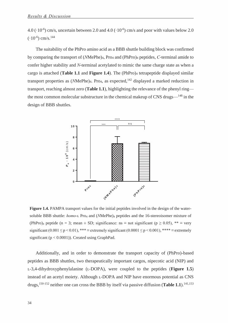
The suitability of the PhPro amino acid as a BBB shuttle building block was confirmed
by comparing the transport of (NMePhe)4, Pro4 and (PhPro)4 peptides, C-terminal amide to
confer higher stability and N-terminal acetylated to mimic the same charge state as when a
cargo is attached (Table 1.1 and Figure 1.4). The (PhPro)4 tetrapeptide displayed similar
transport properties as (NMePhe)4. Pro4, as expected,142 displayed a marked reduction in
transport, reaching almost zero (Table 1.1), highlighting the relevance of the phenyl ring—
the most common molecular substructure in the chemical makeup of CNS drugs—148 in the
design of BBB shuttles.
Pro
4
(NM
ePh
e ) 4
(Ph
Pro
)4
0
2
4
6
8
1 0
Pe
·1
06
(cm
/s)
n s***
****
Figure 1.4. PAMPA transport values for the initial peptides involved in the design of the water-
soluble BBB shuttle: homo-ʟ Pro4 and (NMePhe)4 peptides and the 16-stereoisomer mixture of
(PhPro)4 peptide (n = 3; mean ± SD; significance: ns ≡ not significant (p ≥ 0.05), ** ≡ very
significant (0.001 ≤ p < 0.01), *** ≡ extremely significant (0.0001 ≤ p < 0.001), **** ≡ extremely
significant (p < 0.0001)). Created using GraphPad.
Additionally, and in order to demonstrate the transport capacity of (PhPro)-based
peptides as BBB shuttles, two therapeutically important cargos, nipecotic acid (NIP) and
ʟ-3,4-dihydroxyphenylalanine (ʟ-DOPA), were coupled to the peptides (Figure 1.5)
instead of an acetyl moiety. Although ʟ-DOPA and NIP have enormous potential as CNS
drugs,150-152 neither one can cross the BBB by itself via passive diffusion (Table 1.1).141,153

Chapter 1 Study of passive diffusion BBB shuttles
35
Table 1.1. Transport (%) and effective permeability (Pe) values for homo-ʟ Pro4 and (NMePhe)4
peptides and the 16-stereoisomer mixture of (PhPro)4 peptide, as well for (NMePhe)4 and
(PhPro)4 attached to a therapeutically relevant cargo (ʟ-DOPA and NIP) (n = 3; mean ± SD). The
transport values for (NMePhe)-based peptides were published previously).141,142
Compound Transport (%) Pe · 106 (cm/s)
Pro4 0.02 ± 0.00 0.01 ± 0.00
(NMePhe)4 12.7 ± 2.1 6.8 ± 1.3
(PhPro)4 12.6 ± 0.3 6.88 ± 0.18
ʟ-DOPA 0.0 ± 0.0 0.0 ± 0.0
ʟ-DOPA(NMePhe)4 2.4 ± 0.2 1.10 ± 0.10
ʟ-DOPA-(PhPro)4 16.7 ± 1.7 9.9 ± 1.5
NIP 0.0 ± 0.0 0.0 ± 0.0
NIP-(NMePhe)4 2.8 ± 0.2 1.40 ± 0.10
NIP-(PhPro)4 19 ± 3 11 ± 2
ʟ-DOPA is a prodrug that has been used for the last forty years to treat Parkinson’s
disease,154 however, its uptake mechanism is limited since it has to compete with other
amino acids for amino acid transporters. To improve uptake efficacy and to avoid
interference with amino acid transporters, the challenge of ensuring ʟ-DOPA delivery
through other mechanisms must be tackled. Furthermore, ʟ-DOPA derivatization through
an amide bond can prevent side effects in the periphery by inhibiting the decarboxylation
reaction and enhancing the transport of this molecule to the brain.153,155-159 Once in the
brain, ʟ-DOPA is enzymatically converted to dopamine by aromatic ʟ-amino acid
decarboxylase.
Figure 1.5. Structures for (a) ʟ-DOPA-(PhPro)4 and (b) NIP-(PhPro)4 as homo-ʟ configurations.
Created using ChemBioDraw.

Results & Discussion
36
Nipecotic acid (NIP) is a GABA reuptake inhibitor with great therapeutic potential if
it would be able to cross the BBB.160-162 GABA is the primary inhibitory neurotransmitter
in the CNS, and decreased levels of this molecule are associated with several brain
disorders. The levels of this amino acid in the CNS can be increased either by supplying
GABA or its agonists, or via GABA reuptake inhibitors, such as NIP.
Pe
·1
06
(cm
/s)
0
5
1 0
1 5
x
x - (N M e P h e )4
x - (P h P r o )4
***
***
****
***
**
****
L -D O P A N IP
Figure 1.6. PAMPA transport values for peptides attached to a cargo (ʟ-DOPA and NIP) was
evaluated, as well as that of the cargos alone (n = 3; mean ± SD; significance: ns ≡ not significant
(p ≥ 0.05), ** ≡ very significant (0.001 ≤ p < 0.01), *** ≡ extremely significant (0.0001 ≤ p <
0.001), **** ≡ extremely significant (p < 0.0001)). Created using GraphPad.
Hence, the BBB transport capacity of (PhPro)4 peptide carrying ʟ-DOPA or NIP was
evaluated and compared to the gold-standard (NMePhe)4 equivalent (Figure 1.6). x-
(PhPro)4 was synthesized using Fmoc-SPPS (x ≡ acetyl, ʟ-DOPA (ʟ-3,4
dihydroxyphenylalanine) or NIP ((R)-piperidine-3-carboxylic acid)). (PhPro)-based
peptides displayed superior permeability to their (NMePhe)-based analogs. Unlike
acetylated (PhPro)4 peptide and its (NMePhe)4 analog, which did not display significant
differences in permeability, (PhPro)4 carrying either of the two cargos showed higher
permeability (7-fold; with extremely significant differences) compared to its (NMePhe)4
analog. Furthermore, the ability to cross the BBB appeared to be independent of the type
of cargo attached (i.e. its BBB shuttle capacity was not altered). This observation contrasted
with the findings for the (NMePhe)-based analogs, which showed a significant reduction in
the capacity to cross the BBB (Table 1.1 and Figure 1.6). In contrast, the transport capacity
of Pro4 tetrapeptide was close to zero (Pe = 0.01 (·10-6) cm/s), as was that of the two cargos
(NIP and ʟ-DOPA).

Chapter 1 Study of passive diffusion BBB shuttles
37
1.3. Design and Synthesis of a 16-Steroisomer Library of (PhPro)4
In order to study the impact of chirality at the BBB, we devised a library of 16
stereoisomers of the (PhPro)4 peptide (Figure 1.7). For the 16-stereoisomer library, we first
had to separate the two PhPro enantiomers of the commercially available racemic mixture.
After chiral separation of the two compounds, each PhPro enantiomer, ʟ- and ᴅ-PhPro
((S,S)- and (R,R)-3-phenylpyrrolidine-2-carboxylic acid, respectively), was assigned by the
specific rotation published previously.163,164 All peptides were synthesized by manual
Fmoc-SPPS.82,165,166 The 16 stereoisomers of this library arose from the permutation of the
two amino acid enantiomers in each position and were synthesized via Houghten’s “tea
bag” method.167-169 All peptides were synthesized with a C-terminal amide to confer higher
stability and were N-terminal acetylated to mimic the same charge state as when a cargo is
attached. Additionally, the use of a non-natural amino acid (and in some cases also the use
of the ᴅ-amino acid version) confers improved protease resistance, overcoming one of the
main drawbacks of using peptides as therapeutical agents.
Figure 1.7. The library of the 16 (PhPro)4 stereoisomers; the peptides are split into two transport-
symmetry groups, where Group 1 contains the peptides with higher symmetry and lower
enantiomeric discrimination, and Group 2 comprises the less symmetric peptides related to a
higher enantiomeric discrimination. Created using ChemBioDraw.

Results & Discussion
38
1.4. Physicochemical Characterization of Pro4 and (PhPro)4 Shuttle
Circular dichroism (CD) studies were carried out to confirm the correct (PhPro)4
enantiomeric assignment. The spectra of both homo-ʟ and homo-ᴅ (PhPro)4 were compared
to the homo-ʟ and homo-ᴅ Pro4 analog control peptides (synthesized with enantiomerically
pure building blocks). As expected, the homo-ʟ tetrapeptides (LLLL) displayed a negative
CD spectrum, contrary to the homo-ᴅ (DDDD) ones, which displayed a positive one
(Figure 1.8), thus confirming our initial assignment. The CD spectra of Pro4 peptides
recorded a higher signal, thereby suggesting a more defined secondary structure (i.e. PPII
conformation generally observed with polyproline compounds170 compared to the (PhPro)4
tetrapeptides). The PhPro-peptides studied by CD did not present a known structure-
assigned spectrum.
(n m )
[] M
R(d
eg
·cm
2·d
mo
l-1)
2 1 0 2 2 0 2 3 0 2 4 0 2 5 0
- 2 0 0 0 0
- 1 0 0 0 0
0
1 0 0 0 0
2 0 0 0 0
P r o 4 L L L L
P r o 4 DDDD
a
(n m )
[] M
R(d
eg
·cm
2·d
mo
l-1)
2 1 0 2 2 0 2 3 0 2 4 0 2 5 0
- 5 0 0 0
- 2 5 0 0
0
2 5 0 0
5 0 0 0
(P h P ro )4 DDDD
(P h P ro )4 L L L L
b
Figure 1.8. Circular dichroism spectra of pairs of enantiomers; homo-ᴅ (blue) and homo-ʟ
(purple) of (a) Pro4, displaying a PPII conformation (homo-ʟ/ homo-ᴅ with a weak maximum or
minimum at 228 nm and a strong minimum or maximum at 203 nm, respectively); and (b)
(PhPro)4. Created using GraphPad.

Chapter 1 Study of passive diffusion BBB shuttles
39
Analytical RP-HPLC characterization of the 16 stereoisomers further added to the
characterization, showing complex chromatographic profiles (Figure 1.9a) that were
identical between pairs of enantiomers. Each peak of the profile corresponded to a
conformer with an interconversion rate faster than min-1 (length of the injection cycle),
since reinjection of any of the collected peaks resulted in the same RP-HPLC
chromatogram observed earlier (Figure 1.9b). In some cases, RP-HPLC characterization
at higher temperature (60ºC) resulted in a single peak (Figure 1.9c). Only the stereoisomers
DDLD and LLDL yielded a single RP-HPLC peak, indicating a preferential conformational
arrangement compared to the other stereoisomers.
Figure 1.9. RP-HPLC chromatograms of the DLDD (PhPro)4 stereoisomer (gradient from 50 to
80% CH3CN in 8 min, symmetry C18 column) (a) at room temperature (r.t.), (b) after
chromatographic peak (any of the three peaks observed) reinjection at r.t, and (c) at 60ºC. Created
with Adobe InDesign.
This observation was confirmed by CD, which showed a significantly different
spectrum (maximum/ minimum at 222 nm, respectively) compared to the other
stereoisomers (Figure 1.10). Additionally, both of these enantiomers gelled after solvent
(DIPEA/DCM, 1:1, v/v) evaporation, thereby indicating the adoption of a specific
conformational arrangement that favors this process (not observed with the other
stereoisomers). Each peptide was identified by RP-HPLC-MS and MALDI-TOF MS, and
the observed masses of the individual stereoisomers were in good agreement with the
theoretically calculated molecular weights.

Results & Discussion
40
(n m )
[] M
R(d
eg
·cm
2·d
mo
l-1)
2 1 0 2 2 0 2 3 0 2 4 0 2 5 0
- 5 0 0 0
- 2 5 0 0
0
2 5 0 0
5 0 0 0
(P h P ro )4 L L D L
(P h P ro )4 DDLD
Figure 1.10. Circular dichroism spectra of DDLD (blue) and LLDL (purple) pairs of
enantiomers of (PhPro)4. Created using GraphPad.
Since PhPro was chosen to improve the water-solubility of these BBB shuttles, we
determined this parameter by weighing the lyophilized peptide from a known volume of a
saturated solution. Low water-solubility represents a long standing problem of current BBB
shuttles with for example (NMePhe)4, the “gold-standard” passive transport BBB shuttle,
having a water-solubility that is lower than 1 μM. The (PhPro)4 tetrapeptides registered
water-solubility in the range of 1–5 mM, which can be considered a significant
improvement (1,000–fold) to that shown by (NMePhe)4 tetrapeptides.142 The solubility of
Pro4 peptide was 300 mM.
Chapter 1 Study of passive diffusion BBB shuttles
41
1.5. Passive Diffusion Transport Studies and Chiral Discrimination at the BBB
Chirality plays a key role in many cellular processes and we were interested if this is
also the case during passive diffusion of BBB shuttles through the BBB. To study this
phenomenon, we built a 16-stereoisomer tetrapeptide library via sequentially permuting
each of the four positions with ʟ- and ᴅ-PhPro enantiomers. The passive diffusion of these
peptides through the BBB was evaluated using the PAMPA assay. The majority of the
(PhPro)4 stereoisomers displayed high diffusion through the BBB lipids, except two
peptides (DDLD and its retro-peptide DLDD), with significantly lower transport rates
(Table 1.2 and Figure 1.11).
Table 1.2. Transport (%) and effective permeability (Pe) values for the 16-stereoisomer library
of (PhPro)4 peptide (n = 3; mean ± SD).
Compound Transport (%) Pe · 106 (cm/s)
DDDD 15.2 ± 0.5 8.6 ± 0.3
LLLL 14.3 ± 0.3 7.95 ± 0.15
DDDL 12.34 ± 0.13 6.72 ± 0.07
LLLD 17 ± 5 10 ± 3
DDLD 7.1 ± 0.2 3.64 ± 0.13
LLDL 14 ± 2 7.8 ± 1.2
DLDD 1.76 ± 0.09 0.85 ± 0.04
LDLL 12.8 ± 0.6 7.0 ± 0.3
LDDD 9.27 ± 0.10 4.86 ± 0.05
DLLL 12.8 ± 0.6 7.0 ± 0.4
DDLL 11.2 ± 1.2 6.0 ± 0.6
LLDD 13.5 ± 0.9 7.4 ± 0.5
DLDL 12.7 ± 0.7 7.0 ± 0.4
LDLD 12.7 ± 1.0 7.0 ± 0.6
LDDL 10.61 ± 0.08 5.65 ± 0.04
DLLD 10.5 ± 0.8 5.6 ± 0.4
Propranolol 22.6 ± 0.5 14.3 ± 0.3
Almost all the 16 stereoisomers displayed excellent transport properties (with Pe range
of 4.86-10 (·10-6) cm/s) similar to the gold-standard (NMePhe)4, which displays a transport
capacity of 6.8 (·10-6) cm/s (Table 1.2 and Figure 1.11). Only DDLD and DLDD displayed
significantly lower Pe values (3.64 and 0.85 (·10-6) cm/s, respectively). Interestingly, the

Results & Discussion
42
DDLD/LLDL pair was identified earlier via analytical RP-HPLC and CD analysis
displaying a pronounced but distinct secondary structure compared to the other
stereoisomers (Figure 1.10).
Chirality discrimination of the 16 stereoisomers at the BBB was determined by pairing
the individual enantiomers and determining the enantiomeric discrimination value (De)
(Table 1.3). De is defined as the excess ratio of the transport of each pair of enantiomers,
the higher (TH) minus the lower (TL), then divided by the lower (TL):
(1.2)
By definition, this parameter ranges between 0 (no discrimination) and infinite
(absolute discrimination). We observed values from 0.0 to 6.1 (Table 1.3). Differentially,
the DLDD/LDLL pair of enantiomers showed the highest discrimination, followed by its
retro-pair, DDLD/LLDL, which displayed a value of 1.0.
Table 1.3. Passive diffusion transport enantiomeric discrimination (De) values for each pair of
enantiomers (n = 3; mean ± SD). Two groups are differentiated on the basis of the symmetry and
enantiomeric discrimination (Group 1, higher symmetry, lower enantiomeric discrimination;
Group 2, lower symmetry, higher enantiomeric discrimination).
Group Enantiomer Pair Enantiomeric Discrimination
1
DLDL/ LDLD 0.00 ± 0.00
LDDL/ DLLD 0.01 ± 0.08
DDDD/ LLLL 0.06 ± 0.04
LLDD/ DDLL 0.21 ± 0.13
2
DLLL/ LDDD 0.38 ± 0.07
LLLD/ DDDL 0.4 ± 0.4
LLDL/ DDLD 1.0 ± 0.3
LDLL/ DLDD 6.1 ± 0.4
To further study the impact of chirality on the transport of this family of BBB shuttles,
we classified pairs of enantiomers into two categories on the basis of the similarities
between their transport (Figure 1.11). The peptide enantiomer pairs containing two units
of each PhPro amino acid enantiomer in their sequence (DDLL/LLDD, DLDL/LDLD and
LDDL/DLLD) and the homo-peptides (LLLL/DDDD) showed similar transport between
enantiomers (Group 1). Among this group, homo-peptides showed the highest transport and
the LDDL/DLLD pair the lowest.

Chapter 1 Study of passive diffusion BBB shuttles
43
The transport values of the other enantiomer pairs with only one PhPro amino acid
permutation (Group 2) varied significantly within the pairs (Figure 1.11). DDLD and its
retro-peptide (DLDD) showed the poorest transport capacity, mainly due to membrane
retention (60 and 87%, respectively; data not shown). Their enantiomers (LLDL and LDLL,
respectively), however showed excellent and to Group 1 similar transport properties (7.8
and 7.0 (·10-6) cm/s, respectively). This clearly shows that within two enantiomers the
transport properties can be significantly different (e.g. 7-fold for DLDD and LDLL),
strongly suggesting that chirality plays an important role in passive diffusion.
Pe
·1
06
(cm
/s)
DD
DD
DL
LD
DL
DL
DD
LL
DL
LL
DD
DL
DD
LD
DL
DD
Ste
reo
. Mix
.
0
5
1 0
1 5
n s
n s
n sn s
***
n s
**
****
G ro u p 1 G ro u p 2
Figure 1.11. PAMPA transport values for the 16 individual stereoisomers, paired by
enantiomers, and the 16-stereoisomer mixture (dark blue column). Light blue column
corresponds to the peptide configuration displayed on the graph (e.g. first column = DDDD);
purple column corresponds to the enantiomer of the peptide configuration displayed on the graph
(e.g. first column = LLLL) (n = 3; mean ± SD; significance: ns ≡ not significant (p ≥ 0.05), ** ≡
very significant (0.001 ≤ p < 0.01), *** ≡ extremely significant (0.0001 ≤ p < 0.001), **** ≡
extremely significant (p < 0.0001)). Created using GraphPad.
In order to delve deeper into the chirality-transport relationship, a symmetry model
was devised. Assuming two approximations, considering that the peptide termini (N-

Results & Discussion
44
terminal acetylated and C-terminal amide) do not interfere in the symmetry and the peptide
bond has no direction, i.e. retroenantiomeric-peptides would be identical;171-173 two
enantiomer pair groups were differentiated. Group 1, with the lowest enantiomeric
discrimination (De) values, contains all quasi-meso isomers.d Group 2, with higher De
values, lacks this quasi-meso character (and can be considered as less symmetric than
Group 1). Furthermore, the pairs of enantiomers from Group 1 are retroenantio-peptides
between them but not those belonging to Group 2. Interestingly, the LDLL/ DLDD pair
presented a very high De (6.1) (Figure 1.11 and Table 1.3).
These results show that passive diffusion through biological membranes can be highly
enantiomerically selective. Enantiomeric discrimination is a clear event in passive transport
and can lead to high enantioselectivity. We could show that symmetry plays a crucial role
in this process such that the greater the symmetry (Group 1), the lower the
enantioselectivity. A desymmetrization step could therefore be employed to obtain
compounds with high selectivity to distinct lipid compositions, i.e. different biological
barriers, cell types or disease regions, and even cellular regions, as the lipid composition
differ among them.174-182 We envisage in future work the intriguing possibility to design
chiral shuttles with the unique potential to target membrane-specific cell types. This has
potential applications in oncology where it can be used to target tumors since their
membrane composition often significantly differs from healthy cells.183-186
d By quasi-meso we mean compounds that, assuming that the peptide termini are not relevant and peptide-bond has not directionality, have an accessible aquiral conformation and therefore would be meso forms.

Chapter 2
Study of Actively-Transported BBB Shuttles through
Receptor-Mediated Transcytosis

This chapter will give rise to the following article:
Arranz-Gibert, P.; Prades, R.; Guixer, B.; Guerrero, S.; Araya, E.; Ciudad, S.; Kogan, M. J.; Giralt,
E.; Teixidó, M. HAI Peptide and its Derivatives: Chemical Tools to Study and Enhance the
Biostability and Bioactivity of BBB Shuttles. Submitted to J. Am. Chem. Soc.
Chapter 2 Study of actively-transported BBB shuttles through receptor-mediated transcytosis
47
Although BBB shuttle peptides transported through active mechanisms show promise,
there are few examples in the literature.187 These derive from the use of phage display of
peptides against a receptor that crosses the BBB through receptor-mediated transcytosis
(RMT)188 to the refinement (e.g. computational or medicinal chemistry approaches, like
following bioisosterism rules) of those already known.84 In all these cases, several
experimental tools are required to assess and confirm their performance as effective BBB
shuttles, which could be limited by the concentration-sensitivity relationship (assayed). We
then selected a novel peptide candidate actively transported through RMT to further push
the chemical tools currently used in the field.
Dr. Roger Prades during his thesis pursued the study and improvement of a BBB
shuttle candidate (namely HAI) by chemically modifying its structure to obtain a more
biostable molecule. We then designed novel methods to study this class of peptide and
refined the bioactivity of the peptide by using non-natural amino acids.

Results & Discussion
48
2.1. Previous Studies with HAI Peptide
2.1.1. Studying the Selected Candidate—Mechanistic in vitro Studies, in vivo
Transport Efficiency and Biostability
HAI peptide, with the amino acid sequence HAIYPRH, was found by Lee et al. by
phage display against the human transferrin receptor (TfR).188 Highly expressed in brain
capillaries, TfR mediates the delivery of iron to the brain.115 It is also expressed in choroid
plexus epithelial cells and neurons.116 One of the main advantages of this peptide is that it
interacts in a region of the TfR that does not overlap with the native binding site of
transferrin, thereby avoiding physiological effects on the protein function in vivo and
consequently making this peptide very attractive from the therapeutic point of view. HAI
has been studied for diverse applications such as tumor-targeting189 and oral drug
delivery.190 Its potential as a BBB shuttle has recently been addressed by Kuang et al.191
Given that other peptides interacting with the TfR have the capacity to cross the BBB84 and
that the BBB endothelium is characterized by high presence of TfR114—a feature that can
selectively enhance brain targeting—here study the potential applications of HAI as a BBB
shuttle. Dr. Roger Prades initiated the study of this peptide during his PhD thesis. A series
of experiments were performed to ensure that HAI delivery is TfR-dependent, and at the
same time to establish whether it competes with Tf—an observation previously
reported.188,192 HAI transport (cellular internalization) was promoted by the addition of Tf,
the natural ligand of TfR, which might induce the internalization and transcytosis of the
aforementioned receptor by the cells (Figure 2.1a,b). Thus, the peptide did not compete
with Tf for the same binding pocket at TfR. Moreover, competition assays revealed that
HAI competes with itself for internalization (Figure 2.1c), and incubation of cells with
increasing concentrations of the peptide led to the saturation of internalization (Figure
2.1d). Both observations indicate that HAI is actively transported. In addition, this peptide
co-localizes with Tf when cells are incubated with carboxyfluorescein (Cf)-labeled HAI
(Cf-HAI) and Alexa555-Tf (Figure 2.1e). This observation thus demonstrates that the
internalization of HAI occurs through clathrin-mediated endocytosis, as already described
for the TfR-Tf pair.

Chapter 2 Study of actively-transported BBB shuttles through receptor-mediated transcytosis
49
Figure 2.1. Studies of the internalization mechanism of HAI (mean ± SD): flow cytometry results
after incubating (a) bovine brain endothelial cells (BBECs) or (b) rat astrocytes with Cf-HAI at
50 μM in the absence or presence of transferrin, (c) co-incubation of Cf-HAI with HAI
(competition assay), (d) incubation of BBECs with a range of concentrations of Cf-HAI, and (e)
co-incubation of (left) BBECs or (right) rat astrocytes (co-localization experiments) with Cf-
HAI at 50 μM with fluorescently labeled transferrin (AlexaFluor555). Adapted from Dr. Prades’
thesis.193
Furthermore, HAI was characterized by circular dichroism (CD). This approach
revealed a profile like that of a random coil conformation, with a negative band at 197 nm
and a weak band at 220 nm. Toxicity assays demonstrated that the peptide is not toxic for
BBECs or rat astrocytes, when these cells are incubated with this peptide at a concentration
of 50 μM for 24 h.
To study the in vitro and in vivo potential of HAI to deliver a larger cargo to the CNS,
gold nanoparticles (AuNPs) were decorated with this peptide. and their transport was
evaluated using the bovine BBB in vitro model (Figure 2.2a).194,195 In these models, two

Results & Discussion
50
compartments are separated by a membrane containing a monolayer of endothelial cells
which mimics either the intestinal barrier or BBB. One compartment contains the peptide,
which is incubated for 2 h. The amount of peptide in each compartment is then analyzed to
determine apparent permeability (Papp, in cm/s) and transport (T, in %). We ensured that
the size of our AuNPs and conjugates were adequate (i.e. small enough) for in vivo purposes
but at the same time larger than 20 nm and thus avoiding alteration of the peptide
structure.196 Incubation of the cells with AuNP-HAI at a concentration of 5 nM caused an
increase in transport by more than two orders of magnitude, up to 1.7 (0.1) × 10-7 cm/s,
compared to AuNPs (0.970 (0.003) × 10-9 cm/s). In addition, HAI conjugates showed
slightly higher permeability than AuNP-THR, 1.40 (0.07) × 10-7 cm/s, results obtained with
a previously studied BBB shuttle (THR) carrying the same NPs.123
Figure 2.2. In vitro and in vivo transport of AuNPs (mean ± SD). Using the same in vitro bovine
BBB transport model used for the peptides analyzed previously; (a) apparent permeability (Papp)
was obtained for AuNPs and AuNP-HAI. Then, (b) in vivo studies were performed in rats to
confirm the capacity of HAI to deliver AuNPs into the CNS. In these experiments, the
corresponding AuNPs were injected i.p. using 1.86 mg gold content per kg of body weight, and
the gold content in the brain was determined by INAA at several time points (0.5, 1, 2 h). Created
using GraphPad and Microsoft PowerPoint.
Thanks to a collaboration with the group of Prof. Marcelo J. Kogan, in vivo studies
were performed to compare the delivery of AuNP, AuNP-HAI and AuNP-THR to the brain
in rats (the latter results, compared from Prades et al.).123 Animals were euthanized at 0.5,
1 and 2 h after the i.p. injection. After PBS perfusion, brains were excised, lyophilized, and
dried. Instrumental neutron activation analysis (INAA) and inductively coupled plasma
Chapter 2 Study of actively-transported BBB shuttles through receptor-mediated transcytosis
51
mass spectrometry (ICP-MS) are currently the ‘gold standards’ for quantifying gold in
tissues.197 To measure gold content in the brain (expected to be low or very low), we used
INAA. The samples were then irradiated with neutrons, thereby converting 197Au to 198Au.
The quantification of the γ-ray emitted from the samples allowed us to determine the gold
content in each sample. A progressive accumulation of gold in brain tissue was observed,
reaching much higher amounts than the reported trace levels found in the CNS (2 × 10⁵
mg/kg fresh tissue).197 The conjugation of AuNPs to HAI and THR increased the
concentration of this metal in the brain with respect to naked AuNPs (Figure 2.2b).
Remarkably, AuNP-HAI showed greater penetration than AuNP-THR.
Hence, HAI showed excellent transport through the BBB, thus emerging as a
promising BBB shuttle. Nevertheless, peptides formed by ʟ-amino acids have a short in
vivo half-life, which can reduce their potential bioactivity. The protease sensitivity of this
peptide was tested at a concentration of 150 μM in HBSS/human serum 90:10 (v/v) for 24
h. At several time points, an aliquot was extracted, precipitated serum proteins with
methanol and then analyzed by RP-HPLC. The peptide showed a half-life in serum of
around 5 min, a much shorter time than that expected for a drug-like compound. The
potential proteolytic sites were analyzed by MALDI-TOF, which revealed three main
proteolytic sensitive sites: H-HA↓I↓YPR↓H-NH2 (where ↓ shows the cleavage between two
consecutive residues).
2.1.2. Tuning Protease-Resistance: N-Methylation of Labile Positions vs. the Retro-ᴅ-
Version Approach
Several strategies can be used to increase the in vivo stability of a peptide composed
of ʟ-amino acids. It was selected the N-methylation of sites sensitive to proteolysis and the
retro-enantio approach, which is a topological mimic with a reversed sequence order and
inversed stereochemistry in each α-carbon (i.e. made by ᴅ-amino acids).198 The N-
methylated peptide designed was H-HA(NMe)I(NMe)YPR(NMe)H-NH2, which contained
the sensitive sites (see previous section) protected by an N-methyl group,199,200 namely
(NMe)HAI. The retro-enantio peptide (or retro-ᴅ) was H-hrpyiah-NH2 (where the
lowercase letter denotes ᴅ-amino acid), namely rD-HAI. Thus, the stability of these two
peptides in serum was studied (Figure 2.3). Both displayed excellent protease resistance
(half-life above 24 h).

Results & Discussion
52
Figure 2.3. Stability of HAI, rD-HAI and (NMe)HAI in human serum: (a, c and e) graphic
representation of the remaining peptide vs. time for each peptide (mean ± SD), respectively; (b)
MALDI-TOF traces at 30 min; (d and f) structures of the peptides rD-HAI and (NMe)HAI,
respectively. Adapted from Dr. Prades’ thesis.193
Characterization of the peptides by CD revealed a random coil conformation like that
of the parent version, meaning that in solution they do not adopt a preferential
conformation. Similarly, like the parent peptide, the two derived protease-resistant analogs
did not show toxicity up to 50 μM, as indicated by the MTT assay at 24 h.

Chapter 2 Study of actively-transported BBB shuttles through receptor-mediated transcytosis
53
Figure 2.4. Peptide internalization by cells. Confocal laser scanning microscopy (CLSM) images
of (a, d) BBECs and (b, e) rat astrocytes incubated with the candidates at a concentration of 50
μM at 37ºC for 3 h. Flow cytometry results (mean ± SD) after incubating (c) BBECs and (f) rat
astrocytes with the candidates at a concentration of 50 μM (peptide 3 was incubated at 15 μM)
for 3 h at 37ºC. Adapted from Dr. Prades’ thesis.193
The capacity of these peptides to be internalized by and transported through the
endothelium of the BBB or intestine was then studied. Thus, their transport capacity was
evaluated using two in vitro barrier models, namely in an intestine (Caco-2 assay)201 and in
the previously used BBB model. All the peptides were detected by MALDI-TOF MS in the
acceptor wells. The (NMe)HAI analog displayed a 2-fold increase in internalization with
respect to the parent version (Figure 2.4), while rD-HAI maintained the internalization rate
in BBECs and rat astrocytes. However, the in vitro barrier model of the BBB revealed a
drastic change in transport rate (Table 2.1), rD-HAI showing the highest value (around 1.5
times the rate of the parent version), while (NMe)HAI recorded an intermediate value.
Thus, we selected rD-HAI as the best analog derived from HAI.
Table 2.1. Caco-2 and in vitro bovine BBB model permeability results (mean ± SD). Candidates
were incubated for 2 h at 37ºC using HBSS as buffer, and then analyzed by RP-HPLC to determine
transport and Papp.
Peptide Caco-2 BBB
Papp · 1010 (cm/s) Transport (%) Papp · 106 (cm/s) Transport (%)
HAI 2.0 ± 0.8 0.7 ± 0.3 9 ± 5 10 ± 3
rD-HAI 1.2 ± 1.0 0.4 ± 0.3 14 ± 2 17 ± 3
(NMe)HAI 0.60 ± 0.01 0.200 ± 0.003 11.7 ± 1.3 13.9 ± 1.6

Results & Discussion
54
To ensure that rD-HAI crosses the BBB through active transport, we proceeded to test
its mechanism of transport. The peptide was assayed at increasing concentrations in an in
vitro BBB model and saturation was reached (Figure 2.5); additionally, average transport
increased when rD-HAI was assayed together with Tf (Figure 2.5).
Pe
·1
06
(cm
/s)
rD-H
AI
rD-H
AI
rD-H
AI +
Tf
1 .5M
rD-H
AI +
Tf
3 8M
rD-H
AI
4
5
6
7
8
5 0 M
2 5 0 M
1 2 5 0 M
Figure 2.5. Peptide transport results (mean ± SD; n = 3) from the in vitro human BBB cell-based
model assay. rD-HAI was assayed at three concentrations (50, 250 and 1250 μM) and with Tf
(1.5 or 38 μM). Saturation is reached when increasing concentration and also if Tf is added.
Created using GraphPad.

Chapter 2 Study of actively-transported BBB shuttles through receptor-mediated transcytosis
55
2.2. Amino Acid Replacement Effect on Transport Study Using a Novel Method for
Transport Quantification Based on MALDI-TOF MS
2.2.1. Design of a Peptide Shuttle Library
In this thesis, we studied the effect of amino acid replacement on the capacity of HAI
to cross the BBB. For this purpose, we chemically designed a library of HAI-based BBB
shuttles: a set of HAI analogs containing single modifications in four of the seven available
positions. Amino acid substitutions were chosen by side-chain analogy with the original
residue in the parent peptide (Figure 2.6c).
Figure 2.6. The H-HAIYPRH-NH2 peptide BBB shuttle (1L): (a) light and (b) heavy versions
containing an isotopically labeled acetyl moiety; and (c) the library of analogs of the parent (HAI
or 1L) on the left, and the retro-ᴅ-version (rD-HAI or 6D) and its analogous modifications on
the right. Created using ChemBioDraw and Microsoft PowerPoint.
Two analogy descriptors, bulkiness and bioisosterism,202,203 were used to fine-tune the
shuttle-receptor interaction. Hydrocarbon side-chains were substituted by either longer
chains or analogs containing rings (saturated or aromatic). Heteroatoms were replaced by
bioisosteres202,203 (OH in tyrosine by NH2 or F; imidazolyl in histidine by thiazolyl moiety),
Results & Discussion
56
chemical substitutions with similar physicochemical properties which can exert similar but
modulated biological effect. In addition, half the peptides were derivatives of rD-HAI.
Therefore, in order to identify key residues involved in the ligand-receptor (peptide shuttle-
TfR) interaction,204,205 we studied the molecular features necessary for molecular
recognition (i.e. the pharmacophore) of the peptide.
2.2.2. Design of a MALDI-TOF MS Method for in vitro Transport Quantification
In vitro transport models, either cell-based or non-cellular, are key tools for research
into active and passive transport, respectively.195,206-208 The use of such models usually
involves peptide transport quantification by RP-HPLC with photodiode array (PDA)
detection and identification by MS.84 Both cell-based and PAMPA209 models use a
transwell system with donor (blood side) and acceptor (brain side) compartments. At the
end of the assay, concentrations of the assayed compound in the acceptor well are usually
very low (one or two orders of magnitude lower compared to the donor well). Thus, RP-
HPLC coupled to PDA often requires large amounts of peptide (upper micromolar range)
in the donor compartment, thus implying the evaluation of these compounds at high
concentrations and/or over long periods. Other methods may enhance detection sensitivity
but require unsuitable modifications of the structure of the peptide. Attaching a fluorophore
to the peptide—usually at the N-terminus—can increase detection up to the nanomolar
range.210 Nevertheless, the fluorophore can enhance peptide permeation through other
mechanisms, as some of these compounds have been described to be internalized by
cells.211 Similar issues are encountered with a stable isotope dilution method for cell-
penetrating peptide (CPP) quantification in in vitro assays by MS:212,213 the tag contains a
biotin moiety that can be internalized by other mechanisms.214 Although quantum dots
(QDs) could be used as fluorescent probes, several related issues such as size, coating, and
high cost make them unsuitable for this purpose.215 The use of radiolabeled
peptides/proteins has also been described216 and although they allow an improvement in
quantification, specific facilities and trained operators are required. Such a method involves
a cumbersome manipulation process and often the labeling of tyrosine residues,216 thus
requiring the presence of this amino acid in the peptide sequence. Moreover, side-chain
labeling of peptides may interfere with the peptide-receptor interaction. In addition,
compounds must be assayed in buffer217 (or pretreated before analyzed) when RP-HPLC-

Chapter 2 Study of actively-transported BBB shuttles through receptor-mediated transcytosis
57
PDA is used for quantification, since cell culture media cannot be injected into the HPLC
(or HPLC-MS) system. Furthermore, the components of the media lead to peak-
overlapping, and thus quantification cannot be performed accurately. Therefore, these
experimental constraints impede the obtention of more reliable transport data and hinder
mechanistic studies. Such studies call for peptide concentrations around the limit between
the micro- and nanomolar ranges or even below, in order to ensure non-saturated or non-
crowded transport in carrier-mediated or endocytic mechanisms (e.g. receptor- and
adsorptive-mediated transport), respectively.218,219
Figure 2.7. MALDI-TOF spectra of the pure HAI (1L) peptide (x axis units as [m/z]). Light and
heavy peptides are displayed on the lower and upper the part of the spectrum, respectively.
Created using Adobe Illustrator.
Thus, we envisaged a method that allowed the following: (1) evaluation of a peptide
shuttle as a single molecule with minimal modification; (2) simultaneous identification
(integrity assurance) and transport quantification; (3) improvement in data quality
compared to that provided by RP-HPLC-PDA; (4) use of low peptide concentrations—a
crucial feature for mechanistic studies; and (5) increase in the versatility of these in vitro
models. We then designed a method based on MALDI-TOF MS, since MS is characterized
by a high sensitivity, and MALDI instruments are characterized by their robustness without
the need for an LC system coupled in line, as usually happens ESI MS.

Results & Discussion
58
Figure 2.8. 1H-NMR spectra of the pure HAI (1L) peptide (x axis units as ppm). Heavy and light
peptides are displayed on the upper and the lower part of the spectrum, respectively. Created
using Adobe Illustrator.
Precise peptide quantification by MS can be achieved only when using an internal
standard with a similar mass and the same probability of desorption/ionization and
detection. Thus, the ideal candidate for this purpose is the same peptide but isotopically
labeled.220 Here, in order to develop a new method suited for transport quantification by
MS, instead of using isotopically labeled amino acids we selected a small acetyl moiety
with two isotopic versions—one light and one heavy. This molecule can be easily coupled
at the N-terminus of peptides through a straightforward N-terminal acetylation. The two
acetyl isotopic versions are designed to avoid peak-overlap with the relevant isotopic peaks
of the peptide shuttle (the second, third and fourth isotopic peaks for low molecular weight

Chapter 2 Study of actively-transported BBB shuttles through receptor-mediated transcytosis
59
molecules such as peptide shuttles) arising from the relative natural abundance of each
atom isotope. To fulfill these requirements, we chose a heavy isotopically labeled acetyl
moiety containing three deuterium atoms and one carbon-13 (Figure 2.6b) and displaying
+4 amu compared to the standard light version (Figure 2.6a). Thus, the N-terminus of each
peptide was differentially acetylated through the reaction with the respective symmetric
anhydride (characterization by MALDI-TOF MS (Figure 2.7) and 1H-NMR (Figure 2.8)
of the pure peptides showed a +4 amu increase and the disappearance of the methyl signal
from the acetyl moiety in heavy peptides, respectively with each technique). The presence
of this acetyl moiety, which masks the NH2 terminus (positive) charge, resembles that of a
cargo at the N-terminal of the shuttle.
2.2.3. Transport Analysis of the Peptide Shuttle Library
Peptides were evaluated through the in vitro bovine BBB cell-based model previously
used with HAI and the two protease resistance versions (Figure 2.9a). Transport (T) and
permeability (Papp) for all peptides were quantified by RP-HPLC-PDA and by MALDI-
TOF MS (Figure 2.9b). The former method determined transport by applying the ratio
between the peak areas integrated in the chromatograms from the acceptor and donor wells,
further corrected by the volumes injected and the volumes contained in each well (see Eq.
2.1). The latter entails the spiking of the heavy version of the peptide as internal standard
into an aliquot of the light peptide (Figure 2.9b). The MS intensity for light/heavy ratios
for acceptor and donor wells is representative of the relative amount of peptide in each well
when corrected by their volume (detailed in Eq. 2.2).
(2.1)
where and account for the integrated area in the HPLC chromatograms
of acceptor (at time t = 2h) and donor (at time t0 = 0) wells, respectively; and are the
injected volumes from donor and acceptor wells, respectively; and and are the
volumes in each acceptor and donor well, respectively.
(2.2)

Results & Discussion
60
accounts for the relative amount of the light peptide in the acceptor
well (at time t) compared with a prepared dilution of the heavy isomer (at 2 μM).
determines the relative amount of light peptide in the donor well
(at time t = 0) compared with a prepared dilution of the heavy isomer (at 200 μM; i.e. in
our case R = 100).
Figure 2.9. Scheme of the transport quantification method by MALDI-TOF MS. The in vitro
BBB cell-based model (a) is performed in a transwell system where a membrane cultured with
endothelial cells delimits two compartments (donor and acceptor). The donor compartment
contains the light peptide before starting the experiment; at the end of the assay, a certain amount
of peptide has been transported to the acceptor compartment (if 200 μM is assayed in the donor
compartment, around 2 μM needs to be quantified in the acceptor compartment). Thus, (b) two
aliquots of the heavy peptide are prepared, one at 200 μM and another at 2 μM; 10 μL of the
assayed light peptide (from acceptor or donor compartments) and 10 μL of the heavy version, at
similar concentrations, are mixed. Subsequently, 1 μL of this mixture and 1 μL of an appropriate
MALDI matrix (e.g. ACH matrix solution) are placed on a MALDI plate. Finally, the spectra are
acquired. Created using Adobe InDesign.
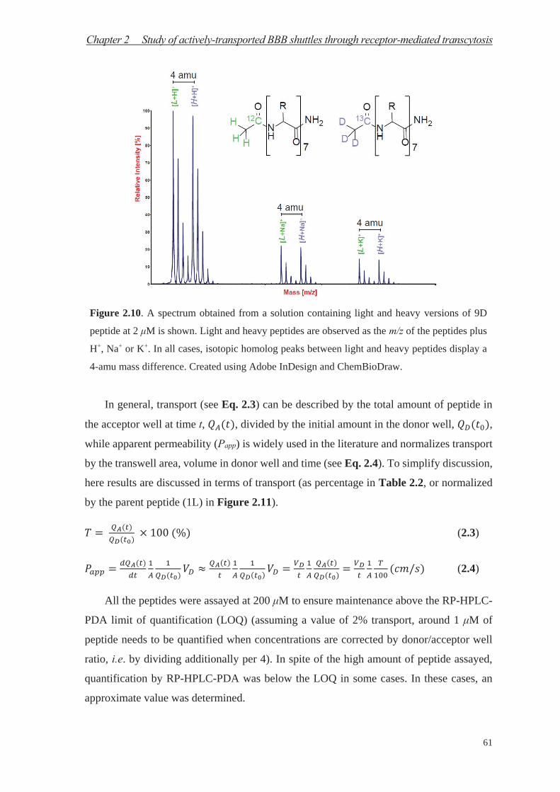
Chapter 2 Study of actively-transported BBB shuttles through receptor-mediated transcytosis
61
Figure 2.10. A spectrum obtained from a solution containing light and heavy versions of 9D
peptide at 2 μM is shown. Light and heavy peptides are observed as the m/z of the peptides plus
H+, Na+ or K+. In all cases, isotopic homolog peaks between light and heavy peptides display a
4-amu mass difference. Created using Adobe InDesign and ChemBioDraw.
In general, transport (see Eq. 2.3) can be described by the total amount of peptide in
the acceptor well at time t, , divided by the initial amount in the donor well, ,
while apparent permeability (Papp) is widely used in the literature and normalizes transport
by the transwell area, volume in donor well and time (see Eq. 2.4). To simplify discussion,
here results are discussed in terms of transport (as percentage in Table 2.2, or normalized
by the parent peptide (1L) in Figure 2.11).
(2.3)
(2.4)
All the peptides were assayed at 200 μM to ensure maintenance above the RP-HPLC-
PDA limit of quantification (LOQ) (assuming a value of 2% transport, around 1 μM of
peptide needs to be quantified when concentrations are corrected by donor/acceptor well
ratio, i.e. by dividing additionally per 4). In spite of the high amount of peptide assayed,
quantification by RP-HPLC-PDA was below the LOQ in some cases. In these cases, an
approximate value was determined.
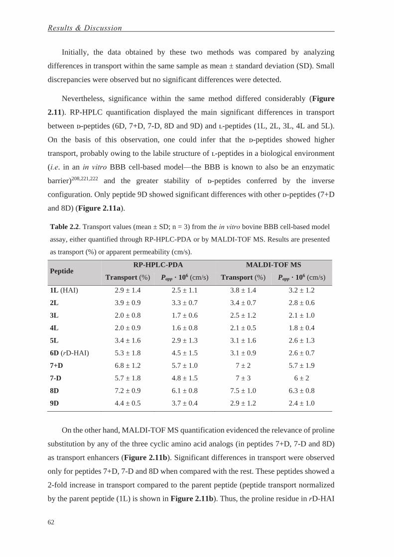
Results & Discussion
62
Initially, the data obtained by these two methods was compared by analyzing
differences in transport within the same sample as mean ± standard deviation (SD). Small
discrepancies were observed but no significant differences were detected.
Nevertheless, significance within the same method differed considerably (Figure
2.11). RP-HPLC quantification displayed the main significant differences in transport
between ᴅ-peptides (6D, 7+D, 7-D, 8D and 9D) and ʟ-peptides (1L, 2L, 3L, 4L and 5L).
On the basis of this observation, one could infer that the ᴅ-peptides showed higher
transport, probably owing to the labile structure of ʟ-peptides in a biological environment
(i.e. in an in vitro BBB cell-based model—the BBB is known to also be an enzymatic
barrier)208,221,222 and the greater stability of ᴅ-peptides conferred by the inverse
configuration. Only peptide 9D showed significant differences with other ᴅ-peptides (7+D
and 8D) (Figure 2.11a).
Table 2.2. Transport values (mean ± SD; n = 3) from the in vitro bovine BBB cell-based model
assay, either quantified through RP-HPLC-PDA or by MALDI-TOF MS. Results are presented
as transport (%) or apparent permeability (cm/s).
Peptide RP-HPLC-PDA MALDI-TOF MS
Transport (%) Papp · 106 (cm/s) Transport (%) Papp · 106 (cm/s)
1L (HAI) 2.9 ± 1.4 2.5 ± 1.1 3.8 ± 1.4 3.2 ± 1.2
2L 3.9 ± 0.9 3.3 ± 0.7 3.4 ± 0.7 2.8 ± 0.6
3L 2.0 ± 0.8 1.7 ± 0.6 2.5 ± 1.2 2.1 ± 1.0
4L 2.0 ± 0.9 1.6 ± 0.8 2.1 ± 0.5 1.8 ± 0.4
5L 3.4 ± 1.6 2.9 ± 1.3 3.1 ± 1.6 2.6 ± 1.3
6D (rD-HAI) 5.3 ± 1.8 4.5 ± 1.5 3.1 ± 0.9 2.6 ± 0.7
7+D 6.8 ± 1.2 5.7 ± 1.0 7 ± 2 5.7 ± 1.9
7-D 5.7 ± 1.8 4.8 ± 1.5 7 ± 3 6 ± 2
8D 7.2 ± 0.9 6.1 ± 0.8 7.5 ± 1.0 6.3 ± 0.8
9D 4.4 ± 0.5 3.7 ± 0.4 2.9 ± 1.2 2.4 ± 1.0
On the other hand, MALDI-TOF MS quantification evidenced the relevance of proline
substitution by any of the three cyclic amino acid analogs (in peptides 7+D, 7-D and 8D)
as transport enhancers (Figure 2.11b). Significant differences in transport were observed
only for peptides 7+D, 7-D and 8D when compared with the rest. These peptides showed a
2-fold increase in transport compared to the parent peptide (peptide transport normalized
by the parent peptide (1L) is shown in Figure 2.11b). Thus, the proline residue in rD-HAI

Chapter 2 Study of actively-transported BBB shuttles through receptor-mediated transcytosis
63
(6D) seems to be a key pharmacophoric site204,223 that leads to an improvement in transport
when substituted by a bulkier residue.
Figure 2.11. Peptide transport results from the in vitro bovine BBB cell-based model assay
obtained through RP-HPLC-PDA (in blue) or MALDI-TOF MS (in green) quantifications; (a)
RP-HPLC-PDA transport quantification significance between ʟ- and ᴅ-peptides (additionally,
peptide 9D showed significant differences (*) with 7+D and 8D); (b) MALDI-TOF MS transport
quantification significance. Significance: ns ≡ not significant (p ≥ 0.05), * ≡ significant (0.01 ≤
p < 0.05), ** ≡ very significant (0.001 ≤ p < 0.01), *** ≡ extremely significant (0.0001 ≤ p <
0.001), **** ≡ extremely significant (p < 0.0001). Created using GraphPad and Adobe InDesign.
Finally, to further study their transport ability and test the versatility of our method for
quantification, we selected the peptides that showed the greatest transport capacity, namely
7-D and 8D (7+D was discarded as it recorded the same transport as 7-D, but the residue
replaced is an ʟ- instead of ᴅ-amino acid). However, we have previously reported the use
of the amino acid substitution in the non-natural residue in 7-D for passive diffusion
transport purposes. In that case, the amino acid in question (PhPro, i.e. phenylproline) was
used as a building block for the tetrapeptide PhPro4, which showed excellent passive
diffusion transport.127 Passive diffusion was ruled out as the mechanism responsible for

Results & Discussion
64
transport when the peptides (1L, 6D, 7-D and 8D) were assayed by PAMPA, an in vitro
physico-chemical model where the transport by passive diffusion of compounds is
evaluated through a membrane containing the lipid composition of interest (in our case, a
porcine brain polar lipid extract (BPLE) to mimic the BBB lipid barrier). The standard
parameter that quantifies transport independently of time and concentrations is effective
permeability (Pe), shown in the Eq. 2.5:
(2.5)
where t is the running time (4 h), CA (t) is the concentration of the compound in the
acceptor well at time t, and CD (t0) is the compound concentration in the donor well before
running the PAMPA assay (t0 = 0 h). Transport (%) values were obtained by dividing the
amount of peptide in the acceptor well at time t, CA (t), by the amount in the donor well at
time zero, CD (t0), multiplied by 100.
Table 2.3. Passive diffusion (effective permeability) of the peptides 1, 6D, 7-D and 8D studied
by PAMPA (n = 3; mean ± SD).
Peptide Pe · 106 (cm/s)
1L (HAI) 0.18 ± 0.11
6D (rD-HAI) 0.34 ± 0.06
7-D 0.33 ± 0.16
8D 0.34 ± 0.08
All of the peptides showed permeability values below 0.4 × 10⁻⁶ cm/s (Table 2.3).
Note that when these values fall below 2.0 × 10⁻⁶ cm/s compounds are considered to be
poorly transported by passive diffusion.144
Table 2.4. Transport values (mean ± SD; n = 3) from the in vitro human BBB cell-based
model assay, quantified by MALDI-TOF MS. Peptides were assayed using Ringer-HEPES
buffer or supplemented ECM medium.
Peptide Ringer-HEPES Buffer Supplemented ECM Medium
Papp · 106 (cm/s) Transport (%) Papp · 106 (cm/s) Transport (%)
1L (HAI) 7.7 ± 1.5 4.8 ± 0.9 0.4 ± 0.2 0.26 ± 0.11
7-D 11.6 ± 2.6 7.2 ± 1.6 3.4 ± 0.8 2.1 ± 0.5
8D 10.6 ± 0.6 6.6 ± 0.3 1.7 ± 0.9 1.1 ± 0.5

Chapter 2 Study of actively-transported BBB shuttles through receptor-mediated transcytosis
65
Here we tested the protease resistance (in human serum) of these two all-ᴅ-peptides
containing a non-natural moiety (7-D and 8D). Both showed high resistance to protease
degradation, as previously observed for the retro-ᴅ-version of HAI, rD-HAI. With these
optimal candidates, we moved on to a more sophisticated in vitro human model of the BBB,
made using brain-like endothelial cells, generated from human cord blood-derived
hematopoietic stem cells co-cultured with bovine pericytes. The aforementioned peptides
were assayed as in the other BBB model in either Ringer-HEPES buffer (the one commonly
used in these assays) or supplemented ECM medium. The use of medium in this assay
allows (1) extension of the incubation time and (2) use of more reliable conditions to those
found physiologically. In this assay, the transport of the analogs 7-D and 8D was around
1.5 times higher than that observed for the parent peptide 1L (HAI). However, when 7-D
and 8D were assayed in medium, they showed 8- and 4-fold the transport values of the
parent peptide (Figure 2.12). Thus, although 7-D and 8D appeared to show similar
transport performance, a more realistic analysis of their shuttle capacity revealed that 7-D
was twice as good as 8D, in spite of the latter exceeding the transport capacity of the parent
peptide (Figure 2.12).
Re
lati
ve
Tra
ns
po
rtto
1L
1 L7 -D 8D
0
3
6
9
1 2
R in g e r H e p e s B u ffe r
S u p p le m e n te d E C M M e d iu m
1 .0 1 .41 .5
4 .1
8 .1
1 .0
Figure 2.12. Peptide transport results from the in vitro human BBB cell-based model assay:
relative transport of 1L, 7-D and 8D peptides with respect to 1L when assayed with Ringer-
HEPES buffer or supplemented ECM medium. Created using GraphPad.
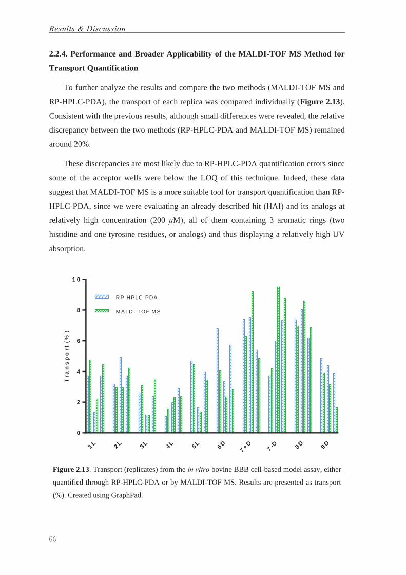
Results & Discussion
66
2.2.4. Performance and Broader Applicability of the MALDI-TOF MS Method for
Transport Quantification
To further analyze the results and compare the two methods (MALDI-TOF MS and
RP-HPLC-PDA), the transport of each replica was compared individually (Figure 2.13).
Consistent with the previous results, although small differences were revealed, the relative
discrepancy between the two methods (RP-HPLC-PDA and MALDI-TOF MS) remained
around 20%.
These discrepancies are most likely due to RP-HPLC-PDA quantification errors since
some of the acceptor wells were below the LOQ of this technique. Indeed, these data
suggest that MALDI-TOF MS is a more suitable tool for transport quantification than RP-
HPLC-PDA, since we were evaluating an already described hit (HAI) and its analogs at
relatively high concentration (200 μM), all of them containing 3 aromatic rings (two
histidine and one tyrosine residues, or analogs) and thus displaying a relatively high UV
absorption.
Tra
ns
po
rt(%
)
1 L 2 L 3 L 4 L 5 L6D
7 + D7 -D 8D 9D
0
2
4
6
8
1 0
R P-HPLC-PDA
M A LD I-TO F M S
Figure 2.13. Transport (replicates) from the in vitro bovine BBB cell-based model assay, either
quantified through RP-HPLC-PDA or by MALDI-TOF MS. Results are presented as transport
(%). Created using GraphPad.

Chapter 2 Study of actively-transported BBB shuttles through receptor-mediated transcytosis
67
2.2.4.1. MALDI-TOF MS Limit of Quantification
Dilution series for three peptides 1L (HAI), 6D (retro-ᴅ-HAI) and 9D, ranging from
200 μM to 1.1 nM, were analyzed by MALDI-TOF MS (on an Applied Biosystems 4800
Plus MALDI-TOF spectrometer, using an ACH-based matrix). The initial dilution was
prepared by mixing 20 μL of two solutions containing 400 or 800 μM of the light or heavy
versions of the peptide, respectively. Consecutive dilutions 1/3 were then prepared by
mixing 10 μL in 20 μL of H2O. Finally, 1 μL of the sample and 1 μL of the ACH matrix
were placed in a MALDI plate. The spectra were acquired and further analyzed to
determine the limit of quantification. First, the experimental light/heavy ratio was
determined for all the dilutions. The RLOQ was then determined for all of them:
(2.6)
where, and are the light/heavy ratio of each sample and the mean (i.e. ,
discarding the last dilution value), respectively. The deviation (%) is calculated as
.
2.2.4.2. RP-HPLC-PDA Limit of Quantification
Dilution series for three peptides 1L (HAI), 6D (retro-ᴅ-HAI) and 9D, ranging from
200 μM to 1.1 nM, were analyzed by RP-HPLC-PDA. An initial solution at 200 μM was
consecutively diluted 1/3 by mixing 100 μL in 200 μL of H2O, up to a total of eleven
dilutions. Finally, a specific volume of the sample, ranging from 5 to 100 μL, was injected
to the HPLC system and then further analyzed (at 220 nm, the selected wavelength with
the highest signal-to-noise; see Table 2.5) to determine the limit of quantification.
Table 2.5. Signal-to-noise ratio determined for the peptide HAI (not acetylated 1L) at diverse
wavelengths: 200, 210 and 220 nm. The signal-to-noise ratio is higher at 220 nm than at the other
two wavelengths.
Wavelength (nm) Signal-to-Noise Ratio (normalized by the lowest value)
200 1.00
210 1.18
220 1.26

Results & Discussion
68
First, the total absorption corrected by the injected volume and dilution ( ) was
determined as:
(2.7)
where, , and are the total absorption area, the absorption area, the injected volume
and the fold-dilution, respectively. Then, the RLOQ was determined for all of them using
Eq. 2.6.
In our MALDI-TOF MS method, we propose that two procedures can be followed in
order to ensure that the concentration of the assayed peptide is above the LOQ. The most
stringent one requires the determination of the LOQ for each peptide. The second one
considers that similar molecules (our library of peptide analogs) have similar ionization
properties. In our case, we first determined the LOQ for peptide 1L (HAI) and afterwards
for 6D (rD-HAI) and 9D. Comparable results were obtained. We then assumed that all the
peptide analogs had a similar LOQ since they all differed from the parent peptide in only
one amino acid substitution and/or had a reversed sequence accompanied by an inverse
configuration (Figure 2.6c).
-L o g 1 0 (C o n c e n t r a t io n ) (M )
De
via
tio
n(%
)
3 .4 3 .9 4 .4 4 .8 5 .3 5 .8 6 .3 6 .7 7 .2 7 .7 8 .2 8 .6
-8 0
-6 0
-4 0
-2 0
0
2 0
M A LD I-TO F M S
R P-HPLC-PDA
Figure 2.15. Determination of the limit of quantification of MALDI-TOF MS and RP-HPLC-
PDA using the peptides 1L, 6D and 9D. Created using GraphPad.
To determine the LOQ of our MALDI-TOF MS approach, we analyzed consecutive
dilutions (from 200 μM to 1 nM) of samples of 1L (HAI), 6D (rD-HAI) and 9D, which
contained light and heavy versions in similar concentrations (the latter double the

Chapter 2 Study of actively-transported BBB shuttles through receptor-mediated transcytosis
69
concentration of the former). Each dilution was analyzed by comparing the light/heavy
ratio with the initial quantification before dilution. The LOQ remained around 3.4 nM in
both cases when an average error of 8% was assumed (Figure 2.15). This value is much
lower than the discrepancy between replicates in this type of assay (e.g. see Figure 2.13).
Thus, MALDI-TOF MS allowed an increase in sensitivity of more than 3 orders of
magnitude compared to RP-HPLC-PDA. Accordingly, all transport quantifications by
MALDI-TOF MS fell within the LOQ.


Chapter 3
Study of Immunogenic Responses to BBB Shuttle Peptides

This chapter will give rise to the following article:
Arranz-Gibert, P.; Ciudad, S.; Seco, J.; García, J.; Giralt, E.; Teixidó, M. Retro-ᴅ-peptides: The
Phantom Therapeutics. Manuscript in preparation.

Chapter 3 Study of immunogenic responses to BBB shuttle peptides
73
During the last three decades, peptides have become privileged therapeutics224 and are
now used in a broad range of applications. The relevant presence of these molecules in
nature and accessible synthesis through well-established solid-phase peptide synthesis
(SPPS)82 has facilitated their study and applicability in the pharma industry.225,226
Compared to therapeutics developed by classical medicinal chemistry (low molecular
weight organic molecules), peptides display a better solubility profile, although this profile
depends on the sequence.227 While the abundance of peptides in nature is advantageous
from the perspective of toxicity, two principal drawbacks are intrinsic to their structure,
namely protease degradation driven by proteolytic enzymes and eventual risk of
immunogenicity.224 In addition, this class of therapeutics displays a short plasma half-life
as a result of kidney clearance. Nevertheless, several approaches have partially resolved
these issues. Recognition by proteases, and thus peptide degradation, can be avoided by
using peptide derivatives like ᴅ-, β-228 or non-natural amino acid peptides, as well as other
classes of peptidomimetics.229 Furthermore, PEGylation230 and other methodologies231 can
extend the plasma half-life of peptides by increasing their molecular weight (more
precisely, their hydrodynamic radius).

Results & Discussion
74
The immunogenicity of peptides (as the native ligands of MHC class I/II) have been
studied extensively.232 While vaccine development requires activation of the immune
response,233 other therapeutic treatments require not to elicit such type of response. Few
approaches have reported effective reduction of immune system recognition/response.
PEGylation230 is probably the most widely used method of choice to reduce
immunogenicity, although adverse immunological effects, i.e. humoral responses, have
been reported.234
Retro-ᴅ-peptides (containing retro-inverso- and retro-enantio-isomers of a parent
peptide) are peptides derived from a parent peptide, made by ʟ-amino acids, which
sequence is inversed and made by ᴅ-amino acids. This rearrangement combines the
properties of ᴅ-peptides, namely protease-resistance, and with the reversed amino acid
sequence leads to an imperfect topology overlapping with that of the parent peptide but
achieve good mimicry for short sequences.198
We have applied the previously described strategy to a family of peptide BBB shuttles
(H-HAIYPRH-NH2 and H-THRPPMWSPVWP-NH2,188 namely HAI and THR peptides,
respectively). BBB shuttles are molecular entities of diverse origin, e.g. synthetic or natural
peptides, which have the capacity to cross the blood-brain barrier (BBB) and, when
covalently conjugated to drugs unable to cross the BBB unaided, can deliver them into the
central nervous system (CNS).235 Those that cross through receptor-mediated transcytosis
are also included in the term ‘molecular Trojan horses’.119,236 Although only a few clinical
trials on BBB shuttles have been reported,237,238 a huge number of these molecules
effectively achieve their aim.84,239 While parent versions of both HAI and THR displayed
efficient BBB permeability in several studies, our laboratory demonstrated the BBB shuttle
capability and protease-resistance of retro-ᴅ-THR84 and retro-ᴅ-HAI (BBB shuttle
capacity studied previously, and protease-resistance tested in human serum shown in
Figure 3.1b).
Here we conducted immunogenicity studies for several peptide BBB shuttles—two
versions of HAI and THR peptides (Fig 3.1a), namely the parent and the respective retro-
ᴅ-peptide. All peptides were obtained by manual SPPS using the Fmoc/tBu strategy.

Chapter 3 Study of immunogenic responses to BBB shuttle peptides
75
Figure 3.1. HAI, THR and the respective retro-ᴅ-versions; (a) peptide sequences and (b)
stability assay in human serum. Retro-ᴅ-versions display a stable structural integrity over time
while parent peptides display exponential degradation (R2 = 0.812 and 0.970, t1/2 = 3.3 and 1.0
h, HAI and THR, respectively. Data on THR and retro-ᴅ-THR extracted from Prades et al.84
During the experiment (24 h) no significant differences were observed when retro-ᴅ-peptides
were compared with the starting amount of peptide. Created using GraphPad, ChemBioDraw and
Adobe InDesign.
The structural conformational arrangements of parent peptides and their respective
retro-ᴅ-versions were initially studied by circular dichroism (CD) (Figure 3.2b), and later
by nuclear magnetic resonance (NMR) spectroscopy (Figure 3.2a); NMR experiments
conducted by Sonia Ciudad and Dr. Jesús García. While the coarse grain analysis by CD is
consistent with similar conformational arrangements but not topologically exact, NMR
adds further precision and shows how these peptides (both for HAI/ retro-ᴅ-HAI and THR/
retro-ᴅ-THR pairs) comprise several sets of thermodynamically stable conformational
arrangements (analysis of NMR conformationally sensitive parameters such as chemical
shifts, coupling constants, NH temperature coefficients and NOE pattern revealed

Results & Discussion
76
analogous results for the ʟ-peptides and their retro-ᴅ-versions, thereby suggesting that they
have similar conformational preferences).
Figure 3.2. Structural analysis of the four peptides: (a) histograms showing the 1Hα, 13Cα and 13Cβ
chemical shift deviations (CSD) from random coil (RC)240 of the major species (of HAI (left)
and THR (right), and their respective retro-ᴅ-versions, aligned by amino acid type), (b) the
circular dichroism (CD) spectra, (c) the cross-RMSD derived from the comparison of the whole
REMD structure sets of parent and retro-ᴅ-version peptides, and the three-dimensional
superposition of one pairing obtained from the cross-RMSD matrix of (d) HAI and retro-ᴅ-HAI,
and of (e) THR and retro-ᴅ-THR. Created using GraphPad, Microsoft PowerPoint and PyMOL.
Chapter 3 Study of immunogenic responses to BBB shuttle peptides
77
Dr. Jesús Seco performed replica exchange molecular dynamics (REMD; Figure 3.2c)
experiments. The overlapping of the two configurational isomers was analyzed. REMD
allowed to determine how these peptides are topologically disposed. These results show
that the two versions of the same peptide can adopt a similar three-dimensional arrangement
(Figure 2c-e), in agreement with CD and NMR data.
To date, several studies have used ᴅ-peptides as tools for vaccine development. In these
cases the peptides have been conjugated to large supramolecular entities, e.g. KLH and
small unilamelar liposomes containing monophosphoryl lipid A.241,242 Nevertheless, the
use of retro-ᴅ-peptides as therapeutics was controversial twenty years ago.243,244 Thus, to
shed light on the immunogenicity of ᴅ-peptides and add further value to these compounds,
we evaluated the immunological responses activated by retro-ᴅ-THR and retro-ᴅ-HAI
peptides and compared them to that of their parent peptides. For this purpose, peptides (not
conjugated) were i.p. administered to mice, and antibody titration was used as parameter to
evaluate immunological (humoral) response by ELISA.
Unconjugated ʟ-versions of both peptides displayed a moderate immunogenicity
response (Figure 3.4a). Antibody titration of the first bleed showed a slightly higher signal
for both peptides compared to the last bleeding. This signal was probably induced by the
transition from IgM to IgG production in B cells. Thus, antibody titration decay is caused
by a decrease in number, which is compensated by an increase in affinity and selectivity.
In contrast, in the case of retro-ᴅ-peptides almost no signal was detected for specific
antibodies (Figure 3.4b). Thus, retro-ᴅ-versions appear to be much less immunogenic than
their corresponding parent peptides.
Nevertheless, in order to discard the possibility that antibody recognition was
dependent on the terminus exposed in these cases (since retro-ᴅ-versions have them
reversed—see Figure 3.1a), we tested the serum of mice immunized against retro-ᴅ-
peptides by attaching these peptides to the other terminus on the ELISA plate (Figure 3.3
and 3.4c). In this regard, we evaluated the response of antibodies to the same peptide
sequence but exposing the other terminus: the inversed order in retro-ᴅ-peptides
(comparing with parent peptides) was corrected by displaying the opposite terminus. As an
example, the HAI peptide attached to BSA (and subsequently to the plate) by the N-
terminus displayed the sequence H2NOC-H←R←P←Y←[…]-BSA-plate, and the peptide
retro-ᴅ-HAI exposing the N-terminus showed H2N-h→r→p→y→[…]-BSA-plate (see

Results & Discussion
78
header of Figure 3.4); compare Figure 3.4a,c, and see Figure 3.1a. Similar results were
obtained as when retro-ᴅ-peptides exposed the C-terminus (Figure 3.4c), thereby
confirming our hypothesis (antibody reactivity does not depend on the termini exposed).
Figure 3.3. Peptide sequences of retro-ᴅ-versions containing the diaminopropionic acid (Dpr)
residue at the C-terminus. Created using ChemBioDraw.
To the best of our knowledge, in all those immunological responses reported in the
literature for either a retro-ᴅ-peptide or ᴅ-peptide (especially concerning vaccines), the
parent one was derived from an existing sequence or even an immunodominant region of a
viral protein.241,242 Thus, these ᴅ- or retro-ᴅ-versions could have generated a rapid and huge
immune response when administered to animals (e.g. serum IgG and other neutralizing
factors against adeno-associated virus are highly prevalent in the healthy population).245 In
addition, a report describing ᴅ-proteins as entities exerting low immunogenicity was
published 20 years ago.246 We therefore hypothesized that retro-ᴅ-peptides with a parent
sequence not related to any existing protein would not be immunogenic per se. To exclude
this possibility, we used a sequence similarity search tool (NCBI BLAST) to identify
analogies between the parent peptides used and the whole data set of known peptide and
protein sequences. No positive result was obtained, as previously described.12
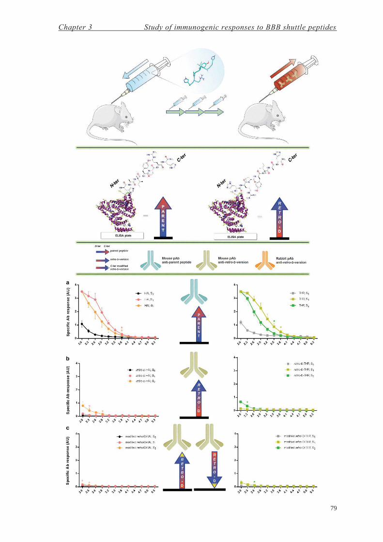
Chapter 3 Study of immunogenic responses to BBB shuttle peptides
79

Results & Discussion
80
Figure 3.4. Titration of humoral response in mice by ELISA: (header) scheme of mice
immunizations (shown with retro-ᴅ-HAI), ELISA evaluation of humoral response with peptide-
BSA conjugates (HAI and retro-ᴅ-HAI shown), and the legend; (a) serum anti-parent peptides
(HAI, THR), serum anti-retro-ᴅ-versions evaluated either with (b) C- or (c) N-terminus-exposed
peptides. Initial consecutive significant differences between S0 with S1 or S2 are labeled by
asterisk/s (no asterisk, no significant differences in the whole dilution interval). Created using
GraphPad, Adobe InDesign, ChemBioDraw, Microsoft PowerPoint and Servier Medical Art.
Although, as shown above, retro-ᴅ-HAI and retro-ᴅ-THR are not immunogenic per
se, they could elicit an immunological response when conjugated to an immunogenic
molecule. Thus, we conjugated these peptides to KLH and s.c. injected into rabbits. After
the immunization, sera were tested by ELISA to identify antibodies displaying specific-
peptide response. A strong increase in humoral response to these peptides (see Figure 3.5)
was observed when compared to when they were used as unconjugated immunogens
(Figure 3.4b). The peptide-specific antibodies generated for both retro-ᴅ-peptides seems
to be an immunoresponse facilitated by the anchoring molecule (KHL in this case). Hence,
in order to be able to use this class of peptide (retro-ᴅ) for vaccine development, they should
be coupled to other immunogenic structures to facilitate recognition by the immune system.
However, if these peptides are to have applications as other types of therapeutic agent (and
an eventual immune response is undesirable), they must be used (1) as a single therapeutic,
(2) attached directly (2a) to the therapeutic agent or (2b) to other non-immunogenic
structures.
Figure 3.5. Titration of humoral response by ELISA in rabbit: serum anti-retro-ᴅ-peptides
conjugated to KLH. Created using GraphPad, Adobe InDesign and Microsoft PowerPoint.
Chapter 3 Study of immunogenic responses to BBB shuttle peptides
81
We have shown that retro-ᴅ-peptides, in addition to overcoming protease-sensitivity,
can be a valuable tool to create therapeutics that do not sensitize the immune system, thus
circumventing the intrinsic problems of ʟ-peptides, derived from their backbone
configuration, and leading to a new class of therapeutics that are not recognizable by the
immune system—phantom therapeutics.
The B cell response (antibody production) in mice immunized with ᴅ- (precisely, retro-
ᴅ) peptides that were not coupled to any molecule was markedly lower compared to those
immunized with the ʟ-versions. We postulate that both versions of the peptide may
eventually be recognized by a B Cell Receptor (BCR/ membrane antibody); nevertheless,
the ᴅ-version cannot be processed by peptidases and much less efficiently presented by the
MCH-II to CD4+ T cells (TH)—the left-handed polyproline II extended conformation
adopted by peptides when loaded in the antigen-binding groove of MCH-II is not accessible
for ᴅ-peptides (thus hydrogen bonding between the amide bond of the peptide and the
MHC-II is hindered, although binding pockets for the side chains are preserved). Therefore,
the survival and proliferative signals to the B cells recognizing ᴅ-peptides are much lower
compared to those that recognize ʟ-peptides. Thus, we propose that ᴅ-peptides have two
mechanisms by which to silence the immune system, namely protease-resistance and the
conformational arrangement of the backbone, both of which are consequences of the
inversed α-carbon stereochemistry.
Since retro-ᴅ-peptides present good topological mimicry with their parent peptides,
they are a simple solution for newly designed therapeutic peptides derived from diverse
source of therapeutics with ʟ-configuration, e.g. natural sources, phage display or
computational design. Nevertheless, given the prior observations related to peptides
composed by ᴅ-amino acids, we recommend a preliminary screening of the sequences of
interest to test their prevalence and type of sources in nature in order to avoid eventual
immune responses in advanced trials. In addition, we envisage the use of retro-ᴅ-peptides
as protease-resistant variants to overcome immunological problems derived the intrinsic
structure of drugs or vectors. As an example, viral vectors applied to gene therapy could be
decorated with these peptides in order to allow them to escape from the immune system,
either attached through a non-immunogenic linker or stand-alone, thereby avoiding
systemic adverse reactions and neutralization by preexisting immunity.247 Furthermore,
their cell-tropism could be modulated (e.g. by using retro-ᴅ-versions of cell-penetrating
peptides (CPP), blood-brain barrier shuttles (BBB shuttles), homing peptides (HP)), in

Results & Discussion
82
order to target more effectively the diseased area and thus allowing the use of lower viral
loads (i.e. reducing side effects and production costs).

Chapter 4
Attempts to Develop a Therapy for Friedreich’s Ataxia at the CNS


Chapter 4 Attempts to develop a therapy for Friedreich’s Ataxia at the CNS
85
Diseases with known simple genetic and metabolic causes are perfect targets for
treatments involving the use of delivery tools to supply a therapeutic agent. In this regard,
we considered Friedreich’s Ataxia (FRDA), a neurodegenerative movement disorder, as a
therapeutic condition in which to test our BBB shuttles, namely retro-ᴅ-THR (rD-THR)
and retro-ᴅ-HAI (rD-HAI).
FRDA was first described by Nicholaus Friedreich in 1863,248 and it is currently the
commonest inherited ataxia, with a prevalence of 1 case per 30,000 individuals (1:30,000)
in Western Europe—249 the highest value is observed in the north of Spain (1:21,000) and
a prevalence gradient is distributed from south-west to north-east in Europe.249,250 The
annual cost of this disease per person in the United Kingdom is between £11,818 and
£18,774, depending on whether the cost of long-term unemployment is included.251 The
onset of symptoms has been established to occur at 15.5 ± 8 years, and the mean time until
the patient becomes confined to a wheelchair is 10.8 ± 6 years. The main signs and
symptoms of this disease are gait and limb ataxia, dysarthria, lower-limb areflexia and
axonal neuropathy, with a prevalence of more than 85%. In addition, cardiomyopathies,
abnormal brainstem and visual evoked potentials, and impaired glucose tolerance (e.g.
diabetes) show a prevalence ranging from 32–63%.252

Results & Discussion
86
FRDA is an autosomal recessive disease caused by a GAA trinucleotide (triplet) repeat
(TNR) expansion located in an intronic region of chromosome 9q13—intron 1 of the
frataxin (FXN) or X25 gene.253 Repetitive sequences comprise 30% of the human
genome.254 TNRs expand during progeny transmission, development and/or in somatic
non-dividing cells when longer than a crucial threshold—i.e. pre-mutation length. In non-
coding regions, like in FRDA, unstable parent-child transmissions initiate from the so-
called pre-mutation allele length—between 31–100 units in FRDA. The repeat number in
FRDA ranges from 70–1,000 units, while the normal repeat number ranges from 5–30
units.254 In this regard, a homopurine-homopirimidine (GAA)n•(TTC)n repeat can adopt a
triplex structure, called H-DNA, under negative supercoiling. For longer sequences, a
similar structure is adopted, the so-called “sticky DNA” (Figure 4.1).255 This conformation
is involved in the diverse mechanisms (replication or repair dependent) that give rise to the
expansion of these repeats. TNRs instability in somatic cells, which also occurs in FRDA
patients, is a repair-dependent mechanism.254 Although “sticky DNA” seems to be involved
in the expansion of these repeats, more extensive DNA methylation and low levels of RNA
polymerase II (RNAPII) and Histone H3 trimethylated at Lys 4 (and Lys 36 downstream
of the repeat) lead to reduction in both transcription initiation and elongation—leading to a
decrease in frataxin mRNA and thus low levels of protein expression.256 The length of
alleles inversely correlates with age at onset of the disorder and directly correlates with
disease severity.257,258 While 96% of the individuals affected by FRDA are homozygous
with a GAA TNR expansion in intron 1, in other cases, allele dysfunction is caused by
insertions, deletions or point mutations.259
Figure 4.1. DNA structures formed by TNRs: (a) H-DNA and (b) sticky DNA formed by the
(GAA)n• (TTC)n repeat. Only one possible isoform, in which the homopurine strand is donated
to the triplex, is shown for the two structures. Watson-Crick and reverse Hoogsteen pairings are
indicated by dots and asterisks, respectively. Adapted from Mirkin.255

Chapter 4 Attempts to develop a therapy for Friedreich’s Ataxia at the CNS
87
Frataxin has five known isoforms. In 1996, Campuzano et al. first identified the X25
gene as the critical region for the FRDA locus.253 This region (five exons, 1–4 & 5a)
encodes a 210-amino acid protein, named frataxin253 (isoform A,260 “canonical”). An
alternative product, isoform B,260 is obtained when exon 5b is transcribed instead of 5a
(five exons, 1–4 & 5b).253 In 2002, Pianese et al. revealed a novel transcript of X25, so-
called frataxin A1, which contains a 8-bp insertion between exons 4 and 5a (five exons, (1–
4) & 8 bp & 5a).260 More recently, in 2012, Xia et al. observed two additional transcripts
localized in the cerebellum and heart—isoforms II and III, respectively. These isoforms
differ from the “canonical” one in the first exon (exon 1 ≡ 1A), which lacks the last 141
nucleotides (exon 1AΔ141) or is substituted by another exon (exon 1B/1BΔ18)—isoforms
II and III, respectively.261 They are localized in the cytosol (isoform II) and nucleus
(isoform III),261 whereas the rest of the isoforms are directed to the mitochondria, since
they contain a mitochondrial localization signal (MLS).262-264 A summary of the diverse
isoforms is shown in Table 4.1. Further work has continued studying isoforms B and A1,265
as well II and III.266
Table 4.1. Frataxin isoforms and their tissue and cellular expression patterns, splicing and
residue length.
Isoform Tissue
Expression
Cellular
Localization
Splicing
(exons)e
Length
(residues)
A253 or I,261 “canonical” ubiquitous mitochondria 1–4 & 5a 210
B253 ubiquitous/ rare mitochondria 1–4 & 5b 171
A1260 ubiquitous/ rare mitochondria (1–4) & 8 bp & 5a 196
II261 cerebellum cytosol (1B/1BΔ18)–4 & 5a 135
III261 heart nucleus (1AΔ141)–4 & 5a 164
Although the relevance of several frataxin isoforms has been pointed out, the
“canonical” one—hereinafter referred as FXN—is the most widely studied. Although the
role of FXN in cellular metabolism is still unclear, it is accepted that it exerts a function in
iron homeostasis by directly binding this ion: (1) serving as iron chaperone during iron-
sulfur (Fe-S) cluster production; (2) participating in iron storage; (3) repairing oxidatively
damaged aconitase Fe-S clusters; (4) regulating oxidative stress/ reactive oxygen species
(ROS); and (5) participating in energy conversion and oxidative phosphorylation.267-270
e Exon 1 ≡ exon 1A, as discussed in Xia et al.261

Results & Discussion
88
Interestingly, FXN orthologs have been observed throughout the diverse kingdoms of life,
e.g. Saccharomyces cerevisiae (Yfh1), Escherichia coli (CyaY) and humans (HsFtx or
FXN).267 In addition, frataxin has also been localized in hydrogenosomes—organelles with
a common ancestry to mitochondria—of Trichomonas vaginalis, a unicellular eukaryote.271
Recently, this protein has been shown to have dual localization in Arabidopsis thaliana,
where chloroplasts and mitochondria contain AtFH, the frataxin homolog.272 Figure 4.2
shows the similarity between structures of Yfh1, CyaY and HsFtx.
Figure 4.2. Structures of diverse frataxin orthologs: from (a) yeast (Yfh1, PDB ID# 2GA5), (b)
human (HsFtx, PDB ID# 1LY7) and (c) bacteria (CyaY, PDB ID# 1SOY). Adapted from Bencze
et al.267
FXN is expressed as pre-protein with the MLS (FXN1–210, precursor, or p),273 and then
cleaved between residues 41–42 and 55–56 by the mitochondrial processing peptidase
(MPP), obtaining FXN42–210 (intermediate42, or i42) and FXN56–210 (mature56, m56),
respectively.274 In addition to these proteolytic events, two C-terminal degradation products
are observed.274 Later, in 2007, Yoon et al. identified a cleavage site between residues 77–
78 promoted by iron to produce a truncated form (degraded78, d78).275 However, in vivo
studies showed that the main mature form of FXN is produced after cleavage between
residues 80–81 (mature81, m81),273 and it was reported to be the isoform with the highest
rescue profile when lacking FXN.276 In this regard, the loss of the N-terminus is linked to
a change in FXN functions,277 as longer sequences correlate with oligomerization, iron
storage and stable interactions with NFS1•ISD11 (sulfur donor complex) and ISCU (iron-
sulfur cluster assembly enzyme),278 while shorter ones are observed with monomeric
configuration and are related to labile iron binding, and dynamic contacts with
NFS1•ISD11 and ISCU.279 A summary of the main features of each metabolic isoform is
shown in Table 4.2.
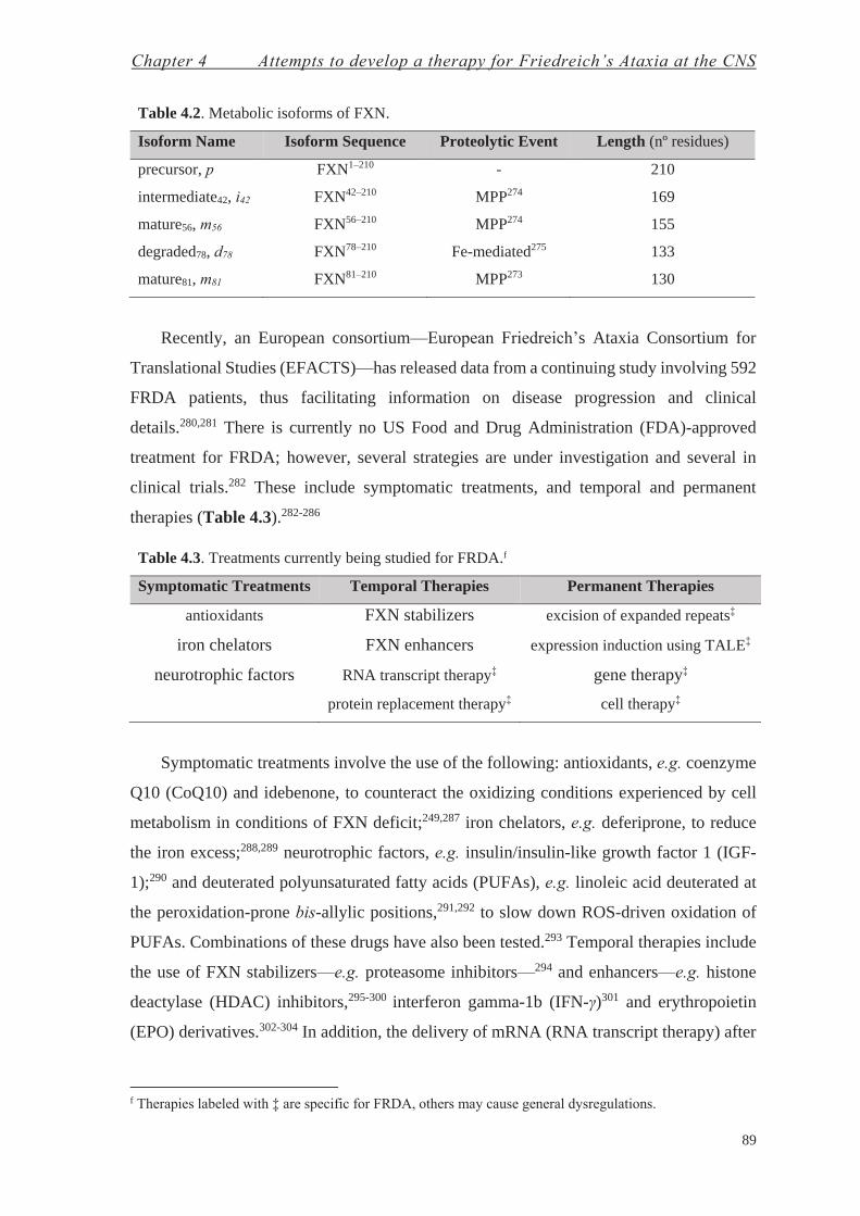
Chapter 4 Attempts to develop a therapy for Friedreich’s Ataxia at the CNS
89
Table 4.2. Metabolic isoforms of FXN.
Isoform Name Isoform Sequence Proteolytic Event Length (nº residues)
precursor, p FXN1–210 - 210
intermediate42, i42 FXN42–210 MPP274 169
mature56, m56 FXN56–210 MPP274 155
degraded78, d78 FXN78–210 Fe-mediated275 133
mature81, m81 FXN81–210 MPP273 130
Recently, an European consortium—European Friedreich’s Ataxia Consortium for
Translational Studies (EFACTS)—has released data from a continuing study involving 592
FRDA patients, thus facilitating information on disease progression and clinical
details.280,281 There is currently no US Food and Drug Administration (FDA)-approved
treatment for FRDA; however, several strategies are under investigation and several in
clinical trials.282 These include symptomatic treatments, and temporal and permanent
therapies (Table 4.3).282-286
Table 4.3. Treatments currently being studied for FRDA.f
Symptomatic Treatments Temporal Therapies Permanent Therapies
antioxidants FXN stabilizers excision of expanded repeats‡
iron chelators FXN enhancers expression induction using TALE‡
neurotrophic factors RNA transcript therapy‡ gene therapy‡
protein replacement therapy‡ cell therapy‡
Symptomatic treatments involve the use of the following: antioxidants, e.g. coenzyme
Q10 (CoQ10) and idebenone, to counteract the oxidizing conditions experienced by cell
metabolism in conditions of FXN deficit;249,287 iron chelators, e.g. deferiprone, to reduce
the iron excess;288,289 neurotrophic factors, e.g. insulin/insulin-like growth factor 1 (IGF-
1);290 and deuterated polyunsaturated fatty acids (PUFAs), e.g. linoleic acid deuterated at
the peroxidation-prone bis-allylic positions,291,292 to slow down ROS-driven oxidation of
PUFAs. Combinations of these drugs have also been tested.293 Temporal therapies include
the use of FXN stabilizers—e.g. proteasome inhibitors—294 and enhancers—e.g. histone
deactylase (HDAC) inhibitors,295-300 interferon gamma-1b (IFN-γ)301 and erythropoietin
(EPO) derivatives.302-304 In addition, the delivery of mRNA (RNA transcript therapy) after
f Therapies labeled with ‡ are specific for FRDA, others may cause general dysregulations.

Results & Discussion
90
intravenous injection showed a half-life greater than one week.305 On the other hand, the
delivery of FXN (protein replacement therapy) using a TAT-FXN construct showed
promise in vivo in a conditional mouse model for FRDA.306
Figure 4.3. FRDA treatment pipeline (March 2016), from Friedreich’s Ataxia Research Alliance
(FARA).

Chapter 4 Attempts to develop a therapy for Friedreich’s Ataxia at the CNS
91
On the other hand, permanent therapies offer a long-term solution without the need for
a continuous treatment.307 In this regard, several studies proposed the use of methodologies
to excise the expanded GAA repeats, like zinc finger nucleases,308 and also the use of
transcription activator-like effector (TALE) proteins to increase the expression of
FXN.309,310 The correction of this disease by means of gene therapy has been addressed
using a bacterial artificial chromosome (BAC)311 and a yeast artificial chromosome
(YAC),312 as well diverse viral vectors including lentiviral vectors,313 adeno-associated
viruses (AAVs),313,314 and herpes simplex virus type 1 (HSV-1) amplicon vectors.315-317
Cell therapies have also been studied using bone marrow stem cells, which induced FXN
expression levels, resistance to oxidative stress and neuroprotection to FRDA cells;318,319
but induced pluripotent stem cells (iPSCs) from FRDA patients have also been successfully
derived to neurons and cardiomyocytes with potential therapeutic applications.320-322
Research funded by Friedreich’s Ataxia Research Alliance (FARA) and the current stage
is shown in Figure 4.3.
In this thesis, we explore the potential of a temporal and a permanent therapy for FRDA
at the central nervous system (CNS), the former based on a protein replacement therapy
and the latter on a gene therapy using HSV-1 amplicon vectors.

Results & Discussion
92
4.1. Protein Replacement Therapy for Friedreich’s Ataxia at the CNS—Chemistry
with Proteins
The year 1922 marked the use of the first protein drug, namely insulin. This hormone
was purified from bovine and porcine pancreas and then used in patients with diabetes
mellitus type I (DM-I).323 Later, the issues related to the use of insulin of animal origin,
including immunological responses, were addressed by producing recombinant human
insulin in Escherichia coli.324 Currently, more than 130 proteins are approved by the FDA
for clinical use (over 95 of which are produced recombinantly), and many more are in
development.324
Here we decided to study the viability of delivering FXN1–210 into the brain as the pre-
protein isoform (precursor, p), since the MLS, not present in mature isoforms, was
necessary to target the mitochondria. Therefore, we devised a therapeutic construct based
on a BBB shuttle bioconjugated at the N-terminus of the protein. The processing of the
MLS at any of the proteolytic sites would then give a native mature isoform (Figure 4.4).
Figure 4.4. Construct designed for the protein replacement therapy for FRDA at the CNS: (BBB
shuttle)-FXN1–210. Proteolytic sites at the N-terminus are shown. Created with Visual Molecular
Dynamics (VMD) and Adobe InDesign, using the structure PDB ID# 1LY7.
Both the BBB shuttle and protein can be ensembled through diverse methodologies,
such as via bioconjugation to Cys,325,326 Lys327,328 or Tyr329-331 residues, but also Staudinger
ligation,332 or reactions between azides and alkynes—the so-called “click chemistry”.333,334
Native chemical ligation (NCL) methodologies enable bioconjugation at the N-terminus
through the reaction of a thioester with an N-terminal Cys-containing protein.335,336 In
addition, chemoenzimatic approaches are promising tools that allow the introduction of
site-specific chemical modifications; however, they require the presence of relatively short
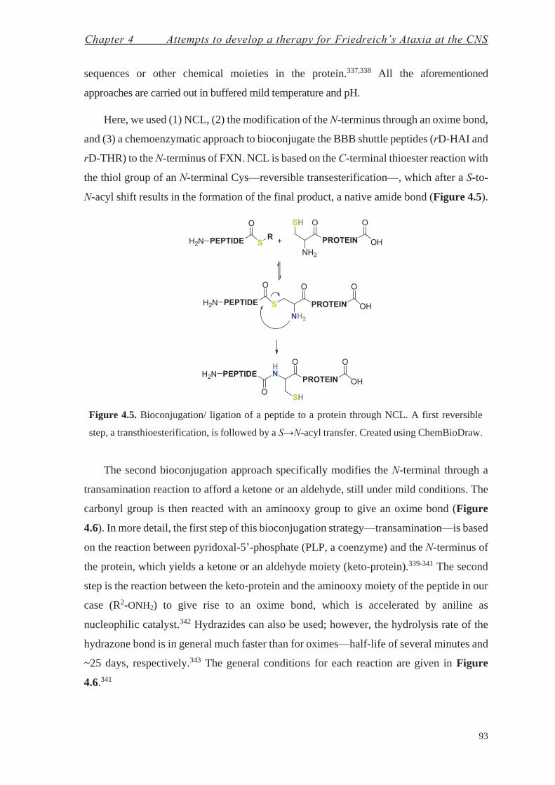
Chapter 4 Attempts to develop a therapy for Friedreich’s Ataxia at the CNS
93
sequences or other chemical moieties in the protein.337,338 All the aforementioned
approaches are carried out in buffered mild temperature and pH.
Here, we used (1) NCL, (2) the modification of the N-terminus through an oxime bond,
and (3) a chemoenzymatic approach to bioconjugate the BBB shuttle peptides (rD-HAI and
rD-THR) to the N-terminus of FXN. NCL is based on the C-terminal thioester reaction with
the thiol group of an N-terminal Cys—reversible transesterification—, which after a S-to-
N-acyl shift results in the formation of the final product, a native amide bond (Figure 4.5).
Figure 4.5. Bioconjugation/ ligation of a peptide to a protein through NCL. A first reversible
step, a transthioesterification, is followed by a S→N-acyl transfer. Created using ChemBioDraw.
The second bioconjugation approach specifically modifies the N-terminal through a
transamination reaction to afford a ketone or an aldehyde, still under mild conditions. The
carbonyl group is then reacted with an aminooxy group to give an oxime bond (Figure
4.6). In more detail, the first step of this bioconjugation strategy—transamination—is based
on the reaction between pyridoxal-5’-phosphate (PLP, a coenzyme) and the N-terminus of
the protein, which yields a ketone or an aldehyde moiety (keto-protein).339-341 The second
step is the reaction between the keto-protein and the aminooxy moiety of the peptide in our
case (R2-ONH2) to give rise to an oxime bond, which is accelerated by aniline as
nucleophilic catalyst.342 Hydrazides can also be used; however, the hydrolysis rate of the
hydrazone bond is in general much faster than for oximes—half-life of several minutes and
~25 days, respectively.343 The general conditions for each reaction are given in Figure
4.6.341
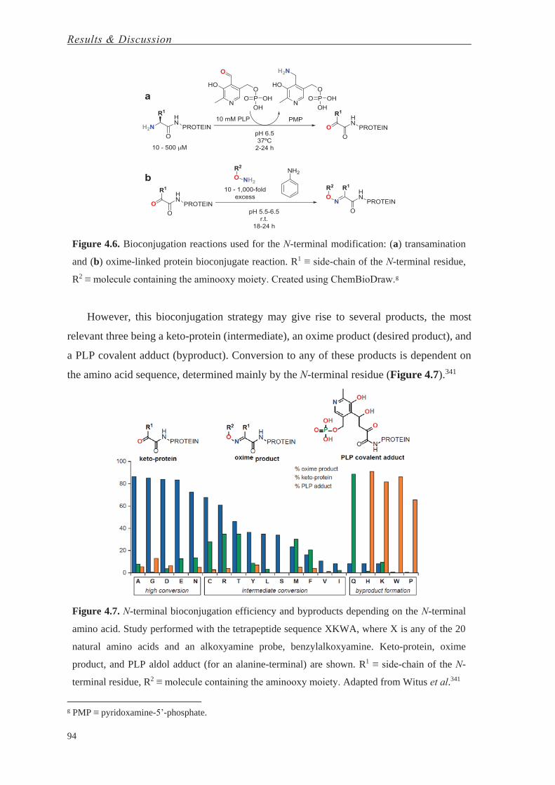
Results & Discussion
94
Figure 4.6. Bioconjugation reactions used for the N-terminal modification: (a) transamination
and (b) oxime-linked protein bioconjugate reaction. R1 ≡ side-chain of the N-terminal residue,
R2 ≡ molecule containing the aminooxy moiety. Created using ChemBioDraw.g
However, this bioconjugation strategy may give rise to several products, the most
relevant three being a keto-protein (intermediate), an oxime product (desired product), and
a PLP covalent adduct (byproduct). Conversion to any of these products is dependent on
the amino acid sequence, determined mainly by the N-terminal residue (Figure 4.7).341
Figure 4.7. N-terminal bioconjugation efficiency and byproducts depending on the N-terminal
amino acid. Study performed with the tetrapeptide sequence XKWA, where X is any of the 20
natural amino acids and an alkoxyamine probe, benzylalkoxyamine. Keto-protein, oxime
product, and PLP aldol adduct (for an alanine-terminal) are shown. R1 ≡ side-chain of the N-
terminal residue, R2 ≡ molecule containing the aminooxy moiety. Adapted from Witus et al.341
g PMP ≡ pyridoxamine-5’-phosphate.

Chapter 4 Attempts to develop a therapy for Friedreich’s Ataxia at the CNS
95
Thus, we decided to use Gly as the terminal residue in our constructs (i.e. Gly-FXN1–
210), since Gly—together with Ala, Asp and Glu—gives the best conversion to the oxime
product (Figure 4.7), whereas the N-terminal residue found in the expressed protein (Met,
or Trp after methionine aminopeptidases (MetAPs) cleavage)344,345 is not suitable for this
bioconjugation strategy due to low oxime product conversion and/or byproduct formation
(Figure 4.7).
The third bioconjugation strategy we selected, the chemoenzymatic approach, is based
on work by Bertozzi and co-workers,338 who showed that the co-expression of the protein
of interest (containing a 6-mer tag (LCTPSR) at the N-terminus together) with the
formylglycine-generating enzyme (FGE) enables the specific modification of the Cys
residue in the tag to formylglycine (fGly)—a residue containing an aldehyde moiety that
can be used for bioconjugation, as previously described. An additional Gly was added at
the N-terminus to allow to work with the aforementioned bioconjugation strategy (Figure
4.8).
Each of these three approaches requires specific reactive moieties in the peptides and
protein. FXN was expressed in E. coli as fusion protein, leaving the N-terminus of the final
construct prepared to each bioconjugation strategy (Table 4.4). A HisTag was placed at the
N-terminus, separated from the FXN construct by a SUMO (small ubiquitin-like modifier)
protein, which then allows specific cleavage at its C-terminus (by using a SUMO protease),
thus leaving the “clean” sequence of the desired protein.346-348 The fusion protein was then
purified by immobilized metal affinity chromatography (IMAC) and cleaved using the
SUMO protease, releasing the fragment MGSSH6-SUMO. The final protein construct was
purified using a reverse-IMAC followed by size-exclusion chromatography (SEC).
Figure 4.8. Co-expression of FGE and FXN construct (HisTag-SUMO-Gly-FGEsite-FXN).
Both proteins are expressed together in E. coli and the FGE site-specific modification (6-mer tag,
in blue) is modified within the cells (Cys to fGly conversion). Created using ChemBioDraw.

Results & Discussion
96
Table 4.4. The diverse FXN constructs expressed depending on the bioconjugation strategy
followed.
Chemical Approach Protein Construct Expressed Final Protein Construct
NCL MGSSH6-SUMO-Cys-FXN Cys-FXN
N-terminal modification MGSSH6-SUMO-GlyLys-FXN GlyLys-FXN
Chemoenzymatic
approach
MGSSH6-SUMO-Gly-FGEsite-FXN +
FGE Gly-FGEsite-FXN
The peptide BBB shuttles were also synthesized specifically for each bioconjugation
approach (Table 4.5).
Table 4.5. The diverse peptides synthesized depending on the bioconjugation strategy followed.
Chemical Approach Peptides Synthesizedh
NCL rD-HAI-Nbz and rD-THR-Nbz
N-terminal modification AO-rD-HAI and AO-rD-THR
Chemoenzymatic approach AO-rD-HAI and AO-rD-THR
Peptides-Nbz were synthesized following the Fmoc-SPPS approach reported by
Blanco-Canosa et al.,335 where first diaminobenzoic acid (Dbz) is introduced in a resin
terminated with a Rink linker. After the standard SPPS synthesis, the C-terminus is
activated through acylation with p-nitrophenylchloroformate followed by the addition of a
base. The peptide-Nbz is finally deprotected and cleaved by TFA treatment with scavengers
(Figure 4.9).
Figure 4.9. Scheme for the synthesis of Nbz-peptides. Created using ChemBioDraw.i
h Nbz ≡ N-acyl-benzimidazolinone, AO ≡ aminooxy moiety, i.e. aminooxyacetyl. i Fmoc-SPPS ≡ 9-fluorenylmethyloxycarbonyl solid-phase peptide synthesis, R1(Protected) ≡ side-chain of the first amino acid coupled to the resin protected for a Fmoc/tBu strategy, DIPEA ≡ N,N-diisopropylethylamine, HBTU ≡ O-(benzotriazol-l-yl)-N,N,N’,N’-tetramethyluronium hexafluorophosphate, HOBt ≡ 1-hydroxybenzotriazole, TIS ≡ triisopropylsilane, TFA ≡ trifluoroacetic acid.

Chapter 4 Attempts to develop a therapy for Friedreich’s Ataxia at the CNS
97
AO-peptides were synthesized using the Fmoc/tBu SPPS strategy. As final building
block, Boc-(aminooxy)acetic acid was coupled by means of the same procedure used for
the other residues. All the peptides shown in Table 4.5 were obtained with high purity
(>95%), as determined by reversed-phase high performance liquid chromatography (RP-
HPLC), and identified by matrix-assisted laser desorption/ionization mass spectrometry
(MALDI-TOF MS) and high-resolution MS (HRMS).
We initially started working with the chemoenzymatic approach; however, after
several trials it was not possible to obtain the protein with the FGE-site modified (Cys to
fGly). Nevertheless, the N-terminal Gly added to the design allowed us to work with the
transamination reaction using PLP. Thus, we reacted Gly-FGEsite-FXN1–210 with PLP
under similar conditions as those shown in Figure 4.6 (50 mM phosphate buffer, pH 6.5,
21 μM protein, 10 mM PLP, 18 – 24 h). However, we were not able to identify the modified
protein (i.e. (2-oxoacetyl)-FGEsite-FXN1-210), not even after reacting with a tag
(benzylhydroxylamine) to increase the mass difference and enable better discrimination by
MS. The fluorescence detection in sodium dodecyl sulfate-polyacrylamide gel
electrophoresis (SDS-PAGE) of the protein modified using fluorescein-5-
thiosemicarbazide. In both cases similar conditions to those previously described in Figure
4.6 (50 mM phosphate buffer, pH 5.5, protein 21 μM, 10 eq. tag, 10 eq. aniline 2–20 h)
were used. In addition, we used an improved version of the catalyst for the oxime bond
forming reaction (aniline was substituted by p-phenylenediamine);349 however, the same
negative results were obtained.
At this point, we suspected that the protein was being hydrolyzed, and we then decided
to perform kinetic studies of the stability of FXN: (1) short-term stability at 37ºC and (2)
long-term stability at 4, -20 and -80ºC—related to practical aspects for bioconjugation and
storage, respectively.
Stability of FXN (using Gly-FXN1–210) at 37ºC in phosphate buffer at pH 6.5 was
studied over 21 h, taking samples at diverse time-points and freezing them in liquid nitrogen
to stop possible degradation. It can be observed in Figure 4.10 that the protein is being
degraded at 37ºC over time. The sample left for 21 h at 37ºC is shown as a faint band in the
gel, much less intense than the time zero sample.

Results & Discussion
98
Figure 4.10. Stability of FXN at 37ºC in phosphate buffer pH 6.5, observed by SDS-PAGE
(marker: BlueStar Prestained Protein Marker, Nippon Genetics). No band observed after 21h
(data not shown). Created with Adobe InDesign.
Regarding the long-term stability, the study was carried out over 6 months with the
protein (Gly-FGEsite-FXN1–210) stored at 4, -20 and -80ºC in 20 mM Tris buffer, pH 7.5,
50 mM NaCl and 5 mM EDTA (to chelate metal ions which might promote hydrolysis). At
several time points (time zero, 1 and 4 weeks, and 3 and 6 months) the protein was
characterized by MALDI-TOF MS, HPLC, HPLC-MS, SDS-PAGE, circular dichroism
(CD) and HRMS (single time point, after two weeks). MALDI-TOF MS appeared to be not
very useful to assess the stability of the protein since the protein signal was only detected
for the sample at time zero—experimental mass: 23.99 kDa, vs. theoretical mass: 23.84
kDa (Figure 4.11a). RP-HPLC analysis revealed the rate of degradation increased from -
80 to 4ºC, as expected (Figure 4.11b and Table 4.6).j HPLC-MS analysis revealed similar
degradation pattern as observed by HPLC—spectra were more complex with degradation
(Figure 4.11c). The deconvolution of the spectrum observed in Figure 4.11c (sample at -
20ºC) gives a mass of 23,843 ± 2 Da (theoretical mass = 23,850 Da). After one week,
degradation was observed in the sample stored at 4ºC by SDS-PAGE, and this becomes
more evident over time (see Figure 4.11d, where a degradation band of ~11 kDa can be
observed). In addition, dimers were formed in samples stored at -20 and -80ºC (Figure
4.11d, where after 3 months a band of ~48 kDa is observed).
j Analytical RP-HPLC was carried out using a C18 column and a linear gradient from 0 to 100% of B. A ≡ H2O 0.045% TFA, and B ≡ CH3CN 0.036% TFA.

Chapter 4 Attempts to develop a therapy for Friedreich’s Ataxia at the CNS
99
Figure 4.11. Data from the stability of FXN: (a) MALDI-TOF MS spectrum recorded from the
sample of FXN at time zero; (b) RP-HPLC chromatograms of the samples stored during 6 months
at 4, -20 and -80ºC, showing an increase in degradation from -80 to 4ºC (the peak at 6 min
corresponds to FXN, and the other peaks at the front are degradation products; (c) spectra of
HPLC-MS electrospray ionization (ESI) of samples preserved at (top) -20 and (bottom) 4ºC
during one week (sample at -80ºC behaved similar to that at -20ºC); (d) SDS-PAGE analysis
after (top) 4 weeks and (bottom) 3 months (marker: BenchMark Pre-Stained Protein Ladder,
Invitrogen); and (e) CD spectra of FXN in 50 mM phosphate buffer, pH 6.5. Created using
GraphPad, Adobe Illustrator and InDesign.
Results & Discussion
100
Spectra from CD did not show differences between the diverse time points and
temperatures (Figure 4.11e). In addition, the CD spectra were recorded in three buffers (20
mM Tris at pH 7.5, 50 mM phosphate at pH 6.5 and 50 mM ammonium acetate at pH 7.4)
and the same profile was observed in all cases. HRMS enabled the identification of FXN
in the three samples after two weeks, however the proportion quantified was higher in
samples preserved at -20 and -80ºC compared to 4ºC.
Table 4.6. Long-term stability of FXN at diverse temperatures, showing the first time point
where differences respect to the time zero were observed.
Method of Analysis Temperature of Storage (ºC)
4 -20 -80
MALDI-TOFk n.d. n.d. n.d.
HPLC-PDA 1 week 3 months 6 months
HPLC-MS 1 week 4 weeks 6 months
SDS-PAGE 1 week - -
CDl n.c. n.c. n.c.
The results are conclusive, preservation of FXN might be at 4ºC for less than 24 h, at
-20ºC for up to 2 months, and for longer at -80ºC. Therefore, we envisage a protein
replacement therapy for the CNS based on the encapsulation of the full-length native
protein (FXN1–210), in polymeric nanoparticles (NPs) conjugated with BBB shuttles. Hence,
the stability of the protein might be increased by the shield provided by the organic polymer
of the NPs. This strategy avoids the bioconjugation process which promotes the degradation
process. To further increase the stability of the protein, the MLS might be redesigned.
k n.d. ≡ not detected, only for time zero. l n.c. ≡ no change observed.

Chapter 4 Attempts to develop a therapy for Friedreich’s Ataxia at the CNS
101
4.2. Gene Therapy for Friedreich’s Ataxia at the CNS—Chemistry with Enveloped
Viral Particles
Howard Temin concluded, in 1961, that stably inherited gene information could be
transferred from a virus—he observed that chicken cells infected with the Rous sarcoma
virus (RSV) stably inherited gene mutations containing the information for its progenies.350
A couple of years later, Rogers et al. demonstrated an initial proof-of-concept of virus
mediated gene transfer.351 Finally, in 1990s, the first gene therapy clinical trials started.352
Nowadays, most of gene therapies are based on viral vectors, however other tools are being
developed, such as organic NPs encapsulating the genetic material, liposomes, etc.—
Figure 4.12 shows the variety of vectors used in current clinical trials worldwide. Thus,
gene therapy includes the use of nucleic acids for treatment, cure or prevention, regardless
the vector used for transferring them.
2 1 .7% A d e n o v iru s
1 8 .3% R e tro v ir u s
1 7 .4% N a k e d / p la s m id D N A
7 .0% V a c c in ia v iru s
6 .7% A d e n o -a s s o c ia te d v iru s
5 .6% L e n t iv iru s
4 .7% L ip o fe c tio n
4 .2% P o x v iru s
3 .5% H e rp e s s im p le x v iru s
7 .7% O th e r v e c to r s
3 .1% U n k n o w n
Figure 4.12. Vectors currently used in gene therapy clinical trials (total = 2409, of which 65%
are for cancer diseases, 10% for monogenic diseases, 7.5% for infectious diseases, 7.4% for
cardiovascular diseases and the rest of the percentage divided in various fields—only 1.8% are
for neurological diseases). Data obtained from The Journal of Gene Medicine.m Created using
GraphPad.
Although other methodologies have been purposed as gene transfer vectors,353 viruses
are specialized structures with high transfection efficiency. In addition, viral vectors are
varied and each one might be suitable for specific purposes (examples currently used in
clinical trials are shown in Figure 4.12). FRDA is characterized by three main genetic
m The Journal of Gene Medicine, Wiley and Sons (http://www.abedia.com/wiley/index.html). Data updated on August 2016.

Results & Discussion
102
features: (1) recessive and (2) monogenic disease, and (3) it is caused by low expression of
FXN and not by an increase in expression or a gain of new function. These three conditions
make the treatment of this disease suitable by delivering a new copy of the gene—one copy
(recessive) of one gene (monogenic) is enough. However, the packing capacity of viral
vectors is limited, in general to several kb (e.g. adenoviruses, retroviruses and lentiviruses
are limited to 8 kb354 whereas adeno-associated viruses (AAVs) to only 4.7 kb)355 and the
complete FRDA genomic locus is about 80 kb (135 kb with the regulatory regions).253,260,316
In addition, frataxin is expressed in diverse isoforms with varied expression patterns
depending on the tissue, as previously explained. In this regard, herpes simplex viruses—
herpes simplex virus type 1 (HSV-1)—are associated to several desired features: (1)
theoretically transgene capacity up to 150 kb,317,356 (2) ability to transduce a broad host
range,356 (3) both dividing and non-dividing cells,356 and (4) very efficient with neuronal
cells.317 Furthemore, (4) these viruses can be obtained with relatively high titers.356
However, HSV-1 vectors do not cross the BBB.357
In this regard, the group of Prof. Díaz-Nido, at Centro de Biología Molecular Severo
Ochoa (CBMSO), has been developing viral vectors based on HSV-1 amplicons. They
express the 80 kb FRDA genomic locus, including all introns, and regulated by the
endogenous promoter contained in a bacterial artificial chromosome (BAC). The
effectiveness of these infectious BACs was proven both in vitro316 and in vivo (mouse).317
In addition, they recently proved the delivery of these viruses gives rise to the expression
of three isoforms (I, II, III). BACs are double stranded DNA vectors (as the viral family of
Herpesviridae, which genome length ranges from 125 to 230 kb)358 based on the E. coli
fertility factor (F-factor) replicon. They are maintained as a circular supercoiled
extrachromosomal single copy plasmid in the bacterial host,359,360 which can allocate up to
300 kb, and are more stable and easy to use compared with yeast artificial chromosomes
(YACs).361,362 Briefly, these genomes are created by means of genetic engineering tools
and then encapsidated in HSV-1-like particles through transfecting Vero2-2 cells with the
components required. These components are the BAC containing the gene of interest; the
ICP27-deleted packaging BAC plasmid with pac and oriS,n and an oversized pac-
(fHSV∆pac∆27 0+); and a small plasmid containing the ICP27—both plasmids
supplemented in trans.316,363-366
n Non-coding viral sequences oriS and pac are the origin of DNA replication and DNA cleavage/packaging signal, respectively.
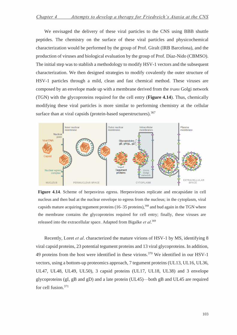
Chapter 4 Attempts to develop a therapy for Friedreich’s Ataxia at the CNS
103
We envisaged the delivery of these viral particles to the CNS using BBB shuttle
peptides. The chemistry on the surface of these viral particles and physicochemical
characterization would be performed by the group of Prof. Giralt (IRB Barcelona), and the
production of viruses and biological evaluation by the group of Prof. Díaz-Nido (CBMSO).
The initial step was to stablish a methodology to modify HSV-1 vectors and the subsequent
characterization. We then designed strategies to modify covalently the outer structure of
HSV-1 particles through a mild, clean and fast chemical method. These viruses are
composed by an envelope made up with a membrane derived from the trans Golgi network
(TGN) with the glycoproteins required for the cell entry (Figure 4.14). Thus, chemically
modifying these viral particles is more similar to performing chemistry at the cellular
surface than at viral capsids (protein-based superstructures).367
Figure 4.14. Scheme of herpesvirus egress. Herpesviruses replicate and encapsidate in cell
nucleus and then bud at the nuclear envelope to egress from the nucleus; in the cytoplasm, viral
capsids mature acquiring tegument proteins (16–35 proteins),368 and bud again in the TGN where
the membrane contains the glycoproteins required for cell entry; finally, these viruses are
released into the extracellular space. Adapted from Bigalke et al.369
Recently, Loret et al. characterized the mature virions of HSV-1 by MS, identifying 8
viral capsid proteins, 23 potential tegument proteins and 13 viral glycoproteins. In addition,
49 proteins from the host were identified in these virions.370 We identified in our HSV-1
vectors, using a bottom-up proteomics approach, 7 tegument proteins (UL13, UL16, UL36,
UL47, UL48, UL49, UL50), 3 capsid proteins (UL17, UL18, UL38) and 3 envelope
glycoproteins (gI, gB and gD) and a late protein (UL45)—both gB and UL45 are required
for cell fusion.371
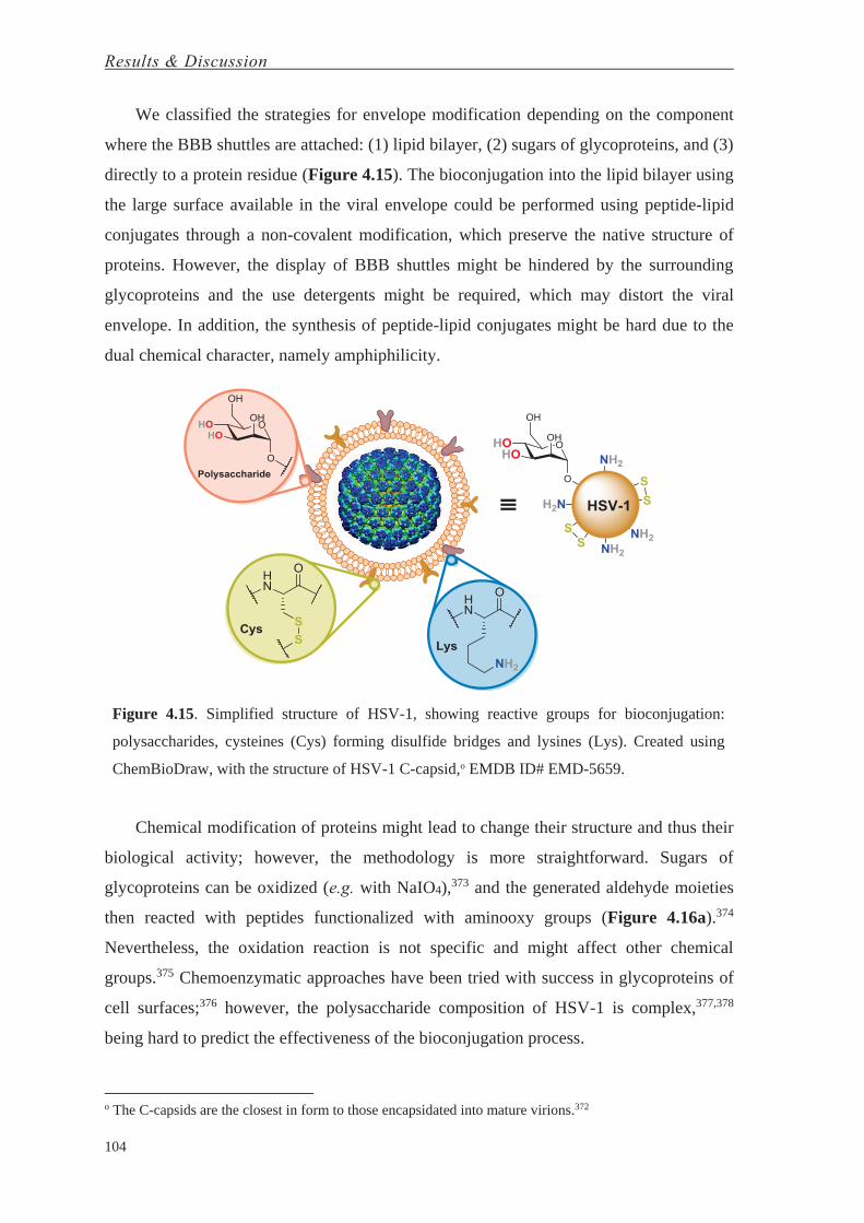
Results & Discussion
104
We classified the strategies for envelope modification depending on the component
where the BBB shuttles are attached: (1) lipid bilayer, (2) sugars of glycoproteins, and (3)
directly to a protein residue (Figure 4.15). The bioconjugation into the lipid bilayer using
the large surface available in the viral envelope could be performed using peptide-lipid
conjugates through a non-covalent modification, which preserve the native structure of
proteins. However, the display of BBB shuttles might be hindered by the surrounding
glycoproteins and the use detergents might be required, which may distort the viral
envelope. In addition, the synthesis of peptide-lipid conjugates might be hard due to the
dual chemical character, namely amphiphilicity.
Figure 4.15. Simplified structure of HSV-1, showing reactive groups for bioconjugation:
polysaccharides, cysteines (Cys) forming disulfide bridges and lysines (Lys). Created using
ChemBioDraw, with the structure of HSV-1 C-capsid,o EMDB ID# EMD-5659.
Chemical modification of proteins might lead to change their structure and thus their
biological activity; however, the methodology is more straightforward. Sugars of
glycoproteins can be oxidized (e.g. with NaIO4),373 and the generated aldehyde moieties
then reacted with peptides functionalized with aminooxy groups (Figure 4.16a).374
Nevertheless, the oxidation reaction is not specific and might affect other chemical
groups.375 Chemoenzymatic approaches have been tried with success in glycoproteins of
cell surfaces;376 however, the polysaccharide composition of HSV-1 is complex,377,378
being hard to predict the effectiveness of the bioconjugation process.
o The C-capsids are the closest in form to those encapsidated into mature virions.372

Chapter 4 Attempts to develop a therapy for Friedreich’s Ataxia at the CNS
105
Strategies facing bioconjugation into protein side-chains are varied. Here, we selected
two straightforward methodologies to covalently modify cysteine and lysine residues. Thiol
reactive maleimides enable specific modification of cysteines (Figure 4.16c). However,
this methodology entails a two-step process, since it requires the reduction of Cys-Cys
disulfide bridges prior to reaction with maleimide groups. This increases the sample
manipulation, and the reduction of disulfide bridges may induce conformational changes
on proteins and thus alter their function.379
Figure 4.16. HSV-1 bioconjugation strategies: (a) oxidation of polysaccharides and reaction of
aminooxy-peptides with the generated aldehydes; (b) reaction of lysine residues with peptides
containing an NHS-ester; and (c) reaction of maleimide-peptides with thiol-free cysteines prior
reduction of disulfide bridges. Created using ChemBioDraw.
Lysine bioconjugation is based on the reaction of nucleophiles with carboxylic acids
activated with good leaving groups—e.g. N-hydroxysuccinimide (NHS). These reactions
are performed in neutral/mild basic pH since primary amines—like in Lys side-chains—
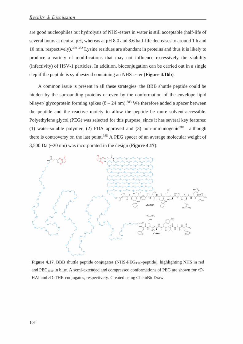
Results & Discussion
106
are good nucleophiles but hydrolysis of NHS-esters in water is still acceptable (half-life of
several hours at neutral pH, whereas at pH 8.0 and 8.6 half-life decreases to around 1 h and
10 min, respectively).380-382 Lysine residues are abundant in proteins and thus it is likely to
produce a variety of modifications that may not influence excessively the viability
(infectivity) of HSV-1 particles. In addition, bioconjugation can be carried out in a single
step if the peptide is synthesized containing an NHS-ester (Figure 4.16b).
A common issue is present in all these strategies: the BBB shuttle peptide could be
hidden by the surrounding proteins or even by the conformation of the envelope lipid
bilayer/ glycoprotein forming spikes (8 – 24 nm).383 We therefore added a spacer between
the peptide and the reactive moiety to allow the peptide be more solvent-accessible.
Polyethylene glycol (PEG) was selected for this purpose, since it has several key features:
(1) water-soluble polymer, (2) FDA approved and (3) non-immunogenic384—although
there is controversy on the last point.385 A PEG spacer of an average molecular weight of
3,500 Da (~20 nm) was incorporated in the design (Figure 4.17).
Figure 4.17. BBB shuttle peptide conjugates (NHS-PEG3500-peptide), highlighting NHS in red
and PEG3500 in blue. A semi-extended and compressed conformations of PEG are shown for rD-
HAI and rD-THR conjugates, respectively. Created using ChemBioDraw.

Chapter 4 Attempts to develop a therapy for Friedreich’s Ataxia at the CNS
107
Figure 4.18. Characterization of NHS-PEG3500-rD-HAI and -rD-THR: (a) 1H-NMR spectrum
(top and bottom, respectively), (b) RP-HPLC chromatograms using a C18 column and a linear
gradient from 0 to 100% of CH3CN (light and strong grey, respectively) and (c) MALDI-TOF
MS spectra (left and right shifted mass distributions, respectively). Created using Adobe
Illustrator and InDesign.
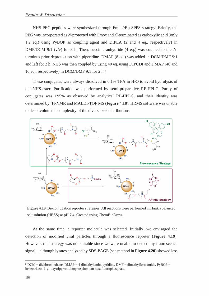
Results & Discussion
108
NHS-PEG-peptides were synthesized through Fmoc/tBu SPPS strategy. Briefly, the
PEG was incorporated as N-protected with Fmoc and C-terminated as carboxylic acid (only
1.2 eq.) using PyBOP as coupling agent and DIPEA (2 and 4 eq., respectively) in
DMF/DCM 9:1 (v/v) for 3 h. Then, succinic anhydride (4 eq.) was coupled to the N-
terminus prior deprotection with piperidine. DMAP (8 eq.) was added in DCM/DMF 9:1
and left for 2 h. NHS was then coupled by using 40 eq. using DIPCDI and DMAP (40 and
10 eq., respectively) in DCM/DMF 9:1 for 2 h.p
These conjugates were always dissolved in 0.1% TFA in H2O to avoid hydrolysis of
the NHS-ester. Purification was performed by semi-preparative RP-HPLC. Purity of
conjugates was >95% as observed by analytical RP-HPLC, and their identity was
determined by 1H-NMR and MALDI-TOF MS (Figure 4.18). HRMS software was unable
to deconvolute the complexity of the diverse m/z distributions.
Figure 4.19. Bioconjugation reporter strategies. All reactions were performed in Hank's balanced
salt solution (HBSS) at pH 7.4. Created using ChemBioDraw.
At the same time, a reporter molecule was selected. Initially, we envisaged the
detection of modified viral particles through a fluorescence reporter (Figure 4.19).
However, this strategy was not suitable since we were unable to detect any fluorescence
signal—although lysates analyzed by SDS-PAGE (see method in Figure 4.20) showed less
p DCM ≡ dichloromethane, DMAP ≡ 4-dimethylaminopyridine, DMF ≡ dimethylformamide, PyBOP ≡ benzotriazol-1-yl-oxytripyrrolidinophosphonium hexafluorophosphate.

Chapter 4 Attempts to develop a therapy for Friedreich’s Ataxia at the CNS
109
staining compared to unmodified sample—and it required to perform two reactions. Thus,
we decided to pursue with a new strategy based on affinity detection using a commercial
NHS-PEG3500-biotin (Figure 4.19). This strategy, in addition to quantification, would also
allow the selective capture of modified proteins using streptavidin-coated magnetic
nanoparticles and posterior identification by HRMS—affinity selection of proteins coupled
to MS (ASMS) (Figure 4.20). Nevertheless, several attempts failed, identifying only the
streptavidin derived from the magnetic nanoparticles.
Figure 4.20. Methodologies used for characterization of HSV-1 viral particles: transmission
electron microscopy (TEM), dynamic light scattering (DLS) and ζ-potential of native or modified
samples; SDS-PAGE, western blot and MS analysis of lysates; and affinity selection of proteins
coupled to MS (ASMS). Created using ChemBioDraw, with the structure of HSV-1 C-capsid,
EMDB ID# EMD-5659.
Other methodologies were used to characterize the native and modified HSV-1
particles (Figure 4.20). Lysates were analyzed by SDS-PAGE after treatment under
denaturing conditions (1% SDS, 5% 2-mercaptoethanol, at 95ºC for 15 min). Silver staining
was used to reveal protein bands since sensitivity is higher compared to that obtained using
Coomassie blue, which did not result in a band pattern when initially used (sensitivity of
Coomassie blue and silver staining is about 0.3–1 μg and 2–5 ng/protein band,
respectively).386 Bioconjugation of NHS-PEG-peptide conjugates resulted in a decrease in
staining; however, the band pattern of native samples did not change (Figure 4.21a),
expected upon covalent addition of one or more ~5 kDa molecules (PEG-peptides). The
same event was also observed for pure proteins (human transferrin (hTf) and bovine serum
albumin (BSA)) modified with NHS-PEG3500-biotin, thus showing that PEG moieties may
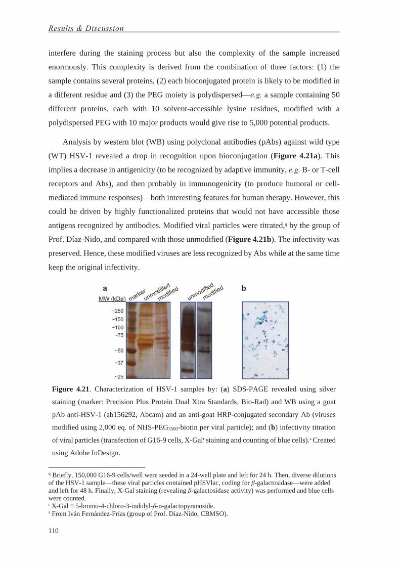
Results & Discussion
110
interfere during the staining process but also the complexity of the sample increased
enormously. This complexity is derived from the combination of three factors: (1) the
sample contains several proteins, (2) each bioconjugated protein is likely to be modified in
a different residue and (3) the PEG moiety is polydispersed—e.g. a sample containing 50
different proteins, each with 10 solvent-accessible lysine residues, modified with a
polydispersed PEG with 10 major products would give rise to 5,000 potential products.
Analysis by western blot (WB) using polyclonal antibodies (pAbs) against wild type
(WT) HSV-1 revealed a drop in recognition upon bioconjugation (Figure 4.21a). This
implies a decrease in antigenicity (to be recognized by adaptive immunity, e.g. B- or T-cell
receptors and Abs), and then probably in immunogenicity (to produce humoral or cell-
mediated immune responses)—both interesting features for human therapy. However, this
could be driven by highly functionalized proteins that would not have accessible those
antigens recognized by antibodies. Modified viral particles were titrated,q by the group of
Prof. Díaz-Nido, and compared with those unmodified (Figure 4.21b). The infectivity was
preserved. Hence, these modified viruses are less recognized by Abs while at the same time
keep the original infectivity.
Figure 4.21. Characterization of HSV-1 samples by: (a) SDS-PAGE revealed using silver
staining (marker: Precision Plus Protein Dual Xtra Standards, Bio-Rad) and WB using a goat
pAb anti-HSV-1 (ab156292, Abcam) and an anti-goat HRP-conjugated secondary Ab (viruses
modified using 2,000 eq. of NHS-PEG3500-biotin per viral particle); and (b) infectivity titration
of viral particles (transfection of G16-9 cells, X-Galr staining and counting of blue cells).s Created
using Adobe InDesign.
q Briefly, 150,000 G16-9 cells/well were seeded in a 24-well plate and left for 24 h. Then, diverse dilutions of the HSV-1 sample—these viral particles contained pHSVlac, coding for β-galactosidase—were added and left for 48 h. Finally, X-Gal staining (revealing β-galactosidase activity) was performed and blue cells were counted. r X-Gal ≡ 5-bromo-4-chloro-3-indolyl-β-ᴅ-galactopyranoside. s From Iván Fernández-Frías (group of Prof. Díaz-Nido, CBMSO).

Chapter 4 Attempts to develop a therapy for Friedreich’s Ataxia at the CNS
111
Attempts to characterize HSV-1 particles by transmission electron microscopy (TEM)
were not successful. We were not able to recognize any viral particle using negative
staining; however, immuno-TEM using the same pAb against HSV-1 used in WB and an
anti-goat 18-nm gold-conjugated Ab helped in the identification of unmodified HSV-1
particles. Low-resolution images were obtained—we are currently improving these results
for both modified and unmodified particles.
Figure 4.22. Size (nm) distribution per number of particles: (red) unmodified sample, and
samples (black) 1, (blue) 2 and (green) 3. Created using Adobe Illustrator.
Size-distribution of both bioconjugated or not HSV-1 samples was assessed by
dynamic light scattering (DLS). We decided to test diverse reaction conditions using NHS-
ester-PEG3500-biotin: (sample 1) adding EDC, (sample 2) adding EDC and concentrating
the sample 10-times and (sample 3) without adding EDC but concentrating the sample 10-
times.t A huge increase in size for sample 1 was observed, whereas samples 2 and 3
maintained roughly the same size distribution (Figure 4.22).
Table 4.7. Relationship between reaction conditions and ζ-potential. Bioconjugation reactions
were performed using 2,000 eq. of NHS-PEG3500-biotin in HBSS buffer at pH 7.4 for 2 h.
Reaction Conditions
Sample ID Relative Conc.u Additional Reagents ζ-Potential (mV)
Unmodified - - -11.2 ± 0.2
1 ×1 EDC -10.9 ± 0.5
2 ×10 EDC -15.2 ± 1.1
3 ×10 - -13.7 ± 0.6
t EDC ≡ 1-ethyl-3-(3-dimethylaminopropyl)carbodiimide. u Concentration of HSV-1 particles: (× 1) ≡ 400 TU/μL; (× 10) ≡ 4,000 TU/μL.

Results & Discussion
112
In addition, bioconjugation of these samples was numerically evaluated by means of
ζ-potential. Whereas sample 1 maintained the same value as the unmodified particles,
samples 2 and 3 showed an increase in the negativity of that parameter (Table 4.7).
Reaction of NHS-esters made up with no charged moieties (PEG-biotin) may lead to a
reduction in the net surface charge, since amino groups (from the side-chains of Lys
residues, with a pKa of ~10.0)387 react to form a neutral amide bond. Following this
rationale, sample 1 could have suffered a crosslinking process between viral particles
(reaction between side-chains containing carboxylic acid and amines promoted by EDC)
which may produce the apparent aggregation shown in Figure 4.22; whereas concentrated
viral samples would be modified with NHS-ester moieties, producing a decrease in the ζ-
potential and preserving the same size as the unmodified viruses. In addition, EDC
apparently did not produce crosslinking between viral particles.
Figure 4.23. HSV-1 bioconjugation at Lys residues. pKas: Lys (in general, ~10.0;387 however, it
depends on solvent exposure and ranges from 5.3 to 10.4),388 Arg (~13.8),389 His (5.8–7.9),390-392
NHS (6.0).393 Created using ChemBioDraw.
Chapter 4 Attempts to develop a therapy for Friedreich’s Ataxia at the CNS
113
We then decided to continue working without adding EDC, since it still produced a ζ-
potential shift, whereas we observed side-effects of EDC in sample 1. Contrary to samples
modified with NHS-PEG3500-biotin, reaction of viral particles with NHS-PEG3500-rD-THR
showed a positive increase in ζ-potential (Table 4.8). Here, the neutral biotin moiety is
replaced by rD-THR, which has one positively charged residue (Arg with a very basic pKa
of 13.8)389 and a His residue which might be slightly positive at pH 7.4 (pKa of 5.8–7.9).390-
392 Therefore, it was expected to reduce the negative ζ-potential value, although producing
a smaller shift (in absolute value) compared with NHS-PEG3500-biotin bioconjugation.
Same result can be expected for rD-HAI—only positive charges from two His and one Arg
residues (Figure 4.23).
Table 4.8. Relationship between reaction conditions and ζ-potential. Bioconjugation reactions
were performed using 2,000 eq. of NHS-PEG-rD-THR in HBSS buffer at pH 7.4 for 2 h.
Reaction Conditions
Sample ID Relative Conc. Additional Reagents ζ-Potential (mV)
Unmodified - - -15.8 ± 1.4
Modified ×10 - -14.5 ± 0.2
Although changes in ζ-potential after bioconjugation shifted consistently in the same
direction (negative and positive for NHS-PEG-biotin and NHS-PEG-rD-THR,
respectively), these results will be confirmed by using radioactive analysis—briefly,
labeling NHS-PEG-rD-THR with a radioisotope, bioconjugating it to HSV-1 particles and
then determining the radioactive signal. In addition, both ζ-potential and radioactive
assessment of NHS-PEG-rD-HAI will be performed. Finally, in vivo studies will be
performed to evaluate both infectivity and transport through the BBB—stereotaxic and
intravenous injections of modified viral particles, respectively.
The methodology here presented can be used to modify other viral particles, enveloped
or not, but also cells—the surface of HSV-1 viruses is more similar to cell surfaces than to
viral capsids.v
v The complete description of the characterization and bioconjugation performed per each sample is shown in the Experimental Section (Product Characterization).


CONCLUSIONS

Conclusions
117
1. A hybrid design combining the “gold standard” passive diffusion BBB shuttle
(NMePhe)4 and the water-soluble amino acid proline increases water-solubility by three
orders of magnitude—low millimolar range—compared to (NMePhe)4.
2. The transport of (PhPro)4 carrying a cargo (NIP and ʟ-DOPA) is around 7-fold higher
compared to that of the (NMePhe)4 shuttle. Furthermore, (PhPro)4 maintains the
permeability when a cargo is attached, whereas (NMePhe)4 shows a marked reduction.
3. The analysis of the transport of the 16-stereoisomer library revealed that
stereochemistry plays a significant role in passive diffusion through biological
membranes.
4. Three analogs from the retro-ᴅ-version of HAI, sharing a substitution in the same
proline residue, show a 2-fold increase in transport compared to the parent peptide. Two
of them show 4- and 8-fold increase in transport when assayed in cell culture medium
instead of buffer.
5. Using a subtle modification (i.e. N-terminal acetylation), a novel method combining in
vitro cell-based BBB models and MALDI-TOF MS allows an increase in sensitivity by
three orders of magnitude compared with a standard method (RP-HPLC-PDA), and the
use of cell culture medium during these assays.
6. The injection of BBB shuttles made with ʟ-amino acids (HAI and THR) in mice elicit
a low immune (humoral) response, which are even lower for their retro-ᴅ-versions
made with ᴅ-amino acids.
7. Attempts to bioconjugate the N-terminus of frataxin (FXN) failed due to the lack of
stability of the N-terminal region of this protein by proteolytic degradation.

Conclusions
118
8. The attachment of BBB shuttles on HSV-1 particles using Lys bioconjugation enables
the modification of these viral particles in a single step, reducing the sample
manipulation and thus preserving their structural integrity better. In addition, the use of
PEG3500 as spacer between the virus and the BBB shuttle peptide hampers antibody
recognition while preserving infectivity of viral bioconjugates.
9. The combination of methods from molecular biology (SDS-PAGE, western blot) with
proteomics (mass spectrometry) and biophysical tools (TEM, DLS and ζ-potential)
enables the characterization of the bioconjugated viruses.

EXPERIMENTAL SECTION


Materials and Methods


Materials & Methods
123
Solid-Phase Synthesis of Compounds
Reagents and Solvents. Protected amino acids and resins were supplied by
Luxembourg Industries (Tel-Aviv, Israel), Neosystem (Strasbourg, France), Calbiochem-
Novabiochem AG (Laüfelfingen, Switzerland), PolyPeptide Laboratories (Torrance, CA
USA), Bachem AG (Bubendorf, Switzerland), PCAS BioMatrix Inc. (St-Jean-sur-
Richelieu, Quebec, Canada) and Iris Biotech (Marktredwitz, Germany). PyBOP, COMU
and Oxyma were provided by Calbiochem-Novabiochem AG. Acetic acid-1-13C,d4 was
obtained from Aldrich (Milwaukee, WI, USA). DIEA and ninhydrin were from Fluka
Chemika (Buchs, Switzerland). HOAt was purchased from GL Biochem Shanghai Ltd.
(Shanghai, China). Solvents for peptide synthesis and RP-HPLC were from Scharlau or
SDS (Barcelona, Spain). Trifluoroacetic acid was purchased from KaliChemie (Bad
Wimpfen, Germany). The other chemicals used were from Aldrich (Milwaukee) and were
of the highest purity commercially available.
General Protocol for SPPS. Standard solid-phase peptide elongation and other solid-
phase manipulations were done manually in polypropylene syringes, each fitted with a
polyethylene porous disk at the bottom. Solvents and soluble reagents were removed by
suction. Between couplings and deprotections, the resin was washed with DMF (5 × 1 min),
DCM (5 × 1 min), and DMF (5 × 1 min), using 5 mL of solvent/g of resin each time. During
couplings, the syringe was left under automatic stirring. Intermittent manual stirring was
applied during deprotections.
In each chapter of this thesis the syntheses were performed using diverse resins and
coupling conditions, here specified.
Identification Tests. After each reaction, the Kaiser test394 was used to identify
primary amines on the N-terminus of the elongating peptide on the solid support. The
chloranil test395 was used to identify secondary amines.
Initial Conditioning of Resin. The Sieber Amide and Fmoc-Rink-Amide AM resins
were conditioned by washing with DMF (5 × 1 min), DCM (5 × 1 min), and DMF (5 × 1
min), followed by 20% piperidine in DMF (1 × 1 min, 2 × 10 min) to remove the Fmoc
group. Finally, resins were washed with DMF (5 × 1 min). H-Rink Amide-ChemMatrix
resin was conditioned by washing with CH3OH (5 × 1 min), DMF (5 × 1 min), DCM (5 ×
1 min), 1% TFA in DCM (5 × 1 min), DCM (5 × 1 min), DMF (5 × 1 min), DCM (5 × 1

Experimental Section
124
min), 5% DIPEA in DCM (5 × 1 min), DCM (5 × 1 min) and finally DMF (5 × 1 min). The
resins used in each chapter of this thesis are specified below:
Chapter Resin
1 Sieber396 Amide
2 Fmoc-Rink-Amide AM394,395
3 H-Rink397 Amide-ChemMatrix
4 H-Rink Amide-ChemMatrix, and Dawson Dbz AM resin335
Fmoc Group Removal. Fmoc group was removed by treating the resin with 20%
piperidine in DMF (3–4 mL/g of resin; 1 × 1 min, 2 × 10 min). For secondary amine Fmoc
deprotection, resin was additionally treated with DBU, toluene, and piperidine in DMF
(5:5:20:70, v/v) (1 × 10 min).
Coupling of the First Amino Acid onto the Resin and Following Couplings. For
the syntheses of HAI analogs, were added sequentially to the resin in DMF (minimal
volume to allow the complete dissolution of the reagents), with the appropriate coupling
reagents. The mixture was allowed to react under stirring in an orbital shaker for 1 h.
Afterwards, the solvent was removed by filtration, and the resin was washed with DMF (5
× 1 min) and DCM (5 × 1 min). The extent of coupling was checked by the appropriate
colorimetric test. When required, a recoupling step was performed using the same previous
conditions but for longer (2 h).
Chapter Coupling Conditions
1 N-protected Fmoc-amino acid, Fmoc-NIP-OH or Fmoc-ʟ-DOPA-OH (4 eq.), PyBOP
(4 eq.) and HOAt (12 eq.), followed by DIPEA (12 eq.)
2 N-protected Fmoc-amino acid (4 eq.), COMU398 (4 eq.), and Oxyma Pure399 (4 eq.),
followed by DIPEA (8 eq.).
3 N-protected Fmoc-amino acids (4 eq.) were activated by Cl-HOBt400 (4 eq.) with
DIPCDI (4 eq.)
4 N-protected Fmoc-amino acids (4 eq.) or Boc-(aminooxy)acetic acid (for AO-peptides)
were activated by Cl-HOBt (4 eq.) with DIPCDI (4 eq.) for the couplings on H-Rink
Amide-ChemMatrix resin, as well for the second and subsequent couplings on Dawson
Dbz AM resin
N-protected Fmoc-amino acids (4 eq.) were activated by HBTU/HOBt (6 eq.) with
DIPEA (9 eq.) for the first coupling on Dawson Dbz AM resin
Materials & Methods
125
N-Terminal Capping. The anhydride of the acetic acid (4 eq.) was prepared by mixing
acetic acid (8 eq.) with DIPCDI (2:1) and adding 1 mL of DCM. Two minutes of agitation
was applied. Afterwards, the solvent was added to the SPPS syringe, and the precipitate
(N,N'-diisopropylurea) was discarded. DCM was added until all the resin was covered, and
DIPEA (4 eq.) was then added. After 30 min with stirring in the orbital shaker, the solvent
was removed by filtration, and the resin was washed with DCM (5 × 1 min), DMF (5 × 1
min) and DCM (5 × 1 min). The extent of coupling was checked by the appropriate
colorimetric test.
Peptide Cleavage. In order to cleave the peptides from the resin, the cleavage cocktail
(TFA/TIS/H2O, 95:2.5:2.5, v/v; except for peptides synthesized in chapter 1, where
TFA/DCM, 2.5:97.5, v/v was used) was added to each syringe. Intermittent manually
agitation was applied for 30–60 min. The solvent was collected in the same plastic tube and
evaporated by a N2 flow.
Work-Up. After evaporation of the cleavage cocktail, 20 mL of methyl tert-butyl ether
(MTBE) was added to the plastic tube containing the dry residue. This tube was centrifuged
at 2,000 × g for 10 min. The solvent was then discarded by decantation. This process was
repeated two more times. After the last washing with MTBE, the final residue was
dissolved in H2O/CH3CN (1:1) and then lyophilized.
Special Considerations. For the synthesis of the 16 stereoisomers of (PhPro)4, resin
was placed in tea bags, which were labeled and then heat-sealed, according to the procedure
developed by Houghten and coworkers (1985).167 Solvents were removed by decantation
after taking the tea bags out of the reaction pot. Washings between synthetic steps were
done with DMF (2 × 1 min), isopropanol (1 × 1 min), DMF (2 × 1 min), using a solvent
volume that covered the bags entirely. In order to use the same solvent for washing steps
as in the synthetic steps, an initial or final washing with DCM (2 × 1 min) was performed.
During couplings the mixture was allowed to react with continuous stirring. One pot was
used per type of coupling/deprotection/colorimetric test. The colorimetric test was done
using a pH indicator, bromophenol blue.401,402 After deprotections and couplings, 1 drop of
1% bromophenol blue in DMF was placed in the DMF volume containing the tea bags. A
positive result is given by an intense blue staining of the resin, which results from the free
amines occurring in the growing peptide (higher pH) after a decoupling step. The lack of
resin staining (natural color of the resin under DMF solvation) is due to the absence of free

Experimental Section
126
amines, expected after a coupling step. The Fmoc group was removed by treating the resin
with 25% piperidine and 1% Triton x100 in DMF (using the minimal volume to cover all
tea bags; 2 × 10 min). In all cases, resin was additionally treated with DBU, toluene, and
piperidine in DMF (5:5:20:70, v/v) (1 × 10 min). A diagram of the synthetic strategy for
the 16 stereoisomers of (PhPro)4 is shown in Scheme M.1.
Scheme M.1. Synthetic strategy for the 16 stereoisomers of (PhPro)4.
A diagram of the synthetic strategy for HAI analogs is shown in Scheme M.2.
Importantly, each peptide was differentially acetylated using CD3-13COOD or CH3-COOH.
Scheme M.2. Strategy for the synthesis of the complete set of HAI analogs with a final splitting
step that allows differential isotopic labeling by means of acetylation.

Materials & Methods
127
Peptides-Nbz were synthesized following a Fmoc-SPPS approach reported by Blanco-
Canosa et al.,335 where first diaminobenzoic acid (Dbz) is incorporated in a resin terminated
with a Rink linker. After the standard SPPS synthesis, the C-terminus is activated through
acylation with p-nitrophenylchloroformate followed by the addition of a base. The peptide-
Nbz is finally deprotected and cleaved by TFA treatment with scavengers (Scheme M.3).
Scheme M.3. Scheme for the synthesis of Nbz-peptides. Created using ChemBioDraw.
NHS-PEG-peptides were synthesized through Fmoc/tBu SPPS strategy. PEG was
incorporated as N-protected with Fmoc and C-terminated as carboxylic acid (Fmoc-
PEG3500-COOH; JenKem Technology USA, Plano, TX, USA), using only 1.2 eq.; PyBOP
and DIPEA (2 and 4 eq., respectively) were added and the reaction was then left in
DMF/DCM 9:1 (v/v) for 3 h with continuous stirring. Then, succinic anhydride (4 eq.) was
coupled to the N-terminus prior desprotection with piperidine (1 × 1 min and 2 × 15 min).
DMAP (8 eq.) was added in DCM/DMF 9:1 and left for 2 h with continuous stirring. NHS
was then coupled by using 40 eq. and, DIPCDI and DMAP (40 and 10 eq., respectively) in
DCM/DMF 9:1 for 2 h. Finally, the resin was cleaved with TFA/TIS/H2O for 2 h, and
lyophilized in H2O/CH3CN prior evaporation with N2 flow.

Experimental Section
128
Peptide and Amino Acid Purification and Characterization
RP-HPLC Purification. The peptides were purified by reverse-phase HPLC using a
Sunfire C18 column (150 × 10 mm × 5 μm, 100 Å; Waters, Milford, CT, USA); solvents:
H2O (0.1% TFA) and CH3CN (0.1% TFA); and flow rate of 3 mL/min. Fmoc-PhPro-OH
amino acid enantiomers were separated using a chiral Lux Cellulose-2 column (150 × 21.2
mm, 5 μm, 1,000 Å, cellulose tris(3-chloro-4-methylphenylcarbamate) stationary phase;
Phenomenex Inc., Torrance, CA, USA); solvents: CH3OH and CH3CN (0.1% TFA); and
flow rate of 3 mL/min.
General Characterization. Compound identity was confirmed using MALDI-TOF
MS (Applied Biosystems 4700 MALDI-TOF spectrometer; PE Applied Biosystems, Foster
City, CA, USA), RP-HPLC-MS (Alliance 2796 with photodiode array (PDA) detector
2998, ESI-MS model Micromass ZQ and Masslynx version 4.0 software; Waters) and
HRMS in a Synapt HDMS (Waters) or LTQ-FT Ultra (Thermo Scientific, Waltham, MA,
USA). Peptide purity was checked by RP-HPLC (Alliance 2695 with PDA detector 2998
and software EmpowerPro 2; Waters) using a Sunfire C18 column (150 × 4.6 mm × 5 μm,
100 Å, Waters); solvents: H2O (0.045% TFA) and CH3CN (0.036% TFA); flow rate of 1
mL/min.
The purity of the Fmoc-PhPro-OH enantiomers was checked by RP-HPLC using a
chiral Lux Cellulose-2 column (150 × 4.6 mm analytical, 5 μm, 1,000 Å, cellulose tris(3-
chloro-4-methylphenylcarbamate) stationary phase; Phenomenex Inc.); solvents: CH3OH
and CH3CN (0.1% TFA); and flow rate of 1 mL/min.
Additionally, 1H-NMR (600 MHz) experiments confirmed the identity and purity of
the set of pure HAI analogs (as well the proper isotopic labeling between pairs of peptides
(either acetylated with acetic acid or with acetic acid-1-13C,d4)) and of PEG-peptide
conjugates. Samples were prepared by dissolving peptides or PEG-peptide conjugates in
H2O/ D2O 80:20 (v/v) or D2O, respectively. Suppression of the water signal was achieved
by WATERGATE W5.403 For PEG-peptide conjugates, d1 = 10 s was used. All
experiments were performed at 298 K.
Specific Rotation. Specific rotation of the PhPro amino acid enantiomers was
determined with the polarimeter P-2000 (Jasco, Easton, MD, USA). Cells with a pathlength
of 1 dm were used. The parameters used during measurements were as follows:

Materials & Methods
129
accumulations (3), and wavelength (589 nm). Samples were dissolved in CH3OH at a final
concentration of 0.0008 g/mL. The values of αD were calculated as shown in Eq. M.1:
(M.1)
where α is the value returned by the instrument, l is the pathlength, c is the
concentration, and αD is the specific rotation.
Amino Acid Analysis. Peptide content and amino acid ratio were determined by
chromatographic separation and quantification of the hydrolyzed product. Peptide samples
were hydrolyzed in 6 M HCl at 110ºC for 16 h. The solvent was then evaporated to dryness
under reduced pressure. The residue was dissolved in 20 mM HCl and derivatized using
the AccQ·Tag protocol (Waters) using 6-aminoquinolyl-N-hydroxysuccinimidyl
carbamate. Finally, samples were analyzed by HPLC.
Water-Solubility. Solubility of peptides was determined by weighing an amount of
the peptide in a vial and dissolving it in a certain volume of water, taking care to ensure
saturation. The vials were then left for 1 h at 37ºC under soft agitation. Afterwards, they
were left for 20 min at room temperature and centrifuged to precipitate any non-dissolved
particle. From that solution, a volume (not higher than 70% of the total volume) was passed
through a 400-μm filter and moved to a weighed glass vial. Finally, the vials were
lyophilized for one day and weighed. Solubility was calculated as the difference of mass
between the glass vials with and without peptide divided by the volume taken.

Experimental Section
130
Structural Data
Circular Dichroism. Circular dichroism spectra were obtained with a J-715 or an 810
UV-Vis spectropolarimeter (Jasco), with a Peltier CDF 426S/426L. Parameters used:
sensitivity (standard (100 mdeg)), start (250 nm), end (190 nm), data pitch (0.1 nm),
scanning mode (continuous), scanning speed (10 nm/min), response (4 sec), band width
(1.0 nm), and accumulation (3). Molar ellipticity was calculated using the Eq. M.2:
(M.2)
where θMR is the molar ellipticity in deg · cm2 · dmol-1, θ is the measured ellipticity
value in mdeg, l is the optical path in cm, C is the concentration of the peptide/protein in
mol/L, and n is the number of residues in the peptide/protein.
NMR. NMR experiments were performed on a Bruker Avance III 600 MHz
spectrometer equipped with a TCI cryoprobe. Samples were prepared by dissolving
peptides (5 mM) in 50 mM NaCl, 25 mM sodium phosphate buffer, H2O/D2O 90:10, at pH
7.4, containing 0.02% NaN3. Chemical shifts were referenced to internal sodium-3-
(trimethylsilyl)propanesulfonate (DSS). Water signal suppression was achieved by
excitation sculpting404. Residue specific assignments were obtained from 2D total
correlated spectroscopy (TOCSY) and correlation spectroscopy (COSY) experiments,
while 2D nuclear Overhauser effect spectroscopy (NOESY) permitted sequence specific
assignments. 13C resonances were assigned from 2D 1H-13C HSQC spectra. All
experiments were performed at 298 K, except NOESY spectra, which were acquired at 278
K. Amide proton temperature coefficients were determined from a series of one-
dimensional spectra acquired between 278 and 308K. TOCSY and NOESY mixing times
were 70 and 250 ms, respectively. Relative populations of the cis/trans isomers were
estimated from integration of amide protons in the 1D 1H-NMR spectra or alternatively,
when 1H integration was precluded by signal overlap, from the relative intensities of 1H-13C-HSQC crosspeaks corresponding to the Pro Cδ resonances. In those cases that 1H
integration was possible, both methods provided identical results.
Protease-Resistance in Human Serum. Peptides at a final concentration of 150 μM
in Hank’s balanced solution salt were incubated at 37°C in the presence of 90% human
serum (from human male AB plasma). At pre-established time points, aliquots of 200 μL
were extracted, and serum proteins were precipitated by addition of 200 μL of acetonitrile

Materials & Methods
131
at 4°C to stop degradation. After 30 min at 4˚C, samples were centrifuged at 10,000 rpm
for 30 min at 4˚C. The supernatant was analyzed by RP-HPLC and MALDI-TOF mass
spectrometry.
Computational Analysis of the Peptide Conformations. The preferential
conformation adopted by each peptide system was evaluated by replica exchange molecular
dynamics Simulations (REMD).405 Simulations started from a linearly extended peptide
conformation built with XLEaP program module from the AMBER14 molecular
mechanics package.406 The Amber ff99SB force field, together with the reoptimized proline
omega-bond angle parameters,407 was used. The initial extended peptide structure was first
subjected to minimization protocol consisting of 1,000 steps of steep decent method
followed by 500 steps of conjugate gradient method. Optimized structures were gradually
heated to 300 K in 200 ps. The final state was chosen as the initial structure for all the 16
replicas. Temperatures were set in a range from 300 to 500 K.408 Generalized Born model409
with an effective salt concentration of 0.2 M was deployed to mimic the solvation effect.
Nonpolar solvation term was approximately represented by surface area term.410 Integral
time step was set to 1 fs. Temperature was regulated using Berendsen thermostat411 with a
coupling time constant of 1 ps. SHAKE algorithm412 was used to constrain all the covalent
bonds involving hydrogen atoms. Swaps between replicas were attempted every 2 ps, and
35% acceptance probability was obtained. Each replica was simulated during 150 ns.
Snapshots were saved every 2 ps. To evaluate the degree of overlap between parent peptides
and their retro-ᴅ-version forms, a non-symmetric RMSD distance matrix was built using
the ptraj module of the Amber package. To preserve the correct alignment between the
parent peptide and their retro-ᴅ-version, distances were computed between analog amino
acids (i.e, the N-terminal amino acid of the parent peptide corresponds to C-terminal amino
acid in its retro-ᴅ-version, and vice versa). The resulting RMSD matrix, composed of 1,000
equally spaced snapshots of the equilibrated part (100 ns) of the parent peptide and of its
retro-ᴅ-version, was subjected to histogram analysis. R software413 was used in all
statistical analyses. Additionally, 2D RMSD plots (mass weighted) for the same 1,000
equally spaced snapshots of each simulation were computed to visually determine the
number of clusters visited by each peptide system during the replica exchange simulation.
In order to further compute the similarity between the conformational space sampled by
the trajectories of the parent peptide and retro-ᴅ-version, essential dynamics
techniques414,415 were used.

Experimental Section
132
In Vitro Assays
Parallel Artificial Membrane Permeability Assay (PAMPA): The PAMPA assay
was used to determine passive diffusion capacity across the BBB. The standard parameter
that quantifies transport independently of time and concentrations is the effective
permeability (Pe), shown in the Eq. M.3:
(M.3)
where t is the running time (4 h), CA (t) is the concentration of the compound in the
acceptor well at time t, and CD (t0) is the compound concentration in the donor well before
running the PAMPA assay (t0 = 0 h). Transport (%) values were obtained by dividing the
amount in the acceptor well at time t, CA (t), and in the donor well at time zero, CD (t0),
multiplied by 100. Membrane retention was calculated from the difference between the
initial amount, CD (t0), and the amounts in donor, CD (t), and acceptor, CA (t), compartments
at the end of the experiment (t = 4 h).
The buffer (System Solution) was prepared from the commercial concentrated solution
from pION (Woburn, MA, USA) by dissolving 5 mL with 200 mL of water. The pH (2.4)
was adjusted to 7.4 by using a 0.5 M NaOH solution. Then, 1-propanol (20%) was added
to the solution.
The samples were dissolved with the System Solution containing 20% 1-propanol (1
mL). Peptides were assayed at concentrations adjusted to allow the best quantification by
RP-HPLC. Propranolol was used as a positive control.
The PAMPA sandwich (96-transwell plate, pION) was separated into the donor and
acceptor plates, and the stirring magnets were added to the donor compartments. Next, 4
μL of a phospholipid mixture supplied by Avantis Polar Lipids (Alabaster, AL, USA)
(porcine brain polar lipid extract; 20 mg/mL in dodecane) was added to the membrane,
located at the bottom of the acceptor compartments. This phospholipid mixture comprised
phosphatidylcholine (PC; 12.6%), phosphatidylethanolamine (PE; 33.1%),
phosphatidylserine (PS; 18.5%), phosphatidylinositol (PI; 4.1%), phosphatidic acid (0.8%),
and other compounds (30.9%).

Materials & Methods
133
Samples containing the peptides (195 μL) were added to the donor compartments,
(three replicates). Afterwards, acceptor wells were placed above the donor plate and filled
with 200 μL of System Solution (20% 1-propanol).
The PAMPA plate was placed into a GUTBOX (containing wet sponges) for 4 h at
room temperature. Agitation was maintained in 25 μm of unstirred water layer (UWL).
After the incubation time, the donor and the acceptor plates were separated, and the samples
were collected from both and placed into separate tubes. The integrity of the samples (donor
and acceptor wells and time zero solutions) was identified by MALDI-TOF spectroscopy.
The samples were also analyzed by RP-HPLC, and Pe was calculated from the integrated
chromatographic peaks.
In Vitro Bovine BBB Cell-Based Model Assay. This assay was an adapted model84
of the method previously published by Gaillard and de Boer.416 Bovine brain endothelial
cells were purchased from Cell applications (San Diego, CA, USA), and astrocytes were
obtained from rat pups (Wistar rats from Charles River, Wilmington, MA, USA). Before
performing the assay, TEER was measured in all transwells (TEER > 100 Ω·cm2).
Transwell plates (Corning Costar 24-well plate, 0.33 cm2 membrane well insert and 0.4 μm
pore size) were purchased from Corning (Corning, NY, USA). Peptides were prepared at a
concentration of 200 μM in Ringer-HEPES buffer containing 20 μM Lucifer yellow (LY)
lithium salt (Sigma-Aldrich) as control (Papp < 17·10-6 cm/s). The apical compartment was
filled with 200 μL of the solution containing the peptide, and 800 μL of Ringer-HEPES
was poured into the basal well. Three replicates of each peptide were assayed. The plate
was left for 2 h in the incubator at 37ºC. Finally, the samples were collected and analyzed
or frozen until analysis. LY fluorescence was measured in a 96-well plate with a Fluoroskan
Ascent Microplate Fluorometer (Thermo Fisher Scientific).
Finally, in order to evaluate the in vitro transport of the peptides, two parameters were
determined, namely transport (T), expressed as a percentage, and apparent permeability
(Papp), described by Eq. M.4 and M.5, respectively.
(M.4)
(M.5)

Experimental Section
134
where, is the amount (mass or mole) of peptide in the acceptor well at the end
of the experiment (at time t); accounts for the amount of peptide initially evaluated
(in the donor well at time t = 0). is the permeability rate of the peptide; is the area
(in cm2) of the membrane delimiting the two wells, donor and acceptor; is the volume
in the donor well (in cm3); and is the time length of the experiment (2 h) in seconds.
and values were obtained from those recorded by RP-HPLC-PDA (220 nm) and
MALDI-TOF MS, and thus these two equations are adapted in each case (see next section).
The apparent permeability was obtained from the transport (T) values expressed in
percentage (shown in the last step of Eq. M.5).
In Vitro Human BBB Cell-Based Model Assay. This assay was performed using the
model published by Prof. Cecchelli in 2014.417 Endothelial cells and pericytes were
defrosted in gelatin-coated Petri dishes (Corning). Pericytes and endothelial cells were
cultured in DMEM pH 6.8 or in supplemented endothelial cell growth medium (Sciencells),
respectively. After 48 h, pericytes (50,000 cells/well) and endothelial cells (80,000
cells/well) were seeded in gelatin-coated 12-well plates or in Matrigel-coated 12-well
transwell inserts, respectively. Transwell plates (Corning Costar 12-well plate, 1.13 cm2
membrane well insert and 0.4 μm pore size) were purchased from Corning. BD matrigel
matrix was from Corning. Medium (endothelial cell medium, ECM from Innoprot, Derio,
Spain) was changed every 2–3 days and assays were performed 7–8 days after seeding.
Lucifer Yellow (50 μM) was used as internal control (Papp < 15·10-6 cm/s). LY fluorescence
was measured in a 96-well plate with a Fluoroskan Ascent Microplate Fluorometer
(Thermo Fisher Scientific).
Compounds were dissolved in Ringer-HEPES at a concentration of 200 μM. Then, 500
μL of the compound and 1,500 μL of Ringer-HEPES alone were introduced in the apical
or in the basolateral compartments, respectively. The plates were set on at 37 ºC for 2 h.
The solutions from both compartments were then recovered and quantified by HPLC and
identified by MALDI-TOF.
RP-HPLC Coupled to PDA Quantification. This method determined peptide
transport by applying the ratio between the peak areas integrated in the chromatograms
from the acceptor and donor wells further correction by the injected volumes and those
contained in each well (Eq. M.4 and M.6).

Materials & Methods
135
(M.6)
where and account for the integrated area in the HPLC chromatograms
of acceptor (at time t = 2h) and donor (at time t0 = 0) wells, respectively; and are the
injected volumes from donor and acceptor wells, respectively; and and are the
volumes in each acceptor and donor well, respectively.
MALDI-TOF MS Quantification. Quantitative MS data were recorded on an
Applied Biosystems 4800 Plus MALDI-TOF spectrometer, using an ACH-based matrix.
Transport quantification by MALDI-TOF MS was performed by mixing 10 μL of the
sample of the in vitro cell-based model and another 10 μL from the heavy peptide as internal
standard at a similar concentration as the light one. Then, 1 μL of this mixture was placed
on the MALDI plate, together with 1 μL of the freshly prepared solution of the selected
matrix (ACH). This MALDI matrix was prepared by dissolving 15 mg of α-cyano-4-
hydroxycinnamic acid (ACH) in 1 mL of H2O/CH3CN 1:1 (v/v) containing 0.1% TFA. MS
spectra were recorded and transport was calculated. This parameter was obtained from the
intensity ratios light/heavy for acceptor and donor wells further corrected by the volumes
of each well (detailed in Eq. M.7).
(M.7)
accounts for the relative amount of the light peptide in the acceptor
well (at time t) compared with a prepared dilution of the heavy isomer (at 2 μM).
determines the relative amount of light peptide in donor well (at
time t = 0) compared with a prepared dilution of the heavy isomer (at 200 μM; i.e. in our
case R = 100).
RP-HPLC-PDA Limit of Quantification. Dilution series for three peptides 1L (HAI),
6D (retro-ᴅ-HAI) and 9D, ranging from 200 μM to 1.1 nM, were analyzed by RP-HPLC-
PDA. An initial solution at 200 μM was consecutively diluted 1/3 by mixing 100 μL in 200
μL of H2O, up to a total of eleven dilutions. Finally, a specific volume of the sample,
ranging from 5 to 100 μL, was injected to the HPLC system and then further analyzed to
determine the limit of quantification. First, the total absorption corrected by the injected
volume and dilution ( ) was determined as:

Experimental Section
136
(M.8)
where, , and are the total absorption area, the absorption area, the injected volume
and the fold-dilution, respectively. Then, the RLOQ was determined for all of them:
(M.9)
where, is the mean (i.e. ; last five dilutions discarded). The deviation (%)
is calculated as .
MALDI-TOF MS Limit of Quantification. Dilution series for three peptides 1L
(HAI), 6D (retro-ᴅ-HAI) and 9D, ranging from 200 μM to 1.1 nM, were analyzed by
MALDI-TOF MS (on an Applied Biosystems 4800 Plus MALDI-TOF spectrometer, using
an ACH-based matrix). The initial dilution was prepared by mixing 20 μL of two solutions
containing 400 or 800 μM of the light or heavy versions of the peptide, respectively.
Consecutive dilutions 1/3 were then prepared by mixing 10 μL in 20 μL of H2O. Finally, 1
μL of the sample and 1 μL of the ACH matrix were placed in a MALDI plate. The spectra
were acquired and further analyzed to determine the limit of quantification. First, the
experimental light/heavy ratio was determined for all the dilutions. The RLOQ was then
determined for all of them:
(M.10)
where, and are the light/heavy ratio of each sample and the mean (i.e. ,
discarding the last dilution value), respectively. The deviation (%) is calculated as
.

Materials & Methods
137
Protein Expression, Purification, Bioconjugation and Characterization
Plasmid Constructs. The plasmids pOPINS-Gly-FGEsite-FXN, pOPINS-Gly-Lys-
FXN and pOPINS-Cys-FXN used were generated in the IRB Protein Expression Core
Facility.
Expression. Protein expressions were performed by the IRB Protein Expression Core
Facility. Co-expression of the frataxin fusion protein (HisTag-SUMO-Gly-FGEsite-FXN1-
210) with formylglycine-generating enzyme (FGE) was performed as described by Bertozzi
and co-workers.338 MGSSH6-SUMO-GlyLys-FXN and MGSSH6-SUMO-Cys-FXN were
expressed in auto-induction418 conditions in B834(DE3) cells. Briefly, a single colony
containing the appropriate plasmid was cultured overnight in 10 mL of Overnight Express
Terrific Broth (Merck, Darmstadt, Germany) with kanamycin and 1% glucose (w/v) at
37ºC. The overnight culture is diluted into 2 L of Overnight Express Terrific Broth and 500
mL are placed in 2 L shake-flasks and incubated at 37ºC with orbital shaking for 4h. The
temperature is then decreased to 25ºC and cultures are left for 24 h more. The cultures are
pelleted by centrifugation using a rotor JLA-8.1000 (Beckman Coulter, Pasadena, CA,
USA) at 8,000 × g for 20 min. The cell pellets are then frozen at -80ºC or re-suspended in
30 mL of cell lysis buffer (50 mM Tris at pH 7.5, 500 mM NaCl, 20 mM imidazole and
0.2% Tween; supplemented with protease inhibitor tablets (Complete EDTA, Roche
Diagnostics, Indianapolis, IN, USA) and DNase I) per 10 g of sample. Samples are passed
through the basic Z cell disruptor (Constant Systems Ltd., Daventry, United Kingdom) at
20 Kpsi and centrifuged at 30,000 × g for 30 min at 4ºC. The cleared lysate is ready for
purification.
Purification. Three purification procedures were performed using an ÄKTAexplorer
(GE Healthcare) fast protein liquid chromatography (FPLC) system: immobilized metal
affinity chromatography (IMAC) and reverse-IMAC both using a 5 mL HiTrap HP column
(GE Healthcare, Little Chalfont, United Kingdom) and size-exclusion chromatography
(SEC) using a HiPrep 26/10 Desalting column (GE Healthcare). Samples (cleared lysates)
were loaded into the IMAC column pre-equilibrated with 50 mM NaH2PO4 at pH 8.0, 300
mM NaCl, 20 mM imidazole with 0.02% Tween, washed with 50 mM Tris at pH 8.0, 500
mM NaCl, 40 mM imidazole, and eluted in the same buffer with 300 mM imidazole Tween.
Fractions containing the correct protein construct were diluted with five parts of 20 mM
Tris at pH 8.0, 200 mM NaCl, 1 mM EDTA, and supplemented with protease inhibitor

Experimental Section
138
tablets (Complete EDTA, Roche Diagnostics). SUMO protease was then added at a
SUMO:SUMO-FXN construct ratio of 1/50 (w/w) and left overnight at 4ºC. Sample was
incubated in 100 mM EDTA and 0.1 mM TCEP for 4 h. It was then buffer exchanged to
50 mM MES at pH 6.0, 300 mM NaCl and 1 mM EDTA. The sample was later diluted in
100 mM Tris at pH 8.0, 5 mM imidazole and 200 mM NaCl, filtered through a 0.2 μm filter
and loaded into the IMAC column pre-equilibrated with 50 mM Tris at pH 8.0, 1 mM
EDTA and 500 mM NaCl, 30 mM imidazole, and eluted with the same buffer containing
500 mM imidazole. The protein sample was then buffer exchanged in 20 mM Tris at pH
7.5, 50 mM NaCl, 5 mM EDTA, supplemented with protease inhibitor tablets (Complete
EDTA, Roche Diagnostics).
Bioconjugation. The protein Gly-FGEsite-FXN1-210 (21 μM) was reacted with PLP in
phosphate buffer 50 mM, pH 6.5, PLP 10 mM, for 18–24 h and subsequently characterized
by SDS-PAGE and HPLC-MS. The buffer exchanged product (21 μM) was further reacted
with benzylhydroxylamine or fluorescein-5-thiosemicarbazide (to facilitate the detection
by MS or fluorescence, respectively) in phosphate buffer 50 mM, pH 5.5, 10 eq. tag, 10 eq.
aniline (or p-phenylenediamine)349 during 2–20 h. Removal of reagents after reaction times
and buffer exchanges were performed by using SEC gravity columns (NAP-5, PD
MidiTrap G-25 or PD-10; GE Healthcare).
Characterization. Protein quantification was performed by absorption measure at 280
nm using NanoDrop. SDS-PAGE, RP-HPLC, RP-HPLC-MS, MALDI-TOF MS and
HRMS were used to determine the purity and identity of protein samples.
SDS-PAGE was performed in a Mini-PROTEAN cell (BioRad, Hercules, CA, USA)
connected to a PowerPac power supply (BioRad) and using Tris-glycine-SDS gels of 10-
15% acrylamide to analyze denatured proteins. Gels were visualized using Coomassie blue
staining (10% AcOH, 0.25 g brilliant blue in H2O) and then discolored (20% CH3OH, 3%
glacial AcOH).
RP-HPLC was used with the same purpose using analogous conditions to those used
for peptides, but using a Sunfire column (150 × 4.6 mm × 5 μm, 100 Å, Waters) and two
additional columns for proteins (BioSuite pC18, 4.6 × 150 mm × 7 μm, 500 Å, Waters;
Widepore XB-C18, 150 × 2.10 mm × 3.6 μm, 200 Å, Phenomenex).
MALDI-TOF MS was carried out similarly as for peptides but using linear mode.
Materials & Methods
139
HRMS (LTQ-FT Ultra, Thermo Scientific) of intact protein (1 mg protein/mL, 20 mM
Tris buffer, 50 mM NaCl, 5 mM EDTA at pH 7.5) were performed by the IRB Mass
Spectrometry and Proteomics Core Facility. Samples diluted to 5 μM with 1% formic acid
aqueous solution were injected. LC-MS coupling was performed with the Advion Triversa
Nanomate (Advion BioSciences, Ithaca, NY, USA) as the nanoESI source performing
nanoelectrospray through chip technology. The Nanomate was attached to an LTQ-FT
Ultra mass spectrometer and operated at a spray voltage of 1.7 kV and a delivery pressure
of 0.5 psi in positive mode (capillary temperature, 200ºC; tube lens, 100 V; and m/z range
of 400 – 2,000 amu).

Experimental Section
140
HSV-1 Bioconjugation and Characterization
Bioconjugation. The bioconjugation of HSV-1 particles was performed by using
2,000 eq. of NHS-PEG3500-maleimide (JenKem Technology USA), -biotin (JenKem
Technology USA) or -peptide (rD-HAI or rD-THR) in HBSS buffer pH 7.4 at r.t.; in
general, the concentration of these particles per μL ranged from 400 to 4,000 (if samples
×1) or ten times higher if samples ×10. To concentrate or wash the viruses from the
reaction, a centrifugation step was performed (100,000 × g, 2 h; using a sucrose cushion).
Finally, they were resuspended in HBSS buffer pH 7.4. The infectivity of these particles
was analyzed by the group of professor Díaz-Nido (CBMSO).
Characterization. A combination of diverse techniques was used to assess the
bioconjugation process and characterize the unmodified HSV-1 particles (SDS-PAGE,
western blot, bottom-up MS, ASMS, TEM (negative staining and immuno-TEM), DLS and
ζ-potential).
SDS-PAGE was performed in a Mini-PROTEAN cell (BioRad) connected to a
PowerPac power supply (BioRad) and using Tris-glycine-SDS gels of 10-15% acrylamide
to analyze denatured proteins (boiled 5 min at 95ºC and with loading buffer, and 2% β-
mercaptoethanol or dithiothreitol (DTT)). Gels were visualized using coomassie blue
staining (10% AcOH, 0.25 g brilliant blue in H2O) and then discolored (20% CH3OH, 3%
glacial AcOH), or by silver staining (using 0.75 mm gels) following five steps: fixation (50
mL CH3OH, 12 mL AcOH, MilliQ H2O up to 100 mL; addition of 50 μL 37%
formaldehyde at the last moment), pretreatment (19.12 mg sodium thiosulfate (Na2S2O3) in
150 mL MilliQ H2O), impregnation (0.2 g silver nitrate (AgNO3), MilliQ H2O up to 100
mL; keeping it wrapped in aluminum foil and adding 74.8 μl 37% formaldehyde at the last
moment), revealing (6 g sodium carbonate (Na2CO3), 2 mL pretreatment solution, MilliQ
H2O up to 100 mL; addition of 50 μl 37% formaldehyde at the last moment) and stop
revealing (50 ml CH3OH, 15 mL AcOH, MilliQ H2O up to 100 mL).
Western blots (WB) were initiated by transferring gels into nitrocellulose membranes
o/n, at 4ºC and using a power supply E802 (Consort, Turnhout, Belgium) with a voltage of
30V. Membranes were then blocked with 5% milk in H2O for 2h at r.t., incubated with the
primary antibody (goat pAb to HSV-1, 4 mg/mL; ab156292, batch GR179872-1; Abcam,
Cambridge, United Kingdom; dilution 1/500) in TBST buffer (50 mM Tris buffer at pH
7.5, 190 mM NaCl, 0.1% Tween 20) 5% milk overnight at 4ºC, washed with TBST buffer

Materials & Methods
141
10-15 min at r.t., incubated with the secondary antibody (anti-goat-HRP conjugate; dilution
1/40,000) in TBST buffer for 1 h at r.t., washed with TBST buffer 10-15 min at r.t., revealed
with ECL substrate (EMD Millipore Immobilon western chemiluminescent HRP substrate
(ECL), Merck Millipore, Billerica, MA, USA) and recorded using Amersham Hyperfilm
ECL (GE Healthcare).
MS bottom-up approach for the characterization of HSV-1 particles was performed by
by the IRB Mass Spectrometry and Proteomics Core Facility. Samples were digested
directly in polyacrylamide gel slices with trypsin (10 ng/μL) in 50 mM NH4HCO3 at 37ºC
overnight, prior reduction with 10 mM DTT for 45 min at 56ºC and alkylation for 30 min
in the dark with 50 mM iodoacetic acid (IAA). Samples were then desalted with C18 tips
(PolyLC, Columbia, MD, USA) and reconstituted in 1% formic acid aqueous solution. LC-
MS coupling was performed with the Advion Triversa Nanomate (Advion BioSciences,
Ithaca, NY, USA) with the nanoESI source performing nanoelectrospray through chip
technology. The Nanomate was attached to an Orbitrap Fusion Lumos Tribrid (Thermo
Scientific) mass spectrometer and operated at a spray voltage of 1.7 kV and a delivery
pressure of 0.5 psi in positive mode (ion transfer tube temperature, 275ºC; RF lens, 30%;
and m/z range of 400 – 1,600 amu). Data processing: Xcalibur software version 2.0SR2
(Thermo Scientific) and Proteome Discoverer software version 1.4 (Thermo Scientific)
with SEQUEST HT algorithm, the UniProt database (HSV release 2016_06 with
contaminants database), and using trypsin digestion (full) with two missed cleavages,
carbamidomethyl in cysteine as static modification and methionine oxidation as dynamic
modification, 10 ppm for precursor mass tolerance and 0.6 Da of MS/MS tolerance.
Affinity selection of proteins coupled to MS (ASMS) was applied to samples modified
with NHS-PEG3500-biotin. Samples (containing ~20,000 HSV-1 particles) were initially
denaturated by adding 1% SDS, 5% 2-mercaptoethanol and leaving at 95ºC for 15 min.
Samples were then left 5 min at r.t. and then incubated with streptavidin-coated magnetic
particles (100 μL of streptavidin-coated magnetic microparticles (1 μm) ref. 49532 (1%
solid); Sigma-Aldrich) at r.t. for 2 h, prior washing of these microparticles with 50 mM
ammonium acetate buffer at pH 7.5. The magnetic microparticles were then washed 3 times
with 50 mM ammonium acetate buffer at pH 7.5, resuspended in the same buffer (45 μL)
and left at 95ºC for 15 min to release the captured biotin moieties. Samples (supernatants)
were processed and analyzed in the IRB Mass Spectrometry and Proteomics Core Facility.
They were reduced with 100 mM DTT for 1 h and carbamidomethylated for 10 min in the

Experimental Section
142
dark with 50 mM IAA. Proteins were then digested with trypsin (2%, w/w) at 37ºC
overnight. The digestion was stopped by adding formic acid to a final concentration of 1%
(v/v). LC-MS coupling was performed with the Advion Triversa Nanomate (Advion
BioSciences) with the nanoESI source performing nanoelectrospray through chip
technology. The Nanomate was attached to an LTQ-FT Ultra (Thermo Scientific) mass
spectrometer and operated at a spray voltage of 1.7 kV and a delivery pressure of 0.5 psi in
positive mode (capillary temperature, 200ºC; tube lens, 120 V; and m/z range of 350 – 2,000
amu). Data processing: Xcalibur software version 2.0SR2 (Thermo Scientific) and
Proteome Discoverer software version 1.4 (Thermo Scientific) with SEQUEST algorithm,
the SwissProt database (HSV1 + BSA + cRAP, release 2014_11), and using trypsin
digestion with two missed cleavages, carbamidomethyl in cysteine as static modification
and methionine oxidation as dynamic modification, 10 ppm for precursor mass tolerance
and 0.6 Da of MS/MS tolerance.
Transmission electron microscopy (TEM) was used to visualize unmodified HSV-1
particles using two labeling procedures, negative staining and immunogold labeling, prior
fixation with 2% paraformaldehyde. Negative staining was performed by placing formvar
(or carbon) coated copper 200 mesh grid (prior treatment with CTA005 Glow Discharger
Unit, Bal-tec, Los Angeles, CA, USA) onto a drop containing the HSV-1 sample for 60 s,
and then the grid is passed through five drops of uranyl acetate, 10 s in each one.
Immunogold labeling is initiated blocking the grids with 5% BSA in PBS 0.01 M (3 × 5
min) and 1% BSA in PBS 0.01 M (1 × 5 min). The grids are then incubated with the primary
antibody (goat pAb to HSV-1, 4 mg/mL; ab156292, batch GR179872-1; Abcam; dilution
1/500) in 1% BSA in PBS 0.01 M for 30 min, washed with 1% BSA in PBS 0.01 M (5 × 5
min), incubated with the secondary antibody (18 nm colloidal gold-AffinityPure donkey
anti-goat IgG (H+L) minimal cross-reaction; Jackson ImmunoResearch, West Grove, PA,
USA) for 30 min, and washed with PBS (5 × 5 min), glutaraldehyde (2% in 10 mM PBS)
for 5 min and water (3 × 1 min). The treated grids are incubated with uranyl oxalate (2%
in 10 mM PBS) for 1min. Finally, the grids from both procedures are dried and left for 2 h
before visualizing in the TEM instrument (JEM-1010, JEOL, Tokyo, Japan).
Dynamic light scattering (DLS) and ζ-potential were measured in disposable capillary
cells (DTS1070 from Malvern Instruments, Malvern, United Kingdom) containing viral
samples using a Zsizer Nano-S (Malvern Instruments) with the Zsizer software (Malvern
Instruments). Default parameter values were used, at 25ºC and refractive index of 1.41.419

Materials & Methods
143
In Vivo Experiments
Experiments for the evaluation of immunogenic responses to peptides were designed
by our group and performed by AntibodyBCN.
Peptide Conjugation to BSA or KLH. Each peptide (1 mg) was conjugated using
either 1 mg of BSA (≥ 98%) or KLH (premium quality) obtained from Sigma-Aldrich. For
each peptide sample, proteins were dissolved in 0.2 mL of MES buffer (0.1 M, pH 5) and
0.9 mg of EDC dissolved in 0.1 mL of MilliQ water was added. After mixing for 10 min,
1 mg of the corresponding peptide dissolved in 0.55 mL of MES buffer was added and the
mixture was left at room temperature for 3 h. Afterwards, it was filtered using Amicon
Ultra-3K centrifugal filter devices at 5,000 rpm for 30 min, the residue was washed with
MilliQ water and filtered again. Finally, this residue was dissolved in MilliQ water and
freeze-dried in a vial for lyophilization. The lyophilized products were the corresponding
conjugates. Peptide-BSA conjugates were identified through an UltrafleXtreme MALDI-
TOF mass spectrometer (Bruker Daltonics), using a 2,6,-dihydroxyacetophenone (DHAP)
matrix.
Mouse Immunization, Bleedings, and Serum Analysis by ELISA. Four groups of
four BALB/c mice were treated with either one of the parent peptides or their retro-ᴅ-
version. Each mouse received seven doses i.p. of 50 μg of peptide. Complete Freund’s420
adjuvant (CFA) was administered in the first dose, incomplete Freund’s adjuvant (IFA) in
the subsequent five booster injections, and PBS in the last one. Bleedings for the titration
of specific antibody production were performed before the first dose (time zero bleeding)
and five days after the fourth and last dose. The peptide-specific humoral response was
quantified by ELISA. MaxiSorp plates (Nunc) were treated with 0.1 mL of the
corresponding peptide-BSA conjugate (1 μg) per well in carbonate buffer, overnight at 4ºC.
Afterwards, plates were blocked with 0.2 mL PBS-Tween 20 (T20) containing 2% of milk
powder for 2 h a 37ºC. Each sample serum was consecutively diluted 1/2 (starting by
dilution 1:100) in PBS-T20 and incubated for 1 h at 37ºC. Incubation with a secondary
antibody (dil. 1/5,000) anti-Mouse IgG-HRP (ref. R1253HRP; batch 26922; Acris) was left
for 1 h at 37º C. Washes (x3) with PBST (300 μl/well) were applied after each antibody
incubation. Finally, TMB (100 μl/well) was added and left for 30min, when the stop
solution (100 μl, HClaq 1N) blocked the colorimetric reaction. Plates were read at 450 nm.

Experimental Section
144
Rabbit Immunization, Bleedings and Serum Analysis by ELISA. Each rabbit was
immunized s.c. with five doses of 250 μg of the conjugate (peptide-KLH), each at different
localization, altogether with CFA in the initial dose and IFA in the last four. Bleedings were
obtained nine days after the third and the last dose. The peptide-specific humoral response
was quantified by ELISA. Peptides were resuspended (8 mg/mL) in pre-adsorption buffer
(23 mM NHS in DMF and 46 mM DCC in DMF, 1:1 (v/v)). MaxiSorp plates (Nunc) were
treated with 0.1 mL of the corresponding peptide (1 μg), overnight at 4ºC. Afterwards,
plates were blocked with 0.2 mL PBS-T20 containing 2% of milk powder for 2 h a 37ºC.
Each sample serum was consecutively diluted 1/2 (starting by dilution 1:500) in PBS-T20
and incubated for 1 h at 37ºC. Then, an incubation with a secondary antibody (dil. 1/10,000)
anti-Rabbit IgG- HRP (ref. R1364HRP; batch 22489; Acris) was left for 1 h at 37ºC.
Washes (x3) with PBST (300 μl/well) were applied after each antibody incubation. Finally,
TMB (100 μl/well) was added and left for 30 min, when the stop solution (100 μl, HClaq
1N) blocked the colorimetric reaction. Plates were read at 450.
Rabbit Polyclonal Antibodies Purification and Characterization. Half the serum
samples were purified through an affinity column of protein A (HiTrap Protein A HP, GE
Healthcare), dialyzed against PBS × 0.1 (membranes used: SnakeSkin Dialysis Tubing,
10K MWCO, 35 mm; Thermo Scientific) o/n at 4ºC with stirring at 100 rpm, and further
characterized. Antibody purity was checked by SDS-PAGE (12% acrylamide gel;
denaturing conditions (sample treated for 5 min at 100ºC in loading buffer 0.2 M DTT)),
while ELISA was used to test the specificity of the response. ELISA was performed as
before purification.

Product Characterization


Product Characterization
147
Amino Acids
Chapter 1
Amino Acid MW
(g/mol)
MALDI-TOF
[M+Na]+
Purityw
(%)
HPLC tRx
(min)
(0.8 mg/mL)
Absolute
Configuration163,164
Fmoc-ᴅ-PhPro-OH 413.2 436.2 >99 14.2 41.6 S, S
Fmoc-ʟ-PhPro-OH 413.2 436.2 >99 8.5 -38.6 R, R
RP-HPLC characterization (x-axis in min) F
moc
-ᴅ-P
hPro
-OH
Fm
oc-ʟ
-PhP
ro-O
H
w After purification by RP-HPLC. x Isocratic gradient with CH3OH/CH3CN (45:55) in 15 min (1 mL/min) using a Lux Cellulose-2 column (150 × 4.6 mm analytical, 5 μm, 1,000 Å, cellulose tris(3-chloro-4-methylphenylcarbamate) stationary phase; Phenomenex Inc.).

Experimental Section
148
Peptides
Chapter 1 Peptide
Absolute Configuration
HRMS calcd.
[M+H]+
FTMSy
[M+H]+
MALDI-TOF MS
[M+Na]+
Purityz
(%)
HPLC tRaa
(min)
Yield
(%)
Ac-(PhPro)4-NH2
DDDD 752.3812 752.3841 774.5 >99 2.8 – 5.0 13
Ac-(PhPro)4-NH2
DDDL 752.3812 752.3805 774.4 >99 3.0 – 5.0 13
Ac-(PhPro)4-NH2
DDLD 752.3812 752.3832 774.4 >99 3.9 – 5.2 13
Ac-(PhPro)4-NH2
DLDD 752.3812 752.3834 774.4 >99 3.7 – 5.0 8
Ac-(PhPro)4-NH2
LDDD 752.3812 752.3795 774.4 >99 3.0 – 5.3 11
Ac-(PhPro)4-NH2
DDLL 752.3812 752.3826 774.4 >99 3.2 – 4.6 2.7
Ac-(PhPro)4-NH2
DLDL 752.3812 752.3835 774.4 >99 4.8 – 6.2 13
Ac-(PhPro)4-NH2
LDDL 752.3812 752.3835 774.4 >99 3.0 – 5.4 13
Ac-(PhPro)4-NH2
DLLD 752.3812 752.3825 774.4 >99 3.2 – 5.6 13
Ac-(PhPro)4-NH2
LDLD 752.3812 752.3813 774.4 >99 4.8 – 6.2 13
Ac-(PhPro)4-NH2
LLDD 752.3812 752.3799 774.4 >99 3.3 – 4.6 29
Ac-(PhPro)4-NH2
DLLL 752.3812 752.3812 774.4 >99 2.9 – 5.2 11
Ac-(PhPro)4-NH2
LDLL 752.3812 752.3814 774.4 >99 3.7 – 5.0 8
Ac-(PhPro)4-NH2
LLDL 752.3812 752.3813 774.4 >99 3.9 – 5.2 13
Ac-(PhPro)4-NH2
LLLD 752.3812 752.3800 774.4 >99 2.9 – 5.0 13
Ac-(PhPro)4-NH2
LLLL 752.3812 752.3816 774.4 >99 2.8 – 5.0 13
Ac-(PhPro)4-NH2
16-stereoisomer mixture 752.3812 752.3823 774.4 n.a. n.a. 69
ʟ-DOPA-(PhPro)4-NH2
16-stereoisomer mixture 889.42833 889.42820 911.5 n.a. n.a. 18
NIP-(PhPro)4-NH2
16-stereoisomer mixture 821.43850 821.43785 843.4 n.a. n.a. 18
Ac-(Pro)4-NH2
LLLL 448.2555 448.2552 470.2 >99 3.92 100
y LTQ-FT Ultra/Synapt HDMS. z n.a. ≡ not applicable. After purification by RP-HPLC. aa Gradient from 0 to 100% or 50 to 80% CH3CN in 8 min (1 mL/min) using a Sunfire C18 column (150 × 4.6 mm × 5 μm, 100 Å, Waters) for Pro- or PhPro-based peptides, respectively.

Product Characterization
149
RP-HPLC chromatograms and MALDI-TOF spectra (x-axis in min and m/z, respectively)
Ac-
(PhP
ro) 4
-NH
2
DD
DD
Ac-
(PhP
ro) 4
-NH
2
LL
LL
Ac-
(PhP
ro) 4
-NH
2
DD
DL
Ac-
(PhP
ro) 4
-NH
2
LL
LD
Ac-
(PhP
ro) 4
-NH
2
DD
LD
Ac-
(PhP
ro) 4
-NH
2
LL
DL

Experimental Section
150
Ac-
(PhP
ro) 4
-NH
2
DL
DD
Ac-
(PhP
ro) 4
-NH
2
LD
LL
Ac-
(PhP
ro) 4
-NH
2
DL
LL
Ac-
(PhP
ro) 4
-NH
2
LD
DD
Ac-
(PhP
ro) 4
-NH
2
DD
LL
Ac-
(PhP
ro) 4
-NH
2
LL
DD

Product Characterization
151
Ac-
(PhP
ro) 4
-NH
2
DL
DL
Ac-
(PhP
ro) 4
-NH
2
LD
LD
Ac-
(PhP
ro) 4
-NH
2
LD
DL
Ac-
(PhP
ro) 4
-NH
2
DL
LD

Experimental Section
152
Chapter 2 Peptide
(light/heavy)bb
HRMS calcd.
[M+H]+
FTMScc
[M+H]+
MALDI-TOF
[M+H]+
Puritydd
(%)
HPLC tRee
(min)
Yield
(%)
Ac-HAIYPRH-NH2
(1L)
934.5006/
938.5228
934.5010/
938.5236 934.4/ 938.4 97/ 97 3.60/ 3.69 11/15
Ac-HA(hCha)YPRH-NH2
(2L)
988.5475/
992.5697
988.5482/
992.5690 988.5/ 992.5 100/ 100 3.87/ 3.84 15/16
Ac-HAI(Tic)PRH-NH2
(3L)
946.5006/
950.5227
946.5027/
950.5231 946.4/ 950.4 100/ 100 3.72/ 3.72 12/8
Ac-HAI(aPhe)PRH-NH2
(4L)
933.5166/
937.5388
933.5200/
937.5429 933.4/ 937.7 94/ 94 3.41/ 3.45 69/54
Ac-HAI(FPhe)PRH-NH2
(5L)
936.4963/
940.5184
936.5001/
940.5162 936.6/ 940.6 95/ 96 4.03/ 4.00 55/48
Ac-hrpyiah-NH2
(6D)
934.5006/
938.5228
934.5046/
938.5210 934.6/ 938.6 95/ 94 3.38/ 3.37 58/63
Ac-hr(ʟ-PhPro)yiah-NH2
(7+D)
1010.5319/
1014.5541
1010.5290/
1014.5558 1010.6/ 1014.6 95/ 95 3.74/ 3.73 53/77
Ac-hr(ᴅ-PhPro)yiah-NH2
(7-D)
1010.5319/
1014.5541
1010.5325/
1014.5559 1010.6/ 1014.6 94/ 95 3.80/ 3.79 70/66
Ac-hr(ᴅ-Pip)yiah-NH2
(8D)
948.5162/
952.5384
948.5162/
952.5397 948.6/ 952.6 99/ 99 3.47/ 3.46 10/5
Ac-(ᴅ-Tha)rpyiah-NH2
(9D)
951.4618/
955.4840
951.4603/
955.4844 951.6/ 955.5 98/ 97 3.65/ 3.64 29/12
bb hCha ≡ homocyclohexyl-ʟ-alanine; Tic ≡ 7-hydroxy-(S)-1.2.3.4-tetrahydroisoquinoline-3-carboxylic acid; aPhe ≡ 4-amino-ʟ-phenylalanine; FPhe ≡ 4-fluoro-ʟ-phenylalanine; ʟ-PhPro ≡ (2S, 3S)-3-phenylpirrolidine-2-carboxylic acid; ᴅ-PhPro ≡ (2R, 3R)-3-phenylpirrolidine-2-carboxylic acid; ᴅ-Pip ≡ ᴅ-pipecolic acid; ᴅ-Tha ≡ 4-thiazoyl-ᴅ-alanine; lowercase letter means ᴅ-amino acid. cc LTQ-FT Ultra/Synapt HDMS. ddAfter purification by RP-HPLC. ee Gradient from 0 to 100% CH3CN in 8 min (1 mL/min) using a Sunfire C18 column (150 × 4.6 mm × 5 μm, 100 Å, Waters).

Product Characterization
153
RP-HPLC chromatograms and MALDI-TOF spectra (x-axis in min and m/z, respectively)
1L
ligh
t
1L
heav
y
2L
ligh
t
2L
heav
y
3L
ligh
t

Experimental Section
154
3L
heav
y
4L
ligh
t
4L
heav
y
5L
ligh
t
5L
heav
y

Product Characterization
155
6D
ligh
t
6D
heav
y
7+D
ligh
t
7+D
heav
y
7-D
ligh
t

Experimental Section
156
7-D
heav
y
8D
ligh
t
8D
heav
y
9D
ligh
t
9D
heav
y

Product Characterization
157
1H-NMR spectra (x-axis in ppm)
1L
ligh
t/ h
eavy
6D
ligh
t/ h
eavy
2L
ligh
t/ h
eavy
7+D
ligh
t/ h
eavy
3L
ligh
t/ h
eavy
7-D
ligh
t/ h
eavy

Experimental Section
158
4L
ligh
t/ h
eavy
8D
ligh
t/ h
eavy
5L
ligh
t/ h
eavy
9D
ligh
t/ h
eavy

Product Characterization
159
Chapter 3 Peptide
HRMS calcd.
[M]
FTMSff
[M]
MALDI-TOF
[M+H]+
Puritygg
(%)
HPLC tRhh
(min)
HAI 891.48275 891.48155 892.6 >99 3.26
THR 1488.74486 1488.74256 1489.9 >99 4.36
retro-ᴅ-HAI 891.48275 891.48244 892.7 >99 3.61
retro-ᴅ-THR 1488.74486 1488.74466 1489.9 >99 4.50
retro-ᴅ-HAI-Dpr 977.53077 977.52875 978.5 >99 3.20
retro-ᴅ-THR-Dpr 1574.79287 1574.79039 1575.9 >99 4.07
ff LTQ-FT Ultra/Synapt HDMS. gg After purification by RP-HPLC. hh Gradient from 0 to 100% CH3CN in 8 min (1 mL/min) using a Sunfire C18 column (150 × 4.6 mm × 5 μm, 100 Å, Waters).
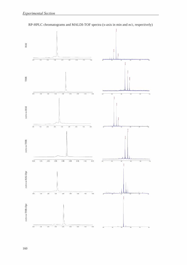
Experimental Section
160
RP-HPLC chromatograms and MALDI-TOF spectra (x-axis in min and m/z, respectively) H
AI
TH
R
retr
o-ᴅ-
HA
I
retr
o-ᴅ-
TH
R
retr
o-ᴅ-
HA
I-D
pr
retr
o-ᴅ-
TH
R-D
pr

Product Characterization
161
Chapter 4 Peptide
HRMS calcd.
[M+H]+
FTMSii
[M+H]+
MALDI-TOF
[M+H]+
Purityjj
(%)
HPLC tRkk
(min)
rD-HAI-Nbz 1052.5173 1052.5158 1052.6 94 3.50
rD-THR-Nbzll 1648.77214 1648.77002 1649.7 92 4.25
AO-rD-HAI 965.50605 965.50641 965.7 93 3.40
AO-rD-THR 1562.76787 1562.76851 1563.0 100 4.33
NHS-PEG-rD-HAI n.a.mm n.a. n.a. 96 4.69
NHS-PEG-rD-THR n.a. n.a. n.a. 96 5.02
ii LTQ-FT Ultra/Synapt HDMS. jj After purification by RP-HPLC. kk Gradient from 0 to 100% CH3CN in 8 min (1 mL/min) using a Sunfire C18 column (150 × 4.6 mm × 5 μm, 100 Å, Waters). ll HRMS calcd. and FTMS as [M]. mm n.a. ≡ not applicable.

Experimental Section
162
RP-HPLC chromatograms and MALDI-TOF spectra (x-axis in min and m/z, respectively) rD
-HA
I-N
bz
rD-T
HR
-Nbz
AO
-rD
-HA
I
AO
-rD
-TH
R
NH
S-P
EG
-rD
-HA
I
NH
S-P
EG
-rD
-TH
R

Product Characterization
163
1H-NMR spectra
NH
S-P
EG
-rD
-HA
IN
HS
-PE
G-r
D-T
HR
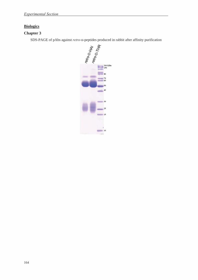
Experimental Section
164
Biologics
Chapter 3 SDS-PAGE of pAbs against retro-ᴅ-peptides produced in rabbit after affinity purification

Product Characterization
165
Chapter 4
HSV-1 bioconjugation and characterization by batch
PEG compound HSV-1 batch Bioconjugation methodnn Characterization
NHS-PEG-MAL 2 NHS-PEG-MAL, 6,000 eq.; 2 h
Cf-Cys 6,000 eq.; 1 h at r.t. on o/n at 4ºC TEM (neg. stain.), DLS,
SDS-PAGE, Fluorescence
NHS-PEG-MAL 2 NHS-PEG-MAL, 50 eq.; 2 h
Cf-Cys 50 eq.; 1h at r.t. and o/n at 4ºC TEM (neg. stain.), DLS,
SDS-PAGE, Fluorescence
NHS-PEG-MAL 3 NHS-PEG-MAL, 2,000 eq.; 2 h
Cf-Cys 2,000 eq.; 4 h TEM (neg. stain.), DLS,
SDS-PAGE, Fluorescence
NHS-PEG-MAL 3 NHS-PEG-MAL, 1 mM; 2 h
Cf-Cys 1 mM; 4 h TEM (neg. stain.), DLS,
SDS-PAGE, Fluorescence
NHS-PEG-biotin 1 NHS-PEG-biotin, 2,000 eq.; 2 h SDS-PAGE
NHS-PEG-biotin 6 NHS-PEG-biotin, 2,000 eq.; 2 h SDS-PAGE, WB
NHS-PEG-biotin 6 NHS-PEG-biotin, 100 eq.; 2 h SDS-PAGE, WB
NHS-PEG-biotin 8 NHS-PEG-biotin, 2,000 eq.; vol. 1/10, 2 h SDS-PAGE, WB DLS, ζ-potential
NHS-PEG-biotin 8 NHS-PEG-biotin, 2,000 eq.; EDC, 2,000 eq.; conc. ×10, 2 h SDS-PAGE, WB
DLS, ζ-potential, ASMS
NHS-PEG-biotin 8 NHS-PEG-biotin, 2,000 eq.; EDC, 2,000 eq.; conc. ×1, 2 h SDS-PAGE, WB DLS, ζ-potential
NHS-PEG-biotin 10 NHS-PEG-biotin, 2,000 eq.; conc. ×10, 2 h Only infectivity
NHS-PEG-biotin 10 NHS-PEG-biotin, 2,000 eq.; EDC, 2,000 eq.; conc. ×10, 2 h Only infectivity
- 11 - MS
NHS-PEG-rD-THR 15 NHS-PEG-rD-THR, 2,000 eq.; conc. ×10, 2 h DLS, ζ-potential
NHS-PEG-rD-THR 19 NHS-PEG-rD-THR, 2,000 eq.; conc. ×10, 2 h DLS, ζ-potential
NHS-PEG-rD-THR 20 NHS-PEG-rD-THR, 2,000 eq.; conc. ×10, 2 h DLS, ζ-potential
NHS-PEG-rD-THR 22 NHS-PEG-rD-THR, 2,000 eq.; conc. ×10, 2 h SDS-PAGE, DLS, ζ-
potential
nn Unless specified, reactions performed at room temperature.


REFERENCES


References
169
(1) Miller, G. On the origin of the nervous system. Science 2009, 325, 24. (2) Nakanishi, N.; Yuan, D.; Jacobs, D. K.; Hartenstein, V. Early development, pattern, and reorganization of the planula nervous system in Aurelia (Cnidaria, Scyphozoa). Dev. Genes Evol. 2008, 218, 511. (3) Martindale, M. Q. The evolution of metazoan axial properties. Nat. Rev. Genet. 2005, 6, 917. (4) Dunn, C. W.; Hejnol, A.; Matus, D. Q.; Pang, K.; Browne, W. E.; Smith, S. A.; Seaver, E.; Rouse, G. W.; Obst, M.; Edgecombe, G. D.; Sorensen, M. V.; Haddock, S. H. D.; Schmidt-Rhaesa, A.; Okusu, A.; Kristensen, R. M.; Wheeler, W. C.; Martindale, M. Q.; Giribet, G. Broad phylogenomic sampling improves resolution of the animal tree of life. Nature 2008, 452, 745. (5) Ryan, J. F.; Pang, K.; Schnitzler, C. E.; Nguyen, A. D.; Moreland, R. T.; Simmons, D. K.; Koch, B. J.; Francis, W. R.; Havlak, P.; Program, N. C. S.; Smith, S. A.; Putnam, N. H.; Haddock, S. H.; Dunn, C. W.; Wolfsberg, T. G.; Mullikin, J. C.; Martindale, M. Q.; Baxevanis, A. D. The genome of the ctenophore Mnemiopsis leidyi and its implications for cell type evolution. Science 2013, 342, 1242592. (6) Moroz, L. L.; Kocot, K. M.; Citarella, M. R.; Dosung, S.; Norekian, T. P.; Povolotskaya, I. S.; Grigorenko, A. P.; Dailey, C.; Berezikov, E.; Buckley, K. M.; Ptitsyn, A.; Reshetov, D.; Mukherjee, K.; Moroz, T. P.; Bobkova, Y.; Yu, F.; Kapitonov, V. V.; Jurka, J.; Bobkov, Y. V.; Swore, J. J.; Girardo, D. O.; Fodor, A.; Gusev, F.; Sanford, R.; Bruders, R.; Kittler, E.; Mills, C. E.; Rast, J. P.; Derelle, R.; Solovyev, V. V.; Kondrashov, F. A.; Swalla, B. J.; Sweedler, J. V.; Rogaev, E. I.; Halanych, K. M.; Kohn, A. B. The ctenophore genome and the evolutionary origins of neural systems. Nature 2014, 510, 109. (7) Nakanishi, N.; Sogabe, S.; Degnan, B. M. Evolutionary origin of gastrulation: Insights from sponge development. BMC Biol. 2014, 12, 26. (8) Solnica-Krezel, L.; Sepich, D. S. Gastrulation: Making and shaping germ layers. Annu. Rev. Cell Dev. Biol. 2012, 28, 687. (9) Lu, C. C.; Brennan, J.; Robertson, E. J. From fertilization to gastrulation: Axis formation in the mouse embryo. Curr. Opin. Genet. Dev. 2001, 11, 384. (10) Wozniak, M. A.; Chen, C. S. Mechanotransduction in development: A growing role for contractility. Nat. Rev. Mol. Cell Biol. 2009, 10, 34. (11) Lowe, C. J.; Wu, M.; Salic, A.; Evans, L.; Lander, E.; Stange-Thomann, N.; Gruber, C. E.; Gerhart, J.; Kirschner, M. Anteroposterior patterning in Hemichordates and the origins of the Chordate nervous system. Cell 2003, 113, 853. (12) Knecht, A. K.; Bronner-Fraser, M. Induction of the neural crest: A multigene process. Nat. Rev. Genet. 2002, 3, 453. (13) Murry, C. E.; Keller, G. Differentiation of embryonic stem cells to clinically relevant populations: Lessons from embryonic development. Cell 2008, 132, 661. (14) Evseenko, D.; Zhu, Y.; Schenke-Layland, K.; Kuo, J.; Latour, B.; Ge, S.; Scholes, J.; Dravid, G.; Li, X.; MacLellan, W. R.; Crooks, G. M. Mapping the first stages of mesoderm commitment during differentiation of human embryonic stem cells. Proc. Natl. Acad. Sci. U. S. A. 2010, 107, 13742. (15) Stemple, D. L. Structure and function of the notochord: An essential organ for chordate development. Development 2005, 132, 2503. (16) Grapin-Botton, A.; Melton, D. A. Endoderm development: From patterning to organogenesis. Trends Genet., 16, 124. (17) Halton, D. W.; Gustafsson, M. K. S. Functional morphology of the platyhelminth nervous system. Parasitology 1996, 113, S47. (18) Telford, M. J. Animal phylogeny. Curr. Biol. 2006, 16, R981. (19) Telford, Maximilian J.; Budd, Graham E.; Philippe, H. Phylogenomic insights into animal evolution. Curr. Biol. 2015, 25, R876. (20) Rankin, C. H. From gene to identified neuron to behaviour in Caenorhabditis elegans. Nat. Rev. Genet. 2002, 3, 622. (21) Jorgensen, E. M.; Mango, S. E. The art and design of genetic screens: Caenorhabditis elegans. Nat. Rev. Genet. 2002, 3, 356. (22) Bishop, N. A.; Guarente, L. Two neurons mediate diet-restriction-induced longevity in C. elegans. Nature 2007, 447, 545. (23) Hobert, O.; Johnston, R. J.; Chang, S. Left-right asymmetry in the nervous system: The Caenorhabditis elegans model. Nat. Rev. Neurosci. 2002, 3, 629. (24) Chatterjee, N.; Sinha, S. Understanding the mind of a worm: hierarchical network structure underlying nervous system function in C. elegans. Prog. Brain Res. 2007, 168, 145. (25) Shubin, N.; Tabin, C.; Carroll, S. Deep homology and the origins of evolutionary novelty. Nature 2009, 457, 818. (26) Benito-Gutiérrez, È.; Arendt, D. CNS evolution: New insight from the mud. Curr. Biol. 2009, 19, R640.

References
170
(27) Mineta, K.; Nakazawa, M.; Cebrià, F.; Ikeo, K.; Agata, K.; Gojobori, T. Origin and evolutionary process of the CNS elucidated by comparative genomics analysis of planarian ESTs. Proc. Natl. Acad. Sci. U. S. A. 2003, 100, 7666. (28) Le Douarin, N. M.; Dupin, E. The neural crest in vertebrate evolution. Curr. Opin. Genet. Dev. 2012, 22, 381. (29) Abbott, N. J. Dynamics of CNS barriers: Evolution, differentiation, and modulation. Cell. Mol. Neurobiol. 2005, 25, 5. (30) Abbott, N. J. In Physiology and Pharmacology of the Blood-Brain Barrier; Bradbury, M. W. B., Ed.; Springer: Berlin-Heidelberg, Germany, 1992; Vol. 103, p 371. (31) Banerjee, S.; Bhat, M. A. Neuron-glial interactions in blood-brain barrier formation. Annu. Rev. Neurosci. 2007, 30, 235. (32) Matter, K.; Balda, M. S. Signalling to and from tight junctions. Nat. Rev. Mol. Cell Biol. 2003, 4, 225. (33) Kniesel, U.; Wolburg, H. Tight junctions of the blood–brain barrier. Cell. Mol. Neurobiol. 2000, 20, 57. (34) Wolburg, H.; Lippoldt, A. Tight junctions of the blood–brain barrier: Development, composition and regulation. Vascul. Pharmacol. 2002, 38, 323. (35) Rubin, L. L.; Staddon, J. M. The cell biology of the blood-brain barrier. Annu. Rev. Neurosci. 1999, 22, 11. (36) Lun, M. P.; Monuki, E. S.; Lehtinen, M. K. Development and functions of the choroid plexus-cerebrospinal fluid system. Nat. Rev. Neurosci. 2015, 16, 445. (37) Vorbrodt, A. W.; Dobrogowska, D. H. Molecular anatomy of intercellular junctions in brain endothelial and epithelial barriers: electron microscopist’s view. Brain Res. Rev. 2003, 42, 221. (38) Brown, P. D.; Davies, S. L.; Speake, T.; Millar, I. D. Molecular mechanisms of cerebrospinal fluid production. Neuroscience 2004, 129, 955. (39) Del Bigio, M. R. The ependyma: A protective barrier between brain and cerebrospinal fluid. Glia 1995, 14, 1. (40) Nabeshima, S.; Reese, T. S.; Landis, D. M. D.; Brightman, M. W. Junctions in the meninges and marginal glia. J. Comp. Neurol. 1975, 164, 127. (41) Saunders, N. R.; Ek, C. J.; Habgood, M. D.; Dziegielewska, K. M. Barriers in the brain: A renaissance? Trends Neurosci. 2008, 31, 279. (42) Gaillard, P. J.; Visser, C. C.; de Boer, A. G. Targeted delivery across the blood–brain barrier. Expert Opin. Drug Deliv. 2005, 2, 299. (43) Pardridge, W. M. The blood-brain barrier: Bottleneck in brain drug development. NeuroRx 2005, 2, 3. (44) Hynynen, K. Ultrasound for drug and gene delivery to the brain. Adv. Drug Del. Rev. 2008, 60, 1209. (45) Obermeier, B.; Daneman, R.; Ransohoff, R. M. Development, maintenance and disruption of the blood-brain barrier. Nat. Med. 2013, 19, 1584. (46) Banks, W. A. From blood-brain barrier to blood-brain interface: New opportunities for CNS drug delivery. Nat. Rev. Drug Discov. 2016, 15, 275. (47) Armulik, A.; Genove, G.; Mae, M.; Nisancioglu, M. H.; Wallgard, E.; Niaudet, C.; He, L.; Norlin, J.; Lindblom, P.; Strittmatter, K.; Johansson, B. R.; Betsholtz, C. Pericytes regulate the blood-brain barrier. Nature 2010, 468, 557. (48) Daneman, R.; Zhou, L.; Kebede, A. A.; Barres, B. A. Pericytes are required for blood-brain barrier integrity during embryogenesis. Nature 2010, 468, 562. (49) Abbott, N. J.; Ronnback, L.; Hansson, E. Astrocyte-endothelial interactions at the blood-brain barrier. Nat. Rev. Neurosci. 2006, 7, 41. (50) Persidsky, Y.; Ramirez, S. H.; Haorah, J.; Kanmogne, G. D. Blood–brain barrier: Structural components and function under physiologic and pathologic conditions. J. Neuroimmune Pharmacol. 2006, 1, 223. (51) Hawkins, B. T.; Davis, T. P. The blood-brain barrier/neurovascular unit in health and disease. Pharmacol. Rev. 2005, 57, 173. (52) Huber, J. D.; Egleton, R. D.; Davis, T. P. Molecular physiology and pathophysiology of tight junctions in the blood–brain barrier. Trends Neurosci. 2001, 24, 719. (53) Harris, T. J. C.; Tepass, U. Adherens junctions: From molecules to morphogenesis. Nat. Rev. Mol. Cell Biol. 2010, 11, 502. (54) Patabendige, A.; Skinner, R. A.; Morgan, L.; Joan Abbott, N. A detailed method for preparation of a functional and flexible blood–brain barrier model using porcine brain endothelial cells. Brain Res. 2013, 1521, 16.

References
171
(55) Chaitali, G.; Vikram, P.; Jorge, G.-M.; Damir, J.; Nicola, M. Blood-brain barrier P450 enzymes and multidrug transporters in drug resistance: A synergistic role in neurological diseases. Curr. Drug Metab. 2011, 12, 742. (56) Saunders, N. R.; Dreifuss, J. J.; Dziegielewska, K. M.; Johansson, P. A.; Habgood, M. D.; Mollgard, K.; Bauer, H. C. The rights and wrongs of blood-brain barrier permeability studies: A walk through 100 years of history. Front. Neurosci. 2014, 8, 404. (57) Lipinski, C. A.; Lombardo, F.; Dominy, B. W.; Feeney, P. J. Experimental and computational approaches to estimate solubility and permeability in drug discovery and development settings. Adv. Drug Del. Rev. 2012, 64, Supplement, 4. (58) Davis, J. T.; Okunola, O.; Quesada, R. Recent advances in the transmembrane transport of anions. Chem. Soc. Rev. 2010, 39, 3843. (59) Wei, L. Adsorptive-mediated brain delivery systems. Curr. Pharm. Biotechnol. 2012, 13, 2340. (60) Qian, Z. M.; Li, H.; Sun, H.; Ho, K. Targeted drug delivery via the transferrin receptor-mediated endocytosis pathway. Pharmacol. Rev. 2002, 54, 561. (61) Mayor, S.; Pagano, R. E. Pathways of clathrin-independent endocytosis. Nat. Rev. Mol. Cell Biol. 2007, 8, 603. (62) Bareford, L. M.; Swaan, P. W. Endocytic mechanisms for targeted drug delivery. Adv. Drug Del. Rev. 2007, 59, 748. (63) May, R. C.; Machesky, L. M. Phagocytosis and the actin cytoskeleton. J. Cell Sci. 2001, 114, 1061. (64) Özen, I.; Deierborg, T.; Miharada, K.; Padel, T.; Englund, E.; Genové, G.; Paul, G. Brain pericytes acquire a microglial phenotype after stroke. Acta Neuropathol. 2014, 128, 381. (65) Balabanov, R.; Washington, R.; Wagnerova, J.; Dore-Duffy, P. CNS microvascular pericytes express macrophage-like function, cell surface integrin αM, and macrophage marker ED-2. Microvasc. Res. 1996, 52, 127. (66) Tomás-Camardiel, M.; Venero, J. L.; Herrera, A. J.; De Pablos, R. M.; Pintor-Toro, J. A.; Machado, A.; Cano, J. Blood–brain barrier disruption highly induces aquaporin-4 mRNA and protein in perivascular and parenchymal astrocytes: Protective effect by estradiol treatment in ovariectomized animals. J. Neurosci. Res. 2005, 80, 235. (67) Pratten, M. K.; Lloyd, J. B. Pinocytosis and phagocytosis: The effect of size of a particulate substrate on its mode of capture by rat peritoneal macrophages cultured in vitro. BBA-Gen. Subjects 1986, 881, 307. (68) Merrifield, C. J.; Moss, S. E.; Ballestrem, C.; Imhof, B. A.; Giese, G.; Wunderlich, I.; Almers, W. Endocytic vesicles move at the tips of actin tails in cultured mast cells. Nat. Cell Biol. 1999, 1, 72. (69) Lajoie, J. M.; Shusta, E. V. Targeting receptor-mediated transport for delivery of biologics across the blood-brain barrier. Annu. Rev. Pharmacool. Toxicol. 2015, 55, 613. (70) McMahon, H. T.; Boucrot, E. Molecular mechanism and physiological functions of clathrin-mediated endocytosis. Nat. Rev. Mol. Cell Biol. 2011, 12, 517. (71) Greenwood, J.; Heasman, S. J.; Alvarez, J. I.; Prat, A.; Lyck, R.; Engelhardt, B. Review: Leucocyte–endothelial cell crosstalk at the blood–brain barrier: A prerequisite for successful immune cell entry to the brain. Neuropathol. Appl. Neurobiol. 2011, 37, 24. (72) Pachter, J. S.; de Vries, H. E.; Fabry, Z. The blood-brain barrier and its role in immune privilege in the central nervous system. J. Neuropathol. Exp. Neurol. 2003, 62, 593. (73) Engelhardt, B.; Ransohoff, R. M. Capture, crawl, cross: The T cell code to breach the blood–brain barriers. Trends Immunol. 2012, 33, 579. (74) Del Maschio, A.; De Luigi, A.; Martin-Padura, I.; Brockhaus, M.; Bartfai, T.; Fruscella, P.; Adorini, L.; Martino, G.; Furlan, R.; De Simoni, M. G.; Dejana, E. Leukocyte recruitment in the cerebrospinal fluid of mice with experimental meningitis is inhibited by an antibody to junctional adhesion molecule (Jam). J. Exp. Med. 1999, 190, 1351. (75) Millan, J.; Hewlett, L.; Glyn, M.; Toomre, D.; Clark, P.; Ridley, A. J. Lymphocyte transcellular migration occurs through recruitment of endothelial ICAM-1 to caveola- and F-actin-rich domains. Nat. Cell Biol. 2006, 8, 113. (76) Miner, J. J.; Diamond, M. S. Mechanisms of restriction of viral neuroinvasion at the blood–brain barrier. Curr. Opin. Immunol. 2016, 38, 18. (77) Kim, K. S. Mechanisms of microbial traversal of the blood-brain barrier. Nat. Rev. Micro. 2008, 6, 625. (78) Ley, K.; Laudanna, C.; Cybulsky, M. I.; Nourshargh, S. Getting to the site of inflammation: The leukocyte adhesion cascade updated. Nat. Rev. Immunol. 2007, 7, 678. (79) Gustavsson, A.; Svensson, M.; Jacobi, F.; Allgulander, C.; Alonso, J.; Beghi, E.; Dodel, R.; Ekman, M.; Faravelli, C.; Fratiglioni, L.; Gannon, B.; Jones, D. H.; Jennum, P.; Jordanova, A.; Jönsson, L.; Karampampa, K.; Knapp, M.; Kobelt, G.; Kurth, T.; Lieb, R.; Linde, M.; Ljungcrantz, C.; Maercker, A.; Melin, B.; Moscarelli, M.; Musayev, A.; Norwood, F.; Preisig, M.; Pugliatti, M.; Rehm, J.; Salvador-Carulla,

References
172
L.; Schlehofer, B.; Simon, R.; Steinhausen, H.-C.; Stovner, L. J.; Vallat, J.-M.; den Bergh, P. V.; van Os, J.; Vos, P.; Xu, W.; Wittchen, H.-U.; Jönsson, B.; Olesen, J. Cost of disorders of the brain in Europe 2010. Eur. Neuropsychopharmacol. 2011, 21, 718. (80) Olesen, J.; Gustavsson, A.; Svensson, M.; Wittchen, H. U.; Jönsson, B.; study group, C.; European Brain, C. The economic cost of brain disorders in Europe. Eur. J. Neurol. 2012, 19, 155. (81) DiLuca, M.; Olesen, J. The cost of brain diseases: A burden or a challenge? Neuron 2014, 82, 1205. (82) Merrifield, R. B. Solid phase peptide synthesis. I. The synthesis of a tetrapeptide. J. Am. Chem. Soc. 1963, 85, 2149. (83) Getz, J. A.; Rice, J. J.; Daugherty, P. S. Protease-resistant peptide ligands from a knottin scaffold library. ACS Chem. Biol. 2011, 6, 837. (84) Prades, R.; Oller-Salvia, B.; Schwarzmaier, S. M.; Selva, J.; Moros, M.; Balbi, M.; Grazu, V.; de La Fuente, J. M.; Egea, G.; Plesnila, N.; Teixido, M.; Giralt, E. Applying the retro-enantio approach to obtain a peptide capable of overcoming the blood-brain barrier. Angew. Chem. Int. Ed. 2015, 54, 3967. (85) Kastin, A. J.; Akerstrom, V. Nonsaturable entry of neuropeptide Y into brain. Am. J. Physiol. Endocrinol. Metab. 1999, 276, E479. (86) Kastin, A. J.; Akerstrom, V. Orexin A but not Orexin B rapidly enters brain from blood by simple diffusion. J. Pharmacol. Exp. Ther. 1999, 289, 219. (87) Kannan, R.; Kuhlenkamp, J. F.; Jeandidier, E.; Trinh, H.; Ookhtens, M.; Kaplowitz, N. Evidence for carrier-mediated transport of glutathione across the blood-brain barrier in the rat. J. Clin. Invest. 1990, 85, 2009. (88) Bachhawat, A. K.; Thakur, A.; Kaur, J.; Zulkifli, M. Glutathione transporters. BBA-Gen. Subjects 2013, 1830, 3154. (89) Banks, W. A.; Kastin, A. J.; Fischman, A. J.; Coy, D. H.; Strauss, S. L. Carrier-mediated transport of enkephalins and N-Tyr-MIF-1 across blood-brain barrier. Am. J. Physiol. Endocrinol. Metab. 1986, 251, E477. (90) Barrera, C. M.; Banks, W. A.; Kastin, A. J. Passage of Tyr-MIF-1 from blood to brain. Brain Res. Bull. 1989, 23, 439. (91) Banks, W. A.; Kastin, A. J. Peptide transport systems for opiates across the blood-brain barrier. Am. J. Physiol. Endocrinol. Metab. 1990, 259, E1. (92) Drin, G.; Rousselle, C.; Scherrmann, J.-M.; Rees, A. R.; Temsamani, J. Peptide delivery to the brain via adsorptive-mediated endocytosis: Advances with SynB vectors. AAPS PharmSci 2002, 4, 61. (93) Frankel, A. D.; Pabo, C. O. Cellular uptake of the tat protein from human immunodeficiency virus. Cell 1988, 55, 1189. (94) Vivès, E.; Brodin, P.; Lebleu, B. A truncated HIV-1 Tat protein basic domain rapidly translocates through the plasma membrane and accumulates in the cell nucleus. J. Biol. Chem. 1997, 272, 16010. (95) Hervé, F.; Ghinea, N.; Scherrmann, J.-M. CNS delivery via adsorptive transcytosis. The AAPS Journal 2008, 10, 455. (96) Ruan, G.; Agrawal, A.; Marcus, A. I.; Nie, S. Imaging and tracking of Tat peptide-conjugated quantum dots in living cells: New insights into nanoparticle uptake, intracellular transport, and vesicle shedding. J. Am. Chem. Soc. 2007, 129, 14759. (97) Derossi, D.; Joliot, A. H.; Chassaing, G.; Prochiantz, A. The third helix of the Antennapedia homeodomain translocates through biological membranes. J. Biol. Chem. 1994, 269, 10444. (98) Derossi, D.; Calvet, S.; Trembleau, A.; Brunissen, A.; Chassaing, G.; Prochiantz, A. Cell Internalization of the third helix of the Antennapedia homeodomain is receptor-independent. J. Biol. Chem. 1996, 271, 18188. (99) Boisguérin, P.; Deshayes, S.; Gait, M. J.; O'Donovan, L.; Godfrey, C.; Betts, C. A.; Wood, M. J. A.; Lebleu, B. Delivery of therapeutic oligonucleotides with cell penetrating peptides. Adv. Drug Del. Rev. 2015, 87, 52. (100) Koren, E.; Torchilin, V. P. Cell-penetrating peptides: Breaking through to the other side. Trends Mol. Med. 2012, 18, 385. (101) Milletti, F. Cell-penetrating peptides: classes, origin, and current landscape. Drug Discov. Today 2012, 17, 850. (102) Hussain, M. M.; Strickland, D. K.; Bakillah, A. The mammalian low-density lipoprotein receptor family. Annu. Rev. Nutr. 1999, 19, 141. (103) Zensi, A.; Begley, D.; Pontikis, C.; Legros, C.; Mihoreanu, L.; Wagner, S.; Büchel, C.; von Briesen, H.; Kreuter, J. Albumin nanoparticles targeted with Apo E enter the CNS by transcytosis and are delivered to neurones. J. Control. Release 2009, 137, 78. (104) Re, F.; Cambianica, I.; Sesana, S.; Salvati, E.; Cagnotto, A.; Salmona, M.; Couraud, P.-O.; Moghimi, S. M.; Masserini, M.; Sancini, G. Functionalization with ApoE-derived peptides enhances the interaction with

References
173
brain capillary endothelial cells of nanoliposomes binding amyloid-beta peptide. J. Biotechnol. 2011, 156, 341. (105) Wang, D.; El-Amouri, S. S.; Dai, M.; Kuan, C.-Y.; Hui, D. Y.; Brady, R. O.; Pan, D. Engineering a lysosomal enzyme with a derivative of receptor-binding domain of apoE enables delivery across the blood–brain barrier. Proc. Natl. Acad. Sci. U. S. A. 2013, 110, 2999. (106) Agnello, V.; Ábel, G.; Elfahal, M.; Knight, G. B.; Zhang, Q.-X. Hepatitis C virus and other Flaviviridae viruses enter cells via low density lipoprotein receptor. Proc. Natl. Acad. Sci. U. S. A. 1999, 96, 12766. (107) Abbruscato, T. J.; Lopez, S. P.; Mark, K. S.; Hawkins, B. T.; Davis, T. P. Nicotine and cotinine modulate cerebral microvascular permeability and protein expression of ZO-1 through nicotinic acetylcholine receptors expressed on brain endothelial cells. J. Pharm. Sci. 2002, 91, 2525. (108) Zhan, C.; Li, B.; Hu, L.; Wei, X.; Feng, L.; Fu, W.; Lu, W. Micelle-based brain-targeted drug delivery enabled by a nicotine acetylcholine receptor ligand. Angew. Chem. Int. Ed. 2011, 50, 5482. (109) Schnell, M. J.; McGettigan, J. P.; Wirblich, C.; Papaneri, A. The cell biology of rabies virus: Using stealth to reach the brain. Nat. Rev. Micro. 2010, 8, 51. (110) Kumar, P.; Wu, H.; McBride, J. L.; Jung, K.-E.; Hee Kim, M.; Davidson, B. L.; Kyung Lee, S.; Shankar, P.; Manjunath, N. Transvascular delivery of small interfering RNA to the central nervous system. Nature 2007, 448, 39. (111) Liu, Y.; Huang, R.; Han, L.; Ke, W.; Shao, K.; Ye, L.; Lou, J.; Jiang, C. Brain-targeting gene delivery and cellular internalization mechanisms for modified rabies virus glycoprotein RVG29 nanoparticles. Biomaterials 2009, 30, 4195. (112) Dautry-Varsat, A.; Ciechanover, A.; Lodish, H. F. pH and the recycling of transferrin during receptor-mediated endocytosis. Proc. Natl. Acad. Sci. U. S. A. 1983, 80, 2258. (113) Aisen, P. Entry of iron into cells: A new role for the transferrin receptor in modulating iron release from transferrin. Ann. Neurol. 1992, 32, S62. (114) Jefferies, W. A.; Brandon, M. R.; Hunt, S. V.; Williams, A. F.; Gatter, K. C.; Mason, D. Y. Transferrin receptor on endothelium of brain capillaries. Nature 1984, 312, 162. (115) Rouault, T. A. Iron metabolism in the CNS: Implications for neurodegenerative diseases. Nat. Rev. Neurosci. 2013, 14, 551. (116) Moos, T.; Morgan, E. H. Transferrin and transferrin receptor function in brain barrier systems. Cell. Mol. Neurobiol. 2000, 20, 77. (117) Wiley, D. T.; Webster, P.; Gale, A.; Davis, M. E. Transcytosis and brain uptake of transferrin-containing nanoparticles by tuning avidity to transferrin receptor. Proc. Natl. Acad. Sci. U. S. A. 2013, 110, 8662. (118) Thorstensen, K.; Romslo, I. The role of transferrin in the mechanism of cellular iron uptake. Biochem. J. 1990, 271, 1. (119) Pardridge, W. M. Drug and gene targeting to the brain with molecular trojan horses. Nat. Rev. Drug Discov. 2002, 1, 131. (120) Pardridge, W. M. Molecular Trojan horses for blood–brain barrier drug delivery. Curr. Opin. Pharmacol. 2006, 6, 494. (121) Pardridge, W. M. Blood–brain barrier drug delivery of IgG fusion proteins with a transferrin receptor monoclonal antibody. Expert Opin. Drug Deliv. 2015, 12, 207. (122) Afergan, E.; Epstein, H.; Dahan, R.; Koroukhov, N.; Rohekar, K.; Danenberg, H. D.; Golomb, G. Delivery of serotonin to the brain by monocytes following phagocytosis of liposomes. J. Control. Release 2008, 132, 84. (123) Prades, R.; Guerrero, S.; Araya, E.; Molina, C.; Salas, E.; Zurita, E.; Selva, J.; Egea, G.; López-Iglesias, C.; Teixidó, M.; Kogan, M. J.; Giralt, E. Delivery of gold nanoparticles to the brain by conjugation with a peptide that recognizes the transferrin receptor. Biomaterials 2012, 33, 7194. (124) Wei, X.; Zhan, C.; Chen, X.; Hou, J.; Xie, C.; Lu, W. Retro-inverso isomer of Angiopep-2: A stable d-peptide ligand inspires brain-targeted drug delivery. Mol. Pharm. 2014, 11, 3261. (125) Gao, H.; Zhang, S.; Cao, S.; Yang, Z.; Pang, Z.; Jiang, X. Angiopep-2 and activatable cell-penetrating peptide dual-functionalized nanoparticles for systemic glioma-targeting delivery. Mol. Pharm. 2014, 11, 2755. (126) Teixidó, M.; Zurita, E.; Malakoutikhah, M.; Tarragó, T.; Giralt, E. Diketopiperazines as a tool for the study of transport across the blood−brain barrier (BBB) and their potential use as BBB-shuttles. J. Am. Chem. Soc. 2007, 129, 11802. (127) Arranz-Gibert, P.; Guixer, B.; Malakoutikhah, M.; Muttenthaler, M.; Guzmán, F.; Teixidó, M.; Giralt, E. Lipid bilayer crossing—The gate of symmetry. Water-soluble phenylproline-based blood-brain barrier shuttles. J. Am. Chem. Soc. 2015, 137, 7357.

References
174
(128) Abbott, N. J. Blood–brain barrier structure and function and the challenges for CNS drug delivery. J. Inherited Metab. Dis. 2013, 36, 437. (129) Pardridge, W. M. Blood–brain barrier delivery. Drug Discov. Today 2007, 12, 54. (130) Träuble, H. The movement of molecules across lipid membranes: A molecular theory. J. Membr. Biol. 1971, 4, 193. (131) Lieb, W.; Stein, W. Non-stokesian nature of transverse diffusion within human red cell membranes. J. Membr. Biol. 1986, 92, 111. (132) Pardridge, W. M. CNS drug design based on principles of blood-brain barrier transport. J. Neurochem. 1998, 70, 1781. (133) Wager, T. T.; Hou, X.; Verhoest, P. R.; Villalobos, A. Moving beyond rules: The development of a central nervous system multiparameter optimization (CNS MPO) approach to enable alignment of druglike properties. ACS Chem. Neurosci. 2010, 1, 435. (134) Spector, M. S.; Selinger, J. V.; Singh, A.; Rodriguez, J. M.; Price, R. R.; Schnur, J. M. Controlling the morphology of chiral lipid tubules. Langmuir 1998, 14, 3493. (135) Selinger, J. V.; Schnur, J. M. Theory of chiral lipid tubules. Phys. Rev. Lett. 1993, 71, 4091. (136) Lalitha, S.; Sampath Kumar, A.; Stine, K. J.; Covey, D. F. Chirality in membranes: First evidence that enantioselective interactions between cholesterol and cell membrane lipids can be a determinant of membrane physical properties. J. Supramol. Chem. 2001, 1, 53. (137) Cruciani, O.; Borocci, S.; Lamanna, R.; Mancini, G.; Segre, A. L. Chiral recognition of dipeptides in phosphatidylcholine aggregates. Tetrahedron: Asymmetry 2006, 17, 2731. (138) Bombelli, C.; Borocci, S.; Cruciani, O.; Mancini, G.; Monti, D.; Segre, A. L.; Sorrenti, A.; Venanzi, M. Chiral recognition of dipeptides in bio-membrane models: The role of amphiphile hydrophobic chains. Tetrahedron: Asymmetry 2008, 19, 124. (139) Bombelli, C.; Borocci, S.; Lupi, F.; Mancini, G.; Mannina, L.; Segre, A. L.; Viel, S. Chiral recognition of dipeptides in a biomembrane model. J. Am. Chem. Soc. 2004, 126, 13354. (140) Chikhale, E.; Ng, K.-Y.; Burton, P.; Borchardt, R. Hydrogen bonding potential as a determinant of the in vitro and in situ blood–brain barrier permeability of peptides. Pharm. Res. 1994, 11, 412. (141) Malakoutikhah, M.; Teixidó, M.; Giralt, E. Toward an optimal blood−brain barrier shuttle by synthesis and evaluation of peptide libraries. J. Med. Chem. 2008, 51, 4881. (142) Malakoutikhah, M.; Prades, R.; Teixidó, M.; Giralt, E. N-methyl phenylalanine-rich peptides as highly versatile blood−brain barrier shuttles. J. Med. Chem. 2010, 53, 2354. (143) Malakoutikhah, M.; Guixer, B.; Arranz-Gibert, P.; Teixido, M.; Giralt, E. 'A la carte' peptide shuttles: tools to increase their passage across the blood-brain barrier. ChemMedChem 2014, 9, 1594. (144) Di, L.; Kerns, E. H.; Fan, K.; McConnell, O. J.; Carter, G. T. High throughput artificial membrane permeability assay for blood–brain barrier. Eur. J. Med. Chem. 2003, 38, 223. (145) Chiang, Y.-C.; Lin, Y.-J.; Horng, J.-C. Stereoelectronic effects on the transition barrier of polyproline conformational interconversion. Protein Sci. 2009, 18, 1967. (146) MacArthur, M. W.; Thornton, J. M. Influence of proline residues on protein conformation. J. Mol. Biol. 1991, 218, 397. (147) Pujals, S.; Giralt, E. Proline-rich, amphipathic cell-penetrating peptides. Adv. Drug Del. Rev. 2008, 60, 473. (148) Ghose, A. K.; Herbertz, T.; Hudkins, R. L.; Dorsey, B. D.; Mallamo, J. P. Knowledge-based, central nervous system (CNS) lead selection and lead optimization for CNS drug discovery. ACS Chem. Neurosci. 2011, 3, 50. (149) Kansy, M.; Senner, F.; Gubernator, K. Physicochemical high throughput screening: Parallel artificial membrane permeation assay in the description of passive absorption processes. J. Med. Chem. 1998, 41, 1007. (150) Katzenschlager, R.; Poewe, W. Parkinson disease: Intestinal levodopa infusion in PD - The first randomized trial. Nat. Rev. Neurol. 2014, 10, 128. (151) Olanow, C. W.; Obeso, J. A.; Stocchi, F. Continuous dopamine-receptor treatment of Parkinson's disease: scientific rationale and clinical implications. Lancet Neurol. 2006, 5, 677. (152) Vossler, D. G.; Morris Iii, G. L.; Harden, C. L.; Montouris, G.; Faught, E.; Kanner, A. M.; Fix, A.; French, J. A. Tiagabine in clinical practice: Effects on seizure control and behavior. Epilepsy Behav. 2013, 28, 211. (153) Del Amo, E. M.; Urtti, A.; Yliperttula, M. Pharmacokinetic role of L-type amino acid transporters LAT1 and LAT2. Eur. J. Pharm. Sci. 2008, 35, 161. (154) Tomlinson, C. L.; Stowe, R.; Patel, S.; Rick, C.; Gray, R.; Clarke, C. E. Systematic review of levodopa dose equivalency reporting in Parkinson's disease. Mov. Disord. 2010, 25, 2649. (155) Cannazza, G.; Di Stefano, A.; Mosciatti, B.; Braghiroli, D.; Baraldi, M.; Pinnen, F.; Sozio, P.; Benatti, C.; Parenti, C. Detection of levodopa, dopamine and its metabolites in rat striatum dialysates

References
175
following peripheral administration of L-DOPA prodrugs by mean of HPLC–EC. J. Pharm. Biomed. Anal. 2005, 36, 1079. (156) Di Stefano, A.; Carafa, M.; Sozio, P.; Pinnen, F.; Braghiroli, D.; Orlando, G.; Cannazza, G.; Ricciutelli, M.; Marianecci, C.; Santucci, E. Evaluation of rat striatal L-Dopa and DA concentration after intraperitoneal administration of l-dopa prodrugs in liposomal formulations. J. Control. Release 2004, 99, 293. (157) Hawkins, R. A.; Mokashi, A.; Simpson, I. A. An active transport system in the blood–brain barrier may reduce levodopa availability. Exp. Neurol. 2005, 195, 267. (158) Di Stefano, A.; Sozio, P.; Cerasa, L. Antiparkinson prodrugs. Molecules 2008, 13, 46. (159) Pinnen, F.; Cacciatore, I.; Cornacchia, C.; Sozio, P.; Iannitelli, A.; Costa, M.; Pecci, L.; Nasuti, C.; Cantalamessa, F.; Di Stefano, A. Synthesis and study of L-Dopa−glutathione codrugs as new anti-Parkinson agents with free radical scavenging properties. J. Med. Chem. 2007, 50, 2506. (160) Barrett-Jolley, R. Nipecotic acid directly activates GABAA-like ion channels. Br. J. Pharmacol. 2001, 133, 673. (161) Ali, F. E.; Bondinell, W. E.; Dandridge, P. A.; Frazee, J. S.; Garvey, E.; Girard, G. R.; Kaiser, C.; Ku, T. W.; Lafferty, J. J.; Moonsammy, G. I.; et al. Orally active and potent inhibitors of gamma-aminobutyric acid uptake. J. Med. Chem. 1985, 28, 653. (162) Krogsgaard-Larsen, P.; Johnston, G. A. R. Inhibition of GABA uptake in rat brain slices by nipecotic acid, various isoxazoles and related compounds. J. Neurochem. 1975, 25, 797. (163) Flamant-Robin, C.; Wang, Q.; Chiaroni, A.; Sasaki, N. A. An efficient method for the stereoselective synthesis of cis-3-substituted prolines: Conformationally constrained α-amino acids. Tetrahedron 2002, 58, 10475. (164) Belokon, Y. N.; Bulychev, A. G.; Pavlov, V. A.; Fedorova, E. B.; Tsyryapkin, V. A.; Bakhmutov, V. A.; Belikov, V. M. Synthesis of enantio- and diastereoiso-merically pure substituted prolines via condensation of glycine with olefins activated by a carbonyl group. J. Chem. Soc., Perkin Trans. 1 1988, 2075. (165) Fields, G. B.; Noble, R. L. Solid phase peptide synthesis utilizing 9-fluorenylmethoxycarbonyl amino acids. Int. J. Pept. Protein Res. 1990, 35, 161. (166) Carpino, L. A.; El-Faham, A.; Minor, C. A.; Albericio, F. Advantageous applications of azabenzotriazole (triazolopyridine)-based coupling reagents to solid-phase peptide synthesis. J. Chem. Soc., Chem. Commun. 1994, 201. (167) Houghten, R. A. General method for the rapid solid-phase synthesis of large numbers of peptides: Specificity of antigen-antibody interaction at the level of individual amino acids. Proc. Natl. Acad. Sci. U. S. A. 1985, 82, 5131. (168) Houghten, R. A.; Pinilla, C.; Blondelle, S. E.; Appel, J. R.; Dooley, C. T.; Cuervo, J. H. Generation and use of synthetic peptide combinatorial libraries for basic research and drug discovery. Nature 1991, 354, 84. (169) Houghten, R. A. Parallel array and mixture-based synthetic combinatorial chemistry: Tools for the next millennium. Annu. Rev. Pharmacool. Toxicol. 2000, 40, 273. (170) Bochicchio, B.; Tamburro, A. M. Polyproline II structure in proteins: Identification by chiroptical spectroscopies, stability, and functions. Chirality 2002, 14, 782. (171) Goodman, M.; Chorev, M. On the concept of linear modified retro-peptide structures. Acc. Chem. Res. 1979, 12, 1. (172) Freidinger, R. M.; Veber, D. F. Peptides and their retro enantiomers are topologically nonidentical. J. Am. Chem. Soc. 1979, 101, 6129. (173) Li, C.; Pazgier, M.; Li, J.; Li, C.; Liu, M.; Zou, G.; Li, Z.; Chen, J.; Tarasov, S. G.; Lu, W.-Y.; Lu, W. Limitations of peptide retro-inverso isomerization in molecular mimicry. J. Biol. Chem. 2010, 285, 19572. (174) Spector, A. A.; Yorek, M. A. Membrane lipid composition and cellular function. J. Lipid Res. 1985, 26, 1015. (175) Lingwood, D.; Simons, K. Lipid rafts as a membrane-organizing principle. Science 2010, 327, 46. (176) Yechiel, E.; Barenholz, Y. Relationships between membrane lipid composition and biological properties of rat myocytes. Effects of aging and manipulation of lipid composition. J. Biol. Chem. 1985, 260, 9123. (177) Boesze-Battaglia, K.; Schimmel, R. Cell membrane lipid composition and distribution: implications for cell function and lessons learned from photoreceptors and platelets. J. Exp. Biol. 1997, 200, 2927. (178) Edidin, M. The state of lipid rafts: From model membranes to cells. Annu. Rev. Biophys. Biomol. Struct. 2003, 32, 257. (179) Simons, K.; Ikonen, E. Functional rafts in cell membranes. Nature 1997, 387, 569. (180) Kummerow, F. A. Modification of cell membrane composition by dietary lipids and its implications for atherosclerosis. Ann. N.Y. Acad. Sci. 1983, 414, 29.

References
176
(181) SoOderberg, M.; Edlund, C.; Alafuzoff, I.; Kristensson, K.; Dallner, G. Lipid composition in different regions of the brain in Alzheimer's disease/senile dementia of Alzheimer's type. J. Neurochem. 1992, 59, 1646. (182) Norton, W. T.; Abe, T.; Poduslo, S. E.; DeVries, G. H. The lipid composition of isolated brain cells and axons. J. Neurosci. Res. 1975, 1, 57. (183) Santos, C. R.; Schulze, A. Lipid metabolism in cancer. FEBS J. 2012, 279, 2610. (184) Maxfield, F. R.; Tabas, I. Role of cholesterol and lipid organization in disease. Nature 2005, 438, 612. (185) van Meer, G.; Voelker, D. R.; Feigenson, G. W. Membrane lipids: Where they are and how they behave. Nat. Rev. Mol. Cell Biol. 2008, 9, 112. (186) Li, Y. C.; Park, M. J.; Ye, S.-K.; Kim, C.-W.; Kim, Y.-N. Elevated levels of cholesterol-rich lipid rafts in cancer cells are correlated with apoptosis sensitivity induced by cholesterol-depleting agents. Am. J. Pathol. 2006, 168, 1107. (187) Oller-Salvia, B.; Sanchez-Navarro, M.; Giralt, E.; Teixido, M. Blood-brain barrier shuttle peptides: An emerging paradigm for brain delivery. Chem. Soc. Rev. 2016, 45, 4690. (188) Lee, J. H.; Engler, J. A.; Collawn, J. F.; Moore, B. A. Receptor mediated uptake of peptides that bind the human transferrin receptor. Eur. J. Biochem. 2001, 268, 2004. (189) Zong, T.; Mei, L.; Gao, H.; Cai, W.; Zhu, P.; Shi, K.; Chen, J.; Wang, Y.; Gao, F.; He, Q. Synergistic dual-ligand doxorubicin liposomes improve targeting and therapeutic efficacy of brain glioma in animals. Mol. Pharm. 2014, 11, 2346. (190) Du, W.; Fan, Y.; Zheng, N.; He, B.; Yuan, L.; Zhang, H.; Wang, X.; Wang, J.; Zhang, X.; Zhang, Q. Transferrin receptor specific nanocarriers conjugated with functional 7peptide for oral drug delivery. Biomaterials 2013, 34, 794. (191) Kuang, Y.; Jiang, X.; Zhang, Y.; Lu, Y.; Ma, H.; Guo, Y.; Zhang, Y.; An, S.; Li, J.; Liu, L.; Wu, Y.; Liang, J.; Jiang, C. Dual functional peptide-driven nanoparticles for highly efficient glioma-targeting and drug codelivery. Mol. Pharm. 2016, 13, 1599. (192) Han, L.; Huang, R.; Liu, S.; Huang, S.; Jiang, C. Peptide-conjugated PAMAM for targeted doxorubicin delivery to transferrin receptor overexpressed tumors. Mol. Pharm. 2010, 7, 2156. (193) Prades, R. Towards a universal blood-brain barrier shuttle: Protease-resistant peptide shuttles with capacity to deliver cargos into the central nervous system, Universitat de Barcelona, 2012. (194) Aday, S.; Cecchelli, R.; Hallier-Vanuxeem, D.; Dehouck, M.; Ferreira, L. Stem cell-based human blood–brain barrier models for drug discovery and delivery. Trends Biotechnol. 2016, 34, 382. (195) Abbott, N. J.; Dolman, D. E. M.; Yusof, S. R.; Reichel, A. In Drug Delivery to the Brain; Hammarlund-Udenaes, M., de Lange, E. C. M., Thorne, R. G., Eds.; Springer: New York, NY, USA, 2014; Vol. 10, p 163. (196) Mandal, H. S.; Kraatz, H.-B. Effect of the surface curvature on the secondary structure of peptides adsorbed on nanoparticles. J. Am. Chem. Soc. 2007, 129, 6356. (197) Khlebtsov, N.; Dykman, L. Biodistribution and toxicity of engineered gold nanoparticles: a review of in vitro and in vivo studies. Chem. Soc. Rev. 2011, 40, 1647. (198) Fletcher, M. D.; Campbell, M. M. Partially modified retro-inverso peptides: Development, synthesis, and conformational behavior. Chem. Rev. 1998, 98, 763. (199) Chatterjee, J.; Rechenmacher, F.; Kessler, H. N-methylation of peptides and proteins: An important element for modulating biological functions. Angew. Chem. Int. Ed. 2013, 52, 254. (200) Miller, S. C.; Scanlan, T. S. Site-selective N-methylation of peptides on solid support. J. Am. Chem. Soc. 1997, 119, 2301. (201) Artursson, P.; Palm, K.; Luthman, K. Caco-2 monolayers in experimental and theoretical predictions of drug transport. Adv. Drug Del. Rev. 2012, 64, Supplement, 280. (202) Patani, G. A.; LaVoie, E. J. Bioisosterism: A rational approach in drug design. Chem. Rev. 1996, 96, 3147. (203) Cramer, R. D.; Clark, R. D.; Patterson, D. E.; Ferguson, A. M. Bioisosterism as a molecular diversity descriptor: Steric fields of single “topomeric” conformers. J. Med. Chem. 1996, 39, 3060. (204) Leach, A. R.; Gillet, V. J.; Lewis, R. A.; Taylor, R. Three-dimensional pharmacophore methods in drug discovery. J. Med. Chem. 2010, 53, 539. (205) Markt, P.; Feldmann, C.; Rollinger, J. M.; Raduner, S.; Schuster, D.; Kirchmair, J.; Distinto, S.; Spitzer, G. M.; Wolber, G.; Laggner, C.; Altmann, K.-H.; Langer, T.; Gertsch, J. Discovery of novel CB2 receptor ligands by a pharmacophore-based virtual screening workflow. J. Med. Chem. 2009, 52, 369. (206) Cecchelli, R.; Berezowski, V.; Lundquist, S.; Culot, M.; Renftel, M.; Dehouck, M.-P.; Fenart, L. Modelling of the blood-brain barrier in drug discovery and development. Nat. Rev. Drug Discov. 2007, 6, 650.

References
177
(207) Balimane, P. V.; Chong, S. Cell culture-based models for intestinal permeability: A critique. Drug Discov. Today 2005, 10, 335. (208) Wilhelm, I.; Krizbai, I. A. In vitro models of the blood–brain barrier for the study of drug delivery to the brain. Mol. Pharm. 2014, 11, 1949. (209) Mensch, J.; Melis, A.; Mackie, C.; Verreck, G.; Brewster, M. E.; Augustijns, P. Evaluation of various PAMPA models to identify the most discriminating method for the prediction of BBB permeability. Eur. J. Pharm. Biopharm. 2010, 74, 495. (210) Zhou, Y.; Yoon, J. Recent progress in fluorescent and colorimetric chemosensors for detection of amino acids. Chem. Soc. Rev. 2012, 41, 52. (211) Tang, F.; Ouyang, H.; Yang, J. Z.; Borchardt, R. T. Bidirectional transport of rhodamine 123 and Hoechst 33342, fluorescence probes of the binding sites on P-glycoprotein, across MDCK–MDR1 cell monolayers. J. Pharm. Sci. 2004, 93, 1185. (212) Burlina, F.; Sagan, S.; Bolbach, G.; Chassaing, G. Quantification of the cellular uptake of cell-penetrating peptides by MALDI-TOF mass spectrometry. Angew. Chem. Int. Ed. 2005, 44, 4244. (213) Burlina, F.; Sagan, S.; Bolbach, G.; Chassaing, G. A direct approach to quantification of the cellular uptake of cell-penetrating peptides using MALDI-TOF mass spectrometry. Nat. Protocols 2006, 1, 200. (214) Uchida, Y.; Ito, K.; Ohtsuki, S.; Kubo, Y.; Suzuki, T.; Terasaki, T. Major involvement of Na(+)-dependent multivitamin transporter (SLC5A6/SMVT) in uptake of biotin and pantothenic acid by human brain capillary endothelial cells. J. Neurochem. 2015, 134, 97. (215) Delehanty, J.; Mattoussi, H.; Medintz, I. Delivering quantum dots into cells: Strategies, progress and remaining issues. Anal. Bioanal. Chem. 2009, 393, 1091. (216) Fenart, L.; Cecchelli, R. In The Blood-Brain Barrier; Nag, S., Ed.; Humana Press: Totowa, NJ, USA, 2003; Vol. 89, p 277. (217) Lundquist, S.; Renftel, M.; Brillault, J.; Fenart, L.; Cecchelli, R.; Dehouck, M.-P. Prediction of drug transport through the blood-brain barrier in vivo: A comparison between two in vitro cell models. Pharm. Res. 2002, 19, 976. (218) Poller, B.; Wagenaar, E.; Tang, S. C.; Schinkel, A. H. Double-transduced MDCKII cells to study Human P-glycoprotein (ABCB1) and breast cancer resistance protein (ABCG2) interplay in drug transport across the blood−brain barrier. Mol. Pharm. 2011, 8, 571. (219) Pardridge, W. M. Drug transport across the blood-brain barrier. J. Cereb. Blood Flow Metab. 2012, 32, 1959. (220) Domon, B.; Aebersold, R. Mass spectrometry and protein analysis. Science 2006, 312, 212. (221) Aigner, A.; Wolf, S.; Gassen, H. G. Transport and detoxication: Principles, approaches, and perspectives for research on the blood-brain barrier. Angew. Chem. Int. Ed. 1997, 36, 24. (222) Hitchcock, S. A.; Pennington, L. D. Structure−brain exposure relationships. J. Med. Chem. 2006, 49, 7559. (223) Wängler, C.; Chowdhury, S.; Höfner, G.; Djurova, P.; Purisima, E. O.; Bartenstein, P.; Wängler, B.; Fricker, G.; Wanner, K. T.; Schirrmacher, R. Shuttle–cargo fusion molecules of transport peptides and the hD2/3 receptor antagonist fallypride: A feasible approach to preserve ligand–receptor binding? J. Med. Chem. 2014, 57, 4368. (224) Vlieghe, P.; Lisowski, V.; Martinez, J.; Khrestchatisky, M. Synthetic therapeutic peptides: Science and market. Drug Discov. Today 2010, 15, 40. (225) Recent patent applications relating to peptide therapeutics. Nat. Biotech. 2006, 24, 656. (226) Bray, B. L. Large-scale manufacture of peptide therapeutics by chemical synthesis. Nat. Rev. Drug Discov. 2003, 2, 587. (227) Antosova, Z.; Mackova, M.; Kral, V.; Macek, T. Therapeutic application of peptides and proteins: Parenteral forever? Trends Biotechnol. 2009, 27, 628. (228) Cheloha, R. W.; Maeda, A.; Dean, T.; Gardella, T. J.; Gellman, S. H. Backbone modification of a polypeptide drug alters duration of action in vivo. Nat. Biotech. 2014, 32, 653. (229) Avan, I.; Hall, C. D.; Katritzky, A. R. Peptidomimetics via modifications of amino acids and peptide bonds. Chem. Soc. Rev. 2014, 43, 3575. (230) Harris, J. M.; Chess, R. B. Effect of pegylation on pharmaceuticals. Nat. Rev. Drug Discov. 2003, 2, 214. (231) Schellenberger, V.; Wang, C.-w.; Geething, N. C.; Spink, B. J.; Campbell, A.; To, W.; Scholle, M. D.; Yin, Y.; Yao, Y.; Bogin, O.; Cleland, J. L.; Silverman, J.; Stemmer, W. P. C. A recombinant polypeptide extends the in vivo half-life of peptides and proteins in a tunable manner. Nat. Biotech. 2009, 27, 1186. (232) Neefjes, J.; Jongsma, M. L. M.; Paul, P.; Bakke, O. Towards a systems understanding of MHC class I and MHC class II antigen presentation. Nat. Rev. Immunol. 2011, 11, 823. (233) Purcell, A. W.; McCluskey, J.; Rossjohn, J. More than one reason to rethink the use of peptides in vaccine design. Nat. Rev. Drug Discov. 2007, 6, 404.

References
178
(234) Ishida, T.; Ichihara, M.; Wang, X.; Yamamoto, K.; Kimura, J.; Majima, E.; Kiwada, H. Injection of PEGylated liposomes in rats elicits PEG-specific IgM, which is responsible for rapid elimination of a second dose of PEGylated liposomes. J. Control. Release 2006, 112, 15. (235) Malakoutikhah, M.; Teixidó, M.; Giralt, E. Shuttle-mediated drug delivery to the brain. Angew. Chem. Int. Ed. 2011, 50, 7998. (236) Chen, Y.; Liu, L. Modern methods for delivery of drugs across the blood–brain barrier. Adv. Drug Del. Rev. 2012, 64, 640. (237) Kurzrock, R.; Gabrail, N.; Chandhasin, C.; Moulder, S.; Smith, C.; Brenner, A.; Sankhala, K.; Mita, A.; Elian, K.; Bouchard, D.; Sarantopoulos, J. Safety, pharmacokinetics, and activity of GRN1005, a novel conjugate of Angiopep-2, a peptide facilitating brain penetration, and paclitaxel, in patients with advanced solid tumors. Mol. Cancer Ther. 2012, 11, 308. (238) Steeg, P. S.; Camphausen, K. A.; Smith, Q. R. Brain metastases as preventive and therapeutic targets. Nat. Rev. Cancer 2011, 11, 352. (239) Liu, S.; Guo, Y.; Huang, R.; Li, J.; Huang, S.; Kuang, Y.; Han, L.; Jiang, C. Gene and doxorubicin co-delivery system for targeting therapy of glioma. Biomaterials 2012, 33, 4907. (240) Wishart, D. S.; Bigam, C. G.; Holm, A.; Hodges, R. S.; Sykes, B. D. 1H, 13C and 15N random coil NMR chemical shifts of the common amino acids. I. Investigations of nearest-neighbor effects. J. Biomol. NMR 1995, 5, 67. (241) Guichard, G.; Benkirane, N.; Zeder-Lutz, G.; van Regenmortel, M. H.; Briand, J. P.; Muller, S. Antigenic mimicry of natural L-peptides with retro-inverso-peptidomimetics. Proc. Natl. Acad. Sci. U. S. A. 1994, 91, 9765. (242) Briand, J.-P.; Benkirane, N.; Guichard, G.; Newman, J. F. E.; Van Regenmortel, M. H. V.; Brown, F.; Muller, S. A retro-inverso peptide corresponding to the GH loop of foot-and-mouth disease virus elicits high levels of long-lasting protective neutralizing antibodies. Proc. Natl. Acad. Sci. U. S. A. 1997, 94, 12545. (243) Guichard, G.; Muller, S.; van Regenmortel, M.; Briand, J. P.; Mascagni, P.; Giralt, E. Structural limitations to antigenic mimicry achievable with retroinverso (all-D-retro) peptides. Trends Biotechnol. 1996, 14, 44. (244) Hervé, M.; Maillére, B.; Mourier, G.; Texier, C.; Leroy, S.; Ménez, A. On the immunogenic properties of retro-inverso peptides. Total retro-inversion of T-cell epitopes causes a loss of binding to MHC II molecules. Mol. Immunol. 1997, 34, 157. (245) Boutin, S.; Monteilhet, V.; Veron, P.; Leborgne, C.; Benveniste, O.; Montus, M. F.; Masurier, C. Prevalence of serum IgG and neutralizing factors against adeno-associated virus (AAV) types 1, 2, 5, 6, 8, and 9 in the healthy population: Implications for gene therapy using AAV vectors. Hum. Gene Ther. 2010, 21, 704. (246) Dintzis, H. M.; Symer, D. E.; Dintzis, R. Z.; Zawadzke, L. E.; Berg, J. M. A comparison of the immunogenicity of a pair of enantiomeric proteins. Proteins: Struct., Funct., Bioinf. 1993, 16, 306. (247) Nayak, S.; Herzog, R. W. Progress and prospects: Immune responses to viral vectors. Gene Ther. 2010, 17, 295. (248) Friedreich, N. Ueber degenerative Atrophie der spinalen Hinterstränge. Archiv für pathologische Anatomie und Physiologie und für klinische Medicin 1863, 26, 391. (249) Schulz, J. B.; Boesch, S.; Burk, K.; Durr, A.; Giunti, P.; Mariotti, C.; Pousset, F.; Schols, L.; Vankan, P.; Pandolfo, M. Diagnosis and treatment of Friedreich Ataxia: A European perspective. Nat. Rev. Neurol. 2009, 5, 222. (250) Vankan, P. Prevalence gradients of Friedreich's Ataxia and R1b haplotype in Europe co-localize, suggesting a common Palaeolithic origin in the Franco-Cantabrian ice age refuge. J. Neurochem. 2013, 126, 11. (251) Giunti, P.; Greenfield, J.; Stevenson, A. J.; Parkinson, M. H.; Hartmann, J. L.; Sandtmann, R.; Piercy, J.; O’Hara, J.; Casas, L. R.; Smith, F. M. Impact of Friedreich’s ataxia on health-care resource utilization in the United Kingdom and Germany. Orphanet J. Rare Dis. 2013, 8, 38. (252) Dürr, A.; Cossee, M.; Agid, Y.; Campuzano, V.; Mignard, C.; Penet, C.; Mandel, J.-L.; Brice, A.; Koenig, M. Clinical and genetic abnormalities in patients with Friedreich's Ataxia. New Engl. J. Med. 1996, 335, 1169. (253) Campuzano, V.; Montermini, L.; Moltò, M. D.; Pianese, L.; Cossée, M.; Cavalcanti, F.; Monros, E.; Rodius, F.; Duclos, F.; Monticelli, A.; Zara, F.; Cañizares, J.; Koutnikova, H.; Bidichandani, S. I.; Gellera, C.; Brice, A.; Trouillas, P.; De Michele, G.; Filla, A.; De Frutos, R.; Palau, F.; Patel, P. I.; Di Donato, S.; Mandel, J.-L.; Cocozza, S.; Koenig, M.; Pandolfo, M. Friedreich's Ataxia: Autosomal recessive disease caused by an intronic GAA triplet repeat expansion. Science 1996, 271, 1423. (254) McMurray, C. T. Mechanisms of trinucleotide repeat instability during human development. Nat. Rev. Genet. 2010, 11, 786. (255) Mirkin, S. M. Expandable DNA repeats and human disease. Nature 2007, 447, 932.

References
179
(256) Kumari, D.; Biacsi, R. E.; Usdin, K. Repeat expansion affects both transcription initiation and elongation in Friedreich Ataxia cells. J. Biol. Chem. 2011, 286, 4209. (257) Filla, A.; De Michele, G.; Cavalcanti, F.; Pianese, L.; Monticelli, A.; Campanella, G.; Cocozza, S. The relationship between trinucleotide (GAA) repeat length and clinical features in Friedreich Ataxia. Am. J. Hum. Genet. 1996, 59, 554. (258) Schöls, L.; Amoiridis, G.; Przuntek, H.; Frank, G.; Epplen, J. T.; Epplen, C. Friedreich's Ataxia. Revision of the phenotype according to molecular genetics. Brain 1997, 120, 2131. (259) Galea, C. A.; Huq, A.; Lockhart, P. J.; Tai, G.; Corben, L. A.; Yiu, E. M.; Gurrin, L. C.; Lynch, D. R.; Gelbard, S.; Durr, A.; Pousset, F.; Parkinson, M.; Labrum, R.; Giunti, P.; Perlman, S. L.; Delatycki, M. B.; Evans-Galea, M. V. Compound heterozygous FXN mutations and clinical outcome in Friedreich Ataxia. Ann. Neurol. 2016, 79, 485. (260) Pianese, L.; Tammaro, A.; Turano, M.; De Biase, I.; Monticelli, A.; Cocozza, S. Identification of a novel transcript of X25, the human gene involved in Friedreich Ataxia. Neurosci. Lett. 2002, 320, 137. (261) Xia, H.; Cao, Y.; Dai, X.; Marelja, Z.; Zhou, D.; Mo, R.; Al-Mahdawi, S.; Pook, M. A.; Leimkühler, S.; Rouault, T. A.; Li, K. Novel frataxin isoforms may contribute to the pathological mechanism of Friedreich Ataxia. PLoS One 2012, 7, e47847. (262) Koutnikova, H.; Campuzano, V.; Foury, F.; Dolle, P.; Cazzalini, O.; Koenig, M. Studies of human, mouse and yeast homologues indicate a mitochondrial function for frataxin. Nat. Genet. 1997, 16, 345. (263) Babcock, M.; de Silva, D.; Oaks, R.; Davis-Kaplan, S.; Jiralerspong, S.; Montermini, L.; Pandolfo, M.; Kaplan, J. Regulation of mitochondrial iron accumulation by Yfh1p, a putative homolog of frataxin. Science 1997, 276, 1709. (264) Priller, J.; Scherzer, C. R.; Faber, P. W.; MacDonald, M. E.; Yong, A. B. Frataxin gene of Friedreich's Ataxia is targeted to mitochondria. Ann. Neurol. 1997, 42, 265. (265) Abruzzo, P. M.; Marini, M.; Bolotta, A.; Malisardi, G.; Manfredini, S.; Ghezzo, A.; Pini, A.; Tasco, G.; Casadio, R. Frataxin mRNA isoforms in FRDA patients and normal subjects: Effect of tocotrienol supplementation. BioMed Res. Int. 2013, 2013, 9. (266) Pérez-Luz, S.; Gimenez-Cassina, A.; Fernández-Frías, I.; Wade-Martins, R.; Díaz-Nido, J. Delivery of the 135 kb human frataxin genomic DNA locus gives rise to different frataxin isoforms. Genomics 2015, 106, 76. (267) Bencze, K. Z.; Kondapalli, K. C.; Cook, J. D.; McMahon, S.; Millán-Pacheco, C.; Pastor, N.; Stemmler, T. L. The structure and function of frataxin. Crit. Rev. Biochem. Mol. Biol. 2006, 41, 269. (268) Pastore, A.; Puccio, H. Frataxin: A protein in search for a function. J. Neurochem. 2013, 126, 43. (269) Colin, F.; Martelli, A.; Clémancey, M.; Latour, J.-M.; Gambarelli, S.; Zeppieri, L.; Birck, C.; Page, A.; Puccio, H.; Ollagnier de Choudens, S. Mammalian frataxin controls sulfur production and iron entry during de novo Fe4S4 cluster assembly. J. Am. Chem. Soc. 2013, 135, 733. (270) Parent, A.; Elduque, X.; Cornu, D.; Belot, L.; Le Caer, J.-P.; Grandas, A.; Toledano, M. B.; D’Autréaux, B. Mammalian frataxin directly enhances sulfur transfer of NFS1 persulfide to both ISCU and free thiols. Nature Communications 2015, 6, 5686. (271) Dolezal, P.; Dancis, A.; Lesuisse, E.; Sutak, R.; Hrdý, I.; Embley, T. M.; Tachezy, J. Frataxin, a conserved mitochondrial protein, in the hydrogenosome of Trichomonas vaginalis. Eukaryot. Cell 2007, 6, 1431. (272) Turowski, V. R.; Aknin, C.; Maliandi, M. V.; Buchensky, C.; Leaden, L.; Peralta, D. A.; Busi, M. V.; Araya, A.; Gomez-Casati, D. F. Frataxin is localized to both the chloroplast and mitochondrion and is involved in chloroplast Fe-S protein function in Arabidopsis. PLoS One 2015, 10, e0141443. (273) Condò, I.; Ventura, N.; Malisan, F.; Rufini, A.; Tomassini, B.; Testi, R. In vivo maturation of human frataxin. Hum. Mol. Genet. 2007, 16, 1534. (274) Cavadini, P.; Adamec, J.; Taroni, F.; Gakh, O.; Isaya, G. Two-step processing of human frataxin by mitochondrial processing peptidase: Precursor and intermediate forms are cleaved at different rates. J. Biol. Chem. 2000, 275, 41469. (275) Yoon, T.; Dizin, E.; Cowan, J. A. N-terminal iron-mediated self-cleavage of human frataxin: Regulation of iron binding and complex formation with target proteins. J. Biol. Inorg. Chem. 2007, 12, 535. (276) Schmucker, S.; Argentini, M.; Carelle-Calmels, N.; Martelli, A.; Puccio, H. The in vivo mitochondrial two-step maturation of human frataxin. Hum. Mol. Genet. 2008, 17, 3521. (277) Faraj, S. E.; Venturutti, L.; Roman, E. A.; Marino-Buslje, C. B.; Mignone, A.; Tosatto, S. C. E.; Delfino, J. M.; Santos, J. The role of the N-terminal tail for the oligomerization, folding and stability of human frataxin. FEBS Open Bio 2013, 3, 310. (278) Gakh, O.; Ranatunga, W.; Smith, D. Y. t.; Ahlgren, E. C.; Al-Karadaghi, S.; Thompson, J. R.; Isaya, G. Architecture of the Human Mitochondrial Iron-Sulfur Cluster Assembly Machinery. J. Biol. Chem. 2016, 291, 21296.
References
180
(279) Gakh, O.; Bedekovics, T.; Duncan, S. F.; Smith, D. Y.; Berkholz, D. S.; Isaya, G. Normal and Friedreich Ataxia cells express different isoforms of frataxin with complementary roles in iron-sulfur cluster assembly. J. Biol. Chem. 2010, 285, 38486. (280) Reetz, K.; Dogan, I.; Costa, A. S.; Dafotakis, M.; Fedosov, K.; Giunti, P.; Parkinson, M. H.; Sweeney, M. G.; Mariotti, C.; Panzeri, M.; Nanetti, L.; Arpa, J.; Sanz-Gallego, I.; Durr, A.; Charles, P.; Boesch, S.; Nachbauer, W.; Klopstock, T.; Karin, I.; Depondt, C.; vom Hagen, J. M.; Schöls, L.; Giordano, I. A.; Klockgether, T.; Bürk, K.; Pandolfo, M.; Schulz, J. B. Biological and clinical characteristics of the European Friedreich's Ataxia consortium for translational studies (EFACTS) cohort: A cross-sectional analysis of baseline data. Lancet Neurol. 2015, 14, 174. (281) De Michele, G.; Filla, A. Movement disorders: Friedreich Ataxia today - preparing for the final battle. Nat. Rev. Neurol. 2015, 11, 188. (282) Aranca, T. V.; Jones, T. M.; Shaw, J. D.; Staffetti, J. S.; Ashizawa, T.; Kuo, S.-H.; Fogel, B. L.; Wilmot, G. R.; Perlman, S. L.; Onyike, C. U.; Ying, S. H.; Zesiewicz, T. A. Emerging therapies in Friedreich's Ataxia. Neurodegener. Dis. Manag. 2016, 6, 49. (283) Anzovino, A.; Lane, D. J. R.; Huang, M. L. H.; Richardson, D. R. Fixing frataxin: ‘Ironing out’ the metabolic defect in Friedreich's Ataxia. Br. J. Pharmacol. 2014, 171, 2174. (284) Richardson, T. E.; Kelly, H. N.; Yu, A. E.; Simpkins, J. W. Therapeutic strategies in Friedreich's Ataxia. Brain Res. 2013, 1514, 91. (285) Strawser, C. J.; Schadt, K. A.; Lynch, D. R. Therapeutic approaches for the treatment of Friedreich’s ataxia. Expert Rev. Neurother. 2014, 14, 947. (286) Pandolfo, M. Treatment of Friedreich's Ataxia. Expert Opin. Orphan Drugs 2013, 1, 221. (287) Parkinson, M. H.; Schulz, J. B.; Giunti, P. Co-enzyme Q10 and idebenone use in Friedreich's Ataxia. J. Neurochem. 2013, 126, 125. (288) Pandolfo, M.; Arpa, J.; Delatycki, M. B.; Le Quan Sang, K. H.; Mariotti, C.; Munnich, A.; Sanz-Gallego, I.; Tai, G.; Tarnopolsky, M. A.; Taroni, F.; Spino, M.; Tricta, F. Deferiprone in Friedreich Ataxia: A 6-Month randomized controlled trial. Ann. Neurol. 2014, 76, 509. (289) Pandolfo, M.; Hausmann, L. Deferiprone for the treatment of Friedreich's Ataxia. J. Neurochem. 2013, 126, 142. (290) Sanz-Gallego, I.; Torres-Aleman, I.; Arpa, J. IGF-1 in Friedreich’s Ataxia – Proof-of-concept trial. Cerebellum Ataxias 2014, 1, 10. (291) Cotticelli, M. G.; Crabbe, A. M.; Wilson, R. B.; Shchepinov, M. S. Insights into the role of oxidative stress in the pathology of Friedreich Ataxia using peroxidation resistant polyunsaturated fatty acids. Redox Biol. 2013, 1, 398. (292) Abeti, R.; Uzun, E.; Renganathan, I.; Honda, T.; Pook, M. A.; Giunti, P. Targeting lipid peroxidation and mitochondrial imbalance in Friedreich's Ataxia. Pharmacol. Res. 2015, 99, 344. (293) Arpa, J.; Sanz-Gallego, I.; Rodríguez-de-Rivera, F. J.; Domínguez-Melcón, F. J.; Prefasi, D.; Oliva-Navarro, J.; Moreno-Yangüela, M. Triple therapy with deferiprone, idebenone and riboflavin in Friedreich's Ataxia – open-label trial. Acta Neurol. Scand. 2014, 129, 32. (294) Rufini, A.; Fortuni, S.; Arcuri, G.; Condò, I.; Serio, D.; Incani, O.; Malisan, F.; Ventura, N.; Testi, R. Preventing the ubiquitin–proteasome-dependent degradation of frataxin, the protein defective in Friedreich's Ataxia. Hum. Mol. Genet. 2011, 20, 1253. (295) Jacoby, D.; Rusche, J.; Iudicello, M.; De Mercanti, S.; Clerico, M.; Gibbin, M.; Longo, F.; Miao, W.; Rai, M.; Piga, A.; Pandolfo, M.; Durelli, L. Epigenetic therapy for Friedreich’s Ataxia: A phase I clinical trial (PL1.003). Neurology 2014, 82. (296) Soragni, E.; Miao, W.; Iudicello, M.; Jacoby, D.; De Mercanti, S.; Clerico, M.; Longo, F.; Piga, A.; Ku, S.; Campau, E.; Du, J.; Penalver, P.; Rai, M.; Madara, J. C.; Nazor, K.; O'Connor, M.; Maximov, A.; Loring, J. F.; Pandolfo, M.; Durelli, L.; Gottesfeld, J. M.; Rusche, J. R. Epigenetic therapy for Friedreich Ataxia. Ann. Neurol. 2014, 76, 489. (297) Gottesfeld, J. M.; Rusche, J. R.; Pandolfo, M. Increasing frataxin gene expression with histone deacetylase inhibitors as a therapeutic approach for Friedreich's Ataxia. J. Neurochem. 2013, 126, 147. (298) Chan, P. K.; Torres, R.; Yandim, C.; Law, P. P.; Khadayate, S.; Mauri, M.; Grosan, C.; Chapman-Rothe, N.; Giunti, P.; Pook, M.; Festenstein, R. Heterochromatinization induced by GAA-repeat hyperexpansion in Friedreich's Ataxia can be reduced upon HDAC inhibition by vitamin B3. Hum. Mol. Genet. 2013, 22, 2662. (299) Libri, V.; Yandim, C.; Athanasopoulos, S.; Loyse, N.; Natisvili, T.; Law, P. P.; Chan, P. K.; Mohammad, T.; Mauri, M.; Tam, K. T.; Leiper, J.; Piper, S.; Ramesh, A.; Parkinson, M. H.; Huson, L.; Giunti, P.; Festenstein, R. Epigenetic and neurological effects and safety of high-dose nicotinamide in patients with Friedreich's Ataxia: an exploratory, open-label, dose-escalation study. Lancet 2014, 384, 504. (300) Soragni, E.; Gottesfeld, J. M. Translating HDAC inhibitors in Friedreich’s ataxia. Expert Opin. Orphan Drugs 2016, 4, 961.

References
181
(301) Seyer, L.; Greeley, N.; Foerster, D.; Strawser, C.; Gelbard, S.; Dong, Y.; Schadt, K.; Cotticelli, M. G.; Brocht, A.; Farmer, J.; Wilson, R. B.; Lynch, D. R. Open-label pilot study of interferon gamma-1b in Friedreich Ataxia. Acta Neurol. Scand. 2015, 132, 7. (302) Sturm, B.; Stupphann, D.; Kaun, C.; Boesch, S.; Schranzhofer, M.; Wojta, J.; Goldenberg, H.; Scheiber-Mojdehkar, B. Recombinant human erythropoietin: Effects on frataxin expression in vitro. Eur. J. Clin. Invest. 2005, 35, 711. (303) Boesch, S.; Nachbauer, W.; Mariotti, C.; Sacca, F.; Filla, A.; Klockgether, T.; Klopstock, T.; Schöls, L.; Jacobi, H.; Büchner, B.; vom Hagen, J. M.; Nanetti, L.; Manicom, K. Safety and tolerability of carbamylated erythropoietin in Friedreich's Ataxia. Mov. Disord. 2014, 29, 935. (304) Egger, K.; Clemm von Hohenberg, C.; Schocke, M. F.; Guttmann, C. R. G.; Wassermann, D.; Wigand, M. C.; Nachbauer, W.; Kremser, C.; Sturm, B.; Scheiber-Mojdehkar, B.; Kubicki, M.; Shenton, M. E.; Boesch, S. White matter changes in patients with Friedreich's Ataxia after treatment with erythropoietin. J. Neuroimaging 2014, 24, 504. (305) Nabhan, J. F.; Wood, K. M.; Rao, V. P.; Morin, J.; Bhamidipaty, S.; LaBranche, T. P.; Gooch, R. L.; Bozal, F.; Bulawa, C. E.; Guild, B. C. Intrathecal delivery of frataxin mRNA encapsulated in lipid nanoparticles to dorsal root ganglia as a potential therapeutic for Friedreich’s ataxia. Sci. Rep. 2016, 6, 20019. (306) Vyas, P. M.; Tomamichel, W. J.; Pride, P. M.; Babbey, C. M.; Wang, Q.; Mercier, J.; Martin, E. M.; Payne, R. M. A TAT–frataxin fusion protein increases lifespan and cardiac function in a conditional Friedreich's Ataxia mouse model. Hum. Mol. Genet. 2012, 21, 1230. (307) Evans-Galea, M. V.; Pébay, A.; Dottori, M.; Corben, L. A.; Ong, S. H.; Lockhart, P. J.; Delatycki, M. B. Cell and gene therapy for Friedreich Ataxia: Progress to date. Hum. Gene Ther. 2014, 25, 684. (308) Li, Y.; Polak, U.; Bhalla, A. D.; Rozwadowska, N.; Butler, J. S.; Lynch, D. R.; Dent, S. Y. R.; Napierala, M. Excision of expanded GAA repeats alleviates the molecular phenotype of Friedreich's Ataxia. Mol. Ther. 2015, 23, 1055. (309) Tremblay, J. P.; Chapdelaine, P.; Coulombe, Z.; Rousseau, J. Transcription activator-like effector proteins induce the expression of the frataxin gene. Hum. Gene Ther. 2012, 23, 883. (310) Chapdelaine, P.; Coulombe, Z.; Chikh, A.; Gerard, C.; Tremblay, J. P. A potential new therapeutic approach for Friedreich Ataxia: Induction of frataxin expression with TALE proteins. Mol Ther Nucleic Acids 2013, 2, e119. (311) Sarsero, J. P.; Li, L.; Holloway, T. P.; Voullaire, L.; Gazeas, S.; Fowler, K. J.; Kirby, D. M.; Thorburn, D. R.; Galle, A.; Cheema, S.; Koenig, M.; Williamson, R.; Ioannou, P. A. Human BAC-mediated rescue of the Friedreich Ataxia knockout mutation in transgenic mice. Mamm. Genome 2004, 15, 370. (312) Pook, M. A.; Al-Mahdawi, S.; Carroll, C. J.; Cossée, M.; Puccio, H.; Lawrence, L.; Clark, P.; Lowrie, M. B.; Bradley, J. L.; Cooper, M. J.; Kœnig, M.; Chamberlain, S. Rescue of the Friedreich's Ataxia knockout mouse by human YAC transgenesis. Neurogenetics 2001, 3, 185. (313) Fleming, J.; Spinoulas, A.; Zheng, M.; Cunningham, S. C.; Ginn, S. L.; McQuilty, R. C.; Rowe, P. B.; Alexander, I. E. Partial correction of sensitivity to oxidant stress in Friedreich Ataxia patient fibroblasts by frataxin-encoding adeno-associated virus and lentivirus vectors. Hum. Gene Ther. 2005, 16, 947. (314) Perdomini, M.; Belbellaa, B.; Monassier, L.; Reutenauer, L.; Messaddeq, N.; Cartier, N.; Crystal, R. G.; Aubourg, P.; Puccio, H. Prevention and reversal of severe mitochondrial cardiomyopathy by gene therapy in a mouse model of Friedreich's Ataxia. Nat. Med. 2014, 20, 542. (315) Lim, F.; Palomo, G. M.; Mauritz, C.; Gimenez-Cassina, A.; Illana, B.; Wandosell, F.; Diaz-Nido, J. Functional recovery in a Friedreich's Ataxia mouse model by frataxin gene transfer using an HSV-1 amplicon vector. Mol. Ther. 2007, 15, 1072. (316) Gomez-Sebastian, S.; Gimenez-Cassina, A.; Diaz-Nido, J.; Lim, F.; Wade-Martins, R. Infectious delivery and expression of a 135 kb human FRDA genomic DNA locus complements Friedreich's Ataxia deficiency in human cells. Mol. Ther. 2007, 15, 248. (317) Gimenez-Cassina, A.; Wade-Martins, R.; Gomez-Sebastian, S.; Corona, J. C.; Lim, F.; Diaz-Nido, J. Infectious delivery and long-term persistence of transgene expression in the brain by a 135-kb iBAC-FXN genomic DNA expression vector. Gene Ther. 2011, 18, 1015. (318) Kemp, K.; Mallam, E.; Hares, K.; Witherick, J.; Scolding, N.; Wilkins, A. Mesenchymal stem cells restore frataxin expression and increase hydrogen peroxide scavenging enzymes in Friedreich Ataxia fibroblasts. PLoS One 2011, 6, e26098. (319) Jones, J.; Estirado, A.; Redondo, C.; Bueno, C.; Martínez, S. Human adipose stem cell–conditioned medium increases survival of Friedreich's Ataxia cells submitted to oxidative stress. Stem Cells Dev. 2012, 21, 2817. (320) Ku, S.; Soragni, E.; Campau, E.; Thomas, E. A.; Altun, G.; Laurent, L. C.; Loring, J. F.; Napierala, M.; Gottesfeld, J. M. Friedreich's Ataxia induced pluripotent stem cells model intergenerational GAA TTC triplet repeat instability. Cell Stem Cell 2010, 7, 631.

References
182
(321) Liu, J.; Verma, P. J.; Evans-Galea, M. V.; Delatycki, M. B.; Michalska, A.; Leung, J.; Crombie, D.; Sarsero, J. P.; Williamson, R.; Dottori, M.; Pébay, A. Generation of induced pluripotent stem cell lines from Friedreich Ataxia patients. Stem Cell Rev. 2011, 7, 703. (322) Hick, A.; Wattenhofer-Donzé, M.; Chintawar, S.; Tropel, P.; Simard, J. P.; Vaucamps, N.; Gall, D.; Lambot, L.; André, C.; Reutenauer, L.; Rai, M.; Teletin, M.; Messaddeq, N.; Schiffmann, S. N.; Viville, S.; Pearson, C. E.; Pandolfo, M.; Puccio, H. Neurons and cardiomyocytes derived from induced pluripotent stem cells as a model for mitochondrial defects in Friedreich’s ataxia. Dis. Model. Mech. 2013, 6, 608. (323) Banting, F. G.; Best, C. H.; Collip, J. B.; Campbell, W. R.; Fletcher, A. A. Pancreatic extracts in the treatment of diabetes mellitus. Can. Med. Assoc. J. 1922, 12, 141. (324) Leader, B.; Baca, Q. J.; Golan, D. E. Protein therapeutics: A summary and pharmacological classification. Nat. Rev. Drug Discov. 2008, 7, 21. (325) Chalker, J. M.; Bernardes, G. J. L.; Lin, Y. A.; Davis, B. G. Chemical modification of proteins at cysteine: Opportunities in chemistry and biology. Chem. Asian J. 2009, 4, 630. (326) Hoyle, C. E.; Lowe, A. B.; Bowman, C. N. Thiol-click chemistry: A multifaceted toolbox for small molecule and polymer synthesis. Chem. Soc. Rev. 2010, 39, 1355. (327) Chen, X.; Muthoosamy, K.; Pfisterer, A.; Neumann, B.; Weil, T. Site-selective lysine modification of native proteins and peptides via kinetically controlled labeling. Bioconjugate Chem. 2012, 23, 500. (328) Raindlová, V.; Pohl, R.; Hocek, M. Synthesis of aldehyde-linked nucleotides and DNA and their bioconjugations with lysine and peptides through reductive amination. Chem. Eur. J. 2012, 18, 4080. (329) Joshi, N. S.; Whitaker, L. R.; Francis, M. B. A three-component Mannich-type reaction for selective tyrosine bioconjugation. J. Am. Chem. Soc. 2004, 126, 15942. (330) Jones, M. W.; Mantovani, G.; Blindauer, C. A.; Ryan, S. M.; Wang, X.; Brayden, D. J.; Haddleton, D. M. Direct peptide bioconjugation/PEGylation at tyrosine with linear and branched polymeric diazonium salts. J. Am. Chem. Soc. 2012, 134, 7406. (331) Ban, H.; Nagano, M.; Gavrilyuk, J.; Hakamata, W.; Inokuma, T.; Barbas, C. F. Facile and stabile linkages through tyrosine: Bioconjugation strategies with the tyrosine-click reaction. Bioconjugate Chem. 2013, 24, 520. (332) van Berkel, S. S.; van Eldijk, M. B.; van Hest, J. C. M. Staudinger ligation as a method for bioconjugation. Angew. Chem. Int. Ed. 2011, 50, 8806. (333) Moses, J. E.; Moorhouse, A. D. The growing applications of click chemistry. Chem. Soc. Rev. 2007, 36, 1249. (334) Lutz, J.-F.; Zarafshani, Z. Efficient construction of therapeutics, bioconjugates, biomaterials and bioactive surfaces using azide–alkyne “click” chemistry. Adv. Drug Del. Rev. 2008, 60, 958. (335) Blanco-Canosa, J. B.; Dawson, P. E. An efficient Fmoc-SPPS approach for the generation of thioester peptide precursors for use in native chemical ligation. Angew. Chem. Int. Ed. 2008, 47, 6851. (336) Dawson, P. E.; Kent, S. B. H. Synthesis of native proteins by chemical ligation. Annu. Rev. Biochem. 2000, 69, 923. (337) Rabuka, D. Chemoenzymatic methods for site-specific protein modification. Curr. Opin. Chem. Biol. 2010, 14, 790. (338) Carrico, I. S.; Carlson, B. L.; Bertozzi, C. R. Introducing genetically encoded aldehydes into proteins. Nat. Chem. Biol. 2007, 3, 321. (339) Scheck, R. A.; Dedeo, M. T.; Iavarone, A. T.; Francis, M. B. Optimization of a biomimetic transamination reaction. J. Am. Chem. Soc. 2008, 130, 11762. (340) Gilmore, J. M.; Scheck, R. A.; Esser-Kahn, A. P.; Joshi, N. S.; Francis, M. B. N-terminal protein modification through a biomimetic transamination reaction. Angew. Chem. Int. Ed. 2006, 45, 5307. (341) Witus, L. S.; Francis, M. Site-specific protein bioconjugation via a pyridoxal 5′-phosphate-mediated N-terminal transamination reaction. Curr. Protoc. Chem. Biol. 2009, 2, 125. (342) Dirksen, A.; Hackeng, T. M.; Dawson, P. E. Nucleophilic catalysis of oxime ligation. Angew. Chem. Int. Ed. 2006, 45, 7581. (343) Kalia, J.; Raines, R. T. Hydrolytic stability of hydrazones and oximes. Angew. Chem. Int. Ed. 2008, 47, 7523. (344) Xiao, Q.; Zhang, F.; Nacev, B. A.; Liu, J. O.; Pei, D. Protein N-terminal processing: Substrate specificity of Escherichia coli and Human methionine aminopeptidases. Biochemistry 2010, 49, 5588. (345) Frottin, F.; Martinez, A.; Peynot, P.; Mitra, S.; Holz, R. C.; Giglione, C.; Meinnel, T. The proteomics of N-terminal methionine cleavage. Mol. Cell. Proteomics 2006, 5, 2336. (346) Mukhopadhyay, D.; Dasso, M. Modification in reverse: The SUMO proteases. Trends Biochem. Sci 2007, 32, 286. (347) Malakhov, M. P.; Mattern, M. R.; Malakhova, O. A.; Drinker, M.; Weeks, S. D.; Butt, T. R. SUMO fusions and SUMO-specific protease for efficient expression and purification of proteins. J. Struct. Funct. Genomics 2004, 5, 75.

References
183
(348) Wang, H.; Xiao, Y.; Fu, L.; Zhao, H.; Zhang, Y.; Wan, X.; Qin, Y.; Huang, Y.; Gao, H.; Li, X. High-level expression and purification of soluble recombinant FGF21 protein by SUMO fusion in Escherichia coli. BMC Biotechnol. 2010, 10, 14. (349) Rashidian, M.; Mahmoodi, M. M.; Shah, R.; Dozier, J. K.; Wagner, C. R.; Distefano, M. D. A highly efficient catalyst for oxime ligation and hydrazone–oxime exchange suitable for bioconjugation. Bioconjugate Chem. 2013, 24, 333. (350) Temin, H. M. Mixed infection with two types of Rous sarcoma virus. Virology 1961, 13, 158. (351) Rogers, S.; Pfuderer, P. Use of viruses as carriers of added genetic information. Nature 1968, 219, 749. (352) Wirth, T.; Parker, N.; Ylä-Herttuala, S. History of gene therapy. Gene 2013, 525, 162. (353) Yin, H.; Kanasty, R. L.; Eltoukhy, A. A.; Vegas, A. J.; Dorkin, J. R.; Anderson, D. G. Non-viral vectors for gene-based therapy. Nat. Rev. Genet. 2014, 15, 541. (354) Thomas, C. E.; Ehrhardt, A.; Kay, M. A. Progress and problems with the use of viral vectors for gene therapy. Nat. Rev. Genet. 2003, 4, 346. (355) Kotterman, M. A.; Schaffer, D. V. Engineering adeno-associated viruses for clinical gene therapy. Nat. Rev. Genet. 2014, 15, 445. (356) Sena-Esteves, M.; Saeki, Y.; Fraefel, C.; Breakefield, X. O. HSV-1 amplicon vectors—Simplicity and versatility. Mol. Ther. 2000, 2, 9. (357) During, M. J. In Viral Vectors; Loewy, A. D., Ed.; Academic Press: San Diego, CA, USA, 1995, p 89. (358) Roizmann, B.; Desrosiers, R. C.; Fleckenstein, B.; Lopez, C.; Minson, A. C.; Studdert, M. J. The family Herpesviridae: an update. The Herpesvirus study group of the international committee on taxonomy of viruses. Arch. Virol. 1992, 123, 425. (359) Shizuya, H.; Birren, B.; Kim, U. J.; Mancino, V.; Slepak, T.; Tachiiri, Y.; Simon, M. Cloning and stable maintenance of 300-kilobase-pair fragments of human DNA in Escherichia coli using an F-factor-based vector. Proc. Natl. Acad. Sci. U. S. A. 1992, 89, 8794. (360) Connor, M.; Peifer, M.; Bender, W. Construction of large DNA segments in Escherichia coli. Science 1989, 244, 1307. (361) Monaco, A. P.; Larin, Z. YACs, BACs, PACs and MACs: Artificial chromosomes as research tools. Trends Biotechnol. 1994, 12, 280. (362) Kim, U.-J.; Birren, B. W.; Slepak, T.; Mancino, V.; Boysen, C.; Kang, H.-L.; Simon, M. I.; Shizuya, H. Construction and characterization of a human bacterial artificial chromosome library. Genomics 1996, 34, 213. (363) Saeki, Y.; Fraefel, C.; Ichikawa, T.; Breakefield, X. O.; Chiocca, E. A. Improved helper virus-free packaging system for HSV amplicon vectors using an ICP27-deleted, oversized HSV-1 DNA in a bacterial artificial chromosome. Mol. Ther. 2001, 3, 591. (364) Hibbitt, O. C.; Wade-Martins, R. Delivery of large genomic DNA inserts >100 kb using HSV-1 amplicons. Curr. Gene Ther. 2006, 6, 325. (365) Kasai, K.; Saeki, Y. DNA-based methods to prepare helper virus-free Herpes amplicon vectors and versatile design of amplicon vector plasmids. Curr. Gene Ther. 2006, 6, 303. (366) Saeki, Y.; Breakefield, X. O.; Chiocca, E. A. In Viral Vectors for Gene Therapy: Methods and Protocols; Machida, C. A., Ed.; Humana Press: Totowa, NJ, USA, 2003; Vol. 76, p 51. (367) Kellam, B.; De Bank, P. A.; Shakesheff, K. M. Chemical modification of mammalian cell surfaces. Chem. Soc. Rev. 2003, 32, 327. (368) Johnson, D. C.; Baines, J. D. Herpesviruses remodel host membranes for virus egress. Nat. Rev. Micro. 2011, 9, 382. (369) Bigalke, J. M.; Heldwein, E. E. Nuclear exodus: Herpesviruses lead the way. Annu. Rev. Virol. 2016, 3, 387. (370) Loret, S.; Guay, G.; Lippé, R. Comprehensive characterization of extracellular Herpes simplex virus type 1 virions. J. Virol. 2008, 82, 8605. (371) Haanes, E. J.; Nelson, C. M.; Soule, C. L.; Goodman, J. L. The UL45 gene product is required for herpes simplex virus type 1 glycoprotein B-induced fusion. J. Virol. 1994, 68, 5825. (372) Tandon, R.; Mocarski, E.; Conway, J. The A, B, Cs of Herpesvirus capsids. Viruses 2015, 7, 899. (373) Amer, H.; Nypelö, T.; Sulaeva, I.; Bacher, M.; Henniges, U.; Potthast, A.; Rosenau, T. Synthesis and characterization of periodate-oxidized polysaccharides: Dialdehyde xylan (DAX). Biomacromolecules 2016, 17, 2972. (374) Agarwal, P.; Bertozzi, C. R. Site-specific antibody–drug conjugates: The nexus of bioorthogonal chemistry, protein engineering, and drug development. Bioconjugate Chem. 2015, 26, 176.

References
184
(375) Huang, J.; Qin, H.; Sun, Z.; Huang, G.; Mao, J.; Cheng, K.; Zhang, Z.; Wan, H.; Yao, Y.; Dong, J.; Zhu, J.; Wang, F.; Ye, M.; Zou, H. A peptide N-terminal protection strategy for comprehensive glycoproteome analysis using hydrazide chemistry based method. Sci. Rep. 2015, 5, 10164. (376) Mercer, N.; Ramakrishnan, B.; Boeggeman, E.; Verdi, L.; Qasba, P. K. Use of novel mutant galactosyltransferase for the bioconjugation of terminal N-acetylglucosamine (GlcNAc) residues on live cell surface. Bioconjugate Chem. 2013, 24, 144. (377) Mårdberg, K.; Nyström, K.; Tarp, M. A.; Trybala, E.; Clausen, H.; Bergström, T.; Olofsson, S. Basic amino acids as modulators of an O-linked glycosylation signal of the herpes simplex virus type 1 glycoprotein gC: Functional roles in viral infectivity. Glycobiology 2004, 14, 571. (378) Biller, M.; Mårdberg, K.; Hassan, H.; Clausen, H.; Bolmstedt, A.; Bergström, T.; Olofsson, S. Early steps in O-linked glycosylation and clustered O-linked glycans of herpes simplex virus type 1 glycoprotein C: effects on glycoprotein properties. Glycobiology 2000, 10, 1259. (379) Bulaj, G. Formation of disulfide bonds in proteins and peptides. Biotechnol. Adv. 2005, 23, 87. (380) Mattson, G.; Conklin, E.; Desai, S.; Nielander, G.; Savage, M. D.; Morgensen, S. A practical approach to crosslinking. Mol. Biol. Rep. 1993, 17, 167. (381) Brinkley, M. A brief survey of methods for preparing protein conjugates with dyes, haptens and crosslinking reagents. Bioconjugate Chem. 1992, 3, 2. (382) Cline, G. W.; Hanna, S. B. Kinetics and mechanisms of the aminolysis of N-hydroxysuccinimide esters in aqueous buffers. J. Org. Chem. 1988, 53, 3583. (383) Stannard, L. M.; Fuller, A. O.; Spear, P. G. Herpes simplex virus glycoproteins associated with different morphological entities projecting from the virion envelope. J. Gen. Virol. 1987, 68, 715. (384) Roberts, M. J.; Bentley, M. D.; Harris, J. M. Chemistry for peptide and protein PEGylation. Adv. Drug Del. Rev. 2002, 54, 459. (385) Schellekens, H.; Hennink, W. E.; Brinks, V. The immunogenicity of polyethylene glycol: Facts and fiction. Pharm. Res. 2013, 30, 1729. (386) Sasse, J.; Gallagher, S. R. Staining proteins in gels. Curr. Protoc. Mol. Biol. 2009, Supplement 85, 10.6.1. (387) Zhang, M.; Vogel, H. J. Determination of the side chain pKa values of the lysine residues in calmodulin. J. Biol. Chem. 1993, 268, 22420. (388) Isom, D. G.; Castañeda, C. A.; Cannon, B. R.; García-Moreno E., B. Large shifts in pKa values of lysine residues buried inside a protein. Proc. Natl. Acad. Sci. U. S. A. 2011, 108, 5260. (389) Fitch, C. A.; Platzer, G.; Okon, M.; Garcia-Moreno E, B.; McIntosh, L. P. Arginine: Its pKa value revisited. Protein Sci. 2015, 24, 752. (390) Sali, D.; Bycroft, M.; Fersht, A. R. Stabilization of protein structure by interaction of α-helix dipole with a charged side chain. Nature 1988, 335, 740. (391) Tanokura, M.; Tasumi, M.; Miyazawa, T. 1H Nuclear magnetic resonance studies of histidine-containing di- and tripeptides. Estimation of the effects of charged groups on the pKa value of the imidazole ring. Biopolymers 1976, 15, 393. (392) Sancho, J.; Serrano, L.; Fersht, A. R. Histidine residues at the N- and C-termini of α-helixes: Perturbed pKas and protein stability. Biochemistry 1992, 31, 2253. (393) Ames, D. E.; Grey, T. F. The synthesis of some N-hydroxyimides. J. Chem. Soc. 1955, 631. (394) Kaiser, E.; Colescott, R. L.; Bossinger, C. D.; Cook, P. I. Color test for detection of free terminal amino groups in the solid-phase synthesis of peptides. Anal. Biochem. 1970, 34, 595. (395) Vojkovsky, T. Detection of secondary amines on solid phase. Pept. Res. 1995, 8, 236. (396) Sieber, P. A new acid-labile anchor group for the solid-phase synthesis of C-terminal peptide amides by the Fmoc method. Tetrahedron Lett. 1987, 28, 2107. (397) Rink, H. Solid-phase synthesis of protected peptide fragments using a trialkoxy-diphenyl-methylester resin. Tetrahedron Lett. 1987, 28, 3787. (398) El-Faham, A.; Funosas, R. S.; Prohens, R.; Albericio, F. COMU: A safer and more effective replacement for benzotriazole-based uronium coupling reagents. Chem. Eur. J. 2009, 15, 9404. (399) Subirós-Funosas, R.; Prohens, R.; Barbas, R.; El-Faham, A.; Albericio, F. Oxyma: An efficient additive for peptide synthesis to replace the benzotriazole-based HOBt and HOAt with a lower risk of explosion. Chem. Eur. J. 2009, 15, 9394. (400) Albericio, F. Developments in peptide and amide synthesis. Curr. Opin. Chem. Biol. 2004, 8, 211. (401) Krchňák, V.; Vágner, J.; Šafář, P.; Lebl, M. Noninvasive continuous monitoring of solid-phase peptide synthesis by acid-base indicator. Collect. Czech. Chem. Commun. 1988, 53, 2542. (402) Cho, J. K.; White, P. D.; Klute, W.; Dean, T. W.; Bradley, M. Self-indicating resins: Sensor beads and in situ reaction monitoring. J. Comb. Chem. 2003, 5, 632. (403) Liu, M.; Mao, X.-a.; Ye, C.; Huang, H.; Nicholson, J. K.; Lindon, J. C. Improved WATERGATE pulse sequences for solvent suppression in NMR spectroscopy. J. Magn. Reson. 1998, 132, 125.
References
185
(404) Hwang, T. L.; Shaka, A. J. Water suppression that works. Excitation sculpting using arbitrary wave-forms and pulsed-field gradients. J. Magn. Reson., Series A 1995, 112, 275. (405) Sugita, Y.; Okamoto, Y. Replica-exchange molecular dynamics method for protein folding. Chem. Phys. Lett. 1999, 314, 141. (406) Case, D. A.; Berryman, J. T.; Betz, R. M.; Cerutti, D. S.; Cheatham, T. E.; III; Darden, T. A.; Duke, R. E.; Giese, T. J.; Gohlke, H.; Goetz, A. W.; Homeyer, N.; Izadi, S.; Janowski, P.; Kaus, J.; Kovalenko, A.; Lee, T. S.; LeGrand, S.; Li, P.; Luchko, T.; Luo, R.; Madej, B.; Merz, K. M.; Monard, G.; Needham, P.; Nguyen, H.; Nguyen, H. T.; Omelyan, I.; Onufriev, A.; Roe, D. R.; Roitberg, A.; Salomon-Ferrer, R.; Simmerling, C. L.; Smith, W.; Swails, J.; Walker, R. C.; Wang, J.; Wolf, R. M.; Wu, X.; York, D. M.; Kollman, P. A. AMBER 2015; University of California: San Francisco, CA, USA, 2015. (407) Doshi, U.; Hamelberg, D. Reoptimization of the AMBER force field parameters for peptide bond (omega) torsions using accelerated molecular dynamics. The Journal of Physical Chemistry B 2009, 113, 16590. (408) Patriksson, A.; van der Spoel, D. A temperature predictor for parallel tempering simulations. Phys. Chem. Chem. Phys. 2008, 10, 2073. (409) Mongan, J.; Simmerling, C.; McCammon, J. A.; Case, D. A.; Onufriev, A. Generalized born model with a simple, robust molecular volume correction. J. Chem. Theory Comput. 2007, 3, 156. (410) Weiser, J.; Shenkin, P. S.; Still, W. C. Approximate atomic surfaces from linear combinations of pairwise overlaps (LCPO). J. Comput. Chem. 1999, 20, 217. (411) Berendsen, H. J. C.; Postma, J. P. M.; van Gunsteren, W. F.; DiNola, A.; Haak, J. R. Molecular dynamics with coupling to an external bath. J. Chem. Phys. 1984, 81, 3684. (412) Ryckaert, J.-P.; Ciccotti, G.; Berendsen, H. J. C. Numerical integration of the cartesian equations of motion of a system with constraints: Molecular dynamics of n-alkanes. J. Comput. Phys. 1977, 23, 327. (413) R Development Core Team R: A language and environment for statistical computing; R Foundation for Statistical Computing: Vienna, Austria, 2010. (414) Rueda, M.; Ferrer-Costa, C.; Meyer, T.; Pérez, A.; Camps, J.; Hospital, A.; Gelpí, J. L.; Orozco, M. A consensus view of protein dynamics. Proc. Natl. Acad. Sci. U. S. A. 2007, 104, 796. (415) Van Aalten, D. M. F.; De Groot, B. L.; Findlay, J. B. C.; Berendsen, H. J. C.; Amadei, A. A comparison of techniques for calculating protein essential dynamics. J. Comput. Chem. 1997, 18, 169. (416) Gaillard, P.; de Boer, A. In Drug Delivery Systems; Jain, K., Ed.; Humana Press: Totowa, NJ, USA, 2008; Vol. 437, p 161. (417) Cecchelli, R.; Aday, S.; Sevin, E.; Almeida, C.; Culot, M.; Dehouck, L.; Coisne, C.; Engelhardt, B.; Dehouck, M.-P.; Ferreira, L. A stable and reproducible human blood-brain barrier model derived from hematopoietic stem cells. PLoS One 2014, 9, e99733. (418) Studier, F. W. Protein production by auto-induction in high-density shaking cultures. Protein Expr. Purif. 2005, 41, 207. (419) Ymeti, A.; Greve, J.; Lambeck, P. V.; Wink, T.; van, H.; Beumer; Wijn, R. R.; Heideman, R. G.; Subramaniam, V.; Kanger, J. S. Fast, ultrasensitive virus detection using a Young interferometer sensor. Nano Lett. 2007, 7, 394. (420) Freund, J.; McDermott, K. Sensitization to horse serum by means of adjuvants. Exp. Biol. Med. 1942, 49, 548.


SUMMARY IN CATALAN

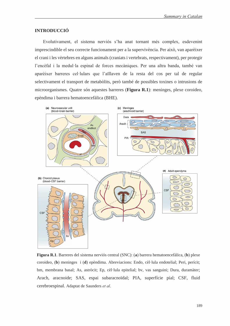
Summary in Catalan
189
INTRODUCCIÓ
Evolutivament, el sistema nerviós s’ha anat tornant més complex, esdevenint
imprescindible el seu correcte funcionament per a la supervivència. Per això, van aparèixer
el crani i les vèrtebres en alguns animals (craniats i vertebrats, respectivament), per protegir
l’encèfal i la medul·la espinal de forces mecàniques. Per una altra banda, també van
aparèixer barreres cel·lulars que l’aïllaven de la resta del cos per tal de regular
selectivament el transport de metabòlits, però també de possibles toxines o intrusions de
microorganismes. Quatre són aquestes barreres (Figura R.1): meninges, plexe coroideo,
epèndima i barrera hematoencefàlica (BHE).
Figura R.1. Barreres del sistema nerviós central (SNC): (a) barrera hematoencefàlica, (b) plexe
coroideo, (b) meninges i (d) epèndima. Abreviacions: Endo, cèl·lula endotelial; Peri, perícit;
bm, membrana basal; As, astròcit; Ep, cèl·lula epitelial; bv, vas sanguini; Dura, duramàter;
Arach, aracnoide; SAS, espai subaracnoïdal; PIA, superfície pial; CSF, fluid
cerebroespinal. Adaptat de Saunders et al.

Summary in Catalan
190
La BHE és la barrera que es troba en els capil·lars del cervell, i està formada per
diversos tipus cel·lulars (incloent cèl·lules endotelials, perícits, astròcits i neurones) i la
matriu extracel·lular (Figura R.2). En aquests capil·lars, les cèl·lules endotelials tenen
unes unions cèl·lula-cèl·lula especials, anomenades unions estretes, que deixen menys
espai intercel·lular. A més, també formen una barrera enzimàtica, i selectiva a través de
diversos mecanismes de transport.
Figura R.2. Estructura de la barrera hematoencefàlica: cèl·lules endotelials, perícits, astròcits i
neurones i membrana basal (matriu extracel·lular). Adaptat de Banks.
Aquesta barrera suposa un impediment per al desenvolupament de tractaments per
malalties que afecten al sistema nerviós central (SNC), ja que impedeix que més del 98%
dels fàrmacs de baix pes molecular no la puguin creuar, tal com gairebé el 100% dels d’alt
pes molecular. En aquest sentit, els costos pels tractaments de les malalties que afecten al
SNC a Europa van assolir els 798.000 milions d’euros, uns 1.550 € per càpita. Per això, la
investigació de mètodes per poder enviar fàrmacs al cervell s’ha anat incrementant
exponencialment en els darrers anys.

Summary in Catalan
191
Diversos mecanismes permeten el transport a través de la BHE. El transport passiu,
que no involucra l’ús de la maquinària cel·lular i per tant d’energia, conté dos mecanismes:
hidrofílic o paracel·lular, i lipofílic o transcel·lular. El primer, restringit al transport entre
cèl·lules, està molt impedit per la presència de les unions estretes. El segon depèn de les
característiques fisicoquímiques de compost, que li permetran ser transportat a través de la
membrana plasmàtica (5 normes de Lipinski).
Figura R.3. Entrada de ferro a través de transferrina (Tf) i el seu receptor (TfR). A pH 7.4—
exterior cel·lular—, la Tf difèrrica (holoTf) s’uneix al TfR, mentre que no ho fa la apoTf; i a pH
5.5—vesícula endocítica—el ferro és dissociat (després de reduir-lo la NADH:ferricianur
oxidoreductasa) i transportat al citosol per la DMT1, però la apoTf es manté unida al TfR;
finalment, la apoTf és retornada a la membrana cel·lular i dissociada a pH 7.4. Creada usant
ChemBioDraw.
Per una altra banda, hi ha els mecanismes de transport actiu que comprenen els
transportadors proteics i el transport endocític. L’últim és interessant des d’un punt de vista
terapèutic, ja que permet transportar entitats moleculars més grans. Dins dels mecanismes
endocítics s’hi troben la endocitosi mediada per adsorció (EMA) o per receptor (EMR). La
EMA es basa en les interaccions més o menys inespecífiques entre molècules carregades
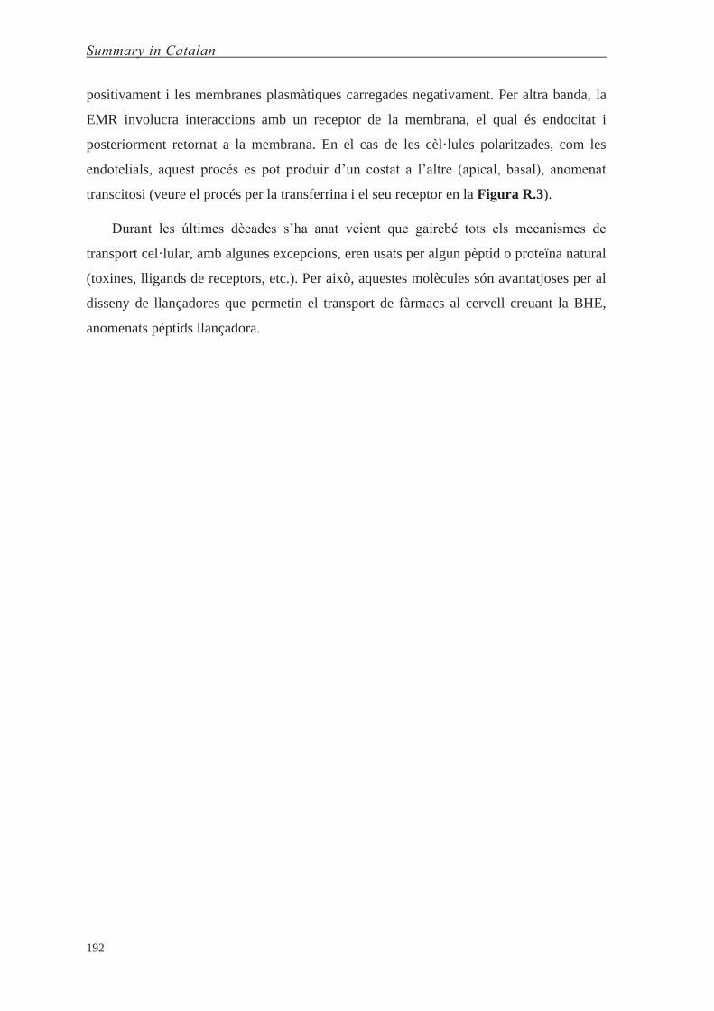
Summary in Catalan
192
positivament i les membranes plasmàtiques carregades negativament. Per altra banda, la
EMR involucra interaccions amb un receptor de la membrana, el qual és endocitat i
posteriorment retornat a la membrana. En el cas de les cèl·lules polaritzades, com les
endotelials, aquest procés es pot produir d’un costat a l’altre (apical, basal), anomenat
transcitosi (veure el procés per la transferrina i el seu receptor en la Figura R.3).
Durant les últimes dècades s’ha anat veient que gairebé tots els mecanismes de
transport cel·lular, amb algunes excepcions, eren usats per algun pèptid o proteïna natural
(toxines, lligands de receptors, etc.). Per això, aquestes molècules són avantatjoses per al
disseny de llançadores que permetin el transport de fàrmacs al cervell creuant la BHE,
anomenats pèptids llançadora.

Summary in Catalan
193
OBJECTIUS
Aquesta tesi està dividida en quatre capítols que es corresponen als objectius
principals. Els primers tres objectius estan centrats en recerca bàsica de pèptids llançadora,
mentre que el quart té un caràcter més aplicat.
Objectiu 1: Dissenyar, sintetitzar i avaluar una nova família de pèptids llançadora que
creuen a través de transport passiu.
1.1. Millorar la baixa solubilitat dels pèptids llançadora basats en (NMePhe)4.
1.2. Millorar la seva capacitat com a llançadores (mantenir el transport un cop s’hi
incorpora la molècula terapèutica).
1.3. Estudiar el rol de l’estereoquímica en el transport per difusió passiva.
Objectiu 2: Estudiar una nova família de pèptids llançadora que creuen per transcitosi
mitjançada per receptor i desenvolupar una metodologia basada en la combinació de l’ús
d’espectrometria de masses MALDI-TOF i models in vitro (cel·lulars) per avaluar el
transport a través de la BHE.
Objectiu 3: Estudiar i comparar les respostes immunogèniques produïdes per pèptids
llançadora fets d’aminoàcids ʟ i les seves respectives versions retro-ᴅ, formats per
aminoàcids ᴅ.
Objectiu 4: Realitzar uns estudis preliminars amb l’objectiu final de desenvolupar una
teràpia per Atàxia de Friedreich (AF) al sistema nerviós central (SNC).
4.1. Estudiar la viabilitat d’una teràpia de reemplaçament proteic per AF basada en la
conjugació directa de pèptids llançadora a frataxina (FXN).
4.2. Millorar els mètodes de bioconjugació per modificar herpesvirus amb pèptids
llançadora per desenvolupar una teràpia gènica per AF al SNC.
4.3. Caracteritzar fisicoquímicament i biològicament les partícules virals abans i després
de modificar-les.

Summary in Catalan
194
RESULTATS I DISCUSSIÓ
Capítol 1. Estudi de pèptids llançadora que creuen per difusió passiva
El transport de molècules al SNC a través de difusió passiva es fa per la via
transcel·lular, ja que la paracel·lular està molt impedida. Per a poder creuar usant la via
lipofílica (transcel·lular), les molècules han de tenir unes característiques fisicoquímiques
concretes, tals com tenir limitats els grups polars i el pes molecular (més detallat en les 5
normes de Lipinski).
Anteriorment, al laboratori s’havien desenvolupat uns pèptids llançadora basats en
(NMePhe)4 que presentaven un molt bon transport a través de difusió passiva, però la baixa
solubilitat (submicromolar) impedia l’ús més pràctic d’aquests compostos. Per això, es va
dissenyar un pèptid llançadora en que els seus aminoàcids fossin híbrids entre NMePhe i
l’aminoàcid natural N-alquilat, amb gran solubilitat en aigua, prolina. Així, el disseny final
va ser la fenilprolina (PhPro) (veure Figura R.4).
Un cop fet el disseny es va avaluar el transport del pèptid (PhPro)4, fent servir un model
in vitro fisicoquímic de transport passiu (PAMPA), i posteriorment es va comparar amb
l’anterior pèptid llançadora (NMePhe)4. El disseny híbrid va mostrar propietats de transport
similars a (NMePhe)4, mentre que la Pro4 pràcticament no va poder creuar (Figura R.5).
Figura R.4. Estructura del (a) pèptid llançadora (NMePhe)4, (b) poliprolina hidrofílica Pro4, i (c)
el pèptid llançadora híbrid (PhPro)4; homo-ʟ, C-terminal amida, i N-terminal acetilat. Creada amb
ChemBioDraw.
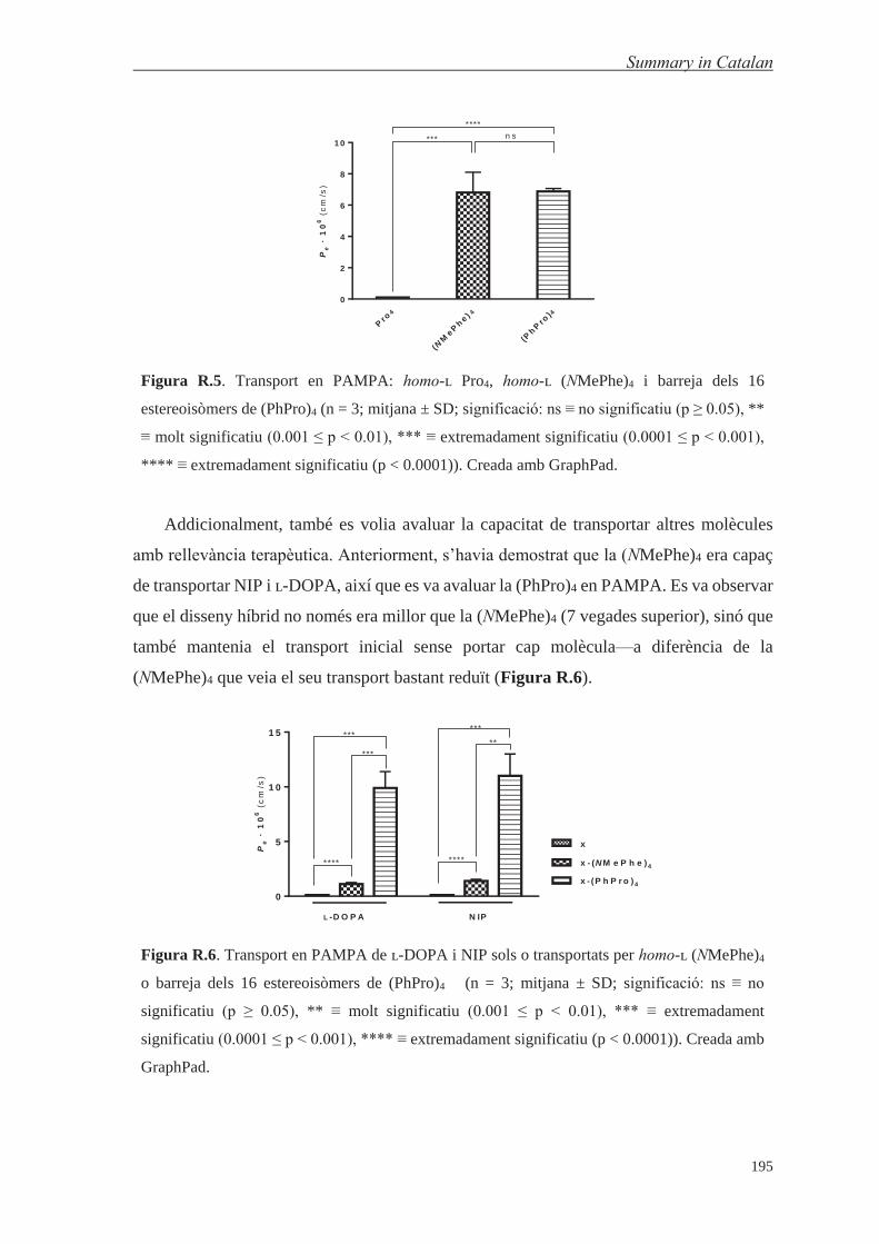
Summary in Catalan
195
Pro
4
(NM
ePh
e ) 4
(Ph
Pro
)4
0
2
4
6
8
1 0
Pe
·1
06
(cm
/s)
n s***
****
Figura R.5. Transport en PAMPA: homo-ʟ Pro4, homo-ʟ (NMePhe)4 i barreja dels 16
estereoisòmers de (PhPro)4 (n = 3; mitjana ± SD; significació: ns ≡ no significatiu (p ≥ 0.05), **
≡ molt significatiu (0.001 ≤ p < 0.01), *** ≡ extremadament significatiu (0.0001 ≤ p < 0.001),
**** ≡ extremadament significatiu (p < 0.0001)). Creada amb GraphPad.
Addicionalment, també es volia avaluar la capacitat de transportar altres molècules
amb rellevància terapèutica. Anteriorment, s’havia demostrat que la (NMePhe)4 era capaç
de transportar NIP i ʟ-DOPA, així que es va avaluar la (PhPro)4 en PAMPA. Es va observar
que el disseny híbrid no només era millor que la (NMePhe)4 (7 vegades superior), sinó que
també mantenia el transport inicial sense portar cap molècula—a diferència de la
(NMePhe)4 que veia el seu transport bastant reduït (Figura R.6).
Pe
·1
06
(cm
/s)
0
5
1 0
1 5
x
x - (N M e P h e )4
x - (P h P r o )4
***
***
****
***
**
****
L -D O P A N IP
Figura R.6. Transport en PAMPA de ʟ-DOPA i NIP sols o transportats per homo-ʟ (NMePhe)4
o barreja dels 16 estereoisòmers de (PhPro)4 (n = 3; mitjana ± SD; significació: ns ≡ no
significatiu (p ≥ 0.05), ** ≡ molt significatiu (0.001 ≤ p < 0.01), *** ≡ extremadament
significatiu (0.0001 ≤ p < 0.001), **** ≡ extremadament significatiu (p < 0.0001)). Creada amb
GraphPad.

Summary in Catalan
196
L’objectiu principal d’aquest disseny híbrid era la millora de la solubilitat i per tant es
va determinar la solubilitat en aigua del tetrapèptid (PhPro)4. El valor obtingut va ser
intermedi entre la de (NMePhe)4 (submicromolar) i la de Pro4 (300 mM), en el rang
mil·limolar baix. Això suposa una millora de tres ordres de magnitud respecte al disseny
anterior.
Pe
·1
06
(cm
/s)
DD
DD
DL
LD
DL
DL
DD
LL
DL
LL
DD
DL
DD
LD
DL
DD
Ba rr
e ja
d'e
s tere
ois
om
.
0
5
1 0
1 5
n s
n s
n sn s
***
n s
**
****
G ru p 1 G ru p 2
Figura R.7. Transport en PAMPA dels 16 estereoisòmers individuals, aparellats per
enantiòmers, i la barreja dels 16 estereoisòmers (columna blau fosc). La columna blau clar
correspon a la configuració mostrada en el gràfic (primera columna = DDDD); la columna lila
representa l’enantiòmer de la configuració mostrada en el gràfic (primera columna = LLLL) (n
= 3; mitjana ± SD; significació: ns ≡ no significantiu (p ≥ 0.05), ** ≡ molt significantiu (0.001 ≤
p < 0.01), *** ≡ extremadament significantiu (0.0001 ≤ p < 0.001), **** ≡ extremadament
significatiu (p < 0.0001)). Creada amb GraphPad.
Finalment, es va estudiar el rol de l’estereoquímica en el transport per difusió passiva
a través de membranes biològiques aprofitant que l’aminoàcid PhPro té dues
configuracions cis possibles, (S,S) i (R,R). Així, es va sintetitzar la biblioteca de 16
estereoisòmers de la (PhPro)4 i es va avaluar per PAMPA. Es va observar que hi havia
discriminació enantiomèrica, i que aquesta era més important com més asimètrics eren els
pèptids (Grup 2) (Figura R.7).

Summary in Catalan
197
Capítol 2. Estudi de pèptids llançadora que creuen la BHE a través de transcitosi
mitjançada per receptor
El descobriment i posterior desenvolupament tant de pèptids llançadora com d’altres
compostos que creuin la BHE per transport actiu requereix l’ús de models in vitro cel·lulars
que simulen aquesta barrera. Tan mateix, es requereix una millora dels mètodes analítics
de quantificació per a poder avaluar el transport de compostos assajats a menor
concentració, així permetent estudis mecanístics, i condicions més semblants a les
fisiològiques.
Es va dissenyar inicialment una biblioteca de 10 anàlegs d’un pèptid llançadora (HAI),
el qual s’havia estudiat i desenvolupat prèviament en el laboratori (Figura R.8). El propòsit
d’aquesta peptidoteca era doble: per una banda, estudiar les diferències de transport segons
quin residu s’ha substituït per un aminoàcid anàleg no natural, i per l’altra, fer-la servir per
validar i desenvolupar un mètode de quantificació basat en marcatge isotòpic i
espectrometria de masses MALDI-TOF.
Figura R.8. Pèptid llançadora H-HAIYPRH-NH2 (1L): versions (a) lleugera, i (b) pesada,
ambdues marcades isotòpicament amb un grup acetil; i (c) biblioteca d’anàlegs de HAI (1L) a la
esquerra, i de la versió retro-ᴅ (rD-HAI o 6D) a la dreta. Creada amb ChemBioDraw i Microsoft
PowerPoint.

Summary in Catalan
198
El mètode es basa en marcar isotòpicament de manera diferencial cada pèptid, una
versió amb un grup acetil sense marcatge (versió lleugera), i una altra amb marcatge (quatre
deuteris i un carboni 13; versió pesada) que resulta en un increment de massa de +4 unitats
de massa atòmiques (Figura R.9). Un dels pèptids és avaluat, i l’altre s’usa com a patró
intern per quantificar usant espectrometria de masses MALDI-TOF.
Figura R.9. Esquema del mètode quantificació del transport usant espectrometria de masses
MALDI-TOF. El model in vitro cel·lular de BHE (a) és preparat en un sistema de pouets on
cèl·lules endotelials són cultivades en una membrana que separa dos compartiments (donador i
acceptor). Abans de l’experiment, el donador conté el pèptid lleuger a assajar; al final de
l’experiment, una quantitat del pèptid haurà passat al pouet acceptor (si s’assaja el pèptid a 200
μM en el compartiment donador, aproximadament una concentració de 2 μM s’ha de quantificar
en el pouet acceptor al final de l’experiment). Així, (b) dues alíquotes de la versió pesada es
preparen, una a 200 μM i la altra a 2 μM; 10 μL del pèptid lleuger (dels compartiments acceptor
i donador) i 10 μL de la versió pesada a una concentració similar són barrejats. Seguidament, 1
μL d’aquesta barreja i 1 μL d’una matriu de MALDI apropiada (per exemple ACH) són col·locats
en la placa de MALDI. Finalment, els espectres són adquirits. Creada usant Adobe InDesign.

Summary in Catalan
199
Al comparar l’anàlisi de les mostres quantificades per RP-HPLC-PDA o utilitzant el
nou mètode basat en espectrometria de masses es van observar petites diferències. Tot i
això, l’anàlisi conjunt dels resultats amb un mateix mètode mostra que les diferències
estadísticament significatives no són iguals pels resultats obtinguts amb cadascun dels
mètodes. El mètode de RP-HPLC-PDA mostra pràcticament només diferències entre els
pèptids ʟ i ᴅ, mentre que la metodologia d’espectrometria de masses revela que les
diferències estadísticament significatives són només amb tres anàlegs, tots amb la
substitució a la prolina (Figura R.10). En dos d’ells el reemplaçament de l’aminoàcid
substituït és un dels enantiòmers de PhPro.
Figura R.10. Transport dels pèptids en el model boví in vitro de BHE analitzats per RP-HPLC-
PDA (en blau) o espectrometria de masses MALDI-TOF (en verd); (a) quantificació del
transport per RP-HPLC-PDA mostra diferències significatives entre pèptids ʟ i ᴅ (i també,
diferències significatives (*) entre el pèptid 9D amb el 7+D i el 8D); (b) significació de la
quantificació del transport per MALDI-TOF MS. Significació: ns ≡ no significatiu (p ≥ 0.05), *
≡ significatiu (0.01 ≤ p < 0.05), ** ≡ molt significatiu (0.001 ≤ p < 0.01), *** ≡ extremadament
significatiu (0.0001 ≤ p < 0.001), **** ≡ extremadament significatiu (p < 0.0001). Creada usant
GraphPad i Adobe InDesign.

Summary in Catalan
200
Addicionalment, dos d’ells (no s’avalua el pèptid amb la substitució de ʟ-PhPro)
s’assagen en un model in vitro cel·lular de BHE usant medi de cultiu, revelant que aquestes
diferències de transport entre el pèptid origina (HAI) i els anàlegs encara és més gran (4 i
8 vegades) (veure Figura R.11). T
ran
sp
ort
Re
lati
ua
1L
1 L7 -D 8D
0
3
6
9
1 2
T a m p ó R in g e r H e p e s
M e d i E C M su p le m e n ta t
1 .0 1 .41 .5
4 .1
8 .1
1 .0
Figura R.11. Transport dels pèptids en el model humà in vitro de BHE (usant dissolució
tamponada o medi cel·lular durant l’assaig): transport relatiu a 1L. Creada usant GraphPad.
El mètode de quantificació del transport per espectrometria de masses ha permès una
millora de la sensibilitat de tres ordres de magnitud (de mil·limolar, pel mètode de RP-
HPLC-PDA, a micromolar), i a més augmentar la versatilitat dels mètodes in vitro cel·lulars
de BHE (permet la quantificació de mostres amb medi cel·lular).

Summary in Catalan
201
Capítol 3. Estudi de les respostes immunogèniques produïdes per pèptids llançadora
La immunogenicitat dels pèptids, com a lligands natius dels MHC de classe I i II, s’ha
estudiat extensivament. Se sap que hi ha seqüències que produeixen una resposta
immunogènica molt més forta que altres. Tanmateix, també s’han estudiat les respostes
immunogèniques en front de pèptids ᴅ, trobant disparitat en els resultats obtinguts. Així,
no es pot predir el grau de resposta immunogènica contra una seqüència d’aminoàcids ᴅ
concreta.
Figura R.12. Avaluació de la resposta immunològica (humoral) de diversos pèptids llançadora
en ratolí: (a) sèrum anti-(HAI, THR), sèrum anti-versions retro-ᴅ avaluades amb els extrems (b)
C- o (c) N-terminal exposats. Les primeres diferències significatives de manera consecutiva en
les dilucions s’assenyala amb un asterisc. Creada amb GraphPad, Adobe InDesign,
ChemBioDraw, Microsoft PowerPoint i Servier Medical Art.
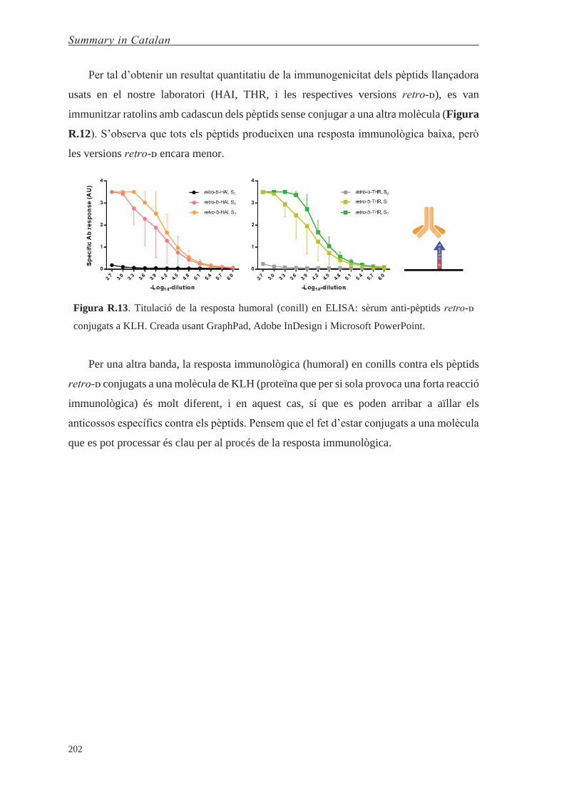
Summary in Catalan
202
Per tal d’obtenir un resultat quantitatiu de la immunogenicitat dels pèptids llançadora
usats en el nostre laboratori (HAI, THR, i les respectives versions retro-ᴅ), es van
immunitzar ratolins amb cadascun dels pèptids sense conjugar a una altra molècula (Figura
R.12). S’observa que tots els pèptids produeixen una resposta immunològica baixa, però
les versions retro-ᴅ encara menor.
Figura R.13. Titulació de la resposta humoral (conill) en ELISA: sèrum anti-pèptids retro-ᴅ
conjugats a KLH. Creada usant GraphPad, Adobe InDesign i Microsoft PowerPoint.
Per una altra banda, la resposta immunològica (humoral) en conills contra els pèptids
retro-ᴅ conjugats a una molècula de KLH (proteïna que per si sola provoca una forta reacció
immunològica) és molt diferent, i en aquest cas, sí que es poden arribar a aïllar els
anticossos específics contra els pèptids. Pensem que el fet d’estar conjugats a una molècula
que es pot processar és clau per al procés de la resposta immunològica.

Summary in Catalan
203
Capítol 4. Intents per desenvolupar una teràpia per Atàxia de Friedreich al SNC
L’atàxia de Friedreich (FRDA) és una malaltia neurodegenerativa de base genètica. La
causa principal és una expansió de la repetició del triplet (GAA) localitzada en el primer
intró del gen de la frataxina (FXN), una proteïna mitocondrial codificada en el nucli. Això
produeix una reducció de l’expressió de FXN, el que provoca alteracions en el metabolisme
cel·lular, sobretot en metabolisme del ferro i estat redox. Pel fet de que l’origen de la FRDA
sigui una deficiència en la funció de la proteïna, i no un mal funcionament d’aquesta, la
malaltia és recessiva. Això fa que una teràpia de reemplaçament proteic sigui, en principi,
una opció adequada. Per una altra banda, com que només hi ha un gen implicat, és a dir que
és monogènica, la teràpia gènica també es contempla com a opció, ja que n’hi ha prou amb
tenir una còpia del gen que expressi la proteïna funcional.
Així, vam explorar inicialment el desenvolupament d’una teràpia de reemplaçament
proteic. L’extrem N-terminal de la proteïna conté la senyal de localització mitocondrial, la
qual es processa durant la translocació de la proteïna al mitocondri. Per això, es va dissenyar
el constructe terapèutic amb el pèptid llançadora unit a l’extrem N-terminal de la frataxina
(un cop processat, queda la proteïna madura).
Figura R.14. Constructe dissenyat per la teràpia de reemplaçament proteic per FRDA al SNC:
(pèptid llançadora)–FXN1–210. S’observa els llocs de proteòlisi del N-terminal. Creada amb
Visual Molecular Dynamics (VMD) i Adobe InDesign, fent servir la estructura PDB ID# 1LY7.
Tan mateix, aquesta estratègia no va tenir èxit degut a la ràpida hidròlisi de la proteïna,
que no va permetre dur a terme la bioconjugació—la velocitat de proteòlisi era major que
la velocitat de la reacció de bioconjugació. Tot i això, es va fer un seguiment de la estabilitat
de la proteïna durant 6 mesos a 4, -20 i -80ºC usant diverses tècniques de caracterització
(MALDI-TOF, HPLC, HPLC-MS, SDS-PAGE, CD).
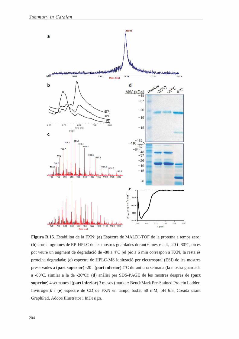
Summary in Catalan
204
Figura R.15. Estabilitat de la FXN: (a) Espectre de MALDI-TOF de la proteïna a temps zero;
(b) cromatogrames de RP-HPLC de les mostres guardades durant 6 mesos a 4, -20 i -80ºC, on es
pot veure un augment de degradació de -80 a 4ºC (el pic a 6 min correspon a FXN, la resta és
proteïna degradada; (c) espectre de HPLC-MS ionització per electrosprai (ESI) de les mostres
preservades a (part superior) -20 i (part inferior) 4ºC durant una setmana (la mostra guardada
a -80ºC, similar a la de -20ºC); (d) anàlisi per SDS-PAGE de les mostres després de (part
superior) 4 setmanes i (part inferior) 3 mesos (marker: BenchMark Pre-Stained Protein Ladder,
Invitrogen); i (e) espectre de CD de FXN en tampó fosfat 50 mM, pH 6.5. Creada usant
GraphPad, Adobe Illustrator i InDesign.

Summary in Catalan
205
La proteïna resulta ser poc estable a 4ºC (vida mitjana de menys d’una setmana),
mentre que a -20 i -80ºC l’estabilitat es manté millor (mesos). Veure la Taula R.1 per un
resum de resultats, i la Figura R.15 per la caracterització amb les diverses tècniques.
Taula R.1. Estabilitat de la frataxina a llarg termini a diverses temperatures, mostrant les
diferències respecte la mostra temps zero.
Mètode d’anàlisi Temperatura de preservació (ºC)
4 -20 -80
MALDI-TOFoo n.d. n.d. n.d.
HPLC-PDA 1 setmana 3 mesos 6 mesos
HPLC-MS 1 setmana 4 setmanes 6 mesos
SDS-PAGE 1 setmana - -
CDpp n.c. n.c. n.c.
Addicionalment, es va analitzar l’estabilitat de la proteïna a 37ºC (Figura R.16).
S’observa que la proteïna s’ha hidrolitzat gairebé completament al cap de 7 h, i a les 21 h
ni es detecta la banda.
Figura R.16. Estabilitat de la frataxina a 37ºC tampó fosfat pH 6.5, analitzat per SDS-PAGE
(marcador: BlueStar Prestained Protein Marker, Nippon Genetics). Banda no observada després
de 21 h (no ensenyat). Creada amb Adobe InDesign.
D’altra banda, el gen de la frataxina codifica per diverses isoformes (Taula R.2) que
són expressades diferencialment en diversos teixits. Això dificulta una teràpia de
reemplaçament proteic on es tinguin en compte totes aquestes isoformes. En canvi, el grup
del Prof. Díaz-Nido ha desenvolupat unes partícules virals, basades en herpesvirus (HSV-
1), que contenen tot el gen de la frataxina i que són capaces d’expressar les isoformes I, II
i III. També s’ha demostrat la seva capacitat infectiva i terapèutica in vivo.
oo n.d. ≡ no detectat, només pel temps zero. pp n.c. ≡ sense canvis observats.

Summary in Catalan
206
Taula R.2. Isoformes metabòliques de la frataxina.
Nom de la isoforma Seqüència Proteòlisi Longitud (nº residus)
precursor, p FXN1–210 - 210
intermedi42, i42 FXN42–210 MPP 169
madura56, m56 FXN56–210 MPP 155
degradada78, d78 FXN78–210 Ferro 133
madura81, m81 FXN81–210 MPP 130
Tot i aquests resultats prometedors, aquestes partícules no són capaces de creuar la
BHE, i per tant d’arribar al cervell. Per això, vam dissenyar una estratègia basada en la
bioconjugació de pèptids llançadora en les lisines de les proteïnes de la envolta d’aquests
virus (membrana lipídica amb proteïnes i glicoproteïnes).
Figura R.17. Estructura simplificada de HSV-1, mostrant els grups reactius per a la
bioconjugació: polisacàrids, cisteïnes (Cys) formant ponts disulfur, i lisines (Lys). Creada usant
ChemBioDraw, amb l’estructura de la càpsida C de HSV-1,qq EMDB ID# EMD-5659.
qq Les càpsides C són les formes més properes als virions madurs.

Summary in Catalan
207
Figura R.18. Caracterització de NHS-PEG3500-rD-HAI i -rD-THR: (a) espectre de 1H-RMN
(part superior i inferior, respectivament), (b) cromatogrames de RP-HPLC usant una columna
C18 i un gradient lineal de 0 a 100% amb CH3CN (gris fluix i fort, respectivament) i (c) espectre
de MALDI-TOF MS (distribució de masses de la dreta i de l’esquerra, respectivament). Creada
usant Adobe Illustrator i InDesign.

Summary in Catalan
208
A part, per tal d’aconseguir una millor exposició dels pèptids llançadora i que
poguessin interactuar amb el receptor del qual en són lligands (TfR), es va col·locar un
espaiador entre el grup reactiu a lisines (ester de NHS) i el pèptid llançadora. Es va
seleccionar un polietilenglicol de 3500 Da per dur a terme aquesta funció, ja que és un
polímer soluble en aigua, de baixa immunogenicitat i aprovat per la FDA. Així, es van
preparar els compostos NHS-PEG3500-(pèptid llançadora) usant metodologia de síntesi de
pèptids en fase sòlida (SPPS) adaptada a les nostres condicions (pels pèptids llançadora rD-
HAI i rD-THR). Posteriorment, es van caracteritzar per espectrometria de masses MALDI-
TOF, RMN de protó i RP-HPLC (R.18; pureses >95%).
Figura R.19. Estratègies reporteres de bioconjugació. Totes les reaccions fetes en HBSS a pH
7.4. Creat usant ChemBioDraw.
Per tal de quantificar la funcionalització de les partícules virals es van dissenyar dues
estratègies basades, respectivament, en fluorescència i afinitat (Figura R.19). Finalment,
per caracteritzar aquests conjugats HSV-1 amb pèptids llançadora es van fer servir diverses
tècniques (SDS-PAGE, western blot, espectrometria de masses, TEM, DLS i potencial ζ)
(Figura R.20). Les tècniques que van ser més útils per a avaluar la bioconjugació van
resultar DLS i potencial ζ, que van permetre seguir la mida d’aquestes partícules i la
conjugació, respectivament.
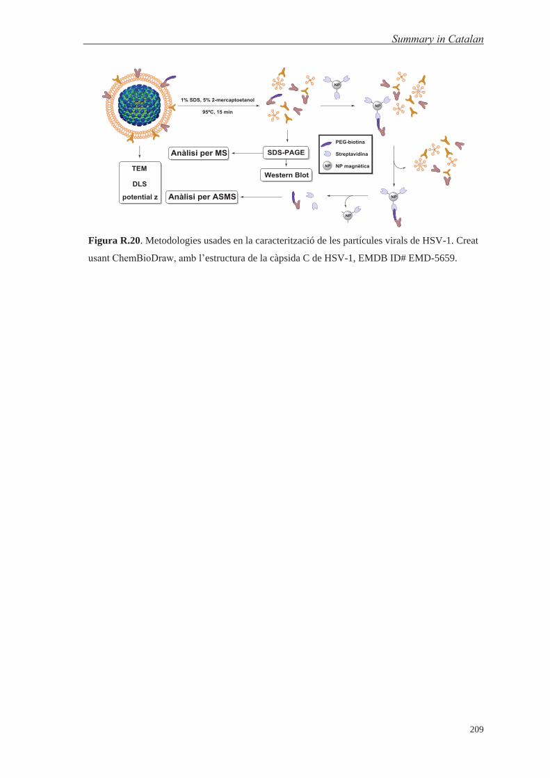
Summary in Catalan
209
Figura R.20. Metodologies usades en la caracterització de les partícules virals de HSV-1. Creat
usant ChemBioDraw, amb l’estructura de la càpsida C de HSV-1, EMDB ID# EMD-5659.

Summary in Catalan
210
CONCLUSIONS 1. Un disseny híbrid que combina els pèptids llançadora de transport passiu (NMePhe)4 i
l’aminoàcid prolina ha assolit valors de solubilitat en aigua en el rang mil·limolar baix,
incrementat en tres ordres de magnitud respecte el disseny previ.
2. El transport de (PhPro)4 és set vegades superior comparat amb el pèptid (NMePhe)4,
ambdós portant NIP o ʟ-DOPA. A més, el (PhPro)4 manté la permeabilitat tant lliure
com portant càrregues, a diferència del (NMePhe)4 que disminueix el seu transport.
3. L’anàlisi del transport de la biblioteca dels 16 estereoisòmers ha revelat que la
estereoquímica juga un paper important en la difusió passiva a través de membranes
biològiques.
4. Tres anàlegs de la versió retro-ᴅ del pèptid llançadora HAI, tots compartint la
substitució de la mateixa prolina, tenen un transport dues vegades el del pèptid original.
Dos d’ells incrementen la diferència de transport respecte l’original (quatre i vuit
vegades superior) un cop assajats en medi de cultiu.
5. Usant una petita modificació (acetilació de l’N-terminal), un nou mètode que combina
models in vitro (cel·lulars) de la BHE i espectrometria de masses MALDI-TOF permet
un increment en la sensibilitat de tres ordres de magnitud comparant amb el mètode
estàndard (RP-HPLC-PDA), i el ús de medi de cultiu en l’assaig.
6. La injecció de pèptids llançadora formats per aminoàcids ʟ (HAI i THR) produeix una
baixa resposta immunològica (humoral), la qual encara és més baixa per les respectives
versions retro-ᴅ, formades per aminoàcids ᴅ.
7. L’intent de bioconjugar l’N-terminal de la frataxina (FXN) no han tingut èxit degut a la
falta d’estabilitat de d’aquesta regió de la proteïna.
Summary in Catalan
211
8. La unió de pèptids llançadora sobre herpesvirus usant la bioconjugació a lisines ha
permès la modificació d’aquestes partícules virals en un sol pas, reduint la manipulació
de la mostra i així preservant millor la seva integritat estructural. A més, l’ús de PEG3500
com a espaiador entre el virus i el pèptid llançadora dificulta el reconeixement per
anticossos i al mateix temps manté la infectivitat.
9. La combinació de mètodes de biologia molecular (SDS-PAGE, western blot), de
proteòmica (espectrometria de masses) i biofísics (TEM, DLS i potencial ζ) ha permès
la caracterització dels virus modificats.


Top Related
Copyright © 2022 FDOKUMEN

