


Bahasa
Halaman
Hukum

HAL Id: hal-02988700https://hal.univ-lorraine.fr/hal-02988700
Submitted on 4 Nov 2020
HAL is a multi-disciplinary open accessarchive for the deposit and dissemination of sci-entific research documents, whether they are pub-lished or not. The documents may come fromteaching and research institutions in France orabroad, or from public or private research centers.
L’archive ouverte pluridisciplinaire HAL, estdestinée au dépôt et à la diffusion de documentsscientifiques de niveau recherche, publiés ou non,émanant des établissements d’enseignement et derecherche français ou étrangers, des laboratoirespublics ou privés.
Assessment of the role of Trichomonas tenax in theetiopathogenesis of humain periodontitis: A systematic
reviewCatherine Bisson, Sophie-Myriam Dridi, Marie-Claire Machouart
To cite this version:Catherine Bisson, Sophie-Myriam Dridi, Marie-Claire Machouart. Assessment of the role of Tri-chomonas tenax in the etiopathogenesis of humain periodontitis: A systematic review. PLoS ONE,Public Library of Science, 2019, 14 (12), pp.e0226266. �10.1371/journal.pone.0226266�. �hal-02988700�
RESEARCH ARTICLE
Assessment of the role of Trichomonas tenax
in the etiopathogenesis of human
periodontitis: A systematic review
C. BissonID1,2*, S. M. Dridi3, M. Machouart1,4
1 Stress, Immunity, Pathogens Laboratory, EA7300 Lorraine University, Faculty of Medicine, Vandoeuvre-
lès- Nancy, France, 2 CHU Odontologie de Nancy - Department of Periodontology– Rue du Docteur
Heydenreich, Nancy, University Lorraine, France, 3 CHU Odontologie de Nice -Department of
periodontology- CHU Saint Roch – Department of Periodontology, Nice, France, 4 CHU Brabois,
Parasitology - Mycology Laboratory, CHU Nancy-Brabois, Vandoeuvre-les -Nancy, France
Abstract
Objective
This systematic review was to assess the presence of Trichomonas tenax in patients with
periodontitis and to elucidate its potential role in the onset and development of this disease.
Method
Systematic review was conducted according to the Preferred Reporting Items for System-
atic Reviews and Meta-Analyses (PRISMA) guidelines and by consulting the five databases:
Medline, Science Direct, Web of Science, Dentistry and Oral Science Sources and
Cochrane Central Register of Controlled Trials. Following Koch’s postulates revisited by
Socransky as PICO framework, this collection data was only including full text of clinical tri-
als concerning patients with periodontitis, case-reports and in vitro research published
between 1960 and March 2019.
Results
On the 376 studies identified, only 25 fulfilled our eligible criteria. Most of these studies were
in vitro research articles designed to evaluate potential virulence factors, and others were
clinical trials (case-control studies, randomized controlled trial) and case-reports. The analy-
sis of these papers has shown that i) Trichomonas tenax is more frequently detected in den-
tal biofilm from sites with periodontitis than in healthy sites; ii) this live flagellate seems
capable of producing diverse enzymes that could participate in periodontal breakdown and
has the capacity to adhere to epithelial cells, its lysed form could induce the synthesis of IL-8
from macrophage cell lines; iii) the impact of non-surgical treatment of periodontitis have not
been thoroughly evaluated on the presence of T. tenax
PLOS ONE | https://doi.org/10.1371/journal.pone.0226266 December 17, 2019 1 / 20
a1111111111
a1111111111
a1111111111
a1111111111
a1111111111
OPEN ACCESS
Citation: Bisson C, Dridi SM, Machouart M (2019)
Assessment of the role of Trichomonas tenax in
the etiopathogenesis of human periodontitis: A
systematic review. PLoS ONE 14(12): e0226266.
https://doi.org/10.1371/journal.pone.0226266
Editor: Salomon Amar, New York Medical College,
UNITED STATES
Received: May 14, 2019
Accepted: November 24, 2019
Published: December 17, 2019
Copyright: © 2019 Bisson et al. This is an open
access article distributed under the terms of the
Creative Commons Attribution License, which
permits unrestricted use, distribution, and
reproduction in any medium, provided the original
author and source are credited.
Data Availability Statement: All relevant data are
within the paper and its Supporting Information
files.
Funding: This systematic review was supported by
a grant from the Conseil Regional du Grand Nancy.
Competing interests: The authors have declared
that no competing interest exist.
Conclusions
This systematic review has reported the presence of T. tenax more frequently in diseased
than healthy sites and the capacity of this flagellate to synthesis enzymes which could par-
ticipate to the degradation of periodontal tissues. Nevertheless, these data do not meet all
the postulates and are not enough to provide firm conclusions about the role of T. tenax in
the etiopathogenesis of periodontitis.
Introduction
The global burden of oral disorders and their associated sequelae have been assessed at various
dates (1990, 2005 and 2010) by Marcenes et al. [1]. These authors concluded that DALYS (Dis-
ability-Adjusted Life Years) induced by oral conditions had increased by 20.8% between 1990
and 2010 due to population growth, the ageing process and severe periodontitis. Severe peri-
odontitis is the sixth most prevalent disease affecting millions of people worldwide and because
of its dental and systemic impacts should therefore be a public health priority [2].
Periodontitis is recognized as an inflammatory disease, mainly induced by pathobionts
such as Porphyromonas gingivalis in synergy with other putative pathogens present in a com-
plex and diversified biofilm. These periodontopathogens cause an impaired immune response
leading to the loss of periodontal tissue [3]. But neither immune deficiencies nor the virulence
of periodontal pathogens can explain why, despite the identification of periodontal pathogenic
bacteria in the saliva, only a few teeth are severely affected by periodontitis in patients, or why
one tooth presented alveolar bone and soft tissue destruction while the periodontium of its
neighbour was barely affected.
In order to explain these clinical observations, but also the failure in stabilizing periodonti-
tis in certain patients, the authors attempted to explore other potential etiologies of periodontal
diseases. The oral cavity harbours a rich and complex biofilm that includes bacteria, but also
viruses, fungi and protozoans [4]. All these microorganisms could be implicated directly or
indirectly in the development of periodontitis. A systematic review with meta-analysis evaluat-
ing the microbiota from patients with periodontitis has shown a significant presence of some
herpesviruses and other authors have identified fungi (Candida genus), both of which could
have a potential pathological role in this disease [5,6]. Moreover, two main protozoans have
been identified in the oral cavity: Entamoeba gingivalis belonging to the rhizopod class and
Trichomonas tenax to the zoomastigophora one. As regards Trichomonas tenax, some authors
suggested its implication in periodontal diseases [7, 8]. A recent review concerning T. tenaxand periodontal diseases has been published in which the authors concluded that there was a
correlation between the presence of the flagellate and the periodontal diseases and that this
protozoan was involved in inflammatory process of gingivitis [9]. Nevertheless, this review did
not follow the PRISMA (Preferred Reporting Items for Systematic Reviews and Meta-Analy-
ses) guidelines and the PICO (Patient Intervention Comparison Outcome) framework as rec-
ommended by the Centre for Evidence-Based Medicine [10, 11]. In this review, 47 clinical
studies about prevalence of the protozoan in periodontal diseases were not classified according
to population (adults, children), presence of systemic disease, previous antibiotic consump-
tion, and the keywords used for the literature research on periodontal diseases assessment
were not detailed.
Therefore, we propose here the first systematic review of literature to investigate the pres-
ence of T. tenax in periodontitis in adult patients and to assess its potential role in the
Trichomonas in periodontitis
PLOS ONE | https://doi.org/10.1371/journal.pone.0226266 December 17, 2019 2 / 20
etiopathogenesis of periodontitis, according to Koch’s postulates revisited by Socransky [12]:
(i) postulate “Association” (elevated level of microorganism in lesions of periodontitis), (ii)
postulate “Elimination” (microorganism elimination resulted in successful therapy), (iii) pos-
tulate “Host response” (microorganism induces immune response: elevated serum and local
antibodies, cytokines production), (iv) postulate “Virulence factors” (microorganism produces
enzymes inducing a periodontal degradation) and (v) postulate “Animal studies” (microorgan-
ism induces disease in gnotobiotic animal).
Firstly, we investigate through literature data the presence of T. tenax in diseased sites and
its absence in healthy sites in adult patients with periodontitis, secondly the impact of non-sur-
gical periodontal treatment on the decrease or elimination of this flagellate in healed sites and
thirdly the potential virulence factors of this protozoan which could cause the destruction of
periodontal tissue.
Methods
Protocol and study design
This systematic review was elaborated by following the PRISMA guidelines [10] (S1 Table).
Eligibility criteria
In this review, the focused questions addressed using the PICO framework, aimed to evaluate
the potential pathogenicity of Trichomonas tenax in the development of periodontitis accord-
ing to Koch’s postulates revisited by Socransky [12]:
1. Is T. tenax (I) identified in high numbers in sites with periodontitis (P) and in lesser num-
bers or not at all in healthy sites? (postulate “Association”)
2. Does the non-surgical treatment of periodontitis induce the decrease or the absence of
T. tenax in the healed periodontal pocket and/or periodontal pocket with depth reduction?
(C) (postulate “Elimination”)
3. Does T. tenax produce virulence factors (adhesion capacity and tissue invasion, immuno-
modulating molecules, toxicity) and can it induce similar periodontal diseases in an appro-
priate animal model? (O) (postulates “Virulence factors”, “Host response” and “Animal
studies”).
Information sources and research strategy for relevant studies
The five databases MEDLINE, Science Direct, Web of Science, Dentistry and Oral Sciences
Source and Cochrane Central Register of Controlled Trials were consulted and our research
was conducted on all relevant literature up to March 2019 using MeSH, and the following key-
words: Trichomonas tenax AND (periodontal diseases), Trichomonas tenax AND (periodonti-
tis), Trichomonas tenax AND (antibody) OR (pathogenicity) OR (virulence) OR (enzyme) OR
(cytokines) OR (toxicity) OR (adhesion) OR (adherence) OR (invasion) OR (invasive).
No publication was selected prior to the 1960s, because terms used to evaluate periodontal
status were confusing (pyorrhoea, dying periosteum. . .). The electronic research was con-
ducted independently by two authors (CB, SMD).
Study selection
Only full text articles published in the English, French, Italian and Spanish/Portuguese lan-
guages were considered.
Trichomonas in periodontitis
PLOS ONE | https://doi.org/10.1371/journal.pone.0226266 December 17, 2019 3 / 20
All studies relating to Trichomonas tenax were considered eligible for inclusion if they were
original articles in peer-reviewed journals conducted in humans and were:
• Cross sectional, case-control studies, controlled trials
• Case reports or case series (accepted for research pathogenicity factors).
• In vitro studies with evaluation of potential virulence factors and animal models of
periodontitis.
All reviews of literature were excluded from the selected articles because they give redun-
dant informations and are not detailed enough.
Inclusion criteria. Studies that were selected for this analysis include the following
criteria:
• Adult population (� 18 years)
• Patients diagnosed with periodontitis using one or more of the following terms: pocket > 3
mm and/or CAL> 2 mm (Clinical Attachment Level), bone loss, grade of the periodontal
tissue degradation (moderate or severe periodontitis, Armitage classification, 1999) [13].
• Patients diagnosed with periodontitis with no surgical treatment in the last 3 months (to
evaluate the prevalence of T. tenax in periodontal pockets)
• Patients diagnosed with periodontitis treated by non-surgical treatment (to evaluate the
impact of the therapy on protozoan in the healed sites).
• Studies with collection of dental biofilm (supra and/or subgingival)
• In vitro studies with detailed protocol
Exclusion criteria. Articles were excluded from the selection because of the presence of:
• Adolescent and children population (< 18 years)
• Unclear periodontal diagnosis of patients included, no precise description of periodontal
parameters
• Periodontitis as manifestations of systemic disease
• Patients who received surgical periodontal therapy
• Patients taking medication that could modify the oral microbiota
• Studies relating only to the collection of saliva, tooth decay or oral mucosa.
• In vitro studies with other trichomonads species than T. tenax.
Data selection process and elements
Data were collected independently by two authors (CB, SMD) and definitively included
by these two authors, and all disagreements were resolved after discussion and/or with
the intervention of a third author (MM). Reasons of exclusion of the articles were fully
detailed in S1 Table, and quality appraisal of included clinical studies was described in S2
Table.
If the study was published twice, the article presenting the most complete data set was
selected.
Trichomonas in periodontitis
PLOS ONE | https://doi.org/10.1371/journal.pone.0226266 December 17, 2019 4 / 20
All publications meeting the eligibility criteria were included, processed for data extraction
and categorized according to the PICO framework:
Population: adult patients with periodontitis
Intervention: presence of trichomonads in sites with periodontitis
Comparison: absence or reduction of the flagellates in diseased sites after non-surgical
treatment
Outcome: potential virulence factors of the protozoan (adhesion, tissue invasion, cytokines,
antibody, toxicity).
Risk of bias in individual studies and across studies
Risk of bias was assessed through the Newcastle-Ottawa Scale (NOS) for case-control studies
and modified NOS for cross-sectional studies [14]. The following criteria were assessed:
• Representativeness of the exposed cohort
• Ascertainment of exposure
• Quality of measurements/protocol and outcomes: periodontal parameters and precise proto-
col of collection of biofilm are evaluated.
For in vitro research articles, protocols [time of incubation, strain of T. tenax, different
MOI (Multiplication of Infection), different cells line] used by each author are detailed.
Results
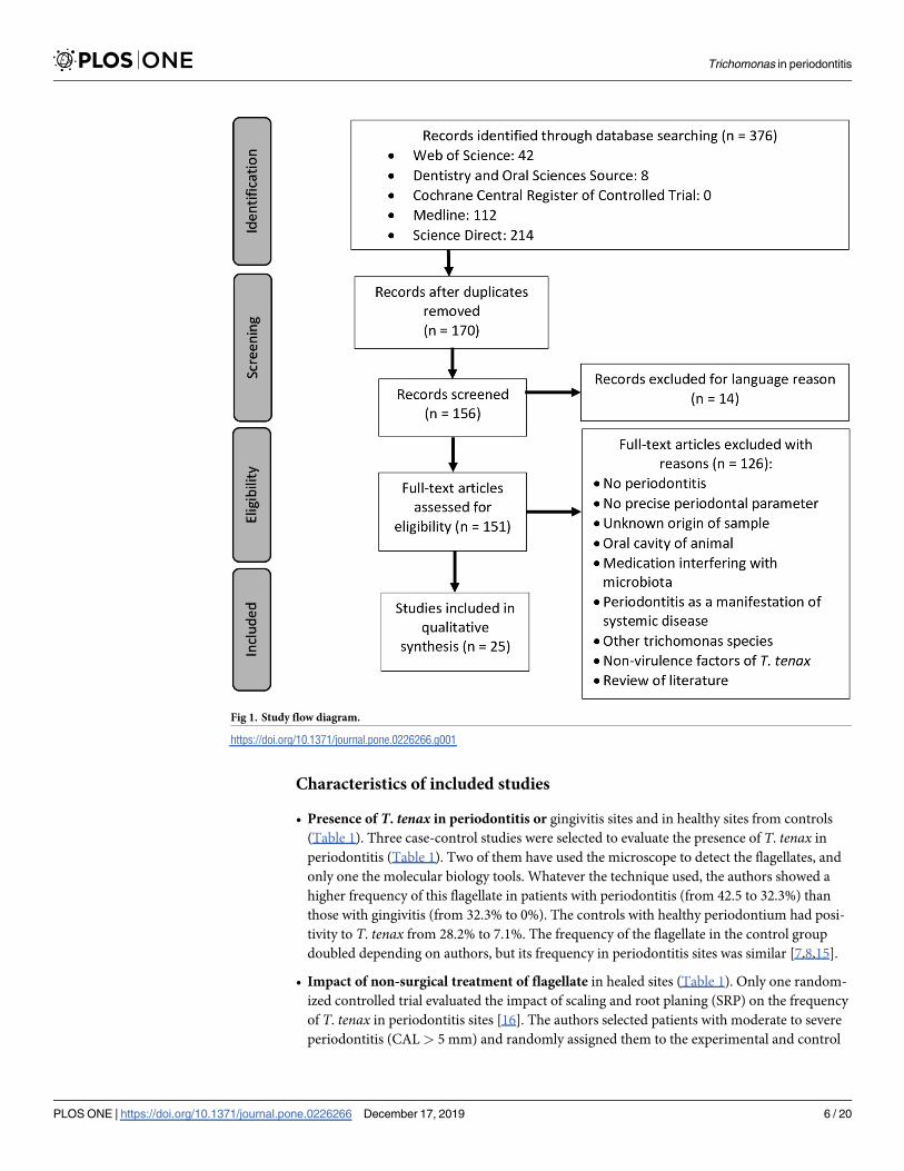
Study selection
As shown in Fig 1, a total of 376 articles were identified from the previously described elec-
tronic database search. After duplicate removal and language exclusion, a total of 156 articles
were obtained. Finally, after the removal of non-full text and non-relevant papers, 25 articles
were deemed appropriate to be included. The main reasons for exclusion were: the absence of
periodontal status, lack of precise periodontal parameters, the identification of trichomonads
in animal studies but not in human studies, the studies included patients with medication
modifying microbiota, the diagnosis of periodontitis as a manifestation of systemic disease,
other trichomonads species than T. tenax, the absence of a T. tenax factor of virulence. Some
clinical studies were discarded because essential details were missing:
1. One study included patients with periodontitis as well as patients with periodontitis and
diabetes and did not specify the proportion of patients T. tenax-positive among those with
diabetes.
2. one study includes both children and adults but did not specify the T. tenax positivity
according to the population age.
3. In other studies, some included patients had already been treated with antibiotics, without
anyone knowing the proportion of patients T. tenax positive among those receiving or not
antibiotics.
4. Finally, in some papers the origin of the sample was unknown such as “rinsing sample”
and/or samples from saliva, mucosa, decay.
Trichomonas in periodontitis
PLOS ONE | https://doi.org/10.1371/journal.pone.0226266 December 17, 2019 5 / 20
Characteristics of included studies
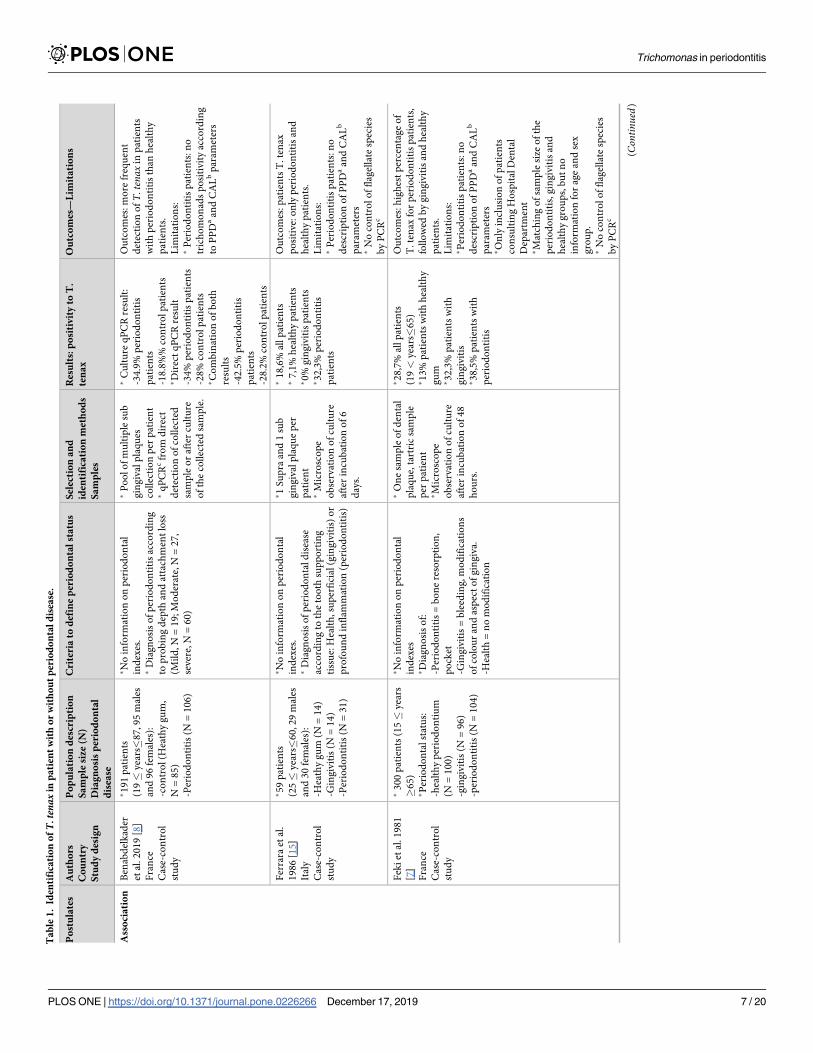
• Presence of T. tenax in periodontitis or gingivitis sites and in healthy sites from controls
(Table 1). Three case-control studies were selected to evaluate the presence of T. tenax in
periodontitis (Table 1). Two of them have used the microscope to detect the flagellates, and
only one the molecular biology tools. Whatever the technique used, the authors showed a
higher frequency of this flagellate in patients with periodontitis (from 42.5 to 32.3%) than
those with gingivitis (from 32.3% to 0%). The controls with healthy periodontium had posi-
tivity to T. tenax from 28.2% to 7.1%. The frequency of the flagellate in the control group
doubled depending on authors, but its frequency in periodontitis sites was similar [7,8,15].
• Impact of non-surgical treatment of flagellate in healed sites (Table 1). Only one random-
ized controlled trial evaluated the impact of scaling and root planing (SRP) on the frequency
of T. tenax in periodontitis sites [16]. The authors selected patients with moderate to severe
periodontitis (CAL> 5 mm) and randomly assigned them to the experimental and control
Fig 1. Study flow diagram.
https://doi.org/10.1371/journal.pone.0226266.g001
Trichomonas in periodontitis
PLOS ONE | https://doi.org/10.1371/journal.pone.0226266 December 17, 2019 6 / 20
Ta
ble
1.
Iden
tifi
cati
on
ofT.
tena
xin
pa
tien
tw
ith
or
wit
ho
ut
per
iod
on
tal
dis
ease
.
Po
stu
late
sA
uth
ors
Co
un
try
Stu
dy
des
ign
Po
pu
lati
on
des
crip
tio
n
Sa
mp
lesi
ze(N
)
Dia
gn
osi
sp
erio
do
nta
l
dis
ease
Cri
teri
ato
def
ine
per
iod
on
tal
sta
tus
Sel
ecti
on
an
d
iden
tifi
cati
on
met
ho
ds
Sa
mp
les
Res
ult
s:p
osi
tiv
ity
toT
.
ten
ax
Ou
tco
mes
—L
imit
ati
on
s
Ass
oci
ati
on
Ben
abd
elk
ader
etal
.2
01
9[8
]
Fra
nce
Cas
e-co
ntr
ol
stu
dy
�1
91
pat
ien
ts
(19�
yea
rs�
87
,95
mal
es
and
96
fem
ales
):
-co
ntr
ol
(Hea
thy
gu
m,
N=
85
)
-Per
iod
on
titi
s(N
=1
06
)
�N
oin
form
atio
no
np
erio
do
nta
l
ind
exes
.�
Dia
gn
osi
so
fp
erio
do
nti
tis
acco
rdin
g
top
rob
ing
dep
than
dat
tach
men
tlo
ss
(Mil
d,N
=1
9;M
od
erat
e,N
=2
7,
sever
e,N
=6
0)
�P
oo
lo
fm
ult
iple
sub
gin
giv
alp
laq
ues
coll
ecti
on
per
pat
ien
t�
qP
CR
cfr
om
dir
ect
det
ecti
on
of
coll
ecte
d
sam
ple
or
afte
rcu
ltu
re
of
the
coll
ecte
dsa
mp
le.
�C
ult
ure
qP
CR
resu
lt:
-34
.9%
per
iod
on
titi
s
pat
ien
ts
-18
.8%
%co
ntr
ol
pat
ien
ts�D
irec
tq
PC
Rre
sult
-34
%p
erio
do
nti
tis
pat
ien
ts
-28
%co
ntr
ol
pat
ien
ts�C
om
bin
atio
no
fb
oth
resu
lts
-42
.5%
per
iod
on
titi
s
pat
ien
ts
-28
.2%
con
tro
lp
atie
nts
Ou
tco
mes
:m
ore
freq
uen
t
det
ecti
on
ofT.tenax
inp
atie
nts
wit
hp
erio
do
nti
tis
than
hea
lth
y
pat
ien
ts.
Lim
itat
ion
s:�
Per
iod
on
titi
sp
atie
nts
:n
o
tric
ho
mo
nad
sp
osi
tiv
ity
acco
rdin
g
toP
PD
aan
dC
AL
bp
aram
eter
s
Fer
rara
etal
.
19
86
[15
]
Ital
y
Cas
e-co
ntr
ol
stu
dy
�5
9p
atie
nts
(25�
yea
rs�
60
,29
mal
es
and
30
fem
ales
):
-Hea
thy
gu
m(N
=1
4)
-Gin
giv
itis
(N=
14
)
-Per
iod
on
titi
s(N
=3
1)
�N
oin
form
atio
no
np
erio
do
nta
l
ind
exes
.�
Dia
gn
osi
so
fp
erio
do
nta
ld
isea
se
acco
rdin
gto
the
too
thsu
pp
ort
ing
tiss
ue:
Hea
lth
,su
per
fici
al(g
ing
ivit
is)
or
pro
fou
nd
infl
amm
atio
n(p
erio
do
nti
tis)
�1
Su
pra
and
1su
b
gin
giv
alp
laq
ue
per
pat
ien
t�
Mic
rosc
op
e
ob
serv
atio
no
fcu
ltu
re
afte
rin
cub
atio
no
f6
day
s.
�1
8,6
%al
lp
atie
nts
�7
,1%
hea
lth
yp
atie
nts
�0
%g
ing
ivit
isp
atie
nts
�3
2,3
%p
erio
do
nti
tis
pat
ien
ts
Ou
tco
mes
:p
atie
nts
T.
ten
ax
po
siti
ve:
on
lyp
erio
do
nti
tis
and
hea
lth
yp
atie
nts
.
Lim
itat
ion
s:�
Per
iod
on
titi
sp
atie
nts
:n
o
des
crip
tio
no
fP
PD
aan
dC
AL
b
par
amet
ers
�N
oco
ntr
ol
of
flag
ella
tesp
ecie
s
by
PC
Rc
Fek
iet
al.1
98
1
[7]
Fra
nce
Cas
e-co
ntr
ol
stu
dy
�3
00
pat
ien
ts(1
5�
yea
rs
�6
5)
�P
erio
do
nta
lst
atu
s:
-hea
lth
yp
erio
do
nti
um
(N=
10
0)
-gin
giv
itis
(N=
96
)
-per
iod
on
titi
s(N
=1
04
)
�N
oin
form
atio
no
np
erio
do
nta
l
ind
exes
�D
iag
no
sis
of:
-Per
iod
on
titi
s=
bo
ne
reso
rpti
on
,
po
cket
-Gin
giv
itis
=b
leed
ing,
mo
dif
icat
ion
s
of
colo
ur
and
asp
ect
of
gin
giv
a.
-Hea
lth
=n
om
od
ific
atio
n
�O
ne
sam
ple
of
den
tal
pla
qu
e,ta
rtri
csa
mp
le
per
pat
ien
t�M
icro
sco
pe
ob
serv
atio
no
fcu
ltu
re
afte
rin
cub
atio
no
f4
8
ho
urs
.
�2
8,7
%al
lp
atie
nts
(19<
yea
rs�
65
)�1
3%
pat
ien
tsw
ith
hea
lth
y
gu
m�3
2,3
%p
atie
nts
wit
h
gin
giv
itis
�3
8,5
%p
atie
nts
wit
h
per
iod
on
titi
s
Ou
tco
mes
:h
igh
est
per
cen
tag
eo
f
T.te
nax
for
per
iod
on
titi
sp
atie
nts
,
foll
ow
edb
yg
ing
ivit
isan
dh
ealt
hy
pat
ien
ts.
Lim
itat
ion
s:�P
erio
do
nti
tis
pat
ien
ts:n
o
des
crip
tio
no
fP
PD
aan
dC
AL
b
par
amet
ers
�O
nly
incl
usi
on
of
pat
ien
ts
con
sult
ing
Ho
spit
alD
enta
l
Dep
artm
ent
�M
atch
ing
of
sam
ple
size
of
the
per
iod
on
titi
s,g
ing
ivit
isan
d
hea
lth
yg
rou
ps,
bu
tn
o
info
rmat
ion
for
age
and
sex
gro
up
.�
No
con
tro
lo
ffl
agel
late
spec
ies
by
PC
Rc
(Continued)
Trichomonas in periodontitis
PLOS ONE | https://doi.org/10.1371/journal.pone.0226266 December 17, 2019 7 / 20
Ta
ble
1.
(Co
nti
nu
ed)
Po
stu
late
sA
uth
ors
Co
un
try
Stu
dy
des
ign
Po
pu
lati
on
des
crip
tio
n
Sa
mp
lesi
ze(N
)
Dia
gn
osi
sp
erio
do
nta
l
dis
ease
Cri
teri
ato
def
ine
per
iod
on
tal
sta
tus
Sel
ecti
on
an
d
iden
tifi
cati
on
met
ho
ds
Sa
mp
les
Res
ult
s:p
osi
tiv
ity
toT
.
ten
ax
Ou
tco
mes
—L
imit
ati
on
s
Eli
min
ati
on
Ras
hid
iM
ayb
od
i
etal
.20
16
[16
]
Iran
Ran
do
miz
ed
Cli
nic
altr
ial
46
pat
ien
ts
(30<
yea
rs<
50
)�
case
gro
up
(15
wit
h
sever
ean
d8
wit
h
mo
der
ate
per
iod
on
titi
s)�co
ntr
ol
gro
up
(14
wit
h
sever
ean
d9
wit
h
mo
der
ate
per
iod
on
titi
s)�ex
clu
sio
n:p
revio
us
con
sum
pti
on
of
anti
bio
tics
,
imm
un
om
od
ula
tors
�M
od
erat
e(C
AL
=3
–4
mm
)to
sever
e
(CA
L�
5m
m)
per
iod
on
titi
s
(cla
ssif
icat
ion
of
Arm
itag
e,1
99
8).
�C
AL
of
case
gro
up
:5
.74±
1.7
mm
�C
AL
of
con
tro
lg
rou
p:5
.64±
1.7
mm
Fo
rea
chp
atie
nt,
coll
ecti
on
of:
1d
enta
lp
laq
ue
and
1sa
liva
sam
ple
s�F
or
con
tro
lg
rou
p,
sam
ple
sco
llec
ted
bef
ore
OH
I�F
or
case
gro
up
,
sam
ple
sco
llec
ted
bef
ore
OH
Ian
d3
wee
ks
afte
r
SR
P.
�G
iem
sast
ain
ing
and
mic
rosc
op
eo
bse
rvat
ion
of
sam
ple
�B
asel
ine
freq
uen
cy(%
)o
f
T.tenax
for
bo
thg
rou
p:n
ot
rep
ort
ed�C
orr
elat
ion
bet
wee
n
sever
ity
of
per
iod
on
titi
san
d
freq
uen
cyo
fT.tenax
in
den
tal
pla
qu
e:r
=0
.56
5,
p�
0.0
05
)�Id
enti
fica
tio
no
fT.tenax
on
lyin
den
tal
pla
qu
eo
f
fem
ale
�3
wee
ks
afte
rS
RP
,
sig
nif
ican
tre
du
ctio
no
fT.
tenax
freq
uen
cyin
sali
va
bu
t
no
tin
den
tal
pla
qu
e.
Ou
tco
mes
:C
AL
bis
rep
rese
nta
tive
of
the
deg
ree
of
deg
rad
atio
no
f
per
iod
on
tal
tiss
ues
Lim
itat
ion
s:�N
oas
sess
men
to
fo
utc
om
e,n
o
des
crip
tio
no
fp
erio
do
nta
lin
dex
es
(ex
cep
tfo
rC
AL
b),
no
rep
ort
of
corr
elat
ion
bet
wee
np
erio
do
nta
l
par
amet
ers
and
T.te
nax
pre
sen
ce.
�N
ore
po
rto
fb
lin
dn
ess
of
dat
a
coll
ecti
on
�O
nly
incl
usi
on
of
pat
ien
ts
con
sult
ing
Sch
oo
lo
fD
enti
stry
.�
No
info
rmat
ion
on
level
of
infe
ctio
n(P
PD
an
ot
rep
ort
ed).
�N
oco
ntr
ol
of
flag
ella
tesp
ecie
s
by
PC
Rc
PP
Da:
Per
iod
on
tal
Po
cket
sD
epth
,
CA
Lb:
Cli
nic
alA
ttac
hm
ent
Lev
el,
PC
Rc:
Po
lym
eras
eC
hai
nR
eact
ion
/
qP
CR
c:
qu
anti
tati
veP
CR
.
htt
ps:
//doi.o
rg/1
0.1
371/jo
urn
al.p
one.
0226266.t001
Trichomonas in periodontitis
PLOS ONE | https://doi.org/10.1371/journal.pone.0226266 December 17, 2019 8 / 20
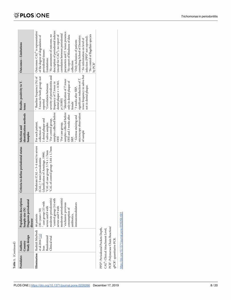
group. Before receiving an oral hygiene instruction, a sample of dental biofilm was collected
from the patients in each group. Three weeks after SRP, a second biofilm sample was col-
lected from patients in the experimental group. The authors concluded that the SRP could
reduce the number of T. tenax in saliva but not in the dental plaque of patients with moder-
ate to severe periodontitis. No detailed description of PPD (Periodontal Pocket Depth) and
CAL before and after SRP was reported.
• Potential virulence factors from protozoans (Table 2). Authors have detected the capacity
of T. tenax ATCC 30207 to adhere to diverse cells and induce cytotoxicity [17], while others
studies showed no adherence or cytotoxic property [18–19]. Moreover, flagellates produce
numerous enzymes, such as cysteine protease, cathepsin B like protein, haemolysins, metal-
loproteinase and other proteinases that could participate in the breakdown of collagen, gela-
tin observed in periodontal diseases [20–25]. Live trophozoites of the Hs-4 strain of T. tenaxfailed to induce secretion of IL-1β, IL-8, IL-10 or TNFα from macrophages. Lysed trophozo-
ites stimulated synthesis of IL-8 from phagocytic cells [26]. The intravenous injection of T.
tenax antigen and/or live flagellates in the rabbit elicits the production of antibodies [27,28].
The observation by transmission electron microscopy revealed the presence of bacteria
inside the T. tenax cytoplasm: some were dividing while others were dead.
• Research on the potential pathogenicity and invasive power of T. tenax in extra-oral
location (Table 3). Flagellates have been isolated from sputum, pleural sample, bronchoal-
veolar lavage, cervical lymph nodes, vaginal swabs, pharyngeal samples, urines and subhepa-
tic abscess pus [29–35]. Generally, the identification was a microscope observation with
staining or PCR. The authors concluded an aspiration of the parasite from the oral cavity to
the lung location, and autoinoculation via hands or through oral sex for vaginal and urine
identification. However, in two case reports, T. tenax could have migrated via the lymphatic
vessels to the cervical lymph node and via bloodstream to subhepatic abscess. Some authors
suggested a potential pathogenic role and/or flagellate co-infection agent in various infec-
tions. But most of these publications are case reports presenting infectious disease in immu-
nocompromised patients.
Risk of bias and quality assessment of selected studies and across studies
• Regarding the case-control studies (Table 1). For some studies, the absence of detailed pro-
tocol of collection of the microbiota makes it difficult to draw any conclusions on the locali-
zation of the periodontal colonisation of this protozoan: in sub-gingival and/or supra-
gingival biofilm? Even if no study has selected an appropriate sample size of patients to pro-
vide sufficient statistical power to detect the flagellate prevalence in periodontitis, studies
showing its presence in periodontal pocket could not be discarded. Two of three case-control
studies used the microscope to detect T. tenax which may be biased because its phenotypic
characteristics are quite similar to those of T. vaginalis and T. hominis, but these species are
very rarely described in the mouth [32]. Quality assessment of clinical studies is detailed in
S2 Table.
• Regarding the randomized controlled trials (Table 1). No periodontal parameters have
been established and measurable before and after SRP, periodontal healing cannot be evalu-
ated according to the protozoan decrease. The authors only specified the degree of periodon-
tal degradation with CAL parameter but not the infected periodontal status of the patients
(no PPD data). The researcher did not discuss the credibility of their findings with the
Trichomonas in periodontitis
PLOS ONE | https://doi.org/10.1371/journal.pone.0226266 December 17, 2019 9 / 20
Table 2. Potential virulence factors of T. tenax.
Postulates Authors Experiment Results Outcomes—Limitations
Virulence
factors
Adhesion,
Tissue
invasion,
Cytotoxicity
Ribeiro et al.2015 [17]
�Adhesion T. tenax ATCCa 30207 (axenic
medium)—mammalian cell monolayer
(epithelial cells, HeLae, MDCKc) (SEM study).�Cytotoxicity assays (MOI 5:1, flagellates: cells
HeLa, MDCK) MTTb evaluation.�Interactions (MOIh 1:1) T. tenax- 3D
spheroid (epithelial cells and fibroblasts) SEMf
study, during 2,6,12 & 24h.
�Significant time dependent T. tenaxcytotoxicity on MDCKc and HeLae
viability.� Significant damage of 3D spheroid by
T. tenax� 3D spheroid penetration of T. tenax�Damage and disrupting of MDCKc
and HeLae monolayers by T. tenax.� Adhesion of T. tenax on different cells
(MDCKc, HeLa, gum cells, 3D
spheroid)
Outcomes: capacity to adhere on different cells,
to penetrate inside 3D spheroids and to induce
cytotoxic effects and damages of cells monolayer
(disrupting of cells monolayer, phagocytose of
membrane portions of MDCKc cells, induction
of apoptotic bodies and membrane blebbing of
HeLae cells) = evidence of virulence supporting
a pathogenic role for T. tenax.
Limitations: in vitro cellular model not fully
reflect clinical reality. Differences between
virulence factors of ATCCa and wild clinical
strains.
Alderete et al.1985 [18]
�Adhesion T. tenax ATCCa (axenic medium.)
or T. vaginalis—Cells monolayer (HEp-2 d,
HeLae and human fibroblast cell lines).� Interactions (MOIh 5:1) T. tenax- cells,
during 30 minutes.
� T. tenax: no adhesion, no damage to
HeLa� T. vaginalis: adhesion to cell
monolayers, cytotoxicity on HeLae
Outcomes: no adhesion and toxicity capacity of
T. tenax ATCC (lack of cell adhesion and HeLae
cytotoxicity capacities).
Limitations: time of contact for test adherence
too fast. Differences between virulence factors
of ATCCa and wild clinical strains.
Alderete et al.1984 [19]
�Cytotoxicity: T. tenax ATCCa (axenic
medium) or T. vaginalis NHY 286 -epithelial
cells (HEp-2 d and HeLae)� Interactions (MOIh 1:1, 5:1), T. tenax-cells,
during 20h.�Evaluation of viability of host cells (trypan
blue uptake and release of 3H-thymidine of
labelled cells)
� No or little cytotoxicity of HeLae cell
monolayer due to T. tenax.� Extensive disruption of monolayers
and irreversible cellular damage due to
alive T. vaginalis.
Outcomes: no measurable cytotoxicity on
HeLae cells by T. tenax = commensal protozoan
of normal flora of oral cavity.
Limitations: in vitro study not always reflect
clinical reality, cytotoxicity from only one strain
of T. tenax, cell lines no representative of
cytotoxicity on primary cell line.
Ribaux et al.1983 [45]
�Two strains of T. tenax (one from patient
ulcerative gingivitis, other from laboratory,
xenic medium)�Research of fibronectin (immunofluorescence
technique)
�Fibronectin like protein: localisation
on plasma membrane and axostyle.�Areas of intense labelling at contact
zones between protozoa and bacteria.
Outcomes: capacity to produce fibronectin like
protein with potential role in adhesion of T.
tenax to gingival cells, connective tissues and in
phagocytosis of bacteria = potential evidence of
involvement in microbiota dysbiosis of
periodontal diseases.
Limitations: in vitro study of protozoan
interactions not always reflect of clinical reality.
Virulence
factors
Enzymes
El Sibaei et al.
2012 [20]
� T. tenax from patients with periodontitis,
gingivitis.�Analysis of protein profile of lysates by
SDS-PAGEi and proteinases by non-
denaturing gelatin-SDS-PAGE.
�Identification of 19 proteinases bands,
among them detection of cysteine-
proteinase.
Outcomes: capacity of clinical isolate of T.
tenax to have proteolytic activity, putative
virulence factor involving in degradation of
periodontal tissues.
Limitations: in vitro study not always reflect of
clinical reality, microscopic identification of
trichomonads without control by PCRj of
species.
Yamamoto
et al. 2000
[21]
�T. tenax ATCCa 30207 (axenic medium).�Identification of enzyme by electrophoresis
and effect of enzyme inhibitors
� Identification of cathepsin B-like
protein
Outcomes: T. tenax capacity to produce
proteinase potentially hydrolysing acid soluble
type I collagen, gelatin and participating to
degradation of periodontal tissues.
Limitations: T. tenax ATCC strain no
representative of all virulence factors of clinical
wild strains.
Nagao et al.2000 [22]
�Test of hemolytic activities of T. tenax ATCCa
30207 (axenic medium) (intact cell, culture
supernatant, culture filtrate, cells debris and
lipid enriched fractions) on animal and human
erythrocytes.� Identification of enzyme with chemical and
proteinase inhibitors.
�One hemolysin protein-like
(supernatant, filtrate of T. tenax),
inhibited by various cysteine-proteinase
inhibitors�Other hemolysin lipid-like (in intact
cells, cell-debris, lipid enriched
fractions)
Outcomes: T. tenax ATCC capacity to produce
hemolysins: potential virulence factor
contributing to the periodontal disease process.
Limitations: in vitro study and ATCC strain: no
representative of all virulence factors of clinical
wild strains.
Segovic et al.1998 [23]
�T. tenax (dental plaque, axenic broth
medium.)�Analyse of protozoan lysates
� Major proteolytic activity at acid pH
and weak one at basic pH�Identification of different
endopeptidases in cell lysates
Outcomes: capacity of clinical isolates to
synthesize endopeptidases: potential virulence
factor in the periodontal disease process
Limitations: in vitro study = not fully
representative of mechanisms of patient disease.
(Continued)
Trichomonas in periodontitis
PLOS ONE | https://doi.org/10.1371/journal.pone.0226266 December 17, 2019 10 / 20

Table 2. (Continued)
Postulates Authors Experiment Results Outcomes—Limitations
Bozner &
Demes 1991
[24]
�Culture filtrate of T. tenax (private strain,
axenic medium).�Analysis of collagen degradation with
different inhibitors of T. tenax proteolysis
activity (Electrophoresis)
� Temperature dependent
collagenolytic activities on I, III, IV and
V collagens.�Involvement of cysteine proteinase in
cleavage of collagen.
Outcomes: collagenolytic activity of T. tenax =
potential virulence factor contributing to
degradation of periodontal tissues.
Limitations: in vitro study, and evaluation of
enzyme from only one strain: no representative
of all virulence factors of clinical wild strains
and pathogenic mecanisms in patient.
Bozner &
Demes 1991
[25]
�Lysates and culture filtrate of T. tenax (private
strain, axenic medium).�Test of specific inhibitors on T. tenaxproteolysis activity.�Electrophoretic analysis of T. tenaxproteinases.
�Proteolytic activity of T. tenax:
- cysteine proteinase SH-dependent, pH
optimum of 4–7 (from lysate T. tenaxand culture filtrate).
- metalloproteinase SH-independent,
pH optimum of 8–9 (from lysate T.
tenax).
Outcomes: T. tenax capacity to produce
proteinases = potential participation to the
periodontal disease process.
Limitations: in vitro study, and evaluation of
enzymatic activities from only one strain: no
representative of all virulence factors of clinical
wild strains and pathogenic mecanisms in
patient.
Ribaux 1979
[55]
�Isolation of T. tenax from periodontal
patients.�Trichomonads grown in xenic medium
�TEMg observation: numerous
phagocyted bacteria inside
trichomonads cytoplasm. Lysed bacteria
in some phagosomes, bacterial division
in other.
Outcomes: phagocyted bacteria inside
cytoplasm trichomonads, some of them stay
alive, other are killed. Bacterial degradation by
flagellates. Potential synergy between flagellate
and host immune cells in fight against bacterial
colonization.
Limitations: no identification of phagocytised
bacteria (pathogens or commensal?). No
identification by PCRj of trichomonads isolate.
« Host
response »
Innate
response
Govro et al.
2016 [26]
�Macrophage (THP-1 cells stimulated)�Hs-4 strain T. tenax (axenic medium)�MOIh T. tenax: THP (1:5, 1:10, 1:20 with alive
and lysed T. tenax) during 4,8 & 16h.�Analysis of cytokines (TNFα, IL-1β, IL-8 &
IL-10)
� No cytokine production with alive T.
tenax� Higher IL-8 after 16h with dead T.
tenax (higher MOIh, 1:5)
Outcomes: no production of cytokines such as
TNFα, IL-1β & IL-10 after interactions of
macrophage with live or lysed T. tenax.
Synthesis of IL-8 after interaction of
macrophage with dead flagellates (MOIj, 1:5):
evidence of non-pathogenic role of the
trichomonads in the etiopathogenesis of
periodontitis.
Limitations: THP-1 cell line not fully
representative of reactions of primary culture
cells. Need to test other trichomonads as clinical
wild strains with different MOIj.
« Host
response »
Adaptative
host responses
Ioli et al. 1987
[27]
� Antigen (centrifugation of cyclic cryolysis
laboratory strain of T. tenax).�Antibody production (administration of T.
tenax antigen in rabbit)�Analysis of T.tenax antibody in
haemodialyzed blood of patients.
� Detection of antibodies against T.
tenax in blood of patients’
haemodialysis
Outcomes: identification of T. tenax antibodies,
potential evidence of adaptative host response to
trichomonads.
Limitations: no data regarding the medium
(xenic or axenic). No indication of origin of
flagellate identified in the blood of patients’
haemodialysis.
Kott and
Adler. 1961
[28]
�T. vaginalis and T. hominis (patient strains,
axenic medium).�T. tenax (xenic medium).� Intravenous injections of alive flagellate
suspension in rabbits.�Agglutination tests
� Detection of 2 types of T. tenaxantibody:
-one agglutinin
-other which paralysed flagella� No agglutination of T. tenaxantibodies with T. vaginalis and T.
hominis.
Outcomes: identification of T. tenax antibodies
which are not evidence of its pathogenicity
because intravenous injection of the flagellate is
not an evidence of host response to
trichomonads.
Limitations: No confirmation of T. tenaxcapacity to induce antibody production.
ATCCa: American Type Cell Collection,
MTTb: 3(4-5-dimethylthiazol-2-yl)-2,5-diphenyltetrazolium bromide),
MDCKc: Madin-Darby canine kidney =,
HEp-2d (Human epithelial cell),
HeLae: human urogenital and vaginal cells),
SEMf and TEMg: Scanning Electronic Microscope and Transmission Electronic Microscope,
MOIh: Multiplicity Of Infection (trichomonads: Epithelial cells),
SDS-PAGEi: Sodium Dodecyl Sulfate PolyAcrylamide Gel Electrophoresis,
PCRj: Polymerase Chain Reaction.
https://doi.org/10.1371/journal.pone.0226266.t002
Trichomonas in periodontitis
PLOS ONE | https://doi.org/10.1371/journal.pone.0226266 December 17, 2019 11 / 20
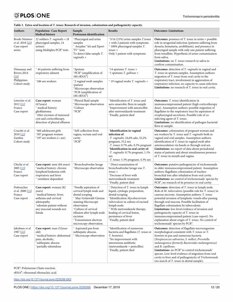
Table 3. Extra-oral location of T. tenax: Research of invasion, colonization and pathogenicity capacity.
Authors Population- Case Report
Medical history
Sample
Protozoan identification
Results Outcomes–Limitations
Brosh-Nisimov
et al. 2018 [29]
Case report
series
22 soldiers T. vaginalis + (8
pharyngeal samples, 14
urines)
using Multiplex PCRa tests
�Pharyngeal and urine
samples� Anyplex™ kit and Xpert-
TV™ tests
To detect false sample T.
vaginalis +
�3/14 (21%) urine samples T.tenax+ with 2 patients with symptoms� 100% pharyngeal samples T.
tenax +
Only 1 patient with symptoms
Outcomes: presence of T. tenax in urine = possible
role in urogenital infection (patients suffering from
dysuria, hematuria, urolithiasis), and presence in
pharyngeal sample with only one patient suffering
from tonsillitis. Hypothesis of urine contamination
from saliva.
Limitations: no T. tenax research in saliva to
confirm contamination.
Dimasuay and
Rivera 2014
[30]
Philippines
Cohort study
� 44 patients suffering from
respiratory ailment
�Sputum�PCRa (amplification of
18S rRNAb)
�14 sputums T. tenax +�1 sputums T. gallinae +
Outcomes: detection of T. vaginalis in vaginal and
T. tenax in sputum samples. Assumption authors:
migration of T. tenax from oral cavity to the
respiratory tract, involvement in aggravation of
respiratory infection, no capacity to cause infection.
Limitations: no research of T. tenax in oral cavity.
�186 sex workers �2 vaginal swab samples
/patient�Microscope observation�PCR (amplification of
18S rRNAb)
� 19 vaginal swabs T. vaginalis +
Leterrier et al.2012 [31]
France
Case report
Case report: woman
(67years)� medical history:
glioblastoma�After excision of tumoural
cyst and corticotherapy,
detection of pleural effusion
�Pleural fluid sample�Microscope observation�Culture�PCRa
�Identification of T. tenax and
aero-anaerobic flora in sample�Improvement with amoxicillin
after metronidazole treatment�Finally, patient died
Outcomes: T. tenax identification in
immunocompromised patient (high corticotherapy
dose). Assumption authors: possible migration of
flagellates to the respiratory tract by aspiration of
oropharyngeal secretions. Possible role of co-
infecting agent of T. tenax.
Limitations: no identification of pathogen bacterial
flora in sample.
Crucitti et al.2010 [32]
Zambia
Cohort study
�460 adolescent girls�307 pregnant women�197 sex workers (= sex)
�Self-collection from
vagina, rectum and oral
cavity�PCRa
�Identification in vaginal
infection of:
-T. vaginalis: 24,6% ado, 32,2%
pregnant, 33,2 sex
-T. tenax: 0.7% ado, 0.3% pregnant�Identification in oral cavity of:
-T. vaginalis: 0.7% pregnant, 1.1%
sex
-T. tenax: 5.3% pregnant, 4.3% sex
Outcomes: colonization of pregnant woman and
sex workers by T. tenax and T. vaginalis both in
vaginal and oral samples. Assumption authors:
identification of T. tenax in vaginal swab after
autoinoculation via hands or through oral sex.
Limitations: no report of data about periodontal
status of patients and about simultaneous presence
of T. tenax in mouth and vagina.
Chiche et al.2005 [33]
France
Case report
Case report: men (84 years)�medical history: chronic
lymphoid leukemia with
respiratory and fever� ventilator dependent
�Bronchoalveolar lavage�Microscope observation
� Direct examination of
bronchoalveolar lavage was T.
tenax +�Decrease of fever with
metronidazole treatment�Finally, patient died
Outcomes: putative pathogenicity of trichomonads
in older immunocompromised patient. Assumption
authors: flagellates colonization of tracheo-
bronchial tree after inhalation from oral cavity.
Limitations: no control of trichomonads’ species by
PCRa, no research of its presence in oral cavity.
Duboucher
et al. 2000 [34]
France
Case report
Case report: woman (82
years)�medical history: fever,
asthenia and cervical
adenopathy�edentate patient without
any mucosal wounds nor
fistula
�Needle aspiration of
cervical lymph node and
dental scraping� May-Grunwald-Giemsa
staining Microscope
observation�Culture of cervical
effusion after lymph node
excision�Transmission electron
microscopy observation.
� Detection of T. tenax in lymph
liquid, cytologic preparation,
dental scraping�IdentificationMycobacteriumtuberculosis in culture of excised
lymph node.� With metronidazole therapy,
healing of cervical lesion,
persistence of fever�Finally, patient died
Outcomes: detection of T. tenax in lymph node.
Role of M. tuberculosis (possible role for T. tenax) in
caseous necrosis. Assumption authors: T. tenaxpotential invasion of lymphatic vessels after passing
through oral mucosa. Possible facilitation of
flagellate colonization by tuberculosis.
Limitations: low-level evidence of invasion and
pathogenicity capacity of T. tenax in
immunocompromised patient (case-report). No
explanation about origin of T. tenax. No control of
trichomonads’ species by PCRa.
Jakobsen et al.1987 [35]
Case report
Case report: man (52year-
old)�medical history: abdominal
pain, alcoholic,�subhepatic abscess�partially edentulous
� Aspirated pus from
subhepatic abscess.� Microscope observation
�Identification of numerous
bacteria and flagellates (T. tenax or
T. hominis).�No improvement with
intravenous antibiotic
(metronidazole + penicillin).�Finally, patient died
Outcomes: detection of flagellate microorganism
morphological consistent with T. tenax or T.
hominis in pus and numerous bacteria
(Streptococcus salivarius, S. milleri, Prevotellamelanogenica (formerly Bacteroides melanogenicus)and B. capillosus.Limitations: no PCRa to control trichomonads’
species. Low-level evidence of migration from oral
cavity to liver and of pathogenicity of Trichomonads(no search of T. tenax in dental sample).
PCRa: Polymerase Chain reaction;
rRNAb: ribosomal ribonucleic acid.
https://doi.org/10.1371/journal.pone.0226266.t003
Trichomonas in periodontitis
PLOS ONE | https://doi.org/10.1371/journal.pone.0226266 December 17, 2019 12 / 20
healing or not of sites with periodontitis, and only reported differences in the frequency of
flagellate detection before and after the treatment in both saliva and biofilm sample. S2 Table
shows the details of the quality assessment.
• As regards the laboratory studies (Table 2). Numerous publications have explored the
potential pathogenicity of T. tenax. However, the authors generally used an ATCC strain
that could not fully reflect the virulence of clinical isolates and used cell lines for cytotoxicity
testing and cytokine production. Certainly, cell lines can react differently from primary cell
lines that constitute the cellular model with the closest similarity to the patient cells. More-
over, the methodological heterogeneity of protocols, the few T. tenax strains available and
the xenic culture medium used in some studies make it difficult to evaluate results. No ani-
mal model of periodontitis has been found with T. tenax.
Medium used in some studies make it difficult to evaluate results. No animal model of peri-
odontitis has been found with T. tenax.
• As regards the extra-oral location of T. tenax to research the pathogenic potential and
invasive power (Table 3). Some studies identified flagellates under the microscope, which is
not the most appropriate technique to confirm the trichomonads species. The authors postu-
lated the inhalation of flagellates from the oral cavity to the respiratory tract without any
dental sample analysis [30,31,33]. The majority of publications were case reports or case
report series; further investigations are required to conclude of any T. tenax pathogenicity in
an extra-oral location.
Discussion
This systematic review has evaluated the role of T. tenax in the etiopathogenesis of periodonti-
tis using Koch’s postulates revisited by Socransky as PICO framework. The principle findings
were discussed for each postulate and concerned in particular two of them: “Association” and
“Virulence factors”.
Among the various postulates and causal models that could provide evidence of pathoge-
nicity for a putative periodontal pathogen, Koch’s postulates revisited by Socransky is the most
well-known and used. Unfortunately, the implementation of these postulates is limited to cul-
tivable microbiota and has never be adapted to uncultivable microorganisms identified by
molecular biology that could also be suspected of initiating or participating in the pathogenesis
of the disease.
These Koch’s postulates must be adapted to new technologies and could be based on the
detection of nucleic acid sequence as suggested by Fredricks and Relman [36]. But these last
postulates cannot be applied to this literature review, because in most of the clinical studies the
authors identified T. tenax using microscopy.
It is increasingly clear that periodontitis is the result of the synergy of microorganisms
embedded in a biofilm [37]. But among all the detected microorganisms the Socransky modi-
fied postulates remains a way to discriminate pathogenic bacteria in periodontitis. In this
review, these postulates are applied to protozoan to compare their virulence factors to those of
well-known periodontopathogens.
The prevalence of Trichomonas tenax in periodontitis
Periodontitis is an inflammatory disease resulting from dysbiotic microbial communities of
pathobiont and keystone pathogens, which develop virulence factors inducing periodontal
Trichomonas in periodontitis
PLOS ONE | https://doi.org/10.1371/journal.pone.0226266 December 17, 2019 13 / 20
tissue degradation. The dysbiosis of periodontal microbiota results in the breakdown of the
bacterial balance in favour of periodontopathogens and to the detriment of commensal bacte-
ria [38]. The pathogens are present in relative low abundance in the healthy sites and their
quantity is proportional to the PPD in diseased sites.
The real prevalence of T. tenax in periodontitis could not be established for two reasons:
(i) the patients sample size seems to be insufficient to achieve a statistical significance, (ii) the
periodontal parameters were not detailed enough. Nevertheless, this protozoan has been
found in healthy sites as well as sites with gingivitis and periodontitis, from 7.1 to 28.2%, 0 to
32.3% and 32.3 to 42.5% respectively. No indication on the quantity of flagellates in the various
sites is known. The implementation of the dysbiosis concept for T. tenax cannot be totally
excluded as the flagellate is detected in both pathological and healthy sites as indicated for peri-
odontal pathogens [7,8,15,39]. The identification method (direct observation or observation of
the culture after the incubation time) and the heterogeneity of the sampling protocol make it
difficult to conclude on the specific identification of the protozoan in periodontitis due to the
morphological similarities with T. hominis and T. vaginalis. Nevertheless, one case-control
study used the PCR assay method and confirmed the presence of trichomonad species in sam-
ples from both patients with or without periodontitis. Some clinical studies have identified the
flagellate with PCR, but they could not be included in this review because authors have selected
patients with periodontal diseases as manifestation of systemic disease and/or patients without
any description of periodontium [40, 41, 32].
The postulate “Elimination”
The postulate “Elimination” evaluates the impact of non-surgical periodontal therapy on the
decrease or elimination of a putative pathogen in a healed site. Only one randomly controlled
trial evaluates the impact of scaling and root planing (SRP) on the frequency of protozoan
identification in patients with periodontitis (CAL > 5 mm) [16]. Surprisingly, the SRP treat-
ment induced a decrease of T. tenax in saliva but not in dental plaque. Some authors found a
high frequency of the flagellate in patients with poor hygiene [42].
One might expect that improved hygiene and a debridement of the periodontal pocket
would reduce the number of flagellates, as demonstrated for bacteria [43]. Only one periodon-
topathogen was not eliminated after SRP: Aggregatibacter actinomycetemcomitans [44]. This
pathobiont presents numerous virulence factors and a tissue and cell invasiveness capacity that
could explain “its resistance” to non-surgical treatment, but these properties have not been
described for flagellates. In the absence of periodontal parameters description before and after
treatment and in the absence of dental sample location (subgingival and/or supragingival), it is
difficult to draw solid conclusions from this study.
The postulate “Virulence factors”
Numerous authors have evaluated the factors that may respond to the postulate “Virulence
factors” in relation to T. tenax. Cellular adhesion is the first step in tissue colonisation for any
microorganism. The adhesion and colonisation capacity has clearly been described for T. vagi-nalis but not for T. tenax [18,19]. In 1983, Ribaux et al. described fibronectin-like protein
located on plasma membrane and protozoan axostyle, which could potentially have a role in
T. tenax adhesion to eukaryotic cells and in the phagocytosis of bacteria. These authors dem-
onstrated a particularly intense labelling localised in the contact zone between bacteria and
two strains of T. tenax by indirect immunostaining, one isolated from a patient and the other
from a laboratory strain, cultured in xenic medium [45]. This aspect was confirmed by Ribeiro
Trichomonas in periodontitis
PLOS ONE | https://doi.org/10.1371/journal.pone.0226266 December 17, 2019 14 / 20
et al. who demonstrated the ability of T. tenax ATCC 30207 to adhere to both cell lines and pri-
mary cells (patient epithelial cells) [17].
The explanation of the inconsistencies between the Alderete and Ribeiro adhesion results
could be attributed to the different times of protozoan cell interaction: thirty minutes (no con-
tact between the flagellates and cells) and 2 hours (visible interaction between the flagellates
and the epithelial cells) respectively [17– 19].
Moreover, Ribeiro has also shown the ability of T. tenax to induce cytotoxic effects such as
damage and disruption of cell monolayers. The oral protozoan appears to phagocytose mem-
brane portions of MDCK (Mardin-Darby Canine Kidney) epithelial cells and induces mem-
brane blebbing and apoptotic bodies in HeLa cells. The epithelial tissue is the first barrier
protecting the periodontium. The potential capacity of T. tenax to induce damage in cell
monolayers could promote and facilitate its invasion into the deepest connective tissue. This
hypothesis should be confirmed in animal models and with the analysis of gingival tissue of
patients with periodontitis. This cytotoxicity would prove the pathogenic role of T. tenax and
its potential participation in the cell breakdown described in periodontal diseases [46]. Like-
wise, Alderete observed no measurable cytotoxicity due to the too short contact of T. tenaxwith epithelial cell lines [18].
Numerous articles dealt with the capacity of T. tenax to produce enzymes. The authors have
identified some of them and assessed their properties. In 1991, Bozner and Demes, and more
recently El Sibaei, identified a temperature-dependant proteinase capable of degrading types I,
III, IV and V collagen, that would be cysteine proteinases or metallo-proteinases [20,24,25].
These enzymes are cell-associated and extracellular proteinases released from alive T. tenax[25]. Moreover, Yamamoto et al. have shown that an ATCC T. tenax strain can produce a spe-
cific enzyme that hydrolyses acid soluble type I collagen as well as gelatin, described as cathep-
sin B [21]. Numerous publications have described one of the main virulence factors in
periodontitis: cysteine proteinases, mainly produced by P. gingivalis but also by Tannerella for-sythia, are capable of directly degrading host tissues, activating the host proenzymes or neu-
tralizing the host immune system [47,48]. The collagenolytic activities of T. tenax proteases
could also, as indicated for bacterial cysteine proteases, participate in host matrix protein dam-
age and play a role in the etiopathogenesis of periodontal diseases [49]. But Bozner et al. con-
cluded that it would be of interest to purify T. tenax enzymes and test their effect on
extracellular matrix proteins in the oral cavity [25]. In addition to these enzymes, T. tenaxseems to be able to produce haemolysins, which are also synthesised by many pathobionts
such as P. gingivalis, Prevotella intermedia, A. actinomycetemcomitans and Treponema denti-cola [50–53, 22].
The postulate “Host responses”
All bacteria regarded as periodontal pathogens can trigger a "Host response" to both innate
and adaptative immunity. The only study assessing the response of macrophages to contami-
nation with live ATCC T. tenax strain failed to show pro-inflammatory cytokine production
[26]. Nevertheless, interleukine-8 is the only macrophage-produced cytokine after 6 hours of
incubation with flagellate lysates. The absence of cytokine synthesis with live flagellate could
be the result of the low ratio of trichomonads: macrophage, 1:5. Certainly, Ribeiro’s cytotoxic-
ity assay used an inverted ratio (5:1). T. tenax seems capable of inducing antibody production
as shown in the blood analysis of haemodialysis patients [27]. Moreover, Kott and Adler
detected two types of antibodies: one targeting the T. tenax flagella and another an agglutinin,
but this result is not relevant to show the potential pathogenicity of T. tenax [28]. Indeed,
intravenous injection of a commensal bacteria could induce the production of systemic
Trichomonas in periodontitis
PLOS ONE | https://doi.org/10.1371/journal.pone.0226266 December 17, 2019 15 / 20
antibodies [54]. Ioli et al. have detected antibodies against T. tenax in the blood of haemodialy-
sis patients without controlling the presence of bone loss around teeth of these patients. So, the
conclusion of the flagellate involvement in periodontitis should be cautiously considered. All
of these in vitro studies provided some possible responses to the question of the pathogenicity
of T. tenax. But these in vitro cellular models generally used cell lines and ATCC T. tenax strain
that cannot fully replicate the in vivo interaction between the T. tenax wild-type strain and pri-
mary human gingival cells.
Potential pathogenicity in extra-oral location of Trichomonas tenaxT. tenax is described in the literature as an inhabitant of the oral cavity, but some studies have
shown this flagellate in extra-oral locations such as the vagina, the respiratory system, the liver
and the lymph node of an immunocompromised patient with severe infectious disease. Most
of these articles are case reports which have the lowest level of evidence of all clinical studies
and conclude that T. tenax has no invasive power (via bloodstream) or pathogenicity. The
authors hypothesized that the flagellate moved from the oral cavity to the lung or was self-inoc-
ulated via the hands or through oral sex to the vagina and was only a co-factor aggravating the
underlying infection. Duboucher et al. could not explain the lymphatic colonization of the
flagellate, although an invasion via the lymphatic vessels, and postulated a putative role in the
cervical adenopathy of the patient as this protozoan was the only microorganism identified in
the lymph node [34]. Even if three weeks later a culture of the excised lymph node showed
Mycobacterium tuberculosis, the authors suggest that the observed caseous necrosis could be
attributed not only toM. tuberculosis but also to T. tenax [34]. Jakobsen speculated about a
possible migration through the blood to explain the presence of T. tenax in subhepatic abscess
[35]. Only Brosh-Nissimov et al. have reported the presence of the flagellate in extra-oral loca-
tions in immunocompetent patients with urogenital infection. These authors concluded that
T. tenax could be one of the potential etiologies of this dysuria [29].
Strengths and limitations
As far as is known, this review is the first one to investigate the role of T. tenax in periodontitis
using PRISMA guidelines and Koch’s postulates revisited by Socransky. These results shown
an interesting evaluation of the role of this flagellate in the periodontitis of human adult with-
out any systemic nor genetic disease. Nevertheless, this review presents some limitations:
(i) language restriction, (ii) limited number of papers with precise periodontal parameters
detecting T. tenax, (iii) majority of the included articles identified this trichomonads by micro-
scope which is not the most accurate technique, (iv) regarding research articles, authors used
either ATCC or patient strain which may not be representative of all wild strains in patients
with periodontitis (v) and the use of different protocols (MOI, time of incubation, cell lines)
makes comparison difficult and does not allow a firm conclusion.
Conclusions
Even if T. tenax appears to be identified in patients with periodontitis, there are still many
unanswered questions. The cellular model with flagellate has demonstrated its acknowledged
adhesion, cytotoxicity and enzyme production that could contribute to periodontal tissue
breakdown brought about by periodontopathogens. Its direct pathogenic role in periodontitis
but also in extra-oral infections remains unclear and requires further investigations. Periodon-
tal microbiota dysbiosis is not only an imbalance in the relative abundance of certain microbial
species found in diseased sites, but also involves polymicrobial synergy leading to immune dys-
functions that result in alveolar bone and soft tissue destruction. In the absence of direct
Trichomonas in periodontitis
PLOS ONE | https://doi.org/10.1371/journal.pone.0226266 December 17, 2019 16 / 20
pathogenicity, T. tenax could be indirectly involved in the etiopathogenesis of periodontitis.
Certainly, the bacteria phagocytized by the flagellate may be either lysed or alive inside its cyto-
plasm and be protected from immune cells and antibiotics action [55]. In this new context, the
roles of individual bacteria and their interaction with host and also with other microorganisms
such as protozoan must be evaluated.
Supporting information
S1 Table. PRISMA checklist.
(DOC)
S2 Table. Reasons for exclusion of publications recorded after removal of duplicates.
(XLSX)
S3 Table. Quality assessment of clinical studies included (based on Newcastle Ottawa
Scale).
(XLSX)
Author Contributions
Conceptualization: C. Bisson, S. M. Dridi.
Formal analysis: C. Bisson, S. M. Dridi, M. Machouart.
Investigation: C. Bisson, S. M. Dridi.
Methodology: C. Bisson, S. M. Dridi, M. Machouart.
Resources: C. Bisson, S. M. Dridi.
Writing – original draft: C. Bisson, S. M. Dridi.
Writing – review & editing: C. Bisson, S. M. Dridi, M. Machouart.
References1. Marcenes W, Kassebaum NJ, Bernabe E, Flaxman A, Naghavi M, Lopez A, et al. Global burden of oral
conditions in 1990–2010: a systematic analysis. J Dent Res. 2013; 92: 592–597. https://doi.org/10.
1177/0022034513490168 PMID: 23720570
2. Kassebaum NJ, Bernabe E, Dahiya M, Bhandari B, Murray CJ, Marcenes W. Global burden of severe
periodontitis in 1990–2010: a systematic review and meta-regression. J Dent Res. 2014; 93: 1045–
1053. https://doi.org/10.1177/0022034514552491 PMID: 25261053
3. Hajishengallis G, Moutsopoulos NM, Hajishengallis E, Chavakis T. Immune and regulatory functions of
neutrophils in inflammatory bone loss. Seminars in Immunology. 2016; 28: 146–58. https://doi.org/10.
1016/j.smim.2016.02.002 PMID: 26936034
4. Avila M, Ojcius DM, Yilmaz O. The oral microbiota: living with a permanent guest. DNA Cell Biol. 2009;
28: 405–411. https://doi.org/10.1089/dna.2009.0874 PMID: 19485767
5. Li F, Zhu C, Deng FY, Wong MCM, Lu HX, Feng XP. Herpesviruses in etiopathogenesis of aggressive
periodontitis: A meta-analysis based on case-control studies. PLoS One. 2017; 12: e0186373. https://
doi.org/10.1371/journal.pone.0186373 PMID: 29036216
6. Canabarro A, Valle C, Farias MR, Santos FB, Lazera M, Wanke B. Association of subgingival coloniza-
tion of Candida albicans and other yeasts with severity of chronic periodontitis. J Periodontal Res. 2013;
48: 428–432. https://doi.org/10.1111/jre.12022 PMID: 23137301
7. Feki A, Molet B, Haag R, Kremer M. [Protozoa of the human oral cavity (epidemiological correlations
and pathogenic possibilities]. J Biol Buccale. 1981; 9: 155–161. PMID: 6943141
8. Benabdelkader S, Andreani J, Gillet A, Terrer E, Pignoly M, Chaudet H, et al. Specific clones of Tricho-
monas tenax are associated with periodontitis. PLoS One. 2019; 14: e0213338. https://doi.org/10.
1371/journal.pone.0213338 PMID: 30856220
Trichomonas in periodontitis
PLOS ONE | https://doi.org/10.1371/journal.pone.0226266 December 17, 2019 17 / 20
9. Marty M, Lemaitre M, Kemoun P, Morrier JJ, Monsarrat P. Trichomonas tenax and periodontal dis-
eases: a concise review. Parasitology. 2017; 144: 1417–1425. https://doi.org/10.1017/
S0031182017000701 PMID: 28583214
10. Moher D, Liberati A, Tetzlaff J, Altman DG. PRISMA Group. Preferred reporting items for systematic
reviews and meta-analyses: the PRISMA Statement. Open Med. 2009; 3:e123–30. PMID: 21603045
11. Hardi AC, Fowler SA. Evidence-Based Medicine and Systematic Review Services at Becker Medical
Library. Mo Med. 2014; 111: 416–418. PMID: 25438363
12. Socransky SS, Haffajee AD. The bacterial etiology of destructive periodontal disease: current concepts.
J Periodontol. 1992; 63: 322–331.
13. Armitage GC. Development of a classification system for periodontal diseases and conditions. Ann Per-
iodontol. 1999; 4: 1–6. https://doi.org/10.1902/annals.1999.4.1.1 PMID: 10863370
14. Wells G, Tugwell P, Shea B, Guyatt G, Peterson J, Zytaruk N, et al. Osteoporosis Methodology Group
and The Osteoporosis Research Advisory Group. Meta-analyses of therapies for postmenopausal oste-
oporosis. V. Meta-analysis of the efficacy of hormone replacement therapy in treating and preventing
osteoporosis in postmenopausal women. Endocr Rev. 2002; 23: 529–539. PMID: 12202468
15. Ferrara A, Conca R, Grassi L Jr, de Carneri I. Possible pathogenic role of Trichomonas tenax in chronic
periodontitis. Ann Ist Super Sanita. 1986; 22: 253–255. PMID: 3752801
16. Rashidi Maybodi F, Haerian Ardakani A, Fattahi Bafghi A, Haerian Ardakani A, Zafarbakhsh A. The
Effect of Nonsurgical Periodontal Therapy on Trichomonas Tenax and Entamoeba Gingivalis in
Patients with Chronic Periodontitis. J Dent (Shiraz). 2016; 17: 171–176.
17. Ribeiro LC, Santos C, Benchimol M. Is Trichomonas tenax a Parasite or a Commensal? Protist. 2015;
166: 196–210. https://doi.org/10.1016/j.protis.2015.02.002 PMID: 25835639
18. Alderete JF, Garza GE. Specific nature of Trichomonas vaginalis parasitism of host cell surfaces. Infect
Immun. 1985; 50: 701–708. PMID: 3877690
19. Alderete JF, Pearlman E. Pathogenic Trichomonas vaginalis cytoxicity to cell culture monolayers. Br J
Vener Dis. 1984; 60: 99–105. https://doi.org/10.1136/sti.60.2.99 PMID: 6608395
20. El Sibaei MM, Abdel-Fattah NS, Ahmed SA, Abou-Seri HM. Growth kinetics, antigen profiling, and pro-
teinase activity of Egyptian Trichomonas tenax isolates derived from patients having oral infections.
Exp Parasitol. 2012; 130: 416–422. https://doi.org/10.1016/j.exppara.2012.01.018 PMID: 22348932
21. Yamamoto A, Asaga E, Nagao E, Igarashi T, Goto N. Characterization of the Cathepsin B-like Protein-
ases of Trichomonas Tenax ATCC 30207. Oral Microbiol Immunol. 2000; 15: 360–364. https://doi.org/
10.1034/j.1399-302x.2000.150604.x PMID: 11154432
22. Nagao E, Yamamoto A, Igarashi T, Goto N, Sasa R. Two Distinct Hemolysins in Trichomonas Tenax
ATCC 30207. Oral Microbiol Immunol. 2000; 15: 355–359. https://doi.org/10.1034/j.1399-302x.2000.
150603.x PMID: 11154431
23. Segović S, Buntak-Kobler D, GalićN, KatunarićM. Trichomonas Tenax Proteolytic Activity. Coll Antro-
pol. 1998; 22: 45–49. PMID: 9951139
24. Bozner P, Demes P. Degradation of Collagen Types I, III, IV and V by Extracellular Proteinases of an
Oral Flagellate Trichomonas Tenax. Arch Oral Biol.1991; 36: 765–770. https://doi.org/10.1016/0003-
9969(91)90043-t PMID: 1747075
25. Bozner P, Demes P. Cell-associated and extracellular proteolytic activity of an oral flagellate, Trichomo-
nas tenax. Arch Oral Biol. 1991; 36: 77–83. https://doi.org/10.1016/0003-9969(91)90056-z PMID:
2012529
26. Govro EJ, Stuart MK. Cytokine response of human THP-1 macrophages to Trichomonas tenax. Exp
Parasitol. 2016; 169: 77–80. https://doi.org/10.1016/j.exppara.2016.07.011 PMID: 27497807
27. Ioli A, Ferlazzo M, Fossari MT, Gembillo C, Skotarczak B, Vasi A, et al. Trichomonas tenax in haemo-
dyalized patients-serologic research. Wiad Parazytol. 1987; 33: 165–167. PMID: 3687008
28. Kott H, Adler S. A Serological Study of Trichomonas Sp. Parasitic in Man. Trans R Soc Trop Med Hyg.
1961; 55: 333–344. https://doi.org/10.1016/0035-9203(61)90102-x PMID: 13753502
29. Brosh-Nissimov T, Hindiyeh M, Azar R, Smollan G, Belausov N, Mandelboim M, et al. A false-positive
Trichomonas vaginalis result due to Trichomonas tenax presence in clinical specimens may reveal a
possible T. tenax urogenital infection. Clin Microbiol Infect. 2019; 25:123–124. https://doi.org/10.1016/j.
cmi.2018.09.011 PMID: 30267929
30. Dimasuay KG, Rivera WL. First report of Trichomonas tenax infections in the Philippines. Parasitol Int.
2014; 63: 400–402. https://doi.org/10.1016/j.parint.2013.12.015 PMID: 24406842
31. Leterrier M, Morio F, Renard B, Poirier AS, Miegeville M, Chambreuil G. Trichomonads in Pleural Effu-
sion: Case Report, Literature Review and Utility of PCR for Species Identification. New Microbiol. 2012;
35: 83–87. PMID: 22378558
Trichomonas in periodontitis
PLOS ONE | https://doi.org/10.1371/journal.pone.0226266 December 17, 2019 18 / 20
32. Crucitti T, Jespers V, Mulenga C, Khondowe S, Vandepitte J, Buve A. Trichomonas vaginalis is highly
prevalent in adolescent girls, pregnant women, and commercial sex workers in Ndola, Zambia. Sex
Transm Dis. 2010; 37: 223–227. PMID: 19940808
33. Chiche L, Donati S, Corno G, Benoit S, Granier I, Chouraki M, et al. Trichomonas tenax in pulmonary
and pleural diseases. Presse Med. 2005; 34: 1371–1372. PMID: 16292189
34. Duboucher C, Farto-Bensasson F, Cheron M, Peltier JY, Beaufils F, Perie G. Lymph Node Infection by
Trichomonas Tenax: Report of a Case with Co-Infection by Mycobacterium Tuberculosis. Hum
Pathol.2000; 31: 1317–1321. https://doi.org/10.1053/hupa.2000.18502 PMID: 11070125
35. Jakobsen EB, Friis-Moller A, Friis J. Trichomonas Species in a Subhepatic Abscess. Eur J Clin Micro-
biol. 1987; 6: 296–297. https://doi.org/10.1007/bf02017616 PMID: 3622496
36. Fredericks DN, Relman DA. Sequence-based identification of microbial pathogens: a reconsideration of
Koch’s postulates. Clin Microbiol Rev. 1996; 9: 18–33. PMID: 8665474
37. Perez-Chaparro PJ, Goncalves C, Figueiredo LC, Faveri M, Lobão E, Tamashiro N, et al. Newly identi-
fied pathogens associated with periodontitis: a systematic review. J Dent Res. 2014; 93: 846-858.
https://doi.org/10.1177/0022034514542468 PMID: 25074492
38. Hajishengallis G. Immunomicrobial pathogenesis of periodontitis: keystones, pathobionts, and host
response. Trends Immunol. 2014; 35: 3–11. https://doi.org/10.1016/j.it.2013.09.001 PMID: 24269668
39. Chen C, Hemme C, Beleno J, Shi Z, Ning D, Qin Y, et al. Oral microbiota of periodontal health and dis-
ease and their changes after nonsurgical periodontal therapy. ISME J. 2018; 12: 1210–1224. https://
doi.org/10.1038/s41396-017-0037-1 PMID: 29339824
40. Mehr A.K, Zarandi A, Anush K. Prevalence of Oral Trichomonas tenax in Periodontal Lesions of Down
Syndrome in Tabriz, Iran. J Clin Diagn Res. 2015; 9:88–90.
41. Dybicz M, Perkowski K, Sędzikowska A, Baltaza W, Chomicz L. Studies on prevalence of infection with
Trichomonas tenax identified by molecular techniques–in respect to oral health of patients with various
systemic disease requiring immunosuppressive therapy. Ann Parasitol. 2018; 64: 193–197. PMID:
30316209
42. Wantland WW, Lauer D. Correlation of some oral hygiene variables with age, sex, and incidence of oral
protozoa. J Dent Res. 1970; 49: 293–297. https://doi.org/10.1177/00220345700490021601 PMID:
5264592
43. Rosalem W, Rescala B, Teles RP, Fischer RG, Gustafsson A, Figueredo CM. Effect of non-surgical
treatment on chronic and aggressive periodontitis: clinical, immunologic, and microbiologic findings. J
Periodontol. 2011; 82: 979–989. https://doi.org/10.1902/jop.2011.100579 PMID: 21309718
44. Muller HP, Heinecke A, Borneff M, Kiencke C, Knopf A, Pohl S. Eradication of Actinobacillus actinomy-
cetemcomitans from the oral cavity in adult periodontitis. J Periodontal Res. 1998; 33: 49–58. https://
doi.org/10.1111/j.1600-0765.1998.tb02291.x PMID: 9524321
45. Ribaux CL, Magloire H, Joffre A, Morrier JJ. Immunohistochemical localization of fibronectin-like protein
on the cell surface of the oral flagelatte Trichomonas tenax. J Biol Buccale. 1983; 11: 41–51. PMID:
6343365
46. Bugueno IM, Khelif Y, Seelam N, Morand DN, Tenenbaum H, Davideau JL, et al. Porphyromonas gingi-
valis Differentially Modulates Cell Death Profile in Ox-LDL and TNF-α Pre-Treated Endothelial Cells.
PLoS One. 2016; 11: e0154590. https://doi.org/10.1371/journal.pone.0154590 PMID: 27124409
47. De Diego I, Veillard F, Sztukowska MN, Guevara T, Potempa B, Pomowski A, et al. Structure and
mechanism of cysteine peptidase gingipain K (Kgp), a major virulence factor of Porphyromonas gingiva-
lis in periodontitis. J Biol Chem. 2014; 289: 32291–32302. https://doi.org/10.1074/jbc.M114.602052
PMID: 25266723
48. Sahingur SE, Xia XJ, Alamgir S, Honma K, Sharma A, Schenkein HA. DNA from Porphyromonas gingi-
valis and Tannerella forsythia induce cytokine production in human monocytic cell lines. Mol Oral Micro-
biol. 2010; 25: 123–135. https://doi.org/10.1111/j.2041-1014.2009.00551.x PMID: 20331800
49. Tokuda M, Duncan M, Cho MI, Kuramitsu HK. Role of Porphyromonas gingivalis protease activity in col-
onization of oral surfaces. Infect Immun. 1996; 64: 4067–4073. PMID: 8926070
50. Hosogi Y, Hayakawa M, Abiko Y. Monoclonal antibody against Porphyromonas gingivalis hemaggluti-
nin inhibits hemolytic activity. Eur J Oral Sci. 2001; 109: 109–113. https://doi.org/10.1034/j.1600-0722.
2001.00995.x PMID: 11347654
51. Balashova NV, Crosby JA, Al Ghofaily L, Kachlany SC. Leukotoxin confers beta-hemolytic activity to
Actinobacillus actinomycetemcomitans. Infect Immun. 2006; 74: 2015–2021. https://doi.org/10.1128/
IAI.74.4.2015-2021.2006 PMID: 16552030
52. Kachlany SC. Aggregatibacter actinomycetemcomitans leukotoxin: from threat to therapy. J Dent Res.
2010; 89: 561–570. https://doi.org/10.1177/0022034510363682 PMID: 20200418
Trichomonas in periodontitis
PLOS ONE | https://doi.org/10.1371/journal.pone.0226266 December 17, 2019 19 / 20
53. Chu L, Ebersole JL, Kurzban GP, Holt SC. Cystalysin, a 46-kilodalton cysteine desulfhydrase from
Treponema denticola, with hemolytic and hemoxidative activities. Infect Immun. 1997; 65: 3231–3238.
PMID: 9234780
54. Macpherson AJ, Harris NL. Interactions between commensal intestinal bacteria and the immune sys-
tem. Nat Rev Immunol. 2004; 4: 478–85. https://doi.org/10.1038/nri1373 PMID: 15173836
55. Ribaux CL. Study of an oral protozoan Trichomonas tenax using scanning and transmission electron
microscopy. J Biol Buccale. 1979; 7: 157–168. PMID: 287668
Trichomonas in periodontitis
PLOS ONE | https://doi.org/10.1371/journal.pone.0226266 December 17, 2019 20 / 20
Top Related
Copyright © 2022 FDOKUMEN

