


Bahasa
Halaman
Hukum

Arabidopsis UEV1D Promotes Lysine-63–LinkedPolyubiquitination and Is Involved in DNA Damage Response W
Rui Wen,a J. Antonio Torres-Acosta,b,1 Landon Pastushok,a Xiaoqin Lai,a,2 Lindsay Pelzer,a
Hong Wang,b,3 and Wei Xiaoa,3
a Department of Microbiology and Immunology, University of Saskatchewan, Saskatoon, Saskatchewan, Canada S7N 5E5b Department of Biochemistry, University of Saskatchewan, Saskatoon, Saskatchewan, Canada S7N 5E5
DNA damage tolerance (DDT) in budding yeast requires Lys-63–linked polyubiquitination of the proliferating cell nuclear
antigen. The ubiquitin-conjugating enzyme Ubc13 and the Ubc enzyme variant (Uev) methyl methanesulfonate2 (Mms2) are
required for this process. Mms2 homologs have been found in all eukaryotic genomes examined; however, their roles in
multicellular eukaryotes have not been elucidated. We report the isolation and characterization of four UEV1 genes from
Arabidopsis thaliana. All four Uev1 proteins can form a stable complex with At Ubc13 or with Ubc13 from yeast or human and
can promote Ubc13-mediated Lys-63 polyubiquitination. All four Uev1 proteins can replace yeast MMS2 DDT functions in vivo.
Although these genes are ubiquitously expressed in most tissues, UEV1D appears to express at a much higher level in
germinating seeds and in pollen. We obtained and characterized two uev1d null mutant T-DNA insertion lines. Compared with
wild-type plants, seeds from uev1d null plants germinated poorly when treated with a DNA-damaging agent. Those that
germinated grew slower, and the majority ceased growth within 2 weeks. Pollen from uev1d plants also displayed a moderate
but significant decrease in germination in the presence of DNA damage. This report links Ubc13-Uev with functions in DNA
damage response in Arabidopsis.
INTRODUCTION
Cellular DNA is subject to assaults by environmental factors and
endogenous metabolites. The alteration of DNA can lead to mu-
tagenesis, genome rearrangements, and cell death (Friedberg
et al., 2006). To maintain genome integrity, all living organisms
have evolved a variety of DNA repair mechanisms to protect cells
from DNA damage. However, despite great advances made
during the last decade in the field of DNA repair and mutagen-
esis, the molecular mechanisms of DNA damage tolerance (DDT)
in eukaryotes, especially in multicellular eukaryotes, have not yet
been well characterized. In the lower eukaryote Saccharomyces
cerevisiae, a DDT process known as DNA postreplication repair
(PRR) facilitates DNA synthesis in the presence of replication-
blocking lesions in the template. PRR consists of two branches:
an error-prone (mutagenesis) branch and an error-free branch.
The error-prone branch is mediated by specialized DNA poly-
merases, including Polz (Rev3 þ Rev7), Polh, and Rev1, which
are required for translesion DNA synthesis (TLS). By contrast, the
error-free branch is mediated by the ubiquitin (Ub)-conjugating
enzyme (Ubc or E2)–Ubc variant (Uev) complex Ubc13-Mms2
(for methyl methanesulfonate2), which acts to prevent sponta-
neous and DNA damage–induced mutagenesis (Broomfield
et al., 2001; Barbour and Xiao, 2003). It is now clear that yeast
PRR is accomplished by stepwise covalent modifications of the
proliferating cell nuclear antigen (PCNA) encoded by POL30. In
response to DNA damage, the Rad6-Rad18 ubiquitination com-
plex monoubiquitinates Pol30 at the Lys-164 residue, then the
Mms2-Ubc13-Rad5 complex further polyubiquitinates Pol30
through Lys-63–linked chains (Hoege et al., 2002). It is thus
assumed that monoubiquitinated Pol30 promotes error-prone
TLS, whereas polyubiquitinated Pol30 promotes error-free by-
pass of damaged templates (Stelter and Ulrich, 2003; Pastushok
and Xiao, 2004). The same Lys-164 residue can also be cova-
lently modified by SUMO (for small ubiquitin-related modifier),
which requires the Siz1-Ubc9 complex; sumoylated Pol30 re-
cruits the DNA helicase Srs2 to stalled replication forks to pre-
vent inappropriate recombination (Papouli et al., 2005; Pfander
et al., 2005).
Ubc13 and Uev homologs are found in all eukaryotes exam-
ined to date (Villalobo et al., 2002; Pastushok and Xiao, 2004),
suggesting that their functions are conserved throughout eukary-
otes. Despite numerous reports of the isolation and character-
ization of eukaryotic UBC13 and MMS2/UEV1 genes and
indications that they may be involved in DNA repair and damage
tolerance, the functions of these genes remain uncharacterized
in a multicellular organism. With regard to the fact that the
1 Current address: Institute of Applied Genetics and Cell Biology,Department of Applied Plant Sciences and Plant Biotechnology,University of Natural Resources and Applied Life Sciences, Muthgasse18, 1190 Vienna, Austria.2 Current address: Department of Biology, University of Pennsylvania,Philadelphia, PA 19104.3 Address correspondence to [email protected] or [email protected] authors responsible for distribution of materials integral to thefindings presented in this article in accordance with the policy describedin the Instructions for Authors (www.plantcell.org) are: Hong Wang([email protected]) and Wei Xiao ([email protected]).W Online version contains Web-only data.www.plantcell.org/cgi/doi/10.1105/tpc.107.051862
The Plant Cell, Vol. 20: 213–227, January 2008, www.plantcell.org ª 2008 American Society of Plant Biologists
deletion of UBC13 (Yamamoto et al., 2006a, 2006b) and possibly
MMS2/UEV1 in mammals may cause embryonic lethality, we
turned our attention to Arabidopsis thaliana as an alternative
multicellular model organism to study the Lys-63 polyubiquiti-
nation and DDT pathway. Analysis of the Arabidopsis genome
database indicates that most genes involved in DNA repair are
highly conserved between plants and mammals (Tuteja et al.,
2001). Most importantly, plants are much more tolerant of
genomic instability and chromosome rearrangements, making
the plant a suitable model organism to study DNA repair/toler-
ance defects.
Protein ubiquitination and its role in regulating protein degra-
dation have been extensively studied in plants (Bachmair et al.,
2001; Devoto et al., 2003; Vierstra, 2003; Moon et al., 2004). The
conventional ubiquitination process is via Lys-48–linked Ub
chain formation, which targets these proteins for degradation
through 26S proteasomes (Hochstrasser, 1996). By contrast, Ub
chains linked to substrates through Ub–Lys-63 regulate diverse
functions in a nonproteolytic manner (Pickart, 2001a). To date,
Ubc13 is the only known E2 capable of polyubiquitinating target
proteins via Lys-63–linked chains, and this activity absolutely
requires a Uev as a cofactor (Hofmann and Pickart, 1999;
McKenna et al., 2001; Pastushok et al., 2005).
Arabidopsis genes involved in TLS, including REV3 (Sakamoto
et al., 2003), REV1, REV7 (Takahashi et al., 2005), POLK (Garcia-
Ortiz et al., 2004), and POLH (Curtis and Hays, 2007), have been
isolated and characterized. TLS appears to play an important
role in the tolerance of DNA damage in plants. We recently
showed that two Arabidopsis UBC13 genes could complement a
yeast ubc13 null mutant for spontaneous mutagenesis as well as
interact with yeast and human Uev proteins (Wen et al., 2006),
suggesting that the same pathway for an error-free DDT also
exists in plants. However, the roles of UBC13 in DDT have not
been directly assessed, and there has been no experimental
evidence for plant Ubc13-Uev complex formation or for Lys-63–
linked protein ubiquitination. In this report, we describe the
molecular cloning and functional characterization of four Arabi-
dopsis UEV1 genes and report a case of mutant phenotypes in
DNA damage response when one of the UEV1 genes is inacti-
vated.
RESULTS
Isolation of Arabidopsis UEV1 Genes
To identify Arabidopsis UEV1 genes, a human Mms2 sequence
(Xiao et al., 1998) was used to search for homologs in the
Arabidopsis protein database (through The Arabidopsis Infor-
mation Resource [TAIR]; www.arabidopsis.org). Four hypothet-
ical proteins with a high degree of similarity (E-values < 2e-38)
were found and named At Uev1A (At1g23260), At Uev1B
(At1g70660), At Uev1C (At2g36060), and At Uev1D (At3g52560).
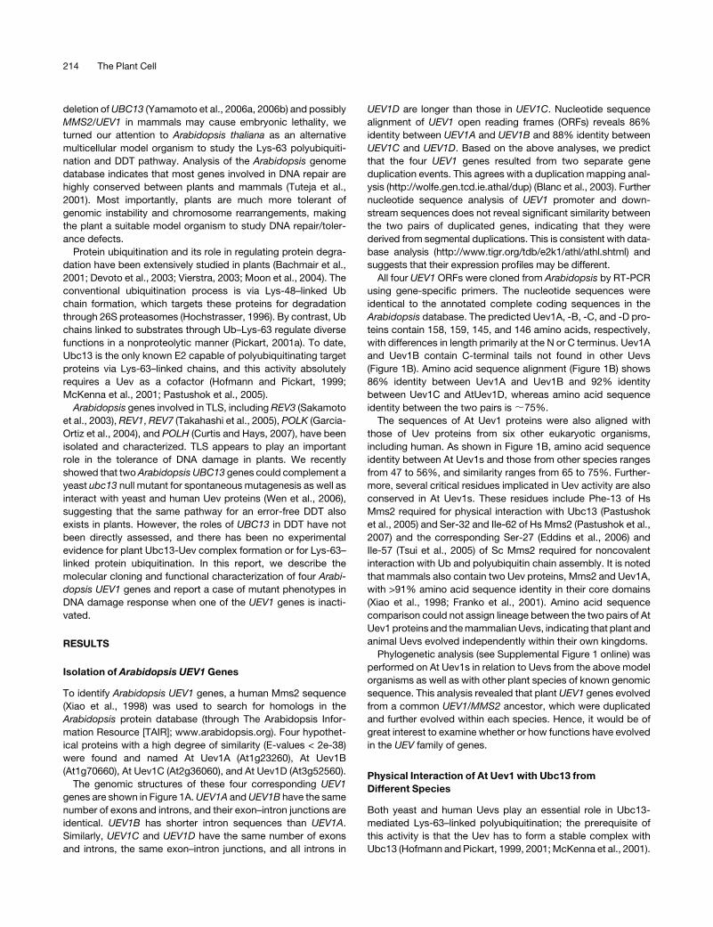
The genomic structures of these four corresponding UEV1
genes are shown in Figure 1A. UEV1A and UEV1B have the same
number of exons and introns, and their exon–intron junctions are
identical. UEV1B has shorter intron sequences than UEV1A.
Similarly, UEV1C and UEV1D have the same number of exons
and introns, the same exon–intron junctions, and all introns in
UEV1D are longer than those in UEV1C. Nucleotide sequence
alignment of UEV1 open reading frames (ORFs) reveals 86%
identity between UEV1A and UEV1B and 88% identity between
UEV1C and UEV1D. Based on the above analyses, we predict
that the four UEV1 genes resulted from two separate gene
duplication events. This agrees with a duplication mapping anal-
ysis (http://wolfe.gen.tcd.ie.athal/dup) (Blanc et al., 2003). Further
nucleotide sequence analysis of UEV1 promoter and down-
stream sequences does not reveal significant similarity between
the two pairs of duplicated genes, indicating that they were
derived from segmental duplications. This is consistent with data-
base analysis (http://www.tigr.org/tdb/e2k1/athl/athl.shtml) and
suggests that their expression profiles may be different.
All four UEV1 ORFs were cloned from Arabidopsis by RT-PCR
using gene-specific primers. The nucleotide sequences were
identical to the annotated complete coding sequences in the
Arabidopsis database. The predicted Uev1A, -B, -C, and -D pro-
teins contain 158, 159, 145, and 146 amino acids, respectively,
with differences in length primarily at the N or C terminus. Uev1A
and Uev1B contain C-terminal tails not found in other Uevs
(Figure 1B). Amino acid sequence alignment (Figure 1B) shows
86% identity between Uev1A and Uev1B and 92% identity
between Uev1C and AtUev1D, whereas amino acid sequence
identity between the two pairs is ;75%.
The sequences of At Uev1 proteins were also aligned with
those of Uev proteins from six other eukaryotic organisms,
including human. As shown in Figure 1B, amino acid sequence
identity between At Uev1s and those from other species ranges
from 47 to 56%, and similarity ranges from 65 to 75%. Further-
more, several critical residues implicated in Uev activity are also
conserved in At Uev1s. These residues include Phe-13 of Hs
Mms2 required for physical interaction with Ubc13 (Pastushok
et al., 2005) and Ser-32 and Ile-62 of Hs Mms2 (Pastushok et al.,
2007) and the corresponding Ser-27 (Eddins et al., 2006) and
Ile-57 (Tsui et al., 2005) of Sc Mms2 required for noncovalent
interaction with Ub and polyubiquitin chain assembly. It is noted
that mammals also contain two Uev proteins, Mms2 and Uev1A,
with >91% amino acid sequence identity in their core domains
(Xiao et al., 1998; Franko et al., 2001). Amino acid sequence
comparison could not assign lineage between the two pairs of At
Uev1 proteins and the mammalian Uevs, indicating that plant and
animal Uevs evolved independently within their own kingdoms.
Phylogenetic analysis (see Supplemental Figure 1 online) was
performed on At Uev1s in relation to Uevs from the above model
organisms as well as with other plant species of known genomic
sequence. This analysis revealed that plant UEV1 genes evolved
from a common UEV1/MMS2 ancestor, which were duplicated
and further evolved within each species. Hence, it would be of
great interest to examine whether or how functions have evolved
in the UEV family of genes.
Physical Interaction of At Uev1 with Ubc13 from
Different Species
Both yeast and human Uevs play an essential role in Ubc13-
mediated Lys-63–linked polyubiquitination; the prerequisite of
this activity is that the Uev has to form a stable complex with
Ubc13 (Hofmann and Pickart, 1999, 2001; McKenna et al., 2001).
214 The Plant Cell
Figure 1. Sequence Analysis of At UEV1 Genes and Their Products.
(A) Genomic organization of UEV1. Open boxes, untranslated region; closed boxes, coding regions; solid lines, introns; dotted lines, identical intron–
exon alignment between different UEV1 genes.
(B) Amino acid sequence alignment of At Uev1 and Uevs from six other organisms. The sequences were aligned and edited using the BioEdit program
version 5.0.9 (Hall, 1999). Residues are highlighted when 50% or more are identical. Critical residues for Mms2/Uev functions are indicated with
asterisks underneath the residues.
At UEV1 in Ubiquitination and DNA Damage Tolerance 215

In order to detect this interaction, we performed a yeast two-
hybrid assay (Fields and Song, 1989) between the cloned At
UEV1s and UBC13 genes from different species. All four At Uev1
proteins were able to interact with either At Ubc13A or At
Ubc13B; however, the strength of interaction appears to be
different. At Uev1A and At Uev1B gave positive results with At
Ubc13s under high stringency (SD-Ade for 3 d), but At Uev1C and
At Uev1D gave weak and no interaction, respectively, under the
same conditions (Figure 2A). Nevertheless, all of the above
interactions are robust and deemed strong, as none of the
negative controls reveal positive interactions under low strin-
gency and many bona fide positive interactions may not survive
as low as 1 mM 1,2,4-aminotriazole concentration under the
same experimental conditions. The UEV1D-4 clone was identi-
fied among initial UEV1D clones; its ORF contains a three-
nucleotide (GTA) insertion at position 175 that would encode the
additional amino acid Val. This has been predicted to be a
splicing variant of UEV1D (At3g52560.2) in the Arabidopsis
genome database. Uev1D-4 appears to be able to interact with
Ubc13, albeit at a reduced affinity compared with Uev1D (Figure
2A). The physiological significance of this variant has yet to be
investigated. In addition, the yeast two-hybrid analyses showed
that all four At Uev1 proteins are able to physically interact with
Ubc13 from yeast or human, and the strength of interaction
follows the trend Uev1A ¼ Uev1B > Uev1C > Uev1D (Figure 2B;
see Supplemental Figure 2A online).
To further confirm the physical interaction between Uev1 and
Ubc13 in vitro, a glutathione S-transferase (GST)–affinity pull-
down assay was conducted. As shown in Figure 2C, purified
GST-Uev1A (lane 6) and GST-Uev1D (lane 7) are able to specif-
ically interact with Ubc13A. As a negative control, GST alone
(lane 5) does not bind to Ubc13A under the same experimental
conditions. Similar results were also obtained with Uev1B and
Uev1C (see Supplemental Figure 2B online). Hence, all four Uev1
proteins are able to form stable heterodimers with Ubc13.
Uev1 Is Required for Ubc13-Mediated Lys-63–Linked
Polyubiquitination in Vitro
It has been reported that while yeast and human Ubc13s are
bona fide E2 enzymes capable of forming active-site thioesters
with Ub, a Uev is absolutely required for Ub chain assembly.
Furthermore, these chains are linked through Lys-63 instead of
Figure 2. Biochemical Properties of Uev1.
(A) Physical interaction between Ubc13 and Uev1 in a yeast two-hybrid
assay. The PJ69-4A transformants carrying one Gal4AD (from pGAD424)
and one Gal4BD (from pGBT9) were replicated onto various plates as
indicated and incubated for 3 d or as specified before being photo-
graphed. The result is representative of at least five independent trans-
formants from each treatment.
(B) Physical interactions between At Uev1A/D and Ubc13 from yeast or
human in a yeast two-hybrid assay. Experimental conditions were the
same as in (A).
(C) Protein interactions between Uev1A/D and Ubc13 by an affinity pull-
down assay. Purified GST (lane 5), GST-Uev1A (lane 6), or GST-Uev1D
(lane 7) was added to GST microspin columns. Following incubation, the
columns were spun and washed, and purified Ubc13A was added to the
column. After reincubation and washing, the column contents were
eluted with reduced glutathione, followed by SDS-PAGE gel analysis.
Lanes 1 to 4 contain purified input proteins as indicated at top. Note that
spontaneous cleavage occurred in the two GST-Uev1 protein samples
(lanes 3 and 4).
(D) Ub conjugation by Ubc13, Uev1A, and Uev1D. An in vitro Ub
conjugation assay was performed using purified proteins as indicated.
Assay samples were subjected to SDS-PAGE, and a protein gel blot
using an anti-Ub antibody was assayed to monitor poly-Ub formation.
The low background of spontaneously formed di-Ub in the absence of E2
or Uev (lanes 1, 2, and 6) is commonly observed in these reactions
(McKenna et al., 2001).
216 The Plant Cell
the conventional Lys-48 linkages (Hofmann and Pickart, 1999,
2001; McKenna et al., 2001). We previously reported the cloning
and characterization of the At UBC13 genes (Wen et al., 2006).
With the cloning of the UEV1 genes in this study, we were able to
ask whether Ubc13 requires Uev1 for the assembly of Lys-63–
linked poly-Ub chains. As shown in Figure 2D, Ubc13A and
Ubc13D alone cannot generate free poly-Ub chains (lanes 2 and
6, respectively). Ubc13A with Uev1A (lane 3) and Uev1D (lane 7)
can generate di- and tri-Ub chains. Furthermore, the poly-Ub
chains generated are linked through Lys-63, since poly-Ub
conjugates were not detected when using a Ub-K63R mutant
that lacks Lys-63 (lanes 4 and 8), but were detected when using
the Ub-K48R mutant that lacks the predominant Lys-48 residue
for conjugation but retains Lys-63 (lanes 5 and 9). Similar results
were also obtained with Uev1B and Uev1C (see Supplemental
Figure 2C online).
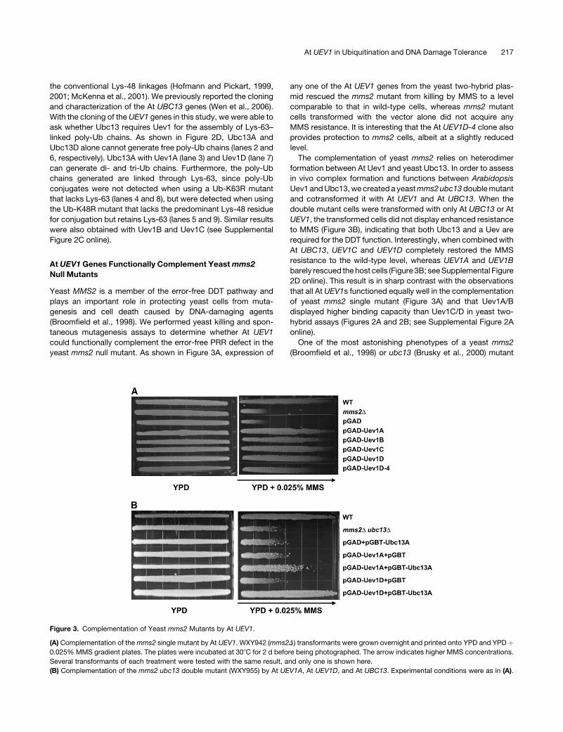
At UEV1 Genes Functionally Complement Yeast mms2
Null Mutants
Yeast MMS2 is a member of the error-free DDT pathway and
plays an important role in protecting yeast cells from muta-
genesis and cell death caused by DNA-damaging agents
(Broomfield et al., 1998). We performed yeast killing and spon-
taneous mutagenesis assays to determine whether At UEV1
could functionally complement the error-free PRR defect in the
yeast mms2 null mutant. As shown in Figure 3A, expression of
any one of the At UEV1 genes from the yeast two-hybrid plas-
mid rescued the mms2 mutant from killing by MMS to a level
comparable to that in wild-type cells, whereas mms2 mutant
cells transformed with the vector alone did not acquire any
MMS resistance. It is interesting that the At UEV1D-4 clone also
provides protection to mms2 cells, albeit at a slightly reduced
level.
The complementation of yeast mms2 relies on heterodimer
formation between At Uev1 and yeast Ubc13. In order to assess
in vivo complex formation and functions between Arabidopsis
Uev1 and Ubc13, we created a yeast mms2 ubc13 double mutant
and cotransformed it with At UEV1 and At UBC13. When the
double mutant cells were transformed with only At UBC13 or At
UEV1, the transformed cells did not display enhanced resistance
to MMS (Figure 3B), indicating that both Ubc13 and a Uev are
required for the DDT function. Interestingly, when combined with
At UBC13, UEV1C and UEV1D completely restored the MMS
resistance to the wild-type level, whereas UEV1A and UEV1B
barely rescued the host cells (Figure 3B; see Supplemental Figure
2D online). This result is in sharp contrast with the observations
that all At UEV1s functioned equally well in the complementation
of yeast mms2 single mutant (Figure 3A) and that Uev1A/B
displayed higher binding capacity than Uev1C/D in yeast two-
hybrid assays (Figures 2A and 2B; see Supplemental Figure 2A
online).
One of the most astonishing phenotypes of a yeast mms2
(Broomfield et al., 1998) or ubc13 (Brusky et al., 2000) mutant
Figure 3. Complementation of Yeast mms2 Mutants by At UEV1.
(A) Complementation of the mms2 single mutant by At UEV1. WXY942 (mms2D) transformants were grown overnight and printed onto YPD and YPD þ0.025% MMS gradient plates. The plates were incubated at 308C for 2 d before being photographed. The arrow indicates higher MMS concentrations.
Several transformants of each treatment were tested with the same result, and only one is shown here.
(B) Complementation of the mms2 ubc13 double mutant (WXY955) by At UEV1A, At UEV1D, and At UBC13. Experimental conditions were as in (A).
At UEV1 in Ubiquitination and DNA Damage Tolerance 217
is its massive increase in spontaneous mutagenesis, indicating
that these genes play an important role in protecting cells from
genome instability. Indeed, in this experiment, the mms2 mutant
strain showed an increase of >20-fold in spontaneous mutagen-
esis compared with wild-type cells (Table 1). When the same
mms2 mutant was transformed with a plasmid expressing an At
UEV1, the spontaneous mutation rate was reduced to a level
similar to that of the wild-type cells (Table 1). Again, UEV1C and
UEV1D appear to be more effective than UEV1A and UEV1B in
limiting mutagenesis. Collectively, the results obtained from the
yeast complementation experiments suggest that At UEV1
genes are able to replace the PRR function of yeast MMS2 and
that perhaps UEV1C and UEV1D are more efficient than UEV1A
and UEV1B in such a function in Arabidopsis.
At UEV1 Expression in Different Tissues and under Stresses
Since UEV1 is presumed to be involved in DDT and the
ubiquitination process is often involved in stress responses, we
analyzed UEV1 expression under various stress conditions.
Arabidopsis cell suspension culture was subjected to treatments
as indicated and total RNA was isolated for RNA gel blot
hybridization. The results from samples of 24-h treatments are
presented in Supplemental Figure 3A online. It appears that
UEV1 expression is slightly decreased after treatment with MMS
or H2O2 and slightly increased after treatment with abscisic acid
or mannitol, although for the latter two treatments the transcript
level of the control UBQ11 was also higher.
Since all four UEV1 genes share >72% nucleotide sequence
identity in their core coding region and all four predicted tran-
scripts are similar in size, we suspect that the UEV1C probe used
for RNA gel blot hybridization actually detected all four UEV1
transcripts. Given the fact that the two human Uev homologs
(UEV1A and hMMS2) play distinct roles in cellular metabolism
(Andersen et al., 2005) and our observation in this study that the
two pairs of UEV1 genes may function differently, it is important
to assess the expression of individual UEV1 genes. To fulfill this
objective, we analyzed the existing microarray data (available
from www.Arabidopsis.org) for individual UEV1 gene expression
profiles and found no evidence of strong stress responses after
treatment of Arabidopsis plants (see Supplemental Figure 3B
online). This analysis suggests that various environmental
stresses used in this study have little effect on the expression
of UEV1 genes at the transcriptional level.
The expression of UEV1 genes in different tissues was also
determined by RNA gel blot hybridization (see Supplemental
Figure 3C online) and by analyzing the microarray data (Figure
4A; see Supplemental Figure 4 online). While most tissues
express variable levels of each UEV1 transcript, UEV1D appears
to show a higher level of expression than the other three UEV1
genes in most tissues examined (Figure 4A; see Supplemental
Figure 4 online). Greater differences in transcript levels of the
UEV1 genes were found in samples from pollen and geminating
seeds. The microarray data indicate that 3 h after seed germi-
nation, the expression of UEV1C and UEV1D is much higher than
that of UEV1A and UEV1B and that UEV1D is essentially the only
UEV1 transcript detected from pollen. To validate the microarray
data, we performed RT-PCR with various tissues, including ger-
minating seeds and pollen. Under the conditions used, the amount
of PCR product was not excessive and was deemed to reflect the
amount of cDNA template. Representative results are shown in
Figure 4B and summarized as follows. First, all four UEV1 genes
are indeed expressed in most common tissues, such as root,
shoot, leaf, and stem. Second, only UEV1D transcript is detectable
in pollen under our experimental conditions, consistent with the
microarray data. Third, 6 h after seed germination, all transcripts
except UEV1B are detected, while after 2 d of seed germination,
only UEV1A and UEV1D transcripts are found, with UEV1D at a
clearly higher level than UEV1A. Microarray data show little
expression of UEV1A in 3-h germinating seeds, but we consis-
tently observed UEV1A transcript by PT-PCR in the sample we
used. These differences may be due to the conditions used in the
microarray experiments and in this study.
uev1d Mutant Plants Are Sensitive to the DNA-Damaging
Agent MMS
The analysis of UEV1 expression as well as the observation that
in combination with UBC13, UEV1D but not UEV1A could com-
pletely rescue the yeast ubc13 mms2 double mutant, prompted
us to focus our attention on UEV1D. We reasoned that uev1d
mutant plants may display compromised tolerance to DNA
damage in pollen and during seed germination. The UEV1D
T-DNA insertion line SALK_064912 was obtained from the ABRC
(www.arabidopsis.org), and the allele was named uev1d-1. Se-
quence analysis revealed that the T-DNA was inserted in the first
intron of UEV1D, with the left border oriented toward the 39 end of
the gene (Figure 5A). The gene-specific primers (SP1 and SP2)
and a primer specific to the left border sequence (LB1) were used
to confirm the insertion of T-DNA (Figure 5B). To further confirm
that UEV1D expression was abolished by this T-DNA insertion,
total RNA was extracted from seedlings of wild-type and homo-
zygous uev1d-1 plants and analyzed by RT-PCR for the expres-
sion of four UEV1 genes. As shown in Figure 5D, a fragment
corresponding to the UEV1D ORF could be amplified from wild-
type plants but not from the uev1d-1 line, while the expression of
the other three At UEV1 genes remained unaltered.
The homozygous uev1d-1 plants do not display apparent
morphological variations. In order to investigate the possible role
of UEV1D in protecting cells from DNA damage, we analyzed
Table 1. Effects of At UEV1 on the Spontaneous Mutation Rate of the
S. cerevisiae mms2 Mutant
Straina Key Alleles Rate (310�8)b Foldc
DBY747 Wild type 3.2 6 0.18 1.00
WXY642/pGAD424 mms2D 70.2 6 7.96 22.10
WXY642/At UEV1A mms2D At UEV1A 8.1 6 0.16 2.53
WXY642/At UEV1B mms2D At UEV1B 7.1 6 0.13 2.22
WXY642/At UEV1C mms2D At UEV1C 5.3 6 0.98 1.66
WXY642/At UEV1D mms2D At UEV1D 4.9 6 0.56 1.53
a All strains are isogenic derivatives of DBY747.b The spontaneous mutation rates are the average of three independent
experiments with standard deviations.c Relative to the wild-type mutation rate.
218 The Plant Cell

the effect of MMS on seed germination, considering that UEV1D
is strongly expressed during seed germination. We examined
three controls—wild-type Arabidopsis ecotype Columbia, a
T-DNA insertion line (SALK_042050) not affecting UEV1 genes,
and a wild type segregant line derived from the initial
SALK_064912 seeds (1d-1WT)—along with the homozygous
uev1d-1 T-DNA insertion line. Three parameters related to seed
germination were surveyed. First, the percentage of seeds
that germinated in the presence of various concentrations of
MMS was scored after a 5-d incubation. Seeds from uev1d-1
plants were much more sensitive to MMS treatment than
any of the three control plants, and this response was dose-
dependent (Figure 6A). By contrast, in the absence of MMS,
the uev1d-1 seeds did not show a noticeable difference from
controls in the percentage of seed germination. Second, it
was observed that the homozygous uev1d-1 seedlings were
dying relatively quickly in the presence of MMS and displayed
bleached pale cotyledons rather than the normal green cotyle-
dons. Thus, the percentage of germinated seeds with green
cotyledons was scored after 13 d. The data clearly indicate
that the uev1d-1 line had reduced numbers of viable seedlings in
the presence of MMS. In particular, in the presence of 0.01%
MMS, 75 to 90% of control seedlings were viable, as judged
by green seedlings, compared with <15% viable uev1d-1 seed-
lings under the same growth condition (Figures 6B and 6C).
Finally, the average fresh weight of 13-d uev1d-1 mutant seed-
lings was reduced compared with that in control seedlings after
MMS treatments. More specifically, with 0.005% MMS treat-
ment, even though almost all uev1d-1 seedlings remained green,
they only had half the fresh weight of the wild-type seedlings
(Figure 6D).
To ensure that the above observations were specific to
the T-DNA insertion at UEV1D, we obtained the second UEV1D
T-DNA insertion line SALK_052144 from the ABRC, in which the
T-DNA was inserted in the third exon of UEV1D (Figure 5A). We
confirmed the T-DNA insertion by genomic PCR (Figure 5C) and
named it uev1d-2. RT-PCR analysis demonstrated that the
UEV1D mRNA is absent in the homozygous uev1d-2 line (Figure
5D). Phenotypic analyses showed that, like uev1d-1, the uev1d-2
mutant is hypersensitive to MMS treatment during seed germi-
nation (Figures 6E and 6F). From these results, we conclude that
UEV1D is required for tolerance to DNA damage during seed
germination.
To assess whether the observed uev1d mutant phenotype is
specific to DNA damage or is triggered by general stress, we
measured seed germination in the presence of up to 0.2 M NaCl.
There was no significant difference in the response of wild-type
and uev1d plants to the salt stress (data not shown).
We also attempted to assess the role of UEV1D in pollen ger-
mination by measuring the percentage of pollen germination in
the presence of MMS. As shown in Figure 7, inactivation of UEV1D
resulted in a moderate but significant decrease in pollen germi-
nation. In the presence of 0.005% MMS, 33% of wild-type pollens
germinated, while only 20% of uev1d-1 pollens germinated after
8 h of incubation, indicating that UEV1D also plays a critical role
in protecting pollen from environmental DNA damage.
uev1a Mutant Plants Do Not Display MMS Sensitivity
Since UEV1A is the only other UEV1 gene expressed during
seed germination, we were interested in the phenotypes of this
mutant plant. Unfortunately, a uev1a T-DNA insertion mutant line
is not available from the ABRC; instead, we found a line
(FLAG_128G02) with a T-DNA insertion at the fourth exon (see
Supplemental Figure 5A online) from the Institut Jean-Pierre
Figure 4. Tissue Distribution of UEV1 Expression.
(A) Relative expression of UEV1 transcripts in different tissues was de-
termined using data from the Arabidopsis NASCArrays microarray data-
base (http://affymetrix.Arabidopsis.info/narrays/experimentbrowse.pl)
(Craigon et al., 2004). R, roots of 17-d plants; S, shoots of 8-d seedlings;
L, rosette leaf 2 of 17-d plants; RL, mature rosette leaves of 23-d plants;
St, second internode of 21-d plants; F, stage 12 flowers of 21-d plants;
P, mature pollen; G3h, seed germinating for 3 h. The original microarray
data are from AtGenExpress: Expression Atlas of Arabidopsis Develop-
ment (TAIR accession number 1006710873: ATGE_9, ATGE_12, ATGE_24,
ATGE_27, ATGE_33, ATGE_73, and ATGE_96 samples) (Schmid et al.,
2005), except for G3h data, which are from AtGenExpress: Expression
Profiling of Early Germinating Seeds (TAIR accession number 1007966994:
RIKEN-PRESTON2 sample).
(B) Expression of UEV1 transcripts in different tissues analyzed by RT-
PCR. The At4g33380 gene was assayed as an input control (Czechowski
et al., 2005). The exposure time of the gels is shown at left (BioDoc-It
System; UVP). C, cell suspension; R, roots of 13-d seedlings; S, shoots of
13-d seedlings; L3, leaves of 3-week plants; L5, leaves of 5-week plants;
St, stems of 5-week plants; F, floral tissues of 5-week plants; G6h and G2d,
seeds germinating on Petri dishes for 6 h and 2 d, respectively; P, pollen.
At UEV1 in Ubiquitination and DNA Damage Tolerance 219

Bourgin collection. We obtained this line, screened the segre-
gants, and confirmed the homozygous uev1a mutant (uev1a-1)
by both genomic PCR (see Supplemental Figure 5B online) and
RT-PCR (see Supplemental Figure 5C online). Seed germination
assays were performed under the same experimental conditions
described above. uev1a-1 mutant plants did not display en-
hanced MMS sensitivity compared with its wild-type segregants
or with the parental strain Ws-4 (data not shown). This result
indicates that inactivation of UEV1A does not alter DDT during
Arabidopsis seed germination.
DISCUSSION
Lys-63–linked polyubiquitination of target proteins is considered
to be a fundamentally different process from conventional Lys-
48–linked polyubiquitination, which targets proteins for degra-
dation via the 26S proteasome (Hochstrasser, 1996; Pickart,
2001b). Instead, it is deemed analogous to other posttrans-
lational regulatory processes, such as phosphorylation and
sumoylation, that alter target protein activities. Although Lys-63–
linked polyubiquitination has been reported to be involved in
several cellular processes, including stress response (Arnason
and Ellison, 1994), mitochondrial inheritance (Fisk and Yaffe,
1999), plasma membrane protein endocytosis (Galan and
Haguenauer-Tsapis, 1997), ribosome function (Spence et al., 2000),
innate immunity (Deng et al., 2000; Wang et al., 2001; Zhou et al.,
2004), mitotic cell cycle checkpoint (Bothos et al., 2003), and
DNA repair (Hofmann and Pickart, 1999; Hoege et al., 2002), to
date Ubc13 is the only known E2 capable of mediating Lys-63–
linked Ub chain assembly. Interestingly, genetic analyses to date
define only three functions of Ubc13, namely PRR in yeast
(Hoege et al., 2002) and possibly mammals (Andersen et al.,
2005) through covalent modification of PCNA, activation of
NF-kB by polyubiquitinating NEMO/IKKg in mammals (Zhou
et al., 2004), and involvement in synaptic connectivity between
the giant fiber and a motor neuron in Drosophila. Drosophila
ubc13 is also known as bendless (Muralidhar and Thomas, 1993;
Oh et al., 1994), although the molecular mechanism of Ubc13
activity in this case remains unclear. The unique feature of Ubc13
compared with other Ubcs is its ability to form a stable complex
with a Uev, which is homologous to other Ubcs but lacks the
active Cys residue (Broomfield et al., 1998; Sancho et al., 1998).
This family of Uevs engages a noncovalent interaction with Ub
(McKenna et al., 2001) and orients this acceptor Ub to allow its
Lys-63 residue to be exposed to the C terminus of donor Ub
covalently bound to Ubc13 (McKenna et al., 2003). It appears that
in mammals, Uevs not only facilitate polyubiquitination but also
serve as a regulatory subunit to promote the ubiquitination of
different targets (Andersen et al., 2005). In this study, we iden-
tified four Arabidopsis genes that meet the criteria of encoding a
Figure 5. Confirmation of Two uev1d T-DNA Insertion Mutants.
(A) Genomic structure showing the positions of two T-DNA insertions in UEV1D. Open boxes, exons; closed boxes, UEV1D ORF; lines, introns. SP1, 59
gene-specific primer AtUEV1D-1; SP2, 39 gene-specific primer AtUEV1D-2; LB1, T-DNA left border primer.
(B) and (C) Genomic DNA PCR to confirm uev1d-1 (1d-1) (B) and uev1d-2 (1d-2) (C). The fragment was amplified using three primers (SP1, SP2, and
LB1) in each reaction and genomic DNA from Columbia (WT), 1d-1 (B), or 1d-2 (C) as a template.
(D) RT-PCR detection of the UEV1 transcripts. UEV1 gene-specific primers were used for RT-PCR against total RNA extracted from Columbia (WT),
uev1d-1, and uev1d-2 lines. Total RNA was extracted from flowers.
220 The Plant Cell
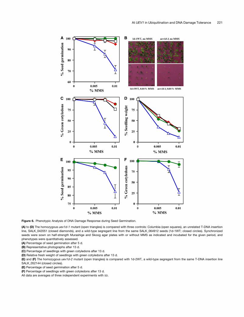
Figure 6. Phenotypic Analysis of DNA Damage Response during Seed Germination.
(A) to (D) The homozygous uev1d-1 mutant (open triangles) is compared with three controls: Columbia (open squares), an unrelated T-DNA insertion
line, SALK_042051 (closed diamonds), and a wild-type segregant line from the same SALK_064912 seeds (1d-1WT; closed circles). Synchronized
seeds were sown on half-strength Murashige and Skoog agar plates with or without MMS as indicated and incubated for the given period, and
phenotypes were quantitatively assessed.
(A) Percentage of seed germination after 5 d.
(B) Representative photographs after 13 d.
(C) Percentage of seedlings with green cotyledons after 13 d.
(D) Relative fresh weight of seedlings with green cotyledons after 13 d.
(E) and (F) The homozygous uev1d-2 mutant (open triangles) is compared with 1d-2WT, a wild-type segregant from the same T-DNA insertion line
SALK_052144 (closed circles).
(E) Percentage of seed germination after 5 d.
(F) Percentage of seedlings with green cotyledons after 13 d.
All data are averages of three independent experiments with SD.
At UEV1 in Ubiquitination and DNA Damage Tolerance 221

Uev and found by sequence analysis that they are two pairs of
duplicated genes. We argue that these UEV1 genes are paralogs
and that they evolved from a common ancestor within plants,
since sequence alignment could not distinguish which Uev1 pair
is more related to one of the two mammalian Uevs (Uev1A or
Mms2). This observation raises two major questions. First, do
Ubc13-Uev complexes from different organisms confer a con-
served function? Second, if gene duplications occurred after
animal–plant separation, how are their functions preserved? In
this study, we demonstrated that all At Uev1 proteins are able to
form stable complexes with At Ubc13 and to promote Ubc13-
mediated Lys-63–linked polyubiquitination. The only other known
Arabidopsis Uev found to date is Cop10, which was identified as a
negative regulator of photomorphogenesis and functions by pro-
moting target protein degradation (Suzuki et al., 2002).
The observation that At UBC13 (Wen et al., 2006) or At UEV1
(this study) is able to functionally complement the corresponding
yeast mutants defective in DDT is insufficient to claim that they
also play the same role in their own host. A good example is
human UEV1A, which confers a similar DDT function in yeast but
is exclusively involved in NF-kB activation instead of DNA repair
(Andersen et al., 2005). In this study, we took advantage of the
fact that UEV1D is the predominant UEV1 gene expressed in
germinating seeds and in pollen and characterized the sensitivity
of uev1d mutant plants to a DNA-damaging agent in these
tissues. Our results clearly show that in the presence of DNA
damage, lack of Uev1D activity compromises seed germination,
seedling survival, and growth. Based on the following observa-
tions, we argue that the above phenotypic effects are specific to
the inactivation of UEV1D. First, two independent UEV1D T-DNA
insertion lines displayed similar phenotypes, while none of three
control lines showed these phenotypes. Second, a uev1a mutant
line also did not show such phenotypes compared with its wild-
type controls. This study, along with previous studies in yeast
and mammalian cells, supports the notion that error-free DDT
promoted by Lys-63–linked polyubiquitination via Ubc13-Uev is
an evolutionarily conserved function throughout eukaryotes.
Despite its predominant expression among the four UEV1
genes, inactivation of UEV1D caused only a very moderate
compromise in pollen tube growth in the presence of a DNA-
damaging agent. This probably reflects the lack of cell division
during pollen germination, whereas DDT is expected to operate
only on replicated DNA (Barbour and Xiao, 2003). It is of great
interest that in contrast with many other plant species, it has been
observed that DNA synthesis in the Arabidopsis sperm nuclei is
initiated prior to anthesis and continues as the pollen tube
develops (Friedman, 1999). Thus, the Arabidopsis sperm nuclei
are essentially in a prolonged S phase at the time of anthesis, in
preparation for eventual double fertilization. Our observation that
in the presence of replication-blocking lesions induced by MMS,
pollen tubes from the uev1d mutant plants did not develop as well
as those from wild-type segregant lines is consistent with the
notion that UEV1D plays a more active role than other UEV1
genes when DNA synthesis occurs in the presence of DNA
damage, which is essentially a DDT activity. We wish to stress
that our analysis does not rule out the possibility that MMS-
induced DNA lesions may inhibit transcription and that MMS can
also directly methylate and damage RNA (Friedberg et al., 2006),
which could contribute to the observed phenotypes, although
Lys-63 ubiquitination has not been linked to these processes.
In our opinion, this study provides an important step toward
understanding Ubc13-Uev–mediated Lys-63 polyubiquitination
in general and the mechanisms of DDT in particular in plants.
Several questions remain to be addressed. First, is UEV1D the
only UEV1 gene involved in error-free DDT? Second, is UEV1D
also involved in other cellular processes? Third, what are the
other cellular processes that also require Ubc13-Uev–mediated
polyubiquitination? We feel that given the near identity in amino
acid sequence and similar complementation phenotypes in
yeast, Uev1C is likely involved in the same cellular processes
as Uev1D. This could explain our failure to detect a DNA repair/
tolerance defect in uev1d plants in a root growth assay (data not
shown). Indeed, the phenotypes of uev1d mutant plants may be
Figure 7. Phenotypic Analysis of DNA Damage Response during in Vitro
Pollen Germination.
(A) Representative in vitro pollen germination images of 1d-1WT and
uev1d-1 with or without MMS treatment as indicated.
(B) Summary of the pollen germination results. Data presented are
averages of three independent experiments with SD. Open bars, 1d-1WT;
closed bars, uev1d-1.
222 The Plant Cell
considered to be moderate, which is due to either the backup or
residual expression of other UEV1 genes or the nature of the
error-free DDT defect in plants. One important aspect of future
work will be to identify a uev1c null mutant, combine this
mutation with uev1d, examine various tissues for a DDT defect,
and relate the results to the UEV1 expression profile. It is also
interesting that the yeast mms2/ubc13 or rev1/rev3/rev7 single
mutants are moderately sensitive to killing by DNA-damaging
agents, but the combination of any two mutations from different
pathways results in strong synergistic interactions (Broomfield
et al., 1998; Xiao et al., 1999). It would be of great interest to
determine whether the combination of uev1d and rev mutations
also results in a synergistic sensitivity to DNA damage in
Arabidopsis.
The two pairs of UEV1s may be involved in different functions
in plants. Although it remains possible that Uev1A and Uev1B are
involved in DDT as well, our observations favor the suggestion
that the Uev1A/B pair is probably involved in other cellular
processes unrelated to DNA damage response. First, although
UEV1A is expressed during seed germination, inactivation of this
gene does not result in compromised seed germination in the
presence of MMS, in sharp contrast with the uev1d mutant lines.
Second, in the presence of UBC13, the DDT activities of UEV1A/
B are much lower than those of UEV1C/D in yeast cells, despite
the fact that Uev1A/B interact with At Ubc13 and Ubc13 from
other species very well and are able to fully complement the
yeast mms2 mutant when yeast Ubc13 is present. This result
also effectively rules out the possibility that partial complemen-
tation by UEV1A/B was due to their poor expression in yeast
cells. Third, it is interesting that human Uev1A contains an
additional N-terminal 25 amino acid residues and plays a distinct
role from that of hMms2; it may be reverted to play a role in DDT
when its N-terminal sequence is experimentally deleted (Andersen
et al., 2005). Similarly, At Uev1A and At Uev1B contain a unique
C-terminal tail that may be critical for their functions other than
DDT. It is difficult at this stage to predict what type of activity
it may be, given the fact that Uev1 appears to have evolved
independently of vertebrate Uev paralogs, that Drosophila
ubc13/bendless confers a very different function than its mam-
malian counterpart, and that other reports have claimed addi-
tional Lys-63–mediated cellular processes and some of them
have also been linked to Ubc13-Uev (Bothos et al., 2003; Doss-
Pepe et al., 2005; Laine et al., 2006). What we can predict is that
additional Arabidopsis Ubc13-Uev1 functions should be medi-
ated by its Lys-63–linked poly-Ub chains and that different Uev1
proteins may serve as cofactors and critical regulators in these
processes. In this regard, future research may focus on the
search for Ubc13-Uev ubiquitination targets and cognate E3s
through bioinformatics and proteomic approaches as well as
through genomic approaches such as microarray analysis of the
At uev1 mutants reported in this study.
METHODS
Plant and Yeast Cell Cultures
Arabidopsis thaliana ecotypes Columbia and Ws and their mutant deriv-
atives were used in this study. The conditions for plant growth and
maintenance of Arabidopsis Columbia cell suspension culture have been
described previously (Wen et al., 2006).
The haploid yeast strains used in this study are listed in Supplemental
Table 1 online. Yeast cells were grown at 308C in either rich YPD or in a
synthetic dextrose (SD) medium (0.67% Bacto-yeast nitrogen base without
amino acids, 2% glucose) supplemented with necessary nutrients as rec-
ommended (Sherman et al., 1983). For solid plates, 2% agar was added to
either YPD or SD medium prior to autoclaving. Yeast cells were transformed
using a LiAc method as described (Ito et al., 1983). The sources and prep-
aration of ubc13D::hisG-URA3-hisG (Brusky et al., 2000) and mms2D::HIS3
(Xiao et al., 1999) cassettes was as described previously.
Cloning Arabidopsis UEV1 cDNAs and Plasmid Construction
To clone At UEV1s, total RNA was isolated from Arabidopsis seedlings
using TRIzol reagent (Invitrogen) for RT-PCR with the ThermoScript
RT-PCR kit (Invitrogen) according to the manufacturer’s instructions.
Each At UEV1 ORF was amplified by PCR from the above cDNA pre-
paration using gene-specific primers (see Supplemental Table 2 online),
and the flanking SalI restriction site was used to clone the PCR products
into the yeast two-hybrid vector pGAD424E (for Gal4AD fusion), which was
derived from pGAD424 (Bartel and Fields, 1995), with a 1-bp frameshift at
the multiple cloning site. The identity of each cloned ORF was verified by
sequencing.
Yeast Two-Hybrid Analysis
The yeast two-hybrid strain PJ69-4A (James et al., 1996) was cotrans-
formed with different combinations of Gal4BD and Gal4AD constructs. The
construction of pGBT-At Ubc13A, pGBT-At Ubc13B (Wen et al., 2006),
pGBT-Sc Ubc13 (Brown et al., 2002), and pGBT-Hs Ubc13 (Pastushok
et al., 2005) has been described previously. The cotransformed colonies
were initially selected on SD-Leu-Trp plates. For each transformation, at
least five independent colonies were plated onto SD-Leu-Trp-His with
various concentrations of 1,2,4-aminotriazole to test the activation of the
GAL1-HIS3 gene and onto SD-Leu-Trp-Ade to detect the activation of the
GAL2-ADE2 reporter gene.
Protein Expression, Purification, and GST Pull-Down Assay
The SalI fragments containing UEV1 ORFs were isolated from the
pGAD424E plasmids and cloned into pGEX6p-2 (Amersham Biosciences).
The resulting pGEX-Uev1s were transformed into Escherichia coli strain
BL21 (DE3)-RIL (Stratagene). The GST-Uev1 fusion proteins were pro-
duced and purified as described previously (McKenna et al., 2001). The
source and preparation of the Ubc13A fusion protein and the protocol for
the GST pull-down assay were as described previously (Wen et al., 2006).
Ub Conjugation Reaction
In vitro Ub conjugation reactions were performed using the purified
Ubc13A and GST-Uev1A proteins as described above, and Ub thioester/
conjugation initiation reagents were purchased from Boston Biochem.
Unless noted otherwise, the 20-mL reaction mixture contained 225 nM E1
enzyme, 450 mM Ub, 1 mM MgATP, 1 mM Ubc13, and 1 mM Uev1 in the
supplied reaction buffer. The K63R and K48R mutant Ub proteins were
purchased from Boston Biochem (UM-K63R and UM-K48R). The conjuga-
tion reactions were performed at 378C for 2 h. Samples were subjected to
SDS-PAGE (12%), and Ub and poly-Ub were detected through protein gel
blots using polyclonal rabbit anti-Ub antibodies (Sigma-Aldrich).
Yeast Killing and Spontaneous Mutagenesis Assay
Yeast strain HK580-10D and its isogenic mms2D single or ubc13D mms2D
double mutants were either singly transformed with pGAD-Uev1 or
At UEV1 in Ubiquitination and DNA Damage Tolerance 223

cotransformed with pGAD-Uev1 and pGBT-Ubc13A, and transformants
were selected on SD-Leu or SD-Leu-Trp plates, respectively. The gradient
plate assay was performed as described previously (Pastushok et al., 2005).
Yeast strain DBY747 and its mms2D derivative WXY642 bear a trp1-
289 amber mutation that can be reverted to Trpþ by several different
mutation events (Xiao and Samson, 1993). WXY642 was transformed with
pGAD-UEV1 or vector pGAD424E, and transformants were selected on
SD-Leu plates. The spontaneous mutagenesis assay was performed as
described previously (Wen et al., 2006).
Expression Analysis by RNA Gel Blot and RT-PCR
To determine the expression of UEV1 genes under different stress
conditions, Arabidopsis cell suspension culture was used. The culture
was maintained for 5 d after subculture and then subjected to various
treatments as specified. After a 24-h treatment, total RNA was isolated
and 15 mg of RNA from each sample was used for RNA gel blot analysis as
described (Wang et al., 1995). The DNA fragment containing the UEV1C
ORF was isolated from an agarose gel after restriction enzyme digestion
and electrophoresis, and the UBQ11 gene probe was amplified using
gene-specific primers (see Supplemental Table 2 online). The DNA
fragment was labeled with a 32P-dCTP using the Random Primer labeling
kit from Invitrogen. The membrane containing total RNA was hybridized
with the UEV1C probe, stripped, and hybridized with the UBQ11 probe.
For RT-PCR analysis, total RNA from various tissues was isolated using
TRIzol and treated with DNaseI (Promega). Total RNA from mature pollen
was extracted as described (Fei et al., 2004), and total RNA from
germinating seeds was extracted as described (Vicient and Delseny,
1999). Reverse transcript synthesis of the first-strand cDNA was per-
formed with the SuperScript RT-PCR III system (Invitrogen) using the
protocol as described (Karsai et al., 2002). Briefly, 2 to 4 mg of total RNA
for each sample were treated with DNase I (Roche Diagnostics) and
reverse-transcribed with Moloney murine leukemia virus reverse tran-
scriptase (Invitrogen) and d(T)18. The final input amount of cDNA used for
RT-PCR was adjusted by analyzing the expression of the At4g33380
control gene (Czechowski et al., 2005). Experiments were performed
using UEV1 gene-specific primer pairs with different cycle conditions (22,
28, and 35 cycles) to make sure that the amount of PCR product was not
excessive and that the differences among different tissue samples were
not disturbed by saturation of PCR amplification. Eight microliters of each
reaction was used for agarose gel electrophoresis. All RT-PCR series
were assayed at least twice with highly consistent results.
Analysis of Microarray Expression Data
Expression values of UEV1 genes were obtained from publicly available
Affymetrix ATH1 array data available up to June 2006 (http://affymetrix.
Arabidopsis.info/) with a total of 2392 arrays. A subset of 237 arrays was
used to calculate the average expression levels and to compare the
expression levels that correspond with (1) 63 conditions from the Devel-
opmental Affymetrix Gene Expression Atlas: AtGenExpress (Schmid
et al., 2005), (2) three root stages (Birnbaum et al., 2003), (3) five root
layer tissues (Nawy et al., 2005), (4) four pollen stages (Honys and Twell,
2004), (5) four flower induction treatments (Schmid et al., 2003), and
(6) three germination stages (http://www.weigelworld.org/resources/
microarray/AtGenExpress/). Heat maps representing the expression
levels of At UEV1 genes were produced with the Genesis 1.6.0 Beta
1 program (Sturn et al., 2002) (http://genome.tugraz.at/) using absolute
expression values as standards in the Nottingham Arabidopsis Stock
Centre’s International Affymetrix Service.
Seed Germination Assays
The homozygous UEV1D T-DNA insertion lines SALK_064912 (uev1d-1)
and SALK_052144 (uev1d-2) were used in the sensitivity assay to the
DNA-damaging agent MMS. To exclude any possible nonspecific effect,
we used three controls, the wild-type Arabidopsis Columbia, a T-DNA
insertion line not related to UEV1 genes (SALK_042050), and a homozy-
gous wild-type segregant line (1d-1WT or 1d-2WT) derived from the initial
mutant seeds received. For the uev1a-1 mutant, the parental wild-type
line Ws-4 and a homozygous wild-type (1a-1WT) segregant from
FLAG_128G02 were used as controls. The identity of the wild-type and
mutant segregants was determined by genomic PCR and RT-PCR. In
addition, to minimize the effect of individual plants, seeds of three
homozygous mutant plants were pooled and used for the assay. Seeds
were surface-sterilized with 20% bleach and 0.1% Triton X-100 for 20
min, followed by three rinses in sterile water. After sterilization, seeds
were suspended in 0.1% agarose and stored at 48C in the dark for 3 d to
synchronize germination. Three days later, the seeds were removed from
the dark and sown on half-strength Murashige and Skoog agar plates
supplemented with different concentrations of MMS. Each plate was
planted with 50 seeds, and at least three plates (150 seeds) were used for
each treatment. After a 5-d incubation in a growth chamber, the germi-
nation of the seeds was surveyed. After a 13-d incubation, the color of the
cotyledons was rated (green versus nongreen) while the first pair of true
leaves was still small. Since seedlings with nongreen cotyledons at this
stage were dead or dying, the percentage of seedlings with green cotyle-
dons was an indicator of seedling viability. The fresh weight of seedlings
was also determined and was used as an indicator of seedling growth.
In Vitro Pollen Germination Assay
To quantitatively measure pollen germination efficiency in the presence of
MMS, 2 mL of the germination medium (Fan et al., 2001) containing 1%
agar with or without 0.005% MMS was poured into a 35-mm Petri dish to
form a thin layer. Freshly anther-dehisced flowers at stage 13 or 14
(Smyth et al., 1990) were randomly picked and used to carefully touch the
central area of the agar plate in order to spread pollen grains. The Petri
dishes were incubated in a humid chamber at 268C for 8 h without light
before counting pollen germination and photographing. For each plate,
>400 pollen grains were counted using a phase-contrast microscope for
each plate, and three plates were used for each treatment.
Accession Numbers
Sequence data from this article can be found in the GenBank/EMBL data
libraries under the following accession numbers: NP_011428.1 (Saccha-
romyces cerevisiae); NP_588162.1 (Schizosaccharomyces pombe);
NP_647959.1 (Drosophila melanogaster); NP_076074.2 (Mus musculus);
At Uev1A ¼ NP_565834.1 (At1g23260), At Uev1B ¼ NP_564994.1
(At1g70660), At Uev1C ¼ NP_850259.1 (At2g36060), and At Uev1D ¼NP_566968.1 (At3g52560). (Arabidopsis thaliana); NP_493578.1 (Caeno-
rhabditis elegans); and Hs Mms2 ¼ NP_003341.1 and Hs Uev1A ¼NP_068823.2 (Homo sapiens).
Supplemental Data
The following materials are available in the online version of this article.
Supplemental Figure 1. Phylogenetic Analyses of Hypothetical Uev
Family Proteins from Different Organisms.
Supplemental Figure 2. Characterization of Arabidopsis UEV1B and
UEV1C.
Supplemental Figure 3. UEV1 Expression Profiles.
Supplemental Figure 4. Heat Map Showing the Transcript Levels of
Arabidopsis UEV1 Genes in Different Tissues and Developmental
Stages.
Supplemental Figure 5. Confirmation of the uev1a-1 T-DNA Insertion
Mutant.
224 The Plant Cell
Supplemental Table 1. S. cerevisiae Strains.
Supplemental Table 2. Oligonucleotide Sequences.
ACKNOWLEDGMENTS
We thank Vipon Sawhney for the protocol on collecting pollen samples,
Gordon Gray for the Arabidopsis cell suspension culture, Bernard Kunz
for critical comments on the manuscript, and Michelle Hanna for
proofreading the manuscript. This work was supported by Canadian
Institutes of Health Research Operating Grant MOP-53240 to W.X. and a
Natural Sciences and Engineering Research Council of Canada Discov-
ery Grant to H.W.
Received March 24, 2007; revised December 6, 2007; accepted Decem-
ber 17, 2007; published January 4, 2008.
REFERENCES
Andersen, P.L., Zhou, H., Pastushok, L., Moraes, T., McKenna, S.,
Ziola, B., Ellison, M.J., Dixit, V.M., and Xiao, W. (2005). Distinct
regulation of Ubc13 functions by the two ubiquitin-conjugating en-
zyme variants Mms2 and Uev1A. J. Cell Biol. 170: 745–755.
Arnason, T., and Ellison, M.J. (1994). Stress resistance in Saccharo-
myces cerevisiae is strongly correlated with assembly of a novel type
of multiubiquitin chain. Mol. Cell. Biol. 14: 7876–7883.
Bachmair, A., Novatchkova, M., Potuschak, T., and Eisenhaber, F.
(2001). Ubiquitylation in plants: A post-genomic look at a post-
translational modification. Trends Plant Sci. 6: 463–470.
Barbour, L., and Xiao, W. (2003). Regulation of alternative replication
bypass pathways at stalled replication forks and its effects on
genome stability: A yeast model. Mutat. Res. 532: 137–155.
Bartel, P.L., and Fields, S. (1995). Analyzing protein-protein interac-
tions using two-hybrid system. Methods Enzymol. 254: 241–263.
Birnbaum, K., Shasha, D.E., Wang, J.Y., Jung, J.W., Lambert, G.M.,
Galbraith, D.W., and Benfey, P.N. (2003). A gene expression map of
the Arabidopsis root. Science 302: 1956–1960.
Blanc, G., Hokamp, K., and Wolfe, K.H. (2003). A recent polyploidy
superimposed on older large-scale duplications in the Arabidopsis
genome. Genome Res. 13: 137–144.
Bothos, J., Summers, M.K., Venere, M., Scolnick, D.M., and
Halazonetis, T.D. (2003). The Chfr mitotic checkpoint protein func-
tions with Ubc13-Mms2 to form Lys-63-linked polyubiquitin chains.
Oncogene 22: 7101–7107.
Broomfield, S., Chow, B.L., and Xiao, W. (1998). MMS2, encoding a
ubiquitin-conjugating-enzyme-like protein, is a member of the yeast
error-free postreplication repair pathway. Proc. Natl. Acad. Sci. USA
95: 5678–5683.
Broomfield, S., Hryciw, T., and Xiao, W. (2001). DNA postreplication
repair and mutagenesis in Saccharomyces cerevisiae. Mutat. Res.
486: 167–184.
Brown, M., Zhu, Y., Hemmingsen, S.M., and Xiao, W. (2002). Structural
and functional conservation of error-free DNA postreplication repair in
Schizosaccharomyces pombe. DNA Repair (Amst.) 1: 869–880.
Brusky, J., Zhu, Y., and Xiao, W. (2000). UBC13, a DNA-damage-
inducible gene, is a member of the error-free postreplication repair
pathway in Saccharomyces cerevisiae. Curr. Genet. 37: 168–174.
Craigon, D.J., James, N., Okyere, J., Higgins, J., Jotham, J., and
May, S. (2004). NASCArrays: A repository for microarray data gener-
ated by NASC’s transcriptomics service. Nucleic Acids Res. 32:
D575–D577.
Curtis, M.J., and Hays, J.B. (2007). Tolerance of dividing cells to
replication stress in UVB-irradiated Arabidopsis roots: Requirements
for DNA translesion polymerases h and z. DNA Repair (Amst.) 6:
1341–1358.
Czechowski, T., Stitt, M., Altmann, T., Udvardi, M.K., and Scheible,
W.R. (2005). Genome-wide identification and testing of superior
reference genes for transcript normalization in Arabidopsis. Plant
Physiol. 139: 5–17.
Deng, L., Wang, C., Spencer, E., Yang, L., Braun, A., You, J.,
Slaughter, C., Pickart, C., and Chen, Z.J. (2000). Activation of
the IkB kinase complex by TRAF6 requires a dimeric ubiquitin-
conjugating enzyme complex and a unique polyubiquitin chain. Cell
103: 351–361.
Devoto, A., Muskett, P.R., and Shirasu, K. (2003). Role of ubiquitina-
tion in the regulation of plant defence against pathogens. Curr. Opin.
Plant Biol. 6: 307–311.
Doss-Pepe, E.W., Chen, L., and Madura, K. (2005). a-Synuclein and
parkin contribute to the assembly of ubiquitin lysine 63-linked multi-
ubiquitin chains. J. Biol. Chem. 280: 16619–16624.
Eddins, M.J., Carlile, C.M., Gomez, K.M., Pickart, C.M., and
Wolberger, C. (2006). Mms2-Ubc13 covalently bound to ubiquitin
reveals the structural basis of linkage-specific polyubiquitin chain
formation. Nat. Struct. Mol. Biol. 13: 915–920.
Fan, L.M., Wang, Y.F., Wang, H., and Wu, W.H. (2001). In vitro
Arabidopsis pollen germination and characterization of the inward
potassium currents in Arabidopsis pollen grain protoplasts. J. Exp.
Bot. 52: 1603–1614.
Fei, H., Zhang, R., Pharis, R.P., and Sawhney, V.K. (2004). Pleiotropic
effects of the male sterile33 (ms33) mutation in Arabidopsis are
associated with modifications in endogenous gibberellins, indole-
3-acetic acid and abscisic acid. Planta 219: 649–660.
Fields, S., and Song, O. (1989). A novel genetic system to detect
protein-protein interactions. Nature 340: 245–246.
Fisk, H.A., and Yaffe, M.P. (1999). A role for ubiquitination in mito-
chondrial inheritance in Saccharomyces cerevisiae. J. Cell Biol. 145:
1199–1208.
Franko, J., Ashley, C., and Xiao, W. (2001). Molecular cloning and
functional characterization of two murine cDNAs which encode Ubc
variants involved in DNA repair and mutagenesis. Biochim. Biophys.
Acta 1519: 70–77.
Friedberg, E.C., Walker, G.C., Wolfram, S., Wood, R.D., Schultz,
R.A., and Ellenberger, T. (2006). DNA Repair and Mutagenesis, 2nd
ed. (Washington, DC: ASM Press).
Friedman, W.E. (1999). Expression of the cell cycle in sperm of
Arabidopsis: Implications for understanding patterns of gametogen-
esis and fertilization in plants and other eukaryotes. Development 126:
1065–1075.
Galan, J.M., and Haguenauer-Tsapis, R. (1997). Ubiquitin lys63 is
involved in ubiquitination of a yeast plasma membrane protein. EMBO
J. 16: 5847–5854.
Garcia-Ortiz, M.V., Ariza, R.R., Hoffman, P.D., Hays, J.B., and
Roldan-Arjona, T. (2004). Arabidopsis thaliana AtPOLK encodes a
DinB-like DNA polymerase that extends mispaired primer termini
and is highly expressed in a variety of tissues. Plant J. 39:
84–97.
Hall, T.A. (1999). BioEdit: A user-friendly biological sequence alignment
editor and analysis program for Windows 95/98/NT. Nucleic Acids
Symp. Ser. 41: 95–98.
Hochstrasser, M. (1996). Ubiquitin-dependent protein degradation.
Annu. Rev. Genet. 30: 405–439.
Hoege, C., Pfander, B., Moldovan, G.L., Pyrowolakis, G., and
Jentsch, S. (2002). RAD6-dependent DNA repair is linked to modi-
fication of PCNA by ubiquitin and SUMO. Nature 419: 135–141.
At UEV1 in Ubiquitination and DNA Damage Tolerance 225

Hofmann, R.M., and Pickart, C.M. (1999). Noncanonical MMS2-
encoded ubiquitin-conjugating enzyme functions in assembly of novel
polyubiquitin chains for DNA repair. Cell 96: 645–653.
Hofmann, R.M., and Pickart, C.M. (2001). In vitro assembly and
recognition of Lys-63 polyubiquitin chains. J. Biol. Chem. 276:
27936–27943.
Honys, D., and Twell, D. (2004). Transcriptome analysis of haploid male
gametophyte development in Arabidopsis. Genome Biol. 5: R85.
Ito, H., Fukuda, Y., Murata, K., and Kimura, A. (1983). Transformation
of intact yeast cells treated with alkali cations. J. Bacteriol. 153:
163–168.
James, P., Halladay, J., and Craig, E.A. (1996). Genomic libraries and
a host strain designed for highly efficient two-hybrid selection in
yeast. Genetics 144: 1425–1436.
Karsai, A., Muller, S., Platz, S., and Hauser, M.T. (2002). Evaluation
of a homemade SYBR green I reaction mixture for real-time PCR
quantification of gene expression. Biotechniques 32: 790–792,
794–796.
Laine, A., Topisirovic, I., Zhai, D., Reed, J.C., Borden, K.L., and
Ronai, Z. (2006). Regulation of p53 localization and activity by Ubc13.
Mol. Cell. Biol. 26: 8901–8913.
McKenna, S., Moraes, T., Pastushok, L., Ptak, C., Xiao, W.,
Spyracopoulos, L., and Ellison, M.J. (2003). An NMR-based model
of the ubiquitin-bound human ubiquitin conjugation complex Mms2.
Ubc13. The structural basis for lysine 63 chain catalysis. J. Biol.
Chem. 278: 13151–13158.
McKenna, S., Spyracopoulos, L., Moraes, T., Pastushok, L., Ptak,
C., Xiao, W., and Ellison, M.J. (2001). Noncovalent interaction
between ubiquitin and the human DNA repair protein Mms2 is
required for Ubc13-mediated polyubiquitination. J. Biol. Chem. 276:
40120–40126.
Moon, J., Parry, G., and Estelle, M. (2004). The ubiquitin-proteasome
pathway and plant development. Plant Cell 16: 3181–3195.
Muralidhar, M.G., and Thomas, J.B. (1993). The Drosophila bendless
gene encodes a neural protein related to ubiquitin-conjugating en-
zymes. Neuron 11: 253–266.
Nawy, T., Lee, J.Y., Colinas, J., Wang, J.Y., Thongrod, S.C., Malamy,
J.E., Birnbaum, K., and Benfey, P.N. (2005). Transcriptional profile of
the Arabidopsis root quiescent center. Plant Cell 17: 1908–1925.
Oh, C.E., McMahon, R., Benzer, S., and Tanouye, M.A. (1994).
bendless, a Drosophila gene affecting neuronal connectivity, encodes
a ubiquitin-conjugating enzyme homolog. J. Neurosci. 14: 3166–3179.
Papouli, E., Chen, S., Davies, A.A., Huttner, D., Krejci, L., Sung, P.,
and Ulrich, H.D. (2005). Crosstalk between SUMO and ubiquitin on
PCNA is mediated by recruitment of the helicase Srs2p. Mol. Cell 19:
123–133.
Pastushok, L., Moraes, T.F., Ellison, M.J., and Xiao, W. (2005). A
single Mms2 ‘‘key’’ residue insertion into a Ubc13 pocket determines
the interface specificity of a human Lys-63 ubiquitin conjugation
complex. J. Biol. Chem. 280: 17891–17900.
Pastushok, L., Spyracopoulos, L., and Xiao, W. (2007). Two Mms2
residues that cooperatively interact with ubiquitin are critical for Lys-
63-linked polyubiquitination. FEBS Lett. 581: 5343–5348.
Pastushok, L., and Xiao, W. (2004). DNA postreplication repair mod-
ulated by ubiquitination and sumoylation. Adv. Protein Chem. 69:
279–306.
Pfander, B., Moldovan, G.L., Sacher, M., Hoege, C., and Jentsch, S.
(2005). SUMO-modified PCNA recruits Srs2 to prevent recombination
during S phase. Nature 436: 428–433.
Pickart, C.M. (2001a). Ubiquitin enters the new millennium. Mol. Cell 8:
499–504.
Pickart, C.M. (2001b). Mechanisms underlying ubiquitination. Annu.
Rev. Biochem. 70: 503–533.
Sakamoto, A., Lan, V.T., Hase, Y., Shikazono, N., Matsunaga, T., and
Tanaka, A. (2003). Disruption of the AtREV3 gene causes hypersen-
sitivity to ultraviolet B light and g-rays in Arabidopsis: Implication of
the presence of a translesion synthesis mechanism in plants. Plant
Cell 15: 2042–2057.
Sancho, E., et al. (1998). Role of UEV-1, an inactive variant of the E2
ubiquitin-conjugating enzymes, in in vitro differentiation and cell cycle
behavior of HT-29-M6 intestinal mucosecretory cells. Mol. Cell. Biol.
18: 576–589.
Schmid, M., Davison, T.S., Henz, S.R., Pape, U.J., Demar, M.,
Vingron, M., Scholkopf, B., Weigel, D., and Lohmann, J.U.
(2005). A gene expression map of Arabidopsis thaliana development.
Nat. Genet. 37: 501–506.
Schmid, M., Uhlenhaut, N.H., Godard, F., Demar, M., Bressan, R.,
Weigel, D., and Lohmann, J.U. (2003). Dissection of floral induction
pathways using global expression analysis. Development 130: 6001–
6012.
Sherman, F., Fink, G.R., and Hicks, J. (1983). Methods in Yeast
Genetics. (Cold Spring Harbor, NY: Cold Spring Harbor Laboratory
Press).
Smyth, D.R., Bowman, J.L., and Meyerowitz, E.M. (1990). Early flower
development in Arabidopsis. Plant Cell 2: 755–767.
Spence, J., Gali, R.R., Dittmar, G., Sherman, F., Karin, M., and
Finley, D. (2000). Cell cycle-regulated modification of the ribosome by
a variant multiubiquitin chain. Cell 102: 67–76.
Stelter, P., and Ulrich, H.D. (2003). Control of spontaneous and
damage-induced mutagenesis by SUMO and ubiquitin conjugation.
Nature 425: 188–191.
Sturn, A., Quackenbush, J., and Trajanoski, Z. (2002). Genesis:
Cluster analysis of microarray data. Bioinformatics 18: 207–208.
Suzuki, G., Yanagawa, Y., Kwok, S.F., Matsui, M., and Deng, X.W.
(2002). Arabidopsis COP10 is a ubiquitin-conjugating enzyme variant
that acts together with COP1 and the COP9 signalosome in repres-
sing photomorphogenesis. Genes Dev. 16: 554–559.
Takahashi, S., Sakamoto, A., Sato, S., Kato, T., Tabata, S., and
Tanaka, A. (2005). Roles of Arabidopsis AtREV1 and AtREV7 in
translesion synthesis. Plant Physiol. 138: 870–881.
Tsui, C., Raguraj, A., and Pickart, C.M. (2005). Ubiquitin binding site of
the ubiquitin E2 variant (UEV) protein Mms2 is required for DNA
damage tolerance in the yeast RAD6 pathway. J. Biol. Chem. 280:
19829–19835.
Tuteja, N., Singh, M.B., Misra, M.K., Bhalla, P.L., and Tuteja, R.
(2001). Molecular mechanisms of DNA damage and repair: Progress
in plants. Crit. Rev. Biochem. Mol. Biol. 36: 337–397.
Vicient, C.M., and Delseny, M. (1999). Isolation of total RNA from
Arabidopsis thaliana seeds. Anal. Biochem. 268: 412–413.
Vierstra, R.D. (2003). The ubiquitin/26S proteasome pathway, the
complex last chapter in the life of many plant proteins. Trends Plant
Sci. 8: 135–142.
Villalobo, E., Morin, L., Moch, C., Lescasse, R., Hanna, M., Xiao, W.,
and Baroin-Tourancheau, A. (2002). A homologue of CROC-1 in a
ciliated protist (Sterkiella histriomuscorum) testifies to the ancient
origin of the ubiquitin-conjugating enzyme variant family. Mol. Biol.
Evol. 19: 39–48.
Wang, C., Deng, L., Hong, M., Akkaraju, G.R., Inoue, J., and Chen,
Z.J. (2001). TAK1 is a ubiquitin-dependent kinase of MKK and IKK.
Nature 412: 346–351.
Wang, H., Datla, R., Georges, F., Loewen, M., and Cutler, A.J. (1995).
Promoters from kin1 and cor6.6, two homologous Arabidopsis thali-
ana genes: Transcriptional regulation and gene expression induced by
low temperature, ABA, osmoticum and dehydration. Plant Mol. Biol.
28: 605–617.
Wen, R., Newton, L., Li, G., Wang, H., and Xiao, W. (2006). Arabidopsis
226 The Plant Cell

thaliana UBC13: Implication of error-free DNA damage tolerance
and Lys-63-linked polyubiquitylation in plants. Plant Mol. Biol. 61:
241–253.
Xiao, W., Chow, B.L., Fontanie, T., Ma, L., Bacchetti, S., Hryciw, T.,
and Broomfield, S. (1999). Genetic interactions between error-prone
and error-free postreplication repair pathways in Saccharomyces
cerevisiae. Mutat. Res. 435: 1–11.
Xiao, W., Lin, S.L., Broomfield, S., Chow, B.L., and Wei, Y.F.
(1998). The products of the yeast MMS2 and two human homologs
(hMMS2 and CROC-1) define a structurally and functionally con-
served Ubc-like protein family. Nucleic Acids Res. 26: 3908–3914.
Xiao, W., and Samson, L. (1993). In vivo evidence for endogenous DNA
alkylation damage as a source of spontaneous mutation in eukaryotic
cells. Proc. Natl. Acad. Sci. USA 90: 2117–2121.
Yamamoto, M., et al. (2006a). Key function for the Ubc13 E2 ubiquitin-
conjugating enzyme in immune receptor signaling. Nat. Immunol. 7:
962–970.
Yamamoto, M., Sato, S., Saitoh, T., Sakurai, H., Uematsu, S., Kawai,
T., Ishii, K.J., Takeuchi, O., and Akira, S. (2006b). Cutting edge:
Pivotal function of Ubc13 in thymocyte TCR signaling. J. Immunol.
177: 7520–7524.
Zhou, H., Wertz, I., O’Rourke, K., Ultsch, M., Seshagiri, S., Eby, M.,
Xiao, W., and Dixit, V.M. (2004). Bcl10 activates the NF-kB pathway
through ubiquitination of NEMO. Nature 427: 167–171.
At UEV1 in Ubiquitination and DNA Damage Tolerance 227

DOI 10.1105/tpc.107.051862; originally published online January 4, 2008; 2008;20;213-227Plant Cell
Wei XiaoRui Wen, J. Antonio Torres-Acosta, Landon Pastushok, Xiaoqin Lai, Lindsay Pelzer, Hong Wang and
Damage ResponseLinked Polyubiquitination and Is Involved in DNA− Promotes Lysine-63Arabidopsis UEV1D
This information is current as of May 27, 2014
Supplemental Data http://www.plantcell.org/content/suppl/2007/12/20/tpc.107.051862.DC1.html
References http://www.plantcell.org/content/20/1/213.full.html#ref-list-1
This article cites 74 articles, 34 of which can be accessed free at:
Permissions https://www.copyright.com/ccc/openurl.do?sid=pd_hw1532298X&issn=1532298X&WT.mc_id=pd_hw1532298X
eTOCs http://www.plantcell.org/cgi/alerts/ctmain
Sign up for eTOCs at:
CiteTrack Alerts http://www.plantcell.org/cgi/alerts/ctmain
Sign up for CiteTrack Alerts at:
Subscription Information http://www.aspb.org/publications/subscriptions.cfm
is available at:Plant Physiology and The Plant CellSubscription Information for
ADVANCING THE SCIENCE OF PLANT BIOLOGY © American Society of Plant Biologists
Top Related
Copyright © 2022 FDOKUMEN

