


Bahasa
Halaman
Hukum

Biochemical and Biophysical Research Communications 310 (2003) 421–432
BBRCwww.elsevier.com/locate/ybbrc
Androgen regulation of the human FERM domain encoding geneEHM2 in a cell model of steroid-induced differentiation
Sanjay Chauhan,a Ritu Pandey,b Jeffrey F. Way,a Thomas C. Sroka,c
Manolis C. Demetriou,c Susan Kunz,a Anne E. Cress,c,d,e David W. Mount,b,e
and Roger L. Miesfelda,e,*
a Department of Biochemistry and Molecular Biophysics, University of Arizona, Tucson, AZ 85721, USAb Division of Bioinformatics, The Arizona Cancer Center, University of Arizona, Tucson, AZ 85721, USA
c Cancer Biology Graduate Program, University of Arizona, Tucson, AZ 85721, USAd Department of Cell Biology and Anatomy, University of Arizona, Tucson, AZ 85721, USA
e Department of Molecular and Cellular Biology, University of Arizona, Tucson, AZ 85721, USA
Received 22 August 2003
Abstract
We have developed a cell model to investigate steroid control of differentiation using a subline of HT1080 cells (HT-AR1) that
have been engineered to express the human androgen receptor. Dihydrotestosterone (DHT) treatment of HT-AR1 cells induced
growth arrest and cytoskeletal reorganization that was associated with the expression of fibronectin and the neuroendocrine markers
chromogranin A and neuron-specific enolase. Expression profiling analysis identified the human FERM domain-encoding gene
EHM2 as uniquely induced in HT-AR1 cells as compared to 16 other FERM domain containing genes. Since FERM domain
proteins control cytoskeletal functions in differentiating cells, and the human EHM2 gene has not been characterized, we investi-
gated EHM2 steroid-regulation, genomic organization, and sequence conservation. We found that DHT, but not dexamethasone,
induced the expression of a 3.8 kb transcript in HT-AR1 cells encoding a 504 amino acid protein, and moreover, that human brain
tissue contains a 5.8 kb transcript encoding a 913 amino acid isoform. Construction of an unrooted phylogenetic tree using 98
FERM domain proteins revealed that the human EHM2 gene is a member of a distinct subfamily consisting of nine members, all of
which contain a highly conserved 325 amino acid FERM domain.
� 2003 Elsevier Inc. All rights reserved.
Keywords: FERM domain; Band 4.1; HT1080; Dihydrotestosterone; Chromogranin; Fibronectin; Neuroendocrine; EPB41L4B; Yurt
Epithelial cell migration from one region of the em-
bryo to another is a central theme in developmental
biology and numerous genetic mutants affecting these
processes have been identified [1]. Moreover, the reor-ganization of intracellular cytoskeletal structures, and
mechanisms of cell attachment to extracellular matrices,
are among the many epithelial cell processes that can be
investigated using cancer cell line models [2,3]. Proteins
involved in mediating cell migration and attachment
have been identified using molecular genetic and bio-
chemical approaches and many of these proteins contain
highly conserved protein interaction domains. One such
* Corresponding author. Fax: 1-520-621-1697.
E-mail address: [email protected] (R.L. Miesfeld).
0006-291X/$ - see front matter � 2003 Elsevier Inc. All rights reserved.
doi:10.1016/j.bbrc.2003.08.147
family of proteins are those containing a FERM (Four.1
protein, ezrin, radixin, moesin) domain which functions
as a protein docking surface with the cytosolic tail
of transmembrane proteins such as CD44 [4].Based on sequence similarities, FERM domain pro-
teins have previously been divided into five subfamilies
[5] which have a conserved molecular structure consist-
ing of three subdomains that form a cloverleaf with
lobes of �100 amino acids each (F1, F2, and F3) [6–10].
The F3 domain has been shown to be important in
target protein binding [11] and is the site of intramo-
lecular regulation by the C-terminal domain of someFERM proteins [7]. Activation of some FERM proteins
can occur by phosphorylation of threonine residues in
the C-terminal domain [12], or through binding of
422 S. Chauhan et al. / Biochemical and Biophysical Research Communications 310 (2003) 421–432
phosphoinositides to the FERM domain [13], both ofwhich alter the intramolecular folding of the protein and
expose target protein binding sites in the FERM domain
[14]. Most FERM domain proteins contain one or more
functional components in the C-terminal half of the
protein that encode an actin or spectrin binding domain
[4], a protein tyrosine phosphatase activity [15], or even
a Rho GEF function [16]. Activated ezrin, for example,
binds to the cytosolic tail of the membrane proteinCD44 through its FERM domain, and to actin mole-
cules through an actin binding domain, thereby tether-
ing actin filaments to the plasma membrane [17].
We are interested in understanding how steroid re-
ceptors control cell-specific responses through modula-
tion of intracellular signal transduction pathways and
have developed a molecular genetic model to investigate
androgen signaling in the human cancer cell lineHT1080 [18]. The human HT1080 cell line expresses
endogenous glucocorticoid receptor (GR), but not the
closely related androgen (AR), progesterone (PR) or
mineralocorticoid (MR) receptors [19], all of which have
the potential to regulate similar genes through a highly
conserved DNA binding domain [20]. Importantly, it
has been shown that glucocorticoid treated HT1080 cells
undergo a well-characterized morphological change inculture that involves increased fibronectin expression
[21–23] and alterations in matrix attachment properties
[24]. Based on our earlier studies of AR and GR sig-
naling in cell lines derived from mouse thymocytes [25],
rat hepatocytes [26], and rat prostate epithelial cells
[27,28] suggesting that some, but not all, steroid re-
sponses are shared between AR and GR, we wondered if
ectopic expression and ligand activation of human ARin HT1080 cells would elicit the same cytoskeletal re-
organization observed with activated GR [21–24].
We report here that sublines of HT1080 cells ex-
pressing physiological levels of AR and GR are growth
arrested by treatment with dihydrotestosterone (DHT)
but not with dexamethasone (Dex). More importantly,
we found that DHT-treatment induced a rapid alter-
ation in cell morphology characterized by cytoskeletalreorganization and expression of neuroendocrine dif-
ferentiation markers. Based on the proposed role of
FERM domain proteins in mediating such cytoskeletal
changes in differentiating cells [17], we analyzed DHT-
regulated expression of known FERM domain encod-
ing genes and identified the human EHM2 gene as a
uniquely induced early response gene in HT-AR1 cells.
Finally, since the genomic organization and expressionof the human EHM2 gene have not been previously
characterized, we performed bioinformatics analysis
and found that EHM2 is exceptional amongst FERM
domain containing genes in both its androgen-regu-
lated profile and distinct 325 amino acid FERM do-
main which is highly conserved in humans, flies, and
worms.
Experimental procedures
Generation of HT1080-derived HT-AR cell lines. The human AR
cDNA expression vector pDC-hAR-PAC was made by subcloning a
3.1 kb BglII–BamHI fragment from pCMV5-hAR [29] into the BamHI
site of the pBluescript SK phagemid (Stratagene, La Jolla, CA). The
AR cDNA insert from the pBSK+ plasmid was then excised using
NotI–XhoI and subcloned into pDC-PAC, a derivative of pDC-Hyg
[26] to create pDC-hAR-PAC. HT1080 cells were obtained from Dr.
Ray Nagle and stably transfected with pDC-hAR-PAC or the empty
vector pDC-Puro using DOTAP cationic lipid (Avanti Polar Lipids,
Alabaster, AL) as described [26]. Puromycin-resistant HT1080 sub-
clones were isolated after 10–14 days in selection media which were
made by first combining DMEM with defined calf bovine serum (CBS)
(Hyclone, Logan, Utah) to 10% (v/v), streptomycin (Sigma Chemical,
St. Louis, MO) to 1% (w/v), penicillin G (Sigma) to 1% (w/v), and
0.3 lg/ml puromycin (Sigma), and then filter sterilized with a 0.22 lMfilter membrane. Puromycin-resistant subclones were characterized
using a DHT-dependent transactivation assay based on a mouse
mammary tumor virus reporter plasmid containing the firefly luciferase
gene (MMTV-Luc) [27]. Expression of functional AR protein in se-
lected HT-AR subclones was confirmed by Western blotting using an
anti-AR antibody. HT-AR cell lines were cultured in puromycin se-
lection media 5 days prior to performing experiments in 10% CBS or
5% C/S CBS DMEM culture media.
Cell proliferation analysis. Cell proliferation was measured by
plating HT1080-derived sublines in culture media in six-well plates
(Falcon) at 10,000 cells/well 4 h prior to adding dihydrotestosterone or
dexamethasone at the indicated concentrations. Triplicate wells were
used for each data point and cells were harvested at 3, 5, or 7 days after
hormone treatment. Cells were washed with PD, trypsinized, and
stained with trypan blue, and viable cells were counted using a he-
macytometer (Fischer Scientific).
Microscopy. Cells were seeded at high (40,000/dish) or low (1200/
dish) density on a Delta T dish (0.15mm; Bioptechs, Butler, PA) and
grown overnight in DMEM selection media containing 10% normal
calf serum or charcoal stripped (C/S) calf serum at 37 �C. The fol-
lowing morning, fresh media were added with or without DHT
(10�6 M) and the cells were incubated for 1 h at 37 �C. Images were
captured using a grayscale CCD camera (ORCA-100, Hamamatsu,
Japan) mounted on an inverted Olympus IMT2 microscope (Olympus
America, Melville, NY) equipped with a BiopTechs Delta T live cell
system (Bioptechs, Butler, PA) under a humidified (5% CO2 balanced
with ultrazero air) atmosphere.
Immunocytochemical localization. Detection of protein expression
in HT-AR1 cells by immunostaining was performed using antibodies
specific for cytokeratin 5 (mouse monoclonal antibody RCK 103, 1:10
dilution, BD PharMingen). For these experiments, cells were grown on
12mm coverslips coated with human fibronectin, allowed to adhere
overnight, and then supplemented with fresh media�DHT. After 4
days, the coverslips were harvested and dipped in PBS followed by
fixation in chilled 100% methanol for 10min. The coverslips were
subsequently dipped six times in cold acetone and air-dried. Diluted
primary antibodies were applied to cells for 30min at room tempera-
ture and then the coverslips were washed three times in PBS for 5min
each. Primary antibodies were detected with anti-rabbit Alexa 568
(Molecular Probes, Eugene, OK) using dilution 1:400 and the same
incubation and wash procedure. Cell nuclei were stained with Hoechst
33258 (Sigma) using a stock of 2mg/ml diluted to 1:100 for use, rinsed
twice in PBS for 2–3min each, and post-fixed in 100% ethanol, and
mounted using Mowial (Sigma). Stained cells were visualized using
appropriate filters on a Zeiss Axiophot 200 microscope and imaged
using Axiovision software.
Membrane blotting. Northern blots were performed using total
RNA that was isolated from untreated and DHT treated HT-AR1 cells
(4, 12, 24, and 48 h) using the RNA-Bee TM RNA isolation reagent
S. Chauhan et al. / Biochemical and Biophysical Research Communications 310 (2003) 421–432 423
(Tel-test, Friendswood, TX, USA). Human RNA from various tissues
was obtained from BD Biosciences (Clontech, Palo Alto, CA). Equal
amounts of RNA (20lg) from HT-AR1 cells or human tissues were
run on denaturing formaldehyde gels and transferred to Duralon-UV
membranes (Stratagene, La Jolla, CA) by standard capillary methods
using 10� SSC. Subsequently, RNA was UV cross-linked to the
membrane using a UV Stratalinker (Stratagene) instrument. Prime-
a-Gene labeling system (Promega) was used to radioactively label
fibronectin or EHM2 cDNA probes according to manufacturer’s
instructions. Membranes were hybridized in 50% deionized formam-
ide, 10% dextran sulfate, 1% (w/v) SDS, 1M NaCl, and 100lg/ml
denatured salmon sperm DNA at 42 �C for 16 h using a Gene Roller
hybridization chamber (Savant Instruments, Holbrook, NY). Blots
were washed twice at 42 �C in 2� SSC, 0.1% SDS, and twice at 42 �C in
0.1� SSC, 0.1% SDS.
Western blots were performed using protein isolated from cells that
were lysed on the plate with RIPA buffer (150mM NaCl, 50mM Tris,
5mM EDTA, 1% (v/v) Triton X-100, 1% (w/v) deoxycholate, and 0.1%
(w/v) SDS, pH 7.5) plus protease inhibitors (PMSF, 2mM; leupeptin
and aprotinin, 1 lg/ml). The harvested lysate was briefly sonicated on
ice and protein concentration was determined by the BCA assay
(Pierce, Rockford, IL). Each sample was prepared by adding 20 lg of
protein with equal volume of 2� reducing sample buffer. Samples were
boiled for 5min and after a quick chill on ice they were loaded onto 8%
or 12% SDS–PAGE gels. Following electrophoresis, proteins were
electrotransferred to Millipore Immobilon-P polyvinylidene fluoride
(PVDF) membrane (Millipore, Bedford, MA, USA) and incubated
with antibodies specific for androgen receptor (Santa Cruz Biotech-
nology C-19), fibronectin (Accurate Chemical and Scientific Corpo-
ration), a-tubulin (Sigma B512), chromogranin A (NeoMarkers
LK2H10) or neuron-specific enolase (NeoMarkers E27). Secondary
antibodies used in Western blotting were conjugated to horseradish
peroxidase and visualized by chemiluminescence (ECL Western Blot-
ting Detection System, Amersham, Arlington Heights, IL, USA).
Expression profiling. Total RNA was isolated from untreated and
DHT-treated HT-AR1 cells using the RNA-Bee TM RNA isolation
reagent (Tel-test, Friendswood, TX, USA). Fluorescently labeled
cDNA was prepared from 30 lg of total RNA using Micromax direct
cDNA microarray system (NEN Life Science Products, Boston, MA)
and Cy3 (control) or Cy5 (experimental) fluorescent dyes. Labeled
cDNA was purified using Qiagen QIAquick PCR purification kit
(Qiagen, Valencia, CA), combined into one tube, and precipitated at
)20 �C overnight using 3M sodium acetate (pH 5.2) and isopropanol.
Expression profiling analysis using a human cDNA microarray slides
containing 5300 human gene sequences obtained from the Microarray
Core Service of Arizona Cancer Center (http://www.azcc-micro-
array.arl.arizona.edu) was performed as previously described [30].
Slides were scanned for Cy3 and Cy5 fluorescent emission using Gene
Pix 4000A microarray Scanner and analyzed by Gene Pix Pro 4.0
software (Axon Instruments). Cluster and TreeView analysis [31] was
used to analyze the fluorescence intensities and to format the intensity
data as log-transformed ratio values. The Arizona Cancer Center
BioResource for Gene Array analysis (Biorag) was used for initial
screening of differentially expressed genes to identify candidates most
likely involved in cytoskeletal functions. This online resource can be
accessed at http://www.biorag.org.
RT-PCR amplification of EHM2 fragment. Poly(A)þ RNA was
isolated from HT-AR1 cells treated with DHT (10�6 M) for 48 h using
poly(A)Ttract mRNA isolation system III (Promega, Madison, WI).
Complementary DNA was prepared using random primer mix and
SuperScript II RNase H-Reverse Transcriptase in 20 ll reaction mix-
ture according to supplier’s protocol (Invitrogen, Carlsbad, CA). PCR
was performed using a human EHM2 primer set (50-AGGATTTG
TTTGATCAGATTG-30 and 50-AATGGAAGAGAGATGCTTC-30)
based on GenBank sequences shared by the sequence files AF153416,
AF153418, and AB032179 using standard conditions (hot start; 3min
at 94 �C, 1min at 94 �C, 1min at 55 �C, and 1min at 68 �C for 34 cy-
cles). The EHM2 PCR product was visualized by electrophoresis on a
0.8% agarose gel stained with ethidium bromide.
Analysis of protein families. BlastP searches were performed against
the GenBank protein database to identify proteins that are similar to
Band 4.1 family. Proteins and domains were aligned using ClustalW
[32], edited with Genedoc (http://www.psc.edu/biomed/genedoc/), and
an unrooted phylogenetic tree was constructed by the distance method
using the neighbor-joining algorithm implemented in the program
Neighbor in the PHYLIP (3.5) package [33]. The Dayhoff PAM model
of protein evolution was used to compute the distances between the
sequences [34] using the PROTDIST program. This analysis allowed
the identification of the most similar protein sequences in the same or
different organism based upon protein sequence similarity in the
multiple sequence alignment. Assignments of likely orthology were
based upon the presence of a strongly supported cluster of highly
similar sequences from diverse organisms. A Bootstrap analysis was
performed using PHYLIP to assess how well the branch patterns were
supported in the predicted phylogenetic tree. This method resampled
columns in the multiple sequence alignment to generate 500 new
alignments, each of which was used to produce a new tree. The number
of alignments that support each branch pattern in the tree was then
assessed and is reported in Fig. 6. When as clear majority of bootstrap
trees (>70%) were in agreement, support was considered to be good. In
many cases, bootstrap support was excellent, in the 95–100% range.
Results
DHT treatment of HT-AR1 cells induces a neuroendo-
crine cell differentiation phenotype
To take advantage of the well-characterized steroid
response in HT1080 cells that had previously been
studied using the glucocorticoid analog dexamethasone
(Dex) [21–24], and to test the possibility that AR and
GR signaling pathways in HT1080 cells may be partially
overlapping, we stably integrated human AR cDNA
into HT1080 cells using the eukaryotic expression vector
pDC-PAC. Puromycin-resistant sublines were isolatedthat expressed near physiological levels of AR protein
(HT-AR1 and HT-AR2) based on Western blots that
included a protein sample from the human prostate cell
line LNCaP [35] (Fig. 1A). A puromycin-resistant vec-
tor-only control subline (HT-VC1) was also isolated,
which like the parental HT1080 cell line [19], did not
express AR protein. Steroid treatment of these HT1080
sublines with Dex or DHT showed that while the growthof HT-VC1 cells was not effected by either DHT or Dex
(Fig. 1B), growth of both the HT-AR1 and HT-AR2 cell
cultures was >90% inhibited (Figs. 1C and D) by DHT,
but not Dex, treatment. This differential steroid re-
sponse was not due to the levels of functional AR and
GR protein in the HT-AR1 and HT-AR2 cell lines, since
transactivation assays using a mouse mammary tumor
virus reporter gene showed that both cell lines re-sponded similarly to Dex and DHT (SK and RM, un-
published data). Based on the similar level of AR
protein in HT-AR1 cells and the well-characterized
LNCaP human prostate cell line (Fig. 1A), we chose to
use the HT-AR1 cell line for all subsequent studies.
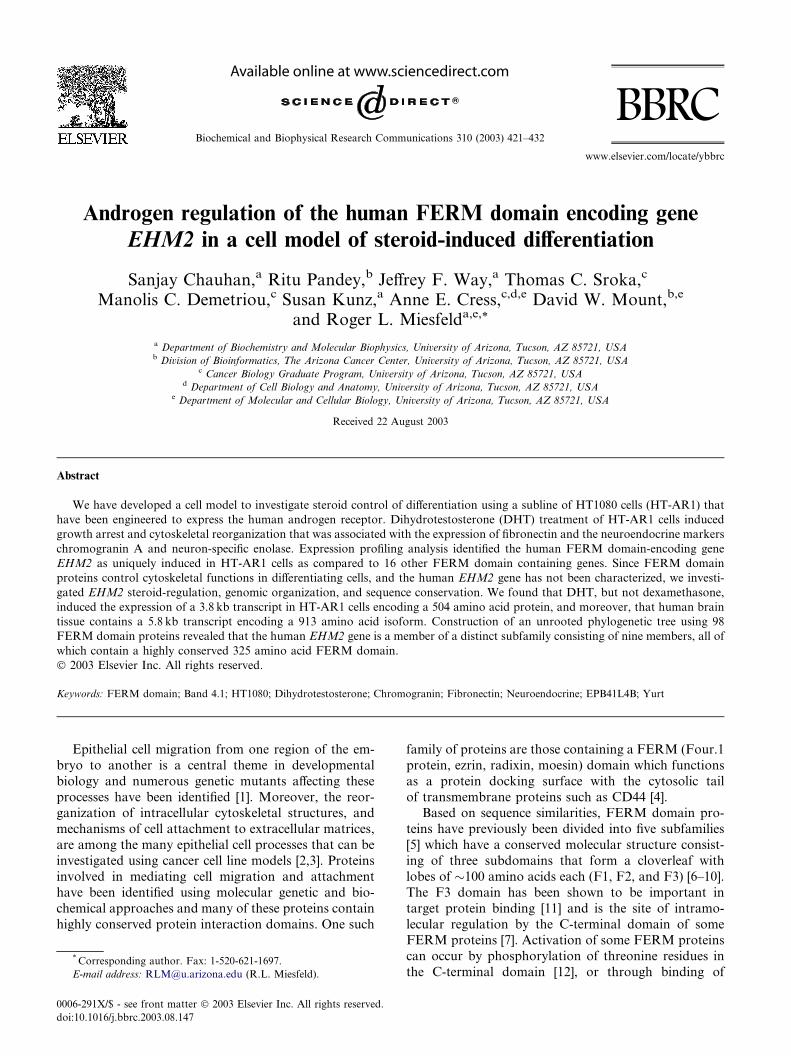
Fig. 1. Growth arrest and fibronectin expression in HT1080 sublines expressing human AR. (A) Western blot of protein extracts from puromycin-
resistant HT1080 sublines transfected with the pDC-PAC vector (HT-VC1), or with the AR expression vector pDC-AR-PAC (HT-AR1, HT-AR2).
The blot also contains a protein extract isolated from the human prostate cancer cell line LNCaP [35], for comparison. The membrane was probed
with anti-AR or anti-tubulin antibodies. Results of cell proliferation assays are also presented using HT-VC1 (B), HT-AR1 (C) or HT-AR3 (D) cells
grown in culture media with or without 1 lM DHT or Dex for 3, 5 or 7 days. The number of viable cells in each triplicate well was determined by
trypan blue exclusion. (E) Northern blot of fibronectin expression using total RNA isolated from HT-AR1 cells cultured in DHT for 0, 4, 12, 24, and
48 h. The level of L7 ribosomal protein (L7 RP) RNA was used as a loading control. (F) Western blot of fibronectin protein levels in HT-AR1 cells
following DHT treatment as described in (E). The level of a-tubulin protein was used as a loading control.
424 S. Chauhan et al. / Biochemical and Biophysical Research Communications 310 (2003) 421–432
Dexamethasone treatment of HT1080 cells has been
shown to induce high levels of fibronectin expression
[21–23] without altering cell proliferation rates or cell
morphology [21]. We observed a stark difference be-tween the effect of Dex and DHT on the growth of AR
expressing HT1080 sublines (Figs. 1C and D) and
therefore determined if DHT treatment led to differences
in the steady-state levels of fibronectin RNA and pro-
tein. As shown in Fig. 1E, fibronectin RNA levels were
significantly increased after 24 and 48 h of DHT treat-
ment as measured by Northern blots. Western blots
using an anti-fibronectin antibody showed a coincidingincrease in fibronectin protein levels in DHT treated
HT-1080 cells (Fig. 1F). These data indicated that the
shared induction of fibronectin expression by GR [21–
23] and AR (Fig. 1F) in HT-AR1 cells was likely to be
distinct from the observed AR-specific growth arrest
phenotype (Fig. 1D).
During the course of these experiments, we noticed
that DHT treatment of HT-AR1 cells led to the ap-pearance of neuronal-like membrane structures, sug-
gesting that the growth arrest phenotype in HT-AR1
cells may involve activation of a differentiation pathway.
As shown in Fig. 2, DHT treatment of HT-AR1 cells
cultured in normal serum resulted in the appearance of
multiple extensions protruding from the plasma mem-
brane (panel b). In addition, when HT-AR1 cells were
cultured in charcoal-stripped (C/S) serum, cell surface
structures resembling secretory granules were readily
apparent in the DHT-treated cultures (panel d). To
better visualize DHT-dependent changes in the cyto-
skeleton, we cultured the cells on coverslips coated withfibronectin and performed immunostaining with an anti-
keratin 5 antibody and Hoechst dye. As can be seen in
panel f of Fig. 2A, the organization of keratin networks
was also dramatically altered in HT-AR1 cells, sug-
gesting that androgens activate a signaling pathway that
controls cytoskeletal organization in these cells. Finally,
as shown in Fig. 2B, Western blotting experiments using
antibodies directed against the neuronal-specific pro-teins chromogranin A (CgA) and neural specific enolase
(NSE) revealed that HT-AR1 cells constitutively express
both of these proteins (Fig. 2B), and moreover, that
DHT treatment increases CgA and NSE protein levels
relative to that of tubulin (Fig. 2B). Both CgA and NSE
are considered neuroendocrine cell markers that have
been shown to be expressed in numerous tissues in-
cluding prostate [36].Although the parental HT1080 cell line had been
originally described as an outgrowth of an undifferenti-
ated fibrosarcoma isolated from the acetabulum of a
35-year-old male [18], and it is often compared to fibro-
blast-derived cell lines [37] and normal human fibroblasts
[22], the results presented in Fig. 2 suggest that the
AR-mediated growth arrest and neuronal differentiation
phenotype of HT1080 cells was similar to reconstituted
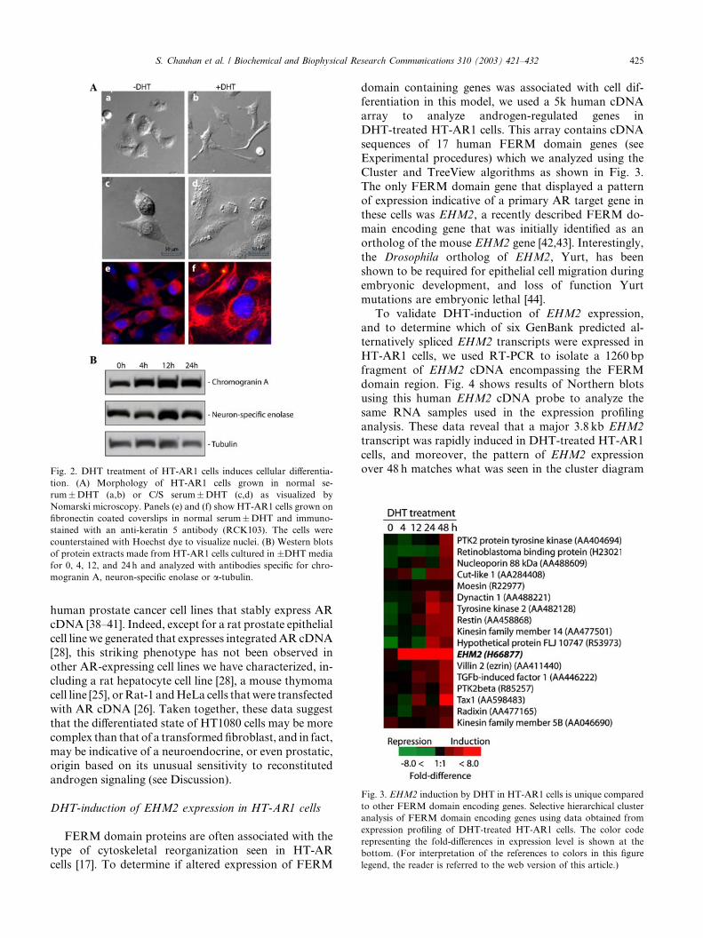
Fig. 3. EHM2 induction by DHT in HT-AR1 cells is unique compared
to other FERM domain encoding genes. Selective hierarchical cluster
analysis of FERM domain encoding genes using data obtained from
expression profiling of DHT-treated HT-AR1 cells. The color code
representing the fold-differences in expression level is shown at the
bottom. (For interpretation of the references to colors in this figure
legend, the reader is referred to the web version of this article.)
Fig. 2. DHT treatment of HT-AR1 cells induces cellular differentia-
tion. (A) Morphology of HT-AR1 cells grown in normal se-
rum�DHT (a,b) or C/S serum�DHT (c,d) as visualized by
Nomarski microscopy. Panels (e) and (f) show HT-AR1 cells grown on
fibronectin coated coverslips in normal serum�DHT and immuno-
stained with an anti-keratin 5 antibody (RCK103). The cells were
counterstained with Hoechst dye to visualize nuclei. (B) Western blots
of protein extracts made from HT-AR1 cells cultured in �DHT media
for 0, 4, 12, and 24 h and analyzed with antibodies specific for chro-
mogranin A, neuron-specific enolase or a-tubulin.
S. Chauhan et al. / Biochemical and Biophysical Research Communications 310 (2003) 421–432 425
human prostate cancer cell lines that stably express AR
cDNA [38–41]. Indeed, except for a rat prostate epithelial
cell line we generated that expresses integratedAR cDNA[28], this striking phenotype has not been observed in
other AR-expressing cell lines we have characterized, in-
cluding a rat hepatocyte cell line [28], a mouse thymoma
cell line [25], orRat-1 andHeLa cells thatwere transfected
with AR cDNA [26]. Taken together, these data suggest
that the differentiated state of HT1080 cells may be more
complex than that of a transformed fibroblast, and in fact,
may be indicative of a neuroendocrine, or even prostatic,origin based on its unusual sensitivity to reconstituted
androgen signaling (see Discussion).
DHT-induction of EHM2 expression in HT-AR1 cells
FERM domain proteins are often associated with the
type of cytoskeletal reorganization seen in HT-AR
cells [17]. To determine if altered expression of FERM
domain containing genes was associated with cell dif-ferentiation in this model, we used a 5k human cDNA
array to analyze androgen-regulated genes in
DHT-treated HT-AR1 cells. This array contains cDNA
sequences of 17 human FERM domain genes (see
Experimental procedures) which we analyzed using the
Cluster and TreeView algorithms as shown in Fig. 3.
The only FERM domain gene that displayed a pattern
of expression indicative of a primary AR target gene inthese cells was EHM2, a recently described FERM do-
main encoding gene that was initially identified as an
ortholog of the mouse EHM2 gene [42,43]. Interestingly,
the Drosophila ortholog of EHM2, Yurt, has been
shown to be required for epithelial cell migration during
embryonic development, and loss of function Yurt
mutations are embryonic lethal [44].
To validate DHT-induction of EHM2 expression,and to determine which of six GenBank predicted al-
ternatively spliced EHM2 transcripts were expressed in
HT-AR1 cells, we used RT-PCR to isolate a 1260 bp
fragment of EHM2 cDNA encompassing the FERM
domain region. Fig. 4 shows results of Northern blots
using this human EHM2 cDNA probe to analyze the
same RNA samples used in the expression profiling
analysis. These data reveal that a major 3.8 kb EHM2
transcript was rapidly induced in DHT-treated HT-AR1
cells, and moreover, the pattern of EHM2 expression
over 48 h matches what was seen in the cluster diagram
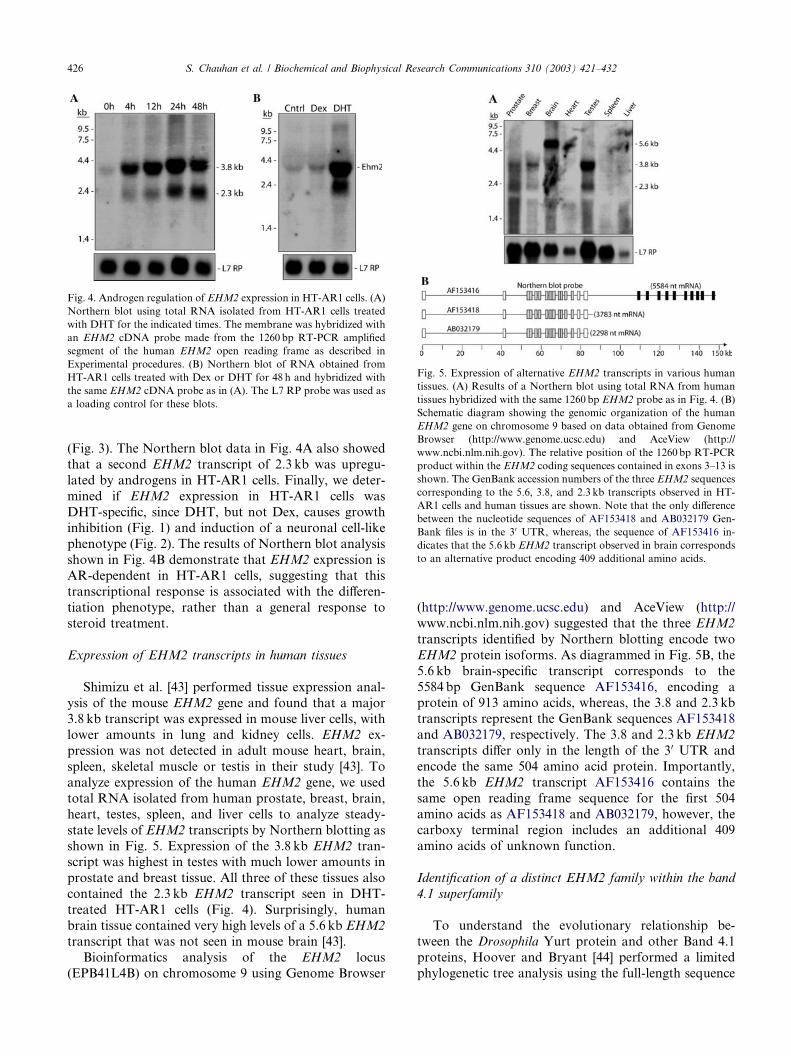
Fig. 4. Androgen regulation of EHM2 expression in HT-AR1 cells. (A)
Northern blot using total RNA isolated from HT-AR1 cells treated
with DHT for the indicated times. The membrane was hybridized with
an EHM2 cDNA probe made from the 1260 bp RT-PCR amplified
segment of the human EHM2 open reading frame as described in
Experimental procedures. (B) Northern blot of RNA obtained from
HT-AR1 cells treated with Dex or DHT for 48 h and hybridized with
the same EHM2 cDNA probe as in (A). The L7 RP probe was used as
a loading control for these blots.
Fig. 5. Expression of alternative EHM2 transcripts in various human
tissues. (A) Results of a Northern blot using total RNA from human
tissues hybridized with the same 1260 bp EHM2 probe as in Fig. 4. (B)
Schematic diagram showing the genomic organization of the human
EHM2 gene on chromosome 9 based on data obtained from Genome
Browser (http://www.genome.ucsc.edu) and AceView (http://
www.ncbi.nlm.nih.gov). The relative position of the 1260bp RT-PCR
product within the EHM2 coding sequences contained in exons 3–13 is
shown. The GenBank accession numbers of the three EHM2 sequences
corresponding to the 5.6, 3.8, and 2.3 kb transcripts observed in HT-
AR1 cells and human tissues are shown. Note that the only difference
between the nucleotide sequences of AF153418 and AB032179 Gen-
Bank files is in the 30 UTR, whereas, the sequence of AF153416 in-
dicates that the 5.6 kb EHM2 transcript observed in brain corresponds
to an alternative product encoding 409 additional amino acids.
426 S. Chauhan et al. / Biochemical and Biophysical Research Communications 310 (2003) 421–432
(Fig. 3). The Northern blot data in Fig. 4A also showed
that a second EHM2 transcript of 2.3 kb was upregu-
lated by androgens in HT-AR1 cells. Finally, we deter-
mined if EHM2 expression in HT-AR1 cells was
DHT-specific, since DHT, but not Dex, causes growthinhibition (Fig. 1) and induction of a neuronal cell-like
phenotype (Fig. 2). The results of Northern blot analysis
shown in Fig. 4B demonstrate that EHM2 expression is
AR-dependent in HT-AR1 cells, suggesting that this
transcriptional response is associated with the differen-
tiation phenotype, rather than a general response to
steroid treatment.
Expression of EHM2 transcripts in human tissues
Shimizu et al. [43] performed tissue expression anal-
ysis of the mouse EHM2 gene and found that a major
3.8 kb transcript was expressed in mouse liver cells, withlower amounts in lung and kidney cells. EHM2 ex-
pression was not detected in adult mouse heart, brain,
spleen, skeletal muscle or testis in their study [43]. To
analyze expression of the human EHM2 gene, we used
total RNA isolated from human prostate, breast, brain,
heart, testes, spleen, and liver cells to analyze steady-
state levels of EHM2 transcripts by Northern blotting as
shown in Fig. 5. Expression of the 3.8 kb EHM2 tran-script was highest in testes with much lower amounts in
prostate and breast tissue. All three of these tissues also
contained the 2.3 kb EHM2 transcript seen in DHT-
treated HT-AR1 cells (Fig. 4). Surprisingly, human
brain tissue contained very high levels of a 5.6 kb EHM2
transcript that was not seen in mouse brain [43].
Bioinformatics analysis of the EHM2 locus
(EPB41L4B) on chromosome 9 using Genome Browser
(http://www.genome.ucsc.edu) and AceView (http://
www.ncbi.nlm.nih.gov) suggested that the three EHM2
transcripts identified by Northern blotting encode two
EHM2 protein isoforms. As diagrammed in Fig. 5B, the
5.6 kb brain-specific transcript corresponds to the
5584 bp GenBank sequence AF153416, encoding aprotein of 913 amino acids, whereas, the 3.8 and 2.3 kb
transcripts represent the GenBank sequences AF153418
and AB032179, respectively. The 3.8 and 2.3 kb EHM2
transcripts differ only in the length of the 30 UTR and
encode the same 504 amino acid protein. Importantly,
the 5.6 kb EHM2 transcript AF153416 contains the
same open reading frame sequence for the first 504
amino acids as AF153418 and AB032179, however, thecarboxy terminal region includes an additional 409
amino acids of unknown function.
Identification of a distinct EHM2 family within the band
4.1 superfamily
To understand the evolutionary relationship be-
tween the Drosophila Yurt protein and other Band 4.1
proteins, Hoover and Bryant [44] performed a limited
phylogenetic tree analysis using the full-length sequence
S. Chauhan et al. / Biochemical and Biophysical Research Communications 310 (2003) 421–432 427
of 19 FERM domain proteins. Their analysis suggestedthat EHM2, Yurt, and a FERM domain protein from
Caenorhabditis elegans (T04C9) constitute a subfamily
that is most closely related to NBL4 proteins as orig-
inally proposed by Shimizu et al. [43]. Since their study
included only a limited number of the nearly 100
FERM domain containing gene sequences in the
GenBank database, and was based on comparisons
using sequences outside of the FERM domain, weperformed a much more extensive study to determine if
EHM2-related proteins were indeed a separate sub-
group. For our analysis we limited the comparison to a
�220 amino acid segment of the FERM domain
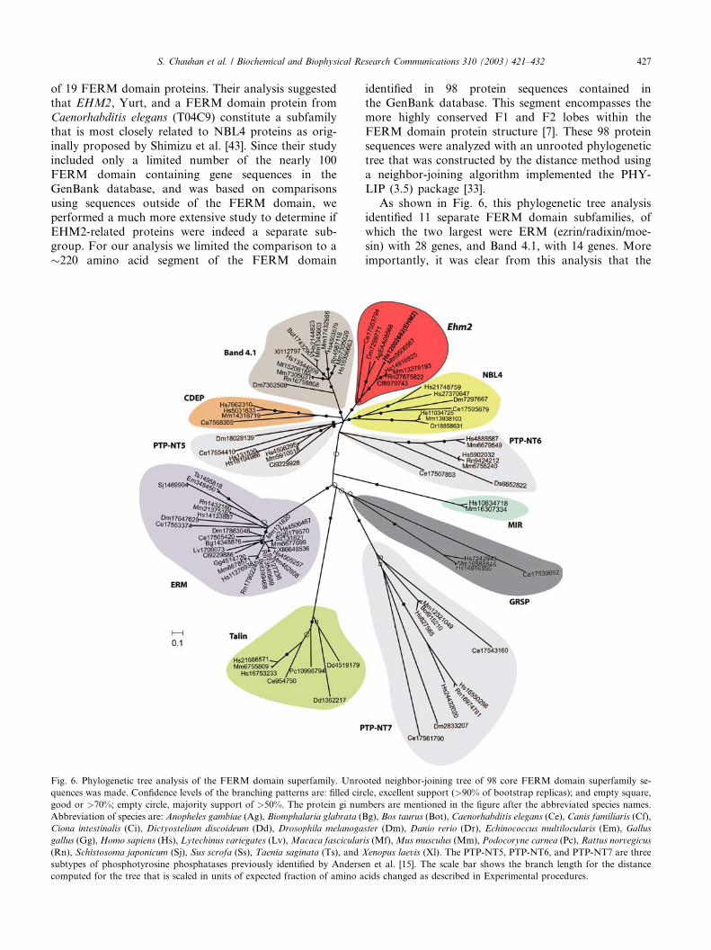
Fig. 6. Phylogenetic tree analysis of the FERM domain superfamily. Unro
quences was made. Confidence levels of the branching patterns are: filled cir
good or >70%; empty circle, majority support of >50%. The protein gi num
Abbreviation of species are: Anopheles gambiae (Ag), Biomphalaria glabrata (B
Ciona intestinalis (Ci), Dictyostelium discoideum (Dd), Drosophila melanoga
gallus (Gg),Homo sapiens (Hs), Lytechinus variegates (Lv),Macaca fascicular
(Rn), Schistosoma japonicum (Sj), Sus scrofa (Ss), Taenia saginata (Ts), and X
subtypes of phosphotyrosine phosphatases previously identified by Anderse
computed for the tree that is scaled in units of expected fraction of amino a
identified in 98 protein sequences contained inthe GenBank database. This segment encompasses the
more highly conserved F1 and F2 lobes within the
FERM domain protein structure [7]. These 98 protein
sequences were analyzed with an unrooted phylogenetic
tree that was constructed by the distance method using
a neighbor-joining algorithm implemented the PHY-
LIP (3.5) package [33].
As shown in Fig. 6, this phylogenetic tree analysisidentified 11 separate FERM domain subfamilies, of
which the two largest were ERM (ezrin/radixin/moe-
sin) with 28 genes, and Band 4.1, with 14 genes. More
importantly, it was clear from this analysis that the
oted neighbor-joining tree of 98 core FERM domain superfamily se-
cle, excellent support (>90% of bootstrap replicas); and empty square,
bers are mentioned in the figure after the abbreviated species names.
g), Bos taurus (Bot), Caenorhabditis elegans (Ce), Canis familiaris (Cf),
ster (Dm), Danio rerio (Dr), Echinococcus multilocularis (Em), Gallus
is (Mf),Mus musculus (Mm), Podocoryne carnea (Pc), Rattus norvegicus
enopus laevis (Xl). The PTP-NT5, PTP-NT6, and PTP-NT7 are three
n et al. [15]. The scale bar shows the branch length for the distance
cids changed as described in Experimental procedures.
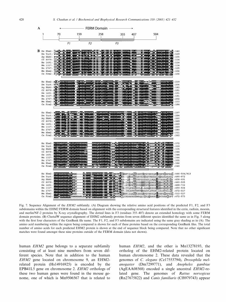
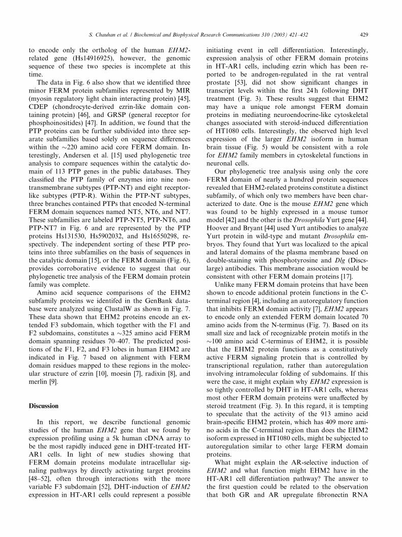
Fig. 7. Sequence Alignment of the EHM2 subfamily. (A) Diagram showing the relative amino acid positions of the predicted F1, F2, and F3
subdomains within the EHM2 FERM domain based on alignment with the corresponding structural features identified in the ezrin, radixin, moesin,
and merlin/NF-2 proteins by X-ray crystallography. The dotted lines in F3 (residues 355–407) denote an extended homology with some FERM
domain proteins. (B) ClustalW sequence alignment of EHM2 subfamily proteins from seven different species identified the same as in Fig. 5 along
with the first four characters of the GenBank file name. The F1, F2, and F3 subdomains are indicated using the same gray shading as in (A). The
amino acid numbering within the region being compared is shown for each of these proteins based on the corresponding GenBank files. The total
number of amino acids for each predicted EHM2 protein is shown at the end of sequence block being compared. Note that no other significant
matches were found amongst these nine proteins outside of the FERM domain (data not shown).
428 S. Chauhan et al. / Biochemical and Biophysical Research Communications 310 (2003) 421–432
human EHM2 gene belongs to a separate subfamily
consisting of at least nine members from seven dif-ferent species. Note that in addition to the human
EHM2 gene located on chromosome 9, an EHM2-
related protein (Hs14916925) is encoded by the
EPB41L5 gene on chromosome 2. EHM2 orthologs of
these two human genes were found in the mouse ge-
nome, one of which is Mn9506567 that is related to
human EHM2, and the other is Mn13278193, the
ortholog of the EHM2-related protein located onhuman chromosome 2. These data revealed that the
genomes of C. elegans (Ce17553794), Drosophila mel-
anogaster (Dm7299771), and Anopheles gambiae
(AgEAA08568) encoded a single ancestral EHM2-re-
lated gene. The genomes of Rattus norvegicus
(Rn27675822) and Canis familiaris (Cf8979743) appear
S. Chauhan et al. / Biochemical and Biophysical Research Communications 310 (2003) 421–432 429
to encode only the ortholog of the human EHM2-related gene (Hs14916925), however, the genomic
sequence of these two species is incomplete at this
time.
The data in Fig. 6 also show that we identified three
minor FERM protein subfamilies represented by MIR
(myosin regulatory light chain interacting protein) [45],
CDEP (chondrocyte-derived ezrin-like domain con-
taining protein) [46], and GRSP (general receptor forphosphoinositides) [47]. In addition, we found that the
PTP proteins can be further subdivided into three sep-
arate subfamilies based solely on sequence differences
within the �220 amino acid core FERM domain. In-
terestingly, Andersen et al. [15] used phylogenetic tree
analysis to compare sequences within the catalytic do-
main of 113 PTP genes in the public databases. They
classified the PTP family of enzymes into nine non-transmembrane subtypes (PTP-NT) and eight receptor-
like subtypes (PTP-R). Within the PTP-NT subtypes,
three branches contained PTPs that encoded N-terminal
FERM domain sequences named NT5, NT6, and NT7.
These subfamilies are labeled PTP-NT5, PTP-NT6, and
PTP-NT7 in Fig. 6 and are represented by the PTP
proteins Hs131530, Hs5902032, and Hs16550298, re-
spectively. The independent sorting of these PTP pro-teins into three subfamilies on the basis of sequences in
the catalytic domain [15], or the FERM domain (Fig. 6),
provides corroborative evidence to suggest that our
phylogenetic tree analysis of the FERM domain protein
family was complete.
Amino acid sequence comparisons of the EHM2
subfamily proteins we identifed in the GenBank data-
base were analyzed using ClustalW as shown in Fig. 7.These data shown that EHM2 proteins encode an ex-
tended F3 subdomain, which together with the F1 and
F2 subdomains, constitutes a �325 amino acid FERM
domain spanning residues 70–407. The predicted posi-
tions of the F1, F2, and F3 lobes in human EHM2 are
indicated in Fig. 7 based on alignment with FERM
domain residues mapped to these regions in the molec-
ular structure of ezrin [10], moesin [7], radixin [8], andmerlin [9].
Discussion
In this report, we describe functional genomic
studies of the human EHM2 gene that we found by
expression profiling using a 5k human cDNA array tobe the most rapidly induced gene in DHT-treated HT-
AR1 cells. In light of new studies showing that
FERM domain proteins modulate intracellular sig-
naling pathways by directly activating target proteins
[48–52], often through interactions with the more
variable F3 subdomain [52], DHT-induction of EHM2
expression in HT-AR1 cells could represent a possible
initiating event in cell differentiation. Interestingly,expression analysis of other FERM domain proteins
in HT-AR1 cells, including ezrin which has been re-
ported to be androgen-regulated in the rat ventral
prostate [53], did not show significant changes in
transcript levels within the first 24 h following DHT
treatment (Fig. 3). These results suggest that EHM2
may have a unique role amongst FERM domain
proteins in mediating neuroendocrine-like cytoskeletalchanges associated with steroid-induced differentiation
of HT1080 cells. Interestingly, the observed high level
expression of the larger EHM2 isoform in human
brain tissue (Fig. 5) would be consistent with a role
for EHM2 family members in cytoskeletal functions in
neuronal cells.
Our phylogenetic tree analysis using only the core
FERM domain of nearly a hundred protein sequencesrevealed that EHM2-related proteins constitute a distinct
subfamily, of which only two members have been char-
acterized to date. One is the mouse EHM2 gene which
was found to be highly expressed in a mouse tumor
model [42] and the other is theDrosophila Yurt gene [44].
Hoover and Bryant [44] used Yurt antibodies to analyze
Yurt protein in wild-type and mutant Drosophila em-
bryos. They found that Yurt was localized to the apicaland lateral domains of the plasma membrane based on
double-staining with phosphotyrosine and Dlg (Discs-
large) antibodies. This membrane association would be
consistent with other FERM domain proteins [17].
Unlike many FERM domain proteins that have been
shown to encode additional protein functions in the C-
terminal region [4], including an autoregulatory function
that inhibits FERM domain activity [7], EHM2 appearsto encode only an extended FERM domain located 70
amino acids from the N-terminus (Fig. 7). Based on its
small size and lack of recognizable protein motifs in the
�100 amino acid C-terminus of EHM2, it is possible
that the EHM2 protein functions as a constitutively
active FERM signaling protein that is controlled by
transcriptional regulation, rather than autoregulation
involving intramolecular folding of subdomains. If thiswere the case, it might explain why EHM2 expression is
so tightly controlled by DHT in HT-AR1 cells, whereas
most other FERM domain proteins were unaffected by
steroid treatment (Fig. 3). In this regard, it is tempting
to speculate that the activity of the 913 amino acid
brain-specific EHM2 protein, which has 409 more ami-
no acids in the C-terminal region than does the EHM2
isoform expressed in HT1080 cells, might be subjected toautoregulation similar to other large FERM domain
proteins.
What might explain the AR-selective induction of
EHM2 and what function might EHM2 have in the
HT-AR1 cell differentiation pathway? The answer to
the first question could be related to the observation
that both GR and AR upregulate fibronectin RNA
430 S. Chauhan et al. / Biochemical and Biophysical Research Communications 310 (2003) 421–432
and protein levels, but only AR activation leads togrowth arrest and neuroendocrine-like cell differentia-
tion in the HT-AR1 cell line. This result would sug-
gest that a subset of AR-regulated genes in this model
system is not shared by GR, and moreover, that these
genes have either gained, or retained, sensitivity to
androgen signaling when functional AR is provided by
stable transfection. One possibility is that the HT1080
cell line established by Rasheed et al. [18] could havebeen derived from an androgen-sensitive primary
cancer, for example, a myofibroblastoma that origi-
nated in smooth muscle tissue [54]. In support of this
idea, DHT treatment of HT-AR1 cells induces the
expression of the motor protein myosin 1B (MYO1B)
(Chauhan et al., submitted), which could indicate that
HT1080 cells originated from muscle tissue. Interest-
ingly, unconventional myosins such as MYO1B havebeen shown to function in vesicle transport in neurons
[55] which would be consistent with our observation
that androgen treatment leads to the appearance of
secretory granules and induces the expression of
chromogranin A and neuron-specific enolase in HT-
AR1 cells (Fig. 2). Since six independent AR-ex-
pressing subclones of HT1080 cells we have estab-
lished share this same DHT-induced neuronal cell-likephenotype (Way and Miesfeld, unpublished data), we
suggest that HT1080 cells possess previously unknown
stem cell properties that may have been retained
during cancer cell progression or reactivated by sub-
sequent genomic rearrangements.
In summary, we found that activation of androgen
signaling in the human HT1080 fibrosarcoma cell line
resulted in dramatic cytoskeletal reorganization andneuroendocrine-like cell differentiation that was asso-
ciated with induction of the human FERM domain
encoding gene EHM2. The unique expression profile
of EHM2 in DHT-treated HT-AR1 cells, and its
distinct 325 amino acid FERM domain sequence,
suggests that this recently discovered gene may have
a regulatory role in mediating cytoskeletal reorgani-
zation in steroid-regulated cell differentiation path-ways.
Note added in proof
Genotyping of HT-AR1 cells and an HT-1080 cell stock ob-
tained directly from American Type Culture Collection showed that
the two cell lines match at 11/11 loci, thereby confirming that the
HT-AR1 and HT-1080 cell lines are isogenic (AEC and RLM,
unpublished data).
Acknowledgments
We thank Jordan Roberts for his efforts in characterizing the HT-
AR cell lines and Ray Nagle for the gift of HT1080 cells. This work
was supported by NIH Grants PO1 CA 56666 and CA 75152 (A.E.C.),
and through the generous support of the Jack Findlay Doyle II
Charitable Fund (R.L.M.).
References
[1] H.A. Schneiderman, P.J. Bryant, Genetic analysis of develop-
mental mechanisms in Drosophila, Nature 234 (1971) 187–194.
[2] A. Gautreau, D. Louvard, M. Arpin, ERM proteins and NF2
tumor suppressor: the Yin and Yang of cortical actin organization
and cell growth signaling, Curr. Opin. Cell Biol. 14 (2002) 104–109.
[3] H. Ponta, L. Sherman, P.A. Herrlich, CD44: from adhesion
molecules to signalling regulators, Nat. Rev. Mol. Cell Biol. 4
(2003) 33–45.
[4] C.X. Sun, V.A. Robb, D.H. Gutmann, Protein 4.1 tumor
suppressors: getting a FERM grip on growth regulation, J. Cell
Sci. 115 (2002) 3991–4000.
[5] K. Takeuchi, A. Kawashima, A. Nagafuchi, S. Tsukita, Structural
diversity of band 4.1 superfamily members, J. Cell Sci. 107 (Part 7)
(1994) 1921–1928.
[6] B.G. Han, W. Nunomura, Y. Takakuwa, N. Mohandas, B.K. Jap,
Protein 4.1R core domain structure and insights into regulation of
cytoskeletal organization, Nat. Struct. Biol. 7 (2000) 871–875.
[7] M.A. Pearson, D. Reczek, A. Bretscher, P.A. Karplus, Structure
of the ERM protein moesin reveals the FERM domain fold
masked by an extended actin binding tail domain, Cell 101 (2000)
259–270.
[8] K. Hamada, T. Shimizu, T. Matsui, S. Tsukita, T. Hakoshima,
Structural basis of the membrane-targeting and unmasking
mechanisms of the radixin FERM domain, EMBO J. 19 (2000)
4449–4462.
[9] T. Shimizu, A. Seto, N. Maita, K. Hamada, S. Tsukita, T.
Hakoshima, Structural basis for neurofibromatosis type 2. Crystal
structure of the merlin FERM domain, J. Biol. Chem. 277 (2002)
10332–10336.
[10] W.J. Smith, N. Nassar, A. Bretscher, R.A. Cerione, P.A. Karplus,
Structure of the active N-terminal domain of ezrin, J. Biol. Chem.
278 (2003) 4949–4956.
[11] K. Hamada, T. Shimizu, S. Yonemura, S. Tsukita, T. Hakoshima,
Structural basis of adhesion-molecule recognition by ERM
proteins revealed by the crystal structure of the radixin–ICAM-2
complex, EMBO J. 22 (2003) 502–514.
[12] F. Nakamura, M.R. Amieva, H. Furthmayr, Phosphorylation of
threonine 558 in the carboxyl-terminal actin-binding domain of
moesin by thrombin activation of human platelets, J. Biol. Chem.
270 (1995) 31377–31385.
[13] M. Hirao, N. Sato, T. Kondo, S. Yonemura, M. Monden, T.
Sasaki, Y. Takai, S. Tsukita, Regulation mechanism of ERM
(ezrin/radixin/moesin) protein/plasma membrane association: pos-
sible involvement of phosphatidylinositol turnover and Rho-
dependent signaling pathway, J. Cell Biol. 135 (1996) 37–51.
[14] P. Mangeat, C. Roy, M. Martin, ERM proteins in cell adhesion
and membrane dynamics, Trends Cell Biol. 9 (1999) 187–192.
[15] J.N. Andersen, O.H. Mortensen, G.H. Peters, P.G. Drake, L.F.
Iversen, O.H. Olsen, P.G. Jansen, H.S. Andersen, N.K. Tonks,
N.P. Moller, Structural and evolutionary relationships among
protein tyrosine phosphatase domains, Mol. Cell. Biol. 21 (2001)
7117–7136.
[16] T. Kubo, T. Yamashita, A. Yamaguchi, H. Sumimoto, K.
Hosokawa, M. Tohyama, A novel FERM domain including
guanine nucleotide exchange factor is involved in Rac signaling
and regulates neurite remodeling, J. Neurosci. 22 (2002) 8504–
8513.
S. Chauhan et al. / Biochemical and Biophysical Research Communications 310 (2003) 421–432 431
[17] A. Bretscher, K. Edwards, R.G. Fehon, ERM proteins and
merlin: integrators at the cell cortex, Nat. Rev. Mol. Cell Biol. 3
(2002) 586–599.
[18] S. Rasheed, W.A. Nelson-Rees, E.M. Toth, P. Arnstein, M.B.
Gardner, Characterization of a newly derived human sarcoma cell
line (HT-1080), Cancer 33 (1974) 1027–1033.
[19] F. Bulens, P. Merchiers, I. Ibanez-Tallon, A. De Vriese, L. Nelles,
F. Claessens, A. Belayew, D. Collen, Identification of a multi-
hormone responsive enhancer far upstream from the human
tissue-type plasminogen activator gene, J. Biol. Chem. 272 (1997)
663–671.
[20] R.L. Miesfeld, Glucocorticoid action: biochemistry, in: L.J.
DeGroot, J.L. Jameson (Eds.), Endocrinology, W.B. Saunders,
Philadelphia, 2000, pp. 1647–1654.
[21] D.C. Dean, R.F. Newby, S. Bourgeois, Regulation of fibronec-
tin biosynthesis by dexamethasone, transforming growth factor
beta, and cAMP in human cell lines, J. Cell Biol. 106 (1988)
2159–2170.
[22] N. Oliver, R.F. Newby, L.T. Furcht, S. Bourgeois, Regulation of
fibronectin biosynthesis by glucocorticoids in human fibrosar-
coma cells and normal fibroblasts, Cell 33 (1983) 287–296.
[23] K.A. Brenner, S.A. Corbett, J.E. Schwarzbauer, Regulation of
fibronectin matrix assembly by activated Ras in transformed cells,
Oncogene 19 (2000) 3156–3163.
[24] N. Kondoh, M. Shuda, M. Arai, T. Oikawa, M. Yamamoto,
Activation of anchorage-independent growth of HT1080 human
fibrosarcoma cells by dexamethasone, In Vitro Cell Dev. Biol.
Anim. 38 (2002) 111–117.
[25] M.S. Chapman, D.J. Askew, U. Kuscuoglu, R.L. Miesfeld,
Transcriptional control of steroid-regulated apoptosis in murine
thymoma cells, Mol. Endocrinol. 10 (1996) 967–978.
[26] D.A. Gordon, N.L. Chamberlain, F.A. Flomerfelt, R.L. Miesfeld,
A cell-specific and selective effect on transactivation by the
androgen receptor, Exp. Cell Res. 217 (1995) 368–377.
[27] D.C. Whitacre, K.J. Karnas, R.L. Miesfeld, Analysis of gluco-
corticoid and androgen receptor gene fusions delineates domains
required for transcriptional specificity, Endocrine 15 (2001)
111–118.
[28] D.C. Whitacre, S. Chauhan, T. Davis, D. Gordon, A.E. Cress, R.
Miesfeld, Androgen-induction of in vitro prostate cell differenti-
ation, Cell Growth Differ. 13 (2002) 1–11.
[29] N.L. Chamberlain, E.D. Driver, R.L. Miesfeld, The length and
location of CAG trinucleotide repeats in the androgen receptor N-
terminal domain affect transactivation function, Nucleic Acids
Res. 22 (1994) 3181–3186.
[30] S. Chauhan, C.H. Leach, S. Kunz, J.W. Bloom, R.L. Miesfeld,
Glucocorticoid regulation of human eosinophil gene expression, J.
Steroid Biochem. Mol. Biol. 84 (2003) 441–452.
[31] M.B. Eisen, P.T. Spellman, P.O. Brown, D. Botstein, Cluster
analysis and display of genome-wide expression patterns, Proc.
Natl. Acad. Sci. USA 95 (1998) 14863–14868.
[32] J.D. Thompson, D.G. Higgins, T.J. Gibson, CLUSTAL W:
improving the sensitivity of progressive multiple sequence align-
ment through sequence weighting, position-specific gap penalties
and weight matrix choice, Nucleic Acids Res. 22 (1994) 4673–4680.
[33] J. Felsenstein, PHYLIP-Phylogeny inference package version
(3.2), Cladistics 5 (1989) 164–166.
[34] R.M. Schwartz, M.O. Dayhoff, B.C. Orcult, A model of evolu-
tionary change in proteins, in: M.O. Dayhoff (Ed.), Atlas of
Protein Sequence and Structure, National Biomedical Research
Foundation, Washington, DC, 1978.
[35] W.D. Tilley, C.M. Wilson, M. Marcelli, M.J. McPhaul, Androgen
receptor gene expression in human prostate carcinoma cell lines,
Cancer Res. 50 (1990) 5382–5386.
[36] E.M. Wilson, Y. Oh, V. Hwa, R.G. Rosenfeld, Interaction of
igf-binding protein-related protein 1 with a novel protein, neuro-
endocrine differentiation factor, results in neuroendocrine differ-
entiation of prostate cancer cells, J. Clin. Endocrinol. Metab. 86
(2001) 4504–4511.
[37] W.P. Schiemann, G.C. Blobe, D.E. Kalume, A. Pandey, H.F.
Lodish, Context-specific effects of fibulin-5 (DANCE/EVEC) on
cell proliferation, motility, and invasion. Fibulin-5 is induced by
transforming growth factor-beta and affects protein kinase
cascades, J. Biol. Chem. 277 (2002) 27367–27377.
[38] H.Y. Zhau, S.M. Chang, B.Q. Chen, Y. Wang, H. Zhang, C. Kao,
Q.A. Sang, S.J. Pathak, L.W. Chung, Androgen-repressed
phenotype in human prostate cancer, Proc. Natl. Acad. Sci.
USA 93 (1996) 15152–15157.
[39] M.T. Ling, K.W. Chan, C.K. Choo, Androgen induces differen-
tiation of a human papillomavirus 16 E6/E7 immortalized
prostate epithelial cell line, J. Endocrinol. 170 (2001) 287–296.
[40] S. Yuan, J. Trachtenberg, G.B. Mills, T.J. Brown, F. Xu, A.
Keating, Androgen-induced inhibition of cell proliferation in an
androgen-insensitive prostate cancer cell line (PC-3) transfected
with a human androgen receptor complementary DNA, [pub-
lished erratum appears in Cancer Res. 55 (3) (1995) 719], Cancer
Res. 53 (1993) 1304–1311.
[41] L.E. Heisler, A. Evangelou, A.M. Lew, J. Trachtenberg, H.P.
Elsholtz, T.J. Brown, Androgen-dependent cell cycle arrest and
apoptotic death in PC-3 prostatic cell cultures expressing a full-
length human androgen receptor, Mol. Cell. Endocrinol. 126
(1997) 59–73.
[42] Y. Hashimoto, N. Shindo-Okada, M. Tani, K. Takeuchi, H.
Toma, J. Yokota, Identification of genes differentially expressed in
association with metastatic potential of K-1735 murine melanoma
by messenger RNA differential display, Cancer Res. 56 (1996)
5266–5271.
[43] K. Shimizu, Y. Nagamachi, M. Tani, K. Kimura, T. Shiroishi, S.
Wakana, J. Yokota, Molecular cloning of a novel NF2/ERM/4.1
superfamily gene, ehm2, that is expressed in high-metastatic
K1735 murine melanoma cells, Genomics 65 (2000) 113–120.
[44] K.B. Hoover, P.J. Bryant, Drosophila Yurt is a new protein-4.1-
like protein required for epithelial morphogenesis, Dev. Genes
Evol. 212 (2002) 230–238.
[45] Y. Koyano, T. Kawamoto, A. Kikuchi, M. Shen, Y. Kuruta, S.
Tsutsumi, K. Fujimoto, M. Noshiro, K. Fujii, Y. Kato, Chon-
drocyte-derived ezrin-like domain containing protein (CDEP), a
rho guanine nucleotide exchange factor, is inducible in chondro-
cytes by parathyroid hormone and cyclic AMP and has trans-
forming activity in NIH3T3 cells, Osteoarthr. Cartil. 9 (Suppl A)
(2001) S64–S68.
[46] P.A. Olsson, L. Korhonen, E.A. Mercer, D. Lindholm, MIR is a
novel ERM-like protein that interacts with myosin regulatory
light chain and inhibits neurite outgrowth, J. Biol. Chem. 274
(1999) 36288–36292.
[47] J.K. Klarlund, J. Holik, A. Chawla, J.G. Park, J. Buxton, M.P.
Czech, Signaling complexes of the FERM domain-containing
protein GRSP1 bound to ARF exchange factor GRP1, J. Biol.
Chem. 276 (2001) 40065–40070.
[48] K. Takahashi, T. Sasaki, A. Mammoto, K. Takaishi, T. Kamey-
ama, S. Tsukita, Y. Takai, Direct interaction of the Rho GDP
dissociation inhibitor with ezrin/radixin/moesin initiates the acti-
vation of the Rho small G protein, J. Biol. Chem. 272 (1997)
23371–23375.
[49] K. Takahashi, T. Sasaki, A. Mammoto, I. Hotta, K. Takaishi, H.
Imamura, K. Nakano, A. Kodama, Y. Takai, Interaction of
radixin with Rho small G protein GDP/GTP exchange protein
Dbl, Oncogene 16 (1998) 3279–3284.
[50] O.D. Perez, S. Kinoshita, Y. Hitoshi, D.G. Payan, T. Kitamura,
G.P. Nolan, J.B. Lorens, Activation of the PKB/AKT pathway by
ICAM-2, Immunity 16 (2002) 51–65.
[51] O. Speck, S.C. Hughes, N.K. Noren, R.M. Kulikauskas, R.G.
Fehon, Moesin functions antagonistically to the Rho pathway to
maintain epithelial integrity, Nature 421 (2003) 83–87.

432 S. Chauhan et al. / Biochemical and Biophysical Research Communications 310 (2003) 421–432
[52] G. Di Paolo, L. Pellegrini, K. Letinic, G. Cestra, R. Zoncu, S.
Voronov, S. Chang, J. Guo, M.R. Wenk, P. De Camilli, Recruit-
ment and regulation of phosphatidylinositol phosphate kinase type
1 gamma by the FERM domain of talin, Nature 420 (2002) 85–89.
[53] S.T. Pang, K. Dillner, X. Wu, A. Pousette, G. Norstedt, A.
Flores-Morales, Gene expression profiling of androgen defi-
ciency predicts a pathway of prostate apoptosis that involves
genes related to oxidative stress, Endocrinology 143 (2002)
4897–4906.
[54] M.B. Morgan, J.V. Pitha, Myofibroblastoma of the breast
revisited: an etiologic association with androgens?, Hum. Pathol.
29 (1998) 347–351.
[55] X. Wu, G. Jung, J.A. Hammer 3rd, Functions of unconventional
myosins, Curr. Opin. Cell Biol. 12 (2000) 42–51.
Top Related
Copyright © 2022 FDOKUMEN

