


Bahasa
Halaman
Hukum


Genomics Proteomics Bioinformatics xxx (2013) xxx–xxx
Genomics Proteomics Bioinformatics
www.elsevier.com/locate/gpbwww.sciencedirect.com
ORIGINAL RESEARCH
An Integrative Meta-analysis of MicroRNAs
in Hepatocellular Carcinoma
Mahmoud ElHefnawi 1,*, Bangli Soliman 1, Nourhan Abu-Shahba 2,
Marwa Amer 3,4
1 Centre of Excellence for Advanced Sciences, Informatics and Systems Department, National Research Centre, Cairo 12622, Egypt2 Stem Cells Research Group, Centre of Excellence for Advanced Sciences, Medical Molecular Genetics Department, National
Research Centre, Cairo 12622, Egypt3 Biology Department, American University in Cairo (AUC), New Cairo 11211, Egypt4 Faculty of Biotechnology, Misr University for Science and Technology (MUST), 6th of October City 16432, Egypt
Received 12 December 2012; revised 14 September 2013; accepted 14 November 2013Available online xxxx
*
E
Pe
C
16byht
Pte
KEYWORDS
Hepatocellular carcinoma;
miRNA;
Target prediction;
Integrative bioinformatics;
Cancer hallmarks
Corresponding author.-mail: [email protected]
er review under responsibil
hinese Academy of Sciences a
Production an
72-0229/$ - see front matter ªElsevier B.V. All rights reserv
tp://dx.doi.org/10.1016/j.gpb.20
lease cite this article in pressomics Bioinformatics (2013),
(ElHefn
ity of B
nd Gene
d hostin
2013 Beijed.13.05.007
as: ElHefhttp://d
Abstract We aimed to shed new light on the roles of microRNAs (miRNAs) in liver cancer using
an integrative in silico bioinformatics analysis. A new protocol for target prediction and functional
analysis is presented and applied to the 26 highly differentially deregulated miRNAs in hepatocel-
lular carcinoma. This framework comprises: (1) the overlap of prediction results by four out of five
target prediction tools, including TargetScan, PicTar, miRanda, DIANA-microT and miRDB
(combining machine-learning, alignment, interaction energy and statistical tests in order to mini-
mize false positives), (2) evidence from previous microarray analysis on the expression of these tar-
gets, (3) gene ontology (GO) and pathway enrichment analysis of the miRNA targets and their
pathways and (4) linking these results to oncogenesis and cancer hallmarks. This yielded new
insights into the roles of miRNAs in cancer hallmarks. Here we presented several key targets
and hundreds of new targets that are significantly enriched in many new cancer-related hallmarks.
In addition, we also revealed some known and new oncogenic pathways for liver cancer. These
included the famous MAPK, TGFb and cell cycle pathways. New insights were also provided into
Wnt signaling, prostate cancer, axon guidance and oocyte meiosis pathways. These signaling and
developmental pathways crosstalk to regulate stem cell transformation and implicate a role of
miRNAs in hepatic stem cell deregulation and cancer development. By analyzing their complete
awi M).
eijing Institute of Genomics,
tics Society of China.
g by Elsevier
ing Institute of Genomics, Chinese Academy of Sciences and Genetics Society of China. Production and hosting
nawi M et al, An Integrative Meta-analysis of MicroRNAs in Hepatocellular Carcinoma. Genomics Pro-x.doi.org/10.1016/j.gpb.2013.05.007

Figure 1 A portrayal of the importa
The deregulated miRNA targets in H
deregulation on carcinogenesis and m
2 Genomics Proteomics Bioinformatics xxx (2013) xxx–xxx
Please cite this article in press as: ElHefteomics Bioinformatics (2013), http://d
interactome, we proposed new categorization for some of these miRNAs as either tumor-suppres-
sors or oncomiRs with dual roles. Therefore some of these miRNAs may be addressed as therapeu-
tic targets or used as therapeutic agents. Such dual roles thus expand the view of miRNAs as active
maintainers of cellular homeostasis.
Introduction
MicroRNAs (miRNAs) are a group of short non-coding RNA
post-transcriptional regulatory molecules found in many spe-cies, including human, which play a major role in many funda-mentally important biological processes [1]. About 3% ofhuman genes encode miRNAs [2,3] and >1500 miRNA genes
have been predicted or experimentally shown to play criticalroles in normal cellular functions [4], which are often foundin fragile sites on chromosomes [5]. The expression of miRNAs
is highly specific for the tissue and the developmental stage[6,7]. Several miRNA profile signatures are being advancedas markers of different cancers [8,9]. They are involved in
numerous cellular processes, including cell cycle, proliferation,apoptosis and response to stress [10]. Although miRNA regu-lation mainly utilizes the RNA interference pathway to
suppress the expression of protein-encoding genes at posttran-scriptional level [11], other modes of action are emerging [12].Principles of miRNA target binding show that miRNA has keytargets that it binds to in almost complete complementarity at
the target sites (the 30UTR of mature mRNAs), leading to theirdegradation (canonical binding). In addition, for most targets,50 dominant complementarity to the seed region or
incomplete complementarity to 50 and 30 (30UTR compensa-tory) occurs, resulting in translational suppression [11,12].Biogenesis and transcription of miRNAs have been thoroughly
reviewed [13] and more insights into miRNA transcriptionregulation are ongoing. miRNAs can work both as tumorsuppressors and as oncogenes [8,9,14,15]. As tumor
nt miRNA targets linked to the h
CC were assigned based on GO a
etastasis for HCC and other com
nawi M et al, An Integrative Meta-x.doi.org/10.1016/j.gpb.2013.05.007
suppressors, they repress oncogenic targets, but are usu-ally down-regulated in cancer tissues [15]. Others are up-regu-lated and have a stimulating role for cancer progression
[14,16]. These miRNAs can up regulate multiple cancerhallmarks [17] (Figure 1) through induction of differentpathways and biological processes (adhesion, proliferation,transcription, translation and inflammation), hence, several
cancer hallmarks that contribute to cancer initiation and devel-opment are affected [17]. This dual role as oncomiRs andtumor suppressors has stimulated multiple studies on miRNAs
and cancers [8,10], prompting full identifications of miRNAtarget genes [18].
The difficulty of miRNA target prediction and biological
validation has been a major obstacle to miRNA research.Experimental identification of miRNA targets is still slow,since some miRNAs are difficult to isolate by cloning due to
low expression, low stability, tissue specificity and problemsin cloning procedures. Computational algorithms have beendeveloped to identify miRNA target genes, since the 30UTRsof transcripts were shown to contain miRNA binding sites
[19] (Figure 2). To develop such algorithms, principles of miR-NA target recognition are often established based on empiricalevidence. A lot of features are used by mammalian target pre-
diction programs. These include base pairing pattern, thermo-dynamic stability of the miRNA–mRNA duplex [20],comparative sequence analysis of target sites in different
species, multiple target site evaluation, site accessibility andUTR context [20–22]. The well known miRanda programemploys a two-step algorithm: in the first step, an alignment
allmarks of cancer
nnotations using DAVID tool, highlighting the impact of miRNA
mon cancers.
analysis of MicroRNAs in Hepatocellular Carcinoma. Genomics Pro-

miRanda
Step 1 Data collec�on: miRNA expression in hepatocellular carcinoma
Step 2 miRNA target predic�on
TargetScan
PicTar miRDBDIANA- microT
Intersec�on of results
Iden�fying the new targets from the experimentally-validated ones
Step 3 Hybridiza�on free energy and binding predic�on (RNAhybrid and
RNAup)
Sta�s�cal valida�on using shuffling, Pvalue calcula�on and microarray gene
expression data for confirma�on
Step 4 Target set func�on and pathway enrichment analysis
Step 5 Data analysis : interac�on and rela�onship with cancer hallmarks
Figure 2 A flow chart illustrating our new improved protocol for the miRNA target prediction steps and functional analysis
ElHefnawi M et al / Wholistic Analysis of MicroRNAs in Liver Cancer 3
algorithm is used to align the seed region of the miRNA to the
30UTRs of the target mRNAs and in the second step, cross-species conservation and target accessibility are used forconfirming the target, in addition to the several other afore-mentioned factors [22,23]. More recently, several new algo-
rithms have been developed using machine-learningapproaches like support vector machine (SVM), artificial neu-ral network (ANN) such as the MTar tool [24] and Bayesian
classifiers [25], trained on known miRNA target datasets.Some recent tools also combined other features like negativeexamples for improving the specificity and sensitivity. Still,
the sensitivity and particularly the specificity of target predic-tion can be improved [26,27]. The use of combinations of tar-get prediction tools as recently presented by the miRWalk andthe mirror servers address some of these issues [28,29]. How-
ever, the mirror server analyzes combinations of miRNAs,not individual ones, and miRWalk only finds targets basedon its algorithm, and then find the scores of those combina-
tions using other tools. This motivated us to employ an im-proved approach for target prediction based on consensus oftools and multiple statistical steps.
Hepatocellular carcinoma (HCC) is a worldwide healthproblem with tools for early diagnosis and novel therapies ur-gently needed. Prognosis and survival rates can be improved
significantly in cases of early diagnosis. Hence, the need arisesfor finding early biomarkers and drug targets as well as noveltherapeutic intervention strategies [30]. Several studies indi-cated that some miRNAs were differentially expressed in liver
disease and could be potential biomarkers [31–37]. Interest-ingly, different miRNAs and molecular mechanisms wereunravelled for HCV-induced and HBV-induced HCC [31]. In
addition, pathways related to apoptosis, DNA damage, recom-bination and signal transduction were activated in HBV,whereas those related to antigen presentation, lipid metabo-
lism, cell cycle, proteasome and immune response were acti-vated in HCV [31].
Please cite this article in press as: ElHefnawi M et al, An Integrative Meta-teomics Bioinformatics (2013), http://dx.doi.org/10.1016/j.gpb.2013.05.007
Furthermore, there has been a controversy in the literature
about the roles of miRNAs in different cancers. Most reportspoint to miRNA deregulation being associated with the genesisand development of cancers [14,38,39]. Other studies reportedmiRNAs as contributing to cellular immune responses to
pathogens and cancers [40–46].The large number of high throughput data generated and
the different expression profiling studies in liver cancer has
triggered this investigation of a holistic look on the functionalroles of miRNAs in liver cancer [31–37,47]. Here, we aim toinvestigate the functional roles of deregulated miRNAs during
initiation and development of HCC by performing a unifiedmeta-analysis. To our knowledge, this integrative in silico bio-informatics analysis has not been previously performed. Animproved protocol for miRNA target prediction with multiple
steps of statistical validation was introduced to help minimizefalse positives. The analysis steps included identification of keymiRNAs deregulated in HCC from different reports in the lit-
erature [16,31–37,47], followed by identification of their targetgenes using an integrated in silico approach. Identification ofthe key enriched pathways and gene ontology annotations
which affected cancer hallmarks were then conducted. Finally,at Step 4 of the framework, we attempted to classify the impor-tant miRNAs as tumor suppressors or oncomiRs.
This analysis unravelled the participation of miRNAs inregulation of key oncogenic and new pathways affecting livercancer, such as the MAPK, TGFb, Wnt, cell cycle and oocytemeiosis pathways that drive tumorigenic transformations of
somatic and stem cells. Also, different roles for the miRNAsexamined have been revealed, many of which have been previ-ously validated by experimental studies, thus providing
support to our findings. For example, a new role for HCV-in-duced, HCC-upregulated miR-96 has been inferred in sup-pressing expression of some important oncogenes. This
analysis also led us to infer that some miRNAs are up-regu-lated target oncogenes (upregulate tumor suppressor miRNAs)
analysis of MicroRNAs in Hepatocellular Carcinoma. Genomics Pro-

4 Genomics Proteomics Bioinformatics xxx (2013) xxx–xxx
and thus contribute to ‘‘fighting’’ cancer progression, while‘‘mixed-effect miRNAs’’ were found that have both tumorsuppressors and oncogenes as targets, thus playing a dual role.
The novel protocol for comprehensive meta-analysis proposedin this study could be extended to other cancers.
Results
The miRNAs with highly differential expression in cancerous
versus non-cancerous tissue were identified from publishedmiRNA profiling studies [17,31–39,47] as well as in the Pheno-miR database (www.mips.helmholtz-muenchen.de/phenomir/). These miRNAs, their expression levels and their predicted
and validated target genes are listed in Table S1. There are17 miRNAs with high expression in HCC (including miR-18,miR-224, miR-21, miR-182, miR-183, miR-222, miR-96,
miR-9, miR-216, miR-155, miR-301, miR-221, miR-324-5p,miR-186, miR-151, miR-106b and miR-374). Additionally,there are 9 miRNAs with low expression in HCC (miR-199a-
3p, miR-125a, miR-195, miR-199a-5p, miR-200a, miR-122a,miR-139 miR-214 and miR-34a).
Improved prediction of miRNA targets
To find miRNA target genes with a good compromise betweensensitivity and specificity, several steps were included to mini-
Figure 3 Secondary structure hybridization and MFE of different miR
Shown are the examples for hybridization between miRNAs and their
GHR (B), miR-122a and GIT1 (C), miR-199a-3p and FOXQ1 (D), miR
CELSR1 (G), miR-224 and NUP153(H), indicating different modes of
targets that would undergo degradation similar to siRNA mode of ac
would undergo translational suppression). All these examples show the
have MFE < �20 kcal/mol (I). Green represents miRNA and red repr
Please cite this article in press as: ElHefnawi M et al, An Integrative Meta-teomics Bioinformatics (2013), http://dx.doi.org/10.1016/j.gpb.2013.05.007
mize false positives and false negatives (Figure 2). (A) First weidentify the overlap, which is the consensus among four out offive different algorithms; (B) identifying seed-region full com-
plementarity and low hybridization energies and (C) statisticalanalysis through a process of shuffling the miRNAs was per-formed for target validation (P < 0.005 for all targets); and
(D) finally, expression of miRNAs and their targets was anti-correlated using liver cancer microarray studies [48]. Suchexpression anti-correlation provides another layer of evidence,
suggesting that they are real miRNA targets.The miRNA targets commonly picked up by 4 out of 5 dif-
ferent programs (TargetScan, PicTar, miRanda, DIANA-mi-croT and miRDB) (Figure 2 and methods for details) were
identified with a Perl script (Table S1). These were then com-pared to the experimentally-validated targets according tomiRTarBase and miRecord to identify novel targets. The
important miRNA target sets that contribute to the hallmarksof cancer are presented in Figure 1. Further improvements toreduce false positives were achieved by making use of informa-
tion from a previous gene expression analysis study in HCC[48]. The target genes that showed expression inversely-corre-lated with that of miRNAs were collected from the liver micro-
array dataset available as a supplementary file [48] and areindicated by an asterisk (*) in Table S1. Moreover, RNAhy-brid was used to calculate the minimum free energy (MFE)of the duplex miRNA:mRNA [49]. RNAhybrid was optimized
NA-target pairs
respective target genes for miR-195 and FGF-7 (A), miR-195 and
-182 and RASA1 (E), miR-182 and MTSS1 (F), miR-199a-5p and
target recognition exhibited by miRNAs (canonical for some key
tion, while 50 dominant and 30UTR compensatory for targets that
fertility of our approach of unified target prediction, as all targets
esents the target sequence. MFE stands for minimum free energy.
analysis of MicroRNAs in Hepatocellular Carcinoma. Genomics Pro-

Table 1 Examples of transcription factors/regulators targeted by some miRNAs examined in this study
Gene name Function miRNA miRNA expression in HCC
Sox 5 Transcription factor has-mir-96 ›E2F1 Transcription factor hsa-mir-106b ›E2F5 Transcription factor hsa-mir-96 ›
hsa-mir-106b ›hsa-mir-34a fl
NFYB Transcription factor hsa-mir-222 ›ETS1 Transcription factor hsa-mir199a-5p fl
Proto-oncogene hsa-miR-155 ›MEF2D Transcription factor hsa-miR-182 ›BACH2 Transcription regulator hsa-miR-182 ›FOXQ1 Transcription factor hsa-miR-199a-3p flCITED2 Positive regulation of TGFb receptor signaling
Negative regulation of cell migration, motion and apoptosis
hsa-miR-199a-3p fl
PTPRF Positive regulation of cell development hsa-miR-199a-3p fl
ElHefnawi M et al / Wholistic Analysis of MicroRNAs in Liver Cancer 5
to show the hybridization at the 30UTR of the target genes.The common targets were thus confirmed according to low
MFE values. Examples of the miRNA-gene interactions areillustrated in Figure 3. Some of miRNAs can bind at differentsites within the 30UTR of the target gene at different MFEs
and different modes of binding as illustrated in the descriptionof Figure 3. The key targets of miRNA would have canonicalmatching and these miRNAs employ the similar mode of gene
silencing as siRNAs through mRNA cleavage and degradation[11,12]. Statistical shuffling to determine the significance of thetarget prediction approach was then performed, which was sig-nificant (P < 0.005) for all the predicted targets. The extre-
mely low P values (<60.05) indicate that these targets arelikely to represent true targets. These results highlight the highspecificity and richness of our approach in using consensus
predictions for targets and combining microarray validation,whenever possible, and free energy hybridization/target acces-sibility. This detailed analysis enabled us to define different
modes of binding for some of the targets and hence their pro-spective regulation modes (mRNA cleavage versus transla-tional suppression). The recent miRWalk and mirror serverswere also used [26,27], providing a platform for target predic-
tion using the miRWalk algorithm to check the prediction re-sults across different tools and mirror to provide acombinatorial view of targets of these set of miRNAs.
Some examples of target genes with high prediction scoresusing most tools include CPEB4, PLAG1, TP53INP2,PRKCE, BCL2, CUGBP22, FOXQ1, PEX5, PEX13, FGF7,
ETS1, E2F5, RASA1 and ARHGEF12 (see Table S1 for thefull names and miRNAs targeting these genes). In Table 1
we listed some important predicted transcription factors
(TFs) and regulators of growth factors and their regulatorygenes that have been targeted by some miRNAs examined inthis study due to their important link to cancer and potentialas drug targets/tumor suppressors.
Enrichment analysis of gene ontology annotations and pathways
Finally, statistical functional enrichment analysis was per-
formed using the DAVID server with Bonferroni correctionfor multiple testing for a sample of seven key miRNAs (thecommon core targets). This was followed by a comprehensive
function and pathway enrichment analysis using the GeneTrailsuite with false discovery rate (FDR) correction for multiple
Please cite this article in press as: ElHefnawi M et al, An Integrative Meta-teomics Bioinformatics (2013), http://dx.doi.org/10.1016/j.gpb.2013.05.007
testing [50]. Most of the gene ontology (GO) annotations wereassociated with regulation of cell cycle, transcription, cell
adhesion, cell signaling, apoptosis and proliferation pathways(Figures 4 and 5, S1–S3 and Tables 2–4). Therefore, a stronglink exists between these target pathways and the hallmarks
of cancer [51,52] and how the miRNAs can affect carcinogen-esis of the liver [48,50–53]. Enriched pathways were evaluatedusing the gene set enrichment analysis of GeneTrail. These en-
riched pathways that are targeted by these miRNAs and af-fected genes and significance level are summarized inTable 2. Some important genes and miRNAs appear in morethan one pathway, highlighting potential cross talks between
these pathways. The predicted targets covered almost all thecancer hallmarks. The DAVID bioinformatics tool was usedto calculate the P value of the most significant GO annotations
with FDR correction. This was done for the target set of thefirst seven miRNAs in Table S1 as shown in Figure 4. In Fig-ure 5, the enriched GO annotations and pathways of the target
set of the 22 miRNAs using GenTrail are presented as well asin Tables 2–4. The results of GeneTrail analysis showed a sig-nificant P-value for some important GO annotations and path-ways. It was interesting that these GO annotations and
pathways were highly linked to oncogenesis, transcription,growth control and growth factors that affect the cell duringoncogenesis and the cell cycle (Figures 4 and 5).
The pathways that showed significant enrichment for themiRNA target set from Table S1 using the miRNA targetgenes were of two categories: active pathways and inactive
pathways. Active pathways include MAPK signaling pathway(P= 0.0030), TGFb signaling pathway (P = 0.0030), regula-tion of the actin cytoskeleton pathway (P = 0.0173), prostate
cancer pathway (P = 0.0227), cell cycle pathway(P= 0.0247), axon guidance pathway, which controls regula-tion of actin cytoskeleton (P = 0.0117), Wnt signaling path-way (P = 0.0053) and oocyte meiosis pathway (P = 0.0053).
On the other hand, there is only one pathway in the categoryof inactive pathways, which is the metabolic pathway(P= 0.0030).
The most common functions of the predicted targets in-cluded cell adhesion, proliferation, cell cycle regulation andapoptosis (Figure 5), which cover all the hallmarks of cancer
(see Figure 1 for illustration) [50,51,54]. The significant enrich-ment of many GO annotations of the 26 miRNAs provides aclear picture for having a definitive impact on all HCC/other
analysis of MicroRNAs in Hepatocellular Carcinoma. Genomics Pro-

Figure 4 GO functional categories of the targets for miR-122a, miR-199 a-3p, miR-182, miR-195, miR-221, miR-224 and miR-96 that are
differentially expressed in HCC
The functional categories that are enriched in response to miRNA deregulation in HCC were analyzed using DAVID with P < 0.05. The
result shows deregulation in transcriptional-related processes such as activity of transcription factors, gene expression and cellular
biosynthetic process.
Figure 5 Highly enriched pathways and GO terms for the miRNA target gene set
The functional categories that are enriched in response to miRNA deregulation in HCC were analyzed using GeneTrail with the
enrichment analysis option (P < 0.05) and Bonferroni and FDR corrections.
6 Genomics Proteomics Bioinformatics xxx (2013) xxx–xxx
Please cite this article in press as: ElHefnawi M et al, An Integrative Meta-analysis of MicroRNAs in Hepatocellular Carcinoma. Genomics Pro-teomics Bioinformatics (2013), http://dx.doi.org/10.1016/j.gpb.2013.05.007

Table 2 KEGG pathway enrichment analysis for the deregulated miRNA target set
KEGG_Subcategory Expected Observed P value
(FDR)
miRNAs miRNA
expression
Target genes
MAPK signaling pathway 8.49658 21 (up) 0.00300 miR-96 › PPP3R1, MAP2K1, CACNB4
miR-222 › PPP3R1, NTF3, NLK
miR-21 › NTF3
miR-221 › NLK
miR-106b › MAP3K2, CRK, DUSP2, RPS6KA5,
MAP3K14
miR-214 fl CRK
miR-155 › MAP3K14, FGF7
miR-34a fl CACNB3, RRAS, PDGFRA
miR-195 fl FGF7, CACNB1
miR-183 › MEF2C, MAP3K4, NTRK2, MAPK8IP1
miR-199a-3p fl MAP3K4
miR-182 › RASA1
Metabolic 35.6411 17 (up) 0.00300 miR-34a › FUT8, GALNT7, NDST1, ACSL4,
GLCE, ACSL1
miR-214 fl GALNT7
miR-224 › ACSL4
miR-186 › ACSL4, BCAT1
miR-155 › UPP2, BCAT1
miR-183 › IDH2, GPAM, AMD1, MTMR6, SMPD3
miR-195 fl PISD
miR-96 › ABAT, GALNT2, EXT1
TGFb (down-regulated) 2.7049 11 0.00300 miR-96 › E2F5
miR-106b › E2F5, BMPRII, RBL2, ZFYVE9,
RBL1, SMAD7
miR-34a fl E2F5, ACVR2B
miR-21 › SMAD7
miR-155 › GDF6, SP1
miR-183 › PPP2CA, PPP2C
Oocyte meiosis 3.62775 12 0.00532 miR-96 › PPP3R1, ITPR1, ITPR2, MAP2K1, FBXW11
miR-34a fl CCNE2
miR-155 › YWHAZ
miR-183 › PPP2CA, PPP2CB
miR-195 fl BTRC
miR-222 › PPP3R1
miR-214 fl YWHAZ
Wnt signaling pathway
(up-regulated 88% of genes)
4.80518 14 0.00532 miR-96 › PPP3R1, FBXW11
miR-222 › PPP3R1, NLK
miR-106b › NFAT5, ANGL1
miR-34a fl DAAM1, FOSL1, LEF1
miR-155 › CSNK1A1
miR-183 › PPP2CA, LRP6, PPP2CB
miR-195 fl BTRC, AXIN2
miR-221 › NLK
miR-186 › NFAT5
Axon guidance (up-regulated) 4.10509 12 0.01178 miR-96 › PPP3R1
miR-221 › GNAI3
miR-106b › NTN4, DPYSL5, EPHA4, NFAT5,
CFL2, LIMK1, DPYSL2
miR-155 › SEMA5A
miR-182 › RASA1
miR-224 › ARHGEF12, DPYSL2
miR-222 › PPP3R1
miR-96 › NTN4
miR-183 › EPHA4
miR-186 › NFAT5
ElHefnawi M et al / Wholistic Analysis of MicroRNAs in Liver Cancer 7
Please cite this article in press as: ElHefnawi M et al, An Integrative Meta-analysis of MicroRNAs in Hepatocellular Carcinoma. Genomics Pro-teomics Bioinformatics (2013), http://dx.doi.org/10.1016/j.gpb.2013.05.007
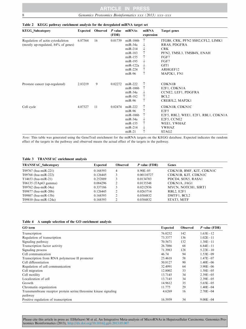
Table 2 KEGG pathway enrichment analysis for the deregulated miRNA target set
KEGG_Subcategory Expected Observed P value
(FDR)
miRNAs miRNA
expression
Target genes
Regulation of actin cytoskeleton
(mostly up-regulated, 84% of genes)
6.87364 16 0.01739 miR-106b › ITGB8, CRK, PFN2 SSH2,CFL2, LIMK1
miR-34a fl RRAS, PDGFRA
miR-214 fl CRK
miR-183 › PFN2, TMSL3, TMSB4X, ENAH
miR-155 › FGF7
miR-195 fl FGF7
miR-122a fl GIT1
miR-224 › ARHGEF12
miR-96 › MAP2K1, FN1
Prostate cancer (up-regulated) 2.83219 9 0.02272 miR-222 › CDKN1B
miR-106b › E2F1, CDKN1A
miR-34a fl CCNE2, LEF1, PDGFRA
miR-182 › BCL2
miR-96 › CREB3L2, MAP2K1
Cell cycle 4.07327 11 0.02474 miR-222 › CDKN1B, CDKN1C
miR-96 › E2F5
miR-106b › E2F5, RBL2, WEE1, E2F1, RBL1, CDKN1A
miR-34a fl E2F5, CCNE2
miR-155 › WEE1, YWHAZ
miR-214 fl YWHAZ
miR-21 › STAG2
Note: This table was generated using the GeneTrail enrichment for the miRNA targets on the KEGG database. Expected indicates the random
effect of the targets in the pathway and observed means the actual effect of the targets in the pathway.
Table 3 TRANSFAC enrichment analysis
TRANSFAC_Subcategory Expected Observed P value (FDR) Genes
T09767 (hsa-miR-221) 0.168593 4 8.90E–05 CDKN1B, BMF, KIT, CDKN1C
T09768 (hsa-miR-222) 0.126445 3 0.00110727 CDKN1B, KIT, CDKN1C
T14653 (hsa-miR-21) 0.252889 3 0.0134703 PDCD4, SOX5, RASA1
T06135 (TAp63 gamma) 0.084296 2 0.0135548 CDKN1A, JAG1
T09762 (hsa-miR-34a) 0.337186 3 0.0212936 MYCN, NOTCH1, SIRT1
T09877 (hsa-miR-20b) 0.126445 2 0.0263714 RBL2, E2F1
T09807 (hsa-miR-15b) 0.168593 2 0.0384832 DMTF1, BCL2
T09810 (hsa-miR-124a) 0.168593 2 0.0384832 STAT3, MITF
Table 4 A sample selection of the GO enrichment analysis
GO term Expected Observed P value (FDR)
Transcription 76.0252 142 1.63E�12Regulation of transcription 73.3377 136 1.02E�11Signaling pathway 70.5671 132 1.38E�11Transcription factor activity 26.7086 68 6.84E�11Signaling process 71.3983 128 5.22E�10Cell communication 46.74 94 1.53E�09Transcription from RNA polymerase II promoter 25.4618 58 1.47E�07Cell differentiation 50.8127 90 1.40E�06Regulation of cell communication 32.4991 64 3.90E�06Cell migration 12.8002 33 1.58E�05Cell motility 13.7145 34 2.39E�05Localization of cell 13.7145 34 2.39E�05Growth 14.9612 35 5.65E�05Chromatin organization 11.775 29 1.40E�04Transmembrane receptor protein serine/threonine kinase signaling
pathway
4.6269 16 2.70E�04
Positive regulation of transcription 16.5959 34 9.00E�04
8 Genomics Proteomics Bioinformatics xxx (2013) xxx–xxx
Please cite this article in press as: ElHefnawi M et al, An Integrative Meta-analysis of MicroRNAs in Hepatocellular Carcinoma. Genomics Pro-teomics Bioinformatics (2013), http://dx.doi.org/10.1016/j.gpb.2013.05.007

Figure 6 Important pathways in HCC
Shown is the mTOR/AKP/PIP3 pathway that contributes to transformation of nodules into metastatic counterparts [57]. The target genes
are indicated in green and key pathway phenotypes are shown at the bottom. Deactivation of the MAPK pathway through inhibition/
repression of the up-regulating miRNAs or activation of suppressing miRNAs might be useful as an alternative therapeutic intervention
strategy. Genes are indicated in pink ovals; FGF signaling pathway, actin cytoskeleton pathway and mTOR pathway are represented with
lines in black, red and green, respectively.
ElHefnawi M et al / Wholistic Analysis of MicroRNAs in Liver Cancer 9
cancer hallmarks (Figures 4–6). Whether these effects are posi-tive or negative depends on the specific miRNA(s) and theirspecific targets. This important impact of miRNAs in cancerhas opened new therapeutic modalities for liver cancer by sup-
pression or induction of these miRNAs [40–44].
Discussion
This study reports an improved protocol for prediction andanalysis of novel miRNA targets that have been shown to be
deregulated in liver cancer. It also sheds more light on theirfunctional roles in relative to oncogenesis. Our study identifiedseveral targets related to oncogenesis and metastasis. Some ofthe predicted miRNA targets in this study have already been
experimentally validated (according to databases of experi-mentally verified targets such as miRecord and miRTarBase).
The improved miRNA target prediction protocol
In this study, functional role of miRNA targets in liver cancerwas evaluated and a comprehensive analysis of deregulated
miRNAs in HCC was performed, yielding novel insight intocarcinogenesis and metastasis. Below we discuss some of theimportant novel targets, according to pathway analysis andGO functional category enrichment analysis. The presence of
tens of already experimentally-validated targets, after inspec-tion of the experimentally-validated target databases, was the
Please cite this article in press as: ElHefnawi M et al, An Integrative Meta-teomics Bioinformatics (2013), http://dx.doi.org/10.1016/j.gpb.2013.05.007
final step to confirm the reliability of this approach. The setof overlapping targets (core or common targets) would havea high specificity (low false positives), while the set of pooledtargets predicted with different programs would have a high
sensitivity, but high false positives. We used the core commontargets to analyze these deregulated miRNAs in order to get areliable analysis from the functional enrichment analysis. In
discussing some of the key targets in the pathways below; werefer to the validated ones from miRecords and miRTarBasewith [V] and predicted ones with [P]. A quantitative evaluation
and comparison of the performance of our integrated ap-proach is beyond the scope of this manuscript, and faces thechallenge of correlating heterogeneous microarray studies tofind a substantial inverse correlation between a miRNA and
its targets. On a qualitative note, our core set is much smallerthan that presented by a single tool (Figure 2, target predictionstep), and has a high statistical significance (P < 0.01). More
detailed comparisons of methods are aimed in the future. Wethen performed the pathway analysis and GO functional cate-gory enrichment analysis, trying to understand how deregula-
tion of the targets would affect important cellular andmolecular processes, induce/repress critical pathways, andhence contributing to carcinogenesis and metastasis.
Analysis of key target genes and pathways
The global analysis of hundreds of predicted targets in thisstudy highlights the new key targets that were not discussed be-
analysis of MicroRNAs in Hepatocellular Carcinoma. Genomics Pro-

10 Genomics Proteomics Bioinformatics xxx (2013) xxx–xxx
fore. Also, unlike previous reports which only present enrich-ment analysis [35,50], an in-depth analysis is presented and dis-cussed concerning the roles of the miRNAs and the
contribution of their deregulation to cancer progression alongwith their roles in cancer hallmarks. Also, the mode of regula-tion of the different miRNAs whether to suppress or induce
the pathway or the hallmark is inferred. These unravel signif-icant new and valuable information on the different pathwaysaffected by the set of highly differentially regulated miRNAs in
liver cancer, and key genes that are affected in each of the en-riched pathways. Some previous reports on the roles of miR-NAs in liver cancer rely only on experimentally validatedtargets, thus limiting their scope of coverage [31,34,35,55]. In
addition, some studies based their views on the role of a certainmiRNA on only one validated target, which gives a very nar-row and inaccurate perspective. Some exceptions in the litera-
ture include the recent work on global miRNA analysis inbreast cancer [56]. This study found previously mentionedpathways linked to miRNA deregulation and liver cancer, such
as the MAPK, TGFb and cell cycle pathways, discussed instudies and reviews such as [10,34,35,50]. Meanwhile, theGeneTrail analysis showed up regulation of the Wnt signaling
and the oocyte meiosis pathways (Table 2), in addition to axonguidance and actin cytoskeleton which are involved in develop-mental processes and in stem cell differentiation and prolifera-tion, highlighting a possible link between miRNAs and errors
in stem cell differentiation and their transformation into can-cer stem cells [43,46,57]. The role of miRNAs in inducing can-cer stem cells was previously noted for some well known tumor
suppressor miRNAs like miR-34a and others in different tu-mors [16,44,46]. The link between miRNAs and stem cellswas in agreement with previous reports highlighting miRNA
roles in stem cell differentiation and liver cancer [44,57]. Alsoworth noting is emergence of the enriched prostate cancer path-way as an enriched pathway, signifying the important impact of
miRNAs in cancer and offering possible general unified roles forthese miRNAs in closely-related tumors like solid tumors.
The MAPK and mTOR signaling and cancer hallmarks
Interestingly, the MAPK signaling pathway was one of the en-riched pathways with the highest statistical significance (withpart of it and the candidate targets highlighted red in Figure S1
and some others shown in Table 2 and Figure 6 linked to cros-stalks to other important pathways in cancer that are affectedby the miRNA targets). Dual mode of regulation of target
genes by miRNAs was noticed. For example, gene encodingfibroblast growth factor 7 (FGF7) is targeted by two miRNAs:miR-155 [V], which is validated in miRTarBase and miRWalkwith up-regulated expression in HCC, and miR-195 [P] with
down-regulated expression in HCC. FGF7 induces the MAPKpathway by binding to extracellular receptors [58]. FGF7 is in-ferred from its GO annotation to be involved in self-sufficiency
in growth signals and limitless replicative potential, in additionto evading apoptosis and tissue invasion and metastasis [40](Figures 1 and 6). Besides, FGF7 is also a key regulator of
the actin cytoskeleton pathway (shown in pathway targets inTable 2). Furthermore, up regulation of FGF7 expression con-tributes to the FGF receptor signaling pathway [59] and posi-
tively regulates cell division, proliferation and keratinocytemigration pathways, which were also significantly enriched in
Please cite this article in press as: ElHefnawi M et al, An Integrative Meta-teomics Bioinformatics (2013), http://dx.doi.org/10.1016/j.gpb.2013.05.007
the GO analysis (Figures 4 and 6 for part of the FGF path-way). Sufficiency in growth signals is revealed by significantlyenriched GO terms such as growth and positive regulation of
transcription (Figures 3 and 4). Another important key gene,mitogen-activated protein kinase kinase 1 (MAP2K1) (Fig-ure S1), is a target for miR-96 [P], which is up regulated in
HCC. MAP2K1 is the upstream activator of MAPK, thusoccupying a central role in the MAPK pathway. Given its highselectivity to MAPK and elevated levels of constitutively acti-
vated MAP2K1 is frequently-observed in carcinoma cell lines[60], MAP2K1 represents an excellent target for pharmacolog-ical intervention.
Similarly, suppression of the MAPK signaling pathway
could be inferred by miR-34 [V]. miR-34 suppresses platelet-derived growth factor A (PDGFRA) [V] (validated in miRTar-Base) and other oncogenes that have been validated previously
such as c-Met, NOTCH and Wnt1 [41]. Although of its pre-dominant down expression in many cancer cell lines, the mixedlevels of miR-34a in different liver cancer cell lines reported by
different investigators may reflect the opposing mechanismsthat affect its expression (activated by P53 and inactivatedby methylation and many other factors) and contribute to
enhancement or decrease of its tumor suppressor role infighting cancer. Expression of miR-34a is activated by p53 pro-tein, thus resulting in a high tumor-suppressing mode [61].Also, miR-34 contributes to decreasing proliferation by target-
ing CCNE2 Cyclin E2 [V] (validated in miRTarBase andmiRWalk), which may allow proliferation when miR-34 isdown regulated. From our analysis and previous reports,
hsa-miR-34 also targets PDGFRA and E2F5, all of which in-duce proliferation and replication [41]. Interestingly, miR-34,which is currently introduced as a replacement therapy for dif-
ferent cancers in the clinic, plays a clearly-defined role in stemcell regulation through regulating the Wnt signaling pathway[41].
In agreement with this, growth hormone receptor (GHR) isanother predicted target for down-regulated miR-195 [P] incancer tissue (Figures 6 and S1). miR-195 contributes to evad-ing apoptosis, sustained angiogenesis, cell growth, tissue inva-
sion and metastasis. This miRNA is an important tumor-suppressor [45]. Insensitivity to anti-growth signals, anotherimportant cancer hallmark, is illustrated by suppression of a
tumor suppressor of MAPK signaling, the SPRY4 gene [62].SPRY4 is predicted to be a target for miR-182 [P], which ishighly expressed in HCC. As a result, SPRY4 expression is
down-regulated in HCC. Additionally, RAS p21 protein acti-vator 1 (RASA1), a target of up-regulated miR-182 [V] [63],stimulates the GTPase activity of normal RAS p21 but notits oncogenic counterpart, hence, acting as a suppressor of
RAS and thereby allowing control of cellular proliferationand differentiation. Due to its down regulation, the activeform of RAS is stimulated, which functions as an oncogene.
These data support the inference of an oncomiR role formiR-182. Also, expression of receptor-type tyrosine-proteinphosphatase F (PTPRF) that is involved in the negative regu-
lation of epidermal growth factor receptor (EGFR) signalingpathway [64] would be up regulated, since it is a target fordown-regulated miR-199a-3p [V] [65], leading to insensitivity
to anti-growth signals. CACNB4 is one of the important genesin the MAPK pathway (controlled by miR-96 [V], which ishighly expressed in HCC), hence, helping in controlling theMAPK signaling pathway.
analysis of MicroRNAs in Hepatocellular Carcinoma. Genomics Pro-

ElHefnawi M et al / Wholistic Analysis of MicroRNAs in Liver Cancer 11
The TGFb pathway
The TGFb was the second most highly enriched pathway (Fig-ure S3). Bone morphogenetic protein (BMP) receptor II(BMPRII), through which the BMP ligand transduces its sig-
nals in the TGFb pathway, was predicted as a target formiR-106b over-expressed in HCC. Upon ligand binding,BMPRII phosphorylates and activates BMPRI, which theninitiates downstream signaling by phosphorylating the recep-
tor-regulated Smads (R-Smads) [57]. Smad7, a negative regula-tor of TGFb signaling, is targeted by miR-106b [V] [36] andmiR-21 [V] [66] highly-expressed in HCC, resulting in down
regulation of Smad7.
Cell cycle pathway
The E2F family plays a crucial role in the control of cell cycleand regulation of tumor suppressor proteins. E2F5, a regula-tor of the TGFb and cell cycle pathways [67], was targeted
by miR-96 [P], miR- 34a [V] (validated in miRTarBase) andmiR-106 [P]. Down regulation of E2F5 through this combina-torial mechanism by miR-96 and miR-106 inhibits the synthe-sis of DNA, thus repressing the S phase. On the other hand,
E2F5 is involved in cell cycle regulation by inhibiting c-Myc,an oncogene and a transcription factor that is believed to reg-ulate expression of 15% of all genes [68]. Thus a reduced level
of E2F5 would lead to compromised control over c-myc. Thesedata suggest that involvement of different miRNAs withopposing roles for E2F5 which itself has dual roles could result
in different outcomes.
Other new pathways linked to stem cells and their regulation
that are significantly enriched in our analysis
Two additional pathways that were enriched for the miRNAtarget set genes include regulation of actin cytoskeleton andaxon guidance. Cancer cells are marked by their ability to mi-
grate and invade the adjacent tissues. Interestingly, severalstudies have shown that proteins linking migratory signals toactin cytoskeleton are up regulated in cancer tissues [69,70].
For example, fibronectin 1 (FN1) plays an important role inthe actin cytoskeleton pathway because it activates the integrin(ITG) gene. ITG in turn activates a series of other genes, lead-
ing to the formation of actin fibers and polymerization [71,72],which connects the cells to each other. Therefore, the axonguidance pathway is involved in regulation of the actin cyto-skeleton and represents a key factor in the formation of the
neuronal network (Figures 6 and S2). Both pathways are in-ferred to be linked with tissue invasion and metastasis cancerhallmark.
The oocyte meiosis pathway is another enriched pathwayfor our gene set (part of which is shown in Figure S3). Thispathway is highly important during the differentiation of stem
cells [73]. MAP2K1 that was described in the aforementionedMAPK pathway is one of the most important genes in thispathway and contributes to control of stem cells and their
oncogenic transformation.Some other interesting targets in this pathway include aden-
ylate cyclase 2 (ADCY2) and ADCY6, which are targets formiR-182. ADCY6 has been experimentally verified previously
while ADCY2 is reported in this work for the first time.
Please cite this article in press as: ElHefnawi M et al, An Integrative Meta-teomics Bioinformatics (2013), http://dx.doi.org/10.1016/j.gpb.2013.05.007
ADCY2 is involved in the chemokine signaling pathway andis one of the key genes in the oocyte meiosis pathway. Once itis activated through progesterone induction, it activates a series
of gene cascades leading to completion of the meiosis process.
Conclusion
Some important conclusions that could be drawn from thisstudy on some of the general features of miRNAs roles in liver
cancer are as follows. (1) The miRNA targets, as revealed fromtheir enriched GO annotations and pathways, cover many ofthe known hallmarks of cancer [51,52,54]. Expression of manygrowth and transcription factors is up regulated due to the
down regulation of their regulating miRNAs. (2) Positive con-tribution to cancer development is implicated for many down-regulated miRNAs that suppress important oncogenes, such as
miR-122, miR-214, miR-199a-3p/5p and miR-34a, and for sev-eral up-regulated miRNAs that suppress tumor-suppressors,such as miR-182 and miR-186 oncomiRs. Some miRNAs
may play dual roles by targeting both tumor-suppressors andoncogenes. Therefore, without complete analysis of their tar-gets and pathways (interactome), caution should be taken indefining the role of deregulated miRNAs in liver cancer, as
well as in using miRNAs in cancer therapeutics. (3) Some miR-NAs down regulate cancer hallmarks by downregulating someoncogenic pathways, which was also inferred by other studies
[74]. Although there is an ongoing debate on what are cancerhall marks and the complex interplay between causes, onco-genic events, signal transduction programs and hallmarks
[54,75], it is clear that miRNA deregulation can have dualroles. (4) miR-96 was inferred in this study to play a tumorsuppressor role by targeting MAP2K1, E2F5 and CACNB4.
miR-96 was reported to suppress oncogenes such as glypi-can-3 (GPC3) [76], although an oncomiR role was inferredas well. Silencing of miR96 was associated with decreasedexpression of osteopontin and forkhead box O1 (FOXO1)
and FOXO3a and hence decreased invasion and metastasisand proliferation, respectively [77,78]. Some targets werefound to be controlled by more than one miRNA. Also, some
TFs like TP63 gamma coordinate with miRNAs for regulationof some targets, supporting the miRNA-TF regulatory net-work analysis [79]. (5) Targets of miR-96, miR-106b, miR-
34a and miR-155 were most often highly represented in themost enriched oncogenic pathways, suggesting their involve-ment in liver cancer.
Materials and methods
The miRNA data were downloaded from miRBase release 19
(http://www.mirbase.org/). The steps of this comprehensiveglobal analysis of differentially-regulated miRNAs in liver can-cer are detailed below and also illustrated in Figure 2. This
workflow is similar to the integrative automated siRNA designand selection protocol and tool that we previously devised [80].
Protocol for miRNA target prediction
miRNA prediction was done using five different programs inthe present study to ensure high specificity in target prediction.The programs that were used include TargetScan 5.1 [81],
analysis of MicroRNAs in Hepatocellular Carcinoma. Genomics Pro-

12 Genomics Proteomics Bioinformatics xxx (2013) xxx–xxx
PicTar [82], DIANA-microT v3.0 [83], miRDB [84] and miR-anda [23]. Targets that were commonly predicted by 4 or 5out of 5 programs were retained. The programs were chosen
to represent different approaches for miRNA target predic-tion. The miRanda prediction software identified potentialbinding sites by looking for high-complementarity regions on
the 30UTRs, which is called seed region [23]. Then the resultingbinding sites are evaluated thermodynamically using the Vien-na RNA folding package. The second program TargetScan 5.1
[81] combines thermodynamics-based modeling of RNA–RNAduplex interactions with comparative sequence analysis to pre-dict miRNA targets conserved across multiple mammaliangenomes. TargetScan mainly depends on perfect complemen-
tarity to the seed region of miRNA and then extends to com-plementarity outside the region. PicTar also depends on theseed region complementarity and its most distinguishing fea-
ture is its probabilistic identification of combinations of targetsites [82]. DIANA-microT 3.0 [83] searches in the UTRs forstringent seed paring (at least 7 consecutive Watson–Crick
pairs) to the miRNA. The last algorithm miRDB uses a newlydeveloped SVM classifier [84].
Identification of experimentally-validated targets and
differentially-expressed predicted targets in HCC
Next, the targets were compared against already predicted andvalidated targets known in databases such as the database of
experimentally-validated targets in miRTarBase (http://mirtar-base.mbc.nctu.edu.tw/) and miRecord (http://mirecords.bio-lead.org/) [79].
Combining microarray data
The microarray dataset showing differentially-expressed genes
in hepatocellular carcinoma patients [48] was used to correlatethe predicted targets and those differentially-expressed in HCCpatients. The two-sample t-test was used for correlation anal-
ysis. This was yet another step to improve the reliability ofthe predictions of the miRNA targets.
Ranking according to MFE
Ranking of miRNA targets was done using the RNAhybridprogram according to MFE, (DG) [36,81]. Some importantexamples are illustrated in Figure 3.
Shuffling and statistical analysis of the targets
Statistical analysis of the targets was performed to support the
validity of the prediction. We used Jemboss (graphical userinterface for European Molecular Biology Open SoftwareSuite) Shuffle Seq program [85], in which the target’s sequences
were shuffled 500 times as previously described [17]. The 100sets for each of the target genes were independently searchedagainst its reported predicted miRNA using miRanda soft-ware. Z scores were calculated for each target using the equa-
tion Z ¼ X�lr (where X is the miRanda score for real gene, l
and d are the mean and standard deviation for miRanda scoreof each miRNA with shuffle gene). Then, P values were calcu-
lated to estimate the significance of the predicted targets.
Please cite this article in press as: ElHefnawi M et al, An Integrative Meta-teomics Bioinformatics (2013), http://dx.doi.org/10.1016/j.gpb.2013.05.007
Statistical enrichment analysis of the predicted target set
Statistically overrepresented functions were revealed in a sam-ple of seven miRNA targets as a first indication of essential en-riched terms by using the database for annotation,
visualization and integrated discovery (DAVID) (http://davi-d.abcc.ncifcrf.gov/summary.jsp). This was combined with amore comprehensive analysis, enrichment analysis of the pre-dicted target set, using the GeneTrail tool [86] (http://gene-
trail.bioinf.uni-sb.de/) with FDR and Bonferroni correctionsfor multiple items in the set. The list of predicted genes wascompared against a random list of genes to predict which func-
tions and pathways are enriched in the target genes and signif-icance level [50]. GeneTrail was used again to obtain thefunctions of different miRNA target sets and their combina-
tions for identifying their possible roles in cancer.
Authors’ contributions
ME conceived the idea and designed the study. ME, MA andBS performed the study, analyzed the data and drafted themanuscript. MA, NA and ME revised the manuscript. All
authors read and approved the final manuscript.
Competing interests
The authors report no competing interests.
Acknowledgements
We acknowledge partial support through Science and Technol-ogy Development Fund (STDF) by Egyptian Ministry of Sci-entific Research (Grant No. 1169 and 1679). We thank the help
and assistance of Dr. Suher Zada and of many research assis-tants in the group for their long-standing efforts. We are verygrateful to Gehad Abdel Rahman, Mohamed Mysara, Soha
Gamal ElDin, Asmaa Ezzat, Lesley Twaitol, Dr. FareedAboul-Ela, Dr. Manal ElHemshary especially among othersfor their roles in editing/revising and data analysis during the
course of this publication. We also thank Dr. Marc Windischfor editing and revising the manuscript.
Appendix A. Supplementary material
Supplementary data associated with this article can be found,
in the online version, at http://dx.doi.org/10.1016/j.gpb.2013.05.007.
References
[1] Bartel DP. MicroRNAs: genomics, biogenesis, mechanism, and
function. Cell 2004;116:281–97.
[2] Sassen S, Miska EA, Caldas C. MicroRNA––implications for
cancer. Virchows Arch 2008;452:1–10.
analysis of MicroRNAs in Hepatocellular Carcinoma. Genomics Pro-

ElHefnawi M et al / Wholistic Analysis of MicroRNAs in Liver Cancer 13
[3] Xiao ZD, Diao LT, Yang JH, Xu H, Huang MB, Deng YJ, et al.
Deciphering the transcriptional regulation of microRNA genes in
humans with ACTLocater. Nucleic Acids Res 2013;41:e5.
[4] Holland B, Wong J, Li M, Rasheed S. Identification of human
microRNA-like sequences embedded within the protein-encoding
genes of the human immunodeficiency virus. PLoS One 2013;8:
e58586.
[5] Calin GA, Sevignani C, Dumitru CD, Hyslop T, Noch E,
Yendamuri S, et al. Human microRNA genes are frequently
located at fragile sites and genomic regions involved in cancers.
Proc Natl Acad Sci U S A 2004;101:2999–3004.
[6] Xu S, Witmer PD, Lumayag S, Kovacs B, Valle D. MicroRNA
(miRNA) transcriptome of mouse retina and identification of a
sensory organ-specific miRNA cluster. J Biol Chem 2007;282:
25053–66.
[7] Chen YJ, Min J, Shang CZ, Ren M, Peng XX, Cao J, et al.
MicroRNA differential expression profile during differentiation of
embryonic stem cells towards hepatocytes induced by sodium
butyrate. Zhongguo Yi Xue Ke Xue Yuan Xue Bao 2008;30:
469–73.
[8] Cho WC. MicroRNAs: potential biomarkers for cancer diagnosis,
prognosis and targets for therapy. Int J Biochem Cell Biol
2010;42:1273–81.
[9] Bartels CL, Tsongalis GJ. MicroRNAs: novel biomarkers for
human cancer. Clin Chem 2009;55:623–31.
[10] Chen F, Hu SJ. Effect of microRNA-34a in cell cycle, differen-
tiation, and apoptosis: a review. J Biochem Mol Toxicol 2012;26:
79–86.
[11] Brennecke J, Stark A, Russell RB, Cohen SM. Principles of
microRNA-target recognition. PLoS Biol 2005;3:e85.
[12] Brodersen P, Voinnet O. Revisiting the principles of microRNA
target recognition and mode of action. Nat Rev Mol Cell Biol
2009;10:141–8.
[13] Graves P, Zeng Y. Biogenesis of mammalian microRNAs: a global
view. Genomics Proteomics Bioinformatics 2012;10:239–45.
[14] Esquela-Kerscher A, Slack FJ. Oncomirs – microRNAs with a
role in cancer. Nat Rev Cancer 2006;6:259–69.
[15] Yan K, Gao J, Yang T, Ma Q, Qiu X, Fan Q, et al. MicroRNA-
34a inhibits the proliferation and metastasis of osteosarcoma cells
both in vitro and in vivo. PLoS One 2012;7:e33778.
[16] Lodygin D, Tarasov V, Epanchintsev A, Berking C, Knyazeva T,
Korner H, et al. Inactivation of mir-34a by aberrant cpg
methylation in multiple types of cancer. Cell Cycle 2008;7:
2591–600.
[17] Varnholt H. The role of microRNAs in primary liver cancer. Ann
Hepatol 2008;7:104–13.
[18] Worley LA, Long MD, Onken MD, Harbour JW. Micro-RNAs
associated with metastasis in uveal melanoma identified by
multiplexed microarray profiling. Melanoma Res 2008;18:184–90.
[19] Lee RC, Feinbaum RL, Ambros V. The C. elegans heterochronic
gene lin-4 encodes small RNAs with antisense complementarity to
lin-14. Cell 1993;75:843–54.
[20] Mendes ND, Freitas AT, Sagot MF. Current tools for the
identification of miRNA genes and their targets. Nucleic Acids
Res 2009;37:2419–33.
[21] Watanabe Y, Tomita M, Kanai A. Computational methods for
microRNA target prediction. Methods Enzymol 2007;427:65–86.
[22] Min H, Yoon S. Got target? Computational methods for
microRNA target prediction and their extension. Exp Mol Med
2010;42:233–44.
[23] John B, Enright AJ, Aravin A, Tuschl T, Sander C, Marks DS.
Human microRNA targets. PLoS Biol 2004;2:e363.
[24] Chandra V, Girijadevi R, Nair AS, Pillai SS, Pillai RM. MTar: a
computational microRNA target prediction architecture for
human transcriptome. BMC Bioinformatics 2010;11:S2.
[25] Liu H, Yue D, Zhang L, Chen Y, Gao SJ, Huang Y. A Bayesian
approach for identifying miRNA targets by combining sequence
Please cite this article in press as: ElHefnawi M et al, An Integrative Meta-teomics Bioinformatics (2013), http://dx.doi.org/10.1016/j.gpb.2013.05.007
prediction and gene expression profiling. BMC Genomics
2010;11:S12.
[26] Lekprasert P, Mayhew M, Ohler U. Assessing the utility of
thermodynamic features for microRNA target prediction under
relaxed seed and no conservation requirements. PLoS One
2011;6:e20622.
[27] Barbato C, Arisi I, Frizzo ME, Brandi R, Da Sacco L, Masotti A.
Computational challenges in miRNA target predictions: to be or
not to be a true target? J Biomed Biotechnol 2009;2009:803069.
[28] Dweep H, Sticht C, Pandey P, Gretz N. MiRWalk – database:
prediction of possible miRNA binding sites by ‘‘walking’’ the
genes of three genomes. J Biomed Inform 2011;44:839–47.
[29] Friedman Y, Naamati G, Linial M. MiRror: a combinatorial
analysis web tool for ensembles of microRNAs and their targets.
Bioinformatics 2010;26:1920–1.
[30] Temirak A, Abdulla M, Elhefnawi M. Rational drug design for
identifying novel multi-target inhibitors for hepatocellular carci-
noma. Anticancer Agents Med Chem 2012;12:1088–97.
[31] Ura S, Honda M, Yamashita T, Ueda T, Takatori H, Nishino R,
et al. Differential microRNA expression between hepatitis B and
hepatitis C leading disease progression to hepatocellular carci-
noma. Hepatology 2009;49:1098–112.
[32] Pei Y, Zhang T, Renault V, Zhang X. An overview of hepato-
cellular carcinoma study by omics-based methods. Acta Biochim
Biophys Sin (Shanghai) 2009;41:1–15.
[33] Hou J, Lin L, Zhou W, Wang Z, Ding G, Dong Q, et al.
Identification of miRNomes in human liver and hepatocellular
carcinoma reveals mir-199a/b-3p as therapeutic target for hepa-
tocellular carcinoma. Cancer Cell 2011;19:232–43.
[34] Huang S, He X. The role of microRNAs in liver cancer
progression. Br J Cancer 2011;104:235–40.
[35] Murakami Y, Yasuda T, Saigo K, Urashima T, Toyoda H,
Okanoue T, et al. Comprehensive analysis of microRNA expres-
sion patterns in hepatocellular carcinoma and non-tumorous
tissues. Oncogene 2006;25:2537–45.
[36] Wang Y, Lee AT, Ma JZI, Wang J, Ren J, Yang Y, et al. Profiling
microRNA expression in hepatocellular carcinoma reveals micr-
oRNA-224 up-regulation and apoptosis inhibitor-5 as a microR-
NA-224-specific target. J Biol Chem 2008;283:13205–15.
[37] Barshack I, Meiri E, Rosenwald S, Lebanony D, Bronfeld M,
Aviel-Ronen S, et al. Differential diagnosis of hepatocellular
carcinoma from metastatic tumors in the liver using microRNA
expression. Int J Biochem Cell Biol 2010;42:1355–62.
[38] Pineau P, Volinia S, McJunkin K, Marchio A, Battiston C, Terris
B, et al. MiR-221 overexpression contributes to liver tumorigen-
esis. Proc Natl Acad Sci U S A 2010;107:264–9.
[39] Hirata H, Ueno K, Shahryari V, Tanaka Y, Tabatabai ZL,
Hinoda Y, et al. Oncogenic miRNA-182-5p targets Smad4 and
RECK in human bladder cancer. PLoS One 2012;7:e51056.
[40] Coulouarn C, Factor VM, Andersen JB, Durkin ME, Thorgeirs-
son SS. Loss of miR-122 expression in liver cancer correlates with
suppression of the hepatic phenotype and gain of metastatic
properties. Oncogene 2009;28:3526–36.
[41] Gupta OP, Permar V, Koundal V, Singh UD, Praveen S.
MicroRNA regulated defense responses in Triticum aestivum L.
during Puccinia graminis f.sp. tritici infection. Mol Biol Rep
2012;39:817–24.
[42] Watanabe Y, Kishi A, Yachie N, Kanai A, Tomita M. Compu-
tational analysis of microRNA-mediated antiviral defense in
humans. FEBS Lett 2007;581:4603–10.
[43] Shimono Y, Zabala M, Cho RW, Lobo N, Dalerba P, Qian D,
et al. Downregulation of miRNA-200c links breast cancer stem
cells with normal stem cells. Cell 2009;138:592–603.
[44] Bader AG. MiR-34 – a microRNA replacement therapy is headed
to the clinic. Front Genet 2012;3:120.
[45] Xu T, Zhu Y, Xiong Y, Ge YY, Yun JP, Zhuang SM.
MicroRNA-195 suppresses tumorigenicity and regulates G1/S
analysis of MicroRNAs in Hepatocellular Carcinoma. Genomics Pro-

14 Genomics Proteomics Bioinformatics xxx (2013) xxx–xxx
transition of human hepatocellular carcinoma cells. Hepatology
2009;50:113–21.
[46] Ji Q, Hao X, Zhang M, Tang W, Yang M, Li L, et al. MicroRNA
miR-34 inhibits human pancreatic cancer tumor-initiating cells.
PLoS One 2009;4:e6816.
[47] Huang YS, Dai Y, Yu XF, Bao SY, Yin YB, Tang M, et al.
Microarray analysis of microRNA expression in hepatocellular
carcinoma and non-tumorous tissues without viral hepatitis. J
Gastroenterol Hepatol 2008;23:87–94.
[48] Mas VR, Maluf DG, Archer KJ, Yanek K, Williams B, Fisher
RA. Differentially expressed genes between early and advanced
hepatocellular carcinoma (HCC) as a potential tool for selecting
liver transplant recipients. Mol Med 2006;12:97–104.
[49] Rehmsmeier M, Steffen P, Hochsmann M, Giegerich R. Fast and
effective prediction of microRNA/target duplexes. RNA 2004;10:
1507–17.
[50] Backes C, Meese E, Lenhof HP, Keller A. A dictionary on
microRNAs and their putative target pathways. Nucleic Acids
Res 2010;38:4476–86.
[51] Hanahan D, Weinberg RA. The hallmarks of cancer. Cell 2000;
100:57–70.
[52] Hanahan D, Weinberg RA. Hallmarks of cancer: the next
generation. Cell 2011;144:646–74.
[53] Jiang J, Gusev Y, Aderca I, Mettler TA, Nagorney DM, Brackett
DJ, et al. Association of microRNA expression in hepatocellular
carcinomas with hepatitis infection, cirrhosis, and patient survival.
Clin Cancer Res 2008;14:419–27.
[54] Lazebnik Y. What are the hallmarks of cancer? Nat Rev Cancer
2010;10:232–3.
[55] Bala S, Marcos M, Szabo G. Emerging role of microRNAs in liver
diseases. World J Gastroenterol 2009;15:5633–40.
[56] Uhlmann S, Mannsperger H, Zhang JD, Horvat EA, Schmidt C,
Kublbeck M, et al. Global microRNA level regulation of EGFR-
driven cell-cycle protein network in breast cancer. Mol Syst Biol
2012;8:570.
[57] Xia H, Ooi LL, Hui KM. MicroRNA-216a/217-induced epithe-
lial-mesenchymal transition targets PTEN and SMAD7 to pro-
mote drug resistance and recurrence of liver cancer. Hepatology
2013;58:629–41.
[58] Fata JE, Mori H, Ewald AJ, Zhang H, Yao E, Werb Z, et al. The
MAPK(ERK-1,2) pathway integrates distinct and antagonistic
signals from TGFalpha and FGF7 in morphogenesis of mouse
mammary epithelium. Dev Biol 2007;306:193–207.
[59] Turner N, Grose R. Fibroblast growth factor signalling: from
development to cancer. Nat Rev Cancer 2010;10:116–29.
[60] Choi YL, Soda M, Ueno T, Hamada T, Haruta H, Yamato A,
et al. Oncogenic MAP2K1 mutations in human epithelial tumors.
Carcinogenesis 2012;33:956–61.
[61] Dalgard CL, Gonzalez M, deNiro JE, O’Brien JM. Differential
microRNA-34a expression and tumor suppressor function in
retinoblastoma cells. Invest Ophthalmol Vis Sci 2009;50:4542–51.
[62] Yang X, Gong Y, Tang Y, Li H, He Q, Gower L, et al. Spry1 and
Spry4 differentially regulate human aortic smooth muscle cell
phenotype via Akt/FoxO/myocardin signaling. PLoS One 2013;8:
e58746.
[63] Xu J, Wong C. A computational screen for mouse signaling
pathways targeted by microRNA clusters. RNA 2008;14:1276–83.
[64] Du WW, Fang L, Li M, Yang X, Liang Y, Peng C, et al.
MicroRNA miR-24 enhances tumor invasion and metastasis by
targeting PTPN9 and PTPRF to promote EGF signaling. J Cell
Sci 2013;126:1440–53.
[65] Amer M, El-Ahwany E, Elhefnawi M, Awad A, Abdel Gawad N,
Zada S, et al. Prediction of miRNA target genes involved in liver
cancer pathways and its validation. J Hepatol 2013;58:S120.
[66] Smith AL, Iwanaga R, Drasin DJ, Micalizzi DS, Vartuli RL, Tan
AC, et al. The miR-106b-25 cluster targets Smad7, activates
TGF-b signaling, and induces EMT and tumor initiating cell
Please cite this article in press as: ElHefnawi M et al, An Integrative Meta-teomics Bioinformatics (2013), http://dx.doi.org/10.1016/j.gpb.2013.05.007
characteristics downstream of Six1 in human breast cancer.
Oncogene 2012;31:5162–71.
[67] Jiang Y, Yim SH, Xu HD, Jung SH, Yang SY, Hu HJ, et al. A
potential oncogenic role of the commonly observed E2F5 over-
expression in hepatocellular carcinoma. World J Gastroenterol
2011;17:470–7.
[68] Gearhart J, Pashos EE, Prasad MK. Pluripotency redux ––
advances in stem-cell research. N Engl J Med 2007;357:1469–72.
[69] Yamaguchi H, Condeelis J. Regulation of the actin cytoskeleton
in cancer cell migration and invasion. Biochim Biophys Acta
2007;1773:642–52.
[70] Bloom L, Ingham KC, Hynes RO. Fibronectin regulates assembly
of actin filaments and focal contacts in cultured cells via the
heparin-binding site in repeat III13. Mol Biol Cell 1999;10:
1521–36.
[71] Galbraith CG, Yamada KM, Galbraith JA. Polymerizing actin
fibers position integrins primed to probe for adhesion sites.
Science 2007;315:992–5.
[72] Vicente-Manzanares M, Choi CK, Horwitz AR. Integrins in
cell migration – the actin connection. J Cell Sci 2009;122:
199–206.
[73] Takebe N, Warren R, Ivy SP. Breast cancer growth and
metastasis: interplay between cancer stem cells, embryonic
signaling pathways and epithelial-to-mesenchymal transition.
Breast Cancer Res 2011;13:211.
[74] Gusev Y. Computational methods for analysis of cellular func-
tions and pathways collectively targeted by differentially
expressed microRNA. Methods 2008;44:61–72.
[75] Floor SL, Dumont JE, Maenhaut C, Raspe E. Hallmarks of
cancer: of all cancer cells, all the time? Trends Mol Med
2012;18:509–15.
[76] Maurel M, Jalvy S, Ladeiro Y, Combe C, Vachet L, Sagliocco F,
et al. A functional screening identifies five microRNAs control-
ling glypican-3: role of miR-1271 down-regulation in hepatocel-
lular carcinoma. Hepatology 2013;57:195–204.
[77] Chen RX, Xia YH, Xue TC, Ye SL. Suppression of microRNA-
96 expression inhibits the invasion of hepatocellular carcinoma
cells. Mol Med Rep 2012;5:800–4.
[78] Xu D, He X, Chang Y, Xu C, Jiang X, Sun S, et al. Inhibition of
miR-96 expression reduces cell proliferation and clonogenicity of
HepG2 hepatoma cells. Oncol Rep 2013;29:653–61.
[79] Wang J, Haubrock M, Cao KM, Hua X, Zhang CY, Wingender
E, et al. Regulatory coordination of clustered microRNAs based
on microRNA-transcription factor regulatory network. BMC Syst
Biol 2011;5:199.
[80] Mysara M, Garibaldi JM, Elhefnawi M. MysiRNA-designer: a
workflow for efficient siRNA design. PLoS One 2011;6:e25642.
[81] Lewis BP, Burge CB, Bartel DP. Conserved seed pairing, often
flanked by adenosines, indicates that thousands of human genes
are microRNA targets. Cell 2005;120:15–20.
[82] Krek A, Grun D, Poy MN, Wolf R, Rosenberg L, Epstein EJ,
et al. Combinatorial microRNA target predictions. Nat Genet
2005;37:495–500.
[83] Kiriakidou M, Nelson PT, Kouranov A, Fitziev P, Bouyioukos C,
Mourelatos Z, et al. A combined computational–experimental
approach predicts human microRNA targets. Genes Dev 2004;18:
1165–78.
[84] Wang X. MiRDB: a microRNA target prediction and func-
tional annotation database with a wiki interface. RNA 2008;14:
1012–7.
[85] Rice P, Longden I, Bleasby A. EMBOSS: the European Molecular
Biology Open Software Suite. Trends Genet 2000;16:276–7.
[86] Keller A, Backes C, Al-Awadhi M, Gerasch A, Kuntzer J,
Kohlbacher O, et al. GeneTrailExpress: a web-based pipeline for
the statistical evaluation of microarray experiments. BMC Bioin-
formatics 2008;9:552.
analysis of MicroRNAs in Hepatocellular Carcinoma. Genomics Pro-
Top Related
Copyright © 2022 FDOKUMEN

