


Bahasa
Halaman
Hukum


Accepted Manuscript
A CRPS-IgG-Transfer-Trauma Model Reproducing Inflammatory and PositiveSensory Signs associated with Complex Regional Pain Syndrome
Valéria Tékus, Zsófia Hajna, Éva Borbély, Adrienn Markovics, Teréz Bagoly,János Szolcsányi, Victoria Thompson, Ágnes Kemény, Zsuzsanna Helyes,Andreas Goebel
PII: S0304-3959(13)00555-1DOI: http://dx.doi.org/10.1016/j.pain.2013.10.011Reference: PAIN 9017
To appear in: PAIN
Received Date: 15 August 2013Revised Date: 11 October 2013Accepted Date: 14 October 2013
Please cite this article as: V. Tékus, Z. Hajna, É. Borbély, A. Markovics, T. Bagoly, J. Szolcsányi, V. Thompson,Á. Kemény, Z. Helyes, A. Goebel, A CRPS-IgG-Transfer-Trauma Model Reproducing Inflammatory and PositiveSensory Signs associated with Complex Regional Pain Syndrome, PAIN (2013), doi: http://dx.doi.org/10.1016/j.pain.2013.10.011
This is a PDF file of an unedited manuscript that has been accepted for publication. As a service to our customerswe are providing this early version of the manuscript. The manuscript will undergo copyediting, typesetting, andreview of the resulting proof before it is published in its final form. Please note that during the production processerrors may be discovered which could affect the content, and all legal disclaimers that apply to the journal pertain.
CRPS passive transfer
1
A CRPS-IgG-Transfer-Trauma Model Reproducing
Inflammatory and Positive Sensory Signs
associated with Complex Regional Pain Syndrome
Valéria Tékusa,b,*, Zsófia Hajnaa,b,*, Éva Borbélya,b, Adrienn Markovicsa,b, Teréz Bagolya,b,
János Szolcsányia,b,c, Victoria Thompsond, Ágnes Keménya,b, Zsuzsanna Helyesa,b,c,*, Andreas
Goebeld,e,*
aDepartment of Pharmacology and Pharmacotherapy, Faculty of Medicine, University of
Pécs, H-7624, Pécs, Szigeti u. 12., Hungary bJános Szentágothai Research Centre, University of Pécs, H-7634, Pécs, Ifjúság u. 34.,
Hungary cPharmInVivo Ltd., H-7629, Pécs, Szondi György u. 10., Hungary dDepartment of Translational Medicine, University of Liverpool, Liverpool, UK eThe Walton Centre NHS Foundation Trust, Liverpool, UK
Number of Text Pages: 27
Number of Figures and Tables: 6
Corresponding author:
Andreas Goebel, address: Pain Research Institute, Department of Translational Medicine,
Liverpool L7 7AL; +44 151 529 5820 (phone); +44 151 529 5821 (Fax);
*Valéria Tékus and Zsófia Hajna made equal contributions to the present work and similarly,
Zsuzsanna Helyes and Andreas Goebel contributed equally to this paper.

CRPS passive transfer
2
ABSTRACT
The etiology of Complex Regional Pain Syndrome (CRPS), a highly painful, usually
posttraumatic condition affecting limbs, is unknown but recent results have suggested an
autoimmune contribution. To confirm a role for pathogenic autoantibodies, we established a
passive transfer-trauma model. Prior to undergoing hindlimb plantar skin and muscle incision
mice were injected with either serum-IgG obtained from chronic CRPS patients or matched
healthy volunteers, or with saline. Unilateral hindlimb plantar skin and muscle incision was
performed to induce typical, mild tissue injury. Mechanical hyperalgesia, paw swelling, heat-
and cold- sensitivity, weight-bearing ability, locomotor activity, motor coordination, paw
temperature and body weight were investigated for 8 days. After sacrifice, pro-inflammatory
sensory neuropeptides and cytokines were measured in paw tissues. CRPS patient IgG
treatment significantly increased hindlimb mechanical hyperalgesia and edema in the incised
paw, compared to healthy subject IgG, or saline treatment. Plantar incision induced a
remarkable elevation of substance P immunoreactivity on day 8, which was significantly
increased by CRPS-IgG. In this passive-transfer-trauma model for CRPS, serum-IgG from
chronic CRPS patients induced clinical- and laboratory features resembling the human
disease. These results support the hypothesis that autoantibodies may contribute to the CRPS
pathophysiology, and that autoantibody-removing therapies may be effective treatment for
longstanding CRPS.

CRPS passive transfer
3
Introduction
Complex Regional Pain Syndrome (CRPS) is usually a limb-confined chronic pain syndrome
arising after trauma. It is associated with sensory, motor, autonomic, bone and skin changes,
but the lead symptom is pain. Its causes are unknown, but recent evidence indicates that the
condition is associated with anti-autonomic autoantibodies, and the finding that some patients
respond to immunoglobulin treatment suggests a possible involvement of autoantibody-
mediated autoimmunity [12; 13; 22; 25].
Diseases arising from pathogenic autoantibodies can sometimes be transferred to rodents by
intra-peritoneal injection of serum-IgG from patients (‘passive transfer’). This may occur, for
example in myasthenia gravis and pemphigus [2; 37], where pertinent cell-surface epitopes
are structurally-preserved between species. Successful passive transfer experiments of this
type can thus directly establish autoantibody involvement in a disease [31] and can provide a
model for future mechanistic and therapeutic studies. We have previously shown that the
passive transfer of serum-IgG from patients with longstanding CRPS decreases exploratory
behavior in normal mice, but IgG treatment fails to induce hyperalgesia or edema in normal
mice [14; 16]. CRPS is usually a posttraumatic condition, and successful passive transfer
might depend on the induction of trauma; for example, trauma-induced regional inflammation
may be required to allow the binding of circulating autoantibodies (and the bound IgG may in
turn then enhance that inflammation). We therefore hypothesised that passive transfer of
patient serum-IgG to mice, followed by limb trauma would induce pertinent features of
CRPS. The activities of neuropeptides substance P (SP), and calcitonin gene-related peptide
(CGRP) are abnormally high in CRPS-affected skin, and concentrations of both tumor
necrosis factor alpha (TNF-alpha) and interleukin 6 (IL-6) are raised [20; 38] in affected-skin
blister fluid. In a post-fracture rodent model of CRPS, interleukin-1 (IL-1) and nerve growth
factor are raised in affected skin, likely induced by enhanced substance P signalling[17; 21].

CRPS passive transfer
4
Since these mediators have been suggested to contibute to the ’neurogenic inflammation’ [38]
that is a characteristic of the clinical presentation, we determined the concentrations of these
molecules in this passive-transfer model of CRPS.
PATIENTS AND METHODS
Patients and preparation of samples
For internal pilot experiments, after obtaining written informed consent, we first obtained
150ml of blood (pre-intervention), from an unaffected limb of two patients with longstanding
CRPS who participated in an open clinical trial of low-dose immunoglobulin maintenance
treatment for CRPS (ISRCTN63226217), and from two healthy volunteers [15]. Blood
samples were centrifuged, and the serum stored frozen at -20°C for later use. IgG fractions
were prepared using protein G beads (Sigma-Aldrich, Gillingham, UK) as previously
described [8]. Briefly, serum was diluted 1:3 with PBS, passed through a protein G column,
and the non-bound fraction (’flowthrough’) was collected and retained. The bound IgG was
eluted using 100mM glycine pH 2.3, the pH adjusted to 7.4 using 1M Tris pH8 and then
dialysed overnight at 4°C in PBS using a 10kDa dialysis membrane (Fisher Scientific,
Loughborough, UK). The concentration of IgG present after dialysis was determined using a
modified Lowry assay (DC protein assay, BioRad, Hemel Hempstead, UK) and adjusted to 8-
9mg/ml, either by dilution with PBS, or by further dialysis against a sucrose solution (Sigma-
Aldrich). The non-IgG ‘flowthrough’ serum fractions were concentrated back to their original
concentrations, and their levels of IgM, IgG and IgA were determined using an
immunoturbidimetric assay (Fortress Diagnostics, Antrim, UK), to confirm IgG removal but
retention of IgM/IgA. Both IgG-, and some flowthrough fractions were sterile-filtered using
syringe driven 0.2µM filter units (Millipore, Watford, UK), stored at 4°C and used within 3
months.
CRPS passive transfer
5
After completion of internal pilot experiments with these four IgG preparations, and after
obtaining separate ethical permission (12/NW/0126), and individual written informed
consents, 150 ml of blood was obtained from unaffected limbs of additional four patients and
from four healthy volunteers. For inclusion, patients fulfilled 2012 IASP CRPS diagnostic
criteria (clinical criteria, [18]). They had a disease duration of more than 1 year, a pain
intensity of 5 or higher on an 11 point numerical rating scale (0-10 NRS), clinical evidence
for static mechanical hyperalgesia (pain to light pressure in the affected limb, the most
frequent CRPS-associated positive sensory sign [11]), and no other significant pains or
medical disorders. Healthy volunteers were age- (± 10 years) and gender-matched to the
patients, and had no chronic pain problems; their first-degree relatives had no known history
of autoimmune disorders. These eight blood samples were processed as described above.
Animals
Experiments were performed on female C57Bl/6 mice (8–10 weeks old, 18–23 g). The
original breeding pairs were purchased from Jackson Laboratories (USA) through Charles-
River Hungary. The animals were bred and kept in the Animal House of the Department of
Pharmacology and Pharmacotherapy of the University of Pécs under standard conditions at
24–25 °C, provided with rodent chow and water ad libitum.
Experimental design
Mice (n=5-7 per group) were treated with the IgG-fractions obtained from the 6 CRPS
patients and the 6 healthy volunteers. Flowthrough- and saline-treated groups also served as
controls (Figure 1). After acclimatization and conditioning, three control measurements were
performed in the week prior the first injection day (days -4,-3,-2, with day 0 defined as the
day of the limb injury, Figure 1). . Mice were treated intraperitoneally with serum-IgG,

CRPS passive transfer
6
flowthrough or saline in the mornings of days -1 and 0, then the injections were repeated on
days 5 and 6. We chose these re-injection times as we assumed metabolism of the injected
proteins, with some loss of activity from day 5 or 6 [16]. Each mouse received a total injected
volume of 6 ml (1 ml in the morning and an additional 0.5 ml in the afternoon of each
injection day).
We incised the plantar skin and muscle of one hindlimb under general anaesthesia (see below)
on day 0 in the afternoon. Measurements were taken from day 1 to day 8 (or day 10 in one
experiment). The animals were sacrificed after the last measurement and their paws were
stored frozen at -80°C for later analysis of tissue-neuropeptides and cytokines (further detail
on the excision method is given in the section ’Inflammatory Neuropeptides’ below). The
operative procedure, as well as each measurement, were performed by the same investigators
blinded to the treatment condition.
Plantar skin and muscle incision
We used the hindpaw plantar incision model originally developed in rats [7], which has
previously been adapted for use in mice [3; 30]. The model evokes a significant decline of the
mechanonociceptive threshold with a maximum one day after surgery, which persists for 7-8
days. Mice were anaesthetized with a combination of ketamine (Richter Gedeon, Hungary,
100 mg/kg i.p.) and xylazine (Eurovet Animal Health BV, Netherlands, 5 mg/kg i.p.) on day
0, and a 0.5 cm long midline incision was performed starting 0.2 cm from the heel, involving
skin, fascia and muscle. The wound was closed by a suture with sterile 5/0 silk thread, treated
locally with povidone iodine. Suture removal (by the animals) usually occured within one day
after the operation[10]. Because silk suture itself might be inflammatory, we recorded the
time of suture removal to exclude any potential confounding effects from unequal removal
CRPS passive transfer
7
times between experimental groups. All operations were performed by the same blinded
operator.
Analytical techniques
Mechanosensitivity
Mechanical hyperalgesia of the plantar surface of the paw was determined by dynamic plantar
aesthesiometry (Ugo Basile 37400, Comerio, Italy), which is a modified, electronic von Frey
technique. The animals were placed on the metal mesh surface in small boxes and allowed to
move freely. After cessation of exploratory behaviour, the operator placed the stimulator unit
under the paw of the animals using the adjustable angled-mirror to position the filament
below the target area. The filament was positioned to the middle region of the plantar incision.
After pressing the ‘start’ key, an electrodynamic actuator advanced a straight metal filament,
which touched the plantar surface of the paw, and excerted an increasing upward force until
the animal removed its paw. The paw withdrawal threshold is numerically shown in grams on
a digital screen. Baseline measurements were performed on both limbs for each group in the
week before experiments (three measurements, the average of which was the respective group
baseline control). The mechanonociceptive threshold was then measured on experimental
days 1, 2, 3, 7 and 8 (and additionally on days 9 and 10 in one experiment, Figure 1) , and
values were expressed as percentage change from the baseline values. Reduced values, i.e.
decreased mechanonociceptive threshold, are considered to reflect hyperalgesia [5; 35]..
Paw volume
Paw volume was measured by plethysmometry (Ugo Basile Plethysmometer 7140, Comerio,
Italy). This instrument consists of two vertical interconnected water-filled Perspex cells, the
larger of which was used to measure volume displacement induced by immersion of the
CRPS passive transfer
8
mouse paw. Paws were immersed to the border of the hairy skin. The water level in an
interconnected smaller tube, which contains a force transducer, generated a proportional
volume measurement of the mouse paw, expressed in cm3. The paw volumes were measured
before passive transfer experiments (baseline), and on days 1, 2 ,3 , 7 and 8 of the
experimental period (Figure 1). Edema was expressed as a percentage compared to the initial
volumes [35].
Heat and cold sensitivity
The thermonociceptive threshold of the paw was measured with an increasing-temperature hot
plate (IITC Life Science, Woodland Hills, USA) [1]. The threshold was defined as the lowest
temperature, which evoked a nocifensive reaction. We tested freely-moving mice placed on
the plate [5], on days 1, 2, 3, 7 and 8. Cold sensitivity was determined by the withdrawal
latency after immersing the affected paw in 0 oC water. Mice were gently held by the same
technician; the cut-off time was 180 seconds [29]. The test was performed at baseline, and on
days 3, 7 and 8.
Spontaneous weight bearing
Spontaneous weight bearing on the hindlimbs was determined with the incapacitance tester
(Linton Instrumentation, Norfolk, UK). Results were expressed as percentage of weight
distributed on the injured hindlimb. After habituation and three controls to establish baseline,
measurements were performed on days 7 and 8.
Spontaneous locomotor activity (open field test)
The locomotor activity was assessed in a minimally-anxiogenic open field test. This test can
investigate rodent exploratory behaviour, and CRPS-IgG injected intact mice without tissue
CRPS passive transfer
9
injury, had abnormal test results in our earlier experiments [14]. The apparatus comprised a
wooden 60 cm x 40 cm box with a floor divided into 16 equal squares (4x4), where the mice
could move freely. After placing the animal into one of the corners of the box, the behaviour
of the mouse was recorded by a video camera over 5 min: number of squares crossed with all
four paws, number of rearings, time spent with moving and grooming, as well as time spent in
the central region of the box. The test was performed on days 0 (after the two injections, a few
hours before limb injury), and day 6
Motor performance and coordination (RotaRod test)
Motor function and coordination was investigated with an accelerating RotaRod apparatus
(Ugo Basile 37400, Comerio, Italy). Animals were examined for their ability to maintain
balance on a rotating wheel, which increased in speed from 4 to 40 revolutions per minute
(rpm) over a 5-min period, starting after 10 seconds at constant 4 rpm. The outcome was the
speed at which the mouse fell off the rod, as previously described [14]. Testing was on both
days 0 (after injection, before limb injury), and 6
Paw Temperature
Paw temperature was determined with a contact thermometer on day 7 of the experiments.
Body weight
Body weight was measured every day at the same time during the day.
Inflammatory neuropeptides and cytokines in tissue homogenates
After all functional testing had been completed on day 8 (or day 10 in one experiment, Figure
1), all animals were deeply anaesthetized, then sacrificed by cervical dislocation. The whole

CRPS passive transfer
10
paws were excised (including skin and all sub-dermal tissues), the toes were removed and the
weight was measured. The limb parts were snap-frozen in liquid nitrogen and then kept at -
80°C until further processing. The frozen paws were later thawed, chopped into small pieces
then homogenized in 2 ml ice-cold double-distilled water at 0 oC. The homogenate was
centrifuged at 10 000 g at 4 °C for 10 min and the supernatant was immediately removed for
measuring inflammatory sensory neuropeptides and cytokines.
CGRP- and SP-like immunoreactivities were measured by radioimmunoassays as previously
described [27; 28]. For cytokine experiments, we first analysed paw tissues from 2 randomly
selected animals/group; where differences between groups were evident, all paws were re-
tested for the mediator. Tumor necrosis factor alpha, interleukin 6 (IL-6) and interleukin 1-
beta (IL-1β) were simultaneously detected using a Luminex® 100™ xMAP system
(Invitrogen Mouse Inflammatory panel; AtheNA Multi-Lyte), according to manufacturer’s
instructions. Results were calculated on the basis of MFI (mean fluorescent intensity) with
MasterPlex QT software. For confirmatory experiments, tumor necrosis factor-alpha (TNF-α)
was measured with sandwich ELISA kits (Millipore ChemiKine and BD Biosciences,
respectively). Mediator concentrations were normalized to the wet weight of the tissues and
expressed as fmol/mg for the neuropeptides and pg/mg for the cytokines.
Animal ethics
All experimental procedures were carried out according to the 1998/XXVIII Act of the
Hungarian Parliament on Animal Protection and Consideration Decree of Scientific
Procedures of Animal Experiments (243/1988) and complied with the recommendations of
the International Association for the Study of Pain and the Helsinki Declaration. The studies
were approved by the Ethics Committee on Animal Research of Pécs University according to
the Ethical Codex of Animal Experiments (license no. BA 02/2000-9-2011). Animals were
CRPS passive transfer
11
randomized in all experimental assessments. The examiner taking the measurements was
blinded from the treatment the animals received.
Statistical analysis
Data from the first two ‘pilot’ experiments (n=2 Healthy-, and n=2 CRPS-IgG preparations)
were analysed as internal pilots, on the bases of which both primary and secondary
behavioural outcomes (primary outcome: mechanosensitivity on day 7, secondary outcome:
limb swelling on day 2), and the number of additional experiments were determined for the
study. The protocol submitted to gain ethics committee permission for taking additional blood
samples listed these outcomes (web appendix). For the overall outcomes, all experimental
results, including those from the two ‘pilot’ experiments and from the additional experiments
were then analysed together (overall n=6 Healthy, and n=6 CRPS-IgG preparations). To
calculate differences in the overall behavioural results between the CRPS-IgG, Healthy-IgG
and saline experiments, the pooled data of all experiments were expressed for each
experimental day as means ± standard errors of means (SEM), and analysed using two-way
repeated measures ANOVA followed by Bonferroni’s post hoc test. The primary, and
secondary behavioural outcomes of each of the individual CRPS-IgG/Healthy-IgG injected
animal groups (n=6 Healthy-, and n=6 CRPS groups, n=5-7 animals/group) were calculated,
and the group values were compared between CRPS-IgG and Healthy-IgG groups. For
mediator investigations, data from saline, Healthy-IgG, and CRPS-IgG experiments were
pooled, for injured and non-injured limbs separately, and analysed using ANOVA and
Bonferroni’s post-test for each mediator. Bonferroni’s correction for multiple comparisions
was then applied to correct for multiple tested mediators. Bonferroni adjusted values of
P<0.05 were regarded as statistically significant (2-tailed tests).
CRPS passive transfer
12
RESULTS
Patients, and pilot experiments
The demographic and disease characteristics of the six included CRPS patients are given in
Table 1, and the study design is shown in Figure 1. The first two, ‘pilot’, experiments,
conducted each with one pair of Healthy-, and CRPS serum-IgG samples (see statistics
section), indicated that the CRPS-IgG injected animals had early increased limb swelling
(most prominent on day 2), and increased mechanical hyperalgesia later (most prominent on
day 7) after trauma, compared to Healthy serum-IgG injected mice (day 0 = the day of limb
trauma). Limb mechanical hyperalgesia in the CRPS group in the first of these two pilot
experiments did not revert to the control value at the pre-planned final measurement day 8
(not shown). In order to explore the maximal duration of this effect, measurements in the
second pilot experiment were continued for two additional days, at which time limb allodynia
had normalised to control values. Pooled results from these two pilot experiments are shown
in the protocol figure in the web Appendix. In these two pilot experiments, no consistent
differences between Healthy- and CRPS serum-IgG injected groups were observed in the heat
hyperalgesia, incapacitance, open field, rotarod, or limb temperature tests. We consequently
defined the primary outcome for our study as limb mechanical hyperalgesia on day 7, and the
secondary outcome as limb swelling on day 2.
We conducted additional sets of experiments, using 4 additional Healthy-IgG, 4 CRPS-IgG,
and 3 flowthrough samples (‘flowthrough’= serum depleted of serum-IgG, details are given in
the Methods section); 1 flowthrough sample was derived from a Healthy- preparation, and 2
samples were derived from CRPS preparations, see the Methods section). For these additional
sets of experiments, final measurements were taken on day 8, followed by sacrifice of the
animals, in order to harvest tissues at a time of anticipated residual behavioural effects in the
CRPS-IgG injected groups.

CRPS passive transfer
13
Predefined outcomes:
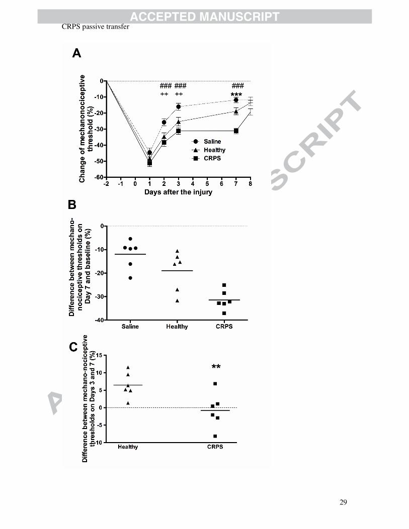
a) Primary outcome mechanical hyperalgesia (n=6 Healthy-IgG preparations and n=6
CRPS-IgG preparations)
The animals in all three groups (Saline, Healthy-IgG, CRPS-IgG) had removed their stitches
at the time of our first inspection, within 16h of the operation. The preoperative
mechanonociceptive thresholds of the affected limbs were 7.22 ± 0.09 g, 7.23 ± 0.09 g and
7.09 ± 0.10 g in the saline-, Healthy-IgG and CRPS-IgG-treated mice, respectively (no
significant differences). Plantar skin-muscle incision decreased these thresholds by 45-50% in
all three groups, 1 day after surgery, demonstrating an increased pain sensation/nocifensive
reaction to mild painful stiumli. In the saline-treated control group, this mechanical
hyperalgesia recovered to -25.74 ± 2.30 % and -16.01 ± 2.24 % on days 2 and 3, and was
thereafter maintained at the latter level. There was a main effect of time F(5,535)=245.2,
p<0.0001, and a significant difference between the mechanonociceptive thresholds of the
three treatment groups (F2,107)=19.98, p<0001. There was a significantly reduced
mechanonociceptive threshold in the CRPS IgG-, when compared with the Healthy IgG-
treated mice on day 7, (Bonferroni posttest, p<0.001, Figure 2A), in line with prediction
(primary outcome). On day 7 the mean threshold in the CRPS group (-31 ± 1.7 %) was 63%
decreased compared to the Healthy group (-19 ± 3.42%). Although two of the 6 control
preparations elicited strong mechanical hyperalgesia on day 7 (Figure 2B), the extent of the
hyperalgesia elicited by CRPS-IgG was always greater than in Healthy, in each individual
experiment. Mechanical hyperalgesia of mice treated with sample flowthroughs did not differ
significantly from the saline-treated control group. Between days 3-7, limb hyperalgesia
remained unchanged in the CRPS-IgG group, while there was significant recovery in the
Healthy-IgG group (Figure 2C); a slower/stalled recovery in the CRPS-IgG group when
CRPS passive transfer
14
compared to Healthy was also evident in each individual experiment. On the contralateral,
non-injured side there was less than 5% change in the mechanonociceptive thresholds in
either of the three groups (individual outcomes, flowthrough- and contralateral data not
shown).
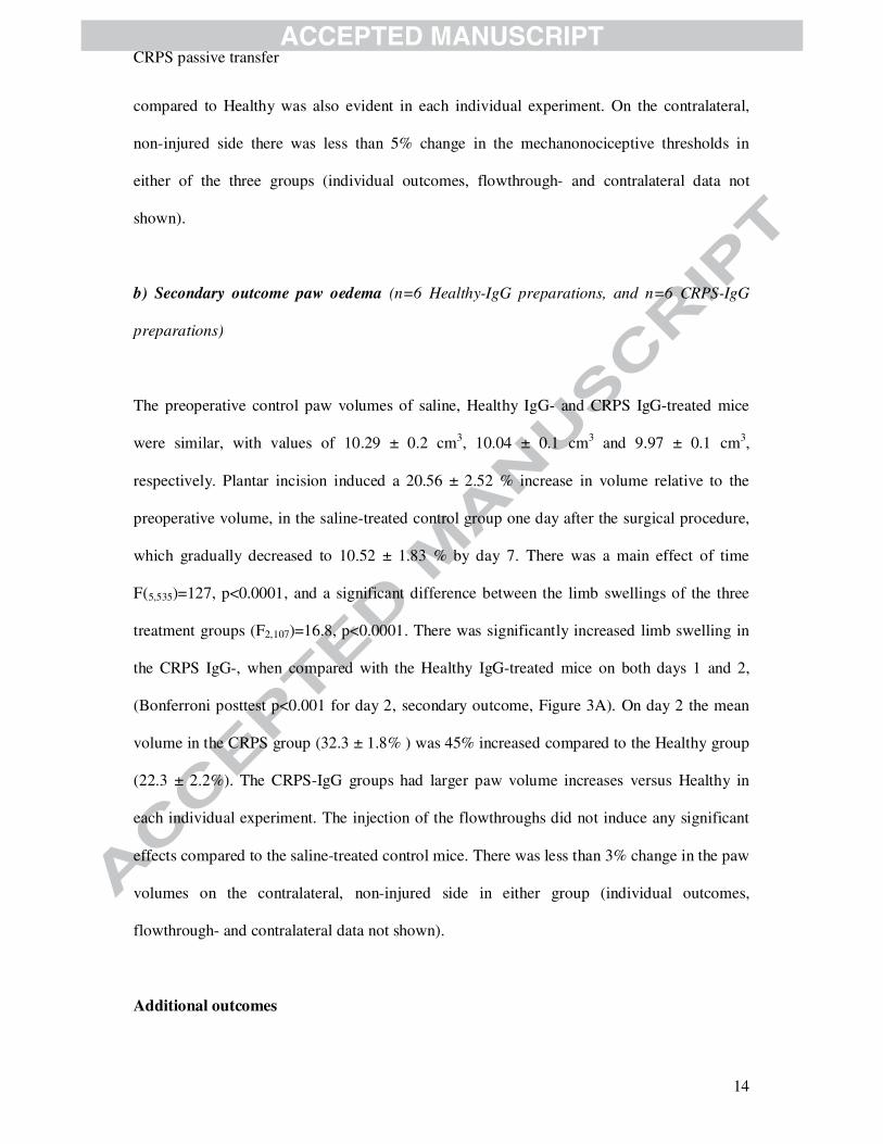
b) Secondary outcome paw oedema (n=6 Healthy-IgG preparations, and n=6 CRPS-IgG
preparations)
The preoperative control paw volumes of saline, Healthy IgG- and CRPS IgG-treated mice
were similar, with values of 10.29 ± 0.2 cm3, 10.04 ± 0.1 cm3 and 9.97 ± 0.1 cm3,
respectively. Plantar incision induced a 20.56 ± 2.52 % increase in volume relative to the
preoperative volume, in the saline-treated control group one day after the surgical procedure,
which gradually decreased to 10.52 ± 1.83 % by day 7. There was a main effect of time
F(5,535)=127, p<0.0001, and a significant difference between the limb swellings of the three
treatment groups (F2,107)=16.8, p<0.0001. There was significantly increased limb swelling in
the CRPS IgG-, when compared with the Healthy IgG-treated mice on both days 1 and 2,
(Bonferroni posttest p<0.001 for day 2, secondary outcome, Figure 3A). On day 2 the mean
volume in the CRPS group (32.3 ± 1.8% ) was 45% increased compared to the Healthy group
(22.3 ± 2.2%). The CRPS-IgG groups had larger paw volume increases versus Healthy in
each individual experiment. The injection of the flowthroughs did not induce any significant
effects compared to the saline-treated control mice. There was less than 3% change in the paw
volumes on the contralateral, non-injured side in either group (individual outcomes,
flowthrough- and contralateral data not shown).
Additional outcomes
CRPS passive transfer
15
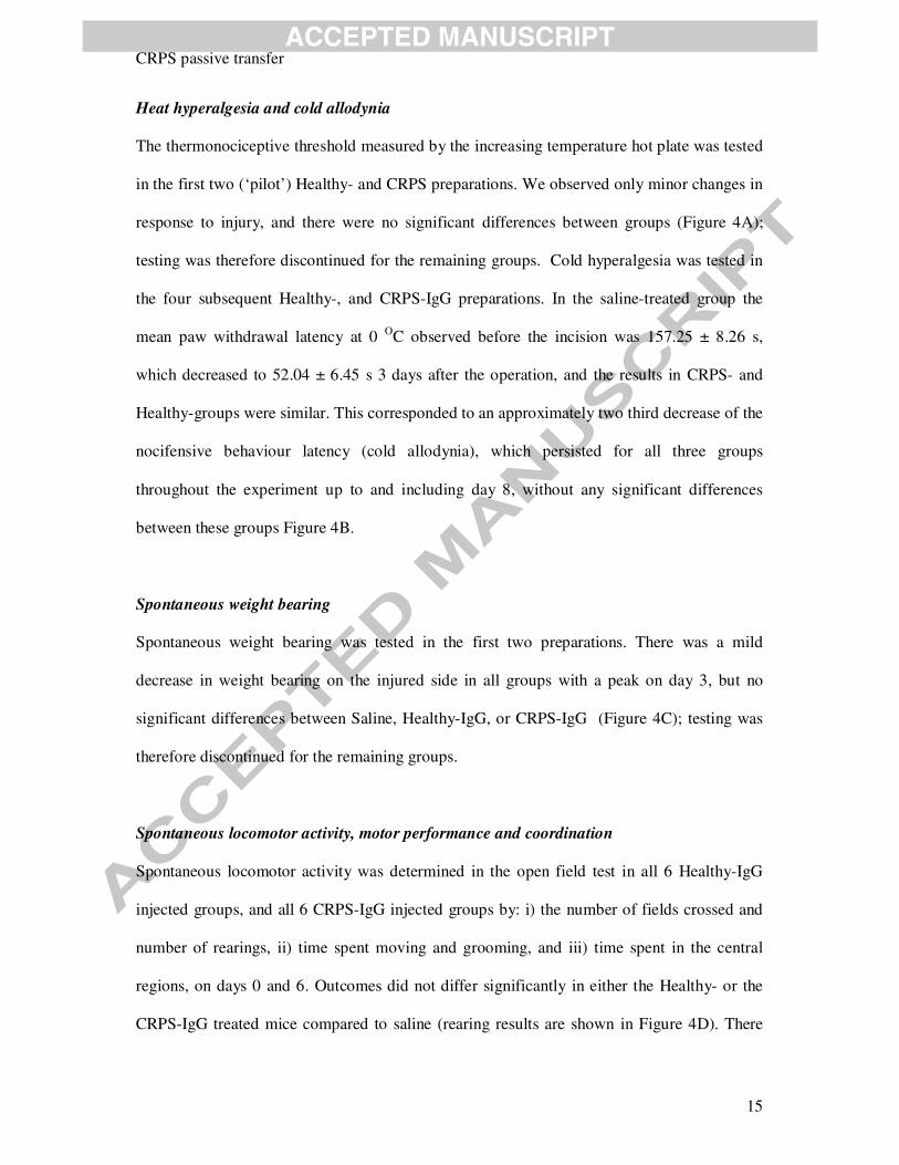
Heat hyperalgesia and cold allodynia
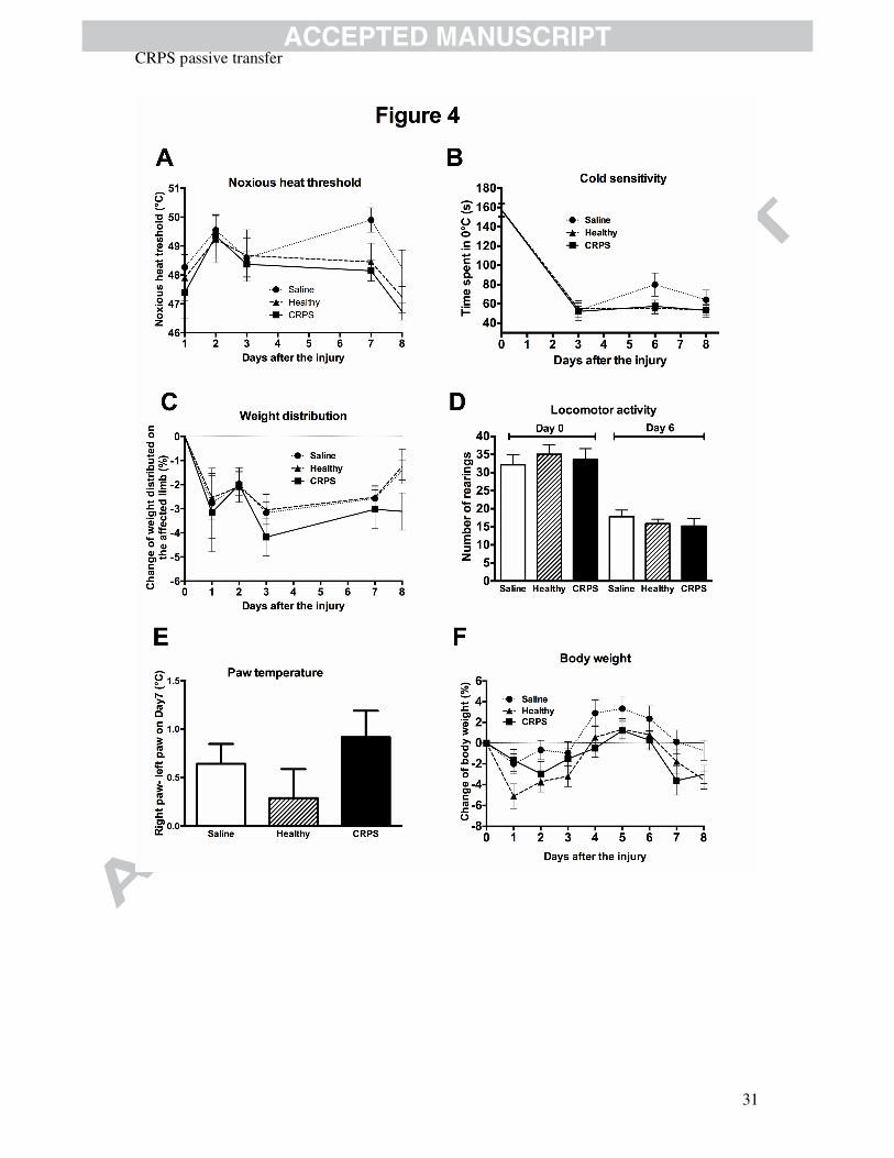
The thermonociceptive threshold measured by the increasing temperature hot plate was tested
in the first two (‘pilot’) Healthy- and CRPS preparations. We observed only minor changes in
response to injury, and there were no significant differences between groups (Figure 4A);
testing was therefore discontinued for the remaining groups. Cold hyperalgesia was tested in
the four subsequent Healthy-, and CRPS-IgG preparations. In the saline-treated group the
mean paw withdrawal latency at 0 OC observed before the incision was 157.25 ± 8.26 s,
which decreased to 52.04 ± 6.45 s 3 days after the operation, and the results in CRPS- and
Healthy-groups were similar. This corresponded to an approximately two third decrease of the
nocifensive behaviour latency (cold allodynia), which persisted for all three groups
throughout the experiment up to and including day 8, without any significant differences
between these groups Figure 4B.
Spontaneous weight bearing
Spontaneous weight bearing was tested in the first two preparations. There was a mild
decrease in weight bearing on the injured side in all groups with a peak on day 3, but no
significant differences between Saline, Healthy-IgG, or CRPS-IgG (Figure 4C); testing was
therefore discontinued for the remaining groups.
Spontaneous locomotor activity, motor performance and coordination
Spontaneous locomotor activity was determined in the open field test in all 6 Healthy-IgG
injected groups, and all 6 CRPS-IgG injected groups by: i) the number of fields crossed and
number of rearings, ii) time spent moving and grooming, and iii) time spent in the central
regions, on days 0 and 6. Outcomes did not differ significantly in either the Healthy- or the
CRPS-IgG treated mice compared to saline (rearing results are shown in Figure 4D). There
CRPS passive transfer
16
was also no significant difference in rotarod performance between groups on day 0..In the
first two experimental,,sets we observed generally increased performances in all groups on
day 6, possibly attributable to learning, and with no significant differences between groups,
and therefore discontinued further testing on day 6.
Paw temperature
There were no differences between CRPS-IgG, and saline or Healthy-IgG groups in either the
absolute paw temperatures of the injured limbs, or in the mean absolute temperature
differences between the respective injured- and non-injured paws on day 7 upon testing of the
first three Healthy- and CRPS-injected groups (Figure 4E). We discontinued temperature
testing in the remaining experiments.
Changes of body weight
The mean body weight was tested in all saline-, Healthy-IgG (n=6), and CRPS-IgG (n=6)
treated groups. It was 18.26 ± 0.25 g, 18.95 ± 0.29 g, and 18.53 ± 0.23 g, respectively on day
-2 of the experiment. The weights did not change significantly during the 8 days of the
experiment (Figure 5F).
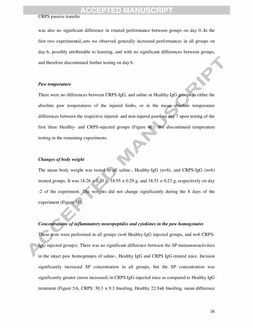
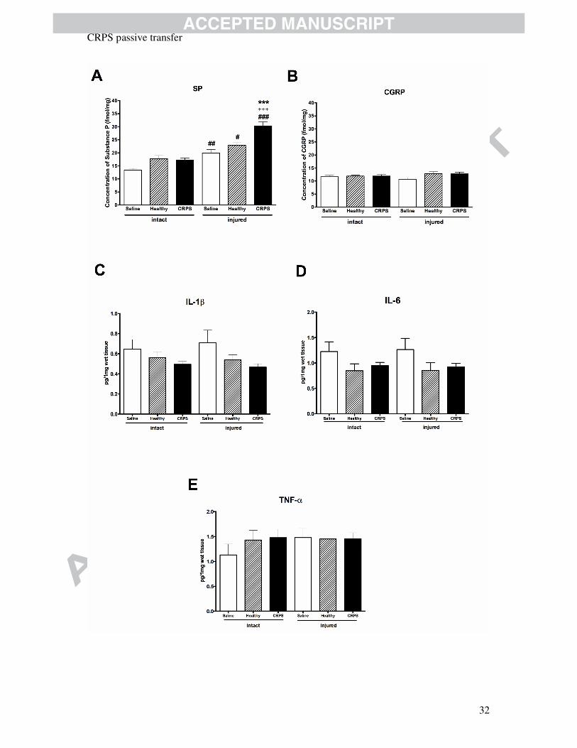
Concentrations of inflammatory neuropeptides and cytokines in the paw homogenates
These tests were performed in all groups (n=6 Healthy-IgG injected groups, and n=6 CRPS-
IgG injected groups). There was no significant difference between the SP-immunoreactivities
in the intact paw homogenates of saline-, Healthy IgG and CRPS IgG-treated mice. Incision
significantly increased SP concentration in all groups, but the SP concentration was
significantly greater (more increased) in CRPS IgG-injected mice as compared to Healthy IgG
treatment (Figure 5A, CRPS: 30.3 ± 9.3 fmol/mg, Healthy 22.9±6 fmol/mg, mean difference
CRPS passive transfer
17
7.5 fmol/mg, 95% CI of the difference: 2.8-12.1 fmol/mg, p<0.001 one-way ANOVA with
Bonferroni post-test). In contrast, CGRP-immunoreactivity did not change either in response
to injury, or IgG treatments (Figure 5B). Neuropeptide concentrations in the flowthough-
injected groups did not differ from saline groups (data not shown). IL-1β and IL-6
concentrations were not different between groups (5C-D). Although there appeared to be a
significant difference in TNF-concentration in a limited number of preparations (see
Methods), no differences were seen when all paw tissues were investigated (Figure 5E).
Following a Bonferroni correction for multiple comparisons for the 5 measured mediators, SP
remained significantly raised (p<0.005).
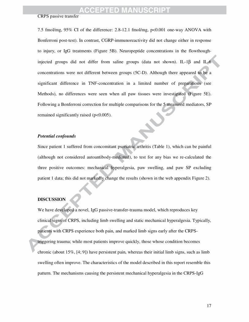
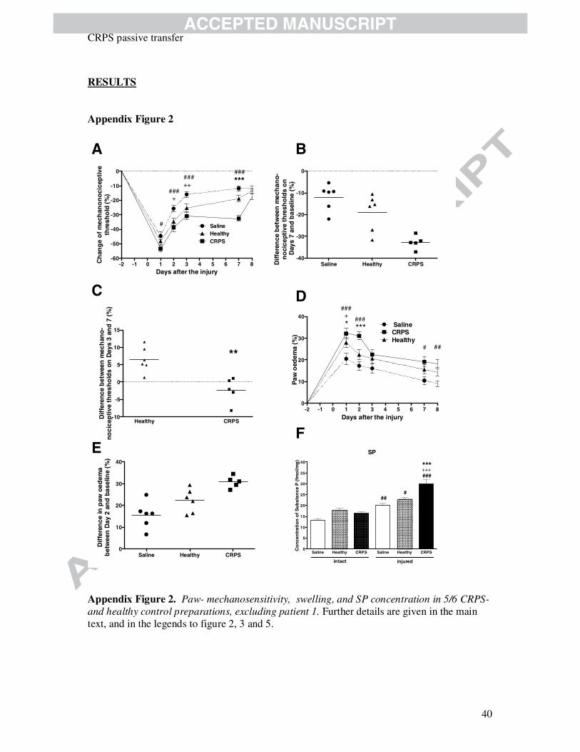
Potential confounds
Since patient 1 suffered from concomitant psoriatric arthritis (Table 1), which can be painful
(although not considered autoantibody-mediated), to test for any bias we re-calculated the
three positive outcomes: mechanical hyperalgesia, paw swelling, and paw SP excluding
patient 1 data; this did not markedly change the results (shown in the web appendix Figure 2).
DISCUSSION
We have developed a novel, IgG passive-transfer-trauma model, which reproduces key
clinical signs of CRPS, including limb swelling and static mechanical hyperalgesia. Typically,
patients with CRPS experience both pain, and marked limb signs early after the CRPS-
triggering trauma; while most patients improve quickly, those whose condition becomes
chronic (about 15%, [4; 9]) have persistent pain, whereas their initial limb signs, such as limb
swelling often improve. The characteristics of the model described in this report resemble this
pattern. The mechanisms causing the persistent mechanical hyperalgesia in the CRPS-IgG
CRPS passive transfer
18
group require further study. These results suggest a possible role of pathogenetic
autoantibodies in patients with longstanding CRPS.
The rapid normalisation of all behavioural abnormalities by day 8 was similar to that
described in earlier, passive-transfer-only experiments, and is likely explained by the rodent’s
metabolism of the injected human IgG [16]. Reinjection at day 7 or 8 might have overcome
this, but would have required both availability of larger amounts of sera, and co-application of
cyclophosphamide to supress serum sickness, with uncertain impact on the unknown
immunological mechanisms [8]
It has been suggested that cold allodynia is due to peripheral nerve terminal sensitization,
while mechanical hyperalgesia involves central sensitization processes [26]. Our results,
showing a disparate outcome for cold allodynia (no difference between groups), versus
mechanical hyperalgesia (increased in CRPS-IgG injected animals), may suggest a
contribution of central sensitisation to the CRPS-IgG-induced effect on mechanical
hyperalgesia, or alternatively a long-lasting flooring effect in the cold-allodynia experiments
may be responsible (’central sensitisation’ = a peripheral trauma-induced change to the
response properties of dorsal horn neurons causing peripheral hypersensitivity [24]). Previous
work by others has demonstrated an absence of cold allodynia in the plantar incision mouse
model when this was assessed by measuring paw-licking latency on a cold plate at 0 oC [33].
The difference in our results, which show generally strong postoperative cold allodynia, when
compared to these previously reported results, might be explained through the larger area of
contact of the injured paw in the water bath in our experiments. Our findings were unrelated
to the treatment group, and are therefore not an indication of transfer of a CRPS-related
feature.
The lack of difference between groups in the open field tests contrasts with our earlier results
[14]; we propose that the limb trauma common to all groups exerted an overriding effect on
CRPS passive transfer
19
day 6. Open field rearing is sensitive to a range of confounding factors [36]. The 2-4 weeks
higher rodent age in the present experiments, and differing environmental factors in the two
laboratories, may be responsible for the observed lack of a difference on day 0 in the present
study.
In addition to the functional and morphological results, an important finding that parallels
human disease [38] is the significantly increased concentration of the inflammatory
neuropeptide SP in the injured paws of CRPS-IgG-injected animals. Concentrations of CGRP,
as well as the cytokines TNF-alpha, IL-1, and IL-6 were normal, contrasting results from
investigations in CRPS-affected human skin [20; 38], and/or animal models after fracture [32;
39]. While CGRP is mostly of sensory neuronal origin [6], SP is also localized in immune
cells besides the capsaicin-sensitive peptidergic afferents [19]. Furthermore, these two
peptides are only partially co-localized in the sensory nerve terminals [34]. Of note,
behavioural abnormalities had largely resolved in most experiments at the time of tissue
harvest on day 8 (Figures 2,3). It is possible that the tissue concentrations of these mediators
would differ at earlier time points from the concentration on day 8. Previous studies by other
groups in a hindpaw incision model (without IgG transfer) have established a central role for
SP in sustaining hyperalgesia. For example SP-deficient mice display decreased mechanical
hyperalgesia after incision, compared to wild-type mice [32]. Thus, it is possible that there is
an interaction between the observed CRPS-IgG effects, such that increased tissue SP may
result in increased hyperalgesia. Further studies would be required to clarify any such
interactions.
The major strength of the study is the delivery of a model in which both limb injury and
variant human condition (specific IgG serum-autoantibodies) are necessary elements, as in the
clinical disease. The model consistently produced behavioural effects in the CRPS-injected
groups.

CRPS passive transfer
20
A limitation is that the clinical picture of CRPS was incompletely reproduced. For example,
CRPS is associated with an unstable temperature difference between affected and unaffected
limbs, which may in part be caused by sympathetic dysfunction [23]. Enhancement of
hyperalgesia and edama alone, as observed in this model, would not be sufficient to merit a
CRPS diagnosis under the IASP 2012 CRPS criteria. Although the model does not produce all
features of human CRPS, given that multiple contributing mechanism have been suggested in
the literature [12; 25], other clinical signs (e.g. temperature differences) might derive from
unrelated mechanisms. Further, as we have not compared the effects of serum-IgG from
patients with different clinical phenotypes (e.g. patients with-, or without mechanical
hyperalgesia), we cannot draw any conclusion as to whether the clinical signs in patients
correlated with the behavioural abnormalities in the rodents. As we only analysed 2 out of 5-
7 samples/group for mediator concentrations, we may have missed significant differences in
concentrations, but differences between groups in the tested samples were very small, so that
this risk should be small. A pragmatic limitation for the model’s usability is that the time-
windows for significantly enhanced mechanical allodynia or paw swelling are relatively
narrow.
In summary, in this first passive-transfer-trauma model for CRPS, serum-IgG from patients
with longstanding CRPS induced clinical and laboratory features of the human disease,
indicating a relevant role of IgG autoantibodies. The epitope-specificities of the pathogenic
autoantibodies remain unknown requiring further research. Our results should be relevant to
clinical research, because they suggest the value of considering autoantibody-removing
therapies for longstanding CRPS. The model may provide a novel tool to study both the
CRPS disease mechanisms, and candidate therapies.
CRPS passive transfer
21
Acknowledgements
The authors thank Prof. Angela Vincent and Dr. Maria Leite, both Oxford/UK for technical
advice, Mrs. Katalin Gógl and Mrs. Dóra Ömböli for their expert technical assistance in the
experiments, Mrs. Lorna Murray and Dr. Susmita Oommans for help with bloods acquisition.
This study was supported by SROP-4.2.2.A-11/1/KONV-2012-0024, SROP-4.2.2.B-10/
1/2010-0029, 57073T01, the Pain Relief Foundation, Liverpool/UK, CSL-Behring, BPL, and
Biotest. Dr. Goebel declares having received research support, consultancy or speaker
honoraria for Pfizer, Axsome, Biotest, Baxter, CSL-Behring, Grifols, BPL. The other authors
declare no conflict of interest.

CRPS passive transfer
22
References
[1] Almasi R, Petho G, Bolcskei K, Szolcsanyi J. Effect of resiniferatoxin on the noxious heat threshold temperature in the rat: a novel heat allodynia model sensitive to analgesics. British journal of pharmacology 2003;139(1):49-58.
[2] Anhalt GJ, Labib RS, Voorhees JJ, Beals TF, Diaz LA. Induction of pemphigus in neonatal mice by passive transfer of IgG from patients with the disease. The New England journal of medicine 1982;306(20):1189-1196.
[3] Banik RK, Woo YC, Park SS, Brennan TJ. Strain and sex influence on pain sensitivity after plantar incision in the mouse. Anesthesiology 2006;105(6):1246-1253.
[4] Birklein F, Riedl B, Sieweke N, Weber M, Neundorfer B. Neurological findings in complex regional pain syndromes--analysis of 145 cases. Acta NeurolScand 2000/4;101(4):262-269.
[5] Bolcskei K, Helyes Z, Szabo A, Sandor K, Elekes K, Nemeth J, Almasi R, Pinter E, Petho G, Szolcsanyi J. Investigation of the role of TRPV1 receptors in acute and chronic nociceptive processes using gene-deficient mice. Pain 2005;117(3):368-376.
[6] Brain SD, Cox HM. Neuropeptides and their receptors: innovative science providing novel therapeutic targets. British journal of pharmacology 2006;147 Suppl 1:S202-211.
[7] Brennan TJ, Vandermeulen EP, Gebhart GF. Characterization of a rat model of incisional pain. Pain 1996/3;64(3):493-501.
[8] Buckley C, Vincent A. Autoimmune channelopathies. NatClinPractNeurol 2005/11;1(1):22-33.
[9] de MM, Huygen FJ, Hoeven-Borgman M, Dieleman JP, Ch Stricker BH, Sturkenboom MC. Outcome of the complex regional pain syndrome. ClinJPain 2009/9;25(7):590-597.
[10] Furedi R, Bolcskei K, Szolcsanyi J, Petho G. Effects of analgesics on the plantar incision-induced drop of the noxious heat threshold measured with an increasing-temperature water bath in the rat. European journal of pharmacology 2009;605(1-3):63-67.
[11] Gierthmuhlen J, Maier C, Baron R, Tolle T, Treede RD, Birbaumer N, Huge V, Koroschetz J, Krumova EK, Lauchart M, Maihofner C, Richter H, Westermann A. Sensory signs in complex regional pain syndrome and peripheral nerve injury. Pain 2012;153(4):765-774.
[12] Goebel A. Complex regional pain syndrome in adults. Rheumatology (Oxford) 2011/10;50(10):1739-1750.
CRPS passive transfer
23
[13] Goebel A, Baranowski AP, Maurer K, Ghiai A, McCabe C, Ambler G. Intravenous Immunoglobulin Treatment of Complex Regional Pain Syndrome: A Randomized Trial. AnnInternMed 2010;152(3):152-158.
[14] Goebel A, Leite MI, Yang L, Deacon R, Cendan CM, Fox-Lewis A, Vincent A. The passive transfer of immunoglobulin G serum antibodies from patients with longstanding Complex Regional Pain Syndrome. Eur J Pain 2011;15(5):504 e501-506.
[15] Goebel A, Misbah, S., McKiver, K., Haynes, L., Burton, J., Philips, C., Frank, B., Poole, H. Immunoglobulin Maintenance Therapy in Lonstanding Complex Regional Pain Syndrome. Rheumatology (Oxford) 2013.
[16] Goebel A, Stock M, Deacon R, Sprotte G, Vincent A. Intravenous immunoglobulin response and evidence for pathogenic antibodies in a case of complex regional pain syndrome 1. AnnNeurol 2005/3;57(3):463-464.
[17] Guo TZ, Offley SC, Boyd EA, Jacobs CR, Kingery WS. Substance P signaling contributes to the vascular and nociceptive abnormalities observed in a tibial fracture rat model of complex regional pain syndrome type I. Pain 2004;108(1-2):95-107.
[18] Harden RN, Bruehl S, Perez RS, Birklein F, Marinus J, Maihofner C, Lubenow T, Buvanendran A, Mackey S, Graciosa J, Mogilevski M, Ramsden C, Chont M, Vatine JJ. Validation of proposed diagnostic criteria (the "Budapest Criteria") for Complex Regional Pain Syndrome. Pain 2010/8;150(2):268-274.
[19] Howard MR, Millward-Sadler SJ, Vasilliou AS, Salter DM, Quinn JP. Mechanical stimulation induces preprotachykinin gene expression in osteoarthritic chondrocytes which is correlated with modulation of the transcription factor neuron restrictive silence factor. Neuropeptides 2008;42(5-6):681-686.
[20] Huygen FJ, De Bruijn AG, De Bruin MT, Groeneweg JG, Klein J, Zijistra FJ. Evidence for local inflammation in complex regional pain syndrome type 1. MediatorsInflamm 2002/2;11(1):47-51.
[21] Kingery WS. Role of neuropeptide, cytokine, and growth factor signaling in complex regional pain syndrome. Pain Med 2010/8;11(8):1239-1250.
[22] Kohr D, Singh P, Tschernatsch M, Kaps M, Pouokam E, Diener M, Kummer W, Birklein F, Vincent A, Goebel A, Wallukat G, Blaes F. Autoimmunity against the beta(2) adrenergic receptor and muscarinic-2 receptor in complex regional pain syndrome. Pain 2011;152(12):2690-2700.
[23] Krumova EK, Frettloh J, Klauenberg S, Richter H, Wasner G, Maier C. Long-term skin temperature measurements - a practical diagnostic tool in complex regional pain syndrome. Pain 2008;140(1):8-22.
[24] Latremoliere A, Woolf CJ. Central sensitization: a generator of pain hypersensitivity by central neural plasticity. J Pain 2009/9;10(9):895-926.

CRPS passive transfer
24
[25] Marinus J, Moseley GL, Birklein F, Baron R, Maihofner C, Kingery WS, Van Hilten JJ. Clinical features and pathophysiology of complex regional pain syndrome. Lancet Neurol 2011/7;10(7):637-648.
[26] Meyer RA, Ringkamp, M., Campbell, J.N, Raja, S.N. . Peripheral mechanisms of cutaneous nociception. In: SB McMahon, Kotzenburg, M., editor. Textbook of Pain. Amsterdam: Elsevier, 2006. pp. 3-33.
[27] Nemeth J, Gorcs T, Helyes Z, Oroszi G, Kocsy T, Pinter E, Szolcsanyi J. Development of a new sensitive CGRP radioimmunoassay for neuropharmacological research. Neurobiology (Bp) 1998;6(4):473-475.
[28] Nemeth J, Oroszi G, Than M, Helyes ZS, Pinter E, Farkas B, Szolcsanyi J. Substance P radioimmunoassay for quantitative characterization of sensory neurotransmitter release. Neurobiology (Bp) 1999;7(4):437-444.
[29] Pizziketti RJ, Pressman NS, Geller EB, Cowan A, Adler MW. Rat cold water tail-flick: a novel analgesic test that distinguishes opioid agonists from mixed agonist-antagonists. European journal of pharmacology 1985;119(1-2):23-29.
[30] Pogatzki-Zahn EM, Zahn PK, Brennan TJ. Postoperative pain--clinical implications of basic research. Best practice & research Clinical anaesthesiology 2007;21(1):3-13.
[31] Rose NR, Bona C. Defining criteria for autoimmune diseases (Witebsky's postulates revisited). ImmunolToday 1993/9;14(9):426-430.
[32] Sahbaie P, Shi X, Guo TZ, Qiao Y, Yeomans DC, Kingery WS, Clark JD. Role of substance P signaling in enhanced nociceptive sensitization and local cytokine production after incision. Pain 2009;145(3):341-349.
[33] Scherer M, Reichl SU, Augustin M, Pogatzki-Zahn EM, Zahn PK. The assessment of cold hyperalgesia after an incision. Anesthesia and analgesia 2010;110(1):222-227.
[34] Schulze E, Witt M, Fink T, Hofer A, Funk RH. Immunohistochemical detection of human skin nerve fibers. Acta histochemica 1997;99(3):301-309.
[35] Szabo A, Helyes Z, Sandor K, Bite A, Pinter E, Nemeth J, Banvolgyi A, Bolcskei K, Elekes K, Szolcsanyi J. Role of transient receptor potential vanilloid 1 receptors in adjuvant-induced chronic arthritis: in vivo study using gene-deficient mice. The Journal of pharmacology and experimental therapeutics 2005;314(1):111-119.
[36] Teeling JL, Felton LM, Deacon RM, Cunningham C, Rawlins JN, Perry VH. Sub-pyrogenic systemic inflammation impacts on brain and behavior, independent of cytokines. Brain BehavImmun 2007/8;21(6):836-850.
[37] Toyka KV, Brachman DB, Pestronk A, Kao I. Myasthenia gravis: passive transfer from man to mouse. Science 1975/10/24;190(4212):397-399.
[38] Weber M, Birklein F, Neundorfer B, Schmelz M. Facilitated neurogenic inflammation in complex regional pain syndrome. Pain 2001;91(3):251-257.

CRPS passive transfer
25
[39] Wei T, Guo TZ, Li WW, Hou S, Kingery WS, Clark JD. Keratinocyte expression of inflammatory mediators plays a crucial role in substance P-induced acute and chronic pain. Journal of neuroinflammation 2012;9:181.
Summary: The transfer of serum-IgG from patients with longstanding CRPS to mice,
followed by mild hindpaw trauma causes typical signs of CRPS.
Figure legends
Figure 1. Study flow chart. N=number of preparations tested; i.p.=intraperitoneally; one pair
of CRPS-IgG and Healthy-IgG injected mice was sacrificed on day 10 instead of day 8,
because abnormal values had not returned to normal on day 8 (details in the results section).
Figure 2. Effect of serum-IgGs derived from CRPS patients in comparison with the IgGs of
healthy controls and saline on plantar incision-induced mechanical hyperalgesia of the mouse
paw. The serum-IgG samples, or normal saline, in a total volume of 1.5 ml/mouse/day were
administered intraperitoenally on days -1, 0, and 5, 6. Panel A shows the pooled results from
all experiments expressed as percentage change compared to baseline and shown as means ±
S.E.M. (n=30-37 animals/group); ***p<0.001 CRPS vs. Healthy, ###p<0.001 CRPS vs.
saline and ++p<0.05 Healthy vs. saline (repeated measures two-way ANOVA with
Bonferroni’s modified posttest). In Panels B and C, each point represents a group of mice (5-7
animals per group) treated with one CRPS patient-, or one healthy control serum-IgG, or
normal saline, horizontal lines represent means. Panel B shows the day 7 results for each
individual experiment. The differences between percentage hyperalgesia values on days 7 and
3 (slope of the curve) are described in Panel C; dot plots of the means of the twelve IgG-
injected groups are shown; horizontal bars denote means

CRPS passive transfer
26
Figure 3. Effects of serum-IgGs derived from CRPS patients, serum-IgGs of healthy controls,
and saline on plantar incision-induced paw oedema. The injection schedule is as described in
Figures 1/2. Panel A shows the pooled results from all experiments, expressed as percentage
increase over the respective baseline values, as means ± S.E.M. (n=30-37 animals/group);
***p<0.001 CRPS vs. Healthy, ###p<0.001 CRPS vs. saline and +p<0.05 Healthy vs. saline
(repeated measures two-way ANOVA followed by Bonferroni’s modified posttest). In Panel
B, each dot represents the mean of the results from one group of mice injected with one
CRPS- or one Healthy-IgG preparation, or saline, respectively (5-7 animals per group).
Horizontal lines represent means.
Figure 4. Behavioural outcomes and body weight following human IgG treatment and
plantar skin-muscle incision. (A) The noxious heat thresholds were measured with an
increasing temperature hot plate in the first two experimental groups; (B) cold sensitivity was
determined by the withdrawal latency after immersing the paws into iced water, performed in
all but the first two experimental groups; (C) spontaneous weight distribution on the affected
limb was tested in the first two experimental groups; (D) locomotor activity (rearing) in the
open field, on both days 0 and 6; (E) paw temperature (absolute differences between the
injured and non-injured contralateral paws) measured with a surface thermometer on day 7 in
three groups; (G) Body weight was determined in all animals on each experimental day.
Values are expressed as means ± S.E.M. ; (n=12-37 animals/treated group). There were no
significant differences between groups in any of these 6 tests.
Figure 5. Effects of human IgG treatments on sensory neuropeptide and inflammatory
cytokine concentrations in the hindpaws. Concentrations of (A) substance P (SP) and (B)

CRPS passive transfer
27
calcitonin gene-related peptide (CGRP) were measured by radioimmunoassay in hindpaw
homogenates excised after sacrifice on day 8. Concentrations of (C) Interleukin 1-beta (IL-
1β), (D) Interleukin-6 (IL-6), and (E) tumor necrosis factor-alpha (TNF-α) were measured by
Luminex and/or ELISA, respectively, from the same samples. Columns show the means ±
S.E.M. of either n=30-37 mice per group (SP, CGRP, TNF-α), or 6-13 animals per group for
IL-1β and IL-6 (see Methods). #p<0.05, ##p<0.01 ###p<0.001 vs. respective intact limbs,
***p<0.001 vs. Healthy-, and +++p<0.001 vs. saline of the injured side (one-way ANOVA
followed by Bonferroni’s modified post-test).

CRPS passive transfer
28
Heat- and Mechanosensitivity, Paw oedema, Weight distribution
Days: 0-1
-2
Plantar skin and muscle incision
Sacrifice and tissue harvest Treatments :
(1 + 0.5 ml i.p. daily) Saline
Healthy CRPS
Flowthrough
-3
-4
7 3 821 6 54
Open field and RotaRod
Body weight
Cold allodynia
Experimental design
Paw temperature
Conditioning, and 3 baseline measurements
CRPS passive transfer
29

CRPS passive transfer
30
CRPS passive transfer
31
CRPS passive transfer
32

CRPS passive transfer
33
Table 1.
Patient No 1 2 3 4 5 6
Age 48 43 60 34 37 50
Sex F M M F F F
Affected limb Lower Both lower Upper Lower Lower Lower
Inciting event Episode of dancing Minor knock Operation on
the brachial
plexus for a
benign tumor
Operation
for Freiberg’s
syndrome,
and re-
operations
Operation
for Morton
neuroma,
Ankle
fracture,
followed by
operations
CRPS type 1 1 2 1 Not
determined
1
Disease duration (years) 6 5.1 7 5-7* 1 8-10*
Pain intensity 7 7.5 10 7 5.5 8
Pain to light pressure
applied onto the skin of
the affected limb
+ + + + + +
Concomitant disease Psoriatric arthritis,
gluten-sensitive
enteropathy,
pernicious anemia
none Past
myocardial
infarction
Freiberg’s
syndrome
Asthma Diabetes II,
high blood
pressure,
high
cholesterol,
overactive
thyroid
Table 1. Demographic and disease characteristics of the study patients. F=female; M=male; CRPS type = 1
without/2 with associated injury to a major nerve; Pain intensity=average 24h pain intensity on the day of
enrollment, on a 11-point numeric rating scale (0-10), with 0=no pain, 10=pain as bad as you can imagine; * the
disease onset followed one of several operations, exact time details are not memorized or recorded.

CRPS passive transfer
34
The etiology of Complex Regional Pain Syndrome (CRPS), a highly painful, usually
posttraumatic condition affecting limbs, is unknown but recent results have suggested an
autoimmune contribution. To confirm a role for pathogenic autoantibodies, we established a
passive transfer-trauma model. Prior to undergoing hindlimb plantar skin and muscle incision
mice were injected with either serum-IgG obtained from chronic CRPS patients or matched
healthy volunteers, or with saline. Unilateral hindlimb plantar skin and muscle incision was
performed to induce typical, mild tissue injury. Mechanical hyperalgesia, paw swelling, heat-
and cold- sensitivity, weight-bearing ability, locomotor activity, motor coordination, paw
temperature and body weight were investigated for 8 days. After sacrifice, pro-inflammatory
sensory neuropeptides and cytokines were measured in paw tissues. CRPS patient IgG
treatment significantly increased hindlimb mechanical hyperalgesia and edema in the incised
paw, compared to healthy subject IgG, or saline treatment. Plantar incision induced a
remarkable elevation of substance P immunoreactivity on day 8, which was significantly
increased by CRPS-IgG. In this passive-transfer-trauma model for CRPS, serum-IgG from
chronic CRPS patients induced clinical- and laboratory features resembling the human
disease. These results support the hypothesis that autoantibodies may contribute to the CRPS
pathophysiology, and that autoantibody-removing therapies may be effective treatment for
longstanding CRPS.
CRPS passive transfer
35
Supplementary Appendix METHODS Protocol for Serum Sampling
NOTE: THIS PROTOCOL HAS BEEN MODIFIED FROM THE ORIGINAL VERSION BY REMOVAL OF SPONSOR NAME AND ADDRESSES, REFERENCES, AND PARTS RELATING TO OTHER THAN PASSIVE-TRANSFER RESEARCH. Protocol version 1.0, Date 10.01.2012 Study Number: 1. General Information 1.1. Title Serum Autoantibodies in Complex Regional Pain Syndrome 1.2. Sponsor a) The University of Liverpool b) Co-sponsor: The Walton Centre NHS Trust 1.3. Authorized signant for the sponsor a) xx, The University of Liverpool b) xx, The Walton Centre Clinical Trials Unit 1.4. Responsible Investigator at site Dr. Andreas Goebel 1.5. Medical/Technical Departments involved in the study a) The Walton Centre NHS Trust outpatient department b) The Walton Centre NHS Trust Clinical Trials Unit 2 Study Summary Title: Serum Autoantibodies in Complex Regional Pain Syndrome Indication: Complex Regional Pain Syndrome (CRPS) Rationale: CRPS is a severe, posttraumatic pain syndrome affecting a limb. The causes and sustaining factors for CRPS are unknown. There is evidence that some patients have serum autoantibodies, however their specificity and pathogenicity has not been fully established. There is a recognized need to understand trigger and regulatory mechanisms for CRPS and this project will contribute to ascertain the role of serum autoantibodies. Study Design: A comparative study Number of Centres: This study will be carried out as a single centre study. Patient selection criteria: Patients will be selected from those seen by the principle investigator at the Walton Centre NHS Trust. Expected outcome: We expect that CRPS-IgG, but not Control-IgG will elicit important aspects of the clinical picture of CRPS upon passive transfer to rodents. 3. Background Information Complex Regional Pain Syndrome, CRPS (previously known as ‘reflex sympathetic dystrophy’, ‘causalgia’, ‘Sudeck’s dystrophy’ and ‘algodystrophy’), is a painful, usually posttraumatic, condition in a limb with an incidence of 2.6 per 10.000 person years (1). It may be further classified into CRPS I and II, based on the absence or presence of injury to major nerves. CRPS is characterised by sympathetic, sensory and motor dysfunction in the affected limb, but the cardinal symptom is incapacitating severe

CRPS passive transfer
36
pain. Although spontaneous resolution can occur, the persistence of CRPS beyond 6 months typically leads to the development of a chronic, debilitating condition with profound and lasting adverse impact on quality of life, activities of daily living, and the ability to retain gainful employment (2). The aetiology of CRPS remains uncertain and the treatment is therefore empirical and of limited efficacy. Possible mechanisms underlying CRPS symptoms include altered cortical representation, both somatosensory and motor, of the affected limb (3), central nervous system-mediated abnormal sympathetic activation in this limb (4), facilitated neurogenic inflammation, and aberrant immune responses (5;6). We and others have recently demonstrated that autoimmune mechanisms may contribute to the CRPS pathophysiology. We first showed that CRPS sera bind to both peripheral sensory and autonomic nerves more frequently than sera from healthy control subjects (7;8). Subsequently it was established that binding to autonomic nerves can also be detected using FACS analysis, that binding to autonomic nerves elicits functional effects on these cells in vitro (9), and that these effects are elicited through specific epitopes on both muscarinic and adrenergic receptors. Recently we demonstrated that the passive transfer of CRPS serum-IgG to mice elicits abnormal behaviour (10) and, independently that patients will respond to treatment with intravenous immunoglobulin, further supporting an autoantibody-mediated autoimmune pathogenesis (10-12). This research suggests that CRPS is associated with specific serum-autoantibodies. The autoantibody etiology of CRPS has yet to be established. The classical way to establish the autoantibody etiology of a disease is the passive autoantibody transfer from man to rodents (13). We had previously shown that CRPS serum-IgG elicits abnormal signs in a passive transfer model, but stimulus-evoked pain was not induced, thus this model did not sufficiently reproduce CRPS (12). We have now extended this model into a ‘passive transfer-trauma model’ for early CRPS, with encouraging results (Figure). 4. Research objective and purpose In this research we will test the hypothesis that the passive transfer to rodents of CRPS serum-IgG (but not control serum-IgG) will elicit a syndrome in these animals which resembles CRPS. We plan to confirm that our rodent model is valid, i.e. that it sufficiently reproduces early CRPS as suggested by our experiments (Figure). We will also further characterize the model with regards to both behavioural and immunohistochemical parameters. 5. Trial Conduct a) patient/volunteer identification, approach and enrolment CRPS patients will be identified from those newly seen or seen within the last five years seen at the Walton Centre NHS Trust by the PI. Potentially suitable patients will be approached for their interest to participate in the study, either in the Walton Centre outpatient department during regular clinic appointments (potentially interested patients are then given a patient information sheet), or though letters sent to their home addresses (with included patient information sheet), or over the phone by a person not involved in the trial (e.g. a nurse from the clinical trials centre), with subsequent sending of a information sheet, whichever is most convenient. Those patients who have previously indicated that they wish to be contacted for future research studies, and entered their name into a list together with contact details will be contacted either over the phone or, where applicable per e-mail. Where no
CRPS passive transfer
37
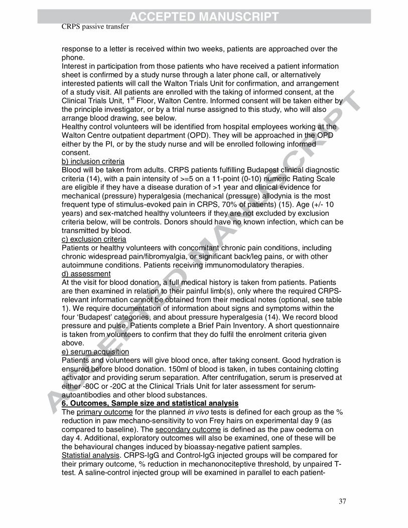
response to a letter is received within two weeks, patients are approached over the phone. Interest in participation from those patients who have received a patient information sheet is confirmed by a study nurse through a later phone call, or alternatively interested patients will call the Walton Trials Unit for confirmation, and arrangement of a study visit. All patients are enrolled with the taking of informed consent, at the Clinical Trials Unit, 1st Floor, Walton Centre. Informed consent will be taken either by the principle investigator, or by a trial nurse assigned to this study, who will also arrange blood drawing, see below. Healthy control volunteers will be identified from hospital employees working at the Walton Centre outpatient department (OPD). They will be approached in the OPD either by the PI, or by the study nurse and will be enrolled following informed consent. b) inclusion criteria Blood will be taken from adults. CRPS patients fulfilling Budapest clinical diagnostic criteria (14), with a pain intensity of >=5 on a 11-point (0-10) numeric Rating Scale are eligible if they have a disease duration of >1 year and clinical evidence for mechanical (pressure) hyperalgesia (mechanical (pressure) allodynia is the most frequent type of stimulus-evoked pain in CRPS, 70% of patients) (15). Age (+/- 10 years) and sex-matched healthy volunteers if they are not excluded by exclusion criteria below, will be controls. Donors should have no known infection, which can be transmitted by blood. c) exclusion criteria Patients or healthy volunteers with concomitant chronic pain conditions, including chronic widespread pain/fibromyalgia, or significant back/leg pains, or with other autoimmune conditions. Patients receiving immunomodulatory therapies. d) assessment At the visit for blood donation, a full medical history is taken from patients. Patients are then examined in relation to their painful limb(s), only where the required CRPS-relevant information cannot be obtained from their medical notes (optional, see table 1). We require documentation of information about signs and symptoms within the four ‘Budapest’ categories, and about pressure hyperalgesia (14). We record blood pressure and pulse. Patients complete a Brief Pain Inventory. A short questionnaire is taken from volunteers to confirm that they do fulfil the enrolment criteria given above. e) serum acquisition Patients and volunteers will give blood once, after taking consent. Good hydration is ensured before blood donation. 150ml of blood is taken, in tubes containing clotting activator and providing serum separation. After centrifugation, serum is preserved at either -80C or -20C at the Clinical Trials Unit for later assessment for serum-autoantibodies and other blood substances. 6. Outcomes, Sample size and statistical analysis The primary outcome for the planned in vivo tests is defined for each group as the % reduction in paw mechano-sensitivity to von Frey hairs on experimental day 9 (as compared to baseline). The secondary outcome is defined as the paw oedema on day 4. Additional, exploratory outcomes will also be examined, one of these will be the behavioural changes induced by bioassay-negative patient samples. Statistial analysis. CRPS-IgG and Control-IgG injected groups will be compared for their primary outcome, % reduction in mechanonociteptive threshold, by unpaired T-test. A saline-control injected group will be examined in parallel to each patient-
CRPS passive transfer
38
healthy control-injected group pair, to validate the experimental technique, however this group will not be included into the primary analysis. The secondary outcome, paw swelling will be tested by using a plethysmometer as % paw swelling over baseline, and compared between groups by T-test. We will further explore the outcomes of cold and heat sensitivity, open-field and rota-rod motor behaviour, limb temperature and weight. 7. Safety Bruising and some short term pain upon taking blood is expected. 8. Access to source data Access to trial data will be restricted to the investigators involved in the study, and only anonymised data will be retained on computer. 9. Quality control and assurance This trial will be quality assured from the Walton Centre Research and Development (R&D) department. 10. Ethics
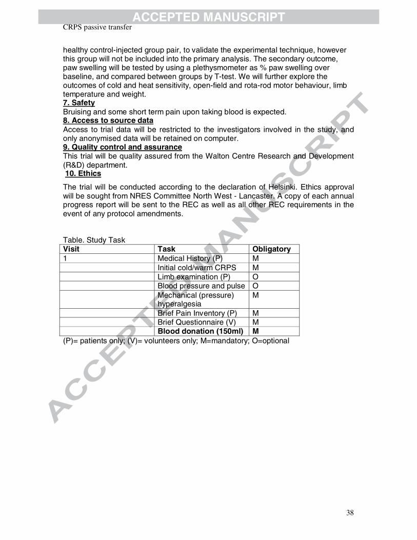
The trial will be conducted according to the declaration of Helsinki. Ethics approval will be sought from NRES Committee North West - Lancaster. A copy of each annual progress report will be sent to the REC as well as all other REC requirements in the event of any protocol amendments. Table. Study Task Visit Task Obligatory 1 Medical History (P) M Initial cold/warm CRPS M Limb examination (P) O Blood pressure and pulse O Mechanical (pressure)
hyperalgesia M
Brief Pain Inventory (P) M Brief Questionnaire (V) M Blood donation (150ml) M (P)= patients only; (V)= volunteers only; M=mandatory; O=optional

CRPS passive transfer
39
-2 -1 0 1 2 3 4 5 6 7 8 9 10 11 12-50
-45
-40
-35
-30
-25
-20
-15
-10
-5
0
days after injection
Cha
nge
of m
echa
nono
cice
ptiv
e tr
esho
ld (%
)
CRPSHealthy controlSaline
Figure. Mechanial hyperalgesia in a passive transfer-trauma model for CRPS. Shown are the combined results from two experiments. Serum-IgG (patient or healthy control) or an equal volume of normal saline was injected intraperitoneally into groups of 6-7 animals on days -1 and 0 in the mornings. A skin-muscle incision was performed under general anaesthesia on the evening of day 0. Reinjection of serum-IgG was on days 5 and 6. The error bars show the standard error of the mean. Hyperalgesia was measured with an electronic dynamic plantar anaesthesiometer (DPA), and the result expressed as percentage change from baseline. The difference between groups was significant on day 7 (ANOVA, p<0.0001), equally the difference between CRPS and healthy control groups was also significant (ANOVA post-test, p<0.05). On day 7, CRPS-IgG induced a relative enhancement of mechanical allodynia of 75% when compared to Control-IgG. Since 6 tests were performed overall (swelling/weight/incapacitance/open field/rotarod/temperature), at 7 time points, the day 7 results are not significant, following correction for multiple tests. In separate experiments, flowthrough preparations (serum without IgG fraction) were also tested, but did not result in any significant changes from saline (not shown).
CRPS passive transfer
40
RESULTS Appendix Figure 2
-2 -1 0 1 2 3 4 5 6 7 8-60
-50
-40
-30
-20
-10
0
Saline HealthyCRPS
***
+
++
#
Days after the injury
Ch
ang
e o
f m
ech
ano
no
cice
pti
ve
thre
sho
ld (
%)
###
###
###
Saline Healthy CRPS-40
-30
-20
-10
0
Dif
fere
nce
bet
wee
n m
ech
ano
-n
oci
cep
tive
th
resh
old
s o
n D
ays
7 an
d b
asel
ine
(%)
Healthy CRPS-10
-5
0
5
10
15
**
Dif
fere
nce
bet
wee
n m
ech
ano
-n
oci
cep
tive
th
resh
old
s o
n D
ays
3 an
d 7
(%
)
-2 -1 0 1 2 3 4 5 6 7 80
10
20
30
40
Saline CRPSHealthy
***###
# ##
+
*
Days after the injury
Paw
oed
ema
(%)
###
Saline Healthy CRPS0
10
20
30
40
Dif
fere
nce
in p
aw o
edem
a b
etw
een
Day
2 a
nd
bas
elin
e (%
)
Saline Healthy CRPS Saline Healthy CRPS0
5
10
15
20
25
30
35
40
intact injured
SP
##
***+++###
#
Co
nce
ntr
atio
n o
f S
ub
stan
ce P
(fm
ol/m
g)
A B
C D
EF
Appendix Figure 2. Paw- mechanosensitivity, swelling, and SP concentration in 5/6 CRPS- and healthy control preparations, excluding patient 1. Further details are given in the main text, and in the legends to figure 2, 3 and 5.
Top Related
Copyright © 2022 FDOKUMEN

