Year Book 2019/2020 - Malaysian Society of Anaesthesiologists
72
-
Upload
khangminh22 -
Category
Documents
-
view
1 -
download
0
Transcript of Year Book 2019/2020 - Malaysian Society of Anaesthesiologists

MALAYSIAN SOCIETY OFANAESTHESIOLOGISTS
Year Book 2019/2020
K E S E LAMATAN D A L A M B I US
Published byMalaysian Society of AnaesthesiologistsUnit 1.6, Level 1, Enterprise 3BTechnology Park Malaysia, Jalan Innovasi 1Bukit Jalil, 57000 Kuala Lumpur, Wilayah PersekutuanTel: (603) 8996 0700, 8996 1700, 8996 2700Fax: (603) 8996 4700Email : [email protected]
Copyright © 2020 Malaysian Society of Anaesthesiologists
All rights reserved. No part of this book may be reproduced in any form or by any meanswithout prior permission from the Publisher.
Pusat Kebangsaan ISBN/ISSN MalaysiaISSN 2462-1307


1
CONTENTS
2 Foreword From The President Of The Malaysian Society Of Anaesthesiologists
3 Preface From The Editors
4 Acknowledgements - Reviewers
5 Airway Management In COVID-19: A Clinically Applicable Narrative Review Asmah Zainudin, Wan Fadzlina Wan Muhd Shukeri
10 Severe Dengue With Hemophagocytic Lymphohistiocytosis: An Intensive Care Perspective Mohd Nazri Ali, Nik Azman Nik Adib
15 Anaesthesia For Deep Brain Stimulation Surgery Mohd Hasyizan Hassan, Laila Ab Mukmin, Abdul Rahman Izaini Ghani
24 Depth Of Anaesthesia Monitoring: Current Issues For Future Development Abdul Karim Bin Othman
31 Setting-Up An ERAS®-Based Bariatric Surgery Centre: Our Experience Laila Ab Mukmin, Sanihah Che Omar, Wan Fadzlina Wan Muhd Shukeri
39 Rapid, Clean And Soft Intravenous Anaesthetics - Are We There Yet? Mohd Fahmi Bin Lukman
45 Post-Operative Hyperalgesia And The Role Of Ketamine Kamaruddin Ibrahim
50 What’s New In The Approach To The Paediatric Difficult Airway? Sivaraj Chandran
56 The Greying Of Malaysians: Implications For Anaesthesiologists Chong Soon Eu, Rhendra Hardy Mohamad Zaini
63 Perioperative Management Of Parturient With Congenital Heart Disease Nazuha Mohd Najid

2
It is with great pride and pleasure that I write this Foreword for the MSA Year Book 2019/2020.
Congratulations to the team especially to the Editors, Associate Professor Dr W Mohd Nazaruddin W Hassan and Dr Wan Fadzlina Wan Muhd Shukeri, from Universiti Sains Malaysia, who have worked really hard at crafting this year’s scintillating Year Book.
The theme for this year is “Embrace the Challenge” taking into consideration the huge challenges anaesthesiologists are facing in looking after patients undergoing complex surgery and who are critically ill. It is with significant coincidence that the year 2020 is the year where anaesthesiologists are pushed to the forefront in the fight against COVID-19. This is due to our skill set which involves resuscitation and critical care, that compels us to be ever ready to tackle the pandemic. The articles featured here will hopefully serve as an up-to-date guide in enhancing our knowledge in anaesthesiology and critical care.
I would like to thank the Editors, contributing authors, reviewers and all those who are involved in making the Year Book a success.
I hope this Year Book will be a useful guide and good reading particularly for the MSA members. This will also serve to highlight the academic contributions of MSA members in the area of anaesthesiology and critical care.
Enjoy the read and stay safe and healthy.
Professor Dr Marzida MansorPresidentMalaysian Society of Anaesthesiologists
Foreword

3
We would like to thank the Malaysian Society of Anaesthesiologists for the opportunity given to us as Editors for this 2019/2020 Year Book. It has been a rewarding experience in our professional life. Our sincere thanks to all the authors and the reviewers who helped make this Year Book successful. Thank you also to the readers. We appreciate your continuing attention and support.
This year’s issue contains ten articles that address major issues across different sub-specialities within our discipline following the theme “Embrace the Challenge”. The year 2020 marks a major change in healthcare after the declaration of COVID-19 pandemic, which is regarded as the “greatest challenge of our age”. In the very First Chapter of this Year Book, the authors highlight some critical concepts on airway management in COVID-19, one of our chief tasks as anaesthesiologists in fighting this pandemic. While we take pride in our efforts in controlling the COVID-19 pandemic, dengue is still endemic in some parts of our country. In Chapter Two, the authors perform a literature review on an important complication of severe dengue from an intensive care perspective.
Deep brain stimulation surgery is emerging as one of the treatment modalities for movement disorders such as Parkinson disease. The anaesthetic management of this surgery is presented in a clinically applicable review by authors in Chapter Three. Meanwhile, in this modern era of anaesthesia, a reliable depth of anaesthesia monitoring is keenly sought. The current issues and controversies in this area are reviewed by the authors in Chapter Four. Among today’s public health challenge is obesity, of which the anaesthesiologists too are impacted. In Chapter Five, the authors share their anaesthetic experience in setting up and running an ERAS®-based bariatric surgery centre. As the practice of anaesthesia continues to evolve, so is the search for the ideal anaesthetic agent. The author in Chapter Six performs a narrative review on some of the newly available anaesthetic agents.
Despite latest advances, recent surveys do not show any major improvement in the post-operative pain management. Chapter Seven is therefore dedicated to the issue of post-operative hyperalgesia, where the author features the important role of ketamine. In the meantime, paediatric difficult airway remains one of the most challenging clinical situations faced by anaesthesiologists. In Chapter Eight, the author presents a review of the new concepts and evidence in the paediatric difficult airway management. In Chapter Nine, the authors address the issues related to population ageing, in which the latest update with regards to the optimal anaesthetic care of the elderly presenting for surgery is reviewed. Finally, the author in Chapter Ten presents a case scenario, followed by a discussion, of the perioperative management of parturient with congenital heart disease. Pleasant reading!
Associate Professor Dr W Mohd Nazaruddin W HassanDr Wan Fadzlina Wan Muhd Shukeri EditorsMSA Year Book 2019/2020
Preface

4
Acknowledgements - Reviewers
This Year Book would not have been possible without the contributions from the following reviewers:
Professor Dr Karis MisiranProfessor and Senior Consultant AnaesthesiologistDepartment of Anaesthesiology and Intensive CareFaculty of MedicineUniversiti Teknologi MARASelangorMalaysia
Dr Omar Sulaiman Consultant AnaesthesiologistDepartment of Anaesthesiology and Intensive CareHospital Sultanah Aminah Johor Bahru JohorMalaysia
Dr Vanitha A/P M Sivanaser Consultant AnaesthesiologistDepartment of Anaesthesiology and Intensive CareHospital Kuala LumpurKuala LumpurMalaysia
Dr Muhammad Zihni AbdullahConsultant Intensivist and AnaesthesiologistDepartment of Anaesthesiology and Intensive CareHospital Tengku Ampuan AfzanKuantan, PahangMalaysia
Associate Professor Dr Azarinah IzahamAssociate Professor and Consultant AnaesthesiologistDepartment of Anaesthesiology and Intensive CareFaculty of MedicineUniversiti Kebangsaan MalaysiaKuala LumpurMalaysia
Associate Professor Dr Abdul Hadi MohamedAssociate Professor, Consultant Anaesthesiologist and Pain SpecialistDepartment of Anaesthesiology and Critical CareKuliyyah of MedicineInternational Islamic University MalaysiaPahangMalaysia
Associate Professor Dr Azrina Md RalibAssociate Professor and Consultant AnaesthesiologistDepartment of Anaesthesiology and Critical CareKuliyyah of MedicineInternational Islamic University MalaysiaPahangMalaysia
Professor Dr Ina Ismiarti ShariffudinProfessor and Consultant AnaesthesiologistDepartment of AnaesthesiologyFaculty of MedicineUniversity of MalayaKuala LumpurMalaysia
Datin Dr Siti Nidzwani Mohd MahdiSenior Lecturer and Consultant AnaesthesiologistDepartment of Anaesthesiology and Intensive CareFaculty of MedicineUniversiti Kebangsaan Malaysia Kuala LumpurMalaysia

MALAYSIAN SOCIETY OF ANAESTHESIOLOGISTS
5
INTRODUCTION
Anaesthesiologists worldwide have been called to the front lines in fighting the COVID-19 pandemic. One of our chief tasks is to perform the intubation. However, intubation, and the steps leading up to and following it, is one of the highest-risk moments for COVID-19 transmission to the airway providers.1 This article is written to provide a review of the emerging best practices for airway management in suspected or confirmed COVID-19 patients, based on our experience and current evidence from the literature.
DROPLET OR AIRBORNE TRANSMISSION?
One of the key concerns with COVID-19 is that the modality of its transmission remains uncertain. As a revision, droplet transmission is caused by viral particles within small drops of bodily fluids. Airborne transmission, on the other hand, is through particles small enough to be borne on air currents and can spread much further in closed spaces. Polymerase chain reaction testing of COVID-19 patients’ rooms, even those who had only mild respiratory symptoms, showed contamination in fan vents on top of gross contamination of surfaces like shoes, handles, and toilet seats prior to cleaning.2 As such, the possibility that COVID-19 transmission is airborne cannot be ruled out.3 Many of the current recommendations are based on the fact that droplet precautions will not be enough to protect against the infection transmission during airway management of COVID-19 patients.4,5
MINIMIZING AEROSOLIZATION
Several commonly performed medical procedures are likely to aerosolize patient sputum and thus increase the risk of exposing providers to COVID-19
transmission. Intubation is one procedure that should be considered at a time when production of airborne particles is a certainty.1 As such, limiting the components of the intubation that can send aerosolized virus into the room should be a priority. This will be discussed further in the next few sections of this article.
Other than intubation, non-invasive ventilation (NIV), high-flow nasal cannula (HFNC), suctioning and bag-valve mask ventilation (BVM) all have been linked to aerosolization and infection transmission in COVID-19.1 Therefore, their use should be implemented with discretion. The use of NIV and HFNC for suspected or confirmed COVID-19 patients should be minimized and limited to negative pressure rooms (NPR) if required.4 For this same reason, some literature recommends that all patients requiring oxygen flows greater than 6 L/min be cared for in NPR.1 Despite concerns for infection transmission, NIV may be required to achieve adequate preoxygenation for safe intubation of a severely hypoxemic patient. If used, the providers should make every attempt to minimize air leaks by ensuring a tight seal and the benefits of NIV use must always be weighed against the risks of infection transmission, even in NPR.4
PERSONAL PROTECTIVE EQUIPMENT
Current recommendations advocate the use of personal protective equipment (PPE) which is protective to airborne transmission prior to airway management in COVID-19 patients.4,5 Airborne PPE for COVID-19 consists of eye protection, a fit-tested respirator such as an N95 mask, a fluid-resistant gown, and gloves. Intubation providers may consider double gloving such that one layer of gloves may be discarded after securing the
Airway Management In COVID-19: A Clinically Applicable Narrative Review
Asmah Zainudin1, Wan Fadzlina Wan Muhd Shukeri2
1 Consultant Intensivist and Anaesthesiologist, Department of Anaesthesiology and Intensive Care, Hospital Sultanah Bahiyah, Alor Setar, Kedah, Malaysia2 Senior Lecturer and Consultant Anaesthesiologist, Department of Anaesthesiology and Intensive Care, School of Medical Sciences, Universiti Sains Malaysia, Kubang Kerian, Kelantan, Malaysia

YEAR BOOK 2019/2020
6
airway, before handling any other equipment. A powered air-purifying respirator (PAPR) provides high calibre protection and may be worn if available (Figure 1).
stethoscope. In addition, they are less expensive and more readily available. N95 masks do not prevent contamination of face and neck and can be rendered ineffective by poor fit.6,7 Decontamination of face and neck should therefore be considered after airway management with an N95 mask.
PAPRs offer some advantages when compared to N95 masks. The advantages include highest level of protection from aerosolized particles,6 approval as an alternative when fit test of N95 respirator has failed or difficult to perform, continuous usage and reusability after proper cleaning. The disadvantages include requiring connection to a power source such as a battery, impaired communication due to the noise of positive airflow and filter, inability to use a stethoscope, and the risk of contamination for anyone disposing of or re-processing the PAPR filter.
Should We Use the “Aerosol Box” for Intubation?
The high demand and limited supply for PPE has led to multiple innovations in battling the COVID-19 pandemic. One such innovation is the Aerosol Box, a device designed as a protection of the providers from patients’ droplets or aerosols spillage during procedures such as intubation. The original idea was from Dr Lai Hsien-Yung, an anaesthesiologist from Taiwan.8 However, review of the literature reveals no retrievable evidence on its efficacy and safety at the time of this writing.
However, in-situ simulation studies showed longer intubation time, reduced first-pass intubation and possibility of increased exposure to aerosols with the application of this box.9,10 Restricted hand movements for procedure manoeuvres required trained personnel and may cause body discomfort. Care should be taken of false sense of security from infection transmission with the usage of this box. Stronger evidences are required to ensure the effectiveness of the Aerosol Box as a protective device for airway providers against COVID-19 transmission.
N95 or PAPR?
When comparing the N95 mask to a PAPR for protection of the providers performing airway management, the pros and cons of each PPE must be considered. N95 masks filter approximately 95% of aerosolized particles (< 5 μm) and droplets (5-50 μm), are more readily available and faster to don, do not require a power source, and allow use of a
Figure 1: The author (left) donning the personal protective equipment with a powered air-purifying respirator

MALAYSIAN SOCIETY OF ANAESTHESIOLOGISTS
7
AVOIDANCE OF ‘CRASH’ INTUBATIONS
Given the increased risk of exposure to providers, all efforts should be made to avoid emergently intubating COVID-19 patients. The providers must consider elective intubation for proactive management of Acute Respiratory Distress Syndrome (ARDS) and viral containment in any patient with increasing oxygen requirement above 6 L/min, worsening PaO2/FiO2 ratio, or increased work of breathing or respiratory rate or for whom the need for NIV or HFNC is being considered.11 Whether intubation is appropriate in patients meeting the above decompensating criteria must be a case-by-case decision; however, it is imperative that intubation not be a consideration reserved for those in extremis.
PERI-INTUBATION PRECAUTIONS IN COVID-19
Pre-Intubation
The plan for airway management should be conducted in NPR with full airborne precautions. In preparation for intubation, the patient must have intravenous (IV) access and, at minimum, the basic physiologic monitors recommended by the American Society of Anaesthesiologists. The airway providers will need airway equipment readily available including a working suction, laryngeal mask airways, gum elastic bougies, video laryngoscopy (VL), waveform capnography, and a ventilator at bedside. All necessary equipment and medications should be prepared prior to entry into the room so as to minimize the duration of possible exposure. The team should ensure that a high efficiency particulate air filter is placed on the ventilator circuit directly at the site of connection with the endotracheal tube (ETT) prior to use. Based on patient comorbidities and potential haemodynamic instability during the intubation process, appropriate vasopressors should be available and in line prior to intubation. It is also important to designate roles, for example, one provider is to manage airway, and another is to administer drugs.
During Intubation
High-quality preoxygenation with 100% inspired oxygen for 5 to 10 minutes is important to optimize patients prior to airway management. BVM should be avoided in COVID-19 patients when possible. Prior studies have found that BVM before intubation was associated with an increased risk of COVID-19 transmission.12 If BVM is required, low tidal volumes should be used, and all precautions should be taken to avoid leaks. Some experts recommend placing an LMA immediately after induction, if post-induction ventilation is required, to lower the risk of aerosolization.
The choice of neuromuscular blocking agents for these patients remains a topic of debate. Cheung et al. recommend a modified rapid sequence intubation approach using a high dose of nondepolarizing agent, such as rocuronium rather than succinylcholine.13 The longer duration of action of rocuronium prevents aerosolization via patient coughing in the event of multiple attempts at intubation. An increased dose of rocuronium (>1.2 mg/kg) reduces time to drug onset which decreases the risk of patient coughing during intubation. Use of IV lignocaine (1.5 mg/kg) and avoidance of fentanyl are additional strategies which may help to prevent coughing.4
Routine use of VL has been suggested to provide additional distance between the intubating provider and the airway. If this is unsuccessful on two attempts, the threshold to proceed to surgical airway should be lowered.4 The time to cuff inflation and connection to ventilator circuit should be minimized. Of utmost importance, the most experienced provider should perform the intubation.
Post-Intubation
Following intubation, any equipment that was in contact with the airway should be immediately disposed of or contained within a plastic bag for decontamination. Any contaminated PPE should be removed as soon as possible, and outer gloves replaced.
YEAR BOOK 2019/2020
8
treatments post-extubation should be ideally avoided or at least minimized in favour of metered dose inhalers, if necessary. It is also important to limit coughing during the extubation, and the use of lignocaine or other adjuncts may be considered. Circuit flows should be discontinued prior to extubation. Once extubated, the ETT and other airway supplies should be immediately disposed in a sealed container. Lastly, the time between extubation and covering of patients’ airway with a mask should be minimized.
CONCLUSION
As the pandemic continues to spread, it is our hope that dissemination of evidence-based management recommendations is occurring at an even greater speed. The most recent recommendations include close monitoring of respiratory status for early signs of failure, cautious use of NIV and HFNC and consideration of early intubation, use of VL, and minimizing coughing and viral aerosolization during induction with medications such as lidocaine and high dose rocuronium. We also emphasize efforts to reduce infectious risk such as by limiting the providers caring for COVID-19 patients to an experienced group of clinicians and ensuring proper availability, donning, and doffing of airborne precaution PPE.
Data on the ideal ventilation parameters specific to COVID-19 patients is lacking. Up to 67% of critically ill COVID-19 patients may develop ARDS, therefore, the clinicians should consider lung ventilation strategies that have been established for the management of ARDS.14 It is also important to maintain appropriate levels of sedation and, if necessary, paralysis to limit patient-ventilator dyssynchrony.
Extubation
Extubation poses another significant risk of COVID-19 transmission.1 As such, it should be performed with caution. It is recommended to designate roles and safely limit the number of providers present for extubation. A detailed post-extubation oxygen support plan should be established, considering that NIV and HFNC pose transmission risk. The benefits of their use post-extubation should be weighed against the risk of infection transmission. In such scenarios, a tracheostomy rather than a trial extubation should be considered, especially if the patient has the history of intubation difficulty.4 In order to ensure a successful extubation, providers should consider a prolonged spontaneous breathing trial.
Routine checking of cuff leak prior to extubation should be performed. Aerosolizing nebulizer
References
1. Cheung JC, Ho LT, Cheng JV, Cham EYK, Lam KN. Staff safety during emergency airway management for COVID-19 in Hong Kong. Lancet Respir Med 2020;8:e19
2. Ong SWX, Tan YK, Chia PY, Lee TH, Ng OT, Wong MSY, et al. Air, surface environmental, and personal protective equipment contamination by Severe Acute Respiratory Syndrome Coronavirus 2 (SARS-CoV-2) from a symptomatic patient. JAMA 2020;323:1610-2
3. European Centre for Disease Prevention and Control. Infection Prevention and Control for COVID-19 in Healthcare Settings - March 2020. Stockholm: ECDC; 2020
4. Sullivan EH, Gibson LE, Berra L, Marvin GC, Edward AB. In-hospital airway management of COVID-19 patients. Crit Care 2020;24:292
5. Sorbello M, El-Boghdadly K, Di Giacinto I, Cataldo R, Esposito C, Falcetta S, et al. The Italian coronavirus disease 2019 outbreak: recommendations from clinical practice. Anaesthesia 2020;75:724-32
6. Roberts V. To PAPR or not to PAPR? Can J Respir Ther 2014;50:87-90
7. Daugherty EL. Health care worker protection in mass casualty respiratory failure: infection control, decontamination, and personal protective equipment. Respir Care 2008;53:201-12

MALAYSIAN SOCIETY OF ANAESTHESIOLOGISTS
9
8. Lai HY. 2020. Aerosol Box.[https://sites.google.com/view/aerosolbox/home?authuser=0] Accessed September 1, 2020
9. Begley JL, Lavery KE, Nickson CP, Brewster DJ. The aerosol box for intubation in coronavirus disease 2019 patients: an in-situ simulation crossover study. Anaesthesia 2020;75:1014-21
10. Simpson JP, Wong DN, Verco L, Carter R, Dzidowski M, Chan PY. Measurement of airborne particle exposure during simulated tracheal intubation using various proposed aerosol containment devices during the COVID-19 pandemic. Anaesthesia 2020 doi:10.1111/anae.15188
11. Zucco L, Levy N, Ketchandji D, Aziz M, Ramachandran SK. Perioperative considerations for the 2019 novel coronavirus (COVID-19). Anesthesia Patient Safety Foundation 2020
12. Tran K, Cimon K, Severn M, Pessoa-Silva CL, Conly J. Aerosol generating procedures and risk of transmission of acute respiratory infections to healthcare workers: a systematic review. PLoS One 2012;7:e35797
13. Cheung JC, Ho LT, Cheng JV, Cham EYK, Lam KN. Staff safety during emergency airway management for COVID-19 in Hong Kong. Lancet Respir Med 2020 doi: https://doi.org/10.1016/s2213-2600(20)30084-9
14. Yang X, Yu Y, Xu J. Clinical course and outcomes of critically ill patients with SARS-CoV-2 pneumonia in Wuhan, China: a single-centered, retrospective, observational study. Lancet Respir Med 2020 doi: https://doi.org/10.1016/S2213-2600(20)30079-5

YEAR BOOK 2019/2020
10
INTRODUCTION
Dengue fever is a mosquito-borne viral infectious disease that is transmitted by female mosquitoes mainly of the species Aedes aegypti and, to a lesser extent, Aedes albopictus. Mosquito bites therefore contribute significantly to the morbidity and mortality related to dengue infection. Worldwide, the incidence of dengue infection has grown dramatically, with yearly infections now estimated at 100 to 400 million.1,2 The American region alone has reported 3.1 million cases, with more than 25,000 classified as severe dengue. High numbers of cases have also been reported in Asian countries;2 Bangladesh (101,000), Malaysia (131,000), the Philippines (420,000) and Vietnam (320,000).
The global increase in dengue incidence is also evident in Malaysia, which has seen a continued and significant increase since the year 2000. Most of the dengue cases in Malaysia (70-80%) were reported in urban areas, where factors such as high population density, rapid ‘urbanisation’ and poor drainage systems favour dengue transmission. However, despite the increase in dengue cases over the years, the overall case fatality rate has been reduced from 0.6% in 2000 to 0.2% in 2014 (the national target is less than 0.2%).3 Nevertheless, the case fatality rate for severe dengue remains significantly high, especially in cases that progress to dengue with complications.
One of the known complications of dengue is hemophagocytic lymphohistiocytosis (HLH), which has a high reported mortality rate ranging between 14.6%8 and 30%15. A recent local study showed that 12% of patients with severe dengue had HLH and a mortality rate of 43%.6 In this article, we will review the current classification of dengue and its clinical features, with particular attention to HLH, in terms of its diagnosis and management.
DENGUE CLASSIFICATION
Dengue infection was previously classified based on the 1997 World Health Organization (WHO) Guidelines as Dengue Fever (DF), Dengue Haemorrhagic Fever (DHF) and Dengue Shock Syndrome (DSS). However, several limitations of the earlier classification led to publication of a revised WHO Classification in 2009 (Figure 1). The revised guidelines now recommend a more practical and clinical approach that addresses the severity of the disease, rather than attempting to separate the disease into distinct types.
Severe Dengue With Hemophagocytic Lymphohistiocytosis: An Intensive Care Perspective
Mohd Nazri Ali1, Nik Azman Nik Adib2
1 Consultant Anaesthesiologist and Intensivist, Department of Anaesthesiology and Intensive Care, Hospital Raja Perempuan Zainab II, Kota Bharu, Kelantan, Malaysia2 Consultant Anaesthesiologist and Intensivist, Department of Anaesthesiology and Intensive Care, Hospital Sultanah Nur Zahirah, Kuala Terengganu, Terengganu, Malaysia
CLINICAL FEATURES
A classical dengue infection will progress in three phases: the febrile, critical and recovery phases. During the febrile phase, dengue infection should
CRITERIA FOR DENGUE + WARNING SIGNS CRITERIA FOR SEVERE DENGUE
Probable dengue- Live in/travel to dengue endemic/hotspot/ outbreak area- Fever and 2 of the following criteria: • Nausea, vomiting • Rash • Aches and pains • Leucopaenia • Any warning sign
Warning signs*• Abdominal pain or tenderness• Persistent vomiting (>3 times per day)• Persistent diarrhoea (>3 times per day)• Clinical fluid accumulation• Mucosalbleed• Lethargy, confusion, restlessness• Tender liver• Laboratory: increase in HCT concurrent with rapid decrease in platelet count
*(requiring strict observation & medical intervention)
Severe plasma leakage leading to:• Shock (DSS)• Fluid accumulation with respiratory distress
Severe bleedingAs eveluated by clinician
Severe organ involvementLiver: AST or ALT > 1000CNS: Impaired consciousnessHeart and other organsLaboratory-confirmed
dengue(important when no sign of plasma leakage)
DENGUE + WARNING SIGNS SEVERE DENGUE
1. Severe plasma leakage2. Severe haemorrhage3. Severe organ impairment
with warning signs
without
Adapted: World Health Organization. Dengue Guidelines for Diagnosis, Treatment, Prevention and Control - New Edition 2009. WHO: Geneva; 2009
Figure 1: 2009 WHO Dengue Classification and Level of Severity

MALAYSIAN SOCIETY OF ANAESTHESIOLOGISTS
11
Barr virus, Dengue virus and possibly SARS-CoV-2 (COVID-19 pathogen). Although the precise mechanism remains unclear, a commonly accepted postulation is that the virus infection stimulates an abnormal immune reaction, resulting in an excessive activation of CD4-helper cells, a proliferation of macrophages and a large secretion of cytokines.
In dengue infection, HLH is an uncommon but potentially fatal complication, with a high mortality rate. It is characterised by hyper-inflammation, uncontrolled proliferation of activated lymphocytes, prolonged fever, pancytopaenia, jaundice and organomegaly, such as hepatomegaly and/or splenomegaly.
Dengue with HLH is frequently under-diagnosed, which eventually leads to delayed initiation of appropriate therapy and results in increased morbidity and mortality. Failure to diagnose HLH in dengue patients mainly occurs due to the lack of specific diagnostic criteria and the similarity of other complications common in dengue infection, such as sepsis and multiorgan failure. The demonstration of haemophagocytosis in bone marrow is diagnostic but non-specific. However, the increased risk of bleeding when performing bone marrow biopsy limits its use in patients with dengue.
The pooled case fatality rate for dengue infection associated with HLH is 14.6-30%. An early diagnosis of HLH is critical since the mortality and morbidity increase exponentially with a delay in diagnosis8 and hence treatment. Dengue with possible HLH should be suspected if a patient remains febrile and ill with cytopaenia, transaminitis and multiorgan failure beyond the classical critical phase.
Although specific diagnostic tools are lacking, the 2004 diagnostic guidelines for HLH, as proposed by the Histiocyte Society, are still widely used in clinical practice. However, these guidelines are poorly validated for acquired forms of HLH and for HLH in the adult population, because the original criteria were developed for the primary forms of HLH and in the paediatric age group. A more recent diagnostic tool, the H-Score, is a set of weighted
be suspected when a patient has a high-grade fever that is accompanied by symptoms such as severe headache, retro-orbital pain, generalised body ache, nausea, and vomiting and petaechial rash. A patient typically enters what is called the critical phase at about three to seven days after onset of the illness; this phase lasts about 24 to 48 hours. During this period, the temperature usually drops (below 38�C/100�F), the white cell count shows a decreasing trend and thrombocytopaenia and evidence of plasma leakage with haemoconcentration appears. During this critical phase, severe dengue may develop and potentially result in more serious complications, such as liver failure, encephalopathy, carditis with cardiac failure and acute kidney injury,7 with significant morbidity and mortality. In uncomplicated dengue cases, the patient will enter recovery phase, usually 48 hours after critical phase, when plasma leakage stops, the white cell count and platelet level slowly normalise and general well-being improves.
Dengue-Haemophagocytosis Lymphohistiocytosis
Haemophagocytosis is a pathologic term indicating inappropriate stimulation of macrophages, engulfing (phagocytosis) erythrocytes, leukocytes, platelets, and their precursor cells, simultaneously with the production of high amounts of pro-inflammatory cytokines,9 described as a cytokine storm. These are important features in patients with Hemophagocytic Syndrome or, more specifically, Haemophagocytic Lymphohistiocytosis (HLH).
HLH occurs as two distinct types: familial (primary), which is more common in the paediatric age group, or acquired (secondary), which is more common in the adult population.18 Familial HLH, as the name implies, is an inherited form of the disease, whereas acquired HLH occurs after immunologic activation19 in response to systemic infection, immunodeficiency syndromes, malignancy or autoimmune diseases.11 Nevertheless, both forms of HLH are characterised by very similar haematopathologic findings.
Almost one-third of the cases of acquired HLH are induced by infections by viruses such as Epstein

YEAR BOOK 2019/2020
12
criteria that allows a more effective estimation of an individual’s risk of having HLH, especially in the adult population. However, a performance study (mixed primary and secondary HLH) showed
that the H-Score is more appropriate for children. In adults, the H-Score performed better when determined at presentation of the illness.16
Supportive Treatment
Ideally, the patient should be admitted to the intensive care unit, as the patient will usually need close organ monitoring and possibly early organ support, especially cardiovascular and respiratory. Fluid resuscitation is of utmost importance, especially when dealing with severe dengue that is accompanied by significant plasma leakage. Treatment with 0.9% saline is recommended as the fluid of choice; alternatively, a balanced crystalloid fluid, like sterofundin, can also be used for resuscitation and maintenance fluid in severe dengue with plasma leakage or HLH.3 The use of any of the colloids has no clear advantage over crystalloids in terms of the overall outcome and mortality. However, a colloid may be preferred as the fluid of choice in patients with intractable shock and whose haemodynamics remain unresponsive after crystalloid administration. Prolonged use of a colloid as the sole maintenance fluid should
All four serotypes of Dengue virus (DENV1-4) may be associated with HLH, although more cases of HLH were reported to be associated with DENV 1 and DENV 4 than with DENV 2 and DENV 3.8 Fever, splenomegaly, hepatomegaly, anaemia, thrombocytopaenia, coagulopathy and serum ferritin >500μg/L are common signs and symptoms for dengue with HLH.8,19 Therefore, in cases of severe dengue that are unresponsive to conventional therapy, elevated ferritin levels should be sought for early diagnosis of dengue with HLH.10 A raised level of lactate dehydrogenase (LDH) will also increase the possibility of the presence of severe dengue with HLH.17
MANAGEMENT
The goal of therapy for management of the dengue patient with HLH is to suppress life threatening inflammatory processes using supportive and specific measures.19,20
Table I: Diagnosing HLH
HLH-2004 Guidelines - Presence of molecular diagnosis consistent with HLH or >5 out of 8 criteria. H-Score - >250 = Probability HLH 99%, <90 = Probability HLH <1%
Parameter
Fever (�C)SplenomegalyOrganomegaly
Cytopenia
Ferritin (ng/mL)Triglycerides (mmol/L)Fibrinogen (g/L)Hemophagocytosis in bone marrowAspartate aminotransferase (IU/L)Known underlying immunosuppression
Adapted HLH-2004 Guidelines
0 (<38.5) or 1 (>38.5)0 (no) or 1 (yes)
0 (one lineage) or 1 (two or three lineages)b
0 (<500) or 1 (>500)0 (<3) or 1 (>3)0 (>1.5) or 1 (<1.5)d
0 (no) or 1 (yes)
H-Score
0 (<38.4), 33 (38.4-39.4), or 49 (>39.4)
0 (no), 23 (hepatomegaly or splenomegaly), or 38 (hepatomegaly and splenomegaly)0 (one lineage), 24 (two lineages), or 34 (three lineages)c
0 (<2,000), 35 (2,000-6,000) or 50 (>6,000)0 (<1.5), 44 (1.5-4), or 64 (>4)0 (>2.5) or 30 (<2.5)0 (no) or 35 (yes)0 (<30) or 19 (>30)0 (no) or 18 (yes)

MALAYSIAN SOCIETY OF ANAESTHESIOLOGISTS
13
be avoided. Hydroxyethyl starch (HES) solution is contraindicated in dengue patients with severe hepatic dysfunction, fluid overload (e.g. pulmonary oedema and congestive cardiac failure) or renal failure and in patients receiving dialysis.3 Albumin is another choice of fluid that can be used during resuscitation, especially if the patient has already received a large amount of crystalloid. Albumin as a resuscitation fluid in cases of DSS has not been studied; however, based on its extensive use in critically ill patients, 4-5% albumin is comparable to crystalloid and may be better in the subgroup of septic patients.
Dengue with HLH is also commonly associated with severe hepatitis, although the pathogenesis of liver involvement during dengue infection is poorly understood. The potential mechanisms of liver injury include direct effects of the virus, immunological injury due to a dysregulated immune response, ischemic injury due to hypotensive episodes and a hepatotoxic effect of medications, such as acetaminophen or herbal remedies.13 Data are currently insufficient regarding the use of IV N-acetylcysteine (NAC) for the treatment of acute liver failure in dengue infection. However, if NAC is used, the suggested regime is as follows:
• 100mg/kg/day as infusion for five days3
OR• 150mg/kg infusion over 15-60 min, followed by 12.5mg/kg/h for 4 h and then 6.25mg/kg/h.3
Specific Treatment
Corticosteroids are potent immune modulators and are used therapeutically for a broad spectrum of diseases, including autoimmune, allergic
and inflammatory diseases. T cell dependent immune dysregulation is suggested as a possible pathophysiology in HLH; therefore, corticosteroids (mainly IV methylprednisolone or dexamethasone) have been used as a treatment for dengue with HLH. However, the evidence from trials using corticosteroids in dengue is inconclusive, and the quality of the evidence is low. The Cochrane Database Systemic Review 2014 concluded that the evidence is insufficient to evaluate the effects of corticosteroids as a treatment for early-stage dengue fever and dengue-related shock.14
Some studies have reported the use of IV immunoglobulin, either alone or with steroid, in HLH. However, these studies have shown conflicting outcomes. Etoposide, which is a chemotherapy medication, has also been used in HLH, although the exact mechanism and its role in hyperinflammation treatment are not well understood.
CONCLUSION
HLH in dengue infection poses both diagnostic and therapeutic difficulties. Lack of specific diagnostic criteria leads to under-diagnosis. However, in patients with persistent fever in conjunction with transaminitis, hyperferritinaemia and cytopaenia, the diagnosis of HLH should be considered. Treating HLH is also very challenging, as no strong evidence yet supports any specific treatment that is significantly associated with better survival rates. Clearly, a need exists for well-designed trials that investigate the therapeutic effects, especially those of steroids and other specific treatments. Early diagnosis, together with a proven specific treatment, will have a significant impact on the outcome of dengue infection with HLH.
References
1. Bhatt S, Gething PW, Brady OJ, et al. The global distribution and burden of dengue. Nature 2013;496:504-7
2. World Health Organization. WHO fact sheet: dengue and severe dengue. Available at https://www.who.int/en/news-room/fact-sheets/detail/dengue-andsevere-dengue. Accessed 19 June 2020

YEAR BOOK 2019/2020
14
3. Clinical Practice Guideline; Management of dengue infection in adult (Third edition 2015)
4. Henter JI, Horne A, Arico M, et al. HLH-2004: diagnostic and therapeutic guidelines for hemophagocytic lymphohistiocytosis. Pediatr Blood Cancer 2007;48:124-31
5. Fardet L, Galicier L, Lambotte O, et al. Development and validation of the HScore, a score for the diagnosis of reactive hemophagocytic syndrome. Arthritis Rheumatol 2014;66:2613-20
6. Foong Kee Kan, Cheng Cheng Tan, Tatiana von Bahr Greenwood, Khairil E. Khalid, Premaa Supramaniam, Ida Hed Myrberg, et al. Dengue Infection Complicated by Hemophagocytic Lymphohistiocytosis: Experiences From 180 Patients With Severe Dengue. CID 2020:70(1 June);2247
7. Osnaya-Romero N, Perez-Guille M-G, Andrade-García S, et al. Neurological complications and death in children with dengue virus infection: report of two cases. J Venomous Anim Toxins Incl Trop Dis 2017;23(1):25
8. Hoang Thi Nam Giang,Keita Banno, Le Huu Nhat Minh, Lam Tuyet Trinh, Le Thai Loc, Asmaa Eltobgy, Luu Lam Thang Tai, et al; Dengue hemophagocytic syndrome: A systematic review and meta-analysis on epidemiology, clinical signs, outcomes, and risk factors. Rev Med Virol 2018;e2005
9. Karras A, Hermine O. Hemophagocytic syndrome. Rev Med Interne 2002;23(9):768-778
10. George MR. Hemophagocytic lymphohistiocytosis: review of etiologies and management. J Blood Med 2014;5:69-86
11. Nair V, Das S, Sharma A, et al. A clinicopathological analysis of 26 patients with infection-associated haemophagocytic lymphohistiocytosis and the importance of bone marrow phagocytosis for the early initiation of immunomodulatory treatment. Postgrad Med J 2013;89(1050):185-192
12. Biswas T. Macrophage activation syndrome in secondary dengue: a rare presentation. Asian J Sci Technol 2017;08(01):4141-4143
13. Trung DT, Thao le TT, Hien TT, et al. Liver involvement associated with dengue infection in adults in Vietnam. Am J Trop Med Hyg 2010 Oct;83(4):774-80
14. Zhang, F.Kramer, C.V., 2014. Corticosteroid for dengue infection. Cochrane Database Syat. Rev, 7,CD003488
15. T Sharp, L Gaul, et al. Fatal Hemophagocytic Lymphohistiocytosis Associated with Locally Acquired Dengue Viral nfection - New Mexico and Texas, 2012. Morbidity and Mortality Weekly Report, Center of Disease Control and Prevention 2014;63:3
16. France D, Bhavna M, Alina F, et al. Performance of H-score for Diagnosis of Hemaphagocytic Lymphohistiocytosis in Adult and Pediatric Patients. Am J Clin Pathol 2016 June; 145:862-870
17. Mehta R, Nandania K, Desai K, Patel A, Shah K .Clinical and Laboratory Features of Severe Dengue Hepatitis (Liver Failure Mimics): Early Predictors of Fatality. J Liver Clin Res 2015;2(3):1020
18. Paul La Rosee, AnnaCarin Horne, Melissa Hines, Tatiana von Bahr Greenwood, Rafal Machowicz, Nancy Berliner, et al. Recommendations for the management of hemophagocytic lymphohistiocytosis in adults. Blood 2019;133(23):2465-2477
19. A.-S. De Koninck, J. Dierick, S. Steyaert, P. Taelman. Hemophagocytic lymphohistiocytosis and dengue infection: rare case report. Acta Clinica Belgica 2014;Vol. 69 No.3:210-213
20. Melissa R George. Hemophagocytic lymphohistiocytosis: review of etiologies and management. Journal of Blood Medicine 2014;5:69-86

MALAYSIAN SOCIETY OF ANAESTHESIOLOGISTS
15
INTRODUCTION
Deep brain stimulation (DBS) surgery is one of the modalities of treatment for patients with Parkinson and movement disorders who fail medical therapy and for those with chronic pain and psychiatric disorders.1-3 A DBS system has three implanted parts i.e. the neurostimulator, the lead and the extension wire. A neurostimulator is a device that creates electric pulse to block the faulty nerve signal causing tremor or abnormal movement whereas a lead is a coated wire with its tip (electrode) embedded into the specific targeted area of the brain.
The therapeutic targets for electrode placement for movement disorders are the subthalamic nucleus (STN), the globus pallidus interna (GPi), and the ventralis intermedius nucleus (VIN) of the thalamus. These structures are deep within the brain and small in size; therefore, locating the treatment area and improving placement accuracy require the use of a stereotactic frame with a coordinated system and intraoperative electrophysiological guidance that uses microelectrode recording (MER) and macrostimulation testing.4
Proper and optimal anaesthetic management is prudent, whilst ensuring the patient’s comfort and analgesia. The first stage of surgery involves insertion of the electrodes into the specified area of the brain, while the second stage includes internalisation of the lead(s) and implantation of the programmable impulse generator. Here, we discuss issues pertaining to the DBS surgery, including the preoperative assessment, brief of surgical process, the intraoperative conduct, postoperative care and safety issues in patient with implanted DBS.
THE PREOPERATIVE ASSESSMENT
Since the patients subjected to DBS surgery are in extreme ages, and approximately a quarter of them are geriatrics (especially those with Parkinson disease), a thorough preoperative assessment is essential.5 Inevitably, physiological changes occur in association with aging, including reduction of the functional reserve of the cardiorespiratory system and deterioration of kidney function. An elderly demonstrates an exaggerated response to CNS depressants due to underlying age-related declines in central nervous system (CNS) function and increased sensitivity to benzodiazepines, anaesthetic agents and opioids. Thus, identification of any related issues and optimisation of the patient comorbidities are required prior to DBS surgery.
A patient with long-standing Parkinson’s disease may develop various complications as a result of the disease progression and drug treatment. The patient is at risk of developing obstructive lung disease and has abnormal control and function of the upper airway, which leads to pharyngeal muscle dysfunction, coughing and obstructive sleep apnoea. Autonomic dysfunction put the patient at risk for cardiac arrhythmia, hypertension or orthostatic hypotension. Uncoordinated muscle movement may lead to dysphagia and a subsequent poor nutritional status. Intraoperative difficulties should be anticipated, since associated speech impairments and confusion will interfere with patient cooperation and comprehension to some extent and the presence of resting tremor with associated rigidity will impede positioning. The common side effects of Parkinson’s medication are listed in Table I.
Anaesthesia For Deep Brain Stimulation Surgery
Mohd Hasyizan Hassan1, Laila Ab Mukmin1, Abdul Rahman Izaini Ghani2
1 Senior Lecturer and Consultant Anaesthesiologist, Department of Anaesthesiology and Intensive Care, School of Medical Sciences, Universiti Sains Malaysia, Kubang Kerian, Kelantan, Malaysia2 Professor and Consultant Neurosurgeon, Department of Neurosciences, School of Medical Sciences, Universiti Sains Malaysia, Kubang Kerian, Kelantan, Malaysia

YEAR BOOK 2019/2020
16
Table I: Various drugs for Parkinson’s disease, its mechanism of action and associated adverse effect and drugs interaction
Action
Dopamine precursor with metabolic inhibitor
MAOI reduce levodopa and dopamine degradation
COMT inhibitors reduce levodopa and dopamine degradation
Dopamine receptor agonist
Antimuscarinic
Abbreviation: MAOI- Monoamine Oxidase Inhibitor, COMT- cathecol-o-metyl transferase, L-dopa- Levodopa
Drugs
Levodopa / carvidopa
Selegiline, rasagiline, safinamide
Entacapone, tolcapone
Pramipexole, ropinirole
Trihexyphenidyl, benztropine
Adverse Effect / Drugs Interaction
Orthostatic hypotension, hypertension, hallucination, nausea and vomiting
Hypertension, orthostatic hypotension, potentiation of L-dopa related side effect, abnormal glucose metabolism; exaggerated response with pethidine
Potentiation of L-dopa related side effect, diarrhoea, orange colour urine, hepatotoxicity
Orthostatic hypotension, hallucination and psychosis, impulse control disorders, peripheral oedema
Dry mouth and eyes, constipation, hallucination, confusion, thickening of saliva and secretion
candidates include those younger age patients with advanced Parkinson's disease with excellent levodopa (LD) response, non-LD-responsive motor symptoms, absence or very mild cognitive impairment and psychiatric symptoms. However, Hanna et al. reported DBS surgery improved elderly patient’s symptomatology and reduction of the Levodopa after surgery.5 DBS surgery is generally contraindicated in the patients with brain atrophy, uncooperative, demented or those with multiple comorbidities that preclude safe surgery.
Regarding premedication, the beta-blockers are best avoided since they reduce or complicate intraoperative tremor testing. By contrast, clonidine can be helpful, as it is less likely to induce cognitive impairment.6 Benzodiazepines can cause over-sedation or the production of a paradoxical agitation in this group of patients; thus, sedative premedication is also best avoided.6 Droperidol must be avoided in patients with Parkinson's disease and dystonia, given the potential for exacerbation of the symptomatology.6 Antiemetics, like metoclopramide, block dopamine receptors which can cause undesirable
A patient with essential tremor may have associated bradycardia or first degree heart block as an effect of beta blocker administration. The dystonic patient is at risk of haemodynamic instability and laryngospasm. The epileptic patient has episodes of recurrent seizures associated with growth and developmental delays and can be on multiple anti-seizure medications which alter drug interactions.
Preoperatively, careful attention and anticipation to surgical related anaesthetic consideration are prudent. The patient is placed in a semi-sitting position with slightly flexed neck intraoperatively. Thus, the anaesthetist needs to assess the underlying neck rigidity and possibility of difficult airway in preoperative assessment. The procedure can also be lengthy and complicated needing careful judgment of the appropriate sedative agents or mode of anaesthesia intraoperatively. Some patients will experience worsening symptoms due to cessation of medication (e.g. drugs for treatment of motor symptoms) to allow the neuro-physician to do clinical testing. Careful patient selection for DBS surgery is mandatory to ensure good outcome. Suitable

MALAYSIAN SOCIETY OF ANAESTHESIOLOGISTS
17
block. However, most recent studies advocate usage of the scalp block in conjunction with the conscious sedation (CS)/monitored anaesthesia care (MAC) especially when dealing with bilateral DBS insertion.9-11 The origin, innervation, surface landmarks and possible complications of each respective nerve are explained in Table II. An additional short-acting local anaesthetic (lignocaine) can be administered to the area of the scalp pin or the stereotactic frame.
Anaesthetic Technique
Several anaesthesia choices are available for DBS surgery. The operation can be performed either by the asleep-awake-asleep (AAA) technique, a monitored anaesthesia care (MAC) technique or even general anaesthesia. The first two techniques require supplementation with a scalp block or LA infiltration. In the AAA technique, the airway interventions should be performed before placement of the rigid stereotactic frame in magnetic resonance imaging (MRI) or computed tomography (CT) unit since intubation is difficult due to the presence of the stereotactic frame. The laryngoscopy view is restricted, and positioning is suboptimal; therefore, the airway should be secured with fiberoptic intubation or a laryngeal mask airway (LMA) if airway manipulation is done after placement of the stereotactic frame. The LMA is easily inserted and removed and better tolerated in lighter planes of anaesthesia. However, the Parkinson’s patient has increased risk of regurgitation and aspiration; hence, careful judgement is required.
Monitored anaesthesia care (MAC) or conscious sedation is an optimum and preferable anaesthesia technique for DBS surgery.8-10 This technique allows the patient to be relatively awake while providing comfort during clinical testing throughout the intraoperative period. The Bispectral index (BIS)9 or cerebral state monitor (CSM) is used to monitor the state of sedation and the end-tidal CO2 is monitored for respiration. All patients received oxygen via a nasal cannula or a face mask, and the nasal airway, LMA and endotracheal tube are reserved as rescue airway devices.
extrapyramidal side effects. Thus, ondansetron is a better first-line choice, as is dexamethasone.7
BRIEF SURGICAL PROCESS
DBS surgery involves a two-stage operation which is insertion of the microelectrodes with a neurostimulator and installation of the pulse generator. The microelectrode is connected to the pulse generator with the extension wire which runs from the scalp, behind the ear, down the neck, and to the chest. The installation of the pulse generator can be done in the same setting or scheduled for subsequent operation approximately one to two weeks after the first surgery. In our centre, both stages are done in one operation. Nevertheless, some centres prefer the second stage operation to be done later since the presence of “microlesion” due to oedema surrounding the electrodes will improve the patient’s symptoms without any stimulation.8 Preferably, the first stage of operation is done under awake craniotomy. The surgery starts with placement of the stereotactic frame and the patient will subsequently be subjected to the MRI (magnetic resonance imaging) or CT (computerised tomography) scanning for anatomical localization and target coordinates for accurate location of the electrodes. The electrode is inserted 10-15mm above the target site and is advanced 0.5-1mm along its trajectory toward the target nuclei while spontaneous neuronal discharges are recorded. The neurophysiologists use the variations in spontaneous firing rates between specific nuclei and movement-related changes. Macrostimulation involves the clinical testing of the patient's motor symptoms versus acceptable side effect thresholds. As the second stage of the surgery does not involve neuro assessment and clinical testing, the operation is performed under general anaesthesia.
INTRAOPERATIVE CONDUCT
The Scalp Block
The area of the scalp can be anaesthetised with infiltration of the local anaesthetic at the stereotactic frame pin and burr hole site or by using the scalp

YEAR BOOK 2019/2020
18
The patient is put on flexion at the lower cervical spine and extension at the atlanto-occipital joint; therefore, the position should be carefully assessed, and support padding inserted to allow venous drainage from the head and neck area and to ensure comfort. The legs should be flexed and supported under the knees to a sitting position. The use of clear plastic drapes will avoid claustrophobia and permit the anaesthesiologist to maintain verbal and eye contact with the patient. All emergency drugs and rescue antiepileptic drugs are prepared beforehand. The most common drugs used in MAC are dexmedetomidine, remifentanil and propofol since they have fast onset and offset and do not
interfere with intraoperative neuromonitoring. The anaesthetic drug effects on microelectrode recording and stimulation are explained in Table III.
Initial sedation is employed with a loading dose of dexmedetomidine of up to 1mg/kg/min for 10 minutes. Sedation is maintained with dexmedetomidine at an infusion rate of 0.2-0.7mg/kg/h. A low dose of dexmedetomidine allows MER whilst providing analgesia, ensuring haemodynamic stability and preserving spontaneous breathing.14-16 A target control infusion (TCI) of remifentanil at up to 1 to 2ng/mL is started to accommodate pain during the scalp block. A TCI using a combination of
Auriculotemporal
Greater occipital
Lesser occipital
Mandibular division of trigeminal nerve
C1-C4 spinal nerve
C2 spinal nerve
Tragus, anterior portion of the ear, posterior portion of the temple
Posterior part of the scalp to vertex
Lateral area of posterior scalp to the ear
15mm ventral to tragus of the ear close to superficial temporal artery
Medial third between the occipital protuberance and mastoid process close to occipital artery
lateral third between the occipital protuberance and mastoid process close to occipital artery
Facial nerve injury, intraarterial injection, intraarticular injection
Intraarterial injection
Nerve Origin Innervation Surface Landmarks Complication
Supraorbital
Supratrochlear
Zygomaticotemporal
Ophthalmic division of trigeminal nerve
Ophthalmic division of trigeminal nerve
Maxillary division of trigeminal nerve
Forehead to lambdoid
Lower part of forehead
Forehead and temporal area
Supraorbital fissure
Medial corner of the orbit and lateral to nasal apex
Midway between supraorbital and auriculotemporal nerve
Direct nerve injection, eyelid and orbital injury, mechanical ptosis12
Table II: The nerve supply for the scalp for scalp block

MALAYSIAN SOCIETY OF ANAESTHESIOLOGISTS
19
propofol and remifentanil allows fast and titratable sedation and smooth emergence, with a median wake-up time of nine minutes after cessation of infusion.17 In our practice, TCI propofol is only used as a rescue sedation with incremental titration of 0.1 to 1mcg/ml to achieve targeted BIS and Ramsay Alertness and Sedation Score (RASS). Propofol has synergistic effect in combination with remifentanil and a dose of more than 2mcg/ml will induce unconsciousness and apnoea.9,18
The goal of sedation includes maintaining a BIS value of 65-85 and RASS of 3 before performing neuro assessment.9,19 All sedation is discontinued at least 15 minutes before the microelectrode recordings (MERs) and macrostimulation. Time to awakening is identified with the help of the BIS and the assessment of sedation. The BIS is targeted to be more than 90 before MER and macrostimulation. Subsequently, the sedation can be restarted upon closing the dura
after electrode insertion. The TCI of remifentanil and propofol can be increased to 3 to 7ng/mL and 3 to 5µg/mL, respectively, to allow LMA insertion for the second stage of the operation (i.e. implantation of the programmable impulse generator). An awake craniotomy for DBS microelectrode placement provides a tolerable and successful procedure with shorter intensive care time and hospitalisation as compared to other techniques.20
The general anaesthesia technique is the least preferable and is typically reserved for specific groups of uncooperative and over-anxious patients (i.e. paediatrics), for those with severe dystonia or choreoathetosis or for patients with uncontrolled independent movements due to termination of drugs. Intraoperative mapping, micro recording and stimulation testing can be problematic and cause a sparse, disorganised and unreliable firing pattern under general anaesthesia.21 However, Lin
Table III: The profile of anaesthetic agents towards MER9,10,13
Anaesthetic Group (Drug)
Benzodiazepines (midazolam)
Phenol derivative (propofol)
NMDA receptor antagonist (ketamine)
Opioid (remifentanil)
Alpha-2-agonist (dexmedetomidine)
Advantages
Anxiolysis
Fast onset and offsetSuitable for TCIPredictable emergence
Amplification of MER amplitudeAnalgesic properties
Short actingSuitable for TCI
Less effect on MERAnxiolytic and analgesic effectArousable sedationDoes not ameliorate parkinsonism signPreserves respirationStable haemodynamic profile
Disadvantages
Abolishes MERAlters the threshold for stimulationInduces dyskinesia
Attenuation of MERPharmacokinetic in elderly with Parkinson’s might differMay induce dyskinesia
Thalamocortical dissociation (hallucination, dissociative anaesthesia)Difficult titrationDelayed awakening/recovery
Suppression of tremorsCan induce hyperalgesia on prolonged infusion
High doses can abolish MERHypotension, bradycardia in elderly patient

YEAR BOOK 2019/2020
20
Venous air embolism (VAE) would also be another cause for desaturation and possible since the surgery is done in a semi-sitting position. In spontaneously breathing patient with sitting position, the pressure gradient between the surgical site and the right atrium will be increased. Air suctioning into the vessel would be exaggerated if the patient’s intravascular volume is also depleted favouring VAE. Incidence of VAE is approximately at 4.2% and the earliest sign in awake patient would be sudden intractable cough.9 Usage of intraoperative precordial doppler increases sensitivity of detection venous air embolism.25
Intraoperative hypertension or hypotension due to autonomic dysfunction or vasovagal responses should be carefully controlled with intravenous agents. In a case of intraoperative hypertension, a beta-blocker, labetalol or esmolol is commonly used.26 We prefers usage of short acting beta-blocker with caution if the tremor assessment is not required. In our centre, we also used a bolus dose of short acting calcium channel blocker i.e. IV nicardipine 2mg for hypertension. However, it is crucial to determine the causes of hypertension and possibility of anxiety and pain should always be excluded. Philip et al. found that administration of the scalp block provides better haemodynamic profile than local infiltration during intraoperative period.27
Khatib et al. reported 2.8% incidence of intracranial haemorrhage in DBS surgery.28 Identifiable risk factors associated with increased risk of intracerebral haemorrhage includes advanced patient’s age, hypertension, number of microelectrode insertion and its location. GPi site has the highest incidence of bleeding whereas VIM site has the lowest. Although the risk is relatively low, delayed intracranial bleeding can occur as a result of degenerative vasculopathy, venous infarct or cerebral oedema.29 The other associated complications of DBS surgery include pneumocephalus and neurological deficit.
found that desflurane anaesthesia allowed adequate MERs for successful DBS insertion.22 Ketamine causes an exaggeration of neuronal firing and neuromonitoring, but its use with remifentanil for induction and maintenance for DBS for Parkinson under general anaesthesia makes MER guidance both possible and reliable.23 DBS insertion under general anaesthesia is possible with careful titration of anaesthetics and with the use of limited electrophysiologic mapping.
Intraoperative Complication of DBS Surgery
DBS surgery is associated with several important and detrimental potential complications that must be identified and managed properly.9 Intraoperative seizure results from surgery conducted close to the eloquent structures or with direct electrical stimulation. A seizure episode is brief as a result of direct electrical stimulation and can be managed with cold saline and avoiding immediate restimulation after seizure. Sarang et al. found that in comparison to other opioid, remifentanil reduced incidence of seizure exaggerated by neuroleptic analgesia.24 Antiepileptics, (IV Diazepam 5mg) and airway intervention with LMA is preserved in patient with persistent seizure.
The intraoperative hypoxia and desaturation are contributed by disease progression leading to deficient lung function and pharyngeal activity, obstructive sleep apnoea or worsening vital capacity or as a sequel of airway obstruction due to over-sedation. Placement of stereotactic frame and patient’s positioning makes intubation and optimal airway management difficult. The drugs used to provide sedation should always be easily titratable and fast in onset as well as offset. These criteria make remifentanil, propofol and dexmedetomidine as among the best choices in current practice. As airway obstruction happened, the infusion of the drugs is immediately terminated, and assisted breathing is performed at best to maintain satisfactory oxygenation and ventilation. Thus, in emergency, securing the airway with LMA is the best option.9

MALAYSIAN SOCIETY OF ANAESTHESIOLOGISTS
21
heating of the leads and cause movement of the neurostimulator, thereby leading to dysfunction or unintended stimulation.32 Golestanirad et al. reported that radiofrequency (RF) induced heating, which is measured through specific absorption rate (SAR) at the tip of DBS electrode, is significantly higher in both 1.5T and 3T MRI.33 Rezai et al. recommended usage of 1.5T MRI system with limitation of SAR to 0.4W/kg.34 Although the new-generation electrodes are found to be relatively ‘MRI safe’, further studies are needed to determine whether more restriction should be applied. Tagliati et al. reported that out of 3481 patients with DBS implant who underwent MRI imaging, only one patient had implantable pulse generator failure.35 Surgical diathermy can damage the DBS leads, causing temporary suppression of the neurostimulator. Reprogramming of the neurostimulator might be required postoperatively. Bipolar diathermy is preferable, but if unipolar diathermy is necessary, a lower voltage mode and installation of the ground plate as far as possible from the neurostimulator prevents malfunction of the neurostimulator.
CONCLUSION
Thorough preoperative assessment and counselling are important to determine the precautions and performance of anaesthesia during DBS surgery to ensure patient comfort and safety. Monitored anaesthesia care (MAC) with local anaesthetic/scalp block infiltration is most suitable for microelectrode recording and macrostimulation for determining the precise location for electrode placement. Understanding the pharmacology of anaesthetic drugs is prudent to ensure smoothness of the procedure. Finally, the teamwork, cooperation and constant communication between the patient, neurosurgeon, anaesthetist, neurologist and neurophysiologist are essential for a successful DBS surgery.
ACKNOWLEDGEMENTS
This writing was partially sponsored by the Short-Term Research Grant USM 304/PPSP/6315094.
Postoperative Care
In the recovery bay, the patient’s vital signs are continuously monitored, with a target blood pressure (BP) and heart rate (HR) of 20% of the baseline value, satisfactory ventilation and oxygenation of SPO2 >92%. Patient neurology is reassessed postoperatively. Hypothermia is tackled with warming devices and the adequacy of fluid balance and urine output is monitored. Analgesia is started with an aim of a visual analogue score (VAS) of <3 with multimodal analgesia. The patient can be discharged from the recovery ward once the Aldrete score is more than 8. Further close observation is done in neurocritical care. Multimodal analgesia is provided in the postoperative period. Elderly patients are sensitive to opioids, so patient-controlled analgesia (PCA) with morphine or fentanyl, without background infusion, is an alternative way to avoid unnecessary opioid administration.30 Since the surgical access through a burr hole is quite small, and the patient is supplemented with long acting local anaesthetics via a scalp block, the moderate analgesia with IV acetaminophen would be adequate. A meta-analysis by Bala et al. found that no opioid was required in the patient who underwent craniotomy and was given scalp block intraoperatively.31 IV diclofenac or other non-steroidal anti-inflammatory drugs (NSAIDS) should be used with precaution of risk of intracranial bleeding. The CT scan is repeated post operatively to appreciate any evidence of intracerebral bleeding and pneumocephalus.
SAFETY ISSUES IN PATIENT WITH IMPLANTABLE DBS
No modifications are required for routine general or regional anaesthesia in the patient with implanted DBS.32 However, drugs with extra-pyramidal side effects are best avoided. The peripheral nerve stimulators can still be used for neuromuscular block assessment and for peripheral nerve blocks.32 MRI systems generate powerful electromagnetic fields that can produce interactions with the implanted components of the DBS and can therefore be potentially hazardous. The MRI can induce

YEAR BOOK 2019/2020
22
References
1. Larson PS. Deep brain stimulation for movement disorders. Neurotherapeutics 2014;11:465-474
2. Kovanlikaya I, Heier L, Kaplitt M. Treatment of chronic pain: diffusion tensor imaging identification of the ventroposterolateral nucleus confirmed with successful deep brain stimulation. Stereotact Funct Neurosurg 2014;92:365-371
3. Kocabicak E, Temel Y, Hollig A, et al. Current perspectives on deep brain stimulation for severe neurological and psychiatric disorders. Neuropsychiatr Dis Treat 2015;11:1051-1066
4. Akakin A, Yilmaz B, Kilic T, Rhoton AL., Jr Anatomy of
the subthalamic nucleus, with correlation of deep brain stimulation. J Neurosurg 2015;11:1-5
5. Hanna, J. A., et al. Comparison of elderly and young patient populations treated with deep brain stimulation for Parkinson's disease: long-term outcomes with up to 7 years of follow-up. J. Neurosurg 2018;131(3):807-812
6. Erickson KM, Cole DJ. Anesthetic considerations for awake craniotomy for epilepsy and functional neurosurgery. Anesthesiol Clin 2012;30:241-268
7. Chen MS, Hong CL, Chung HS, et al. Dexamethasone effectively reduces postoperative nausea and vomiting in a general surgical adult patient population. Chang Gung Med J 2006;29:175-181
8. Venkatraghavan L, Manninen P, Mak P, Lukitto K, Hodaie
M, Lozano A. Anesthesia for functional neurosurgery: Review of complications. J Neurosurg Anesthesiol 2006;18:64-7
9. Chakrabarti, Rajkalyan, Mahmood Ghazanwy, and Anurag Tewari. Anesthetic challenges for deep brain stimulation: a systematic approach. North American Journal of Medical Sciences 2014;6(8):359
10. Grant, Ryan, Shaun E. Gruenbaum, and Jason Gerrard. Anaesthesia for deep brain stimulation: a review. Current opinion in anaesthesiology 2015;28(5):505
11. Guilfoyle, Mathew R., et al. Regional scalp block for postcraniotomy analgesia: a systematic review and meta-analysis. Anesthesia & Analgesia 2013;116(5):1093-1102
12. Hassan MH, Wan Hassan WMN, Kandasamy R, Chong SE. Complete left eye mechanical ptosis: case report. J of Neuroanaesth. Crit Care 2018;05(02):111-113
13. Ali, Zulfiqar. Intraoperative neurophysiologic monitoring and anaesthetic implications. Ind. J of Anaesth 2019;63(2):81
14. Rozet I. Anesthesia for functional neurosurgery: The role of dexmedetomidine. Curr Opin Anaesthesiol 2008;21:537-43
15. Hsu YW, Cortinez LI, Robertson KM, Keifer JC,
Sum-Ping ST, Moretti EW, et al. Dexmedetomidine pharmacodynamics: Part I: Crossover comparison of the respiratory effects of dexmedetomidine and remifentanil in healthy volunteers. Anesthesiology 2004;101:1066-76
16. Bustillo MA, Lazar RM, Finck AD, Fitzsimmons B, Berman MF, Pile-Spellman J, et al. Dexmedetomidine may impair cognitive testing during endovascular embolization of cerebral arteriovenous malformations: A retrospective case report series. J Neurosurg Anesthesiol 2002;14:209-12
17. Keifer JC, Dentchev D, Little K, Warner DS, Friedman AH, Borel CO. A retrospective analysis of a remifentanil/propofol general anesthetic for craniotomy before awake functional brain mapping. Anesth Analg 2005;101:502-8
18. Xu, Xiao-ping, et al. Propofol requirement for induction of unconsciousness is reduced in patients with parkinson’s disease: A case control study. BioMed research international 2015;1-5
19. Ozlu O, Sanalbas S, Yazicioglu D, Utebey G, Baran I. Sedation and regional anesthesia for deep brain stimulation in Parkinson’s disease. J. of Anesth 2014;1-6
20. Taylor MD, Bernstein M. Awake craniotomy with brain mapping as the routine surgical approach to treating patients with supratentorial intraaxial tumors: A prospective trial of 200 cases. J Neurosurg 1999;90:35-41
21. Hertel F, Züchner M, Weimar I, Gemmar P, Noll B, Bettag M, et al. Implantation of electrodes for deep brain stimulation of the subthalamic nucleus in advanced Parkinson's disease with the aid of intraoperative microrecording under general anesthesia. Neurosurgery 2006;59:1138-45
22. Lin SH, Chen TY, Lin SZ, Shyr MH, Chou YC, Hsieh WA,
et al. Subthalamic deep brain stimulation after anesthetic inhalation in Parkinson disease: A preliminary study. J Neurosurg 2008;109:238-44
23. Lettieri C, Rinaldo S, Devigili G, Pauletto G, Verriello
L, Budai R, et al. Deep brain stimulation: Subthalamic nucleus electrophysiological activity in awake and anesthetized patients. Clin Neurophysiol 2012;123:2406-13

MALAYSIAN SOCIETY OF ANAESTHESIOLOGISTS
23
24. Sarang A, Dinsmore J. Anesthesia for awake craniotomy evolution of a technique that facilitates awake neurological testing. Br J Anaesth 2003;90:161-5
25. Balki M, Manninen PH, McGuire GP, El-Beheiry H, Bernstein M. Venous air embolism during awake craniotomy in a supine patient. Can J Anaesth 2003;50:835-8
26. Ghazanwy M, Chakrabarti R, Tewari A, Sinha A. Awake
craniotomy: A qualitative review and future challenges. Saudi J Anaesth 2014;8(4):529-539
27. Krauss P, Marahori NA, Oertel MF, Barth F, Stieglitz
LH. Better hemodynamics and less antihypertensive medication: comparison of scalp block and local infiltration anesthesia for Skull-Pin placement in awake deep brain stimulation surgery. World neurosurgery 2018;120:991-9
28. Khatib R, Ebrahim Z, Rezai A, et al. Perioperative events during deep brain stimulation: the experience at cleveland clinic. J Neurosurg Anesthesiol 2008;20(1):36-40
29. Wang, Xin, et al. Clinical analysis and treatment of symptomatic intracranial hemorrhage after deep brain stimulation surgery. Brit. J. of Neurosurg 2017;31(2):217-222
30. Stoneham, M. D., et al. Pain following craniotomy: a preliminary study comparing PCA morphine with intramuscular codeine phosphate. Anaesthesia 1996;51(12):1176-1178
31. Bala I, Gupta B, Bhardwaj N, Ghai B, Khosla VK. Effect of scalp block on postoperative pain relief in craniotomy patients. Anaesth Intensive Care 2006;34:224-7
32. Dobbs, Patrick, James Hoyle, and J. Rowe. Anaesthesia and deep brain stimulation. Cont. Edu. in Anaesth. Crit. Care & Pain 2009;9(5):157-161
33. Golestanirad, Laleh, et al. RF-induced heating in tissue near bilateral DBS implants during MRI at 1.5 T and 3T: The role of surgical lead management. Neuroimage 2019;184:566-576
34. Rezai, Ali R., et al. Neurostimulation system used for deep brain stimulation (DBS): MR safety issues and implications of failing to follow safety recommendations. Investigative radiology 2004;39(5):300-303
35. Tagliati, Michele, et al. Safety of MRI in patients with implanted deep brain stimulation devices. Neuroimage 2009;47:T53-T57

YEAR BOOK 2019/2020
24
INTRODUCTION
The choice of drugs and techniques for providing anaesthesia in the modern era is influenced by several factors, including the development of new surgical procedures, the prevalence of day surgery cases, and, more importantly, the demand to deliver high quality anaesthesia for every patient. Advanced monitoring of anaesthetic drug effectiveness is required to optimise the quality and delivery of anaesthetic drugs, to improve patient outcomes, and to reduce costs.
In providing a good anaesthesia, there must be a balance between the amount of anaesthetic drugs given and the state of arousal of the patient. However, in real life, this balance is commonly disturbed because of the varied intensity of surgical stimulation and the haemodynamic effects of anaesthetic drugs on patients. Ultimately, it is common for every anaesthetist to face this clinical imbalance between anaesthetic requirements of patients and anaesthetic drug administration. Therefore, in our modern era of anaesthesia, a reliable depth of anaesthesia (DoA) monitor is keenly sought. Monitoring the DoA should accomplish certain goals; it should show changes in the level of anaesthesia resulting from changes in the concentration of anaesthetics in the blood, yield similar results even when different anaesthetic agents are used, show changes corresponding to surgical stimulation, and indicate awareness with no more than a short delay. The use of monitors should also reduce drug consumption, prevent anaesthesia-related complications, enable faster recovery, and reduce intraoperative awareness.1 It must also be cost-effective.
DEPTH OF ANAESTHESIA: FACTS AND CONCEPTS
Awareness comprises both explicit awareness and implicit awareness. Explicit awareness refers to the
conscious recollection of events, either spontaneously or because of direct questioning. Implicit awareness involves implicit memories which exist without conscious recall but can alter patients’ behaviours after an event. However, consciousness always refers to explicit awareness. The key anatomical structures of the central nervous system that contribute to awareness and consciousness are the brain stem, the pons, the thalamus (thalamic nuclei) and the brain cortex along with their connecting neural pathways.2 Although there are many factors that can contribute to awareness under anaesthesia, they can be categorised broadly as follows:3
�Problems with patient dose requirement variability, in which there is an unexpected patient-specific variability in dose requirements of an anaesthetic agent, which may result from an altered expression, the function of target receptors in the brain, or incorrect calculation of anaesthetic agents doses in an obese patient.
�Problemstoleratingthesideeffectsofanaesthetic agents, in which patients may be unable to tolerate a calculated dose of an anaesthetic agent because of low and limited physiological reserves related to poor cardiac function, severe hypovolemia, or extreme age (particularly in elderly patients with multiple comorbidities).
�Problems detecting the clinical signs of awareness or light anaesthesia, in which the physiological characteristics that would indicate the need for a dose change of an anaesthetic agent may be masked by factors such as the use of β-adrenergic blocker medications or the presence of cardiac pacemaker.
�Problems with equipment and drug delivery mechanisms, in which the intended drug delivery systems may be compromised by events such as equipment malfunction or misuse.
Depth Of Anaesthesia Monitoring: Current Issues For Future Development
Abdul Karim Bin OthmanHead of Unit and Consultant Anaesthesiologist, Faculty of Medicine, Universiti Sultan Zainal Abidin, Kuala Terengganu, Terengganu, Malaysia

MALAYSIAN SOCIETY OF ANAESTHESIOLOGISTS
25
There is no general hypothesis for the mechanism of anaesthesia. Furthermore, there is little consensus about which physiological features constitute anaesthesia. In 1957, Woodbridge offered four components of general anaesthesia: sensory blockade, motor blockade, reflex blockade of autonomic reflexes, and loss of consciousness. In 1974, Eger described two components of anaesthesia: amnesia and immobility. However, in 1987, Prys-Roberts reduced anaesthesia to onecomponent: suppression of conscious perception of noxious stimuli in which the patient neither perceives nor recalls noxious stimuli because of drug-induced unconsciousness. In 2002, Heinke described three components in defining anaesthesia: unconsciousness, amnesia, and immobility. In the latest (2007) definition, Orser defined anaesthesia as including sedation, unconsciousness, immobility, amnesia, and other components.8 As argued by Dr Richard Landers from the University of SouthernQueensland, when combining all these components in defining anaesthesia, there is a considerable challenge in measuring DoA.6
THE ‘DEPTH OF ANAESTHESIA’ CONCEPT
As stated by the UK National Institute for Health and Care Excellence, ‘depth’ is metaphorically apt but quantitatively hollow, as ‘depth of anaesthesia’ monitors do not measure ‘depth’ or consciousness; instead, they measure dose-related changes in electroencephalogram (EEG) parameters derived from proprietary algorithms that can be used to estimate the probability of unconsciousness. We still lack a universally accepted definition of ‘consciousness’. Sadly, the DoA concept still has not been proven, and we are still unable to measure consciousness. Therefore, there is no conventional comparator technology that we can use in assessing the DoA during routine clinical use. Furthermore, current DoA monitors express their output as a number in a range from zero to 100. This does not align with the anaesthetists’ understanding. This is because, in the modern practice of anaesthesia, the term ‘depth of anaesthesia’ and the definitions of stages are irrelevant. Anaesthesia is not about ‘deep’ or ‘light’; it is mainly either adequate or inadequate.8
The reason some patients require a higher dose of anaesthetic agents than others remains unknown, and it may be multifactorial. In preclinical studies involving mice, Cheng et al. revealed that a genetic deficiency in one type of receptor for inhibitory neurotransmitter γ-aminobutyric acid (receptors that contain the α5 subunit) conferred resistance to the memory-blocking properties of the anaesthetic etomidate.4 Other preclinical studies have shown that the expression of this memory-blocking receptor changes after long-term exposure to alcohol or persistent seizures. Concurrent medications may also adversely affect the metabolism and distribution of anaesthetic agents. Lastly, humanstudies have shown that the immobilising dose of anaesthetic agents may vary by as much as 24% in populations with different genetic backgrounds.5 Thus, pharmacogenetics may be one of the factors contributing to intraoperative awareness.
Exactly how these components of memory function, loss of consciousness, pain perception, and sensory and autonomic blockade work together and determine the overall level of anaesthesia, however, is still not understood in detail.6 The literature commonly defines DoA as the degree to which the central nervous system is depressed by general anaesthetic agents, depending on the potency of the anaesthetic agents and the concentration or dosage that is administered. One reason for DoA monitoring in general anaesthesia is to detect and warn doctors that a patient’s state is not suitable for surgery.7 However, in discussing the concept of DoA monitoring, there are several facts that we need to consider thoroughly. First, most of the responses suppressed by anaesthetic agents are not central nervous system responses; instead, they are responses from the peripheral nervous system. Moreover, the responses generated by drug concentrations across therapeutic ranges show considerable variation in terms of the drug concentration that constitutes an adequate anaesthesia for a particular stimulus. DoA is often referred to as the probability of non-response to a stimulus. However, this often leads to the question of which response and what stimulus.

YEAR BOOK 2019/2020
26
gender, low body temperature, acid base imbalance, low blood glucose, and cerebral ischaemia, have a significant influence on EEG monitoring. Adding to this, the limitations of various proprietary algorithms have been recognised as reflecting a probability function of the clinical state rather than the actual physiological parameters.
DOA MONITORING: CURRENT ISSUES AND CONTROVERSY
Neuromuscular Block May Mimic Deep Anaesthesia Index Values, Even in Awake Subjects
Awake paralysis is one of the main factors for distress during awareness. Therefore, the detection of consciousness seems particularly important when neuromuscular blocking drugs are administered. Current EEG-based monitors analyse the EEG spectrum in a range where cortex activity and EMG activity overlap. However, it is unclear to what extent the index is based on analysis of brain activity, to what extent EMG parameters may contribute to an anaesthetic index, and, more importantly, whether a proprietary index may subsequently be influenced by neuromuscular block. The disadvantage of the inclusion of muscle activity is the potential dependence of an index on muscle activity to calculate an index value that indicates consciousness. Neuromuscular block may decrease EMG activity, and this decrease may lead to a misinterpretation of neuromuscular block as (deep) anaesthesia. A study done by Schneider et al. on awake paralysis in consenting volunteers suggested that it may be useful to use an EEG-based monitor but that its usefulness under neuromuscular blocks is probably limited.12
Diagnostic Performance
DoA monitors have not been evaluated like other diagnostic tools used in clinical practice, especially in terms of sensitivity and specificity. This is mainly because of the unavailability of an agreed-upon reference test for consciousness.13 Moreover, calibration of DoA monitors is performed either against a set of surrogate parameters, such as loss of
DOA MONITORING: HOW DOES IT WORK?
Many of the currently available brain function monitoring devices predominantly draw on EEG measurements to provide a clinically meaningful output. The differences between all these monitoring devices usually lie in the particular EEG data selected and how the data are ‘cleaned up’ and analysed.9 Basically, the index of DoA monitors is the weighted sum of three sub-parameters: relative beta ratio, which is most influential during light anaesthesia; SynchFastSlow, which predominates during surgical levels of hypnosis; and burst suppression, which detects very deep anaesthesia.10
However, none of these disparate parameters can be used alone, as each has a specific range of influence where it performs best. These monitors use a proprietary algorithm that allows the three descriptors to sequentially dominate as the EEG’s characteristics change with increasing anaesthetic concentration. Finally, it transforms the nonlinear stages of the anaesthetic drugs’ relative effects on the EEG into an easy-to-use, dimensionless number ranging from 100 (fully awake) to zero (isoelectric EEG).10 These easy-to-use, dimensionless numbers do not truly reflect changes in an anaesthetic concentration, as these indices also reflect other, unrelated conditions that exert their own EEG effects. Such indices are basically EEG-derived parameters; therefore, anything that would change an EEG would change the interpretation of data from these brain function monitors. For example, hypothermia, hypoglycaemia, hypovolemia, hypotension, hepatic encephalopathy, or physiological sleep can change the indices to the same extent.11
As mentioned by N. H. Green in the correspondence section of the British Journal of Anaesthesia, there is no gold standard for identifying awareness, despite the fact that the clinical feasibility of DoA monitors has been established by identifying EEG changes in response to anaesthetic agents and clinical state alterations. As discussed above, the main limitation of DoA monitoring is that various physiological factors influence EEG monitoring. These physiological factors, including age, race,

MALAYSIAN SOCIETY OF ANAESTHESIOLOGISTS
27
response to verbal command or loss of eyelash reflex, or by correlating the output against that of other DoA monitors.14 The fact is that DoA monitoring is not a test in isolation. Outcomes from anaesthesia depend not only on what a monitor shows, but also on a complex intervention that involves an anaesthetist’s response to a monitor and to other clinical signs, modulated by their judgement of the likelihood of awareness or overdose.15 This is an anaesthetist’s key professional skill, and it is extremely difficult to mimic this through automation.16
Small Clinical Trial Versus Large Clinical Trial
Theoretically, using EEG signals to monitor DoA should reduce the incidence of intraoperative awareness, lead to a reduction in drug consumption, prevent anaesthesia-related adverse events, and enable fast recovery from anaesthesia. These benefits have been associated and proven with DoA monitoring in small clinical trials. However, large clinical studies of EEG-based monitoring DoA have failed to confirm the results of these smaller studies.17
Reduction in Drug Consumption and Postoperative Adverse Events
Cochrane’s review involving 31 randomised studies concluded that bispectral index (BIS) monitoring was not associated with an overall reduction in the amount of propofol or volatile anaesthetic agent used.18 Avidan et al. also could not confirm that BIS monitoring was more likely to lead to a reduction in the administration of volatile anaesthesia during general anaesthesia compared to end-tidal anaesthetic-agent concentration (ETAC).19
An important finding by Mashour et al. related to intraoperative awareness in an unselected surgical population was that there was no difference between the BIS group and an ETAC group with regard to the postoperative incidence of nausea and vomiting or discharge time from the recovery room.20
EEG-Based Monitoring to Prevent Intraoperative Awareness During General Anaesthesia
Avidan et al. (2008) were unable to confirm the ability of BIS monitoring to prevent awareness during general anaesthesia in patients with a higher risk of awareness under anaesthesia.21 In this study, 2,000 patients were assigned to BIS-guided anaesthesia (BIS value in the range of 40-60) or to ETAC (minimum alveolar concentration within the range of 0.7-1.3). Two cases of definite awareness occurred in each group (absolute difference of 0%). The investigators concluded that these results did not support the routine use of BIS monitoring as part of standard anaesthesia practice.
In a similar multicentre randomised study involving 6,041 patients at high risk of awareness, seven patients in the BIS group had definite intraoperative awareness, whereas two patients from the ETAC group had definite intraoperative awareness.19 Possible awareness was ascribed to19 patients in the BIS group and eight patients in the ETAC group. The total definite and possible awareness rate was 0.47%. The investigators concluded that BIS-guided anaesthesia was not superior to ETAC-guided anaesthesia with respect to awareness under anaesthesia. Interestingly, 41% of cases of awareness in this study occurred when ETAC and BIS were within the study protocol’s target ranges; therefore, intraoperative awareness was not preventable with either monitoring method.
All studies of DoA monitoring thus far have been underpowered. This is because if the incidence of awareness is 0.01% and DoA monitoring (BIS monitoring) was able to produce a 50% reduction in the incidence of awareness, such a study would require almost 41,000 patients to be included in a prospective randomised trial.17 Moreover, in 2012, Langsjo et al. revealed that not only cortical brainstructures but also deep subcortical structures are involved in human consciousness and arousal.22 This may explain why DoA monitoring in general, based as it is on cortex EEG signals, fails in the prevention of awareness during anaesthesia.

YEAR BOOK 2019/2020
28
Depth of Anaesthesia and Post-Operative Cognitive Dysfunction
Based on the current literature, there is insufficient evidence to correlate DoA or type of anaesthesia and the incidence of postoperative cognitive dysfunction (POCD).23 Specific to the effect of depth of anaesthesia on cognitive impairment, a recent meta-analysis comparing cognitive outcomes in patients receiving low versus high DoA as measured by BIS monitoring concluded that the depth of anaesthesia didnotsignificantlyimpacttheriskofPOCD.24 Hou et al. revealed that when other factors, including the type of analgesic given, were carefully controlled, cognitive performance on postoperative Day 1 was significantly lower in a group with a target BIS of 40-50 when compared with a group with a target BIS of 55-65.25 However, there was no difference in cognitive performance measured on postoperative Day 3 or 7. The authors raised the question of whether a lower BIS has any meaningful clinical or economic significance.
Currently, studies of the validity of BIS and other EEG-based DoA monitoring do not take age or the presence of any underlying cognitive dysfunction into account, and therefore may not serve as a reliable surrogate of DoA monitoring in elderly or those with cognitive dysfunction including patients with stroke, dementia or delirium.6 Therefore, we are hoping that additional studies will seek to validate DoA monitoring in the elderly and those with cognitive dysfunction population and investigate further whether DoA impacts the risk for cognitive impairment among these patients.2
DoA Monitoring: Where Are We?
It is generally accepted that the main responsibility of the anaesthesia professional is to keep the patient safe. However, resources, liability concerns, patient needs, and clinical scenarios play a major role in determining the monitoring needs for any given patient. The World Health Organization and the World Federation of Societies of Anaesthesiologists have used a tiered approach in defining the ‘core standards’ for monitoring that should be used whenever a patient is anaesthetised. A ‘highly
recommended’ standard is considered mandatory; if the standard is not met, providing the anaesthesia is unsafe and unacceptable, particularly in terms of those parameters that describe the cardiopulmonary system.‘Recommended’and‘suggested’standardsshould be practised ‘when resources allow and if appropriate for the health care being provided’ (e.g. physiological parameters related to the anaesthetic state, such as immobility or unconsciousness).28
Although patient safety is a universal goal in providing anaesthesia, for a certain reason, the recommendation for it is not universal. For instance, for the developing world, professional organisations are sensitive to resource limitations and are reluctant to impose requirements that are difficult to comply with. However, in the more-developed world, the differences in recommendations are more difficult to understand because the resource constraints are not as significant.28
CONCLUSION
As discussed above, there is some ambiguity about the role of DoA monitoring in detecting awareness under anaesthesia. Despite the challenges we have discussed, including the results of several studies and the existing literature as a whole, we believe that levels of hypnosis during general anaesthesia, including total intravenous anaesthesia, should be monitored by an EEG-based monitoring system,
especially in patients at high risk of awareness and adverse outcomes.17 More importantly, EEG-based monitoring of DoA has introduced into our daily clinical practice a way to monitor the effects of general anaesthesia on the main target organ - the brain.
The application of an anaesthesia index will prove beneficial despite its limitations. However, if such an index is used, it is essential to know not only its advantages but also its limitations, and it is important to avoid misinterpretation of the index values.11 Finally, we believe that all monitoring is an evolving process. If we are able to prevent harm to a few of our patients with ‘primitive’ instruments while the industry continues to enhance instrument capability, we are better off than not helping anyone.9

MALAYSIAN SOCIETY OF ANAESTHESIOLOGISTS
29
Table I: Comparing different monitoring standards of different organisations. (APSF Newsletter, October 2019).
Guidelines on monitoring in anaesthesia (Hong Kong College of Anaesthesiologists)
International standards for a safe practice of anaesthesia (World Health Organization and World Federation of Societies of Anaesthesiologists)
Recommendationsforstandardsofmonitoringduringanaesthesia and recovery (Association of Anaesthetists of Great Britain and Ireland)
Code of ethics, standards of practice, monitoring, and education International Federation of Nurse Anaesthetists)
‘Equipment to monitor the anaesthetic effect on the brain should be applied, especially for patients at high risk of awareness, for example, those receiving total intravenous anaesthesia with a muscle relaxant’.
‘Use. . . while not universally recommended or used, is suggested, particularly in cases at risk of awareness under general anaesthesia or postoperative delirium’.
‘Use of depth of anaesthesia monitors, for example processed EEG monitoring. . . when patients are anaesthetised with total intravenous techniques and neuromuscular blocking drugs, to reduce the risk of accidental awareness during general anaesthesia’.
‘Application of an electronic device intended to measure cerebral function, particularly in cases with high risk of awareness under general anaesthesia should be “considered”’.
References
1. Musialowicz,T.,Lahtinen,P.CurrentStatusofEEG-BasedDepth-of-Consciousness Monitoring During General Anesthesia. Curr Anesthesiol Rep 2014;4:251-260
2. Campagna JA, Miller KW, Forman SA. Mechanisms of action of inhaled anesthetics. N. Engl J Med 2003;348:2110-2124
3. Beverly A, Orser C, David Mazer, Andrew J. Baker. Awareness during Anesthesia. CMAJ Jan 2008;178(2):185-188
4. Cheng VY, Martin LJ, Elliott EM, et al. Alpha5GABAAreceptors mediate the amnestic but not sedative-hypnotic effects of the general anesthetic etomidate. J Neurosci 2006;26:3713-20
5. Ezri T, Sessler D, Weisenberg M, et al. Association of ethnicity with the minimum alveolar concentration of sevoflurane. Anesthesiology 2007;107:9-14
6. Schneider G. Intraaoperative Wachheit. Anästhesol Intensivmed Not-Fallmed Schmerzther 2003;38:75-84
7. Landers R, Wen P, Pather S. Depth of anaesthesia:measuring or guessing? IEEE International Conference on Nano/Molecular Medicine and Engineering, IEEE NANOMED. 2010, p.76-81
8. Schneider,G.;Sebel,P.S.Monitoringdepthofanaesthesia.European Journal of Anaesthesiology May 1997;14:21-28
9. Shander, Aryeh MD*; Lobel, Gregg P. MD*; Mathews,Donald M. MD† Brain Monitoring and the Depth of Anesthesia: Another Goldilocks Dilemma. Anesthesia & Analgesia February 2018;126(2):705-709
10. JohnDoyle,D.,Dahaba,A.A.&LeManach,Y.Advancesin anesthesia technology are improving patient care, but many challenges remain. BMC Anesthesiol 2018;18:39
11. Dahaba,AshrafA.,MD,MSc,PhDDifferentConditionsThatCouldResult in the Bispectral Index Indicating anIncorrect Hypnotic State. Anesthesia & Analgesia September 2005;101(3):765-773
12. G. Schneider, S. Pilge. Restrict relaxants, be aware, andknow the limitations of your depth of anaesthesia monitor. British Journal of Anaesthesia 2015;115(1);i11-i12
YEAR BOOK 2019/2020
30
13. Rutjes AWS, Reitsma JB, Coomarasamy A, Khan KS,BossuytPMM.Evaluationofdiagnostictestswhenthereisno gold standard. A review of methods. Health Technology Assessment 2007;11(50):1-164
14. Vakkuri, A., Yli-Hankala, A., Talja, P., Mustola, S.,Tolvanen-Laakso, H., Sampson, T. and Viertiö-Oja, H.Time-frequency balanced spectral entropy as a measure of anesthetic drug effect in central nervous system during sevoflurane, propofol, and thiopental anesthesia. Acta Anaesthesiologica Scandinavica 2004;48:145-153
15. Smith,A.F.,Mort,M.,Goodwin,D.andPope,C.Makingmonitoring ‘work’: human-machine interaction and patient safety in anaesthesia. Anaesthesia 2003;58:1070-1078
16. Janda, M., Simanski, O., Bajorat, J., Pohl, B., Noeldge-Schomburg,G.F.E.andHofmockel,R.Clinicalevaluationof a simultaneous closed-loop anaesthesia control system for depth of anaesthesia and neuromuscular blockade*. Anaesthesia 2011;66:1112-1120
17. Musialowicz,T.,Lahtinen,P.CurrentStatusofEEG-BasedDepth-of-Consciousness Monitoring During General Anesthesia. Curr Anesthesiol Rep 2014;4:251-260
18. Punjasawong Y, Boonjeungmonkol N, PhongchiewboonA. Bispectral index for improving anaesthetic delivery and post-operative recovery. Cochrane Library 2010;1-70
19. Avidan, Michael S.; Burnside, Beth A.; Zhang, Lini;Kamal, Saima; Torres, Brian; Evers, Alex S.; Gradwohl, Stephen; Palanca, Ben J.; Lin, Nan; Glick, David; Karl,Leah; O'Connor, Michael; and Mashour, George A.,"Prevention of intraoperative awareness in a high risksurgical population." The New England Journal of Medicine 2011;365(7):591-600
20. George A. Mashour, Amy Shanks, Kevin K. Tremper,
Sachin Kheterpal, Christopher R. Turner, Satya KrishnaRamachandran, Paul Picton, Christa Schueller, MichelleMorris, John C. Vandervest, Nan Lin, Michael S.Avidan; Prevention of Intraoperative Awareness withExplicit Recall in an Unselected Surgical Population:A Randomized Comparative Effectiveness Trial.Anesthesiology 2012;117(4):717-725
21. Avidan MS, Zhang L. Burnside BA et al. Anesthesiaawareness and the bispectral index. N Engl J Med 2008;358:1097-108
22. JaakkoW.Långsjö,MichaelT.Alkire,KimmoKaskinoro,Hiroki Hayama, Anu Maksimow, Kaike K. Kaisti, Sargo Aalto, Riku Aantaa, Satu K. Jääskeläinen, AnttiRevonsuo, Harry Scheinin. Returning from Oblivion:Imaging the Neural Core of Consciousness. J of Neurosci 2012;32(14):4935-4943
23. SilbertBS,EveredLA,ScottDA.Incidenceofpostoperativecognitive dysfunction after general or spinal anesthesia for extracorporeal shock wave lithotripsy. Br J Anaesth 2014;113:84-91
24. LuX, JinX,Yang S,XiaY. The correlation of thedepthof anesthesia and postoperative cognitive impairment: a meta-analysis based on randomized controlled trials. J Clin Anesth 2018;45:55-9
25. HouR,WangH,QiuY,LiS.POCDinpatientsreceivingtotal knee replacement under deep vs light anesthesia: a randomized controlled trial. Brain Behav 2018:e00910
26. Erdogan MA, Demirbilek S, Erdil F, Aydogan MS, Ozturk E, Togal T, et al. The effects of cognitive impairment on anaesthetic requirement in the elderly. Eur J Anaesthesiol 2011;29:326-31
27. Belrose JC, Noppens RR. Anesthesiology and cognitiveimpairment: a narrative review of current clinical literature. BMC Anaesthesiology 2019;19:241
28. Gelb AW, Morriss WW, Johnson W, et al. World Health Organization-World Federation of Societies of Anaesthesiologists (WHO-WFSA) International Standards foraSafePracticeofAnesthesia.Internationalstandardsfor a safe practice of anesthesia workgroup. Can J Anaesth 2018;65:698-708

MALAYSIAN SOCIETY OF ANAESTHESIOLOGISTS
31
INTRODUCTION
As the burden of obesity-related diseases has become increasingly apparent in recent times, there has also been a corresponding increase in the demand for surgical treatment of obesity i.e. bariatric surgery.1 This has prompted Universiti Sains Malaysia (USM) Health Campus under the initiative of the surgical department to embark on the journey of setting up a bariatric surgery centre that implements the principles of Enhanced Recovery After Surgery (ERAS®). In brief, ERAS® is a clinical pathway that incorporates several perioperative evidence-based interventions with the aim of maintaining physiological function, enhancing mobilisation, effecting pain control and facilitating early post-operative oral nutrition.2 In this article, we will be highlighting the processes, challenges and lessons learned from our three years of anaesthesia experience in setting up and running an ERAS®-based bariatric surgery centre in the USM Hospital, Kelantan, Malaysia.
OUR HISTORY
Our ERAS®-based bariatric surgery centre is first established in late 2015. A dedicated multi-disciplinary team is set up to facilitate the implementation and adherence of Enhanced Recovery After Bariatric Surgery (ERABS) protocol. The team is composed of surgeons, nurses, anaesthesiologists, endocrinologists, patient coordinators, dietitians and psychologists.
The team first on-site study visit was to Taiwan in March 2016, followed by another visit in June 2016. Both visits were to observe and learn from Dr Chih-Kun Huang, a world-renowned bariatric surgeon, who is also the President of Taiwan Obesity Support Association and the founding Chairman of International Excellence Federation for Bariatric & Metabolic Surgery.
Our very first bariatric surgery, a sleeve gastrectomy, was successfully performed on 15th October 2016. The total number of bariatric surgeries performed since then until February 2020 was 61 cases: 40 cases of sleeve gastrectomy, 13 cases of Roux-en-Y gastric bypass, six cases of sleeve-gastrectomy-Roux-en-Y configuration and two cases of mini gastric bypass. Cases were scheduled on a specified list every few months, in a dedicated operation room with dedicated scrub nurses.
The mean age of our patients was 41.67 years, with mean body mass index (BMI) of 50.53kg/m2, and male to female ratio of 1:3.8. Percentage of obesity based on WHO classification was: 1.64% class I (BMI 30 to <35kg/m2), 11.48% class II (BMI 35 to <40kg/m2) and 86.89% class III (BMI ≥40kg/m2). Within class III obesity, 30 patients were morbidly obese, 11 patients were super morbidly obese (BMI ≥50kg/m2) and the remaining 11 patients were categorised as super super morbidly obese (BMI ≥60kg/m2). Slightly over 90% of the patients had diabetes mellitus (DM) and 70% had hypertension while none were with active cardiac disease. Of note, 90% of the patients had STOP-BANG score of three or more, out of which 60% had a score of >5, putting them at high risk for obstructive sleep apnoea (OSA). All the patients with high risk of OSA underwent sleep study prior to the operation. Three patients were diagnosed with OSA requiring continuous positive airway pressure therapy pre-operatively.
IMPLEMENTATION OF ERABS PROTOCOL
The main objective of the ERABS protocol is an efficient, safe, and evidence-based bariatric care which can be achieved by standardisation of a specific protocol. It is essentially a multi-modal and multi-disciplinary approach in which the workload is distributed amongst different members of a fixed-care team which consists of dedicated multi-disciplinary members. The flow of ERABS protocol implemented in Hospital USM is shown in Figure 1.
Setting-Up An ERAS®-Based Bariatric Surgery Centre: Our Experience
Laila Ab Mukmin, Sanihah Che Omar, Wan Fadzlina Wan Muhd ShukeriSenior Lecturers and Consultant Anaesthesiologists, Department of Anaesthesiology and Intensive Care, School of Medical Sciences, Universiti Sains Malaysia, Kubang Kerian, Kelantan, Malaysia

YEAR BOOK 2019/2020
32
to the planned bariatric surgery. During the pre-anaesthetic visit, patients were counselled regarding the processes involved in anaesthesia, particularly with regards to the importance of deep breathing exercise and semi-sitting position for reducing the risk of lung collapse and thus hypoxaemia post-operatively. They were encouraged to take high carbohydrate drink the night before surgery, move about in the ward and were given subcutaneous heparin as venous thromboprophylaxis. All patients were put on a standard fasting guideline. Aspiration prophylaxis consisting of metoclopramide 10mg IV and ranitidine 50mg IV were given pre-operatively. Sedative pre-medications were not routinely prescribed. This pre-operative strategy for ERABS was based on the ERAS® society evidenced-based recommendation (Table I).3
ANP, Autonomic neuropathy; DM, Diabetic mellitus. Adapted from: Thorell A, MacCormick AD, Awad S. Guidelines for perioperative care in bariatric surgery: Enhanced Recovery After Surgery (ERAS) Society Recommendations. World J Surg. 2016;40:2065-83
Pre-Operative Strategy for ERABS
Comorbidities including DM, hypertension and hyperlipidaemia if present, were optimised prior
Figure 1: Flow of Enhanced Recovery after Bariatric Surgery (ERABS) Protocol in Hospital USM
Referral
Primary surgicalassessment
Multidisciplinaryconsultation
Pre-operativeoptimisation
Pre-operativeanaesthesia visit
Discharge to home
Discharge to ward
Day of bariartricsurgery
Post-operativecare in Surgical
HDU
Table I: ERAS® Recommendations for Preoperative Anaesthetic Management of Bariatric Surgery
Interventions Recommendation Level of Evidence Recommendation GradePreoperative information, education and counsellingSmoking and alcohol cessationSmoking and alcohol cessationGlucocorticoids
Preoperative fasting
Patients should receive preoperative counselling
Cigarette smoking should be stopped at least 4 weeks before surgery
Patients with history of alcohol abuse should strictly adhered to abstinence for at least 2 yearsDexamethasone 8mg IV 90 min prior to induction of anaesthesia for reduction of PONV as well as inflammatory responseObese patients may have clear fluids up to 2 hours and solids up to 6 hours prior to induction of anaesthesiaFurther data are necessary in DM patients with autonomic neuropathy due to potential risk of aspiration
Moderate
High
Low
Low
Non-DM obese: High
DM without ANP ModerateDM with ANPLow
Strong
Strong
Strong
Strong
Strong
Weak
Weak
Carbohydrate loading
Further data are required on metabolic and clinical benefits of carbohydrate loading in morbidly obese patients. Further data are needed on carbohydrate loading in patients with gastro-oesophageal reflux who may be at increased risk of aspiration during anaesthetic induction
Low Strong

MALAYSIAN SOCIETY OF ANAESTHESIOLOGISTS
33
Intra-Operative Strategy for ERABS and the Challenges
The intra-operative strategy for ERABS that we implemented is based on the ERAS® society evidenced-based recommendation (Table II).3
Table II: ERAS® Recommendations for Intra-operative Anaesthetic Management of Bariatric Surgery
Interventions Recommendation Level of Evidence Recommendation GradeAirway management
Standardised anaesthetic protocol
Perioperative fluid management
Ventilation strategies
Monitoring of anaesthetic depth
Anaesthetists should be aware of the specific difficulties in managing bariatric airway
The current evidence does not allow recommendation of specific anaesthetic agents or techniques
Excessive intraoperative fluids are not needed to prevent rhabdomyolysis and maintain urine output. Functional parameters, such as stroke volume variation, facilitate goal-directed fluid therapy and avoid intraoperative hypotension and excessive fluid administration. Post-operative fluid infusions should be discontinued as soon as practicable with preference given to use of the enteral route
Lung protective ventilation should be adopted for elective bariatric surgery
BIS monitoring of anaesthetic depth should be considered where end-tidal anaesthetic gas monitoring is not employed
Tracheal intubation remains the reference for airway management
Moderate
Low
Maintenance vs liberal fluid regimens: Moderate
Reduce stress response: Moderate
Open surgery: High
Laparoscopic surgery: Moderate
Moderate
High
Moderate
Strong
Weak
Maintenance regimens: Strong
Strong
Strong
Strong
Ventilation strategies
Neuromuscular block
PONV
Patient positioning in an anti-Trendelenburg, flexed hip, anti- or beach chair positioning, particularly in the absence of pneumoperitoneum improves pulmonary mechanics and gas exchange
Deep neuromuscular block improves surgical performance
A multimodal approach to PONV prophylaxis should be adopted in all patients
Ensuring full reversal of neuromuscular blockade improves patient recovery
Objective qualitative monitoring of neuromuscular blockade improves patient recovery
Low
Low
Low
Moderate
Moderate
Weak
Weak
Strong
Strong
Strong
BIS, bi-spectral index; PONV, post-operative nausea vomiting. Adapted from: Thorell A, MacCormick AD, Awad S. Guidelines for perioperative care in bariatric surgery: Enhanced Recovery After Surgery (ERAS) Society Recommendations. World J Surg. 2016;40:2065-83

YEAR BOOK 2019/2020
34
Intra-Operative Monitoring
In the dedicated operation room for bariatric surgery, a normal sized surgical table was able to accommodate all patients except for two. The two patients who had the highest BMI of 91.4kg/m2 and 90.8kg/m2 required an additional surgical table for appropriate fit. There was no specific recommendation from ERABS guideline on the intra-operative monitoring devices. Standard intra-operative monitoring included electrocardiogram, pulse oximetry, blood pressure (BP), temperature, end-tidal carbon dioxide and peripheral nerve monitoring. In the initial few cases, patients were put on invasive monitoring with intra-arterial blood pressure (IABP) with the intention to obtain a reliable BP reading. However, as more cases were being performed, we learned that most patients can be reliably monitored with non-invasive BP measured either on the upper arm or the wrist. IABP can be reserved for patients with BMI of more than 50kg/m2 or those with a large upper-arm circumference. None of the patients required central venous access and all patients had at least one 18 G cannula inserted peripherally.
Airway Management
All patients had Mallampati score of 1 or 2, with short neck being the only risk factor for potential difficult airway. Proper positioning by elevation of the shoulder and neck with blankets and pillows improved oxygenation and patient’s comfort level. Standard Macintosh laryngoscopes of variable sizes, McCoy blades and video laryngoscopes were made available during the induction. All patients were planned for tracheal intubation and controlled ventilation intra-operatively. Pre-oxygenation in sitting position is proven to significantly extend the tolerance to apnoea in obese patients when compared with the supine position. All patients were induced with rapid sequence intubation (RSI) either with suxamethonium or rocuronium i.e. modified RSI. All intubations were uneventful, and no episodes of hypoxaemia were recorded except for one patient who developed severe bronchospasm after tracheal intubation and had to be admitted to
the surgical intensive care unit. She was extubated well the next day and rescheduled for the operation later which went uneventfully.
Monitoring of Anaesthetic Depth
As the objective of ERABS is faster recovery, we used induction agents that were short acting such as propofol and fentanyl. For maintenance of anaesthesia, we have utilised either volatile agents with desflurane or total intravenous anaesthesia (TIVA) with propofol and remifentanil. BI-spectral Index (BIS) monitoring was limited in supply and therefore was reserved for patients with BMI of 45kg/m2 or more. More specifically, the BIS monitoring was given priority to patients receiving TIVA where end-tidal anaesthetic gas monitoring was not employable. When guided by BIS monitoring, both propofol and volatile anaesthetic agents were to maintain a BIS range of 40-60 with the lowest plasma concentration and minimum alveolar concentration, respectively. As per guideline, use of BIS monitoring is recommended in obese patients as titration of plasma concentration to the value of BIS could reduce the risk of underestimation or overestimation of a real plasma concentration by target-controlled infusion algorithm in TIVA.3
Neuromuscular Blockade
In our practice, maintenance of neuromuscular blockade (NMB) was achieved with either rocuronium or atracurium, guided by train-of-four (TOF) monitoring. TOF-guided neuromuscular monitoring has been shown to facilitate the use of NMB to obtain optimum surgical condition for laparoscopic surgery in obese patients where the thickened abdominal wall can be a challenge. Recently, there has been a growing interest in the use of deep NMB during bariatric surgery, which is theoretically advantageous for both surgeons and patients. Several randomised-controlled trials demonstrated that deep NMB improves surgical conditions and reduces post-operative pain in laparoscopic surgery.4 However, the ERAS® society recommendation on deep NMB to improve surgical condition was categorised as weak with low level

MALAYSIAN SOCIETY OF ANAESTHESIOLOGISTS
35
of evidence (Table II). In our practice, the surgeons were generally satisfied with the surgical condition at TOF level of 0 (post-tetanic count less than 8), negating the need for a profound NMB.
Ventilation Strategy
Ventilation strategy using low tidal volume (6-8 mL/kg) while maintaining normoxia and normocarbia was feasible for all but the one case mentioned earlier. We attributed this to the reverse Trendelenburg position, which theoretically facilitates lung ventilation. No significant haemodynamic compromise was observed, despite the risk of hypovolaemia secondary to reduced venous return from the reverse Trendelenburg position.
Intra-Operative Fluid Therapy
We used maintenance fluid therapy consisting of balanced isotonic solution according to Holliday-Segar formulae of 4-2-1. None of the patients required blood product transfusion. The monitoring of intravascular volume was additionally guided by automated pulse pressure variation (PPV) in patients with IABP monitoring. No additional fluid was given in account of the traditional ‘third space loss’. Research findings from a study utilising functional parameters such as stroke volume variation supported lower infusion volumes for maintenance of haemodynamic parameters in patients undergoing laparoscopic bariatric surgery.6
Prevention of Post-Operative Nausea and Vomiting
All patients were administered multi-modal anti-emetic at three different time intervals: metoclopramide 10mg IV in the ward upon departure to the Operating Theatre (OT), dexamethasone 4mg IV before induction and ondansetron 4mg IV prior to abdominal closure. In the first 24 hours of post-operative period, only two patients developed post-operative nausea and vomiting (PONV), while six patients complained of bloating sensation without nausea or vomiting. The low incidence of PONV among our patients is probably the result of
avoidance of morphine usage and the use of multi-modal PONV prophylaxis. The use of a combination of anti-emetics in bariatric surgery is supported by a randomised trial demonstrating the superiority of triple combination of haloperidol, dexamethasone and ondansetron over a single or double combination in laparoscopic sleeve gastrectomy.7 Other multi-modal anti-emetic regimes with significant reduction of PONV were combination of dexamethasone, cyclizine and prochlorperazine.8 Opioid-free TIVA technique with propofol, ketamine, and dexmedetomidine has also been shown to reduce the incidence of PONV in bariatric surgery. It is associated with a large reduction in the relative risk of PONV with an absolute risk reduction of 17.3% (number-needed-to-treat = 6) compared with balanced anaesthesia.9 Perhaps the opioid-free TIVA technique is an alternative for the obese patients with an increased risk of PONV.
Reversal of Anaesthesia
All patients were extubated in the OT except for one patient with severe OSA who was extubated in the intensive care unit. This patient was put on non-invasive ventilation in the immediate period post-extubation. Otherwise, all patients were maintained in semi-setting position post-extubation with oxygen supplementation via face mask or nasal prong and discharged to the surgical high dependency unit. We noted no differences in time to extubation between standard reversal in the case of atracurium and sugammadex in the case of rocuronium when the use was guided by TOF.
Post-Operative Care and Lessons Learned
The cornerstone of a successful ERABS programme is in the early mobilisation and oral nutrition post-operatively. These can only be achieved if adequate analgesia together with prevention of PONV is attained. The ERAS society strongly recommends the use of multimodal analgesia and anti-emetics interventions in the ERABS pathway.3 Table III summarised the recommended individual interventions for post-operative care in bariatric surgery.

YEAR BOOK 2019/2020
36
Table III: ERAS® Recommendations for Post-operative Anaesthetic Management
Interventions Recommendation Level of Evidence Recommendation Grade
Post-operative analgesia
Non-invasive positive pressure ventilation
Thromboprophylaxis
Post-operative oxygenation
Multimodal systemic medication and local anaesthetic infiltration technique should be combined
Prophylactic routine post-operative CPAP is not recommended in obese patients without diagnosed OSA
CPAP therapy should be considered in patients with BMI >50kg/m2, severe OSA or oxygen saturation <90% on oxygen supplementation
Obese patients with OSA on home CPAP therapy should use their equipment in the immediate postoperative period
Patients with OHS should receive post-operative BiPAP prophylactically along with intensive care level monitoring
Obese patients with OSA on home CPAP therapy should use their equipment in the immediate postoperative period
Should involve mechanical and pharmacological measures with LMWH
Obese patients without OSA should be supplemented with oxygen prophylactically in head-elevated or semi-sitting position in the immediate post-operative period
Dosage and duration of treatment should be individualized
Uncomplicated patients with OSA should receive oxygen in a semi-sitting position. Monitoring for possible increasing frequency of apnoeic episodes should be diligent. A low threshold for initiation of positive pressure support must be maintained in the presence of signs of respiratory distress
High
Moderate
Low
Moderate
Low
Moderate
High
Prophylactic oxygen therapy: Low
Positioning in post-operative period: High
Dosage of LMWH: Low
High
Strong
Weak
Strong
Strong
Strong
Strong
Strong
Strong
Strong
Weak
Strong
BiPAP, biphasic positive airway pressure; CPAP, continuous positive airway pressure; LMWH, low molecular weight heparin; OHS, Obesity Hypoventilation Syndrome; OSA, obstructive sleep apnoea. Adapted from: Thorell A, MacCormick AD, Awad S. Guidelines for perioperative care in bariatric surgery: Enhanced Recovery After Surgery (ERAS) Society Recommendations. World J Surg. 2016;40:2065-83

MALAYSIAN SOCIETY OF ANAESTHESIOLOGISTS
37
with oxygen saturation ranging from 95-100% in the first 24 hours post-operatively when placed in semi-sitting position with low flow oxygen supplement via nasal prong or venturi mask. Three patients were extubated and connected to NIV post-operatively. Although tissue oxygen saturation and pulmonary function have been reported to return to normal within the first 24 hours after surgery, there is not enough evidence to recommend a minimum duration of oxygen supplementation.10 Thus, the use and the duration of post-operative oxygen supplementation needs to be individualised.
The low requirement of post-operative NIV among our patients could be the result of multiple effective interventions. These include peri-operative elevated positioning of the patients, which ensures minimal atelectasis and adequate tissue oxygenation, superior analgesic and antiemetic regimes facilitating early mobilisation and the less invasive nature of laparoscopic surgery.
CONCLUSION
USM Hospital is still on a learning curve in bariatric surgery and so is our anaesthetic experience. The review of our anaesthetic outcome so far favours the implementation of ERABS protocol, and this achievement would not have been possible without the unwavering commitment from our multi-disciplinary team members. With the growing burden of obesity worldwide, anaesthesiologists should equip themselves with updated knowledge on the anaesthetic management of bariatric surgery. It is our hope that the sharing of our experience and some of the review provided have helped the readers to increase their knowledge in this field.
Post-Operative Analgesia
While there are no specific recommendations regarding which analgesic is best suited for the obese population, it is universally accepted that long-acting opioid such as morphine should be avoided or used sparingly. In our practice, the use of opioid is limited to fentanyl at 2mcg/kg during induction and an infusion of remifentanil during maintenance of anaesthesia. In addition, intra-operative multimodal analgesia was achieved in our patients using parecoxib 40mg IV, paracetamol 1000mg IV, local anaesthesia infiltration of lignocaine 2% at the trocar sites and intraperitoneal flushing of 80mL 0.25% plain bupivacaine solution over the left diaphragm before abdominal closure. Control of post-operative pain was continued with the parecoxib IV and paracetamol IV; the post-operative numeral rating pain score ranged between 2 to 3 on this analgesic regime. None on the patients required rescue opioid medication till discharge. We conclude from our experience that adequate pain control can be achieved without the use of morphine.
Post-Operative Oxygenation
Ten percent of our patients had a STOP-BANG score of 3 to 5 (intermediate risk for OSA) and 50% had a score of >4 (high risk for OSA). In the sleep study evaluation, three patients were diagnosed with OSA requiring non-invasive ventilation (NIV) peri-operatively. The concern regarding maintenance of adequate oxygenation and prevention of lung collapse in morbidly obese patients has prompted our team to prepare NIV for post-operative oxygenation. Despite this concern, almost all of the patients were able to maintain good oxygenation
References
1. Centers for Disease Control and Prevention. Adult obesity facts [https://www.cdc.gov/obesity/data/adult.html] Accessed September 1, 2020
2. Kehlet H, Wilmore DW. Multimodal strategies to improve surgical outcome. Am J Surg 2002;183:630-41
3. Thorell A, MacCormick AD, Awad S. Guidelines for perioperative care in bariatric surgery: Enhanced Recovery After Surgery (ERAS) Society Recommendations. World J Surg 2016;40:2065-83

YEAR BOOK 2019/2020
38
4. Bart T, Chris H, Martini MB, Erik O, Bas V, Ronald SL, et al. Deep neuromuscular block improves surgical conditions during bariatric surgery and reduces postoperative pain: A randomized double-blind controlled trial. doi: 10.1371/journal.pone.0167907
5. Altermatt FR, Munoz AE, Cortinez LI. Pre-oxygenation in the obese patient: effects of position on tolerance to apnoea. Br J Anaesth 2005;95:706-9
6. Jain AK, Dutta A. Stroke volume variation as a guide to fluid administration in morbidly obese patients undergoing laparoscopic bariatric surgery. Obes Surg 2010;20:709-715
7. Benevides ML, Oliveira SS, de Aguilar-Nascimento JE. The combination of haloperidol, dexamethasone, and ondansetron for prevention of postoperative nausea and vomiting in laparoscopic sleeve gastrectomy: a randomized double-blind trial. Obes Surg 2013;23:1389-96
8. Bamgbade OA, Oluwole O, Khaw RR. Perioperative antiemetic therapy for fast-track laparoscopic bariatric surgery. Obes Surg 2018;28:1296-1301
9. Ziemann-Gimmel P, Goldfarb AA, Koppman J, Marema RT. Opioid-free total intravenous anaesthesia reduces postoperative nausea and vomiting in bariatric surgery beyond triple prophylaxis. Br J Anaesth 2014;112:906-11
10. Eichenberger A, Proietti S, Wicky S. Morbid obesity and postoperative pulmonary atelectasis: an underestimated problem. Anesth Analg 2002;95:1788-92

MALAYSIAN SOCIETY OF ANAESTHESIOLOGISTS
39
INTRODUCTION
Traditionally, intravenous anaesthetics have been utilized for the induction of anaesthesia. Thiopental, which was the gold standard of intravenous anaesthetics for 50 years, was introduced into clinical practice in 1934. It has a rapid, smooth onset of sedative and hypnotic effects, predictable pharmacokinetics, and smooth emergence. However, thiopental has a long context-sensitive half-time that makes it less suitable for use as an infusion.1 Propofol, a widely used intravenous anaesthetic, is remarkable, as patients are awake and orientated shortly after administration, and it lacks any of the hangover effects associated with older anaesthetics.1 However, it causes pain on injection and dose-dependent hypotension. Etomidate, a GABAA receptor
agonist, has become popular because of its safe haemodynamic profile, but it causes adrenocortical suppression. A benzodiazepine such as midazolam is also a hemodynamically stable induction agent, but during a continuous infusion, the metabolite accumulates and exerts a more prolonged effect.1
In order to improve safety, efficacy, onset, recovery profile, and predictability, new anaesthetic agents have been continuously developed, whether from new formulations of existing agents or totally new chemical entities. However, as anaesthesia practitioners, we must understand that there is no single perfect anaesthetic agent and that the search for a near-ideal agent must continue. Hemmings described the characteristics of an ideal intravenous anaesthetic agent (Table I):2
Rapid, Clean And Soft Intravenous Anaesthetics - Are We There Yet?
Mohd Fahmi Bin LukmanHead and Consultant Anaesthesiologist, Unit of Anaesthesiology, Faculty of Medicine and Defence Health, National Defence University of Malaysia, Kuala Lumpur, Malaysia
Table I: Properties of the Ideal Intravenous Anaesthetic Agent
Pharmacodynamic / Pharmacokinetic Properties
Physiochemical Properties
Hypnosis and amnesiaRapid onset (time of one arm-brain circulation)Rapid metabolism to inactive metabolitesMinimal cardiovascular and respiratory depressionNo histamine release or hypersensitivity reactionsNontoxic, nonmutagenic, noncarcinogenicNo untoward neurologic effects, such as seizures, myoclonus, antanalgesia, neurotoxicityOther beneficial effects: analgesia, antiemetic, neuroprotection, cardioprotectionPharmacokinetic-based models to guide accurate dosingAbility to continuously monitor delivery
Water-solubleStable formulation, nonpyrogenicNonirritating: painless on intravenous injectionSmall volume needed for inductionInexpensive to prepare and formulateAntimicrobial preparation

YEAR BOOK 2019/2020
40
This article contrasts new anaesthetic agents with some commonly used intravenous anaesthetics, namely midazolam, propofol, and etomidate.
REMIMAZOLAM
Midazolam was first discovered in 1976 and is a widely used sedative and anxiolytic. It is frequently used to produce anxiolysis, anterograde amnesia, sedation, and hypnosis, but it can also be used as a muscle relaxant and anticonvulsant.1 It has a favourable safety profile and can be reversed by flumazenil to manage excessive sedation or respiratory depression.1 Midazolam is metabolized by the hepatic cytochrome P450 system via oxidation and glucuronic conjugation; hence, patients with liver disease experience prolonged recovery.1 As with other benzodiazepines, it has no analgesic properties. Remimazolam is a new, short-acting, ester-based anaesthetic agent that works via a receptor associated with GABAA, which operates on the chloride channel, as does midazolam. Like remifentanil, it is metabolized rapidly by esterases and hence is independent of hepatic or renal enzymes.3,4 Accumulation of the drug should not occur, as it is converted into an inactive carboxylic acid metabolite.5 In addition to organ-independent metabolism, remimazolam is reversible with flumazenil. As a result, it has a relatively short context-sensitive half-life of 7-8 minutes, even after a two-hour infusion.6
In the United States, remimazolam was initially developed for procedural sedation, such as for colonoscopies, and in Europe, it is undergoing development for general anaesthesia in both cardiac and non-cardiac surgeries.7-9 It is also being considered for ICU sedation, as many critically ill patients have end-organ dysfunction and would benefit from the organ-independent metabolism of this medication.
In essence, remimazolam takes advantage of the hypnotic and amnestic effects of midazolam with a rapid offset of remifentanil.10 It has side effects similar to midazolam, such as headache, somnolence, and decreased blood pressure, but
these can be reversed by flumazenil.11 More trials are needed to investigate remimazolam’s metabolism after prolonged infusion, as there is currently only information about its use in bolus injection. There is still a lack of data of remimazolam among certain groups of patients, such as pregnant patients and paediatric populations. Many questions still need to be answered before it can be used in clinical practice.
NEW PROPOFOL DERIVATIVES AND ALTERNATIVES
Propofol (2,6-diisopropylphenol) was first used in 1977 and has become one of the most commonly used intravenous anaesthetics. It is still considered a near-ideal agent due to its pharmacokinetic profile, such as a desirable rapid onset, a predictable context-sensitive half-time, rapid emergence, minimal side effects, and antiemetic properties. Due to these favorable features, propofol could be used in many settings, including induction and maintenance of general anaesthesia, and as a sedative-hypnotic in outpatient procedures and intensive care units. However, propofol is associated with several disadvantages, such as the risk of bacterial contamination and hyperlipidemia, due to its oil emulsion preparation. Pain on injection occurs in approximately 60-70% of patients when propofol is administered peripherally alone.1 An extremely rare but potentially fatal risk is propofol infusion syndrome (PIS), which was first observed in children in the 1990s and, subsequently, in adults after it was used for sedation in an ICU setting. In order to improve and overcome these disadvantages, new propofol formulations and alternatives have been developed and tested.
Propofol is hydrophobic oil that requires a lipid emulsion as a vehicle of administration. The emulsion comprises 10% soybean oil, 2.25% glycerol, and 1.2% egg phospholipids. One of its main disadvantages is pain on injection. Unsurprisingly, for some patients, the most painful part in the perioperative period is during the induction phase.12 Among the 33 most common anaesthesia problems in outpatient procedures, pain from injection is ranked third.13 The exact mechanism is unknown, but it is influenced by
MALAYSIAN SOCIETY OF ANAESTHESIOLOGISTS
41
injection rate, concentration of propofol, patient age, and pre-treatment medications such as lignocaine, among other factors.14
To overcome the drawbacks of propofol, there were many attempts to modify its formula, including modifications to the emulsion. Microemulsions of propofol are easier to produce and thermodynamically stable but cause more pain. A novel micro to macro approach of destabilizing a microemulsion immediately prior to injection has been proposed to improve stability and reduce pain on injection.15 Another approach is to increase the proportion of medium-chain triglycerides (MCTs) in the emulsion. MCTs are more polar and thus are metabolized more rapidly than long-chain triglycerides (LCTs). Propofol-LCT/MCT, a mixture of long-chain and medium-chain triglycerides in the carrier emulsion, causes less injection pain than LCT emulsive propofol because of the decreased concentration of propofol in the aqueous phase.16 Pharmacokinetics and pharmacodynamics of the propofol remained unaffected by the emulsion, pain on injection decreased, and the elimination of triglycerides increased. Propofol Lipuro® is one example of this formulation.
Fospropofol is a water-soluble prodrug of propofol and was approved for monitored anaesthesia care by the FDA in December 2008.17 It is hydrolyzed by endothelial alkaline phosphatases in vivo after the intravenous administration releasing propofol, phosphate, and formaldehyde.18 Being water-soluble, the administration of fospropofol yields less pain, less risk of contamination, and less hypertriglyceridemia, but its major drawbacks are delayed onset time and longer elimination time.18 The most common adverse effects associated with the administration of this medication are self-limiting paresthesia and pruritus, generally occurring in the perineal and perianal regions within five minutes of the initial dose.17 Fospropofol has a lower incidence of hypotension, respiratory depression, apnoea, and loss of airway patency, but unintended deep level sedation can still occur.19 Hence, with its usage, practitioners still must be able to maintain an adequate airway and support cardiorespiratory function.19
AZD-3043 (AstraZeneca US, Wilmington, DE, USA) is a positive allosteric modulator of the GABAA receptor that is extensively and rapidly metabolized to inactive nontoxic metabolites through hydrolysis by esterases present in blood and the liver.20 Like propofol, it is a water-insoluble drug formulated in an oil emulsion. AZD-3043 was given to rats intravenously via a bolus injection, and it produced rapid onset of hypnosis. Emergence time was rapid and relatively unaffected by the duration of the infusion.21 When given to healthy volunteers, the onset and offset effects of AZD-3043 were fast.22 The volume of distribution was low with high clearance, resulting in a short elimination half-life.23 In contrast to propofol, there were no reports of pain on injection. However, erythema, chest discomfort, dyspnea, and episodes of involuntary movement were reported.24
Phaxan™ (PHAX, Chemic Labs, Canton, MA, USA) is an aqueous solution of 10mg/mL alphaxalone and 13% 7-sulfobutylether β-cyclodextrin (betadex). It has fast onset and offset, equal to propofol, but with less drop in blood pressure.25 In rats, it had a greater therapeutic index than propofol.25 In the first human study, compared to the equivalent dose of propofol, PHAX caused fast-onset, short-duration anaesthesia with fast cognitive recovery (similar to propofol). There was less cardiovascular depression, no pain on injection, and no airway obstruction.26
ETOMIDATE DERIVATIVES
Etomidate, a highly potent hypnotic agent, was introduced in 1972. It is often touted as a hemodynamically stable induction agent with minimal effects on mean arterial pressure, stroke volume, cardiac index, systemic vascular resistance, and pulmonary vascular resistance.27 There are mixed data regarding its effects on the respiratory system. Its side effects include pain on injection, myoclonus, and most significantly, adrenocortical suppression. Etomidate inhibits the activity of the enzyme 11β-hydroxylase and prevents the conversion of cholesterol to cortisol. Hence, prolonged infusion of etomidate in critically ill patients might increase mortality. It has been postulated that a single dose is

YEAR BOOK 2019/2020
42
adrenal suppression; thus, MOC-carboetomidate is suitable for use in critically ill patients. Like carboetomidate, MOC-carboetomidate is poorly soluble in water and has a slower onset of action compared to MOC-etomidate. In rat models, both MOC-carboetomidate and carboetomidate have similar onset of induction times.32
Cyclopropyl-methoxycarbonyl metomidate (CPMM) is a second-generation etomidate. Longer infusion of other etomidate derivatives, such as MOC-etomidate, may lead to prolonged hypnosis due to accumulation of its metabolites, and this leads to the development of higher potency esters, which are slowly metabolized with less accumulation of metabolites. CPMM is a novel, potent, positive allosteric modulator of the GABAA receptor. In rat brain models, CPMM was ten times more potent than MOC-etomidate and metabolite concentrations were nearly 100 times lower.28 Again, in a rat study, electroencephalogram recovery times of four minutes were achieved equally after infusion of CPMM for five or 120 minutes. Etomidate had a similar four-minute recovery time after a five-minute infusion, but the recovery time was 31 minutes following a 120-minute infusion.34,35 A recent in vivo CPMM animal study confirmed that compared to etomidate, CPMM was more rapidly metabolized and hence had a shorter duration of sedative-hypnotic action. Recovery times after CPMM administration were also independent of infusion duration, and adrenocortical activity profiles were comparable to propofol after prolonged infusions.36 It seems that CPMM is a very favourable drug due to its high potency and low metabolite build-up. In addition, compared to etomidate, CPMM produced lower plasma cytokine concentration and improved survival in lipopolysaccharide inflammatory sepsis models.37 Thus, given its context insensitivity, it may confer better outcomes in septic patients. CPMM appears to be the best of the current etomidate derivatives for use in prolonged infusions.
CONCLUSION
There are no perfect IV anaesthetic agents, but the search for one will never stop. One of the approaches
sufficient to transiently suppress the adrenocortical axis. Many studies show that there were no direct adverse outcomes following a single dose of etomidate, but this has not been supported by other studies. Hence, recent advancements in etomidate derivatives aim to retain its advantages and preserve adrenocortical function. As an example, by substituting the nitrogen in the imidazole ring with a methylene group, carboetomidate is formed, which reduces the adrenocortical suppression effect.28 Therefore, it could be used as a sedative in sepsis patients in an intensive care setting.29 In rat models, carboetomidate inhibits the 5-HT3 receptor and may decrease the emetogenic properties of etomidate.30
Methoxycarbonyl-etomidate (MOC-etomidate) is an etomidate derivative that has rapid onset of action, high hypnotic potency, and haemodynamic stability. It acts on the GABAA receptor, and much like remifentanil and esmolol, it is designed to undergo rapid and predictable metabolic breakdown. It contains a metabolically labile ester moiety that is rapidly hydrolyzed by esterases to form carboxylic acid metabolite, which has adrenocortical inhibitory levels 300 to 400 times lower than its parent compound.28 After single intravenous administration in rat models, there was no adrenocortical suppression.31 However, its metabolites accumulate during prolonged infusions, which lead to a longer recovery time. Thus, it is less suitable as an infusion in an intensive care setting.
Methoxycarbonyl-carboetomidate (MOC-carboetomidate) is another etomidate derivative meant to combine the rapid metabolism of MOC-etomidate and the minimal adrenal suppression of carboetomidate. By incorporating both structures, the goal was to produce an extremely short duration molecule that would not suppress steroid synthesis.32 It is metabolized by ester hydrolysis, but the potency of the parent compound is not affected. It has no adrenal suppression, which eliminates the most detrimental side effect of etomidate, probably reflecting carboetomidate’s lower binding affinity to 11β-hydroxylase and its inability to form a coordination bond with the heme iron at the enzyme’s active site.33 This advantage prevents

MALAYSIAN SOCIETY OF ANAESTHESIOLOGISTS
43
it to be metabolized by non-specific esterases in the blood. Pharmacodynamic profiles could be improved by altering the parent compound, resulting in anaesthetic agents with fewer unwanted systemic effects. However, before extensive and universal use in patients, large clinical trials are needed to ensure safety of the new IV anaesthetic agent.
is targeted modification of existing compounds such as midazolam, propofol, and etomidate in order to improve their pharmacodynamic and pharmacokinetic properties. Pharmacokinetic improvement could be achieved via the transformation of the parent compound, such as the addition of an ester linkage, which could allow
References
1. Abola R, Geralemou S, Szafran M, Gan TJ. Intravenous Anaesthetics In Barash PG et al. (Ed.). Clinical Anaesthesia, 8th ed. Philadelphia, Phil: Wolters Kluwer, 2017:486-504
2. Hemmings HC. The pharmacology of intravenous anaesthetics induction agents: a primer. Anaesthesiol News 2010 October:9-16
3. Rogers WK, McDowell TS. Remimazolam, a short-acting GABA (A) receptor agonist for intravenous sedation and/or anaesthesia in day-case surgical and nonsurgical procedures. IDrugs 2010;13(12):929-937
4. Kitpatrick GJ, McIntyre MS, Cox RF, et al. CNS 7056: a novel ultra-short acting benzodiazepine. Anaesthesiology 2007;107(1):60-66
5. Wiltshire HR, Kilpatrick GJ, Tilbrook GS, et al. A placebo- and midazolam-controlled phase 1 single ascending-dose study evaluating the safety, pharmacokinetics, and pharmacodynamics of remimazolam (CNS 7056): Part II. Population pharmacokinetic and pharmacodynamic modelling and simulation. Anaesth Analg 2012;115(2):284-296
6. Antonik LJ, Goldwater DR, Kilpatrick GJ, et al. A placebo- and midazolam-controlled phase 1 single ascending-dose study evaluating the safety, pharmacokinetics, and pharmacodynamics of remimazolam (CNS 7056): Part I. Safety, efficacy, and basic pharmacokinetics. Anaesth Analg 2012;115(2):274-283
7. Borkett KM, Riff DS, Schwartz HI, et al. A phase IIa, randomized, double-blind study of remimazolam (CNS 7056) versus midazolam for sedation in upper gastrointestinal endoscopy. Anaesth Analg 2015;120(4):771-780
8. Pambianco DJ, Borkett KM, Riff DS, et al. A phase IIb study comparing the safety and efficacy of remimazolam and midazolam in patients undergoing colonoscopy. Gastrointest Endosc 2016;83(5):984-992
9. Worthington MT, Antonik LJ, Goldwater DR, et al.: A phase Ib, dose-finding study of multiple doses of remimazolam (CNS 7056) in volunteers undergoing colonoscopy. Anaesth Analg 2013;117(5):1093-1100
10. Goudra BG, Singh PM. Remimazolam: the future of its sedative potential. Saudi J Anaesth 2014;8(3):388-391
11. Bansal S, Singhal S. Remimazolam (CNS 7056): an emerging sedative and general anaesthetic. Journal of Clinical and Diagnostic Research 2018;12(3):UE01-UE03
12. Jalota L, Kalira V, George E, etc. Prevention of pain on injection of propofol: systematic review and meta-analysis. BMJ 2011;342:d1110
13. Wu Q, Zhang N, Shen Y, et al. Influence of polyvinyl chloride infusion extension tube on propofol injection pain: a randomised controlled study. Eur J Anaesthesiol 2014;3(12):663-668
14. Wang W, Zhou L, Wu LX, et al. 5-HT3 receptor antagonists for propofol injection pain: a meta-analysis of randomized controlled trials. Clin Drug Investig 2016;36(4):243-253
15. Damitz R, Chauhan A. Micro to macro (M2M) - a novel approach for intravenous delivery of propofol. Int J Pharm 2015;494(1):218-226
16. Sun NC, Wong AY, Irwin MG. A comparison of pain on intravenous injection between two preparations of propofol. Anaesth Analg 2005;101(3):675-678
17. Mahajan B, Kaushal S, Mahajan R. Fospropofol. J Pharmacol Pharmacother 2012;3:293-296
18. Fechner J, Ihmsen H, Hatterscheid D, et al. Pharmacokinetics and clinical pharmacodynamics of the new propofol prodrug GPI 15715 in volunteer. Anaesthesiology 2003;99:303-313

YEAR BOOK 2019/2020
44
28. Chitilian HV, Eckenhoff RG, Raines DE. Anaesthetic drug development: Novel drugs and new approaches. Surg Neurol Int 2013;4(Suppl 1):S2-S10
29. Jackson WL. Carboetomidate: Will it eliminate the etomidate debate? Crit Care Med 2012;40:333-334
30. Desai R, Miller KW, Raines DE. The pyrrole etomidate analog carboetomidate potently inhibits human 5-HT3A receptor function: Comparisons with etomidate and potential implications for emetogenesis. Anaesth Analg 2013;116:573-579
31. Cotten JF, Hussain SS, Forman SA, et al. Methoxycarbonyl-etomidate: A novel rapidly metabolized and ultra-short acting etomidate analogue that does not produce prolonged adrenocortical suppression. Anaesthesiology 2009;111(2):240-249
32. Pejo E, Cotten JF, Kelly EW, et al. In vivo and in vitro pharmacological studies of methoxycarbonyl-carboetomidate. Anaesth Analg 2012;115:297-304
33. Shanmugasundararaj S, Zhou X, Neunzig J, et al. Carboetomidate: An analog of etomidate that interacts weakly with 11ß-hydroxylase. Anaesth Analg 2013;116:1249-1256
34. Ge R, Pejo E, Husain SS, Cotten JF, Raines DE. Electroencephalographic and hypnotic recoveries after brief and prolonged infusions of etomidate and optimized soft etomidate analogs. Anaesthesiology 2012;117:1037-1043
35. Ge R, Pejo E, Gallin H, et al. The pharmacology of cyclopropyl-methoxycarbonyl metomidate: A comparison with propofol. Anaesth Analg 2014;118:563-567
36. Campagna JA, Pojasek K, Grayzel D, et al. Advancing novel anaesthetics: Pharmacodynamic and pharmacokinetic studies of cyclopropyl-methoxycarbonyl metomidate in dogs. Anaesthesiology 2014;121:1203-1216
37. Santer P, Pejo E, Feng Y, et al. Cyclopropyl-methoxycarbonyl metomidate: Studies in a lipopolysaccharide inflammatory model of sepsis. Anaesthesiology 2015;123:368-376
19. Patel PM, Patel HH, Roth DM. General anaesthetics and therapeutic gases. In: Brunton LL, Chabner BA, Knollmann BC, editors. Goodman and Gilmans: The pharmacological basis of therapeutics. 12th ed. New York: McGraw-Hill Medical Publishing Division; 2011. p.527-564
20. Egan TD, Shafer SL, Jenkins TE, et al. The pharmacokinetics and pharmacodynamics of THRX-918661, a novel sedative/hypnotic agent. Anaesthesiology 2003; A516. Available from www.asaabstracts.com
21. Egan TD, Obara S, Jenkins TE, et al. AZD-3043: a novel, metabolically labile sedative-hypnotic agent with rapid and predictable emergence from hypnosis. Anaesthesiology 2012;116(6):1267-1277
22. Bjornsson MA, Norberg A, Kalman S, et al. A recirculatory model for pharmacokinetics and the effects on bispectral index after intravenous infusion of the sedative and anaesthetic AZD-3043 in healthy volunteers. Anaesth Analg 2015;121(4):904-913
23. Norberg A, Koch P, Ahlen K, et al. A bolus and bolus followed by infusion study of AZD-3043, an investigational intravenous drug for sedation and anaesthesia: safety and pharmacodynamics in healthy male and female volunteers. Anaesth Analg 2015;121(4):894-903
24. Sear JW, Egan TD. David (Propofol Wannabes) Versus Goliath (Propofol): AZD-3043 goes up against the giant! Anaesth Analg 2015;121(4):849-851
25. Goodchild CS, Serrao JM, Kolosov A, et al. Alphaxalone reformulated: A water-soluble intravenous anaesthetic preparation in sulfobutyl-ether-β-cyclodextrin. Anaesth Analg 2015;120(5):1025-1031
26. Monagle J, Siu L, Worrell J, et al. A phase 1c trial comparing the efficacy and safety of a new aqueous formulation of alphaxalone with propofol. Anaesth Analg 2015;121(4):914-924
27. Gooding JM, Corsen G. Effect of etomidate on the cardiovascular system. Anaesth Analg 1977;56(5):717-719

MALAYSIAN SOCIETY OF ANAESTHESIOLOGISTS
45
INTRODUCTION
Post-operative hyperalgesia (POH) is a condition characterised by signs and symptoms of severe pain that occurs even after adequate doses of opioids administration. If left untreated, POH can produce poor impact to every human physiological system. Therefore, prompt recognition and accurate diagnosis of POH is imperative as this can change the pain outcome. The aim of this review is to describe the prevalence, causes and pathophysiologic mechanism of POH, as well as clinical features that can direct the anaesthesiologists towards the correct diagnosis of the condition, and its differentials. The management approach upon diagnosing POH will focus on the role of ketamine, the most notable pharmacological agent that antagonise the abnormal central sensitisation that is the basis of POH.
PREVALENCE
To date, the prevalence of POH is still unknown. However, yearly statistics proved that the occurrence of post-operative pain is still significant. For example, local data from Malaysia national audit on post-operative pain management showed that the proportion of patients complaining of moderate to severe pain was 64% for the hospitals that provide Acute Pain Service (APS) and 76% for the hospitals without APS team.1 This finding implies that it is important to be concerned about the effectiveness of our post-operative pain management.
PREDISPOSING FACTORS
The two main predisposing factors of POH are opioid-induced hyperalgesia (OIH) and nociceptive-induced hyperalgesia (NIH) and these will be discussed in greater detail below.2 In addition, there are several other causes for POH that merit a brief mention as follows.
Opioid-Induced Hyperalgesia
Opioids are the pharmacological cornerstone of modern pain therapy.3 Opioids possess specific sites of action on the brain and along the peripheral nervous system. These include:
•Mu opioid receptor (MOR), situated at outer laminae dorsal horn of spinal cord; •DeltaopioidReceptor (DOR), locatedatdorsal horn; •KappaOpioidreceptor,locatedatouterlaminae dorsal horn of lumbosacral cord, and received sensory input from visceral organ; and •MOR and DOR neurons, originated at periaqueductal gravy area or the rostral ventromedial medulla, and descend to spinal cord to inhibit pain transmission.
However, in certain situations, opioids may exacerbate tissue sensitization leading to hyperalgesia. This is known as OIH. OIH is the most common cause of POH.2 Most opioids are capable of inducing OIH in the post-operative period.
Remifentanil
Remifentanil owns similar analgesic properties asfentanyl and morphine.2 However, its administration will enhance theN-Methyl-DEAspartateReceptor(NMDAR) responses. The suggested mechanismis that remifentanil enhances NMDAR activityvia its action at DOR, which induces OIH.4 From a pharmacological standpoint, after remifentanil infusion is ceased, there is an activation of pro-nociceptive system leading to hyperalgesia. Clinically, it has been observed that a combination of remifentanil and ketamine infusions inhibit central pain sensitization; this combination reduced post-operative morphine consumption compared to remifentanil infusion alone.5
Post-Operative Hyperalgesia And The Role Of Ketamine
Kamaruddin IbrahimConsultantAnaesthesiologist and Pain Specialist, Senior Lecturer, Department ofAnaesthesiologyandIntensiveCare,SchoolofMedicalSciences,UniversitiSainsMalaysia,KubangKerian,Kelantan,Malaysia

YEAR BOOK 2019/2020
46
Fentanyl
Repeated administration of fentanyl allows fordose-dependent reduction of pain thresholds even after fading of its analgesic effect. Like remifentanil, fentanyl also activates NMDAR systems andprovokes pro-nociception, leading to hyperalgesia.6 The higher the dose of fentanyl used, the greater is the risk to develop OIH. In this respect, ketamine pre-treatment has been shown to enhance the earlier response to the analgesic effect of fentanyl and prevented the drug’s late development of long-lasting hyperalgesia.7
Morphine
Systemic and intrathecal administration of morphine hasbeenexperimentallyshowntoenhanceNMDARsystems, and induced hyperalgesia.8 Unlike the fentanyl group of opioids, hyperalgesia due to morphine usually occurs after prolong use (days to weeks).9
Tramadol
TramadolisaweakMORagonist.Anexperimentalstudy suggested that a single dose of tramadol in non-operated rats could induce an immediate hypoalgesic action, followed by hyperalgesia, starting 24 hours after its administration, and lasting for week.10 In humans, a recent case report described OIH that resulted from the administration of tramadol in two individuals.11 Although the occurrence of tramadol-induced hyperalgesia is rare, it still needs to be considered in order to provide our patients with appropriate treatment, such as ketamine.
Methadone
Methadone is anNMDR antagonist like ketamineand helps in providing analgesia.12 It also activates DOR,and induces incompletecross-tolerancewithMOR, particularly with morphine.13 However, methadone can still cause OIH if its dosing schedule outlasts its short pro-analgesic half-life.14
Nociceptive-Induced Hyperalgesia
Surgical incision will damage the tissue and injured nervous system. This is known as surgical hyperalgesia or NIH. Tissue damage will release active substances such as prostaglandin which can alter the physiology of sensory and motor systems. This activation sensitizes the nociceptors and the central nervous system and is called ‘nociceptive neuroplasticity’. NIH has three stages:
1. ‘Activation’ for acute injury 2. ‘Modulation’ for subacute peripheral and central sensitization 3. ‘Modification, ’in which the change is permanent. This need to be carefully differentiated with OIH as NIH can also manifest like allodynia and hyperalgesia
Viscero-Visceral Hyperalgesia
Viscero-visceral hyperalgesia is a condition in which patients suffer from co-existing allogenic conditions. It is due to the presence of two or more internal organs that are innervated by the same neural trunk. Intervention or injury in one organ may cause a cross-sensitization with the second organ, transferring the original visceral pain symptoms to the other, including somatic areas with referred hyperalgesia.15 In other words, abnormal nociceptive behaviour can indeed come from another injured organ and the pain can also be diverted to the nearest somatic structure leading to viscera-viscera-somatic pain.
Peripheral Adreno-Sympathetic Activity
Hyperalgesia can be caused by a cross talk between somatic-sympathetic neural pathways located at dorsal root ganglion as seen inComplexRegionalPain Syndrome. Activation of adrenergic receptor of the sympathetic nervous systems can be referred to somatic distribution causing pain facilitation. Pain sensitization also occurs due to imbalances between descending inhibition and sympathetic outflow.

MALAYSIAN SOCIETY OF ANAESTHESIOLOGISTS
47
Non-Nociceptive Preoperative Environmental Stress
Anxiety can worsen pain sensitivity. Additionally, anxiety, depression, psychological instability or vulnerability, catastrophizing behaviour, and preoperative irrational abnormal responses to pain stimuli, directly correlate with severity and abnormal sensitivity, as well as persistence of post-operative pain.2
Pre-Operative Pain
Recent studies indicated that pre-operative paindirectly correlates with the severity of post-operative pain. The existence of chronic pain before surgery even may induce POH.16,17
PATHOPHYSIOLOGIC MECHANISM
The exact pathophysiologic mechanism of POH is still not fully understood. In general, POH of any causes is the result of hyper-excitation of the pain pathway at central nervous system i.e. brain and spinal cord. It occurs when central glutamatergic pathways,mainlylocatedatNMDARcrossway,arebeing activated. Pain promotes synthesis of glutamic acidwhichlaterfloodsintheareaofNMDAR,thusfacilitating sensitization. This is currently regarded as the key pro-nociceptive mechanism that enables hyperalgesia provocation.2
DIAGNOSIS
POH is suspected when the post-operative pain is paradoxically severe in intensity that may be accompanied by abnormal pain sensation. Clinically, POH presents like severe pain that is normally associated with hyperalgesia and allodynia requiring the anaesthesiologist to administer more opioids. The pain is persistently severe even though the patient is already sedated. Below are the clinical features pointing that a patient might suffer from POH:2
1. High or repeated doses of opioids administration intra-operatively;
2. Increased sensitivity to pain or previously tolerated pain; 3. Unexplained pain; 4. Presence of allodynia and/or hyperalgesia; 5. Increasing strong opioids demands within 48 hours post-operatively; 6. Reducedpainintensityfollowingadministration of strong opioids; and 7. Presence of opioids toxicity such as sedation, myoclonus, delirium or seizures.
DIFFERENTIAL DIAGNOSES
The usual differential diagnoses that we need to think of are Opioid-Induced Tolerance (OIT), disease progression and surgical complications.18 OIT occurred when effectiveness of opioids is reduced, and higher opioid doses are needed to reach the intended analgesic effects. Early presentations aredetected at recovery bay as patient suffered from severe pain and opioid optimization is needed to combat the pain. Compared to OIH, OIT usually improves after further administration of opioid. In OIT, the pain is intense at the early time of recovery, whereas in OIH, severe pain occurs at the later stage of recovery. Management of OIT is by increasing the opioid dosage till optimum.
Pharmacologically, OIT occurs due to loss of opioid potency or desensitization of anti-nociceptive pathway in response to opioid administration intra-operatively. Surgery cannot escape from complication such as bleeding that can cause internal compressiontotheinternalorganstructure.Diseaseprogression as commonly occur in unresectable malignancy may progress into severe uncontrolled painpost-operatively.Discussionwithasurgeonismandatory in determining the complication and the nature of surgical findings.
THE ROLE OF KETAMINE
Ketamine has been studied for years in treatingPOH,specifically inOIH.KetamineisanNMDARantagonistasitwillcausedesensitizationviaNMDAinhibition. It acts as an immuno-modulator rather than as an immuno-suppressor, whereby it prevents

YEAR BOOK 2019/2020
48
In addition to the beneficial effects of ketamine at alleviating post-operative pain after opioid-anaesthesia, ketamine has also been proved useful in preventing POH. The pre-incisional use of ketamine is currently one of the most common anaesthetic practices, aiming at preventing OIH and POH. Many animal studies have demonstrated that punctuate-associated hyperalgesia can be inhibited by pre-incisional ketamine, thus suppressing central sensitization.22 The prospect of preventing POH, after bone tumour resection performed under opioid-balanced general anaesthesia by 5-25mg ketamine administered intramuscularly before surgery, was demonstratedinastudybyRakhmanetal.23 In this study, the authors demonstrated the preventive applicability of ketamine long before the advent of acute pain. However, there is no definite clinical parallelism between the neuropharmacological effects of ketamine and pain scores. This is one of the reasons for lack of conclusions among clinicians regarding ketamine role in POH.
CONCLUSION
POH is a condition that is not commonly reported but potentially catastrophic. Hence, anaesthesiologists need to familiarise themselves with the knowledge about this condition. Both correct diagnosis and appropriate management of POH are challenging; they require full understanding of the causes and the underlying pathophysiologic process. NMDRantagonist, mainly using ketamine, is by far the most proven drug for the attenuation as well as the prevention of POH.
the release of local pro-inflammatory mediators and cytokine secretions without blunting the tissue healing.19 Anti-inflammatory effects of ketamine can be seen as reductions in the anti-inflammatory markers including interleukin-6, interleukin-10, C-reactive protein and tumour necrosis factor-α.19
In most cases of POH, the patients already received a high dose of opioids, usually in the form of fentanyl or morphine, to treat the severe pain in recovery. Occasionally, we detected that the patients already suffered from opioid side-effects such as over-sedation and yet the pain was still poorly controlled. Although such a situation might not be commonly encountered, its occurrence can make the attending anaesthesiologist feel miserable.
After ruling out disease progression and complication of surgery, a low dose of ketamine can be given as 0.5mg/kg bolus or as 0.2mg/kg bolus followed by infusion at 0.15-0.5mg/kg/hour.20,21 Generally, the ketamine infusion is started at sub-therapeutic doses and the infusion rate is then gradually escalated to the therapeutic level. This can be limited by improvement of the pain or appearance of the side-effects of ketamine such as hallucination, illusion and nightmare. The ketamine infusion can be continued up until 14 days. These two techniques are proven to reduce opioids consumption without significant side-effects. A referral to the APS team is mandatory as the patients need to be followed up.
References
1. Ungku Kamariah UA. Pain as the 5th vital sign implementation in MOH Hospitals. Newsletter of the Malaysian Society of Anaesthesiologists and the College of Anaesthesiologists 2009;11:2
2. Weinbroum Avi A. Postoperative hyperalgesia - A clinically applicable narrative review. Pharmacol Res. http://dx.doi.org/10.1016/j.phrs.2017.02.012
3. TrangT,Al-HasaniR,SalveminiD,SalterMW,GutsteinH,Cahill CM. Pain and poppies: The good, the bad and the ugly of opioid analgesics. J Neurosci 2015;35:13879-88
4. Zhao M, Joo DT. Enhancements of spinal N-methyl-D-aspartate receptor function by remifentanil action at delta-opioid receptors as a mechanism for acute opioid induced hyperalgesia or tolerance. Anesthesiology 2008;109:308-317

MALAYSIAN SOCIETY OF ANAESTHESIOLOGISTS
49
15. GiamberardinoMA,ConstantiniR,AffaitatiG,FabrizioA,LapennaD,TafuriE,etal.Viscero-visceralhyperalgesia:characterization in different clinical model. Pain 2010;151:307-22
16. Kalkman CJ, Visser K, Moen J, Bonsel GJ, GrobbeeDE, Moons KG. Preoperative prediction of severepostoperative pain. Pain 2003;105:415-23
17. Pan PH, Coghill R, Houle TT, Seid MH, Windel WM,ParkerRL,etal.Multifactorialpreoperativepredictorsforpost-caesarean section pain and analgesic requirement. Anesthesiology 2006;104:417-25
18. Kim SH, Stoicea N, Soghomonyan S, Bergese SD.Intraoperative use of remifentanil and opioid induced hyperalgesia/acute opioid tolerance: systematic review. Front Pharmacol 2014;5:108
19. De Kock M, Loix S, Lavand'homme P. Ketamine andperipheral inflammation. CNS Neurosci Ther 2013;19:403-410
20. Javid MJ, Hajijafari M, Hajipour A, Makarem J, KhazaeipourZ.Evaluationofalowdoseketamineinposttonsillectomy pain relief: a randomized trial comparing intravenous and subcutaneous ketamine in pediatrics. Anesth Pain Med 2012;2:85-89
21. Kurdi MS, Theerth KA, Deva RS. Ketamine: Currentapplications in anesthesia, pain, and critical care. Anesth Essays Res 2014;8:283-290
22. StubhaugA, BreivikH, Eide PK, KreunenM, FossAO.Mapping of punctuate hyperalgesia around a surgical incision demonstrates that ketamine is a powerful suppressor of central sensitization to pain following surgery. Acta Anaesthesiol Scand 1997;41:1124-32
23. Rakhman E, ShmainD,White I, EksteinMP, KollenderY,ChazanS,etal.Repeatedandescalatingpreoperativesubanesthetic doses of ketamine for postoperative pain control in patients undergoing tumor resection: a randomized, placebo-controlled, double-blind trial. Clin Ther 2011;33:863-73
5. Guignard B, Coste C, Costes H, Sessler C, Lebrault W, Morris W. Supplementing desflurane-remifentanil anesthesia with small dose ketamine reduces perioperative opioid analgesic requirements. Anesth Analg 2002;95:103-108
6. Richebe P, Rivat C, Laulin JP, Maurette P, Simonnet G.Ketamine improves the management of exaggeratedpostoperative pain observed in perioperative fentanyl-treated rats. Anesthesiology 2005;102:421-28
7. Benrath J, Brechtel J, Stark J, Sandkuhler J. Low dose of S+-ketamine prevents long-term potentiation in pain pathways under strong opioid analgesia in the rat spinal cord in vivo. Br J Anaesth 2005;95:518-23
8. Bihel F. Opioid adjuvant strategy: improving opioid effectiveness. Future Med Chem 2016;8:339-54
9. Ali MM. Hyperalgesic response in a patient receiving high concentrations of spinal morphine. Anesthesiology 1986;65:449-50
10. Abreu M, Aguado J, Benito J, Garcia-Fernandez J, Gomez de Segura IA. Tramadol-induced hyperalgesia and its prevention by ketamine in rats: a randomised experimental study. Eur J Anaesthesiol 2015;32:735-41
11. Lee SH,Cho ST, LeeHG,Choi JI,YoonMH,KimWM.Tramadol induced paradoxical hyperalgesia. Pain Physician 2013;16:41-44
12. GormalAL,ElliotKJ,InturrisiCE.TheD-andL-isomersof methadone bind to the non-competitive site on the N-methyl-D-aspartate (NMDA) receptor in rat forebrainand spinal cord. Neuroscience Letters 1997;223:1-4
13. DuPen A, Shen D, Ersek M. Mechanisms of opioid-induced tolerance and hyperalgesia. Pain Manag Nurs 2007;8:113-21
14. Koppert W, Schmelz M. The impact of opioid-inducedhyperalgesia for postoperative pain. Best Practice Res Clin Anaesthesiol 2007;21:65-83

YEAR BOOK 2019/2020
50
INTRODUCTION
The act of securing the airway in paediatric patients is a crucial element of care in the perioperative and critical care settings. Due to various factors, which can be anatomical, congenital or acquired, the management of paediatric airway is relatively more difficult than in adults. Paradoxically, clinical data regarding paediatric difficult airway is somewhat limited, with most recommendations being extrapolated from the adult data. Yet, failure to manage the airway is one of the primary events leading to morbidity and mortality, even among providers who are trained in paediatric airway management. This article aims to focus on the emerging trends and techniques using existing tools to safely handle paediatric difficult airway.
MANAGING PAEDIATRIC DIFFICULT AIRWAY
Problems in paediatric airway management can be divided into the following three categories, which will be individually discussed in greater details below: 1. Difficult mask ventilation; 2. Difficult tracheal intubation; and 3. Cannot intubate and cannot ventilate.
Difficult Mask Ventilation
Difficult mask ventilation in the paediatric population can be either patient-related or equipment-related factors. Factors that are patient-related can be due to anatomical variations or functional issues, while those that are equipment-
related include inappropriate mask sizes, circuit-related problems and oxygen supply.
Management strategies for difficult mask ventilation are dependent on the cause. If the problem is due to patient factor, these steps that can be taken to improve mask ventilation:
1. Adjustment of the head position, application of jaw thrust, and placement of shoulder role; 2. Ventilation using the two-person technique (the first person manages the airway and another one performs the ambu-bagging); 3. Increasing the depth of anaesthesia; 4. Application of positive end expiratory pressure (PEEP); 5. Decompression of the stomach with naso- or orogastric tube to prevent splinting of diaphragm; and 6. Insertion of nasal or oral airway while ensuring the patient in adequate depth of anaesthesia.
If these steps are unable to effectively mask ventilate the patient and the child begins to desaturate, it is recommended to deepen the anaesthesia and to insert the supraglottic airway. If the supraglottic airway fails, an attempt can be made to visualize the vocal cords and intubate without muscle relaxant under deep anaesthesia using sevoflurane or propofol. Once the airway is secured, either by supraglottic airway or intubation, the surgery may be continued. Figure 1 shows the Association of Paediatric Anaesthetists (APA) guideline for difficult mask ventilation during induction of anaesthesia in children aged 1 to 8 years old.1
What’s New In The Approach To The Paediatric Difficult Airway?
Sivaraj ChandranPaediatric Anaesthesiologist, Department of Anaesthesiology and Intensive Care, Hospital Tengku Ampuan Afzan, Kuantan, Pahang, Malaysia

MALAYSIAN SOCIETY OF ANAESTHESIOLOGISTS
51
Difficult Tracheal Intubation
Simple strategies such as proper positioning of the child, external laryngeal manipulation, and giving good depth of anaesthesia can help in visualization of vocal cord which may aid in tracheal intubation. The use of simple aids such as bougie may help in
intubation as well. Straight bladed laryngoscopes are traditionally used in children under one year old, but may be useful in older children too, or in patients with relative macroglossia. Figure 2 shows the APA guideline for unanticipated difficult tracheal intubation during induction of anaesthesia in children aged 1 to 8 years old.1
Difficult mask ventilation (MV) - during routineinduction of anaesthesia in a child aged 1 to 8 years
Step B Insert oropharyngeal airway
Step C Second-line: Insert SAD (e.g. LMA™)
Step A Optimise head position Check equipment Depth of anaesthesia
Call for helpDifficult MV Give 100% oxygen
Consider:• Adjusting chin lift/jaw thrust• Inserting shoulder roll if <2 years• Neutral head position if >2 years• Adjusting cricoid pressure if used• Ventilating using two person bag mask technique
Assess for cause of difficult mask ventilation• Light anaesthesia• Laryngospasm• Gastric distension - pass OG/NG tube
Maintain anaesthesia/CPAPDeepen anaesthesia (Propofol first line)• If relaxant given - intubate• If intubation not successful, go to unanticipated difficult tracheal intubation algorithm
Consider changing:• Circuit• Mask• ConnectorsIf equipment failure is suspected, change to self-inflating bag and isolate from anaesthetic machine promptly
Consider deepening anaesthesiaUse CPAP
Consider:• SAD (e.g. LMA™) malposition/blockage• Equipment malfunction• Bronchospasm• Pneumothorax
Go to scenario cannot intubatecannot ventilate (CICV)
Wake up patientGood airway
Proceed
Continue
Call for help again if not arrived
• Insert SAD (e.g. LMA™) - not >3 attempts• Consider nasopharyngeal airway • Release cricoid pressure
Yes
Fail
SucceedNo
SpO2 >80%
Attempt intubation• Consider paralysisSpO2 <80%
SAD = supraglottic airway device
Figure 1: The Association of Paediatric Anaesthetists (APA) guideline for difficult mask ventilation during routine induction of anaesthesia in children aged 1 to 8 years old. Available from http://www.apagbi.org.uk/publications/apaguidelines
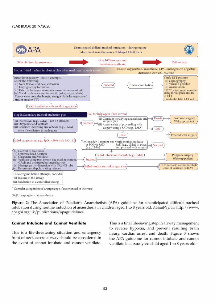
YEAR BOOK 2019/2020
52
Cannot Intubate and Cannot Ventilate
This is a life-threatening situation and emergency front of neck access airway should be considered in the event of cannot intubate and cannot ventilate.
This is a final life-saving step in airway management to reverse hypoxia, and prevent resulting brain injury, cardiac arrest and death. Figure 3 shows the APA guideline for cannot intubate and cannot ventilate in a paralysed child aged 1 to 8 years old.1
Unanticipated di�cult tracheal intubation—during routineinduction of anaesthesia in a child aged 1 to 8 years
Step B. Secondary tracheal intubation plan
Step A. Initial tracheal intubation plan when mask ventilation is satisfactory
Call for helpDi�cult direct laryngoscopy Give 100% oxygen andmaintain anaesthesia
(ii) Oxygenate and ventilate(iii) Consider increasing size of SAD (e.g., LMA)
once if ventilation is inadequate
(i) Convert to face mask(ii) Optimise head position
(iii) Oxygenate and ventilate(iv) Ventilate using two-person bag mask technique,
CPAP, and oro/nasopharyngeal airway(v) Manage gastric distension with OG/NG tube
(vi) Reverse nondepolarisating relaxant
Verify ETT position(i) Capnography
(ii) Visual if possible(iii) AuscultationIf ETT is too small considerusing throat pack and tie
If in doubt, take ETT out
(i) Consider modifying anaesthesia and
(ii) Assess safety of proceeding with
Tracheal intubation
Following intubation attempts, consider
Go to scenario cannot intubatecannot ventilate (CICV)
Postpone surgeryWake up patient
Postpone surgeryWake up patient
Failed intubation via SAD
Failed ventilation and oxygenation
Proceed with surgery
Call for help again if not arrivedUnsafe
Safe
Safe
Succeed
Succeed
Succeed
Succeed
Ensure: oxygenation, anaesthesia, CPAP, management of gastric
Direct laryngoscopy—not >4 attempts Check the following:
(i) Neck �exion and head extension(ii) Laryngoscopy technique
(iii) External laryngeal manipulation—remove or adjust(iv) Vocal cords open and immobile (adequate paralysis)
Failed intubation with good oxygenation
(i) Consider 1 attempt at FOI via SAD
(ii) Verify intubation, leave
and proceed with surgery
SAD = supraglottic airway device
distension with OG/NG tube
If poor view, consider bougie, straight blade laryngoscope∗and/or smaller ETT
(i) Insert SAD (e.g., LMA)—not >3 attempts
Failed oxygenation, e.g., SpO2 <90% with FiO2 1.0
(e.g., LMA)
∗Consider using indirect laryngoscope if experienced in their use
(i) Trauma to the airway(ii) Extubation in a controlled setting
(e.g., LMA)SAD (e.g., LMA) in place,
SAD (e.g., LMA)
surgery plan
surgery using a
to ETT
Figure 2: The Association of Paediatric Anaesthetists (APA) guideline for unanticipated difficult tracheal intubation during routine induction of anaesthesia in children aged 1 to 8 years old. Available from http://www.apagbi.org.uk/publications/apaguidelines

MALAYSIAN SOCIETY OF ANAESTHESIOLOGISTS
53
patient is breathing spontaneously or apnoeic in a variety of ways such as via nasal cannulas, masks, nasopharyngeal airways, and high flow nasal cannulas.
High flow nasal cannula is a technique that was originally used in neonates to provide respiratory support for premature infants, decreasing the need for intubation. Now, it has expanded its role in difficult airway in delaying hypoxia in children during apnoea after induction of anaesthesia. Studies
DEVICES AND TECHNIQUES FOR THE PAEDIATRIC DIFFICULT AIRWAY
Oxygenation for the Paediatric Difficult Airway
The primary aim in managing paediatric airway is to maintain oxygenation and avoid hypoxemia. Mortality occurs because of inability to oxygenate rather than inability to intubate. Oxygen delivery during intubation attempts reduces the incidence of hypoxemia. Oxygen can be delivered while a
Cannot intubate and cannot ventilate (CICV) in a paralysed anaesthetised child aged 1 to 8 years
Step B Attempt wake up if maintaining SpO2 >80%
Step C Airway rescue techniques for CICV (SpO2 <80% and falling) and/or heart rate decreasing
Step A Continue to attempt oxygenation and ventilation
Call for helpFailed intubationinadequate ventilation Give 100% oxygen
If rocuronium or vecuronium used, consider suggamadex (16mg/kg) for full reversal
Prepare for rescue techniques in case child deteriorates
Call for specialist ENT assistance
Cannula cricothyroidotomy• Extend the neck (shoulder roll)• Stabilise larynx with non-dominant hand• Access the cricoithyroidotomy membrane with a dedicated 14/16 gauge cannula• Aim in a caudad direction• Confirm position by air aspiration using a syringe with saline• Connect to either: • adjustable pressure limiting device, set to lowest delivery pressure or • 4Bar O2 source with a flowmeter (match flow l/min to child’s age) and Y connector• Cautiously increase inflation pressure/flow rate to achieve adequate chest expansion Wait for full expiration before next inflation• Maintain uper airway patency to aid expiration
*Note: Cricothyroidotomy techniques can have serious complications and training is required - only use in life-threatening situations and convert to a definitive airway as soon as possible
Percutaneous cannula cricothyroidotomy / transtracheal jet ventilation (pressure limited)
ENT not available
ENT available
Call for help again if not arrived
Succeed
Fail
• FiO2 1.0• Optimise head position and chin lift/jaw thrust• Insert oropharyngeal airway or SAD (e.g. LMA™)• Ventilate using two person bag mask technique• Manage gastric distension with an OG/NG tube
Consider:• Surgical tracheostomy• Rigid bronchoscopy + ventilate / jet ventilation (pressure limited)
Continue jet ventilation set to lowest delivery pressure until wake up or definitive airway established
• Perform surgical cricothyroidotomy / transtracheal and insertion of ETT / tracheostomy tube*• Consider passive O2 insufflation while preparing
SAD = supraglottic airway device
Figure 3: The Association of Paediatric Anaesthetists (APA) guideline for cannot intubate and cannot ventilate in paralysed anaesthetised children aged 1 to 8 years old. Available from http://www.apagbi.org.uk/publications/apaguidelines

YEAR BOOK 2019/2020
54
shows that those children who received trans-nasal humidified high flow nasal cannula doubled their time to desaturation to 92%.2
Flexible Fibreoptic Bronchoscopes
A flexible fibreoptic bronchoscope is considered as the gold standard of difficult airway management in paediatric patients.3 Intubation using this device can be achieved via nasal or oral endotracheal tube (ETT) or through a supraglottic airway device (SAD). It allows the operator to manoeuvre the ETT tip around and bypass distortions in airway anatomy. Limitations include lack of patient cooperation, blood and secretions that can obscure the view and no suction ports in the smaller bronchoscopes.
Video-Laryngoscopes
Video-laryngoscopes (VLs) are gaining popularity in the management of difficult airway in the paediatric population.4,5 Various designs of VL are being marketed nowadays with much more improved design and portability. VLs have two types of blades, which can be non-angulated or hyper-angulated. Non-angulated blades are those with the design of standard Miller and Macintosh but have video added adjacent to the light source. The hyper-angulated blades have an acute distal blade angulation that facilitates visualizing of larynx. The available paediatric VL options are GlideScope™ Video Laryngoscope (Verathon, Bothell, Washington, USA), Storz DCI™, C-MAC® Video Laryngoscope (Karl Storz, Tuttlingen, Germany), Truview PCD™ Infant (Truphatek, Netanya, Israel), Airtraq™ Disposable Optical Laryngoscope (Prodol Meditec, Vizcaya, Spain), and Pentax-AWS™ (Pentax Corporation, Tokyo, Japan).
Supraglottic Airway Devices
Supraglottic Airway Devices (SAD) play a major role in the management of difficult airway both in adult and paediatric population. They are useful in condition of difficult ventilation and failed intubation as the devices helps to maintain the oxygenation and ventilation. Newer SAD device
such as Ambu and I-Gel have been designed with improve efficiency and can be used as a conduit for fibreoptic-guided tracheal intubation.
Optical Stylets
Optical stylets can be used as an alternative option in managing difficult airway in paediatrics. One of the beneficial characteristics of optical stylets is that it adapts to the natural curve of the airway, allowing intubation without aligning the oral, pharyngeal, and tracheal axes. Some of the limitations are that they cannot be used for nasal intubation; small lens can be obscured with blood and secretions. Some of the examples of optical stylets that are available in market are Bonfils (Karl Storz) and Shikani Optical Stylet (SOS; Clarus Medical).
PEARLS IN MANAGING PAEDIATRIC DIFFICULT AIRWAY
Some of the clinical pearls in managing paediatric difficult airway are:
1. Do not panic, remain calm. Panicking is not going to make things better. Call for help early. 2. Maintain spontaneous ventilation at all times until airway is secured. 3. Use supplemental oxygen at all times during tracheal intubation attempts either via nasal cannula, nasopharyngeal airway, bronchoscopic adapter, side port of rigid bronchoscope or intubating mask. 4. Always have SAD standby. 5. Have a well-structured airway plan with multidisciplinary approach including the Ear Nose and Throat team.
CONCLUSION
Paediatric patients with anticipated difficult airway should be cared for only in a tertiary care facility with qualified caregivers, including paediatric anaesthesiologists and appropriate surgical support. Current practice guidelines and recommendations should be reviewed and practised so that the individuals and institution can be ready to act

MALAYSIAN SOCIETY OF ANAESTHESIOLOGISTS
55
quickly when problematic airway scenarios arise. Skills with rescue airway techniques, including indirect VL, SAD-assisted fibreoptic bronchoscopy, and needle cricothyrotomy should be acquired and maintained using simulation or clinical practice in the none-emergent scenario. As the study of
the management of the paediatric difficult airway progresses, there will be ongoing improvements in clinical care. Future multi-centre studies are needed to identify risk factors, evaluate treatment algorithms, and determine outcomes in the difficult paediatric airway.
References
1. The Association of Paediatric Anaesthetists (APA) Guidelines for difficult airway management in children. 2012. [http://www.apagbi.org.uk/publications/apaguidelines]Accessed September 2, 2020
2. S. Humphreys, P. Lee-Archer, G. Reyne, D. Long, T. Williams, A. Schibler, Transnasal humidified rapid-insufflation ventilatory exchange (THRIVE) in children: a randomized controlled trial. Br J Anaesth 2017;118:232-38
3. Totoz T, Erkalp K, Taskin S, Dalkilinc U, Selcan A. Use of awake flexible fiberoptic bronchoscopic nasal intubation in secure airway management for reconstructive surgery in a pediatric patient with burn contracture of the neck. Case Rep Anesthesiol 2018:8981561
4. Sun Y, Lu Y, Huang Y, Jiang H. Pediatric video laryngoscope versus direct laryngoscope: a meta-analysis of randomized controlled trials. Pediatr Anesth 2014;24:1056-65
5. Abdelgadir IS, Phillips RS, Singh D, Moncreiff MP, Lumsden JL. Videolaryngoscopy versus direct laryngoscopy for tracheal intubation in children (excluding neonates). Cochrane Database Syst Rev 2017;5:CD011413

YEAR BOOK 2019/2020
56
INTRODUCTION
Malaysia’s population is ‘greying’. The percentage of the elderly population, defined as those who are 65 years and over, has risen over the past several years: 4.94% in 2010, 5.98% in 2015 and 7% in 2020.1 By 2030, the elderly proportion of the population is estimated to rise to more than 15%.2 Greying of the population can be expected to increase the number of surgeries performed in the elderly. This situation poses unique challenges to the anaesthesiologists because not only advance age affects anaesthesia administration, it also has a negative impact on peri-operative outcome. This article intends to discuss the latest update with regards to the optimal anaesthetic care of the elderly. It is not meant to be comprehensive, but rather to focus on some of the most significant issues in this area.
PRE-OPERATIVE ASSESSMENT
Elderly patients have high prevalence towards comorbidities, which may significantly affect their anaesthetic care peri-operatively. Pre-operative assessment of the elderly patients is important to detect the presence and severity of the comorbidities.
Technically, the American Society of Anesthesiologists (ASA) physical status score does not include age as a criterion. However, many anaesthesiologists routinely assign an ASA II classification to otherwise healthy patients over 65 years old. This practice should probably be revised because age by itself does not necessarily increase the peri-operative risk. Instead, increased peri-operative risk is most likely due to underlying comorbidities.
‘Routine’ pre-operative testing is no longer recommended in the elderly.3 Studies have found no difference in complications among patients
who received routine pre-operative testing e.g. electrocardiogram (ECG), full blood count and renal function test, compared with those who underwent the indicated tests based on individualised medical conditions.4
Neurocognitive Assessment
Elderly patients are at a high risk of post-operative cognitive dysfunction (POCD).5 Some cases may be reversible; however, in a small percentage of patients, the cognitive decline persists for a long term. This cognitive decline appears to accelerate after surgery in elderly patients who were diagnosed with pre-existing cognitive impairment.5 Thus, the ASA Brain Health Initiative guidelines suggest that baseline cognition should be evaluated in all patients older than 65 years. Conclusive evidence on specific anaesthetic practice to prevent POCD is pending, but the role of dexmedetomidine, an α-2 agonist, may be promising.6
Cardiopulmonary Reserve Assessment
Elderly patients are vulnerable to peri-operative cardiac adverse events. Strict adherence to the American College of Cardiology/American Heart Association clinical practice guidelines may reduce unnecessary cardiac complications.7 The online-based American College of Surgeons National Surgical Quality Improvement Program risk calculator has been recommended in the guidelines. Routine pre-operative ECG is common but has been shown to be less specific and is thus of limited value in predicting post-operative cardiac complications compared with the severity of comorbidities.8 Echocardiography may instead provide insights into ventricular function and valve status and may be considered in patients with significant cardiac comorbidities.9
The Greying Of Malaysians: Implications For Anaesthesiologists
Chong Soon Eu1, Rhendra Hardy Mohamad Zaini2
1 Senior Lecturer and Anaesthesiologist, Advanced Medical and Dental Institute, Universiti Sains Malaysia, Pulau Pinang, Malaysia2 Associate Professor and Consultant Anaesthesiologist, Department of Anaesthesiology and Intensive Care, School of Medical Sciences, Universiti Sains Malaysia, Kubang Kerian, Kelantan, Malaysia

MALAYSIAN SOCIETY OF ANAESTHESIOLOGISTS
57
Pulmonary reserve decreases with ageing. A metabolic equivalent (METs) is a simple and easy way to screen for exercise intolerance. However, the cardiopulmonary exercise test (CPET) may be a more reliable method than METs to predict peri-operative risk. If the CPET is unavailable, the inexpensive six-minute walk test may be used.10 This test requires the patient to walk continuously for six minutes; the further the distance covered, the better the functional capacity. Pre-habilitation, the practice of enhancing a patient’s functional capacity before surgery, is an important measure in reducing the risk of post-operative complications among the elderly. The areas of intervention are mainly, but not limited to, muscle, cardiac and respiratory functions.
Renal Function Assessment
The glomerular filtration rate, creatinine clearance and renal functional reserve decline with ageing. These parameters may be underestimated by the
renal function test alone.11 This declining effect is compounded by underlying comorbidities and may lead to further deterioration of renal function. Plasma concentration of renally excreted intravenous (IV) anaesthetic agents may be increased.12 In addition, the elderly’s kidneys are more vulnerable to the nephrotoxic effects of IV contrast and medications, such as nonsteroidal anti-inflammatory drugs and certain antibiotics.
Frailty Assessment
Frailty is an important concept worth emphasizing. This condition refers to the decreased physiological reserve and resistance to stressors. The degree of frailty is a predictor of poor outcome in the elderly.18,19 Pre-operative frailty (Table I) is associated with increased risk of post-operative mortality, prolonged hospital stays and functional decline. The frailty score can aid patients and clinicians to make reasonable informed decisions.
Table I: Frailty Criteria
Shrinking (weight loss)
Weakness (grip strength)
Exhaustion
Activity level
Walking speed
Yes = 1; No = 0 Total the number of points for each criterion.0 - 1: Not Frail2 - 3: Intermediate frail 4 - 5: Frail
Please notify surgeon
Unintentional weight loss >4.5kg in the past year
Lowest 20th percentile measured by hand-held dynamometer
Feeling exhausted 3 or more days a week
Low physical activity, i.e. kilocalorie expenditure less than 20th percentile for gender
Slow walking speed, defined as lowest 20th percentile walking 15 feet at normal pace
}Reproduced from: Makary MA, Segev DL, Pronovost PJ, Syin D, Bandeen-Roche K, Patel P, et al. Frailty as a predictor of surgical outcomes in older patients. J Am Coll Surg. 2010;210:901-8

YEAR BOOK 2019/2020
58
Medication History Assessment
The elderly commonly takes more than ten types of medications a day. The incidence of drug-related adverse event therefore can be expected to be high in this age group.
Dual-antiplatelet therapy or oral anticoagulants may preclude the use of a neuraxial anaesthetic technique. Patients with high risk of thromboembolism might require bridging therapy, in which extra caution may be required for those with reduced renal function. Monoamine oxidase inhibitors may lead to potentially fatal interaction with pethidine and ephedrine. The pre-operative use of psychotropic medications may be associated with increased risk for post-operative delirium. For patients with impaired cognition, H1 antagonists and anticholinergic agents, such as scopolamine or atropine, should be avoided.
Patients with dementia are occasionally treated with cholinesterase inhibitors, e.g. memantine and rivastigmine, which may lead to reduced plasma cholinesterase, thereby prolonging the action duration of suxamethonium. This treatment may also reduce the effects of non-depolarising neuromuscular blocking agents. Thus, high doses are required to achieve a proper degree of neuromuscular blockade. Neuromuscular monitoring may be required intra-operatively.
Nutritional Status Assessment
Elderly patients often lack appetite and may be malnourished. Malnutrition is a frequent yet often overlooked problem in pre-operative elderly patients. Optimising nutritional status is thus important to reduce post-operative complications and enhance recovery. Prolonged fasting time should be avoided unless indicated. Studies have shown that fasting times last longer than recommended, with 15 hours fasting being related to increased cardiac stress and hypothermia.20
COVID-19 Screening
The year 2020 marked a major change in healthcare and overall human lifestyle after the declaration of COVID-19 pandemic. This change is especially true for the elderly population. According to studies, advanced age is significantly associated with high morbidity and mortality in COVID-19 infection and the elderly can thus be regarded as high-risk population.21 This condition is attributed to the weaker immune system of the elderly compared with the younger population.
COVID-19 has a long period of incubation. Therefore, the possibility of undiagnosed infection should not be neglected. Appropriate screening needs to be conducted in accordance with the risk stratification followed by confirmation test. For example, an elderly patient from a nursing home may be considered to have moderate probability for COVID-19, and confirmation test should be conducted pre-operatively. According to the outcome of massive screening activities started on 4th May 2020 in Malaysian nursing homes, COVID-19 was present in 8% of these centres.22
INTRAOPERATIVE MANAGEMENT
Regional versus General Anaesthesia
The mode of anaesthesia should be guided by surgical requirements, comorbidities and patient preferences. Between general and regional anaesthesia, studies have shown no difference in the outcome including a 30-day mortality.23 However, compared with general anaesthesia, regional anaesthesia favours a shorter length of stay. Other advantages of regional anaesthesia include sympatholytic effects that may reduce blood loss, fewer incidence of deep vein thrombosis and provision of analgesia in the immediate post-operative period.
In the era of COVID-19 pandemic, guidelines have recommended regional anaesthesia as the anaesthesia technique of choice, where appropriate.24 Regional anaesthesia is preferred because general anaesthesia may be aerosol generating, causing

MALAYSIAN SOCIETY OF ANAESTHESIOLOGISTS
59
this group of patients.28 Clinical judgment based on meticulous monitoring of volume status and tissue perfusion remains the most important factor.
The recent National Institute for Health and Care Excellence guideline recommends the restrictive blood transfusion threshold at haemoglobin of 7.0g/dL for patients who are free from major haemorrhage, acute coronary syndrome (ACS) or chronic anaemia and a haemoglobin threshold of 8.0g/dL for patients with ACS.29
SELECTION AND DOSING OF ANAESTHETIC AGENTS
Ageing is associated with progressive losses of functional reserve in all organ systems, reduced brain size and neuronal density. Pharmacodynamic sensitivity increases with age for all sedative agents including opioids, benzodiazepines and volatile anaesthetics, with exaggerated respiratory depressant effects.
Short-acting agents are preferred with reduced initial doses and with a long interval between repeated doses if indicated. Minimal alveolar concentration decreases for all volatile anaesthetic agents.30 End-tidal brain equilibration is prolonged, resulting in late onset of bi-spectral index change detection. The dose of inhalational agents should also be decreased by approximately 6% per decade after the age of 40 years.31
Anxiolytics
The elderly population shows an increased brain sensitivity towards anxiolytics, decreased clearance and increased volume of distribution.32 Hence, a reduction in midazolam dose is necessary, with a prolonged interval between additional doses if indicated. Midazolam may occasionally cause paradoxical worsening of agitation in elderly patients. Caution is also required when fentanyl is concomitantly administered with midazolam. Long-acting benzodiazepines, e.g. lorazepam and diazepam, are not recommended for the elderly.
potential infective harm to healthcare personnel and detrimental complications of undiagnosed COVID-19 patients undergoing such procedures. Although these methods are operator- and skill-dependent, regular practice, especially with ultrasound guidance, may improve the outcome. In general, the choice of opting for regional anaesthesia must still be balanced on a case-by-case basis against potential risks.
Monitoring
Standard ASA monitoring, including that of oxygenation, ventilation, blood pressure (BP), heart rate and temperature, are necessary regardless any mode of anaesthetic techniques in elderly patients.
Pieces of evidence prove the invasive monitoring for these patients. Intra-operative hypotension may contribute to a cardiac event, which is detrimental. Vasoconstrictor agent is often necessary in elderly, either in small boluses or in continuous infusion. Elderly patients with pre-existing severe cardiovascular disease should be monitored with intra-arterial BP monitoring during major surgery with anticipated rapid blood loss or large fluid shifts.
Central venous catheterisation is recommended when vasoactive drugs are anticipated. Pulmonary artery catheters have not been proven to be beneficial in elderly patients and are not routinely recommended.25 Alternatively, non-invasive cardiac index monitoring has been shown to be reasonably reliable in elderly patients in guiding goal-directed haemodynamic therapy peri-operatively.26 If available, transoesophageal echocardiography may also be used when severe haemodynamic compromise is anticipated.27
Fluid Management and Blood Transfusion
Given the labile BP of the elderly, adequate volume status is important to ensure the effective myocardial contractility and adequate tissue perfusion. Fluid management in elderly patients with heart failure can be especially challenging. At present, evidence is still lacking to guide optimal fluid therapy in

YEAR BOOK 2019/2020
60
Sedative Agents
Given the risk of hypotension, the dose of induction agents are usually reduced by 20% to 30% and bolus injections should be administered with titrations. Etomidate is often the preferred anaesthetic induction agent for elderly patients with known cardiovascular compromise or haemodynamic instability owing to its minimal haemodynamic side effects. This drug is also commonly used in elderly patients with unknown cardiovascular and intravascular volume status.
The median effective dose of dexmedetomidine is low in older adults. Thus, a low loading dose of 30% to 60% is required for elderly patients. Apart from the low incidence of POCD, dexmedetomidine attenuates peri-operative stress and inflammation and protects the immune function of elderly patients.8-10 Thus, this drug may contribute to decreased post-operative complications. Very-low-dose nocturnal dexmedetomidine (0.1-0.2mcg/kg/hour) may also provide beneficial effect in preventing intensive care unit delirium, without causing any adverse related to this drug at higher doses.
Opioids
All opioids are approximately twice as potent in the elderly due to the age-related increased brain sensitivity to opioids.33 For ultra-short-acting remifentanil, the decreases in volume of distribution and clearance lead to a 50% reduction of the dose required. Morphine presents an enhanced analgesic effect and prolonged duration of action in elderly patients. This finding is due to the reduced renal clearance of the drug and its active metabolite, morphine-6-glucuronide. Clearance is further reduced in patients with renal insufficiency. Thus, morphine should be used with extra caution.
POST-OPERATIVE PAIN MANAGEMENT
Inadequate pain relief is associated with increased incidence of delirium and morbidity in the elderly. Although pain perception decreases with age,
post-operative analgesia is a critical aspect of peri-operative anaesthetic care of elderly patients.34
A multi-modal and opioid sparing approach for pain management is currently widely accepted to reduce the risk of opioid-related side effects. This approach includes the employment of paracetamol, selective cyclooxygenase-II inhibitors, neuraxial anaesthesia and ultrasound-guided nerve blocks. In the era of COVID-19 outbreak, opioid sparing anaesthesia is also important because it allows early re-mobilisation, shortens hospital stay and reduces the risk of hospital-acquired infection.
For mild post-operative pain, paracetamol is one of the best choices, unless contraindicated. Paracetamol IV may be used when oral or rectal administration is not an option. For moderate to severe pain, the addition of scheduled paracetamol to an opioid patient-controlled analgesia (PCA) results in a synergistic effect in pain control and has been shown to lower overall opioid requirement.35 For those who are unable to use PCA due to cognitive dysfunction, age-adjusted regular doses of IV or subcutaneous analgesics are reasonable alternatives.
CONCLUSION
Elderly patients are uniquely susceptible and particularly sensitive to the stress of hospitalization, surgery and anaesthesia in ways that are only partially understood. Accordingly, minimizing perioperative risk in elderly patients requires thorough pre-operative assessment of organ function and reserve, careful intra-operative management of coexisting morbidities, and vigilant post-operative pain management.

MALAYSIAN SOCIETY OF ANAESTHESIOLOGISTS
61
References
1. World Bank Group. 2020. Malaysia Economic Monitor Reports [https://www.worldbank.org/en/country/malaysia/publication/malaysia-economic-monitor-reports] Accessed September 1, 2020
2. Hirschmann R. 2019. Share of population older than 65 years in Malaysia from 2015 to 2019 [https://bit.ly/2YIVFH4] Accessed September 1, 2020
3. Apfelbaum JL, Connis RT, Nickinovich DG, American Society of Anesthesiologists Task Force on Preanesthesia E, Pasternak LR, et al. Practice advisory for preanesthesia evaluation: an updated report by the American Society of Anesthesiologists Task Force on Preanesthesia Evaluation. Anesthesiology 2012;116:522-38
4. Schein OD, Katz J, Bass EB, Tielsch JM, Lubomski LH, Feldman MA, et al. The value of routine preoperative medical testing before cataract surgery. N Engl J Med 2000;342:168-75
5. Patel D, Lunn AD, Smith AD, Lehmann DJ, Dorrington KL. Cognitive decline in the elderly after surgery and anaesthesia: results from the Oxford Project to Investigate Memory and Ageing (OPTIMA) cohort. Anaesthesia 2016;71:1144-52
6. Pasin L, Landoni G, Nardelli P, Belletti A, Di Prima AL, Taddeo D, et al. Dexmedetomidine reduces the risk of delirium, agitation and confusion in critically Ill patients: a meta-analysis of randomized controlled trials. J Cardiothorac Vasc Anesth 2014;28:1459-66
7. Fleisher LA, Fleischmann KE, Auerbach AD, Barnason SA, Beckman JA, Bozkurt B, et al. 2014 ACC/AHA Guideline on Perioperative Cardiovascular Evaluation and Management of Patients Undergoing Noncardiac Surgery. J Am Coll Cardiol 2014;64:e77-e137
8. Liu LL, Dzankic S, Leung JM. Preoperative Electrocardiogram Abnormalities Do Not Predict Postoperative Cardiac Complications in Geriatric Surgical Patients. J Am Geriatr Soc 2002;50:1186-91
9. Lim B-G, Lee I-O. Anesthetic management of geriatric patients. Korean J Anesthesiol 2020;73:8-29
10. Sinclair RC, Batterham AM, Davies S, Cawthorn L, Danjoux GR. Validity of the 6 min walk test in prediction of the anaerobic threshold before major non-cardiac surgery. Br J Anaesth 2012;108:30-5
11. Giannelli SV, Patel KV, Windham BG, Pizzarelli F, Ferrucci L, Guralnik JM. Magnitude of underascertainment of impaired kidney function in older adults with normal serum creatinine. J Am Geriatr Soc 2007;55:816-23
12. Rivera R, Antognini JF. Perioperative drug therapy in elderly patients. Anesthesiology 2009;110:1176-81
13. Cheung A, Haas B, Ringer TJ, McFarlan A, Wong CL. Canadian study of health and aging clinical frailty scale: Does it predict adverse outcomes among geriatric trauma patients? J Am Coll Surg 2017;225:658-65
14. Makary MA, Segev DL, Pronovost PJ, Syin D, Bandeen-Roche K, Patel P, et al. Frailty as a predictor of surgical outcomes in older patients. J Am Coll Surg 2010;210:901-8
15. Yeniay O, Tekgul ZT, Okur O, Koroglu N. Unexpectedly prolonged fasting and its consequences on elderly patients undergoing spinal anesthetics: A prospective observational study. Acta Cir Bras 2019;34:3
16. Borges do Nascimento IJ, Cacic N, Abdulazeem HM, von Groote TC, Jayarajah U, Weerasekara I, et al. Novel coronavirus infection (COVID-19) in humans: A scoping review and meta-analysis. J Clin Med 2020;9:941
17. Kejal Hasmuk HS, Maw Pin Tan. 2020. New report: The COVID-19 long-term care situation in Malaysia [https://ltccovid.org/wp-content/uploads/2020/05/Malaysia-LTC-COVID-situation-report-21-May-2.pdf] Accessed May 21, 2020
18. O'Donnell CM, McLoughlin L, Patterson CC, Clarke M, McCourt KC, McBrien ME, et al. Perioperative outcomes in the context of mode of anaesthesia for patients undergoing hip fracture surgery: systematic review and meta-analysis. Br J Anaesth 2018;120:37-50
19. Malaysian Society Of Anaesthesiologists. 2020. Guidelines for the Management of Patients Presenting for Surgery during the COVID-19 Pandemic. [http://www.msa.net.my/view_file.cfm?fileid=194] Accessed April 6, 2020
20. American Society of Anesthesiologists Task Force on Pulmonary Artery Catheterization. Practice guidelines for pulmonary artery catheterization: an updated report by the American Society of Anesthesiologists Task Force on Pulmonary Artery Catheterization. Anesthesiology 2003;99:988-1014

YEAR BOOK 2019/2020
62
21. Brown CVR, Shoemaker WC, Wo CCJ, Chan L, Demetriades D. Is Noninvasive Hemodynamic Monitoring Appropriate for the Elderly Critically Injured Patient? J trauma 2005;58:102-7
22. American Society of Anesthesiologists, Society of Cardiovascular Anesthesiologists Task Force on Transesophageal Echocardiography. Practice guidelines for perioperative transesophageal echocardiography. An updated report by the American Society of Anesthesiologists and the Society of Cardiovascular Anesthesiologists Task Force on Transesophageal Echocardiography. Anesthesiology 2010;112:1084-96
23. Punjasawadwong Y, Chau-in W, Laopaiboon M, Punjasawadwong S, Pin-on P. Processed electroencephalogram and evoked potential techniques for amelioration of postoperative delirium and cognitive dysfunction following non-cardiac and non-neurosurgical procedures in adults. Cochrane Db Sys Rev 201;5:CD011283
24. National Institute for Health and Care Excellence. 2015. Blood transfusion [https://www.nice.org.uk/guidance/ng24/chapter/Recommendations#red-blood-cells-2] Accessed September 1, 2020
25. Nickalls RW, Mapleson WW. Age-related iso-MAC charts for isoflurane, sevoflurane and desflurane in man. Br J Anaesth 2003;91:170-4
26. Shamsuddin Akhtar GAR. Aging at Systems Level. Oxford Textbook of Anaesthesia for the Elderly Patient 2014
27. Rivera R, Antognini JF. Perioperative drug therapy in elderly patients. Anesthesiology 2009;110:1176-81
28. Shafer SL. The Pharmacology of Anesthetic Drugs in Elderly Patients. Anesthesiol Clin North Am 2000;18:1-29
29. Gibson SJ, Helme RD. Age-related differences in pain perception and report. Clin Geriatr Med 2001;17:433-56
30. Remy C, Marret E, Bonnet F. Effects of acetaminophen on morphine side-effects and consumption after major surgery: meta-analysis of randomized controlled trial. Br J Anaesth 2005;94:505-13

MALAYSIAN SOCIETY OF ANAESTHESIOLOGISTS
63
INTRODUCTION
Congenital heart disease (CHD) complicates 0.8 percent of all live births worldwide. In this era, more than 80 percent of neonates with moderate to complex CHD survive to adulthood. The vast majority of women with cardiac disease will have an anaesthetic intervention in their lifetime. Inevitably, they will be referred to anaesthesia clinics either for regional analgesia for labour or anaesthesia for operative delivery.
CASE-SCENARIO
A 26-year-old woman Gravida 2 Para 1 at her 35 weeks of gestation with a background history of surgically repaired Tetralogy of Fallot with residual ventricular septal defect was referred from cardiac-obstetric combined clinic for early anaesthetic assessment. She had surgical repair at the age of six years old. However, she did not have any record documenting type of surgical repair or any complications postoperatively. Due to her husband’s work, her first pregnancy was followed up in one of the tertiary hospitals in southern Malaysia. Her pregnancy which was two years ago was uneventful and she had an assisted vacuum vaginal delivery without epidural analgesia. At that time, her echocardiography showed left ventricular ejection fraction (LVEF) of 60-65%, residual ventricular septal defect (VSD) flow of 140mmHg, mild tricuspid regurgitation (TR) with pulmonary artery systolic pressure (PASP) of 28mmHg. Her postpartum period was uneventful.
Due to logistic reason, she requested for further follow-up for her current pregnancy in our centre. She has been tolerating her current pregnancy well without having any failure symptom. She had good exercise tolerance with baseline observations of systemic arterial oxygen saturation of 96-99% on air,
arterial blood pressure 110/70mmHg, and heart rate of 90 beats per minute in sinus rhythm. Pansystolic murmur was heard all over the precordium. Echocardiography showed dilated right atrium and ventricle, residual VSD with peak pressure gradient (PPG) of 120mmHg, moderate TR with PASP 52+3mmHg, LVEF 68% with paradoxical IVS movement. She was commenced on oral sildenafil 25 milligram three times per day. She was allowed for vaginal delivery with shortened second stage.
1. What is the best labour analgesia that you could offer her? 2. What is your anaesthetic plan should she need emergency Caesarean section?
DISCUSSION
Congenital heart disease (CHD) complicates 0.8% of all live births worldwide. In this era, more than 80% of neonates with moderate to complex CHD survive to adulthood. Over the last two decades and in the future, the number of pregnant women with CHD is and will steadily grow thanks to the advancement in medical imaging, surgical intervention and post-operative care which improves survival to adulthood. The vast majority of women with cardiac disease will have an anaesthetic intervention. Parturient will be referred to anaesthesia clinics either for regional analgesia for labour or anaesthesia for operative delivery. After a period of follow up in special cardiac centre, most of them will be discharged for continuation of care in other tertiary centres. Unfortunately, in our country, there is still a lack of a dedicated database or networking compiling the information of patients with congenital heart disease. Brief yet concise information allows effective communication between centres that have been previously and are currently taking care of the parturient.
Perioperative Management Of Parturient With Congenital Heart Disease
Nazuha Mohd NajidAnaesthesiologist, Department of Anaesthesiology and Critical Care, Hospital Sultanah Bahiyah, Alor Setar, Kedah, Malaysia

YEAR BOOK 2019/2020
64
Comprehensive understanding of the individual patient’s cardiac anatomy, previous operations, and current physiological changes during pregnancy is crucial to ensure a successful delivery. Detailed multidisciplinary planning and regular antenatal review may allow the parturient to have smooth labour or Caesarean delivery. Anaesthesia planning should include mode of analgesia or anaesthesia with the least haemodynamic compromise.
The pregnant woman with cardiac disease, whether congenital or acquired, should be seen as early as possible in her pregnancy. The optimal management of women with pre-existing cardiac disease begins best before conception. Normal physiologic changes of pregnancy and childbirth are the greatest cardiovascular challenge of a woman’s life. Most are tolerated well in normal parturient but may exacerbate or worsened in women with pre-existing cardiovascular disease. Arrhythmias and cardiac failure are the most common morbidities in adult with CHD (11%), while mortality is 0.5% which is considerably low compared to parturient with valvular heart disease or cardiomyopathy.1
Four major changes that are important in the presence of cardiovascular disease are the increase in blood volume, heart rate, cardiac output and stroke volume, reduction in systemic vascular resistance (SVR), labour physiology and hypercoagubility state. Maximal haemodynamic load is reached during the second trimester. However, there are two important peaks that may tilt a pregnant lady with underlying cardiac disease towards cardiac failure. The first is towards the end of the second trimester of pregnancy which coincides with the plateau of the haemodynamic load during pregnancy. The second peak occurs in the peri- and early postpartum period putting parturient susceptible to volume overload and heart failure.2 In the immediate postpartum period there is an exaggerated 70 to 80% increase in cardiac output contributed by auto-transfusion, loss of low resistance placental circulation and relief of vena cava compression.
During antenatal assessment, a high index of suspicion should be considered in pregnant women
with unexplained tachycardia, hypotension, a new onset of resting shortness of breath, chest pain, severe orthopnoea requiring more than four pillows, paroxysmal nocturnal dyspnoea, and palpitations associated with symptoms such as collapse or syncope and saturation <94% with or without personal history of CHD. During physical examination, there may be an elevated JVP, hepatomegaly, a new regurgitant murmur, and bibasal crepitations. These warning signs should be sought during clinical assessment as it could indicate exacerbation of known or undiagnosed cardiac disease.
The modified WHO risk stratification system (mWHO) is most widely accepted and validated in pregnant women with known cardiovascular disease.4 Such risk stratification also provided guidance on resources a parturient with heart disease may require, or whether she will need to be transferred to a tertiary care facility for delivery. WHO Class III defects are associated with a high risk of morbidity and increased risk of maternal mortality. This include the diagnoses of mechanical heart valves, systemic right ventricle lesions, the Fontan palliation, unrepaired or palliated cyanotic lesions, Marfan syndrome with aorta <40 mm, severe systemic atrioventricular (AV) valve regurgitation, asymptomatic left ventricular outflow tract stenosis with gradient >50mm Hg, left AV valve stenosis with valve area <2.0cm2, and systemic ventricular ejection fraction 30-40%.
Pregnancy is highly discouraged in WHO class IV pregnancy (include severe symptomatic aortic valve stenosis, severe left AV valve stenosis, pulmonary hypertension, systemic ventricular ejection fraction <30%, New York Heart Association (NYHA) functional class III and IV, Marfan syndrome with aorta >45mm, bicuspid aortic valve with aorta >50mm, and severe coarctation of the aorta. These patients should be encouraged to proceed with surgical correction before pregnancy to allow for a lower-risk future. Termination of pregnancy should be discussed with empathy. A women with mWHO class III and IV who wishes to continue with her pregnancy should be managed ideally in a

MALAYSIAN SOCIETY OF ANAESTHESIOLOGISTS
65
tertiary centre by a pregnancy heart team members consisting of maternal fetal medicine subspecialist, obstetric anaesthesiologist, and cardiologist with subspecialty in congenital heart disease, cardiac surgeon, cardiac anaesthesiologist, neonatologist, and mental health specialist.
Progression of pregnancy may lead to further complications, for example, pre-eclampsia, multiple pregnancies and hypertensive disease of pregnancy. This will lead to inevitable increase in haemodynamic load and failure to meet the needs and subsequently increase the risk of cardiovascular complications in vulnerable patients.5
Multidisciplinary Delivery Planning
Formalised multidisciplinary care should be planned at 32 to 34 weeks of gestation or even earlier in ill parturient.6 Operative delivery is indicated for obstetric reasons or destabilized maternal condition. Other indications include women who are on anticoagulant who has not been switched to heparin two weeks prior to delivery, Marfan’s syndrome with aortic diameter more than 45mm, and acute or chronic aortic dissection. A stable cardiac disease alone is not an indication for a Caesarean delivery as it has not been shown to confer additional benefit.7 Nevertheless, in the highest risk patients, elective Caesarean delivery may be preferable as it allow attendance of all experts and avoidance of emergency delivery. Anaesthetic assessment should clearly outline necessary plan for both elective and emergency situations and this should be conveyed to the mothers and other parties involved in their care. Additional contingency plans that should also be considered are the possible need of perimortem Caesarean section or extracorporeal membrane oxygenation (ECMO). Anticoagulation must be carefully reviewed and managed during pregnancy and adjusted accordingly to allow feasibility of neuraxial analgesia or anaesthesia and delivery.
Anaesthetic Management for Labour and Delivery
Analgesia
The aim of labour analgesia in parturient with cardiac disease is to provide an efficacious analgesia while maintaining a stable maternal haemodynamic and fetal circulation. Labour analgesia may be provided via an epidural or intravenous route. An epidural route is preferable, and a working epidural may provide an advantage should the mother require emergency Caesarean section. Early initiation of labour epidural is advisable at the onset of discomfort in spontaneous labour or first sensation of contractions for induced labour. Epidural technique should take into consideration of providing sacral nerve root coverage for assisted second stage. Epidural-only or combined spinal-epidural (CSE) techniques can be performed. If adopting CSE technique, intrathecal opioid alone without the addition of local anaesthetic may provide a slower onset of sympathectomy. Presence of cerebral spinal fluids (CSF) via spinal needle provides a definitive end point for the likely positioning of the epidural needle tip within the epidural space.
A dural puncture epidural (DPE) technique has been increasingly popular with possible benefit of better-quality epidural analgesia with more successful sacral nerve root coverage.8 The technique is similar to CSE, but the medication is not given via intrathecal route. All medications for analgesia or anaesthesia are introduced through the epidural catheter into the epidural space relying on the dural puncture that acts as conduit for medication translocation. By avoiding direct intrathecal medication administration, fewer adverse effects such as hypotension, itchiness and asymmetrical block are observed with DPE compared to CSE.9 This is of particular advantage in pregnant women with heart disease. However, the technique chosen should be according to the anaesthesiologist or institution experience and familiarity. It is more important to

YEAR BOOK 2019/2020
66
obstetric complications. It allows commencement or titration of inotropes or vasopressors and sampling of arterial blood gas. In a scenario where Caesarean delivery is needed, continuous arterial monitoring may allow careful titration of epidural top-up or anaesthestic drugs if general anaesthesia (GA) is required instead. Central venous pressure (CVP) value may not be reliable in spontaneously breathing and constantly moving labouring parturient, but it helps in haemodynamic management in high risk group that are prone to have pulmonary oedema, hypotension or peripartum haemorrhage. It also aids in vasopressors, inotropes and other resuscitation drug administration. Pulmonary artery catheter is rarely used although it may guide titration of pulmonary vasodilators in patient with severe pulmonary hypertension. As epidural and invasive monitoring are inserted in an awake parturient, caution should be taken by maintaining communication and adequate local anaesthetic infiltration to reduce cardiac stress secondary to pain and anxiety.
Anaesthesia for Caesarean Delivery
Neuraxial anaesthesia is generally preferable in most parturient with cardiac diseases as it avoids potential detrimental haemodynamic instability post induction and emergence. Maternal airway is a potential difficult airway. Volatile agents may also lead to uterine relaxation and further putting the vulnerable parturient at risk of postpartum haemorrhage. Positive pressure ventilation leads to further increase in pulmonary artery pressures and right ventricular pressure in parturient with pulmonary hypertension. As neuraxial anaesthesia leads to reduction in SVR, consideration should be given to modify neuraxial technique especially in patients with left ventricular outflow tract obstruction or cyanotic congenital heart disease. Different modes of neuraxial anaesthesia are summarized in Table I. Nonetheless, there are situations that may provide advantage with GA:
Haemodynamic Monitoring
Labouring mother with cardiac disease should have continuous haemodynamic monitoring to help predict and prevent maternal cardiac or obstetric events. The extent of monitoring is highly dependence on severity of underlying cardiac disease and anticipated perioperative cardiovascular and obstetric complication.
Continuous pulse oximetry with visible pletymosgraphy display and audible alarms should be available. This allows detection of desaturation that may indicate shunt reversal due to drop in SVR. It may also aid in rapid determination of abnormal ECG activity versus artefact due to shivering or movement. Continuous 5-leads ECG allow detection of arrythmias or ischemia. Invasive arterial blood pressure monitoring should be considered in labouring parturient with high risk of haemodynamic instability during cardiac or
ensure that the epidural is providing satisfactory analgesia and haemodynamic stability via regular review. Early recognition and replacement of ineffective epidural alleviate the risk of emergent conversion to general anaesthesia should the need for emergency Caesarean section arises. In a cardiac patient deemed to have a detrimental effect of inadvertent intravascular adrenaline, a test dose with local anaesthetic alone should be considered.
Option of labour neuraxial technique
Combine spinalepidural
Dural punctureepidural
Intrathecalopioids only
Successfulcoverage of
sacral nerve root
Epidural
Consider▪ Lost of Resistance (LOR) technique with saline for intra/extracardiac▪ Test dose without adrenaline
Figure 1: Neuraxial technique that may be employed for labour analgesia

MALAYSIAN SOCIETY OF ANAESTHESIOLOGISTS
67
Table I: Different modes of neuraxial anaesthesia for parturient with cardiovascular pathology
Neuraxial technique
Single shot subarachnoid block
Advantages
Reliable, fast onset
Disadvantages
Rapid drop in SVR, BP causing decompensation in most cardiac parturient making it less favorable technique
Epidural anaesthesia
Combined spinal epidural anaesthesia
Continuous spinal anaesthesia (CSA)
Gradual onset ensuring haemodynamic stability
Combining reliability and symmetry of initial spinal flexibility of epidural for extension of desired level of block
•Allowfinecontrolof block characteristics•Goodcardiovascular stability
•Lessdenseblock•Asymmetryblock•Greaterincidenceofinadequateanaesthesia•RiskofemergentconversiontoGA
Concerns about the risk of the untested epidural catheter
•Unfamiliartechniquetomanyanaesthetists•Higherincidenceofpostduralpuncture headache
Benefits of haemodynamic stability that CSA can provide may outweigh the risk of complications from this technique.
Table II: Example of anaesthesia conducts in parturient with cardiac disease. Adapted with permission from Obstetric Anaesthesia and Analgesia Service (OAS) 2014, Hospital Kuala Lumpur.12
Pre-induction
Uterotonic agent
Postoperative Monitoring
CSE; combined spinal epidural; CVP, central venous pressure; GA, general anaesthesia; HDU, high-dependency unit; ICU, intensive care unit; MAP, mean arterial pressure; PCA, patient-controlled analgesia, TAP; transversus abdominis plane block.
BP, blood pressure; GA, general anaesthesia; SVR, systemic vascular resistance.
Analgesia
Reversal
Induction
GA
•Maintainsinusrhythm(phenylephrineinfusioncanhelpmaintainingheartrate)•maintaingoodcardiacoutput(mightneeddopamineordobutamineinfusion)to prevent hypotension. (MAP to keep at baseline of above 70mmHg)• Invasivelinei.e.arteriallineandCVPmonitoringpriortoregional/GA•Leftuterinetilt
Oxytocin 5unit in 5cc N/S over 5mins then followed by 40unit in 40ml NS to run 5-10ml/hr
ICU or HDU
Morphine, TAP block ± PCA morphine
•Sugammadex or•mixtureofglycopyrrolatewith neostigmine
Epidural morphine/epidural local anaesthetic with fentanyl
• Inductionagentofchoice- etomidate 0.2mg/kg, •Titrationmidazolamupto5mg•Highdoseopioid-IVfentanyl 10-15µg/kg during induction. •Musclerelaxant-suxamethonium can induce bradyarrthhmias. Consider rocuronium and ensure sugammadex availability
Neuraxial Anaesthesia
•LowdosesequentialCSE ○ Spinal (0.5-1ml of heavy 0.5% bupivacaine) + 25µg fentanyl ○ Epidural drug: (2% of lignocaine in 1:200 000 Adrenaline), 3-5ml every 5 minutes until T5-T6 is reached.

YEAR BOOK 2019/2020
68
avoided in parturient prone to develop pulmonary oedema.
Uterotonic Agent
Parturient with cardiac disease may have exaggerated response to the adverse effects of oxytocin and other uterotonics. Oxytocin administration leads to peripheral vasodilatation, hypotension, and increased cardiac output, contributed by increase in heart rate and stroke volume, which may not be tolerated well in susceptible parturient.11 In most situations, oxytocin should be titrated carefully according to the state of uterine contraction directly observed by the obstetrician while maintaining vigilance at haemodynamic monitoring. The decrease in SVR from oxytocin can be counteracted by careful titration of phenylephrine or maintaining vasoactive medication infusion. A low dose infusion with 5 to 10 units per hour can be used post-delivery with careful monitoring.
Ergometrine causes pulmonary vasoconstriction and hypertension while prostaglandin F2∝ (carboprost) can cause severe bronchospasm, hypertension, cardiovascular collapse and pulmonary oedema making these two uterotonic agents unsuitable in most cases. Surgical options such as an intrauterine balloon that can be left in one to two days after Caesarean section or vaginal delivery, uterine compression sutures (e.g. B-lynch suture) should be considered.
CONCLUSION
As we are moving into the next era of advanced congenital cardiac surgical intervention, more children will survive into adulthood. They will be seen not only for non-obstetric surgical intervention but may also require intervention for their labour and delivery. Each case is unique and different in presentation. Hence, multi-disciplinary team approach is important for the successful perioperative management of parturient with congenital heart disease.
•Parturient that has high likelihood of rapid haemodynamic deterioration/ongoing haemodynamic disaster •Inabilityofparturienttolieflat •Avoiding the need for emergent intraoperative conversion to GA •Inparturientwithmorbidarrhythmiasthatmay require intraoperative electrical cardioversion •Allowing trans-oesophageal echocardiography (TOE) insertion •Avoidingriskofspinalorepiduralhaematoma in ○ Parturient on maintenance anticoagulant, inhaled nitric oxide or pulmonary vasodilators ○ Parturient requiring ECMO institution •Experiencedanaesthesiologistwellversedwith special haemodynamic of complex, sometimes uncorrected congenital heart, or end stage heart disease with ventricular assist device
No particular anaesthetic technique is superior from the other but cardiovascular stability is the ultimate goal. Pre-induction preparation should include ensuring the availability of all basic and advanced cardiovascular monitoring. As it had been emphasized before, the planning for monitoring invasiveness and contingency plan is highly dependent on maternal cardiovascular status, underlying cardiac pathology and obstetric complications. Postoperative monitoring should continue in high dependency or intensive care unit.
Vasoactive medications are now being recommended as prophylaxis against hypotension following spinal or combined spinal-epidural anaesthesia at Caesarean section.10 Women with cardiac disease should be assessed on an individual basis. Vasoactive medications should be chosen according to their desirable pharmacological properties in lieu with parturient cardiovascular response. Phenylephrine is currently the recommended vasoactive medication to maintain SVR post spinal anaesthesia. Some parturient may benefit further with additional beta agonist activity provided by noradrenaline or metaraminol. Left uterine displacement should be adopted. Preloading or co-loading should be

MALAYSIAN SOCIETY OF ANAESTHESIOLOGISTS
69
References
1. J.W. Roos-Hesselink, T.P. Ruys, J.I. Stein, et al., for the ROPAC Investigators Outcome of pregnancy in patients with structural or ischaemic heart disease: results of a registry of the European Society of Cardiology. Eur Heart J 2013;34:657-665
2. Greutmann, Matthias, Petronella G. Pieper. Pregnancy in women with congenital heart disease. European Heart Journal 2015;36(37):2491-2499
3. Hameed, Afshan B., et al. Pregnancy-related cardiovascular deaths in California: beyond peripartum cardiomyopathy. American Journal of Obstetrics and Gynecology 2015;213(3):379-e1
4. Balci, Ali, et al. Prospective validation and assessment of cardiovascular and offspring risk models for pregnant women with congenital heart disease. Heart 2014;100(17):1373-1381
5. Drenthen, Willem, et al. Predictors of pregnancy complications in women with congenital heart disease. European Heart Journal 2010;31(17):2124-2132
6. Royal College of Obstetricians & Gynaecologists. Cardiac disease and pregnancy. Good Practice 2011;13
7. Asfour V, Murphy MO, Attia R. Is vaginal delivery or caesarean section the safer mode of delivery in patients with adult congenital heart disease? Interact Cardiovasc Thorac Surg 2013;17(1):144-150
8. Cappiello E, O’Rourke N, Segal S, Tsen LC. A randomized trial of dural puncture epidural technique compared with the standard epidural technique for labor analgesia. Anesth Analg 2008;107:646-1651
9. Chau A, Bibbo C, Huang CC, et al. Dural puncture epidural technique improves labor analgesia quality with fewer side effects compared with epidural and combined spinal epidural techniques: A Randomized Clinical Trial. Anesth Analg 2017;124(2):560-569
10. Kinsella, S.M., Carvalho, B., Dyer, R.A., Fernando, R. et al. International consensus statement on the management of hypotension with vasopressors during Caesarean section under spinal anaesthesia. Anaesthesia 2018;73:71-92
11. Langesaeter E, Rosseland LA, Stubhaug A. Haemodynamic effects of repeated doses of oxytocin during Caesarean delivery in healthy parturients. British Journal of Anaesthesia 2009;103:260-2
12. Razak TA. Anaesthesia for parturient with heart disease. Guidelines for Obstetric Anaesthesia and Analgesia Service (OAS) Hospital Kuala Lumpur 2014:43-4