World Journal of Critical Care Medicine
218
World Journal of Critical Care Medicine World J Crit Care Med 2017 February 4; 6(1): 1-90 Published by Baishideng Publishing Group Inc ISSN 2220-3141 (online)
-
Upload
khangminh22 -
Category
Documents
-
view
0 -
download
0
Transcript of World Journal of Critical Care Medicine

World Journal of Critical Care MedicineWorld J Crit Care Med 2017 February 4; 6(1): 1-90
Published by Baishideng Publishing Group Inc
ISSN 2220-3141 (online)

World Journal ofCritical Care MedicineW J C C M
EDITOR-IN-CHIEFBart Van Rompaey, Wilrijk
GUEST EDITORIAL BOARD MEMBERSHsing I Chen, HualienSheng-Hsien Chen, Yong-KangYih-Sharng Chen, TaipeiYung-Chang Chen, TaipeiDer-Yang Cho, TaichungCheng-Keng Chuang, TaoyuanHow-Ran Guo, TainanBang-Gee Hsu, HualienChien-Wei Hsu, KaohsiungWen-Jinn Liaw, TaipeiYan-Ren Lin, ChanghuaJiunn-Jye Sheu, Kaohsiung Hsien
MEMBERS OF THE EDITORIAL BOARD
ArgentinaEduardo Chuluyan, Buenos AiresAdrian A Inchauspe, Berazategui
AustraliaZsolt J Balogh, NewcastleZoltan H Endre, SydneyNam Q Nguyen, South AustraliaAlistair D Nichol, VictoriaGeorg M Schmolzer, VictoriaAndrew T Slack, Brisbane
Ravindranath Tiruvoipati, Frankston
AustriaLars-Peter Kamolz, ViennaSylvia Knapp, Vienna
BangladeshSaidur R Mashreky, Dhaka
BelgiumTeresinha Leal, BrusselsManu LNG Malbrain, AntwerpJean-Louis Vincent, Brussels
BrazilLuciano CP Azevedo, São PauloPatricia RM Rocco, Rio de JaneiroMarcos A Rossi, Ribeirao PretoRenato Seligman, Porto Alegre
CanadaDouglas D Fraser, OntarioPierre A Guertin, QuebecMarc G Jeschke, TorontoConstantine J Karvellas, EdmontonWolfgang M Kuebler, TorontoXi Yang, Winnipeg
ChinaXiang-Dong Chen, ChengduXu-Lin Chen, HefeiWong Tak Chuen, Hong KongMing-Xu Da, LanzhouHuang-Xian Ju, NanjingTing-Bo Liang, Hangzhou Peng-Lin Ma, BeijingChung-Wah D Siu, Hong KongYong-ming Yao, BeijingJia-Ping Zhang, ChongqingWei-Dong Zhou, Beijing
CroatiaAlan Sustic, Rijeka
CubaJesús Pérez-Nellar, La Habana
DenmarkDan S Karbing, Aalborg East
EgyptIbrahim Abouomira, CairoHanan Ibrahim, CairoAmr M Moghazy, IsmailiaAyman A Yousef, Tanta
I
Editorial Board2016-2019
The World Journal of Critical Care Medicine Editorial Board consists of 235 members, representing a team of worldwide experts in critical care medicine. They are from 44 countries, including Argentina (2), Australia (7), Austria (2), Bangladesh (1), Belgium (4), Brazil (4), Canada (6), China (23), Croatia (1), Cuba (1), Denmark (1), Egypt (4), Finland (1), France (6), Germany (9), Greece (9), Hungary (1), India (10), Iran (2), Israel (6), Italy (13), Japan (6), Jordan (1), Mexico (1), Morocco (1), Netherlands (4), New Zealand (3), Norway (1), Poland (1), Portugal (4), Russia (1), Saudi Arabia (2), Singapore (2), Slovenia (1), South Africa (1), Spain (6), Sweden (1), Switzerland (3), Thailand (1), Tunisia (1), Turkey (3), United Kingdom (7), United States (70), and Uruguay (1).
February 26, 2016WJCCM|www.wjgnet.com

FinlandAsko A Riutta, Tampere
FranceJean-Marc Cavaillon, ParisBruno Mégarbane, ParisSaad Nseir, LilleNicolas Terzi, CaenJean-Francois Timsit, La Tronche CedexBenoit Vallet, Lille
GermanyHendrik Bracht, UlmMichael E Czaplik, AachenGerrit Grieb, AachenTobias Keck, FreiburgPhilipp Kobbe, AachenAlexander Koch, AachenMarc Maegele, CologneAndrzej A Piatkowski, AachenArmin R Sablotzki, Leipzig
GreeceIoanna Dimopoulou, AthensDimitrios Karakitsos, AthensPetros Kopterides, AthensGregory Kouraklis, AthensAthanasios D Marinis, PiraeusGeorge Nakos, IoanninaPapaioannouE Vasilios, AlexandroupolisTheodoros Xanthos, AthensSpyros G Zakynthinos, Athens
HungaryZoltan Rakonczay, Szeged
IndiaRitesh Agarwal, ChandigarhRachna Agarwal, DelhiMohammad F Butt, SrinagarMohan Gurjar, LucknowDeven Juneja, New Delhi Farhad N Kapadia, MumbaiVikram Kate, PuducherryPramod Kumar, ManipalMedha Mohta, DelhiSrinivas Rajagopala, Bangalore
IranHemmat Maghsoudi, TabrizHomayoun Sadeghi-Bazargani, Tabriz
IsraelAlexander Becker, AfulaYoram Kluger, HaifaYona Kosashvili, ZerrifinKobi Peleg, Tel Hashomer Ilan Sela, RehovotPierre Singer, Petah Tikva
ItalyGiacomo Bellani, MonzaGiovanni Camussi, TurinAnselmo Caricato, RomePiero Ceriana, PaviaAntonio Chiaretti, RomeDavide A Chiumello, MilanoAlfredo Conti, MessinaPaolo Cotogni, TurinDaniele M De Luca, RomaVincenzo De Santis, RomeLuca La Colla, ParmaRaffaele Scala, LuccaGiovanni Vento, Roma
JapanKeishiro Aoyagi, Kurume citySatoshi Hagiwara, OitaYuichi Hattori, ToyamaHideo Inaba, KanazawaEisuke Kagawa, HiroshimaChieko Mitaka, Tokyo
JordanFeras I Hawari, Amman
MexicoSilvio A Ñamendys-Silva, Mexico City
MoroccoRedouane Abouqal, Rabat
NetherlandsWim A Buurman, MaastrichtMartin CJ Kneyber, GroningenPatrick Schober, AmsterdamArie Barend V Vugt, Enschede
New ZealandSultan Al-Shaqsi, DunedinArman A Kahokehr, WhangareiJohn W Pickering, Christchurch
NorwayUlf R Dahle, Oslo
PolandMaciej Owecki, Poznań
PortugalErnestina R Gomes, PortoCristina Granja, MatosinhosJosé A Lopes, LisbonPedro Póvoa, Lisbon
RussiaKonstantin A Popugaev, Moscow
Saudi ArabiaRitesh G Menezes, DammamMohamed T Suliman, Tabuk
SingaporeSanjay H Chotirmall, SingaporeDevanand Anantham, Singapore
SloveniaŠtefek Grmec, Maribor
South AfricaDamian Clarke, Pietermaritzburg
SpainDavid Jimenez, MadridJuan A Llompart-Pou, Palma de MallorcaAntonio T Martí, BarcelonaJuan C Montejo-González, MadridEnrique A Piacentini, TerrassaAlonso M Rodriguez, Madrid
SwedenMihai Oltean, Gothenburg
SwitzerlandDieter Cadosch, ZurichMihael Potocki, BaselJohn F Stover, Zurich
II February 26, 2016WJCCM|www.wjgnet.com
III February 26, 2016WJCCM|www.wjgnet.com
ThailandViroj Wiwanitkit, Bangkok
TunisiaMabrouk Bahloul, Sfax
TurkeyYusuf K Coban, MalatyaBensu Karahalil, AnkaraAli Nayci, Mersin
United KingdomSammy Al-Benna, NottinghamGiles N Cattermole, OrpingtonFrantisek Duska, NottinghamJames N Fullerton, LondonChristina Jones, PrescotSameer Khan, MiddlesbroughGeorge Ntoumenopoulos, London
United StatesEdward Abraham, Winston-SalemBernard R Bendok, ChicagoMichael Blaivas, AtlantaCharles D Boucek, PittsburghRonald Bronicki, Houston
Robert C Cantu, ConcordMarylou Cardenas-Turanzas, HoustonArchana Chatterjee, OmahaPaul A Checchia, St. LouisRubin I Cohen, New Hyde ParkStephen Cohn, San AntonioDonald E Craven, BurlingtonRuy J Cruz Jr, PittsburghFrancis C Dane, RoanokeMarc A de Moya, BostonSteven M Donn, Ann ArborChristopher P Farrell, WynnewoodMarcos A Fernandez, NashvilleKevin N Foster, PhoenixBarry D Fuchs, PhiladelphiaRichard P Gonzalez, MobileAlan H Hall, LaramieJijo John, GilbertJason N Katz, Chapel HillSalah G Keyrouz, Little RockImran Khalid, JeddahDeborah A Kuhls, Las VegasGregory L Larkin, New HavenChristos Lazaridis, CharlestonJames A Lin, Los AngelesYahia M Lodi, SyracuseRoger M Loria, RichmondAigang Lu, CincinnatiRudolf Lucas, AugustaO. John Ma, PortlandRobert T Mallet, Fort WorthWilliam T McGee, MiamiMark McKenney, MiamiMichael Moussouttas, PhiladelphiaOliver HJ Muensterer, Bronx
Rahul Nanchal, MilwaukeeMichael Steven Niederman, MineolaGary F Nieman, SyracuseJames M O’Brien, ColumbusMartin Oudega, MiamiCatherine M Preissig, DuluthVirginia Prendergast, PhoenixRamesh Raghupathi, PhiladelphiaMiren A Schinco, JacksonvilleCarl I Schulman, MiamiL Keith Scott, ShreveportKevin N Sheth, BaltimoreJenni Short, SalinaRonald F Sing, CharlottePhilip C Spinella, St. LouisRobert M Starke, CharlottesvilleStanislaw PA Stawicki, ColumbusDavid C Stockwell, WashingtonStanislav Svetlov, AlachuaMaged A Tanios, Long BeachNeal J Thomas, HersheyNancy M Tofil, BirminghamBalagangadhar R Totapally, MiamiSteven N Vaslef, DurhamJoseph C Watson, Falls ChurchJohn S Wilgis, OrlandoDavid C Willms, San DiegoHao-Dong Xu, RochesterXiao-Ming Xu, IndianapolisMidori A Yenari, San Francisco
UruguayWilliam Manzanares, Montevideo
World Journal ofCritical Care MedicineW J C C M
Contents
IWJCCM|www.wjgnet.com February 4, 2017|Volume 6|Issue 1|
Quarterly Volume 6 Number 1 February 4, 2017
REVIEW1 Practicalstrategiesforincreasingefficiencyandeffectivenessincriticalcareeducation
Joyce MF, Berg S, Bittner EA
MINIREVIEWS13 Managementofparenteralnutritionincriticallyillpatients
Cotogni P
21 Exertionalrhabdomyolysisandheatstroke:Bewareofvolatileanestheticsedation
Heytens K, De Bleecker J, Verbrugghe W, Baets J, Heytens L
28 Nutrientstimulationofmesentericbloodflow-implicationsforoldercriticallyillpatients
Nguyen TAN, Abdelhamid YA, Phillips LK, Chapple LS, Horowitz M, Jones KL, Deane AM
ORIGINAL ARTICLE Basic Study
37 ImpactofhighdosevitaminConplateletfunction
Mohammed BM, Sanford KW, Fisher BJ, Martin EJ, Contaifer Jr D, Warncke UO, Wijesinghe DS, Chalfant CE, Brophy DF,
Fowler Ⅲ AA, Natarajan R
Retrospective Study
48 Riskfactorsformortalityinpostoperativeperitonitisincriticallyillpatients
Launey Y, Duteurtre B, Larmet R, Nesseler N, Tawa A, Mallédant Y, Seguin P
Observational Study
56 Implementationofenteralfeedingprotocolinanintensivecareunit:Before-and-afterstudy
Padar M, Uusvel G, Starkopf L, Starkopf J, Reintam Blaser A
65 Timing,methodanddiscontinuationofhydrocortisoneadministrationforsepticshockpatients
Ibarra-Estrada MA, Chávez-Peña Q, Reynoso-Estrella CI, Rios-Zermeño J, Aguilera-González PE, García-Soto MA,
Aguirre-Avalos G
Prospective Study
74 Comparisonofinhaledmilrinone,nitricoxideandprostacyclininacuterespiratorydistresssyndrome
Albert M, Corsilli D, Williamson DR, Brosseau M, Bellemare P, Delisle S, Nguyen AQN, Varin F
REVIEW

ContentsWorld Journal of Critical Care Medicine
Volume 6 Number 1 February 4, 2017
IIWJCCM|www.wjgnet.com February 4, 2017|Volume 6|Issue 1|
79 Attributablecostofanosocomialinfectionintheintensivecareunit:Aprospectivecohortstudy
Chacko B, Thomas K, David T, Paul H, Jeyaseelan L, Peter JV
CASE REPORT85 IntravenousvitaminCasadjunctivetherapyforenterovirus/rhinovirus inducedacuterespiratorydistress
syndrome
Fowler Ⅲ AA, Kim C, Lepler L, Malhotra R, Debesa O, Natarajan R, Fisher BJ, Syed A, DeWilde C, Priday A, Kasirajan V
ContentsWorld Journal of Critical Care Medicine
Volume 6 Number 1 February 4, 2017
EDITORS FOR THIS ISSUE
Responsible Assistant Editor: Xiang Li Responsible Science Editor: Jin-Xin KongResponsible Electronic Editor: Dan Li Proofing Editorial Office Director: Xiu-Xia SongProofing Editor-in-Chief: Lian-Sheng Ma
EDITORIALOFFICEXiu-Xia Song, DirectorWorld Journal of Critical Care MedicineBaishideng Publishing Group Inc8226 Regency Drive, Pleasanton, CA 94588, USATelephone: +1-925-2238242Fax: +1-925-2238243E-mail: [email protected] Desk: http://www.wjgnet.com/esps/helpdesk.aspxhttp://www.wjgnet.com
PUBLISHERBaishideng Publishing Group Inc8226 Regency Drive, Pleasanton, CA 94588, USATelephone: +1-925-223-8242Fax: +1-925-223-8243E-mail: [email protected] Desk: http://www.wjgnet.com/esps/helpdesk.aspxhttp://www.wjgnet.com
PUBLICATIONDATEFebruary 4, 2017
COPYRIGHT© 2017 Baishideng Publishing Group Inc. Articles published by this Open-Access journal are distributed under the terms of the Creative Commons Attribution Non-commercial License, which permits use, distribution, and reproduction in any medium, provided the original work is properly cited, the use is non commercial and is otherwise in compliance with the license.
SPECIALSTATEMENTAll articles published in journals owned by the Baishi-deng Publishing Group (BPG) represent the views and opinions of their authors, and not the views, opinions or policies of the BPG, except where otherwise expli-citly indicated.
INSTRUCTIONSTOAUTHORShttp://www.wjgnet.com/bpg/gerinfo/204
ONLINESUBMISSIONhttp://www.wjgnet.com/esps/
IIIWJCCM|www.wjgnet.com
ABOUT COVER
AIM AND SCOPE
INDExING/ABSTRACTING
FLYLEAF
February 4, 2017|Volume 6|Issue 1|
NAMEOFJOURNALWorld Journal of Critical Care Medicine
ISSNISSN 2220-3141 (online)
LAUNCHDATEFebruary 4, 2012
FREQUENCYQuarterly
EDITOR-IN-CHIEFBart Van Rompaey, BSc, MSc, PhD, Associate Professor, Nurse, Faculty of Medicine and Health Sciences, Department of Nursing and midwifery, Cen-tre for Research and Innovation in Care, University of Antwerp, Wilrijk 2610, Antwerp, Belgium
EDITORIALBOARDMEMBERSAll editorial board members resources online at http://www.wjgnet.com/2220-3141/editorialboard.htm
EditorialBoardMemberofWorldJournalofCriticalCareMedicine ,Dr.DavideAChiumello,MD,DipartimentodiAnestesia,RianimazioneeTerapiadeldolore,FondazioneIRCCSCàGranda-OspedaleMaggiorePoliclinico,20145Milano,Italy
World Journal of Critical Care Medicine (World J Crit Care Med, WJCCM, online ISSN 2220-3141, DOI: 10.5492) is a peer-reviewed open access academic journal that aims to guide clinical practice and improve diagnostic and therapeutic skills of clinicians.
WJCCM covers topics concerning severe infection, shock and multiple organ dysfunc-tion syndrome, infection and anti-infection treatment, acute respiratory distress syndrome and mechanical ventilation, acute kidney failure, continuous renal replacement therapy, rational nutrition and immunomodulation in critically ill patients, sedation and analgesia, cardiopulmonary cerebral resuscitation, fluid resuscitation and tissue perfusion, coagulant dysfunction, hemodynamic monitoring and circulatory support, ICU management and treatment control, and application of bronchofiberscopy in critically ill patients.
We encourage authors to submit their manuscripts to WJCCM. We will give priority to manuscripts that are supported by major national and international foundations and those that are of great clinical significance.
World Journal of Critical Care Medicine is now indexed in PubMed, PubMed Central.
I-III EditorialBoard

Maurice F Joyce, Sheri Berg, Edward A Bittner
REVIEW
� February 4, 20�7|Volume 6|Issue �|WJCCM|www.wjgnet.com
Practical strategies for increasing efficiency and effectiveness in critical care education
Maurice F Joyce, Sheri Berg, Edward A Bittner, Department of Anesthesia, Critical Care and Pain Medicine, Massachusetts General Hospital, Harvard Medical School, Boston, MA 02114, United States
Author contributions: All authors contributed to this paper with conception and design of the study, literature review and analysis, drafting and critical revision and editing, and final approval of the final version.
Conflict-of-interest statement: No potential conflicts of interest. No financial support.
Open-Access: This article is an open-access article which was selected by an in-house editor and fully peer-reviewed by external reviewers. It is distributed in accordance with the Creative Commons Attribution Non Commercial (CC BY-NC 4.0) license, which permits others to distribute, remix, adapt, build upon this work non-commercially, and license their derivative works on different terms, provided the original work is properly cited and the use is non-commercial. See: http://creativecommons.org/licenses/by-nc/4.0/
Manuscript source: Invited manuscript
Correspondence to: Edward A Bittner, MD, PhD, MSEd, Department of Anesthesia, Critical Care and Pain Medicine, Mass-achusetts General Hospital, Harvard Medical School, 55 Fruit Street, WHT 437, Boston, MA 02114, United States. [email protected]: +1-617-6435044
Received: September 1, 2016Peer-review started: September 5, 2016First decision: September 29, 2016Revised: October 30, 2016Accepted: December 13, 2016Article in press: December 14, 2016Published online: February 4, 2017
AbstractTechnological advances and evolving demands in
medical care have led to challenges in ensuring ade-quate training for providers of critical care. Reliance on the traditional experience-based training model alone is insufficient for ensuring quality and safety in patient care. This article provides a brief overview of the existing educational practice within the critical care environment. Challenges to education within common daily activities of critical care practice are reviewed. Some practical evidence-based educational approaches are then described which can be incorporated into the daily practice of critical care without disrupting workflow or compromising the quality of patient care. It is hoped that such approaches for improving the efficiency and efficacy of critical care education will be integrated into training programs.
Key words: Medical education; Critical care; Educational efficiency; Educational efficacy; Bedside teaching; Flipped classroom; Patient handover; Multidisciplinary team practice; In situ simulation; Procedural training
© The Author(s) 2017. Published by Baishideng Publishing Group Inc. All rights reserved.
Core tip: Evidence-based approaches for improving the efficiency and efficacy of critical care education have been developed and should be integrated into training programs. While a variety of such approaches are described in this paper and elsewhere in the medical education literature they share common characteristics. These include utilizing methods to rapidly identify learner needs, teaching directly to those needs, and providing specific feedback on performance. In addition these approaches emphasize active learning activities and integrate educational experiences from the class-room and clinical settings.
Joyce MF, Berg S, Bittner EA. Practical strategies for increasing efficiency and effectiveness in critical care education. World J Crit Care Med 2017; 6(1): 1-12 Available from: URL: http://
World Journal ofCritical Care MedicineW J C C M
Submit a Manuscript: http://www.wjgnet.com/esps/Help Desk: http://www.wjgnet.com/esps/helpdesk.aspxDOI: �0.5492/wjccm.v6.i�.�
World J Crit Care Med 20�7 February 4; 6(�): �-�2ISSN 2220-3�4� (online)
© 20�7 Baishideng Publishing Group Inc. All rights reserved.
2 February 4, 20�7|Volume 6|Issue �|WJCCM|www.wjgnet.com
Joyce MF et al . Practical strategies for critical care education
www.wjgnet.com/2220-3141/full/v6/i1/1.htm DOI: http://dx.doi.org/10.5492/wjccm.v6.i1.1
INTRODUCTIONCritical care is a demanding medical specialty in terms of its complexity, the frequency of life threatening situations and the need for rapid decisionmaking based on incomplete data. The breadth and depth of medical knowledge and technical skill necessary for critical care practice continue to rapidly increase yet the time available for education of trainees has not. Limitations in the duty hours of trainees have reduced clinical exposure and allow less time for traditional methods of education[1]. Increasing clinical volume, administrative responsibilities, and documentation and billing requirements increasingly compete for the time that faculty has available for teaching. It is our mandate as critical care practitioners to educate and ensure that we have competent clinicians able to deliver high quality care to our critically ill patients. It is therefore necessary that we find a solution to the dilemma of providing safe and highquality care while also providing the necessary education for trainees in clinical settings. Approaches to teaching and learning which account for the exponential growth in medical knowledge, unique learning needs and time constraints of the learners, while adapting to the dynamic and clinically demanding environments of critical care practice are urgently needed[2,3].
In this article, we provide a brief overview of the existing educational practice within the critical care environment. We then discuss challenges to education within common daily activities of critical care practice including bedside care, procedures, handover, and crisis management. Some practical educational approaches are described which can be incorporated into the daily practice of critical care without disrupting workflow or compromising the quality of patient care (Table 1). It is hoped that such approaches will increase the efficiency and efficacy of education that is offered to critical care clinicians, not only during training but throughout their careers.
The intensive care unit learning practice and educational deficienciesThe intensive care unit (ICU) provides unique opportunities for knowledge and skill acquisition in a dynamic and fastpaced clinical environment. There are opportunities to learn technical skills such as airway management, central line placement and ultrasonography, as well as nontechnical skills such as teamwork, communication, and leadership. Surveys suggest that there is no standardized approach to trainee education within critical care medicine, reflecting highly variable ICU environments and practice patterns[4,5]. Such variation in educational practice is noteworthy as it may affect the quality of trainees’ education through varied exposure to different
patient cases, opportunities to perform procedures, experience with different attending physician practice styles and total teaching time. Despite this lack of a standardized structure, many programs use similar traditional clinical teaching methods. Bedside teaching is the most common format for trainee education and a majority of programs also offer didactic lectures and informal teaching sessions[4]. In addition an increasing number of programs include access to an online ‘‘core curriculum’’ of critical care topics[4]. With these didactic approaches, trainees in critical care acquire knowledge and skills through processes of “active” learning by participating in bedside teaching rounds and by directly administering patient care, while “passive” learning occurs through the use of lectures, conferences and journal clubs.
Bedside teaching, often conducted during ICU rounds, is an essential component of critical care education, as it covers clinical assessment, conduct of the physical exam and decision making. In addition the importance of multidisciplinary communication, bedside manners, professionalism, and other essential clinical skills are emphasized[6]. Involving the entire team in bedside rounds also contributes to multidisciplinary team development and improved patient care[7,8]. Educating during bedside care is not a passive activity; rather it requires skill by the critical care provider. Appropriate tailoring of educational topics to trainee needs in relation to current patients provides the trainee with the satisfaction of having learned something directly relevant to patient care, promoting active learning as well as providing a powerful motivational boost and educational reinforcement. “Conference room” teaching typically consists of a combination of standard “core” lectures (e.g., mechanical ventilation, sepsis and shock) and flexible teaching topics based on current relevance to bedside care. While core lectures ensure that trainees are provided a certain amount of fundamental knowledge, flexible educational activities are designed to complement core lectures in order to tailor learning to the specific needs and interests of current team members and are typically initiated in response to issues identified during the bedside rounding.
Critical care has long had an “apprenticeship style” of training in which long hours and “see onedo oneteach one” were the primary means of fostering learning. However, workhour restrictions, generational differences and increasing external regulations have altered this traditional approach. While these methods of providing critical care education are longstanding, there is mounting evidence that they are no longer sufficient. Many studies have reported suboptimal education of trainees in areas that are fundamental to critical care practice including deficiencies in medical knowledge, procedural skills, handover, communication and crisis management[918]. In addition, there is evidence that methods for education of critical care trainees have changed little since the Accreditation Council for Graduate Medical Education (ACGME) instituted duty hour standards and core competencies[1,19]. These

3 February 4, 20�7|Volume 6|Issue �|WJCCM|www.wjgnet.com
deficiencies and an apparent lack of progress in critical care education may have a detrimental effect on patient safety and the quality of care. Perhaps it is because of the apprenticestyle educational tradition in critical care that we have been slow to identify and adopt “best practices” of modern education theory for fostering
experiential learning. In the age of reduced work hours and increased focus on patient safety, however, we are caught between less experienced clinicians at the bedside and imposed requirements for ensuring clinical competence. To successfully address these challenges requires a different educational experience. The following
ICU Activity Challenges to teaching Strategies for improvement
Rounding/bedside care Complexity, unpredictability, rapid pace of clinical care limits time available for teaching
Use of effective, time efficient methods to identify learner needs, teaching to those specific needs, and
providing feedbackSimultaneously instructing trainees while caring for
critically ill patientsExamples:
Two-minute observation, one-minute preceptor, activated demonstration and teaching scripts
Lecture/didactics Wide breadth and depth of knowledge required to care for critically ill patients
Integrate “in-class” experiences with “out-of-class” learning
Varying backgrounds and training levels of the learners
Practicing clinical decision-making in the classroom allows trainees to learn from their mistakes in a safe
environment It is not possible expose trainees to all relevant critical
care topics Example:
Flipped classroomThe efficacy of traditional lectures is low
Performing procedures (vascular access, airway management, bronchoscopy, chest tube placement ultrasonography, etc.)
Trainees need to acquire procedural competence with a number of diagnostic and therapeutic tools
Multifaceted learning strategies with performance assessed and mastery demonstrated away from the
clinical setting Finding the optimal balance between providing
procedural opportunities for trainees and ensuring patient safety
Examples: Computer-based learning, task trainers, and
simulation to provide conceptual and technical understanding
Observing and then performing procedures in elective settings, before attempting high risk procedures on
critically ill patients Just-in-time training immediately prior to actual
performance Use of adjunct technology (e.g., ultrasound,
videolaryngoscopy)Patient handover Handovers are complex communication tasks Develop learning strategies for ensuring information
management and collaboration to generate a shared understanding of patients and reduce clinical
uncertainty The process is often error prone and substandard
handovers have been linked to adverse events Critically ill patients are particularly vulnerable to
ineffective handovers Examples:
Discussions of approaches to diagnosis and management of specific conditions promotes learning
Providing feedback on clinical actions taken in the preceding shift
Providing feedback on clinical actions taken in the preceding shift
Direct supervision of the handover process by experienced clinicians to ensure that communication
of critical patient information is occurring and to answer clinical questions
Supplementing the handover with short educational modules relevant to the patients receiving care
Using handovers to evaluate trainee performance and provide formative feedback
Limited evidence for a “best” approachFaculty may have limited experience with new
handover processes
Multidisciplinary team practice High clinical workloads, finding common time to practice, disruption of clinical activities, and cost
Multidisciplinary training incorporated into the activities of daily practice (in situ simulation) can be
inexpensive and less disruptive to staffingTraining specifically designed to improve team
dynamics is new for many critical care cliniciansExample:
Regular repetition of commonly occurring scenarios can be used to reinforce learning and teamwork
In situ simulation can be used to interrogate departmental and hospital processes in real practice
conditions
Table 1 Teaching challenges and strategies for increasing efficiency and effectiveness in critical care education
ICU: Intensive care unit.
Joyce MF et al . Practical strategies for critical care education
4 February 4, 20�7|Volume 6|Issue �|WJCCM|www.wjgnet.com
sections provide approaches for increasing educational efficiency and efficacy during the daily activities of ICU practice. They are founded in educational theory and meant to be readily integrated into existing critical care practice regardless of the size, practice characteristics or economic resources.
Strategies for teaching with limited timeEducators in the ICU environment face the formidable challenge of simultaneously instructing trainees while caring for critically ill patients in a clinical environment where complexity and the knowledge required for decision making is high, time available for teaching is limited, and interruptions are frequent. Due to increased and competing demands on critical care faculty, the time available for clinical teaching appears to be in decline[20,21]. An even greater barrier to teaching than a heavy clinical workload is the misconception that “real teaching” requires an extended formal lecture. With this teaching misconception in mind, clinicians are understandably reluctant to teach because it interferes with patient care. As clinical educators it is important to recognize that every patient interaction has teachable moments. To maximize learning opportunities, educators must be attentive to identifying these moments and then making them pertinent to a learner’s needs. Even small amounts of time focused on teaching can offer important learning opportunities for trainees to acquire new insights and skills. To achieve this efficient and effective teaching approach, a variety of strategies can be successfully employed. These educational strategies share common characteristics including: (1) identifying the learner’s needs; (2) teaching directed to meet those specific needs; and (3) providing performance feedback.
Identifying the learner’s needs saves time by not teaching what the learner already knows or is not ready for. Assessment of the learner’s level of knowledge requires asking good questions as well as the ability to listen and observe. Questions are the educator’s “primary diagnostic tool” to ascertain the learner’s current level of knowledge and experience with similar situations[21]. Questions that precede a patient encounter can help the educator to ascertain the learner’s understanding and experience with the clinical problem at handfor example, “How do we assess delirium in this patient?”. While questions that follow the learner’s presentation of a patient can guide the educator’s decisions about how and what to teachfor example, “How do you think we should manage this problem?”.
A period of brief observation can be an effective means of assessing the learner’s abilities instead of making inferences based on a patient presentation alone. The “twominute observation model” is a well described method in which the teacher observes a patient encounter in order to obtain more specific information about the trainee’s learning needs which can be used for providing guidance or feedback[21]. This technique is effective for teaching both history and physical exam skills as well as for teaching communication skills. In
advance of the patient encounter, the teacher and learner should agree on which aspect of the interaction will be targeted for the brief observationsuch as establishing patient rapport, history taking, physical examination, or discussion with nurse, consultant or family member. As with other learnercentered models, the instructor should set clear expectations, directly observe the learner and provide specific feedback and teaching.
The “oneminute preceptor model” is another focused teaching tool that is easy to implement while engaging in patient care[22]. This method uses a 5 step approach: (1) query the learner about what he/she thinks is going on with the patient; (2) probe for underlying reasoning or alternative explanations; (3) teach a general principle; (4) reinforce what was done well; and (5) correct any errors and make suggestions for improvement. In a mere one minute, the instructor is able to obtain a brief assessment of the trainee, provide an educational pearl, and deliver immediate positive and negative feedback. Research on the oneminute preceptor model suggests that it is an effective and efficient method of engaging learners in highlevel case discussions of clinical problems, and its use is associated with strong satisfaction by both learners and teachers[23,24].
“Activated demonstration” is a model in which the learner is asked to observe the clinical teacher performing a skill that is unfamiliar to the learner[25]. After preparing the learner with a preview of the upcoming teaching points, the learner is given a specific assignment to complete while observing, such as “Watch how I perform the laryngoscopy”, and provided expectations in terms of participation. After the demonstration, the teacher “activates” the learner by asking him or her to describe what was observed. A brief discussion of relevant learning points then occurs in which the rationale for the actions is examined and further study may be assigned.
“Teaching scripts” are concise, preprepared highyield lessons that the instructor can teach the learner when the appropriate clinical setting arises[26]. To be most effective the script should be adapted to account for the trainee level, the patient’s clinical circumstances, and the disease process under consideration[27]. Examples of teaching scripts might include “choosing sedation drugs for an intubated patient” or “fluid management in ARDS”. Over time, seasoned clinicians naturally create a portfolio of scripts that they can effortlessly access, but educators at all levels can proactively develop teaching scripts. Limiting the number of learning topics discussed to 2 or 3 per day will increase their significance and the attention paid to each of them[28]. Too many topics can overwhelm the learner, ultimately reducing the educational impact. Finally, it is imperative to briefly review and summarize the important learning topics that were covered and discuss related learning activities. For example, “to review, today we discussed ventilatory management for ARDS. This afternoon, our critical care fellow will share a recent article that is related to our discussion”. This summary reinforces prior learning and encourages evidencebased practice as well as peertopeer education.
Joyce MF et al . Practical strategies for critical care education

5 February 4, 20�7|Volume 6|Issue �|WJCCM|www.wjgnet.com
Feedback is a powerful instructional strategy that can be effectively provided with limited time[29]. The key to feedback is providing specific descriptive comments about a learner’s performance. The “AskTellAsk” model is a common model for giving feedback[30]. With this approach the teacher first sets the stage for providing feedback by telling the learner, “I would like to give you feedback”. Then, the instructor asks the learner to assess his/her own performance with a question like, “How do you think you did?”. Next, the teacher provides his/her own observations (importantly, positive and corrective), addresses the learner’s selfassessment and provides an action plan for improvement. This approach incorporates the learner’s perspective, avoids judgment and promotes the skill of selfreflection. The timing and location of providing feedback may vary depending on the issue and urgency. Onthespot feedback based on events occurring at the bedside has the advantage of providing patientcentered intraining evaluations, which are a cornerstone of medical education[31]. In addition trainees highly value feedback related to specific behaviors performed at the bedside, associating high quality teaching with feedback pertaining to specific behaviors such as bedside skills and case presentations[32]. Delaying constructive criticism until a later time might be beneficial in some circumstances to avoid feelings of trainee embarrassment. However it is important to consider that a delay in feedback might also lead to continuation of incorrect and potentially harmful patient care, thus quick contextspecific feedback is beneficial in most circumstances with a plan for more extensive discussion in a quiet, “safe” environment at a later time.
Revamping ICU lectures - “flipping” the classroomLearning within the ICU environment is challenging, not only because of the complexity and rapid pace of patient care but also because of the breadth of knowledge required to care for critically ill patients. A number of critical care organizations have undertaken the task of defining learning objectives for trainees in the critical care setting[3335]. Given the time constraints associated with clinical practice it is not possible to expose trainees to every topic relevant to critical care. Lectures are a common method of covering a “core curriculum” in critical care yet the efficiency and efficacy of this educational approach is low. It has been shown that learners’ attention decreases after only ten minutes and learners only remember approximately 20% of the transmitted content following a lecture[36]. Consequently, there is need for new educational methods that result in more efficient and effective knowledge transmission than provided in traditional conference room lectures. These new methods should not be limited to the transmission of purely factual knowledge, but should provide the opportunity to apply this knowledge to problem solving in practice.
The “flipped classroom” is a novel instructional paradigm designed to increase learning by integrating inclass experience with outofclass learning[37]. In this paradigm, learners first gain exposure to new material
individually, usually via reading or watching instructional videos. Formal teaching time is then used for learningcentered activities that build on the preclass work rather than providing traditional lectures. During the formal teaching time, an instructor facilitates traineedriven discussion of the material via question and answer, discussion, case studies, problembased learning, and other facetoface activities. By applying their new knowledge with the guidance of a facilitator, trainees have access to immediate feedback from peers and faculty, which will help them more readily recognize and correct errors in thinking. These “active learning” activities will allow for complex problem solving, peer interaction, and better prepare learners to function independently.
The flipped classroom paradigm is particularly well suited for the ICU learning environment, where acquisition of “core” critical care knowledge is necessary before progressing to the more complex clinical problem solving that is required for patient care[38]. Practicing clinical decisionmaking in the classroom improves knowledge retention and has the further inherent advantage that the trainees can learn from their mistakes in a safe environment without endangering patients. The flexibility afforded by the flipped classroom allows for learning despite the unpredictability of the ICU environment, as learning materials may be made available to learners regardless of clinical demands or their particular shift schedule. The mechanism used to expose learners to new learning material can vary from simple textbook readings to lecture videos or podcasts. Preclass assignments can be varied based on differing backgrounds and training levels of the learners. If videobased educational materials are used, they can be paused and replayed, allowing learners to move through the material at their own pace. Using varied formats to present educational content can also support differences in individual learning styles and preferences.
There is no single approach to flipping the classroom in practice. The means of delivering educational content and the ways in which facetoface activities are used can vary with the subject matter, characteristics of the learners, preferences of the instructor, and available resources. It is essential however that in and out of class activities are carefully integrated to optimize the beneficial effects and encourage trainees to be prepared for the inclass activities. Well written objectives that inform the trainees what they are going to learn and how they are going to be assessed should be clearly linked to each individualized learning task.
Some practical tips for flipping the classroom include[39]: (1) learners must be provided resources to acquire factual knowledge prior to the classroom phase. Providing short educational videos, many of which are readily available via Open Educational Resources are effective, provided they are matched to the desired learning objectives[40]. The use of other, nondigital material is, however, equally possible; (2) implemented technology should ideally be easilyaccessible and ideally already be familiar to the learners; (3) activities both
Joyce MF et al . Practical strategies for critical care education
6 February 4, 20�7|Volume 6|Issue �|WJCCM|www.wjgnet.com
in the preclass and classroom phases must be wellstructured. Trainees will accept demands for learning more easily when content and time requirements are firmly defined; (4) incentive systems should be implemented to encourage trainees to complete the preclass activities before the classroom phase. For example, short multiple choice quizzes could be given with correct answers; (5) methods of assessment should be implemented to provide feedback to the trainees on their knowledge acquisition and learning performance achieved through the preclass and classroom activities; and (6) feedback from trainees is essential to the success of the flipped classroom. Trainees should be encouraged to provide this feedback regularly throughout the learning process including the preclass activities.
While there is limited literature to date exploring the flipped classroom model in the context of critical care education, evidence of its efficacy from other areas of undergraduate and graduate medical education do appear promising[38,39,41].
Improving procedural trainingCritical care trainees must gain procedural competence in a number of technical domains, including vascular access, airway management, bronchoscopy, chest tube placement, and critical care ultrasonography. A fundamental challenge in procedural training is to find the optimal balance between providing educational opportunities for trainees and ensuring safe, efficient patient care. While it can be argued that it is inappropriate to allow an inexperienced trainee to perform a procedure in a highrisk situation, such as in the care of a critically ill patient, it can also be argued that unless trainees are allowed such practice, there will be fewer and fewer clinicians competent to perform lifesaving procedures. Since the introduction of the dutyhour limits, concern has arisen that trainees may not be getting as much experience in procedural skills as they once did[42,43]. Given the rapidly changing landscape of critical care practice, with an ever increasing number of diagnostic and therapeutic tools to master, it is necessary that trainees receive highquality procedural teaching. Although a variety of frameworks for procedural teaching exist in the literature, many training programs continue to rely on an apprenticeship model. The trainers themselves may have varying amounts of expertise with a given procedure which further complicates training. To address these challenges, the literature supports a standardized approach to procedural education with performance assessed and mastery demonstrated away from the clinical setting[4446]. Multifaceted learning strategies that incorporate computerbased learning, task trainers, and simulation to provide the necessary conceptual and technical understanding of the fundamentals of procedures, followed by observing and then performing procedures on healthy patients in the operating room or other elective situations, have been recommended to facilitate procedural learning before the trainee attempts high risk procedures on critically ill patients[47,48]. Computerbased instruction can provide essential information about a
procedure, including its indications, required equipment, and procedural steps. Computerbased learning has been shown to be an effective alternative for providing fundamentals of central line placement, basic ultrasound training and acquisition of knowledge required for difficult airway management[4952]. After learners receive fundamental information on a procedure, task trainers and simulation can be employed to teach technical skills. Handson approaches offer learners physical training in performing procedures and opportunities to rehearse these skills in context without the risk of patient harm. A number of studies have demonstrated that deliberate practice with the use of simulation can improve skills in the clinical environment[5355]. Use of procedural checklists can be helpful during the technical training to evaluate each step in procedural performance and to appropriately modify behaviors[54]. Adjunct technology can also be utilized to facilitate procedural learning and performance. For example ultrasound use can improve understanding of relevant anatomy and is supported by data demonstrating superiority in overall success and complication reduction for CVL placement, arterial catheter insertion, thoracentesis, and paracentesis[48]. Use of video laryngoscopy, which provides shared views of the airway, improves trainer and trainee collaboration, resulting in more rapid learning curves and increased intubation success rates[55,56].
Even with prior simulation experience, it may be unrealistic to expect trainees to move directly into a dynamic environment such as the ICU and perform procedural skills, especially during crisis situations. Controlled patient encounters that involve performing procedures under elective conditions with supervision by experienced clinicians may help to translate skills that were learned in simulation exercises into the clinical environment in a safe manner. Justintime training (JITT) has also been proposed as a training approach to translate learning from the controlled simulation environment into the actual patient setting. With JITT, trainees practice procedural skills and refresh muscle memory immediately prior to performing the procedure on a patient. The JITT concept is based on literature showing that both knowledge and technical skills decay over time and therefore the clinician benefits from training “justintime”, moments before the procedure[57]. It has also been described in reducing undesirable outcomes in acute procedures, including CPR skills, endotracheal tube placement, central venous catheter insertion and lumbar puncture[5860]. In addition JITT has been shown to reduce the time to successful completion of procedures, and may even play a role in longterm retention of procedural skills[61].
To facilitate JITT all that is needed is a lowfidelity task trainer that is specific to the chosen procedure. Ideally, this task trainer should be a portable model that can be stored and easily accessed in the critical care practice environment. If possible, authentic equipment should be set aside and dedicated for JITT. Prior to beginning the procedure on a patient, the preceptor instructs the learner to perform the procedure on a task trainer as if it were a real patient using a checklist of critical actions.
Joyce MF et al . Practical strategies for critical care education

7 February 4, 20�7|Volume 6|Issue �|WJCCM|www.wjgnet.com
For example, in the case of utilizing JITT for endotracheal intubation, skills that can be practiced include: Proper positioning of the patient; effective bagvalvemask ventilation techniques; correctly maneuvering a laryngoscope; visualizing the vocal cords; inserting the endotracheal tube through the glottic opening; correctly using airway adjuncts, if needed, as rescue airway devices (e.g., laryngeal mask airway or bougie).
The preceptor monitors the learner’s performance with each skill and provides continuous formative feedback. The learner is encouraged to ask questions throughout the process and the preceptor is provided the opportunity to correct mistakes in realtime and optimize performance which will be immediately transferrable to the actual procedure moments later. When the preceptor is satisfied with the trainee’s demonstrated procedural skills, they can proceed to performing the procedure on the patient. Using this approach, trainee confidence and procedural readiness should improve thereby increasing patient safety.
Facilitating education during handoverEffective handovers allow a team of multiple providers to deliver safe and high quality care by ensuring continuity. Despite its crucial role in ensuring safe and effective patient care, a number of studies have characterized the process as haphazard and error prone and have linked substandard handovers to adverse events[62]. Critically ill patients are particularly vulnerable to ineffective handovers given their complex clinical history and severity of their condition[63]. Patient handover has been identified as a priority to ensure patient safety and the ACGME requires that all training programs monitor handovers[64]. A growing number of studies have proposed educational interventions to improve handovers; however studies that demonstrate improvement in actual patient outcomes based on these interventions are still limited[65]. A recent systematic review found that there were four primary methods for teaching handovers: (1) providing online materials such as videos, texts, and protocols; (2) lectures and group sessions; (3) simulation activities; and (4) roleplaying exercises[66]. Common content themes of these educational handover interventions include: (1) information management; (2) teamwork, leadership; and communication; and (3) error awareness and professional behavior[66].
In addition to facilitating continuity of care, handovers can also provide an active learning opportunity[67,68]. Handovers, by nature, are associated with clinical uncertainty, making it important for participants to reduce uncertainty through active dialogue[67]. This active dialogue promotes learning through discussions of approaches to diagnosis and management of specific conditions and may also occur through feedback on clinical actions taken in the preceding shift. For example, if a trainee admits a patient and makes a preliminary diagnosis that was confirmed after their shift, he or she should be provided this feedback, thus affirming his or her diagnostic approach. This postshift feedback can encourage trainees to reflect on
the results of their clinical actions even when they are not present to see them unfold. This feedback approach is especially important given current duty hour restrictions.
In addition to relying on the clinical exchange of information and discussions regarding patient care as a way to promote learning, there are more deliberate methods which can be employed to ensure that learning takes place during handovers. The most obvious of these is direct supervision of the handover process by experienced clinicians (faculty, fellows, etc.) who can provide guidance to trainees. It is important to recognize that while experienced clinicians can supervise the handover, faculty may have limited experience with new handover processes and faculty development may be required before implementation. Supervision of handovers serves as a way to ensure not only that critical patient information is being communicated, but also as a means to answer the clinical questions that arise during the course of the shift, thereby ensuring that all learners have access to clinical teaching. In addition to direct supervision, another approach to enhance the learning process during handovers is to supplement it with short educational modules tailored to a current case or a set of cases that are commonly encountered. With this approach, the handover is linked to practicebased learning and improvement (an ACGME core competency), allowing learners to integrate new knowledge into their clinical practice.
One way for clinicians to optimize the supervision of handovers and associated teaching is to stratify them according to case complexity[69]. Severity of illness, worsening disease trajectory, or incompleteness of the medical history or diagnostic workup, for example, are factors that increase the importance of an effective handover for ensuring care of a vulnerable patient. In contrast, the handover of a stable or otherwise wellcharacterized patient, even if the handover is performed poorly, is less likely to lead to an adverse event. Factors that increase the risk of an ineffective handover include the degree of familiarity of the clinicians with the patient, the type of handover, and the level of experience of the clinicians involved[69]. At a minimum the handovers for complex, critically ill patients should be supervised until trainees have demonstrated the ability to perform them effectively and consistently. Even after competency has been demonstrated there also may be benefits to continuing some level of handover supervision. From the standpoint of improving patient safety, a skilled observer can reduce handover errors by providing realtime feedback to the participants, thereby contributing to enhanced accuracy and encouraging experiential learning[69].
Handovers can also be used to evaluate trainee performance and provide formative feedback, as it provides an opportunity to directly observe behaviors related to communication as well as competencies such as professionalism. Evaluations can occur during the handover or as a summary at the end of a rotation. While realtime evaluations have the benefit of providing
Joyce MF et al . Practical strategies for critical care education
� February 4, 20�7|Volume 6|Issue �|WJCCM|www.wjgnet.com
immediate feedback, summary evaluations at the end of a rotation have the advantage of enabling trainees to assess improvements in handover performance over time and after repeated interactions. Ideally, handover evaluation should be competencybased and linked to specific, observable behaviors. The quality of the handover content can be assessed using questions such as, “was anticipatory guidance provided and easy to interpret?” or “did ‘todo’ items include a rationale?” Evaluating the receivers of handover content may be more difficult, but observable behaviors could include actions indicating active engagement such as asking questions, taking notes and maintaining eye contact[70]. In addition to monitoring the quality of verbal exchange between senders and receivers of the handover, written documentation to facilitate the handover can be assessed for accuracy and readability. Often, a structured template is used to facilitate the transfer of verbal information during handovers. However, documents that are used to support handovers, whether on paper or generated electronically often contain errors, which most often result from a failure to keep these documents uptodate. Therefore, examining the accuracy of the information in the document, and making certain that key elements such as medications, allergies and code status are updated, will help to ensure the accuracy of information transmission during handover.
Handovers consist of a series of complex communication tasks and it is critical that trainees acquire the specific skills required to both give and receive them. These skills include developing strategies for information management, managing handover dialogue through active listening, asking questions, and collaborating to generate a shared understanding for optimal exchange of information necessary to guide patient care. The skills required for effective handover communication will improve with greater supervision and feedback from experienced clinicians.
In-situ team based trainingThe management of critically ill patients requires multidisciplinary teams to work collaboratively. Core elements of team performance (e.g., leadership, adaptability, mutual trust, closedloop communication) impact the quality and safety of patient care[7173]. Despite the importance of team performance on patient outcomes, providing training specifically designed to improve team dynamics is a relatively new concept for many medical specialties including critical care. Evidence suggests that to improve multidisciplinary team performance it is necessary to train as a multidisciplinary team[73]. In situ simulation training has been recognized as a technique to improve multidisciplinary team performance[74,75]. Training within the actual critical care environment allows teams to test their effectiveness in a controlled manner and to interrogate departmental and hospital processes in real time and in real locations[74]. In addition, in situ simulation has the advantage that it can be incorporated into the activities of daily practice which is less disruptive
to staffing.Team composition during in situ simulation training
should reflect normal working practice including different professions and levels of training. Team members should train in their normal roles and at their own skill level and scope of practiceclinicians should not be expected to perform a skill outside of their scope of practice. While it is possible to teach technical skills using multidisciplinary in situ simulation, it is arguably better suited to teaching nontechnical skills[74,75]. Although it is possible to spend a large amount of money on high fidelity simulation equipment, it is not essential. A basic platform which is adequate for most critical care simulations only requires a vital sign monitor with adjustable parameters, a clinical bed space, and some clinical consumables. There is little evidence that enhanced fidelity creates a better learning environment[76]. In fact, enhanced fidelity may actually detract from the learning environment depending on the learning objectives. In many cases, real people playing the role of a patient (so called “standardized patient actors”) are just as effective and are more realistic especially for scenarios focusing on communication and teamwork. Audiovisual equipment can also be useful for recording the simulation to enhance discussion during debriefing. However, this is not essential. It is feasible to use a phone camera or tablet as a lowcost solution. If video or audio recording is performed, participants must provide consent prior to the session and storage and usage of the electronic media must be controlled.
When initiating an in situ simulation program it is generally best to start with simple scenarios aimed at participants’ readiness level which will invoke challenge rather than frustration or embarrassment. More complicated or complex scenarios can then be introduced once the program has been established. Regular repetition of commonly occurring scenarios such as cardiac arrest, emergency intubation, and sepsis management can be used to reinforce learning and teamwork by applying a “practice until perfect” approach. Complexity may also be added to simple scenarios through the use of embedded participants who play a role that is intended to add cognitive noise or conflict to the scenario.
It is important to spend time preparing the simulation participants prior to the scenario[77]. This “prebriefing” is a time when the facilitator describes the purpose of the simulation, the learning objectives, the process of debriefing, and clarifies expectations. This prebriefing should also include a confidentiality agreement and an explanation of the rules of simulation engagement, including a description of the simulated “patient”, the limitations of the simulation and how they will be overcome, what equipment is available, how drugs and fluids can be “administered,” and safety rules (e.g., the use of a live defibrillator). Adopting a “stop word”, which will immediately terminate the simulation, is also important to ensure participant safety. It is also essential that an environment of trust is created early on, typically during the prebriefing. If participants feel safe and understand how they are expected to participate as a team prior to
Joyce MF et al . Practical strategies for critical care education
9 February 4, 20�7|Volume 6|Issue �|WJCCM|www.wjgnet.com
the session, they will have the maximum opportunity for learning within the time available.
Debriefing is an essential, and arguably the most important, element of simulation because it encourages selfreflection which promotes a deeper level of understanding and thereby increases the likelihood of successful transfer of acquired knowledge and skills to the clinical setting[78]. Debriefing should take place immediately after the simulation especially in the critical care environment where participants must return to clinical responsibilities. When allocating time for an in situ simulation session, it is important to allocate sufficient time for debriefing. As a simple guideline, the debriefing session should be allocated at least the same amount of time as the duration of the simulation scenario itself[75]. Standardized debriefing formats have been suggested to ensure that key components are covered within the limited time frame[79]. The debriefing session should be designed to achieve the learning objectives and tailored to the specific participant and team characteristics. Learning objectives are often specified beforehand, but may also evolve within the simulation. With prespecified objectives, such as improving particular team behaviors, the debriefing session affords the opportunity to examine how closely participants’ performance approached the goal target, and furthermore, what additional learning is required to bridge the gaps between performance and the target. With evolving objectives, participants may be asked to reflect on the observed evolution of the scenario and to evaluate how the behaviors, attitudes, and choices demonstrated in the simulation relate to real life situations[79,80].
An individual should be designated to facilitate the debriefing process. The facilitator should not “script” the debriefing process but rather should provide sufficient discussion prompts and tools to ensure that participants actively engage in critical analysis, shared reflection and application of the experience to clinical practice. The facilitator is also responsible for ensuring that time and pace is managed effectively. When facilitating a debriefing some simple approaches can be very effective: Start by asking open ended questions such as “how did it go?”. As participants respond, rephrase their responses back to them as skills that are part of the learning objectives. Next ask, “what could you do better?”. When asked this question, the participants will invariably bring up many management areas that you were going to mention. Finally inquire, “what will you do differently next time?”. This will help the trainees focus on making meaningful but simple changes for the next time a similar situation is encountered. The facilitator should close the debriefing by prompting the participants for questions or addressing any specific issues that were not discussed with open ended questions. Debriefing is a time when participants may feel most vulnerable to criticism in front of their peers. This vulnerability may be particularly pronounced with in situ simulation since participants work closely with each other. For this reason, creating a friendly and supportive atmosphere is imperative. In summary, in situ
simulation has the potential to improve patient safety by strengthening skills in teamwork and communication that are essential for wellfunctioning critical care teams.
CONCLUSIONTechnological advances and evolving demands in medical care have led to challenges in ensuring adequate training for providers of critical care. Evidence suggests that reliance on the traditional experiencebased model alone is insufficient for ensuring quality and safety in patient care. Evidencebased approaches for improving the efficiency and efficacy of critical care education, have been developed and should be integrated into training programs. While a variety of such approaches are described in this paper they share common characteristics. These include utilizing methods to rapidly identify learner needs, teaching directly to those needs, and providing specific feedback on performance. In addition these approaches emphasize active learning activities and integrate educational experiences from the classroom and clinical settings. Finally such approaches share the advantage that can be incorporated into the daily practice of critical care without substantial cost, workflow disruption or compromise in the quality of patient care. Moving forward, it is imperative that critical care educators keep abreast of emerging educational technologies including personalized learning, mobile technologies and learning analytics[80]. While there is sparse literature describing the benefits and limitations, such technology has the potential to enhance learning and clinical competence within the critical care setting.
REFERENCES1 Sabri N, Sun NZ, Cummings BA, Jayaraman D. The Perceived
Effect of Duty Hour Restrictions on Learning Opportunities in the Intensive Care Unit. J Grad Med Educ 2015; 7: 48-52 [PMID: 26217422 DOI: 10.4300/JGME-D-14-00180.1]
2 Tainter CR, Wong NL, Bittner EA. Innovative strategies in critical care education. J Crit Care 2015; 30: 550-556 [PMID: 25702843 DOI: 10.1016/j.jcrc.2015.02.001]
3 Croley WC, Rothenberg DM. Education of trainees in the intensive care unit. Crit Care Med 2007; 35: S117-S121 [PMID: 17242600 DOI: 10.1097/01.CCM.0000252917.25301.18]
4 Almoosa KF, Goldenhar LM, Puchalski J, Ying J, Panos RJ. Critical care education during internal medicine residency: a national survey. J Grad Med Educ 2010; 2: 555-561 [PMID: 22132277 DOI: 10.4300/JGME-D-10-00023.1]
5 Barrett H, Bion JF. An international survey of training in adult intensive care medicine. Intensive Care Med 2005; 31: 553-561 [PMID: 15750798 DOI: 10.1007/s00134-005-2583-7]
6 Peters M, Ten Cate O. Bedside teaching in medical education: a literature review. Perspect Med Educ 2014; 3: 76-88 [PMID: 24049043 DOI: 10.1007/s40037-013-0083-y]
7 Kim MM, Barnato AE, Angus DC, Fleisher LA, Kahn JM. The effect of multidisciplinary care teams on intensive care unit mortality. Arch Intern Med 2010; 170: 369-376 [PMID: 20177041 DOI: 10.1001/archinternmed.2009.521]
8 Lane D, Ferri M, Lemaire J, McLaughlin K, Stelfox HT. A systematic review of evidence-informed practices for patient care rounds in the ICU*. Crit Care Med 2013; 41: 2015-2029 [PMID: 23666096 DOI: 10.1097/CCM.0b013e31828a435f]
9 Cox CE, Carson SS, Ely EW, Govert JA, Garrett JM, Brower RG, Morris DG, Abraham E, Donnabella V, Spevetz A, Hall
Joyce MF et al . Practical strategies for critical care education

�0 February 4, 20�7|Volume 6|Issue �|WJCCM|www.wjgnet.com
JB. Effectiveness of medical resident education in mechanical ventilation. Am J Respir Crit Care Med 2003; 167: 32-38 [PMID: 12406827 DOI: 10.1164/rccm.200206-624OC]
10 Wilcox SR, Seigel TA, Strout TD, Schneider JI, Mitchell PM, Marcolini EG, Cocchi MN, Smithline HA, Lutfy-Clayton L, Mullen M, Ilgen JS, Richards JB. Emergency medicine residents’ knowledge of mechanical ventilation. J Emerg Med 2015; 48: 481-491 [PMID: 25497896 DOI: 10.1016/j.jemermed.2014.09.059]
11 Mueller UW, Potter JM. Polymerization of human transcortin in plasma. J Steroid Biochem 1984; 20: 1261-1266 [PMID: 6431194 DOI: 10.1016/j.amjmed.2005.08.007]
12 Boots RJ, Egerton W, McKeering H, Winter H. They just don’t get enough! Variable intern experience in bedside procedural skills. Intern Med J 2009; 39: 222-227 [PMID: 19402860 DOI: 10.1111/j.1445-5994.2009.01699.x]
13 Marshall JC, Kwong W, Kommaraju K, Burns KE. Determinants of Citation Impact in Large Clinical Trials in Critical Care: The Role of Investigator-Led Clinical Trials Groups. Crit Care Med 2016; 44: 663-670 [PMID: 26571189 DOI: 10.1186/cc11126]
14 Cleland JA, Ross S, Miller SC, Patey R. “There is a chain of Chinese whispers”: empirical data support the call to formally teach handover to prequalification doctors. Qual Saf Health Care 2009; 18: 267-271 [PMID: 19651929 DOI: 10.1136/qshc.2008.029983]
15 Sawatsky AP, Mikhael JR, Punatar AD, Nassar AA, Agrwal N. The effects of deliberate practice and feedback to teach standardized handoff communication on the knowledge, attitudes, and practices of first-year residents. Teach Learn Med 2013; 25: 279-284 [PMID: 24112195 DOI: 10.1080/10401334.2013.827970]
16 McCallister JW, Gustin JL, Wells-Di Gregorio S, Way DP, Mastronarde JG. Communication skills training curriculum for pulmonary and critical care fellows. Ann Am Thorac Soc 2015; 12: 520-525 [PMID: 25734699 DOI: 10.1513/AnnalsATS.201501-039OC]
17 Hope AA, Hsieh SJ, Howes JM, Keene AB, Fausto JA, Pinto PA, Gong MN. Let’s Talk Critical. Development and Evaluation of a Communication Skills Training Program for Critical Care Fellows. Ann Am Thorac Soc 2015; 12: 505-511 [PMID: 25741996 DOI: 10.1513/AnnalsATS.201501-040OC]
18 Hayes CW, Rhee A, Detsky ME, Leblanc VR, Wax RS. Residents feel unprepared and unsupervised as leaders of cardiac arrest teams in teaching hospitals: a survey of internal medicine residents. Crit Care Med 2007; 35: 1668-1672 [PMID: 17507825 DOI: 10.1097/01.CCM.0000268059.42429.39]
19 Chudgar SM, Cox CE, Que LG, Andolsek K, Knudsen NW, Clay AS. Current teaching and evaluation methods in critical care medicine: has the Accreditation Council for Graduate Medical Education affected how we practice and teach in the intensive care unit? Crit Care Med 2009; 37: 49-60 [PMID: 19050627 DOI: 10.1097/CCM.0b013e31819265c8]
20 Colletti JE, Flottemesch TJ, O’Connell T, Ankel FK, Asplin BR. Teaching and clinical efficiency: competing demands. West J Emerg Med 2012; 13: 186-193 [PMID: 22900111 DOI: 10.5811/westjem.2011.10.6842]
21 Irby DM, Wilkerson L. Teaching when time is limited. BMJ 2008; 336: 384-387 [PMID: 18276715 DOI: 10.1136/bmj.39456.727199.AD]
22 Neher JO, Gordon KC, Meyer B, Stevens N. A five-step “micro-skills” model of clinical teaching. J Am Board Fam Pract 1992; 5: 419-424 [PMID: 1496899]
23 Aagaard E, Teherani A, Irby DM. Effectiveness of the one-minute preceptor model for diagnosing the patient and the learner: proof of concept. Acad Med 2004; 79: 42-49 [PMID: 14690996]
24 Farrell SE, Hopson LR, Wolff M, Hemphill RR, Santen SA. What’s the Evidence: A Review of the One-Minute Preceptor Model of Clinical Teaching and Implications for Teaching in the Emergency Department. J Emerg Med 2016; 51: 278-283 [PMID: 27377967 DOI: 10.1016/j.jemermed.2016.05.007]
25 Wilkerson L, Sarkin RT. Arrows in the Quiver: evaluation of a workshop on ambulatory teaching. Acad Med 1998; 73: S67-S69 [PMID: 9795655]
26 McGee S. A piece of my mind. Bedside teaching rounds recon-sidered. JAMA 2014; 311: 1971-1972 [PMID: 24846031 DOI: 10.1001/jama.2013.286201]
27 Gonzalo JD, Heist BS, Duffy BL, Dyrbye L, Fagan MJ, Ferenchick G, Harrell H, Hemmer PA, Kernan WN, Kogan JR, Rafferty C, Wong R, Elnicki DM. The art of bedside rounds: a multi-center qualitative study of strategies used by experienced bedside teachers. J Gen Intern Med 2013; 28: 412-420 [PMID: 23129164 DOI: 10.1007/s11606-012-2259-2]
28 Bhave M, Brzezinski M. Teaching in the ICU: A Comprehensive Review. ICU Director 2013; 4: 270-278 [DOI: 10.1177/1944451613510317]
29 Rudolph JW, Simon R, Raemer DB, Eppich WJ. Debriefing as for-mative assessment: closing performance gaps in medical education. Acad Emerg Med 2008; 15: 1010-1016 [PMID: 18945231 DOI: 10.1111/j.1553-2712.2008.00248.x]
30 Green GM, Chen EH. Top 10 ideas to improve your bedside teaching in a busy emergency department. Emerg Med J 2015; 32: 76-77 [PMID: 25239953 DOI: 10.1136/emermed-2014-204211]
31 Watling CJ, Lingard L. Toward meaningful evaluation of medical trainees: the influence of participants’ perceptions of the process. Adv Health Sci Educ Theory Pract 2012; 17: 183-194 [PMID: 20143260 DOI: 10.1007/s10459-010-9223-x]
32 Gonzalo JD, Heist BS, Duffy BL, Dyrbye L, Fagan MJ, Ferenchick G, Harrell H, Hemmer PA, Kernan WN, Kogan JR, Rafferty C, Wong R, Elnicki MD. Content and timing of feedback and reflection: a multi-center qualitative study of experienced bedside teachers. BMC Med Educ 2014; 14: 212 [PMID: 25304386 DOI: 10.1186/1472-6920-14-212]
33 Bion JF, Barrett H. Development of core competencies for an international training programme in intensive care medicine. Intensive Care Med 2006; 32: 1371-1383 [PMID: 16841214 DOI: 10.1007/s00134-006-0215-5]
34 Buckley JD, Addrizzo-Harris DJ, Clay AS, Curtis JR, Kotloff RM, Lorin SM, Murin S, Sessler CN, Rogers PL, Rosen MJ, Spevetz A, King TE, Malhotra A, Parsons PE. Multisociety task force recommendations of competencies in Pulmonary and Critical Care Medicine. Am J Respir Crit Care Med 2009; 180: 290-295 [PMID: 19661252 DOI: 10.1164/rccm.200904-0521ST]
35 Fessler HE, Addrizzo-Harris D, Beck JM, Buckley JD, Pastores SM, Piquette CA, Rowley JA, Spevetz A. Entrustable professional activities and curricular milestones for fellowship training in pulmonary and critical care medicine: executive summary from the Multi-Society Working Group. Crit Care Med 2014; 42: 2290-2291 [PMID: 25226119 DOI: 10.1097/CCM.0000000000000615]
36 Hartley J, Cameron A. Some Observations on the Efficiency of Lecturing. Educ Rev 1967; 20: 30-37 [DOI: 10.1080/0013191670200103]
37 Prober CG, Khan S. Medical education reimagined: a call to action. Acad Med 2013; 88: 1407-1410 [PMID: 23969367 DOI: 10.1097/ACM.0b013e3182a368bd]
38 Tainter CR, Wong NL, Cudemus-Deseda GA, Bittner EA. The “Flipped Classroom” Model for Teaching in the Intensive Care Unit: Rationale, Practical Considerations, and an Example of Successful Implementation. J Intensive Care Med 2016; Epub ahead of print [PMID: 26912409 DOI: 10.1177/0885066616632156]
39 Tolks D, Schäfer C, Raupach T, Kruse L, Sarikas A, Gerhardt-Szép S, Kllauer G, Lemos M, Fischer MR, Eichner B, Sostmann K, Hege I. An Introduction to the Inverted/Flipped Classroom Model in Education and Advanced Training in Medicine and in the Healthcare Professions. GMS J Med Educ 2016; 33: Doc46 [PMID: 27275511 DOI: 10.3205/zma001045]
40 Kleinpell R, Ely EW, Williams G, Liolios A, Ward N, Tisherman SA. Web-based resources for critical care education. Crit Care Med 2011; 39: 541-553 [PMID: 21169819 DOI: 10.1097/CCM.0b0 13e318206b5b5]
41 Young TP, Bailey CJ, Guptill M, Thorp AW, Thomas TL. The flipped classroom: a modality for mixed asynchronous and synchronous learning in a residency program. West J Emerg Med 2014; 15: 938-944 [PMID: 25493157 DOI: 10.5811/westjem.
Joyce MF et al . Practical strategies for critical care education
�� February 4, 20�7|Volume 6|Issue �|WJCCM|www.wjgnet.com
2014.10.23515]42 Dehmer JJ, Amos KD, Farrell TM, Meyer AA, Newton WP,
Meyers MO. Competence and confidence with basic procedural skills: the experience and opinions of fourth-year medical students at a single institution. Acad Med 2013; 88: 682-687 [PMID: 23524922 DOI: 10.1097/ACM.0b013e31828b0007]
43 Promes SB, Chudgar SM, Grochowski CO, Shayne P, Isenhour J, Glickman SW, Cairns CB. Gaps in procedural experience and competency in medical school graduates. Acad Emerg Med 2009; 16 Suppl 2: S58-S62 [PMID: 20053213 DOI: 10.1111/j.1553-2712.2009.00600.x]
44 Grantcharov TP, Reznick RK. Teaching procedural skills. BMJ 2008; 336: 1129-1131 [PMID: 18483056 DOI: 10.1136/bmj.39517. 686956.47]
45 Nestel D, Groom J, Eikeland-Husebø S, O’Donnell JM. Simulation for learning and teaching procedural skills: the state of the science. Simul Healthc 2011; 6 Suppl: S10-S13 [PMID: 21817857 DOI: 10.1097/SIH.0b013e318227ce96]
46 Huang GC, McSparron JI, Balk EM, Richards JB, Smith CC, Whelan JS, Newman LR, Smetana GW. Procedural instruction in invasive bedside procedures: a systematic review and meta-analysis of effective teaching approaches. BMJ Qual Saf 2016; 25: 281-294 [PMID: 26543067 DOI: 10.1136/bmjqs-2014-003518]
47 Lammers RL, Davenport M, Korley F, Griswold-Theodorson S, Fitch MT, Narang AT, Evans LV, Gross A, Rodriguez E, Dodge KL, Hamann CJ, Robey WC. Teaching and assessing procedural skills using simulation: metrics and methodology. Acad Emerg Med 2008; 15: 1079-1087 [PMID: 18828833 DOI: 10.1111/j.1553-2712.2008.00233.x]
48 McSparron JI, Michaud GC, Gordan PL, Channick CL, Wahidi MM, Yarmus LB, Feller-Kopman DJ, Makani SS, Koenig SJ, Mayo PH, Kovitz KL, Thomson CC. Simulation for Skills-based Education in Pulmonary and Critical Care Medicine. Ann Am Thorac Soc 2015; 12: 579-586 [PMID: 25700209 DOI: 10.1513/AnnalsATS.201410-461AR]
49 Chenkin J, Lee S, Huynh T, Bandiera G. Procedures can be learned on the Web: a randomized study of ultrasound-guided vascular access training. Acad Emerg Med 2008; 15: 949-954 [PMID: 18778380 DOI: 10.1111/j.1553-2712.2008.00231.x]
50 Xiao Y, Seagull FJ, Bochicchio GV, Guzzo JL, Dutton RP, Sisley A, Joshi M, Standiford HC, Hebden JN, Mackenzie CF, Scalea TM. Video-based training increases sterile-technique compliance during central venous catheter insertion. Crit Care Med 2007; 35: 1302-1306 [PMID: 17414726 DOI: 10.1097/01.CCM.0000263457.81998.27]
51 Platz E, Liteplo A, Hurwitz S, Hwang J. Are live instructors replaceable? Computer vs. classroom lectures for EFAST training. J Emerg Med 2011; 40: 534-538 [PMID: 19892506 DOI: 10.1016/j.jemermed.2009.08.030]
52 Bello G, Pennisi MA, Maviglia R, Maggiore SM, Bocci MG, Montini L, Antonelli M. Online vs live methods for teaching difficult airway management to anesthesiology residents. Intensive Care Med 2005; 31: 547-552 [PMID: 15754200 DOI: 10.1007/s00134-005-2561-0]
53 Barsuk JH, McGaghie WC, Cohen ER, O’Leary KJ, Wayne DB. Simulation-based mastery learning reduces complications during central venous catheter insertion in a medical intensive care unit. Crit Care Med 2009; 37: 2697-2701 [PMID: 19885989]
54 Huang GC, Newman LR, Schwartzstein RM, Clardy PF, Feller-Kopman D, Irish JT, Smith CC. Procedural competence in internal medicine residents: validity of a central venous catheter insertion assessment instrument. Acad Med 2009; 84: 1127-1134 [PMID: 19638784 DOI: 10.1097/ACM.0b013e3181acf491]
55 De Jong A, Molinari N, Conseil M, Coisel Y, Pouzeratte Y, Belafia F, Jung B, Chanques G, Jaber S. Video laryngoscopy versus direct laryngoscopy for orotracheal intubation in the intensive care unit: a systematic review and meta-analysis. Intensive Care Med 2014; 40: 629-639 [PMID: 24556912 DOI: 10.1007/s00134-014-3236-5]
56 Kory P, Guevarra K, Mathew JP, Hegde A, Mayo PH. The impact of video laryngoscopy use during urgent endotracheal intubation
in the critically ill. Anesth Analg 2013; 117: 144-149 [PMID: 23687228 DOI: 10.1213/ANE.0b013e3182917f2a]
57 Weaver SJ, Newman-Toker DE, Rosen MA. Reducing cognitive skill decay and diagnostic error: theory-based practices for continuing education in health care. J Contin Educ Health Prof 2012; 32: 269-278 [PMID: 23280530 DOI: 10.1002/chp.21155]
58 Cheng A, Brown LL, Duff JP, Davidson J, Overly F, Tofil NM, Peterson DT, White ML, Bhanji F, Bank I, Gottesman R, Adler M, Zhong J, Grant V, Grant DJ, Sudikoff SN, Marohn K, Charnovich A, Hunt EA, Kessler DO, Wong H, Robertson N, Lin Y, Doan Q, Duval-Arnould JM, Nadkarni VM. Improving cardiopulmonary resuscitation with a CPR feedback device and refresher simulations (CPR CARES Study): a randomized clinical trial. JAMA Pediatr 2015; 169: 137-144 [PMID: 25531167 DOI: 10.1001/jamape diatrics.2014.2616]
59 Nishisaki A, Donoghue AJ, Colborn S, Watson C, Meyer A, Brown CA, Helfaer MA, Walls RM, Nadkarni VM. Effect of just-in-time simulation training on tracheal intubation procedure safety in the pediatric intensive care unit. Anesthesiology 2010; 113: 214-223 [PMID: 20526179 DOI: 10.1097/ALN.0b013e3181e19bf2]
60 Kessler D, Pusic M, Chang TP, Fein DM, Grossman D, Mehta R, White M, Jang J, Whitfill T, Auerbach M. Impact of Just-in-Time and Just-in-Place Simulation on Intern Success With Infant Lumbar Puncture. Pediatrics 2015; 135: e1237-e1246 [PMID: 25869377 DOI: 10.1542/peds.2014-1911]
61 Braga MS, Tyler MD, Rhoads JM, Cacchio MP, Auerbach M, Nishisaki A, Larson RJ. Effect of just-in-time simulation training on provider performance and patient outcomes for clinical procedures: a systematic review. BMJ Simulation and Technology Enhanced Learning 2015; 1: 94-100 [DOI: 10.1136/bmjstel-2015-000058]
62 Lane-Fall MB, Collard ML, Turnbull AE, Halpern SD, Shea JA. ICU Attending Handoff Practices: Results From a National Survey of Academic Intensivists. Crit Care Med 2016; 44: 690-698 [PMID: 26588827 DOI: 10.1097/CCM.0000000000001470]
63 Colvin MO, Eisen LA, Gong MN. Improving the Patient Handoff Process in the Intensive Care Unit: Keys to Reducing Errors and Improving Outcomes. Semin Respir Crit Care Med 2016; 37: 96-106 [PMID: 26820277]
64 Accreditation Council for Graduate Medical Education. Common Program Requirements. [updated 2011 Dec 6]. Available from: URL: http://www.acgme.org/Portals/0/PFAssets/ProgramRequirements/CPRs_07012016.pdf
65 Cohen MD, Hilligoss PB. The published literature on handoffs in hospitals: deficiencies identified in an extensive review. Qual Saf Health Care 2010; 19: 493-497 [PMID: 20378628 DOI: 10.1136/qshc.2009.033480]
66 Gordon M, Findley R. Educational interventions to improve handover in health care: a systematic review. Med Educ 2011; 45: 1081-1089 [PMID: 21933243 DOI: 10.1111/j.1365-2923.2011. 04049.x]
67 Arora VM, Reed DA, Fletcher KE. Building continuity in handovers with shorter residency duty hours. BMC Med Educ 2014; 14 Suppl 1: S16 [PMID: 25560954 DOI: 10.1186/1472-6920-14-S1-S16]
68 Sanfey H, Stiles B, Hedrick T, Sawyer RG. Morning report: com-bining education with patient handover. Surgeon 2008; 6: 94-100 [PMID: 18488775]
69 Wohlauer MV, Arora VM, Horwitz LI, Bass EJ, Mahar SE, Philibert I. The patient handoff: a comprehensive curricular blueprint for resident education to improve continuity of care. Acad Med 2012; 87: 411-418 [PMID: 22361791 DOI: 10.1097/ACM.0b013e318248e766]
70 Bhabra G, Mackeith S, Monteiro P, Pothier DD. An experimental comparison of handover methods. Ann R Coll Surg Engl 2007; 89: 298-300 [PMID: 17394718 DOI: 10.1308/003588407X168352]
71 Reader TW, Flin R, Mearns K, Cuthbertson BH. Developing a team performance framework for the intensive care unit. Crit Care Med 2009; 37: 1787-1793 [PMID: 19325474 DOI: 10.1097/CCM.0b013e31819f0451]
72 Manser T. Teamwork and patient safety in dynamic domains of healthcare: a review of the literature. Acta Anaesthesiol Scand
Joyce MF et al . Practical strategies for critical care education
�2 February 4, 20�7|Volume 6|Issue �|WJCCM|www.wjgnet.com
2009; 53: 143-151 [PMID: 19032571 DOI: 10.1111/j.1399-6576. 2008.01717.x]
73 Reader TW, Cuthbertson BH. Teamwork and team training in the ICU: where do the similarities with aviation end? Crit Care 2011; 15: 313 [PMID: 22136283 DOI: 10.1186/cc10353]
74 Patterson MD, Geis GL, Falcone RA, LeMaster T, Wears RL. In situ simulation: detection of safety threats and teamwork training in a high risk emergency department. BMJ Qual Saf 2013; 22: 468-477 [PMID: 23258390 DOI: 10.1136/bmjqs-2012-000942]
75 Spurr J, Gatward J, Joshi N, Carley SD. Top 10 (+1) tips to get started with in situ simulation in emergency and critical care departments. Emerg Med J 2016; 33: 514-516 [PMID: 26969169 DOI: 10.1136/emermed-2015-204845]
76 Norman G, Dore K, Grierson L. The minimal relationship between simulation fidelity and transfer of learning. Med Educ 2012; 46: 636-647 [PMID: 22616789 DOI: 10.1111/j.1365-2923.2012.04243.x]
77 Rudolph JW, Raemer DB, Simon R. Establishing a safe container for learning in simulation: the role of the presimulation briefing. Simul Healthc 2014; 9: 339-349 [PMID: 25188485 DOI: 10.1097/SIH.0000000000000047]
78 Sawyer T, Eppich W, Brett-Fleegler M, Grant V, Cheng A. More Than One Way to Debrief: A Critical Review of Healthcare Simulation Debriefing Methods. Simul Healthc 2016; 11: 209-217 [PMID: 27254527 DOI: 10.1097/SIH.0000000000000148]
79 Patterson MD, Blike GT, Nadkarni VM. EditorsIn: Henriksen K, Battles JB, Keyes MA, Grady ML, editors. SourceAdvances in Patient Safety: New Directions and Alternative Approaches. Rockville (MD): Agency for Healthcare Research and Quality (US); In Situ Simulation: Challenges and Results. 2008 [PMID: 21249938]
80 Spector JM. Emerging Educational Technologies and Research Directions. Educ Technol Soc 2013; 16: 21-30
P- Reviewer: Kandil SB S- Editor: Qiu S L- Editor: A E- Editor: Li D
Joyce MF et al . Practical strategies for critical care education
Paolo Cotogni
MINIREVIEWS
13 February 4, 2017|Volume 6|Issue 1|WJCCM|www.wjgnet.com
Management of parenteral nutrition in critically ill patients
Paolo Cotogni, Department of Anesthesia and Intensive Care, Pain Management and Palliative Care, S. Giovanni Battista Hospital, University of Turin, 10123 Turin, Italy
Author contributions: Cotogni P developed the research question and review design, drafted and finalized the manuscript.
Conflict-of-interest statement: The author declares no conflict of interests for this article.
Open-Access: This article is an open-access article which was selected by an in-house editor and fully peer-reviewed by external reviewers. It is distributed in accordance with the Creative Commons Attribution Non Commercial (CC BY-NC 4.0) license, which permits others to distribute, remix, adapt, build upon this work non-commercially, and license their derivative works on different terms, provided the original work is properly cited and the use is non-commercial. See: http://creativecommons.org/licenses/by-nc/4.0/
Manuscript source: Invited manuscript
Correspondence to: Paolo Cotogni, MD, MSc, Department of Anesthesia and Intensive Care, Pain Management and Palliative Care, S. Giovanni Battista Hospital, University of Turin, Via Giovanni Giolitti 9, 10123 Turin, Italy. [email protected]: +39-011-5171634Fax: +39-011-5171634
Received: September 3, 2016Peer-review started: September 7, 2016First decision: September 29, 2016Revised: October 30, 2016Accepted: December 7, 2016Article in press: December 9, 2016Published online: February 4, 2017
AbstractArtificial nutrition (AN) is necessary to meet the nutri-tional requirements of critically ill patients at nutrition risk because undernutrition determines a poorer prognosis in these patients. There is debate over which route of delivery of AN provides better outcomes and lesser
complications. This review describes the management of parenteral nutrition (PN) in critically ill patients. The first aim is to discuss what should be done in order that the PN is safe. The second aim is to dispel “myths” about PN-related complications and show how prevention and monitoring are able to reach the goal of “near zero” PN complications. Finally, in this review is discussed the controversial issue of the route for delivering AN in critically ill patients. The fighting against PN complications should consider: (1) an app-ropriate blood glucose control; (2) the use of olive oil- and fish oil-based lipid emulsions alternative to soybean oil-based ones; (3) the adoption of insertion and care bundles for central venous access devices; and (4) the implementation of a policy of targeting “near zero” catheter-related bloodstream infections. Adopting all these strategies, the goal of “near zero” PN complications is achievable. If accurately managed, PN can be safely provided for most critically ill patients without expecting a relevant incidence of PN-related complications. Moreover, the use of protocols for the management of nutritional support and the presence of nutrition support teams may decrease PN-related complications. In conclusion, the key messages about the management of PN in critically ill patients are two. First, the dangers of PN-related complications have been exaggerated because complications are uncommon; moreover, infectious complications, as mechanical complications, are more properly catheter-related and not PN-related complications. Second, when enteral nutrition is not feasible or tolerated, PN is as effective and safe as enteral nutrition.
Key words: Enteral nutrition; Intensive care; Nutritional support; Vascular access; Artificial nutrition
© The Author(s) 2017. Published by Baishideng Publishing Group Inc. All rights reserved.
Core tip: The goal of parenteral nutrition (PN) is to com-plete the therapy without complications. But the goal of “near zero” PN-related complications is achievable
World Journal ofCritical Care MedicineW J C C M
Submit a Manuscript: http://www.wjgnet.com/esps/Help Desk: http://www.wjgnet.com/esps/helpdesk.aspxDOI: 10.5492/wjccm.v6.i1.13
World J Crit Care Med 2017 February 4; 6(1): 13-20ISSN 2220-3141 (online)
© 2017 Baishideng Publishing Group Inc. All rights reserved.
14 February 4, 2017|Volume 6|Issue 1|WJCCM|www.wjgnet.com
Cotogni P. Parenteral nutrition in critically ill patients
if appropriate prevention and monitoring procedures for reducing PN complications are instituted. The key message of this review is the strong recommendation for the development and implementation of protocols for the safe management of PN in critically ill patients, in which each healthcare professional will be actively engaged. If accurately managed, PN can be safely provided for most critically ill patients without expecting a relevant incidence of PN-related complications.
Cotogni P. Management of parenteral nutrition in critically ill patients. World J Crit Care Med 2017; 6(1): 13-20 Available from: URL: http://www.wjgnet.com/2220-3141/full/v6/i1/13.htm DOI: http://dx.doi.org/10.5492/wjccm.v6.i1.13
INTRODUCTIONNowadays, the debate on the use of artificial nutrition (AN) in critically ill patients is a hot topic. In fact, many controversies still remain on several aspects of nutritional support of these patients, e.g., timing, quality of macronutrients, safety, incidence of complications, and route of delivery[1,2].
This review has not the purpose to extensively discuss the indications for parenteral nutrition (PN) that are clearly stated in the guidelines, but describes the management of PN in critically ill patients. The first aim is to discuss what should be done in order that the PN is safe. The second aim is to dispel “myths” about PNrelated complications and show how prevention and monitoring are able to reach the goal of “near zero” PN complications. Finally, in this review is discussed the controversial issue of the route for delivering AN in critically ill patients.
INDICATIONS FOR PARENTERAL NUTRITIONGenerally speaking, guidelines[35] recommend in critically ill patients to initiate enteral nutrition (EN) if oral food intake is insufficient, and PN if EN is not sufficient or feasible. But, the key question is: When to use PN in critically ill patients? Nowadays, the role of PN in critically ill patients is one of the most controversial topic in debate. In fact, there are conflicting recommendations released by the American Society of Parenteral and Enteral Nutrition (ASPEN)[3] and the European Society for Clinical Nutrition and Metabolism (ESPEN)[4] regarding the use of PN in this population. For this reason, the indications for PN in critically ill patients were copied from the guidelines and “pasted” in an Appendix enclosed to this review.
PARENTERAL NUTRITION SAFETYPN is a complex prescription therapy associated as every
other therapy with the potential risk of adverse effects. To prescribe, compound, and dispense PN in critically ill patients are three key components of a multifaceted process involving different healthcare professionals representing the discipline of medicine, nursing, pharmacy, and dietetics, frequently working together in a Nutrition Support Team[6].
Specifically, the physician prescribing PN should have some competences in indications for PN, infection control, peripheral and central vascular access devices (VADs) and all their related complications. As a matter of fact, PN complications can occur both because of the PN itself and as the result of the PN process.
Moreover, lapses, mistakes, and errors may occur during all the phases of PN. Examples of lapses in the verification process include PN administration: (1) to the wrong patient; (2) by the wrong route, infusing a central mixture via a peripheral vein or through an incorrect tubing connection; and (3) at the wrong infusion rate. Mistakes concerning wrong infusion rates are among the most frequent errors quoted by literature. These errors pose the risk for patient harm due to potential severe metabolic alterations such as hyperglycemia or fat overload.
A proper management of the PN can get full advantage from its beneficial impact on the patient’s condition and lessen the potential adverse effects. Actually, the ASPEN continuously releases clinical guidelines and consensus recommendations about PN safety[7].
The introduction in the daily practice of the “allinone” bags (i.e., the commercially available premade multichambered bags) without doubt has improved the PN safety. The ASPEN guidelines[7], comparing the compounded with the multichambered PN formulations, state that the latter have many clinical advantages and better meet the needs of patients. In fact, the “allinone” bags allow continuous and stable infusion of all macronutrients. In particular, optimal nitrogen utilization has been found to be achieved when the administration of all macronutrients is performed simultaneously.
Since 2000, Pichard et al[8] demonstrated that the use of 3compartment bags for PN was less expensive than separate bottles and hospital compounded bags. Moreover, these authors demonstrated that standard formulas satisfied the needs of more than 95% of the adult patients. PontesArruda et al[9] documented that in critically ill patients the delivery of PN through compounded bags was associated with a significantly higher rate of bloodstream infections (BSIs) and central lineassociated bloodstream infections (CLABSIs). These authors suggested that the use of multichamber bags may play a role in decreasing the rate of BSIs in critically ill patients receiving PN.
Another crucial issue about PN safety is the Ysite compatibility of intravenous (IV) drugs with PN. Administering PN and medications at the Ysite is not recommended, but unfortunately cannot be avoided when a large number of IV drugs have to be coinfused

15 February 4, 2017|Volume 6|Issue 1|WJCCM|www.wjgnet.com
like in critically ill patients. In an interesting study[10], the physicochemical compatibility of the contact 1:1 between many medications and PN was evaluated in vitro after 1 and 4 h. The authors found an incompatibility after 1 h among PN and the following medications at the tested concentrations: Albumin (200 mg/mL), esomeprazole (0.8 mg/mL), pantoprazole (0.8 mg/mL), tropisetron (1 mg/mL), and fluorouracil (25 and 50 mg/mL). Moreover, they reported an incompatibility after 4 h among PN and the following antibiotics: Cefepime (100 mg/mL) and amoxicillin (50 mg/mL) plus acid clavulanic (10 mg/mL).
The ASPEN recommends the use of inline filters for PN delivery to reduce potential harms due to microorganisms, air emboli, particulates, and microprecipitates[7]. Inline filters are required for at increased risk critically ill patients (immunesuppressed or infants, neonates, and children), but their use is controversial in not atrisk ones.
PARENTERAL NUTRITION COMPLICATIONSTo reduce the rate of PNrelated complications the first recommendation is to know well them. Every healthcare professional involved in the management of critically ill patient should be prepared to recognize early the onset of a PN complication and intervene for managing it. The PNrelated complications can be classified as: (1) metabolic; (2) infectious; and (3) mechanical.
Metabolic complicationsThe metabolic complications that can occur acutely are altered hydration status, electrolyte disturbances, and hyperglycemia. These complications are common in critically ill patients, but fairly easy to manage. On the contrary, other severe metabolic complications such as PNassociated liver dysfunctions (i.e., steatosis, cholestasis, and gallstones) and metabolic bone disease may be caused by longterm PN use (i.e., years). Indeed, this is not the case for critically ill patients that usually are on PN for weeks and rarely for few months.
To decrease the incidence of metabolic complications is very important to identify them early by monitoring. The latter should be clinical and laboratorybased. It is strongly recommended the monitoring of fluid balance targeting “near zero”, as well as the daily check for edema and fluid retention. Moreover, a wellscheduled laboratory monitoring should be designed for checking electrolytes (particularly, phosphate, magnesium, and calcium), renal (particularly, Estimated Glomerular Filtration Rate) and liver function (transaminases, bilirubin, and gammaglutamyl transferase). In case of prolonged PN (i.e., weeks or months), the laboratory monitoring should be designed for checking trace elements deficiencies (selenium, zinc, and copper), as well as potential anemia causes (vitamin B12, folic acid, iron, and copper).
However, the most important recommendation to reduce the incidence of metabolic complications is to
prevent them. At the beginning of the use of PN, the administration of high doses of glucose frequently caused hyperglycemia. Hyperglycemia (i.e., glucose > 10 mmol/L or > 180 mg/dL) contributes to severe infections, organ dysfunctions, and death in critically ill patients and therefore should be carefully avoided. Currently, glucoseinduced abnormalities can be prevented by choosing PN formulations with a reduced glucose amount. In hospitalized patients with hyperglycemia, glycemic control is usually easily achieved by the IV pumpdriven infusion of shortacting insulin or by the addition of shortacting insulin into the bag (12 U/10 g of dextrose).
In 2001, Van den Berghe et al[11] demonstrated in a randomized controlled trial (RCT) in critically ill patients a remarkable positive impact of an intensive insulin therapy on mortality and several other outcomes in case of hyperglycemia. However, many authors subsequently reported that there was a higher incidence of severe hypoglycemia (i.e., 2.2 mmol/L or 40 mg/dL) in patients treated to the tighter limits (i.e., 4.4 mmol/L or 80 mg/dL). Therefore, in our hospital we recommend to use for blood glucose control a cut off of 8.3 mmol/L (or 150 mg/dL). From a practical point of view, an appropriate blood glucose monitoring, based on clinical conditions and infusions scheduled, is able to reduce the risk of both hyperglycemia and hypoglycemia.
Lipidinduced abnormalities arise very rarely in critically ill patients on PN. When these alterations occur generally are more frequent related to liver dysfunction/failure than to PN. When triglyceride levels become greater than 5 mmol/dL (or > 400 mg/dL), we recommend to reduce the fat provision (e.g., reducing the opening of the lipid compartment of the bag). Specifically, we suggest a frequency of lipid administration of 1 to 4 times per week in proportion to the triglyceride levels, although evidencebased data supporting this policy are lacking.
Another hot topic in the debate over the nutritional support of critically ill patients is the issue of the quality of lipid therapy. This issue is still controversial due to, at least in part, inconclusive or contradicting results in several clinical trials using Ⅳ lipid emulsions alternative to soybean oilbased Ⅳ lipid emulsions. In 2013, Manzanares et al[12] concluded that alternative oilbased lipid emulsions may be associated with reductions in length of stay in intensive care unit (ICU), duration of mechanical ventilation, and mortality in critically ill patients, but lack of statistical precision prevents any recommendations until further studies confirm these positive effects.
In 2014, in a prospective multicenter study was compared the use of different lipid emulsions (i.e., soybean oil, olive oil, and fish oil) in critically ill patients and found that patients receiving olive oil and fish oil had a shorter time to termination of mechanical ventilation alive and a shorter time to ICU discharge alive[13].
High doses of protein intake may lead to high levels of nitrogencontaining compounds such as urea and creatinine, metabolic acidosis, and hypertonic dehy
Cotogni P. Parenteral nutrition in critically ill patients
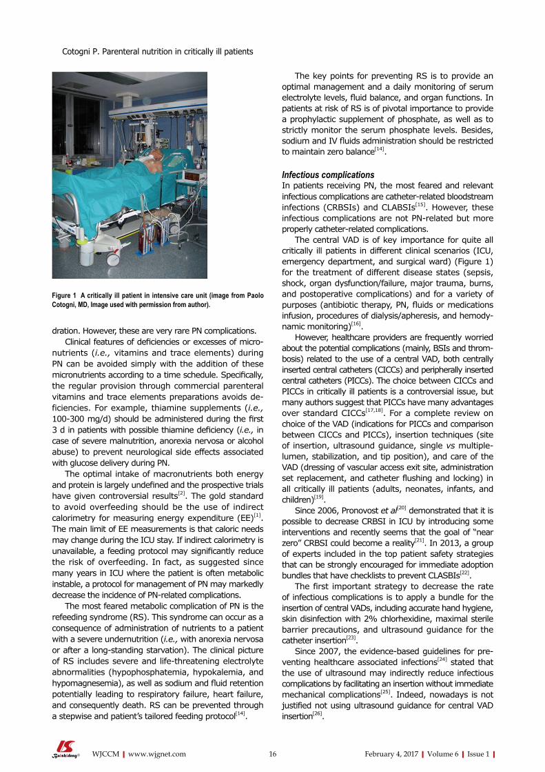
16 February 4, 2017|Volume 6|Issue 1|WJCCM|www.wjgnet.com
dration. However, these are very rare PN complications.Clinical features of deficiencies or excesses of micro
nutrients (i.e., vitamins and trace elements) during PN can be avoided simply with the addition of these micronutrients according to a time schedule. Specifically, the regular provision through commercial parenteral vitamins and trace elements preparations avoids deficiencies. For example, thiamine supplements (i.e., 100300 mg/d) should be administered during the first 3 d in patients with possible thiamine deficiency (i.e., in case of severe malnutrition, anorexia nervosa or alcohol abuse) to prevent neurological side effects associated with glucose delivery during PN.
The optimal intake of macronutrients both energy and protein is largely undefined and the prospective trials have given controversial results[2]. The gold standard to avoid overfeeding should be the use of indirect calorimetry for measuring energy expenditure (EE)[1]. The main limit of EE measurements is that caloric needs may change during the ICU stay. If indirect calorimetry is unavailable, a feeding protocol may significantly reduce the risk of overfeeding. In fact, as suggested since many years in ICU where the patient is often metabolic instable, a protocol for management of PN may markedly decrease the incidence of PNrelated complications.
The most feared metabolic complication of PN is the refeeding syndrome (RS). This syndrome can occur as a consequence of administration of nutrients to a patient with a severe undernutrition (i.e., with anorexia nervosa or after a longstanding starvation). The clinical picture of RS includes severe and lifethreatening electrolyte abnormalities (hypophosphatemia, hypokalemia, and hypomagnesemia), as well as sodium and fluid retention potentially leading to respiratory failure, heart failure, and consequently death. RS can be prevented through a stepwise and patient’s tailored feeding protocol[14].
The key points for preventing RS is to provide an optimal management and a daily monitoring of serum electrolyte levels, fluid balance, and organ functions. In patients at risk of RS is of pivotal importance to provide a prophylactic supplement of phosphate, as well as to strictly monitor the serum phosphate levels. Besides, sodium and IV fluids administration should be restricted to maintain zero balance[14].
Infectious complicationsIn patients receiving PN, the most feared and relevant infectious complications are catheterrelated bloodstream infections (CRBSIs) and CLABSIs[15]. However, these infectious complications are not PNrelated but more properly catheterrelated complications.
The central VAD is of key importance for quite all critically ill patients in different clinical scenarios (ICU, emergency department, and surgical ward) (Figure 1) for the treatment of different disease states (sepsis, shock, organ dysfunction/failure, major trauma, burns, and postoperative complications) and for a variety of purposes (antibiotic therapy, PN, fluids or medications infusion, procedures of dialysis/apheresis, and hemodynamic monitoring)[16].
However, healthcare providers are frequently worried about the potential complications (mainly, BSIs and thrombosis) related to the use of a central VAD, both centrally inserted central catheters (CICCs) and peripherally inserted central catheters (PICCs). The choice between CICCs and PICCs in critically ill patients is a controversial issue, but many authors suggest that PICCs have many advantages over standard CICCs[17,18]. For a complete review on choice of the VAD (indications for PICCs and comparison between CICCs and PICCs), insertion techniques (site of insertion, ultrasound guidance, single vs multiplelumen, stabilization, and tip position), and care of the VAD (dressing of vascular access exit site, administration set replacement, and catheter flushing and locking) in all critically ill patients (adults, neonates, infants, and children)[19].
Since 2006, Pronovost et al[20] demonstrated that it is possible to decrease CRBSI in ICU by introducing some interventions and recently seems that the goal of “near zero” CRBSI could become a reality[21]. In 2013, a group of experts included in the top patient safety strategies that can be strongly encouraged for immediate adoption bundles that have checklists to prevent CLASBIs[22].
The first important strategy to decrease the rate of infectious complications is to apply a bundle for the insertion of central VADs, including accurate hand hygiene, skin disinfection with 2% chlorhexidine, maximal sterile barrier precautions, and ultrasound guidance for the catheter insertion[23].
Since 2007, the evidencebased guidelines for preventing healthcare associated infections[24] stated that the use of ultrasound may indirectly reduce infectious complications by facilitating an insertion without immediate mechanical complications[25]. Indeed, nowadays is not justified not using ultrasound guidance for central VAD insertion[26].
Figure 1 A critically ill patient in intensive care unit (image from Paolo Cotogni, MD, Image used with permission from author).
Cotogni P. Parenteral nutrition in critically ill patients
17 February 4, 2017|Volume 6|Issue 1|WJCCM|www.wjgnet.com
The second relevant strategy to decrease the incidence of infectious complications is to apply a bundle for the care of central VADs, including the use of biopatch, semipermeable transparent dressing, 2% chlorhexidine for skin disinfection, and sutureless devices for the catheter care.
Another important strategy to decrease the rate of CRBSI is the selection of the exit site[27]. Many authors suggest the importance to move the exit site of the central VAD from the neck or the supraclavicular area to the infraclavicular area for CICCs or the upper midarm for PICCs.
Mechanical complicationsThe mechanical complications that can more frequently occur are: Lumen occlusion, catheter dislocation, rupture of external tract, and the most feared venous thrombosis. However, these complications, as infectious complications, are catheterrelated and not PNrelated complications. According to the ESPEN guidelines, central catheterrelated venous thrombosis may be prevented by: (1) the use at insertion of the ultrasound guidance; (2) the use of a VAD with the smallest caliber compatible with the patient’s need; and (3) the position of the tip of the central VAD between the superior vena cava and the right atrium (at or near the socalled atriocaval junction)[23].
ENTERAL VS PARENTERAL NUTRITIONBased on the experience of Dudrick et al[28], the use of PN was introduced in the late 1960s. Since then, without any doubt PN helped greatly many critically ill patients to recover from previously lifelimiting clinical conditions. However, the diffuse use of this therapy in all patients, even with extensive indications, brought reservations regarding its benefits and increased the role of EN in the subsequent years.
In the late 1980s emerging evidence from animal studies supported the concept that EN promotes gut function and prevents the translocation of intestinal bacteria. Therefore, total PN was considered to be a “dangerous” form of therapy (e.g., “more harm than good” or “a poison”) and this belief resulted in EN becoming the new standard of care in AN.
The PN is also criticized because it is more expensive than EN. Indeed, both EN and PN are relatively cheap treatments, especially if compared to other therapies that the critically ill patients need to survive.
All together, these concerns influenced the decisionmaking of physicians about the choice between EN and PN for the nutrition support of critically ill patients.
In fact, in 2000 Heyland[29] questioned if PN in critically ill patients was more harm than good because there were studies comparing EN with PN suggesting that PN was associated with increased complications and mortality in some subgroups of these patients. On the contrary, Jeejeebhoy[30] in 2001 had an absolutely
opposite judgment regarding PN and stated that the rate of PNrelated complications have been overstated.
In critically ill patients, EN is the recommended method of nutritional support when the patient is unable to have an adequate oral intake of nutrients to meet his/her nutritional requirements and the gastrointestinal tract is functional. The enteral route is efficient and costeffective, however it is not always as easy as it looks.
Also EN may be the cause of complications that can be classified as: (1) metabolic (e.g., RS may occur also with EN); (2) gastrointestinal (e.g., early satiety, nausea, vomiting, and diarrhea); and (3) mechanical or tuberelated (e.g., malposition, dislodgement, and clogging, both in case of nasogastric tube and percutaneous endoscopic gastrostomy). Actually, the most feared complication of EN is pulmonary aspiration because it can be a lifethreatening condition.
The debate over the topic of the route for delivering AN in critically ill patients are relevant and attractive. Since several years, metaanalysis and RCTs comparing EN and PN found conflicting results as regards the benefits of EN vs PN in critically ill patients. In my opinion, there is a great misunderstanding in this debate; in particular, that EN and PN are competitors. On the contrary, the selection of EN or PN for delivering nutritional support should be tailored on an individual basis.
In fact, the route chosen for providing AN should be appropriate to the patient’s clinical conditions and should frequently be evaluated for persistent appropriateness, as well as for its adequacy in meeting nutritional requirements of the patient. Not infrequently, in critically ill patients the gastrointestinal tract is not able to tolerate the administration of the prescribed amount of EN formula. If a patient develops intestinal dysfunction/failure due to his/her critical illnesses, PN is more efficient to meet patient’s nutritional needs and better tolerated than EN.
In 2014, Harvey et al[31] reported the results of a RCT evaluating the route of early nutrition support in 2400 adult critically ill patients. The trial compared early NE with early PN and demonstrated that: (1) the PN and the EN groups did not have significant differences in rates of adverse events, treated infectious complications, and other 14 secondary outcomes; and (2) the target intake was not achieved in most patients although the caloric intake was comparable between the groups. Therefore, the authors concluded that: (1) there was no association between 30d mortality and the route for delivering the early nutritional support; and (2) early PN, as it is generally administered, is neither more beneficial nor more harmful than EN in critically ill patients.
SUMMARYThe PNrelated complications are catheterrelated or metabolic complications. In 2005, Beghetto et al[32] reported that PN was an independent risk factor for central venous catheterrelated infection in nonselected
Cotogni P. Parenteral nutrition in critically ill patients
18 February 4, 2017|Volume 6|Issue 1|WJCCM|www.wjgnet.com
hospitalized adult patients. In the past years, the rate of catheterrelated complications varied from 1.5 to 4.9 episodes per 1000 catheter days, depending on the inhospital patient population, severity of illness, and the type of central VAD[33]. However, after the Pronovost paper[20], the widespread diffusion of guidelines on care, diagnosis, and therapy of complications of central venous catheters access in PN patients[23], and the introduction of nutrition support teams[6], the incidence of PNrelated complications is markedly reduced.
In fact, in the RCT of Harvey et al[31] the mean number of infectious complications was 0.22 in the parenteral group. Moreover, many studies demonstrated that the goal of “near zero” CRBSI[21] has been achieved; actually, in the recent years the incidence of catheterrelated infections in ICU patients varied from 0[18,3436] to 2.4[37] episodes per 1000 catheter days.
Moreover, the optimization of energy provision with supplemental PN in critically ill patients could reduce nosocomial infections, antibiotic usage, and time on mechanical ventilation, and consequently overall healthcare costs[38].
It is well known that hepatic dysfunctions are common PNrelated metabolic complications in patients receiving longterm PN[39], but this is not the case for critically ill patients that generally receive PN for weeks.
PN is effective and safe when EN is not feasible or tolerated. However, in these patients receiving PN, endogenous infection is more significant than exogenous infection. Indeed, the lack of EN significantly disrupts the usual gastrointestinal microbiota, leads to mucosal gut atrophy and impaired intestinal barrier function. This may determine an impaired immunity favoring endotoxin absorption, macrophagemediated and cytokinemediated inflammation, and dangerous bacterial translocation[33].
The question is: How to decrease the incidence of CRBSIs in this condition? The suggestion is that all patients receiving PN, because of EN was initially not tolerated or feasible, should be assessed every day for tolerance of EN and a combined administration of PN and EN should be initiated as soon as feasible.
An emerging and intriguing issue is the use of probiotics, alone or in combination with prebiotics, in critically ill patients to restore the balance of gastrointestinal microbiota with a beneficial impact on intestinal permeability and bacterial translocation. However, further studies in this field are needed before a clear recommendation can be released on the possible therapeutic use of pre and probiotics for the protection of the gut in ICU patients[40].
CONCLUSIONThe AN is necessary to meet the nutritional requirements of critically ill patients at nutrition risk because undernutrition determines a poorer prognosis in these patients. There is debate over which route of delivery of AN provides better outcomes and lesser complications in
critically ill patients.The fighting against PN complications should consider:
(1) an appropriate blood glucose control; (2) the use of olive oil and fish oilbased lipid emulsions alternative to soybean oilbased ones; (3) the adoption of insertion and care centralline bundles; and (4) the implementation of a policy of targeting “near zero” CRBSIs. Adopting all these strategies, the goal of “near zero” PN complications is achievable.
If accurately managed, PN can be safely provided for most critically ill patients without expecting a relevant incidence of PNrelated complications. Moreover, the use of protocols for the management of nutritional support and the presence of nutrition support teams may decrease PNrelated complications.
In conclusion, the key messages about the management of PN in critically ill patients are two. First, the dangers of PNrelated complications have been exaggerated because complications are uncommon; moreover, infectious complications, as mechanical complications, are more properly catheterrelated and not PNrelated complications. Second, when EN is not feasible or tolerated, PN is as effective and safe as EN.
REFERENCES1 Berger MM, Pichard C. Development and current use of parenteral
nutrition in critical care - an opinion paper. Crit Care 2014; 18: 478 [PMID: 25184816 DOI: 10.1186/s13054-014-0478-0]
2 Preiser JC, van Zanten AR, Berger MM, Biolo G, Casaer MP, Doig GS, Griffiths RD, Heyland DK, Hiesmayr M, Iapichino G, Laviano A, Pichard C, Singer P, Van den Berghe G, Wernerman J, Wischmeyer P, Vincent JL. Metabolic and nutritional support of critically ill patients: consensus and controversies. Crit Care 2015; 19: 35 [PMID: 25886997 DOI: 10.1186/s13054-015-0737-8]
3 Taylor BE, McClave SA, Martindale RG, Warren MM, Johnson DR, Braunschweig C, McCarthy MS, Davanos E, Rice TW, Cresci GA, Gervasio JM, Sacks GS, Roberts PR, Compher C. Guidelines for the Provision and Assessment of Nutrition Support Therapy in the Adult Critically Ill Patient: Society of Critical Care Medicine (SCCM) and American Society for Parenteral and Enteral Nutrition (A.S.P.E.N.). Crit Care Med 2016; 44: 390-438 [PMID: 26771786 DOI: 10.1097/CCM.0000000000001525]
4 Singer P, Berger MM, Van den Berghe G, Biolo G, Calder P, Forbes A, Griffiths R, Kreyman G, Leverve X, Pichard C. ESPEN Guidelines on Parenteral Nutrition: intensive care. Clin Nutr 2009; 28: 387-400 [PMID: 19505748 DOI: 10.1016/j.clnu.2009.04.024]
5 Kreymann KG, Berger MM, Deutz NE, Hiesmayr M, Jolliet P, Kazandjiev G, Nitenberg G, van den Berghe G, Wernerman J, Ebner C, Hartl W, Heymann C, Spies C. ESPEN Guidelines on Enteral Nutrition: Intensive care. Clin Nutr 2006; 25: 210-223 [PMID: 16697087 DOI: 10.1016/j.clnu.2006.01.021]
6 Hvas CL, Farrer K, Donaldson E, Blackett B, Lloyd H, Forde C, Garside G, Paine P, Lal S. Quality and safety impact on the provision of parenteral nutrition through introduction of a nutrition support team. Eur J Clin Nutr 2014; 68: 1294-1299 [PMID: 25248359 DOI: 10.1038/ejcn.2014.186]
7 Boullata JI, Gilbert K, Sacks G, Labossiere RJ, Crill C, Goday P, Kumpf VJ, Mattox TW, Plogsted S, Holcombe B. A.S.P.E.N. clinical guidelines: parenteral nutrition ordering, order review, compounding, labeling, and dispensing. JPEN J Parenter Enteral Nutr 2014; 38: 334-377 [PMID: 24531708 DOI: 10.1177/0148607114521833]
8 Pichard C, Schwarz G, Frei A, Kyle U, Jolliet P, Morel P, Romand JA, Sierro C. Economic investigation of the use of three-
Cotogni P. Parenteral nutrition in critically ill patients
19 February 4, 2017|Volume 6|Issue 1|WJCCM|www.wjgnet.com
compartment total parenteral nutrition bag: prospective randomized unblinded controlled study. Clin Nutr 2000; 19: 245-251 [PMID: 10952795 DOI: 10.1054/clnu.2000.0106]
9 Pontes-Arruda A, Dos Santos MC, Martins LF, González ER, Kliger RG, Maia M, Magnan GB. Influence of parenteral nutrition delivery system on the development of bloodstream infections in critically ill patients: an international, multicenter, prospective, open-label, controlled study--EPICOS study. JPEN J Parenter Enteral Nutr 2012; 36: 574-586 [PMID: 22269899 DOI: 10.1177/0148607111427040]
10 Bouchoud L, Fonzo-Christe C, Klingmüller M, Bonnabry P. Compatibility of intravenous medications with parenteral nutrition: in vitro evaluation. JPEN J Parenter Enteral Nutr 2013; 37: 416-424 [PMID: 23112277 DOI: 10.1177/0148607112464239]
11 van den Berghe G, Wouters P, Weekers F, Verwaest C, Bruyninckx F, Schetz M, Vlasselaers D, Ferdinande P, Lauwers P, Bouillon R. Intensive insulin therapy in critically ill patients. N Engl J Med 2001; 345: 1359-1367 [PMID: 11794168 DOI: 10.1056/NEJMoa011300]
12 Manzanares W, Dhaliwal R, Jurewitsch B, Stapleton RD, Jeejeebhoy KN, Heyland DK. Alternative lipid emulsions in the critically ill: a systematic review of the evidence. Intensive Care Med 2013; 39: 1683-1694 [PMID: 23812404 DOI: 10.1007/s00134-013-2999-4]
13 Edmunds CE, Brody RA, Parrott JS, Stankorb SM, Heyland DK. The effects of different IV fat emulsions on clinical outcomes in critically ill patients. Crit Care Med 2014; 42: 1168-1177 [PMID: 24351374 DOI: 10.1097/CCM.0000000000000146]
14 Khan LU, Ahmed J, Khan S, Macfie J. Refeeding syndrome: a literature review. Gastroenterol Res Pract 2011; 2011: pii: 410971 [PMID: 20886063 DOI: 10.1155/2011/410971]
15 Lorente L, Villegas J, Martín MM, Jiménez A, Mora ML. Catheter-related infection in critically ill patients. Intensive Care Med 2004; 30: 1681-1684 [PMID: 15160239 DOI: 10.1007/s00134-004-2332-3]
16 Palmer D, McFie J. Venous access for parenteral nutrition. In: Payne James J, Grimble GK, Silk, DBA. Artificial nutrition support in clinical practice. London, UK, 2001: 379-400
17 Griffiths VR, Philpot P. Peripherally inserted central catheters (PICCs): do they have a role in the care of the critically ill patient? Intensive Crit Care Nurs 2002; 18: 37-47 [PMID: 12008876 DOI: 10.1054/iccn.2002.1615]
18 Pittiruti M, Brutti A, Celentano D, Pomponi M, Biasucci DG, Annetta MG, Scoppettuolo G. Clinical experience with power-injectable PICCs in intensive care patients. Crit Care 2012; 16: R21 [PMID: 22305301 DOI: 10.1186/cc11181]
19 Cotogni P, Pittiruti M. Focus on peripherally inserted central catheters in critically ill patients. World J Crit Care Med 2014; 3: 80-94 [PMID: 25374804 DOI: 10.5492/wjccm.v3.i4.80]
20 Pronovost P, Needham D, Berenholtz S, Sinopoli D, Chu H, Cosgrove S, Sexton B, Hyzy R, Welsh R, Roth G, Bander J, Kepros J, Goeschel C. An intervention to decrease catheter-related bloodstream infections in the ICU. N Engl J Med 2006; 355: 2725-2732 [PMID: 17192537 DOI: 10.1056/NEJMoa061115]
21 Ruiz-Santana S, Saavedra P, León C. “Near zero” catheter-related bloodstream infections: turning dreams into reality*. Crit Care Med 2012; 40: 3083-3084 [PMID: 23080438 DOI: 10.1097/CCM.0b013e3182632748]
22 Shekelle PG, Pronovost PJ, Wachter RM, McDonald KM, Schoelles K, Dy SM, Shojania K, Reston JT, Adams AS, Angood PB, Bates DW, Bickman L, Carayon P, Donaldson L, Duan N, Farley DO, Greenhalgh T, Haughom JL, Lake E, Lilford R, Lohr KN, Meyer GS, Miller MR, Neuhauser DV, Ryan G, Saint S, Shortell SM, Stevens DP, Walshe K. The top patient safety strategies that can be encouraged for adoption now. Ann Intern Med 2013; 158: 365-368 [PMID: 23460091 DOI: 10.7326/0003-4819-158-5-201303051-00001]
23 Pittiruti M, Hamilton H, Biffi R, MacFie J, Pertkiewicz M. ESPEN Guidelines on Parenteral Nutrition: central venous catheters (access, care, diagnosis and therapy of complications). Clin Nutr 2009; 28:
365-377 [PMID: 19464090 DOI: 10.1016/j.clnu.2009.04.004]24 Loveday HP, Wilson JA, Pratt RJ, Golsorkhi M, Tingle A, Bak
A, Browne J, Prieto J, Wilcox M. epic3: national evidence-based guidelines for preventing healthcare-associated infections in NHS hospitals in England. J Hosp Infect 2014; 86 Suppl 1: S1-S70 [PMID: 24330862 DOI: 10.1016/S0195-6701(13)60012-2]
25 Lamperti M, Bodenham AR, Pittiruti M, Blaivas M, Augoustides JG, Elbarbary M, Pirotte T, Karakitsos D, Ledonne J, Doniger S, Scoppettuolo G, Feller-Kopman D, Schummer W, Biffi R, Desruennes E, Melniker LA, Verghese ST. International evidence-based recommendations on ultrasound-guided vascular access. Intensive Care Med 2012; 38: 1105-1117 [PMID: 22614241 DOI: 10.1007/s00134-012-2597-x]
26 Bodenham AR. Can you justify not using ultrasound guidance for central venous access? Crit Care 2006; 10: 175 [PMID: 17129362 DOI: 10.1186/cc5079]
27 Pittiruti M, Migliorini I, Emoli A, Dolcetti L, Pomponi M, Scoppettuolo G, LaGreca A. Preventing central venous catheter related infections: catheter site selection and insertion technique significantly affect the chances of adequate catheter site care. Intensive Care Med 2007; 33 Suppl: S13
28 Dudrick SJ, Wilmore DW, Vars HM, Rhoads JE. Long-term total parenteral nutrition with growth, development, and positive nitrogen balance. Surgery 1968; 64: 134-142 [PMID: 4968812]
29 Heyland DK. Parenteral nutrition in the critically-ill patient: more harm than good? Proc Nutr Soc 2000; 59: 457-466 [PMID: 10997674 DOI: 10.1017/S002966510000063X]
30 Jeejeebhoy KN. Total parenteral nutrition: potion or poison? Am J Clin Nutr 2001; 74: 160-163 [PMID: 11470715]
31 Harvey SE, Parrott F, Harrison DA, Bear DE, Segaran E, Beale R, Bellingan G, Leonard R, Mythen MG, Rowan KM. Trial of the route of early nutritional support in critically ill adults. N Engl J Med 2014; 371: 1673-1684 [PMID: 25271389 DOI: 10.1056/NEJMoa1409860]
32 Beghetto MG, Victorino J, Teixeira L, de Azevedo MJ. Parenteral nutrition as a risk factor for central venous catheter-related infection. JPEN J Parenter Enteral Nutr 2005; 29: 367-373 [PMID: 16107600 DOI: 10.1177/0148607105029005367]
33 Mundi MS , Nystrom EM, Hurley DL, McMahon MM. Management of Parenteral Nutrition in Hospitalized Adult Patients. JPEN J Parenter Enteral Nutr 2016; Epub ahead of print [PMID: 27587535 DOI: 10.1177/0148607116667060]
34 Berenholtz SM, Pronovost PJ, Lipsett PA, Hobson D, Earsing K, Farley JE, Milanovich S, Garrett-Mayer E, Winters BD, Rubin HR, Dorman T, Perl TM. Eliminating catheter-related bloodstream infections in the intensive care unit. Crit Care Med 2004; 32: 2014-2020 [PMID: 15483409 DOI: 10.1097/01.CCM.0000142399.70913.2F]
35 Pronovost PJ, Goeschel CA, Colantuoni E, Watson S, Lubomski LH, Berenholtz SM, Thompson DA, Sinopoli DJ, Cosgrove S, Sexton JB, Marsteller JA, Hyzy RC, Welsh R, Posa P, Schumacher K, Needham D. Sustaining reductions in catheter related bloodstream infections in Michigan intensive care units: observational study. BMJ 2010; 340: c309 [PMID: 20133365 DOI: 10.1136/bmj.c309]
36 DePalo VA, McNicoll L, Cornell M, Rocha JM, Adams L, Pronovost PJ. The Rhode Island ICU collaborative: a model for reducing central line-associated bloodstream infection and ventilator-associated pneumonia statewide. Qual Saf Health Care 2010; 19: 555-561 [PMID: 21127114 DOI: 10.1136/qshc.2009.038265]
37 Peredo R, Sabatier C, Villagrá A, González J, Hernández C, Pérez F, Suárez D, Vallés J. Reduction in catheter-related bloodstream infections in critically ill patients through a multiple system intervention. Eur J Clin Microbiol Infect Dis 2010; 29: 1173-1177 [PMID: 20533071 DOI: 10.1007/s10096-010-0971-6]
38 Heidegger CP, Berger MM, Graf S, Zingg W, Darmon P, Costanza MC, Thibault R, Pichard C. Optimisation of energy provision with supplemental parenteral nutrition in critically ill patients: a randomised controlled clinical trial. Lancet 2013; 381: 385-393 [PMID: 23218813 DOI: 10.1016/S0140-6736(12)61351-8]
39 Raman M, Allard JP. Parenteral nutrition related hepato-biliary
Cotogni P. Parenteral nutrition in critically ill patients

20 February 4, 2017|Volume 6|Issue 1|WJCCM|www.wjgnet.com
disease in adults. Appl Physiol Nutr Metab 2007; 32: 646-654 [PMID: 17622278 DOI: 10.1139/H07-056]
40 Manzanares W, Lemieux M, Langlois PL, Wischmeyer PE.
Probiotic and synbiotic therapy in critical illness: a systematic review and meta-analysis. Crit Care 2016; 19: 262 [PMID: 27538711 DOI: 10.1186/s13054-016-1434-y]
P- Reviewer: Dunser MW, Ren HT, Yu WK S- Editor: Qiu S L- Editor: A E- Editor: Li D
Cotogni P. Parenteral nutrition in critically ill patients

Karel Heytens, Jan De Bleecker, Walter Verbrugghe, Jonathan Baets, Luc Heytens
MINIREVIEWS
21 February 4, 2017|Volume 6|Issue 1|WJCCM|www.wjgnet.com
Exertional rhabdomyolysis and heat stroke: Beware of volatile anesthetic sedation
Karel Heytens, Department of Anesthesiology, University Hospital Antwerp, 2650 Edegem, Belgium
Jan De Bleecker, Department of Neurology, AZ Sint-Lucas, 9000 Ghent, Belgium
Jan De Bleecker, Department of Neurology, University Hospital Ghent, 9000 Ghent, Belgium
Walter Verbrugghe, Department of Intensive Care, University Hospital Antwerp, 2650 Edegem, Belgium
Jonathan Baets, Department of Neurology, University Hospital Antwerp, 2650 Edegem, Belgium
Jonathan Baets, Laboratory of Neurogenetics and Biobank, Institute Born-Bunge, University of Antwerp, 2610 Wilrijk, Belgium
Luc Heytens, MH Research Unit, University of Antwerp, 2610 Wilrijk, Belgium
Luc Heytens, Departments of Anesthesiology and Neurology, University Hospital Antwerp, 2650 Edegem, Belgium
Author contributions: Heytens K is the main author of the manuscript; De Bleecker J and Baets J provided information about the different cases and reviewed the manuscript; Verbrugghe W helped with his experience on volatile anesthetic sedation; Heytens L designed the aim of the manuscript and acted as co-writer.
Conflict-of-interest statement: The authors have no interests with the manufacturers of the AnaConDaR or MirusTM devices. Heytens L is a medical expert with Norgine NV, manufacturer of DantriumR.
Open-Access: This article is an open-access article which was selected by an in-house editor and fully peer-reviewed by external reviewers. It is distributed in accordance with the Creative Commons Attribution Non Commercial (CC BY-NC 4.0) license, which permits others to distribute, remix, adapt, build upon this work non-commercially, and license their derivative works on different terms, provided the original work is properly cited and the use is non-commercial. See: http://creativecommons.org/licenses/by-nc/4.0/
Manuscript source: Invited manuscript
Correspondence to: Luc Heytens, MD, PhD, MH Research Unit, University of Antwerp, Campus Drie Eiken, 2610 Wilrijk, Belgium. [email protected]: +32-34-433926
Received: June 28, 2016 Peer-review started: July 1, 2016First decision: September 5, 2016Revised: October 21, 2016 Accepted: November 16, 2016Article in press: November 17, 2016Published online: February 4, 2017
AbstractIn view of the enormous popularity of mass sporting events such as half-marathons, the number of patients with exertional rhabdomyolysis or exercise-induced heat stroke admitted to intensive care units (ICUs) has increased over the last decade. Because these patients have been reported to be at risk for malignant hyperthermia during general anesthesia, the intensive care community should bear in mind that the same risk of life-threatening rhabdomyolysis is present when these patients are admitted to an ICU, and volatile anesthetic sedation is chosen as the sedative technique. As illustrated by the three case studies we elaborate upon, a thorough diagnostic work-up is needed to clarify the subsequent risk of strenuous exercise, and the anesthetic exposure to volatile agents in these patients and their families. Other contraindications for the use of volatile intensive care sedation consist of known malignant hyperthermia susceptibility, congenital myopathies, Duchenne muscular dystrophy, and intracranial hypertension.
Key words: Exertional rhabdomyolysis; Heat stroke; Intensive care sedation; Inhalational anesthetics; Malignant hyperthermia; Congenital myopathies
World Journal ofCritical Care MedicineW J C C M
Submit a Manuscript: http://www.wjgnet.com/esps/Help Desk: http://www.wjgnet.com/esps/helpdesk.aspxDOI: 10.5492/wjccm.v6.i1.21
World J Crit Care Med 2017 February 4; 6(1): 21-27ISSN 2220-3141 (online)
© 2017 Baishideng Publishing Group Inc. All rights reserved.

22 February 4, 2017|Volume 6|Issue 1|WJCCM|www.wjgnet.com
Heytens K et al . Heatstroke and volatile anesthetic sedation
© The Author(s) 2017. Published by Baishideng Publishing Group Inc. All rights reserved.
Core tip: Recent research has shown that a substantial proportion of patients with exercise-induced heatstroke harbor mutations in the ryanodine-receptor one gene on Chromosome 19 (RYR1), encoding for the principal calcium-release channel in striated muscle. These same mutations are known to result in a massively increased calcium-conductivity and life-threatening rhabdomyolysis when malignant hyperthermia (MH) susceptible patients are exposed to volatile anesthetics during general anesthesia. In view of this, exposure to volatile anesthetic sedation - an emerging trend in intensive care units - is contraindicated, not only in patients with known MH susceptibility and other congenital myopathies, but also in patients admitted because of exertional rhabdomyolysis and heatstroke.
Heytens K, De Bleecker J, Verbrugghe W, Baets J, Heytens L. Exertional rhabdomyolysis and heat stroke: Beware of volatile anesthetic sedation. World J Crit Care Med 2017; 6(1): 21-27 Available from: URL: http://www.wjgnet.com/2220-3141/full/v6/i1/21.htm DOI: http://dx.doi.org/10.5492/wjccm.v6.i1.21
INTRODUCTIONExercise-induced rhabdomyolysis (EIR), also known as exertional rhabdomyolysis, is a clinical emergency characterized by extensive post-exercise muscle necrosis and the release of intracellular muscle components into the circulation. The diagnosis is confirmed by a significantly elevated serum creatine kinase (CK) level and/or the presence of myoglobinemia and myoglobinuria. There is no definitive pathological CK cut-off, and therefore, clinical symptoms such as muscle swelling, myalgia, and tenderness occurring with low-intensity exercise, or lasting several days longer than expected, are important clinical signs.
The most severe end of the clinical spectrum-and one of the leading causes of death in young athletes-is known as exertional heat stroke (EHS), a syndrome clinically defined as a body temperature over 40 ℃, with severe rhabdomyolysis and signs of encephalopathy ranging from confusion to convulsions or coma. It is often complicated by multiorgan failure, including acute renal and hepatic failure, and it may result in death if appropriate treatment in an intensive care setting is delayed.
It has been reported for over two decades that a significant part of patients with a history of EIR and/or heat stroke is at increased risk of developing malignant hyperthermia (MH) during general anesthesia[1,2].
MH is well known among anesthetists. It is inherited as an autosomal dominant, genetically heterogeneous trait that manifests as an acute rhabdomyolysis during general anesthesia when susceptible individuals are exposed to volatile anesthetics and/or succinylcholine.
The incidence is generally estimated to be approximately 1 in 50000 general anesthesias. In the 1970-1980s, mortality was over 80%, but now, it is fortunately less than 5%[3].
A fulminant MH-crisis is characterized by a com-bination of clinical and biochemical events: Inappropriate hypercapnia and respiratory acidosis, polypnea, tach-yarrhythmia, rapidly increasing body temperature, sweating, generalized muscle rigidity, hemodynamic instability, dark urine due to myoglobinuria, significant CK-increase (often > 10.000 IU/L), and postoperative stiffness and myalgia. Death may result from severe hyperkalemia in combination with respiratory and meta-bolic acidosis, acute renal failure, hyperthermia > 42 ℃, disseminated intravascular coagulation and fatal cardiac arrhythmias.
Mortality has decreased significantly over the last 20 years because of our better understanding of the syndrome, the use of end-tidal CO2 (ETCO2) monitoring resulting in earlier diagnosis, and the availability of the antidote dantrolene.
Treatment consists of the immediate discontinuation of volatile anesthetic agents and the administration of dantrolene. The surgical procedure should be terminated as quickly as possible and if the procedure cannot be aborted, the anesthesia technique has to be converted to total intravenous anesthesia. Dantrolene should be administered as a loading bolus of 2.5 mg/kg Ⅳ. Subsequent doses of 1 mg/kg Ⅳ can be repeated up to a maximum dose of 10 mg/kg, and are administered until the clinical signs (hypercapnia, hyperthermia, rigidity) abate. Concomitantly minute volume should be maximized to reduce respiratory acidosis.
Despite adequate treatment, complications still occur in 20% of patients. The most common complication is renal dysfunction and acute renal failure. The complication rate increases to ≥ 30% when 20 or more minutes elapse between the first clinical sign and dantrolene treatment[4].
This life-threatening anesthesia-related complication is due to the occurrence of point mutations in the genes coding for the calcium-release channel of the sarcoplasmic reticulum, e.g., the didyropyridine - ryanodine receptor complex, resulting in a disturbed skeletal muscle calcium homeostasis. The ryanodine receptor (RYR1 gene on Chromosome 19q) contains the actual “calcium pore”. The NH2-terminal of this protein forms cytosolic protrusions that extend toward, and make contact with, the voltage-gated dihydropyridine receptor located in the T-tubular wall. The corresponding gene for this protein is CACNA1S on Chromosome 1. Depolarization of the sarcolemma and T-tubule first activates the dihydropyridine receptor which in turn activates the ryanodine receptor and opens the calcium-channel as such.
As illustrated in Figure 1, the muscle from patients harboring RYR1 and/or DHPR mutations upon exposure to volatile anesthetics has been shown to exhibit an increased calcium-conductivity and massive calcium re-lease from the SR, in turn resulting in sustained muscle
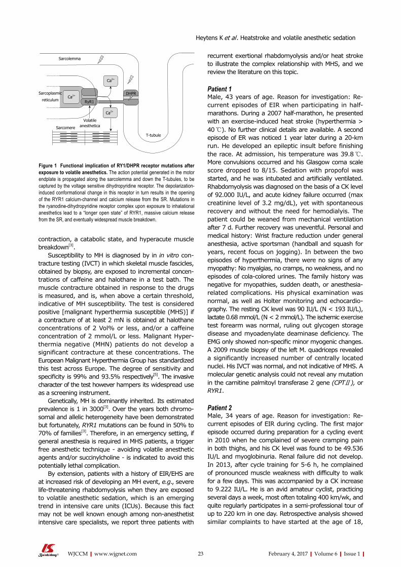
23 February 4, 2017|Volume 6|Issue 1|WJCCM|www.wjgnet.com
contraction, a catabolic state, and hyperacute muscle breakdown[3].
Susceptibility to MH is diagnosed by in in vitro con-tracture testing (IVCT) in which skeletal muscle fascicles, obtained by biopsy, are exposed to incremental concen-trations of caffeine and halothane in a test bath. The muscle contracture obtained in response to the drugs is measured, and is, when above a certain threshold, indicative of MH susceptibility. The test is considered positive [malignant hyperthermia susceptible (MHS)] if a contracture of at least 2 mN is obtained at halothane concentrations of 2 Vol% or less, and/or a caffeine concentration of 2 mmol/L or less. Malignant Hyper-thermia negative (MHN) patients do not develop a significant contracture at these concentrations. The European Malignant Hyperthermia Group has standardized this test across Europe. The degree of sensitivity and specificity is 99% and 93.5% respectively[5]. The invasive character of the test however hampers its widespread use as a screening instrument.
Genetically, MH is dominantly inherited. Its estimated prevalence is 1 in 3000[3]. Over the years both chromo-somal and allelic heterogeneity have been demonstrated but fortunately, RYR1 mutations can be found in 50% to 70% of families[3]. Therefore, in an emergency setting, if general anesthesia is required in MHS patients, a trigger free anesthetic technique - avoiding volatile anesthetic agents and/or succinylcholine - is indicated to avoid this potentially lethal complication.
By extension, patients with a history of EIR/EHS are at increased risk of developing an MH event, e.g., severe life-threatening rhabdomyolysis when they are exposed to volatile anesthetic sedation, which is an emerging trend in intensive care units (ICUs). Because this fact may not be well known enough among non-anesthetist intensive care specialists, we report three patients with
recurrent exertional rhabdomyolysis and/or heat stroke to illustrate the complex relationship with MHS, and we review the literature on this topic.
Patient 1Male, 43 years of age. Reason for investigation: Re-current episodes of EIR when participating in half-marathons. During a 2007 half-marathon, he presented with an exercise-induced heat stroke (hyperthermia > 40 ℃). No further clinical details are available. A second episode of ER was noticed 1 year later during a 20-km run. He developed an epileptic insult before finishing the race. At admission, his temperature was 39.8 ℃. More convulsions occurred and his Glasgow coma scale score dropped to 8/15. Sedation with propofol was started, and he was intubated and artificially ventilated. Rhabdomyolysis was diagnosed on the basis of a CK level of 92.000 IU/L, and acute kidney failure occurred (max creatinine level of 3.2 mg/dL), yet with spontaneous recovery and without the need for hemodialyis. The patient could be weaned from mechanical ventilation after 7 d. Further recovery was uneventful. Personal and medical history: Wrist fracture reduction under general anesthesia, active sportsman (handball and squash for years, recent focus on jogging). In between the two episodes of hyperthermia, there were no signs of any myopathy: No myalgias, no cramps, no weakness, and no episodes of cola-colored urines. The family history was negative for myopathies, sudden death, or anesthesia-related complications. His physical examination was normal, as well as Holter monitoring and echocardio-graphy. The resting CK level was 90 IU/L (N < 193 IU/L), lactate 0.68 mmol/L (N < 2 mmol/L). The ischemic exercise test forearm was normal, ruling out glycogen storage disease and myoadenylate deaminase deficiency. The EMG only showed non-specific minor myogenic changes. A 2009 muscle biopsy of the left M. quadriceps revealed a significantly increased number of centrally located nuclei. His IVCT was normal, and not indicative of MHS. A molecular genetic analysis could not reveal any mutation in the carnitine palmitoyl transferase 2 gene (CPTⅡ), or RYR1.
Patient 2Male, 34 years of age. Reason for investigation: Re-current episodes of EIR during cycling. The first major episode occurred during preparation for a cycling event in 2010 when he complained of severe cramping pain in both thighs, and his CK level was found to be 49.536 IU/L and myoglobinuria. Renal failure did not develop. In 2013, after cycle training for 5-6 h, he complained of pronounced muscle weakness with difficulty to walk for a few days. This was accompanied by a CK increase to 9.222 IU/L. He is an avid amateur cyclist, practicing several days a week, most often totaling 400 km/wk, and quite regularly participates in a semi-professional tour of up to 220 km in one day. Retrospective analysis showed similar complaints to have started at the age of 18,
Sarcolemma
Ca2+
Sarcoplasmic
reticulumCa2+
RyR1
Ca2+
Sarcomere
Volatile anesthetica
T-tubule
DHPR
Figure 1 Functional implication of RY1/DHPR receptor mutations after exposure to volatile anesthetics. The action potential generated in the motor endplate is propagated along the sarcolemma and down the T-tubules, to be captured by the voltage sensitive dihydropyridine receptor. The depolarization-induced conformational change in this receptor in turn results in the opening of the RYR1 calcium-channel and calcium release from the SR. Mutations in the ryanodine-dihydropyridine receptor complex upon exposure to inhalational anesthetics lead to a “longer open state” of RYR1, massive calcium release from the SR, and eventually widespread muscle breakdown.
Heytens K et al . Heatstroke and volatile anesthetic sedation
24 February 4, 2017|Volume 6|Issue 1|WJCCM|www.wjgnet.com
mainly consisting of myalgia with feeling of stiffness, and intermittent myoglobinuria and CK increase. In between the crises, there is normal mobility and power, and no CK increase. He had not yet been exposed to volatile anesthetics. The family history is negative for anesthesia-related events. His father ran marathons, reporting no clinical events. His neurological examination was normal, and functional and biochemical tests showed a basal CK serum level of 293 U/L, a lactate of 0.8 mmol/L, a normal ischemic forearm exercise test and EMG. Muscle biopsy of the left M. quadriceps femoris in 2011 showed an increased number of fibers with internal nuclei and a minor increase of neutral fat drops in some type 1 fibers. These were interpreted as unspecific findings. A repeat muscle biopsy of the left M. quadriceps femoris in 2014 again demonstrated findings of mild myopathic disease: Increased number of fibers with internal nuclei (25%) but also a small number of “cores”, which are zones devoid of enzymatic staining. IVCT results: 8 mN contracture at 2 Vol% Halothane, 6 mN contracture with 2 mmol/L Caffeine, indicative of MHS. Molecular genetic analysis did not reveal any mutation in the CPTⅡgene or RYR1.
Patient 3Male, 56 years of age. Reason for investigation: Recurrent myalgia confined to the thigh muscles, described as a sensation of severe tension or cramps, sometimes unilateral, sometimes bilateral. The type of effort appeared not to be related to the complaints: At times a prolonged effort did not provoke any myalgia, whereas at other times, the pain/cramps started after as short an effort of 5-10 min such as descending a staircase. CK values measured during an episode of myalgia rose up to 14.000 IE/L. He is also an amateur cyclist, but complaints were only noticed after the age of 40. There was no previous exposure to volatile anesthetics, and the family history was also negative for anesthesia-related events. His son is not active in sports (his daughter is), but until now has not mentioned any complaints. His clinical examination was normal, but he showed an increase in basal CK value of 269-1771 IU/L at diverse occasions, lactate was 1.2 mmol/L, and the ischemic forearm test was normal. A 2015 muscle biopsy only showed atypical myogenic anomalies. The IVCT was abnormal (5 mN contracture with 2 mmol/L Caffeine, 14 mN contracture with 2 Vol% Halothane), demonstrating MHS. Molecular genetic analysis did not reveal a mutation in the CPTⅡ gene, but RYR1- mutation analysis demonstrated a base-pair change in exon 43 (p.N2342S; c.7025A > G).
DISCUSSIONThere is an emerging trend of using inhalational an-esthetics for ICU sedation[6]. The principle motivation is that inhalational anesthetics have an interesting pharmacokinetic profile compared with the usual com-bination of propofol or benzodiazepine with an analgesic drug. Several practical advantages have been proposed, such as rapid onset and offset of action, low potential for
accumulation in fat tissue, drug clearance through the lungs and therefore independent of liver and/or renal function, no tachyphylaxis, an opium sparing effect, bronchodilatory properties, and end-organ protective properties, such as ischemic preconditioning of the heart, although clinical data remain limited on this.
Until about a decade ago, the technical prerequisites for ICU use of volatile anesthetics were not fulfilled, and therefore, their use remained confined to the OR. Technological advances, however, have greatly sim-plified the application of inhalational anesthetics for ICU sedation. An important milestone was reached in 2005 with the introduction of a volatile anesthetic reflection filter, the anesthetic conserving device (AnaConDaTM, Sedana Medical, Uppsala, Sweden), retaining 90% of the volatile anesthetic and thereby enormously reducing the consumption of volatile anesthetic. Since then, other AnaConDaTM competing devices have been produced and tested such as the MirusTM (Pall Medical, Dreieich, Germany)[7]. Suitable volatile agents are isoflurane, sevoflurane, and desflurane. From a drug delivery point of view, these technical developments have indeed resulted in easy titration to the clinical end-point and the possibility of breath-by-breath bedside monitoring. A 0.3-0.6 minimal alveolar concentration of sevoflurane is most often sufficient in the ICU setting. In view of the increased CO2 through the systems’ enlarged dead space (100 mL), minute volume usually has to be increased by being guided by ETCO2 or blood gas analysis. Desflurane is not commonly used in the ICU in view of its boiling point of 22.8 ℃, requiring a vaporizer heated to 39 ℃, and the higher cost. It can be administered by the MirusTM device, not the AnaConDaTM.
Recent studies have indicated that the currently available scavenger devices that are based on a silica zeolite matrix, adequately adsorb the volatile agents and thereby minimize environmental pollution by volatile an-esthetic agents guaranteeing workplace safety[8].
The question still remains to be answered whether volatile anesthetics will really become and stay a player in critical care sedation[6]; however, the technique is being increasingly used in Europe and North-America.
A pilot randomized controlled trial is currently being set up to evaluate the practicability and dangers related to volatile anesthetic agents when used for long-term critical care sedation[9]. This prospective multicenter trial is blinded to the data analyst and aims to recruit 60 adult ICU patients requiring mechanical ventilation and sedation for at least 48 h, in which 40 patients will receive inhaled isoflurane and 20 patients will receive intravenous midazolam and/or propofol, titrated to a targeted Sedation Analgesia Score. Primary outcomes will assess adherence to the particular sedation protocol and atmospheric volatile concentration levels. Secondary outcomes that will be investigated include the quality of sedation, delirium, vasoactive drug support, time to extubation, serum fluoride levels, and mortality.
With an ever-rising concern for adverse events involved in our handling of patients, for any technique to
Heytens K et al . Heatstroke and volatile anesthetic sedation
25 February 4, 2017|Volume 6|Issue 1|WJCCM|www.wjgnet.com
stand a chance of proving itself, all side-effects have to be clearly identified and communicated to the end users.
In this sense, Purrucker et al[10] has issued a warning concerning the use of volatile anesthetic sedation in patients with a high risk of intracranial hypertension. Switching from Ⅳ sedation to sevoflurane decreased MAP and CPP in one-third of the patients studied to such an extent that the early termination of sevoflurane administration was required. The mechanism felt to be responsible was vasodilation in response to a decreasing MAP and a slightly raised partial-pressure carbon dioxide in patients with an already low baseline cerebral compliance.
In this paper, we want to warn about the use of volatile anesthetic sedation in a second subset of ICU patients, in particular those with EIR/EHS in need of intensive care.
Although EIR/EHS is most often encountered in a military setting, the recent worldwide trend to organize mass (semi)marathons has resulted in a significant increase in the frequency of this problem in ICUs world-wide. Indeed, the Centers for Disease Control and Prevention of the United States reported that EHI occurs both during practice and competition, with a disturbing trend of increasing incidence, and mentioning EIR/EHS as one of the leading causes of death in young athletes each year[11].
General anesthesia with volatile anesthetic agents, such as the currently used sevoflurane and desflurane, is known to potentially induce acute rhabdomyolysis in patients with a genetic predisposition to MH. Logically, the same risk is present when genetically MHS patients are exposed to volatile anesthetic sedation in the in-tensive care setting. A recent publication reported on the development of MH in an ICU patient sedated with sevoflurane[12].
Other groups of patients are also at risk. A link between MHS and EIR has been suggested in the anesthesia literature for over two decades. The earlier reports linked MHS with EIR by demonstrating a positive in vitro contracture test. One of the larger series published[2] reports on the IVCT results in 12 unrelated patients with EIR. Ten of the 12 patients had IVCT results indicative of MHS. In the ensuing years, reports occurred providing evidence that “MH-mutations” in RYR1 were being associated with EIR. In 2002, Davis et al[13] reported two patients with EIR in which MHS was confirmed through in-vitro contracture testing and the presence of a RYR1 mutation.
Several similar cases were reported, and in 2013, Dlamini et al[14] published a large series of 39 unrelated families with rhabdomyolysis and/or exertional myalgia in which nine heterozygous mutations were found in 14 families, several of them recurrent. Five of these mutations had previously been associated with MH. They conclude that RYR1 mutations account for a substantial proportion of patients presenting with unexplained rhab-domyolysis and/or exertional myalgia, but also that various stressors (e.g., pain, environmental heat, viral infections, drugs) may need to be present to elicit acute
rhabdomyolysis. They also suggest that “additional family studies are paramount in order to identify potentially MH susceptible relatives”.
In a 2014 paper, Zhao et al[15] actually raised the question on whether the two disorders represent one and the same disorder, which they called the Human Stress Syndrome. This hypothesis has gained support, and MH and EIR/EHS are increasingly believed to be different presentations of the “expanding spectrum of RYR1-related myopathies”[16].
In view of the growing evidence that EIR in a substantial portion of the patients admitted to the ICU is a “non-anesthetic RYR1-related rhabdomyolysis”[16],
it is of great importance for the patients and families involved to undergo a thorough investigation on the cause of the life-threatening rhabdomyolysis, certainly if the problem has been found to be recurrent. The following reasons substantiate this statement: (1) several “common” underlying causes with significant impact on patients’ lives have to be ruled out, such as sickle cell trait, CPTⅡ deficiency, McArdle’s disease (glycogen storage disease Ⅴ), myoadenylate deaminase deficiency, and others; (2) to identify potentially MHS individuals as implications for future general anesthesia are important. If a patient is found to be MHS, preventive measures concerning the anesthetic technique have to be taken. Over the last decade, several experts have stated that individuals who have a history of EHS should be screened for MHS[2,16-18]. Even though the IVCT is an invasive test, it is still considered to be the most sensitive and specific test to determine a patient’s predisposition to MHS. The estimation of the MH risk in a particular patient is certainly not straightforward, as illustrated by the case presentations. Patient 1 had a negative IVCT and a negative genetic analysis, and is considered to be non-MHS. Therefore, volatile anesthetics can safely be administered in the operating room/ICU to this patient. Patient 2 had a positive IVCT, but no mutation was found upon RYR1 sequencing. However, it is well known that a RYR1 mutation is found in only 50%-70% of patients with a positive IVCT[19]. CACNA1S cDNA sequencing was not performed in our patients in view of the cost and the large number of sequence changes reported, most of which are of unclear significance[20]. In this patient, volatile anesthesia has to be avoided in the future, because he is considered MHS by IVCT. Patient 3 had a positive IVCT and a positive RYR1 mutation (p.N2342S; c.7025A>G - exon 43) that was previously linked to MH, and therefore, this patient is clearly at risk for MH upon exposure to volatile anesthetic agents; and (3) If RYR1 mutations are found, the condition should be con-sidered to be hereditary, and additional family studies are indicated.
The patient should be seen by a neurologist, because a large number of diverse etiologies have been implicated in acute EIR/EHS, and certain additional clinical features can aid to define the most appropriate investigations. A guideline for a diagnostic EIR/HS workup has been suggested by Capacchione et al[21]. Because of the rarity
Heytens K et al . Heatstroke and volatile anesthetic sedation
26 February 4, 2017|Volume 6|Issue 1|WJCCM|www.wjgnet.com
and heterogeneity of these conditions, however, this EIR/HS workup remains a real diagnostic challenge.
A third group of patients at risk when given volatile anesthetic sedation are patients with congenital myo-pathies. This is a group of rare genetic muscle disorders (6 in 100000 live births) characterized by different structural abnormalities in skeletal muscle fibers either observable by light- or EM microscopy, and symptoms of hypotonia and muscle weakness present at birth (although the clinical expression may be delayed until childhood or even adult life). These myopathies are genetically hetero-geneous, but a substantial subgroup is linked with “gain-of-function” mutations in the RYR1-gene, resulting in increased calcium conductance of the calcium-release channel and the potential for acute rhabdomyolysis when exposed to volatile anesthetics. This has been shown to be the case for central core disease, multiminicore disease, centronuclear myopathy, congenital fiber type disproportion, late-onset axial myopathy, and King-Denborough syndrome[22].
The propensity of patients with muscular dystrophies (especially Duchenne muscular dystrophy) to react adversely to volatile anesthetic sedation is well known; however, this does not occur through the presence of RYR1 mutations but rather is the result of “toxic effects” of these agents as well as succinylcholine on the fragile sarcolemma. Prolonged exposure to volatile anesthetic agents, and certainly the use of succinylcholine, has to be avoided in this group of patients.
If intensive care sedation is needed in these patients, a combination of propofol, benzodiazepines, morphino-mimetics, neuraxial block, and, as last resort, non-depolarizing neuromuscular blockers can be used. Dexmedetomidine use for procedural sedation is safe.
CONCLUSIONDominantly inherited MHS is rare. Therefore, in the general population, life-threatening acute rhabdomyolysis following exposure to volatile anesthetics either in the operating room or the ICU is seldom encountered. However, because a substantial proportion of patients with EIR/EHS and congenital myopathies harbor RYR1 mutations resulting in an increased calcium-conductivity of the Ca-release channel of the SR, volatile anesthetic sedation should not be used in these high-risk patients.
Contraindications for volatile anesthetic sedation in intensive care consist of: (1) known susceptibility for MH; (2) congenital myopathies (central core disease, multiminicore disease, centronuclear myopathy, congenital fiber type disproportion, late-onset axial myopathy, and King-Denborough syndrome); (3) duchenne muscular dystrophy; (4) exertional rhabdomyolysis; and (5) intra-cranial hypertension.
REFERENCES1 Hopkins PM, Ellis FR, Halsall PJ. Evidence for related myopathies
in exertional heat stroke and malignant hyperthermia. Lancet 1991;
338: 1491-1492 [PMID: 1683922 DOI: 10.1016/0140-6736(91)92304-K]
2 Wappler F, Fiege M, Steinfath M, Agarwal K, Scholz J, Singh S, Matschke J, Schulte Am Esch J. Evidence for susceptibility to malignant hyperthermia in patients with exercise-induced rhab-domyolysis. Anesthesiology 2001; 94: 95-100 [PMID: 11135728 DOI: 10.1097/00000542-200101000-00019]
3 Rosenberg H, Pollock N, Schiemann A, Bulger T, Stowell K. Malignant hyperthermia: a review. Orphanet J Rare Dis 2015; 10: 93 [PMID: 26238698 DOI: 10.1186/s13023-015-0310-1]
4 Riazi S, Larach MG, Hu C, Wijeysundera D, Massey C, Kraeva N. Malignant hyperthermia in Canada: characteristics of index anesthetics in 129 malignant hyperthermia susceptible probands. Anesth Analg 2014; 118: 381-387 [PMID: 23842196 DOI: 10.1213/ANE.0b013e3182937d8b]
5 Ording H, Brancadoro V, Cozzolino S, Ellis FR, Glauber V, Gonano EF, Halsall PJ, Hartung E, Heffron JJ, Heytens L, Kozak-Ribbens G, Kress H, Krivosic-Horber R, Lehmann-Horn F, Mortier W, Nivoche Y, Ranklev-Twetman E, Sigurdsson S, Snoeck M, Stieglitz P, Tegazzin V, Urwyler A, Wappler F. In vitro contracture test for diagnosis of malignant hyperthermia following the protocol of the European MH Group: results of testing patients surviving fulminant MH and unrelated low-risk subjects. The European Malignant Hyperthermia Group. Acta Anaesthesiol Scand 1997; 41: 955-966 [PMID: 9311391 DOI: 10.1111/j.1399-6576.1997.tb04820.x]
6 Jerath A, Parotto M, Wasowicz M, Ferguson ND. Volatile Anes-thetics. Is a New Player Emerging in Critical Care Sedation? Am J Respir Crit Care Med 2016; 193: 1202-1212 [PMID: 27002466 DOI: 10.1164/rccm.201512-2435CP]
7 Bomberg H, Glas M, Groesdonk VH, Bellgardt M, Schwarz J, Volk T, Meiser A. A novel device for target controlled administration and reflection of desflurane--the Mirus™. Anaesthesia 2014; 69: 1241-1250 [PMID: 25040673 DOI: 10.1111/anae.12798]
8 Wong K, Wasowicz M, Grewal D, Fowler T, Ng M, Ferguson ND, Steel A, Jerath A. Efficacy of a simple scavenging system for long-term critical care sedation using volatile agent-based anesthesia. Can J Anaesth 2016; 63: 630-632 [PMID: 26670802 DOI: 10.1007/s12630-015-0562-1]
9 Jerath A, Ferguson ND, Steel A, Wijeysundera D, Macdonald J, Wasowicz M. The use of volatile anesthetic agents for long-term critical care sedation (VALTS): study protocol for a pilot randomized controlled trial. Trials 2015; 16: 560 [PMID: 26646404 DOI: 10.1186/s13063-015-1083-5]
10 Purrucker JC, Renzland J, Uhlmann L, Bruckner T, Hacke W, Steiner T, Bösel J. Volatile sedation with sevoflurane in intensive care patients with acute stroke or subarachnoid haemorrhage using AnaConDa®: an observational study. Br J Anaesth 2015; 114: 934-943 [PMID: 25823541 DOI: 10.1093/bja/aev070]
11 Centers for Disease Control and Prevention. Heat illness among high school athletes --- United States, 2005-2009. MMWR Morb Mortal Wkly Rep 2010; 59: 1009-1013 [PMID: 20724966]
12 Rosenberg H, Schuster F, Johannsen S. The introduction of a lightweight mini vaporizer and malignant hyperthermia. Can J Anaesth 2015; 62: 319 [PMID: 25398599]
13 Davis M, Brown R, Dickson A, Horton H, James D, Laing N, Marston R, Norgate M, Perlman D, Pollock N, Stowell K. Malignant hyperthermia associated with exercise-induced rhabdomyolysis or congenital abnormalities and a novel RYR1 mutation in New Zealand and Australian pedigrees. Br J Anaesth 2002; 88: 508-515 [PMID: 12066726 DOI: 10.1093/bja/88.4.508]
14 Dlamini N, Voermans NC, Lillis S, Stewart K, Kamsteeg EJ, Drost G, Quinlivan R, Snoeck M, Norwood F, Radunovic A, Straub V, Roberts M, Vrancken AF, van der Pol WL, de Coo RI, Manzur AY, Yau S, Abbs S, King A, Lammens M, Hopkins PM, Mohammed S, Treves S, Muntoni F, Wraige E, Davis MR, van Engelen B, Jungbluth H. Mutations in RYR1 are a common cause of exertional myalgia and rhabdomyolysis. Neuromuscul Disord 2013; 23: 540-548 [PMID: 23628358 DOI: 10.1016/j.nmd.2013.03.008]
15 Zhao X, Song Q, Gao Y. Hypothesis: exertional heat stroke-induced myopathy and genetically inherited malignant hyper-
Heytens K et al . Heatstroke and volatile anesthetic sedation
27 February 4, 2017|Volume 6|Issue 1|WJCCM|www.wjgnet.com
thermia represent the same disorder, the human stress syndrome. Cell Biochem Biophys 2014; 70: 1325-1329 [PMID: 24948473 DOI: 10.1007/s12013-014-0059-5]
16 Snoeck M, Treves S, Molenaar JP, Kamsteeg EJ, Jungbluth H, Voermans NC. “Human Stress Syndrome” and the Expanding Spectrum of RYR1-Related Myopathies. Cell Biochem Biophys 2016; 74: 85-87 [PMID: 26972305 DOI: 10.1007/s12013- 015-0704-7]
17 Grogan H, Hopkins PM. Heat stroke: implications for critical care and anaesthesia. Br J Anaesth 2002; 88: 700-707 [PMID: 12067009 DOI: 10.1093/bja/88.5.700]
18 Sambuughin N, Capacchione J, Blokhin A, Bayarsaikhan M, Bina S, Muldoon S. The ryanodine receptor type 1 gene variants in African American men with exertional rhabdomyolysis and malignant hyperthermia susceptibility. Clin Genet 2009; 76: 564-568 [PMID: 19807743 DOI: 10.1111/j.1399-0004.2009.01251.x]
19 Carpenter D, Robinson RL, Quinnell RJ, Ringrose C, Hogg M, Casson F, Booms P, Iles DE, Halsall PJ, Steele DS, Shaw
MA, Hopkins PM. Genetic variation in RYR1 and malignant hyperthermia phenotypes. Br J Anaesth 2009; 103: 538-548 [PMID: 19648156 DOI: 10.1093/bja/aep204]
20 Carpenter D, Ringrose C, Leo V, Morris A, Robinson RL, Halsall PJ, Hopkins PM, Shaw MA. The role of CACNA1S in predisposition to malignant hyperthermia. BMC Med Genet 2009; 10: 104 [PMID: 19825159 DOI: 10.1186/1471-2350-10-104]
21 Capacchione JF, Muldoon SM. The relationship between ex-ertional heat illness, exertional rhabdomyolysis, and malignant hyperthermia. Anesth Analg 2009; 109: 1065-1069 [PMID: 19617585 DOI: 10.1213/ane.0b013e3181a9d8d9]
22 Snoeck M, van Engelen BG, Küsters B, Lammens M, Meijer R, Molenaar JP, Raaphorst J, Verschuuren-Bemelmans CC, Straathof CS, Sie LT, de Coo IF, van der Pol WL, de Visser M, Scheffer H, Treves S, Jungbluth H, Voermans NC, Kamsteeg EJ. RYR1-related myopathies: a wide spectrum of phenotypes throughout life. Eur J Neurol 2015; 22: 1094-1112 [PMID: 25960145 DOI: 10.1111/ene.12713]
P- Reviewer: Beltowski J, Lin JA, Oji C, Stocco G S- Editor: Ji FF L- Editor: A E- Editor: Li D
Heytens K et al . Heatstroke and volatile anesthetic sedation
Thu AN Nguyen, Yasmine Ali Abdelhamid, Liza K Phillips, Leeanne S Chapple, Michael Horowitz, Karen L Jones, Adam M Deane
MINIREVIEWS
28 February 4, 2017|Volume 6|Issue 1|WJCCM|www.wjgnet.com
Nutrient stimulation of mesenteric blood flow - implications for older critically ill patients
Thu AN Nguyen, Yasmine Ali Abdelhamid, Leeanne S Chapple, Adam M Deane, Discipline of Acute Care Medicine, University of Adelaide, Adelaide 5005, Australia
Liza K Phillips, Michael Horowitz, Karen L Jones, Adam M Deane, National Health and Medical Research Council Centre for Research Excellence in Translating Nutritional Science to Good Health, Adelaide 5000, Australia
Liza K Phillips, Michael Horowitz, Endocrine and Metabolic Unit, Royal Adelaide Hospital, Adelaide 5000, Australia
Liza K Phillips, Michael Horowitz, Karen L Jones, Discipline of Medicine, University of Adelaide, Adelaide 5005, Australia
Adam M Deane, Intensive Care Unit, Royal Melbourne Hospital, University of Melbourne, Parkville, Victoria 3050, Australia
Author contributions: All authors equally contributed to this paper including conception and design of the study, literature review and analysis, drafting and critical revision and editing, and final approval of the final version.
Conflict-of-interest statement: No potential conflicts of interest.
Open-Access: This article is an open-access article which was selected by an in-house editor and fully peer-reviewed by external reviewers. It is distributed in accordance with the Creative Commons Attribution Non Commercial (CC BY-NC 4.0) license, which permits others to distribute, remix, adapt, build upon this work non-commercially, and license their derivative works on different terms, provided the original work is properly cited and the use is non-commercial. See: http://creativecommons.org/licenses/by-nc/4.0/
Manuscript source: Invited manuscript
Correspondence to: Adam M Deane, MBBS, PhD, Intensive Care Unit, Royal Melbourne Hospital, University of Melbourne, 300 Grattan Street, Parkville, Victoria 3050, Australia. [email protected]: +61-3-93429234
Received: August 26, 2016Peer-review started: August 27, 2016First decision: December 13, 2016Revised: December 16, 2016Accepted: January 2, 2017Article in press: January 3, 2017Published online: February 4, 2017
AbstractNutrient ingestion induces a substantial increase in mesenteric blood flow. In older persons (aged ≥ 65 years), particularly those with chronic medical conditions, the cardiovascular compensatory response may be inadequate to maintain systemic blood pressure during mesenteric blood pooling, leading to postprandial hypotension. In older ambulatory persons, postprandial hypotension is an important pathophysiological condition associated with an increased propensity for syncope, falls, coronary vascular events, stroke and death. In older critically ill patients, the administration of enteral nutrition acutely increases mesenteric blood flow, but whether this pathophysiological response is protective, or precipitates mesenteric ischaemia, is unknown. There are an in-creasing number of older patients surviving admission to intensive care units, who are likely to be at increased risk of postprandial hypotension, both during, and after, their stay in hospital. In this review, we describe the prevalence, impact and mechanisms of postprandial hypotension in older people and provide an overview of the impact of postprandial hypotension on feeding prescriptions in older critically ill patients. Finally, we provide evidence that postprandial hypotension is likely to be an unrecognised problem in older survivors of critical illness and discuss potential options for management.
Key words: Postprandial hypotension; Enteral nutrition; Critical care; Aged; Mesenteric ischaemia
World Journal ofCritical Care MedicineW J C C M
Submit a Manuscript: http://www.wjgnet.com/esps/Help Desk: http://www.wjgnet.com/esps/helpdesk.aspxDOI: 10.5492/wjccm.v6.i1.28
World J Crit Care Med 2017 February 4; 6(1): 28-36ISSN 2220-3141 (online)
© 2017 Baishideng Publishing Group Inc. All rights reserved.

29 February 4, 2017|Volume 6|Issue 1|WJCCM|www.wjgnet.com
Nguyen T et al . Postprandial hypotension in the older critically ill patients
© The Author(s) 2017. Published by Baishideng Publishing Group Inc. All rights reserved.
Core tip: In older ambulatory persons, postprandial hypotension is an important pathophysiological con-dition associated with an increased propensity to coronary vascular events, stroke and death. In older critically ill patients, the administration of enteral nutrition acutely increases mesenteric blood flow, but whether this pathophysiological response is protective, or pre-cipitates mesenteric ischaemia, is unknown. We herein describe the prevalence, impact and mechanisms and management of postprandial hypotension in older people. We finally provide an overview of the impact of postprandial hypotension on feeding prescriptions in and evidence that postprandial hypotension is likely to be an unrecognised problem in older survivors of critical illness.
Nguyen TAN, Abdelhamid YA, Phillips LK, Chapple LS, Horowitz M, Jones KL, Deane AM. Nutrient stimulation of mesenteric blood flow - implications for older critically ill patients. World J Crit Care Med 2017; 6(1): 28-36 Available from: URL: http://www.wjgnet.com/2220-3141/full/v6/i1/28.htm DOI: http://dx.doi.org/10.5492/wjccm.v6.i1.28
INTRODUCTIONIngestion of nutrients initiates a complex process involving precise coordination between the gastrointestinal tract, autonomic and cardiovascular systems to increase intestinal blood flow, whilst simultaneously maintaining circulatory homeostasis[1,2]. Age and disease-related changes may compromise cardiovascular compensatory mechanisms, which, particularly in older persons, may result in a clinically relevant postprandial fall in blood pressure, known as postprandial hypotension (PPH). PPH is inconsistently defined but is generally regarded as a reduction in systolic blood pressure of ≥ 20 mmHg, or a decrease to ≤ 90 mmHg, that occurs within two hours of a meal and persists for at least 30 min[3]. This definition is empiric and derived from the definition of orthostatic hypotension[4]. It is important to recognise that although PPH frequently coexists with orthostatic hypotension, PPH is a distinct entity. However PPH may well occur more frequently, and have more substantive implications, than orthostatic hypotension[5,6].
EPIDEMIOLOGY A recent meta-analysis reported that PPH occurs in about 20% of “healthy” older persons, about 30%-40% of nursing home residents, 20%-91% of hospitalised patients aged ≥ 65 years, about 40% of people with diabetes, and 40%-100% of patients with Parkinson’s disease[7]. The wide range of reported prevalence in each group reflect
the small cohort sizes and the confounding effect of lack of standardisation of methodology between studies; including the definition of PPH, composition of test meal, timing of meal ingestion, technique and duration of blood pressure measurement, and use of concomitant medications. However, it is clear that in each of these groups the prevalence of PPH is high and that the very elderly and patients with diseases associated with auto-nomic dysfunction are at particular risk. Surprisingly, the prevalence of PPH in elderly survivors of critical illness has not been evaluated.
CLINICAL IMPORTANCE OF POSTPRANDIAL HYPOTENSIONPPH is now recognised as an important pathophysiological condition, not only because of its high prevalence, but also due to the associated substantial morbidity and mortality[3]. In older people in the community, PPH is a strong predictor of syncope, falls, coronary events and stroke - irrespective of whether the individual has symptoms[8]. In a prospective study of 499 nursing home residents, Aronow et al[8] reported that the postprandial fall in systolic blood pressure was an independent risk factor for falls, coronary events, stroke and all-cause mortality. Supportive data are also provided by two case-control studies that report that the magnitude and prevalence of PPH are substantially greater in patients with a history of falls or syncope when compared to controls[9,10]. Furthermore, in a five-year study of nursing home residents, PPH was found to be an independent determinant of mortality (RR = 1.79; 95%CI: 1.19-2.68); with a “dose-response”, such that all-cause mortality increased 13% for every 10 mmHg decrease in post-prandial systolic blood pressure (RR = 1.13; 95%CI: 1.03-1.24)[11].
As indicated, preliminary data suggest that it is important to identify PPH even in those patients who are unaware of the condition. While PPH is associated with adverse outcomes, more than half (about 60%) of patients with PPH may be asymptomatic and, therefore, do not seek treatment[5,6]. For example, Kohara et al[12] studied 70 patients hospitalised with essential hypertension and reported that the prevalence of lacunar infarcts was increased two-fold in patients with asymptomatic PPH. The strong association between “asymptomatic” PPH and stroke has also been evident in larger cohorts of older people residing in nursing home facilities and ambulatory older people living in the community[8,13]. While this association does not establish causality, it provides a compelling rationale to diagnose PPH, which is a simple and inexpensive process[7], and to determine whether interventions that attenuate PPH mitigate the risk of adverse outcomes, such as cerebrovascular events[14]. The latter approach is to some extent compromised by the current lack of established effective management strategies[15].
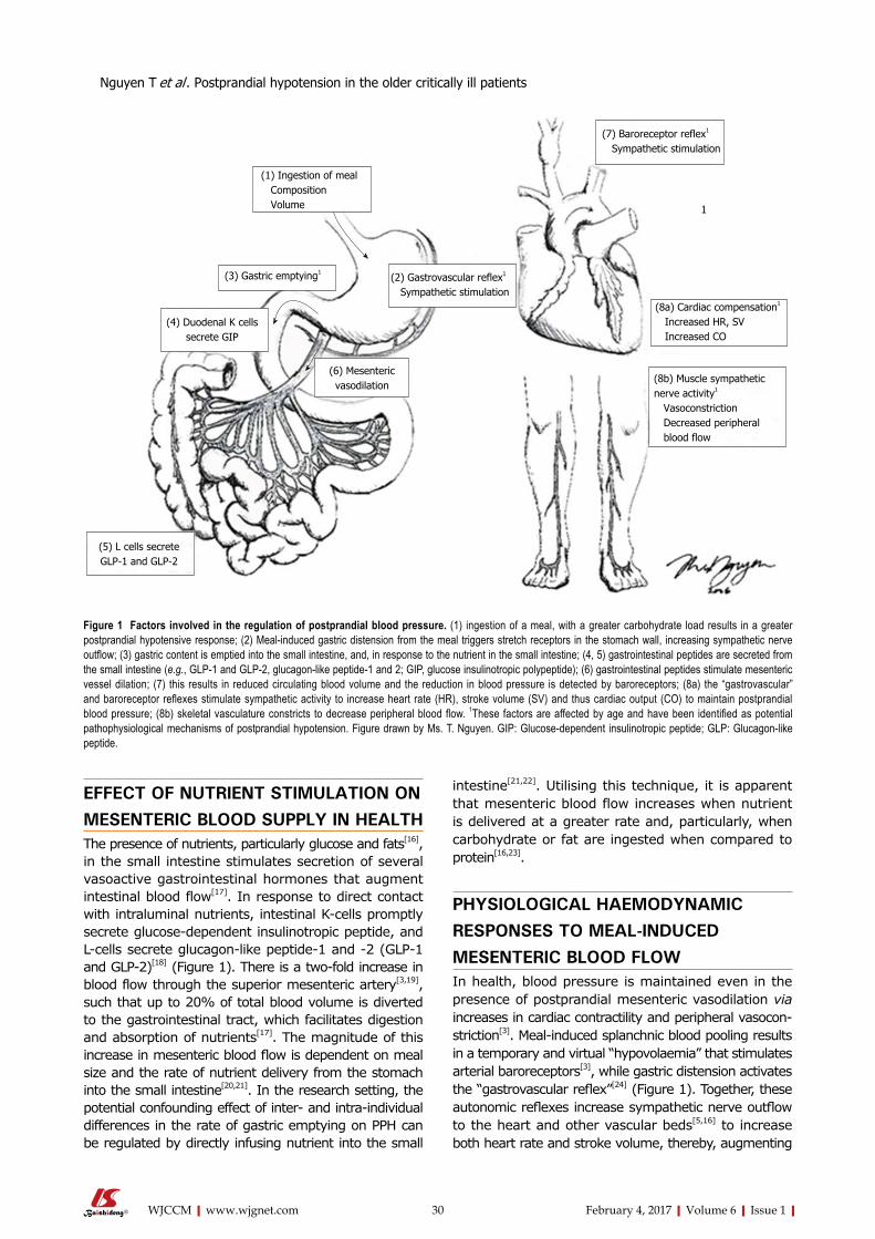
30 February 4, 2017|Volume 6|Issue 1|WJCCM|www.wjgnet.com
EFFECT OF NUTRIENT STIMULATION ON MESENTERIC BLOOD SUPPLY IN HEALTHThe presence of nutrients, particularly glucose and fats[16], in the small intestine stimulates secretion of several vasoactive gastrointestinal hormones that augment intestinal blood flow[17]. In response to direct contact with intraluminal nutrients, intestinal K-cells promptly secrete glucose-dependent insulinotropic peptide, and L-cells secrete glucagon-like peptide-1 and -2 (GLP-1 and GLP-2)[18] (Figure 1). There is a two-fold increase in blood flow through the superior mesenteric artery[3,19], such that up to 20% of total blood volume is diverted to the gastrointestinal tract, which facilitates digestion and absorption of nutrients[17]. The magnitude of this increase in mesenteric blood flow is dependent on meal size and the rate of nutrient delivery from the stomach into the small intestine[20,21]. In the research setting, the potential confounding effect of inter- and intra-individual differences in the rate of gastric emptying on PPH can be regulated by directly infusing nutrient into the small
intestine[21,22]. Utilising this technique, it is apparent that mesenteric blood flow increases when nutrient is delivered at a greater rate and, particularly, when carbohydrate or fat are ingested when compared to protein[16,23].
PHYSIOLOGICAL HAEMODYNAMIC RESPONSES TO MEAL-INDUCED MESENTERIC BLOOD FLOW In health, blood pressure is maintained even in the presence of postprandial mesenteric vasodilation via increases in cardiac contractility and peripheral vasocon-striction[3]. Meal-induced splanchnic blood pooling results in a temporary and virtual “hypovolaemia” that stimulates arterial baroreceptors[3], while gastric distension activates the “gastrovascular reflex”[24] (Figure 1). Together, these autonomic reflexes increase sympathetic nerve outflow to the heart and other vascular beds[5,16] to increase both heart rate and stroke volume, thereby, augmenting
(1) Ingestion of meal Composition Volume
(2) Gastrovascular reflex1
Sympathetic stimulation
(3) Gastric emptying1
(4) Duodenal K cells secrete GIP
(5) L cells secrete GLP-1 and GLP-2
(6) Mesenteric vasodilation
(7) Baroreceptor reflex1
Sympathetic stimulation
1
(8a) Cardiac compensation1
Increased HR, SV Increased CO
(8b) Muscle sympathetic nerve activity1
Vasoconstriction Decreased peripheral blood flow
Figure 1 Factors involved in the regulation of postprandial blood pressure. (1) ingestion of a meal, with a greater carbohydrate load results in a greater postprandial hypotensive response; (2) Meal-induced gastric distension from the meal triggers stretch receptors in the stomach wall, increasing sympathetic nerve outflow; (3) gastric content is emptied into the small intestine, and, in response to the nutrient in the small intestine; (4, 5) gastrointestinal peptides are secreted from the small intestine (e.g., GLP-1 and GLP-2, glucagon-like peptide-1 and 2; GIP, glucose insulinotropic polypeptide); (6) gastrointestinal peptides stimulate mesenteric vessel dilation; (7) this results in reduced circulating blood volume and the reduction in blood pressure is detected by baroreceptors; (8a) the “gastrovascular” and baroreceptor reflexes stimulate sympathetic activity to increase heart rate (HR), stroke volume (SV) and thus cardiac output (CO) to maintain postprandial blood pressure; (8b) skeletal vasculature constricts to decrease peripheral blood flow. 1These factors are affected by age and have been identified as potential pathophysiological mechanisms of postprandial hypotension. Figure drawn by Ms. T. Nguyen. GIP: Glucose-dependent insulinotropic peptide; GLP: Glucagon-like peptide.
Nguyen T et al . Postprandial hypotension in the older critically ill patients
31 February 4, 2017|Volume 6|Issue 1|WJCCM|www.wjgnet.com
cardiac output[3]. In parallel, the increase in muscle sympathetic nerve activity leads to a compensatory vasoconstriction of skeletal vasculature[25].
MECHANISMS UNDERLYING POSTPRANDIAL HYPOTENSION IN AMBULANT OLDER PERSONSThe pathophysiology of PPH reflects multiple factors that impair reflex cardiovascular compensation[3]. Given that mesenteric blood flow appears to be essentially unaffected by age[22], it has been postulated that autonomic dysfunction is the main, albeit not sole contributor, to PPH[7,26,27]. Masuda et al[28] estimated that healthy older people require a two to three-fold increase in sympathetic nerve activity to maintain postprandial blood pressure. However, with age, the sensitivity of the gastrovascular and baroreceptor reflexes diminishes[25,29], such that gastric distension may have minimal, or no effect, on plasma noradrenaline concentrations[3]. Con-sequently, the hypertensive and muscle sympathetic nerve activity responses following ingestion is blunted in apparently “healthy” older people[22,25]. In addition, PPH is common in individuals with autonomic impairment associated with primary autonomic failure, multiple system atrophy, Parkinson’s disease or diabetes mellitus, conditions that are all prevalent in older people[30]. In autonomic failure, the postprandial increase in cardiac output is attenuated, indicative of a diminished compensatory response during mesenteric vasodilation[27].
PHYSIOLOGICAL RESPONSES TO ENTERAL NUTRITION IN THE CRITICALLY ILL Administration of enteral nutrition (EN) is part of stan-dard care of critically ill patients, although the optimal timing for the commencement of EN in patients with shock, and/or who are receiving substantive doses of catecholamines, remains controversial[31]. EN has several theoretical advantages over parenteral nutrition, including the stimulation of mesenteric blood flow and bowel contractility, as well as the release of trophic hormones[31]. In addition, early (within 24-48 h) initiation of EN supports commensal bacteria and favours maintenance of the structural and functional integrity of the gut mucosal barrier, including the gut-associated lymphoid tissue[32,33]. Consequently, feeding via the enteral route may limit bacterial overgrowth and attenuate translocation of gastrointestinal organisms and toxins[33,34]. However, in patients with established shock, postprandial nutrient-stimulated demand for mesenteric blood flow may potentially complicate systemic haemodynamics, while the increase in mesenteric blood flow may be deleterious via reperfusion injury[35]. The clinical dilemma as to whether EN protects against, or exacerbates, mesenteric ischaemia during critical illness, has been reviewed by
several groups[35-37].
SLOWER GASTRIC EMPTYING IN CRITI-CALLY ILL PATIENTS MAY MITIGATE POSTPRANDIAL HYPOTENSION Despite EN being a frequently administered inter-vention, there is a paucity of information regarding its effects on gastrointestinal peptides and mesenteric blood supply in the critically ill[38,39]. However, because of the frequent delay in gastric emptying associated with critical illness[40], the rate of exposure of nutrient to the small intestinal mucosa is diminished in many patients[41] that should, intuitively, attenuate vasoactive gastrointestinal peptide secretion. Our group has, however, reported increases in fasting and postprandial GLP-1 concentrations in the critically ill, particularly in those with feed intolerance[42]. This may represent the effect of undigested carbohydrates and fats remaining in the distal small intestine and colon, resulting in sustained secretion of gastrointestinal peptides. Alternatively, this may be secondary to an increased sensitivity to hormone secretion or decreased hormone clearance during critical illness.
IMPLICATIONS OF CHANGES IN MESENTERIC BLOOD SUPPLY DURING ENTERAL FEEDINGIt has been suggested that administration of EN to those patients with haemodynamic compromise or hypoxia could be harmful[35]. According to this concept, fasting mesenteric blood supply is marginal, and the introduction of EN will increase demand beyond oxygen delivery capacity, thereby provoking mesenteric ischaemia[43,44]. While non-occlusive mesenteric ischaemia occurs in < 1% of critically ill patients, it carries substantial mortality (up to 80% in some series)[45].
The pathophysiology of non-occlusive mesenteric ischaemia in the critically ill is incompletely understood, but it is usually preceded by hypotension or hypovo-laemia[46]. It has been suggested that during systemic hypotension mesenteric blood supply may be “sacrificed” to preserve systemic blood pressure and, in the pre-sence of artheromatous plaques, which are normally associated with subclinical stenosis, this leads to critical ischaemia[47]. It has also been proposed that disordered autoregulation of mesenteric vasculature causes intense vasospasm of the superior mesenteric artery, even when systemic blood pressure is normal, which may be exacerbated during reperfusion[48]. The tips of the intestinal villi are considered to be especially sensitive to ischaemia, particularly given their reliance on a so-called “counter-current exchanger system” for oxygen delivery[36]. Arterial blood is supplied via the central arterial vessel that arborises at the tip of the villus forming a dense subepithelial network of capillaries and
Nguyen T et al . Postprandial hypotension in the older critically ill patients

32 February 4, 2017|Volume 6|Issue 1|WJCCM|www.wjgnet.com
oxygen cross-diffuses from the central supplying vessel to the peripheral limb of the vascular hairpin loop[49]. It has been proposed that when mesenteric blood flow is compromised the velocity of blood flow in the hairpin vascular loops is decreased leading to extravascular oxygen shunting at the base of villi[49], which causes local oxygen deficits at the villi tips, ultimately resulting in ischaemic injury and cell death[36,49].
The tips of intestinal villi are essential for nutrient absorption, and it has been hypothesised that non-specific symptoms of gastrointestinal intolerance represents one of the earliest signs of injury[46]. The presence of unabsorbed nutrient in the bowel lumen results in fluid shifts, bacterial overgrowth and fermentation, potentially causing marked bowel distension[46]. Patients may, therefore, initially present with nausea, diarrhoea, bloating and abdominal distension. According to this theory, as the bowel wall is stretched further, there is a progressive increase in capillary sludging and a reduction in mucosal perfusion[46]. The resultant increased mural and vascular permeability allows translocation of fluid, bacteria and toxins across the bowel wall, which induces third-space fluid shifts and activates a cascade of cytokines and oxidative radicals that exacerbate the ischaemic episode[48]. Furthermore, changes frequently associated with age, such as the presence of congestive heart failure, dysrhythmias or cardiogenic shock, are likely to exacerbate the processes in the development of mucosal ischaemia, thereby iden-tifying older critically ill patients as a high-risk group[46]. However, previous case series of critically ill patients with non-occlusive mesenteric ischaemia include a large proportion of relatively young patients[50,51], which appears inconsistent with the proposed events in this model of pathophysiology.
Moreover, there is conflicting data, which suggest that during a period of systemic hypotension EN is protective and may reduce, or even prevent, non-occlusive mesenteric ischaemia[43]. A number of studies in animal models have demonstrated that small intestinal nutrient stimulates superior mesenteric artery blood flow and mucosal microcirculatory flow[34,43,52-54]. However, it should be recognised that these studies frequently use relatively young animals and an “acute insult” model[55]. Therefore, extrapolation of these data to older critically ill humans, who characteristically have considerable co-morbid illnesses and have been receiving liquid EN for a number of days, should be made highly circumspectly.
There is also a concern that changes in mesenteric blood supply stimulated by EN will lead to redistribution of cardiac output to the mesenteric circulation, thereby, “stealing” blood/oxygen from other organs including the heart and brain[43]. It is well established that PPH is associated with coronary vascular events and stroke in the “healthy” ambulant older persons and hospitalised patients with hypertension potentially due to this “stealing” phenomenon[3]. Whether this phenomenon occurs in the critically ill, and has clinical implications, is
unknown.
NUTRIENT STIMULATES MESENTERIC BLOOD FLOW DURING CRITICAL ILLNESSTo improve understanding of mesenteric blood flow during enteral feeding in the critically ill several inves-tigators have “bypassed” the stomach and delivered nutrient directly into the small intestine. Revelley et al[38] reported that a standard polymeric nutrient liquid administered via a postpyloric tube to nine patients one-day post-cardiopulmonary bypass, who were also receiving catecholamine support, was associated with an approximately 30% increase in postprandial hepatosplanchnic blood flow with minimal impact on systemic haemodynamics. Rokyta et al[56] also reported that standard polymeric nutrient liquid infused via a postpyloric tube to ten patients with severe sepsis (mean age 61 years and n = 8 receiving catecholamine support) increased hepatosplanchnic blood flow. These investigators found that blood pressure was unaffected by nutrient administration, but that there were modest increases in cardiac output, measured using pulmonary artery thermodilution, when EN was commenced[56]. However, both studies used indocyanine green clearance to measure hepatosplanchnic blood supply, which is dependent on adequate hepatic perfusion and function, and may well be less predictable in the critically ill than in health. Furthermore, both groups utilised a mixed nutrient liquid delivered at a rate (0.75 kcal/min), which is less than normal physiological gastric emptying (1-4 kcal/min)[21] and standard feeding regimens[57,58]. Accordingly, this rate is not known to stimulate changes in mesenteric blood flow in ambulatory older people[22], and is not the rate of gastric emptying in many critically ill patients[59]. Our group evaluated the effect of liquid glucose (2 kcal/min) infused directly into the small intestine in critically ill patients aged ≥ 65 years[39]. Compared to healthy age-matched persons, we observed that postprandial mesenteric blood flow measured by duplex ultrasound is attenuated in older critically ill patients (n = 11, but only one patient had established shock and required exogenous noradrenaline), which was associated with reduced glucose absorption, while mean arterial pressure was unaffected by nutrient infusion at this rate[39].
In summary, while there are limited data relating to the acute effect of nutrient on mesenteric blood flow, it appears that nutrient does increase macrovascular blood flow. In older critically ill patients with shock, there is no clear evidence that EN precipitates or protects against mesenteric ischaemia, or exacerbates hypotension, in this group. Nonetheless, feeding prescriptions that limit delivery to ≤ 1.5 kcal/min of a mixed nutrient liquid are likely to be well tolerated.
Nguyen T et al . Postprandial hypotension in the older critically ill patients
33 February 4, 2017|Volume 6|Issue 1|WJCCM|www.wjgnet.com
PREVALENCE AND OUTCOMES OF OLDER PEOPLE IN THE ICU Given the aging population and improved survival to older age, there is an increasing demand for health care services in older persons, including services provided in the intensive care unit (ICU) for critically ill patients[60,61]. Recent multicentre cohort studies indicate that > 50% of ICU admissions are for patients aged ≥ 65 years, with 8%-13% of admissions being the very old (aged ≥ 80 years)[60,62]. Indeed, the prevalence of older critically ill patients admitted to ICUs is projected to rise by 3%-5% annually[60,62]. The increased rate of hospitalisation and admission to ICU in this group is attributable, in part, to the higher prevalence of chronic illness and organ impairment associated with older age[63].
Mortality and health care resource utilisation during, and following, hospital stay in older ICU survivors are substantial[62]. Approximately 16% of ICU patients die in hospital, with older patients being two- to three-fold more likely to die, making up about 70% of ICU non-survivors[60,62]. Six-months after hospital discharge, almost half of ICU survivors have presented to the em-ergency department and one-third required hospital readmission[62]. Within five years of hospital discharge, one-third of survivors of critical illness die, with about 70% of ICU non-survivors being aged ≥ 65 years[62]. Those who survive critical illness have a greater reduction in physical function post-ICU requiring more rehabilitation services and utilisation of long-term care facilities[62,64]. Accordingly, it is evident that older survivors of ICU represent a group that may benefit from increased follow-up and novel interventions, particularly when considering the burden associated with health care utilisation following critical illness.
POTENTIAL FOR PPH IN OLDER SURVIVORS OF CRITICAL ILLNESS All critically ill patients, regardless of age, are at high risk of acute autonomic nerve dysfunction due to the insult critical illness inflicts on organs, which disrupts the inter-organ communication network[65]. Spectral analysis of heart rate variability is frequently used to assess sympathetic-parasympathetic balance and cardiorespiratory interac-tions non-invasively[65]. While the precise prevalence of autonomic dysfunction in the critically ill is unknown it appears to be is poor prognostic marker for patients within the ICU[65]. Acute autonomic dysfunction, as evidenced as attenuation in heart rate variability, has been reported to be associated with the development of multiple organ dysfunction, cardiac arrhythmias, and death, and it can persist for prolonged periods even after discharge from hospital[66-68]. Schmidt and colleagues prospectively followed 90 critically ill patients with score-defined multiple organ dysfunction (56 patients were on catecholamine support), and reported about 95% of patients had significantly reduced heart rate variability,
which was not affected by the administration of sedatives or catecholamines[65]. These investigators also reported that heart rate variability was comparable in young (< 40 years, n = 9), middle aged (40-60 years, n = 31) and older (> 60 years, n = 45), but baroreflex sensitivity declined with age[65]. Given that the baroreceptor reflex and cardiac autonomic function are fundamental to the maintenance of postprandial blood pressure, it is intuitively plausible that older patients who survive critical illness and have autonomic dysfunction represent a group at risk of PPH. However, there is limited data as to the prevalence of PPH in survivors of critical illness and it is also possible that delayed gastric emptying or attenuated superior mesenteric blood flow, which are both observed during critical illness, persist after ICU, and this would mitigate the risk of PPH.
POTENTIAL INTERVENTIONS FOR PATIENTS WITH PPH Management of PPH can be non-pharmacological, or pharmacological and attenuate PPH by targeting the mechanism(s) involved in the pathophysiology of PPH, as specified in Figure 1[15]. Interventions, such as consuming smaller, more frequent meals, reducing carbohydrate content and protein “pre-loads”, to reduce the rate of glucose absorption in the small intestine may be effective, as this has been postulated to reduce the magnitude and duration of increased mesenteric blood flow[23]. The simple task of drinking approximately 350 mL of water immediately prior to nutrient ingestion, to maximise gastric distension, attenuates PPH, probably via the gastrovascular reflex[69]. Gastric emptying can be slowed with the use of guar and other “pre-load” stimulants[15]. Inhibition of gastrointestinal peptides may also be achieved via the use of alpha-glucosidase inhibitors (e.g., acarbose) or somatostatin analogues (e.g., octreo-tide)[15,70]. Alternatively, sympathetic nerve activity can be directly stimulated via postprandial exercise or caffeine[15]. However, the evidence to support the efficacy of these interventions is limited as studies have, for the main part been acute and limited to small cohorts, often including individuals who do not clearly meet the criteria for diagnosis of PPH. Nevertheless, the use of inexpensive interventions, such as eating smaller meals and drinking water may be sufficient to attenuate PPH.
CONCLUSIONPPH is recognised as an important pathophysiological condition, which is prevalent in older people (aged ≥ 65 years) living within the community, and is associated with considerable morbidity and mortality. Demographic changes have resulted in an older population within the ICU and this group is likely to be particularly susceptible to PPH due to their co-morbid conditions, as well as the frequent critical illness-associated autonomic dysfunction. While administration of EN will acutely increase me-
Nguyen T et al . Postprandial hypotension in the older critically ill patients

34 February 4, 2017|Volume 6|Issue 1|WJCCM|www.wjgnet.com
senteric blood flow in this group, whether this patho-physiological response is protective, harmful, or has no effect on blood pressure, remains uncertain. Current management strategies for PPH are limited. Further work is required to determine the prevalence of this condition in older survivors of critical illness and evaluate novel interventions in this cohort.
REFERENCES1 Oberman AS, Gagnon MM, Kiely DK, Nelson JC, Lipsitz LA.
Autonomic and neurohumoral control of postprandial blood pressure in healthy aging. J Gerontol A Biol Sci Med Sci 2000; 55: M477-M483 [PMID: 10952372]
2 Takamori M, Hirayama M, Kobayashi R, Ito H, Mabuchi N, Nakamura T, Hori N, Koike Y, Sobue G. Altered venous capacitance as a cause of postprandial hypotension in multiple system atrophy. Clin Auton Res 2007; 17: 20-25 [PMID: 17139443 DOI: 10.1007/s10286-006-0378-8]
3 Jansen RW, Lipsitz LA. Postprandial hypotension: epidemiology, pathophysiology, and clinical management. Ann Intern Med 1995; 122: 286-295 [PMID: 7825766]
4 Freeman R, Wieling W, Axelrod FB, Benditt DG, Benarroch E, Biaggioni I, Cheshire WP, Chelimsky T, Cortelli P, Gibbons CH, Goldstein DS, Hainsworth R, Hilz MJ, Jacob G, Kaufmann H, Jordan J, Lipsitz LA, Levine BD, Low PA, Mathias C, Raj SR, Robertson D, Sandroni P, Schatz IJ, Schondorf R, Stewart JM, van Dijk JG. Consensus statement on the definition of orthostatic hypotension, neurally mediated syncope and the postural tachycardia syndrome. Auton Neurosci 2011; 161: 46-48 [PMID: 21393070 DOI: 10.1016/j.autneu.2011.02.004]
5 Imai C, Muratani H, Kimura Y, Kanzato N, Takishita S, Fukiyama K. Effects of meal ingestion and active standing on blood pressure in patients & gt; or = 60 years of age. Am J Cardiol 1998; 81: 1310-1314 [PMID: 9631968]
6 Maurer MS, Karmally W, Rivadeneira H, Parides MK, Bloomfield DM. Upright posture and postprandial hypotension in elderly persons. Ann Intern Med 2000; 133: 533-536 [PMID: 11015166]
7 Trahair LG, Horowitz M, Jones KL. Postprandial hypotension: a systematic review. J Am Med Dir Assoc 2014; 15: 394-409 [PMID: 24630686 DOI: 10.1016/j.jamda.2014.01.011]
8 Aronow WS, Ahn C. Association of postprandial hypotension with incidence of falls, syncope, coronary events, stroke, and total mortality at 29-month follow-up in 499 older nursing home residents. J Am Geriatr Soc 1997; 45: 1051-1053 [PMID: 9288010]
9 Puisieux F, Bulckaen H, Fauchais AL, Drumez S, Salomez-Granier F, Dewailly P. Ambulatory blood pressure monitoring and postprandial hypotension in elderly persons with falls or syncopes. J Gerontol A Biol Sci Med Sci 2000; 55: M535-M540 [PMID: 10995052]
10 Schoon Y, Olde Rikkert MG, Rongen S, Lagro J, Schalk B, Claassen JA. Head turning-induced hypotension in elderly people. PLoS One 2013; 8: e72837 [PMID: 23977361 DOI: 10.1371/journal.pone.0072837]
11 Fisher AA, Davis MW, Srikusalanukul W, Budge MM. Post-prandial hypotension predicts all-cause mortality in older, low-level care residents. J Am Geriatr Soc 2005; 53: 1313-1320 [PMID: 16078956 DOI: 10.1111/j.1532-5415.2005.53415.x]
12 Kohara K, Jiang Y, Igase M, Takata Y, Fukuoka T, Okura T, Kitami Y, Hiwada K. Postprandial hypotension is associated with asymptomatic cerebrovascular damage in essential hypertensive patients. Hypertension 1999; 33: 565-568 [PMID: 9931166]
13 Tabara Y, Okada Y, Uetani E, Nagai T, Igase M, Kido T, Ochi N, Ohara M, Takita R, Kohara K, Miki T. Postprandial hypotension as a risk marker for asymptomatic lacunar infarction. J Hypertens 2014; 32: 1084-1090; discussion 1090 [PMID: 24695394 DOI: 10.1097/hjh.0000000000000150]
14 Parati G, Bilo G. Postprandial blood pressure fall: another
dangerous face of blood pressure variability. J Hypertens 2014; 32: 983-985 [PMID: 24695391 DOI: 10.1097/hjh.0000000000000172]
15 Ong AC, Myint PK, Potter JF. Pharmacological treatment of postprandial reductions in blood pressure: a systematic review. J Am Geriatr Soc 2014; 62: 649-661 [PMID: 24635650 DOI: 10.1111/jgs.12728]
16 Gentilcore D, Hausken T, Meyer JH, Chapman IM, Horowitz M, Jones KL. Effects of intraduodenal glucose, fat, and protein on blood pressure, heart rate, and splanchnic blood flow in healthy older subjects. Am J Clin Nutr 2008; 87: 156-161 [PMID: 18175750]
17 Fara JW, Rubinstein EH, Sonnenschein RR. Intestinal hormones in mesenteric vasodilation after intraduodenal agents. Am J Physiol 1972; 223: 1058-1067 [PMID: 4654340]
18 Kar P, Cousins CE, Annink CE, Jones KL, Chapman MJ, Meier JJ, Nauck M, Horowitz M, Deane AM. Effects of glucose-dependent insulinotropic polypeptide on gastric emptying, glycaemia and insulinaemia during critical illness: a prospective, double blind, randomised, crossover study. Crit Care 2015; 19: 20 [PMID: 25613747 DOI: 10.1186/s13054-014-0718-3]
19 Kearney MT, Cowley AJ, Stubbs TA, Evans A, Macdonald IA. Depressor action of insulin on skeletal muscle vasculature: a novel mechanism for postprandial hypotension in the elderly. J Am Coll Cardiol 1998; 31: 209-216 [PMID: 9426042]
20 Puvi-Rajasingham S, Mathias CJ. Effect of meal size on post-prandial blood pressure and on postural hypotension in primary autonomic failure. Clin Auton Res 1996; 6: 111-114 [PMID: 8726096]
21 Vanis L, Gentilcore D, Rayner CK, Wishart JM, Horowitz M, Feinle-Bisset C, Jones KL. Effects of small intestinal glucose load on blood pressure, splanchnic blood flow, glycemia, and GLP-1 release in healthy older subjects. Am J Physiol Regul Integr Comp Physiol 2011; 300: R1524-R1531 [PMID: 21389332 DOI: 10.1152/ajpregu.00378.2010]
22 Trahair LG, Vanis L, Gentilcore D, Lange K, Rayner CK, Horowitz M, Jones KL. Effects of variations in duodenal glucose load on blood pressure, heart rate, superior mesenteric artery blood flow and plasma noradrenaline in healthy young and older subjects. Clin Sci (Lond) 2012; 122: 271-279 [PMID: 21942924 DOI: 10.1042/cs20110270]
23 O’Donovan D, Feinle C, Tonkin A, Horowitz M, Jones KL. Postprandial hypotension in response to duodenal glucose delivery in healthy older subjects. J Physiol 2002; 540: 673-679 [PMID: 11956353]
24 Vanis L, Gentilcore D, Hausken T, Pilichiewicz AN, Lange K, Rayner CK, Feinle-Bisset C, Meyer JH, Horowitz M, Jones KL. Effects of gastric distension on blood pressure and superior mesenteric artery blood flow responses to intraduodenal glucose in healthy older subjects. Am J Physiol Regul Integr Comp Physiol 2010; 299: R960-R967 [PMID: 20554933 DOI: 10.1152/ajpregu.00235.2010]
25 Fagius J, Ellerfelt K, Lithell H, Berne C. Increase in muscle nerve sympathetic activity after glucose intake is blunted in the elderly. Clin Auton Res 1996; 6: 195-203 [PMID: 8902315]
26 Lagro J, Meel-van den Abeelen A, de Jong DL, Schalk BW, Olde Rikkert MG, Claassen JA. Geriatric hypotensive syndromes are not explained by cardiovascular autonomic dysfunction alone. J Gerontol A Biol Sci Med Sci 2013; 68: 581-589 [PMID: 23070881 DOI: 10.1093/gerona/gls214]
27 Kooner JS, Raimbach S, Watson L, Bannister R, Peart S, Mathias CJ. Relationship between splanchnic vasodilation and postprandial hypotension in patients with primary autonomic failure. J Hypertens Suppl 1989; 7: S40-S41 [PMID: 2632742]
28 Masuda Y, Kawamura A. Role of the autonomic nervous system in postprandial hypotension in elderly persons. J Cardiovasc Pharmacol 2003; 42 Suppl 1: S23-S26 [PMID: 14871024]
29 van Orshoven NP, Oey PL, van Schelven LJ, Roelofs JM, Jansen PA, Akkermans LM. Effect of gastric distension on cardiovascular parameters: gastrovascular reflex is attenuated in the elderly. J Physiol 2004; 555: 573-583 [PMID: 14724212 DOI: 10.1113/
Nguyen T et al . Postprandial hypotension in the older critically ill patients
35 February 4, 2017|Volume 6|Issue 1|WJCCM|www.wjgnet.com
jphysiol.2003.056580]30 Trahair LG, Kimber TE, Flabouris K, Horowitz M, Jones KL.
Gastric emptying, postprandial blood pressure, glycaemia and splanchnic flow in Parkinson’s disease. World J Gastroenterol 2016; 22: 4860-4867 [PMID: 27239112 DOI: 10.3748/wjg.v22.i20.4860]
31 McClave SA, Taylor BE, Martindale RG, Warren MM, Johnson DR, Braunschweig C, McCarthy MS, Davanos E, Rice TW, Cresci GA, Gervasio JM, Sacks GS, Roberts PR, Compher C. Guidelines for the Provision and Assessment of Nutrition Support Therapy in the Adult Critically Ill Patient: Society of Critical Care Medicine (SCCM) and American Society for Parenteral and Enteral Nutrition (A.S.P.E.N.). JPEN J Parenter Enteral Nutr 2016; 40: 159-211 [PMID: 26773077 DOI: 10.1177/0148607115621863]
32 Jabbar A, Chang WK, Dryden GW, McClave SA. Gut immu-nology and the differential response to feeding and starvation. Nutr Clin Pract 2003; 18: 461-482 [PMID: 16215082]
33 Liew VY, Chapman MJ, Nguyen NQ, Cousins CE, Plummer MP, Chapple LA, Abdelhamid YA, Manton ND, Swalling A, Sutton-Smith P, Burt AD, Deane AM. A prospective observational study of the effect of critical illness on ultrastructural and microscopic morphology of duodenal mucosa. Crit Care Resusc 2016; 18: 102-108 [PMID: 27242108]
34 Gianotti L, Alexander JW, Gennari R, Pyles T, Babcock GF. Oral glutamine decreases bacterial translocation and improves survival in experimental gut-origin sepsis. JPEN J Parenter Enteral Nutr 1995; 19: 69-74 [PMID: 7658604]
35 McClave SA, Chang WK. Feeding the hypotensive patient: does enteral feeding precipitate or protect against ischemic bowel? Nutr Clin Pract 2003; 18: 279-284 [PMID: 16215051]
36 Cresci G, Cúe J. The patient with circulatory shock: to feed or not to feed? Nutr Clin Pract 2008; 23: 501-509 [PMID: 18849555 DOI: 10.1177/0884533608323431]
37 Yang S, Wu X, Yu W, Li J. Early enteral nutrition in critically ill patients with hemodynamic instability: an evidence-based review and practical advice. Nutr Clin Pract 2014; 29: 90-96 [PMID: 24449685 DOI: 10.1177/0884533613516167]
38 Revelly JP, Tappy L, Berger MM, Gersbach P, Cayeux C, Chioléro R. Early metabolic and splanchnic responses to enteral nutrition in postoperative cardiac surgery patients with circulatory compromise. Intensive Care Med 2001; 27: 540-547 [PMID: 11355123]
39 Sim JA, Horowitz M, Summers MJ, Trahair LG, Goud RS, Zaknic AV, Hausken T, Fraser JD, Chapman MJ, Jones KL, Deane AM. Mesenteric blood flow, glucose absorption and blood pressure responses to small intestinal glucose in critically ill patients older than 65 years. Intensive Care Med 2013; 39: 258-266 [PMID: 23096428 DOI: 10.1007/s00134-012-2719-5]
40 Kar P, Jones KL, Horowitz M, Chapman MJ, Deane AM. Measurement of gastric emptying in the critically ill. Clin Nutr 2015; 34: 557-564 [PMID: 25491245 DOI: 10.1016/j.clnu.2014. 11.003]
41 Deane AM, Rayner CK, Keeshan A, Cvijanovic N, Marino Z, Nguyen NQ, Chia B, Summers MJ, Sim JA, van Beek T, Chapman MJ, Horowitz M, Young RL. The effects of critical illness on intestinal glucose sensing, transporters, and absorption. Crit Care Med 2014; 42: 57-65 [PMID: 23963126 DOI: 10.1097/CCM. 0b013e318298a8af]
42 Summers MJ, DI Bartolomeo AE, Zaknic AV, Chapman MJ, Nguyen NQ, Zacharakis B, Rayner CK, Horowitz M, Deane AM. Endogenous amylin and glucagon-like peptide-1 concentrations are not associated with gastric emptying in critical illness. Acta Anaesthesiol Scand 2014; 58: 235-242 [PMID: 24410108 DOI: 10.1111/aas.12252]
43 Kazamias P, Kotzampassi K, Koufogiannis D, Eleftheriadis E. Influence of enteral nutrition-induced splanchnic hyperemia on the septic origin of splanchnic ischemia. World J Surg 1998; 22: 6-11 [PMID: 9465754]
44 Kles KA, Wallig MA, Tappenden KA. Luminal nutrients exacerbate intestinal hypoxia in the hypoperfused jejunum. JPEN J
Parenter Enteral Nutr 2001; 25: 246-253 [PMID: 11531215]45 Park WM, Gloviczki P, Cherry KJ, Hallett JW, Bower TC,
Panneton JM, Schleck C, Ilstrup D, Harmsen WS, Noel AA. Contemporary management of acute mesenteric ischemia: Factors associated with survival. J Vasc Surg 2002; 35: 445-452 [PMID: 11877691]
46 Schunn CD, Daly JM. Small bowel necrosis associated with post-operative jejunal tube feeding. J Am Coll Surg 1995; 180: 410-416 [PMID: 7719544]
47 Fiddian-Green RG. Splanchnic ischaemia and multiple organ failure in the critically ill. Ann R Coll Surg Engl 1988; 70: 128-134 [PMID: 3044239]
48 Bradbury AW, Brittenden J, McBride K, Ruckley CV. Mesenteric ischaemia: a multidisciplinary approach. Br J Surg 1995; 82: 1446-1459 [PMID: 8535792]
49 Lundgren O, Haglund U. The pathophysiology of the intestinal countercurrent exchanger. Life Sci 1978; 23: 1411-1422 [PMID: 362102]
50 Scaife CL, Saffle JR, Morris SE. Intestinal obstruction secondary to enteral feedings in burn trauma patients. J Trauma 1999; 47: 859-863 [PMID: 10568712]
51 Marvin RG, McKinley BA, McQuiggan M, Cocanour CS, Moore FA. Nonocclusive bowel necrosis occurring in critically ill trauma patients receiving enteral nutrition manifests no reliable clinical signs for early detection. Am J Surg 2000; 179: 7-12 [PMID: 10737569]
52 Inoue S, Lukes S, Alexander JW, Trocki O, Silberstein EB. Increased gut blood flow with early enteral feeding in burned guinea pigs. J Burn Care Rehabil 1989; 10: 300-308 [PMID: 2507547]
53 Gosche JR, Garrison RN, Harris PD, Cryer HG. Absorptive hyperemia restores intestinal blood flow during Escherichia coli sepsis in the rat. Arch Surg 1990; 125: 1573-1576 [PMID: 2123086]
54 Bortenschlager L, Roberts PR, Black KW, Zaloga GP. Enteral feeding minimizes liver injury during hemorrhagic shock. Shock 1994; 2: 351-354 [PMID: 7743361]
55 Bihari S, Maiden M, Deane A, Fuchs R, Fraser J, Bersten AD, Bellomo R. Preclinical research in critical care - the Australasian perspective. Crit Care Resusc 2015; 17: 151-152 [PMID: 26282251]
56 Rokyta R, Matejovic M, Krouzecky A, Senft V, Trefil L, Novak I. Post-pyloric enteral nutrition in septic patients: effects on hepato-splanchnic hemodynamics and energy status. Intensive Care Med 2004; 30: 714-717 [PMID: 14767586]
57 Alberda C, Gramlich L, Jones N, Jeejeebhoy K, Day AG, Dhaliwal R, Heyland DK. The relationship between nutritional intake and clinical outcomes in critically ill patients: results of an international multicenter observational study. Intensive Care Med 2009; 35: 1728-1737 [PMID: 19572118 DOI: 10.1007/s00134-009-1567-4]
58 Peake SL, Davies AR, Deane AM, Lange K, Moran JL, O’Connor SN, Ridley EJ, Williams PJ, Chapman MJ. Use of a concentrated enteral nutrition solution to increase calorie delivery to critically ill patients: a randomized, double-blind, clinical trial. Am J Clin Nutr 2014; 100: 616-625 [PMID: 24990423 DOI: 10.3945/ajcn.114.086322]
59 Kar P, Plummer MP, Chapman MJ, Cousins CE, Lange K, Horowitz M, Jones KL, Deane AM. Energy-Dense Formulae May Slow Gastric Emptying in the Critically Ill. JPEN J Parenter Enteral Nutr 2016; 40: 1050-1056 [PMID: 26038421 DOI: 10.1177/0148607115588333]
60 Bagshaw SM, Webb SA, Delaney A, George C, Pilcher D, Hart GK, Bellomo R. Very old patients admitted to intensive care in Australia and New Zealand: a multi-centre cohort analysis. Crit Care (London, England) 2009; 13: R45 [PMID: 19335921 DOI: 10.1186/cc7768]
61 Heyland DK, Stelfox HT, Garland A, Cook D, Dodek P, Kutsogiannis J, Jiang X, Turgeon AF, Day AG. Predicting Performance Status 1 Year After Critical Illness in Patients 80 Years or Older: Development of a Multivariable Clinical Prediction
Nguyen T et al . Postprandial hypotension in the older critically ill patients

36 February 4, 2017|Volume 6|Issue 1|WJCCM|www.wjgnet.com
Model. Crit Care Med 2016; 44: 1718-1726 [PMID: 27075141 DOI: 10.1097/CCM.0000000000001762]
62 Hill AD, Fowler RA, Pinto R, Herridge MS, Cuthbertson BH, Scales DC. Long-term outcomes and healthcare utilization following critical illness--a population-based study. Crit Care 2016; 20: 76 [PMID: 27037030 DOI: 10.1186/s13054-016-1248-y]
63 Haas LE, Karakus A, Holman R, Cihangir S, Reidinga AC, de Keizer NF. Trends in hospital and intensive care admissions in the Netherlands attributable to the very elderly in an ageing population. Crit Care 2015; 19: 353 [PMID: 26423744 DOI: 10.1186/s13054-015-1061-z]
64 Campion EW, Mulley AG, Goldstein RL, Barnett GO, Thibault GE. Medical intensive care for the elderly. A study of current use, costs, and outcomes. JAMA 1981; 246: 2052-2056 [PMID: 6793740]
65 Schmidt H, Müller-Werdan U, Hoffmann T, Francis DP, Piepoli MF, Rauchhaus M, Prondzinsky R, Loppnow H, Buerke M, Hoyer D, Werdan K. Autonomic dysfunction predicts mortality in patients with multiple organ dysfunction syndrome of different age groups. Crit Care Med 2005; 33: 1994-2002 [PMID: 16148471]
66 Eick C, Rizas KD, Meyer-Zürn CS, Groga-Bada P, Hamm W, Kreth F, Overkamp D, Weyrich P, Gawaz M, Bauer A. Auto-
nomic nervous system activity as risk predictor in the medical emergency department: a prospective cohort study. Crit Care Med 2015; 43: 1079-1086 [PMID: 25738854 DOI: 10.1097/ccm.0000000000000922]
67 Baguley IJ, Heriseanu RE, Felmingham KL, Cameron ID. Dysautonomia and heart rate variability following severe traumatic brain injury. Brain Inj 2006; 20: 437-444 [PMID: 16716989 DOI: 10.1080/02699050600664715]
68 Mazzeo AT, La Monaca E, Di Leo R, Vita G, Santamaria LB. Heart rate variability: a diagnostic and prognostic tool in anesthesia and intensive care. Acta Anaesthesiol Scand 2011; 55: 797-811 [PMID: 21658013 DOI: 10.1111/j.1399-6576.2011.02466.x]
69 Deguchi K, Ikeda K, Sasaki I, Shimamura M, Urai Y, Tsukaguchi M, Touge T, Takeuchi H, Kuriyama S. Effects of daily water drinking on orthostatic and postprandial hypotension in patients with multiple system atrophy. J Neurol 2007; 254: 735-740 [PMID: 17420927 DOI: 10.1007/s00415-006-0425-3]
70 Jansen RW, Peeters TL, Lenders JW, van Lier HJ, v’t Laar A, Hoefnagels WH. Somatostatin analog octreotide (SMS 201-995) prevents the decrease in blood pressure after oral glucose loading in the elderly. J Clin Endocrinol Metab 1989; 68: 752-756 [PMID: 2646315]
P- Reviewer: Hortobagyi T S- Editor: Qi Y L- Editor: A E- Editor: Li D
Nguyen T et al . Postprandial hypotension in the older critically ill patients

37 February 4, 2017|Volume 6|Issue 1|WJCCM|www.wjgnet.com
Bassem M Mohammed, Department of Clinical Pharmacy, Faculty of Pharmacy, Cairo University, Cairo 11562, Egypt
Bassem M Mohammed, Erika J Martin, Daniel Contaifer Jr, Urszula Osinska Warncke, Dayanjan S Wijesinghe, Donald F Brophy, Department of Pharmacotherapy and Outcomes Science, Virginia Commonwealth University, Richmond, VA 23298, United States
Kimberly W Sanford, Department of Pathology, Virginia Commonwealth University, Richmond, VA 23298, United States
Bernard J Fisher, Alpha A Fowler Ⅲ, Ramesh Natarajan, Division of Pulmonary Disease and Critical Care Medicine, Department of Internal Medicine, Virginia Commonwealth University, Richmond, VA 23298, United States
Charles E Chalfant, Research and Development, Hunter Holmes McGuire Veterans Administration Medical Center, the Department of Biochemistry and Molecular Biology, the VCU Massey Cancer Center, the VCU Institute of Molecular Medicine and the VCU Johnson Center for Critical Care and Pulmonary Research, Virginia Commonwealth University, Richmond, VA 23298, United States
Author contributions: Sanford KW and Natarajan R designed the study; Mohammed BM, Fisher BJ, Martin EJ, Contaifer Jr D, Warncke UO, Wijesinghe DS and Natarajan R performed the research and analyzed the data; Mohammed BM, Fisher BJ, Chalfant CE, Brophy DF and Natarajan R wrote the manuscript; all authors drafted the article and made critical revisions related to the intellectual content of the manuscript, and approved the final version of the article.
Supported by Virginia Blood Foundation, No. 11 (To KS and RN); Department of Veterans Affairs (Merit Review Award), No. 5I01BX001792 (To CEC); National Institutes of Health, No. 1U01HD087198 (To CEC); National Institutes of Health, No. 1S10OD010641 (To CEC); National Institutes of Health, No. 5R01HL125353 (To CEC); VCU Massey Cancer Center with funding from National Institutes of Health, No. P30CA016059. The contents of this manuscript do not represent the views of the
Department of Veterans Affairs or the United States Government.
Conflict-of-interest statement: To the best of the authors’ knowledge, no conflict of interest exists.
Data sharing statement: Dataset available from the corresponding author ([email protected]).
Open-Access: This article is an openaccess article which was selected by an inhouse editor and fully peerreviewed by external reviewers. It is distributed in accordance with the Creative Commons Attribution Non Commercial (CC BYNC 4.0) license, which permits others to distribute, remix, adapt, build upon this work noncommercially, and license their derivative works on different terms, provided the original work is properly cited and the use is noncommercial. See: http://creativecommons.org/licenses/bync/4.0/
Manuscript source: Invited manuscript
Correspondence to: Ramesh Natarajan, PhD, Professor of Medicine, Division of Pulmonary Disease and Critical Care Medicine, Department of Internal Medicine, Virginia Commonwealth University, PO Box 980050, Richmond, VA 23298, United States. [email protected]: +18048271013Fax: +18046280325
Received: July 26, 2016Peer-review started: July 29, 2016First decision: September 2, 2016Revised: October 15, 2016Accepted: November 1, 2016Article in press: November 2, 2016Published online: February 4, 2017
AbstractAIMTo examine the effect of high doses of vitamin C (VitC) on ex vivo human platelets (PLTs).
World Journal ofCritical Care MedicineW J C C M
Submit a Manuscript: http://www.wjgnet.com/esps/Help Desk: http://www.wjgnet.com/esps/helpdesk.aspxDOI: 10.5492/wjccm.v6.i1.37
World J Crit Care Med 2017 February 4; 6(1): 37-47ISSN 2220-3141 (online)
© 2017 Baishideng Publishing Group Inc. All rights reserved.
Bassem M Mohammed, Kimberly W Sanford, Bernard J Fisher, Erika J Martin, Daniel Contaifer Jr, Urszula Osinska Warncke, Dayanjan S Wijesinghe, Charles E Chalfant, Donald F Brophy, Alpha A Fowler Ⅲ, Ramesh Natarajan
ORIGINAL ARTICLE
Impact of high dose vitamin C on platelet functionBasic Study
38 February 4, 2017|Volume 6|Issue 1|WJCCM|www.wjgnet.com
Mohammed BM et al . High dose VitC on platelet function
METHODSPlatelet concentrates collected for therapeutic or pro-phylactic transfusions were exposed to: (1) normal saline (control); (2) 0.3 mmol/L VitC (Lo VitC); or (3) 3 mmol/L VitC (Hi VitC, final concentrations) and stored appropriately. The VitC additive was preservative-free buffered ascorbic acid in water, pH 5.5 to 7.0, adjusted with sodium bicarbonate and sodium hydroxide. The doses of VitC used here correspond to plasma VitC levels reported in recently completed clinical trials. Prior to supplementation, a baseline sample was collected for analysis. PLTs were sampled again on days 2, 5 and 8 and assayed for changes in PLT function by: Thromboelastography (TEG), for changes in viscoelastic properties; aggregometry, for PLT aggregation and adenosine triphosphate (ATP) secretion in response to collagen or adenosine diphosphate (ADP); and flow cytometry, for changes in expression of CD-31, CD41a, CD62p and CD63. In addition, PLT intracellular VitC content was measured using a fluorimetric assay for ascorbic acid and PLT poor plasma was used for plasma coagulation tests [prothrombin time (PT), partial thrombplastin time (PTT), functional fibrinogen] and Lipidomics analysis (UPLC ESI-MS/MS).
RESULTSVitC supplementation significantly increased PLTs intra-cellular ascorbic acid levels from 1.2 mmol/L at baseline to 3.2 mmol/L (Lo VitC) and 15.7 mmol/L (Hi VitC, P < 0.05). VitC supplementation did not significantly change PT and PTT values, or functional fibrinogen levels over the 8 d exposure period (P > 0.05). PLT function assayed by TEG, aggregometry and flow cytometry was not significantly altered by Lo or Hi VitC for up to 5 d. However, PLTs exposed to 3 mmol/L VitC for 8 d demonstrated significantly increased R and K times by TEG and a decrease in the α-angle (P < 0.05). There was also a fall of 20 mm in maximum amplitude associated with the Hi VitC compared to both baseline and day 8 saline controls. Platelet aggregation studies, showed uniform declines in collagen and ADP-induced platelet aggregations over the 8-d study period in all three groups (P > 0.05). Collagen and ADP-induced ATP secretion was also not different between the three groups (P > 0.05). Finally, VitC at the higher dose (3 mmol/L) also induced the release of several eicosanoids including thromboxane B2 and prostaglandin E2, as well as products of arachidonic acid metabolism via the lipoxygenases pathway such as 11-/12-/15-hydroxyicosatetraenoic acid (P < 0.05).
CONCLUSIONAlterations in PLT function by exposure to 3 mmol/L VitC for 8 d suggest that caution should be exerted with prolonged use of intravenous high dose VitC.
Key words: Platelet function; Thromboelastography; Flow cytometry; Platelet lipidomics; Vitamin C
© The Author(s) 2017. Published by Baishideng Publishing Group Inc. All rights reserved.
Core tip: High dose intravenous vitamin C (VitC) is
often used by Complementary and Alternate Medicine practitioners for a variety of ailments. Moreover, use of high dose VitC by mainstream physicians as an adjunct in the treatment of sepsis, sepsis induced acute lung injury, cancer and burns is on the rise. However, there is no information on the impact of these high doses VitC on normal platelet (PLT) function. Prolonged exposure of ex vivo PLTs to high doses of VitC altered some PLT functions as assessed by thromboelastography. How-ever, short term exposure (< 8 d) or low dose exposure had almost no impact on PLT function.
Mohammed BM, Sanford KW, Fisher BJ, Martin EJ, Contaifer Jr D, Warncke UO, Wijesinghe DS, Chalfant CE, Brophy DF, Fowler Ⅲ AA, Natarajan R. Impact of high dose vitamin C on platelet function. World J Crit Care Med 2017; 6(1): 3747 Available from: URL: http://www.wjgnet.com/22203141/full/v6/i1/37.htm DOI: http://dx.doi.org/10.5492/wjccm.v6.i1.37
INTRODUCTIONPlatelets (PLTs) are central to physiologic processes involved in hemostasis and thrombosis[1]. While an immune function of PLTs has been described in the literature[2], recent studies point to an incompletely understood role for PLTs in a myriad of host immune responses. These studies point to altered PLT function in numerous disease states including inflammation, acute respiratory distress syndrome, atherosclerosis and cancer[35].
Lascorbic acid is the reduced form of vitamin C (VitC). It is a water soluble molecule with strong antioxidant properties[6,7]. PLTs possess Na+dependent VitC transporters (SVCT2) and this allows them to actively transport VitC intracellularly[8]. Although normal VitC concentrations in plasma are 5080 µmol/L, PLTs can hold up to 4 mmol/L of intracellular VitC[9]. This is 5080 fold higher than circulating VitC concentrations in plasma[10]. Studies have shown that VitC plays several roles in platelet functions, including reduction of reactive oxygen species[11], inhibition of expression of the proinflammatory CD40 ligand (CD40L)[12], inhibition of thromboxane B2 formation[13] and stimulation of prostaglandin E1 production[14]. This underscores the important role of VitC for normal platelet metabolic functions.
While VitC at normal physiological concentrations is critical for PLT function, there is virtually no information on the impact of high concentrations of VitC on PLT function. High dose intravenous VitC was predominantly used by Complementary and Alternate Medicine (CAM) practitioners. However, there has been a recent trend to use high dose intravenous VitC to treat many chronic, untreatable or intractable disease states. At the present time, high dose intravenous VitC is often used as an adjunct in the treatment of sepsis, sepsis induced acute lung injury, cancer, iron deficiency in hemodialysis patients and even in the burn protocol[1519]. A few studies have reported that high dose intravenous VitC has

39 February 4, 2017|Volume 6|Issue 1|WJCCM|www.wjgnet.com
complications in those with renal impairment or glucose 6 phosphate dehydrogenase deficiency. But, in general, its use appears relatively safe in multiple published randomized clinical trials. Yet, since the scale of such use is on the rise, it is vital that the safety of high dose VitC be examined in greater detail. To address this need, we examined the effect of exposing human PLTs to high doses of VitC. The doses used in this study were typically those reported in many of the recently completed randomized clinical trials[1519]. We also used PLTs under ex vivo conditions for these studies. These PLTs were primarily collected for therapeutic or prophylactic transfusions and stored appropriately. We examined the effect of high doses VitC on a variety of PLT functions, both at rest and following activation, over an 8 d period.
MATERIALS AND METHODSPlatelet concentrate preparationPlatelet concentrates (PCs) were prepared by Virginia Blood Services (Richmond, VA) following standard operating procedures. Briefly, freshly collected, whole blood was centrifuged at low speed (soft spin 1500 × g) to separate platelet rich plasma (PRP). PRP was subjected to a second centrifugation (hard spin 5000 × g), then all but 50 mL of supernatant plasma was removed to concentrate the PLTs. The PLTs were resuspended in residual plasma and stored with agitation at 22 ℃24 ℃ for 8 d at the Virginia Commonwealth University Transfusion Medicine Center.
Experimental design and study groups PCs were treated with one of three additives: Normal saline (control); 0.3 mmol/L VitC (Lo VitC); or 3 mmol/L VitC (Hi VitC) as final concentrations. We used 6-10 PC’s per treatment arm. The VitC additive was preservativefree buffered ascorbic acid in water (Ascor L500, McGuff Pharmaceuticals, Santa Ana, CA), pH 5.5 to 7.0 adjusted with sodium bicarbonate and sodium hydroxide. Prior to supplementation, an initial baseline sample was collected at the blood supplier facility and transported to participating laboratories for analysis. PCs that passed standard screening tests were transported to the Virginia Commonwealth University Transfusion Medicine Center and sampled again on days 2, 5 and 8.
Sample processingPLT samples were collected using sterile technique and processed. An initial PLT count was obtained and a portion of the sample was used to obtain platelet poor plasma (PPP) by centrifugation at 2000 × g for 10 min. The resultant PPP was then used to adjust the sample platelet concentration to 230270 × 103/ µL (Adj. PRP).
Platelet pH and ascorbate analysisAn aliquot of the unadjusted PC was used for pH determination. For ascorbate determination, Adj. PRP (500 µL) was pelleted by centrifugation; washed with room
temperature phosphate buffered saline; deproteinized in 100 µL of cold 20% trichloroacetic acid followed by addition of 100 µL of cold 0.2% dithiothreitol to prevent oxidation. Platelet lysates were vortexed and centrifuged at 10000 g for 10 min 4 ℃. The supernatants were stored at 80 ℃ for batch analysis. Total ascorbate was assessed using a Tempol-OPDA based fluorescence end-point assay as previously described[20].
Plasma coagulation testsAliquots of PPP were assayed for prothrombin time (PT), activated partial thromboplastin time, and functional fibrinogen using the Stago STA Compact Coagulation Analyzer (Diagnostica Stago Inc., Parsippany, NJ) according to manufacturer’s instructions.
Measurement of platelet functionViscoelastic properties measurement: The viscoelastic properties of PRP were measured in duplicate on a thromboelastography analyzer [thromboelastography (TEG) 5000, Haemonetics Corp., Braintree, maximum amplitude (MA)] using published methods[21]. Briefly, 30 µL of 0.2 mmol/L CaCl2 and 330 µL of PRP were loaded into the TEG cup sequentially and test parameters (i.e., R, K, α and MA) recorded.
Platelet aggregation and secretion: PLT aggregation in response to 2 µg/mL collagen or 10 µmol/L adenosine diphosphate (ADP) stimulation was measured by optical density using PRP. Simultaneously, the associated PLT adenosine triphosphate (ATP) secretion was measured via luminescence using ChronoLumeTM reagent. Respective PPP aliquots of each sample were used as blanks. All runs were done in duplicate on a Chronolog Series 500 aggregometer (ChronoLog Corp., Havertown, PA) according to manufacturer’s instructions.
Platelet flow cytometryReagents: Human thrombin (T7009), GlyProArgPro (GPRP) tetrapeptide inhibitor of fibrin polymerization, and serum bovine albumin were all obtained from SigmaAldrich (St. Louis, MO); phosphatebuffered saline and formalin from Beckman Coulter (Fullerton, CA); fluorescein isothiocynate conjugated CD41a (CD41a-FITC) and CD-31 (CD31-FITC), and phycoerythrin con-jugated CD62p (CD62p-PE) and CD63 (CD63-PE) were obtained from BD Biosciences (San Jose, CA); ADP from ChronoLog Corp (Havertown, PA).
Procedure: Adj. PRP was diluted (1:10) using sterile saline (0.9% NaCl). Eight tubes were prepared per sample; 1 unstained, 3 CD62p-PE (alone, thrombin 0.5 U/mL, or ADP 10 µmol/L), 3 CD63-PE (alone, thrombin 0.5 U/mL, or ADP 10 µmol/L), and 1 containing the corresponding isotypesmatched monoclonal antibodies mixture (negative control). CD41a-FITC and/or CD31-FITC were used to set the platelets gate for acquisition. GPRP (0.5 mmol/L) was added prior to thrombin
Mohammed BM et al . High dose VitC on platelet function
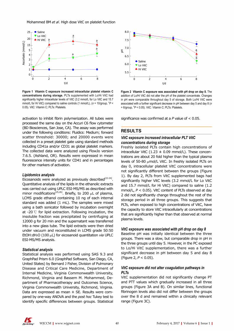
40 February 4, 2017|Volume 6|Issue 1|WJCCM|www.wjgnet.com
activation to inhibit fibrin polymerization. All tubes were processed the same day on the Accuri C6 flow cytometer (BD Biosciences, San Jose, CA). The assay was performed under the following conditions: Fluidics: Medium; forward scatter threshold: 30000; and 20000 events were collected in a preset platelet gate using standard methods including CD41a and/or CD31 as global platelet markers. The collected data were analyzed using FlowJo version 7.6.5. (Ashland, OR). Results were expressed in mean fluorescence intensity units for CD41 and in percentages for other markers of activation.
Lipidomics analysisEicosanoids were analyzed as previously described[2224]. Quantitative analysis of the lipids in the ethanolic extracts was carried out using UPLC ESIMS/MS as described with minor modifications[25,26]. Briefly, to 200 µL of plasma, LCMS grade ethanol containing 10 ng of each internal standard was added (1 mL). The samples were mixed using a bath sonicator followed by incubation overnight at 20 ℃ for lipid extraction. Following incubation, the insoluble fraction was precipitated by centrifuging at 12000 g for 20 min and the supernatant was transferred into a new glass tube. The lipid extracts were then dried under vacuum and reconstituted in LCMS grade 50:50 EtOH:dH2O (100 µL) for eicosanoid quantitation via UPLC ESIMS/MS analysis.
Statistical analysisStatistical analysis was performed using SAS 9.3 and GraphPad Prism 6.0 (GraphPad Software, San Diego, CA, United States) by Bernard J Fisher, Division of Pulmonary Disease and Critical Care Medicine, Department of Internal Medicine, Virginia Commonwealth University, Richmond, Virginia and Bassem M. Mohammed, De-partment of Pharmacotherapy and Outcomes Science, Virginia Commonwealth University, Richmond, Virginia. Data are expressed as mean ± SE. Results were com-pared by oneway ANOVA and the post hoc Tukey test to identify specific differences between groups. Statistical
significance was confirmed at a P value of < 0.05.
RESULTSVitC exposure increased intracellular PLT VitC concentrations during storageFreshly isolated PLTs contain high concentrations of intracellular VitC (1.23 ± 0.09 mmol/L). These concen-trations are about 20 fold higher than the typical plasma levels of 5080 µmol/L VitC. In freshly isolated PLTs on day 0, intracellular platelet VitC concentrations were not significantly different between the groups (Figure 1). By day 2, PLTs from VitC supplemented bags had significantly higher VitC levels (3.2 mmol/L for Lo VitC and 15.7 mmol/L for Hi VitC) compared to saline (1.2 mmol/L, P < 0.05). VitC content of PLTs observed at day 2 did not significantly change throughout the rest of the storage period in all three groups. This suggests that PLTs, when exposed to high concentrations of VitC, have the capacity to store VitC intracellularly at concentrations that are significantly higher than that observed at normal plasma levels.
VitC exposure was associated with pH drop on day 8 Baseline pH was initially identical between the three groups. There was a slow, but comparable drop in pH in the three groups until day 5. However, in the PC exposed to Lo/Hi VitC supplementation, there was a further significant decrease in pH between day 5 and day 8 (Figure 2, P < 0.05).
VitC exposure did not alter coagulation pathways in PLTsVitC supplementation did not significantly change PT and PTT values which gradually increased in all three groups (Figure 3A and B). On similar lines, functional fibrinogen levels also did not differ between the groups over the 8 d and remained within a clinically relevant range (Figure 3C).
25
20
15
5
4
3
2
1
0
Asco
rbat
e (m
mol
/L)
0 2 4 6 8
t/d
SalineLo VitCHi VitC a
a
a a
a
a
Figure 1 Vitamin C exposure increased intracellular platelet vitamin C concentrations during storage. PLTs supplemented with Lo/Hi VitC had significantly higher intracellular levels of VitC (3.2 mmol/L for Lo VitC and 15.7 mmol/L for Hi VitC) compared to saline controls (1 mmol/L), (n = 10/group, aP < 0.05). VitC: Vitamin C; PLTs: Platelets.
8.0
7.5
7.0
6.5
6.0
5.5
pH
0 2 4 6 8
t/d
SalineLo VitCHi VitC
Figure 2 Vitamin C exposure was associated with pH drop on day 8. The addition of Lo/Hi VitC did not alter the pH of the platelet concentrate. Changes in pH were comparable throughout day 5 of storage. Both Lo/Hi VitC were associated with a further significant decrease in pH between day 5 and day 8 (n = 6/group, aP < 0.05). VitC: Vitamin C; PLTs: Platelets.
a
a
Mohammed BM et al . High dose VitC on platelet function

41 February 4, 2017|Volume 6|Issue 1|WJCCM|www.wjgnet.com
High VitC exposure impacts PLT function on day 8Over the first 5 d, the addition of Hi or Lo VitC had no deleterious impact on any of the TEG parameters compared to the saline control (Figure 4). However, on day 8 significant differences were associated with extended storage in Hi VitC. Specifically, R and K times were extended in the Hi VitC group when compared to saline group (Figure 4A and B, P < 0.05). In agreement with Ktime data, a decrease in αangle was observed in the Hi VitC group on day 8 (Figure 4C, P < 0.05). On day 8, there was a fall of 20 mm in MA associated with the Hi
VitC compared to both baseline and day 8 saline controls (Figure 4D, P < 0.05).
Platelet aggregation studies, showed uniform declines in Collagen and ADP-induced platelet aggregations over the study period in all three groups (Figure 5A and B). In addition, Lo/Hi VitC addition did not alter Collagen and ADP-induced ATP secretion throughout the study (Figure 5C and D). Furthermore, flow cytometric analysis of CD62 and CD63 expression profiles, showed that VitC supplementation had no effect on basal CD62p and CD63 expression during storage (Figure 6A and D). Following ADP (Figure 6B) or thrombin (Figure 6C) stimulation, the CD62 expression showed a steady decrease that was not significantly different across the three groups (except for Hi VitC vs saline on day 8, P < 0.05). The expression profiles for CD63 differed depending on whether the PC were stimulated with ADP or thrombin (Figure 6E and F). However, the observed flow cytometric analysis changes were not significant across the three groups over 8 d.
Effects of VitC exposure on eicosanoids metabolism in PLTsEicosanoids analysis was carried out on days 0, 2, 5 and 8 using aliquots of the PPP fraction of each sample. The levels of the free polyunsaturated fatty acids: Arachidonic acid (AA), eicosapentanoic acid (EPA), and docosahexanoic acid (DHA) did not differ significantly across the three groups throughout the study period (Figure 7AC). Interestingly, free AA levels showed an initial drop from baseline in the three groups, and then remained unchanged through day 8. Formation of 11/12/15HETE, products of AA metabolism via the lipoxygenases pathway showed a gradual increase over storage time (Figure 7D-F). The magnitudes of changes were not significantly different between the Lo VitC and the saline groups. However, levels of 11HETE was significantly higher at days 5 and 8 in the Hi VitC group when compared to saline (Figure 7D, P < 0.05). Hi VitC supplementation significantly augmented 12-HETE levels on days 2, 5 and 8 (Figure 7E, P < 0.05). With respect to 15-HETE, only day 8 levels were significantly higher in the Hi VitC group compared to saline (Figure 7F, P < 0.05). On similar lines, thromboxane B2 (TXB2) (the stable metabolite of TXA2) and prostaglandin E2 (PGE2), products of AA metabolism via the cyclooxygenases pathway were not significantly different between the Lo VitC and saline group. However, Hi VitC supplementation was associated with significantly higher TXB2 levels on days 5 and 8 (Figure 7G, P < 0.05); and significantly higher PGE2 levels on days 2 and 8 compared to saline (Figure 7H, P < 0.05).
DISCUSSIONVitC is one of the most enduring and popular alternative medical treatments sought after. Beyond its oral use to treat scurvy, parenteral VitC has been used by CAM practitioners for more than 6 decades[2729]. The most
40
30
20
10
0
PT (
s)
0 2 4 6 8
t/d
SalineLo VitCHi VitC
55
50
45
40
35
30
PT (
s)
0 2 4 6 8
t/d
SalineLo VitCHi VitC
350
300
250
200
150
Fibr
inog
en (
mg/
dL)
0 2 4 6 8
t/d
SalineLo VitCHi VitC
Figure 3 Vitamin C exposure did not alter coagulation pathways in platelets. PT, PTT and Fibrinogen were performed to detect major impacts on the intrinsic, extrinsic and common coagulation pathways. Using platelet poor plasma we observed no significant changes across the saline, Lo- and Hi VitC groups in the PT (A), PTT (B), and Fibrinogen (C) profiles over storage (n =10/group). VitC: Vitamin C; PT: Prothrombin time; PTT: Partial thrombplastin time.
A
B
C
Mohammed BM et al . High dose VitC on platelet function

42 February 4, 2017|Volume 6|Issue 1|WJCCM|www.wjgnet.com
controversial use of high dose VitC as a cancer treatment was promoted by the Nobel Laureate, Cameron et al[30,31]. Recently published evidence has demonstrated that intravenous, but not oral administration of VitC produces pharmacologic plasma concentrations of VitC[32]. This has elucidated possible mechanisms of action of intravenous VitC and for the first time made therapeutic effects, biologically plausible[33]. In the past few years this therapeutic option has been implemented most often as adjunct therapy in diverse conditions such as sepsis, infections, autoimmune diseases and cancers[1519]. The basis for use of high dose intravenous VitC has been established in preclinical studies in which VitC modulated coagulopathies in disease states. For example, Swarbeck et al[34] showed that VitC attenuates plasminogen activator inhibitor1 expression and release in an in vitro model of sepsis. On similar lines Secor et al[35] showed that VitC reduces mouse platelet aggregation and surface Pselectin expression in an ex vivo model of sepsis. However, to date, no studies have directly examined the effect of high doses of VitC on human PLT function. To address this, we asked the question whether exposure to high doses of VitC, as would normally be observed with high dose intravenous VitC therapy, have any effect on PLT function.
In our study we found that PLTs exposed to VitC rapidly accumulated millimolar quantities of VitC as early as day 2 and maintained these levels throughout the study
period (Figure 1). Savini et al[8] showed that human PLTs possess the VitC transporter SVCT2, which enable PLTs to increase intracellular levels of VitC. Importantly, while PLTs typically have approximately 4 mmol/L intracellular VitC in normal plasma, exposure to 3 mmol/L VitC increased intracellular VitC levels to > 15 mmol/L. This is significant, especially in cardiovascular pathologies since PLT activation and aggregation are modulated by reactive oxygen species[36]. VitC can alter the oxidative state of PLTs and inhibit the expression of CD40L, a transmembrane protein with pro-inflammatory and pro-thrombotic properties[12]. Indeed, oral administration of VitC has been reported to reduce arterial stiffness and platelet aggregation[37].
VitC did not alter the pH of the PC throughout the standard 5 day storage period when compared to saline controls (Figure 2). Unlike the saline treated PLTs whose pH stayed in the neutral range on day 8, there was a significant drop in pH in the VitC treated PLTs. However, it is unlikely that these pH changes would significantly alter blood pH due to presence of the carbonic acidbicarbonate buffer, the phosphate buffer system, which consists of phosphoric acid (H3PO4) in equilibrium with dihydrogen phosphate ion (H2PO4
) and H+ and hemoglobin that play an important role in regulating the pH of the blood.
Our results show that the changes in PT, PTT and fibrinogen were comparable across the three groups
30
20
10
0
R (m
in)
0 2 4 6 8
t/d
SalineLo VitCHi VitC
a
10
8
6
4
2
0
K (m
in)
0 2 4 6 8
t/d
SalineLo VitCHi VitC
a
80
60
40
20
0
Angl
e (d
egre
es)
0 2 4 6 8
t/d
SalineLo VitCHi VitC
a
70
60
50
40
30
MA
(mm
)
0 2 4 6 8
t/d
SalineLo VitCHi VitC
a
A B
C D
Figure 4 High vitamin C exposure impacts platelet function on day 8. No significant differences were observed between saline, Lo VitC and Hi VitC throughout the standard 5-d storage period in any of the TEG parameters analyzed (n = 10/group). On day-8, R- (A) and K- (B) times were extended in the Hi VitC group compared to saline (n = 10, aP < 0.05). Similar changes were observed for the α-angle (C) on day-8 (n = 10, aP < 0.05). The Hi VitC group was also associated with a significantly lower MA (D) on day-8 compared to the saline group (n = 10, aP < 0.05). VitC: Vitamin C; TEG: Thromboelastography.
Mohammed BM et al . High dose VitC on platelet function

43 February 4, 2017|Volume 6|Issue 1|WJCCM|www.wjgnet.com
throughout the study period and that exposure to high doses of VitC did not adversely impact these parameters (Figure 3). Only a few studies have employed TEG to evaluate functionality of human PLTs in the PC’s[3841]. However, unlike previous studies that adjusted PRP counts using freshly thawed PPP, we used same sample PPP to make adjustments. While TEG parameters across the three groups were comparable, some critical differences were observed on day 8 in the Hi VitC treated PLTs (Figure 4). The Hi VitC treated group showed a prolonged Rtime, and a reduced MA as compared to the saline controls. Also, the Hi VitC groups had significantly delayed kinetics which was evidenced by prolonged Ktimes and reduced αangle. It is unclear at this time why these changes occurred and future mechanistic approaches are needed to explain this finding. However, the corollary from these studies is that this inhibitory effect of VitC may have deleterious clinical implications if high dose VitC therapy is instituted for 8 d or more, or if plasma VitC levels are maintained at 3 mmol/L or higher for long periods of time.
Platelet aggregation studies show that Lo/Hi VitC addition did not alter the baseline or collagen and ADP-induced platelet aggregations (Figure 5A and B) and collagen and ADP-induced ATP secretion throughout the study (Figure 5C and D). Flow cytometry showed that VitC supplementation had no effect on basal CD62p
and CD63 expression during storage (Figure 6A and D). Following ADP (Figure 6B) or thrombin (Figure 6C) stimulation, the CD62 expression showed a steady decrease that was not significantly different across the three groups (except for Hi VitC). Lo/Hi VitC induced changes in CD63 expression with ADP or thrombin (Figure 6E and F) were not significant over 8 d. These results imply that exposure of normal PLTs to high concentrations of VitC has virtually no impact on agonist induced platelet aggregation under these conditions.
Although we did not observe differences in the levels of free AA, EPA and DHA in the plasma of stored PCs, exposure to Hi VitC was associated with a significant increase in the levels of PGE2, TXB2, 11, 12 and 15HETE (Figure 7). Some of these free fatty acids (FFAs) have roles in host defense against potential pathogenic or opportunistic microorganisms. Indeed, there is extensive literature demonstrating the antibacterial effects of various free fatty acids from a wide range of biological sources including plants, animals and algae[42]. Whilst their antibacterial mode of action is still poorly understood, studies have shown that their prime target is the cell membrane where FFAs disrupt the electron transport chain and oxidative phosphorylation. Besides interfering with cellular energy production, FFA also inhibit enzyme activity, impair nutrient uptake, and participate in the generation of peroxidation and
80
60
40
20
0
Agg
(%)
0 2 4 6 8
t/d
SalineLo VitCHi VitC
40
30
20
10
0
Agg
(%)
0 2 4 6 8
t/d
SalineLo VitCHi VitC
0.8
0.6
0.4
0.2
0.0
ATP
(nm
ol/L
)
0 2 4 6 8
t/d
SalineLo VitCHi VitC
0.8
0.6
0.4
0.2
0.0
ATP
(nm
ol/L
)
0 2 4 6 8
t/d
SalineLo VitCHi VitC
A B
C D
Figure 5 Vitamin C exposure did not affect agonist-stimulated aggregation or adenosine triphosphate secretion by platelets. Using adj. PRP aliquots, addition of Lo/Hi VitC did not alter Collagen-induced PLTs aggregation (A) and ATP secretion (C) as well as ADP-induced PLTs aggregation (B) and ATP secretion (D) when compared to saline controls (n = 10/group). VitC: Vitamin C; PLTs: Platelets; ADP: Adenosine diphosphate; PRP: Platelet rich plasma; ATP: Adenosine triphosphate.
Mohammed BM et al . High dose VitC on platelet function

44 February 4, 2017|Volume 6|Issue 1|WJCCM|www.wjgnet.com
autooxidation degradation products or direct lysis of bacterial cells. While intravenous VitC has been shown to reduce bacterial burden and improve survival in preclinical models of sepsis[4345] it remains to be determined whether the mechanism involves the induction of these FFAs.
As discussed above, both TXB2 and PGE2 levels were significantly higher in PLTs exposed to Hi VitC (Figure 7). Along with possible bacteriostatic effects, there are other potentially beneficial effects associated with induction of these metabolites. For example, a recent study by Bruegel et al[46] demonstrated that reduced release of 11HETE, PGE2 and TXB2 was associated with increased disease severity and poor prognosis in septic patients. PGE2 plays a dual role balancing PLTs response by stimulation or suppression; and is more generally involved in fine tuning the pro/antiinflammatory response[47]. While TXA2 production is associated with PLTs activation,
recent data have supported a protective role of TXA2 via its inhibitory regulation of iNOS in the vasculature. In this regard, TXA2 was found to overcome vascular hyporesponsiveness and help maintaining the vascular tone[48]. The increased production of 12 and 15HETEs observed in Hi VitC treated bags may also be a protective mechanism against the significantly increased TXA2
[49,50]. In sum, exposure of PLTs to high doses of VitC alters endogenous production of lipid mediators by PLTs. These mediators could have unappreciated, yet far reaching impacts on not just PLTs function but on the entire circulatory system.
We recognize that our studies had a few limitations. PLTs in storage bags are not in their normal physiologic environment. They do not interact with endothelial cells or other cell types in these storage bags; they are highly concentrated; and also have access only to a finite amount of nutrients[51]. Accumulation of products
Mohammed BM et al . High dose VitC on platelet function
60
40
20
0
CD62
exp
ress
ion
(%)
0 2 4 6 8
t/d
SalineLo VitCHi VitC
100
90
80
70
60
50
40
CD62
exp
ress
ion
(%)
0 2 4 6 8
t/d
SalineLo VitCHi VitC
a
100
90
80
70
60
CD62
exp
ress
ion
(%)
0 2 4 6 8
t/d
SalineLo VitCHi VitC
50
40
30
20
10
0
CD63
exp
ress
ion
(%)
0 2 4 6 8
t/d
SalineLo VitCHi VitC
50
40
30
20
10
0
CD63
exp
ress
ion
(%)
0 2 4 6 8
t/d
SalineLo VitCHi VitC
100
80
60
40
20
0
CD63
exp
ress
ion
(%)
0 2 4 6 8
t/d
SalineLo VitCHi VitC
A B
C D
E F
Figure 6 Vitamin C exposure did not significantly alter basal or agonist-stimulated CD62 or CD63 expression profiles. Flow cytometry showed that basal expression of both CD62 (A) and CD63 (D) did not differ with the addition of Lo/Hi VitC. ADP-stimulated CD62 (B) expression was lower on day 8 in the Hi VitC group (n = 10/group, aP < 0.05), but CD63 (E) expression was not. Thrombin-stimulated CD62 (C) and CD63 (F) expression also did not differ across the three groups (n = 10/group). VitC: Vitamin C; ADP: Adenosine diphosphate.

45 February 4, 2017|Volume 6|Issue 1|WJCCM|www.wjgnet.com
of metabolism in the storage bags and other factors associated with platelets storage beyond 5 d could impact the system buffering capacity leading to the drop in pH observed in our study. This change in pH combined with the closed nature of the ex vivo system could also account for the observed effects. A second limitation is that in vitro testing of stored PLTs has limitations. While
some PLTs functions are lost during storage, others may be recovered in vivo following transfusion. As suggested by Cardigan et al[51], changes observed in stored PLTs might not necessarily abrogate in vivo hemostatic activities. Whether storage of PCs in VitC truly affects hemostatic activities under in vivo conditions remains to be determined as a future endeavor.
500
400
300
200
100
0
Arac
hido
nic
acid
(ng
/mL)
0 2 4 6 8
t/d
SalineLo VitCHi VitC
150
100
50
0Eico
sape
ntan
oic
avid
(ng
/mL)
0 2 4 6 8
t/d
SalineLo VitCHi VitC
800
600
400
200
0
Doc
osah
exan
oic
acid
(ng
/mL)
0 2 4 6 8
t/d
SalineLo VitCHi VitC
15
10
5
0
11-H
ETE
(ng/
mL)
0 2 4 6 8
t/d
SalineLo VitCHi VitC
a
a
800
600
400
200
0
12-H
ETE
(ng/
mL)
0 2 4 6 8
t/d
SalineLo VitCHi VitC
a
a
a
15
10
5
0
15-H
ETE
(ng/
mL)
0 2 4 6 8
t/d
SalineLo VitCHi VitC
a
50
40
30
20
10
0
TXB2
(ng
/mL)
0 2 4 6 8
t/d
SalineLo VitCHi VitC
a
a
2.5
2.0
1.5
1.0
0.5
0.0
PGE2
(ng
/mL)
0 2 4 6 8
t/d
SalineLo VitCHi VitC
a
a
A B
C D
E F
G H
Figure 7 Hi vitamin C, but not Lo vitamin C exposure, was associated with significant changes in the eicosanoid profile over time. Addition of Lo/Hi VitC did not affect the levels of the PUFA: AA (A), EPA (B) and DHA (C) in comparison to saline controls (n = 10/group). Addition of Hi VitC was associated with significantly higher levels of 11-HETE (D) on days 5 and 8 (n = 10/group, aP < 0.05); 12-HETE (E) on days 2, 5 and 8 (n = 10/group, aP < 0.05); and 15-HETE (F) on day 8 (n = 10/group, aP < 0.05). TXB2 (G) was also significantly higher in the Hi VitC group on days 5 and 8 (n = 10/group, aP < 0.05). In addition, PGE2 levels (H) were significantly higher on days 2 and 8 in the Hi VitC group (n = 10/group, aP < 0.05). VitC: Vitamin C; PUFA: Polyunsaturated fatty acids; AA: Arachidonic acid; EPA: Eicosapentanoic acid; DHA: Docosahexanoic acid; TXB2: Thromboxane B2; PGE2: Prostaglandin E2.
Mohammed BM et al . High dose VitC on platelet function

46 February 4, 2017|Volume 6|Issue 1|WJCCM|www.wjgnet.com
COMMENTSBackgroundVitamin C (VitC) is a key modulator of platelet (PLT) function. Platelets store high intracellular concentrations of VitC, which then modify its oxidative state and play a role in its ability to aggregate. High dose intravenous VitC is increasingly being used both by Complementary and Alternate Medicine practitioners and by licensed medical practitioners as adjunct therapy for wide ranging diseases including sepsis, sepsis induced acute lung injury, multiple cancers, iron deficiency in hemodialysis patients and burns. However, there is no information on the impact of high dose VitC on normal PLT function. To address this need, the authors examined the effect of exposing ex vivo human PLTs to high doses of VitC.
Research frontiersIt is well known that VitC is required for normal platelet function. While pre-clinical studies have examined changes in PLT function in disease and the impact of VitC on these functions, no studies have examined PLT function in the presence of such high doses of VitC.
Innovations and breakthroughsThis is the first study to evaluate ex vivo PLT function in the presence of high concentrations of VitC. The innovative approach to use PLT storage bags afforded a reproducible system that allowed for gauging the temporal effects of high doses of VitC on PLT function.
ApplicationsThis study advises moderate levels of caution regarding the extended use of high doses of intravenous VitC. While these high doses have no deleterious impact on PLT function in the short term (up to 5 d), there appear to be un-anticipated effects on PLT function as assessed by thromboelastography (TEG) after 8 d of continuous exposure.
TerminologyTEG, is a hemostatic assay that measures the viscoelastic properties (physical) of whole blood clot formation under low shear stress. It shows the interaction of platelets with the coagulation cascade (aggregation, clot strengthening, fibrin cross linking and fibrinolysis).
Peer-reviewThis is an interesting paper and is worth to be considered for publication.
REFERENCES1 Marcus AJ, Safier LB. Thromboregulation: multicellular modu
lation of platelet reactivity in hemostasis and thrombosis. FASEB J 1993; 7: 516522 [PMID: 8472890]
2 Morrell CN. Immunomodulatory mediators in platelet transfusion reactions. Hematology Am Soc Hematol Educ Program 2011; 2011: 470474 [PMID: 22160076 DOI: 10.1182/asheducation2011.1.470]
3 Thomas MR, Storey RF. The role of platelets in inflammation. Thromb Haemost 2015; 114: 449458 [PMID: 26293514 DOI: 10.1160/TH14121067]
4 Yadav H, Kor DJ. Platelets in the pathogenesis of acute respiratory distress syndrome. Am J Physiol Lung Cell Mol Physiol 2015; 309: L915L923 [PMID: 26320157 DOI: 10.1152/ajplung.00266.2015]
5 Mezouar S, Frère C, Darbousset R, Mege D, Crescence L, DignatGeorge F, PanicotDubois L, Dubois C. Role of platelets in cancer and cancerassociated thrombosis: Experimental and clinical evidences. Thromb Res 2016; 139: 6576 [PMID: 26916298 DOI: 10.1016/j.thromres.2016.01.006]
6 Naidu KA. Vitamin C in human health and disease is still a mystery? An overview. Nutr J 2003; 2: 7 [PMID: 14498993 DOI: 10.1186/1475289127]
7 Figueroa-Méndez R, RivasArancibia S. Vitamin C in Health and Disease: Its Role in the Metabolism of Cells and Redox State
in the Brain. Front Physiol 2015; 6: 397 [PMID: 26779027 DOI: 10.3389/fphys.2015.00397]
8 Savini I, Catani MV, Arnone R, Rossi A, Frega G, Del Principe D, Avigliano L. Translational control of the ascorbic acid transporter SVCT2 in human platelets. Free Radic Biol Med 2007; 42: 608616 [PMID: 17291984 DOI: 10.1016/j.freeradbiomed.2006.11.028]
9 Padayatty SJ, Levine M. Vitamin C: the known and the unknown and Goldilocks. Oral Dis 2016; 22: 463493 [PMID: 26808119 DOI: 10.1111/odi.12446]
10 Levine M, Padayatty SJ, Espey MG. Vitamin C: a concentrationfunction approach yields pharmacology and therapeutic discoveries. Adv Nutr 2011; 2: 7888 [PMID: 22332036 DOI: 10.3945/an.110. 000109]
11 Olas B, Wachowicz B. Resveratrol and vitamin C as antioxidants in blood platelets. Thromb Res 2002; 106: 143148 [PMID: 12182914 DOI: 10.1016/S00493848(02)001019]
12 Pignatelli P, Sanguigni V, Paola SG, Lo Coco E, Lenti L, Violi F. Vitamin C inhibits platelet expression of CD40 ligand. Free Radic Biol Med 2005; 38: 16621666 [PMID: 15917194 DOI: 10.1016/j.freeradbiomed.2005.02.032]
13 Ho PP, Walters CP, Sullivan HR. Biosynthesis of thromboxane B2: assay, isolation, and properties of the enzyme system in human platelets. Prostaglandins 1976; 12: 951970 [PMID: 12539 DOI: 10.1016/00906980(76)901295]
14 Srivastava KC. Ascorbic acid enhances the formation of prostaglandin E1 in washed human platelets and prostacyclin in rat aortic rings. Prostaglandins Leukot Med 1985; 18: 227233 [PMID: 3925463 DOI: 10.1016/02621746(85)900228]
15 Tanaka H, Matsuda T, Miyagantani Y, Yukioka T, Matsuda H, Shimazaki S. Reduction of resuscitation fluid volumes in severely burned patients using ascorbic acid administration: a randomized, prospective study. Arch Surg 2000; 135: 326331 [PMID: 10722036 DOI: 10.1001/archsurg.135.3.326]
16 Fowler AA, Syed AA, Knowlson S, Sculthorpe R, Farthing D, DeWilde C, Farthing CA, Larus TL, Martin E, Brophy DF, Gupta S, Fisher BJ, Natarajan R. Phase I safety trial of intravenous ascorbic acid in patients with severe sepsis. J Transl Med 2014; 12: 32 [PMID: 24484547 DOI: 10.1186/147958761232]
17 Levy TE. Vitamin C, Infectious Diseases, and Toxins: Curing the Incurable. Philadelphia: Xlibris, 2002
18 Riordan NH, Riordan HD, Casciari JJ. Clinical and experimental experiences with intravenous vitamin C. J Orthomolecular Med 2000; 15: 201203. Available from: URL: http://orthomolecular.org/library/jom/2000/articles/2000v15n04p201.shtml
19 Hoffer LJ, Levine M, Assouline S, Melnychuk D, Padayatty SJ, Rosadiuk K, Rousseau C, Robitaille L, Miller WH. Phase I clinical trial of i.v. ascorbic acid in advanced malignancy. Ann Oncol 2008; 19: 19691974 [PMID: 18544557 DOI: 10.1093/annonc/mdn377]
20 Mohammed BM, Fisher BJ, Huynh QK, Wijesinghe DS, Chalfant CE, Brophy DF, Fowler AA, Natarajan R. Resolution of sterile inflammation: role for vitamin C. Mediators Inflamm 2014; 2014: 173403 [PMID: 25294953 DOI: 10.1155/2014/173403]
21 White NJ, Contaifer D, Martin EJ, Newton JC, Mohammed BM, Bostic JL, Brophy GM, Spiess BD, Pusateri AE, Ward KR, Brophy DF. Early hemostatic responses to trauma identified with hierarchical clustering analysis. J Thromb Haemost 2015; 13: 978988 [PMID: 25816845 DOI: 10.1111/jth.12919]
22 Wijesinghe DS, Allegood JC, Gentile LB, Fox TE, Kester M, Chalfant CE. Use of high performance liquid chromatographyelectrospray ionizationtandem mass spectrometry for the analysis of ceramide1phosphate levels. J Lipid Res 2010; 51: 641651 [PMID: 19654423 DOI: 10.1194/jlr.D000430]
23 Wijesinghe DS, Brentnall M, Mietla JA, Hoeferlin LA, Diegelmann RF, Boise LH, Chalfant CE. Ceramide kinase is required for a normal eicosanoid response and the subsequent orderly migration of fibroblasts. J Lipid Res 2014; 55: 12981309 [PMID: 24823941 DOI: 10.1194/jlr.M048207]
24 Wijesinghe DS, Chalfant CE. SystemsLevel Lipid Analysis Methodologies for Qualitative and Quantitative Investigation of Lipid Signaling Events During Wound Healing. Adv Wound
COMMENTS
Mohammed BM et al . High dose VitC on platelet function
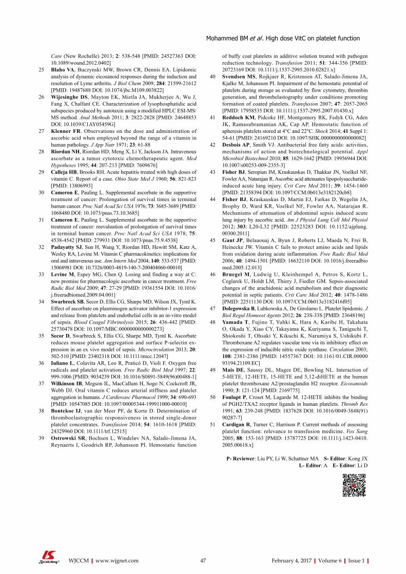
47 February 4, 2017|Volume 6|Issue 1|WJCCM|www.wjgnet.com
Care (New Rochelle) 2013; 2: 538548 [PMID: 24527363 DOI: 10.1089/wound.2012.0402]
25 Blaho VA, Buczynski MW, Brown CR, Dennis EA. Lipidomic analysis of dynamic eicosanoid responses during the induction and resolution of Lyme arthritis. J Biol Chem 2009; 284: 2159921612 [PMID: 19487688 DOI: 10.1074/jbc.M109.003822]
26 Wijesinghe DS, Mayton EK, Mietla JA, Mukherjee A, Wu J, Fang X, Chalfant CE. Characterization of lysophosphatidic acid subspecies produced by autotaxin using a modified HPLC ESIMS/MS method. Anal Methods 2011; 3: 28222828 [PMID: 24648853 DOI: 10.1039/C1AY05459G]
27 Klenner FR. Observations on the dose and administration of ascorbic acid when employed beyond the range of a vitamin in human pathology. J App Nutr 1971; 23: 6188
28 Riordan NH, Riordan HD, Meng X, Li Y, Jackson JA. Intravenous ascorbate as a tumor cytotoxic chemotherapeutic agent. Med Hypotheses 1995; 44: 207213 [PMID: 7609676]
29 Calleja HB, Brooks RH. Acute hepatitis treated with high doses of vitamin C. Report of a case. Ohio State Med J 1960; 56: 821823 [PMID: 13806993]
30 Cameron E, Pauling L. Supplemental ascorbate in the supportive treatment of cancer: Prolongation of survival times in terminal human cancer. Proc Natl Acad Sci USA 1976; 73: 36853689 [PMID: 1068480 DOI: 10.1073/pnas.73.10.3685]
31 Cameron E, Pauling L. Supplemental ascorbate in the supportive treatment of cancer: reevaluation of prolongation of survival times in terminal human cancer. Proc Natl Acad Sci USA 1978; 75: 45384542 [PMID: 279931 DOI: 10.1073/pnas.75.9.4538]
32 Padayatty SJ, Sun H, Wang Y, Riordan HD, Hewitt SM, Katz A, Wesley RA, Levine M. Vitamin C pharmacokinetics: implications for oral and intravenous use. Ann Intern Med 2004; 140: 533537 [PMID: 15068981 DOI: 10.7326/00034819140720040406000010]
33 Levine M, Espey MG, Chen Q. Losing and finding a way at C: new promise for pharmacologic ascorbate in cancer treatment. Free Radic Biol Med 2009; 47: 2729 [PMID: 19361554 DOI: 10.1016/j.freeradbiomed.2009.04.001]
34 Swarbreck SB, Secor D, Ellis CG, Sharpe MD, Wilson JX, Tyml K. Effect of ascorbate on plasminogen activator inhibitor1 expression and release from platelets and endothelial cells in an invitro model of sepsis. Blood Coagul Fibrinolysis 2015; 26: 436442 [PMID: 25730478 DOI: 10.1097/MBC.0000000000000273]
35 Secor D, Swarbreck S, Ellis CG, Sharpe MD, Tyml K. Ascorbate reduces mouse platelet aggregation and surface Pselectin expression in an ex vivo model of sepsis. Microcirculation 2013; 20: 502510 [PMID: 23402318 DOI: 10.1111/micc.12047]
36 Iuliano L, Colavita AR, Leo R, Praticò D, Violi F. Oxygen free radicals and platelet activation. Free Radic Biol Med 1997; 22: 9991006 [PMID: 9034239 DOI: 10.1016/S08915849(96)004881]
37 Wilkinson IB, Megson IL, MacCallum H, Sogo N, Cockcroft JR, Webb DJ. Oral vitamin C reduces arterial stiffness and platelet aggregation in humans. J Cardiovasc Pharmacol 1999; 34: 690693 [PMID: 10547085 DOI: 10.1097/0000534419991100000010]
38 Bontekoe IJ, van der Meer PF, de Korte D. Determination of thromboelastographic responsiveness in stored singledonor platelet concentrates. Transfusion 2014; 54: 16101618 [PMID: 24329960 DOI: 10.1111/trf.12515]
39 Ostrowski SR, Bochsen L, Windeløv NA, SaladoJimena JA, Reynaerts I, Goodrich RP, Johansson PI. Hemostatic function
of buffy coat platelets in additive solution treated with pathogen reduction technology. Transfusion 2011; 51: 344356 [PMID: 20723169 DOI: 10.1111/j.15372995.2010.02821.x]
40 Svendsen MS, Rojkjaer R, Kristensen AT, SaladoJimena JA, Kjalke M, Johansson PI. Impairment of the hemostatic potential of platelets during storage as evaluated by flow cytometry, thrombin generation, and thrombelastography under conditions promoting formation of coated platelets. Transfusion 2007; 47: 20572065 [PMID: 17958535 DOI: 10.1111/j.15372995.2007.01430.x]
41 Reddoch KM, Pidcoke HF, Montgomery RK, Fedyk CG, Aden JK, Ramasubramanian AK, Cap AP. Hemostatic function of apheresis platelets stored at 4°C and 22°C. Shock 2014; 41 Suppl 1: 5461 [PMID: 24169210 DOI: 10.1097/SHK.0000000000000082]
42 Desbois AP, Smith VJ. Antibacterial free fatty acids: activities, mechanisms of action and biotechnological potential. Appl Microbiol Biotechnol 2010; 85: 16291642 [PMID: 19956944 DOI: 10.1007/s0025300923553]
43 Fisher BJ, Seropian IM, Kraskauskas D, Thakkar JN, Voelkel NF, Fowler AA, Natarajan R. Ascorbic acid attenuates lipopolysaccharideinduced acute lung injury. Crit Care Med 2011; 39: 14541460 [PMID: 21358394 DOI: 10.1097/CCM.0b013e3182120cb8]
44 Fisher BJ, Kraskauskas D, Martin EJ, Farkas D, Wegelin JA, Brophy D, Ward KR, Voelkel NF, Fowler AA, Natarajan R. Mechanisms of attenuation of abdominal sepsis induced acute lung injury by ascorbic acid. Am J Physiol Lung Cell Mol Physiol 2012; 303: L20L32 [PMID: 22523283 DOI: 10.1152/ajplung. 00300.2011]
45 Gaut JP, Belaaouaj A, Byun J, Roberts LJ, Maeda N, Frei B, Heinecke JW. Vitamin C fails to protect amino acids and lipids from oxidation during acute inflammation. Free Radic Biol Med 2006; 40: 14941501 [PMID: 16632110 DOI: 10.1016/j.freeradbiomed.2005.12.013]
46 Bruegel M, Ludwig U, Kleinhempel A, Petros S, Kortz L, Ceglarek U, Holdt LM, Thiery J, Fiedler GM. Sepsisassociated changes of the arachidonic acid metabolism and their diagnostic potential in septic patients. Crit Care Med 2012; 40: 14781486 [PMID: 22511130 DOI: 10.1097/CCM.0b013e3182416f05]
47 Dolegowska B, Lubkowska A, De Girolamo L. Platelet lipidomic. J Biol Regul Homeost Agents 2012; 26: 23S33S [PMID: 23648196]
48 Yamada T, Fujino T, Yuhki K, Hara A, Karibe H, Takahata O, Okada Y, Xiao CY, Takayama K, Kuriyama S, Taniguchi T, Shiokoshi T, Ohsaki Y, Kikuchi K, Narumiya S, Ushikubi F. Thromboxane A2 regulates vascular tone via its inhibitory effect on the expression of inducible nitric oxide synthase. Circulation 2003; 108: 23812386 [PMID: 14557367 DOI: 10.1161/01.CIR.00000 93194.21109.EC]
49 Mais DE, Saussy DL, Magee DE, Bowling NL. Interaction of 5HETE, 12HETE, 15HETE and 5,12diHETE at the human platelet thromboxane A2/prostaglandin H2 receptor. Eicosanoids 1990; 3: 121124 [PMID: 2169775]
50 Fonlupt P, Croset M, Lagarde M. 12HETE inhibits the binding of PGH2/TXA2 receptor ligands in human platelets. Thromb Res 1991; 63: 239248 [PMID: 1837628 DOI: 10.1016/00493848(91)902877]
51 Cardigan R, Turner C, Harrison P. Current methods of assessing platelet function: relevance to transfusion medicine. Vox Sang 2005; 88: 153163 [PMID: 15787725 DOI: 10.1111/j.14230410. 2005.00618.x]
P- Reviewer: Liu PY, Li W, Schattner MA S- Editor: Kong JX L- Editor: A E- Editor: Li D
Mohammed BM et al . High dose VitC on platelet function

48 February 4, 2017|Volume 6|Issue 1|WJCCM|www.wjgnet.com
Yoann Launey, Benjamin Duteurtre, Raphaëlle Larmet, Nicolas Nesseler, Audrey Tawa, Yannick Mallédant, Philippe Seguin, Anesthésie Réanimation 1, Centre Hospitalier Universitaire de Rennes, F-35000 Rennes, France
Author contributions: Launey Y, Mallédant Y and Seguin P contributed to study design/planning; Launey Y, Duteurtre B, Nesseler N, Tawa A, Mallédant Y and Seguin P contributed to study conduct; Launey Y, Duteurtre B, Nesseler N, Mallédant Y and Seguin P contributed to data analysis; Launey Y, Duteurtre B, Mallédant Y and Seguin P contributed to writing paper; all authors contributed to revising paper.
Institutional review board statement: This study was reviewed and approved by the ethics committee of Rennes University hospital.
Informed consent statement: The ethics committee waived informed consent.
Conflict-of-interest statement: None.
Data sharing statement: No additional data are available.
Open-Access: This article is an open-access article which was selected by an in-house editor and fully peer-reviewed by external reviewers. It is distributed in accordance with the Creative Commons Attribution Non Commercial (CC BY-NC 4.0) license, which permits others to distribute, remix, adapt, build upon this work non-commercially, and license their derivative works on different terms, provided the original work is properly cited and the use is non-commercial. See: http://creativecommons.org/licenses/by-nc/4.0/
Manuscript source: Invited manuscript
Correspondence to: Philippe Seguin, MD, PhD, Professor, Anesthésie Réanimation 1, Centre Hospitalier Universitaire de Rennes, Inserm U991, F-35000 Rennes, France. [email protected]: +33-2-99289371Fax: +33-2-99282421
Received: August 19, 2016
Peer-review started: August 23, 2016 First decision: September 28, 2016 Revised: November 14, 2016 Accepted: December 7, 2016Article in press: December 9, 2016Published online: February 4, 2017
AbstractAIMTo identify the risk factors for mortality in intensive care patients with postoperative peritonitis (POP).
METHODSThis was a retrospective analysis using a prospective database that includes all patients hospitalized in a surgical intensive care unit for POP from September 2006 to August 2011. The data collected included demographics, comorbidities, postoperative severity parameters, bac-teriological findings, adequacy of antimicrobial therapy and surgical treatments. Adequate source control was defined based on a midline laparotomy, infection source control and intraoperative peritoneal lavage. The number of reoperations needed was also recorded.
RESULTSA total of 201 patients were included. The overall mortality rate was 31%. Three independent risk factors for mortality were identified: The Simplified Acute Phy-siological Ⅱ Score (OR = 1.03; 95%CI: 1.02-1.05, P < 0.001), postoperative medical complications (OR = 6.02; 95%CI: 1.95-18.55, P < 0.001) and the number of reoperations (OR = 2.45; 95%CI: 1.16-5.17, P = 0.015). Surgery was considered as optimal in 69% of the cases, but without any significant effect on mortality.
CONCLUSIONThe results from the large cohort in this study emphasize the role of the initial postoperative severity parameters in
World Journal ofCritical Care MedicineW J C C M
Submit a Manuscript: http://www.wjgnet.com/esps/Help Desk: http://www.wjgnet.com/esps/helpdesk.aspxDOI: 10.5492/wjccm.v6.i1.48
World J Crit Care Med 2017 February 4; 6(1): 48-55ISSN 2220-3141 (online)
© 2017 Baishideng Publishing Group Inc. All rights reserved.
Yoann Launey, Benjamin Duteurtre, Raphaëlle Larmet, Nicolas Nesseler, Audrey Tawa, Yannick Mallédant, Philippe Seguin
ORIGINAL ARTICLE
Risk factors for mortality in postoperative peritonitis in critically ill patients
Retrospective Study

49 February 4, 2017|Volume 6|Issue 1|WJCCM|www.wjgnet.com
Launey Y et al . Postoperative peritonitis/mortality
the prognosis of POP. No predefined criteria for optimal surgery were significantly associated with increased mortality, although the number of reoperations appeared as an independent risk factor of mortality.
Key words: Mortality; Postoperative peritonitis; Risk factors; Surgery
© The Author(s) 2017. Published by Baishideng Publishing Group Inc. All rights reserved.
Core tip: This retrospective study performed from a prospective data base analysed the risk factor for mortality in 201 patients admitted for postoperative peritonitis (POP) in a surgical intensive care unit. Three independent risk factors for mortality were identified: The Simplified Acute Physiological Ⅱ Score, postoperative medical complications and the number of reoperations. This study emphasizes the role of the initial postopera-tive severity parameters in the prognosis of POP. No predefined criteria for optimal surgery were significantly associated with increased mortality, although the number of reoperations appeared as an independent risk factor of mortality.
Launey Y, Duteurtre B, Larmet R, Nesseler N, Tawa A, Mallédant Y, Seguin P. Risk factors for mortality in postoperative peritonitis in critically ill patients. World J Crit Care Med 2017; 6(1): 48-55 Available from: URL: http://www.wjgnet.com/2220-3141/full/v6/i1/48.htm DOI: http://dx.doi.org/10.5492/wjccm.v6.i1.48
INTRODUCTIONPostoperative peritonitis (POP), defined as peritonitis occurring after a planned or urgent abdominal surgery, is an infrequent (occurring in approximately equal to the 2%-3% of laparotomies)[1,2], but serious event, with a mortality rate ranging from 30% to 35%[3-5]. The principles of POP management are based on an early diagnosis, optimized surgical source control, adequate antimicrobial therapy and the control of organ failure(s), if necessary[6,7]. Despite clinical, biological and radiological tools, the diagnosis of POP in the postoperative period remains challenging and the surgical source control is not always easy to perform in recently operated abdo-mens[8-10]. Moreover, multi-drug resistant (MDR) bacteria are frequently isolated in cases of POP, potentially leading to an inadequate antimicrobial therapy and a worsening prognosis[3,5]. Finally, peritonitis is shown to be a frequent condition related to death due to multiple organ failure. In this context, reoperation and postoperative immune depression may favour sepsis and the development of organ failure[11-14].
All of these factors may explain the high mortality observed in association with POP and illustrate the need to evaluate POP separately from other types of intra-abdominal infections. Nevertheless, few studies have evaluated the risk factors for mortality in POP, especially
in critically ill patients[8,15,16]. We hypothesized that POP may have specific chara
cteristics and risk factors for mortality that could help physician in the care of the patients with POP. Accordingly, we performed an analysis using a prospective database to determine the risk factors for mortality associated with POP in patients who required intensive care.
MATERIALS AND METHODSWe performed a retrospective analysis from a pro-spective database that aimed to include all patients with POP. This database was completed from September 2006 to August 2011 in a surgical critical care unit of a university hospital (Rennes - France). All patients older than 18 years of age who were admitted for POP were included. POP was defined as a peritoneal infection developing after intraabdominal surgery. Only the first episode of POP was taken into account. Patients who had focal abscess(es) drained under computed tomography (CT)-scan guidance and/or who had more than one previous episode of POP before intensive care unit (ICU) admission were excluded. Infections were confirmed macroscopically and/or based on the identification of one or several pathogens in peritoneal sample. Patients were followed up from the first day of hospitalization until their discharge from the hospital or death if it occurred during hospitalization. This study was reviewed and approved by the ethics committee of Rennes University hospital which waived informed consent according to the retrospective design (Avis n° 16-129).
The following data were prospectively collected in the first 24 h: Age, sex, origin of patient (Rennes University hospital or another hospital), hospitalization over the previous 3-mo, antibiotic therapy in the 3 mo previous to the current hospital admission, immunocompromised status (defined based on systemic treatments with corticosteroids or other immunosuppressive drugs, che-motherapy or radiotherapy in the 3 mo previous to the admission), co-morbidities (assessed based on MacCabe score and the following severity scores: Simplified Acute Physiological Score Ⅱ (SAPS Ⅱ), Acute Physiology and Chronic Health Evaluation Ⅱ (APACHE Ⅱ) and Sequential Organ Failure Assessment (SOFA)[17-19]. The status (urgent or nonurgent) of the first intraabdominal surgery was also recorded.
Surgery assessment included the cause of POP and the delay between the first surgery and the reoperation. An optimal surgical treatment was qualified as adequate when the 3 following criteria were met: (1) middle laparotomy; (2) ileostomy or colostomy in cases of injury/perforation below the transverse mesocolon injury or drainage above the transverse mesocolon injury; and (3) careful peritoneal washing of the entire peritoneal cavity with at least 6 L of warm physiological serum and until obtaining a macroscopically clean cavity. Coelioscopy and/or a primary anastomosis were considered as in-adequate because their roles in the current recommen-dations are not well-established[6,7]. The number of

50 February 4, 2017|Volume 6|Issue 1|WJCCM|www.wjgnet.com
reoperations and the surgical complications, including abdominal wall abscesses, intra-abdominal abscesses, CT-guided drainage of abdominal abscesses and the need for subsequent reoperation, were also reported. Moreover, in our unit, relaparotomies were not planned and were only performed on-demand.
If required, antibiotic prophylaxis for the first surgery was prescribed according to the recommendations of the French Society of Anesthesia and Critical Care (Recommandations pour la pratique de l’antibioprophy-laxie en chirurgie. Actualisation 1999. www.sfar.org). Antimicrobial therapies applied between the first surgery and reoperations beginning at least 24 h before the reoperation and lasting more than 24 h were noted. Empirical antimicrobial treatment for the first episode of POP was administered according to the local protocol and included cefotaxime and metronidazole for early POP (< 5 d from the initial surgery) and ticarcillin-clavulanate and amikacin for late POP (≥ 5 d from the initial surgery). Because of the low frequency of methicillin resistant Staphylococcus aureus and Enterococcus faecium, the use of vancomycin was not considered. Effects of antimicrobial therapy on Enterococcus species for ongoing POP and the rates of escalation or de-escalation of antimicrobial therapy were reported. Empirical antimicrobial therapies were re-evaluated based on microbiological data and the susceptibility of the isolated microorganisms. Treatments against fungi were only administered in cases of positive, direct examinations of the peritoneal liquid or positive cultures and included the use of fluconazole or an echinocandin. Bacteremia was recorded and defined based on at least one positive blood culture (2 positive samples in cases of coagulase-negative Staphylococcus) and were linked to the intra-abdominal infection if the same microorganisms were recovered in each sample. The duration of antimicrobial therapy ranged from 7 to 10 d.
The isolated microorganisms and the presence of multidrug resistance strains were reported. For each bacterium, the antibiotic sensitivity was determined using the disk-diffusion method. Bacteria were matched into 3 categories: Sensitive, intermediate and resistant. MDR bacteria were defined as follows: Methicillinresistant Staphylococcus aureus; Enterococcus spp. resistant to vancomycin and to high concentrations of gentamycin; Enterobacteriaceae producing extended-spectrum beta-lactamase or overexpressing third-generation cephalo-sporinase; Pseudomonas aeruginosa resistant to ticarcillin, ceftazidime, carbapenem or ciprofloxacin; Acinetobacter spp. resistant to carbapenem and/or ticarcillin and/or aminoglycosides[5].
Medical complications included septic shock, acute re-spiratory distress syndrome (ARDS), and acute renal failure. Septic shock was defined based on the Bone criteria[20] and ARDS according to international recommendations[21]. Acute renal failure was defined based on a serum creatinine level and uraemia and/or a urine output and/or a need for dialysis[22]. In cases of chronic renal failure, acute renal failure was defined as an increase of serum
creatinine or uraemia and/or urine output and/or the need for dialysis[22]. Lengths of ICU and hospital stays and mortality rates were reported.
Statistical analysisAll statistical analysis were performed with SAS software version 9.2 (SAS Institute, Cary, NC, United States). Mean values and standard deviations were used to describe quantitative data, and a t-test or Wilcoxon test were used as needed. Numbers, ranges, and per-centages were used to describe qualitative data, and a χ 2 test or Fisher’s test was used as needed. The multivariate analysis was designed by selecting variables with a P-value < 0.20 in the univariate analysis to build a logistic regression model. The results are expressed with ORs and 95% confidence intervals (95%CI). Results were considered significant at a P-value < 0.05.
RESULTSA total of 201 patients were included in this study. The overall mortality rate was 31% (63/201). The patients’ baseline characteristics, severity scores and the deter-minants of the initial surgery are detailed in Table 1. In a univariate analysis, age, comorbidity evaluated based on McCabe scores and severity at admission in ICU (based on SOFA, APACHE Ⅱ and SAPS Ⅱ scores) were significantly associated with mortality.
The causes of POP were anastomosis leakage (40%), necrosis/ischaemia (20%), traumatic perforation (12%) and miscellaneous (28%) and were not different between the non-survivors and survivors. Surgical procedures were deemed optimal in 69% of the cases (140/201) and the rate did not differ between non-survivors and survivors [71% (45/63) vs 69% (95/138); P = 0.743]. Details of surgical source control and the number reoperations are provided in Table 2. No significant influence of surgical parameters on the prognoses was found between non-survivors and survivors (Table 2).
Antimicrobial treatment prior to POP (prophylaxis and/or therapy) and changes during the postoperative period (escalation or de-escalation) are provided in Table 3. The microorganisms isolated from the peritoneal fluid (Table 4) and the mean number of microorganisms isolated per patient did not differ between non-survivors and survivors (Table 4). A total of 440 microorganisms were identified in 196 patients [non-survivors, n = 139 (61 patients, 2 had no growth) and survivors, n = 301 (135 patients, 3 had no growth)]. A total of 46 patients had at least one MDR bacteria recovered from their peritoneal fluid [nonsurvivors = 28% (17/61) and survivors = 21% (29/135), P = 0.378]. Bacteremia did not differ between the 2 groups [non-survivors = 33% (21/63) and survivors = 26% (36/138); P = 0.268].
The occurrence of medical complications was identified as a potential risk factor for mortality in the univariate analysis, and the length of hospital stay was significantly shorter for non-survivors (Table 3).
In the multivariate analysis, three independent risk
Launey Y et al . Postoperative peritonitis/mortality
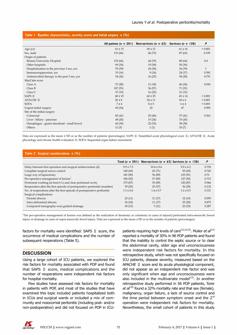
51 February 4, 2017|Volume 6|Issue 1|WJCCM|www.wjgnet.com
factors for mortality were identified: SAPS Ⅱ score, the occurrence of medical complications and the number of subsequent reoperations (Table 5).
DISCUSSIONUsing a large cohort of ICU patients, we explored the risk factors for mortality associated with POP and found that SAPS Ⅱ score, medical complications and the number of reoperations were independent risk factors for hospital mortality.
Few studies have assessed risk factors for mortality in patients with POP, and most of the studies that have examined this topic included patients hospitalized both in ICUs and surgical wards or included a mix of com-munity and nosocomial peritonitis (including post- and/or non-postoperative) and did not focused on POP in ICU-
patients requiring high levels of care[15,16,23]. Mulier et al[15] reported a mortality of 30% in 96 POP patients and found that the inability to control the septic source or to clear the abdominal cavity, older age and unconsciousness were independent risk factors for mortality. In this retrospective study, which was not specifically focused on ICU patients, disease severity, measured based on the APACHE Ⅱ score and its acute physiological component, did not appear as an independent risk factor and was only significant when age and unconsciousness were also included in the multivariate model[15]. In another retrospective study performed in 56 POP patients, Torer et al[16] found a 32% mortality rate and that sex (female), malignancy, organ failure, a lack of source control and the time period between symptom onset and the 2nd operation were independent risk factors for mortality. Nevertheless, the small cohort of patients in this study
All patients (n = 201) Non-survivors (n = 63) Survivors (n = 138) P
Age (yr) 63 ± 15 69 ± 12 61 ± 16 < 0.001Sex, male 133 (66) 46 (73) 87 (63) 0.199Origin of patients Rennes University Hospital 132 (66) 44 (70) 88 (64) 0.4 Other hospitals 69 (34) 19 (30) 50 (36) Hospitalization in the previous 3 mo, yes 78 (39) 24 (38) 54 (39) 1 Immunosuppression, yes 33 (16) 9 (14) 24 (17) 0.581 Antimicrobial therapy in the past 3 mo, yes 54 (26) 16 (25) 38 (28) 0.751MacCabe score Class A 57 (28) 11 (18) 46 (34) 0.036 Class B 107 (53) 36 (57) 71 (51) Class C 37 (19) 16 (25) 21 (15)SAPS Ⅱ 48 ± 19 60 ± 25 43 ± 14 < 0.001APACHE Ⅱ 20 ± 8 24 ± 11 18 ± 6 < 0.001SOFA 7 ± 4 8 ± 5 6 ± 4 < 0.001Urgent initial surgery 69 (34) 22 47 0.905Site of the initial surgery Colorectal 82 (41) 25 (40) 57 (41) 0.363 Liver - biliary - pancreas 48 (24) 15 (24) 33 (24) Oesophagus - gastro-duodenal - small bowel 60 (30) 22 (35) 38 (28) Others 11 (5) 1 (1) 10 (7)
Table 1 Baseline characteristics, severity scores and initial surgery n (%)
Data are expressed as the mean ± SD or as the number of patients (percentages). SAPS Ⅱ: Simplified acute physiological score Ⅱ; APACHE Ⅱ: Acute physiology and chronic health evaluation Ⅱ; SOFA: Sequential organ failure assessment.
Total (n = 201) Non-survivors (n = 63) Survivors (n = 138) P
Delay between first operation and surgical reintervention (d) 9.9 ± 7.5 10.4 ± 9.6 9.9 ± 6.2 0.718Complete surgical source control 140 (69) 45 (71) 95 (69) 0.743Large way of laparotomy 181 (90) 56 (89) 125 (91) 0.71Per-operative management of lesions1 184 (92) 57 (89) 127 (92) 0.713Peritoneal washing (at least 6 L) and clear peritoneal cavity 175 (87) 55 (89) 120 (87) 0.946Reoperation after the first episode of postoperative peritonitis (number) 59 (29) 23 (37) 36 (29) 0.132No. of reoperations after the first episode of postoperative peritonitis 1.3 ± 0.6 1.4 ± 0.7 1.2 ± 0.5 0.121Surgical complications Parietal abscess 23 (11) 11 (17) 12 (10) 0.095 Intra-abdominal abscess 36 (18) 11 (17) 25 (20) 0.875 Computed tomography-scan guided drainage 30 (15) 7 (11) 23 (19) 0.287
Table 2 Surgical considerations n (%)
1The per-operative management of lesions was defined as the realization of ileostomy or colostomy in cases of injured/perforated infra-mesocolic bowel injury or drainage in cases of supra-mesocolic bowel injury. Data are expressed as the mean ± SD or as the number of patients (percentages).
Launey Y et al . Postoperative peritonitis/mortality
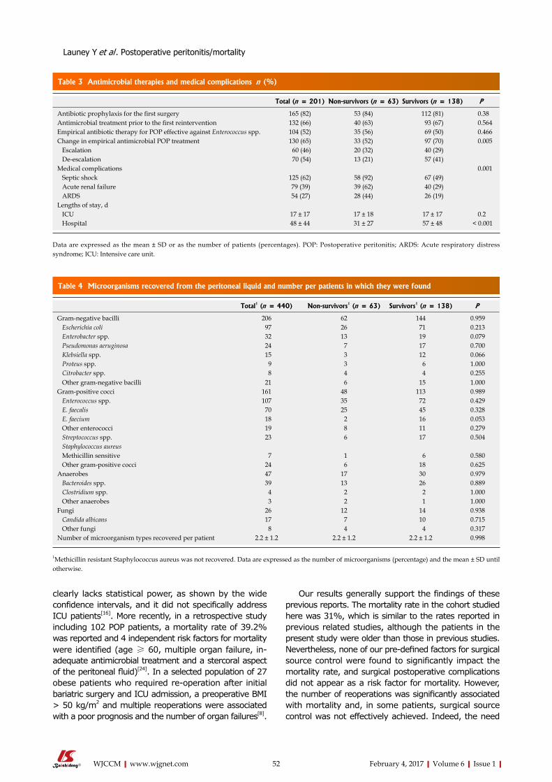
52 February 4, 2017|Volume 6|Issue 1|WJCCM|www.wjgnet.com
clearly lacks statistical power, as shown by the wide confidence intervals, and it did not specifically address ICU patients[16]. More recently, in a retrospective study including 102 POP patients, a mortality rate of 39.2% was reported and 4 independent risk factors for mortality were identified (age ≥ 60, multiple organ failure, in-adequate antimicrobial treatment and a stercoral aspect of the peritoneal fluid)[24]. In a selected population of 27 obese patients who required re-operation after initial bariatric surgery and ICU admission, a preoperative BMI > 50 kg/m2 and multiple reoperations were associated with a poor prognosis and the number of organ failures[8].
Our results generally support the findings of these previous reports. The mortality rate in the cohort studied here was 31%, which is similar to the rates reported in previous related studies, although the patients in the present study were older than those in previous studies. Nevertheless, none of our predefined factors for surgical source control were found to significantly impact the mortality rate, and surgical postoperative complications did not appear as a risk factor for mortality. However, the number of reoperations was significantly associated with mortality and, in some patients, surgical source control was not effectively achieved. Indeed, the need
Total (n = 201) Non-survivors (n = 63) Survivors (n = 138) P
Antibiotic prophylaxis for the first surgery 165 (82) 53 (84) 112 (81) 0.38Antimicrobial treatment prior to the first reintervention 132 (66) 40 (63) 93 (67) 0.564Empirical antibiotic therapy for POP effective against Enterococcus spp. 104 (52) 35 (56) 69 (50) 0.466Change in empirical antimicrobial POP treatment 130 (65) 33 (52) 97 (70) 0.005 Escalation 60 (46) 20 (32) 40 (29) De-escalation 70 (54) 13 (21) 57 (41)Medical complications 0.001 Septic shock 125 (62) 58 (92) 67 (49) Acute renal failure 79 (39) 39 (62) 40 (29) ARDS 54 (27) 28 (44) 26 (19)Lengths of stay, d ICU 17 ± 17 17 ± 18 17 ± 17 0.2 Hospital 48 ± 44 31 ± 27 57 ± 48 < 0.001
Table 3 Antimicrobial therapies and medical complications n (%)
Data are expressed as the mean ± SD or as the number of patients (percentages). POP: Postoperative peritonitis; ARDS: Acute respiratory distress syndrome; ICU: Intensive care unit.
Total1 (n = 440) Non-survivors1 (n = 63) Survivors1 (n = 138) P
Gram-negative bacilli 206 62 144 0.959 Escherichia coli 97 26 71 0.213 Enterobacter spp. 32 13 19 0.079 Pseudomonas aeruginosa 24 7 17 0.700 Klebsiella spp. 15 3 12 0.066 Proteus spp. 9 3 6 1.000 Citrobacter spp. 8 4 4 0.255 Other gram-negative bacilli 21 6 15 1.000Gram-positive cocci 161 48 113 0.989 Enterococcus spp. 107 35 72 0.429 E. faecalis 70 25 45 0.328 E. faecium 18 2 16 0.053 Other enterococci 19 8 11 0.279 Streptococcus spp. 23 6 17 0.504 Staphylococcus aureus Methicillin sensitive 7 1 6 0.580 Other gram-positive cocci 24 6 18 0.625Anaerobes 47 17 30 0.979 Bacteroides spp. 39 13 26 0.889 Clostridium spp. 4 2 2 1.000 Other anaerobes 3 2 1 1.000Fungi 26 12 14 0.938 Candida albicans 17 7 10 0.715 Other fungi 8 4 4 0.317Number of microorganism types recovered per patient 2.2 ± 1.2 2.2 ± 1.2 2.2 ± 1.2 0.998
Table 4 Microorganisms recovered from the peritoneal liquid and number per patients in which they were found
1Methicillin resistant Staphylococcus aureus was not recovered. Data are expressed as the number of microorganisms (percentage) and the mean ± SD until otherwise.
Launey Y et al . Postoperative peritonitis/mortality

53 February 4, 2017|Volume 6|Issue 1|WJCCM|www.wjgnet.com
to re-operate after the first episode of POP was 29%, and among these patients, 27% had persistent intra-abdominal infections. Moreover, surgical reoperation under septic peritoneal conditions and inflammation, along with the inherent risk of new bowel injuries, was sometimes associated with difficulties in closing the abdominal wall, which may have played a role in worsen-ing the mortality rate of these patients.
We found a significant influence of the initial severity scores in predicting mortality. Indeed, the SAPS Ⅱ score and medical complications were independently associated with mortality. This confirms the need for the early identification of patients at risk and who have severe symptom to avoid delays in reoperations, which favours the occurrence of organ failure and bacterial growth in the peritoneal cavity and worsens their prognosis for survival[15]. Our results emphasize that the initial hours following POP diagnosis are crucial in the prognosis of POP. Thus, a rapid control of organ failure is required to achieve a better outcome. POP management is based on 3 goals: Supportive care of septic shock, early and adequate antibiotic treatment and the early surgery. Previous reports have shown that a failure to achieve these goals increases the mortality rate[3,25,26].
For initial antimicrobial treatments, we found that patients who survived had a greater rate of secondary adaptation to antibiotics, as reflected by treatment deescalation. In the cases of antibiotic escalation, the bacteria recovered were resistant and/or not covered by the initial antimicrobial treatment, consequently leading to a potentially higher risk of mortality, although in our study, this parameter was included in the multivariate analysis[3,25,26]. In cases of de-escalation, we assumed that the bacteria recovered were completely targeted by the empirical antimicrobial treatment. In addition, MDR bacteria were found in 23% of patients, which is a lower rate than previously reported, but this did not influence the mortality rate[3,27]. Riché et al[28], in a prospective cohort of 68 POP patients admitted to a surgical ICU, found that yeasts recovered in POP patients were associated with an increased risk of death at day 30 after surgery, whereas Enterococcus spp. and anaerobes recovered were not. In our study, no bacterial (notably Enterococcus spp.) or fungal species were found to impact the mortality rate. The impact of the Enterococcus spp. recovered from POP patients on mortality is controversial, and we did not find a relationship between Enterococcus spp. and mortality[29-31]. Finally, we did not find that age influenced mortality, but the population we studied was older than that of other studies[15,32,33]. Controversies exist regarding age as a risk factor for mortality in ICUs, and factors
other than age itself, such as previous comorbidities and/or frailty, may have a better prognostic significance[34,35]. This issue has been poorly studied in ICU patients with peritonitis. In 163 patients with secondary peritonitis, excluding patients with POP, Hynninen et al[32] showed that previous functional status was an independent risk factor for mortality overall but not in the ICU patients.
Several limitations to the interpretation of our data are worth noting. First, this is a retrospective and monocentric study, but data were prospectively collected, and one third of the patients came from another hospital. Moreover, the management of POP was standardized regardless of whether it was for surgical procedures or postoperative ICU management. Second, we assessed only 3 surgical criteria (the type of laparotomy, the intra-operative management of the lesions and the quality of washing), but many other surgical factors not reported in our database may affect outcomes, such as the ex-perience of the surgeon, the duration of the surgery, the quality of the drainage and the stitching of abdominal wall. Third, our inclusion criteria were stringent because we excluded POP that had been operated on using coelio-scopy because we believe that the coelioscopy does not have a sufficiently well-defined role in the surgical management of peritonitis[6,7,36]. Fourth, biological markers of inflammation have not been recorded in our database. It might have allowed a better stratification of the peritonitis severity, but were not recommended in usual practice in a recent guideline[36].
This study confirms the negative role of the initial severity criteria and the deleterious role of multiple reo-perations, which constitute an indirect sign of inadequate source control, in assessing mortality in patients with POP. An early and successful first surgery is required to increase the chances of a definitive and efficient treat-ment of POP.
COMMENTSBackgroundPostoperative peritonitis (POP) is a rare but severe disease, associated with a challenging diagnosis and a high mortality rate. Multiple organ-failure is a predominant explanation of this burden. But, in addition to supportive care, surgery represents the cornerstone of peritonitis treatment. The timing and adequacy of surgical source control are paramount concerns. Suboptimal surgery may lead to an overwhelmingly negative effect on outcome. In this study, the authors focused on a more refined peritonitis patient’s population to better precise the risk factors of mortality especially the impact of surgical parameters.
Research frontiersWhereas several data exist on the risk factors of mortality in secondary peritonitis, large sample specific studies on POP are scarce. Moreover, surgery has a central role in the management of POP. Identifying more accurately the role of surgical parameters in POP management could affect the way of peritonitis treatment.
Innovations and breakthroughsThis paper is the larger sample size study of selected patients with POP, in which the authors investigated the risk factors of mortality, especially the impact of surgical parameters but also the medical complications. This study confirms
Odds ratio 95%CI P
Simplified acute physiological score Ⅱ 1.03 1.02-1.05 < 0.0001Medical postoperative complications 6.02 1.95-18.55 < 0.0001Number of subsequent reoperations 2.45 1.16-5.17 0.0154
Table 5 Multivariate analysis for the risk factors for mortality
COMMENTS
Launey Y et al . Postoperative peritonitis/mortality

54 February 4, 2017|Volume 6|Issue 1|WJCCM|www.wjgnet.com
the prominent role of medical complications in the poor outcome of POP, however, it found out no surgical risk factor of mortality.
ApplicationsThese data confirm and recall the prominent negative role of severity parameters in POP outcome. No surgical factor has been found to impact negatively the mortality but larger sample size study with more surgical parameters is needed. Moreover, no patient was treated with laparoscopy but new investigations could be drawn in this perspective.
TerminologyPOP belongs to the usual group of secondary peritonitis. More precisely, it includes broadly postoperative abdominal abscesses or diffuse peritoneal infection following abdominal surgery. POP is usually caused by leakage of gut contents, but also by spreading of residual infection or by the occurrence gut ischemia. The severity of POP relies on the associated multiple organ failure.
Peer-reviewThis is a well written paper with a very relevant topic. It is well researched.
REFERENCES1 Pessaux P, Msika S, Atalla D, Hay JM, Flamant Y. Risk factors for
postoperative infectious complications in noncolorectal abdominal surgery: a multivariate analysis based on a prospective multicenter study of 4718 patients. Arch Surg 2003; 138: 314-324 [PMID: 12611581]
2 Manilich E, Vogel JD, Kiran RP, Church JM, Seyidova-Khoshknabi D, Remzi FH. Key factors associated with postoperative com-plications in patients undergoing colorectal surgery. Dis Colon Rectum 2013; 56: 64-71 [PMID: 23222282 DOI: 10.1097/DCR.0b013e31827175f6]
3 Montravers P, Gauzit R, Muller C, Marmuse JP, Fichelle A, Desmonts JM. Emergence of antibiotic-resistant bacteria in cases of peritonitis after intraabdominal surgery affects the efficacy of empirical antimicrobial therapy. Clin Infect Dis 1996; 23: 486-494 [PMID: 8879770]
4 Roehrborn A, Thomas L, Potreck O, Ebener C, Ohmann C, Goretzki PE, Röher HD. The microbiology of postoperative peritonitis. Clin Infect Dis 2001; 33: 1513-1519 [PMID: 11568851 DOI: 10.1086/323333]
5 Seguin P, Fédun Y, Laviolle B, Nesseler N, Donnio PY, Mallédant Y. Risk factors for multidrug-resistant bacteria in patients with post-operative peritonitis requiring intensive care. J Antimicrob Chemother 2010; 65: 342-346 [PMID: 20008043 DOI: 10.1093/jac/dkp439]
6 Solomkin JS, Ristagno RL, Das AF, Cone JB, Wilson SE, Rotstein OD, Murphy BS, Severin KS, Bruss JB. Source control review in clinical trials of anti-infective agents in complicated intra-abdominal infections. Clin Infect Dis 2013; 56: 1765-1773 [PMID: 23463643 DOI: 10.1093/cid/cit128]
7 Bosscha K, van Vroonhoven TJ, van der Werken C. Surgical management of severe secondary peritonitis. Br J Surg 1999; 86: 1371-1377 [PMID: 10583280 DOI: 10.1046/j.1365-2168. 1999.01258.x]
8 Kermarrec N, Marmuse JP, Faivre J, Lasocki S, Mognol P, Chosidow D, Muller C, Desmonts JM, Montravers P. High mortality rate for patients requiring intensive care after surgical revision following bariatric surgery. Obes Surg 2008; 18: 171-178 [PMID: 18175195 DOI: 10.1007/s11695-007-9301-1]
9 Bader FG, Schröder M, Kujath P, Muhl E, Bruch HP, Eckmann C. Diffuse postoperative peritonitis -- value of diagnostic parameters and impact of early indication for relaparotomy. Eur J Med Res 2009; 14: 491-496 [PMID: 19948445 DOI: 10.1186/2047-783X-14-11-491]
10 Paugam-Burtz C, Dupont H, Marmuse JP, Chosidow D, Malek L, Desmonts JM, Mantz J. Daily organ-system failure for diagnosis of persistent intra-abdominal sepsis after postoperative peritonitis.
Intensive Care Med 2002; 28: 594-598 [PMID: 12029408 DOI: 10.1007/s00134-002-1250-5]
11 Guillou PJ. Biological variation in the development of sepsis after surgery or trauma. Lancet 1993; 342: 217-220 [PMID: 8100934 DOI: 10.1016/0140-6736(93)92303-B]
12 Unalp HR, Kamer E, Kar H, Bal A, Peskersoy M, Ali Onal M. Urgent abdominal re-explorations. World J Emerg Surg 2006; 1: 10 [PMID: 16759414 DOI: 10.1186/1749-7922-1-10]
13 Wakefield CH, Carey PD, Foulds S, Monson JR, Guillou PJ. Changes in major histocompatibility complex class II expression in monocytes and T cells of patients developing infection after surgery. Br J Surg 1993; 80: 205-209 [PMID: 8443652 DOI: 10.1002/bjs.1800800224]
14 Hecker A, Uhle F, Schwandner T, Padberg W, Weigand MA. Diagnostics, therapy and outcome prediction in abdominal sepsis: current standards and future perspectives. Langenbecks Arch Surg 2014; 399: 11-22 [PMID: 24186147 DOI: 10.1007/s00423-013-1132-z]
15 Mulier S, Penninckx F, Verwaest C, Filez L, Aerts R, Fieuws S, Lauwers P. Factors affecting mortality in generalized postoperative peritonitis: multivariate analysis in 96 patients. World J Surg 2003; 27: 379-384 [PMID: 12658477 DOI: 10.1007/s00268-002-6705-x]
16 Torer N, Yorganci K, Elker D, Sayek I. Prognostic factors of the mortality of postoperative intraabdominal infections. Infection 2010; 38: 255-260 [PMID: 20393782 DOI: 10.1007/s15010-010-0021-4]
17 Le Gall JR, Lemeshow S, Saulnier F. A new Simplified Acute Physiology Score (SAPS II) based on a European/North American multicenter study. JAMA 1993; 270: 2957-2963 [PMID: 8254858]
18 Siro CA, Bastos PG, Knaus WA, Wagner DP. APACHE II scores in the prediction of multiple organ failure syndrome. Arch Surg 1991; 126: 528-529 [PMID: 2009070 DOI: 10.1001/archsurg.1991.014 10280132022]
19 Vincent JL, Moreno R, Takala J, Willatts S, De Mendonça A, Bruining H, Reinhart CK, Suter PM, Thijs LG. The SOFA (Sepsis-related Organ Failure Assessment) score to describe organ dysfunction/failure. On behalf of the Working Group on Sepsis-Related Problems of the European Society of Intensive Care Medicine. Intensive Care Med 1996; 22: 707-710 [PMID: 8844239 DOI: 10.1007/BF01709751]
20 Bone RC, Sibbald WJ, Sprung CL. The ACCP-SCCM consensus conference on sepsis and organ failure. Chest 1992; 101: 1481-1483 [PMID: 1600757 DOI: 10.1378/chest.101.6.1481]
21 Bernard GR, Artigas A, Brigham KL, Carlet J, Falke K, Hudson L, Lamy M, Legall JR, Morris A, Spragg R. The American-European Consensus Conference on ARDS. Definitions, mechanisms, relevant outcomes, and clinical trial coordination. Am J Respir Crit Care Med 1994; 149: 818-824 [PMID: 7509706 DOI: 10.1164/ajrccm.149.3.7509706]
22 Bellomo R, Kellum J, Ronco C. Acute renal failure: time for consensus. Intensive Care Med 2001; 27: 1685-1688 [PMID: 11810109 DOI: 10.1007/s00134-001-1120-6]
23 Rüttinger D, Kuppinger D, Hölzwimmer M, Zander S, Vilsmaier M, Küchenhoff H, Jauch KW, Hartl WH. Acute prognosis of critically ill patients with secondary peritonitis: the impact of the number of surgical revisions, and of the duration of surgical therapy. Am J Surg 2012; 204: 28-36 [PMID: 22226144 DOI: 10.1016/j.amjsurg.2011.07.019]
24 Marzougui Y, Missaoui K, Hannachi Z, Dhibi Y, Kouka J, Dziri C, Houissa M. [Postoperative peritonitis: pronostic factors of mortality]. Arch Inst Pasteur Tunis 2014; 91: 67-76 [PMID: 26485772]
25 Schneider CP, Seyboth C, Vilsmaier M, Küchenhoff H, Hofner B, Jauch KW, Hartl WH. Prognostic factors in critically ill patients suffering from secondary peritonitis: a retrospective, observational, survival time analysis. World J Surg 2009; 33: 34-43 [PMID: 18979129 DOI: 10.1007/s00268-008-9805-4]
26 Sturkenboom MC, Goettsch WG, Picelli G, in ‘t Veld B, Yin DD, de Jong RB, Go PM, Herings RM. Inappropriate initial treatment of secondary intra-abdominal infections leads to increased risk of clinical failure and costs. Br J Clin Pharmacol 2005; 60: 438-443
Launey Y et al . Postoperative peritonitis/mortality
55 February 4, 2017|Volume 6|Issue 1|WJCCM|www.wjgnet.com
[PMID: 16187977 DOI: 10.1111/j.1365-2125.2005.02443.x]27 Augustin P, Kermarrec N, Muller-Serieys C, Lasocki S, Chosidow
D, Marmuse JP, Valin N, Desmonts JM, Montravers P. Risk factors for multidrug resistant bacteria and optimization of empirical antibiotic therapy in postoperative peritonitis. Crit Care 2010; 14: R20 [PMID: 20156360 DOI: 10.1186/cc8877]
28 Riché FC, Dray X, Laisné MJ, Matéo J, Raskine L, Sanson-Le Pors MJ, Payen D, Valleur P, Cholley BP. Factors associated with septic shock and mortality in generalized peritonitis: comparison between community-acquired and postoperative peritonitis. Crit Care 2009; 13: R99 [PMID: 19552799 DOI: 10.1186/cc7931]
29 Seguin P, Brianchon C, Launey Y, Laviolle B, Nesseler N, Donnio PY, Malledant Y. Are enterococci playing a role in postoperative peritonitis in critically ill patients? Eur J Clin Microbiol Infect Dis 2012; 31: 1479-1485 [PMID: 22076551 DOI: 10.1007/s10096- 011-1467-8]
30 Sotto A, Lefrant JY, Fabbro-Peray P, Muller L, Tafuri J, Navarro F, Prudhomme M, De La Coussaye JE. Evaluation of antimicrobial therapy management of 120 consecutive patients with secondary peritonitis. J Antimicrob Chemother 2002; 50: 569-576 [PMID: 12356803 DOI: 10.1093/jac/dkf167]
31 Dupont H, Vael C, Muller-Serieys C, Chosidow D, Mantz J, Marmuse JP, Andremont A, Goossens H, Desmonts JM. Prospective evaluation of virulence factors of enterococci isolated from patients with peritonitis: impact on outcome. Diagn Microbiol Infect Dis 2008; 60: 247-253 [PMID: 18060725 DOI: 10.1016/j.diagmicrobio.2007.10.006]
32 Hynninen M, Wennervirta J, Leppäniemi A, Pettilä V. Organ dysfunction and long term outcome in secondary peritonitis. Langenbecks Arch Surg 2008; 393: 81-86 [PMID: 17372753 DOI: 10.1007/s00423-007-0160-y]
33 Neri A, Marrelli D, Scheiterle M, Di Mare G, Sforza S, Roviello F. Re-evaluation of Mannheim prognostic index in perforative peritonitis: prognostic role of advanced age. A prospective cohort study. Int J Surg 2015; 13: 54-59 [PMID: 25475872 DOI: 10.1016/j.ijsu.2014.11.035]
34 Le Maguet P, Roquilly A, Lasocki S, Asehnoune K, Carise E, Saint Martin M, Mimoz O, Le Gac G, Somme D, Cattenoz C, Feuillet F, Malledant Y, Seguin P. Prevalence and impact of frailty on mortality in elderly ICU patients: a prospective, multicenter, observational study. Intensive Care Med 2014; 40: 674-682 [PMID: 24651884 DOI: 10.1007/s00134-014-3253-4]
35 Bagshaw SM, Stelfox HT, McDermid RC, Rolfson DB, Tsuyuki RT, Baig N, Artiuch B, Ibrahim Q, Stollery DE, Rokosh E, Majumdar SR. Association between frailty and short- and long-term outcomes among critically ill patients: a multicentre prospective cohort study. CMAJ 2014; 186: E95-102 [PMID: 24277703 DOI: 10.1503/cmaj.130639]
36 Montravers P, Dupont H, Leone M, Constantin JM, Mertes PM, Laterre PF, Misset B, Bru JP, Gauzit R, Sotto A, Brigand C, Hamy A, Tuech JJ. Guidelines for management of intra-abdominal infections. Anaesth Crit Care Pain Med 2015; 34: 117-130 [PMID: 25922057 DOI: 10.1016/j.accpm.2015.03.005]
P- Reviewer: Agaba EA, Bramhall S, Chiu KW, Mizrahi S, Piccinni G S- Editor: Ji FF L- Editor: A E- Editor: Li D
Launey Y et al . Postoperative peritonitis/mortality

56 February 4, 2017|Volume 6|Issue 1|WJCCM|www.wjgnet.com
Martin Padar, Gerli Uusvel, Joel Starkopf, Department of Anaesthesiology and Intensive Care, Tartu University Hospital, 51014 Tartu, Estonia
Liis Starkopf, Department of Public Health, Section of Biostatistics, Faculty of Health and Medical Sciences, University of Copenhagen, 2200 Copenhagen, Denmark
Joel Starkopf, Annika Reintam Blaser, Department of Anaesthesiology and Intensive Care, University of Tartu, 51014 Tartu, Estonia
Annika Reintam Blaser, Department of Anaesthesiology, Intensive Care, Emergency and Pain Medicine, Lucerne Cantonal Hospital, 6000 Lucerne, Switzerland
Author contributions: Padar M, Starkopf J and Reintam Blaser A designed the study; Uusvel G and Starkopf L participated in the data collection and analysis; Padar M, Starkopf J and Reintam Blaser A participated in the interpretation of the results and drafted the manuscript; all the coauthors participated in the development of the final version of the manuscript.
Supported by the Ministry of Education and Research of Estonia (IUT3424).
Institutional review board statement: The study was approved by the institutional review board of Tartu University Hospital.
Informed consent statement: Waiver of informed consent was approved by the Ethics Committee of University of Tartu due to the observational design of the study.
Conflict-of-interest statement: ARB received honoraria for participation in the advisory board meetings of Nestlé, Fresenius and Nutricia. JS has received honoraria for advisory board participation from B. Braun Melsungen AG. The authors declare that they have no conflicts of interest regarding this particular study.
Data sharing statement: No additional data are available.
Open-Access: This article is an openaccess article which was selected by an inhouse editor and fully peerreviewed by external
reviewers. It is distributed in accordance with the Creative Commons Attribution Non Commercial (CC BYNC 4.0) license, which permits others to distribute, remix, adapt, build upon this work noncommercially, and license their derivative works on different terms, provided the original work is properly cited and the use is noncommercial. See: http://creativecommons.org/licenses/bync/4.0/
Manuscript source: Invited manuscript
Correspondence to: Annika Reintam Blaser, MD, PhD, Researcher, Department of Anaesthesiology and Intensive Care, University of Tartu, L. Puusepa 8, 51014 Tartu, Estonia. [email protected]: +3725142281
Received: August 28, 2016Peer-review started: September 1, 2016First decision: October 20, 2016Revised: November 8, 2016Accepted: January 11, 2017Article in press: January 14, 2017Published online: February 4, 2017
AbstractAIMTo determine the effects of implementing an enteral feeding protocol on the nutritional delivery and out-comes of intensive care patients.
METHODSAn uncontrolled, observational before-and-after study was performed in a tertiary mixed medical-surgical intensive care unit (ICU). In 2013, a nurse-driven enteral feeding protocol was developed and implemented in the ICU. Nutrition and outcome-related data from patients who were treated in the study unit from 2011-2012 (the Before group) and 2014-2015 (the After group) were obtained from a local electronic database, the national Population Registry and the hospital’s Infection Control
World Journal ofCritical Care MedicineW J C C M
Submit a Manuscript: http://www.wjgnet.com/esps/Help Desk: http://www.wjgnet.com/esps/helpdesk.aspxDOI: 10.5492/wjccm.v6.i1.56
World J Crit Care Med 2017 February 4; 6(1): 56-64ISSN 2220-3141 (online)
© 2017 Baishideng Publishing Group Inc. All rights reserved.
Martin Padar, Gerli Uusvel, Liis Starkopf, Joel Starkopf, Annika Reintam Blaser
ORIGINAL ARTICLE
Implementation of enteral feeding protocol in an intensive care unit: Before-and-after study
Observational Study

57 February 4, 2017|Volume 6|Issue 1|WJCCM|www.wjgnet.com
Padar M et al . Feeding protocol: A before-and-after study
Service. Data from adult patients, readmissions ex-cluded, who were treated for at least 7 d in the study unit were analysed.
RESULTSIn total, 231 patients were enrolled in the Before and 249 in the After group. The groups were comparable regarding demographics, patient profile, and severity of illness. Fewer patients were mechanically ventilated on admission in the After group (86.7% vs 93.1% in the Before group, P = 0.021). The prevalence of hospital-acquired infections, length of ICU stay and ICU, 30- and 60-d mortality did not differ between the groups. Patients in the After group had a lower 90-d (P = 0.026) and 120-d (P = 0.033) mortality. In the After group, enteral nutrition was prescribed less frequently (P = 0.039) on day 1 but significantly more frequently on all days from day 3. Implementation of the feeding protocol resulted in a higher cumulative amount of en-terally (P = 0.049) and a lower cumulative amount of parenterally (P < 0.001) provided calories by day 7, with an overall reduction in caloric provision (P < 0.001). The prevalence of gastrointestinal symptoms was comparable in both groups, as was the frequency of prokinetic use. Underfeeding (total calories < 80% of caloric needs, independent of route) was observed in 59.4% of the study days Before vs 76.9% After (P < 0.001). Inclusion in the Before group, previous abdominal surgery, intra-abdominal hypertension and the sum of gastrointestinal symptoms were found to be independent predictors of insufficient enteral nutrition.
CONCLUSIONThe use of a nurse-driven feeding protocol improves the delivery of enteral nutrition in ICU patients without concomitant increases in gastrointestinal symptoms or intra-abdominal hypertension.
Key words: Gastrointestinal symptoms; Underfeeding; Nutrition protocol; Feeding protocol; Enteral feeding; Enteral nutrition; Parenteral nutrition; Critical care
© The Author(s) 2017. Published by Baishideng Publishing Group Inc. All rights reserved.
Core tip: Following implementation of a nurse-driven enteral feeding protocol in a mixed medical-surgical intensive care unit (ICU) with a high baseline under-feeding rate, caloric intake via the enteral route was significantly increased during the first week in the ICU without concomitant increases in the frequency of gas-trointestinal symptoms, intra-abdominal hypertension or use of prokinetic medication.
Padar M, Uusvel G, Starkopf L, Starkopf J, Reintam Blaser A. Implementation of enteral feeding protocol in an intensive care unit: Beforeandafter study. World J Crit Care Med 2017; 6(1): 5664 Available from: URL: http://www.wjgnet.com/22203141/full/v6/i1/56.htm DOI: http://dx.doi.org/10.5492/wjccm.v6.i1.56
INTRODUCTIONEnteral feeding (EN) is currently considered the best option for providing nutrition to critically ill patients. The use of the enteral route may specifically reduce disease severity by attenuating the stress response[1] while avoiding the increased infectious morbidity observed with the use of parenteral nutrition (PN)[2]. Starting EN early after admission to an intensive care unit (ICU) is favoured over a delayed approach, as it reduces morbidity and mortality[3,4]. The best clinical outcomes are achieved when over 85% of the prescribed caloric intake is provided[5]. However, inadequate enteral feeding continues to exist in ICUs worldwide[6]; indeed, a previous study[7] conducted in our ICU demonstrated that insufficient enteral feeding occurred in more than half of the patients.
Guidelines issued by the American Society for Parenteral and Enteral Nutrition and the Society of Critical Care Medicine suggest the use of a feeding protocol to improve nutritional outcomes[2]. These protocols aim to standardize and automate the provision of EN, enabling bedside nurses to initiate, monitor and alter the administration of feeds without direct orders from the attending physician[8]. Several studies[912] have shown an increase in nutritional provision with the use of enteral feeding protocols, but the effect of these protocols on relevant patient outcomes has been shown in only a few studies[1315].
The aim of this study was to evaluate the effect of a nursedriven enteral feeding protocol on the amount of nutrition provided and on patient outcomes. An observational beforeandafter study was conducted.
MATERIALS AND METHODSEthical considerationsThe study was approved by the Research Ethics Committee of the University of Tartu with waived informed consent (permit no. 258/T6).
Statistical review statementThe statistical methods of this study were reviewed by a coauthor Liis Starkopf from the Department of Public Health, Section of Biostatistics, Faculty of Health and Medical Sciences, University of Copenhagen.
Patient populationThe 1st Intensive Care Unit of Tartu University Hospital is a 10bed tertiary level mixed medicalsurgical ICU in a regional hospital. Data from patients treated in this department before and after the implementation of a nursedriven feeding protocol were compared. Included in this study were adult patients (at least 18 years of age) who were treated in the ICU for at least 7 consecutive days. Readmissions were excluded.
Design of the studyAn uncontrolled, observational beforeandafter study
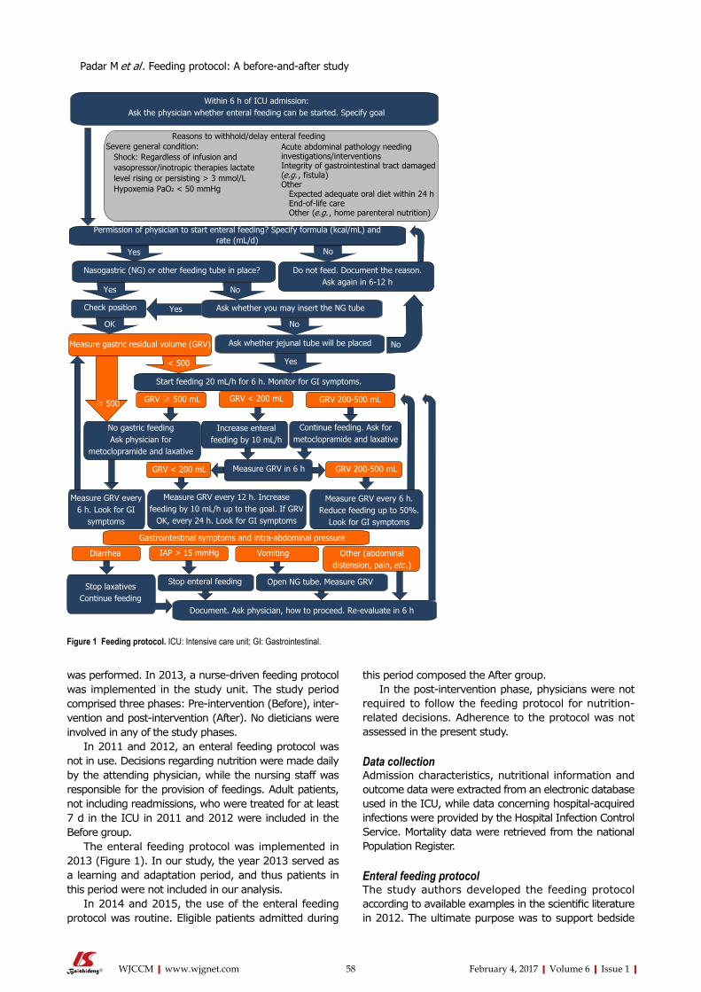
58 February 4, 2017|Volume 6|Issue 1|WJCCM|www.wjgnet.com
was performed. In 2013, a nursedriven feeding protocol was implemented in the study unit. The study period comprised three phases: Preintervention (Before), intervention and postintervention (After). No dieticians were involved in any of the study phases.
In 2011 and 2012, an enteral feeding protocol was not in use. Decisions regarding nutrition were made daily by the attending physician, while the nursing staff was responsible for the provision of feedings. Adult patients, not including readmissions, who were treated for at least 7 d in the ICU in 2011 and 2012 were included in the Before group.
The enteral feeding protocol was implemented in 2013 (Figure 1). In our study, the year 2013 served as a learning and adaptation period, and thus patients in this period were not included in our analysis.
In 2014 and 2015, the use of the enteral feeding protocol was routine. Eligible patients admitted during
this period composed the After group. In the postintervention phase, physicians were not
required to follow the feeding protocol for nutritionrelated decisions. Adherence to the protocol was not assessed in the present study.
Data collectionAdmission characteristics, nutritional information and outcome data were extracted from an electronic database used in the ICU, while data concerning hospitalacquired infections were provided by the Hospital Infection Control Service. Mortality data were retrieved from the national Population Register.
Enteral feeding protocol The study authors developed the feeding protocol according to available examples in the scientific literature in 2012. The ultimate purpose was to support bedside
Within 6 h of ICU admission: Ask the physician whether enteral feeding can be started. Specify goal
Reasons to withhold/delay enteral feedingSevere general condition: Shock: Regardless of infusion and vasopressor/inotropic therapies lactate level rising or persisting > 3 mmol/L Hypoxemia PaO2 < 50 mmHg
Acute abdominal pathology needing investigations/interventionsIntegrity of gastrointestinal tract damaged (e.g. , fistula)Other Expected adequate oral diet within 24 h End-of-life care Other (e.g. , home parenteral nutrition)
Permission of physician to start enteral feeding? Specify formula (kcal/mL) and rate (mL/d)
Yes No
Yes No
No
Yes
OK
Nasogastric (NG) or other feeding tube in place? Do not feed. Document the reason.Ask again in 6-12 h
Check position Yes Ask whether you may insert the NG tube
Measure gastric residual volume (GRV) Ask whether jejunal tube will be placed No
< 500
≥ 500
Start feeding 20 mL/h for 6 h. Monitor for GI symptoms.
GRV ≥ 500 mL GRV < 200 mL GRV 200-500 mL
No gastric feedingAsk physician for
metoclopramide and laxative
Increase enteral feeding by 10 mL/h
Continue feeding. Ask for metoclopramide and laxative
GRV < 200 mL Measure GRV in 6 h GRV 200-500 mL
Measure GRV every 6 h. Look for GI
symptoms
Measure GRV every 12 h. Increase feeding by 10 mL/h up to the goal. If GRV
OK, every 24 h. Look for GI symptoms
Measure GRV every 6 h. Reduce feeding up to 50%.
Look for GI symptoms
Gastrointestinal symptoms and intra-abdominal pressure
Diarrhea IAP > 15 mmHg Vomiting Other (abdominal distension, pain, etc .)
Stop laxativesContinue feeding
Stop enteral feeding Open NG tube. Measure GRV
Document. Ask physician, how to proceed. Re-evaluate in 6 h
Figure 1 Feeding protocol. ICU: Intensive care unit; GI: Gastrointestinal.
Padar M et al . Feeding protocol: A before-and-after study
59 February 4, 2017|Volume 6|Issue 1|WJCCM|www.wjgnet.com
nurses with a structural decision tree for independent decision making in enteral feeding.
VariablesPatient characteristics included age, sex, body mass index (BMI), diagnostic category, occurrence of abdominal surgery, Acute Physiology and Chronic Health Evaluation Ⅱ (APACHE Ⅱ)[16] and Sequential Organ Failure Assessment (SOFA)[17] scores, vasopressor or inotrope treatment and mechanical ventilation (MV) on admission to the ICU. The diagnostic category was defined as surgical or medical according to the diagnoses at ICU admission. Outcome variables were length of ICU stay and MV, prevalence of hospitalacquired infections (ventilatorassociated pneumonia, urinary tract infection, blood stream infection, Clostridium difficile enterocolitis), ICU mortality and 30, 60, 90 and 120d mortality. Hospital-acquired infections were defined as a diagnosis by the Hospital Infection Control Service.
Nutritional support variables included the amount of calories administered daily via enteral and parenteral routes during a patient’s ICU stay. Only data from the first 7 days were included in the analysis. Insufficient EN was defined as the provision of less than 50% of caloric needs via the enteral route and was assessed on day 4 and day 7.
Overfeeding was defined as receiving more than 110% of daily caloric needs via any route, and underfeeding as less than 80%; these variables were analysed as the total incidence during 7 d.
Dextrosebased maintenance infusions were included in the calculations of parenteral calories, whereas the nutritional value of propofol was not taken into account.
Gastrointestinal (GI) symptoms and management variables were recorded daily and were defined and calculated as follows. Absence or presence of bowel sounds was determined daily by a senior intensive care physician using auscultation in a nonprotocolized manner. To measure gastric residual volume (GRV), enteral feeding was stopped, and the nasogastric tube was held closed for 30 min. The tube was then opened and remained open for 30 min with a collection bag mounted to the bed well under the level of the stomach, allowing for the free flow of gastric content. Evacuated content was discarded. Initially, after starting EN, GRV measurements were performed every 6 h. Further measurements were made every 12 h, if two consecutive measurements had yielded less than 200 mL. A large GRV was defined as a total daily volume greater than 500 mL. Bowel distension was defined when confirmed radiologically. Vomiting and GI bleeding were defined as a visible amount of vomit or the presence of a visible amount of blood in stomach contents or stool, respectively. Diarrhoea was defined as the occurrence of liquid stools more than 3 times in a day. Intraabdominal pressure (IAP) was recorded daily in select patients who were considered at risk for intraabdominal hypertension (IAH) according to departmental routine. In those
patients, IAP was measured intermittently every 6 to 12 h (more frequently if the previous IAP was increased 12 mmHg or higher) with a transvesical pressure measurement technique in accordance with the clinical practice guidelines of the World Society of the Abdominal Compartment Syndrome[18]. The sum of GI symptoms was defined as the sum of the daily prevalence (1) or absence (0) of previously described GI symptoms. Prescription of metoclopramide was defined as a standing order of the drug.
Statistical analysisCategorical variables are described as the number of patients and proportions and were compared using χ 2 or Fisher’s exact test. The normality of the distribution of continuous variables was evaluated by KolmogorovSmirnov test. Continuous variables are described as the median and interquartile range if not stated otherwise. Comparisons of continuous variables were performed using an independent samples median test.
Logistic regression analyses were performed to identify the independent predictors of insufficient EN by day 4 and day 7. All admission day variables that positively predicted outcomes in the univariate analysis with P < 0.2 were entered stepwise into a multiple logistic regression model. Coupling variables were added and removed with a stepwise approach to obtain a final optimal model for predicting insufficient EN. Nagelkerke R Square test was used to evaluate the power of the prediction models. The data were analysed using SPSS software (version 23.0, IBM).
RESULTSPatient characteristics and outcome dataIn total, 665 and 683 patients, respectively, were admitted to the ICU before and after the implementation of the feeding protocol. After excluding patients under 18 years of age, readmissions and patients who stayed less than 7 d in the ICU, the study population consisted of 231 patients in the Before and 249 patients in the After group.
The groups did not differ regarding patient age, sex, BMI, casemix, APACHE Ⅱ or SOFA scores nor in the frequency of vasopressor/inotrope therapy at admission. Around half of the patients had a surgical profile, and the majority of them had received abdominal surgery. The proportion of patients who were mechanically ventilated on admission was significantly smaller in the After group. No significant changes between the two groups were found in length of ICU stay, duration of mechanical ventilation, frequency of hospitalacquired infections or ICU, 30d and 60d mortality. However, 90-d and 120-d mortality were significantly lower in the After group (Table 1).
Nutritional supportEN was not initiated during the ICU stay of 19 patients. After implementation of the feeding protocol, significantly
Padar M et al . Feeding protocol: A before-and-after study
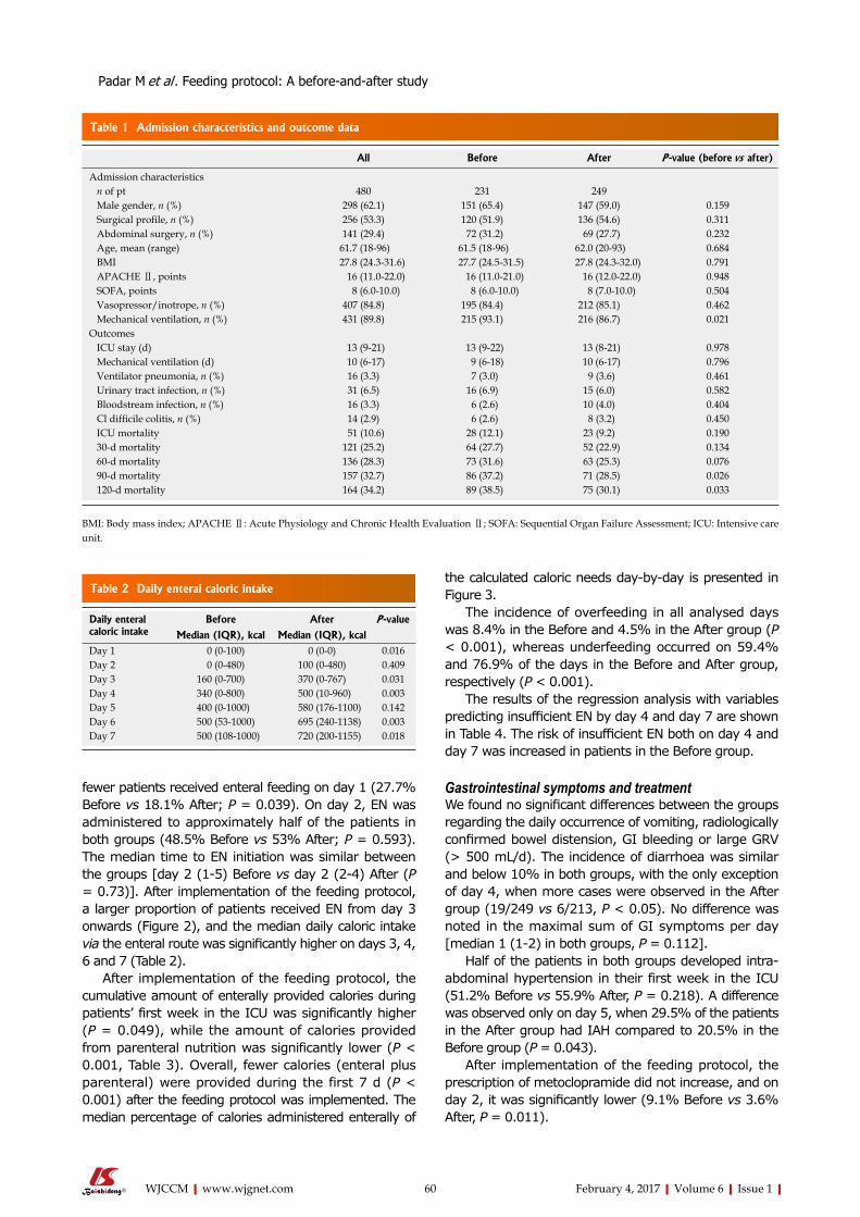
60 February 4, 2017|Volume 6|Issue 1|WJCCM|www.wjgnet.com
fewer patients received enteral feeding on day 1 (27.7% Before vs 18.1% After; P = 0.039). On day 2, EN was administered to approximately half of the patients in both groups (48.5% Before vs 53% After; P = 0.593). The median time to EN initiation was similar between the groups [day 2 (15) Before vs day 2 (24) After (P = 0.73)]. After implementation of the feeding protocol, a larger proportion of patients received EN from day 3 onwards (Figure 2), and the median daily caloric intake via the enteral route was significantly higher on days 3, 4, 6 and 7 (Table 2).
After implementation of the feeding protocol, the cumulative amount of enterally provided calories during patients’ first week in the ICU was significantly higher (P = 0.049), while the amount of calories provided from parenteral nutrition was significantly lower (P < 0.001, Table 3). Overall, fewer calories (enteral plus parenteral) were provided during the first 7 d (P < 0.001) after the feeding protocol was implemented. The median percentage of calories administered enterally of
the calculated caloric needs daybyday is presented in Figure 3.
The incidence of overfeeding in all analysed days was 8.4% in the Before and 4.5% in the After group (P < 0.001), whereas underfeeding occurred on 59.4% and 76.9% of the days in the Before and After group, respectively (P < 0.001).
The results of the regression analysis with variables predicting insufficient EN by day 4 and day 7 are shown in Table 4. The risk of insufficient EN both on day 4 and day 7 was increased in patients in the Before group.
Gastrointestinal symptoms and treatmentWe found no significant differences between the groups regarding the daily occurrence of vomiting, radiologically confirmed bowel distension, GI bleeding or large GRV (> 500 mL/d). The incidence of diarrhoea was similar and below 10% in both groups, with the only exception of day 4, when more cases were observed in the After group (19/249 vs 6/213, P < 0.05). No difference was noted in the maximal sum of GI symptoms per day [median 1 (12) in both groups, P = 0.112].
Half of the patients in both groups developed intraabdominal hypertension in their first week in the ICU (51.2% Before vs 55.9% After, P = 0.218). A difference was observed only on day 5, when 29.5% of the patients in the After group had IAH compared to 20.5% in the Before group (P = 0.043).
After implementation of the feeding protocol, the prescription of metoclopramide did not increase, and on day 2, it was significantly lower (9.1% Before vs 3.6% After, P = 0.011).
Daily enteral caloric intake
Before After P -value
Median (IQR), kcal Median (IQR), kcalDay 1 0 (0-100) 0 (0-0) 0.016Day 2 0 (0-480) 100 (0-480) 0.409Day 3 160 (0-700) 370 (0-767) 0.031Day 4 340 (0-800) 500 (10-960) 0.003Day 5 400 (0-1000) 580 (176-1100) 0.142Day 6 500 (53-1000) 695 (240-1138) 0.003Day 7 500 (108-1000) 720 (200-1155) 0.018
Table 2 Daily enteral caloric intake
Padar M et al . Feeding protocol: A before-and-after study
All Before After P -value (before vs after)
Admission characteristics n of pt 480 231 249 Male gender, n (%) 298 (62.1) 151 (65.4) 147 (59.0) 0.159 Surgical profile, n (%) 256 (53.3) 120 (51.9) 136 (54.6) 0.311 Abdominal surgery, n (%) 141 (29.4) 72 (31.2) 69 (27.7) 0.232 Age, mean (range) 61.7 (18-96) 61.5 (18-96) 62.0 (20-93) 0.684 BMI 27.8 (24.3-31.6) 27.7 (24.5-31.5) 27.8 (24.3-32.0) 0.791 APACHE Ⅱ, points 16 (11.0-22.0) 16 (11.0-21.0) 16 (12.0-22.0) 0.948 SOFA, points 8 (6.0-10.0) 8 (6.0-10.0) 8 (7.0-10.0) 0.504 Vasopressor/inotrope, n (%) 407 (84.8) 195 (84.4) 212 (85.1) 0.462 Mechanical ventilation, n (%) 431 (89.8) 215 (93.1) 216 (86.7) 0.021Outcomes ICU stay (d) 13 (9-21) 13 (9-22) 13 (8-21) 0.978 Mechanical ventilation (d) 10 (6-17) 9 (6-18) 10 (6-17) 0.796 Ventilator pneumonia, n (%) 16 (3.3) 7 (3.0) 9 (3.6) 0.461 Urinary tract infection, n (%) 31 (6.5) 16 (6.9) 15 (6.0) 0.582 Bloodstream infection, n (%) 16 (3.3) 6 (2.6) 10 (4.0) 0.404 Cl difficile colitis, n (%) 14 (2.9) 6 (2.6) 8 (3.2) 0.450 ICU mortality 51 (10.6) 28 (12.1) 23 (9.2) 0.190 30-d mortality 121 (25.2) 64 (27.7) 52 (22.9) 0.134 60-d mortality 136 (28.3) 73 (31.6) 63 (25.3) 0.076 90-d mortality 157 (32.7) 86 (37.2) 71 (28.5) 0.026 120-d mortality 164 (34.2) 89 (38.5) 75 (30.1) 0.033
Table 1 Admission characteristics and outcome data
BMI: Body mass index; APACHE Ⅱ: Acute Physiology and Chronic Health Evaluation Ⅱ; SOFA: Sequential Organ Failure Assessment; ICU: Intensive care unit.
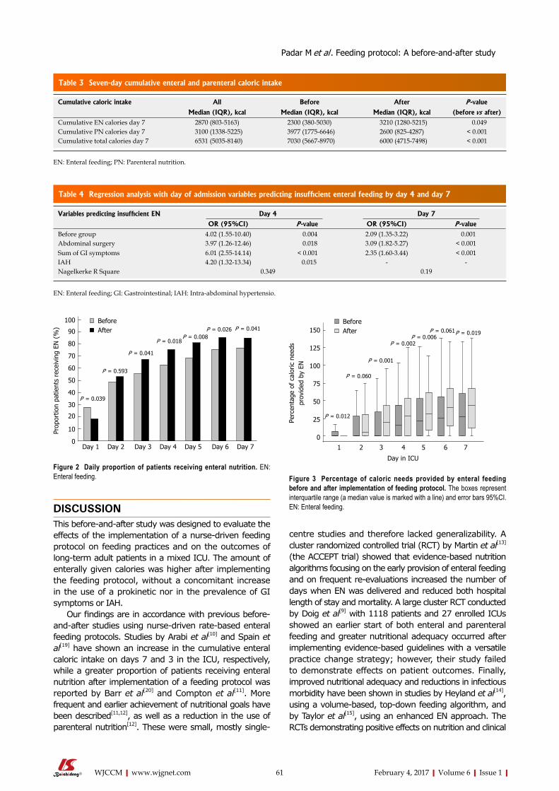
61 February 4, 2017|Volume 6|Issue 1|WJCCM|www.wjgnet.com
DISCUSSIONThis beforeandafter study was designed to evaluate the effects of the implementation of a nursedriven feeding protocol on feeding practices and on the outcomes of longterm adult patients in a mixed ICU. The amount of enterally given calories was higher after implementing the feeding protocol, without a concomitant increase in the use of a prokinetic nor in the prevalence of GI symptoms or IAH.
Our findings are in accordance with previous before-andafter studies using nursedriven ratebased enteral feeding protocols. Studies by Arabi et al[10] and Spain et al[19] have shown an increase in the cumulative enteral caloric intake on days 7 and 3 in the ICU, respectively, while a greater proportion of patients receiving enteral nutrition after implementation of a feeding protocol was reported by Barr et al[20] and Compton et al[11]. More frequent and earlier achievement of nutritional goals have been described[11,12], as well as a reduction in the use of parenteral nutrition[12]. These were small, mostly single
centre studies and therefore lacked generalizability. A cluster randomized controlled trial (RCT) by Martin et al[13] (the ACCEPT trial) showed that evidencebased nutrition algorithms focusing on the early provision of enteral feeding and on frequent reevaluations increased the number of days when EN was delivered and reduced both hospital length of stay and mortality. A large cluster RCT conducted by Doig et al[9] with 1118 patients and 27 enrolled ICUs showed an earlier start of both enteral and parenteral feeding and greater nutritional adequacy occurred after implementing evidencebased guidelines with a versatile practice change strategy; however, their study failed to demonstrate effects on patient outcomes. Finally, improved nutritional adequacy and reductions in infectious morbidity have been shown in studies by Heyland et al[14], using a volumebased, topdown feeding algorithm, and by Taylor et al[15], using an enhanced EN approach. The RCTs demonstrating positive effects on nutrition and clinical
BeforeAfter
Day 1 Day 2 Day 3 Day 4 Day 5 Day 6 Day 7
100
90
80
70
60
50
40
30
20
10
0
Prop
ortio
n pa
tient
s re
ceiv
ing
EN (
%)
P = 0.039
P = 0.593
P = 0.041
P = 0.018P = 0.008
P = 0.026 P = 0.041
Figure 2 Daily proportion of patients receiving enteral nutrition. EN: Enteral feeding.
BeforeAfter
1 2 3 4 5 6 7
150
125
100
75
50
25
0
Perc
enta
ge o
f ca
loric
nee
ds
prov
ided
by
EN
P = 0.012
P = 0.060
P = 0.001
P = 0.002P = 0.006
P = 0.061P = 0.019
Day in ICU
Figure 3 Percentage of caloric needs provided by enteral feeding before and after implementation of feeding protocol. The boxes represent interquartile range (a median value is marked with a line) and error bars 95%CI. EN: Enteral feeding.
Padar M et al . Feeding protocol: A before-and-after study
Cumulative caloric intake All Before After P -value
Median (IQR), kcal Median (IQR), kcal Median (IQR), kcal (before vs after)Cumulative EN calories day 7 2870 (803-5163) 2300 (380-5030) 3210 (1280-5215) 0.049Cumulative PN calories day 7 3100 (1338-5225) 3977 (1775-6646) 2600 (825-4287) < 0.001Cumulative total calories day 7 6531 (5035-8140) 7030 (5667-8970) 6000 (4715-7498) < 0.001
Table 3 Seven-day cumulative enteral and parenteral caloric intake
EN: Enteral feeding; PN: Parenteral nutrition.
Variables predicting insufficient EN Day 4 Day 7
OR (95%CI) P -value OR (95%CI) P -valueBefore group 4.02 (1.55-10.40) 0.004 2.09 (1.35-3.22) 0.001Abdominal surgery 3.97 (1.26-12.46) 0.018 3.09 (1.82-5.27) < 0.001Sum of GI symptoms 6.01 (2.55-14.14) < 0.001 2.35 (1.60-3.44) < 0.001IAH 4.20 (1.32-13.34) 0.015 - -Nagelkerke R Square 0.349 0.19
Table 4 Regression analysis with day of admission variables predicting insufficient enteral feeding by day 4 and day 7
EN: Enteral feeding; GI: Gastrointestinal; IAH: Intra-abdominal hypertensio.
62 February 4, 2017|Volume 6|Issue 1|WJCCM|www.wjgnet.com
outcomes included a variety of interventions, meaning the effects of the feeding protocols were inseparable from those of the whole strategy. In our study, only longterm patients were included, a third of whom had abdominal surgery. Patients with complicated abdominal surgery, requiring a prolonged stay in the ICU, are undoubtedly the most challenging group of patients in terms of successful EN. This aspect needs to be taken into account when interpreting our results, which showed largely insufficient EN both before and after implementation of the feeding protocol. During the time the study was conducted, some changes in the understanding of nutrition in the acute phase of critical illness emerged, including the concept of autophagy and non-inferiority or even possible benefits of underfeeding in the early phase[2126]. These changes may explain why EN was started less frequently on the day of admission (in most cases not the full 24h day) in the After group compared to the Before group.
Interestingly, while the cumulative amount of enteral calories in the first week in the ICU increased, the amount of parenterally given calories decreased by a greater amount. The decreased incidence of overfeeding (considering total calories) after implementation of the feeding protocol did not explain the magnitude of the observed change. Accordingly, implementation of the enteral feeding protocol resulted in decreased total caloric intake. There are some possible explanations for this finding. Shortly before the feeding protocol was implemented, the results of the EPaNIC trial[27] were acknowledged and were potentially interpreted as an argument against early parenteral nutrition. This might have led to a reduction in PN independent of the feeding protocol. Additionally, decisions regarding the initiation of PN continued to be made by physicians in a nonprotocolized way. Therefore, because more patients received EN in the After group, the physicians may have been more likely to withhold supplemental PN in these patients, whereas (full) PN was more likely to be prescribed in patients who remained without EN (larger proportion in the Before group). However, these interactions led to a negative result regarding total caloric intake. Even if significantly increased amounts of enteral calories were administered after implementation of the feeding protocol, they remained far from reaching the caloric targets. As the endeffect, the more pronounced reduction in PN resulted in an even larger caloric deficit in the After group. This is an important finding, indicating the need to plan complex nutritional interventions including EN and PN without the risk of increases in enterally provided calories resulting in an increased total caloric deficit. The presence of a dietician in the ICU would probably also help eliminate this problem. Although the optimal timing of supplemental PN is not known, a cumulative caloric deficit above 4000 kcal should likely be avoided[2830].
It should, however, be noted that the nutritional value of propofol was not included in the caloric calculations in the present study, whereas the awareness regarding the appropriate amounts of calories provided with propofol infusions and regarding the negative impact
of overfeeding[31,32] is increasing. It is not clear whether the propofol dosage influenced physicians’ decisions about the nutrition prescribed in the study period, and furthermore, whether this potential effect varied between the pre and postintervention phases.
The prevalence of GI symptoms is high in critically ill patients[33], and any increases in their prevalence due to more aggressive EN should be avoided. The identified differences in some of the symptoms on single days during the first week in the ICU seemed random and related to the low total number of events. However, the safety of using standard feeding protocols in certain patient groups (e.g., those at increased risk for aspiration or severe bowel distension) was not established in the current study.
The main strengths of our study are the relatively constant patient population in the study unit over the years and the daily documented data on GI symptoms. However, our study has several limitations. The singlecentre design with a limited number of select (stay > 7 d) patients in a mixed ICU, with a significant proportion of patients receiving abdominal surgery, decreases the generalizability of our results. Furthermore, we studied a relatively long time span and it is possible that other nonprotocolized changes in clinical routines might have occurred and influenced the outcomes. Some of the docu-mented GI symptoms occurred very rarely, and therefore the significance of the difference in their prevalence may have changed more or less with each case.
We believe that in addition to describing the magnitude of the effect of a feeding protocol on the delivery of EN and patientrelated outcomes, our study notes a possible pitfall regarding the implications of a nursedriven feeding protocol without standardizing the use of supplemental PN.
The use of a nursedriven feeding protocol is associated with an improved delivery of enteral nutrition without a concomitant increase in the use of prokinetics nor in the prevalence of GI symptoms or IAH in adult ICU patients with an ICU stay of at least 7 d. Increased, but still insufficient, EN may lead to the withholding of PN, resulting in an even larger total caloric deficit. Therefore, the use of an enteral nutrition protocol alone without the presence of a dietician and in absence of a standard for supplemental parenteral nutrition may not prevent severe underfeeding.
ACKNOWLEDGMENTSWe thank all the nurses of the study unit for their great work in making the implementation of the feeding protocol possible.
COMMENTSBackgroundEnteral feeding is the method of choice for providing nutrition to critically ill patients, yet underfeeding continues to be a problem in intensive care units (ICUs) worldwide. This also holds true to their study unit - a mixed ICU with a
Padar M et al . Feeding protocol: A before-and-after study
COMMENTS
63 February 4, 2017|Volume 6|Issue 1|WJCCM|www.wjgnet.com
high proportion of long-staying patients admitted after complicated abdominal surgery. The use of an enteral feeding protocol has been consistently shown to improve the delivery of enteral nutrition (EN) in several studies, however, only a few have reported an effect on relevant patient outcomes.
Research frontiersEarly EN has recently become a hotspot in research of nutritional support for critically ill. Yet not clearly proven, there is some data suggesting that early EN improves important patient-centred outcomes of intensive care. Implementation of a feeding protocol would be the first pragmatic step for any ICU aiming to facilitate EN. This study confirms that with protocol based approach, enteral delivery of nutrients can be significantly enhanced without an increase in gastrointestinal symptoms.
Innovations and breakthroughsIn ICU long-stayers, implementation of the enteral feeding protocol significantly improved the delivery of EN. Unlike most similar studies, the authors reported on gastrointestinal symptoms, intra-abdominal hypertension and the use of prokinetic medications and demonstrated that this improvement in EN did not increase the frequency of aforementioned problems. Importantly, after introduction of the feeding protocol, the use of parenteral nutrition decreased significantly, resulting in a reduction in both parenterally administered and total calories. Accordingly, the prevalence of underfeeding did not decrease despite implementation of the enteral feeding protocol.
ApplicationsThis study demonstrated that use of an enteral feeding protocol was safe in terms of nutrition-related complications. However, in a nutritionally challenging patient population, it also brought along a reduction in overall caloric intake. This finding may warrant implementing a strategy of supplemental parenteral nutrition to help reduce the caloric debt seen in long-staying ICU patients.
TerminologyA nurse-driven enteral feeding protocol refers to an algorithm enabling the bedside nurse to start, monitor and adjust the delivery of enteral tube feedings to patients not capable of oral food intake.
Peer-reviewThis study was well conducted and nicely written. It has enough quality to be published in this journal.
REFERENCES1 Windsor AC, Kanwar S, Li AG, Barnes E, Guthrie JA, Spark
JI, Welsh F, Guillou PJ, Reynolds JV. Compared with parenteral nutrition, enteral feeding attenuates the acute phase response and improves disease severity in acute pancreatitis. Gut 1998; 42: 431435 [PMID: 9577354 DOI: 10.1136/gut.42.3.431]
2 McClave SA, Taylor BE, Martindale RG, Warren MM, Johnson DR, Braunschweig C, McCarthy MS, Davanos E, Rice TW, Cresci GA, Gervasio JM, Sacks GS, Roberts PR, Compher C. Guidelines for the Provision and Assessment of Nutrition Support Therapy in the Adult Critically Ill Patient: Society of Critical Care Medicine (SCCM) and American Society for Parenteral and Enteral Nutrition (A.S.P.E.N.). JPEN J Parenter Enteral Nutr 2016; 40: 159211 [PMID: 26773077 DOI: 10.1177/0148607115621863]
3 Marik PE, Zaloga GP. Early enteral nutrition in acutely ill patients: a systematic review. Crit Care Med 2001; 29: 22642270 [PMID: 11801821 DOI: 10.1097/0000324620011200000005]
4 Doig GS, Heighes PT, Simpson F, Sweetman EA, Davies AR. Early enteral nutrition, provided within 24 h of injury or intensive care unit admission, significantly reduces mortality in critically ill patients: a metaanalysis of randomised controlled trials. Intensive Care Med 2009; 35: 20182027 [PMID: 19777207 DOI: 10.1007/s0013400916644]
5 Heyland DK, Cahill N, Day AG. Optimal amount of calories for critically ill patients: depends on how you slice the cake! Crit
Care Med 2011; 39: 26192626 [PMID: 21705881 DOI: 10.1097/CCM.0b013e318226641d]
6 Heyland DK, Dhaliwal R, Wang M, Day AG. The prevalence of iatrogenic underfeeding in the nutritionally ‘atrisk’ critically ill patient: Results of an international, multicenter, prospective study. Clin Nutr 2015; 34: 659666 [PMID: 25086472 DOI: 10.1016/j.clnu.2014.07.008]
7 Kuslapuu M, Jõgela K, Starkopf J, Reintam Blaser A. The reasons for insufficient enteral feeding in an intensive care unit: A prospective observational study. Intensive Crit Care Nurs 2015; 31: 309314 [PMID: 25864368 DOI: 10.1016/j.iccn.2015.03.001]
8 Heyland DK, Cahill NE, Dhaliwal R, Sun X, Day AG, McClave SA. Impact of enteral feeding protocols on enteral nutrition delivery: results of a multicenter observational study. JPEN J Parenter Enteral Nutr 2010; 34: 675684 [PMID: 21097768 DOI: 10.1177/0148607110364843]
9 Doig GS, Simpson F, Finfer S, Delaney A, Davies AR, Mitchell I, Dobb G. Effect of evidencebased feeding guidelines on mortality of critically ill adults: a cluster randomized controlled trial. JAMA 2008; 300: 27312741 [PMID: 19088351 DOI: 10.1001/jama. 2008.826]
10 Arabi Y, Haddad S, Sakkijha M, Al Shimemeri A. The impact of implementing an enteral tube feeding protocol on caloric and protein delivery in intensive care unit patients. Nutr Clin Pract 2004; 19: 523530 [PMID: 16215149 DOI: 10.1177/0115426504019005523]
11 Compton F, Bojarski C, Siegmund B, van der Giet M. Use of a nutrition support protocol to increase enteral nutrition delivery in critically ill patients. Am J Crit Care 2014; 23: 396403 [PMID: 25179035 DOI: 10.4037/ajcc2014140]
12 Mackenzie SL, Zygun DA, Whitmore BL, Doig CJ, Hameed SM. Implementation of a nutrition support protocol increases the proportion of mechanically ventilated patients reaching enteral nutrition targets in the adult intensive care unit. JPEN J Parenter Enteral Nutr 2005; 29: 7480 [PMID: 15772383 DOI: 10.1177/014860710502900274]
13 Martin CM, Doig GS, Heyland DK, Morrison T, Sibbald WJ. Multicentre, clusterrandomized clinical trial of algorithms for criticalcare enteral and parenteral therapy (ACCEPT). CMAJ 2004; 170: 197204 [PMID: 14734433 DOI: 10.1177/0115426504019003309]
14 Heyland DK, Murch L, Cahill N, McCall M, Muscedere J, Stelfox HT, Bray T, Tanguay T, Jiang X, Day AG. Enhanced proteinenergy provision via the enteral route feeding protocol in critically ill patients: results of a cluster randomized trial. Crit Care Med 2013; 41: 27432753 [PMID: 23982032 DOI: 10.1097/CCM.0b013e 31829efef5]
15 Taylor SJ, Fettes SB, Jewkes C, Nelson RJ. Prospective, randomized, controlled trial to determine the effect of early enhanced enteral nutrition on clinical outcome in mechanically ventilated patients suffering head injury. Crit Care Med 1999; 27: 25252531 [PMID: 10579275 DOI: 10.1097/0000324619991100000033]
16 Knaus WA, Draper EA, Wagner DP, Zimmerman JE. APACHE II: a severity of disease classification system. Crit Care Med 1985; 13: 818829 [PMID: 3928249 DOI: 10.1097/0000324619851000000009]
17 Vincent JL, de Mendonça A, Cantraine F, Moreno R, Takala J, Suter PM, Sprung CL, Colardyn F, Blecher S. Use of the SOFA score to assess the incidence of organ dysfunction/failure in intensive care units: results of a multicenter, prospective study. Working group on “sepsisrelated problems” of the European Society of Intensive Care Medicine. Crit Care Med 1998; 26: 17931800 [PMID: 9824069 DOI: 10.1097/0000324619981100000016]
18 Kirkpatrick AW, Roberts DJ, De Waele J, Jaeschke R, Malbrain ML, De Keulenaer B, Duchesne J, Bjorck M, Leppaniemi A, Ejike JC, Sugrue M, Cheatham M, Ivatury R, Ball CG, Reintam Blaser A, Regli A, Balogh ZJ, D’Amours S, Debergh D, Kaplan M, Kimball E, Olvera C. Intraabdominal hypertension and the abdominal compartment syndrome: updated consensus definitions and clinical practice guidelines from the World Society of the Abdominal Compartment Syndrome. Intensive Care Med 2013; 39: 11901206
Padar M et al . Feeding protocol: A before-and-after study

64 February 4, 2017|Volume 6|Issue 1|WJCCM|www.wjgnet.com
[PMID: 23673399 DOI: 10.1007/s001340132906z]19 Spain DA, McClave SA, Sexton LK, Adams JL, Blanford BS,
Sullins ME, Owens NA, Snider HL. Infusion protocol improves delivery of enteral tube feeding in the critical care unit. JPEN J Parenter Enteral Nutr 1999; 23: 288292 [PMID: 10485441 DOI: 10.1177/0148607199023005288]
20 Barr J, Hecht M, Flavin KE, Khorana A, Gould MK. Outcomes in critically ill patients before and after the implementation of an evidencebased nutritional management protocol. Chest 2004; 125: 14461457 [PMID: 15078758 DOI: 10.1378/chest.125.4.1446]
21 Casaer MP, Van den Berghe G. Nutrition in the acute phase of critical illness. N Engl J Med 2014; 370: 12271236 [PMID: 24670169 DOI: 10.1056/NEJMra1304623]
22 Rice TW, Wheeler AP, Thompson BT, Steingrub J, Hite RD, Moss M, Morris A, Dong N, Rock P. Initial trophic vs full enteral feeding in patients with acute lung injury: the EDEN randomized trial. JAMA 2012; 307: 795803 [PMID: 22307571 DOI: 10.1001/jama.2012.137]
23 Arabi YM, Aldawood AS, Haddad SH, AlDorzi HM, Tamim HM, Jones G, Mehta S, McIntyre L, Solaiman O, Sakkijha MH, Sadat M, Afesh L. Permissive Underfeeding or Standard Enteral Feeding in Critically Ill Adults. N Engl J Med 2015; 372: 23982408 [PMID: 25992505 DOI: 10.1056/NEJMoa1502826]
24 Casaer MP, Van den Berghe G. Editorial on the original article entitled “Permissive underfeeding of standard enteral feeding in critically ill adults” published in the New England Journal of Medicine on June 18, 2015. Ann Transl Med 2015; 3: 226 [PMID: 26539443 DOI: 10.3978/j.issn.23055839.2015.07.22]
25 Hermans G, Casaer MP, Clerckx B, Güiza F, Vanhullebusch T, Derde S, Meersseman P, Derese I, Mesotten D, Wouters PJ, Van Cromphaut S, Debaveye Y, Gosselink R, Gunst J, Wilmer A, Van den Berghe G, Vanhorebeek I. Effect of tolerating macronutrient deficit on the development of intensivecare unit acquired weakness: a subanalysis of the EPaNIC trial. Lancet Respir Med 2013; 1: 621629 [PMID: 24461665 DOI: 10.1016/S22132600(13)701838]
26 Braunschweig CA, Sheean PM, Peterson SJ, Gomez Perez S, Freels S, Lateef O, Gurka D, Fantuzzi G. Intensive nutrition in acute lung injury: a clinical trial (INTACT). JPEN J Parenter Enteral Nutr 2015; 39: 1320 [PMID: 24722769 DOI: 10.1177/0148607114528541]
27 Casaer MP, Mesotten D, Hermans G, Wouters PJ, Schetz M, Meyfroidt G, Van Cromphaut S, Ingels C, Meersseman P, Muller J, Vlasselaers D, Debaveye Y, Desmet L, Dubois J, Van Assche A, Vanderheyden S, Wilmer A, Van den Berghe G. Early versus late parenteral nutrition in critically ill adults. N Engl J Med 2011; 365: 506517 [PMID: 21714640 DOI: 10.1056/NEJMoa1102662]
28 Heidegger CP, Berger MM, Graf S, Zingg W, Darmon P, Costanza MC, Thibault R, Pichard C. Optimisation of energy provision with supplemental parenteral nutrition in critically ill patients: a randomised controlled clinical trial. Lancet 2013; 381: 385393 [PMID: 23218813 DOI: 10.1016/S01406736(12)613518]
29 Villet S, Chiolero RL, Bollmann MD, Revelly JP, Cayeux R N MC, Delarue J, Berger MM. Negative impact of hypocaloric feeding and energy balance on clinical outcome in ICU patients. Clin Nutr 2005; 24: 502509 [PMID: 15899538 DOI: 10.1016/j.clnu.2005.03.006]
30 Dvir D, Cohen J, Singer P. Computerized energy balance and complications in critically ill patients: an observational study. Clin Nutr 2006; 25: 3744 [PMID: 16321459 DOI: 10.1016/j.clnu. 2005.10.010]
31 Grau T, Bonet A, Rubio M, Mateo D, Farré M, Acosta JA, Blesa A, Montejo JC, de Lorenzo AG, Mesejo A. Liver dysfunction associated with artificial nutrition in critically ill patients. Crit Care 2007; 11: R10 [PMID: 17254321 DOI: 10.1186/cc5670]
32 Schulman RC, Mechanick JI. Can nutrition support interfere with recovery from acute critical illness? World Rev Nutr Diet 2013; 105: 6981 [PMID: 23075588 DOI: 10.1159/000341272]
33 Reintam A, Parm P, Kitus R, Kern H, Starkopf J. Gastrointestinal symptoms in intensive care patients. Acta Anaesthesiol Scand 2009; 53: 318324 [PMID: 19243317 DOI: 10.1111/j.13996576. 2008.01860.x]
P- Reviewer: Hokama A, Joh JW S- Editor: Kong JX L- Editor: A E- Editor: Li D
Padar M et al . Feeding protocol: A before-and-after study
65 February 4, 2017|Volume 6|Issue 1|WJCCM|www.wjgnet.com
Miguel A Ibarra-Estrada, Pável E Aguilera-González, Critical Care Unit, Instituto Jalisciense de Cancerología, Guadalajara Jalisco 44280, Mexico
Miguel A Ibarra-Estrada, Pável E Aguilera-González, Critical Care Unit, Hospital General Regional #180, Instituto Mexicano del Seguro Social, Tlajomulco de Zúñiga Jalisco 45655, Mexico
Quetzalcóatl Chávez-Peña, Miguel A García-Soto, Transplant Care Unit, Centro Médico Nacional de Occidente, Instituto Mexicano del Seguro Social, Guadalajara Jalisco 44340, Mexico
Quetzalcóatl Chávez-Peña, Guadalupe Aguirre-Avalos, Centro Universitario de Ciencias de la Salud, Universidad de Guadalajara, Guadalajara Jalisco 44340, Mexico
Claudia I Reynoso-Estrella, Jorge Rios-Zermeño, Guadalupe Aguirre-Avalos, Critical Care Unit, Hospital Civil Fray Antonio Alcalde, Guadalajara Jalisco 44280, Mexico
Author contributions: Ibarra-Estrada MA designed the study, performed data collection, statistical analysis, interpretation of data, and drafted the manuscript; Chávez-Peña Q and Reynoso-Estrella CI performed data collection and helped draft the manuscript; Rios-Zermeño J, Aguilera-González PE, García-Soto MA and Aguirre-Avalos G performed data collection; all authors were involved and approved the final manuscript.
Institutional review board statement: This study has been approved by the scientific and ethics committees at Instituto Jalisciense de Cancerología (INV-01/16), and Hospital Civil Fray Antonio Alcalde (HCG/CEI-0321/16). A copy of approval can be provided on request.
Informed consent statement: All study participants, or their next of kin, provided informed written consent prior to study enrollment.
Conflict-of-interest statement: None of the authors have commercial associations or financial involvements that might pose a conflict of interest related to the content of this article.
Data sharing statement: Data presented in the manuscript is
anonymized, and the risk of identifying individual patients is very low. No additional data is available from the study other than the data stated in this manuscript.
Open-Access: This article is an open-access article which was selected by an in-house editor and fully peer-reviewed by external reviewers. It is distributed in accordance with the Creative Commons Attribution Non Commercial (CC BY-NC 4.0) license, which permits others to distribute, remix, adapt, build upon this work non-commercially, and license their derivative works on different terms, provided the original work is properly cited and the use is non-commercial. See: http://creativecommons.org/licenses/by-nc/4.0/
Manuscript source: Invited manuscript
Correspondence to: Dr. Miguel A Ibarra-Estrada, Critical Care Unit, Instituto Jalisciense de Cancerología, Coronel Calderón 715, Guadalajara Jalisco 44280, Mexico. [email protected]: +52-33-40401508
Received: August 24, 2016Peer-review started: August 25, 2016First decision: October 20, 2016Revised: November 6, 2016Accepted: January 11, 2017Article in press: January 14, 2017Published online: February 4, 2017
AbstractAIMTo characterize the prescribing patterns for hydro-cortisone for patients with septic shock and perform an exploratory analysis in order to identify the variables associated with better outcomes.
METHODSThis prospective cohort study included 59 patients with septic shock who received stress-dose hydrocortisone.
World Journal ofCritical Care MedicineW J C C M
Submit a Manuscript: http://www.wjgnet.com/esps/Help Desk: http://www.wjgnet.com/esps/helpdesk.aspxDOI: 10.5492/wjccm.v6.i1.65
World J Crit Care Med 2017 February 4; 6(1): 65-73ISSN 2220-3141 (online)
© 2017 Baishideng Publishing Group Inc. All rights reserved.
Miguel A Ibarra-Estrada, Quetzalcóatl Chávez-Peña, Claudia I Reynoso-Estrella, Jorge Rios-Zermeño, Pável E Aguilera-González, Miguel A García-Soto, Guadalupe Aguirre-Avalos
ORIGINAL ARTICLE
Timing, method and discontinuation of hydrocortisone administration for septic shock patients
Observational Study

66 February 4, 2017|Volume 6|Issue 1|WJCCM|www.wjgnet.com
Ibarra-Estrada MA et al . Hydrocortisone in septic shock patients
It was performed at 2 critical care units in academic hospitals from June 1st, 2015, to July 31st, 2016. Demographic data, comorbidities, medical management details, adverse effects related to corticosteroids, and outcomes were collected after the critical care physician indicated initiation of hydrocortisone. Univariate comparison between continuous and bolus administration of hydrocortisone was performed, including multivariate analysis, as well as Kaplan-Meier analysis to compare the proportion of shock reversal at 7 d after presentation. Receiver operating characteristic (ROC) curves determined the best cut-off criteria for initiation of hydrocortisone associated with the highest probability of shock reversal. We addressed the effects of the taper strategy for discontinuation of hydrocortisone, noting risk of shock relapse and adverse effects.
RESULTSAll-cause 30-d mortality was 42%. Hydrocortisone was administered as a continuous infusion in 54.2% of patients; time to reversal of shock was 49 h longer in patients who were given a bolus administration [59 h (range, 47.5-90.5) vs 108 h (range, 63.2-189); P = 0.001]. The maximal dose of norepinephrine after initiation of hydrocortisone was lower in patients on continuous infusion [0.19 μg/kg per minute (range, 0.11-0.28 μg)] compared with patients who were given bolus [0.34 μg/kg per minute (range, 0.16-0.49); P = 0.004]. Kaplan-Meier analysis revealed a higher proportion of shock reversal at 7 d in patients with continuous infusion compared to those given bolus (83% vs 63%; P = 0.004). There was a good correlation between time to initiation of hydrocortisone and time to reversal of shock (r = 0.80; P < 0.0001); ROC curve analysis revealed that the best criteria for prediction of shock reversal was a time to initiation of hydrocortisone of ≤ 13 h after administration of norepinephrine, with an area under the curve of 0.81 (P < 0.001). The maximal dose of norepinephrine at initiation of hydrocortisone with the highest association with shock reversal was ≤ 0.28 μg/kg per minute, with an area under the curve of 0.75 (P = 0.0002). On a logistic regression model, hydrocortisone taper was not associated with a lower risk of shock relapse (RR = 1.29; P = 0.17) but was related to a higher probability of hyperglycemia [odds ratio (OR), 5.3; P = 0.04] and hypokalemia (OR = 10.6; P = 0.01).
CONCLUSIONContinuous infusion of hydrocortisone could hasten the resolution of septic shock compared to bolus admini-stration. Earlier initiation corresponds with a higher probability of shock reversal. Tapering strategy is un-necessary.
Key words: Corticosteroids; Hydrocortisone; Timing; Administration; Discontinuation; Septic shock
© The Author(s) 2017. Published by Baishideng Publishing Group Inc. All rights reserved.
Core tip: Until now, the indications, timing, administra-tion, and discontinuation of hydrocortisone for septic shock patients have been widely variable. Our study
found that continuous infusion was the most effective method compared to bolus administration; we also identified a time from vasopressor administration of ≤ 13 h and/or a norepinephrine dose of ≤ 0.28 μg/kg per minute as the best clinical criteria for initiation of hydrocortisone. We found no benefit from the tapering strategy, which was only associated with a higher incidence of hyperglycemia and hypokalemia.
Ibarra-Estrada MA, Chávez-Peña Q, Reynoso-Estrella CI, Rios-Zermeño J, Aguilera-González PE, García-Soto MA, Aguirre-Avalos G. Timing, method and discontinuation of hydrocortisone administration for septic shock patients. World J Crit Care Med 2017; 6(1): 65-73 Available from: URL: http://www.wjgnet.com/2220-3141/full/v6/i1/65.htm DOI: http://dx.doi.org/10.5492/wjccm.v6.i1.65
INTRODUCTIONSince William Schumer stated in the mid-1970s that early administration of adjunctive steroids could be helpful in the management of patients with septic shock[1], investigators have developed experimental animal and human trials to study the role of corticosteroid therapy; however, this benefit remains controversial[2,3]. The con-troversy may exist because the studies have varied in their design, steroid preparation, dose, strategy of administration (intermittent bolus or continuous infusion), length of therapy, time of initiation, and patterns of dis-continuation[4].
Corticosteroids had been shown to be associated with a faster reversal of shock compared to placebo[5-9]. For that reason, the 2012 Surviving Sepsis Campaign Guidelines recommended administration of hydrocortisone (200 mg/d) if hemodynamic stability is not achievable after fluid resuscitation and vasopressor therapy[2]. Never-theless, the patterns in clinical practice remain widely heterogeneous because of differing interpretations of the definition of poor responsiveness of shock to fluid and vasopressor therapy, discrepancy between clinicians’ interpretation of guidelines, discrepancy in clinical practice, and unfamiliarity with existing evidence[10].
The aim of this observational study was to cha-racterize the use of hydrocortisone in septic shock patients in order to identify the most effective method of administration and withdrawal, and to find the best clinical criteria for initiation of corticosteroid therapy to increase the probability of shock reversal.
MATERIALS AND METHODSSettingThis was a prospective cohort study conducted in 2 medical/surgical intensive care units at tertiary academic hospitals from June 1st, 2015, through July 31st, 2016. All patients recruited in Instituto Jalisciense de Cancerología had oncologic disease; there were no
67 February 4, 2017|Volume 6|Issue 1|WJCCM|www.wjgnet.com
oncologic patients recruited at Hospital Civil Fray Antonio Alcalde. Inclusion criteria for patients were a diagnosis of septic shock, defined as sepsis induced hypotension persisting despite adequate fluid resuscitation[2], for which the attending intensivist determined the need for adjunct hydrocortisone therapy at a stress dose (no more than 200 mg/d), regardless of the timing and method of administration. Shock reversal was considered when the arterial pressure remained stable (SAP > 90 mmHg or MAP > 70 mmHg without requirement of new vasopressor infusion) for more than 24 h. Relapse was defined as recurrence of septic shock, requiring norepinephrine resumption within first 7-d after reversal. Patients with a previous diagnosis of adrenal insufficiency, who received hydrocortisone at a dose more than 200 mg/d, and who died within the first 48 h after intensive care unit (ICU) admission were excluded. The scientific and ethics committees at Instituto Jalisciense de Can-cerología (INV-01/16) and Hospital Civil Fray Antonio Alcalde (HCG/CEI-0321/16) approved this investigation.
Data collectionAfter patients with septic shock were deemed can-didates for initiation of hydrocortisone, informed consent was obtained from patient or their next of kin, and data were prospectively collected. Recorded information included demographic data, comorbidities, maximal dose (calculated to ideal body weight) and length of vasopressor requirement, timing, method of admini-stration and discontinuation of hydrocortisone, adverse effects of corticosteroids, ICU length of stay, time to death, and 30-d mortality. The Acute Physiology and Chronic Health Evaluation Ⅱ (APACHE Ⅱ) score and the Sequential Organ Failure Assessment score were calculated within the first day of ICU admission.
Statistical analysisContinuous variables were reported as the mean [standard deviation (SD)] if they were normally distributed, or the median [interquartile range (IQR)] if they were not normally distributed, according to the Shapiro-Wilk test. A Mann-Whitney or t-test was used for comparison between groups as appropriate. We used a two-way mixed ANOVA test for comparison between pre- and post- hydrocortisone maximal doses of norepinephrine. Categorical variables were expressed as the number of measurements (%) and were compared by χ2 test. We constructed receiver-operating characteristic (ROC) curves for time to initiation of hydrocortisone and dosage of norepinephrine at initiation of hydrocortisone to determine the ability for prediction of shock reversal; optimal cut-off values were obtained with the greatest sum of sensitivity and specificity using the Youden index[11]. The relationship between time to initiation of hydrocortisone and total duration of shock was estimated with the Spearman correlation coefficient test. We performed a Kaplan-Meier analysis to compare the shock reversal rate at 7 d between continuous and bolus administration.
Multivariate logistic regression was performed to iden-tify factors associated with shock reversal and adverse effects. Calibration was assessed using the Hosmer-Lemeshow goodness-of-fit test, considered as adequate if P > 0.05[12]. Based on vasopressor dosages in a previous study of septic shock patients with our same settings[13], we determined that 26 patients with continuous infusion and 26 with bolus administration of hydrocortisone would be needed to detect a difference of 0.10 μg/kg per minute in norepinephrine maximal dosage from 12 h after initiation of corticosteroid with a 90% statistical power and type Ⅰ error of 5%. For all tests, P-values were two-sided, and a value lower than 0.05 was considered statistically significant. We used MedCalc (Ver 16.4.3, Ostend, Belgium) for calculating sample size and for the statistical analysis.
The statistical methods of this study were reviewed by Miguel A. Ibarra-Estrada, clinical investigator and biomedical statistics analysis expert from Critical Care Unit, Instituto Jalisciense de Cancerología, Guadalajara Jalisco 44280, Mexico; Critical Care Unit, Hospital General Regional #180, Instituto Mexicano del Seguro Social, Tlajomulco de Zúñiga Jalisco 45655, Mexico.
RESULTSThroughout the study period, 826 patients were ad-mitted to both ICUs, of which 66 (7.9%) had a diagnosis of septic shock; 59 patients met the inclusion criteria because 7 subjects died within the first 48 h (Figure S1, supplementary material). The median age was 57 years (IQR, 38-65), 26 patients (44.1%) were men, 25 patients (42.4%) were oncologic, and 42 patients (71.2%) were surgical patients. The most common source of infection was pneumonia, which presented in 26 patients (44.1%). The mean APACHE Ⅱ score was 21.5 (SD ± 5.8). Hydrocortisone was administered as a continuous infusion in 54.2% of patients; the median dose of nore-pinephrine at initiation of hydrocortisone was 0.3 μg/kg per minute (IQR, 0.18-0.39), there were no systemic steroids administered other than hydrocortisone, time from norepinephrine to initiation of hydrocortisone was 12 h (IQR, 6-27), and length of vasopressor requirement was 83 h (IQR, 49-120). Among survivors, hydrocortisone was tapered in 23 patients (53.5%). Overall 30-d mortality was 42.4%.
Method of administrationThere were no significant differences in demographic and baseline clinical characteristics between patients in the continuous infusion and bolus groups (Table 1). We found no differences in these characteristics between recruitment centers (Table S1, supplementary material). Patients in the bolus group received hydrocortisone 6 h later than patients with continuous infusion (P = 0.01), and time to shock reversal was 49 h longer (P = 0.001). Concerning adverse affects, bolus administration was significantly associated with higher incidence of new
Ibarra-Estrada MA et al . Hydrocortisone in septic shock patients
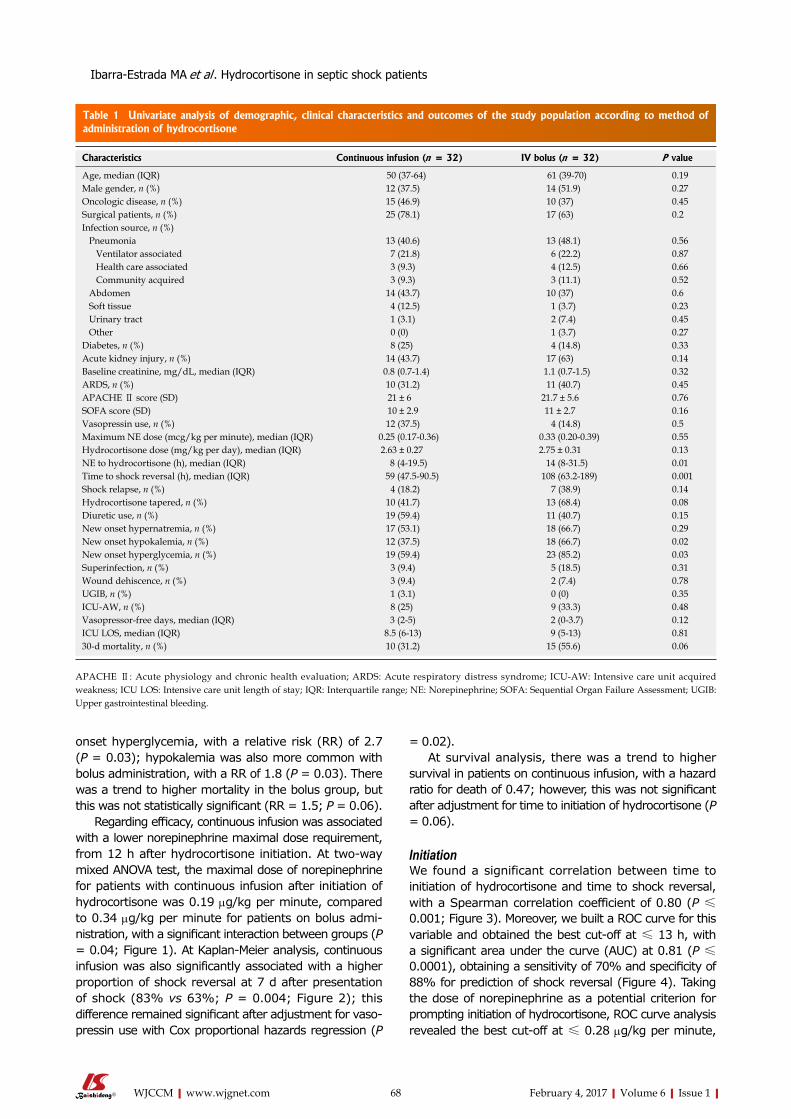
68 February 4, 2017|Volume 6|Issue 1|WJCCM|www.wjgnet.com
onset hyperglycemia, with a relative risk (RR) of 2.7 (P = 0.03); hypokalemia was also more common with bolus administration, with a RR of 1.8 (P = 0.03). There was a trend to higher mortality in the bolus group, but this was not statistically significant (RR = 1.5; P = 0.06).
Regarding efficacy, continuous infusion was associated with a lower norepinephrine maximal dose requirement, from 12 h after hydrocortisone initiation. At two-way mixed ANOVA test, the maximal dose of norepinephrine for patients with continuous infusion after initiation of hydrocortisone was 0.19 μg/kg per minute, compared to 0.34 μg/kg per minute for patients on bolus admi-nistration, with a significant interaction between groups (P = 0.04; Figure 1). At Kaplan-Meier analysis, continuous infusion was also significantly associated with a higher proportion of shock reversal at 7 d after presentation of shock (83% vs 63%; P = 0.004; Figure 2); this difference remained significant after adjustment for vaso-pressin use with Cox proportional hazards regression (P
= 0.02).At survival analysis, there was a trend to higher
survival in patients on continuous infusion, with a hazard ratio for death of 0.47; however, this was not significant after adjustment for time to initiation of hydrocortisone (P = 0.06).
InitiationWe found a significant correlation between time to initiation of hydrocortisone and time to shock reversal, with a Spearman correlation coefficient of 0.80 (P ≤ 0.001; Figure 3). Moreover, we built a ROC curve for this variable and obtained the best cut-off at ≤ 13 h, with a significant area under the curve (AUC) at 0.81 (P ≤ 0.0001), obtaining a sensitivity of 70% and specificity of 88% for prediction of shock reversal (Figure 4). Taking the dose of norepinephrine as a potential criterion for prompting initiation of hydrocortisone, ROC curve analysis revealed the best cut-off at ≤ 0.28 μg/kg per minute,
Characteristics Continuous infusion (n = 32) IV bolus (n = 32) P value
Age, median (IQR) 50 (37-64) 61 (39-70) 0.19Male gender, n (%) 12 (37.5) 14 (51.9) 0.27Oncologic disease, n (%) 15 (46.9) 10 (37) 0.45Surgical patients, n (%) 25 (78.1) 17 (63) 0.2Infection source, n (%) Pneumonia 13 (40.6) 13 (48.1) 0.56 Ventilator associated 7 (21.8) 6 (22.2) 0.87 Health care associated 3 (9.3) 4 (12.5) 0.66 Community acquired 3 (9.3) 3 (11.1) 0.52 Abdomen 14 (43.7) 10 (37) 0.6 Soft tissue 4 (12.5) 1 (3.7) 0.23 Urinary tract 1 (3.1) 2 (7.4) 0.45 Other 0 (0) 1 (3.7) 0.27Diabetes, n (%) 8 (25) 4 (14.8) 0.33Acute kidney injury, n (%) 14 (43.7) 17 (63) 0.14Baseline creatinine, mg/dL, median (IQR) 0.8 (0.7-1.4) 1.1 (0.7-1.5) 0.32ARDS, n (%) 10 (31.2) 11 (40.7) 0.45APACHE Ⅱ score (SD) 21 ± 6 21.7 ± 5.6 0.76SOFA score (SD) 10 ± 2.9 11 ± 2.7 0.16Vasopressin use, n (%) 12 (37.5) 4 (14.8) 0.5Maximum NE dose (mcg/kg per minute), median (IQR) 0.25 (0.17-0.36) 0.33 (0.20-0.39) 0.55Hydrocortisone dose (mg/kg per day), median (IQR) 2.63 ± 0.27 2.75 ± 0.31 0.13NE to hydrocortisone (h), median (IQR) 8 (4-19.5) 14 (8-31.5) 0.01Time to shock reversal (h), median (IQR) 59 (47.5-90.5) 108 (63.2-189) 0.001Shock relapse, n (%) 4 (18.2) 7 (38.9) 0.14Hydrocortisone tapered, n (%) 10 (41.7) 13 (68.4) 0.08Diuretic use, n (%) 19 (59.4) 11 (40.7) 0.15New onset hypernatremia, n (%) 17 (53.1) 18 (66.7) 0.29New onset hypokalemia, n (%) 12 (37.5) 18 (66.7) 0.02New onset hyperglycemia, n (%) 19 (59.4) 23 (85.2) 0.03Superinfection, n (%) 3 (9.4) 5 (18.5) 0.31Wound dehiscence, n (%) 3 (9.4) 2 (7.4) 0.78UGIB, n (%) 1 (3.1) 0 (0) 0.35ICU-AW, n (%) 8 (25) 9 (33.3) 0.48Vasopressor-free days, median (IQR) 3 (2-5) 2 (0-3.7) 0.12ICU LOS, median (IQR) 8.5 (6-13) 9 (5-13) 0.8130-d mortality, n (%) 10 (31.2) 15 (55.6) 0.06
Table 1 Univariate analysis of demographic, clinical characteristics and outcomes of the study population according to method of administration of hydrocortisone
APACHE Ⅱ: Acute physiology and chronic health evaluation; ARDS: Acute respiratory distress syndrome; ICU-AW: Intensive care unit acquired weakness; ICU LOS: Intensive care unit length of stay; IQR: Interquartile range; NE: Norepinephrine; SOFA: Sequential Organ Failure Assessment; UGIB: Upper gastrointestinal bleeding.
Ibarra-Estrada MA et al . Hydrocortisone in septic shock patients

69 February 4, 2017|Volume 6|Issue 1|WJCCM|www.wjgnet.com
with an AUC of 0.75 (P = 0.0002), a sensitivity of 65%, and a specificity of 88% for shock reversal.
Discontinuation and adverse effectsAs expected for patients with shock reversal, length of hydrocortisone administration was significantly longer for patients with the tapering strategy than for patients with sudden discontinuation [121 h (IQR, 81-245) vs 50 h (IQR, 44-101); P = 0.001]. Taper strategy was independently associated with a higher risk of hyper-glycemia (RR = 3.2; P = 0.042), and hypokalemia (RR = 2.8; P = 0.005). Because bolus administration was also independently associated with higher risk of
hyperglycemia and hypokalemia at univariate analysis, we performed logistic regression models adjusting for potential confounding variables; only the taper strategy maintained statistical significance for higher risk of hyperglycemia (Table 2) and had the highest OR for hypokalemia (Table 3).
Hydrocortisone taper was not associated with a lower risk of shock relapse (RR = 1.29; P = 0.17).
Shock reversalIn order to identify the variables associated with a higher probability of shock reversal, we performed a logistic regression model, adjusting for relevant covariables. As shown in Table 4, only the initiation of hydrocortisone ≤
Variable Univariate P value Multivariate P value
NO-H (n = 42) No NO-H (n = 17) Adjusted OR (95%CI)Bolus hydrocortisone, n (%) 19 (45.2) 13 (76.5) 0.04 3.2 (0.5-26.5) 0.99Hydrocortisone taper, n (%) 20 (64.5) 3 (27.3) 0.03 5.3 (1.8-34.5) 0.04Diabetes, n (%) 11 (26.2) 1 (5.9) 0.08 6.2 (0.4-79.0) 0.95
Table 2 Univariate and multivariate logistic regression analysis for relevant factors associated with new-onset hyperglycemia
Goodness-of-fit (Hosmer-Lemeshow). χ 2 = 0.019, P = 1.00; AUC, 0.88 (0.75-0.96), P = 0.0001. NO-H: New-onset hyperglycemia; OR: Odds ratio.
r = 0.80r 2 = 0.64P < 0.001
0 20 40 60 80 100 120 140Time to initiation of hydrocortisone (h)
400
350
300
250
200
150
100
50
0
Tim
e to
sho
ck r
ever
sal (
h)
Figure 3 Correlation between time to initiation of hydrocortisone and total time to shock reversal. Spearman correlation coefficient 0.80, P < 0.001.
AUC 0.81Cut-off ≤ 13 hP < 0.0001
0 20 40 60 80 100
100-specificity
100
80
60
40
20
0
Sens
itivi
ty
Figure 4 Receiver operating characteristic curve analysis of time to initiation of hydrocortisone for prediction of shock reversal. AUC, 0.81, P < 0.0001. Cut-off obtained with Youden index. AUC: Area under the curve.
ContinuousBolus
Interaction between groups, P = 0.04
0.5
0.4
0.3
0.2
0.1Nor
epin
ephr
ine
dose
(μg
/kg
per
min
ute)
Before hydrocortisone After hydrocortisone
Figure 1 Change in maximal norepinephrine dose from 12 h after initiation of hydrocortisone. Comparison between continuous and bolus administration groups, with two-way mixed ANOVA test, P = 0.04.
Continuous
Log rank test, P = 0.004
Bolus
0 25 50 75 100 125 150 175
t/h
100
80
60
40
20
0
Shoc
k re
vers
al (
%)
Figure 2 Kaplan-Meier analysis comparing the rate of septic shock reversal, according to administration of hydrocortisone. At 7 d (168 h), 83% of continuous infusion patients were vasopressor-free compared to 63% of patients who were in the bolus administration group, P = 0.004.
Ibarra-Estrada MA et al . Hydrocortisone in septic shock patients
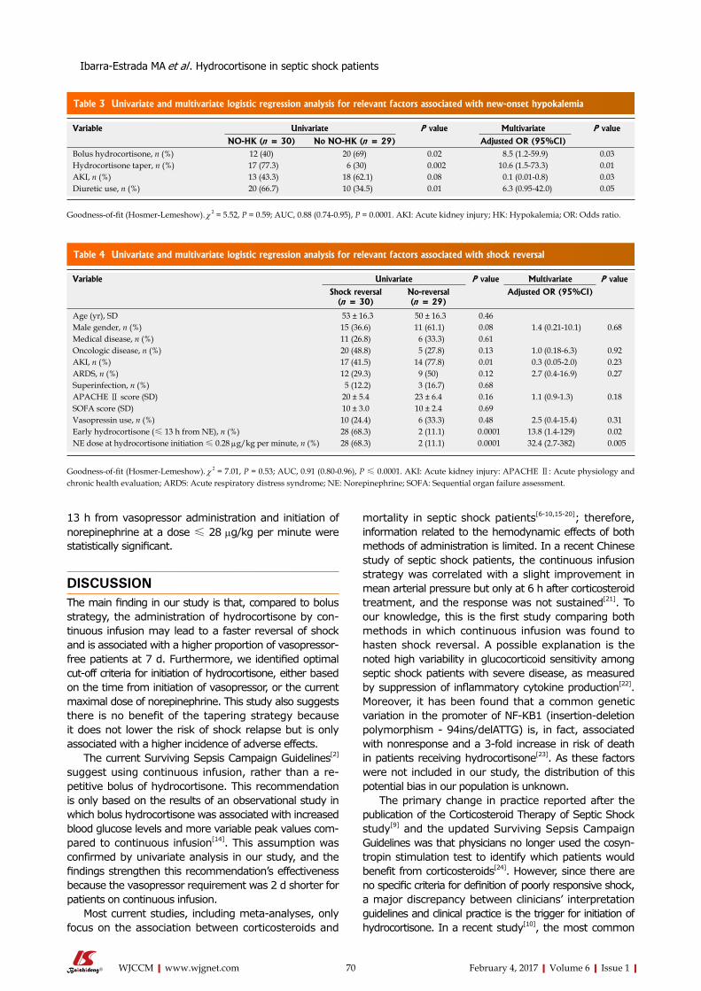
70 February 4, 2017|Volume 6|Issue 1|WJCCM|www.wjgnet.com
13 h from vasopressor administration and initiation of norepinephrine at a dose ≤ 28 μg/kg per minute were statistically significant.
DISCUSSIONThe main finding in our study is that, compared to bolus strategy, the administration of hydrocortisone by con-tinuous infusion may lead to a faster reversal of shock and is associated with a higher proportion of vasopressor-free patients at 7 d. Furthermore, we identified optimal cut-off criteria for initiation of hydrocortisone, either based on the time from initiation of vasopressor, or the current maximal dose of norepinephrine. This study also suggests there is no benefit of the tapering strategy because it does not lower the risk of shock relapse but is only associated with a higher incidence of adverse effects.
The current Surviving Sepsis Campaign Guidelines[2] suggest using continuous infusion, rather than a re-petitive bolus of hydrocortisone. This recommendation is only based on the results of an observational study in which bolus hydrocortisone was associated with increased blood glucose levels and more variable peak values com-pared to continuous infusion[14]. This assumption was confirmed by univariate analysis in our study, and the findings strengthen this recommendation’s effectiveness because the vasopressor requirement was 2 d shorter for patients on continuous infusion.
Most current studies, including meta-analyses, only focus on the association between corticosteroids and
mortality in septic shock patients[6-10,15-20]; therefore, information related to the hemodynamic effects of both methods of administration is limited. In a recent Chinese study of septic shock patients, the continuous infusion strategy was correlated with a slight improvement in mean arterial pressure but only at 6 h after corticosteroid treatment, and the response was not sustained[21]. To our knowledge, this is the first study comparing both methods in which continuous infusion was found to hasten shock reversal. A possible explanation is the noted high variability in glucocorticoid sensitivity among septic shock patients with severe disease, as measured by suppression of inflammatory cytokine production[22]. Moreover, it has been found that a common genetic variation in the promoter of NF-KB1 (insertion-deletion polymorphism - 94ins/delATTG) is, in fact, associated with nonresponse and a 3-fold increase in risk of death in patients receiving hydrocortisone[23]. As these factors were not included in our study, the distribution of this potential bias in our population is unknown.
The primary change in practice reported after the publication of the Corticosteroid Therapy of Septic Shock study[9] and the updated Surviving Sepsis Campaign Guidelines was that physicians no longer used the cosyn-tropin stimulation test to identify which patients would benefit from corticosteroids[24]. However, since there are no specific criteria for definition of poorly responsive shock, a major discrepancy between clinicians’ interpretation guidelines and clinical practice is the trigger for initiation of hydrocortisone. In a recent study[10], the most common
Variable Univariate P value Multivariate P value
NO-HK (n = 30) No NO-HK (n = 29) Adjusted OR (95%CI)Bolus hydrocortisone, n (%) 12 (40) 20 (69) 0.02 8.5 (1.2-59.9) 0.03Hydrocortisone taper, n (%) 17 (77.3) 6 (30) 0.002 10.6 (1.5-73.3) 0.01AKI, n (%) 13 (43.3) 18 (62.1) 0.08 0.1 (0.01-0.8) 0.03Diuretic use, n (%) 20 (66.7) 10 (34.5) 0.01 6.3 (0.95-42.0) 0.05
Table 3 Univariate and multivariate logistic regression analysis for relevant factors associated with new-onset hypokalemia
Goodness-of-fit (Hosmer-Lemeshow). χ 2 = 5.52, P = 0.59; AUC, 0.88 (0.74-0.95), P = 0.0001. AKI: Acute kidney injury; HK: Hypokalemia; OR: Odds ratio.
Variable Univariate P value Multivariate P value
Shock reversal (n = 30)
No-reversal (n = 29)
Adjusted OR (95%CI)
Age (yr), SD 53 ± 16.3 50 ± 16.3 0.46Male gender, n (%) 15 (36.6) 11 (61.1) 0.08 1.4 (0.21-10.1) 0.68Medical disease, n (%) 11 (26.8) 6 (33.3) 0.61Oncologic disease, n (%) 20 (48.8) 5 (27.8) 0.13 1.0 (0.18-6.3) 0.92AKI, n (%) 17 (41.5) 14 (77.8) 0.01 0.3 (0.05-2.0) 0.23ARDS, n (%) 12 (29.3) 9 (50) 0.12 2.7 (0.4-16.9) 0.27Superinfection, n (%) 5 (12.2) 3 (16.7) 0.68APACHE Ⅱ score (SD) 20 ± 5.4 23 ± 6.4 0.16 1.1 (0.9-1.3) 0.18SOFA score (SD) 10 ± 3.0 10 ± 2.4 0.69Vasopressin use, n (%) 10 (24.4) 6 (33.3) 0.48 2.5 (0.4-15.4) 0.31Early hydrocortisone (≤ 13 h from NE), n (%) 28 (68.3) 2 (11.1) 0.0001 13.8 (1.4-129) 0.02NE dose at hydrocortisone initiation ≤ 0.28 μg/kg per minute, n (%) 28 (68.3) 2 (11.1) 0.0001 32.4 (2.7-382) 0.005
Table 4 Univariate and multivariate logistic regression analysis for relevant factors associated with shock reversal
Goodness-of-fit (Hosmer-Lemeshow). χ 2 = 7.01, P = 0.53; AUC, 0.91 (0.80-0.96), P ≤ 0.0001. AKI: Acute kidney injury: APACHE Ⅱ: Acute physiology and chronic health evaluation; ARDS: Acute respiratory distress syndrome; NE: Norepinephrine; SOFA: Sequential organ failure assessment.
Ibarra-Estrada MA et al . Hydrocortisone in septic shock patients

71 February 4, 2017|Volume 6|Issue 1|WJCCM|www.wjgnet.com
clinical threshold for prescribing corticosteroids was the presence of 2 or more vasopressors in 64% of patients. We believe that there should be a global agree-ment according to variables associated with the higher probability of shock reversal. Based on our results at ROC curve analysis and correlation with time to shock reversal, we suggest initiation of hydrocortisone at ≤ 13 h after vasopressor administration. This conclusion agrees with a recent study of severely shocked patients in which the early administration of hydrocortisone (< 9 h) was associated with a significantly lower total time of vasopressors and mortality[25].
In a retrospective review addressing norepinephrine as a trigger for initiation of corticosteroids, the dose was arbitrarily defined as non-weight-based low, moderate, or high[10]. Through a more objective approach, we found a dose of ≤ 0.28 μg/kg per minute to be the best predictor for shock reversal, a lower threshold than what has been used in most studies[9,25,26], reinforcing the recommendation of an earlier initiation of hydrocortisone also based on vasopressor dose[27,28].
Another interesting finding of this study is the apparent lack of benefits to the tapering strategy for discontinuation of hydrocortisone. The Surviving Sepsis Campaign Guidelines suggest tapering from steroids when they are no longer required; therefore, this stra-tegy is the most commonly used (in up to 74% of patients), depending on the duration of hydrocortisone therapy[10]. Unfortunately, there has been no comparative study between tapering and abrupt cessation, and the main argument for this suggestion is a small crossover study in which shock relapse occurred in 30% of patients with sudden discontinuation[29]; however, the study was underpowered to reach that conclusion, because the data arose from a subgroup analysis of 20 patients. Therefore, tapering has a 2D recommendation (the weakest possible) on the GRADE system. Furthermore, there have been other randomized controlled studies in which corticosteroids were abruptly discontinued without reported increased risk of shock relapse[7,8]. In the current study, tapering was only associated with adverse effects; therefore, we suggest this should be avoided, especially for patients with ongoing hyperglycemia and/or hypokalemia.
The main strength of this study is that it was speci-fically designed to compare the efficacy between both methods of administration of hydrocortisone, according to vasopressor requirement, indirectly assessing their effects on immunomodulation and vasomotor tone im-provement.
This study has some limitations; we did not address the effects of the use of some drugs known to affect adrenal function (e.g., etomidate, antifungals, benzodiazepines, and opioids)[30]. Medical management for septic shock patients is always based on the current Surviving Sepsis Campaign Guidelines and are very similar at both hospitals; however, due to the observational and nonrandomized design of the study, we cannot ensure completely homogeneous treatment regarding other
relevant variables associated with improving outcomes (e.g., appropriateness and type of fluid resuscitation and correct and timely use of antibiotics). This study was powered to detect differences in short-term vasopressor requirements and to find the best cut-offs for initiation of hydrocortisone only; therefore, results concerning analysis between groups should be interpreted cautiously, and should be taken as hypothesis-generating data for the design of future clinical randomized controlled trials.
In conclusion, we found that continuous infusion of hydrocortisone could hasten resolution of septic shock compared with bolus administration, and that earlier initiation based on time and/or norepinephrine dose is related with a higher probability of shock reversal. The tapering strategy appears unnecessary and may be only related to additional adverse effects.
ACKNOWLEDGMENTSWe acknowledge Hilario Coronado Magaña, Department Chair of the Intensive Care Unit at Hospital Civil Fray Antonio Alcalde, for his general support of this work. We would also like to thank Victoria L. Clifton, MLIS, ELS, for her assistance with language editing and the editorial preparation of this manuscript.
COMMENTSBackgroundThe latest Surviving Sepsis Campaign Guidelines recommend administration of low-dose hydrocortisone (200 mg/d) when hemodynamic stability is not achievable after fluid resuscitation and vasopressor therapy. Although the benefits of hydrocortisone are increasingly being recognized, several issues concerning its specific indications, clinical criteria for initiation, adequate method of administration, and strategy of discontinuation are unresolved, which leads to widely heterogeneous prescribing practices among critical care physicians. Studies addressing those issues are lacking.
Research frontiersThe reported prescribing patterns of hydrocortisone for patients with septic shock come from studies performed only in the United States and describe a wide variability of clinical practices. In Mexico, this kind of information is scarce.
Innovations and breakthroughsContinuous infusion of hydrocortisone leads to faster resolution of shock than bolus administration. Initiation of hydrocortisone at ≤ 13 h after starting norepinephrine and/or a maximal dose of ≤ 28 μg/kg per minute is associated with a higher probability of shock reversal. There is no benefit from a tapering strategy because this was only shown to lead to additional adverse effects.
ApplicationsThe results of the study add valuable information, which could contribute to the initiation of a widespread agreement regarding specific indications on the correct use of hydrocortisone in septic shock patients.
Peer-reviewIt is a well design study, though performed in a small group of patients.
REFERENCES1 Schumer W. Steroids in the treatment of clinical septic shock. Ann
Surg 1976; 184: 333-341 [PMID: 786190 DOI: 10.1097/00000658
Ibarra-Estrada MA et al . Hydrocortisone in septic shock patients
COMMENTS
72 February 4, 2017|Volume 6|Issue 1|WJCCM|www.wjgnet.com
-197609000-00011]2 Dellinger RP, Levy MM, Rhodes A, Annane D, Gerlach H, Opal
SM, Sevransky JE, Sprung CL, Douglas IS, Jaeschke R, Osborn TM, Nunnally ME, Townsend SR, Reinhart K, Kleinpell RM, Angus DC, Deutschman CS, Machado FR, Rubenfeld GD, Webb SA, Beale RJ, Vincent JL, Moreno R. Surviving sepsis campaign: international guidelines for management of severe sepsis and septic shock: 2012. Crit Care Med 2013; 41: 580-637 [PMID: 23353941 DOI: 10.1097/CCM.0b013e31827e83af]
3 Dellinger RP. Steroid therapy of septic shock: the decision is in the eye of the beholder. Crit Care Med 2008; 36: 1987-1989 [PMID: 18520670 DOI: 10.1097/CCM.0b013e31817d7ee4]
4 Patel GP, Balk RA. Systemic steroids in severe sepsis and septic shock. Am J Respir Crit Care Med 2012; 185: 133-139 [PMID: 21680949 DOI: 10.1164/rccm.201011-1897CI]
5 Bollaert PE, Charpentier C, Levy B, Debouverie M, Audibert G, Larcan A. Reversal of late septic shock with supraphysiologic doses of hydrocortisone. Crit Care Med 1998; 26: 645-650 [PMID: 9559600 DOI: 10.1097/00003246-199804000-00010]
6 Briegel J, Forst H, Haller M, Schelling G, Kilger E, Kuprat G, Hemmer B, Hummel T, Lenhart A, Heyduck M, Stoll C, Peter K. Stress doses of hydrocortisone reverse hyperdynamic septic shock: a prospective, randomized, double-blind, single-center study. Crit Care Med 1999; 27: 723-732 [PMID: 10321661 DOI: 10.1097/00003246-199904000-00025]
7 Yildiz O, Doganay M, Aygen B, Güven M, Keleştimur F, Tutuû A. Physiological-dose steroid therapy in sepsis [ISRCTN36253388]. Crit Care 2002; 6: 251-259 [PMID: 12133187 DOI: 10.1186/cc1498]
8 Annane D, Sébille V, Charpentier C, Bollaert PE, François B, Korach JM, Capellier G, Cohen Y, Azoulay E, Troché G, Chaumet-Riffaud P, Bellissant E. Effect of treatment with low doses of hydrocortisone and fludrocortisone on mortality in patients with septic shock. JAMA 2002; 288: 862-871 [PMID: 12186604 DOI: 10.1001/jama.288.7.862]
9 Sprung CL, Annane D, Keh D, Moreno R, Singer M, Freivogel K, Weiss YG, Benbenishty J, Kalenka A, Forst H, Laterre PF, Reinhart K, Cuthbertson BH, Payen D, Briegel J. Hydrocortisone therapy for patients with septic shock. N Engl J Med 2008; 358: 111-124 [PMID: 18184957 DOI: 10.1056/NEJMoa071366]
10 Contrael KM, Killian AJ, Gregg SR, Buchman TG, Coopersmith CM. Prescribing patterns of hydrocortisone in septic shock: a single-center experience of how surviving sepsis guidelines are interpreted and translated into bedside practice. Crit Care Med 2013; 41: 2310-2317 [PMID: 23787398 DOI: 10.1097/CCM.0b013e31828 cef29]
11 Youden WJ. Index for rating diagnostic tests. Cancer 1950; 3: 32-35 [PMID: 15405679 DOI: 10.1002/1097-0142(1950)3:1<32::AID-CNCR2820030106>3.0.CO;2-3]
12 Hosmer DW, Taber S, Lemeshow S. The importance of assessing the fit of logistic regression models: a case study. Am J Public Health 1991; 81: 1630-1635 [PMID: 1746660 DOI: 10.2105/AJPH.81.12.1630]
13 Ibarra-Estrada MÁ, López-Pulgarín JA, Mijangos-Méndez JC, Díaz-Gómez JL, Aguirre-Avalos G. Respiratory variation in carotid peak systolic velocity predicts volume responsiveness in mechanically ventilated patients with septic shock: a prospective cohort study. Crit Ultrasound J 2015; 7: 29 [PMID: 26123610 DOI: 10.1186/s13089-015-0029-1]
14 Weber-Carstens S, Deja M, Bercker S, Dimroth A, Ahlers O, Kaisers U, Keh D. Impact of bolus application of low-dose hydro-cortisone on glycemic control in septic shock patients. Intensive Care Med 2007; 33: 730-733 [PMID: 17325831 DOI: 10.1007/s00134-007-0540-3]
15 Annane D, Bellissant E, Bollaert PE, Briegel J, Confalonieri M, De Gaudio R, Keh D, Kupfer Y, Oppert M, Meduri GU. Corticosteroids in the treatment of severe sepsis and septic shock in adults: a systematic review. JAMA 2009; 301: 2362-2375 [PMID: 19509383 DOI: 10.1001/jama.2009.815]
16 Annane D, Bellissant E, Bollaert PE, Briegel J, Keh D, Kupfer Y.
Corticosteroids for treating sepsis. Cochrane Database Syst Rev 2015; (12): CD002243 [PMID: 26633262 DOI: 10.1002/14651858.CD002243.pub3]
17 Arabi YM, Aljumah A, Dabbagh O, Tamim HM, Rishu AH, Al-Abdulkareem A, Knawy BA, Hajeer AH, Tamimi W, Cherfan A. Low-dose hydrocortisone in patients with cirrhosis and septic shock: a randomized controlled trial. CMAJ 2010; 182: 1971-1977 [PMID: 21059778 DOI: 10.1503/cmaj.090707]
18 Huh JW, Choi HS, Lim CM, Koh Y, Oh YM, Shim TS, Lee SD, Kim WS, Kim DS, Hong SB. Low-dose hydrocortisone treatment for patients with septic shock: a pilot study comparing 3days with 7days. Respirology 2011; 16: 1088-1095 [PMID: 21726354 DOI: 10.1111/j.1440-1843.2011.02018.x]
19 Tagami T, Matsui H, Fushimi K, Yasunaga H. Low-dose corti-costeroid treatment and mortality in refractory abdominal septic shock after emergency laparotomy. Ann Intensive Care 2015; 5: 32 [PMID: 26514125 DOI: 10.1186/s13613-015-0074-8]
20 Wang C, Sun J, Zheng J, Guo L, Ma H, Zhang Y, Zhang F, Li E. Low-dose hydrocortisone therapy attenuates septic shock in adult patients but does not reduce 28-day mortality: a meta-analysis of randomized controlled trials. Anesth Analg 2014; 118: 346-357 [PMID: 24445635 DOI: 10.1213/ANE.0000000000000050]
21 Chen Z, Yang C, He H, He Z. [The impacts of low-dose cortico-steroids infusion given in different manners on refractory septic shock patients]. Zhonghua Wei Zhong Bing Ji Jiu Yi Xue 2015; 27: 443-447 [PMID: 26049181 DOI: 10.3760/cma.j.issn.2095-4352.2015.06.006]
22 Cohen J, Pretorius CJ, Ungerer JP, Cardinal J, Blumenthal A, Presneill J, Gatica-Andrades M, Jarrett P, Lassig-Smith M, Stuart J, Dunlop R, Starr T, Venkatesh B. Glucocorticoid Sensitivity Is Highly Variable in Critically Ill Patients With Septic Shock and Is Associated With Disease Severity. Crit Care Med 2016; 44: 1034-1041 [PMID: 26963327 DOI: 10.1097/CCM.0000000 000001633]
23 Schäfer ST, Gessner S, Scherag A, Rump K, Frey UH, Siffert W, Westendorf AM, Steinmann J, Peters J, Adamzik M. Hydrocortisone fails to abolish NF-κB1 protein nuclear translocation in deletion allele carriers of the NFKB1 promoter polymorphism (-94ins/delATTG) and is associated with increased 30-day mortality in septic shock. PLoS One 2014; 9: e104953 [PMID: 25133403 DOI: 10.1371/journal.pone.0104953]
24 Bruno JJ, Dee BM, Anderegg BA, Hernandez M, Pravinkumar SE. US practitioner opinions and prescribing practices regarding corticosteroid therapy for severe sepsis and septic shock. J Crit Care 2012; 27: 351-361 [PMID: 22341726 DOI: 10.1016/j.jcrc. 2011.12.011]
25 Katsenos CS, Antonopoulou AN, Apostolidou EN, Ioakeimidou A, Kalpakou GT, Papanikolaou MN, Pistiki AC, Mpalla MC, Paraschos MD, Patrani MA, Pratikaki ME, Retsas TA, Savva AA, Vassiliagkou SD, Lekkou AA, Dimopoulou I, Routsi C, Mandragos KE. Early administration of hydrocortisone replacement after the advent of septic shock: impact on survival and immune response*. Crit Care Med 2014; 42: 1651-1657 [PMID: 24674923 DOI: 10.1097/CCM.0000000000000318]
26 Torgersen C, Luckner G, Schröder DC, Schmittinger CA, Rex C, Ulmer H, Dünser MW. Concomitant arginine-vasopressin and hydrocortisone therapy in severe septic shock: association with mortality. Intensive Care Med 2011; 37: 1432-1437 [PMID: 21779849 DOI: 10.1007/s00134-011-2312-3]
27 Bentzer P, Fjell C, Walley KR, Boyd J, Russell JA. Plasma cytokine levels predict response to corticosteroids in septic shock. Intensive Care Med 2016; 42: 1970-1979 [PMID: 27071387 DOI: 10.1007/s00134-016-4338-z]
28 Gordon AC, Mason AJ, Perkins GD, Stotz M, Terblanche M, Ashby D, Brett SJ. The interaction of vasopressin and corticosteroids in septic shock: a pilot randomized controlled trial. Crit Care Med 2014; 42: 1325-1333 [PMID: 24557425 DOI: 10.1097/CCM.0000 000000000212]
29 Keh D, Boehnke T, Weber-Cartens S, Schulz C, Ahlers O, Bercker S, Volk HD, Doecke WD, Falke KJ, Gerlach H. Immunologic
Ibarra-Estrada MA et al . Hydrocortisone in septic shock patients

73 February 4, 2017|Volume 6|Issue 1|WJCCM|www.wjgnet.com
and hemodynamic effects of “low-dose” hydrocortisone in septic shock: a double-blind, randomized, placebo-controlled, crossover study. Am J Respir Crit Care Med 2003; 167: 512-520 [PMID: 12426230 DOI: 10.1164/rccm.200205-446OC]
30 Greenberg SB, Coursin DB. Timing of corticosteroids in refractory septic shock: a key or wishful thinking?*. Crit Care Med 2014; 42: 1733-1735 [PMID: 24933052 DOI: 10.1097/CCM.000000 0000000361]
P- Reviewer: Katsenos CS, Lazzeri C, Piacentini EA S- Editor: Kong JX L- Editor: A E- Editor: Li D
Ibarra-Estrada MA et al . Hydrocortisone in septic shock patients
74 February 4, 2017|Volume 6|Issue 1|WJCCM|www.wjgnet.com
Martin Albert, Departments of Intensive Care and Medicine, Hôpital du Sacré-Coeur, de Montréal Research Center, Université de Montréal, Montréal H4J 1C5, Canada
Daniel Corsilli, Marc Brosseau, Patrick Bellemare, Department of Intensive Care, Centre Hospitalier Universitaire de Montréal, Université de Montréal, Montréal H2W 1T8, Canada
David R Williamson, Anne QN Nguyen, France Varin, Department of Pharmacy, Université de Montréal, Hôpital du Sacré-Coeur de Montréal Research Center, Montréal H4J 1C5, Canada
Patrick Bellemare, Department of Respiratory Care, Hôpital du Sacré-Coeur de, Montréal H4J 1C5, Canada
Stéphane Delisle, Hôpital du Sacré-Coeur, de Montréal Research Center, Université de Montréal, Montréal H4J 1C5, Canada
Author contributions: All the authors contributed to the manu-script.
Institutional review board statement: The protocol was approved by the Hôpital du Sacré-Coeur de Montréal ethics committee and consent was obtained from patients or their next of kin. The protocol was submitted to and approved by Health Canada.
Informed consent statement: Consent for data sharing was not obtained but the presented data are anonymized and risk of identification is insignificant.
Conflict-of-interest statement: The authors declare no conflicts of interest regarding this manuscript.
Open-Access: This article is an open-access article which was selected by an in-house editor and fully peer-reviewed by external reviewers. It is distributed in accordance with the Creative Commons Attribution Non Commercial (CC BY-NC 4.0) license, which permits others to distribute, remix, adapt, build upon this work non-commercially, and license their derivative works on different terms, provided the original work is properly cited and the use is non-commercial. See: http://creativecommons.org/
licenses/by-nc/4.0/
Manuscript source: Invited manuscript
Correspondence to: Martin Albert, MD, Departments of Intensive Care and Medicine, Hôpital du Sacré-Coeur, de Montréal Research Center, Université de Montréal, 5400 boul, Gouin Ouest, Montréal H4J 1C5, Canada. [email protected]: +1-514-3382050Fax: +1-514-3383557
Received: September 1, 2016Peer-review started: September 5, 2016First decision: September 29, 2016Revised: November 26, 2016Accepted: December 16, 2016Article in press: December 19, 2016Published online: February 4, 2017
AbstractAIMTo evaluate the safety and efficacy of inhaled milrinone in acute respiratory distress syndrome (ARDS).
METHODSOpen-label prospective cross-over pilot study where fifteen adult patients with hypoxemic failure meeting standard ARDS criteria and monitored with a pulmonary artery catheter were recruited in an academic 24-bed medico-surgical intensive care unit. Random sequential administration of iNO (20 ppm) or nebulized epopro-stenol (10 μg/mL) was done in all patients. Thereafter, inhaled milrinone (1 mg/mL) alone followed by inhaled milrinone in association with inhaled nitric oxide (iNO) was administered. A jet nebulization device synchronized with the mechanical ventilation was use to administrate the epoprostenol and the milrinone. Hemodynamic mea-surements and partial pressure of arterial oxygen (PaO2) were recorded before and after each inhaled therapy
World Journal ofCritical Care MedicineW J C C M
Submit a Manuscript: http://www.wjgnet.com/esps/Help Desk: http://www.wjgnet.com/esps/helpdesk.aspxDOI: 10.5492/wjccm.v6.i1.74
World J Crit Care Med 2017 February 4; 6(1): 74-78ISSN 2220-3141 (online)
© 2017 Baishideng Publishing Group Inc. All rights reserved.
Martin Albert, Daniel Corsilli, David R Williamson, Marc Brosseau, Patrick Bellemare, Stéphane Delisle, Anne QN Nguyen, France Varin
ORIGINAL ARTICLE
Comparison of inhaled milrinone, nitric oxide and prostacyclin in acute respiratory distress syndrome
Prospective Study

75 February 4, 2017|Volume 6|Issue 1|WJCCM|www.wjgnet.com
Albert M et al . Comparison of inhaled therapy in ARDS
administration.
RESULTSThe majority of ARDS were of pulmonary cause (n = 13) and pneumonia (n = 7) was the leading underlying initial disease. Other pulmonary causes of ARDS were: Post cardiopulmonary bypass (n = 2), smoke inhalation injury (n = 1), thoracic trauma and pulmonary con-tusions (n = 2) and aspiration (n = 1). Two patients had an extra pulmonary cause of ARDS: A polytrauma patient and an intra-abdominal abscess Inhaled nitric oxide, epoprostenol, inhaled milrinone and the com-bination of inhaled milrinone and iNO had no impact on systemic hemodynamics. No significant adverse events related to study medications were observed. The median increase of PaO2 from baseline was 8.8 mmHg [interquartile range (IQR) = 16.3], 6.0 mmHg (IQR = 18.4), 6 mmHg (IQR = 15.8) and 9.2 mmHg (IQR = 20.2) respectively with iNO, epoprostenol, inhaled milrinone, and iNO added to milrinone. Only iNO and the combination of inhaled milrinone and iNO had a statistically significant effect on PaO2.
CONCLUSIONWhen comparing the effects of inhaled NO, milrinone and epoprostenol, only NO significantly improved oxy-genation. Inhaled milrinone appeared safe but failed to improve oxygenation in ARDS.
Key words: Inhaled milrinone; Nitric oxide; Pulmonary hypertension; Hypoxemia; Acute respiratory distress syndrome; Prostacyclin
© The Author(s) 2017. Published by Baishideng Publishing Group Inc. All rights reserved.
Core tip: To our knowledge, this is the first study testing inhaled milrinone as a therapy in acute respiratory distress syndrome and comparing it to more frequently used inhaled therapies. It shows that inhaled milrinone is safe but is not efficacious.
Albert M, Corsilli D, Williamson DR, Brosseau M, Bellemare P, Delisle S, Nguyen AQN, Varin F. Comparison of inhaled milrinone, nitric oxide and prostacyclin in acute respiratory distress syndrome. World J Crit Care Med 2017; 6(1): 74-78 Available from: URL: http://www.wjgnet.com/2220-3141/full/v6/i1/74.htm DOI: http://dx.doi.org/10.5492/wjccm.v6.i1.74
INTRODUCTIONAcute respiratory distress syndrome (ARDS) is unfortunately a common problem in intensive care units (ICU) and has been associated with significant morbidity and mortality[1]. Hypoxemia and hypercapnia are the primary manifestations of the ventilationperfusion mismatch observed in ARDS patients. Despite several advances in mechanical ventilation, treatment of severe hypoxemia has remained one of the greatest challenges
in the ICU. Among these therapies, inhaled nitric oxide (iNO) is commonly used for the treatment of hypoxemia in ARDS because it allows for selective vasodilation of ventilated units, transforming relative dead space into adequate ventilationperfusion units[1,2]. Regardless of the well documented failure to improve survival, iNO is still of common use because of the oxygenation gain it allows. However, it has substantial cost, has been associated with potential serious side effects such as renal failure and needs a special device for its delivery[2,3]. Inhaled prostacyclin has also been used in ARDS and has been shown to significantly reduce pulmonary artery pressure and increase oxygenation[46]. However, prostacyclin administration is technically challenging given its short halflife and susceptibility to photodegradation[7]. The phosphodiesterase type Ⅲ inhibitor milrinone is a potent pulmonary vasodilator that has been used with success as an inhaled therapy for pulmonary hypertension in cardiac surgery and may be a potential alternative to actual treatment strategies[8,9]. Animal studies have suggested a response to milirinone in acute lung injury[10].
The primary objective of this study was to assess the tolerability and safety of inhaled milrinone in ARDS patients. The secondary objectives included: Evaluation of the efficacy of inhaled milrinone in improving hypoxemia compared to baseline; comparison of the effects of inhaled milrinone, iNO and inhaled epoprostenol in improving hypoxemia and secondary pulmonary hypertension compared to baseline; evaluation of the efficacy of combining inhaled milrinone with iNO on hypoxemia and pulmonary hypertension.
MATERIALS AND METHODSIn an academic 24bed medicosurgical intensive care unit, patients were screened over a 2year period. Adult patients were enrolled if they had hypoxemic respiratory failure meeting standard moderate to severe ARDS criteria: Ratio of the partial pressure of arterial oxygen (PaO2) to the fraction of inspired oxygen (FiO2) of 200 or less, pulmonary capillary wedge pressure (Pcwp) ≤ 18 mmHg and bilateral infiltrates on frontal chest radiograph. Recruited patients also had a pulmonary artery catheter and an arterial line. Patients with severe hemodynamic instability (defined as the need for more than one vasopressor or the use of more than 0.5 μg/kg per minute of norepinephrine), on intravenous milrinone or nitrate derivatives that could not be weaned for study purposes and patients on high frequency oscillatory ventilation were excluded. Patients with a history of hypersensitivity to study medications, pregnant patients and those who participated in another study involving oxymetric values or pulmonary hemodynamics were also excluded.
Patients were randomly administered sequential nebulization of iNO (20 ppm) or epoprostenol (10 μg/mL for a total volume of 5 mL). Thereafter, milrinone (1 mg/mL for a total volume of 5 mL at each nebulisation)

76 February 4, 2017|Volume 6|Issue 1|WJCCM|www.wjgnet.com
alone and in association with iNO was administered. Each drug was nebulized for 20 min and a 30 min washout was allowed between each drug. We used a jet nebulization device ventilator synchronized to nebulize epoprostenol and milrinone (MicroMist® Nebulizer model 1880; Hudson RCI, Temecula, CA, United States). The nebulizer was attached to the inspiratory limb of the ventilator near the endotracheal tube. The mass median diameter obtain with this nebulizer is 2.1 μm and the nebulization flow is between 0.25 and 0.3 mL/min. The ventilatory circuit humidification was stopped during the nebulization. Adjustment of the tidal volume was done during nebulization to obtain the same minute ventilation. The iNO was administered using a standard iNO Delivery system (INOvent®, Ohmeda, Madison, WI, United States) attached to the inspiratory limb of the ventilator. A constant dose of 20 ppm of iNO was used and monitored by the injection device. Blood pressure and oxygenation status were continuously monitored. If hemodynamic instability occurred, defined as a lowering of systolic arterial pressure ≥ 10 mmHg or lowering of mean arterial pressure (MAP) ≥ 5 mmHg, the study medication was stopped. If oxygenation status worsened, defined as a lowering of arterial oxygen saturation ≥ 10% (measured with continuous pulse oximetry and confirmed by arterial gas), the study medication was stopped. Adverse events potentially related to study medications such as increase hemodynamic instability and renal failure was prospectively evaluated. Using a previously published milrinone assay[11], we determined the plasma level of milrinone at the end of the administration of inhaled milrinone or the combination of iNo and inhaled milrinone in eight samples from our last four patients.
Demographic data, APACHE Ⅱ and SOFA scores were collected. The following parameters were measured before and during each specific drug nebulization: MAP, mean pulmonary arterial pressure, thermodilution cardiac output, PaO2, heart rate, central venous pressure and Pcwp. The following parameters were calculated: Systemic
and pulmonary vascular resistances (SVR and PVR), PVR/SVR Ratio, indexed pulmonary vascular resistance, transpulmonary gradient, PaO2/FiO2, cardiac index, shunt and oxygenation index. A patient was considered a responder to study medications if is PaO2 increased of more than 20% from the preinhalation value[2].
As the primary goal of this pilot study was safety and feasibility and no preliminary data existed in this population, a convenience sample of 15 patients was chosen. Given a within patient standard deviation of 11%, the sample size enabled the detection of a 12% difference in PaO2 between baseline and postmilrinone PaO2. Given the small samplesize, demographic and baseline data were described as medians and interquartile range. Continuous data were analysed using Wilcoxon signed rank tests.
The study was performed at Hôpital du SacréCoeur de Montréal, Canada. The local ethics committee approved the protocol and consent was obtained from patients or their next of kin. The protocol was submitted to and approved by Health Canada.
RESULTSFifteen consecutive patients were included in the study (Table 1). The majority of ARDS cases were of pulmonary origin (n = 13) and pneumonia (n = 7) was the leading underlying initial disease. Other pulmonary causes of ARDS were: Post cardiopulmonary bypass (n = 2), smoke inhalation injury (n = 1), thoracic trauma and pulmonary contusions (n = 2) and aspiration (n = 1). Two patients had an extra pulmonary cause of ARDS: A polytrauma patient and an intraabdominal abscess. The main hemodynamic responses are summarized in Table 2. INO, epoprostenol, inhaled milrinone and the combination of inhaled milrinone and iNO did not have any significant impact on measured hemodynamics when compared to baseline (all P > 0.1).
We observed for the oxygenation measurement a median increase of PaO2 from baseline of 8.8 mmHg [interquartile range (IQR) = 16.3], 6.0 mmHg (IQR = 18.4), 6 mmHg (IQR = 15.8) and 9.2 mmHg (IQR = 20.2) respectively with iNO, epoprostenol, inhaled milrinone, and iNO added to milrinone. When compared to baseline, the combination of inhaled milrinone and iNO (P = 0.004) and only iNO had a statistically significant effect (P = 0.036). The median percent response to iNO, epoprostenol, inhaled milrinone and the combination of milrinone and iNO was 11.2% (IQR = 25%), 5.3% (IQR = 24%), 7.9% (IQR = 19%) and 11.8% (IQR = 26%), respectively. The response rate to study medications, defined as an increase of more than 20% from the pre-inhalation value, were 33.3%, 20.0%, 13.3% and 33.3% respectively with iNO, epoprostenol, inhaled milrinone and the combination of inhaled milrinone and iNO. The median PaO2 response of 39.0 mmHg in responders was higher with iNO than with epoprostenol (26.5 mmHg) or milrinone (10 mmHg).
No significant adverse events related to study medi-
Age (yr) 57 (IQR = 22)Gender 12 men, 3 womenSOFA score (ICU admission) 7.5 (IQR = 7)SOFA score (day of protocol) 10.0 (IQR = 5)APACHE-Ⅱ (ICU admission) 23 (IQR = 7)APACHE-Ⅱ (day of protocol) 23.5 (IQR = 7.0)PaO2 (mmHg) 80 (IQR = 39)FiO2 (%) 80 (IQR = 30)PaO2/FiO2 138 (IQR = 68)PEEP (cm H2O) 10 (IQR = 2)MAP (mm Hg) 75 (IQR = 16)mPAP (mm Hg) 28 (IQR = 7)Cardiac index (L/min per square metre) 3.7 (IQR = 2.6)
Table 1 Baseline characteristics of the patients (n = 15)
IQR: Interquartile range; SOFA: Sequential organ failure assessment; APACHE: Acute physiological and chronic health evaluation; PEEP: Positive end-expiratory pressure; MAP: Mean arterial pressure; mPAP: Mean pulmonary arterial pressure.
Albert M et al . Comparison of inhaled therapy in ARDS
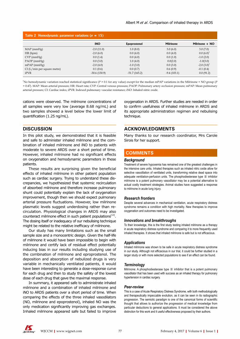
77 February 4, 2017|Volume 6|Issue 1|WJCCM|www.wjgnet.com
cations were observed. The milrinone concentrations of all samples were very low (average 8.68 ng/mL) and two samples showed a level below the lower limit of quantification (1.25 ng/mL).
DISCUSSIONIn this pilot study, we demonstrated that it is feasible and safe to administer inhaled milrinone and the combination of inhaled milrinone and iNO to patients with moderate to severe ARDS over a short period of time. However, inhaled milrinone had no significant effects on oxygenation and hemodynamic parameters in these patients.
These results are surprising given the beneficial effects of inhaled milrinone in other patient population such as cardiac surgery. Trying to understand these discrepancies, we hypothesized that systemic recirculation of absorbed milrinone and therefore increase pulmonary shunt could potentially explain the lack of oxygenation improvement, though then we should expect pulmonary arterial pressure fluctuations. However, low milrinone plasmatic levels suggest underdosing rather than recirculation. Physiological changes in ARDS may also counteract milrinone effect in such patient populations[12]. The dosing itself or inadequacy of our nebulising technique might be related to the relative inefficacy of milrinone.
Our study has many limitations such as the small sample size and a monocentric design. Given the halflife of milrinone it would have been impossible to begin with milrinone and certify lack of residual effect potentially inducing bias in our results including studying use of the combination of milrinone and eproprostenol. The deposition and absorption of nebulized drugs is very variable in mechanically ventilated patients, it would have been interesting to generate a doseresponse curve for each drug and then to study the safety of the lowest dose of each drug that gave the maximal response.
In summary, it appeared safe to administrate inhaled milrinone and a combination of inhaled milrinone and iNO to ARDS patients over a short period of time. When comparing the effects of the three inhaled vasodilators (NO, milrinone and epoprostenol), inhaled NO was the only medication significantly improving gas exchanges. Inhaled milrinone appeared safe but failed to improve
oxygenation in ARDS. Further studies are needed in order to confirm usefulness of inhaled milrinone in ARDS and its appropriate administration regimen and nebulising technique.
ACKNOWLEDGMENTSMany thanks to our research coordinator, Mrs Carole Sirois for her support.
COMMENTSBackgroundTreatment of severe hypoxemia has remained one of the greatest challenges in the intensive care units. Inhaled therapies such as inhaled nitric oxide allow for selective vasodilation of ventilated units, transforming relative dead space into adequate ventilation-perfusion units. The phosphodiesterase type Ⅲ inhibitor milrinone is a potent pulmonary vasodilator may be a potential alternative to actual costly treatment strategies. Animal studies have suggested a response to milrinone in acute lung injury.
Research frontiersDespite several advances in mechanical ventilation, acute respiratory distress syndrome remains a condition with high mortality. New therapies to improve oxygenation and outcomes need to be investigated.
Innovations and breakthroughsTo their knowledge, this is the first study testing inhaled milrinone as a therapy in acute respiratory distress syndrome and comparing it to more frequently used inhaled therapies. It shows that inhaled milrinone is safe but is not efficacious.
ApplicationsInhaled milrinone was shown to be safe in acute respiratory distress syndrome in our study. Although not efficacious in our trial, it could be further studied in a larger study or with more selected populations to see if an effect can be found.
TerminologyMilrinone: A phosphodiesterase type Ⅲ inhibitor that is a potent pulmonary vasodilator that has been used with success as an inhaled therapy for pulmonary hypertension in cardiac surgery.
Peer-reviewThis is a case of Acute Respiratory Distress Syndrome, with both methodologically and therapeutically impeccable evolution, as it can be seen in its radiographic progression. The semiotic paradigm is one of the canonical forms of scientific thought that allows to authorize the progression of medical knowledge from particular deductions to general applications. It must be considered the above distinction for this work and it useful effectiveness proposed by their authors.
iNO Epoprostenol Milrinone Milrinone + NO
MAP (mmHg) -2.0 (11.0) 1.0 (8.0) 3.0 (6.0) 3.0 (7.0)HR (bpm) -2.0 (6.0) 0.0 (4.0) 0.0 (4.0) 0.0 (6.0)1
CVP (mmHg) 0.0 (1.4) 0.0 (4.0) 0.0 (1.0) -1.0 (2.0)PAOP (mmHg) 0.0 (3.0) 1.0 (4.0) 0.0(2.0) -1.0(3.0)mPAP (mmHg) -2.0 (4.0) -1.0 (3.0) 0.0 (3.0) -2.0 (3.0)1
CI (L/min per square metre) 0.1 (0.6) 0.0 (0.7) 0.6 (0.9) -0.1 (0.4)iPVR -30.6 (130.9) -51.7 (165.2) -9.4 (103.1) 0.0 (91.2)
Table 2 Hemodynamic parameter variations (n = 15)
1No hemodynamic variation reached statistical significance (P > 0.1 for any value) except for the median mPAP variations in the Milrinone + NO group (P = 0.47). MAP: Mean arterial pressure; HR: Heart rate; CVP: Central venous pressure; PAOP: Pulmonary artery occlusion pressure; mPAP: Mean pulmonary arterial pressure; CI: Cardiac index; iPVR: Indexed pulmonary vascular resistance; iNO: Inhaled nitric oxide.
COMMENTS
Albert M et al . Comparison of inhaled therapy in ARDS

78 February 4, 2017|Volume 6|Issue 1|WJCCM|www.wjgnet.com
REFERENCES1 Rossaint R, Falke KJ, López F, Slama K, Pison U, Zapol WM.
Inhaled nitric oxide for the adult respiratory distress syndrome. N Engl J Med 1993; 328: 399-405 [PMID: 8357359 DOI: 10.1056/NEJM199302113280605]
2 Griffiths MJ, Evans TW. Inhaled nitric oxide therapy in adults. N Engl J Med 2005; 353: 2683-2695 [PMID: 16371634 DOI: 10.1056/NEJMra051884]
3 Afshari A, Brok J, Møller AM, Wetterslev J. Inhaled nitric oxide for acute respiratory distress syndrome and acute lung injury in adults and children: a systematic review with meta-analysis and trial sequential analysis. Anesth Analg 2011; 112: 1411-1421 [PMID: 21372277 DOI: 10.1213/ANE.0b013e31820bd185]
4 Pappert D, Busch T, Gerlach H, Lewandowski K, Radermacher P, Rossaint R. Aerosolized prostacyclin versus inhaled nitric oxide in children with severe acute respiratory distress syndrome. Anesthesiology 1995; 82: 1507-1511 [PMID: 7793662]
5 Walmrath D, Schneider T, Schermuly R, Olschewski H, Grimminger F, Seeger W. Direct comparison of inhaled nitric oxide and aerosolized prostacyclin in acute respiratory distress syndrome. Am J Respir Crit Care Med 1996; 153: 991-996 [PMID: 8630585 DOI: 10.1164/ajrccm.153.3.8630585]
6 Zwissler B, Kemming G, Habler O, Kleen M, Merkel M, Haller M, Briegel J, Welte M, Peter K. Inhaled prostacyclin (PGI2) versus inhaled nitric oxide in adult respiratory distress syndrome. Am J
Respir Crit Care Med 1996; 154: 1671-1677 [PMID: 8970353 DOI: 10.1164/ajrccm.154.6.8970353]
7 Lowson SM. Inhaled alternatives to nitric oxide. Anesthesiology 2002; 96: 1504-1513 [PMID: 12170067]
8 Haraldsson s A, Kieler-Jensen N, Ricksten SE. The additive pulmonary vasodilatory effects of inhaled prostacyclin and inhaled milrinone in postcardiac surgical patients with pulmonary hypertension. Anesth Analg 2001; 93: 1439-1445, table of contents [PMID: 11726420]
9 Sablotzki A, Starzmann W, Scheubel R, Grond S, Czeslick EG. Selective pulmonary vasodilation with inhaled aerosolized milrinone in heart transplant candidates. Can J Anaesth 2005; 52: 1076-1082 [PMID: 16326679 DOI: 10.1007/BF03021608]
10 Bueltmann M, Kong X, Mertens M, Yin N, Yin J, Liu Z, Koster A, Kuppe H, Kuebler WM. Inhaled milrinone attenuates experimental acute lung injury. Intensive Care Med 2009; 35: 171-178 [PMID: 18972099 DOI: 10.1007/s00134-008-1344-9]
11 Nguyen AQ, Théorêt Y, Chen C, Denault A, Varin F. High performance liquid chromatography using UV detection for the quantification of milrinone in plasma: improved sensitivity for inhalation. J Chromatogr B Analyt Technol Biomed Life Sci 2009; 877: 657-660 [PMID: 19201666 DOI: 10.1016/j.jchromb. 2009.01.024]
12 Cepkova M, Matthay MA. Pharmacotherapy of acute lung injury and the acute respiratory distress syndrome. J Intensive Care Med 2006; 21: 119-143 [PMID: 16672636 DOI: 10.1177/0885066606287045]
P- Reviewer: Denault AY, Kon ZN, Saikia UN S- Editor: Qiu S L- Editor: A E- Editor: Li D
Albert M et al . Comparison of inhaled therapy in ARDS
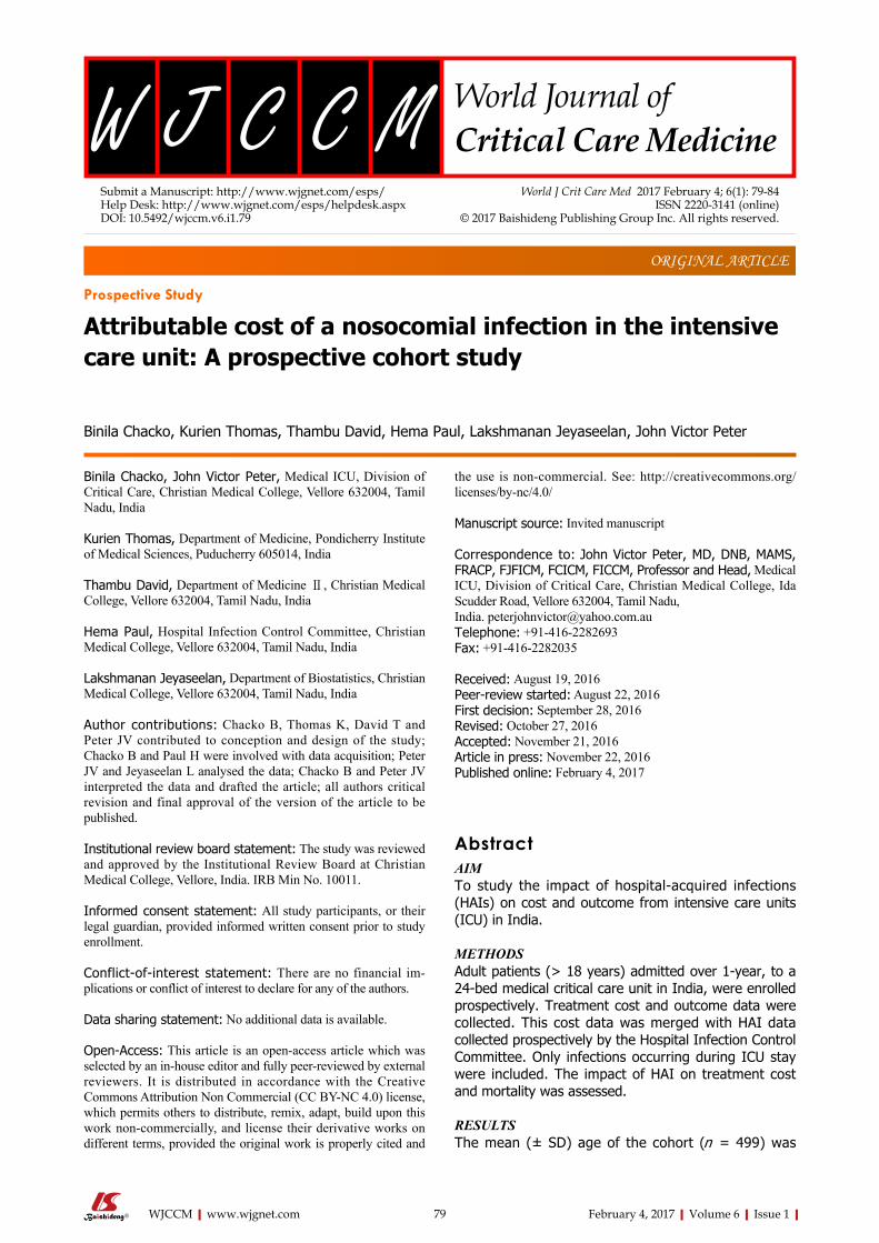
79 February 4, 2017|Volume 6|Issue 1|WJCCM|www.wjgnet.com
Binila Chacko, John Victor Peter, Medical ICU, Division of Critical Care, Christian Medical College, Vellore 632004, Tamil Nadu, India
Kurien Thomas, Department of Medicine, Pondicherry Institute of Medical Sciences, Puducherry 605014, India
Thambu David, Department of Medicine Ⅱ, Christian Medical College, Vellore 632004, Tamil Nadu, India
Hema Paul, Hospital Infection Control Committee, Christian Medical College, Vellore 632004, Tamil Nadu, India
Lakshmanan Jeyaseelan, Department of Biostatistics, Christian Medical College, Vellore 632004, Tamil Nadu, India
Author contributions: Chacko B, Thomas K, David T and Peter JV contributed to conception and design of the study; Chacko B and Paul H were involved with data acquisition; Peter JV and Jeyaseelan L analysed the data; Chacko B and Peter JV interpreted the data and drafted the article; all authors critical revision and final approval of the version of the article to be published.
Institutional review board statement: The study was reviewed and approved by the Institutional Review Board at Christian Medical College, Vellore, India. IRB Min No. 10011.
Informed consent statement: All study participants, or their legal guardian, provided informed written consent prior to study enrollment.
Conflict-of-interest statement: There are no financial implications or conflict of interest to declare for any of the authors.
Data sharing statement: No additional data is available.
Open-Access: This article is an openaccess article which was selected by an inhouse editor and fully peerreviewed by external reviewers. It is distributed in accordance with the Creative Commons Attribution Non Commercial (CC BYNC 4.0) license, which permits others to distribute, remix, adapt, build upon this work noncommercially, and license their derivative works on different terms, provided the original work is properly cited and
the use is noncommercial. See: http://creativecommons.org/licenses/bync/4.0/
Manuscript source: Invited manuscript
Correspondence to: John Victor Peter, MD, DNB, MAMS, FRACP, FJFICM, FCICM, FICCM, Professor and Head, Medical ICU, Division of Critical Care, Christian Medical College, Ida Scudder Road, Vellore 632004, Tamil Nadu, India. [email protected]: +914162282693Fax: +914162282035
Received: August 19, 2016 Peer-review started: August 22, 2016First decision: September 28, 2016Revised: October 27, 2016 Accepted: November 21, 2016Article in press: November 22, 2016Published online: February 4, 2017
AbstractAIMTo study the impact of hospital-acquired infections (HAIs) on cost and outcome from intensive care units (ICU) in India.
METHODSAdult patients (> 18 years) admitted over 1-year, to a 24-bed medical critical care unit in India, were enrolled prospectively. Treatment cost and outcome data were collected. This cost data was merged with HAI data collected prospectively by the Hospital Infection Control Committee. Only infections occurring during ICU stay were included. The impact of HAI on treatment cost and mortality was assessed.
RESULTSThe mean (± SD) age of the cohort (n = 499) was
World Journal ofCritical Care MedicineW J C C M
Submit a Manuscript: http://www.wjgnet.com/esps/Help Desk: http://www.wjgnet.com/esps/helpdesk.aspxDOI: 10.5492/wjccm.v6.i1.79
World J Crit Care Med 2017 February 4; 6(1): 79-84ISSN 2220-3141 (online)
© 2017 Baishideng Publishing Group Inc. All rights reserved.
Binila Chacko, Kurien Thomas, Thambu David, Hema Paul, Lakshmanan Jeyaseelan, John Victor Peter
ORIGINAL ARTICLE
Attributable cost of a nosocomial infection in the intensive care unit: A prospective cohort study
Prospective Study

80 February 4, 2017|Volume 6|Issue 1|WJCCM|www.wjgnet.com
Chacko B et al . Attributable cost of nosocomial infections in ICU
42.3 ± 16.5 years. Acute physiology and chronic health evaluation-Ⅱ score was 13.9 (95%CI: 13.3-14.5); 86% were ventilated. ICU and hospital length of stay were 7.8 ± 5.5 and 13.9 ± 10 d respectively. Hospital mortality was 27.9%. During ICU stay, 76 (15.3%) patients developed an infection (ventilator-associated pneumonia 50; bloodstream infection 35; urinary tract infections 3), translating to 19.7 infections/1000 ICU days. When compared with those who did not develop an infection, an infection occurring during ICU stay was associated with significantly higher treatment cost [median (inter-quartile range, IQR) INR 92893 (USD 1523) (IQR 57168-140286) vs INR 180469 (USD 2958) (IQR 140030-237525); P < 0.001 and longer duration of ICU (6.7 ± 4.5 d vs 13.4 ± 7.0 d; P < 0.01) and hospital stay (12.4 ± 8.2 d vs 21.8 ± 13.9 d; P < 0.001)]. However ICU acquired infections did not impact hospital mortality (31.6% vs 27.2%; P = 0.49).
CONCLUSIONAn infection acquired during ICU stay was associated with doubling of treatment cost and prolonged hospita-lization but did not significantly increase mortality.
Key words: Attributable cost; Nosocomial infection; Length of stay; Mortality; Intensive care
© The Author(s) 2017. Published by Baishideng Publishing Group Inc. All rights reserved.
Core tip: There is paucity of data on the impact of hospital acquired infections (HAIs) on cost and outcome from intensive care units (ICU) in developing countries. In this prospective study of 499 patients admitted over 1-year to a medical ICU in India, there were 19.7 HAIs per 1000 ICU days. Occurrence of infection was associated with significantly higher treatment cost (P < 0.001); the median attributable cost of an infection was 87594 Rupees (USD 1436). Although ICU acquired infections increased ICU length of stay (6.7 ± 4.5 d vs 13.4 ± 7.0 d; P < 0.01), it did not impact mortality (31.6% vs 27.2%; P = 0.49).
Chacko B, Thomas K, David T, Paul H, Jeyaseelan L, Peter JV. Attributable cost of a nosocomial infection in the intensive care unit: A prospective cohort study. World J Crit Care Med 2017; 6(1): 7984 Available from: URL: http://www.wjgnet.com/22203141/full/v6/i1/79.htm DOI: http://dx.doi.org/10.5492/wjccm.v6.i1.79
INTRODUCTIONHealth care associated infection (HAI) is a major preventable complication in critically ill patients across the world[1,2]. Whilst there is a significant body of information and evidence on the cost of these infections from developed countries, primary research from developing countries, in this area, is limited[3,4]. Translation of results of studies from developed countries on the impact and cost of infections to situations in developing countries
may not be appropriate for several reasons: (1) different microbiological profile of HAIs[5,6]; (2) perceived reluctance among physicians regarding treatment of HAIs that is probably based on the impression that these infections are associated with poor survival[7,8]; and (3) limited resources and affordability which argues that resource allocation for the treatment of HAI would steal opportunities away from other potentially treatable patients, waiting for an intensive care unit (ICU) bed. The affordability issue is compounded by the fact that only about 10% of the estimated 70000 ICU beds in India are available in the public sector, where treatment is provided free of cost[9]. This poses a major problem of demandsupply mismatch, not only in the public sector, but also in the private sector since the population that needs to be covered in India is over 1 billion. Minimal subscription to private health insurance and resource pooling being in its infancy results in significant out-of-pocket expenses that push several families below the poverty line[10].
In the light of the above, a study was undertaken to evaluate the “cost” (in terms of money) and “impact” (in terms of clinical outcomes) of HAIs in developing countries. Such studies would facilitate investment on interventions that reduce infection as well as help plan appropriate allocation of the scarce resources of materials (ICU beds and equipment), manpower and money to address this problem in the ICU setting.
MATERIALS AND METHODSIn this study spanning 1year, prospectively collected ICU cost data was merged with HAI data collected prospectively by the Hospital Infection Control Committee (HICC). ICU cost data was obtained from a study that looked at cost-utility as well as willingness-to-pay in patients admitted to the medical ICU[11].
Patients and settingThe study was undertaken in a 24-bed medical critical care unit in a 2500bed, universityaffiliated, private teaching hospital in semiurban India. In this hospital, other than the very few covered by private health insurance, the entire treatment cost is expected to be paid for by the patient.
During a 1year period (JanuaryDecember 2011), adult patients (> 18 years) admitted to the 24bed medical critical care unit were enrolled if they stayed beyond 24h in the ICU. Patients not consenting to participate, patients not admitted under internal medicine (e.g., hematological malignancies, chronic liver disease), or patients with surgical problems were excluded. A diagnosis of HAI was made only when a new infection occurred 48h after hospital admission. The study was approved by the Institutional Review Board and Ethics committee (IRB No. 10011) and consent was obtained from patient or next-of-kin.
Costs“Treatment cost”, obtained from the hospital electronic
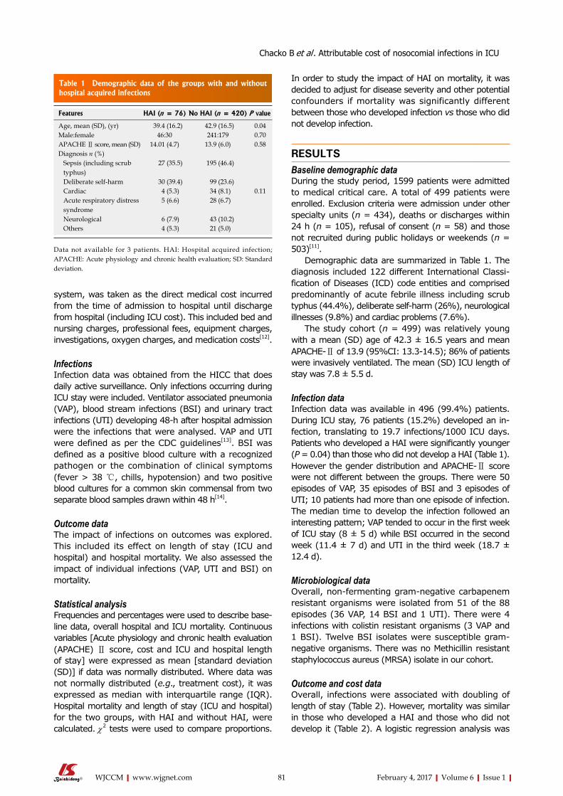
81 February 4, 2017|Volume 6|Issue 1|WJCCM|www.wjgnet.com
system, was taken as the direct medical cost incurred from the time of admission to hospital until discharge from hospital (including ICU cost). This included bed and nursing charges, professional fees, equipment charges, investigations, oxygen charges, and medication costs[12].
InfectionsInfection data was obtained from the HICC that does daily active surveillance. Only infections occurring during ICU stay were included. Ventilator associated pneumonia (VAP), blood stream infections (BSI) and urinary tract infections (UTI) developing 48h after hospital admission were the infections that were analysed. VAP and UTI were defined as per the CDC guidelines[13]. BSI was defined as a positive blood culture with a recognized pathogen or the combination of clinical symptoms (fever > 38 ℃, chills, hypotension) and two positive blood cultures for a common skin commensal from two separate blood samples drawn within 48 h[14].
Outcome dataThe impact of infections on outcomes was explored. This included its effect on length of stay (ICU and hospital) and hospital mortality. We also assessed the impact of individual infections (VAP, UTI and BSI) on mortality.
Statistical analysisFrequencies and percentages were used to describe baseline data, overall hospital and ICU mortality. Continuous variables [Acute physiology and chronic health evaluation (APACHE) Ⅱ score, cost and ICU and hospital length of stay] were expressed as mean [standard deviation (SD)] if data was normally distributed. Where data was not normally distributed (e.g., treatment cost), it was expressed as median with interquartile range (IQR). Hospital mortality and length of stay (ICU and hospital) for the two groups, with HAI and without HAI, were calculated. χ2 tests were used to compare proportions.
In order to study the impact of HAI on mortality, it was decided to adjust for disease severity and other potential confounders if mortality was significantly different between those who developed infection vs those who did not develop infection.
RESULTSBaseline demographic dataDuring the study period, 1599 patients were admitted to medical critical care. A total of 499 patients were enrolled. Exclusion criteria were admission under other specialty units (n = 434), deaths or discharges within 24 h (n = 105), refusal of consent (n = 58) and those not recruited during public holidays or weekends (n = 503)[11].
Demographic data are summarized in Table 1. The diagnosis included 122 different International Classification of Diseases (ICD) code entities and comprised predominantly of acute febrile illness including scrub typhus (44.4%), deliberate selfharm (26%), neurological illnesses (9.8%) and cardiac problems (7.6%).
The study cohort (n = 499) was relatively young with a mean (SD) age of 42.3 ± 16.5 years and mean APACHEⅡ of 13.9 (95%CI: 13.314.5); 86% of patients were invasively ventilated. The mean (SD) ICU length of stay was 7.8 ± 5.5 d.
Infection dataInfection data was available in 496 (99.4%) patients. During ICU stay, 76 patients (15.2%) developed an infection, translating to 19.7 infections/1000 ICU days. Patients who developed a HAI were significantly younger (P = 0.04) than those who did not develop a HAI (Table 1). However the gender distribution and APACHEⅡ score were not different between the groups. There were 50 episodes of VAP, 35 episodes of BSI and 3 episodes of UTI; 10 patients had more than one episode of infection. The median time to develop the infection followed an interesting pattern; VAP tended to occur in the first week of ICU stay (8 ± 5 d) while BSI occurred in the second week (11.4 ± 7 d) and UTI in the third week (18.7 ± 12.4 d).
Microbiological dataOverall, nonfermenting gramnegative carbapenem resistant organisms were isolated from 51 of the 88 episodes (36 VAP, 14 BSI and 1 UTI). There were 4 infections with colistin resistant organisms (3 VAP and 1 BSI). Twelve BSI isolates were susceptible gramnegative organisms. There was no Methicillin resistant staphylococcus aureus (MRSA) isolate in our cohort.
Outcome and cost dataOverall, infections were associated with doubling of length of stay (Table 2). However, mortality was similar in those who developed a HAI and those who did not develop it (Table 2). A logistic regression analysis was
Features HAI (n = 76) No HAI (n = 420) P value
Age, mean (SD), (yr) 39.4 (16.2) 42.9 (16.5) 0.04Male:female 46:30 241:179 0.70APACHE Ⅱ score, mean (SD) 14.01 (4.7) 13.9 (6.0) 0.58Diagnosis n (%) Sepsis (including scrub typhus)
27 (35.5) 195 (46.4)
Deliberate self-harm 30 (39.4) 99 (23.6) Cardiac 4 (5.3) 34 (8.1) 0.11 Acute respiratory distress syndrome
5 (6.6) 28 (6.7)
Neurological 6 (7.9) 43 (10.2) Others 4 (5.3) 21 (5.0)
Table 1 Demographic data of the groups with and without hospital acquired infections
Data not available for 3 patients. HAI: Hospital acquired infection; APACHE: Acute physiology and chronic health evaluation; SD: Standard deviation.
Chacko B et al . Attributable cost of nosocomial infections in ICU

82 February 4, 2017|Volume 6|Issue 1|WJCCM|www.wjgnet.com
not performed in view of the lack of effect of infection on mortality. Additionally, when individual infections were considered separately, there was no mortality difference between those who developed a specific infection [i.e., VAP, BSI or catheter associated urinary tract infection (CAUTI)] vs those who did not develop any infection during ICU stay (Table 2).
An infection acquired in the ICU was associated with doubling of overall cost when compared with patients who did not develop an infection during hospitalization. When VAP, BSI and UTI were analysed independently, the overall cost (median IQR) of each infection was almost similar (Table 3). The median attributable cost of an infection worked out to INR 87594 (USD 1436).
DISCUSSIONThis study provides insight and information on the burden (economic and otherwise) of common HAIs in the medical ICU of a developing country. While it could be argued that there is data from developed countries to this effect, our data with the different spectrum of infections (predominant VAP and few UTI) and microbiology (over 60% of the isolates carbapenem resistant) merit
reporting and discussion.Nosocomial infections, individually and overall in our
study, were associated with doubling of cost without any impact on mortality. The acquisition of infection was also associated with the need for an additional 710 d in the ICU, resulting in further constraining the already limited ICU resources in our setting. Although the increased length of ICU stay is consistent with the limited evidence available for VAP in other countries[15], this has significant hospital infrastructure and public health implications in our setting.
These findings beg a response to the following questions. First, given the lack of impact of infections on mortality despite the antimicrobial resistance patterns, it is worth treating these infections. Second, should there be a focused approach to looking at measures to decrease infections and improving quality of patient care in ICU? On the face of it, the appropriate response to the above questions would be a resounding yes. However as alluded to, in view of the limited resources, treatment of patients with ICU acquired infections is likely to impact bed allocation to a patient with a more reversible problem. This, coupled with the inability to pay for the entire cost of treatment[11], places an additional
Type HAI (n = 76) No HAI1 (n = 420) Cost difference P value
Mean (SD) cost (INR) Any infection 226398 (226268) 115058 (93754) 111340 < 0.0001 VAP 235350 (253421) 115058 (93754) 120292 < 0.001 BSI 283887 (341916) 115058 (93754) 168829 < 0.001 CAUTI 190059 (34096) 115058 (93754) 155963 0.05Median (IQR) cost (INR) Any infection 180469 (140030-237525) 92875 (57243-139104) 87594 < 0.0001 VAP 182991 (133038-238952) 92875 (57243-139104) 90116 < 0.0001 BSI 170753 (141788-238650) 92875 (57243-139104) 77878 < 0.0001 CAUTI 173085 (155818-190352) 92875 (57243-139104) 80210 0.06
Table 3 Comparison of overall cost between those with infection and those without infection
1The cost of no HAI is the same for all sub-categories of analysis based on source of infection since patients who developed any infection were not included in the “no HAI” group. At the time of the study, 1 USD = INR 61. Values in parenthesis indicate standard deviation. INR: Indian rupees; HAI: Hospital acquired infection; VAP: Ventilator associated pneumonia; BSI: Blood stream infection; CAUTI: Catheter associated urinary tract infection; SD: Standard deviation; IQR: Inter-quartile range.
Outcome HAI (n = 76) No HAI (n = 420) P value
ICU length of stay, mean (SD), (d) 13.4 (7.0) 6.7 (4.5) < 0.01Hospital stay, mean (SD), (d) 21.8 (13.9) 12.4 (8.2) < 0.001In-hospital mortality 31.60% 27.20% 0.49Mortality with VAP1 26% 27.2%2 1.0Mortality due to BSI1 37% 27.2%2 0.24CAUTI mortality1 33% 27.2%2 1.0
Table 2 Impact of hospital-acquired infections on outcomes
1Total number of patients with VAP was 50, BSI 35 and CAUTI 3; the total number of patients with individual infections exceed 76 since 10 patients had more than one infection source; 2Indicates patients who had no HAI during the entire course of intensive care stay; thus in the analysis for VAP, those with BSI or CAUTI were excluded from the no HAI group and for BSI those with VAP and CAUTI were excluded from the no HAI group. Data available only on 496 patients. VAP: Ventilator associated pneumonia; BSI: Blood stream infection; CAUTI: Catheter associated urinary tract infection; SD: Standard deviation; HAI: Hospital acquired infection.
Chacko B et al . Attributable cost of nosocomial infections in ICU
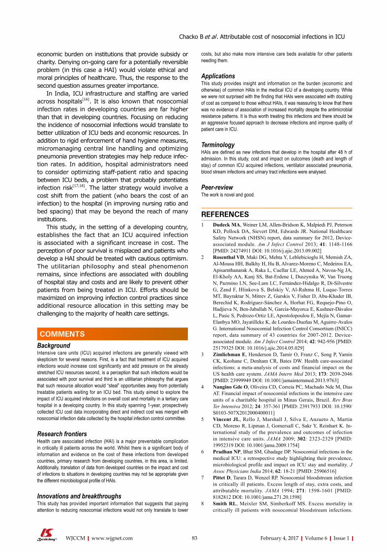
83 February 4, 2017|Volume 6|Issue 1|WJCCM|www.wjgnet.com
economic burden on institutions that provide subsidy or charity. Denying ongoing care for a potentially reversible problem (in this case a HAI) would violate ethical and moral principles of healthcare. Thus, the response to the second question assumes greater importance.
In India, ICU infrastructure and staffing are varied across hospitals[16]. It is also known that nosocomial infection rates in developing countries are far higher than that in developing countries. Focusing on reducing the incidence of nosocomial infections would translate to better utilization of ICU beds and economic resources. In addition to rigid enforcement of hand hygiene measures, micromanaging central line handling and optimizing pneumonia prevention strategies may help reduce infection rates. In addition, hospital administrators need to consider optimizing staffpatient ratio and spacing between ICU beds, a problem that probably potentiates infection risk[17,18]. The latter strategy would involve a cost shift from the patient (who bears the cost of an infection) to the hospital (in improving nursing ratio and bed spacing) that may be beyond the reach of many institutions.
This study, in the setting of a developing country, establishes the fact that an ICU acquired infection is associated with a significant increase in cost. The perception of poor survival is misplaced and patients who develop a HAI should be treated with cautious optimism. The utilitarian philosophy and steal phenomenon remains, since infections are associated with doubling of hospital stay and costs and are likely to prevent other patients from being treated in ICU. Efforts should be maximized on improving infection control practices since additional resource allocation in this setting may be challenging to the majority of health care settings.
COMMENTSBackgroundIntensive care units (ICU) acquired infections are generally viewed with skepticism for several reasons. First, is a fact that treatment of ICU acquired infections would increase cost significantly and add pressure on the already stretched ICU resources second, is a perception that such infections would be associated with poor survival and third is an utilitarian philosophy that argues that such resource allocation would “steal” opportunities away from potentially treatable patients waiting for an ICU bed. This study aimed to explore the impact of ICU acquired infections on overall cost and mortality in a tertiary care hospital in a developing country. In this study spanning 1-year, prospectively collected ICU cost data incorporating direct and indirect cost was merged with nosocomial infection data collected by the hospital infection control committee.
Research frontiersHealth care associated infection (HAI) is a major preventable complication in critically ill patients across the world. Whilst there is a significant body of information and evidence on the cost of these infections from developed countries, primary research from developing countries, in this area, is limited. Additionally, translation of data from developed countries on the impact and cost of infections to situations in developing countries may not be appropriate given the different microbiological profile of HAIs.
Innovations and breakthroughsThis study has provided important information that suggests that paying attention to reducing nosocomial infections would not only translate to lower
costs, but also make more intensive care beds available for other patients needing them.
ApplicationsThis study provides insight and information on the burden (economic and otherwise) of common HAIs in the medical ICU of a developing country. While we were not surprised with the finding that HAIs were associated with doubling of cost as compared to those without HAIs, it was reassuring to know that there was no evidence of association of increased mortality despite the antimicrobial resistance patterns. It is thus worth treating this infections and there should be an aggressive focused approach to decrease infections and improve quality of patient care in ICU.
TerminologyHAIs are defined as new infections that develop in the hospital after 48 h of admission. In this study, cost and impact on outcomes (death and length of stay) of common ICU acquired infections, ventilator associated pneumonia, blood stream infections and urinary tract infections were analysed.
Peer-reviewThe work is novel and good.
REFERENCES1 Dudeck MA, Weiner LM, AllenBridson K, Malpiedi PJ, Peterson
KD, Pollock DA, Sievert DM, Edwards JR. National Healthcare Safety Network (NHSN) report, data summary for 2012, Deviceassociated module. Am J Infect Control 2013; 41: 11481166 [PMID: 24274911 DOI: 10.1016/j.ajic.2013.09.002]
2 Rosenthal VD, Maki DG, Mehta Y, Leblebicioglu H, Memish ZA, AlMousa HH, Balkhy H, Hu B, AlvarezMoreno C, Medeiros EA, Apisarnthanarak A, Raka L, Cuellar LE, Ahmed A, NavoaNg JA, ElKholy AA, Kanj SS, BatErdene I, Duszynska W, Van Truong N, Pazmino LN, SeeLum LC, FernándezHidalgo R, DiSilvestre G, Zand F, Hlinkova S, Belskiy V, AlRahma H, LuqueTorres MT, Bayraktar N, Mitrev Z, Gurskis V, Fisher D, AbuKhader IB, Berechid K, RodríguezSánchez A, Horhat FG, RequejoPino O, Hadjieva N, BenJaballah N, GarcíaMayorca E, KushnerDávalos L, Pasic S, PedrozoOrtiz LE, Apostolopoulou E, Mejía N, GamarElanbya MO, Jayatilleke K, de LourdesDueñas M, AguirreAvalos G. International Nosocomial Infection Control Consortium (INICC) report, data summary of 43 countries for 20072012. Deviceassociated module. Am J Infect Control 2014; 42: 942956 [PMID: 25179325 DOI: 10.1016/j.ajic.2014.05.029]
3 Zimlichman E, Henderson D, Tamir O, Franz C, Song P, Yamin CK, Keohane C, Denham CR, Bates DW. Health careassociated infections: a metaanalysis of costs and financial impact on the US health care system. JAMA Intern Med 2013; 173: 20392046 [PMID: 23999949 DOI: 10.1001/jamainternmed.2013.9763]
4 Nangino Gde O, Oliveira CD, Correia PC, Machado Nde M, Dias AT. Financial impact of nosocomial infections in the intensive care units of a charitable hospital in Minas Gerais, Brazil. Rev Bras Ter Intensiva 2012; 24: 357361 [PMID: 23917933 DOI: 10.1590/S0103507X2012000400011]
5 Vincent JL, Rello J, Marshall J, Silva E, Anzueto A, Martin CD, Moreno R, Lipman J, Gomersall C, Sakr Y, Reinhart K. International study of the prevalence and outcomes of infection in intensive care units. JAMA 2009; 302: 23232329 [PMID: 19952319 DOI: 10.1001/jama.2009.1754]
6 Pradhan NP, Bhat SM, Ghadage DP. Nosocomial infections in the medical ICU: a retrospective study highlighting their prevalence, microbiological profile and impact on ICU stay and mortality. J Assoc Physicians India 2014; 62: 1821 [PMID: 25906516]
7 Pittet D, Tarara D, Wenzel RP. Nosocomial bloodstream infection in critically ill patients. Excess length of stay, extra costs, and attributable mortality. JAMA 1994; 271: 15981601 [PMID: 8182812 DOI: 10.1001/jama.271.20.1598]
8 Smith RL, Meixler SM, Simberkoff MS. Excess mortality in critically ill patients with nosocomial bloodstream infections.
COMMENTS
Chacko B et al . Attributable cost of nosocomial infections in ICU
84 February 4, 2017|Volume 6|Issue 1|WJCCM|www.wjgnet.com
Chest 1991; 100: 164167 [PMID: 2060337 DOI: 10.1378/chest. 100.1.164]
9 Divatia JV, Iyer S. Ten major priorities for intensive care in India. Intensive Care Med 2015; 41: 14681471 [PMID: 25573499 DOI: 10.1007/s0013401436188]
10 Jayaram R, Ramakrishnan N. Cost of intensive care in India. Indian J Crit Care Med 2008; 12: 5561 [PMID: 19742248 DOI: 10.4103/09725229.42558]
11 Thomas K, Peter JV, Christina J, Jagadish AR, Rajan A, Lionel P, Jeyaseelan L, Yadav B, John G, Pichamuthu K, Chacko B, Pari P, Murugesan T, Rajendran K, John A, Sathyendra S, Iyyadurai R, Jasmine S, Karthik R, Mathuram A, Hansdak SG, Abhilash KP, Kumar S, John KR, Sudarsanam TD. Costutility in medical intensive care patients. Rationalizing ongoing care and timing of discharge from intensive care. Ann Am Thorac Soc 2015; 12: 10581065 [PMID: 26011090 DOI: 10.1513/AnnalsATS.201411527OC]
12 Peter JV, Thomas K, Jeyaseelan L, Yadav B, Sudarsan TI, Christina J, Revathi A, John KR, Sudarsanam TD. COST OF INTENSIVE CARE IN INDIA. Int J Technol Assess Health Care 2016: 15 [PMID: 27608529 DOI: 10.1017/S0266462316000398]
13 Horan TC, Andrus M, Dudeck MA. CDC/NHSN surveillance definition of health careassociated infection and criteria for
specific types of infections in the acute care setting. Am J Infect Control 2008; 36: 309332 [PMID: 18538699 DOI: 10.1016/j.ajic.2008.03.002]
14 Suetens C, Morales I, Savey A, Palomar M, Hiesmayr M, Lepape A, Gastmeier P, Schmit JC, Valinteliene R, Fabry J. European surveillance of ICUacquired infections (HELICSICU): methods and main results. J Hosp Infect 2007; 65 Suppl 2: 171173 [PMID: 17540265 DOI: 10.1016/S01956701(07)600383]
15 Safdar N, Dezfulian C, Collard HR, Saint S. Clinical and economic consequences of ventilatorassociated pneumonia: a systematic review. Crit Care Med 2005; 33: 21842193 [PMID: 16215368 DOI: 10.1097/01.CCM.0000181731.53912.D9]
16 Yeolekar ME, Mehta S. ICU care in Indiastatus and challenges. J Assoc Physicians India 2008; 56: 221222 [PMID: 18702381]
17 Schwab F, Meyer E, Geffers C, Gastmeier P. Understaffing, overcrowding, inappropriate nurse: ventilated patient ratio and nosocomial infections: which parameter is the best reflection of deficits? J Hosp Infect 2012; 80: 133139 [PMID: 22188631 DOI: 10.1016/j.jhin.2011.11.014]
18 Stone PW, Pogorzelska M, Kunches L, Hirschhorn LR. Hospital staffing and health careassociated infections: a systematic review of the literature. Clin Infect Dis 2008; 47: 937944 [PMID: 18767987 DOI: 10.1086/591696]
P- Reviewer: Durandy YD, Mitra A S- Editor: Ji FF L- Editor: A E- Editor: Li D
Chacko B et al . Attributable cost of nosocomial infections in ICU
Alpha A Fowler Ⅲ, Christin Kim, Lawrence Lepler, Rajiv Malhotra, Orlando Debesa, Ramesh Natarajan, Bernard J Fisher, Aamer Syed, Christine DeWilde, Anna Priday, Vigneshwar Kasirajan
CASE REPORT
85 February 4, 2017|Volume 6|Issue 1|WJCCM|www.wjgnet.com
Intravenous vitamin C as adjunctive therapy for enterovirus/rhinovirus induced acute respiratory distress syndrome
Alpha A Fowler Ⅲ, Rajiv Malhotra, Orlando Debesa, Ramesh Natarajan, Bernard J Fisher, Aamer Syed, Christine DeWilde, Anna Priday, Division of Pulmonary Disease and Critical Care Medicine, Virginia Commonwealth University School of Medicine, Richmond, VA 23298, United States
Christin Kim, Department of Anesthesia Critical Care, Virginia Commonwealth University School of Medicine, Richmond, VA 23298, United States
Lawrence Lepler, Division of Critical Care Medicine, INOVA Fair Oaks Hospital, INOVA Health Care System, Fairfax, VA 22042, United States
Vigneshwar Kasirajan, Division of Cardiothoracic Surgery, Virginia Commonwealth University School of Medicine, Richmond, VA 23298, United States
Author contributions: Fowler Ⅲ AA is principal investigator, corresponding author, and contributed to study concept, basic and translational research; Kim C and Lepler L contributed to patient’s clinical care, patient followup and manuscript review; Malhotra R contributed to patient’s clinical care; Debesa O contributed to patient’s clinical care and manuscript review; Natarajan R and Fisher BJ contributed to manuscript creation, basic research leading to clinical use; Syed A and Kasirajan V contributed to clinical care and manuscript creation; DeWilde C contributed to clinical care and laboratory coordination; Priday A contributed to FDA regulatory coordinator for IND used for study.
Institutional review board statement: The use of intravenous vitamin C in humans has been approved by the Virginia Commonwealth University Institutional Review Board (HM20000917).
Informed consent statement: Permission for the patient to receive intravenous vitamin C as described in this case report was granted by the patient’s legal next of kin. All patient health information was deidentified and held in strictest confidentiality.
Conflict-of-interest statement: All authors have no conflicts of interests to declare.
Open-Access: This article is an openaccess article which was selected by an inhouse editor and fully peerreviewed by external reviewers. It is distributed in accordance with the Creative Commons Attribution Non Commercial (CC BYNC 4.0) license, which permits others to distribute, remix, adapt, build upon this work noncommercially, and license their derivative works on different terms, provided the original work is properly cited and the use is noncommercial. See: http://creativecommons.org/licenses/bync/4.0/
Manuscript source: Invited manuscript
Correspondence to: Alpha A Fowler Ⅲ, MD, William Talia-ferro Thompson Professor of Medicine, Division of Pulmonary Disease and Critical Care Medicine, Virginia Commonwealth University School of Medicine, 1200 East Broad Street, PO Box 980050, Richmond, VA 23298, United States. [email protected]: +18048289071 Fax: +18048282578
Received: July 12, 2016Peer-review started: July 13, 2016First decision: September 2, 2016Revised: October 26, 2016Accepted: November 16, 2016Article in press: November 17, 2016Published online: February 4, 2017
AbstractWe report a case of virus-induced acute respiratory distress syndrome (ARDS) treated with parenteral vitamin C in a patient testing positive for enterovirus/rhinovirus on viral screening. This report outlines the first use of high dose intravenous vitamin C as an interventional therapy for ARDS, resulting from enterovirus/rhinovirus respiratory infection. From very significant preclinical research performed at Virginia Commonwealth University
World Journal ofCritical Care MedicineW J C C M
Submit a Manuscript: http://www.wjgnet.com/esps/Help Desk: http://www.wjgnet.com/esps/helpdesk.aspxDOI: 10.5492/wjccm.v6.i1.85
World J Crit Care Med 2017 February 4; 6(1): 85-90ISSN 2220-3141 (online)
© 2017 Baishideng Publishing Group Inc. All rights reserved.

86 February 4, 2017|Volume 6|Issue 1|WJCCM|www.wjgnet.com
Fowler Ⅲ AA et al . Intravenous vitamin C to treat ARDS
with vitamin C and with the very positive results of a previously performed phase Ⅰ safety trial infusing high dose vitamin C intravenously into patients with severe sepsis, we reasoned that infusing identical dosing to a patient with ARDS from viral infection would be therapeutic. We report here the case of a 20-year-old, previously healthy, female who contracted respiratory enterovirus/rhinovirus infection that led to acute lung injury and rapidly to ARDS. She contracted the infection in central Italy while on an 8-d spring break from college. During a return flight to the United States, she developed increasing dyspnea and hypoxemia that rapidly developed into acute lung injury that led to ARDS. When support with mechanical ventilation failed, extracorporeal membrane oxygenation (ECMO) was initiated. Twelve hours following ECMO initiation, high dose intravenous vitamin C was begun. The patient’s recovery was rapid. ECMO and mechanical ventilation were discontinued by day-7 and the patient recovered with no long-term ARDS sequelae. Infusing high dose intravenous vitamin C into this patient with virus-induced ARDS was associated with rapid resolution of lung injury with no evidence of post-ARDS fibroproliferative sequelae. Intravenous vitamin C as a treatment for ARDS may open a new era of therapy for ARDS from many causes.
Key words: Intravenous vitamin C; Acute respiratory distress syndrome; Enterovirus/rhinovirus; Acute lung injury; Extracorporeal membrane oxygenation
© The Author(s) 2017. Published by Baishideng Publishing Group Inc. All rights reserved.
Core tip: Enterovirus/rhinovirus has been reported to cause devastating acute lung injury. We report here the first use of high dose intravenous vitamin C to attenuate the acute respiratory distress syndrome that was caused by this viral infection. We have previously reported that vitamin C used in this interventional fashion is a potent anti-inflammatory agent.
Fowler Ⅲ AA, Kim C, Lepler L, Malhotra R, Debesa O, Natarajan R, Fisher BJ, Syed A, DeWilde C, Priday A, Kasirajan V. Intravenous vitamin C as adjunctive therapy for enterovirus/rhinovirus induced acute respiratory distress syndrome. World J Crit Care Med 2017; 6(1): 8590 Available from: URL: http://www.wjgnet.com/22203141/full/v6/i1/85.htm DOI: http://dx.doi.org/10.5492/wjccm.v6.i1.85
INTRODUCTIONViral diseases can produce the acute respiratory distress syndrome (ARDS)[1]. Pandemic viruses are the most common viruses that produce lung injury. Influenza viruses and coronaviruses (e.g., H5N1, H1N1 2009, severe acute respiratory syndrome coronavirus, and middle east respiratory syndrome coronavirus) are
potentially lethal pathogens known to produce lung injury and death from ARDS[2-5]. At the tissue level, lung injury results from increased permeability of the alveolar-capillary membrane that leads to hypoxia, pulmonary edema, and intense cellular infiltration, particularly neutrophilic infiltration. The exact pathogenesis of virus-induced ARDS is slowly becoming understood. Unlike the “cytokine storm” occurring in bacterial sepsis that leads to up-regulation of pro-inflammatory cytokines [e.g., interleukin-1β (IL-1β), IL-8, IL-6] and generation of reactive nitrogen and oxygen species in the vascular space, viruses such as the influenza virus target alveolar epithelium, disabling sodium pump activity, damaging tight junctions, and inducing cell death in infected cells. Cytokines produced by virally infected alveolar epithelial cells activate adjacent lung capillary endothelial cells which then leads to neutrophil infiltration. Subsequent production of reactive oxygen and nitrogen species by infiltrating neutrophils further damages lung barrier function[1]. Apart from pandemic viruses other viruses, are increasingly reported to produce severe ARDS. While most of the approximately 100 strains of enterovirus primarily infect the gastrointestinal tract, enterovirus-D68 (EV-D68) has tropism for the respiratory tract. EV-D68 produces acute respiratory disease ranging from mild upper respiratory tract symptoms to severe pneumonia and lung injury as in the case we describe here. In an outpatient setting, EV-D68 disease has manifested most commonly among persons younger than 20 years and adults aged 50-59 years[6]. In August 2014, EV-D68 emerged as a cause of severe respiratory infections with hospitals in Illinois and Missouri reporting an increased incidence of rhinovirus and enterovirus infection[7]. In this report, 30 of 36 isolates from the nasopharyngeal secretions of patients with severe respiratory illness were identified as EV-D68. Following these reports, an unusually high number of patients with severe respiratory illness were admitted to these facilities, presumably with EV-D68 infection. Enterovirus-D68 leading to ARDS has been reported in China, Japan, and in the United States[8-11]. The report by Farrell et al[11], describes a previously healthy 26-year-old woman who developed severe ARDS following an enterovirus-D68 infection. Despite all critical care support measures, the patient required protracted mechanical ventilation for 32-d, necessitating tracheostomy and endoscopic gastrostomy tube placement. She was discharged alive 55 d following admission. Enterovirus and rhinovirus were recovered from the respiratory secretions of the patient we report here. Extracorporeal membrane oxygenation was rapidly required in our patient’s care following failure of conventional mechanical ventilation. The patient reported by Farrell et al[11] is the full extent of support required for patients with ARDS who ultimately develop a fibroproliferative course as described by Burnham et al[12]. Karhu et al[13] and Choi et al[14] recently reported finding rhinovirus as the etiology of severe community acquired pneumonia and respiratory failure
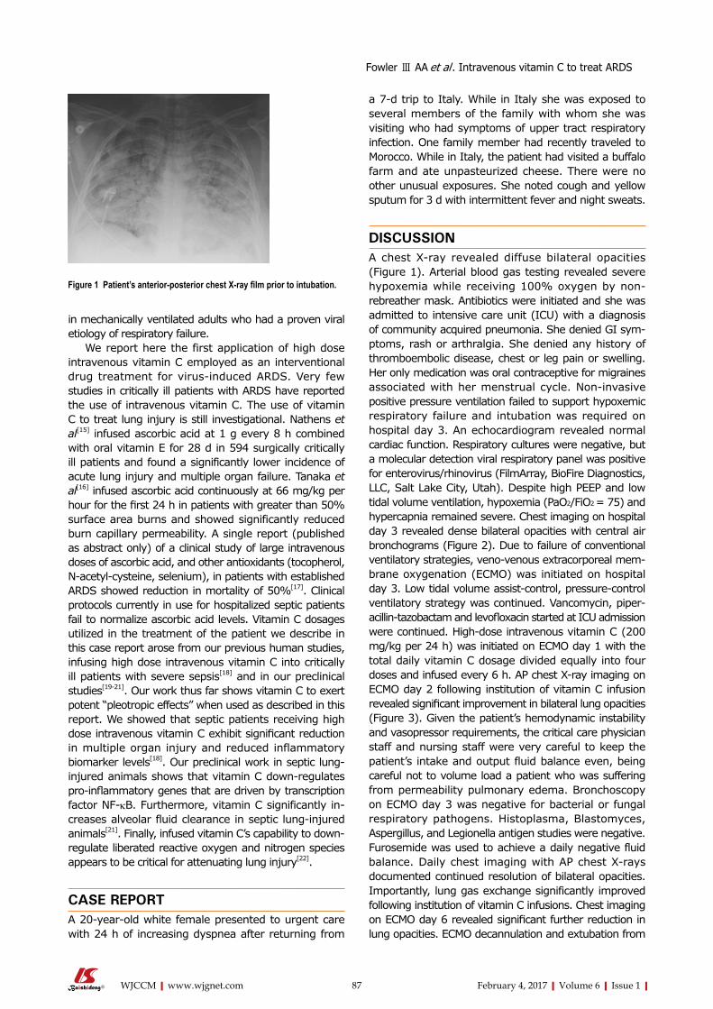
87 February 4, 2017|Volume 6|Issue 1|WJCCM|www.wjgnet.com
in mechanically ventilated adults who had a proven viral etiology of respiratory failure.
We report here the first application of high dose intravenous vitamin C employed as an interventional drug treatment for virus-induced ARDS. Very few studies in critically ill patients with ARDS have reported the use of intravenous vitamin C. The use of vitamin C to treat lung injury is still investigational. Nathens et al[15] infused ascorbic acid at 1 g every 8 h combined with oral vitamin E for 28 d in 594 surgically critically ill patients and found a significantly lower incidence of acute lung injury and multiple organ failure. Tanaka et al[16] infused ascorbic acid continuously at 66 mg/kg per hour for the first 24 h in patients with greater than 50% surface area burns and showed significantly reduced burn capillary permeability. A single report (published as abstract only) of a clinical study of large intravenous doses of ascorbic acid, and other antioxidants (tocopherol, N-acetyl-cysteine, selenium), in patients with established ARDS showed reduction in mortality of 50%[17]. Clinical protocols currently in use for hospitalized septic patients fail to normalize ascorbic acid levels. Vitamin C dosages utilized in the treatment of the patient we describe in this case report arose from our previous human studies, infusing high dose intravenous vitamin C into critically ill patients with severe sepsis[18] and in our preclinical studies[19-21]. Our work thus far shows vitamin C to exert potent “pleotropic effects” when used as described in this report. We showed that septic patients receiving high dose intravenous vitamin C exhibit significant reduction in multiple organ injury and reduced inflammatory biomarker levels[18]. Our preclinical work in septic lung-injured animals shows that vitamin C down-regulates pro-inflammatory genes that are driven by transcription factor NF-κB. Furthermore, vitamin C significantly in-creases alveolar fluid clearance in septic lung-injured animals[21]. Finally, infused vitamin C’s capability to down-regulate liberated reactive oxygen and nitrogen species appears to be critical for attenuating lung injury[22].
CASE REPORTA 20-year-old white female presented to urgent care with 24 h of increasing dyspnea after returning from
a 7-d trip to Italy. While in Italy she was exposed to several members of the family with whom she was visiting who had symptoms of upper tract respiratory infection. One family member had recently traveled to Morocco. While in Italy, the patient had visited a buffalo farm and ate unpasteurized cheese. There were no other unusual exposures. She noted cough and yellow sputum for 3 d with intermittent fever and night sweats.
DISCUSSIONA chest X-ray revealed diffuse bilateral opacities (Figure 1). Arterial blood gas testing revealed severe hypoxemia while receiving 100% oxygen by non-rebreather mask. Antibiotics were initiated and she was admitted to intensive care unit (ICU) with a diagnosis of community acquired pneumonia. She denied GI sym-ptoms, rash or arthralgia. She denied any history of thromboembolic disease, chest or leg pain or swelling. Her only medication was oral contraceptive for migraines associated with her menstrual cycle. Non-invasive positive pressure ventilation failed to support hypoxemic respiratory failure and intubation was required on hospital day 3. An echocardiogram revealed normal cardiac function. Respiratory cultures were negative, but a molecular detection viral respiratory panel was positive for enterovirus/rhinovirus (FilmArray, BioFire Diagnostics, LLC, Salt Lake City, Utah). Despite high PEEP and low tidal volume ventilation, hypoxemia (PaO2/FiO2 = 75) and hypercapnia remained severe. Chest imaging on hospital day 3 revealed dense bilateral opacities with central air bronchograms (Figure 2). Due to failure of conventional ventilatory strategies, veno-venous extracorporeal mem-brane oxygenation (ECMO) was initiated on hospital day 3. Low tidal volume assist-control, pressure-control ventilatory strategy was continued. Vancomycin, piper-acillin-tazobactam and levofloxacin started at ICU admission were continued. High-dose intravenous vitamin C (200 mg/kg per 24 h) was initiated on ECMO day 1 with the total daily vitamin C dosage divided equally into four doses and infused every 6 h. AP chest X-ray imaging on ECMO day 2 following institution of vitamin C infusion revealed significant improvement in bilateral lung opacities (Figure 3). Given the patient’s hemodynamic instability and vasopressor requirements, the critical care physician staff and nursing staff were very careful to keep the patient’s intake and output fluid balance even, being careful not to volume load a patient who was suffering from permeability pulmonary edema. Bronchoscopy on ECMO day 3 was negative for bacterial or fungal respiratory pathogens. Histoplasma, Blastomyces, Aspergillus, and Legionella antigen studies were negative. Furosemide was used to achieve a daily negative fluid balance. Daily chest imaging with AP chest X-rays documented continued resolution of bilateral opacities. Importantly, lung gas exchange significantly improved following institution of vitamin C infusions. Chest imaging on ECMO day 6 revealed significant further reduction in lung opacities. ECMO decannulation and extubation from
Figure 1 Patient’s anterior-posterior chest X-ray film prior to intubation.
Fowler Ⅲ AA et al . Intravenous vitamin C to treat ARDS

88 February 4, 2017|Volume 6|Issue 1|WJCCM|www.wjgnet.com
ventilation occurred on ECMO day 7 (Figure 4). Vitamin C dosing was continued while the patient remained on ECMO. Vitamin C dosing was reduced by half (100 mg/kg per 24 h) for one day following decannulation from ECMO then reduced by half again (50 mg/kg per 24 h) for an additional day. Post-extubation the patient required 4 L/min nasal oxygen for 48 h and then was discharged home on room air. She was discharged home on hospital day 12. Although we did not quantify the plasma ascorbic acid levels in the patient we report here, we have previously reported that critically ill patients with severe sepsis treated with the identical vitamin C infusion protocol achieved plasma ascorbic acid levels of 3.2 mmol, values which are 60 fold higher than normal plasma ascorbic acid levels[18].
In conclusion, we report here the first use of vitamin C as an interventional drug to attenuate lung injury produced by viral infection. The patient described here was discharged home 12 d following hospitalization, requiring no oxygen therapy. Follow-up exam at 1 mo following the patient’s initial hospitalization revealed her to have completely recovered. Figure 5 displays her follow-up chest X-ray film. Importantly, it should be noted that this is a single case report. The role of Vitamin C in this patient’s recovery is not certain, and clearly additional investigation will be required before
this can be recommended as a therapy for ARDS.
ACKNOWLEDGMENTSThe authors are grateful to Virginia Commonwealth University Medical Center in Richmond, Virginia, the Divisions of Medical, Surgical, and Anesthesia Critical Care Medicine, Richmond, Virginia, United States. The pre-clinical work that led up to the use of vitamin C as an interventional agent in humans was supported by the Aubrey Sage McFarlane acute lung injury fund, the VCU Johnson Center for Critical Care and Pulmonary Research.
COMMENTSCase characteristicsA 20-year-old female with no significant medical history presented with acute respiratory failure following a spring break in central Italy. While in Italy she was exposed to a sick contact who was a member of the family she was staying with.
Clinical diagnosisThe clinical diagnosis of severe acute respiratory distress syndrome (ARDS) in this case was established by the extent of respiratory failure present, the radiographic findings, and the need for extracorporeal membrane oxygenator support required. The patient’s exposure to the sick contact in Italy suggested the diagnosis of a viral etiology.
Portable
Figure 2 Patient’s anterior-posterior chest X-ray film on extracorporeal membrane oxygenation day 1.
Figure 3 Patient’s anterior-posterior chest X-ray film on extracorporeal membrane oxygenation day 2.
Portable®
MCJ
Figure 4 Patient’s anterior-posterior chest X-ray film on extracorporeal membrane oxygenation decannulation, extubation day 7.
Portable Semi-upright
Figure 5 Patient’s posterior-anterior chest X-ray film two weeks following hospital discharge.
COMMENTS
Fowler Ⅲ AA et al . Intravenous vitamin C to treat ARDS

89 February 4, 2017|Volume 6|Issue 1|WJCCM|www.wjgnet.com
Differential diagnosisARDS, viral pneumonia, sepsis from unknown etiology.
Laboratory diagnosisThe diagnosis of the etiology of the patient’s respiratory failure was obtained by a panel that uses real-time polymerase chain reaction technology to identify respiratory viral pathogens. FilmArray Respiratory panel is manufactured by BioFire Diagnostics, LLC, Salt Lake City, Utah.
Imaging diagnosisStandard Anterior-Posterior chest X-ray films confirmed the diagnosis of ARDS.
Pathological diagnosisNo lung tissue was obtained from the patient. The diagnosis of ARDS was established by the extent of respiratory failure and the imaging required during the patients hospital stay.
TreatmentIn this case report, the authors describe the first use of high dosage intravenous vitamin C as adjunctive therapy for viral induced ARDS.
Related reportsAt this point in time, there are no other case reports specifically referencing vitamin C as a treatment for ARDS. The authors have previously reported (ref. [18]) the use of high dose vitamin C as an adjunctive therapy for severe sepsis. Many patients in that trial likely could be considered to have had ARDS.
Experiences and lessonsFor many years multiple investigators have conducted clinical treatment trials, searching for effective therapies to assist in the treatment for ARDS. In this case report, the authors may have shed new light on a treatment which may ultimately be effective. The successful outcome described in this case report would suggest that larger trials must be conducted with high dosage intravenous vitamin C.
Peer-reviewThis is an interesting report of use of high dose intravenous vitamin C in ARDS.
REFERENCES1 Short KR, Kroeze EJ, Fouchier RA, Kuiken T. Pathogenesis of
influenzainduced acute respiratory distress syndrome. Lancet Infect Dis 2014; 14: 5769 [PMID: 24239327 DOI: 10.1016/S14733099(13)70286X]
2 Zaki AM, van Boheemen S, Bestebroer TM, Osterhaus AD, Fouchier RA. Isolation of a novel coronavirus from a man with pneumonia in Saudi Arabia. N Engl J Med 2012; 367: 18141820 [PMID: 23075143 DOI: 10.1056/NEJMoa1211721]
3 Guery B, Poissy J, el Mansouf L, Séjourné C, Ettahar N, Lemaire X, Vuotto F, Goffard A, Behillil S, Enouf V, Caro V, Mailles A, Che D, Manuguerra JC, Mathieu D, Fontanet A, van der Werf S. Clinical features and viral diagnosis of two cases of infection with Middle East Respiratory Syndrome coronavirus: a report of nosocomial transmission. Lancet 2013; 381: 22652272 [PMID: 23727167 DOI: 10.1016/S01406736(13)609824]
4 Drosten C, Seilmaier M, Corman VM, Hartmann W, Scheible G, Sack S, Guggemos W, Kallies R, Muth D, Junglen S, Müller MA, Haas W, Guberina H, Röhnisch T, SchmidWendtner M, Aldabbagh S, Dittmer U, Gold H, Graf P, Bonin F, Rambaut A, Wendtner CM. Clinical features and virological analysis of a case of Middle East respiratory syndrome coronavirus infection. Lancet Infect Dis 2013; 13: 745751 [PMID: 23782859 DOI: 10.1016/S1473 3099(13)701543]
5 de Jong MD, Simmons CP, Thanh TT, Hien VM, Smith GJ, Chau TN, Hoang DM, Chau NV, Khanh TH, Dong VC, Qui PT, Cam BV, Ha do Q, Guan Y, Peiris JS, Chinh NT, Hien TT, Farrar J. Fatal outcome of human influenza A (H5N1) is associated with high
viral load and hypercytokinemia. Nat Med 2006; 12: 12031207 [PMID: 16964257 DOI: 10.1038/nm1477]
6 Meijer A, van der Sanden S, Snijders BE, JaramilloGutierrez G, Bont L, van der Ent CK, Overduin P, Jenny SL, Jusic E, van der Avoort HG, Smith GJ, Donker GA, Koopmans MP. Emergence and epidemic occurrence of enterovirus 68 respiratory infections in The Netherlands in 2010. Virology 2012; 423: 4957 [PMID: 22177700 DOI: 10.1016/j.virol.2011.11.021]
7 Midgley CM, Jackson MA, Selvarangan R, Turabelidze G, Obringer E, Johnson D, Giles BL, Patel A, Echols F, Oberste MS, Nix WA, Watson JT, Gerber SI. Severe respiratory illness associated with enterovirus D68 Missouri and Illinois, 2014. MMWR Morb Mortal Wkly Rep 2014; 63: 798799 [PMID: 25211545]
8 Xiang Z, Gonzalez R, Wang Z, Ren L, Xiao Y, Li J, Li Y, Vernet G, ParanhosBaccalà G, Jin Q, Wang J. Coxsackievirus A21, enterovirus 68, and acute respiratory tract infection, China. Emerg Infect Dis 2012; 18: 821824 [PMID: 22516379 DOI: 10.3201/eid1805.111376]
9 Zhang T, Ren L, Luo M, Li A, Gong C, Chen M, Yu X, Wu J, Deng Y, Huang F. Enterovirus D68associated severe pneumonia, China, 2014. Emerg Infect Dis 2015; 21: 916918 [PMID: 25897574 DOI: 10.3201/eid2105.150036]
10 Kaida A, Kubo H, Sekiguchi J, Kohdera U, Togawa M, Shiomi M, Nishigaki T, Iritani N. Enterovirus 68 in children with acute respiratory tract infections, Osaka, Japan. Emerg Infect Dis 2011; 17: 14941497 [PMID: 21801632 DOI: 10.3201/eid1708.110028]
11 Farrell JJ, Ikladios O, Wylie KM, O’Rourke LM, Lowery KS, Cromwell JS, Wylie TN, Melendez EL, Makhoul Y, Sampath R, Bonomo RA, Storch GA. Enterovirus D68associated acute respiratory distress syndrome in adult, United States, 2014. Emerg Infect Dis 2015; 21: 914916 [PMID: 25897542 DOI: 10.3201/eid2105.142033]
12 Burnham EL, Janssen WJ, Riches DW, Moss M, Downey GP. The fibroproliferative response in acute respiratory distress syndrome: mechanisms and clinical significance. Eur Respir J 2014; 43: 276285 [PMID: 23520315 DOI: 10.1183/09031936.00196412]
13 Karhu J, AlaKokko TI, Vuorinen T, Ohtonen P, Syrjälä H. Lower respiratory tract virus findings in mechanically ventilated patients with severe communityacquired pneumonia. Clin Infect Dis 2014; 59: 6270 [PMID: 24729498 DOI: 10.1093/cid/ciu237]
14 Choi SH, Hong SB, Ko GB, Lee Y, Park HJ, Park SY, Moon SM, Cho OH, Park KH, Chong YP, Kim SH, Huh JW, Sung H, Do KH, Lee SO, Kim MN, Jeong JY, Lim CM, Kim YS, Woo JH, Koh Y. Viral infection in patients with severe pneumonia requiring intensive care unit admission. Am J Respir Crit Care Med 2012; 186: 325332 [PMID: 22700859 DOI: 10.1164/rccm.2011122240OC]
15 Nathens AB, Neff MJ, Jurkovich GJ, Klotz P, Farver K, Ruzinski JT, Radella F, Garcia I, Maier RV. Randomized, prospective trial of antioxidant supplementation in critically ill surgical patients. Ann Surg 2002; 236: 814822 [PMID: 12454520]
16 Tanaka H, Matsuda T, Miyagantani Y, Yukioka T, Matsuda H, Shimazaki S. Reduction of resuscitation fluid volumes in severely burned patients using ascorbic acid administration: a randomized, prospective study. Arch Surg 2000; 135: 326331 [PMID: 10722036]
17 Sawyer MAJ, Mike JJ, Chavin K. Antioxidant therapy and survival in ARDS (abstract). Crit Care Med 1989; 17: S153
18 Fowler AA, Syed AA, Knowlson S, Sculthorpe R, Farthing D, DeWilde C, Farthing CA, Larus TL, Martin E, Brophy DF, Gupta S, Fisher BJ, Natarajan R. Phase I safety trial of intravenous ascorbic acid in patients with severe sepsis. J Transl Med 2014; 12: 32 [PMID: 24484547 DOI: 10.1186/147958761232]
19 Fisher BJ, Seropian IM, Kraskauskas D, Thakkar JN, Voelkel NF, Fowler AA, Natarajan R. Ascorbic acid attenuates lipopolysaccharideinduced acute lung injury. Crit Care Med 2011; 39: 14541460 [PMID: 21358394 DOI: 10.1097/CCM.0b013e3182120cb8]
20 Fisher BJ, Kraskauskas D, Martin EJ, Farkas D, Puri P, Massey HD, Idowu MO, Brophy DF, Voelkel NF, Fowler AA, Natarajan R. Attenuation of sepsisinduced organ injury in mice by vitamin C. JPEN J Parenter Enteral Nutr 2014; 38: 825839 [PMID: 23917525 DOI: 10.1177/0148607113497760]
Fowler Ⅲ AA et al . Intravenous vitamin C to treat ARDS

90 February 4, 2017|Volume 6|Issue 1|WJCCM|www.wjgnet.com
21 Fisher BJ, Kraskauskas D, Martin EJ, Farkas D, Wegelin JA, Brophy D, Ward KR, Voelkel NF, Fowler AA, Natarajan R. Mechanisms of attenuation of abdominal sepsis induced acute lung injury by ascorbic acid. Am J Physiol Lung Cell Mol Physiol 2012; 303: L20L32 [PMID: 22523283 DOI: 10.1152/ajplung.
00300.2011]22 Berger MM, Oudemansvan Straaten HM. Vitamin C supple
mentation in the critically ill patient. Curr Opin Clin Nutr Metab Care 2015; 18: 193201 [PMID: 25635594 DOI: 10.1097/MCO. 0000000000000148]
P- Reviewer: Boucek CD, Inchauspe AA, Riutta AA, Willms DC S- Editor: Gong XM L- Editor: A E- Editor: Li D
Fowler Ⅲ AA et al . Intravenous vitamin C to treat ARDS

© 2017 Baishideng Publishing Group Inc. All rights reserved.
Published by Baishideng Publishing Group Inc8226 Regency Drive, Pleasanton, CA 94588, USA
Telephone: +1-925-223-8242Fax: +1-925-223-8243
E-mail: [email protected] Desk: http://www.wjgnet.com/esps/helpdesk.aspx
http://www.wjgnet.com

World Journal of Critical Care MedicineWorld J Crit Care Med 2017 May 4; 6(2): 91-139
Published by Baishideng Publishing Group Inc
ISSN 2220-3141 (online)
World Journal ofCritical Care MedicineW J C C M
EDITOR-IN-CHIEFBart Van Rompaey, Wilrijk
GUEST EDITORIAL BOARD MEMBERSHsing I Chen, HualienSheng-Hsien Chen, Yong-KangYih-Sharng Chen, TaipeiYung-Chang Chen, TaipeiDer-Yang Cho, TaichungCheng-Keng Chuang, TaoyuanHow-Ran Guo, TainanBang-Gee Hsu, HualienChien-Wei Hsu, KaohsiungWen-Jinn Liaw, TaipeiYan-Ren Lin, ChanghuaJiunn-Jye Sheu, Kaohsiung Hsien
MEMBERS OF THE EDITORIAL BOARD
ArgentinaEduardo Chuluyan, Buenos AiresAdrian A Inchauspe, Berazategui
AustraliaZsolt J Balogh, NewcastleZoltan H Endre, SydneyNam Q Nguyen, South AustraliaAlistair D Nichol, VictoriaGeorg M Schmolzer, VictoriaAndrew T Slack, Brisbane
Ravindranath Tiruvoipati, Frankston
AustriaLars-Peter Kamolz, ViennaSylvia Knapp, Vienna
BangladeshSaidur R Mashreky, Dhaka
BelgiumTeresinha Leal, BrusselsManu LNG Malbrain, AntwerpJean-Louis Vincent, Brussels
BrazilLuciano CP Azevedo, São PauloPatricia RM Rocco, Rio de JaneiroMarcos A Rossi, Ribeirao PretoRenato Seligman, Porto Alegre
CanadaDouglas D Fraser, OntarioPierre A Guertin, QuebecMarc G Jeschke, TorontoConstantine J Karvellas, EdmontonWolfgang M Kuebler, TorontoXi Yang, Winnipeg
ChinaXiang-Dong Chen, ChengduXu-Lin Chen, HefeiWong Tak Chuen, Hong KongMing-Xu Da, LanzhouHuang-Xian Ju, NanjingTing-Bo Liang, Hangzhou Peng-Lin Ma, BeijingChung-Wah D Siu, Hong KongYong-ming Yao, BeijingJia-Ping Zhang, ChongqingWei-Dong Zhou, Beijing
CroatiaAlan Sustic, Rijeka
CubaJesús Pérez-Nellar, La Habana
DenmarkDan S Karbing, Aalborg East
EgyptIbrahim Abouomira, CairoHanan Ibrahim, CairoAmr M Moghazy, IsmailiaAyman A Yousef, Tanta
I
Editorial Board2016-2019
The World Journal of Critical Care Medicine Editorial Board consists of 235 members, representing a team of worldwide experts in critical care medicine. They are from 44 countries, including Argentina (2), Australia (7), Austria (2), Bangladesh (1), Belgium (4), Brazil (4), Canada (6), China (23), Croatia (1), Cuba (1), Denmark (1), Egypt (4), Finland (1), France (6), Germany (9), Greece (9), Hungary (1), India (10), Iran (2), Israel (6), Italy (13), Japan (6), Jordan (1), Mexico (1), Morocco (1), Netherlands (4), New Zealand (3), Norway (1), Poland (1), Portugal (4), Russia (1), Saudi Arabia (2), Singapore (2), Slovenia (1), South Africa (1), Spain (6), Sweden (1), Switzerland (3), Thailand (1), Tunisia (1), Turkey (3), United Kingdom (7), United States (70), and Uruguay (1).
February 26, 2016WJCCM|www.wjgnet.com
FinlandAsko A Riutta, Tampere
FranceJean-Marc Cavaillon, ParisBruno Mégarbane, ParisSaad Nseir, LilleNicolas Terzi, CaenJean-Francois Timsit, La Tronche CedexBenoit Vallet, Lille
GermanyHendrik Bracht, UlmMichael E Czaplik, AachenGerrit Grieb, AachenTobias Keck, FreiburgPhilipp Kobbe, AachenAlexander Koch, AachenMarc Maegele, CologneAndrzej A Piatkowski, AachenArmin R Sablotzki, Leipzig
GreeceIoanna Dimopoulou, AthensDimitrios Karakitsos, AthensPetros Kopterides, AthensGregory Kouraklis, AthensAthanasios D Marinis, PiraeusGeorge Nakos, IoanninaPapaioannouE Vasilios, AlexandroupolisTheodoros Xanthos, AthensSpyros G Zakynthinos, Athens
HungaryZoltan Rakonczay, Szeged
IndiaRitesh Agarwal, ChandigarhRachna Agarwal, DelhiMohammad F Butt, SrinagarMohan Gurjar, LucknowDeven Juneja, New Delhi Farhad N Kapadia, MumbaiVikram Kate, PuducherryPramod Kumar, ManipalMedha Mohta, DelhiSrinivas Rajagopala, Bangalore
IranHemmat Maghsoudi, TabrizHomayoun Sadeghi-Bazargani, Tabriz
IsraelAlexander Becker, AfulaYoram Kluger, HaifaYona Kosashvili, ZerrifinKobi Peleg, Tel Hashomer Ilan Sela, RehovotPierre Singer, Petah Tikva
ItalyGiacomo Bellani, MonzaGiovanni Camussi, TurinAnselmo Caricato, RomePiero Ceriana, PaviaAntonio Chiaretti, RomeDavide A Chiumello, MilanoAlfredo Conti, MessinaPaolo Cotogni, TurinDaniele M De Luca, RomaVincenzo De Santis, RomeLuca La Colla, ParmaRaffaele Scala, LuccaGiovanni Vento, Roma
JapanKeishiro Aoyagi, Kurume citySatoshi Hagiwara, OitaYuichi Hattori, ToyamaHideo Inaba, KanazawaEisuke Kagawa, HiroshimaChieko Mitaka, Tokyo
JordanFeras I Hawari, Amman
MexicoSilvio A Ñamendys-Silva, Mexico City
MoroccoRedouane Abouqal, Rabat
NetherlandsWim A Buurman, MaastrichtMartin CJ Kneyber, GroningenPatrick Schober, AmsterdamArie Barend V Vugt, Enschede
New ZealandSultan Al-Shaqsi, DunedinArman A Kahokehr, WhangareiJohn W Pickering, Christchurch
NorwayUlf R Dahle, Oslo
PolandMaciej Owecki, Poznań
PortugalErnestina R Gomes, PortoCristina Granja, MatosinhosJosé A Lopes, LisbonPedro Póvoa, Lisbon
RussiaKonstantin A Popugaev, Moscow
Saudi ArabiaRitesh G Menezes, DammamMohamed T Suliman, Tabuk
SingaporeSanjay H Chotirmall, SingaporeDevanand Anantham, Singapore
SloveniaŠtefek Grmec, Maribor
South AfricaDamian Clarke, Pietermaritzburg
SpainDavid Jimenez, MadridJuan A Llompart-Pou, Palma de MallorcaAntonio T Martí, BarcelonaJuan C Montejo-González, MadridEnrique A Piacentini, TerrassaAlonso M Rodriguez, Madrid
SwedenMihai Oltean, Gothenburg
SwitzerlandDieter Cadosch, ZurichMihael Potocki, BaselJohn F Stover, Zurich
II February 26, 2016WJCCM|www.wjgnet.com

III February 26, 2016WJCCM|www.wjgnet.com
ThailandViroj Wiwanitkit, Bangkok
TunisiaMabrouk Bahloul, Sfax
TurkeyYusuf K Coban, MalatyaBensu Karahalil, AnkaraAli Nayci, Mersin
United KingdomSammy Al-Benna, NottinghamGiles N Cattermole, OrpingtonFrantisek Duska, NottinghamJames N Fullerton, LondonChristina Jones, PrescotSameer Khan, MiddlesbroughGeorge Ntoumenopoulos, London
United StatesEdward Abraham, Winston-SalemBernard R Bendok, ChicagoMichael Blaivas, AtlantaCharles D Boucek, PittsburghRonald Bronicki, Houston
Robert C Cantu, ConcordMarylou Cardenas-Turanzas, HoustonArchana Chatterjee, OmahaPaul A Checchia, St. LouisRubin I Cohen, New Hyde ParkStephen Cohn, San AntonioDonald E Craven, BurlingtonRuy J Cruz Jr, PittsburghFrancis C Dane, RoanokeMarc A de Moya, BostonSteven M Donn, Ann ArborChristopher P Farrell, WynnewoodMarcos A Fernandez, NashvilleKevin N Foster, PhoenixBarry D Fuchs, PhiladelphiaRichard P Gonzalez, MobileAlan H Hall, LaramieJijo John, GilbertJason N Katz, Chapel HillSalah G Keyrouz, Little RockImran Khalid, JeddahDeborah A Kuhls, Las VegasGregory L Larkin, New HavenChristos Lazaridis, CharlestonJames A Lin, Los AngelesYahia M Lodi, SyracuseRoger M Loria, RichmondAigang Lu, CincinnatiRudolf Lucas, AugustaO. John Ma, PortlandRobert T Mallet, Fort WorthWilliam T McGee, MiamiMark McKenney, MiamiMichael Moussouttas, PhiladelphiaOliver HJ Muensterer, Bronx
Rahul Nanchal, MilwaukeeMichael Steven Niederman, MineolaGary F Nieman, SyracuseJames M O’Brien, ColumbusMartin Oudega, MiamiCatherine M Preissig, DuluthVirginia Prendergast, PhoenixRamesh Raghupathi, PhiladelphiaMiren A Schinco, JacksonvilleCarl I Schulman, MiamiL Keith Scott, ShreveportKevin N Sheth, BaltimoreJenni Short, SalinaRonald F Sing, CharlottePhilip C Spinella, St. LouisRobert M Starke, CharlottesvilleStanislaw PA Stawicki, ColumbusDavid C Stockwell, WashingtonStanislav Svetlov, AlachuaMaged A Tanios, Long BeachNeal J Thomas, HersheyNancy M Tofil, BirminghamBalagangadhar R Totapally, MiamiSteven N Vaslef, DurhamJoseph C Watson, Falls ChurchJohn S Wilgis, OrlandoDavid C Willms, San DiegoHao-Dong Xu, RochesterXiao-Ming Xu, IndianapolisMidori A Yenari, San Francisco
UruguayWilliam Manzanares, Montevideo

World Journal ofCritical Care MedicineW J C C M
Contents
IWJCCM|www.wjgnet.com May 4, 2017|Volume 6|Issue 2|
Quarterly Volume 6 Number 2 May 4, 2017
MINIREVIEWS91 Generalizableitemsofquantitativeandqualitativecornerstonesforpersonnelrequirementofphysiciansin
anesthesia
Weiss M, Rossaint R, Iber T
99 Severetraumainthegeriatricpopulation
Llompart-Pou JA, Pérez-Bárcena J, Chico-Fernández M, Sánchez-Casado M, Raurich JM
ORIGINAL ARTICLE Basic Study
107 Femalegonadalhormoneeffectsonmicroglialactivationandfunctionaloutcomes inamousemodelof
moderatetraumaticbraininjury
Umeano O, Wang H, Dawson H, Lei B, Umeano A, Kernagis D, James ML
Retrospective Study
116 Criticalcaremanagementandintensivecareunitoutcomesfollowingcytoreductivesurgerywith
hyperthermicintraperitonealchemotherapy
Kapoor S, Bassily-Marcus A, Alba Yunen R, Tabrizian P, Semoin S, Blankush J, Labow D, Oropello J, Manasia A, Kohli-Seth
R
SYSTEMATIC REVIEWS
124 Characteristicsofpostintensivecaresyndromeinsurvivorsofpediatriccriticalillness:Asystematicreview
Herrup EA, Wieczorek B, Kudchadkar SR
CASE REPORT
135 Hemolyticuremicsyndromeinadults:Acasereport
Pérez-Cruz FG, Villa-Díaz P, Pintado-Delgado MC, Fernández_Rodríguez ML, Blasco-Martínez A, Pérez-Fernández M
REVIEW
ContentsWorld Journal of Critical Care Medicine
Volume 6 Number 2 May 4, 2017
EDITORS FOR THIS ISSUE
Responsible Assistant Editor: Xiang Li Responsible Science Editor: Fang-Fang JiResponsible Electronic Editor: Ya-Jing Lu Proofing Editorial Office Director: Xiu-Xia SongProofing Editor-in-Chief: Lian-Sheng Ma
EDITORIALOFFICEXiu-Xia Song, DirectorWorld Journal of Critical Care MedicineBaishideng Publishing Group Inc7901 Stoneridge Drive, Suite 501, Pleasanton, CA 94588, USATelephone: +1-925-2238242Fax: +1-925-2238243E-mail: [email protected] Desk: http://www.f6publishing.com/helpdeskhttp://www.wjgnet.com
PUBLISHERBaishideng Publishing Group Inc7901 Stoneridge Drive, Suite 501, Pleasanton, CA 94588, USATelephone: +1-925-223-8242Fax: +1-925-223-8243E-mail: [email protected] Desk: http://www.f6publishing.com/helpdeskhttp://www.wjgnet.com
PUBLICATIONDATEMay 4, 2017
COPYRIGHT© 2017 Baishideng Publishing Group Inc. Articles published by this Open-Access journal are distributed under the terms of the Creative Commons Attribution Non-commercial License, which permits use, distribution, and reproduction in any medium, provided the original work is properly cited, the use is non commercial and is otherwise in compliance with the license.
SPECIALSTATEMENTAll articles published in journals owned by the Baishi-deng Publishing Group (BPG) represent the views and opinions of their authors, and not the views, opinions or policies of the BPG, except where otherwise expli-citly indicated.
INSTRUCTIONSTOAUTHORShttp://www.wjgnet.com/bpg/gerinfo/204
ONLINESUBMISSIONhttp://www.f6publishing.com
IIWJCCM|www.wjgnet.com
ABOUT COVER
AIM AND SCOPE
INDExING/ABSTRACTING
FLYLEAF
May 4, 2017|Volume 6|Issue 2|
NAMEOFJOURNALWorld Journal of Critical Care Medicine
ISSNISSN 2220-3141 (online)
LAUNCHDATEFebruary 4, 2012
FREQUENCYQuarterly
EDITOR-IN-CHIEFBart Van Rompaey, BSc, MSc, PhD, Associate Professor, Nurse, Faculty of Medicine and Health Sciences, Department of Nursing and midwifery, Cen-tre for Research and Innovation in Care, University of Antwerp, Wilrijk 2610, Antwerp, Belgium
EDITORIALBOARDMEMBERSAll editorial board members resources online at http://www.wjgnet.com/2220-3141/editorialboard.htm
EditorialBoardMemberofWorldJournalofCriticalCareMedicine ,Dr.David
CWillms,MD,Director,CriticalCare,SharpMemorialHospital,SanDiego,CA
92123,UnitedStates
World Journal of Critical Care Medicine (World J Crit Care Med, WJCCM, online ISSN 2220-3141, DOI: 10.5492) is a peer-reviewed open access academic journal that aims to guide clinical practice and improve diagnostic and therapeutic skills of clinicians.
WJCCM covers topics concerning severe infection, shock and multiple organ dysfunc-tion syndrome, infection and anti-infection treatment, acute respiratory distress syndrome and mechanical ventilation, acute kidney failure, continuous renal replacement therapy, rational nutrition and immunomodulation in critically ill patients, sedation and analgesia, cardiopulmonary cerebral resuscitation, fluid resuscitation and tissue perfusion, coagulant dysfunction, hemodynamic monitoring and circulatory support, ICU management and treatment control, and application of bronchofiberscopy in critically ill patients.
We encourage authors to submit their manuscripts to WJCCM. We will give priority to manuscripts that are supported by major national and international foundations and those that are of great clinical significance.
World Journal of Critical Care Medicine is now indexed in PubMed, PubMed Central.
I-III EditorialBoard

Manfred Weiss, Rolf Rossaint, Thomas Iber
MINIREVIEWS
91 May 4, 2017|Volume 6|Issue 2|WJCCM|www.wjgnet.com
Generalizable items of quantitative and qualitative cornerstones for personnel requirement of physicians in anesthesia
Manfred Weiss, Department of Anesthesiology, University Hospital Ulm, 89081 Ulm, Germany
Rolf Rossaint, Department of Anesthesia and Pain Medicine, University Hospital Aachen, 52074 Aachen, Germany
Thomas Iber, Department of Anesthesia and Intensive Care, Klinikum Mittelbaden Baden-Baden/Bühl, 76532 Baden-Baden, Germany
Author contributions: Weiss M, Rossaint R and Iber T wrote the paper on behalf of the “Forum quality management and economics” of the German Association of Anaesthesiologists (BDA) and the German Society of Anaesthesiology and In-tensive Care Medicine (DGAI); Weiss M, Rossaint R and Iber T were leading in the previous versions and the update and publications in German language of the calculation base for the personnel requirement of physicians in anesthesia including an Excel calculation sheet by the “Forum quality management and economics” focusing on quantitative and qualitative cornerstones for personnel requirement of physicians in anesthesia.
Conflict-of-interest statement: Authors declare no conflict of interests for this article.
Open-Access: This article is an open-access article which was selected by an in-house editor and fully peer-reviewed by external reviewers. It is distributed in accordance with the Creative Commons Attribution Non Commercial (CC BY-NC 4.0) license, which permits others to distribute, remix, adapt, build upon this work non-commercially, and license their derivative works on different terms, provided the original work is properly cited and the use is non-commercial. See: http://creativecommons.org/licenses/by-nc/4.0/
Manuscript source: Invited manuscript
Correspondence to: Manfred Weiss, MD, MBA, Department of Anesthesiology, University Hospital Ulm, Albert-Einstein-Allee 23, 89081 Ulm, Germany. [email protected]: +49-731-50060226Fax: +49-731-50060008
Received: July 31, 2016
Peer-review started: August 1, 2016First decision: October 20, 2016Revised: January 2, 2017Accepted: February 8, 2017Article in press: February 9, 2017Published online: May 4, 2017
AbstractAnesthesiologists perform a broad spectrum of tasks. However, in many countries, there is no legal basis for personnel staffing of physicians in anesthesia. Also, the German diagnosis related groups system for refunding does not deliver such a basis. Thus, in 2006 a new calculation base for the personnel requirement that included an Excel calculation sheet was introduced by the German Board of Anesthesiologists (BDA) and the German Society of Anesthesiology and Intensive Care Medicine (DGAI), and updated in 2009 and 2015. Oriented primarily to organizational needs, in 2015, BDA/DGAI defined quantitative and qualitative cornerstones for personnel requirement of physicians in anesthesia, especially reflecting recent laws governing physician’s working conditions and competence in the field of anesthesia, as well as demands of strengthened legal rights of patients, patient care and safety. We present a workload-oriented model, integrating core working hours, shift work or standby duty, quality of care, efficiency of processes, legal, educational, controlling, local, organizational and economic aspects for calculating personnel demands. Auxiliary tables enable physicians to calculate personnel demands due to differing employee workload, non-patient oriented tasks and reimbursement of full-equivalents due to parental leave, prohibition of employment, or long-term illness. After 10 years of experience with the first calculation tool, we report the generalizable key aspects and items of a necessary calculation tool which may help physicians to justify realistic workload-oriented personnel staffing demands in anesthesia. A modular, flexible
World Journal ofCritical Care MedicineW J C C M
Submit a Manuscript: http://www.f6publishing.com
DOI: 10.5492/wjccm.v6.i2.91
World J Crit Care Med 2017 May 4; 6(2): 91-98
ISSN 2220-3141 (online)

92 May 4, 2017|Volume 6|Issue 2|WJCCM|www.wjgnet.com
Weiss M et al . Physician staffing in anesthesia
nature of a calculation tool should allow adaption to the respective legal and organizational demands of different countries.
Key words: Anesthesia; Service time; Organization; Per-sonnel requirement; Working time directive; Continuing medical education; Patient rights; Patient safety
© The Author(s) 2017. Published by Baishideng Publishing Group Inc. All rights reserved.
Core tip: After 10 years of experience with the first calculation tool, generalizable key aspects and items of an updated calculation tool are presented to help physicians to justify realistic workload-oriented personnel staffing demands in anesthesia. A modular, flexible nature of a calculation tool allows adaption to the respective legal and organizational demands of different countries. A workload-oriented model is presented, integrating core working hours, shift work or standby duty, quality of care, efficiency of processes, legal, educational, controlling, local, organizational and economic aspects. Auxiliary tables reflect differing employee workload, non-patient oriented tasks, parental leave, prohibition of employment, or long-term illness.
Weiss M, Rossaint R, Iber T. Generalizable items of quantitative and qualitative cornerstones for personnel requirement of physicians in anesthesia. World J Crit Care Med 2017; 6(2): 91-98 Available from: URL: http://www.wjgnet.com/2220-3141/full/v6/i2/91.htm DOI: http://dx.doi.org/10.5492/wjccm.v6.i2.91
INTRODUCTIONAnesthesiologists are performing a broad spectrum of tasks. However, in many countries, there is no legal basis for personnel staffing of physicians in anesthesia. Furthermore, taking into account quality of care, it is necessary to calculate the personnel demand by a bottom-up method based on the performed procedures and actions. As in other countries, the German diagnosis related groups (G-DRG) system for refunding does not deliver authoritative staffing recommendations and does not reflect adequately arrangements for staff for 24 h/7 d/365 d a year or aspects of hospitals (central vs decentralized operating theaters, costs for residents and for continuing medical education)[1]. Bearing these aspects in mind, the working group “personnel management of German Board of Anesthesiologists (BDA) and German Society of Anaesthesiology and Intensive Care Medicine (DGAI)” published in German language a workload-oriented modular calculation model for personnel staffing of physicians in anesthesia in 2006[2] and updates in 2009[3] and 2015[4]. Thereby, the actual-state of personnel staffing in anesthesia can be compared with the necessary target-state and allows physician staffing on a workload basis. Due to the modular structure, the BDA/DGAI tool enables an individualised systematic analysis for every
type of hospital[2-4]. After 10 years of experience with the first version of this calculation tool focusing on quantitative personnel demands, the purpose of this paper is to present generalizable quantitative and qualitative items and a modular structure for a computerised calculation tool for widespread use which may help physicians worldwide to justify realistic workload-oriented personnel staffing requirements in anesthesia under the scope of increasingly complex demands.
WORKPLACE RELATED PERFORMANCESBasis of calculation is the orientation on the organization of the Department of Anesthesia. Workplace (in the operating theater) and non-workplace related anesthesiological per-formances are differentiated. The electronic Excel tool is based on the standards defined in the key issue regarding respective personnel physician staffing of anesthesiological procedures defined by BDA/DGAI[5].
Example of a calculation toolSupplementary Tables 1-8 provide an example for an Excel based personnel staffing tool. Relevant calculated data are automatically transferred to the respective next calculation sheets. All input fields are marked in a specific color, e.g., in white. Calculation and text fields are protected by a field protection not allowing inadvertant calculation mistakes and changes. The personnel staffing tool is built up modularly to reflect the individual demand of distinct hospitals. The different calculation sheets are divided into calculation tools and assistant tools. Checklists containing the respective data facilitate the collection of the relevant data before starting with the calculations. The physician staffing tool of BDA/DGAI includes two data checklists [one for the calculation tables (Supplementary Table 1) and one for the auxiliary tables (Supplementary Table 5)], three calculation tables for fulltime equivalents (Supplementary Tables 2-4), and three auxiliary tables (Supplementary Tables 6-8). Calculation tables cover (1) workplace related performances (Supplementary Table 2); (2) non-workplace related performances (Supplementary Table 3); and (3) summary calculation (Supplementary Table 4). Auxiliary tables reflect calculation of personnel staffing regarding the following questions: (1) How can I calculate the annual demand with work-fellows with different annual working times (Supplementary Table 6)? (2) How much time and, thus, personnel do I need for the non-patient related performances (Supplementary Table 7)? and (3) How much reimbursement of full-equivalents due to parental leave, prohibition of employment, or long-term illness do I require (Supplementary Table 8)? For better understanding, we filled the tables with a sample of a virtual hospital.
WORKPLACE RELATED PERFORMANCES Checklist for relevant dataChecklists regarding the data relevant for calculation of
93 May 4, 2017|Volume 6|Issue 2|WJCCM|www.wjgnet.com
full-time equivalents are helpful for collecting the data necessary for personnel calculation tools. Supplementary Table 1 represents such indispensable data for calculations regarding: (1) workplace related performances (required for Supplementary Table 2); (2) non-workplace related performances (Supplementary Table 3); as well as (3) total calculation (Supplementary Table 4). After gathering the relevant data for the calculation sheets, the respective data can be filled in the input fields (marked in white color in Supplementary Tables 2-4).
Calculation of workplace related performancesFor better understanding, we filled Supplementary Table 2 with numbers as a sample, and discuss the background in the following. The first lines serve to find out the gross annual working time per full-time equivalent. To result in the net annual working time, festive seasons and holiday seasons have to be taken into account. Rest allowance reflects holidays and average illness, and have to be defined as percentage of gross annual working time.
The following lines deal with the daily personnel demand due to the company organization of an Anesthesia Department. Workplace related performances reflect operating theater or an otherwise defined workplace (WP) in shift duty. The rationale in behind is that in every place where an anesthesiological workplace runs regularly, also the attendance of at least one anesthesiologist is demanded by German law, since it is not allowed that supervised nurses are preforming anesthesia by themselves. For these kinds of anesthesiological performances, workplace method puts itself forward for calculation of personnel staffing. The latter also include remote locations, i.e., anesthesiological workplaces, not allocated to an operating theater [e.g., angiography, computed tomography (CT), MRT, interventional radiology, cardiac catheter laboratory, radiation, endoscopy]. The basic time module of the calculation is a single-shift opening of a workplace. More-over, senior physicians/specialist per a defined number of workplaces can be calculated for supervision/coordination.
Single-shift opening time of a workplaceWorkplace opening times are defined according to the standard working hours. Underlying core working hours of full-time equivalents (FE) per week of 40 h for instance, daily workplace opening hours of 8 h reflect 480 min. Accordingly, workplace opening hours of 450 and 540 min result from core working hours of 38.5 and 42 h, respectively. Out of the workplace opening hours, the real time of operating can be derived. Underlying 480 min of daily workplace opening time minus 60 min workplace-related make-ready time, 420 min of real time for operating remain. This calculation can be used in different labor agreement areas or in labor agreement areas with various core working hours per week, if the cumulative annual net working time of the employee and not the daily core working time are considered. This calculation is possible also, if the anesthesia workplace runs single, one and a half, or double shift.
One and a half shift and two-shift opening times of a workplaceOne and a half shift and two-shift opening times of a workplace have to take into account time for overlap considering end of surgery/intervention and staff-related make-ready times of the following shift. Therefore, one has to carefully look at the amount of personnel necessary for lunch break due to the act on working hours, how one shift ends up with their tasks including make-ready time and handing over time regarding employees and workplace, and the new shift starts their tasks.
Remote locationsRemote locations imply singular anesthesia workplace, not being placed in an operating theater, which runs recurrently (e.g., angiography, CT, MRT, interventional radiology, cardiac catheter laboratory, radiation, endo-scopy). Regarding process optimization, the ultimate organizational principle is to limit the number of simul-taneously running remote locations and to organize them on a daily basis.
The following has to be scheduled per day and work-place: n = number of anesthesia workplaces; h = units of anesthesia time in minutes or hours place and course of events of the particular remote locations. Further details are determined by the company organization and cannot be specified in a universal model, however should be described orienting, only. It is essential that change of location (with substantial expenditure of time) and, thus, resulting transit times, are deposited in a documentation system or payment is given in lieu of anesthesia time per extra charge. A second essential organizational principle is that remote locations are running during core working times. To arrange for manpower of staff for patients with vital hazards, such as emergency trauma room, emergency caesarean section (call to cut time: 20 min!) outside the operating theater as well as cases of emergency and cardiac resuscitation duty have to be considered regarding numbers and time to spend. Thereby, additional staff can be calculated (Supplementary Table 3).
This way of calculation is very conservative, especially in case of decentralized structures regarding the locations of performances, and acceptable only, if emergencies occur only seldom during core time and during on call duty hours. Physician staffing in emergency ambulance or helicopter should be calculated separately and with one full-time equivalent per 8 h.
Integration of workplace related performancesDifferentiated calculation of personnel staffing workplace related performances can be enabled regarding core working hours, on-call duty hours, shift duty and in-house duty as displayed in Supplementary Table 2.
Supervision/coordinationSupplementary Table 2 also allows to state how many qualified employees (senior physician/specialist) become
Weiss M et al . Physician staffing in anesthesia

94 May 4, 2017|Volume 6|Issue 2|WJCCM|www.wjgnet.com
necessary for supervision/coordination depending on the number of within and outside core working hours simultaneously running workplaces. For example, in Ger-many, BDA/DGAI stated one senior physician/specialist for 7 simultaneously running center workplaces, one senior physician/specialist for 3 simultaneously running decentralized workplaces, and one senior physician/specialist for 2 simultaneously running cardiac surgery workplaces. It is important to acknowledge that in Germany anesthesiology residents are working alone in an OR mostly after 1-8 wk after starting their residency only supervised by consultants.
In Germany, jurisdiction demands that a patient is entitled to claim specialist standard care during core working hours as well as on-call duty, i.e., medical care with regard to the medical specialist standard of an experienced physician[6,7]. If anesthesia is performed by non medical specialists, if medical specialist standard is not ensured qualitatively, an experienced physician has to be available at all times in visual and/or at least in hearing contact[8]. Independent of medical aspects it remains valid that the specialist per se is not the measure of all things but a quality of care, which in the concrete situation matches that of an experienced physician, thus, residents, as long as not safeguarding quality of care in the concrete situation require surveillance, guidance and supervision by an experienced specialist. Taken together, in daily practice, one anesthesiologist per workplace is definitely not enough. To a greater degree, an auxiliary physician, even without function of supervision, is in-dispensable for temporary iatric assistance in difficult cases, management of complications, in postanesthesia care unit, iatric demission from the postanesthesia care unit, documentation and interdisciplinary coordination. Regarding the latter points, BDA/DGAI considered for a domain-specific care of patients at least one experienced senior physician or specialist for seven working places per shift as mandatory. In decentralized or interventional areas, this additional senior physician/specialist position may be needful even per two, three or four anesthesia working places. These numbers should be regarded as minimal standards, which may even not be sufficient in hospitals with maximal medical coverage and critically ill patients. In the cardiac surgery area, due to evermore comorbidity, complexity of interventions and perioperative monitoring (e.g., transesophageal echocardiography, TEE), BDA/DGAI advised one senior physician per two workplaces per shift as prerequisite, even, if the workplaces are staffed with specialists. These preceding quality features and criteria for personnel staffing have to be ensured not only on weekdays and core working hours, but also during on call duty, late or night shifts, as well as on weekends and public holidays by the stakeholders of the hospitals[7]. Underlying the number of daily running anesthesia workplaces (operating theater including decentralized workplaces) and the respective workplace opening hours, the cumulative annual workplace opening hours can be invoiced. By dividing this annual sum of hours with the net annual working time of a full-time
equivalent (around 1700 h with 42 core working hours per week), the number of full-time equivalents for these workplace-related performances can be calculated.
On-call duty In one sheet of the calculation tool (Supplementary Table 2), annual demand of personnel regarding on-call duty can be determined for Monday till Thursday, Friday, Saturday and Sunday, as afforded due to the respective legal and collective bargaining agreements in different countries. Basically, time exposure for on-call duties has to be considered. Due to administration of justice by the European Court of Justice, confessing on-call duty in a hospital as working time[9], the following items are in effect by Working Time Act since January 1st 2007: (1) workaday working time including on-call duty and rest can be extended to maximally 24 h on the basis of collective bargaining agreements; (2) by no later than 24 h of daily working time an unbroken rest period of at least 11 h is to grant principally; (3) average weekly working time is not allowed to exceed 48 h within the legally (six calendar months) or by collective bargaining agreements (one year) fixed compensation period; and (4) only, if collective bargaining agreements allow, with individual consent of the employee, weekly working time can be extended to more than 48 h.
Shift dutyA calculation tool (Supplementary Table 2) can also reflect personnel staffing on the basis of a company organization in shift duty resulting in net annual working hours’ demand. The respective legal working regulations have to be taken into account, e.g., 12.75 h/d at maximum in shift work with at maximum 48 h/wk with standby duty of 54 h at maximum per week in Germany or European countries. For this purpose, a tool differentiating the underlying shift time model is helpful, e.g., early, late and night shift in a 3-shift model.
In-house dutySince a universal tool cannot reflect all possible company organizational aspects, a modular calculation tool is helpful, which allows representation of specialized in-house duties.
When all the relevant white fields in Supplementary Table 2 for a distinct hospital are filled with the res-pective data, staff requirement/week and year in hours are summed up and full-time equivalents/year are displayed for the workplace related performances. Real annual personnel demand in hours can be converted to annual full-time equivalents in that the sum of annual hours is divided through the net annual working time hours of an employee.
The presented Supplementary Table 2 reflects the quality of care demanded by the law of the patients rights and jurisdiction, and, thus, contributes to patient safety. In Germany, in 2013, the patients right law § 630a Abs. 2 BGB[7] came into effect. Thereby, jurisdiction demands that a patient is entitled to claim specialist standard
Weiss M et al . Physician staffing in anesthesia

95 May 4, 2017|Volume 6|Issue 2|WJCCM|www.wjgnet.com
care during core working hours as well as on-call duty, i.e., medical care with regard to the medical specialist standard of an experienced physician[6]. Taken together, under the aspect of patient quality and safety, personnel staffing is more and more defined by jurisdiction. Thus, the patient is eligible for care due to commonly accepted state of medical science, defined by the respective medical scientific specialist societies. In this regard, in March 2015, the BDA and DGAI published quantitative and qualitative professionally quality standards regarding respective personnel physician staffing for the anesthesia workplace and for non-workplace regarded procedures[5]. These standards match the quality of care demanded by the law of the patients rights and jurisdiction. Thereby, these standards serve the patient safety.
NON-WORKPLACE RELATED PERFORMANCES Checklist for relevant dataSupplementary Table 1 shows indispensable data for calculations regarding non-workplace related perfor-mances.
Calculation of non-workplace related performancesNon-workplace related performances (NWPRP) are listed in Supplementary Table 3. The respective background is discussed in the following. NWPRP are those not primarily bound to an operating theater or a firmly defined working place. Typical NWPRP and standard times are listed in Supplementary Tables 1 and 3. All performances have to be mapped via defined standard times or real performance documentations. The respective data can be filled in the input fields (marked in white color) and compared with the suggested standard times.
Consecutively, in the different groups of NWPRP, real annual personnel demand can be converted to annual full-time equivalents (sum of the annual NWPRP in hours divided through net annual working time hours of em-ployee).
Pre-anesthetic assessment The evaluation of a patient before anesthesia with le-gally effective informed consent, for surgery or other therapeutic and/or diagnostic interventions, is one of the most important and inalienable characteristics of modern anesthesia, to minimize risks for patients[10,11]. Medical history, physical examination, critically compilation and evaluation of results at hand are followed by an adequate and the individual risk profile considering medical and legally acceptable information according to patients right law[7]. This should include procedures and proceedings, risks, alternatives and their risks as well as written documentation and legally effective informed consent in planned procedures[12,13]. Ordinarily, the latter one has to be performed personally and directly between patient and physician and/or his representative. Time exposure for these tasks can vary greatly, and in case of huge
demand of information and efforts to throw light on the procedures by the patient, may range from 10 min up to more than 60 min. Regarding personnel staffing, in the context of anesthesiologist’s consultations, basically, an average time exposure exists of at least 25 min per patient (item 1 in Supplementary Table 3). Consultation of an anesthesiologist outside a pre-assessment clinic has to add transit time, e.g., 10 min. Furthermore, compensation of supervision by senior physicians has to be considered regarding domain-specific deficits of residents or physicians lacking knowledge of specialized procedures.
Moreover, time exposure has to be considered re-garding hemotherapeutic actions to be taken, department specific performances, obstetric peridural catheters and emergencies, transportation support, emergency room, in-house emergency calls and resuscitation services, acute pain duties, postanesthesia care unit and holding area, and overlapping initiation of anesthesia.
Performance and/or case related double staffingIn several procedures, double staffing is indispensable to ensure patient safety and adequate medical care (item 19 in Supplementary Table 3). BDA/DGAI recently stated that double staffing should be performed in neonatal and infant surgery (up to 12 mo), sectio caesarea if neonatal care is provided by the anesthesiologist, transplantation of solid organs (liver, heart, lung, pancreas), surgery/interventional care of aortic dissection/aneurysm, aortic arch reparation, patients with/for implantation of cardiac assist systems, medical care of multiple trauma patients, transport/interventions (incl. image diagnostics) of patients with multiple organ dysfunctions, craniotomy in seated position, and neurophysiological monitoring by anesthesiologist[5]. These double staffing has to be performed within core working time and during on-call duty, also. For example, the key issue paper of the German Society for Cardiology demands as quality criterion for the performance of transvalvular implantations of aortic valves (TAVI) the integration of at least two anesthesiologists with at least one-year-long experience in anesthesia of TAVI procedures and cardiosurgery in the cardiac-team[14]. Moreover, anesthesia on call should be available after TAVI for at least 24 h being ready to start within less than 30 min. In January 2015, the German Federal Joint Committee agreed in a guiding principle on the respective structural, personal and professionally qualified demands regarding the performance of minimal invasive heart valve interventions under the aspect of quality protection[15].
Postoperative medical roundPostoperative medical rounds unequivocally contribute to quality assurance[16-20]. Postoperative measurement of arterial oxygen saturation can identify patients with poor prognosis and those requiring intensive care medicine[19,20]. Moreover, postoperative medical rounds focus on diagnosis and treatment of postoperative nausea and vomiting (PONV)[21], pain[22], anesthesia-related complications as well as control and removal of catheters applied and
Weiss M et al . Physician staffing in anesthesia
96 May 4, 2017|Volume 6|Issue 2|WJCCM|www.wjgnet.com
intended for regional anesthesia (peridural and peripheral nerve catheters)[23,24], e.g., postoperative medical rounds can be calculated with at least 10 min per case with a transit time of 5 min in addition, thus, 15 min in total (item 21 in Supplementary Table 3). Again, compensation of supervision by senior physicians has to be considered regarding domain-specific deficits of residents or physicians lacking knowledge of specialized procedures. One should take in mind respective regulatory aspects. E.g., in some countries, refunding of acute postoperative pain therapy demands at least two medical rounds per day per case.
SUMMARY CALCULATIONSummary calculation of full-time employees per year can be calculated by summing up annual hours for workplace (Supplementary Table 2) and non-workplace (Supplementary Table 3) related performances (auto-matically transferred to Supplementary Table 4), supple-mented with double staffing for first-time employees, compensation for residents, demands of use up overtime without pay contingents, compensation for pregnant employees, and time demand for continuing medical education, administrative provisions and management tasks (Supplementary Table 4).
Excess and overtime hours In many hospitals, excess and overtime hours are carried out to yield the annual workload. Regarding excess hours, generally, defined by law, employees cannot be required or allowed to work more than 8 h in a day (or the number of hours in their regular work day if that is longer than 8 h) unless they or their union have agreed in writing that they will work up to a specified number of additional hours in a day. The approval is not required if the weekly limit of 48 h is not exceeded. In Germany, by law, excess hours beyond the regular 8 h in a day are limited to 2 h in a day (at maximum 60 weekly hours in a 6 d week), if within 6 calendar months or 24 wk in average 8 h in a day are worked. Overtime hours include work performed by an employee in excess of a basic workday (typically 8 h a day, 5 d a week) as defined by company rules, job contract, statute, or union (collective) agreement. The European Court of Justice declared that on-call duty with presence at the working place has to be regarded as working time in the sense of directive 2003/88/EG[9], e.g., being in effect by Working Time Act in Germany since January 1st 2007. Thereby, on-call duty accounts for working time, which principally is not allowed to exceed 48 h in a week. By special regulations (in Germany so-called “opt-out”), labor agreements allow, with individual consent of the employee, the extension of the weekly working time to more than 48 h.
Demand of double staffing for first-time employees within first 3 moDouble staffing for employees within the first 3 mo of residency is mandatory, since they cannot ensure
adequate performance and patient safety within their first months of residency. Adequate initial training has been shown to improve process times[25]. These three months include briefing regarding medical devices due to the Medical Devices Act. Additional time expenditure has to be calculated for later briefings in medical devices, instruction into new devices, initial training in specialized domains or upgrade of training events. The minimum relation of coaches and trainees is 0.2.
Specialized and medicolegal demands regarding residentsIncreasing demands by the patients right law and jurisdiction as well as specialized compensation of deficits due to not finalized residency requires, e.g., as stated by BDA/DGAI in Germany, one senior physician/specialist per three working places settled with residents, i.e., 0.33 full-time equivalents per resident (Supplementary Table 4). This ratio may even be higher in remote locations. More and more elderly patients with profoundly greater comorbidity and multimorbidity are operated[26]. Incre-mental complexity of interventions, ultrasound techniques as standard in regional anesthesia and sophisticated monitoring methods, the respective initial, advanced and continuing training as well as escalating supervision and teaching effort have not been adequately incorporated in previous personnel staffing calculations. However, these aspects have to be considered regarding at least necessary quality of patient care and safety. Nowadays, residents expect during and after their postgraduate training authoritative curricula regarding structured continuing medical education, specialist initial skill adaption training and advanced training as well as frequent dialog regarding feedback, advancement and targets[27].
Students in internship (in Germany called “practical year”)BDA/DGAI indicated one physician in addition per 8 students for teaching of students in the internship (Supple-mentary Table 4).
Demand of use of overtime without pay contingents For summary calculation, demands for excess and over-time hours, additional holidays for alternate shifts, shift work and night employments, as well as compensation for pregnant employees have to be considered[28] (Supplementary Table 4). It has to be taken into account that excess and overtime hours increasingly have to be compensated in leisure, and, thus, compensated with additional full-time equivalents. Full-time equivalents have to be given for temporary leave due to pregnancy (Maternity Protection Act; 14 wk around delivery, e.g., in Germany)[29] since employers are fully compensated by policy.
Working time needed for continuing medical education, administration and regulatory decreesDemand of time for continuing medical education (Su-pplementary Table 4) and administrative provisions
Weiss M et al . Physician staffing in anesthesia

97 May 4, 2017|Volume 6|Issue 2|WJCCM|www.wjgnet.com
(Supplementary Table 7) has to be taken into account. Time expenditure regarding regulatory decrees include items such as worker protection, data security, diagnosis related groups, hygiene, devices, hazardous material, ordinance on medical devices, quality management, safety advices for x-rays or transplantation (Supplementary Table 7). Moreover, staff meeting and instructions have to be considered.
For management tasks (Supplementary Table 4), regarding organizational issues, such as budget, invest-ments and staff development, one senior physician per 50 employees has been suggested by BDA/DGAI[5]. It was proposed by BDA (DAGI to leave the head of department regularly out in the cold of the workplace related personnel calculation due to his multiple administrative tasks. In relation to the number of full-time equivalents and the amount of administrative and management tasks, he should be released from workplace related tasks.
All these items, in total sum up to yield the net full-time equivalents. Salaried annual equivalents of employees for on-call duties and excess hours have to be subtracted to end up with the total number of full-time equivalents to be staffed minus salaried workload beyond core working hours (Supplementary Table 4).
AUXILIARY ITEMSChecklist for auxiliary dataSupplementary Table 5 represents prerequisite data for calculations regarding auxiliary tables concerning (1) work-fellows with different annual working times (Supplementary Table 6); (2) non-patient related tasks (Supplementary Table 7); and (3) reimbursement of full-equivalents due to parental leave, prohibition of employment or long-term illness (Supplementary Table 8). This checklist facilitates filling in auxiliary tables.
Auxiliary table calculation of annual demand of work-fellows with different annual working timesAs demonstrated in Supplementary Table 6, the total amount of annual hours needed for personnel staffing can be transferred to an Excel table, in which the amount of the number of existing work-fellows with different daily and weekly working hours can be filled in. By filling in different work-fellows, the missing hours can be adjusted. Thereby, the amount of missing employees manifests, and the lack regarding the missing employees can be filled underlying work-fellows with different annual working times.
Auxiliary table calculation of non-patient oriented tasksSupplementary Table 5 reflects the non-patient oriented tasks, such as leadership and management functions, working groups, administration, work in committees, work in projects, teaching and regulatory decrees. By filling in the annual prospected time in hours, net full-time equivalents can be quoted in Supplementary Table 7. These data are not basis for staff calculation and are not carried over in the summary calculation
in Supplementary Table 4. However, they facilitate the argumentation for the calculation of distinct items of the calculation tool, such as the senior staff member per x employees (e.g., 50) for management tasks personnel/planning/budget in the total calculation (Supplementary Table 4).
Auxiliary table calculation of reimbursement of full-time equivalents due to parental leave, prohibition of employment, long-term illnessDue to acts on working hours, increases in percentage of female physicians with loss of working hours due to protection of working mothers, paid maternity leave, take-up of family leave by women as well as by men, part-time employment[30], as well as altered estimation to the “work/life balance”, the employment market shifted from an offering to a request market[27]. Supplementary Table 8 enables the quoting of full-time equivalents to compensate the parental leave time, as prohibition of employment, illness longer than 6 mo, and, thus, to calculate the respective full-time equivalents needed. In Germany, e.g., compensation of staff demand is required regarding parental leave §§ 15-16[31]. Every parent has the right to take parental leave between months 3-12, as the case may be between 12 mo the 3rd and 8th birthday of the child, which increasingly are taken by females and males. In part-time employment and after time-out of more than 6 mo, procedures for competence maintenance and introductory training have to be considered to gua-rantee quality and patient safety[32].
CONCLUSIONCalculation of personnel staffing in anesthesia departments has to be oriented on basic, up to date, specialized and legal demands as well as actual medical and medicolegal quality and patient safety oriented guidelines. Calculation tools such as the presented one may help to calculate the staff to meet these requirements. The present paper does not reflect personnel staffing of intensive care and intermediate care medicine, emergency medicine, chronic pain as well as palliative medicine. Regarding intensive care medicine, a calculation tool and guidelines have been published reflecting specific items in this setting[33,34].
REFERENCES1 Bundesärztekammer AWMF. Gemeinsame Stellungnahme der
Bundeärztekammer und der AWMF zum Referentenentwurf einer Verordnung zum Fallpauschalensystem für Krankenhäuser (KFPV 2004). Available from: URL: http://www.bundesaerztekammer.de/fileadmin/user_upload/downloads/KFPV0420030914.pdf
2 Iber T, Weiss M, Wagner K, Leidinger W, Sehn N, Garling A, Klöss T. Grundlagen der anästhesiologischen Personalbedarfsplanung im DRG-Zeitalter. Anaesth Intensivmed 2006; 47: S25-S36
3 Iber T, Leidinger W, Sehn N, Garling A, Burgard G, Klöss T, Weiss M. Personalbedarfskalkulation Anästhesie 2009 – Überarbeitung der Kalkulationsgrundlagen für den ärztlichen Dienst aus dem Jahr 2006. Anaesth Intensivmed 2009; 50: S694-S704
4 Weiss M, Rossaint R, Iber T. Calculation of personnel requirements in the field of anaesthesia for 2015 – Revision of the 2009 calculation basics applicable to medical services. Anästh Intensivmed 2015; 56:
Weiss M et al . Physician staffing in anesthesia

98 May 4, 2017|Volume 6|Issue 2|WJCCM|www.wjgnet.com
1-18 5 Aktuell BD, AInfo DG. Aus den Verbänden. Geschäftsführung BDA/
DGAI: Behandlungsqualität und Patientensicherheit: Eckpunkte zur ärztlich-personellen Ausstattung anästhesiologischer Arbeitsplätze in Krankenhäusern. Beschluss des Erweiterten Präsidiums der DGAI vom 20.11.2014 sowie von Präsidium und Ausschuss des BDA vom 15.12.2014. Anästh Intensivmed 2015; 56: 145-154
6 Wienke A. Fachübergreifender Bereitschaftsdienst: Ökonomisch notwendig - rechtlich unzulässig? GMS Mitt AWMF 2010; 7: 11 [DOI: 10.3205/awmf000209]
7 Gesetz zur Verbesserung der Rechte von Patientinnen und Patienten vom 20.2.2013. Bundesgesetzblatt 2013; 9: 277-282
8 Übersicht ZZ. Zu den Anforderungen an die Überwachung eines Assistenzarztes der Anästhesie. BGH online 1993: S2989-S2992
9 Bernd G. Arbeitsrecht: Bereitschaftsdienst ist Arbeitszeit. Dtsch Arztebl 2013; 110: 2
10 Neuburger M, Büttner J. Regionalanästhesie beim antikoagulierten Patienten. Anästh Intensivmed 2007; 11: s159-s162
11 Apfelbaum JL, Connis RT, Nickinovich DG, Pasternak LR, Arens JF, Caplan RA, Connis RT, Fleisher LA, Flowerdew R, Gold BS, Mayhew JF, Nickinovich DG, Rice LJ, Roizen MF, Twersky RS. Practice advisory for preanesthesia evaluation: an updated report by the American Society of Anesthesiologists Task Force on Preanesthesia Evaluation. Anesthesiology 2012; 116: 522-538 [PMID: 22273990 DOI: 10.1097/ALN.0b013e31823c1067]
12 Schara J, Brandt L. [Provision of information to patients. Legal and humanitarian requirements]. Anaesthesist 2008; 57: 937-944; quiz 945-946 [PMID: 18795361 DOI: 10.1007/s00101-008-1441-8]
13 Weis E, Gaibler T, Biermann E. BGH: Telefonische Aufklärung bei Routineeingriffen zulässig/Einwilligung bei minderjährigen Patienten. Anästh Intensivmed 2010; 9: 503-506
14 Kuck KH, Eggebrecht H, Figulla HR, Haude M, Katus H, Möllmann H, Naber CK, Schunkert H, Thiele H, Hamm C. Qualitätskriterien zur Durchführung der transvaskulären Aortenklappenimplantation (TAVI). Positionspapier der Deutschen Gesellschaft für Kardiologie. Kardiologe 2015; 9: 11-26 [DOI: 10.1007/s12181-014-0622-8]
15 Gemeinsamer Bundesausschuss. Beschluss des Gemeinsamen Bundesausschlusses über eine Richtlinie zu minimalinvasiven Herzklappeninterventionen: Erstfassung. Stand: 24. 07. 2015. BAnz AT 24.07.2015 B6. Available from: URL: https://www.g-ba.de/downloads/39-261-2165/2015-01-22_MHI-RL_Erstfassung_konsolidiert-2015-04-16_BAnz
16 Devereaux PJ, Chan MT, Alonso-Coello P, Walsh M, Berwanger O, Villar JC, Wang CY, Garutti RI, Jacka MJ, Sigamani A, Srinathan S, Biccard BM, Chow CK, Abraham V, Tiboni M, Pettit S, Szczeklik W, Lurati Buse G, Botto F, Guyatt G, Heels-Ansdell D, Sessler DI, Thorlund K, Garg AX, Mrkobrada M, Thomas S, Rodseth RN, Pearse RM, Thabane L, McQueen MJ, VanHelder T, Bhandari M, Bosch J, Kurz A, Polanczyk C, Malaga G, Nagele P, Le Manach Y, Leuwer M, Yusuf S. Association between postoperative troponin levels and 30-day mortality among patients undergoing noncardiac surgery. JAMA 2012; 307: 2295-2304 [PMID: 22706835 DOI: 10.1001/jama.2012.5502]
17 Hammill BG, Curtis LH, Bennett-Guerrero E, O‘Connor CM, Jollis JG, Schulman KA, Hernandez AF. Impact of heart failure on patients undergoing major noncardiac surgery. Anesthesiology 2008; 108: 559-567 [PMID: 18362586 DOI: 10.1097/ALN.0b013e31816725ef]
18 Flu WJ, van Kuijk JP, Hoeks SE, Kuiper R, Schouten O, Goei D, Elhendy A, Verhagen HJ, Thomson IR, Bax JJ, Fleisher LA, Poldermans D. Prognostic implications of asymptomatic left ventricular
dysfunction in patients undergoing vascular surgery. Anesthesiology 2010; 112: 1316-1324 [PMID: 20502115 DOI: 10.1097/ALN.0b013e 3181da89ca]
19 Taenzer AH, Pyke JB, McGrath SP, Blike GT. Impact of pulse oximetry surveillance on rescue events and intensive care unit transfers: a before-and-after concurrence study. Anesthesiology 2010; 112: 282-287 [PMID: 20098128 DOI: 10.1097/ALN.0b013e 3181ca7a9b]
20 Pedersen T, Nicholson A, Hovhannisyan K, Møller A, Smith A, Lewis S. Pulse oximetry for perioperative monitoring. Cochrane Database Syst Rev 2014 [DOI: 10.1002/14651858.CD002013.pub3]
21 De Oliveira GS, Castro-Alves LJ, Ahmad S, Kendall MC, McCarthy RJ. Dexamethasone to prevent postoperative nausea and vomiting: an updated meta-analysis of randomized controlled trials. Anesth Analg 2013; 116: 58-74 [PMID: 23223115 DOI: 10.1213/ANE.0b013e31826f0a0a]
22 Gerbershagen HJ, Aduckathil S, van Wijck AJ, Peelen LM, Kalkman CJ, Meissner W. Pain intensity on the first day after surgery: a prospective cohort study comparing 179 surgical procedures. Anesthesiology 2013; 118: 934-944 [PMID: 23392233 DOI: 10.1097/ALN.0b013e31828866b3]
23 Capdevila X, Bringuier S, Borgeat A. Infectious risk of continuous peripheral nerve blocks. Anesthesiology 2009; 110: 182-188 [PMID: 19104185 DOI: 10.1097/ALN.0b013e318190bd5b]
24 Jeng CL, Torrillo TM, Rosenblatt MA. Complications of peripheral nerve blocks. Br J Anaesth 2010; 105 Suppl 1: i97-107 [PMID: 21148659 DOI: 10.1093/bja/aeq273]
25 Hanss R, Roemer T, Hedderich J, Roesler L, Steinfath M, Bein B, Scholz J, Bauer M. Influence of anaesthesia resident training on the duration of three common surgical operations. Anaesthesia 2009; 64: 632-637 [PMID: 19453317 DOI: 10.1111/j.1365-2044.2008.05853.x]
26 Gesundheitsberichtserstattung des Bundes. Available from: URL: http://www.gbe-bund.de
27 Fink U. Ärztemangel im Krankenhaus. Umdenken erforderlich. Dtsch Arztebl 2011; 108: A2117-2118
28 Kalagi N. Nächtlicher Bereitschaftsdienst: Anspruch auf Zusatzurlaub. Dtsch Arztebl 2013; 110: 2
29 Schön BB. [So much you get before and after the birth of your child]. Finanztip. [published 2016 Oct 19]. Available from: URL: http://www.finanztip.de/recht/sozialrecht/mutterschaftsgeld.htm
30 McIntosh CA, Macario A. Part-time clinical anesthesia practice: a review of the economic, quality, and safety issues. Anesthesiol Clin 2008; 26: 707-727, vii [PMID: 19041625 DOI: 10.1016/j.anclin. 2008.07.004]
31 Ministry of Family, Children, Youth, Culture and Sport of the State of North Rhine-Westphalia. Parental and parental leave. Available from: URL: https://www.mfkjks.nrw/elterngeld-und-elternzeit
32 McIntosh CA, Macario A, Streatfeild K. How much work is enough work? Results of a survey of US and Australian anesthesiologists‘ perceptions of part-time practice and part-time training. Anesthesiol Clin 2008; 26: 693-705, vii [PMID: 19041624 DOI: 10.1016/j.anclin.2008.07.005]
33 Weiss M, Marx G, Vagts DA, Schleppers A, Leidinger W, Sehn N, Klöss T, Iber T. Personalbedarfskalkulation “Intensivmedizin“ 2012. Überarbeitung der Kalkulationsgrundlagen für den ärztlichen Dienst aus dem Jahr 2008. Anästh Intensivmed 2012; 2: Suppl. 3: S49-S62: 34
34 MGEPA North-Rhine Westphalia. Hospital plan NRW 2015. Available from: URL: http://www.mgepa.nrw.de/gesundheit/versorgung/krankenhaeuser/krankenhausplan_NRW_2015/index.php
P- Reviewer: Afzal M, Bestas A S- Editor: Kong JX L- Editor: A E- Editor: Lu YJ
Weiss M et al . Physician staffing in anesthesia
Juan Antonio Llompart-Pou, Jon Pérez-Bárcena, Mario Chico-Fernández, Marcelino Sánchez-Casado, Joan Maria Raurich
MINIREVIEWS
99 May 4, 2017|Volume 6|Issue 2|WJCCM|www.wjgnet.com
Severe trauma in the geriatric population
Juan Antonio Llompart-Pou, Jon Pérez-Bárcena, Joan Maria Raurich, Servei de Medicina Intensiva, Hospital Universitari Son Espases, 07120 Palma de Mallorca, Spain
Mario Chico-Fernández, Servicio de Medicina Intensiva, UCI Trauma y Emergencias, Hospital Universitario 12 de Octubre, 28041 Madrid, Spain
Marcelino Sánchez-Casado, Servicio de Medicina Intensiva, Hospital Virgen de la Salud, 45071 Toledo, Spain
Author contributions: Llompart-Pou JA wrote the manuscript; Pérez-Bárcena J, Chico-Fernández M, Sánchez-Casado M and Raurich JM made literature search, provided intellectual contribution and made critical reviews; all authors gave final approval.
Conflict-of-interest statement: Authors declare no conflict of interests for this article.
Open-Access: This article is an open-access article which was selected by an in-house editor and fully peer-reviewed by external reviewers. It is distributed in accordance with the Creative Commons Attribution Non Commercial (CC BY-NC 4.0) license, which permits others to distribute, remix, adapt, build upon this work non-commercially, and license their derivative works on different terms, provided the original work is properly cited and the use is non-commercial. See: http://creativecommons.org/licenses/by-nc/4.0/
Manuscript source: Invited manuscript
Correspondence to: Juan Antonio Llompart-Pou, MD, PhD, Servei de Medicina Intensiva, Hospital Universitari Son Espases, Carretera Valldemossa, 79, 07120 Palma de Mallorca, Spain. [email protected]: +34-87-1205974Fax: +34-87-1909721
Received: January 16, 2017Peer-review started: January 18, 2017First decision: February 15, 2017Revised: March 3, 2017Accepted: March 16, 2017Article in press: March 17, 2017
Published online: May 4, 2017
AbstractGeriatric trauma constitutes an increasingly recognized problem. Aging results in a progressive decline in cellular function which leads to a loose of their capacity to respond to injury. Some medications commonly used in this population can mask or blunt the response to injury. Falls constitute the most common cause of trauma and the leading cause of trauma-related deaths in this population. Falls are complicated by the widespread use of antiplatelets and anticoagulants, especially in patients with brain injury. Under-triage is common in this population. Evaluation of frailty could be helpful to solve this issue. Appropriate triaging and early aggressive management with correction of coagulopathy can improve outcome. Limitation of care and palliative measures must be considered in cases with a clear likelihood of poor prognosis.
Key words: Geriatric trauma; Elderly patients; Severe trauma; Triage; Outcome
© The Author(s) 2017. Published by Baishideng Publishing Group Inc. All rights reserved.
Core tip: Geriatric trauma constitutes an increasing problem. These patients have a limited response to injury. Falls constitute the most relevant mechanism of injury. Specific problems in this population include frailty, under-triage and the combination of traumatic brain injury and use of anticoagulants. Early aggressive treatment and palliative care in cases with a clear likelihood of poor prognosis must be considered.
Llompart-Pou JA, Pérez-Bárcena J, Chico-Fernández M, Sánchez-Casado M, Raurich JM. Severe trauma in the geriatric population. World J Crit Care Med 2017; 6(2): 99-106 Available
World Journal ofCritical Care MedicineW J C C M
Submit a Manuscript: http://www.f6publishing.com
DOI: 10.5492/wjccm.v6.i2.99
World J Crit Care Med 2017 May 4; 6(2): 99-106
ISSN 2220-3141 (online)
100 May 4, 2017|Volume 6|Issue 2|WJCCM|www.wjgnet.com
Llompart-Pou JA et al . Geriatric trauma
from: URL: http://www.wjgnet.com/2220-3141/full/v6/i2/99.htm DOI: http://dx.doi.org/10.5492/wjccm.v6.i2.99
INTRODUCTIONIn spite that different cutoff levels have been suggested, 65 years represents the most commonly accepted age to consider a patient as an elderly or geriatric patient[1,2]. In the following years, it is expected that people over 65 years will represent up to one-fifth of the world population[1,3] and almost 39% of trauma admissions by 2050[4]. This will constitute a major problem worldwide, but especially in developed countries with higher life expectancy.
Geriatric trauma constitutes an increasingly reco-gnized problem[1-4]. Although the number of articles dealing with this topic showed a 6-fold increase in the last 25 years[3], the optimal management of these patients remains to be determined. A specific ad-hoc Geriatric Trauma Committee was constituted by the American Association for the Surgery of Trauma[2]. In summary, participants concluded: Geriatric trauma was an increasing problem with increased secondary hospital admissions; elderly trauma patients were usually un-der-triaged; outcome was better when patients were admitted in higher level trauma centers[2].
Trauma patients aged > 65 years present different comorbidities that imply a higher risk of death and severe disability[5], in spite of having similar severity scores that younger patients[6,7]. Hospital costs are also increased, even in low-energy traumas[1]. In our environment, patients aged > 65 years represent up to 20% of trauma patients admitted to intensive care units[8]. Therefore, specific skills and an evidence-based approach in the management of this population are required.
In this review we will focus on the characteristics of elderly patients, the mechanisms of injury, the areas of controversy and specific strategies to improve outcome.
PHYSIOLOGICAL CONSIDERATIONS AND EFFECT OF MEDICATIONSAging results in a progressive decline in cellular function which leads to a loose of their capacity to respond to injury[4]. Impaired adaptive and homeostatic mechanisms are associated to a diminished physiological reserve[4] and, therefore, an impaired response to a physical insult[4,9]. In addition, comorbidities are commonly asso-ciated with an increased use of medical treatments that can also affect the response to injury[1,3,4].
Specifically, the following conditions are present[1,4,10].
BrainElderly patients are more likely to present baseline neurologic deficits, such as dementia, stroke, hearing and visual loss and less pain reporting[1,4]. All of this
results in gait instability and therefore, in patients prone to low-energy or ground-level falls. Additionally, the use of some medications such as antipsychotic or antidopaminergics can mask symptoms and precipitate falls[1].
CardiovascularMost elderly patients can present hypertension, cardio-vascular disease and impaired sensitivity to catecholamines. In addition, they are receiving chronic medications that can affect hear rate and blood pressure[1,3,4], blunting the response to injury in hemodynamically compromised patients[1].
RespiratoryRespiratory function is compromised in elderly patients through different mechanisms, such as decreased functional residual capacity, impaired mucociliary clearance of bacteria and reduced cough and reduction in compliance[1,4]. These factors result in increased work of breathing and a higher risk of respiratory failure and need of mechanical ventilation, ventilator associated pneumonia and intensive care unit stay[4,11]. Steroids, usually administered in patients with chronic obstructive pulmonary disease can affect wound healing, induce adrenal impairment and have deleterious effects in patients with traumatic brain injury[1,12].
KidneyThere is a fall in glomerular filtration rate and renal tubular function in elderly patients, limiting the ability to tolerate large-volume resuscitation[4]. Additionally, patients with low glomerular filtration rate are prone to develop contrast induced nephropathy[10].
SkeletalThese patients usually present osteoporosis[1]. Osteo-porosis and tendency to fall increase the incidence of hip fractures, which is the most common cause of traumatic injury in elderly patients, mainly in women[4]. Aging bones are more easily fractured with minor trauma[10].
Coagulation: Elderly patients are usually taking anti-platelets or anticoagulants which clearly can affect outcome, especially in patients with traumatic brain injury[1,3,4,7]. The widespread use of novel anticoagulants might complicate the management and outcome in this setting[13,14].
MECHANISMS OF INJURYA recent study showed a linear association between different patterns and intensity of injury and aging[7]. As age increased, low-energy traumas complicated with the use of antiplatelets and anticoagulants were more prevalent, as well as being run-over. High-energy traumas, such as motor vehicle collision under the abuse of toxic substances were less frequent as age increased[7].
FallsConstitute the most common cause of trauma and
101 May 4, 2017|Volume 6|Issue 2|WJCCM|www.wjgnet.com
the leading cause of trauma-related deaths, even in low-energy cases[2,3]. The morbidity and mortality se-ems correlated with frailty[15] and age constitutes the strongest, but not unique, predictor of mortality[16]. A recent systematic review identified six risk factors ap-pearing in more than one study that could be used to predict falls in the geriatric population: Previous fall, living alone, walking aid, depression, cognitive deficit and use of > 6 medications[16].
Motor vehicle collisionThis mechanism accounts for up to one-quarter of pa-tients with geriatric trauma[1], with 26.8% in the 66-75 years group and 14.4% in patients older than 75 years[7]. Elderly patients are more likely to present severe injuries caused by low speed vehicles and have a doubled mortality rate than younger counterparts[1,10].
Blunt vs penetrating traumaElderly patients usually undergone blunt rather than penetrating trauma, which accounts for less than 5% of the cases[7,17]. Most of the cases of penetrating trauma rely on self-inflicted injuries mediated by chronic illness and depression.
CLINICAL FEATURESIn brain injured patients, aging is strongly associated to significant epidural or subdural hematomas even when presenting with mild symptoms[18]. In this setting, the Glasgow Coma Scale is an unreliable clinical tool, and the repeated use of computed tomography of the head becomes essential to rule out increased intracranial pressure[19]. When anticoagulants are used there should be a low threshold to repeat brain imaging, even in cases without any clinical neurologic change[1,19]. If the elderly patient with traumatic brain injury is taking warfarin, there is a significantly increased risk of fatal intracranial hemorrhage. Therefore, such patients benefit from rapid correction[19].
Elderly patients are at a significantly increased risk of high cervical spinal cord injuries (C1-C2) as a result of degenerative changes and stiffening of the lower cervical spine[20]. In this scenario, early spinal evaluation and appropriate spine immobilization are of paramount importance to avoid secondary devastating injuries.
Chest trauma is most likely to be blunt and secondary to motor vehicle accidents. It carries a significant risk of morbidity, even in patients with isolated rib fractures[21]. The risk of mortality after a rib fracture is proportional to the number of fractured ribs and may serve as a predictor of trauma severity and risk of complications[22]. The most common complications include pneumonia and pulmonary contusions[21]. Pneumonia following a rib fracture can be a devastating complication in these patients with limited respiratory function. It is essential to aggressively manage rib fractures, including, when indicated, optimized analgesia and epidural anesthesia and/or rib fixation for pain control[23].
Abdominal trauma has similar characteristics in elderly patients than in younger counterparts, but its management is controversial in elderly patients. A conservative approach can be used, but elderly patients seem to fall to a non-operative management more frequently than younger patients[24]. However, risk must be well-balanced since mortality after laparotomy increases with age and higher lactate levels[25]. In this scenario, a recently developed geriatric emergency surgery score using 5 clinical variables could be helpful in predicting 1-year mortality and assist in preoperative counseling[26]. However, this score has to be specifically validated in elderly trauma patients yet.
Pelvic fractures in the elderly have a higher incidence of complications and mortality than in the younger po-pulation. Specifically, the pattern of injury is different with a higher incidence of lateral compression fractures, which are associated to secondary bleeding requiring angiography for bleeding control[27]. Therefore, these patients should be promptly treated, since associated mortality is high[27]. In patients with initially stable pelvic fractures, advanced age along with injury severity, mental status, prolonged mechanical ventilation, and/or in-hospital blood product administration were predictors of mortality[28].
Osteoporosis is the main contributor factor to skeletal injuries in elderly trauma patients, especially in women. Fractures in the geriatric population are associated to a high morbidity and mortality and diminished quality of life[29]. Forearm and hip fractures are the most common[10]. Elderly patients have a high mortality following a hip fracture. It is mandatory that these patients receive coo-rdinated care including early surgical repair balanced with medical optimization and appropriate rehabilitation. Evidence suggests that a dedicated orthogeriatric team approach to these patients may improve functional outcomes, but its effects on mortality are not clear[30,31].
SPECIFIC AREAS OF INTEREST AND POTENTIAL STRATEGIES TO IMPROVE OUTCOMEUnder-triageConsensus exists that elderly trauma patients are usually under-triaged to trauma centers[1,2,10]. Different underlying causes have been described, such as low-energy mechanisms of injury, unconscious age bias, unreliability of vital signs, the use of medications that can blunt the physiologic response to injury and, until recently, the lack of specific triaging scores[1,10,32]. This is especially surprising if we take into consideration that age itself is a strong determinant of higher mortality in trauma patients[7] and that when aggressive treatment is initiated, the outcome difference between younger and older patients decreases[33]. Elderly patients have a decreased mortality if they are transferred to trauma centers with a high volume of elderly trauma patients[34]. To solve under-triage, different authors
Llompart-Pou JA et al . Geriatric trauma

102 May 4, 2017|Volume 6|Issue 2|WJCCM|www.wjgnet.com
suggested advanced age to be the sole criteria for referral to level Ⅰ trauma centers and activation of the trauma team in the presence of traumatic injuries, since this led to reduced odds of mortality when controlling for severity in before-and-after studies[35,36]. Taking into consideration the evidence available, under-triaging of elderly trauma patients can be considered as a form of ageism.
Another potential strategy is the use of alternative parameters for triage purposes, since conventional scores, the Revised Trauma Score and the Injury Seve-rity Score did not perform well in this population. To this purpose, different alternatives such as a different cutoff for systolic blood pressure in 110 mmHg (due to the high percentage of patients with hypertension in this population)[37], as well as the values of the shock index, modified shock index and age shock index[38,39] were analyzed with improved but not optimal results. Specific geriatric criteria could increase the sensitivity in identifying the need of a trauma center[40], but with limited impact of the number of elderly patients transferred to trauma centers and mortality[41,42]. The recently developed Geriatric Trauma Outcome Score[43], which uses the values of Age, Injury Severity Score and the need of transfusion in the initial 24 h in a dichotomized form can be of outstanding interest for establishing the odds of mortality, but of limited interest for triaging purposes.
FrailtyAge itself could not be an accurate indicator of the ability of the elderly patient to respond to injury. In addition, as previously explained, traditional vital signs and severity scores do not perform well in this specific population. Here the concept of frailty arose. Frailty syndrome is considered as decreased physiologic reserve in multiple organ systems which leads to an impaired ability to withstand physiological stress[3]. Therefore, frail patients are at a higher risk for a variety poor outcomes following injury[44].
Among the different tools developed, the most interesting is the Trauma-specific Frailty Index. This modified 15-component scale was validated in 200 patients and was useful in planning discharge disposition of elderly trauma patients[45]. In a follow-up study with 250 patients with median Injury Severity Score 15 and mean age 77.9 years, forty-four percent of the patients had frailty. These patients were more likely to have in-hospital complications and adverse discharge disposition. All patients who died had frailty[44]. This index was also superior to traditional signs or scores[46]. It is therefore reasonable that the use of this index may help in pla-nning the process of care of geriatric trauma patients.
Traumatic brain injury and anticoagulationIn patients with traumatic brain injury, there is a clear relationship between age and mortality and poor outcomes. One meta-analysis of 5600 severe head-injured patients confirmed the strength of this association, expressed as an odds ratio per 10 years of age of 1.39
(95%CI: 1.3-1.5) for death and 1.46 (95%CI: 1.36-1.56) for unfavorable outcomes in multivariable analyses, reaching 6-mo mortality 72% in patients aged > 65 years[47]. In a more recent report, elderly patients with moderate to severe brain injury had an overall in-hospital mortality rate approaching 30%. Most interestingly, no patient with an admission Glasgow Coma Scale score less than 9 had good outcomes; the mortality for that subgroup was 80%[48]. Therefore, concerns about futility arise in this setting.
This picture is complicated by the common use of anticoagulants in elderly patients[7]. Anticoagulants are commonly used in elderly patients to reduce the risk of potential stroke, but this potential benefit must be weighed against the risk of falls with potentially fatal bleeds[13,49]. Anticoagulant use was associated with progression of known intracranial hemorrhage as well as the development of new foci of hemorrhage on repeated head computed tomography[50]. In this scenario, the use of the CHADS scores could be helpful to distinguish which patients at risk of falls clearly benefit from antiplatelets or anticoagulants[51]. In recent years, new anticoagulants have been developed, which are replacing vitamin K antagonists[13]. More elderly patients face severe trauma under these new anticoagulants[14]. Until recently, specific antidotes were not developed, and urgent treatment was limited to general measures and different doses of prothrombin complex concentrate, activated prothrombin complex concentrate or even recombinant factor Ⅶa[14,52]. All these measures had variable and limited support of clinical evidence[52]. Currently, a specific antidote for direct thrombin inhibitors (dabigatran) is available, idarucizumab, which is a Fab fragment of a monoclonal antibody. In the case of FXa inhibitors (apixaban, edoxaban, rivaroxaban) it has been recently developed the andexanet alfa, a recombinant, modified human factor Xa decoy protein that binds factor Xa inhibitors but does not have intrinsic catalytic activity[52,53]. However, these agents have not been extensively evaluated in trauma patients. Fortunately, and contrary to initially expected, large recently published series showed that trauma patients under new anticoagulants had a better outcome than those anticoagulated with vitamin K antagonists[54,55]. This controversy highlights the need of specific screening protocols for coagulopathy in this setting.
CAN WE PREDICT OUTCOME? The Eastern Association for the Surgery of Trauma published in 2012 their guidelines for the management of elderly trauma patients, concluding that evidence-based care of this population requires aggressive triage, correction of coagulopathy and limitation of care when clinical evidence suggests a clear likelihood of poor prognosis[56]. This implies treating aggressively patients with limited physiological reserve and uncertain outcome. In this context, several tools to predict morbidity and outcome have been developed and can help to make the
Llompart-Pou JA et al . Geriatric trauma

103 May 4, 2017|Volume 6|Issue 2|WJCCM|www.wjgnet.com
right decision.Min et al[57] recently developed a simple clinical risk
normogram to predict mortality-associated geriatric complications in elderly patients using a secondary analysis of the National Trauma Data Bank. They found that elderly patients had complicated and unfavorable clinical courses compared with younger patients if they developed pneumonia, abscess, wound infection, empyema, urinary tract infection, bacteremia, aspiration pneumonia, failure of reduction/fixation, pressure ulcer, deep venous thrombosis, pneumothorax, pulmonary embolism or compartment syndrome[57]. A recent systematic review identified increasing age (those aged > 74 years), increasing severity of injury and low systolic blood pressure as independent predictors of mortality[58]. As detailed above, the Geriatric trauma Outcome Score can be helpful to predict in-hospital mortality with good results[43]. Its performance is even better when patients with care restrictions are excluded. However, it has a poor ability to predict 1-year mortality[59].
Given these limitations, the frailty index[15,44-46] can be of interest to predict which patients are at highest risk of having poor outcomes and then require focused interventions. Joseph et al[60] recently showed that frail patients were more likely to develop in-hospital complications (non-frail: 12%, pre-frail: 17.4%, and frail: 33.4%, P = 0.02) and an adverse discharge disposition. The Edmonton frail scale, which has a great interest in the general population, has not been extensively evaluated in elderly trauma patients except in postoperative state after hip fracture[61]. More interest raised other markers of frailty, such as sarcopenia and osteopenia, that were found to be associated with 1-year mortality in elderly trauma patients[62]. A dichotomy approach of responding vs non-responding at 72 h after intensive treatment could identify patients with higher in-hospital mortality and was associated with differences in end-of-life decision making[63].
POSTACUTE CARE AND PALLIATIVE CAREDespite aggressive treatment, more than 60% of elderly trauma patients who survive are ultimately discharged to different types of facilities, including skilled nursing facilities, assisted living or long-term rehabilitation care[64-66]. To determine which type of facility may be of benefit for these patients is challenging and remains to be elucidated[65]. An appropriate management of these facilities is essential for optimal transfers and prevention of readmissions[66].
In cases with a clear likelihood of poor prognosis despite aggressive initial treatment, especially in tho-se patients aged > 74 years old and with non-res-ponding traumatic brain injury, palliative care must be considered[67]. Limitation of care plays an important role in the high in-hospital mortality of elderly trauma ICU patients in our environment[7], so a comprehensive
approach fulfilling patient needs and comfort is war-ranted[65-67]. Future investigations may deepen in this approach and in the quality of life rather than in-hospital mortality for evaluating outcome in elderly trauma patients[67,68]. Returning to their baseline quality of life is difficult in these patients, even in relatively minor trauma[69]. This expectative must be discussed with the patient (if possible) and relatives when we consider the treatment alternatives of elderly trauma patients. Interventions to reduce frailty in the community are required and are potentially effective to improve the ability to prevent and recover from injuries[70]. Effective interventions included exercise, nutrition, cognitive tra-ining, geriatric assessment and management and pre-habilitation[70].
CONCLUSIONIn summary, elderly trauma patients present specific characteristics that imply increased morbidity and mortality. Appropriate triage, evaluation of frailty and aggressive early management including correction of coagulopathy can improve outcome. In non-responding cases with a clear likelihood of poor prognosis, limitation of care and palliative measures must be considered.
REFERENCES1 Bonne S, Schuerer DJ. Trauma in the older adult: epidemiology and
evolving geriatric trauma principles. Clin Geriatr Med 2013; 29: 137-150 [PMID: 23177604 DOI: 10.1016/j.cger.2012.10.008]
2 Kozar RA, Arbabi S, Stein DM, Shackford SR, Barraco RD, Biffl WL, Brasel KJ, Cooper Z, Fakhry SM, Livingston D, Moore F, Luchette F. Injury in the aged: Geriatric trauma care at the crossroads. J Trauma Acute Care Surg 2015; 78: 1197-1209 [PMID: 26151523 DOI: 10.1097/TA.0000000000000656]
3 Adams SD, Holcomb JB. Geriatric trauma. Curr Opin Crit Care 2015; 21: 520-526 [PMID: 26539925 DOI: 10.1097/MCC.0000 000000000246]
4 Banks SE, Lewis MC. Trauma in the elderly: considerations for anesthetic management. Anesthesiol Clin 2013; 31: 127-139 [PMID: 23351539 DOI: 10.1016/j.anclin.2012.11.004]
5 Keller JM, Sciadini MF, Sinclair E, O’Toole RV. Geriatric trauma: demographics, injuries, and mortality. J Orthop Trauma 2012; 26: e161-e165 [PMID: 22377505 DOI: 10.1097/BOT.0b0 13e318 2324460]
6 Taylor MD, Tracy JK, Meyer W, Pasquale M, Napolitano LM. Trauma in the elderly: intensive care unit resource use and outcome. J Trauma 2002; 53: 407-414 [PMID: 12352472 DOI: 10.1097/01.TA.0000020257.29911.70]
7 Llompart-Pou JA, Chico-Fernández M, Sánchez-Casado M, Alberdi-Odriozola F, Guerrero-López F, Mayor-García MD, González-Robledo J, Ballesteros-Sanz MÁ, Herrán-Monge R, León-López R, López-Amor L, Bueno-González A; Trauma Neurointensive Care Working Group of the Spanish Society of Intensive Care Medicine (SEMICYUC). Age-related injury patterns in Spanish trauma ICU patients. Results from the RETRAUCI. Injury 2016; 47 Suppl 3: S61-S65 [PMID: 27692109 DOI: 10.1016/S0020-1383(16)30608-8]
8 Chico-Fernández M, Llompart-Pou JA, Guerrero-López F, Sánchez-Casado M, García-Sáez I, Mayor-García MD, Egea-Guerrero J, Fernández-Ortega JF, Bueno-González A, González-Robledo J, Servià-Goixart L, Roldán-Ramírez J, Ballesteros-Sanz MÁ, Tejerina-Alvarez E, García-Fuentes C, Alberdi-Odriozola F; en representación del Grupo de Trabajo de Trauma y Neurointensivismo SEMICYUC. Epidemiology of severe trauma in Spain. Registry of trauma in the
Llompart-Pou JA et al . Geriatric trauma
104 May 4, 2017|Volume 6|Issue 2|WJCCM|www.wjgnet.com
ICU (RETRAUCI). Pilot phase. Med Intensiva 2016; 40: 327-347 [PMID: 26440993 DOI: 10.1016/j.medin.2015.07.011]
9 Frankenfield D, Cooney RN, Smith JS, Rowe WA. Age-related differences in the metabolic response to injury. J Trauma 2000; 48: 49-56; discussion 56-57 [PMID: 10647565]
10 Reske-Nielsen C, Medzon R. Geriatric Trauma. Emerg Med Clin North Am 2016; 34: 483-500 [PMID: 27475011 DOI: 10.1016/j.emc.2016.04.004]
11 Chalfin DB. Outcome assessment in elderly patients with critical illness and respiratory failure. Clin Chest Med 1993; 14: 583-589 [PMID: 8222571]
12 Edwards P, Arango M, Balica L, Cottingham R, El-Sayed H, Farrell B, Fernandes J, Gogichaisvili T, Golden N, Hartzenberg B, Husain M, Ulloa MI, Jerbi Z, Khamis H, Komolafe E, Laloë V, Lomas G, Ludwig S, Mazairac G, Muñoz Sanchéz Mde L, Nasi L, Olldashi F, Plunkett P, Roberts I, Sandercock P, Shakur H, Soler C, Stocker R, Svoboda P, Trenkler S, Venkataramana NK, Wasserberg J, Yates D, Yutthakasemsunt S. Final results of MRC CRASH, a randomised placebo-controlled trial of intravenous corticosteroid in adults with head injury-outcomes at 6 months. Lancet 2005; 365: 1957-1959 [PMID: 15936423 DOI: 10.1016/S0140-6736(05)66552-X]
13 Ho P, Brooy BL, Hayes L, Lim WK. Direct oral anticoagulants in frail older adults: a geriatric perspective. Semin Thromb Hemost 2015; 41: 389-394 [PMID: 25973587 DOI: 10.1055/s-0035-1550158]
14 Egea-Guerrero JJ, Quintana Díaz M. New oral anticoagulants in severe trauma patients: enemy at the gates? Med Intensiva 2015; 39: 167-171 [PMID: 25510394 DOI: 10.1016/j.medin.2014.09.008]
15 Joseph B, Phelan H, Hassan A, Orouji Jokar T, O’Keeffe T, Azim A, Gries L, Kulvatunyou N, Latifi R, Rhee P. The impact of frailty on failure-to-rescue in geriatric trauma patients: A prospective study. J Trauma Acute Care Surg 2016; 81: 1150-1155 [PMID: 27602908 DOI: 10.1097/TA.0000000000001250]
16 Carpenter CR, Avidan MS, Wildes T, Stark S, Fowler SA, Lo AX. Predicting geriatric falls following an episode of emergency department care: a systematic review. Acad Emerg Med 2014; 21: 1069-1082 [PMID: 25293956 DOI: 10.1111/acem.12488]
17 Labib N, Nouh T, Winocour S, Deckelbaum D, Banici L, Fata P, Razek T, Khwaja K. Severely injured geriatric population: morbidity, mortality, and risk factors. J Trauma 2011; 71: 1908-1914 [PMID: 21537212 DOI: 10.1097/TA.0b013e31820989ed]
18 Rathlev NK, Medzon R, Lowery D, Pollack C, Bracken M, Barest G, Wolfson AB, Hoffman JR, Mower WR. Intracranial pathology in elders with blunt head trauma. Acad Emerg Med 2006; 13: 302-307 [PMID: 16514123 DOI: 10.1197/j.aem.2005.10.015]
19 Ivascu FA, Howells GA, Junn FS, Bair HA, Bendick PJ, Janczyk RJ. Rapid warfarin reversal in anticoagulated patients with traumatic intracranial hemorrhage reduces hemorrhage progression and mortality. J Trauma 2005; 59: 1131-1137 [PMID: 16385291]
20 Touger M, Gennis P, Nathanson N, Lowery DW, Pollack CV, Hoffman JR, Mower WR. Validity of a decision rule to reduce cervical spine radiography in elderly patients with blunt trauma. Ann Emerg Med 2002; 40: 287-293 [PMID: 12192352]
21 Elmistekawy EM, Hammad AA. Isolated rib fractures in geriatric patients. Ann Thorac Med 2007; 2: 166-168 [PMID: 19727369 DOI: 10.4103/1817-1737.36552]
22 Stawicki SP, Grossman MD, Hoey BA, Miller DL, Reed JF. Rib fractures in the elderly: a marker of injury severity. J Am Geriatr Soc 2004; 52: 805-808 [PMID: 15086666 DOI: 10.1111/j.1532-5415.2004.52223.x]
23 Senekjian L, Nirula R. Rib Fracture Fixation: Indications and Outcomes. Crit Care Clin 2017; 33: 153-165 [PMID: 27894495 DOI: 10.1016/j.ccc.2016.08.009]
24 Harbrecht BG, Peitzman AB, Rivera L, Heil B, Croce M, Morris JA, Enderson BL, Kurek S, Pasquale M, Frykberg ER, Minei JP, Meredith JW, Young J, Kealey GP, Ross S, Luchette FA, McCarthy M, Davis F, Shatz D, Tinkoff G, Block EF, Cone JB, Jones LM, Chalifoux T, Federle MB, Clancy KD, Ochoa JB, Fakhry SM, Townsend R, Bell RM, Weireter L, Shapiro MB, Rogers F, Dunham CM, McAuley CE. Contribution of age and gender to outcome of blunt splenic injury in adults: multicenter study of the eastern association for the surgery of
trauma. J Trauma 2001; 51: 887-895 [PMID: 11706335]25 Joseph B, Zangbar B, Pandit V, Kulvatunyou N, Haider A, O’Keeffe T,
Khalil M, Tang A, Vercruysse G, Gries L, Friese RS, Rhee P. Mortality after trauma laparotomy in geriatric patients. J Surg Res 2014; 190: 662-666 [PMID: 24582068 DOI: 10.1016/j.jss.2014.01.029]
26 Olufajo OA, Reznor G, Lipsitz SR, Cooper ZR, Haider AH, Salim A, Rangel EL. Preoperative assessment of surgical risk: creation of a scoring tool to estimate 1-year mortality after emergency abdominal surgery in the elderly patient. Am J Surg 2017; 213: 771-777 [PMID: 27743591 DOI: 10.1016/j.amjsurg.2016.08.007]
27 Henry SM, Pollak AN, Jones AL, Boswell S, Scalea TM. Pelvic fracture in geriatric patients: a distinct clinical entity. J Trauma 2002; 53: 15-20 [PMID: 12131383]
28 Wang H, Phillips JL, Robinson RD, Duane TM, Buca S, Campbell-Furtick MB, Jennings A, Miller T, Zenarosa NR, Delaney KA. Predictors of mortality among initially stable adult pelvic trauma patients in the US: Data analysis from the National Trauma Data Bank. Injury 2015; 46: 2113-2117 [PMID: 26377773 DOI: 10.1016/j.injury.2015.08.039]
29 Roth T, Kammerlander C, Gosch M, Luger TJ, Blauth M. Outcome in geriatric fracture patients and how it can be improved. Osteoporos Int 2010; 21: S615-S619 [PMID: 21058001 DOI: 10.1007/s00198-010- 1401-4]
30 Wang H, Li C, Zhang Y, Jia Y, Zhu Y, Sun R, Li W, Liu Y. The influence of inpatient comprehensive geriatric care on elderly patients with hip fractures: a meta-analysis of randomized controlled trials. Int J Clin Exp Med 2015; 8: 19815-19830 [PMID: 26884892]
31 Stenqvist C, Madsen CM, Riis T, Jørgensen HL, Duus BR, Lauritzen JB, van der Mark S. Orthogeriatric Service Reduces Mortality in Patients With Hip Fracture. Geriatr Orthop Surg Rehabil 2016; 7: 67-73 [PMID: 27239379 DOI: 10.1177/2151458515625296]
32 Chang DC, Bass RR, Cornwell EE, Mackenzie EJ. Undertriage of elderly trauma patients to state-designated trauma centers. Arch Surg 2008; 143: 776-781; discussion 782 [PMID: 18711038 DOI: 10.1001/archsurg.143.8.776]
33 DeMaria EJ, Kenney PR, Merriam MA, Casanova LA, Gann DS. Aggressive trauma care benefits the elderly. J Trauma 1987; 27: 1200-1206 [PMID: 3682032]
34 Zafar SN, Obirieze A, Schneider EB, Hashmi ZG, Scott VK, Greene WR, Efron DT, MacKenzie EJ, Cornwell EE, Haider AH. Outcomes of trauma care at centers treating a higher proportion of older patients: the case for geriatric trauma centers. J Trauma Acute Care Surg 2015; 78: 852-859 [PMID: 25742246 DOI: 10.1097/TA.0000000000000557]
35 Hammer PM, Storey AC, Bell T, Bayt D, Hockaday MS, Zarzaur BL, Feliciano DV, Rozycki GS. Improving geriatric trauma outcomes: A small step toward a big problem. J Trauma Acute Care Surg 2016; 81: 162-167 [PMID: 27032005 DOI: 10.1097/TA.0000000000001063]
36 Demetriades D, Karaiskakis M, Velmahos G, Alo K, Newton E, Murray J, Asensio J, Belzberg H, Berne T, Shoemaker W. Effect on outcome of early intensive management of geriatric trauma patients. Br J Surg 2002; 89: 1319-1322 [PMID: 12296905 DOI: 10.1046/j.1365-2168.2002.02210.x]
37 Brown JB, Gestring ML, Forsythe RM, Stassen NA, Billiar TR, Peitzman AB, Sperry JL. Systolic blood pressure criteria in the National Trauma Triage Protocol for geriatric trauma: 110 is the new 90. J Trauma Acute Care Surg 2015; 78: 352-359 [PMID: 25757122 DOI: 10.1097/TA.0000000000000523]
38 Pandit V, Rhee P, Hashmi A, Kulvatunyou N, Tang A, Khalil M, O’Keeffe T, Green D, Friese RS, Joseph B. Shock index predicts mortality in geriatric trauma patients: an analysis of the National Trauma Data Bank. J Trauma Acute Care Surg 2014; 76: 1111-1115 [PMID: 24662879 DOI: 10.1097/TA.0000000000000160]
39 Kim SY, Hong KJ, Shin SD, Ro YS, Ahn KO, Kim YJ, Lee EJ. Validation of the Shock Index, Modified Shock Index, and Age Shock Index for Predicting Mortality of Geriatric Trauma Patients in Emergency Departments. J Korean Med Sci 2016; 31: 2026-2032 [PMID: 27822945 DOI: 10.3346/jkms.2016.31.12.2026]
40 Ichwan B, Darbha S, Shah MN, Thompson L, Evans DC, Boulger CT, Caterino JM. Geriatric-specific triage criteria are more sensitive
Llompart-Pou JA et al . Geriatric trauma
105 May 4, 2017|Volume 6|Issue 2|WJCCM|www.wjgnet.com
than standard adult criteria in identifying need for trauma center care in injured older adults. Ann Emerg Med 2015; 65: 92-100.e3
41 Caterino JM, Brown NV, Hamilton MW, Ichwan B, Khaliqdina S, Evans DC, Darbha S, Panchal AR, Shah MN. Effect of Geriatric-Specific Trauma Triage Criteria on Outcomes in Injured Older Adults: A Statewide Retrospective Cohort Study. J Am Geriatr Soc 2016; 64: 1944-1951 [PMID: 27696350 DOI: 10.1111/jgs.14376]
42 Saillant NN, Earl-Royal E, Pascual JL, Allen SR, Kim PK, Delgado MK, Carr BG, Wiebe D, Holena DN. The relationship between processes and outcomes for injured older adults: a study of a statewide trauma system. Eur J Trauma Emerg Surg 2017; 43: 121-127 [PMID: 26510941 DOI: 10.1007/s00068-015-0586-9]
43 Zhao FZ, Wolf SE, Nakonezny PA, Minhajuddin A, Rhodes RL, Paulk ME, Phelan HA. Estimating Geriatric Mortality after Injury Using Age, Injury Severity, and Performance of a Transfusion: The Geriatric Trauma Outcome Score. J Palliat Med 2015; 18: 677-681 [PMID: 25974408 DOI: 10.1089/jpm.2015.0027]
44 Joseph B, Pandit V, Zangbar B, Kulvatunyou N, Hashmi A, Green DJ, O’Keeffe T, Tang A, Vercruysse G, Fain MJ, Friese RS, Rhee P. Superiority of frailty over age in predicting outcomes among geriatric trauma patients: a prospective analysis. JAMA Surg 2014; 149: 766-772 [PMID: 24920308 DOI: 10.1001/jamasurg.2014.296]
45 Joseph B, Pandit V, Zangbar B, Kulvatunyou N, Tang A, O’Keeffe T, Green DJ, Vercruysse G, Fain MJ, Friese RS, Rhee P. Validating trauma-specific frailty index for geriatric trauma patients: a prospective analysis. J Am Coll Surg 2014; 219: 10-17.e1 [PMID: 24952434 DOI: 10.1016/j.jamcollsurg.2014.03.020]
46 Joseph B, Pandit V, Rhee P, Aziz H, Sadoun M, Wynne J, Tang A, Kulvatunyou N, O’Keeffe T, Fain MJ, Friese RS. Predicting hospital discharge disposition in geriatric trauma patients: is frailty the answer? J Trauma Acute Care Surg 2014; 76: 196-200 [PMID: 24368379 DOI: 10.1097/TA.0b013e3182a833ac]
47 Hukkelhoven CW, Steyerberg EW, Rampen AJ, Farace E, Habbema JD, Marshall LF, Murray GD, Maas AI. Patient age and outcome following severe traumatic brain injury: an analysis of 5600 patients. J Neurosurg 2003; 99: 666-673 [PMID: 14567601 DOI: 10.3171/jns.2003.99.4.0666]
48 Utomo WK, Gabbe BJ, Simpson PM, Cameron PA. Predictors of in-hospital mortality and 6-month functional outcomes in older adults after moderate to severe traumatic brain injury. Injury 2009; 40: 973-977 [PMID: 19540490 DOI: 10.1016/j.injury.2009.05.034]
49 Boltz MM, Podany AB, Hollenbeak CS, Armen SB. Injuries and outcomes associated with traumatic falls in the elderly population on oral anticoagulant therapy. Injury 2015; 46: 1765-1771 [PMID: 26117415 DOI: 10.1016/j.injury.2015.06.013]
50 Peck KA, Calvo RY, Schechter MS, Sise CB, Kahl JE, Shackford MC, Shackford SR, Sise MJ, Blaskiewicz DJ. The impact of preinjury anticoagulants and prescription antiplatelet agents on outcomes in older patients with traumatic brain injury. J Trauma Acute Care Surg 2014; 76: 431-436 [PMID: 24458049 DOI: 10.1097/TA.0000000000000107]
51 Inui TS, Parina R, Chang DC, Inui TS, Coimbra R. Mortality after ground-level fall in the elderly patient taking oral anticoagulation for atrial fibrillation/flutter: a long-term analysis of risk versus benefit. J Trauma Acute Care Surg 2014; 76: 642-649 [PMID: 24553530 DOI: 10.1097/TA.0000000000000138]
52 Levi M. Management of bleeding in patients treated with direct oral anticoagulants. Crit Care 2016; 20: 249 [PMID: 27543264 DOI: 10.1186/s13054-016-1413-3]
53 Connolly SJ, Milling TJ, Eikelboom JW, Gibson CM, Curnutte JT, Gold A, Bronson MD, Lu G, Conley PB, Verhamme P, Schmidt J, Middeldorp S, Cohen AT, Beyer-Westendorf J, Albaladejo P, Lopez-Sendon J, Goodman S, Leeds J, Wiens BL, Siegal DM, Zotova E, Meeks B, Nakamya J, Lim WT, Crowther M; ANNEXA-4 Investigators. Andexanet Alfa for Acute Major Bleeding Associated with Factor Xa Inhibitors. N Engl J Med 2016; 375: 1131-1141 [PMID: 27573206 DOI: 10.1056/NEJMoa1607887]
54 Feeney JM, Santone E, DiFiori M, Kis L, Jayaraman V, Montgomery SC. Compared to warfarin, direct oral anticoagulants are associated with lower mortality in patients with blunt traumatic intracranial
hemorrhage: A TQIP study. J Trauma Acute Care Surg 2016; 81: 843-848 [PMID: 27602897 DOI: 10.1097/TA.0000000000001245]
55 Maung AA, Bhattacharya B, Schuster KM, Davis KA. Trauma patients on new oral anticoagulation agents have lower mortality than those on warfarin. J Trauma Acute Care Surg 2016; 81: 652-657 [PMID: 27438683 DOI: 10.1097/TA.0000000000001189]
56 Calland JF, Ingraham AM, Martin N, Marshall GT, Schulman CI, Stapleton T, Barraco RD; Eastern Association for the Surgery of Trauma. Evaluation and management of geriatric trauma: an Eastern Association for the Surgery of Trauma practice management guideline. J Trauma Acute Care Surg 2012; 73: S345-S350 [PMID: 23114492 DOI: 10.1097/TA.0b013e318270191f]
57 Min L, Burruss S, Morley E, Mody L, Hiatt JR, Cryer H, Ha JK, Tillou A. A simple clinical risk nomogram to predict mortality-associated geriatric complications in severely injured geriatric patients. J Trauma Acute Care Surg 2013; 74: 1125-1132 [PMID: 23511155 DOI: 10.1097/TA.0b013e31828273a0]
58 Hashmi A, Ibrahim-Zada I, Rhee P, Aziz H, Fain MJ, Friese RS, Joseph B. Predictors of mortality in geriatric trauma patients: a systematic review and meta-analysis. J Trauma Acute Care Surg 2014; 76: 894-901 [PMID: 24553567 DOI: 10.1097/TA.0b013e3182ab0763]
59 Ahl R, Phelan HA, Dogan S, Cao Y, Cook AC, Mohseni S. Predicting In-Hospital and 1-Year Mortality in Geriatric Trauma Patients Using Geriatric Trauma Outcome Score. J Am Coll Surg 2017; 224: 264-269 [PMID: 28017806 DOI: 10.1016/j.jamcollsurg.2016.12.011]
60 Joseph B, Orouji Jokar T, Hassan A, Azim A, Mohler MJ, Kulvatunyou N, Siddiqi S, Phelan H, Fain M, Rhee P. Redefining the association between old age and poor outcomes after trauma: The impact of frailty syndrome. J Trauma Acute Care Surg 2017; 82: 575-581 [PMID: 28225741 DOI: 10.1097/TA.0000000000001329]
61 Kua J, Ramason R, Rajamoney G, Chong MS. Which frailty measure is a good predictor of early post-operative complications in elderly hip fracture patients? Arch Orthop Trauma Surg 2016; 136: 639-647 [PMID: 26980097 DOI: 10.1007/s00402-016-2435-7]
62 Kaplan SJ, Pham TN, Arbabi S, Gross JA, Damodarasamy M, Bentov I, Taitsman LA, Mitchell SH, Reed MJ. Association of Radiologic Indicators of Frailty With 1-Year Mortality in Older Trauma Patients: Opportunistic Screening for Sarcopenia and Osteopenia. JAMA Surg 2017; 152: e164604 [PMID: 28030710 DOI: 10.1001/jamasurg.2016.4604]
63 Lilley EJ, Williams KJ, Schneider EB, Hammouda K, Salim A, Haider AH, Cooper Z. Intensity of treatment, end-of-life care, and mortality for older patients with severe traumatic brain injury. J Trauma Acute Care Surg 2016; 80: 998-1004 [PMID: 26953761 DOI: 10.1097/TA.0000000000001028]
64 Devore S, Parli SE, Oyler DR, Bernard A. Comprehensive Geriatric Assessment for Trauma: Operationalizing the Trauma Quality Improvement Program Directive. J Trauma Nurs 2016; 23: 337-342 [PMID: 27828887 DOI: 10.1097/JTN.0000000000000244]
65 Ayoung-Chee PR, Rivara FP, Weiser T, Maier RV, Arbabi S. Beyond the hospital doors: Improving long-term outcomes for elderly trauma patients. J Trauma Acute Care Surg 2015; 78: 837-843 [PMID: 25742250 DOI: 10.1097/TA.0000000000000567]
66 Cooper Z, Maxwell CA, Fakhry SM, Joseph B, Lundebjberg N, Burke P, Baracco R. A position paper: The convergence of aging and injury and the need for a Geriatric Trauma Coalition (GeriTraC). J Trauma Acute Care Surg 2017; 82: 419-422 [PMID: 27893640 DOI: 10.1097/TA.0000000000001317]
67 O’Connell K, Maier R. Palliative care in the trauma ICU. Curr Opin Crit Care 2016; 22: 584-590 [PMID: 27661439 DOI: 10.1097/MCC.0000000000000357]
68 Rangel EL. Future Directions of Geriatric Trauma Care: Function and Quality of Life Beyond Survival. JAMA Surg 2017; 152: e164642 [PMID: 28030715 DOI: 10.1001/jamasurg.2016.4642]
69 Moerman S, Vochteloo AJ, Tuinebreijer WE, Maier AB, Mathijssen NM, Nelissen RG. Factors associated with the course of health-related quality of life after a hip fracture. Arch Orthop Trauma Surg 2016; 136: 935-943 [PMID: 27236585 DOI: 10.1007/s00402-016-2474-0]
70 Puts MT, Toubasi S, Andrew MK, Ashe MC, Ploeg J, Atkinson E, Ayala AP, Roy A, Rodríguez Monforte M, Bergman H, McGilton K.
Llompart-Pou JA et al . Geriatric trauma

106 May 4, 2017|Volume 6|Issue 2|WJCCM|www.wjgnet.com
Interventions to prevent or reduce the level of frailty in community-dwelling older adults: a scoping review of the literature and
international policies. Age Ageing 2017; Epub ahead of print [PMID: 28064173 DOI: 10.1093/ageing/afw247]
P- Reviewer: Isik AT, Joseph A, Khajehei M, Lovric Z S- Editor: Song XX L- Editor: A E- Editor: Lu YJ
Llompart-Pou JA et al . Geriatric trauma

Odera Umeano, Haichen Wang, Hana Dawson, Beilei Lei, Afoma Umeano, Dawn Kernagis, Michael L James
ORIGINAL ARTICLE
107 May 4, 2017|Volume 6|Issue 2|WJCCM|www.wjgnet.com
Female gonadal hormone effects on microglial activation and functional outcomes in a mouse model of moderate traumatic brain injury
Odera Umeano, School of Medicine, Duke University, Durham, NC 27710, United States
Haichen Wang, Hana Dawson, Department of Medicine, Division of Neurology, Duke University, Durham, NC 27710, United States
Beilei Lei, Michael L James, Multidisciplinary Neuroprotection Laboratories, Department of Anesthesiology, Duke University, Durham, NC 27710, United States
Afoma Umeano, Department of Chemistry, Harvard University, Cambridge, MA 27710, United States
Dawn Kernagis, Department of Anesthesiology, Duke University, Durham, NC 27710, United States
Author contributions: All authors contributed to this manuscript.
Institutional review board statement: This study was approved by the Duke University Animal Care and Use Committee.
Conflict-of-interest statement: All authors have no conflicts of interest to declare.
Data sharing statement: All data and all data are reposited at Duke University in Durham, NC.
Open-Access: This article is an open-access article which was selected by an in-house editor and fully peer-reviewed by external reviewers. It is distributed in accordance with the Creative Commons Attribution Non Commercial (CC BY-NC 4.0) license, which permits others to distribute, remix, adapt, build upon this work non-commercially, and license their derivative works on different terms, provided the original work is properly cited and the use is non-commercial. See: http://creativecommons.org/licenses/by-nc/4.0/
Manuscript source: Unsolicited manuscript
Correspondence to: Michael L James, Associate Pro-fessor of Anesthesiology and Neurology, Multidisciplinary
Neuroprotection Laboratories, Department of Anesthesiology, Duke University, DUMC 3094, Durham, NC 27710, United States. [email protected]: +1-919-6816472Fax: +1-919-6814698
Received: June 30, 2016Peer-review started: July 1, 2016First decision: August 5, 2016Revised: December 20, 2016 Accepted: January 11, 2017Article in press: January 11, 2017Published online: May 4, 2017
AbstractAIMTo address the hypothesis that young, gonad-intact female mice have improved long-term recovery associated with decreased neuroinflammation compared to male mice.
METHODSEight to ten week-old male, female, and ovariectomized (OVX) mice underwent closed cranial impact. Gonad-intact female mice were injured only in estrus state. After injury, between group differences were assessed using complementary immunohistochemical staining for microglial cells at 1 h, mRNA polymerase chain reaction for inflammatory markers at 1 h after injury, Rotarod over days 1-7, and water maze on days 28-31 after injury.
RESULTSMale mice had a greater area of injury (P = 0.0063), F4/80-positive cells (P = 0.032), and up regulation of inflammatory genes compared to female mice. Male and OVX mice had higher mortality after injury when compared to female mice (P = 0.043). No group
World Journal ofCritical Care MedicineW J C C M
Submit a Manuscript: http://www.f6publishing.com
DOI: 10.5492/wjccm.v6.i2.107
World J Crit Care Med 2017 May 4; 6(2): 107-115
ISSN 2220-3141 (online)
Basic Study
108 May 4, 2017|Volume 6|Issue 2|WJCCM|www.wjgnet.com
Umeano O et al . Gonadal hormones in traumatic brain injury
differences were demonstrated in Rotarod latencies (P = 0.62). OVX mice demonstrated decreased water maze latencies compared to other groups (P = 0.049).
CONCLUSIONDifferences in mortality, long-term neurological recovery, and markers of neuroinflammation exist between female and male mice after moderate traumatic brain injury (MTBI). Unexpectedly, OVX mice have decreased long term neurological function after MTBI when compared to gonad intact male and female mice. As such, it can be concluded that the presence of female gonadal hormones may influence behavioural outcomes after MTBI, though mechanisms involved are unclear.
Key words: Traumatic brain injury; Microglia; Functional recovery; Inflammation; Sex
© The Author(s) 2017. Published by Baishideng Publishing Group Inc. All rights reserved.
Core tip: Differences in mortality, long-term neurological recovery, and markers of neuroinflammation exist between female and male mice after moderate traumatic brain injury (MTBI). Unexpectedly, ovariectomized mice have decreased long term neurological function after MTBI when compared to gonad intact male and female mice.
Umeano O, Wang H, Dawson H, Lei B, Umeano A, Kernagis D, James ML. Female gonadal hormone effects on microglial activation and functional outcomes in a mouse model of moderate traumatic brain injury. World J Crit Care Med 2017; 6(2): 107-115 Available from: URL: http://www.wjgnet.com/2220-3141/full/v6/i2/107.htm DOI: http://dx.doi.org/10.5492/wjccm.v6.i2.107
INTRODUCTION In the United States, up to 6 million people sustain head injury annually[1-3]. Traumatic brain injury (TBI) is often graded as mild, moderate, or severe based on the patient’s initial level of consciousness and presenting Glasgow coma score (GCS)[4]. Mild to moderate TBI (MTBI) is the most common type and occurs from injury of minimal duration and severity[5,6]. However, physiological manifestations such as diminished cer-ebral blood flow, neuroinflammation, impaired neuro-transmission, cerebral edema, and abnormal glucose metabolism may occur after MTBI[2,7,8]. Further, MTBI is most common in young adults, resulting in significant long-term comorbidities, such as depression, substance abuse, chronic pain, unemployment, and post-traumatic stress disorder[6,7]. Currently, no proven therapy exists for patients with MTBI.
Sex differences in recovery after TBI are largely due to female gonadal hormones decreasing acute neuro-inflammation[9,10] and increasing neuronal survival[11,12]. This has resulted in progesterone moving into clinical trial
for patients with moderate to severe TBI[13]. While prior work has largely focused on models of severe TBI where neuroinflammation is most pronounced, these sex effects have not been adequately assessed in MTBI.
Thus, this study sought to assess sex differences in modifying recovery after MTBI. Specifically, the hy-pothesis was that young, gonad-intact female mice have improved long-term recovery associated with decreased neuroinflammation compared to male mice. Additionally, ovariectomized (OVX) mice were included in neurobehavioral outcomes to begin to model potential sex effects in “menopausal” states.
MATERIALS AND METHODSAll experiments were approved by the Duke University Institutional Animal Care and Use Committee and were designed to minimize suffering and numbers of animals. Experimental cohorts consisted of 8-10 wk old C57BL/6J male, gonad-intact female mice, and OVX mice (Jackson Laboratories, Bar Harbor, ME). OVX surgeries were performed at Jackson Laboratories with injury performed 4-6 wk after ovariectomy. All mice were housed in groups of 5 mice/cage in a 12-h day/night light cycle for 5-7 d prior to injury. Prior to and immediately after injury, all animals were provided free access to standard laboratory rodent chow and filtered water. All observers were blinded to grouping during injury and throughout all outcomes measurements.
Experimental groupsThe following experimental groups were utilized after injury with MTBI: Group 1 - Microglial activation/macro-phage recruitment - Stereology for F4/80+ cells was performed in the hippocampus of 16 mice (5 male, 5 female, 3 sham female, 3 sham male) at 1 h after injury; Group 2 - Blood-brain barrier (BBB) permeability - Immunoglobulin G (IgG) staining was performed in the cerebral cortex of 16 mice (5 male, 5 female, 3 sham female, 3 sham male) at 1 h after injury; Group 3 - Inflammatory gene regulation - Reverse transcriptase polymerase chain reactions (RT-PCR) was performed on whole brain samples of 20 mice (5 sham female, 5 sham male, 5 female, 5 male) at 1 h after injury; Group 4 - Neurobehavioral Recovery - Experimental - 59 mice (22 female, 20 male, 17 OVX) were subjected to Rotarod (RR) testing over Days 1-7 and water maze (WM) testing at Days 28-31 after injury. Mortality was assessed during the 31 d of testing; Group 5 - Neurobehavioral Recovery - Sham - 25 mice (10 female, 10 male, 5 OVX) were subjected to Rotarod (RR) testing over Days 1-7 and water maze (WM) testing at Days 28-31 after injury.
Vaginal smearPrior to injury, female mice underwent morning vaginal smears in order to determine reproductive state[14]. Mice in the estrus stage, characterized by having a cluster of irregularly shaped, cornified squamous epithelia cells that lacked nuclei, were used in experiments. Mice in other
109 May 4, 2017|Volume 6|Issue 2|WJCCM|www.wjgnet.com
stages were subsequently smeared on consecutive days until they could be classified as estrus.
MTBI model This murine TBI model was adapted from a previously described model of closed cranial trauma for the rat[15-17]. After anesthesia induction with 4.6% isoflurane, the trachea was intubated and the lungs were mechanically ventilated with 1.6% isoflurane in 30% O2/70% N2. Rectal temperature was maintained at 37 °C. Mice were positioned in a stereotactic device, scalp incised, and the skull exposed. A concave 3-mm metallic disc was glued to the skull immediately caudal to bregma. A 2.0-mm-diameter pneumatic impactor (Air-Power, Inc. High Point, NC) was used to deliver a single midline impact to the disc surface. The impactor was discharged at 6.8 ± 0.2 m/s with head displacement of 3 mm. After impact, anesthesia was discontinued, the animals were allowed to recover spontaneous ventilation, and trachea was extubated. Following recovery, mice were allowed free access to food and water. Adequate MTBI was arbitrarily defined as having day 1 RR latency between 50% and 90% of baseline. Day 1 RR latency greater than 90% of baseline is indicative of inadequate injury, while Day 1 RR latency less than 50% of baseline is indicative of severe TBI. Sham mice underwent comparable anesthesia and surgical manipulation but received no cortical impact.
Immunohistochemistry: Microglial and IgG staining One hour after injury, the mice were anesthetized with 4.6% isoflurane in 30% O2/70% N2 and euthanized using intracardiac perfusion with normal saline. Whole brain samples were removed, placed in formalin and stored at 4 ℃. Axial sections (40 μm) were cut on a vibratome in 20-μm intervals over the rostral-caudal extent of the lesion and collected in cryoprotectant solution containing ethylene glycol, sucrose, and sodium phosphate.
To assess microglia activation/macrophage recruitment staining, F4/80 immunohistochemistry was performed. Free-floating sections were incubated in 1% H2O2 for 5 min and transferred to 0.1% Saponin for 1 h. Next, sections were incubated for 30 min in 10% goat serum followed by two successive blocking steps with Avidin and Biotin for 15 min each. Monoclonal rat anti-mouse F4/80 (MCA497R; Serotec, Raleigh, NC, United States) was applied overnight at 40 ℃ at a dilution of 1:20000. After washing with Phosphate Buffered Saline, biotinylated goat anti-rat IgG secondary antibody (Vector Laboratories, Inc.; 1:3000) was applied for 20 min followed by avidin-biotin-peroxidase complex treatment for 12 min (ABC kit, Vector Laboratories, Inc., Burlingame, CA, United States).
For determining BBB permeability, we stained for presence of IgG, which is usually not present with an intact BBB[18]. For IgG determination, no primary antibody was used; brain slices were incubated using 1:3000 anti-mouse IgG antibody. Staining was visualized with
diaminobenzidine (Vector Laboratories, Inc. Burlingame, CA, United States). Sections were mounted onto slides and allowed to dry overnight. Following immunostaining, all sections were counterstained with hematoxylin (Fisher Scientific, Fair Lawn, NJ, United States) for 4 min. After dehydration, sections were cover slipped using Di-N-Butyle Phthalate in Xylene resin. Determination of IgG presence was determined by the area of fluorescence within the tissue samples.
Stereology Cell counting was conducted using a Nikon 218912 light microscope interfaced with the StereoInvestigator software package (MicroBrightField, Williston, VT, United States). The number of stained cells per cubic millimeter of hippocampus was estimated by the optical fractionator method. The optical fractionator is an unbiased counting method, which is independent of the size, shape, and orientation of the cells to be counted. The parameters of the fractionator sampling scheme were established in a pilot experiment and uniformly applied to all animals. Before counting, all the slides were coded to avoid experimenter bias. As determined by Stereo Investigator, we chose six sagittal sections (40 μm thick) spaced eight sections apart along the dorsal hippocampal formation by systematic random sampling. This number of sections proved sufficient to provide a coefficient of error between 0.09 and 0.11. On each section, the whole hippocampal area was delineated. For microglial quantification, the sampling grid was 399.027 (X) μm × 367.92 (Y) μm and cells were counted within a probe volume defined by the counting frame (50 μm × 50 μm) and the dissector height (11 μm). Only cells within the counting frame or overlapping the right or superior border of the counting frame, and for which nuclei came into focus while focusing down the dissector height, were counted. The total number of F4/80 cells was calculated per hippocampal volume of 1920 μm thickness[19,20].
Ribonucleic acid extraction and RT-PCRFrozen, pulverized whole brain tissue was processed for ribonucleic acid (RNA) extraction at 1 h after injury. RNA quantity and quality were assessed with the NanoDrop ND-1000 spectrophotometer (NanoDrop Technologies, Inc., Wilmington, DE) and by agarose gel electrophoresis. Only samples with a 260/280 ratio between 1.9-2.1, and a 260/230 ratio greater than 2.0, were further processed. First strand complementary deoxyribonucleic acid (cDNA) was generated from 2 μg total RNA using the RT2 First strand kit (SABiosciences, Frederick, MD, United States) according to the manufacturer’s instructions. Gene expression was measured using the Mouse Nuclear Factor kappa B (NFkB) targeted PCR Array (SABiosciences), which profiles the expression of 84 genes related to the NFkB pathway. RT-PCR was performed according to manufacturer’s instructions using the 384-well plate format (4 samples, 96 wells per sample). One sample from each experimental group was run per plate to
Umeano O et al . Gonadal hormones in traumatic brain injury

110 May 4, 2017|Volume 6|Issue 2|WJCCM|www.wjgnet.com
minimize potential batch effect between RT-PCR runs. Quality of the cDNA and PCR efficiency was verified by housekeeping genes and RT-PCR controls included in the PCR Array.
Gene expression data analysis Raw RT-PCR data was analyzed using the Web-Based PCR Array Data Analysis software (SABiosciences) and Microsoft Excel (Microsoft, Redmond, WA). The difference between the sequences of interest and the reference sequence (ΔCt) values, the differences between the experimental and control sequences of interest (ΔΔCt), and the based fold-change were calculated from raw threshold cycle data, using beta-actin and glyceraldehyde 3-phosphate dehydrogenase (GAPDH) as internal stand-ards for normalization.
Rotarod testingAn automated RR (Ugo Basile, Comerio, Italy) was used to assess vestibulomotor function[15,21]. On the day prior to injury, mice underwent a conditioning trial at a set rotational speed for 60 s and then three additional trials with an accelerating rotational speed. The average time to fall from the rotating cylinder in the latter three trials was recorded as baseline latency. On days 1, 3, 5, and 7 post-MTBI, the mice had three consecutive trials with accelerating rotational speed (inter-trial interval = 15 min). The average latency to fall from the rod was recorded. Mice unable to grasp the rotating rod were given a latency value of 0 s. Test was automatically suspended after 600 s of running.
Water maze testingBased on prior data[15,22], at 28 d after injury an aluminium pool (105 cm in diameter, 60 cm in depth) was painted black and filled to 17 cm with water (25 ℃-27 ℃, opacified with powdered milk). The maze was kept in a room dedicated to behavioural testing with light, sound, and visual cues held constant. The goal was a hidden plastic platform, 7.5 cm in diameter, submerged 1 cm below the water surface. Mice were tested for 4 consecutive days with 4 trials per day (inter-trial interval = 1 h). Mice were placed in one of four different quadrants for each trial. Starting quadrants were randomly defined each day. Mice were allowed to search for the platform for a maximum of 90 s at which time the mice were guided to the platform. Mice remained on the platform for 5 s. The mice were kept in heated cages between trials. Our predefined surrogate marker of behavioural learning was water maze latency times, i.e., the time to find the platform and swimming speed were recorded by a computerized video tracking system (Ethovision 2.2.14, Noldus Information Tech-nology, Leesburg, VA).
Statistical analysisParametric analyses were performed on all data sets. Mortality was analysed using the χ 2 test. RR and WM latencies were compared with repeated measures
analysis of variance (ANOVA) with time as the repeated variable and using Dunnet’s method to correct for multiple comparisons. Stereological analysis results, quantitative PCR results, F4/80 stains and BBB permeability were analysed with student’s t-test. Statistical significance was assumed with P < 0.05. All values were expressed as mean ± (standard error of the mean) SEM and were performed on JMP (v7.0.1, SAS, Cary, NC).
RESULTS Immunohistochemical analysisTissue sections of injured area were collected in a standardized fashion. The entire Area of IgG stained brain was greater in male compared to female mice at 1 h after MTBI (male vs female: 32.09 mm2 + 26.82 vs 26.64 mm2 + 26.04, P = 0.006). Further, IgG stained brain area was greater in MTBI male compared to sham male mice (32.09 mm2 + 26.82 vs 3.20 mm2 + 1.70; P = 0.004). F4/80-positive cells were greatest in MTBI male mice compared to MTBI female, sham male, and sham female mice at 1 h after injury (respectively: 0.036 cells/mm3 + 0.02, vs 0.021 cells/mm3 + 0.01, vs 0.006 cells/mm3 + 0.001, vs 0.011 cells/mm3 + 0.001, P = 0.032). Gene expression Using focused arrays, multiple genes were differentially expressed between sham male, sham female, male, and female mice at 1 h after MTBI (Table 1). Using relative expression of gene activation [fold change (FC)] of injured vs sham animals, MTBI in male mice was associated with greater activation of inflammatory genes tumor necrosis factor (TNF-α; FC = 30.257, SD = 0.287), interleukin (IL)-1a (FC = 11.356, SD = 0.252), IL-6 (FC = 4.769. SD = 0.379), and C-X-C motif chemokine (CXCL)-10 (FC = 9.327, SD = 0.172). Compared to female shams, MTBI in female mice was also associated with greater activation of inflammatory genes TNF-α (FC = 10.856, SD = 0.082), IL-6 (FC = 2.952, SD = 0.177), IL-1a (FC = 8.807, SD = 0.109), and CXCL-10 (FC = 5.369, SD = 0.091). When comparing male to female mice after MTBI, inflammatory genes TNF, CXCL-10, and IL-6 had greater expression in male mice (R = 2.786, 1.737, and 1.614, respectively, Figure 1).
Neurobehavioral testingOf the total 59 mice that were injured, 49 survived [20 of 22 (91%) female, 17 of 20 (85%) male, and 12 of 17 (71%) OVX; P = 0.043]. Of the 49 surviving mice, 46 were adequately injured to fulfill the definition of MTBI (19 of 20 female mice, 17 of 17 male mice, and 10 of 12 OVX mice). All surviving animals meeting criteria for MTBI were analyzed (Figure 2). No differences in RR latencies between groups over the first 7 d after injury were found (P = 0.62). OVX mice demonstrated longer WM latencies over 28-31 d after injury (P = 0.049). While overall latencies were not different between male and female mice after MTBI, the difference in WM latencies between the first day and last day of testing was greater for
Umeano O et al . Gonadal hormones in traumatic brain injury

111 May 4, 2017|Volume 6|Issue 2|WJCCM|www.wjgnet.com
Table 1 Significant inflammatory genes in polymerase chain reaction analysis
female mice (M = 32.69 s) than male mice (M = 26.15 s; P = 0.034), which may serve as a corollary marker for learning. Sham data for male, female and OVX mice (Figure 3) show no sex interaction between the groups for water maze latencies.
DISCUSSIONAt one hour after MTBI, female mice demonstrate decreased BBB permeability (IgG), microglial activation/macrophage recruitment (F4/80), and inflammatory gene expression (RT-PCR) compared to male mice. Concurrently, male mice demonstrate greater mortality than female mice after MTBI. Absence of female gonadal
hormones (OVX) results in greater mortality and worse long-term neurobehavioral recovery.
While published data from preclinical models of severe TBI are ubiquitous, modeling of MTBI may be under-represented. In the United states, MTBI represents 75% of the total TBI cases[23]. Further, MTBI may result in long-term disability in 21% of cases[24,25]. In light of clinical trials with progesterone for moderate to severe TBI, potential effects of sex and female gonadal hormones in modifying outcome after MTBI should be investigated.
MTBI has been previously reproduced through pre-clinical injury modeling[26]. In the current model, MTBI was defined as an injury producing Day 1 RR latency between 50% and 90% of the baseline. While somewhat
Gene Fold change males Stdev males Fold change females Stdev females
Ccl22 -4.379926621 0.219833970 -1.808791666 0.07209482Birc3 2.424699902 0.347280537 6.393699028 0.04457375Fasl 1.282942934 0.586876307 3.595274272 0.15614853Cxcl3 1.423783611 0.443555855 4.738673464 0.13768048Il12b 1.887247846 0.153962747 6.969172751 0.07141293Csf2rb -2.029836886 0.504576222 2.002641941 0.04827339C4a -5.138319145 0.111529531 2.068435781 0.09152752Ccl12 1.004666934 0.407715962 12.36078104 0.06841648Il1a 11.35623576 0.252021657 8.806597114 0.10903830Ltb 1.883908897 0.112197741 1.410932285 0.03827274Sele 2.645681512 0.175734401 1.81041664 0.07499996Il1b 15.62412184 0.259189696 10.81446519 0.08663391Cxcl10 9.327227458 0.172197097 5.36922489 0.09101903Il6 4.768742556 0.379381266 2.952867421 0.17661706Icam1 3.304520395 0.364423780 1.778613038 0.12022752Cxcl9 1.25441829 0.250388088 -1.750912312 0.07922075Tnf 30.25742899 0.286872446 10.85969992 0.08157591Lta -1.18052165 0.435317336 -22.22152490 0.08186537Relb 2.632882795 0.110186834 -4.717581418 0.11019118Ifng 2.706432675 0.341618642 -2.698000440 0.12030042Ifnb1 -1.300071257 0.147446990 -8.018307062 0.12328928Adm 4.645463022 0.411030849 -1.324109025 0.10189112Agt 1.850525351 0.353829239 -2.447168033 0.06028075Csf2 3.379073426 0.166397911 -1.105541488 0.07037648Myd88 3.113516447 0.407823225 1.013600401 0.05586930
Ccl2
2Bi
rc3
Fasl
Cxcl
3**
Il12b
**Cs
f2rb
** C4a
Ccl1
2-** Il1a
Ltb
Sele
Il1b
Cxcl
10 Il6Ic
am1
Cxcl
9*Tn
f*Lt
a*Re
lb*
Ifng
*If
nb1*
Adm
*Ag
t*Cs
f2*
Myd
88*
25
20
15
10
5
0
-5
-10
-15
Ratio
val
ue
Genes
Figure 1 Ratio of inflammatory gene expression in experimental male and female mice. Since the ratio is a male/female value, genes with a ratio of less than one are genes that are less expressed in male mice and genes with ratios of greater than two are more expressed in male mice. Genes with ratios close to one are evenly expressed between both sexes.
Umeano O et al . Gonadal hormones in traumatic brain injury
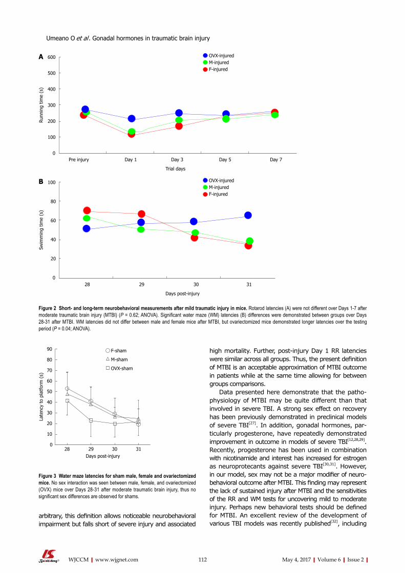
112 May 4, 2017|Volume 6|Issue 2|WJCCM|www.wjgnet.com
arbitrary, this definition allows noticeable neurobehavioral impairment but falls short of severe injury and associated
high mortality. Further, post-injury Day 1 RR latencies were similar across all groups. Thus, the present definition of MTBI is an acceptable approximation of MTBI outcome in patients while at the same time allowing for between groups comparisons.
Data presented here demonstrate that the patho-physiology of MTBI may be quite different than that involved in severe TBI. A strong sex effect on recovery has been previously demonstrated in preclinical models of severe TBI[27]. In addition, gonadal hormones, par-ticularly progesterone, have repeatedly demonstrated improvement in outcome in models of severe TBI[12,28,29]. Recently, progesterone has been used in combination with nicotinamide and interest has increased for estrogen as neuroprotecants against severe TBI[30,31]. However, in our model, sex may not be a major modifier of neuro-behavioral outcome after MTBI. This finding may represent the lack of sustained injury after MTBI and the sensitivities of the RR and WM tests for uncovering mild to moderate injury. Perhaps new behavioral tests should be defined for MTBI. An excellent review of the development of various TBI models was recently published[32], including
600
500
400
300
200
100
0
Runn
ing
time
(s)
OVX-injuredM-injuredF-injured
Pre injury Day 1 Day 3 Day 5 Day 7
Trial days
100
80
60
40
20
0
Swim
min
g tim
e (s
)
OVX-injuredM-injuredF-injured
28 29 30 31
Days post-injury
Figure 2 Short- and long-term neurobehavioral measurements after mild traumatic injury in mice. Rotarod latencies (A) were not different over Days 1-7 after moderate traumatic brain injury (MTBI) (P = 0.62; ANOVA). Significant water maze (WM) latencies (B) differences were demonstrated between groups over Days 28-31 after MTBI. WM latencies did not differ between male and female mice after MTBI, but ovariectomized mice demonstrated longer latencies over the testing period (P = 0.04; ANOVA).
A
B
28 29 30 31Days post-injury
90
80
70
60
50
40
30
20
10
0
Late
ncy
to p
latf
orm
(s)
F-sham
M-sham
OVX-sham
Figure 3 Water maze latencies for sham male, female and ovariectomized mice. No sex interaction was seen between male, female, and ovariectomized (OVX) mice over Days 28-31 after moderate traumatic brain injury, thus no significant sex differences are observed for shams.
Umeano O et al . Gonadal hormones in traumatic brain injury
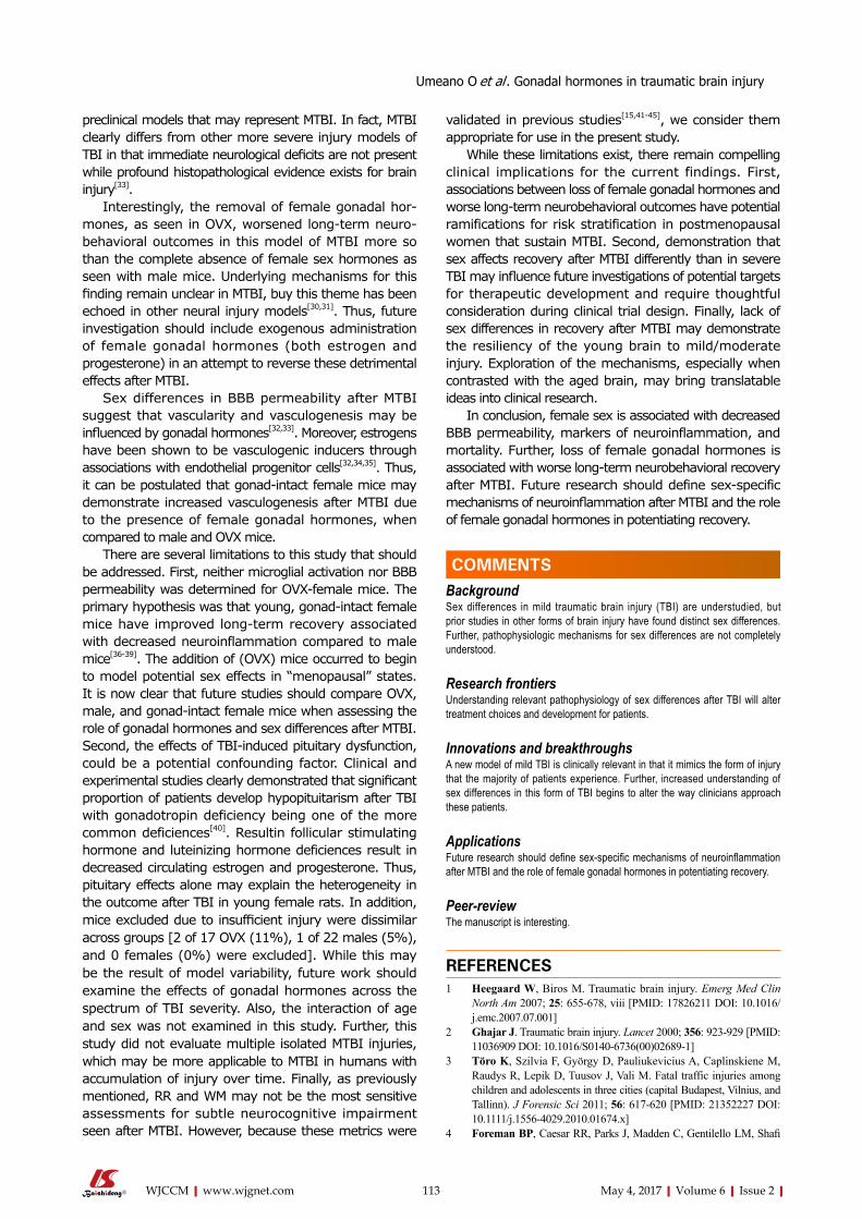
113 May 4, 2017|Volume 6|Issue 2|WJCCM|www.wjgnet.com
preclinical models that may represent MTBI. In fact, MTBI clearly differs from other more severe injury models of TBI in that immediate neurological deficits are not present while profound histopathological evidence exists for brain injury[33].
Interestingly, the removal of female gonadal hor-mones, as seen in OVX, worsened long-term neuro-behavioral outcomes in this model of MTBI more so than the complete absence of female sex hormones as seen with male mice. Underlying mechanisms for this finding remain unclear in MTBI, buy this theme has been echoed in other neural injury models[30,31]. Thus, future investigation should include exogenous administration of female gonadal hormones (both estrogen and progesterone) in an attempt to reverse these detrimental effects after MTBI.
Sex differences in BBB permeability after MTBI suggest that vascularity and vasculogenesis may be influenced by gonadal hormones[32,33]. Moreover, estrogens have been shown to be vasculogenic inducers through associations with endothelial progenitor cells[32,34,35]. Thus, it can be postulated that gonad-intact female mice may demonstrate increased vasculogenesis after MTBI due to the presence of female gonadal hormones, when compared to male and OVX mice.
There are several limitations to this study that should be addressed. First, neither microglial activation nor BBB permeability was determined for OVX-female mice. The primary hypothesis was that young, gonad-intact female mice have improved long-term recovery associated with decreased neuroinflammation compared to male mice[36-39]. The addition of (OVX) mice occurred to begin to model potential sex effects in “menopausal” states. It is now clear that future studies should compare OVX, male, and gonad-intact female mice when assessing the role of gonadal hormones and sex differences after MTBI. Second, the effects of TBI-induced pituitary dysfunction, could be a potential confounding factor. Clinical and experimental studies clearly demonstrated that significant proportion of patients develop hypopituitarism after TBI with gonadotropin deficiency being one of the more common deficiences[40]. Resultin follicular stimulating hormone and luteinizing hormone deficiences result in decreased circulating estrogen and progesterone. Thus, pituitary effects alone may explain the heterogeneity in the outcome after TBI in young female rats. In addition, mice excluded due to insufficient injury were dissimilar across groups [2 of 17 OVX (11%), 1 of 22 males (5%), and 0 females (0%) were excluded]. While this may be the result of model variability, future work should examine the effects of gonadal hormones across the spectrum of TBI severity. Also, the interaction of age and sex was not examined in this study. Further, this study did not evaluate multiple isolated MTBI injuries, which may be more applicable to MTBI in humans with accumulation of injury over time. Finally, as previously mentioned, RR and WM may not be the most sensitive assessments for subtle neurocognitive impairment seen after MTBI. However, because these metrics were
validated in previous studies[15,41-45], we consider them appropriate for use in the present study.
While these limitations exist, there remain compelling clinical implications for the current findings. First, associations between loss of female gonadal hormones and worse long-term neurobehavioral outcomes have potential ramifications for risk stratification in postmenopausal women that sustain MTBI. Second, demonstration that sex affects recovery after MTBI differently than in severe TBI may influence future investigations of potential targets for therapeutic development and require thoughtful consideration during clinical trial design. Finally, lack of sex differences in recovery after MTBI may demonstrate the resiliency of the young brain to mild/moderate injury. Exploration of the mechanisms, especially when contrasted with the aged brain, may bring translatable ideas into clinical research.
In conclusion, female sex is associated with decreased BBB permeability, markers of neuroinflammation, and mortality. Further, loss of female gonadal hormones is associated with worse long-term neurobehavioral recovery after MTBI. Future research should define sex-specific mechanisms of neuroinflammation after MTBI and the role of female gonadal hormones in potentiating recovery.
COMMENTSBackgroundSex differences in mild traumatic brain injury (TBI) are understudied, but prior studies in other forms of brain injury have found distinct sex differences. Further, pathophysiologic mechanisms for sex differences are not completely understood.
Research frontiersUnderstanding relevant pathophysiology of sex differences after TBI will alter treatment choices and development for patients.
Innovations and breakthroughsA new model of mild TBI is clinically relevant in that it mimics the form of injury that the majority of patients experience. Further, increased understanding of sex differences in this form of TBI begins to alter the way clinicians approach these patients.
ApplicationsFuture research should define sex-specific mechanisms of neuroinflammation after MTBI and the role of female gonadal hormones in potentiating recovery.
Peer-reviewThe manuscript is interesting.
REFERENCES1 Heegaard W, Biros M. Traumatic brain injury. Emerg Med Clin
North Am 2007; 25: 655-678, viii [PMID: 17826211 DOI: 10.1016/j.emc.2007.07.001]
2 Ghajar J. Traumatic brain injury. Lancet 2000; 356: 923-929 [PMID: 11036909 DOI: 10.1016/S0140-6736(00)02689-1]
3 Töro K, Szilvia F, György D, Pauliukevicius A, Caplinskiene M, Raudys R, Lepik D, Tuusov J, Vali M. Fatal traffic injuries among children and adolescents in three cities (capital Budapest, Vilnius, and Tallinn). J Forensic Sci 2011; 56: 617-620 [PMID: 21352227 DOI: 10.1111/j.1556-4029.2010.01674.x]
4 Foreman BP, Caesar RR, Parks J, Madden C, Gentilello LM, Shafi
COMMENTS
Umeano O et al . Gonadal hormones in traumatic brain injury
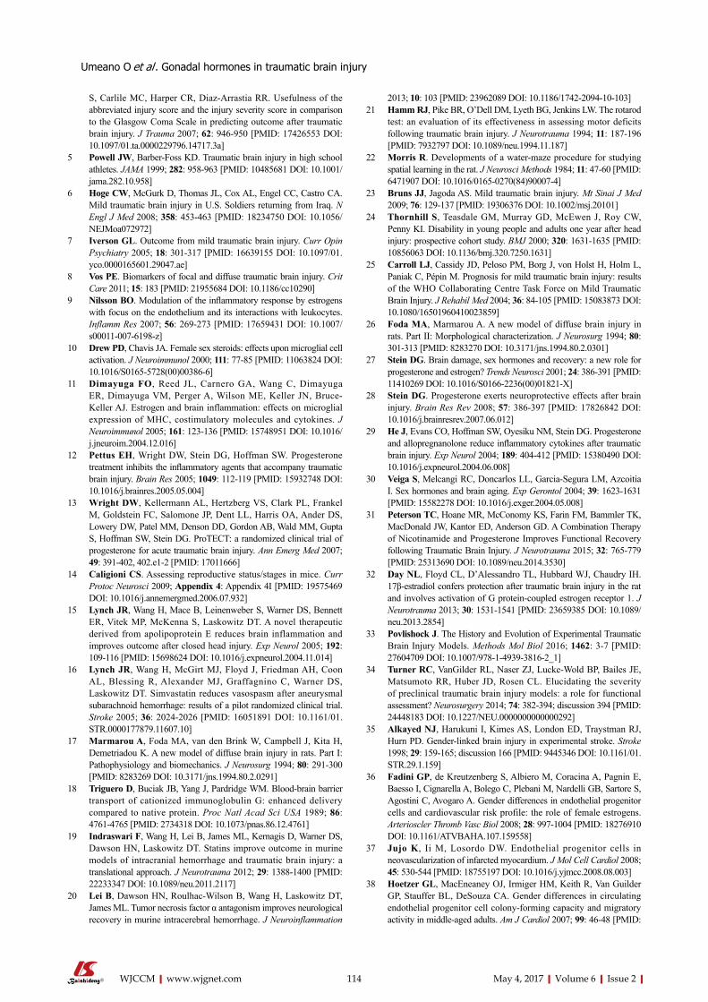
114 May 4, 2017|Volume 6|Issue 2|WJCCM|www.wjgnet.com
S, Carlile MC, Harper CR, Diaz-Arrastia RR. Usefulness of the abbreviated injury score and the injury severity score in comparison to the Glasgow Coma Scale in predicting outcome after traumatic brain injury. J Trauma 2007; 62: 946-950 [PMID: 17426553 DOI: 10.1097/01.ta.0000229796.14717.3a]
5 Powell JW, Barber-Foss KD. Traumatic brain injury in high school athletes. JAMA 1999; 282: 958-963 [PMID: 10485681 DOI: 10.1001/jama.282.10.958]
6 Hoge CW, McGurk D, Thomas JL, Cox AL, Engel CC, Castro CA. Mild traumatic brain injury in U.S. Soldiers returning from Iraq. N Engl J Med 2008; 358: 453-463 [PMID: 18234750 DOI: 10.1056/NEJMoa072972]
7 Iverson GL. Outcome from mild traumatic brain injury. Curr Opin Psychiatry 2005; 18: 301-317 [PMID: 16639155 DOI: 10.1097/01.yco.0000165601.29047.ae]
8 Vos PE. Biomarkers of focal and diffuse traumatic brain injury. Crit Care 2011; 15: 183 [PMID: 21955684 DOI: 10.1186/cc10290]
9 Nilsson BO. Modulation of the inflammatory response by estrogens with focus on the endothelium and its interactions with leukocytes. Inflamm Res 2007; 56: 269-273 [PMID: 17659431 DOI: 10.1007/s00011-007-6198-z]
10 Drew PD, Chavis JA. Female sex steroids: effects upon microglial cell activation. J Neuroimmunol 2000; 111: 77-85 [PMID: 11063824 DOI: 10.1016/S0165-5728(00)00386-6]
11 Dimayuga FO, Reed JL, Carnero GA, Wang C, Dimayuga ER, Dimayuga VM, Perger A, Wilson ME, Keller JN, Bruce-Keller AJ. Estrogen and brain inflammation: effects on microglial expression of MHC, costimulatory molecules and cytokines. J Neuroimmunol 2005; 161: 123-136 [PMID: 15748951 DOI: 10.1016/j.jneuroim.2004.12.016]
12 Pettus EH, Wright DW, Stein DG, Hoffman SW. Progesterone treatment inhibits the inflammatory agents that accompany traumatic brain injury. Brain Res 2005; 1049: 112-119 [PMID: 15932748 DOI: 10.1016/j.brainres.2005.05.004]
13 Wright DW, Kellermann AL, Hertzberg VS, Clark PL, Frankel M, Goldstein FC, Salomone JP, Dent LL, Harris OA, Ander DS, Lowery DW, Patel MM, Denson DD, Gordon AB, Wald MM, Gupta S, Hoffman SW, Stein DG. ProTECT: a randomized clinical trial of progesterone for acute traumatic brain injury. Ann Emerg Med 2007; 49: 391-402, 402.e1-2 [PMID: 17011666]
14 Caligioni CS. Assessing reproductive status/stages in mice. Curr Protoc Neurosci 2009; Appendix 4: Appendix 4I [PMID: 19575469 DOI: 10.1016/j.annemergmed.2006.07.932]
15 Lynch JR, Wang H, Mace B, Leinenweber S, Warner DS, Bennett ER, Vitek MP, McKenna S, Laskowitz DT. A novel therapeutic derived from apolipoprotein E reduces brain inflammation and improves outcome after closed head injury. Exp Neurol 2005; 192: 109-116 [PMID: 15698624 DOI: 10.1016/j.expneurol.2004.11.014]
16 Lynch JR, Wang H, McGirt MJ, Floyd J, Friedman AH, Coon AL, Blessing R, Alexander MJ, Graffagnino C, Warner DS, Laskowitz DT. Simvastatin reduces vasospasm after aneurysmal subarachnoid hemorrhage: results of a pilot randomized clinical trial. Stroke 2005; 36: 2024-2026 [PMID: 16051891 DOI: 10.1161/01.STR.0000177879.11607.10]
17 Marmarou A, Foda MA, van den Brink W, Campbell J, Kita H, Demetriadou K. A new model of diffuse brain injury in rats. Part I: Pathophysiology and biomechanics. J Neurosurg 1994; 80: 291-300 [PMID: 8283269 DOI: 10.3171/jns.1994.80.2.0291]
18 Triguero D, Buciak JB, Yang J, Pardridge WM. Blood-brain barrier transport of cationized immunoglobulin G: enhanced delivery compared to native protein. Proc Natl Acad Sci USA 1989; 86: 4761-4765 [PMID: 2734318 DOI: 10.1073/pnas.86.12.4761]
19 Indraswari F, Wang H, Lei B, James ML, Kernagis D, Warner DS, Dawson HN, Laskowitz DT. Statins improve outcome in murine models of intracranial hemorrhage and traumatic brain injury: a translational approach. J Neurotrauma 2012; 29: 1388-1400 [PMID: 22233347 DOI: 10.1089/neu.2011.2117]
20 Lei B, Dawson HN, Roulhac-Wilson B, Wang H, Laskowitz DT, James ML. Tumor necrosis factor α antagonism improves neurological recovery in murine intracerebral hemorrhage. J Neuroinflammation
2013; 10: 103 [PMID: 23962089 DOI: 10.1186/1742-2094-10-103]21 Hamm RJ, Pike BR, O’Dell DM, Lyeth BG, Jenkins LW. The rotarod
test: an evaluation of its effectiveness in assessing motor deficits following traumatic brain injury. J Neurotrauma 1994; 11: 187-196 [PMID: 7932797 DOI: 10.1089/neu.1994.11.187]
22 Morris R. Developments of a water-maze procedure for studying spatial learning in the rat. J Neurosci Methods 1984; 11: 47-60 [PMID: 6471907 DOI: 10.1016/0165-0270(84)90007-4]
23 Bruns JJ, Jagoda AS. Mild traumatic brain injury. Mt Sinai J Med 2009; 76: 129-137 [PMID: 19306376 DOI: 10.1002/msj.20101]
24 Thornhill S, Teasdale GM, Murray GD, McEwen J, Roy CW, Penny KI. Disability in young people and adults one year after head injury: prospective cohort study. BMJ 2000; 320: 1631-1635 [PMID: 10856063 DOI: 10.1136/bmj.320.7250.1631]
25 Carroll LJ, Cassidy JD, Peloso PM, Borg J, von Holst H, Holm L, Paniak C, Pépin M. Prognosis for mild traumatic brain injury: results of the WHO Collaborating Centre Task Force on Mild Traumatic Brain Injury. J Rehabil Med 2004; 36: 84-105 [PMID: 15083873 DOI: 10.1080/16501960410023859]
26 Foda MA, Marmarou A. A new model of diffuse brain injury in rats. Part II: Morphological characterization. J Neurosurg 1994; 80: 301-313 [PMID: 8283270 DOI: 10.3171/jns.1994.80.2.0301]
27 Stein DG. Brain damage, sex hormones and recovery: a new role for progesterone and estrogen? Trends Neurosci 2001; 24: 386-391 [PMID: 11410269 DOI: 10.1016/S0166-2236(00)01821-X]
28 Stein DG. Progesterone exerts neuroprotective effects after brain injury. Brain Res Rev 2008; 57: 386-397 [PMID: 17826842 DOI: 10.1016/j.brainresrev.2007.06.012]
29 He J, Evans CO, Hoffman SW, Oyesiku NM, Stein DG. Progesterone and allopregnanolone reduce inflammatory cytokines after traumatic brain injury. Exp Neurol 2004; 189: 404-412 [PMID: 15380490 DOI: 10.1016/j.expneurol.2004.06.008]
30 Veiga S, Melcangi RC, Doncarlos LL, Garcia-Segura LM, Azcoitia I. Sex hormones and brain aging. Exp Gerontol 2004; 39: 1623-1631 [PMID: 15582278 DOI: 10.1016/j.exger.2004.05.008]
31 Peterson TC, Hoane MR, McConomy KS, Farin FM, Bammler TK, MacDonald JW, Kantor ED, Anderson GD. A Combination Therapy of Nicotinamide and Progesterone Improves Functional Recovery following Traumatic Brain Injury. J Neurotrauma 2015; 32: 765-779 [PMID: 25313690 DOI: 10.1089/neu.2014.3530]
32 Day NL, Floyd CL, D’Alessandro TL, Hubbard WJ, Chaudry IH. 17β-estradiol confers protection after traumatic brain injury in the rat and involves activation of G protein-coupled estrogen receptor 1. J Neurotrauma 2013; 30: 1531-1541 [PMID: 23659385 DOI: 10.1089/neu.2013.2854]
33 Povlishock J. The History and Evolution of Experimental Traumatic Brain Injury Models. Methods Mol Biol 2016; 1462: 3-7 [PMID: 27604709 DOI: 10.1007/978-1-4939-3816-2_1]
34 Turner RC, VanGilder RL, Naser ZJ, Lucke-Wold BP, Bailes JE, Matsumoto RR, Huber JD, Rosen CL. Elucidating the severity of preclinical traumatic brain injury models: a role for functional assessment? Neurosurgery 2014; 74: 382-394; discussion 394 [PMID: 24448183 DOI: 10.1227/NEU.0000000000000292]
35 Alkayed NJ, Harukuni I, Kimes AS, London ED, Traystman RJ, Hurn PD. Gender-linked brain injury in experimental stroke. Stroke 1998; 29: 159-165; discussion 166 [PMID: 9445346 DOI: 10.1161/01.STR.29.1.159]
36 Fadini GP, de Kreutzenberg S, Albiero M, Coracina A, Pagnin E, Baesso I, Cignarella A, Bolego C, Plebani M, Nardelli GB, Sartore S, Agostini C, Avogaro A. Gender differences in endothelial progenitor cells and cardiovascular risk profile: the role of female estrogens. Arterioscler Thromb Vasc Biol 2008; 28: 997-1004 [PMID: 18276910 DOI: 10.1161/ATVBAHA.107.159558]
37 Jujo K, Ii M, Losordo DW. Endothelial progenitor cells in neovascularization of infarcted myocardium. J Mol Cell Cardiol 2008; 45: 530-544 [PMID: 18755197 DOI: 10.1016/j.yjmcc.2008.08.003]
38 Hoetzer GL, MacEneaney OJ, Irmiger HM, Keith R, Van Guilder GP, Stauffer BL, DeSouza CA. Gender differences in circulating endothelial progenitor cell colony-forming capacity and migratory activity in middle-aged adults. Am J Cardiol 2007; 99: 46-48 [PMID:
Umeano O et al . Gonadal hormones in traumatic brain injury

115 May 4, 2017|Volume 6|Issue 2|WJCCM|www.wjgnet.com
17196460 DOI: 10.1016/j.amjcard.2006.07.061]39 Iwakura A, Luedemann C, Shastry S, Hanley A, Kearney M, Aikawa
R, Isner JM, Asahara T, Losordo DW. Estrogen-mediated, endothelial nitric oxide synthase-dependent mobilization of bone marrow-derived endothelial progenitor cells contributes to reendothelialization after arterial injury. Circulation 2003; 108: 3115-3121 [PMID: 14676142 DOI: 10.1161/01.CIR.0000106906.56972.83]
40 Tanriverdi F, Schneider HJ, Aimaretti G, Masel BE, Casanueva FF, Kelestimur F. Pituitary dysfunction after traumatic brain injury: a clinical and pathophysiological approach. Endocr Rev 2015; 36: 305-342 [PMID: 25950715 DOI: 10.1210/er.2014-1065]
41 James ML, Wang H, Venkatraman T, Song P, Lascola CD, Laskowitz DT. Brain natriuretic peptide improves long-term functional recovery after acute CNS injury in mice. J Neurotrauma 2010; 27: 217-228 [PMID: 19803787 DOI: 10.1089/neu.2009.1022]
42 Laskowitz DT, McKenna SE, Song P, Wang H, Durham L, Yeung N, Christensen D, Vitek MP. COG1410, a novel apolipoprotein E-based
peptide, improves functional recovery in a murine model of traumatic brain injury. J Neurotrauma 2007; 24: 1093-1107 [PMID: 17610350 DOI: 10.1089/neu.2006.0192]
43 James ML, Sullivan PM, Lascola CD, Vitek MP, Laskowitz DT. Pharmacogenomic effects of apolipoprotein e on intracerebral hemorrhage. Stroke 2009; 40: 632-639 [PMID: 19109539 DOI: 10.1161/STROKEAHA.108.530402]
44 Wang H, Lynch JR, Song P, Yang HJ, Yates RB, Mace B, Warner DS, Guyton JR, Laskowitz DT. Simvastatin and atorvastatin improve behavioral outcome, reduce hippocampal degeneration, and improve cerebral blood flow after experimental traumatic brain injury. Exp Neurol 2007; 206: 59-69 [PMID: 17521631 DOI: 10.1016/j.expneurol.2007.03.031]
45 Wang H, Gao J, Lassiter TF, McDonagh DL, Sheng H, Warner DS, Lynch JR, Laskowitz DT. Levetiracetam is neuroprotective in murine models of closed head injury and subarachnoid hemorrhage. Neurocrit Care 2006; 5: 71-78 [PMID: 16960300 DOI: 10.1385/NCC:5:1:71]
P- Reviewer: Soriano-Ursua MA, Tanriverdi F S- Editor: Ji FF L- Editor: A E- Editor: Lu YJ
Umeano O et al . Gonadal hormones in traumatic brain injury

Sumit Kapoor, Adel Bassily-Marcus, Rafael Alba Yunen, Parissa Tabrizian, Sabrine Semoin, Joseph Blankush, Daniel Labow, John Oropello, Anthony Manasia, Roopa Kohli-Seth
ORIGINAL ARTICLE
116 May 4, 2017|Volume 6|Issue 2|WJCCM|www.wjgnet.com
Critical care management and intensive care unit outcomes following cytoreductive surgery with hyperthermic intraperitoneal chemotherapy
Sumit Kapoor, Department of Pulmonary, Critical Care and Sleep, Baylor College of Medicine, Houston, TX 77030, United States
Adel Bassily-Marcus, John Oropello, Anthony Manasia, Roopa Kohli-Seth, Department of Surgery, Critical Care Division, Icahn School of Medicine at Mount Sinai, New York, NY 10029, United States
Rafael Alba Yunen, Department of Pulmonary and Critical Care, Medical College of Georgia, Augusta University, Augusta, GA 30912, United States
Parissa Tabrizian, Sabrine Semoin, Daniel Labow, Department of Surgery, Icahn School of Medicine at Mount Sinai, New York, NY 10029, United States
Joseph Blankush, General Surgery Resident, Vanderbilt University Medical Center, Nashville, TN 37232, United States
Author contributions: Kapoor S and Bassily-Marcus A designed and performed the research and wrote the paper; Alba Yunen R, Tabrizian P and Semoin S contributed to data collection and analysis; Blankush J, Labow D, Oropello J, Manasia A and Kohli-Seth R provided clinical advice; Bassily-Marcus A supervised the report.
Institutional review board statement: This study was reviewed and approved by the Institutional review board of the Icahn School of Medicine at Mount Sinai, New York.
Informed consent statement: Patients were not required to give informed consent to the study because the retrospective analysis used anonymous clinical data that were ob-tained after each patient agreed to treatment by written consent.
Conflict-of-interest statement: We have no financial relationships to disclose.
Data sharing statement: No additional data are available.
Open-Access: This article is an open-access article which was selected by an in-house editor and fully peer-reviewed by external reviewers. It is distributed in accordance with the Creative Commons Attribution Non Commercial (CC BY-NC 4.0) license, which permits others to distribute, remix, adapt, build upon this work non-commercially, and license their derivative works on different terms, provided the original work is properly cited and the use is non-commercial. See: http://creativecommons.org/licenses/by-nc/4.0/
Manuscript source: Unsolicited manuscript
Correspondence to: Adel Bassily-Marcus, MD, FCCM, FCCP, FACP, Associate Professor, Department of Surgery, Critical Care Division, Icahn School of Medicine at Mount Sinai, One Gustave L Levy Place, New York, NY 10029, United States. [email protected]: +1-212-2418867Fax: +1-212-8603669
Received: November 11, 2016 Peer-review started: November 13, 2016 First decision: December 1, 2016Revised: December 18, 2016 Accepted: January 11, 2017Article in press: January 13, 2017Published online: May 4, 2017
AbstractAIMTo study the early postoperative intensive care unit (ICU) management and complications in the first 2 wk of patients undergoing cytoreductive surgery (CRS) and hyperthermic intraperitoneal chemotherapy (HIPEC).
METHODSOur study is a retrospective, observational study per-
World Journal ofCritical Care MedicineW J C C M
Submit a Manuscript: http://www.f6publishing.com
DOI: 10.5492/wjccm.v6.i2.116
World J Crit Care Med 2017 May 4; 6(2): 116-123
ISSN 2220-3141 (online)
Retrospective Study

117 May 4, 2017|Volume 6|Issue 2|WJCCM|www.wjgnet.com
Kapoor S et al . Critical care management of HIPEC patients
formed at Icahn School of Medicine at Mount Sinai, quaternary care hospital in New York City. All adult patients who underwent CRS and HIPEC between January 1, 2007 and December 31, 2012 and admitted to ICU postoperatively were studied. Fifty-one patients came to the ICU postoperatively out of 170 who underwent CRS and HIPEC therapy during the study period. Data analysis was performed using descriptive statistics.
RESULTSOf the 170 patients who underwent CRS and HIPEC therapy, 51 (30%) came to the ICU postoperatively. Mean ICU length of stay was 4 d (range 1-60 d) and mean APACHE Ⅱ score was 15 (range 7-23). Thirty-one/fifty-one (62%) patients developed postoperative complications. Aggressive intraoperative and postoperative fluid resuscitation is required in most patients. Hypo-volemia was seen in all patients and median amount of fluids required in the first 48 h was 6 L (range 1-14 L). Thirteen patients (25%) developed postoperative hypotension with seven requiring vasopressor support. The major cause of sepsis was intraabdominal, with 8 (15%) developing anastomotic leaks and 5 (10%) developing intraabdominal abscess. The median survival was 14 mo with 30 d mortality of 4% (2/51) and 90 d mortality of 16% (8/51). One year survival was 56.4% (28/51). Preoperative medical co morbidities, extent of surgical debulking, intraoperative blood losses, amount of intra op blood products required and total operative time are the factors to be considered while deciding ICU vs non ICU admission.
CONCLUSIONOverall, ICU outcomes of this study population are ex-cellent. Triage of these patients should consider preo-perative and intraoperative factors. Intensivists should be vigilant to aggressive postop fluid resuscitation, pain control and early detection and management of surgical complications.
Key words: Hyperthermic; Abdominal sepsis; Cytoreduction; Carcinomatosis; Respiratory failure; Vasopressors
© The Author(s) 2017. Published by Baishideng Publishing Group Inc. All rights reserved.
Core tip: Our retrospective study highlights the intensive care unit (ICU) management, complications and outcomes of patients undergoing complicated hyperthermic intra-peritoneal chemotherapy procedure for peritoneal car-cinomatosis. Intensivists need to monitor for physiologic derangements post procedure and assess fluid status, provide adequate pain control and have high degree of suspicion for complications like abdominal sepsis. Not all patients need ICU admission post procedure. Our study enlists the factors to be considered for ICU admission vs the floor.
Kapoor S, Bassily-Marcus A, Alba Yunen R, Tabrizian P, Semoin S, Blankush J, Labow D, Oropello J, Manasia A, Kohli-Seth
R. Critical care management and intensive care unit outcomes following cytoreductive surgery with hyperthermic intraperitoneal chemotherapy. World J Crit Care Med 2017; 6(2): 116-123 Available from: URL: http://www.wjgnet.com/2220-3141/full/v6/i2/116.htm DOI: http://dx.doi.org/10.5492/wjccm.v6.i2.116
INTRODUCTIONMalignant peritoneal surface disease, commonly known as peritoneal carcinomatosis (PC) has been considered to be incurable with poor median survival of six months without treatment[1,2]. Cytoreductive surgery (CRS) combined with perioperative hyperthermic intraperitoneal chemotherapy (HIPEC) has been confirmed as a viable treatment option for peritoneal based tumors in 1980s. Today, CRS and HIPEC therapy is used in select patients for treatment of PC from gastric cancer, colorectal cancer, appendiceal cancer, ovarian cancer and primary peritoneal malignancies like mesotheliomas. Few randomized controlled trials have been conducted and demonstrated better survival in patients undergoing CRS-HIPEC in the treatment of colorectal PC compared to systemic chemotherapy alone[3-5]. Multiple nonrandomized single and multicenter phase Ⅱ and few phase Ⅲ trials indicate that CRS and HIPEC may improve survival rate for selected patients with peritoneal metastasis[2,6-8]. There has been an observed increase in survival rates with CRS and HIPEC in colorectal cancer, advanced ovarian cancer, gastric cancer and peritoneal mesothelioma[9-14]. This procedure is technically challenging and has the potential for increased morbidity and mortality[15]. The morbidity and mortality rates reported have ranged from 12%-52% and 0.9%-5.8% respectively at large tertiary hospitals with a high volume of oncology patients[2]. Recent largest reported single center study demonstrated that major prognostic factors include primary site, performance status, completeness of resection and institutional experience[16]. In a re-cent study, 40% of patients developed at least one postoperative in hospital complication[17]. One serious complication related to HIPEC is peritoneal inflammation and systemic inflammatory response syndrome (SIRS) which leads to physiologic changes in different organ systems. Effects on the cardiovascular status, oxygen consumption and hematopoietic parameters of patients have been described in the literature and have been observed in practice[18-21]. The complexity of perioperative anesthetic management of these patients with particular attention to perioperative fluid, pain management and the patient’s coagulation status has been reported in the anesthesiology literature[22,23]. The extensive peritoneal inflammation and the inflammatory response impacts the early postoperative intensive care unit (ICU) course of this patient population. In the present study, we describe the early (first two weeks) postoperative ICU management, long-term morbidity and mortality (day 30 and day 90) of patients admitted to the ICU after undergoing CRS and HIPEC therapy retrospectively over a six years period.

118 May 4, 2017|Volume 6|Issue 2|WJCCM|www.wjgnet.com
MATERIALS AND METHODSStudy designThis retrospective descriptive study was conducted in a 14-bed surgical intensive care unit (SICU) in a large tertiary hospital. With Institutional Review Board approval, all adult patients over 18 years of age who underwent CRS and HIPEC therapy between January 1, 2007 and December 31, 2012 were screened for enrollment in the study (n = 170). Of this, 51 patients were admitted to the SICU postoperatively and were included in the study. Data for this retrospective analysis were retrieved from medical records, operative reports, laboratory and radiologic data. Early (first two weeks) postoperative ICU course, ICU management and long-term surgical morbidity and mortality (day 30 and day 90) were retrospectively studied.
CRS and HIPEC surgery was performed with closed technique and cisplatin and mitomycin C were the che-motherapy agents administered. Patients were often immediately extubated and transferred to the surgical floor. Standard criteria for extubation were used by anesthesiology team including mental status assessment, reversal of muscular paralysis and anesthesia, hemo-dynamic stability and adequate oxygenation and ventilation in addition to intraoperative course and complications. If they were hemodynamically unstable in OR with mean arterial pressure (MAP) less than 65 mmHg and/or systolic blood pressure (SBP) less than 90 mmHg requiring intravenous fluids and vasopressor support, remained on mechanical ventilation or required higher level of care based on intraoperative course, they were admitted to SICU. This was determined by the anesthesia, surgical and critical care teams. At the initial stages of experience with the HIPEC procedure at our center, all patients came to SICU post operatively. With our growing experience, we became more selective in admitting patients to ICU
postoperatively.
RESULTSIn the present study, 51 out of 170 patients came to the ICU postoperatively. The primary diagnosis was PC secondary to metastatic colon, appendiceal, gastric, mesothelial and ovarian tumors. Median ICU length of stay (LOS) was 4 d (range 1-60 d) and median APACHE Ⅱ scores was 15 (range 7-23) (Table 1). Twenty-two patients (43%) received postop epidural analgesia by the anesthesia team. Of the patients admitted to the ICU, 31 (62%) had observed post-operative complications. Twenty-nine patients (57%) needed up to 10 L of fluids intraoperatively for hemodynamic instability and fluid/blood losses. All patients were hypovolemic posto-peratively in the ICU and required intravenous fluids. Median amount of intravenous fluids required in the first 48 h was 6 L (range 1-14 L). Assessment of hypovolemia was done by clinical exam, hemodynamic monitoring with pulse contour analysis and using standard bedside ultrasonography by assessing cardiac chambers and inferior vena cava. Post-operative complications are listed in Table 2. Postoperative hypotension developed in 13 patients (25%), out of which, 7 (13%) developed septic shock requiring vasopressor support. Acute respiratory failure, defined as requirement of mechanical ventilation for more than 48 h was seen in 18 patients (33%). Five patients (10%) developed acute renal failure but none required renal replacement therapy during their ICU course. Seventeen (25%) patients developed abdominal sepsis including anastomotic leak in eight patients (15%), intra abdominal abscess in five patients (10%) and deep wound infection in four (8%) (Figure 1). There were no Central line-associated bloodstream infections (CLABSIs). Total parenteral nutrition (TPN) was initiated in 9 (18%) patients. Median survival was 14 mo. Thirty-day mortality was 4% (2 patients) and 90 d mortality was 16% (8 patients). One year survival was 56.4% (28/51). We also performed sub group analysis of patients admitted to ICU and those admitted to the surgical floor postoperatively (Table 3). Variables like preoperative co morbidities (high ASA scores), extent of surgical debulking (number of organs resected), amount of blood products required intraoperatively, estimated blood loss, total OR time showed statistically significant difference between ICU and non ICU patients. ICU patients had increased postoperative morbidity and hospital LOS compared to non ICU patients. There was no statistically significant difference in 30 and 90 d mortality between these two sub group of patients.
DISCUSSIONICU course and complicationsThere has been a renewed interest in the field of peritoneal surface malignancies over the last 15-20 years,
Table 1 Patient characteristics
Variable n = 51
Age (yr) 56 (18-82)Gender Female 27 (53%)APACHE II 15 (7-23)ICU LOS (d) 4 (1-60)Operative time (min) 413 (65-815)Primary site Colon 19 Appendix 12 Gastric 6 Ovarian 3 Pseudomyxoma peritoneii 6 Mesothelial 3 Others 2EBL (mL) 1125 (50-6500)Peritoneal cancer index (intraop) 13 (3-35)
Data are reported as median and range or n (%). ICU: Intensive care unit; LOS: Length of stay.
Kapoor S et al . Critical care management of HIPEC patients
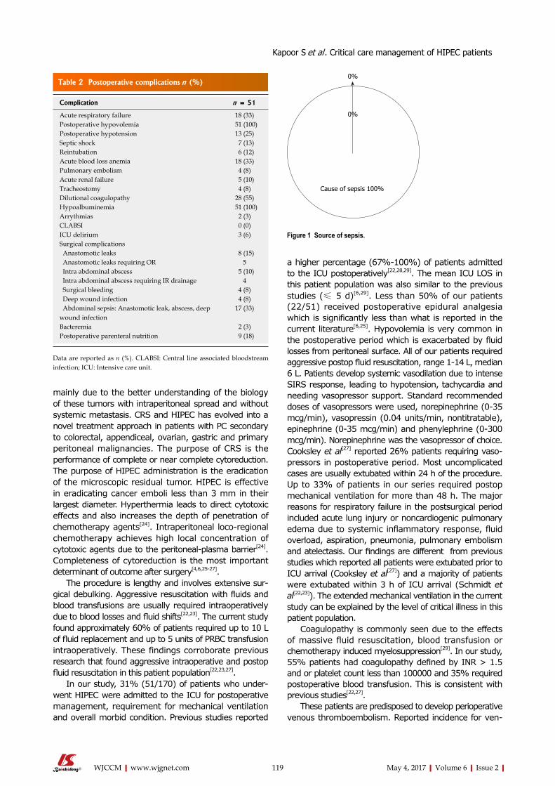
119 May 4, 2017|Volume 6|Issue 2|WJCCM|www.wjgnet.com
mainly due to the better understanding of the biology of these tumors with intraperitoneal spread and without systemic metastasis. CRS and HIPEC has evolved into a novel treatment approach in patients with PC secondary to colorectal, appendiceal, ovarian, gastric and primary peritoneal malignancies. The purpose of CRS is the performance of complete or near complete cytoreduction. The purpose of HIPEC administration is the eradication of the microscopic residual tumor. HIPEC is effective in eradicating cancer emboli less than 3 mm in their largest diameter. Hyperthermia leads to direct cytotoxic effects and also increases the depth of penetration of chemotherapy agents[24]. Intraperitoneal loco-regional chemotherapy achieves high local concentration of cytotoxic agents due to the peritoneal-plasma barrier[24]. Completeness of cytoreduction is the most important determinant of outcome after surgery[4,6,25-27].
The procedure is lengthy and involves extensive sur-gical debulking. Aggressive resuscitation with fluids and blood transfusions are usually required intraoperatively due to blood losses and fluid shifts[22,23]. The current study found approximately 60% of patients required up to 10 L of fluid replacement and up to 5 units of PRBC transfusion intraoperatively. These findings corroborate previous research that found aggressive intraoperative and postop fluid resuscitation in this patient population[22,23,27].
In our study, 31% (51/170) of patients who under-went HIPEC were admitted to the ICU for postoperative management, requirement for mechanical ventilation and overall morbid condition. Previous studies reported
a higher percentage (67%-100%) of patients admitted to the ICU postoperatively[22,28,29]. The mean ICU LOS in this patient population was also similar to the previous studies (≤ 5 d)[6,29]. Less than 50% of our patients (22/51) received postoperative epidural analgesia which is significantly less than what is reported in the current literature[6,25]. Hypovolemia is very common in the postoperative period which is exacerbated by fluid losses from peritoneal surface. All of our patients required aggressive postop fluid resuscitation, range 1-14 L, median 6 L. Patients develop systemic vasodilation due to intense SIRS response, leading to hypotension, tachycardia and needing vasopressor support. Standard recommended doses of vasopressors were used, norepinephrine (0-35 mcg/min), vasopressin (0.04 units/min, nontitratable), epinephrine (0-35 mcg/min) and phenylephrine (0-300 mcg/min). Norepinephrine was the vasopressor of choice. Cooksley et al[27] reported 26% patients requiring vaso-pressors in postoperative period. Most uncomplicated cases are usually extubated within 24 h of the procedure. Up to 33% of patients in our series required postop mechanical ventilation for more than 48 h. The major reasons for respiratory failure in the postsurgical period included acute lung injury or noncardiogenic pulmonary edema due to systemic inflammatory response, fluid overload, aspiration, pneumonia, pulmonary embolism and atelectasis. Our findings are different from previous studies which reported all patients were extubated prior to ICU arrival (Cooksley et al[27]) and a majority of patients were extubated within 3 h of ICU arrival (Schmidt et al[22,23]). The extended mechanical ventilation in the current study can be explained by the level of critical illness in this patient population.
Coagulopathy is commonly seen due to the effects of massive fluid resuscitation, blood transfusion or chemotherapy induced myelosuppression[29]. In our study, 55% patients had coagulopathy defined by INR > 1.5 and or platelet count less than 100000 and 35% required postoperative blood transfusion. This is consistent with previous studies[22,27].
These patients are predisposed to develop perioperative venous thromboembolism. Reported incidence for ven-
Table 2 Postoperative complications n (%)
Complication n = 51
Acute respiratory failure 18 (33)Postoperative hypovolemia 51 (100)Postoperative hypotension 13 (25)Septic shock 7 (13)Reintubation 6 (12)Acute blood loss anemia 18 (33)Pulmonary embolism 4 (8)Acute renal failure 5 (10)Tracheostomy 4 (8)Dilutional coagulopathy 28 (55)Hypoalbuminemia 51 (100)Arrythmias 2 (3)CLABSI 0 (0)ICU delirium 3 (6)Surgical complications Anastomotic leaks 8 (15) Anastomotic leaks requiring OR 5 Intra abdominal abscess 5 (10) Intra abdominal abscess requiring IR drainage 4 Surgical bleeding 4 (8) Deep wound infection 4 (8) Abdominal sepsis: Anastomotic leak, abscess, deep wound infection
17 (33)
Bacteremia 2 (3)Postoperative parenteral nutrition 9 (18)
Data are reported as n (%). CLABSI: Central line associated bloodstream infection; ICU: Intensive care unit.
0%
Cause of sepsis 100%
Figure 1 Source of sepsis.
Kapoor S et al . Critical care management of HIPEC patients
0%
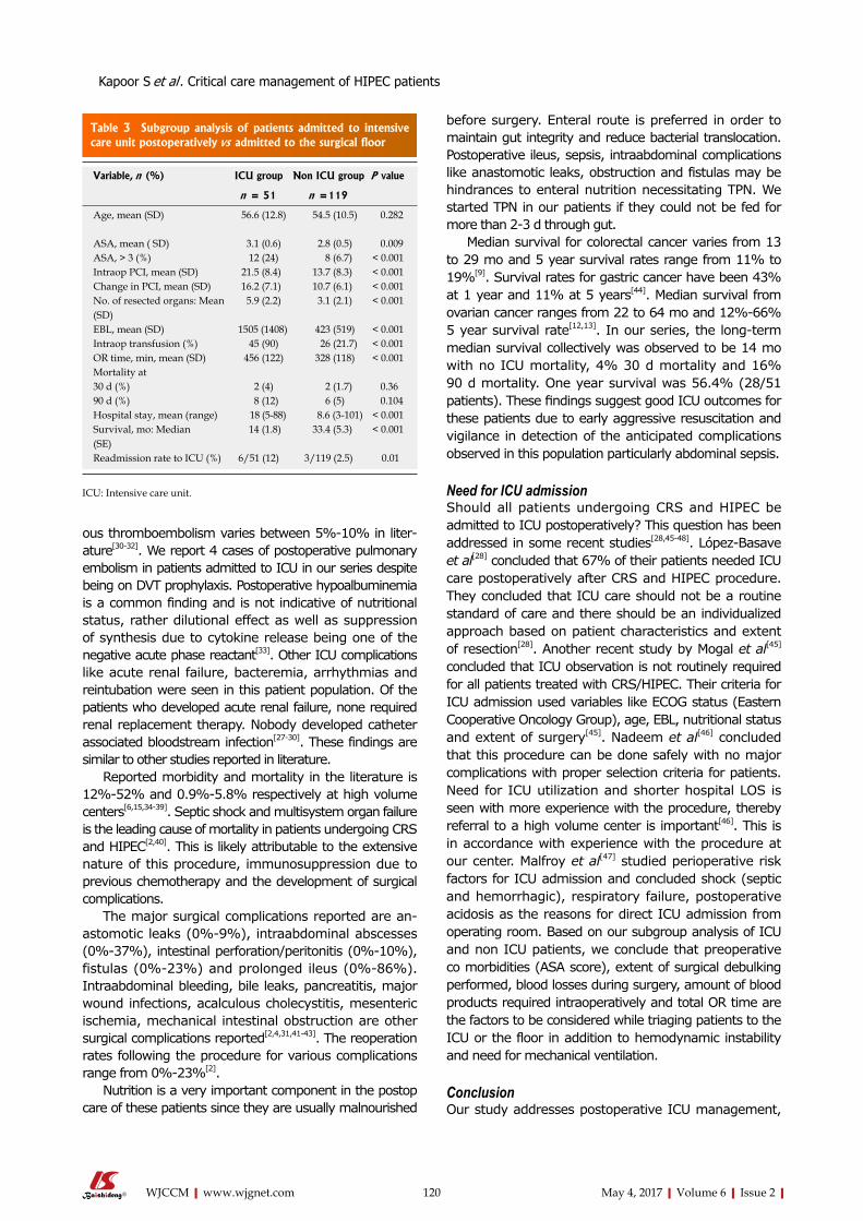
120 May 4, 2017|Volume 6|Issue 2|WJCCM|www.wjgnet.com
ous thromboembolism varies between 5%-10% in liter-ature[30-32]. We report 4 cases of postoperative pulmonary embolism in patients admitted to ICU in our series despite being on DVT prophylaxis. Postoperative hypoalbuminemia is a common finding and is not indicative of nutritional status, rather dilutional effect as well as suppression of synthesis due to cytokine release being one of the negative acute phase reactant[33]. Other ICU complications like acute renal failure, bacteremia, arrhythmias and reintubation were seen in this patient population. Of the patients who developed acute renal failure, none required renal replacement therapy. Nobody developed catheter associated bloodstream infection[27-30]. These findings are similar to other studies reported in literature.
Reported morbidity and mortality in the literature is 12%-52% and 0.9%-5.8% respectively at high volume centers[6,15,34-39]. Septic shock and multisystem organ failure is the leading cause of mortality in patients undergoing CRS and HIPEC[2,40]. This is likely attributable to the extensive nature of this procedure, immunosuppression due to previous chemotherapy and the development of surgical complications.
The major surgical complications reported are an-astomotic leaks (0%-9%), intraabdominal abscesses (0%-37%), intestinal perforation/peritonitis (0%-10%), fistulas (0%-23%) and prolonged ileus (0%-86%). Intraabdominal bleeding, bile leaks, pancreatitis, major wound infections, acalculous cholecystitis, mesenteric ischemia, mechanical intestinal obstruction are other surgical complications reported[2,4,31,41-43]. The reoperation rates following the procedure for various complications range from 0%-23%[2].
Nutrition is a very important component in the postop care of these patients since they are usually malnourished
before surgery. Enteral route is preferred in order to maintain gut integrity and reduce bacterial translocation. Postoperative ileus, sepsis, intraabdominal complications like anastomotic leaks, obstruction and fistulas may be hindrances to enteral nutrition necessitating TPN. We started TPN in our patients if they could not be fed for more than 2-3 d through gut.
Median survival for colorectal cancer varies from 13 to 29 mo and 5 year survival rates range from 11% to 19%[9]. Survival rates for gastric cancer have been 43% at 1 year and 11% at 5 years[44]. Median survival from ovarian cancer ranges from 22 to 64 mo and 12%-66% 5 year survival rate[12,13]. In our series, the long-term median survival collectively was observed to be 14 mo with no ICU mortality, 4% 30 d mortality and 16% 90 d mortality. One year survival was 56.4% (28/51 patients). These findings suggest good ICU outcomes for these patients due to early aggressive resuscitation and vigilance in detection of the anticipated complications observed in this population particularly abdominal sepsis.
Need for ICU admissionShould all patients undergoing CRS and HIPEC be admitted to ICU postoperatively? This question has been addressed in some recent studies[28,45-48]. López-Basave et al[28] concluded that 67% of their patients needed ICU care postoperatively after CRS and HIPEC procedure. They concluded that ICU care should not be a routine standard of care and there should be an individualized approach based on patient characteristics and extent of resection[28]. Another recent study by Mogal et al[45] concluded that ICU observation is not routinely required for all patients treated with CRS/HIPEC. Their criteria for ICU admission used variables like ECOG status (Eastern Cooperative Oncology Group), age, EBL, nutritional status and extent of surgery[45]. Nadeem et al[46] concluded that this procedure can be done safely with no major complications with proper selection criteria for patients. Need for ICU utilization and shorter hospital LOS is seen with more experience with the procedure, thereby referral to a high volume center is important[46]. This is in accordance with experience with the procedure at our center. Malfroy et al[47] studied perioperative risk factors for ICU admission and concluded shock (septic and hemorrhagic), respiratory failure, postoperative acidosis as the reasons for direct ICU admission from operating room. Based on our subgroup analysis of ICU and non ICU patients, we conclude that preoperative co morbidities (ASA score), extent of surgical debulking performed, blood losses during surgery, amount of blood products required intraoperatively and total OR time are the factors to be considered while triaging patients to the ICU or the floor in addition to hemodynamic instability and need for mechanical ventilation.
ConclusionOur study addresses postoperative ICU management,
Table 3 Subgroup analysis of patients admitted to intensive care unit postoperatively vs admitted to the surgical floor
Variable, n (%) ICU group Non ICU group P value
n = 51 n =119
Age, mean (SD) 56.6 (12.8) 54.5 (10.5) 0.282
ASA, mean ( SD) 3.1 (0.6) 2.8 (0.5) 0.009ASA, > 3 (%) 12 (24) 8 (6.7) < 0.001Intraop PCI, mean (SD) 21.5 (8.4) 13.7 (8.3) < 0.001Change in PCI, mean (SD) 16.2 (7.1) 10.7 (6.1) < 0.001No. of resected organs: Mean (SD)
5.9 (2.2) 3.1 (2.1) < 0.001
EBL, mean (SD) 1505 (1408) 423 (519) < 0.001Intraop transfusion (%) 45 (90) 26 (21.7) < 0.001OR time, min, mean (SD) 456 (122) 328 (118) < 0.001Mortality at 30 d (%) 2 (4) 2 (1.7) 0.3690 d (%) 8 (12) 6 (5) 0.104Hospital stay, mean (range) 18 (5-88) 8.6 (3-101) < 0.001Survival, mo: Median (SE)
14 (1.8) 33.4 (5.3) < 0.001
Readmission rate to ICU (%) 6/51 (12) 3/119 (2.5) 0.01
ICU: Intensive care unit.
Kapoor S et al . Critical care management of HIPEC patients
121 May 4, 2017|Volume 6|Issue 2|WJCCM|www.wjgnet.com
morbidity and mortality of patients undergoing CRS and HIPEC therapy. We also studied the factors to be considered during postoperative triage of patients to ICU vs the floor. Need for vasopressors and/or mec-hanical ventilation, preoperative medical co morbidities, extent of surgical debulking, intraoperative blood losses, amount of intra op blood products required and total operative time necessitate ICU admission. Intensivists should be vigilant to aggressive postop resuscitation, need for organ support, monitoring and correcting derangements in the coagulation profile, fluid and electrolytes, pain control, early detection and management of major surgical complications particularly abdominal sepsis. Overall, the ICU outcomes for these patients are excellent.
COMMENTSBackgroundPeritoneal carcinomatosis (PC) from gastric, colorectal, ovarian, appendiceal tumors is associated with poor prognosis. Cytoreductive surgery (CRS) and hyperthermic intraperitoneal chemotherapy (HIPEC) has been shown to improve survival in these cases. But the procedure is associated with increased morbidity and mortality. Extensive surgical resection with hyperthermic chemotherapy generates intense inflammatory response in the body, leading to physiologic changes in many organ systems. Intensivists taking care of these patients in the intensive care unit (ICU) postoperatively need to be aware of derangements like fluid and electrolyte abnormalities, hemodynamic instability, need for intravenous fluids, coagulation abnormalities and increased risk of surgical complications like anastomotic leaks and intra-abdominal abscesses. In this study, the authors describe the early postoperative ICU management, morbidity and mortality of patients admitted to ICU after CRS and HIPEC therapy.
Research frontiersCRS and HIPEC therapy is a complex procedure associated with increased morbidity and mortality. Few prior studies have described the postoperative ICU management and complications related to the procedure. The study provides knowledge and guidance to the Intensivists and surgeons managing these patients in the postoperative period in ICU. It also studies variables to be considered while triaging patients to the ICU vs floor postoperatively.
Innovations and breakthroughsIn this study, the authors describe the postoperative ICU management and complications after CRS and HIPEC procedure for PC. Overall, the outcomes of these patients are excellent. Particular attention should be given to postoperative pain management, aggressive fluid resuscitation, electrolyte and coagulation abnormalities and increased risk for surgical complications like anastomotic leaks, intraabdominal abscesses and wound infections. Factors to be weighed in this population during postoperative triage to ICU vs floor include preoperative medical co morbidities, extent of surgical debulking, intraoperative blood losses, amount of intraoperative blood products transfused and total operative time.
ApplicationsThis study provides insight about postoperative ICU management of patients undergoing CRS and HIPEC therapy. It also describes the variables to be considered while triaging patients to the ICU vs surgical floor in the postoperative period.
TerminologyHyperthermic intraperitoneal chemotherapy (HIPEC): Highly concentrated, heated chemotherapy treatment that is delivered directly to the abdomen during surgery; Cytoreductive surgery (CRS): Surgical procedure used to remove
tumors from patients with metastasis or dissemination to the peritoneal cavity.
Peer-reviewA very interesting manuscript describing the experience of a center involved in cytoreductive surgery and intraperitoneal chemotherapy.
REFERENCES1 Glehen O, Mohamed F, Gilly FN. Peritoneal carcinomatosis from
digestive tract cancer: new management by cytoreductive surgery and intraperitoneal chemohyperthermia. Lancet Oncol 2004; 5: 219-228 [PMID: 15050953 DOI: 10.1016/S1470-2045(04)01425-1]
2 Chua TC, Yan TD, Saxena A, Morris DL. Should the treatment of peritoneal carcinomatosis by cytoreductive surgery and hyperthermic intraperitoneal chemotherapy still be regarded as a highly morbid procedure?: a systematic review of morbidity and mortality. Ann Surg 2009; 249: 900-907 [PMID: 19474692 DOI: 10.1097/SLA.0b013e3181a45d86]
3 Verwaal VJ, Bruin S, Boot H, van Slooten G, van Tinteren H. 8-year follow-up of randomized trial: cytoreduction and hyperthermic intraperitoneal chemotherapy versus systemic chemotherapy in patients with peritoneal carcinomatosis of colorectal cancer. Ann Surg Oncol 2008; 15: 2426-2432 [PMID: 18521686 DOI: 10.1245/s10434-008-9966-2]
4 Verwaal VJ, van Ruth S, de Bree E, van Sloothen GW, van Tinteren H, Boot H, Zoetmulder FA. Randomized trial of cytoreduction and hyperthermic intraperitoneal chemotherapy versus systemic chemotherapy and palliative surgery in patients with peritoneal carcinomatosis of colorectal cancer. J Clin Oncol 2003; 21: 3737-3743 [PMID: 14551293 DOI: 10.1200/JCO.2003.04.187]
5 Cashin PH, Mahteme H, Spång N, Syk I, Frödin JE, Torkzad M, Glimelius B, Graf W. Cytoreductive surgery and intraperitoneal chemotherapy versus systemic chemotherapy for colorectal peritoneal metastases: A randomised trial. Eur J Cancer 2016; 53: 155-162 [PMID: 26751236 DOI: 10.1016/j.ejca.2015.09.017]
6 Roviello F, Marrelli D, Neri A, Cerretani D, de Manzoni G, Pedrazzani C, Cioppa T, Nastri G, Giorgi G, Pinto E. Treatment of peritoneal carcinomatosis by cytoreductive surgery and intraperitoneal hyperthermic chemoperfusion (IHCP): postoperative outcome and risk factors for morbidity. World J Surg 2006; 30: 2033-2040 [PMID: 17006608 DOI: 10.1007/s00268-006-0038-0]
7 Fujimoto S, Takahashi M, Mutou T, Kobayashi K, Toyosawa T. Successful intraperitoneal hyperthermic chemoperfusion for the prevention of postoperative peritoneal recurrence in patients with advanced gastric carcinoma. Cancer 1999; 85: 529-534 [PMID: 10091726]
8 Glehen O, Mithieux F, Osinsky D, Beaujard AC, Freyer G, Guertsch P, Francois Y, Peyrat P, Panteix G, Vignal J, Gilly FN. Surgery combined with peritonectomy procedures and intraperitoneal chemohyperthermia in abdominal cancers with peritoneal carcinomatosis: a phase II study. J Clin Oncol 2003; 21: 799-806 [PMID: 12610177 DOI: 10.1200/JCO.2003.06.139]
9 Yan TD, Black D, Savady R, Sugarbaker PH. Systematic review on the efficacy of cytoreductive surgery combined with perioperative intraperitoneal chemotherapy for peritoneal carcinomatosis from colorectal carcinoma. J Clin Oncol 2006; 24: 4011-4019 [PMID: 16921055 DOI: 10.1200/JCO.2006.07.1142]
10 Shen P, Stewart JH, Levine EA. Cytoreductive surgery and hyperthermic intraperitoneal chemotherapy for peritoneal surface malignancy: overview and rationale. Curr Probl Cancer 2009; 33: 125-141 [PMID: 19647612 DOI: 10.1016/j.currproblcancer.2009.06.003]
11 Sayag-Beaujard AC, Francois Y, Glehen O, Sadeghi-Looyeh B, Bienvenu J, Panteix G, Garbit F, Grandclément E, Vignal J, Gilly FN. Intraperitoneal chemo-hyperthermia with mitomycin C for gastric cancer patients with peritoneal carcinomatosis. Anticancer Res 1999; 19: 1375-1382 [PMID: 10365109]
12 Bijelic L, Jonson A, Sugarbaker PH. Systematic review of cytoreductive surgery and heated intraoperative intraperitoneal
COMMENTS
Kapoor S et al . Critical care management of HIPEC patients
122 May 4, 2017|Volume 6|Issue 2|WJCCM|www.wjgnet.com
chemotherapy for treatment of peritoneal carcinomatosis in primary and recurrent ovarian cancer. Ann Oncol 2007; 18: 1943-1950 [PMID: 17496308 DOI: 10.1093/annonc/mdm13]
13 Chua TC, Robertson G, Liauw W, Farrell R, Yan TD, Morris DL. Intraoperative hyperthermic intraperitoneal chemotherapy after cytoreductive surgery in ovarian cancer peritoneal carcinomatosis: systematic review of current results. J Cancer Res Clin Oncol 2009; 135: 1637-1645 [PMID: 19701772 DOI: 10.1007/s00432-009-0667-4]
14 Bereder J, Glehen O, Habre J, Desantis M, Cotte E, Mounier N, Ray-Cocquard I, Karimdjee B, Bakrin N, Bernard J, Benchimol D, Gilly F. Cytoreductive surgery combined with perioperative intraperitoneal chemotherapy for the management of peritoneal carcinomatosis from ovarian cancer: a multiinstitutional study of 246 patients. J Clin Oncol 2009; 27 (15s): abstr 5542
15 Tabrizian P, Shrager B, Jibara G, Yang MJ, Romanoff A, Hiotis S, Sarpel U, Labow DM. Cytoreductive surgery and hyperthermic intraperitoneal chemotherapy for peritoneal carcinomatosis: outcomes from a single tertiary institution. J Gastrointest Surg 2014; 18: 1024-1031 [PMID: 24577736 DOI: 10.1007/s11605-014-2477-5]
16 Levine EA, Stewart JH, Shen P, Russell GB, Loggie BL, Votanopoulos KI. Intraperitoneal chemotherapy for peritoneal surface malignancy: experience with 1,000 patients. J Am Coll Surg 2014; 218: 573-585 [PMID: 24491244 DOI: 10.1016/j.jamcollsurg.2013.12.013]
17 Iversen LH, Rasmussen PC, Hagemann-Madsen R, Laurberg S. Cytoreductive surgery and hyperthermic intraperitoneal chemotherapy for peritoneal carcinomatosis: the Danish experience. Colorectal Dis 2013; 15: e365-e372 [PMID: 23458368 DOI: 10.1111/codi.12185]
18 Shime N, Lee M, Hatanaka T. Cardiovascular changes during continuous hyperthermic peritoneal perfusion. Anesth Analg 1994; 78: 938-942 [PMID: 8160994]
19 Kanakoudis F, Petrou A, Michaloudis D, Chortaria G, Konstantinidou A. Anaesthesia for intra-peritoneal perfusion of hyperthermic chemotherapy. Haemodynamic changes, oxygen consumption and delivery. Anaesthesia 1996; 51: 1033-1036 [PMID: 8943594]
20 Izzo L, Pierannunzi F, Vari A, Riccardi S, Tarquini S, Fermariello A, Di Giorgio A, Casullo A, Caputo M, Costi U, Al Radhi H, Kharrub Z, Gazzanelli S. [Anaesthesiologic problems about hyperthermic intraoperative intraperitoneal chemotherapy]. G Chir 2004; 25: 361-364 [PMID: 15756960]
21 Esquivel J, Angulo F, Bland RK, Stephens AD, Sugarbaker PH. Hemodynamic and cardiac function parameters during heated intraoperative intraperitoneal chemotherapy using the open “coliseum technique”. Ann Surg Oncol 2000; 7: 296-300 [PMID: 10819370]
22 Schmidt C, Creutzenberg M, Piso P, Hobbhahn J, Bucher M. Peri-operative anaesthetic management of cytoreductive surgery with hyperthermic intraperitoneal chemotherapy. Anaesthesia 2008; 63: 389-395 [PMID: 18336490 DOI: 10.1111/j.1365-2044.2007.05380.x]
23 Schmidt C, Moritz S, Rath S, Grossmann E, Wiesenack C, Piso P, Graf BM, Bucher M. Perioperative management of patients with cytoreductive surgery for peritoneal carcinomatosis. J Surg Oncol 2009; 100: 297-301 [PMID: 19697426 DOI: 10.1002/jso.21322]
24 Esquivel J. Technology of hyperthermic intraperitoneal chemo-therapy in the United States, Europe, China, Japan, and Korea. Cancer J 2009; 15: 249-254 [PMID: 19556912 DOI: 10.1097/PPO.0b013e3181a58e74]
25 Smeenk RM, Verwaal VJ, Zoetmulder FA. Learning curve of combined modality treatment in peritoneal surface disease. Br J Surg 2007; 94: 1408-1414 [PMID: 17631678 DOI: 10.1002/bjs.5863]
26 Glehen O, Kwiatkowski F, Sugarbaker PH, Elias D, Levine EA, De Simone M, Barone R, Yonemura Y, Cavaliere F, Quenet F, Gutman M, Tentes AA, Lorimier G, Bernard JL, Bereder JM, Porcheron J, Gomez-Portilla A, Shen P, Deraco M, Rat P. Cytoreductive surgery combined with perioperative intraperitoneal chemotherapy for the management of peritoneal carcinomatosis from colorectal cancer: a multi-institutional study. J Clin Oncol 2004; 22: 3284-3292 [PMID: 15310771 DOI: 10.1200/JCO.2004.10.012]
27 Cooksley TJ, Haji-Michael P. Post-operative critical care management of patients undergoing cytoreductive surgery and heated intraperitoneal chemotherapy (HIPEC). World J Surg Oncol 2011; 9: 169 [PMID: 22182345 DOI: 10.1186/1477-7819-9-169]
28 López-Basave HN, Morales-Vasquez F, Mendez-Herrera C, Namendys-Silva SA, Luna-Ortiz K, Calderillo-Ruiz G, Cabrera Rojas J, Ruiz-Garcia E, Herrera-Gomez A, Ruiz-Molina JM, Meneses Garcia A. Intensive care unit admission after cytoreductive surgery and hyperthermic intraperitoneal chemotherapy. Is it necessary? J Oncol 2014; 2014: 307317 [PMID: 24864143 DOI: 10.1155/2014/307317]
29 Sugarbaker PH, Alderman R, Edwards G, Marquardt CE, Gushchin V, Esquivel J, Chang D. Prospective morbidity and mortality assessment of cytoreductive surgery plus perioperative intraperitoneal chemotherapy to treat peritoneal dissemination of appendiceal mucinous malignancy. Ann Surg Oncol 2006; 13: 635-644 [PMID: 16523363 DOI: 10.1245/ASO.2006.03.079]
30 Kemal JM, Elshaikh SM, Nabil D, Mohamad AM. The perioperative course and anesthetic challenge for cytoreductive surgery with hyperthermic intraperitoneal chemotherapy. Egyptian Journal of Anesthesia 2013; 29: 311-318 [DOI: 10.1016/j.egja.2013.05.005]
31 Jacquet P, Stephens AD, Averbach AM, Chang D, Ettinghausen SE, Dalton RR, Steves MA, Sugarbaker PH. Analysis of morbidity and mortality in 60 patients with peritoneal carcinomatosis treated by cytoreductive surgery and heated intraoperative intraperitoneal chemotherapy. Cancer 1996; 77: 2622-2629 [PMID: 8640714 DOI: 10.1002/(SICI)1097-0142(19960615)77:12<2622::AID-CNCR28>3.0.CO;2-T]
32 Helm CW, Randall-Whitis L, Martin RS, Metzinger DS, Gordinier ME, Parker LP, Edwards RP. Hyperthermic intraperitoneal chemotherapy in conjunction with surgery for the treatment of recurrent ovarian carcinoma. Gynecol Oncol 2007; 105: 90-96 [PMID: 17173957 DOI: 10.1016/j.ygyno.2006.10.051]
33 Lanuke K, Mack LA, Temple WJ. A prospective evaluation of venous thromboembolism in patients undergoing cytoreductive surgery and hyperthermic intraperitoneal chemotherapy. Can J Surg 2009; 52: 18-22 [PMID: 19234647]
34 Roviello F, Caruso S, Marrelli D, Pedrazzani C, Neri A, De Stefano A, Pinto E. Treatment of peritoneal carcinomatosis with cytoreductive surgery and hyperthermic intraperitoneal chemotherapy: state of the art and future developments. Surg Oncol 2011; 20: e38-e54 [PMID: 20888755 DOI: 10.1016/j.suronc.2010.09.002]
35 Ahmed S, Oropello JM. Critical care issues in oncological surgery patients. Crit Care Clin 2010; 26: 93-106 [PMID: 19944277 DOI: 10.1016/j.ccc.2009.10.004]
36 Glehen O, Osinsky D, Cotte E, Kwiatkowski F, Freyer G, Isaac S, Trillet-Lenoir V, Sayag-Beaujard AC, François Y, Vignal J, Gilly FN. Intraperitoneal chemohyperthermia using a closed abdominal procedure and cytoreductive surgery for the treatment of peritoneal carcinomatosis: morbidity and mortality analysis of 216 consecutive procedures. Ann Surg Oncol 2003; 10: 863-869 [PMID: 14527903]
37 Elias D, Gilly F, Boutitie F, Quenet F, Bereder JM, Mansvelt B, Lorimier G, Dubè P, Glehen O. Peritoneal colorectal carcinomatosis treated with surgery and perioperative intraperitoneal chemotherapy: retrospective analysis of 523 patients from a multicentric French study. J Clin Oncol 2010; 28: 63-68 [PMID: 19917863 DOI: 10.1200/JCO.2009.23.9285]
38 Elias D, Goere D, Blot F, Billard V, Pocard M, Kohneh-Shahri N, Raynard B. Optimization of hyperthermic intraperitoneal chemotherapy with oxaliplatin plus irinotecan at 43 degrees C after compete cytoreductive surgery: mortality and morbidity in 106 consecutive patients. Ann Surg Oncol 2007; 14: 1818-1824 [PMID: 17356950 DOI: 10.1245/s10434-007-9348-1]
39 Levine EA, Stewart JH, Russell GB, Geisinger KR, Loggie BL, Shen P. Cytoreductive surgery and intraperitoneal hyperthermic chemotherapy for peritoneal surface malignancy: experience with 501 procedures. J Am Coll Surg 2007; 204: 943-953; discussion 953-955 [PMID: 17481516 DOI: 10.1016/j.jamcollsurg.2006.12.048]
40 Chua TC, Saxena A, Schellekens JF, Liauw W, Yan TD, Fransi S, Zhao J, Morris DL. Morbidity and mortality outcomes of cytoreductive surgery and perioperative intraperitoneal chemotherapy at a single tertiary institution: towards a new perspective of this treatment. Ann Surg 2010; 251: 101-106 [PMID: 19838105 DOI: 10.1097/SLA.0b013e3181b5ae43]
41 Jaehne J. Cytoreductive procedures-strategies to reduce postoperative
Kapoor S et al . Critical care management of HIPEC patients
123 May 4, 2017|Volume 6|Issue 2|WJCCM|www.wjgnet.com
morbidity and management of surgical complications with special emphasis on anastomotic leaks. J Surg Oncol 2009; 100: 302-305 [PMID: 19697435 DOI: 10.1002/jso.21328]
42 Fujimura T, Yonemura Y, Nakagawara H, Kitagawa H, Fushida S, Nishimura G, Miyazaki I, Shibata K. Subtotal peritonectomy with chemohyperthermic peritoneal perfusion for peritonitis carcinomatosa in gastrointestinal cancer. Oncol Rep 2000; 7: 809-814 [PMID: 10854549]
43 Baratti D, Kusamura S, Iusco D, Bonomi S, Grassi A, Virzì S, Leo E, Deraco M. Postoperative complications after cytoreductive surgery and hyperthermic intraperitoneal chemotherapy affect long-term outcome of patients with peritoneal metastases from colorectal cancer: a two-center study of 101 patients. Dis Colon Rectum 2014; 57: 858-868 [PMID: 24901687 DOI: 10.1097/DCR.0000000000000149]
44 Yonemura Y, Fujimura T, Nishimura G, FallaR T, Katayama K, Tsugawa K, Fushida S, Miyazaki I, Tanaka M, Endou Y, Sasaki T. Effects of intraoperative chemohyperthermia in patients with gastric cancer with peritoneal dissemination. Surgery 1996; 119: 437-444 [PMID: 8644010]
45 Mogal HD, Levine EA, Fino NF, Obiora C, Shen P, Stewart JH, Votanopoulos KI. Routine Admission to Intensive Care Unit After Cytoreductive Surgery and Heated Intraperitoneal Chemotherapy: Not Always a Requirement. Ann Surg Oncol 2016; 23: 1486-1495 [PMID: 26572753 DOI: 10.1245/s10434-015-4963-8]
46 Nadeem A, Al-Tarifi A. ICU outcome of patients undergoing cytoreductive surgery followed by hyperthermic intraperitoneal chemotherapy: a single center study. Crit Care 2015; 19 (Suppl 1): P376 [DOI: 10.1186/cc14456]
47 Malfroy S, Wallet F, Maucort-Boulch D, Chardonnal L, Sens N, Friggeri A, Passot G, Glehen O, Piriou V. Complications after cytoreductive surgery with hyperthermic intraperitoneal chemotherapy for treatment of peritoneal carcinomatosis: Risk factors for ICU admission and morbidity prognostic score. Surg Oncol 2016; 25: 6-15 [PMID: 26979635 DOI: 10.1016/j.suronc.2015.11.003]
48 Padmakumar AV. Intensive Care Management of Patient After Cytoreductive Surgery and HIPEC - A Concise Review. Indian J Surg Oncol 2016; 7: 244-248 [PMID: 27065716 DOI: 10.1007/s13193- 016-0511-7]
P- Reviewer: Belliato M, Cashin PH, Tentes AA, Willms DC S- Editor: Ji FF L- Editor: A E- Editor: Lu YJ
Kapoor S et al . Critical care management of HIPEC patients
Elizabeth A Herrup, Beth Wieczorek, Sapna R Kudchadkar
SYSTEMATIC REVIEWS
124 May 4, 2017|Volume 6|Issue 2|WJCCM|www.wjgnet.com
Characteristics of postintensive care syndrome in survivors of pediatric critical illness: A systematic review
Elizabeth A Herrup, Beth Wieczorek, Departments of Anesthesia and Critical Care Medicine, Johns Hopkins Hospital, Baltimore, MD 21287, United States
Sapna R Kudchadkar, Departments of Anesthesia and Critical Care Medicine and Pediatrics, Johns Hopkins Hospital, Baltimore, MD 21287, United States
Author contributions: All authors contributed equally in developing study concept and design, data collection and organization, and manuscript writing and editing.
Conflict-of-interest statement: No author declares a conflict-ofinterest.
Data sharing statement: The technical appendix is available from the corresponding author at [email protected]. No statistical code or dataset is available as the study did not involve any biostatistics.
Open-Access: This article is an openaccess article which was selected by an inhouse editor and fully peerreviewed by external reviewers. It is distributed in accordance with the Creative Commons Attribution Non Commercial (CC BYNC 4.0) license, which permits others to distribute, remix, adapt, build upon this work noncommercially, and license their derivative works on different terms, provided the original work is properly cited and the use is noncommercial. See: http://creativecommons.org/licenses/bync/4.0/
Manuscript source: Invited manuscript
Correspondence to: Elizabeth A Herrup, MD, Departments of Anesthesia and Critical Care Medicine, Johns Hopkins Hospital, 1800 Orleans Street, Baltimore, MD 21287, United States. [email protected]: +14109552393Fax:+14105025132
Received: October 29, 2016 Peer-review started: November 2, 2016First decision: February 17, 2017Revised: February 25, 2017 Accepted: March 23, 2017
Article in press: March 25, 2017Published online: May 4, 2017
AbstractAIMTo synthesize the available evidence focusing on mor-bidities in pediatric survivors of critical illness that fall within the defined construct of postintensive care syn-drome (PICS) in adults, including physical, neurocognitive and psychological morbidities.
METHODSA comprehensive search was conducted in MEDLINE, EMBASE, the Cochrane Library, PsycINFO, and CINAHL using controlled vocabulary and key word terms to identify studies reporting characteristics of PICS in pediatric intensive care unit (PICU) patients. Two revi-ewers independently screened all titles and abstracts and performed data extraction. From the 3176 articles identified in the search, 252 abstracts were identified for full text review and nineteen were identified for inclusion in the review. All studies reporting characteristics of PICS in PICU patients were included in the final synthesis.
RESULTSNineteen studies meeting inclusion criteria published between 1995 and 2016 were identified and categorized into studies reporting morbidities in each of three cate-gories-physical, neurocognitive and psychological. The majority of included articles reported prospective cohort studies, and there was significant variability in the outcome measures utilized. A synthesis of the studies indicate that morbidities encompassing PICS are well-described in children who have survived critical illness, often resolving over time. Risk factors for development of these morbidities include younger age, lower socioeconomic status, increased number of invasive procedures or inter-ventions, type of illness, and increased benzodiazepine and
World Journal ofCritical Care MedicineW J C C M
Submit a Manuscript: http://www.f6publishing.com
DOI: 10.5492/wjccm.v6.i2.124
World J Crit Care Med 2017 May 4; 6(2): 124-134
ISSN 2220-3141 (online)
125 May 4, 2017|Volume 6|Issue 2|WJCCM|www.wjgnet.com
Herrup EA et al . Characteristics of PICS in survivors of pediatric critical illness
narcotic administration.
CONCLUSIONPICS-related morbidities impact a significant proportion of children discharged from PICUs. In order to further define PICS in children, more research is needed using standardized tools to better understand the scope and natural history of morbidities after hospital discharge. Improving our understanding of physical, neurocognitive, and psychological morbidities after critical illness in the pediatric population is imperative for designing interventions to improve long-term outcomes in PICU patients.
Key words: Pediatric intensive care; Pediatric intensive care unit; Critical illness; Postintensive care syndrome; Post-traumatic stress; Trauma; Patient outcomes
© The Author(s) 2017. Published by Baishideng Publishing Group Inc. All rights reserved.
Core tip: The majority of critically ill children admitted to pediatric intensive care units (PICUs) survive their illness. Therefore, it is critical to address the impact and extent of new or worsening morbidities that these children experience as a consequence of critical illness. Postintensive care syndrome is a well-described pheno-menon in adult ICU patients, defining common types of morbidity that can occur after hospitalization. This review provides a synthesis of the available literature describing physical, neurocognitive and psychological morbidities that develop in the pediatric critical care population.
Herrup EA, Wieczorek B, Kudchadkar SR. Characteristics of postintensive care syndrome in survivors of pediatric critical illness: A systematic review. World J Crit Care Med 2017; 6(2): 124134 Available from: URL: http://www.wjgnet.com/22203141/full/v6/i2/124.htm DOI: http://dx.doi.org/10.5492/wjccm.v6.i2.124
INTRODUCTIONMedical and technological advances over the last several decades have resulted in the lowest rates of mortality in the pediatric intensive care unit (PICU)[1]. Therefore, mortality alone is no longer a sufficient outcome measure for assessing the efficacy of pediatric critical care interventions. Instead, outcomes in pediatric critical care have come to focus on morbidity that exists at and after discharge from the PICU.
The concept of postintensive care syndrome (PICS) is welldescribed in the adult critical care literature[2]. In 2010, a stakeholder’s meeting defined PICS as “new or worsening impairments in physical, cognitive, or mental health status arising after critical illness and persisting beyond acute care hospitalization”. These stakeholders also identified PICU patients as a subgroup of critically ill patients that deserve increased attention in ICU research and literature.
Studies in adults have demonstrated that a significant number of adult critical care survivors develop longlasting morbidities after an ICU admission. For example, a study of acute respiratory distress syndrome (ARDS) survivors found that physical impairments occur in greater than 50% of previously healthy patients in the first year after illness[3]. With regard to cognitive impairment, there is evidence of longlasting mild to moderate cognitive impairment in 24% of ARDS survivors at a median of 6 years after ICU admission[4]. Additionally, a systematic review examining mental health morbidity in adult critical care survivors found a median 28% point prevalence of clinically significant depressive symptoms[5].
Although PICS has drawn attention in the adult ICU, pediatric studies examining morbidity after critical illness are just recently emerging. Moreover, pediatric outcomes after critical illness have not been examined using the lens of PICS, which may be a beneficial approach for uniformly synthesizing available data and designing future studies investigating the effects of pediatric critical illness and treatment. The need to characterize PICS is even more critical due to the high proportion of PICU patients with preexisting comorbidities that may worsen functional outcomes and quality of life after a PICU stay[6].
The objective of this systematic review is to synthesize the available evidence focusing on morbidities in pediatric survivors of critical illness that fall within the construct of PICS described in the adult literature. With a comprehensive view of new and worsening physical, neurocognitive and mental health morbidities affecting critically ill children, future interventional research can be conducted using common language and tools for measuring outcomes with the eventual goal of mitigating these morbidities.
MATERIALS AND METHODSAll prospective, crosssectional and retrospective studies examining physical, neurocognitive and/or mental health morbidity in pediatric critical care survivors after discharge were included if the study included a heterogeneous PICU sample. Studies focusing on one specific disease or subpopulation were excluded (e.g., patients with traumatic brain injury or congenital heart disease) as the aim of this review was to examine the extent of morbidities mentioned above on as broad a group of pediatric critical care survivors as possible. Studies primarily focusing on families or caregivers, as opposed to children, were not included. In addition, studies in neonates admitted to a neonatal intensive care unit were excluded due to key neurodevelopmental differences between the NICU and PICU population.
The literature search (Appendix A) was conducted on March 28, 2016 with the collaboration of an informationist to identify relevant articles and was applied to MEDLINE, EMBASE, the Cochrane Library, PsycINFO, and CINAHL. Controlled vocabulary, such as Medical Subject Headings and Emtree terms, when appropriate, were

126 May 4, 2017|Volume 6|Issue 2|WJCCM|www.wjgnet.com
used in combination with keywords for the concepts of PICUs and PICS. The concept for PICUs included terms such as pediatric intensive care unit, PICU, and pediatric critical care. The concept for PICS included terms such as PICS and postICU syndrome, as well as terms likely to return articles that had a discussion of the physical, cognitive and psychological morbidities associated with a pediatric critical care admission, such as quality of life and wellbeing. An effort was made to account for plurals, acronyms, and synonyms. The search was limited to Englishlanguage articles. Search results yielded 3176 results, which were then reviewed by two independent reviewers for eligibility. A total of 252 of these articles met the inclusion criteria and were retrieved for full text review (Figure 1). In addition, references of the included studies and review articles were inspected in order to identify additional relevant studies.
RESULTSNineteen studies of critically ill children surviving admission who were followed up after hospital discharge to assess for physical, neurocognitive, and mental health morbidities were included in this review (Table 1)[725]. Within each broad morbidity category, heterogeneous patient populations (e.g., age), outcome measures, and followup time points were used. Thus, while the authors intended to perform a quantitative synthesis of included studies, this was not possible and a qualitative synthesis was conducted. We categorized included articles by the type of morbidity after pediatric critical illness according to the definition of PICS previously described. The majority of included articles examined physical and psychological morbidity while a small proportion focused on neurocognitive morbidity following PICU admission.
Physical and functional morbidityNine studies reported physical and functional morbidity
after PICU admission (Table 1). Namachivayam et al[20] examined and compared three different cohorts of PICU patients in the years between 1982 and 2006, evaluating outcomes both at discharge and at followup months to years later. The modified Glasgow Outcome Score (MGOS) was used to assess longterm functional outcomes in 4010 patients. Categories included: (1) normal or functionally normal (both intellectually and physically); (2) mildly disabled but likely independent; (3) moderately disabled and dependent on care; (4) severely disabled and totally dependent on care; and (5) death. The MGOS was determined based on parent/guardian interview or patient interview if the patient had reached adulthood and was independent at time of followup. The proportion of children with moderate to severe disability at longterm follow up was 8% of 700 children in 1982 and 18% of 717 children in 20052006, an increase of 112%. Of note, in 1982 and 1995, a high proportion of children were followed up (100% and 84%, respectively), but 58% of children were lost to followup in the 20052006 cohort. There was also a shorter period of surveillance (median 1.1 years) in the most recent cohort.
Two years later, Namachivayam et al[21] reported the functional outcomes and quality of life of children admitted to a PICU with a length of stay greater than or equal to 28 d over a period of 20 years. Followup after ICU admission was at least 6 mo later with a median of 4 years, and preadmission health status and longterm functional outcomes were assessed using the MGOS. Greater than 50% of survivors (63/117) had a favorable outcome, meaning that they were likely to lead an independent existence (normal, functionally normal or mild disability). However, more than twothirds of the initial cohort admitted to the PICU had an unfavorable outcome (moderate or severe disability or death). Notably, 86% of the longstay patients had preexisting comorbidities.
Iden
tifica
tion
Scre
enin
gEl
igib
ility
Incl
uded
Records identified through database searching(n = 4455)
Additional records identified through other sources(n = 0)
Records after duplicates removed(n = 3176)
Records screened(n = 3176)
Full-text articles assessed for eligibility (n = 252)
Studies included in qualitative synthesis (n = 19)
Records excluded(n = 2924)
Full-text articles excluded, with reasons (n = 233)
Figure 1 PRISMA flow diagram.
Herrup EA et al . Characteristics of PICS in survivors of pediatric critical illness

127 May 4, 2017|Volume 6|Issue 2|WJCCM|www.wjgnet.com
Table 1 Summary of included studies
Ref. Design n Age Patient sample
Timing of follow-up
Measures used Main findings
Physical morbidity after pediatric critical illness Als et al[8] Prospective
cohort88 (100 controls)
5-16 yr PICU admission for ≥ 28 d
5 mo (median) Strengths and Difficulties Questionnaire; Chalder Fatigue Scale; Children’s Sleep Habits Questionnaire; Impact of Event Scale
Significant minority of school aged children at risk for short-term physical morbidity
Colville et al[13] Prospective cohort
97 > 7 yr 3 mo and 1 yr Pediatric QOL Inventory PQLI scores lower at 3 mo compared to non-clinical community sample but comparable at 1 yrPhysical functioning subscale lower in PICU survivors but improved over time
Gemke et al[15] Prospective cohort
226 Children up to 16 yr, excluding infants
1 yr Multiattribute health status classification
Health status in 75% of population preserved
Jayashree et al[16] Prospective cohort
150 1-12 yr 1 yr Multiattribute Health Status Classification
QOL preserved in approximately 75% of patientsNeurological illness risk factor for decline in QOL
Jones et al[17] Descriptive study
1455 ≥ 6 mo 6 mo Health Utilities Index 2 27% of children with no impairments
Knoester et al[18] Prospective cohort
81 1-15 yr 3 mo and 9 mo
Children’s QOL Questionnaire
Health related QOL improves over time
Namachivayam et al[20]
Cohort study and prospective database review
4010 All ages including adults at time of follow-up
2.5-3 yr (1982); 2.5-6 yr (1995); 0.5-2.9 yr (2005-2006)
Modified Glasgow Outcome Score; Health Status Utility Index
Proportion of survivors with moderate or severe disability ↑ in 2005-2006 compared to 1982 and 1995
Namachivayam et al[21]
Prospective cohort
233 Median 4.2 mo at admission
Long-stay patients
> 6 mo; median of 4 yr
Modified Glasgow Outcome Scale; Health Utilities Index Mark 1
Long term functional outcome favorable in 50% of survivorsQOL poor in 68%
Taylor et al[25] Prospective cohort
1032 0-29 yr 2-6 yr Glasgow Outcome Score; Health State Utility Index Mark 1
Majority of children survive with excellent QOL and functional outcome
Neurocognitive morbidity after pediatric critical illness Als et al[7] Prospective
cohort88 (100 controls)
5-16 yr 3-6 mo Same as Als 2015 Children have deficits in neuropsychologic performance and school performance
Als et al[9] Prospective cohort
23 5-16 yr 1 yr Cambridge Neuropsychological Automated Battery; Children’s Memory Scale; Wechsler Abbreviated Scale of Intelligence or Wide Range Intelligence Test
Persistently reduced neuropsychological function in PICU survivors
Psychological morbidity after pediatric critical illness Als et al[8] Prospective
cohort88 (+ 100 controls)
5-16 yr PICU admission for ≥ 28 d
5 mo (median) Strengths and Difficulties Questionnaire; Chalder Fatigue Scale; Children’s Sleep Habits Questionnaire; Impact of Event Scale
Significant minority of school aged children at risk for short-term mental morbidity
Bronner et al[10] Prospective cohort
36 8-17 yr 3 mo and 9 mo
Dutch Children’s Responses to Trauma Inventory
34.5% of children had subclinical PTSD13.8% likely to meet criteria for PTSD
Colville et al[11] Prospective cohort
102 7-17 yr 3 mo ICU Memory Tool; abbreviated Impact of Event Scale
32% reported delusional memoriesPost-traumatic stress scores higher in those with delusional memoriesLonger courses of opiates/benzos associated with delusional memories
Herrup EA et al . Characteristics of PICS in survivors of pediatric critical illness
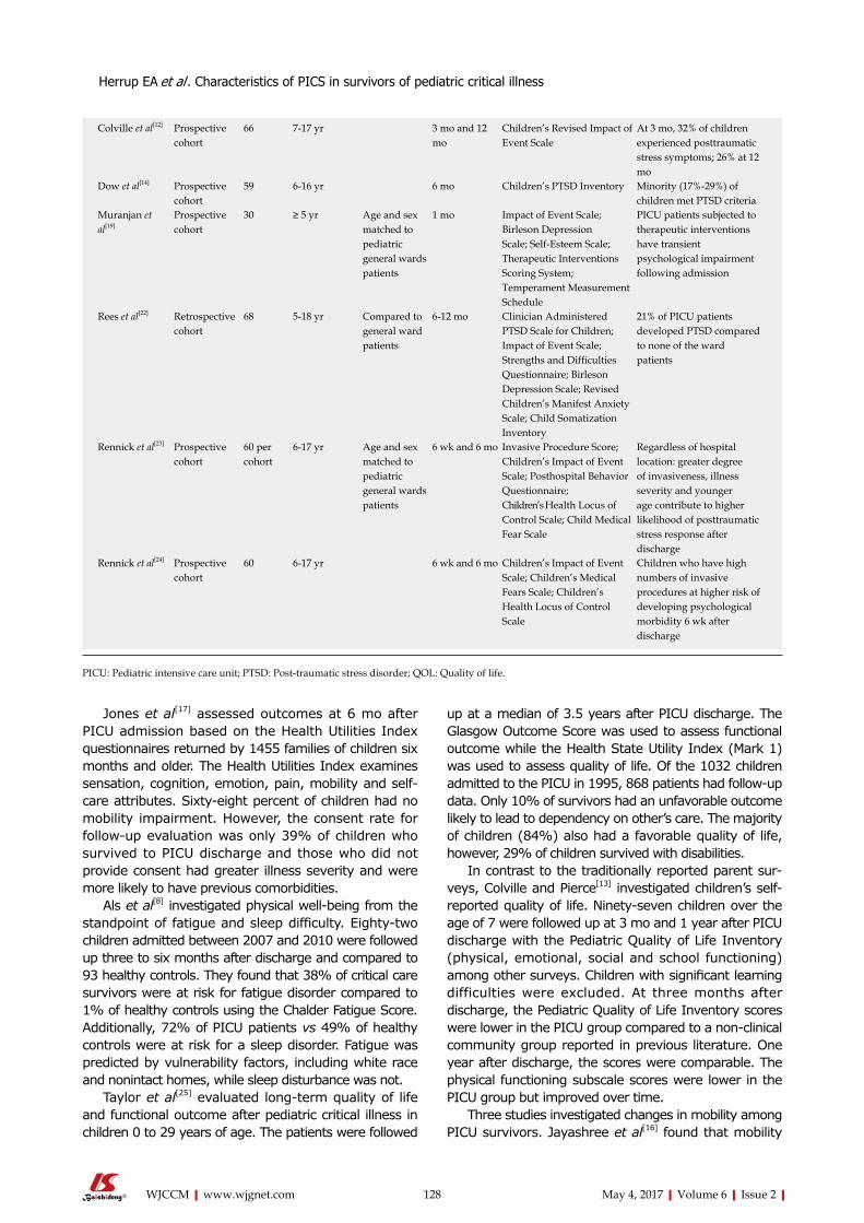
128 May 4, 2017|Volume 6|Issue 2|WJCCM|www.wjgnet.com
Jones et al[17] assessed outcomes at 6 mo after PICU admission based on the Health Utilities Index questionnaires returned by 1455 families of children six months and older. The Health Utilities Index examines sensation, cognition, emotion, pain, mobility and selfcare attributes. Sixtyeight percent of children had no mobility impairment. However, the consent rate for followup evaluation was only 39% of children who survived to PICU discharge and those who did not provide consent had greater illness severity and were more likely to have previous comorbidities.
Als et al[8] investigated physical wellbeing from the standpoint of fatigue and sleep difficulty. Eightytwo children admitted between 2007 and 2010 were followed up three to six months after discharge and compared to 93 healthy controls. They found that 38% of critical care survivors were at risk for fatigue disorder compared to 1% of healthy controls using the Chalder Fatigue Score. Additionally, 72% of PICU patients vs 49% of healthy controls were at risk for a sleep disorder. Fatigue was predicted by vulnerability factors, including white race and nonintact homes, while sleep disturbance was not.
Taylor et al[25] evaluated longterm quality of life and functional outcome after pediatric critical illness in children 0 to 29 years of age. The patients were followed
up at a median of 3.5 years after PICU discharge. The Glasgow Outcome Score was used to assess functional outcome while the Health State Utility Index (Mark 1) was used to assess quality of life. Of the 1032 children admitted to the PICU in 1995, 868 patients had followup data. Only 10% of survivors had an unfavorable outcome likely to lead to dependency on other’s care. The majority of children (84%) also had a favorable quality of life, however, 29% of children survived with disabilities.
In contrast to the traditionally reported parent surveys, Colville and Pierce[13] investigated children’s selfreported quality of life. Ninetyseven children over the age of 7 were followed up at 3 mo and 1 year after PICU discharge with the Pediatric Quality of Life Inventory (physical, emotional, social and school functioning) among other surveys. Children with significant learning difficulties were excluded. At three months after discharge, the Pediatric Quality of Life Inventory scores were lower in the PICU group compared to a nonclinical community group reported in previous literature. One year after discharge, the scores were comparable. The physical functioning subscale scores were lower in the PICU group but improved over time.
Three studies investigated changes in mobility among PICU survivors. Jayashree et al[16] found that mobility
Colville et al[12] Prospective cohort
66 7-17 yr 3 mo and 12 mo
Children’s Revised Impact of Event Scale
At 3 mo, 32% of children experienced posttraumatic stress symptoms; 26% at 12 mo
Dow et al[14] Prospective cohort
59 6-16 yr 6 mo Children’s PTSD Inventory Minority (17%-29%) of children met PTSD criteria
Muranjan et al[19]
Prospective cohort
30 ≥ 5 yr Age and sex matched to pediatric general wards patients
1 mo Impact of Event Scale; Birleson Depression Scale; Self-Esteem Scale; Therapeutic Interventions Scoring System; Temperament Measurement Schedule
PICU patients subjected to therapeutic interventions have transient psychological impairment following admission
Rees et al[22] Retrospective cohort
68 5-18 yr Compared to general ward patients
6-12 mo Clinician Administered PTSD Scale for Children; Impact of Event Scale; Strengths and Difficulties Questionnaire; Birleson Depression Scale; Revised Children’s Manifest Anxiety Scale; Child Somatization Inventory
21% of PICU patients developed PTSD compared to none of the ward patients
Rennick et al[23] Prospective cohort
60 per cohort
6-17 yr Age and sex matched to pediatric general wards patients
6 wk and 6 mo Invasive Procedure Score; Children’s Impact of Event Scale; Posthospital Behavior Questionnaire; Children’s Health Locus of Control Scale; Child Medical Fear Scale
Regardless of hospital location: greater degree of invasiveness, illness severity and younger age contribute to higher likelihood of posttraumatic stress response after discharge
Rennick et al[24] Prospective cohort
60 6-17 yr 6 wk and 6 mo Children’s Impact of Event Scale; Children’s Medical Fears Scale; Children’s Health Locus of Control Scale
Children who have high numbers of invasive procedures at higher risk of developing psychological morbidity 6 wk after discharge
PICU: Pediatric intensive care unit; PTSD: Post-traumatic stress disorder; QOL: Quality of life.
Herrup EA et al . Characteristics of PICS in survivors of pediatric critical illness

129 May 4, 2017|Volume 6|Issue 2|WJCCM|www.wjgnet.com
was affected in 49% of 150 critically ill children prior to admission while at 1 year after discharge 89% of these children had improving or unchanged mobility. Knoester et al[18] reported similar improvement in mobility when studying previously healthy children admitted unexpectedly to a PICU who were then followed up three and nine months after discharge. In the six to twelve year cohort, parents reported worse motor function at three months but not at nine months; adolescents also selfreported worse motor functioning at three months only. Parents and children who completed questionnaires at both time points demonstrated no difference in overall health related quality of life over time. Gemke et al[15] reported healthrelated quality of life of 226 patients, excluding infants, one year after PICU admission. Preadmission health status was estimated based on a questionnaire completed by parents during an interview within 48 h of admission. The multiattribute health status classification (MAHSC) was used as a generic health status measure. At twelve months after discharge, only 10% of patients had deterioration in mobility.
Several other studies evaluating quality of life using various validated measures but only provide an assessment of overall quality of life without results from each category examined (e.g., physical function)[6,2628].
Neurocognitive morbidityTwo of the included studies described neurocognitive morbidities in PICU patients. In a study of the same PICU cohort and healthy controls described under physical and functional morbidities[8], Als et al[7] evaluated neuropsychological function and academic performance. The children admitted to the PICU were placed into three subgroups: Meningoencephalitis, sepsis, and those with nonneurologic and nonseptic critical illness. The neuropsychologic battery was designed to focus on primarily intellectual function, memory and attention. Both the PICU cohort and the healthy controls had average IQs; however, the PICU cohort scored significantly lower on measures of verbal and visual recall and recognition memory, spatial working memory and working memory capacity, and visual sustained attention. Younger age, lower socioeconomic status and the meningoencephalitis subgroup were significantly associated with more widespread impairment. The academic questionnaire completed by teachers of PICU patients and healthy controls revealed more PICU children with deteriorating academic performance, with the sepsis group showing greatest deterioration, most difficulty with schoolwork completion and attention problems. Interestingly, the nonneurologic/nonseptic critical care cohort did not show any difference in school performance compared to healthy controls.
Finally, in a subsequent prospective followup study by Als et al[9], the 23 of 47 children who were identified as neuropsychologically impaired at three to six months following PICU admission were then evaluated 12 mo after PICU discharge in order to better understand
the persistence of these impairments. At the three to six month followup vs twelve month followup, there was no significant change in IQ scores; however, there was improvement in some verbal and visual memory domains. The questionnaires completed by school teachers did not show significant change in children’s global educational progress or learning skills.
Psychological morbidityNine studies focused on psychological morbidity in PICU survivors. Most recently, Dow et al[14] explored the prevalence and presentation of posttraumatic stress in 59 schoolaged children and adolescents six months after PICU admission lasting at least eight hours. Posttraumatic stress disorder (PTSD) was evaluated using the Children’s PTSD Inventory during a clinician administered interview. Depending on the criteria and algorithm used, the prevalence of PTSD found was 17%29%. All patients reported the following symptoms most frequently: Inability to recall aspects of the event, hypervigilance, avoidance of thoughts or feelings, physiological reactivity to trauma reminders, and intrusive thoughts or pictures. Schoolaged children more frequently endorsed avoidance of thoughts and feelings while adolescents more frequently endorsed difficulty concentrating.
Colville and Pierce examined rates and natural history of posttraumatic stress symptoms in families after a pediatric critical care admission, including 66 children at both three and twelve months after PICU discharge[12]. While the study evaluated childparent pairs, the data for children was reported separately. The Children’s Revised Impact of Event Scale (CRIES8), a brief, validated scale which detects posttraumatic symptoms and PTSD was chosen as the questionnaire for children. At the three and twelvemonth time points, 32% and 26% of children demonstrated symptoms of posttraumatic stress respectively, with no significant change in median score. However, seven children who did not initially score positive at three months after discharge scored positive at twelve months. Of note, those children who scored positive for PTSD at twelve months after discharge had higher scores at three months as well as higher Pediatric Index of Mortality (PIM) scores on admission. The authors suggested that the subjective experience of a child influences likelihood of future distress more so than objective experience (e.g., demographic or medical variables). Participants in the study were also asked what the “worst thing” was during PICU admission. Many of the children pointed to one specific distressing event in the PICU (e.g., hallucinations, waking up and not knowing where their parents were). Interestingly, 35% of children stated that the worst experience occurred outside of the PICU (e.g., the inciting incident that led to admission or deterioration prior to admission).
Four years earlier, Colville et al[11] followed 102 children ages 7 to 17 at a median of 3 mo after PICU discharge to determine if delusional memories from PICU admission were associated with posttraumatic stress symptoms.
Herrup EA et al . Characteristics of PICS in survivors of pediatric critical illness

130 May 4, 2017|Volume 6|Issue 2|WJCCM|www.wjgnet.com
Children’s memories were characterized using the ICU Memory Tool and the Children’s Revised Impact of Event Scale was used to screen for posttraumatic symptoms. Thirtytwo percent of the children had delusional memories, and only two patients reported these memories occurring outside of the time of their PICU stay. A longer course of opiates and benzodiazepines was associated with delusional memories, and children with delusional memories had higher posttraumatic stress scores. There was not, however, a significant association between factual memory and posttraumatic stress symptoms.
A number of the included studies focused on risk factors for psychological morbidity after PICU admission. First, Rennick et al[23] conducted a study in 2002 comparing 120 children six weeks and six months after hospital discharge admitted to either a PICU or a general pediatric ward. Several scales were used to assess the presence and degree of psychological impairment. Regardless of admission location, they found that children with a younger age, greater illness severity, and those subjected to a greater number of invasive procedures had ongoing posttraumatic symptoms, lower sense of control over health, and more medical fears six weeks after hospital discharge. At six months after discharge, only the children who were exposed to higher numbers of invasive procedures had significantly more medical fears and symptoms of posttraumatic stress.
As a follow up to this study, Rennick et al[24] investigated if there was a group at higher risk for developing psychological morbidity after PICU discharge. PICU patients were categorized as high risk (vs low risk) for developing psychological morbidity if they had higher severity of illness and higher number of invasive procedures. Similar to findings in the previous study, it was found that the children with high numbers of invasive procedures were at higher risk of developing psychological problems 6 wk after discharge. At 6 mo after discharge, the high risk group had a significantly higher prevalence of posttraumatic stress symptoms.
In another study of risk factors for psychological morbidity, Muranjan et al[19] enrolled 30 medical patients who survived a PICU admission and compared them to a control group of 30 children admitted to a general pediatric ward. Children were followed up one month after discharge to evaluate psychological symptoms. This study found that children admitted to the PICU and general wards had comparable premorbid temperament, as well as selfesteem scale and depression scores. Children admitted to the PICU initially reported more intrusive thoughts compared to the general ward cohort (43% vs 77%, respectively) within the first 24 h after discharge but scores were comparable one month after discharge, suggesting posttraumatic symptoms were short lived. While development of intrusive thoughts correlated with degree of intervention, demographic factors, underlying disease, severity of illness and length of hospitalization did not correlate with degree of psychological morbidity.
In a retrospective cohort, Rees et al[22] aimed to
determine if patients discharged from a PICU had greater psychological morbidity compared to children discharged from a general ward. Children ages five and older were included and followed up 6 to 12 mo after discharge. Children with meningococcal disease, terminal illness, an underlying neurologic disorder, or intentional overdose were excluded. Of patients admitted to the PICU, 21% met diagnostic criteria for PTSD with symptoms of irritability and persistent avoidance of reminders of their admission being most prominent; however, only one (vs four) PICU patients was diagnosed with PTSD at the interview. None of the pediatric ward patients developed PTSD, and there was no significant difference between the two groups in scores relating to anxiety, depression or somatization.
Bronner et al[10] compared the development of PTSD in pediatric critical care survivors to children who survived a major fire disaster. This group investigated the presence of PTSD in a cohort of 36 previously healthy children admitted unexpectedly to the PICU three and/or nine months after discharge. Children completed the Dutch Children’s Responses to Trauma Inventory, which identified 34% with at least subclinical PTSD at 3 mo with 14% of those children likely to meet criteria for PTSD. At nine month followup, 36% of children had at least subclinical PTSD with 18% of those children likely to meet criteria for PTSD. Only two children had a change in score between the three month and nine month followup time points (one going from normal to subclinical, the other subclinical to normal). Compared to survivors of a major fire disaster in the Netherlands, the investigators found that the PTSD scores of children in both groups were comparable. Of note, the PICU population studied in this paper were at risk for possible brain damage, perhaps falsely elevating the number of children who fell into the categories of subclinical PTSD or likely to meet criteria for PTSD due to overlap in symptoms.
Finally, in the same article by Als et al[8] discussing physical morbidity after PICU admission, presence of mental health morbidity was also assessed. A questionnaire was mailed to the families who agreed to participate. Thirtyfour percent of children scored at risk for PTSD. In addition, the PICU group scored significantly worse than the control group on the sections of emotional symptoms and hyperactivity. Moreover, twenty percent of the PICU group scored at high risk for a psychological disorder, compared to 9% in the healthy control group. Septic illness was an independent predictor of posttraumatic stress symptoms.
DISCUSSIONThis systematic review summarizes the available literature from nineteen studies investigating outcomes in survivors of pediatric critical illness within the context of PICS. The synthesis revealed that, similar to adult ICU patients, a wide range of physical, neurocognitive and psychological morbidities occur in PICU patients after discharge[29]. The populations investigated encompass
Herrup EA et al . Characteristics of PICS in survivors of pediatric critical illness
131 May 4, 2017|Volume 6|Issue 2|WJCCM|www.wjgnet.com
a heterogeneous group of subjects with regard to age, type of illness, illness severity, length of hospital stay, and medical interventions. Many, but not all, studies excluded patients with traumatic brain injury or baseline neurocognitive dysfunction. Children were also followed up after hospitalization at different time points, including weeks to months to years later. A major source of heterogeneity was the outcome measures and assessment tools utilized across each category of PICS morbidities. However, this synthesis identifies a critical need to continue to define the landscape of pediatric PICS and guide future research directions by characterizing the types and extent of morbidities that exist in survivors of pediatric critical illness.
Patients and families are substantially impacted by critical illness, and the changing spectrum of critical illness, technological advances, and improved survival has translated to increased morbidity in the pediatric population[1,30]. The sequelae of PICS can lead to reduced quality of life and functional impairments, in addition to increased societal costs[3032]. Major differences between children and adults with regard to the implications of PICS include increased years of survival with morbidity after critical illness and the complex nature of interactions between PICS and the developing brain.
Physical and functional morbidity In several studies investigating physical and functional morbidity in pediatric critical illness survivors, information was gleaned from quality of life surveys that included specific information about physical functioning after PICU admission[13,1518,21,25]. Multiple screening tools were used to determine the presence or absence of morbidity. Some studies used one follow up time point, which could be within a range of times after PICU discharge, while others used multiple follow up time points. Studies indicated that the minority of survivors of pediatric critical illness have physical morbidities, and if they are present after hospital discharge they decrease over time. For example, one study reporting health related quality of life found that only about 10% of patients (excluding infants) had a deterioration in mobility 1 year after PICU admission, compared to “preadmission” health status[15]. However, another study comparing PICU populations in 1982 and 2006 did find that a considerably greater percentage of patients had moderate to severe disabilities in 2006 (8% vs 18%)[20]. This is not a surprising finding as more children survive critical illness with improvements in medical care and technology. However, this trend highlights an increased need for early mobilization and acute rehabilitation initiatives in the PICU to decrease the risk of ICUacquired weakness and potentially increase functional outcomes after discharge[30,3335].
Neurocognitive morbidityOf the three broad types of morbidity included in the definition of PICS, this review yielded the fewest number of articles related to neurocognitive morbidity
after pediatric critical illness. The same team of authors published two papers assessing the neurocognitive function of schoolaged children at 3 to 6 mo and 12 mo after critical illness[7,9]. These patients were compared to healthy controls. While both groups of children had average IQs at the 3 to 6 mo time point, it is important to note that those who survived critical illness had consistently lower scores in verbal and visual domains, particularly in patients who had meningoencephalitis. Moreover, risk factors for greater neurocognitive impairment at time of initial follow up included those of younger age and with lower socioeconomic status. At 12 mo, there was some improvement in verbal and visual domain scores but no significant change in global education progress, suggesting that neurocognitive impairment in certain domains may improve over time, or at least not become worse. Regardless, it is important to consider which children may have more difficulty in school and with certain types of daily tasks after they leave the PICU, whether based on demographics or type of illness, so that they can receive additional educational support once they have returned to school. Largescale studies are needed and ongoing to better characterize the short and longterm neurocognitive morbidity associated with pediatric critical illness in the developing brain[36].
Psychological morbidityAlthough the evidence is limited, psychological morbidity has perhaps been the aspect of PICS that has received the most attention after PICU discharge[8,1012,14,19,2224]. All studies included in our review evaluated schoolaged patients for posttraumatic stress symptoms, which may be related to “post traumatic stress” being included as a specific term in the search strategy encompassing psychiatric morbidity after PICU admission. However, multiple screening tools were utilized to determine the presence or absence of symptoms. Follow up time points ranged from 6 wk to 12 mo after critical illness. The synthesis of studies suggests that psychological morbidity including posttraumatic stress occurs in onethird of PICU survivors. Of importance, several studies investigating two time points found that posttraumatic stress symptoms seem to attenuate over time.
Several studies also aimed to investigate potential risk factors for developing posttraumatic stress symptoms[8,11,19,23,24]. Septic illness was an independent predictor of posttraumatic stress in one study[8], while three others found that increased numbers of invasive procedures or interventions increased the risk of psychological morbidity[19,23,24]. Delusional memories were associated with increased benzodiazepine or narcotic administration during a PICU course[11]. Benzodiazepines are an independent risk factor for delirium in the adult ICU patient, and emerging evidence in pediatrics suggests a similar association[37,38]. With validated screening tools available to diagnose delirium in the PICU, PICU providers are obligated to address risk factors and treat
Herrup EA et al . Characteristics of PICS in survivors of pediatric critical illness
132 May 4, 2017|Volume 6|Issue 2|WJCCM|www.wjgnet.com
delirium using nonpharmacologic and pharmacologic therapies in consultation with child psychiatry[3946]. Sleep disturbances including night terrors are not an uncommon sequelae after ICU discharge in both pediatric and adult patients[8,41,4749]. An improved understanding of risk factors for psychological morbidity is crucial to facilitate anticipatory guidance for families after a child is discharged from the PICU.
Challenges in PICS researchA major barrier to PICS research that is obvious but challenging to resolve is that it is difficult to establish an accurate preadmission baseline for children admitted to the PICU. Thus, studies that attempt to compare premorbid or preadmission conditions of pediatric patients rely upon either parent and/or child memory of a patient’s functioning prior to admission. Families are likely in a significant state of stress when asked to recount their children’s preadmission status within the 48 h of admission. Many of the studies in this review used control groups, such as normal pediatric population controls or pediatric ward patients, in order to understand how the trajectory of survivors of pediatric critical illness compared. However, it would be ideal to understand a patient’s trajectory starting before PICU admission, in the PICU, and after discharge from the PICU in order to best understand if morbidities that develop after a PICU admission are persistent and to what degree compared to preillness condition.
Another impediment is that many of the measurement tools used to assess quality of life, physical, neurocognitive or psychological morbidity rely primarily on parental assessment. This is necessary in many circumstances when a patient’s developmental level does not allow for him or her to understand or answer questions, but parents were also asked to answer questions or fill out surveys in many circumstances when a child could have done the same. A parent’s response may not accurately represent the thoughts, feelings and experiences of the child. For example, one study found that the agreement between parent and child report is poor[50]. In the future, an effort should be made to include child report when possible.
Finally, none of the studies reviewed discuss the impact of delirium on morbidity experienced by survivors of pediatric critical illness. Delirium is an independent predictor of increased hospital length of stay, prolonged mechanical ventilation, longterm cognitive impairments, and PTSD in adults[5154]. There is relatively little data on morbidity associated with delirium in the pediatric critically ill population[55]. Case reports and point prevalence studies suggest rates of pediatric delirium between 10% and 30%[38,43,45,56]. As the concept of pediatric delirium becomes better recognized and studied, it will be interesting to understand the interplay between critically ill children diagnosed with delirium and its effect on longterm morbidities.
Future directionsThis systematic review has identified the wide range
of morbidities that fall within the construct of PICS in PICU patients. An important next step will be to closely examine what is already known about these physical, neurocognitive and psychological morbidities to define pediatric PICS and come to a consensus on the most relevant and generalizable outcome measures. Ultimately, the aim is to develop interventions both in the PICU and after discharge to help mitigate potential ongoing impairment resulting from critical illness and medical interventions. Many PICUs have already started to address ways to minimize morbidity such as promotion of early mobilization, improved sedation plans, delirium screening and even ICU diaries[57] in an effort to blunt the physically and mentally traumatic effects of critical illness. However, it is important for pediatric intensivists to first acknowledge the morbidity that persists beyond PICU discharge and agree upon consistent language and outcome measures so that effective and targeted interventions will become integrated into part of daily care starting in the PICU.
This systematic review evaluates the presence of PICS in pediatric survivors of critical illness. Physical, neurocognitive, and psychological morbidity affects a significant minority of this vulnerable population. These findings will likely help focus future research efforts on this topic in order to gain a better understanding of which children are most susceptible to developing PICS and of what can be done to mitigate these morbidities.
ACKNOWLEDGMENTSThe authors would like to thank Ms. Carrie Price for guidance with the literature search strategy.
COMMENTSBackgroundIn 2010, an adult consortium developed the concept of postintensive care syndrome (PICS), which encompasses physical, cognitive and mental health morbidities that exist in a high percentage of adults after critical illness. The aim of this current review was to summarize the relevant pediatric literature on these types of morbidities in a general pediatric intensive care unit (PICU) population and understand how widespread this recently described syndrome is among pediatric survivors of critical illness.
Research frontiersMortality is a rare event in pediatric critical illness, however, it has often been used as an outcome measure in research studies. More appropriate outcome measures include different types of morbidity. It is important to understand what kinds of morbidities burden children after critical illness and how a multidisciplinary team both in the PICU and in follow up settings can mitigate morbidities that develop during and persist after critical illness.
Innovations and breakthroughsPrevious studies have shown different types of morbidity that exist in survivors of pediatric critical illness but there has been no review of pediatric literature through the lens of PICS.
ApplicationChildren who survive pediatric critical illness can develop physical, neurocognitive and/or psychological morbidity after hospital discharge. Knowing this, the authors can better define the extent and natural history of these morbidities
COMMENTS
Herrup EA et al . Characteristics of PICS in survivors of pediatric critical illness

133 May 4, 2017|Volume 6|Issue 2|WJCCM|www.wjgnet.com
and take measures to mitigate morbidity that results from critical illness and its treatment.
TerminologyPICS: Postintensive care syndrome; PICU: Pediatric intensive care unit.
Peer-reviewHerrup et al present a nice and informative overview. The work is well written, based on a systematic literature review, and informative.
REFERENCES1 Pollack MM, Holubkov R, Funai T, Clark A, Berger JT, Meert K,
Newth CJ, Shanley T, Moler F, Carcillo J, Berg RA, Dalton H, Wessel DL, Harrison RE, Doctor A, Dean JM, Jenkins TL. Pediatric intensive care outcomes: development of new morbidities during pediatric critical care. Pediatr Crit Care Med 2014; 15: 821827 [PMID: 25226501 DOI: 10.1097/PCC.0000000000000250]
2 Needham DM, Davidson J, Cohen H, Hopkins RO, Weinert C, Wunsch H, Zawistowski C, BemisDougherty A, Berney SC, Bienvenu OJ, Brady SL, Brodsky MB, Denehy L, Elliott D, Flatley C, Harabin AL, Jones C, Louis D, Meltzer W, Muldoon SR, Palmer JB, Perme C, Robinson M, Schmidt DM, Scruth E, Spill GR, Storey CP, Render M, Votto J, Harvey MA. Improving longterm outcomes after discharge from intensive care unit: report from a stakeholders’ conference. Crit Care Med 2012; 40: 502509 [PMID: 21946660 DOI: 10.1097/CCM.0b013e318232da75]
3 van der Schaaf M, Beelen A, Dongelmans DA, Vroom MB, Nollet F. Functional status after intensive care: a challenge for rehabilitation professionals to improve outcome. J Rehabil Med 2009; 41: 360366 [PMID: 19363570 DOI: 10.2340/165019770333]
4 Rothenhäusler HB, Ehrentraut S, Stoll C, Schelling G, Kapfhammer HP. The relationship between cognitive performance and employment and health status in longterm survivors of the acute respiratory distress syndrome: results of an exploratory study. Gen Hosp Psychiatry 2001; 23: 9096 [PMID: 11313077 DOI: 10.1016/S01638343(01)001232]
5 Davydow DS, Gifford JM, Desai SV, Bienvenu OJ, Needham DM. Depression in general intensive care unit survivors: a systematic review. Intensive Care Med 2009; 35: 796809 [PMID: 19165464 DOI: 10.1007/s0013400913965]
6 Polic B, Mestrovic J, Markic J, Mestrovic M, Capkun V, Utrobicic I, Jukica M, Radonic M. Longterm quality of life of patients treated in paediatric intensive care unit. Eur J Pediatr 2013; 172: 8590 [PMID: 23052616 DOI: 10.1007/s0043101218430]
7 Als LC, Nadel S, Cooper M, Pierce CM, Sahakian BJ, Garralda ME. Neuropsychologic function three to six months following admission to the PICU with meningoencephalitis, sepsis, and other disorders: a prospective study of schoolaged children. Crit Care Med 2013; 41: 10941103 [PMID: 23385103 DOI: 10.1097/CCM.0b013e318275d032]
8 Als LC, Picouto MD, Hau SM, Nadel S, Cooper M, Pierce CM, Kramer T, Garralda ME. Mental and physical wellbeing following admission to pediatric intensive care. Pediatr Crit Care Med 2015; 16: e141e149 [PMID: 25901544 DOI: 10.1097/PCC.0000000000000424]
9 Als LC, Tennant A, Nadel S, Cooper M, Pierce CM, Garralda ME. Persistence of Neuropsychological Deficits Following Pediatric Critical Illness. Crit Care Med 2015; 43: e312e315 [PMID: 25962081 DOI: 10.1097/CCM.0000000000001075]
10 Bronner MB, Knoester H, Bos AP, Last BF, Grootenhuis MA. Posttraumatic stress disorder (PTSD) in children after paediatric intensive care treatment compared to children who survived a major fire disaster. Child Adolesc Psychiatry Ment Health 2008; 2: 9 [PMID: 18489798 DOI: 10.1186/1753200029]
11 Colville G, Kerry S, Pierce C. Children’s factual and delusional memories of intensive care. Am J Respir Crit Care Med 2008; 177: 976982 [PMID: 18244955 DOI: 10.1164/rccm.200706857OC]
12 Colville G, Pierce C. Patterns of posttraumatic stress symptoms in families after paediatric intensive care. Intensive Care Med 2012; 38: 15231531 [PMID: 22706918 DOI: 10.1007/s0013401226122]
13 Colville GA, Pierce CM. Children’s selfreported quality of life after intensive care treatment. Pediatr Crit Care Med 2013; 14: e85e92 [PMID: 23337805 DOI: 10.1097/PCC.0b013e3182712997]
14 Dow BL, Kenardy JA, Le Brocque RM, Long DA. The diagnosis of posttraumatic stress disorder in schoolaged children and adolescents following pediatric intensive care unit admission. J Child Adolesc Psychopharmacol 2013; 23: 614619 [PMID: 24251643 DOI: 10.1089/cap.2013.0044]
15 Gemke RJ, Bonsel GJ, van Vught AJ. Longterm survival and state of health after paediatric intensive care. Arch Dis Child 1995; 73: 196201 [PMID: 7492154 DOI: 10.1136/adc.73.3.196]
16 Jayshree M, Singhi SC, Malhi P. Follow up of survival and quality of life in children after intensive care. Indian Pediatr 2003; 40: 303309 [PMID: 12736401]
17 Jones S, Rantell K, Stevens K, Colwell B, Ratcliffe JR, Holland P, Rowan K, Parry GJ. Outcome at 6 months after admission for pediatric intensive care: a report of a national study of pediatric intensive care units in the United kingdom. Pediatrics 2006; 118: 21012108 [PMID: 17079584 DOI: 10.1542/peds.20061455]
18 Knoester H, Bronner MB, Bos AP, Grootenhuis MA. Quality of life in children three and nine months after discharge from a paediatric intensive care unit: a prospective cohort study. Health Qual Life Outcomes 2008; 6: 21 [PMID: 18331652 DOI: 10.1186/ 14777525621]
19 Muranjan MN, Birajdar SB, Shah HR, Sundaraman P, Tullu MS. Psychological consequences in pediatric intensive care unit survivors: the neglected outcome. Indian Pediatr 2008; 45: 99103 [PMID: 18310787]
20 Namachivayam P, Shann F, Shekerdemian L, Taylor A, van Sloten I, Delzoppo C, Daffey C, Butt W. Three decades of pediatric intensive care: Who was admitted, what happened in intensive care, and what happened afterward. Pediatr Crit Care Med 2010; 11: 549555 [PMID: 20124947 DOI: 10.1097/PCC.0b013e3181ce7427]
21 Namachivayam P, Taylor A, Montague T, Moran K, Barrie J, Delzoppo C, Butt W. Longstay children in intensive care: longterm functional outcome and quality of life from a 20yr institutional study. Pediatr Crit Care Med 2012; 13: 520528 [PMID: 22805156 DOI: 10.1097/PCC.0b013e31824fb989]
22 Rees G, Gledhill J, Garralda ME, Nadel S. Psychiatric outcome following paediatric intensive care unit (PICU) admission: a cohort study. Intensive Care Med 2004; 30: 16071614 [PMID: 15112035 DOI: 10.1007/s0013400423109]
23 Rennick JE, Johnston CC, Dougherty G, Platt R, Ritchie JA. Children’s psychological responses after critical illness and exposure to invasive technology. J Dev Behav Pediatr 2002; 23: 133144 [PMID: 12055495 DOI: 10.1097/0000470320020600000002]
24 Rennick JE, Morin I, Kim D, Johnston CC, Dougherty G, Platt R. Identifying children at high risk for psychological sequelae after pediatric intensive care unit hospitalization. Pediatr Crit Care Med 2004; 5: 358363 [PMID: 15215006 DOI: 10.1097/01.PCC.0000128603.20501.0D]
25 Taylor A, Butt W, Ciardulli M. The functional outcome and quality of life of children after admission to an intensive care unit. Intensive Care Med 2003; 29: 795800 [PMID: 12595983 DOI: 10.1007/s0013400316906]
26 Ambuehl J, Karrer A, Meer A, Riedel T, Schibler A. Quality of life of survivors of paediatric intensive care. Swiss Med Wkly 2007; 137: 312316 [PMID: 17629809]
27 Mestrovic J, Kardum G, Sustic A, Polic B, Mestrovic M, Markic J, Zanchi J. Neurodevelopmental disabilities and quality of life after intensive care treatment. J Paediatr Child Health 2007; 43: 673676 [PMID: 17854451 DOI: 10.1111/j.14401754.2007.01185.x]
28 Morrison AL, Gillis J, O’Connell AJ, Schell DN, Dossetor DR, Mellis C. Quality of life of survivors of pediatric intensive care. Pediatr Crit Care Med 2002; 3: 15 [PMID: 12793913 DOI: 10.1097/0013047820020100000001]
29 Hashem MD, Nallagangula A, Nalamalapu S, Nunna K, Nausran U, Robinson KA, Dinglas VD, Needham DM, Eakin MN. Patient outcomes after critical illness: a systematic review of qualitative studies following hospital discharge. Crit Care 2016; 20: 345 [PMID: 27782830 DOI: 10.1186/s130540161516x]
30 Hopkins RO, Choong K, Zebuhr CA, Kudchadkar SR. Transforming PICU Culture to Facilitate Early Rehabilitation. J Pediatr
Herrup EA et al . Characteristics of PICS in survivors of pediatric critical illness

134 May 4, 2017|Volume 6|Issue 2|WJCCM|www.wjgnet.com
Intensive Care 2015; 4: 204211 [PMID: 27134761 DOI: 10.1055/s00351563547]
31 Iwashyna TJ, Cooke CR, Wunsch H, Kahn JM. Population burden of longterm survivorship after severe sepsis in older Americans. J Am Geriatr Soc 2012; 60: 10701077 [PMID: 22642542 DOI: 10.1111/j.15325415.2012.03989.x]
32 Cheung AM, Tansey CM, Tomlinson G, DiazGranados N, Matté A, Barr A, Mehta S, Mazer CD, Guest CB, Stewart TE, AlSaidi F, Cooper AB, Cook D, Slutsky AS, Herridge MS. Twoyear outcomes, health care use, and costs of survivors of acute respiratory distress syndrome. Am J Respir Crit Care Med 2006; 174: 538544 [PMID: 16763220 DOI: 10.1164/rccm.200505693OC]
33 Wieczorek B, Ascenzi J, Kim Y, Lenker H, Potter C, Shata NJ, Mitchell L, Haut C, Berkowitz I, Pidcock F, Hoch J, Malamed C, Kravitz T, Kudchadkar SR. PICU Up!: Impact of a Quality Improvement Intervention to Promote Early Mobilization in Critically Ill Children. Pediatr Crit Care Med 2016; 17: e559e566 [PMID: 27759596 DOI: 10.1097/PCC.0000000000000983]
34 Wieczorek B, Burke C, AlHarbi A, Kudchadkar SR. Early mobilization in the pediatric intensive care unit: a systematic review. J Pediatr Intensive Care 2015; 2015: 129170 [PMID: 26380147 DOI: 10.1055/s00351563386]
35 Choong K, Foster G, Fraser DD, Hutchison JS, Joffe AR, Jouvet PA, Menon K, Pullenayegum E, Ward RE. Acute rehabilitation practices in critically ill children: a multicenter study. Pediatr Crit Care Med 2014; 15: e270e279 [PMID: 24777303 DOI: 10.1097/01.pcc.0000448748.47635.b2]
36 Curley MA, Watson RS. Sedation Strategy and Cognitive Outcome After Critical Illness in Early Childhood. In: ClinicalTrials.gov [Internet]. Bethesda (MD): National Library of Medicine (US). 2000 [cited 2016 Oct 28]. Available from: URL: http://clinicaltrials.gov/show/NCT02225041
37 Pandharipande P, Cotton BA, Shintani A, Thompson J, Pun BT, Morris JA, Dittus R, Ely EW. Prevalence and risk factors for development of delirium in surgical and trauma intensive care unit patients. J Trauma 2008; 65: 3441 [PMID: 18580517 DOI: 10.1097/TA.0b013e31814b2c4d]
38 Traube C, Silver G, Reeder RW, Doyle H, Hegel E, Wolfe HA, Schneller C, Chung MG, Dervan LA, DiGennaro JL, Buttram SD, Kudchadkar SR, Madden K, Hartman ME, deAlmeida ML, Walson K, Ista E, Baarslag MA, Salonia R, Beca J, Long D, Kawai Y, Cheifetz IM, Gelvez J, Truemper EJ, Smith RL, Peters ME, O’Meara AM, Murphy S, Bokhary A, Greenwald BM, Bell MJ. Delirium in Critically Ill Children: An International Point Prevalence Study. Crit Care Med 2017; Epub ahead of print [PMID: 28079605 DOI: 10.1097/CCM.0000000000002250]
39 Clinical practice guidelines for the sustained use of sedatives and analgesics in the critically ill adult. Am J Health Syst Pharm 2002; 59: 150178 [PMID: 11826570]
40 Creten C, Van Der Zwaan S, Blankespoor RJ, Leroy PL, Schieveld JN. Pediatric delirium in the pediatric intensive care unit: a systematic review and an update on key issues and research questions. Minerva Anestesiol 2011; 77: 10991107 [PMID: 21617602]
41 Kudchadkar SR, Aljohani OA, Punjabi NM. Sleep of critically ill children in the pediatric intensive care unit: a systematic review. Sleep Med Rev 2014; 18: 103110 [PMID: 23702219 DOI: 10.1016/j.smrv.2013.02.002]
42 Kudchadkar SR, Yaster M, Punjabi NM. Sedation, sleep promotion, and delirium screening practices in the care of mechanically ventilated children: a wakeup call for the pediatric critical care community*. Crit Care Med 2014; 42: 15921600 [PMID: 24717461 DOI: 10.1097/CCM.0000000000000326]
43 Smith HA, Boyd J, Fuchs DC, Melvin K, Berry P, Shintani A, Eden SK, Terrell MK, Boswell T, Wolfram K, Sopfe J, Barr FE, Pandharipande PP, Ely EW. Diagnosing delirium in critically ill children: Validity and reliability of the Pediatric Confusion Assessment Method for the Intensive Care Unit. Crit Care Med 2011; 39: 150157
[PMID: 20959783 DOI: 10.1097/CCM.0b013e3181feb489]44 Smith HA, Gangopadhyay M, Goben CM, Jacobowski NL,
Chestnut MH, Savage S, Rutherford MT, Denton D, Thompson JL, Chandrasekhar R, Acton M, Newman J, Noori HP, Terrell MK, Williams SR, Griffith K, Cooper TJ, Ely EW, Fuchs DC, Pandharipande PP. The Preschool Confusion Assessment Method for the ICU: Valid and Reliable Delirium Monitoring for Critically Ill Infants and Children. Crit Care Med 2016; 44: 592600 [PMID: 26565631 DOI: 10.1097/CCM.0000000000001428]
45 Silver G, Traube C, Kearney J, Kelly D, Yoon MJ, Nash Moyal W, Gangopadhyay M, Shao H, Ward MJ. Detecting pediatric delirium: development of a rapid observational assessment tool. Intensive Care Med 2012; 38: 10251031 [PMID: 22407142 DOI: 10.1007/s001340122518z]
46 Flaigle MC, Ascenzi J, Kudchadkar SR. Identifying Barriers to Delirium Screening and Prevention in the Pediatric ICU: Evaluation of PICU Staff Knowledge. J Pediatr Nurs 2015; 31: 8184 [PMID: 26362671 DOI: 10.1016/j.pedn.2015.07.009]
47 Parsons EC, Hough CL, Vitiello MV, Zatzick D, Davydow DS. Insomnia is associated with quality of life impairment in medicalsurgical intensive care unit survivors. Heart Lung 2015; 44: 8994 [PMID: 25592203 DOI: 10.1016/j.hrtlng.2014.11.002]
48 Parsons EC, Kross EK, Caldwell ES, Kapur VK, McCurry SM, Vitiello MV, Hough CL. Postdischarge insomnia symptoms are associated with quality of life impairment among survivors of acute lung injury. Sleep Med 2012; 13: 11061109 [PMID: 22763017 DOI: 10.1016/j.sleep.2012.05.010]
49 McKinley S, Fein M, Elliott RM, Elliot D. Sleep During And After Critical Illness And Psychological Health In Recovery. Australian Critical Care 2013; 26: 83 [DOI: 10.1016/j.aucc.2013.02.008]
50 Upton P, Lawford J, Eiser C. Parentchild agreement across child healthrelated quality of life instruments: a review of the literature. Qual Life Res 2008; 17: 895913 [PMID: 18521721 DOI: 10.1007/s1113600893505]
51 Ely EW, Gautam S, Margolin R, Francis J, May L, Speroff T, Truman B, Dittus R, Bernard R, Inouye SK. The impact of delirium in the intensive care unit on hospital length of stay. Intensive Care Med 2001; 27: 18921900 [PMID: 11797025 DOI: 10.1007/s0013400111322]
52 Hopkins RO, Jackson JC. Longterm neurocognitive function after critical illness. Chest 2006; 130: 869878 [PMID: 16963688 DOI: 10.1378/chest.130.3.869]
53 Jackson JC, Obremskey W, Bauer R, Greevy R, Cotton BA, Anderson V, Song Y, Ely EW. Longterm cognitive, emotional, and functional outcomes in trauma intensive care unit survivors without intracranial hemorrhage. J Trauma 2007; 62: 8088 [PMID: 17215737 DOI: 10.1097/TA.0b013e31802ce9bd]
54 Thomason JW, Shintani A, Peterson JF, Pun BT, Jackson JC, Ely EW. Intensive care unit delirium is an independent predictor of longer hospital stay: a prospective analysis of 261 nonventilated patients. Crit Care 2005; 9: R375R381 [PMID: 16137350 DOI: 10.1186/cc3729]
55 Schieveld JN, Leroy PL, van Os J, Nicolai J, Vos GD, Leentjens AF. Pediatric delirium in critical illness: phenomenology, clinical correlates and treatment response in 40 cases in the pediatric intensive care unit. Intensive Care Med 2007; 33: 10331040 [PMID: 17457571 DOI: 10.1007/s0013400706378]
56 Janssen NJ, Tan EY, Staal M, Janssen EP, Leroy PL, Lousberg R, van Os J, Schieveld JN. On the utility of diagnostic instruments for pediatric delirium in critical illness: an evaluation of the Pediatric Anesthesia Emergence Delirium Scale, the Delirium Rating Scale 88, and the Delirium Rating ScaleRevised R98. Intensive Care Med 2011; 37: 13311337 [PMID: 21567109 DOI: 10.1007/s001340112244y]
57 Knowles RE, Tarrier N. Evaluation of the effect of prospective patient diaries on emotional wellbeing in intensive care unit survivors: a randomized controlled trial. Crit Care Med 2009; 37: 184191 [PMID: 19050634 DOI: 10.1097/CCM.0b013e31819287f7]
P- Reviewer: Classen CF, Greco G, Sarin YK S- Editor: Ji FF L- Editor: A E- Editor: Lu YJ
Herrup EA et al . Characteristics of PICS in survivors of pediatric critical illness

Fabiel Gerardo Pérez-Cruz, Patricia Villa-Díaz, María Consuelo Pintado-Delgado, María Loreto Fernández_Rodríguez, Ana Blasco-Martínez, María Pérez-Fernández
CASE REPORT
135 May 4, 2017|Volume 6|Issue 2|WJCCM|www.wjgnet.com
Hemolytic uremic syndrome in adults: A case report
Fabiel Gerardo Pérez-Cruz, Internal Medicine Department, Hospital Universitario Príncipe de Asturias, Carretera Alcalá-Meco SN, 28805 Madrid, Spain
Patricia Villa-Díaz, María Consuelo Pintado-Delgado, Critical Care Unit, Hospital Universitario Príncipe de Asturias, Carretera Alcalá-Meco SN, 28805 Madrid, Spain
María Loreto Fernández_Rodríguez, María Pérez-Fernández, Nephrology Department, Hospital Universitario Príncipe de Asturias, Carretera Alcalá-Meco SN, 28805 Madrid, Spain
Ana Blasco-Martínez, Pathological Anatomy Department, Hospital Universitario Príncipe de Asturias, Carretera Alcalá-Meco SN, 28805 Madrid, Spain
Author contributions: Pérez-Cruz FG, Villa-Díaz P and Pintado-Delgado MC wrote the paper; Pérez-Cruz FG, Villa-Díaz P, Pintado-Delgado MC, Fernández_Rodríguez ML, Blasco-Martínez A and Pérez-Fernández M performed the research; Fernández_Rodríguez ML, Blasco-Martínez A and Pérez-Fernández M reviewed the current knowledge.
Institutional review board statement: The study was reviewed and approved by the Institutional Ethics and Clinical Trials Committee Principe de Asturias University Hospital.
Informed consent statement: The patient involved in this case has given written consent to report this case.
Conflict-of-interest statement: None of the authors received financial support for participating in this study.
Open-Access: This article is an open-access article which was selected by an in-house editor and fully peer-reviewed by external reviewers. It is distributed in accordance with the Creative Commons Attribution Non Commercial (CC BY-NC 4.0) license, which permits others to distribute, remix, adapt, build upon this work non-commercially, and license their derivative works on different terms, provided the original work is properly cited and the use is non-commercial. See: http://creativecommons.org/licenses/by-nc/4.0/
Manuscript source: Invited manuscript
Correspondence to: María Consuelo Pintado-Delgado, MD,
PhD, Critical Care Unit, Hospital Universitario Príncipe de Asturias, Carretera Alcalá-Meco SN, Alcalá de Henares, 28805 Madrid, Spain. [email protected]: +34-91-8878100-2205Fax: +34-91-8833430
Received: January 19, 2017Peer-review started: January 19, 2017First decision: March 8, 2017Revised: March 23, 2017Accepted: April 6, 2017Article in press: April 8, 2017Published online: May 4, 2017
AbstractThrombotic microangiopathies (TMA) are microvascular occlusive disorders characterized by platelet aggregation and mechanical damage to erythrocytes, clinically characterized by microangiopatic haemolytic anemia, thrombocytopenia and organ injury. We are reporting a case of a woman patient with severe hemolytic uremic syndrome associated to infectious diarrhoea caused by Shiga toxin-producing pathogen, who were admitted to our intensive care unit. The patient described developed as organ injury, neurological failure and acute renal failure, with need of haemodialysis technique. Due to the severity of the case and the delay in the results of the additional test that help us to the final diagnose, we treated her based on a syndromic approach of TMA with plasma exchange, with favourable clinical evolution with complete recovery of organ failures. We focus on the syndromic approach of these diseases, because thrombotic thrombocytopenic purpura, one of the dis-orders that are included in the syndromes of TMA, is considered a haematological urgency given their high mortality without treatment; and also review the TMA in adults: Their pathogenesis, management and outcomes.
Key words: Hemolytic uremic syndrome; Eculizumab; Thrombotic microangiopathies; Plasma exchange; Adult
World Journal ofCritical Care MedicineW J C C M
Submit a Manuscript: http://www.f6publishing.com
DOI: 10.5492/wjccm.v6.i2.135
World J Crit Care Med 2017 May 4; 6(2): 135-139
ISSN 2220-3141 (online)

136 May 4, 2017|Volume 6|Issue 2|WJCCM|www.wjgnet.com
Pérez-Cruz FG et al . Hemolytic uremic syndrome in adults
© The Author(s) 2017. Published by Baishideng Publishing Group Inc. All rights reserved.
Core tip: This case report of an adult patient with throm-botic microangiopathy associated to infectious diarrhoea caused by Shiga toxin-producing pathogen, shows the syndromic approach of thrombotic microangiopathies in adults, with a review of pathogenesis and management of this diseases.
Pérez-Cruz FG, Villa-Díaz P, Pintado-Delgado MC, Ferná-ndez_Rodríguez ML, Blasco-Martínez A, Pérez-Fernández M. Hemolytic uremic syndrome in adults: A case report. World J Crit Care Med 2017; 6(2): 135-139 Available from: URL: http://www.wjgnet.com/2220-3141/full/v6/i2/135.htm DOI: http://dx.doi.org/10.5492/wjccm.v6.i2.135
INTRODUCTIONThrombotic microangiopathies (TMA) are microvascular occlusive disorders characterized by platelet aggregation and mechanical damage to erythrocytes[1]. With a low incidence[2], they are characterized by microangiopathic hemolytic anemia and thrombocytopenia, with different grades of other organ injury (mainly acute renal failure, neurological and cardiac involvement). Primary TMA syndromes include thrombotic thrombocytopenic purpura (TTP) in their congenital (Upshaw-Schulman syndrome) and acquired varieties, the haemolytic-uremic syndrome (HUS) associated to infections caused by Shiga toxin-producing pathogens and the atypical HUS, associated to uncontrolled activation of the alternative pathways of complement activation[2]. Secondary TMA are as-sociated to HELLP Syndrome and severe preeclampsia in pregnants, or induced by autoimmune disorders, drugs, systemic infections (HIV, H1N1) and organ transplan-tations[2].
CASE REPORT A 67-year-old woman, with a personal history of hyper-tension, type 2 diabetes mellitus and dyslipidemia, attended the emergency department for abdominal pain, bleeding diarrhoea and nausea of 3 d of evolution. On examination at emergency arrival, the blood pressure was 95/71 mmHg, the temperature 36.3 ℃, the oxygen saturation 92% while breathing ambient air; on physical examination highlighted a diffuse abdominal painful without signs of peritonism.
The laboratory test showed leucocytes count 7.3 × 10³ µL, haemoglobin 12.6 g/dL, platelet count 96 × 103 µL; serum creatinine 1.45 mg/dL, serum sodium 132 mmol/L and serum potassium 4.5 mmol/L. She was diagnosed of acute gastroenteritis of probable infectious origin and acute prerenal failure, and treated with fluids, proton pump inhibitor, antiemetics and intravenous antibiotic therapy with ciprofloxacin and metronidazole
after sending for culture stool specimens. Despite the established treatment, the patient continued on anuria with unfavourable evolution on blood test (Table 1). A first blood smear showed a non-significant schistocytes count (< 1%).
Due to the unfavourable evolution an abdominal CT scan was done, suggestive of proctitis (inflammatory/infectious) and cortical hypoperfusion of both kidneys. At third day she was admitted to intensive care unit (ICU) due to neurological failure with bradipsiquia, anuria and worsen of blood test analysis (Table 1) without hemodynamic instability nor respiratory failure. A blood test analysis for anemia study was done showing haemoglobin 11.2 g/dL, haptoglobin < 6.63 mg/dL, lactate dehydrogenase 1125 U/L, elevated schistocytes count on blood smear and negative direct antiglobulin test, being diagnosed of microangiopathic haemolytic anemia. This along with anuric renal failure and thrombocytopenia (post transfusion platelet count 59 × 103 µL) led to the diagnosis of TMA, and a treatment with urgent plasma exchange and methylprednisolone at a dose of 1 mg/kg per day; also the situation of uremia forced to make a first session of haemodialysis. After receiving the result of ADAMTS13 (disintegrin and metalloproteinase with a thrombospondin type 1 motif, member 13) test, 24 h later, which were on normal range (55.9%), the daily sessions of plasma exchange were maintained due to the possibility of being severe atypical HUS due to the deterioration of neurological situation and persistence of anuric renal failure; the levels of complement c3 were on low limit of normal range (75 mg/dL). At fourth day of ICU admission the treatment with plasma exchange was discontinued after isolation of verotoxin 2-secreting Escherichia Coli in stool culture. The patient followed a favourable clinical evolution. She presented progressive improvement of neurological symptoms after the third day of ICU admission, being transferred to ward at fifth day of admission. The improvement of renal failure was slower, with recovery of diuresis at second week and needing haemodialysis session until the 23rd day of hospital st-ay. Renal biopsy (Figure 1) confirmed the diagnosis with histological changes compatible with thrombotic microangiopathic involvement in initial acute phase. At haematological level, the anemia increased at a slow rate, so there were no need of red-cell transfusion until sixth day of hospital stay. Regarding the platelet count, the decrease was more marked requiring platelet transfusion the first day of admission to the ICU to perform invasive techniques.
DISCUSSIONAs shown in the presented case, the delay in the results of the additional tests that help us to the final diagnosis, forces us to carry out a syndromic approach. Given a TMA syndrome it is forced to start the treatment with plasma exchange without delay due to the fact that the delay in initiating the treatment, in case of being a TTP, is known to be correlated with the mortality rate[3]. TTP must be
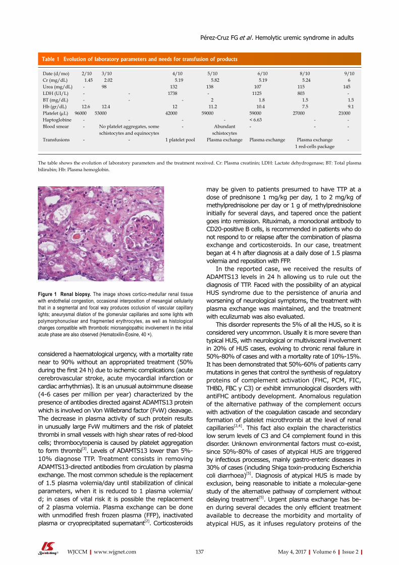
137 May 4, 2017|Volume 6|Issue 2|WJCCM|www.wjgnet.com
considered a haematological urgency, with a mortality rate near to 90% without an appropriated treatment (50% during the first 24 h) due to ischemic complications (acute cerebrovascular stroke, acute myocardial infarction or cardiac arrhythmias). It is an unusual autoimmune disease (4-6 cases per million per year) characterized by the presence of antibodies directed against ADAMTS13 protein which is involved on Von Willebrand factor (FvW) cleavage. The decrease in plasma activity of such protein results in unusually large FvW multimers and the risk of platelet thrombi in small vessels with high shear rates of red-blood cells; thrombocytopenia is caused by platelet aggregation to form thrombi[3]. Levels of ADAMTS13 lower than 5%- 10% diagnose TTP. Treatment consists in removing ADAMTS13-directed antibodies from circulation by plasma exchange. The most common schedule is the replacement of 1.5 plasma volemia/day until stabilization of clinical parameters, when it is reduced to 1 plasma volemia/d; in cases of vital risk it is possible the replacement of 2 plasma volemia. Plasma exchange can be done with unmodified fresh frozen plasma (FFP), inactivated plasma or cryoprecipitated supernatant[2]. Corticosteroids
may be given to patients presumed to have TTP at a dose of prednisone 1 mg/kg per day, 1 to 2 mg/kg of methylprednisolone per day or 1 g of methylprednisolone initially for several days, and tapered once the patient goes into remission. Rituximab, a monoclonal antibody to CD20-positive B cells, is recommended in patients who do not respond to or relapse after the combination of plasma exchange and corticosteroids. In our case, treatment began at 4 h after diagnosis at a daily dose of 1.5 plasma volemia and reposition with FFP.
In the reported case, we received the results of ADAMTS13 levels in 24 h allowing us to rule out the diagnosis of TTP. Faced with the possibility of an atypical HUS syndrome due to the persistence of anuria and worsening of neurological symptoms, the treatment with plasma exchange was maintained, and the treatment with eculizumab was also evaluated.
This disorder represents the 5% of all the HUS, so it is considered very uncommon. Usually it is more severe than typical HUS, with neurological or multivisceral involvement in 20% of HUS cases, evolving to chronic renal failure in 50%-80% of cases and with a mortality rate of 10%-15%. It has been demonstrated that 50%-60% of patients carry mutations in genes that control the synthesis of regulatory proteins of complement activation (FHC, PCM, FIC, THBD, FBC y C3) or exhibit immunological disorders with antiFHC antibody development. Anomalous regulation of the alternative pathway of the complement occurs with activation of the coagulation cascade and secondary formation of platelet microthrombi at the level of renal capillaries[2,4]. This fact also explain the characteristics low serum levels of C3 and C4 complement found in this disorder. Unknown environmental factors must co-exist, since 50%-80% of cases of atypical HUS are triggered by infectious processes, mainly gastro-enteric diseases in 30% of cases (including Shiga toxin-producing Escherichia coli diarrhoea)[5]. Diagnosis of atypical HUS is made by exclusion, being reasonable to initiate a molecular-gene study of the alternative pathway of complement without delaying treatment[5]. Urgent plasma exchange has be-en during several decades the only efficient treatment available to decrease the morbidity and mortality of atypical HUS, as it infuses regulatory proteins of the
Table 1 Evolution of laboratory parameters and needs for transfusion of products
Date (d/mo) 2/10 3/10 4/10 5/10 6/10 8/10 9/10Cr (mg/dL) 1.45 2.02 5.19 5.82 5.19 5.24 6Urea (mg/dL) - 98 132 138 107 115 145LDH (UI/L) - - 1738 - 1125 803 -BT (mg/dL) - - - 2 1.8 1.5 1.5Hb (gr/dL) 12.6 12.4 12 11.2 10.4 7.5 9.1Platelet (µL) 96000 53000 42000 59000 59000 27000 21000Haptoglobine - - - - < 6.63 - -Blood smear - No platelet aggregates, some
schistocytes and equinocytes- Abundant
schistocytes- - -
Transfusions - - 1 platelet pool Plasma exchange Plasma exchange Plasma exchange1 red-cells package
-
The table shows the evolution of laboratory parameters and the treatment received. Cr: Plasma creatinin; LDH: Lactate dehydrogenase; BT: Total plasma bilirubin; Hb: Plasma hemoglobin.
Figure 1 Renal biopsy. The image shows cortico-medullar renal tissue with endothelial congestion, occasional interposition of mesangial cellularity that in a segmental and focal way produces occlusion of vascular capillary lights; aneurysmal dilation of the glomerular capillaries and some lights with polymorphonuclear and fragmented erythrocytes, as well as histological changes compatible with thrombotic microangiopathic involvement in the initial acute phase are also observed (Hematoxilin-Eosine, 40 ×).
Pérez-Cruz FG et al . Hemolytic uremic syndrome in adults

138 May 4, 2017|Volume 6|Issue 2|WJCCM|www.wjgnet.com
normal pathway of complement and eliminates mutant proteins and antiFHC antibodies if they are present. At present, the first line treatment is Eculizumab, an humanized monoclonal IgG2/4 kappa antibody that joints to abnormal protein of C5 complement, blocking its cleavage to C5a and C5b and preventing the generation of the C5b-9 complex of the terminal complement[6]. In our case, this treatment was evaluated, delaying the decision until the arrival of the result of stool culture. After the microbiological confirmation of isolation of verotoxin-producing Escherichia Coli, the definitive diagnosis of typical HUS was reached, stopping treatment with plasma exchange and discouraging treatment with Eculizumab.
Typical HUS is an uncommon disease in adults. It has a relatively good prognosis with a low mortality and with an evolution to chronic renal failure only in 10% of cases; factors of poor prognosis are presence of neurological symptoms and need of renal replacement therapy at the beginning of the disease. Typical HUS is associated to enteric infections by Shiga-toxin bacteria, being the Escherichia coli (serotype O157:H7) the most frequent. Toxin liberation (Shiga-toxin or verotoxin 1 and 2) after ingestion of contaminated foods leads to damage of intestinal mucosa, which explains the characteristic bloody diarrhoea. After penetrating intestinal mucosa, the toxin is transported through the blood to renal tissue damaging endothelium of glomerular capillaries, mesangial cells and tubular cells of kidneys. Endothelium cells damage promotes a prothrombotic state with increase of platelet adhesion and micro-thrombi formation[1,2,7]. Shiga-toxin also induces secretion of abnormal multimers of FvW increasing platelet adhesion. The fact that only 5%-15% of patients who had a gastro-enteric infection develop an HUS indicates that others unknown factors are necessary[2,8]. Treatment remains supportive, the use of antibiotics being controversial since there is an increase of the release of toxin after the death of bacteria leading therefore to an increased risk of developing HUS. In typical HUS plasma exchange has no benefit. The role of Eculizumab is uncertain; although the studies carried out during the 2011 outbreak in Germany did not show benefits[9], there are published short series and isolated cases with good results, always in severe cases[10]. In our case despite the poor prognosis data (neurological involvement and need of haemodialysis technique on the first days) the treatment with that drug was rejected after a favourable evolution and complete recovery of renal function in a short period of four weeks.
COMMENTSCase characteristicsA 67-year-old women, with hypertension, type 2 diabetes mellitus and dyslipidemia, presented to emergency department with abdominal pain, bleeding diarrhoea and nausea of 3 d of evolution.
Clinical diagnosisThe clinical diagnosis of thrombotic microangiopathies (TMA) was based on the presence of thrombocytopenia and the development of acute renal and
neurological failure.
Differential diagnosisOther causes of thrombocytopenia, neurological and acute renal failure as sepsis, and TMA entities.
Laboratory diagnosisThrombocytopenia and anemia, high serum creatinine and lactate dehydrogenase, elevated schistocytes count on blood smear.
TreatmentIn this case report, the authors described the early treatment with plasma exchange.
Related reportsDue to the low incidence of TMA, although with high mortality in some entities, treatment should be based on syndromic approach according to guidelines.
Experiences and lessonsIn patients with TMA, if suspected of thrombotic thrombocytopenic purpura exist, early treatment with plasma exchange must initiated due to its high mortality rate.
Peer-reviewThe clinical course had been good writing and the background of the thrombotic microangiopathies had been well reviewed.
REFERENCES1 Moake JL. Thrombotic microangiopathies. N Engl J Med 2002; 347:
589-600 [PMID: 12192020]2 Contreras E, de la Rubia J, Del Río-Garma J, Díaz-Ricart M, García-
Gala JM, Lozano M; Grupo Español de Aféresis. [Diagnostic and therapeutic guidelines of thrombotic microangiopathies of the Spanish Apheresis Group]. Med Clin (Barc) 2015; 144: 331.e1-331.e13 [PMID: 25433791 DOI: 10.1016/j.medcli.2014.09.013]
3 Scully M, Hunt BJ, Benjamin S, Liesner R, Rose P, Peyvandi F, Cheung B, Machin SJ; British Committee for Standards in Haematology. Guidelines on the diagnosis and management of thrombotic thrombocytopenic purpura and other thrombotic microangiopathies. Br J Haematol 2012; 158: 323-335 [PMID: 22624596 DOI: 10.1111/j.1365-2141.2012.09167.x]
4 Blasco Pelicano M, Rodríguez de Córdoba S, Campistol Plana JM. [Atypical hemolytic uremic syndrome]. Med Clin (Barc) 2015; 145: 438-445 [PMID: 25433773 DOI: 10.1016/j.medcli.2014.08.006]
5 Campistol JM, Arias M, Ariceta G, Blasco M, Espinosa L, Espinosa M, Grinyó JM, Macía M, Mendizábal S, Praga M, Román E, Torra R, Valdés F, Vilalta R, Rodríguez de Córdoba S. An update for atypical haemolytic uraemic syndrome: diagnosis and treatment. A consensus document. Nefrologia 2015; 35: 421-447 [PMID: 26456110 DOI: 10.1016/j.nefro.2015.07.005]
6 Ferreira E, Oliveira N, Marques M, Francisco L, Santos A, Carreira A, Campos M. Eculizumab for the treatment of an atypical hemolytic uremic syndrome with mutations in complement factor I and C3. Nefrologia 2016; 36: 72-75 [PMID: 26541438 DOI: 10.1016/j.nefro. 2015.07.007]
7 George JN, Nester CM. Syndromes of thrombotic microangiopathy. N Engl J Med 2014; 371: 1847-1848 [PMID: 25372103 DOI: 10.1056/ NEJMc1410951]
8 Kavanagh D, Raman S, Sheerin NS. Management of hemolytic uremic syndrome. F1000Prime Rep 2014; 6: 119 [PMID: 25580273 DOI: 10.12703/P6-119]
9 Loos S, Ahlenstiel T, Kranz B, Staude H, Pape L, Härtel C, Vester U, Buchtala L, Benz K, Hoppe B, Beringer O, Krause M, Müller D, Pohl M, Lemke J, Hillebrand G, Kreuzer M, König J, Wigger M, Konrad M, Haffner D, Oh J, Kemper MJ. An outbreak of Shiga toxin-producing
COMMENTS
Pérez-Cruz FG et al . Hemolytic uremic syndrome in adults

139 May 4, 2017|Volume 6|Issue 2|WJCCM|www.wjgnet.com
Escherichia coli O104: H4 hemolytic uremic syndrome in Germany: presentation and short-term outcome in children. Clin Infect Dis 2012; 55: 753-759 [PMID: 22670043 DOI: 10.1093/cid/cis531]
10 Lapeyraque AL, Malina M, Fremeaux-Bacchi V, Boppel T,
Kirschfink M, Oualha M, Proulx F, Clermont MJ, Le Deist F, Niaudet P, Schaefer F. Eculizumab in severe Shiga-toxin-associated HUS. N Engl J Med 2011; 364: 2561-2563 [PMID: 21612462 DOI: 10.1056/NEJMc1100859]
P- Reviewer: Chang CC, Cid J, Watanabe T S- Editor: Song XX L- Editor: A E- Editor: Lu YJ
Pérez-Cruz FG et al . Hemolytic uremic syndrome in adults

© 2017 Baishideng Publishing Group Inc. All rights reserved.
Published by Baishideng Publishing Group Inc7901 Stoneridge Drive, Suite 501, Pleasanton, CA 94588, USA
Telephone: +1-925-223-8242Fax: +1-925-223-8243
E-mail: [email protected] Desk: http://www.f6publishing.com/helpdesk
http://www.wjgnet.com

World Journal of Critical Care MedicineWorld J Crit Care Med 2017 August 4; 6(3): 140-178
Published by Baishideng Publishing Group Inc
ISSN 2220-3141 (online)

World Journal ofCritical Care MedicineW J C C M
EDITOR-IN-CHIEFBart Van Rompaey, Wilrijk
GUEST EDITORIAL BOARD MEMBERSHsing I Chen, HualienSheng-Hsien Chen, Yong-KangYih-Sharng Chen, TaipeiYung-Chang Chen, TaipeiDer-Yang Cho, TaichungCheng-Keng Chuang, TaoyuanHow-Ran Guo, TainanBang-Gee Hsu, HualienChien-Wei Hsu, KaohsiungWen-Jinn Liaw, TaipeiYan-Ren Lin, ChanghuaJiunn-Jye Sheu, Kaohsiung Hsien
MEMBERS OF THE EDITORIAL BOARD
ArgentinaEduardo Chuluyan, Buenos AiresAdrian A Inchauspe, Berazategui
AustraliaZsolt J Balogh, NewcastleZoltan H Endre, SydneyNam Q Nguyen, South AustraliaAlistair D Nichol, VictoriaGeorg M Schmolzer, VictoriaAndrew T Slack, Brisbane
Ravindranath Tiruvoipati, Frankston
AustriaLars-Peter Kamolz, ViennaSylvia Knapp, Vienna
BangladeshSaidur R Mashreky, Dhaka
BelgiumTeresinha Leal, BrusselsManu LNG Malbrain, AntwerpJean-Louis Vincent, Brussels
BrazilLuciano CP Azevedo, São PauloPatricia RM Rocco, Rio de JaneiroMarcos A Rossi, Ribeirao PretoRenato Seligman, Porto Alegre
CanadaDouglas D Fraser, OntarioPierre A Guertin, QuebecMarc G Jeschke, TorontoConstantine J Karvellas, EdmontonWolfgang M Kuebler, TorontoXi Yang, Winnipeg
ChinaXiang-Dong Chen, ChengduXu-Lin Chen, HefeiWong Tak Chuen, Hong KongMing-Xu Da, LanzhouHuang-Xian Ju, NanjingTing-Bo Liang, Hangzhou Peng-Lin Ma, BeijingChung-Wah D Siu, Hong KongYong-ming Yao, BeijingJia-Ping Zhang, ChongqingWei-Dong Zhou, Beijing
CroatiaAlan Sustic, Rijeka
CubaJesús Pérez-Nellar, La Habana
DenmarkDan S Karbing, Aalborg East
EgyptIbrahim Abouomira, CairoHanan Ibrahim, CairoAmr M Moghazy, IsmailiaAyman A Yousef, Tanta
I
Editorial Board2016-2019
The World Journal of Critical Care Medicine Editorial Board consists of 235 members, representing a team of worldwide experts in critical care medicine. They are from 44 countries, including Argentina (2), Australia (7), Austria (2), Bangladesh (1), Belgium (4), Brazil (4), Canada (6), China (23), Croatia (1), Cuba (1), Denmark (1), Egypt (4), Finland (1), France (6), Germany (9), Greece (9), Hungary (1), India (10), Iran (2), Israel (6), Italy (13), Japan (6), Jordan (1), Mexico (1), Morocco (1), Netherlands (4), New Zealand (3), Norway (1), Poland (1), Portugal (4), Russia (1), Saudi Arabia (2), Singapore (2), Slovenia (1), South Africa (1), Spain (6), Sweden (1), Switzerland (3), Thailand (1), Tunisia (1), Turkey (3), United Kingdom (7), United States (70), and Uruguay (1).
February 26, 2016WJCCM|www.wjgnet.com

FinlandAsko A Riutta, Tampere
FranceJean-Marc Cavaillon, ParisBruno Mégarbane, ParisSaad Nseir, LilleNicolas Terzi, CaenJean-Francois Timsit, La Tronche CedexBenoit Vallet, Lille
GermanyHendrik Bracht, UlmMichael E Czaplik, AachenGerrit Grieb, AachenTobias Keck, FreiburgPhilipp Kobbe, AachenAlexander Koch, AachenMarc Maegele, CologneAndrzej A Piatkowski, AachenArmin R Sablotzki, Leipzig
GreeceIoanna Dimopoulou, AthensDimitrios Karakitsos, AthensPetros Kopterides, AthensGregory Kouraklis, AthensAthanasios D Marinis, PiraeusGeorge Nakos, IoanninaPapaioannouE Vasilios, AlexandroupolisTheodoros Xanthos, AthensSpyros G Zakynthinos, Athens
HungaryZoltan Rakonczay, Szeged
IndiaRitesh Agarwal, ChandigarhRachna Agarwal, DelhiMohammad F Butt, SrinagarMohan Gurjar, LucknowDeven Juneja, New Delhi Farhad N Kapadia, MumbaiVikram Kate, PuducherryPramod Kumar, ManipalMedha Mohta, DelhiSrinivas Rajagopala, Bangalore
IranHemmat Maghsoudi, TabrizHomayoun Sadeghi-Bazargani, Tabriz
IsraelAlexander Becker, AfulaYoram Kluger, HaifaYona Kosashvili, ZerrifinKobi Peleg, Tel Hashomer Ilan Sela, RehovotPierre Singer, Petah Tikva
ItalyGiacomo Bellani, MonzaGiovanni Camussi, TurinAnselmo Caricato, RomePiero Ceriana, PaviaAntonio Chiaretti, RomeDavide A Chiumello, MilanoAlfredo Conti, MessinaPaolo Cotogni, TurinDaniele M De Luca, RomaVincenzo De Santis, RomeLuca La Colla, ParmaRaffaele Scala, LuccaGiovanni Vento, Roma
JapanKeishiro Aoyagi, Kurume citySatoshi Hagiwara, OitaYuichi Hattori, ToyamaHideo Inaba, KanazawaEisuke Kagawa, HiroshimaChieko Mitaka, Tokyo
JordanFeras I Hawari, Amman
MexicoSilvio A Ñamendys-Silva, Mexico City
MoroccoRedouane Abouqal, Rabat
NetherlandsWim A Buurman, MaastrichtMartin CJ Kneyber, GroningenPatrick Schober, AmsterdamArie Barend V Vugt, Enschede
New ZealandSultan Al-Shaqsi, DunedinArman A Kahokehr, WhangareiJohn W Pickering, Christchurch
NorwayUlf R Dahle, Oslo
PolandMaciej Owecki, Poznań
PortugalErnestina R Gomes, PortoCristina Granja, MatosinhosJosé A Lopes, LisbonPedro Póvoa, Lisbon
RussiaKonstantin A Popugaev, Moscow
Saudi ArabiaRitesh G Menezes, DammamMohamed T Suliman, Tabuk
SingaporeSanjay H Chotirmall, SingaporeDevanand Anantham, Singapore
SloveniaŠtefek Grmec, Maribor
South AfricaDamian Clarke, Pietermaritzburg
SpainDavid Jimenez, MadridJuan A Llompart-Pou, Palma de MallorcaAntonio T Martí, BarcelonaJuan C Montejo-González, MadridEnrique A Piacentini, TerrassaAlonso M Rodriguez, Madrid
SwedenMihai Oltean, Gothenburg
SwitzerlandDieter Cadosch, ZurichMihael Potocki, BaselJohn F Stover, Zurich
II February 26, 2016WJCCM|www.wjgnet.com

III February 26, 2016WJCCM|www.wjgnet.com
ThailandViroj Wiwanitkit, Bangkok
TunisiaMabrouk Bahloul, Sfax
TurkeyYusuf K Coban, MalatyaBensu Karahalil, AnkaraAli Nayci, Mersin
United KingdomSammy Al-Benna, NottinghamGiles N Cattermole, OrpingtonFrantisek Duska, NottinghamJames N Fullerton, LondonChristina Jones, PrescotSameer Khan, MiddlesbroughGeorge Ntoumenopoulos, London
United StatesEdward Abraham, Winston-SalemBernard R Bendok, ChicagoMichael Blaivas, AtlantaCharles D Boucek, PittsburghRonald Bronicki, Houston
Robert C Cantu, ConcordMarylou Cardenas-Turanzas, HoustonArchana Chatterjee, OmahaPaul A Checchia, St. LouisRubin I Cohen, New Hyde ParkStephen Cohn, San AntonioDonald E Craven, BurlingtonRuy J Cruz Jr, PittsburghFrancis C Dane, RoanokeMarc A de Moya, BostonSteven M Donn, Ann ArborChristopher P Farrell, WynnewoodMarcos A Fernandez, NashvilleKevin N Foster, PhoenixBarry D Fuchs, PhiladelphiaRichard P Gonzalez, MobileAlan H Hall, LaramieJijo John, GilbertJason N Katz, Chapel HillSalah G Keyrouz, Little RockImran Khalid, JeddahDeborah A Kuhls, Las VegasGregory L Larkin, New HavenChristos Lazaridis, CharlestonJames A Lin, Los AngelesYahia M Lodi, SyracuseRoger M Loria, RichmondAigang Lu, CincinnatiRudolf Lucas, AugustaO. John Ma, PortlandRobert T Mallet, Fort WorthWilliam T McGee, MiamiMark McKenney, MiamiMichael Moussouttas, PhiladelphiaOliver HJ Muensterer, Bronx
Rahul Nanchal, MilwaukeeMichael Steven Niederman, MineolaGary F Nieman, SyracuseJames M O’Brien, ColumbusMartin Oudega, MiamiCatherine M Preissig, DuluthVirginia Prendergast, PhoenixRamesh Raghupathi, PhiladelphiaMiren A Schinco, JacksonvilleCarl I Schulman, MiamiL Keith Scott, ShreveportKevin N Sheth, BaltimoreJenni Short, SalinaRonald F Sing, CharlottePhilip C Spinella, St. LouisRobert M Starke, CharlottesvilleStanislaw PA Stawicki, ColumbusDavid C Stockwell, WashingtonStanislav Svetlov, AlachuaMaged A Tanios, Long BeachNeal J Thomas, HersheyNancy M Tofil, BirminghamBalagangadhar R Totapally, MiamiSteven N Vaslef, DurhamJoseph C Watson, Falls ChurchJohn S Wilgis, OrlandoDavid C Willms, San DiegoHao-Dong Xu, RochesterXiao-Ming Xu, IndianapolisMidori A Yenari, San Francisco
UruguayWilliam Manzanares, Montevideo

World Journal ofCritical Care MedicineW J C C M
Contents
IWJCCM|www.wjgnet.com August 4, 2017|Volume 6|Issue 3|
Quarterly Volume 6 Number 3 August 4, 2017
REVIEW140 Immunomodulatoryeffectsofanestheticsinobesepatients
Heil LBB, Silva PL, Pelosi P, Rocco PRM
MINIREVIEWS
153 Generalizableitemsandmodularstructureforcomputerisedphysicianstaffingcalculationonintensivecare
units
Weiss M, Marx G, Iber T
EVIDENCE-BASED MEDICINE164 Effectsofintrapulmonarypercussiveventilationonairwaymucusclearance:Abenchmodel
Fernandez-Restrepo L, Shaffer L, Amalakuhan B, Restrepo MI, Peters J, Restrepo R
172 Algorithm-basedarterialbloodsamplingrecognitionincreasingsafetyinpoint-of-carediagnostics
Peter J, Klingert W, Klingert K, Thiel K, Wulff D, Königsrainer A, Rosenstiel W, Schenk M
REVIEW

ContentsWorld Journal of Critical Care Medicine
Volume 6 Number 3 August 4, 2017
EDITORS FOR THIS ISSUE
Responsible Assistant Editor: Xiang Li Responsible Science Editor: Jin-Xin KongResponsible Electronic Editor: Ya-Jing Lu Proofing Editorial Office Director: Ze-Mao GongProofing Editor-in-Chief: Lian-Sheng Ma
EDITORIALOFFICEXiu-Xia Song, DirectorWorld Journal of Critical Care MedicineBaishideng Publishing Group Inc7901 Stoneridge Drive, Suite 501, Pleasanton, CA 94588, USATelephone: +1-925-2238242Fax: +1-925-2238243E-mail: [email protected] Desk: http://www.f6publishing.com/helpdeskhttp://www.wjgnet.com
PUBLISHERBaishideng Publishing Group Inc7901 Stoneridge Drive, Suite 501, Pleasanton, CA 94588, USATelephone: +1-925-223-8242Fax: +1-925-223-8243E-mail: [email protected] Desk: http://www.f6publishing.com/helpdeskhttp://www.wjgnet.com
PUBLICATIONDATEAugust 4, 2017
COPYRIGHT© 2017 Baishideng Publishing Group Inc. Articles published by this Open-Access journal are distributed under the terms of the Creative Commons Attribution Non-commercial License, which permits use, distribution, and reproduction in any medium, provided the original work is properly cited, the use is non commercial and is otherwise in compliance with the license.
SPECIALSTATEMENTAll articles published in journals owned by the Baishi-deng Publishing Group (BPG) represent the views and opinions of their authors, and not the views, opinions or policies of the BPG, except where otherwise expli-citly indicated.
INSTRUCTIONSTOAUTHORShttp://www.wjgnet.com/bpg/gerinfo/204
ONLINESUBMISSIONhttp://www.f6publishing.com
IIWJCCM|www.wjgnet.com
ABOUT COVER
AIM AND SCOPE
INDExINg/ABSTRACTINg
FLYLEAF
August 4, 2017|Volume 6|Issue 3|
NAMEOFJOURNALWorld Journal of Critical Care Medicine
ISSNISSN 2220-3141 (online)
LAUNCHDATEFebruary 4, 2012
FREQUENCYQuarterly
EDITOR-IN-CHIEFBart Van Rompaey, BSc, MSc, PhD, Associate Professor, Nurse, Faculty of Medicine and Health Sciences, Department of Nursing and midwifery, Cen-tre for Research and Innovation in Care, University of Antwerp, Wilrijk 2610, Antwerp, Belgium
EDITORIALBOARDMEMBERSAll editorial board members resources online at http://www.wjgnet.com/2220-3141/editorialboard.htm
EditorialBoardMemberofWorldJournalofCriticalCareMedicine ,AymanAYousef,MD,AssistantProfessor,DepartmentofAnesthesia,TantaUniversity
Hospitals,Tanta31527,Egypt
World Journal of Critical Care Medicine (World J Crit Care Med, WJCCM, online ISSN 2220-3141, DOI: 10.5492) is a peer-reviewed open access academic journal that aims to guide clinical practice and improve diagnostic and therapeutic skills of clinicians.
WJCCM covers topics concerning severe infection, shock and multiple organ dysfunc-tion syndrome, infection and anti-infection treatment, acute respiratory distress syndrome and mechanical ventilation, acute kidney failure, continuous renal replacement therapy, rational nutrition and immunomodulation in critically ill patients, sedation and analgesia, cardiopulmonary cerebral resuscitation, fluid resuscitation and tissue perfusion, coagulant dysfunction, hemodynamic monitoring and circulatory support, ICU management and treatment control, and application of bronchofiberscopy in critically ill patients.
We encourage authors to submit their manuscripts to WJCCM. We will give priority to manuscripts that are supported by major national and international foundations and those that are of great clinical significance.
World Journal of Critical Care Medicine is now indexed in PubMed, PubMed Central.
I-III EditorialBoard

Luciana Boavista Barros Heil, Pedro Leme Silva, Paolo Pelosi, Patricia Rieken Macedo Rocco
REVIEW
140 August 4, 2017|Volume 6|Issue 3|WJCCM|www.wjgnet.com
Immunomodulatory effects of anesthetics in obese patients
Luciana Boavista Barros Heil, Pedro Leme Silva, Patricia Rieken Macedo Rocco, Laboratory of Pulmonary Investigation, Carlos Chagas Filho Institute of Biophysics, Federal University of Rio de Janeiro, Centro de Ciências da Saúde, Avenida Carlos Chagas Filho, Rio de Janeiro 21941-902, Brazil
Paolo Pelosi, Department of Surgical Sciences and Integrated Diagnostics, IRCCS AOU San Martino-IST, University of Genoa, 16132 Genoa, Italy
Author contributions: Heil LBB and Silva PL designed and conducted the literature review, wrote the manuscript, and prepared the tables; Silva PL prepared the figures, and supervised all the process; Pelosi P and Rocco PRM wrote the manuscript and supervised all the process.
Supported by Brazilian Council for Scientific and Technological Development (CNPq); Carlos Chagas Filho Rio de Janeiro State Foundation (FAPERJ); Department of Science and Technology (DECIT); Brazilian Ministry of Health; and Coordination for the Improvement of Higher Level Personnel (CAPES).
Conflict-of-interest statement: The authors have no conflict of interest.
Open-Access: This article is an open-access article which was selected by an in-house editor and fully peer-reviewed by external reviewers. It is distributed in accordance with the Creative Commons Attribution Non Commercial (CC BY-NC 4.0) license, which permits others to distribute, remix, adapt, build upon this work non-commercially, and license their derivative works on different terms, provided the original work is properly cited and the use is non-commercial. See: http://creativecommons.org/licenses/by-nc/4.0/
Manuscript source: Invited manuscript
Correspondence to: Patricia Rieken Macedo Rocco, MD, PhD, Professor, Laboratory of Pulmonary Investigation, Carlos Chagas Filho Institute of Biophysics, Federal University of Rio de Janeiro, Centro de Ciências da Saúde, Avenida Carlos Chagas Filho, 373, Bloco G-014, Ilha do Fundão, Rio de Janeiro 21941-902, Brazil. [email protected]: +55-21-39386530Fax: +55-21-22808193
Received: January 26, 2017
Peer-review started: February 8, 2017First decision: April 17, 2017Revised: June 27, 2017 Accepted: July 7, 2017Article in press: July 10, 2017Published online: August 4, 2017
AbstractAnesthesia and surgery have an impact on inflam-matory responses, which influences perioperative homeostasis. Inhalational and intravenous anesthesia can alter immune-system homeostasis through multiple processes that include activation of immune cells (such as monocytes, neutrophils, and specific tissue macrophages) with release of pro- or anti-inflammatory interleukins, upregulation of cell adhesion molecules, and overproduction of oxidative radicals. The response depends on the timing of anesthesia, anesthetic agents used, and mechanisms involved in the development of inflammation or immunosuppression. Obese patients are at increased risk for chronic diseases and may have the metabolic syndrome, which features insulin resistance and chronic low-grade inflammation. Evidence has shown that obesity has adverse impacts on surgical outcome, and that immune cells play an important role in this process. Understanding the effects of anesthetics on immune-system cells in obese patients is important to support proper selection of anesthetic agents, which may affect postoperative outcomes. This review article aims to integrate current knowledge regarding the effects of commonly used anesthetic agents on the lungs and immune response with the underlying immunology of obesity. Additionally, it identifies knowledge gaps for future research to guide optimal selection of anesthetic agents for obese patients from an immunomodulatory standpoint.
Key words: Anesthesia; Immune system; Perioperative care; Obesity; Inflammation
© The Author(s) 2017. Published by Baishideng Publishing
World Journal ofCritical Care MedicineW J C C M
Submit a Manuscript: http://www.f6publishing.com
DOI: 10.5492/wjccm.v6.i3.140
World J Crit Care Med 2017 August 4; 6(3): 140-152
ISSN 2220-3141 (online)
141 August 4, 2017|Volume 6|Issue 3|WJCCM|www.wjgnet.com
Heil LBB et al . Anesthetic agents and immunomodulation in obese patients
Group Inc. All rights reserved.
Core tip: Anesthetic agents have been studied not only for their effects on anesthesia and analgesia, but also their action on the lungs and immune system. Obesity is associated with a chronic state of low-grade systemic inflammation, and may predispose to development of comorbidities. Although efforts have been made to develop guidelines for anesthesia in obesity, to date, no ideal drug combination has been found. Optimization of the immunomodulatory properties of anesthetic agents may enable perioperative modulation of inflammatory response in obese patients and improve postoperative outcomes.
Heil LBB, Silva PL, Pelosi P, Rocco PRM. Immunomodulatory effects of anesthetics in obese patients. World J Crit Care Med 2017; 6(3): 140-152 Available from: URL: http://www.wjgnet.com/2220-3141/full/v6/i3/140.htm DOI: http://dx.doi.org/10.5492/wjccm.v6.i3.140
INTRODUCTIONObesity and associated comorbidities are increasing at epidemic proportions globally[1], with a substantial impact on postoperative outcomes for affected individuals undergoing minor or major surgical procedures that require anesthesia. Intravenous and inhalational anesthetics (IAs) have been shown to modulate the innate and adaptive immune responses, as well as indirect effectors of immunity[2,3].
Since obesity results in chronic lowgrade inflammation or metainflammation[4] associated with increased circulating proinflammatory factors, it has been proposed that anesthetic agents may modulate the already altered immune function in obesity, with particular emphasis on pulmonary inflammation.
This review article aims to integrate current knowledge regarding the effects of commonly used anesthetic agents on the lungs and immune response with the underlying immunology of obesity. Additionally, it provides insights and future perspectives into the safe use of anesthetics as immunomodulators for obese patients. Better knowledge of the impact of anesthetic agents on the immune system, especially in the setting of obesity, may improve perioperative management and outcome.
IMMUNE AND INFLAMMATORY CHANGES DUE TO OBESITY: THE ROLE OF IMMUNE CELL INFILTRATION IN ADIPOSE TISSUE Healthy adipose tissue (AT) is composed of a type2 polarized immune system, which maintains AT macro
phages (ATM) in an M2like (proresolution) state. While in this form, AT is mainly composed of eosinophils, invariantchain natural killer T (iNKT) cells[5], and regulatory T (Treg) cells[6], which produce interleukin (IL)4, IL13, and IL10. Adipocytes also contribute to the type 2 immune response through production of adiponectin, which exhibits a strong anti-inflammatory effect[7]. These type 2 immune cells are supported by a stromal structure, which promotes immune cell viability through the production of several cytokines, with IL33 playing a particularly important role[8,9]. Moreover, in order to sustain this environment, AT cells engage in extensive crosstalk to (re)model AT structure and phenotype[10].
The early phases of the dietinduced obesity (DIO) period are characterized by an increase in the amount of fat per adipocyte and an accumulation of immune cells. Acute changes in the microenvironment, such as alterations in oxygen supply and consumption, contribute to triggering a rapid increase in the number of neutrophils[11]. Adipocytes become hypertrophic and hyperplastic. This is associated with a shift in adipokine production from adiponectin to leptin, monocyte chemoattractant protein1 (MCP1), and IL6, as well as resistin, visfatin, tumor necrosis factor (TNF)α, retinol binding protein 4 (RBP4), lipocalin2, and CXCL5[12]. Leptin directly increase the production of several proinflammatory cytokines, such as IL6, TNFα, the chemokines CCL2/MCP1, and leukotriene B4 (LTB4) in peripheral blood monocytes and resident tissue macrophages[13]. Leptin can also induce the production of reactive intermediates in macrophages, neutrophils, and endothelial cells, as well as potentiate interferon (IFN)γ induced expression of nitric oxide (NO) synthase[1416], whereas adiponectin, IL10, and omentin, which have antiinflammatory effects, are downregulated[17]. In addition, innate inflammatory molecules such as acute phase reactants, Creactive protein (CRP)[18], complement components C2, C3, and C4[19,20], and other immunemodulating mediators produced in AT contribute to the intricate connection between fat and its tissueresident immune cells.
The adaptive immunity role is mediated by Tlymphocyte infiltration during early AT inflammation, preceding macrophage recruitment[21,22]. Most of these are CD4+ lymphocytes that differentiate to TH1cells, governing the local inflammatory process through the release of proinflammatory cytokines like IFNγ and TNFα. Tcell recruitment is usually mediated by chemokines released from endothelial cells, stromal cells, or macrophages. While, on the one hand, Tcell derived IFNγ promotes the recruitment of monocytes by MCP1 secretion from preadipocytes, it also activates other cells, including macrophages[21].
Resident and recruited ATM are the most common immune cell types in AT, and their infiltration is associated with AT inflammation[23,24]. Recruited AT macrophages induce tissue inflammation when their

142 August 4, 2017|Volume 6|Issue 3|WJCCM|www.wjgnet.com
polarization shifts from an M2 type to an activated proinflammatory M1 state. Stimuli for this shift toward the M1 phenotype includes systemic factors, such as increase in free fatty acids (FFAs), which stimulates tolllike receptors (TLR)4 on macrophages[25], and activation of the inflammasome, which is responsible for production of the proinflammatory cytokines IL-1b and IL18[26]. In addition, IFNγ is a potent local inducer of M1 polarization during ATM inflammation[27].
The link between metabolism and immunity at the intracellular level occurs through activation of nuclear factorkB (NFkB) and its cytoplasmic inhibitor IkkB. Likewise, other inflammatory factors, such as cJun Nterminal protein kinases (JNK), are activated[28,29]. These proinflammatory mediators are produced in excess, spilling into the peripheral circulation and contributing to the lowgrade systemic inflammation that ultimately influences the development of obesity-associated comorbidities, including the pulmonary immune response, thus contributing to pulmonary inflammation[12,30].
AT immune cells contribute to the maintenance of homeostasis and development of chronic inflammation and are responsible for the mechanisms underlying obesityassociated complications and impairment of normal immune system functioning, thus further perpetuating chronic disease development and metabolic
complications.
LUNG IMMUNE CELLS AND OBESITY-ASSOCIATED INFLAMMATIONSeveral mediators elicited by obesity alter immune and inflammatory responses in the lung, and may induce obesityassociated changes to adipokines and lung immune cells.
LeptinSeveral lung cell types, such as leukocytes, airway smooth muscle cells, alveolar epithelial cells, and macrophages, express the functional leptin receptor, which, when bound to its main ligand (systemic leptin), participates in triggering inflammatory respiratory diseases. Lungs represent a target organ for leptin signaling. In this line, leptin stimulates neutrophil and macrophage release of cytokines (TNFα, IL6, IL12), eicosanoids, and NO and induces neutrophil oxidative burst[31]. Endogenous leptin has two main effects in the lungs (Figure 1). First, it acts as a neutrophil chemoattractant to the lungs[32]. Once neutrophil levels are increased, leptin lengthens neutrophil survival by delaying or inhibiting apoptosis[33]. Additionally, obese patients with increased levels of leptin exhibited increased susceptibility to respiratory infections, in an association that may be independent and
Capillary
- E-selectin- ICAM-1- VCAM-1
VE-cadherin
M2(pro-resolution)
M1(proinflammatory)
IL-10
Adiponectin
- Oxidative burst- Survival
Leptin
Leptin
Leptin
Neutrophil chemoattraction
Adipose tissue
+
+
-
+
Figure 1 Model of obesity-associated pulmonary inflammation. Lung immune cells and inflammation due to obesity. Leptin is implicated in inflammatory respiratory diseases as a neutrophil chemoattractant. The association between obesity and LPS-induced lung inflammation involves an increase in monocytes and lymphocytes, as well as in intracellular adhesion molecule (ICAM)-1 expression in alveolar macrophages, suggesting their polarization toward a pro-inflammatory M1 phenotype. Obesity impairs vascular homeostasis, facilitating increased susceptibility to inflammatory lung vascular diseases by affecting structural cells in the alveolar-capillary membrane. The lung endothelium of obese mice has been shown to express higher levels of leukocyte adhesion markers (E-selectin, ICAM-1, VCAM-1) and lower levels of junctional proteins (VE-cadherin and b-catenin). Adiponectin has anti-inflammatory properties, mainly by its effects on toll-like receptor (TLR) pathway-mediated NF-kB signaling, which regulates the shift from M1 to M2 macrophage polarization, and suppresses differentiation of M1 macrophages by downregulating the pro-inflammatory cytokines TNF-α, MCP-1, and IL-6. Adiponectin also promotes expression of the anti-inflammatory factor IL-10 in macrophages via cAMP-dependent mechanisms. TNF: Tumor necrosis factor; IL: Interleukin; LPS: Lipopolysaccharide.
Heil LBB et al . Anesthetic agents and immunomodulation in obese patients
Alveolus

143 August 4, 2017|Volume 6|Issue 3|WJCCM|www.wjgnet.com
likely additive to metabolic syndromerelated factors[34]. Furthermore, the proinflammatory effects of leptin may contribute to a higher incidence of asthma in the obese population[35]. In chronic obstructive pulmonary disease[36,37], the higher the leptin production, the greater the severity of the disease[38,39]. In the setting of obesity, not only immune cells but also structural cells in the alveolarcapillary membrane are altered. In obese mice, the lung endothelium was found to express higher levels of leukocyte adhesion markers (Eselectin, ICAM1 and VCAM1) and lower levels of junctional proteins (VEcadherin and bcatenin) (Figure 1), providing further evidence that obesity may impair vascular homeostasis and increase susceptibility to inflammatory lung vascular diseases[40].
In short, leptin plays an important role in respiratory immune responses and pathogenesis of inflammatory respiratory conditions by acting on different cell types in the lung.
AdiponectinAdiponectin is a well-defined obesity marker that has anti-inflammatory properties. Its predominant immune-related functions involve suppression of inflammation by clearance of apoptotic cell debris[41] and promotion of an antiinflammatory phenotype in the lung by blunting oxidative stress, inflammation, and angiogenesis. However, several of these immunerelated functions depend on the respective adiponectin receptor. AdipoR1, AdipoR2, Tcadherin, and calreticulin are detected in several lung cells[42]. The structure of adiponectin resembles those of complement factor C1q and of surfactant proteins, which act as pattern recognition molecules limiting lung inflammation[43]. Adiponectin receptors are also involved in the regulation of macrophage proliferation and function. AdipoR1 mediates adiponectin suppression of NFkB activation and proinflammatory cytokine expression in macrophages[44,45], AdipoR2 is involved in adiponectinmediated M2 polarization[46], and Tcadherin has been shown to play an essential role in the stimulatory effects of adiponectin on M2 macrophage proliferation[47]. The antiinflammatory effects of adiponectin are mainly guided by the tolllike receptor (TLR) mediated NFkB signaling pathway, which modulates a shift in macrophage polarization from M1 to M2 (Figure 1) and suppresses differentiation of M1 macrophages by downregulating the proinflammatory cytokines TNF-α, MCP1, and IL6[48,49]. Moreover, adiponectin increases expression of the antiinflammatory factor IL10 in macrophages via cAMPdependent mechanisms[50]. Adiponectin has also been proposed to regulate energy and metabolism by targeting innatelike lymphocytes (ILC2)[10,51], natural killer T (NKT)[52], and gamma delta T (γδT)cells[53].
Adiponectin senses metabolic stress and modulates metabolic adaptation by targeting functions of the innate immune system, including macrophage polarization and lymphocyte activity.
ANESTHESIA, ANESTHETICS, AND IMMUNOMODULATIONAnesthesia and the surgical stress response result in several immunological alterations, which cannot be easily separated. The pharmacological effects of anesthetic drugs (sedation, anesthesia, and analgesia) have been widely studied, as have their actions on several cell types, including inflammatory cells, by altering cytokine release[54]; cytokine receptor expression[55]; phagocytosis or cytotoxic actions[56]; and transcription or translation of protein mediators[57,58]. Depending on the clinical setting, immunosuppression and activation can be either detrimental or beneficial. These effects are clinically important because the balance between pro and anti-inflammatory cytokine secretion is associated with surgical outcomes.
Immune cells are categorized into two lines according to their maturation site: The myeloid lineage, which includes macrophages, dendritic cells (DCs), mast cells, and granulocytes (neutrophils, eosinophils and basophils); and the lymphoid lineage, which is composed of T and B lymphocytes, natural killer (NK) cells, and NK T cells[59,60]. Myeloid cells are considered the main players in innate immunity, and play important roles in adaptive immunity as well; they serve as antigen presenters and macrophages, mast cells, and neutrophils produce several cytokines, thus activating T and B lymphocytes[60]. Immunomodulation can have a dichotomous sense whereby suppression of the immune response can prevent further injury, as observed in models of acute inflammation[61], but also prevent the body from counteracting infections and increase the risk of opportunistic infections. In these scenarios, both inhalational and intravenous anesthetic agents may jeopardize or improve immune function.
IA agentsThe action of IAs on immune cells has been extensively reviewed in preclinical studies[2,62,63]. In vitro experiments on immune cells revealed generally transient, dose and timedependent effects predominantly on neutrophil function[6466], lymphocyte proliferation[67], suppression of inflammatory cytokines in rat alveolar cells, and decrease in the expression of inducible NO synthase by inhibition of voltagedependent calcium channels, reducing intracellular calcium concentrations[68]. However, in an ischemic setting, the suppression of neutrophil adhesion had a positive effect against the deleterious effects of polymorphonuclear cells, improving cardiac function[6972]. Furthermore, exposure to the isoflurane attenuated villus, hepatic, and renal injuries in a mouse model of intestinal ischemia; these effects were mediated via plasma membrane phosphatidylserine externalization and subsequent release of the antiinflammatory and anti-apoptotic cytokine transforming growth factor b1 (TGFb1)[73]. In both studies, the proposed mechanisms for protection rely on modulation
Heil LBB et al . Anesthetic agents and immunomodulation in obese patients

144 August 4, 2017|Volume 6|Issue 3|WJCCM|www.wjgnet.com
of endothelial and neutrophil adhesion molecules and reduction of neutrophil migration and margination into tissues[74]. In human endothelial cells, the effects of isoflurane against TNFαinduced apoptosis are mediated by the phosphorylation of extracellular signalregulated kinase (ERK MAPK) and induction of sphingosine kinase 1 (SK1) to increase production of the lysophospholipid S1P, a cytoprotective signaling molecule product of sphingomyelin hydrolysis that functions as an extracellular ligand for specific G protein-coupled receptors and as an intracellular second messenger[75]. In the context of acute inflammatory lung injury (Figure 2A), isoflurane has been shown to decrease neutrophil influx, as well as the synthesis and expression of macrophage inflammatory protein (MIP)2, IL1b, and the stress proteins heme oxygenase (HO1) and heat shock protein (HSP70)[7679]. These studies showed reduction of proinflammatory cytokine release through several mechanisms: (1) inhibition of NFkB translocation into the nuclei of human epithelial cells[58,76]; (2) inhibition of inducible nitric oxide synthase (iNOS) expression and blockade of NFkB activation in a mouse model of lung injury; (3) inhibition of proapoptotic procaspase8, procaspase3, and inactivated proapoptotic protein Bax expression; (4) promotion of phosphatidylinositol3kinase/Akt activation and enhanced expression of the antiapoptotic Bcell
lymphoma2 (Bcl2)related protein homeostasis[80]; and (5) maintenance of alveolar epithelial adherence by attenuating reduction of zona occludens 1 (ZO1) levels[81].
Intravenous anesthetic agentsThe intravenous anesthetics (IVAs) ketamine and dexmedetomidine, although very important in clinical practice, have wellrecognized and characterized immunomodulatory effects and will not be covered in the present review. The immunomodulatory effects of propofol have been investigated since it is widely used for general anesthesia and for sedation at subanesthetic doses. In vitro studies have shown that use of propofol at clinically relevant plasma concentrations impairs several monocyte and neutrophil functions, such as chemotaxis[82,83], phagocytosis[84], respiratory oxidative burst activity[85] cellular killing processes, and bacterial clearance[56,86] (Figure 2B). Some of these inhibitory properties are related to its lipid vehicle[87]. However, at the intracellular signal transduction level, Nagata et al[88] have proposed that some of the inhibitory effects of propofol on neutrophil activity may be mediated by inhibition of the phosphorylation of the mitogenactivated protein kinases p44/42 MAPK signaling pathway. A role of other pathways (such as p38 MAPK) in neutrophil chemotaxis has also been posited. Recently, Yang et al[89]
Capillary
Inhalational anesthetics Intravenous anesthetics
CapillaryAlveolusAlveolus
Survival PROPOFOL
p44/42 MAPKO2
-
ElastaseAktERK1/2ICAM-1
NF-kB
NF-kB
Cell-to-cellinteraction
MIP-2IL-1b
Inhalational anesthetic agents
Type Ⅰ alveolar epithelial cell Type Ⅱ alveolar epithelial cell
Alveolar macrophage Neutrophil
Lung endothelial cell
--
-
+
-
+
-
A B
Figure 2 Modulatory effects of anesthetic agents on lung immune cells. A: Inhaled anesthetics: Decreased neutrophil influx, synthesis, and expression of macrophage inflammatory protein (MIP)-2, IL-1b, and stress proteins heme oxygenase (HO-1) and heat shock protein (HSP-70). Reduction of pro-inflammatory cytokine release, inhibition of iNOS expression and activity by blockade of NF-kB activation in lung tissue, inhibition of proapoptotic procaspase protein expression, and maintenance of alveolar epithelial adherence by attenuating reduction of zona occludens 1 (ZO-1) levels; B: Intravenous anesthetic (propofol): Impairs neutrophil activity by inhibition of phosphorylation of the mitogen-activated protein kinases p44/42 MAPK signaling pathway and disrupts the downstream signaling pathway involving calcium, Akt, and ERK1/2, which decreases superoxide generation, elastase release, and chemotaxis.
Heil LBB et al . Anesthetic agents and immunomodulation in obese patients

145 August 4, 2017|Volume 6|Issue 3|WJCCM|www.wjgnet.com
proposed a novel mechanism for the anti-inflammatory effects of propofol on fMLFactivated human neutrophils. Propofol decreased superoxide generation, elastase release, and chemotaxis, in a mechanism mediated by competitive blockade of the interaction between fMLF and its formyl peptide receptor (FPR)1, thus disrupting the downstream signaling pathway involving calcium, Akt, and ERK1/2. This provides additional evidence of the potential therapeutic effect of propofol to attenuate neutrophilmediated inflammatory diseases[89]. In an animal model of endotoxemia, the antiinflammatory effect of propofol decreased TNFα, IL1, and IL6 levels[90]. Further research in murine macrophages suggests that propofol suppresses lipopolysaccharide (LPS)/TL R4mediated inflammation through inhibition of NFkB activation[91] and does not affect MAPKs, including ERK1/2, p38 MAPK, or JNK. The antioxidant properties of propofol, capable of regulating reactive oxygen species (ROS)mediated Akt and NFkB signaling, have also been considered. In a clinical study of patients undergoing craniotomy, propofol prevented the decrease in Th1/Th2 cell ratio seen with isoflurane anesthesia[92]. However, no differences in neutrophil function or cellular markers in lymphocytes and monocytes have been observed in patients with severe brain injury requiring longterm sedation with propofol[93].
Studies have demonstrated several effects of propofol in the pulmonary immune response to acute inflammation. It protected cultured alveolar epithelial cells from apoptosis and autophagy by prevention of LPSinduced mitochondrial dysfunction and inhibition of LPSinduced activation of apoptotic signals (caspase 9 activity, ROS overproduction, and Ca2+ accumulation)[94,95]; attenuated iNOS mRNA expression, NO, and TNFα, which was associated with improved survival in a murine model of endotoxininduced acute lung injury[94]; decreased neutrophil influx into the lungs through reduction of ICAM1 expression[96]; reduced apoptosis of lung epithelial cells by downregulation of LPSinduced cytokines (IL6, IL8, TNFα); and reduced levels of hypoxiainducible factor (HIF)1α, a transcription factor essential for regulating oxygen homeostasis[97]. The lipid carrier vehicle or other constituents of propofol formulations may also contribute to these immunomodulatory effects[87,98].
Many IVAs, including propofol, barbiturates, and benzodiazepines, produce their sedative and anesthetic effects on the central nervous system by inhibition of the GABAA receptor[99]. It is also known that immune system cells are capable of synthesizing and releasing GABA neurotransmitters, which are parts of the neuronal GABA signaling system. The absence of a presynaptic terminal defines these channels in immune cells as extrasynapticlike channels[100]. GABAA receptors are present on immune cells, and are a potential site of drug action[101]. Studies have shown that, in asthmatic mice, the anti-inflammatory effect of propofol on Th2 inflammation is mediated by inhibition of Th2 cell differentiation, a mechanism attributed to
induction of apoptosis via the GABA receptor during Th2 development[102].
In contrast, impairment of immune function by anesthetics may play a role in immunocompromised patients. In this line, Wheeler et al[103] demonstrated that, through their actions on the GABAA receptor, propofol and thiopental inhibited monocyte chemotaxis and phagocytosis. The implications clinical proposed reflect this dichotomous sense: If a patient’s primary pathology is inflammatory, the immunomodulatory effects of propofol or thiopental could be therapeutic, but if the immune response is ineffective, these agents may increase the risk of infection[103].
OpioidsAlthough the main role of opioid peptides is the modulation of pain by binding to the opioid receptors widely distributed in the central nervous system, there is evidence of immunomodulatory effects exerted by endogenous and synthetic peptides, which activate opioid receptors. Different opioids show different effects on the immune system; immunosuppressive, immunostimulatory, or dual. Proposed mechanisms and sites of action of opioidmediated immune modulation include: (1) direct action on the immune cells to modulate immune response, with the mu opioid receptor as the main molecular target; (2) the hypothalamicpituitaryadrenal axis (HPA); and (3) modulation of the sympathetic activity, either in isolation or a combination thereof[104]. The interaction of opioids with each of these sites is complex and both species and timedependent. Regarding T helper cell balance, some opioids (fentanyl, methadone) have been shown to induce IL4 and exert an antiinflammatory effect on human T lymphocytes. Conversely, morphine and buprenorphine have not been shown to increase IL4 mRNA or protein levels[105]. The proposed mechanism of this effect is that different agonists at opioid receptors in T cells may induce different signaling pathways or activate certain pathways with differential intensity.
Chronic morphine administration can suppress the innate immune system by inhibiting cytokine secretion, decreasing bacterial clearance by inhibiting macrophage phagocytosis, and altering leukocyte recruitment[106,107]. On the adaptive immune system, morphine interferes with antigen presentation, prevents activation and proliferation of T lymphocytes, and decreases T cell responses, contributing to lymphocyte apoptosis and B cell differentiation into antibodysecreting plasma cells[106,108]. Therefore, morphine use may be advantageous early in the inflammatory process, but after the initial inflammatory stage, its administration might be associated with an increase rate of infection[106].
While many experimental studies have highlighted the significant immunosuppression caused by opioids or their withdrawal[109], the results from clinical studies are still vague. No conclusive evidence exists that opioids contribute to or prevent infections perioperatively, in the
Heil LBB et al . Anesthetic agents and immunomodulation in obese patients

146 August 4, 2017|Volume 6|Issue 3|WJCCM|www.wjgnet.com
Table 1 Clinical studies of effects of anesthesia on immune cells and outcomes in obese patients
ICU, or when used in the treatment of acute or chronic pain. Moreover, coexisting or underlying diseases such as cancer, diabetes mellitus, sepsis, and even obesity can all induce significant alterations in immune status. These comorbidities and some medications often used concomitantly in the perioperative period, such as corticosteroids, might modify the potential role of opioidinduced immunosuppression[110].
IAs and IVAs have diverse immunomodulatory effects that may yield positive or negative consequences on different disease processes (such as endotoxemia, generalized sepsis, tumor growth and metastasis, and ischemiareperfusion injury). Therefore, anesthesiologists should consider the immunomodulatory effects of anesthetic drugs when designing anesthetic protocols for their patients. Considering the influence of obesity and anesthetic agents on lung immune cells, it is important to investigate the possible joint role of these factors, e.g., during anesthesia induction in the obese population.
IMMUNOMODULATORY EFFECTS OF ANESTHETICS IN OBESITYObesity is a heterogenous condition. Interindividual variability in AT distribution, presence of the metabolic syndrome, and other associated comorbidities confer several degrees of risk and require different levels of care, thus creating potential confounders that may affect outcomes in research studies. Therefore, perioperative care and anesthesia in obese patients are a great challenge. To date, several studies has proposed to answer the question of which anesthetic agent is best for the obese patient[111114]. Most of these investigations have evaluated primary outcomes during and after anesthesia[115,116]. Although efforts have been made to develop standardized guidelines or protocols for the anesthetic care of the obese patient[117], there is no known ideal anesthesia technique or drug combination. However, the introduction of enhanced recovery after surgery (ERAS) protocols after obesityrelated and bariatric procedures has gained great acceptance[118,119].
Despite the growing body of evidence supporting significant immunomodulatory effects for several
anesthetic agents, there is a paucity of data on anestheticmediated immunomodulation in obesity. In this line, two small randomized controlled trials enrolling obese surgical patients evaluated the effects of different anesthetic approaches (Table 1). Abramo et al[120] investigated the effects of total intravenous anesthesia (TIVA), inhalation anesthesia (sevoflurane), or xenon anesthesia on serum levels of proinflammatory cytokines (IL6, IL10, TNFα) and NO. The authors observed that xenon anesthesia was superior to the other two strategies in inhibiting postoperative serum TNFα concentrations, but found no differences in other mediators[120]. The effects of ketamine on inflammatory and immune responses after shortduration procedures were similar to those previously reported in nonobese patients[121].
Inhaled anesthetics exert multiple protective effects that enhance perioperative organ function preservation in humans[122] and small animals[2]. Preclinical data have investigated the effects of anesthetic agents on the lowgrade chronic inflammation of obesity[123128]. These studies focused on the interaction of obesity and the metabolic syndrome with the expected protective effects of IAs, but did not evaluate immune system interactions.
In one study, sevoflurane preconditioning failed to induce cardioprotection in obese animals, in contrast to the effect observed in lean animals[123]. This negative effect can be explained by reduced activation of the ROSmediated AMPK signaling pathway[123]. In another study, van den Brom et al[124] showed that sevoflurane has a stronger depressant effect on myocardial function than other agents, thus possibly increasing cardiac vulnerability to limited oxygen supply and increasing risk of ischemia during surgery.
Concerning the role of adrenergic receptors, the longterm metabolic stress seen in obesity and diabetes type 2 alters type α and b adrenoceptor (AR) function and their interaction with isoflurane anesthesia. Bussey et al[125] showed that isoflurane anesthesia enhanced αAR sensitivity, normalized bAR response, and impaired cardiovascular function by reducing hemodynamic compensation during acute stress in experimental obesity and type 2 diabetes. Finally, Zhang
Ref. Population Interventions Comparison Outcome
Abramo et al[120]
Morbidly obese patients undergoing laparoscopic gastric
bypass (n = 30)
TIVASevoflurane anesthesia
Xenon anesthesia
Serum levels of IL-6, IL-10, TNF-α, and NO before anesthesia, at the end of surgery, and 12 h after the end of
surgery
At the end of surgery, IL-10 and TNF-α levels were lower in patients anesthetized
with xenon than in those given sevoflurane or TIVA
Roussabrov et al[121]
Obese patients undergoing short-duration gastric or
uterine surgery (n = 36)
Ketamine (IV) pre- induction compared
with no ketamine before general anesthesia
Serum levels of IL-1b, IL-2, IL-6, TNF- α, lymphocyte proliferation, and NK
cell cytotoxicity
Results to those of previous studies in lean patients: No change in inflammation or
immune response (11 studies), suppressed immune response (9 studies), or enhanced
immune responses (1 study)
Summary of results from clinical studies comparing inhalational and intravenous anesthetics according to population, intervention, comparison, and outcomes. IV: Intravenous; IL: Interleukin; TNF: Tumor necrosis factor; NK: Natural killer cells; NO: Nitric oxide; TIVA: Total intravenous anesthesia.
Heil LBB et al . Anesthetic agents and immunomodulation in obese patients

147 August 4, 2017|Volume 6|Issue 3|WJCCM|www.wjgnet.com
et al[126] showed that the expected cardioprotective effect of sevoflurane against reperfusion injury through interference on myocardial iNOS signaling was absent in hypercholesterolemic rats.
Obesity has been implicated in altering the protective postconditioning effect of sevoflurane anesthesia against cerebral ischemic injury. Molecular analyses demonstrated reduced expression of Kir6.2, a significant mitoKATP channel component in the brain. This reduced Kir6.2 expression may diminish mitoKATP channel activity, contributing to an inability to postcondition the brain against ischemia reperfusioninjury[127]. Furthermore, in a study of mice fed a highfat diet, attenuation of neuroprotection was observed after isoflurane exposure in hippocampal slices exposed to oxygenglucose deprivation. Obese mice exhibited higher levels of carboxylterminal modulator protein (CTMP, an Akt inhibitor) and lower levels of phosphorylated Akt than agematched animals fed a regular diet, suggesting an influence of highfat diet in decreasing prosurvival Akt signaling in the brain. This may explain the higher isoflurane concentrations required to neuroprotect from oxygenglucose deprivation in this study[128]. Table 2 lists recent preclinical studies that assessed the potential cardioprotective and neuroprotective effects of IAs in
animals with obesity and the metabolic syndrome.One study showed that, apart from cardioprotective
effects, 1 h of propofol (but not dexmedetomidine) infusion increased airway resistance and pulmonary inflammation, in an effect mediated by expression of TNFα and IL6 in lung tissue[129]. These results raised questions about the proposed mechanisms of propofol or its lipid vehicles on obesity-associated metainflammation.
CONCLUSIONIf the immunomodulatory properties of anesthetic agents are indeed demonstrated to have impacts on perioperative care and shortterm or even longterm outcomes, this would provide clinicians and researchers with valuable evidence to rethink the use of these agents and improve their usage, particularly in the obese population. A better understanding of the complex relationships and detailed mechanisms whereby anesthetic agents modulate obesityassociated pulmonary inflammation and immune responses is a growing field of study in which additional basicscience and clinical observation data are necessary. Further studies are required to link important pharmacokinetic aspects of these drugs to relevant aspects of lung immune function in obesityrelated inflammatory conditions, as well as to identify
Table 2 Animal studies of effects of inhalational anesthesia in obese or MetS animals
Ref. Population Interventions Comparison Outcome in obese animals Outcome in lean animals
Song et al[123]
Animals fed high-fat vs low-fat diet
Myocardial ischemia and reperfusion
Ctrl x Sevoflurane preconditioning
No sevoflurane cardioprotection
Sevoflurane: ↓ infarct size; ↑endothelial nitric oxide
synthase, myocardial nitrite and nitrate
van den Brom et al[124]
Animals fed western vs control diet
Sevoflurane 2% vs baseline on echocardiographic myocardial
perfusion and function
Myocardial perfusion and systolic function
Sevoflurane: No additional effect on myocardial
perfusion but impaired systolic function
Sevoflurane: ↑ microvascular filling velocity, no change in myocardial perfusion
Bussey et al[125]
Zucker type 2 diabeticZucker obese vs lean counterpart animals
Conscious vs 2% isoflurane anesthesia
Hemodynamic effects (mean
arterial pressure, heart rate) of α or b
adrenoreceptor (AR) stimulation
Isoflurane exacerbated and prolonged α-AR
sensitivity and normalized chronotropic b-AR
responses
Maintenance of ↑ α-AR sensitivity, ↑ chronotropic
b-AR heart rate and mean arterial pressure
responses
Zhang et al[126]
Animals with hypercholesterolemia vs normocholesterolemic
animals
60 min sevoflurane pre-treatment, 12 h before myocardial IR surgery
Expression of myocardial iNOS and
eNOS
No cardioprotectant effects of sevoflurane,
downregulation of eNOS. Interference with iNOS
signaling pathway
Delayed sevoflurane cardioprotection:
decreased infarct size and improved ventricular
functionYang et al[127]
Animals fed high-fat vs low-fat diet
60 min focal cerebral ischemia followed by 24 h of reperfusion
15 min sevoflurane postconditioning
Cerebral infarct volume, neurological
score, motor coordination 24 h after reperfusion
Sevoflurane post-conditioning failed to
confer neuroprotection; no neuroprotective effect of
mitoKATP channel opener
Sevoflurane ↓ infarct size, improved neurological
deficit scores; neuroprotective effect of mitoKATP channel
opener Yu et al[128]
Animals fed high-fat vs low-fat diet
Middle cerebral artery occlusion; Isoflurane post-
treatment after 20 min in vitro ischemia or transient middle
cerebral artery occlusion
Cell injury in hippocampal slices,
brain infarct volume, neurological deficit
Attenuated isoflurane-induced neuroprotection; ↓
Akt signaling pathway
Isoflurane post-treatment ↓ injury
Summary of the results of experimental studies comparing inhalational anesthetics according to population, intervention, comparison, and outcomes. AR: Adrenergic receptor; eNOS: Endothelial nitric oxide; IR: Ischemia-reperfusion.
Heil LBB et al . Anesthetic agents and immunomodulation in obese patients

148 August 4, 2017|Volume 6|Issue 3|WJCCM|www.wjgnet.com
the mechanisms of these interactions so that drugs with potential lung-specific immunosuppressive effects can be identified and their impact evaluated. In the very near future, the perioperative care of the obese patient may also be guided by different anesthetic strategies, with careful regard to immune status.
ACKNOWLEDGMENTSThe authors would like to express their gratitude to Mrs. Moira Elizabeth Schöttler and Mr. Filippe Vasconcellos for their assistance in editing the manuscript.
REFERENCES1 Flegal KM, Kruszon-Moran D, Carroll MD, Fryar CD, Ogden CL.
Trends in Obesity Among Adults in the United States, 2005 to 2014. JAMA 2016; 315: 2284-2291 [PMID: 27272580 DOI: 10.1001/jama.2016.6458]
2 Stollings LM, Jia LJ, Tang P, Dou H, Lu B, Xu Y. Immune Modulation by Volatile Anesthetics. Anesthesiology 2016; 125: 399-411 [PMID: 27286478 DOI: 10.1097/ALN.0000000000001195]
3 Anderson SL, Duke-Novakovski T, Singh B. The immune response to anesthesia: part 2 sedatives, opioids, and injectable anesthetic agents. Vet Anaesth Analg 2014; 41: 553-566 [PMID: 24962601 DOI: 10.1111/vaa.12191]
4 Hotamisligil GS. Inflammation and metabolic disorders. Nature 2006; 444: 860-867 [PMID: 17167474 DOI: 10.1038/nature05485]
5 Schipper HS, Rakhshandehroo M, van de Graaf SF, Venken K, Koppen A, Stienstra R, Prop S, Meerding J, Hamers N, Besra G, Boon L, Nieuwenhuis EE, Elewaut D, Prakken B, Kersten S, Boes M, Kalkhoven E. Natural killer T cells in adipose tissue prevent insulin resistance. J Clin Invest 2012; 122: 3343-3354 [PMID: 22863618 DOI: 10.1172/JCI62739]
6 Vasanthakumar A, Moro K, Xin A, Liao Y, Gloury R, Kawamoto S, Fagarasan S, Mielke LA, Afshar-Sterle S, Masters SL, Nakae S, Saito H, Wentworth JM, Li P, Liao W, Leonard WJ, Smyth GK, Shi W, Nutt SL, Koyasu S, Kallies A. The transcriptional regulators IRF4, BATF and IL-33 orchestrate development and maintenance of adipose tissue-resident regulatory T cells. Nat Immunol 2015; 16: 276-285 [PMID: 25599561 DOI: 10.1038/ni.3085]
7 Ouchi N, Parker JL, Lugus JJ, Walsh K. Adipokines in inflammation and metabolic disease. Nat Rev Immunol 2011; 11: 85-97 [PMID: 21252989 DOI: 10.1038/nri2921]
8 Miller AM, Asquith DL, Hueber AJ, Anderson LA, Holmes WM, McKenzie AN, Xu D, Sattar N, McInnes IB, Liew FY. Interleukin-33 induces protective effects in adipose tissue inflammation during obesity in mice. Circ Res 2010; 107: 650-658 [PMID: 20634488 DOI: 10.1161/CIRCRESAHA.110.218867]
9 Molofsky AB, Savage AK, Locksley RM. Interleukin-33 in Tissue Homeostasis, Injury, and Inflammation. Immunity 2015; 42: 1005-1019 [PMID: 26084021 DOI: 10.1016/j.immuni.2015.06.006]
10 Brestoff JR, Kim BS, Saenz SA, Stine RR, Monticelli LA, Sonnenberg GF, Thome JJ, Farber DL, Lutfy K, Seale P, Artis D. Group 2 innate lymphoid cells promote beiging of white adipose tissue and limit obesity. Nature 2015; 519: 242-246 [PMID: 25533952 DOI: 10.1038/nature14115]
11 Talukdar S, Oh DY, Bandyopadhyay G, Li D, Xu J, McNelis J, Lu M, Li P, Yan Q, Zhu Y, Ofrecio J, Lin M, Brenner MB, Olefsky JM. Neutrophils mediate insulin resistance in mice fed a high-fat diet through secreted elastase. Nat Med 2012; 18: 1407-1412 [PMID: 22863787 DOI: 10.1038/nm.2885]
12 Mancuso P. The role of adipokines in chronic inflammation. Immunotargets Ther 2016; 5: 47-56 [PMID: 27529061 DOI: 10.2147/ITT.S73223]
13 Mancuso P, Canetti C, Gottschalk A, Tithof PK, Peters-Golden M. Leptin augments alveolar macrophage leukotriene synthesis by increasing phospholipase activity and enhancing group IVC iPLA2
(cPLA2gamma) protein expression. Am J Physiol Lung Cell Mol Physiol 2004; 287: L497-L502 [PMID: 15145787 DOI: 10.1152/ajplung.00010.2004]
14 Raso GM, Pacilio M, Esposito E, Coppola A, Di Carlo R, Meli R. Leptin potentiates IFN-gamma-induced expression of nitric oxide synthase and cyclo-oxygenase-2 in murine macrophage J774A.1. Br J Pharmacol 2002; 137: 799-804 [PMID: 12411410 DOI: 10.1038/sj.bjp.0704903]
15 Caldefie-Chezet F, Poulin A, Vasson MP. Leptin regulates fun-ctional capacities of polymorphonuclear neutrophils. Free Radic Res 2003; 37: 809-814 [PMID: 14567439 DOI: 10.1080/1071576031000097526]
16 Bouloumie A, Marumo T, Lafontan M, Busse R. Leptin induces oxidative stress in human endothelial cells. FASEB J 1999; 13: 1231-1238 [PMID: 10385613]
17 Makki K, Froguel P, Wolowczuk I. Adipose tissue in obesity-related inflammation and insulin resistance: cells, cytokines, and chemokines. ISRN Inflamm 2013; 2013: 139239 [PMID: 24455420 DOI: 10.1155/2013/139239]
18 Choi J, Joseph L, Pilote L. Obesity and C-reactive protein in various populations: a systematic review and meta-analysis. Obes Rev 2013; 14: 232-244 [PMID: 23171381 DOI: 10.1111/obr.12003]
19 van Greevenbroek MM, Jacobs M, van der Kallen CJ, Vermeulen VM, Jansen EH, Schalkwijk CG, Ferreira I, Feskens EJ, Stehouwer CD. The cross-sectional association between insulin resistance and circulating complement C3 is partly explained by plasma alanine aminotransferase, independent of central obesity and general inflammation (the CODAM study). Eur J Clin Invest 2011; 41: 372-379 [PMID: 21114489 DOI: 10.1111/j.1365-2362.2010.02418.x]
20 Mamane Y, Chung Chan C, Lavallee G, Morin N, Xu LJ, Huang J, Gordon R, Thomas W, Lamb J, Schadt EE, Kennedy BP, Mancini JA. The C3a anaphylatoxin receptor is a key mediator of insulin resistance and functions by modulating adipose tissue macrophage infiltration and activation. Diabetes 2009; 58: 2006-2017 [PMID: 19581423 DOI: 10.2337/db09-0323]
21 Kintscher U, Hartge M, Hess K, Foryst-Ludwig A, Clemenz M, Wabitsch M, Fischer-Posovszky P, Barth TF, Dragun D, Skurk T, Hauner H, Blüher M, Unger T, Wolf AM, Knippschild U, Hombach V, Marx N. T-lymphocyte infiltration in visceral adipose tissue: a primary event in adipose tissue inflammation and the development of obesity-mediated insulin resistance. Arterioscler Thromb Vasc Biol 2008; 28: 1304-1310 [PMID: 18420999 DOI: 10.1161/ATVBAHA.108.165100]
22 Şelli ME, Wick G, Wraith DC, Newby AC. Autoimmunity to HSP60 during diet induced obesity in mice. Int J Obes (Lond) 2017; 41: 348-351 [PMID: 27899808 DOI: 10.1038/ijo.2016.216]
23 Weisberg SP, McCann D, Desai M, Rosenbaum M, Leibel RL, Ferrante AW Jr. Obesity is associated with macrophage accumulation in adipose tissue. J Clin Invest 2003; 112: 1796-1808 [PMID: 14679176 DOI: 10.1172/jci200319246]
24 Xu H, Barnes GT, Yang Q, Tan G, Yang D, Chou CJ, Sole J, Nichols A, Ross JS, Tartaglia LA, Chen H. Chronic inflammation in fat plays a crucial role in the development of obesity-related insulin resistance. J Clin Invest 2003; 112: 1821-1830 [PMID: 14679177 DOI: 10.1172/JCI19451]
25 Shi H, Kokoeva MV, Inouye K, Tzameli I, Yin H, Flier JS. TLR4 links innate immunity and fatty acid-induced insulin resistance. J Clin Invest 2006; 116: 3015-3025 [PMID: 17053832 DOI: 10.1172/JCI28898]
26 Wen H, Gris D, Lei Y, Jha S, Zhang L, Huang MT, Brickey WJ, Ting JP. Fatty acid-induced NLRP3-ASC inflammasome activation interferes with insulin signaling. Nat Immunol 2011; 12: 408-415 [PMID: 21478880 DOI: 10.1038/ni.2022]
27 Rocha VZ, Folco EJ, Sukhova G, Shimizu K, Gotsman I, Vernon AH, Libby P. Interferon-gamma, a Th1 cytokine, regulates fat inflammation: a role for adaptive immunity in obesity. Circ Res 2008; 103: 467-476 [PMID: 18658050 DOI: 10.1161/CIRCRESAHA.108.177105]
28 Solinas G, Vilcu C, Neels JG, Bandyopadhyay GK, Luo JL, Naugler W, Grivennikov S, Wynshaw-Boris A, Scadeng M, Olefsky JM, Karin M. JNK1 in hematopoietically derived cells contributes to diet-induced inflammation and insulin resistance without affecting obesity. Cell Metab 2007; 6: 386-397 [PMID: 17983584 DOI: 10.1016/j.cmet.2007.09.011]
Heil LBB et al . Anesthetic agents and immunomodulation in obese patients
149 August 4, 2017|Volume 6|Issue 3|WJCCM|www.wjgnet.com
29 Hirosumi J, Tuncman G, Chang L, Görgün CZ, Uysal KT, Maeda K, Karin M, Hotamisligil GS. A central role for JNK in obesity and insulin resistance. Nature 2002; 420: 333-336 [PMID: 12447443 DOI: 10.1038/nature01137]
30 Sethi JK, Vidal-Puig AJ. Thematic review series: adipocyte biology. Adipose tissue function and plasticity orchestrate nutritional adaptation. J Lipid Res 2007; 48: 1253-1262 [PMID: 17374880 DOI: 10.1194/jlr.R700005-JLR200]
31 Procaccini C, Jirillo E, Matarese G. Leptin as an immunomodulator. Mol Aspects Med 2012; 33: 35-45 [PMID: 22040697 DOI: 10.1016/j.mam.2011.10.012]
32 Ubags ND, Vernooy JH, Burg E, Hayes C, Bement J, Dilli E, Zabeau L, Abraham E, Poch KR, Nick JA, Dienz O, Zuñiga J, Wargo MJ, Mizgerd JP, Tavernier J, Rincón M, Poynter ME, Wouters EF, Suratt BT. The role of leptin in the development of pulmonary neutrophilia in infection and acute lung injury. Crit Care Med 2014; 42: e143-e151 [PMID: 24231757 DOI: 10.1097/CCM.0000000000000048]
33 Bruno A, Conus S, Schmid I, Simon HU. Apoptotic pathways are inhibited by leptin receptor activation in neutrophils. J Immunol 2005; 174: 8090-8096 [PMID: 15944317 DOI: 10.4049/jimmunol.174.12.8090]
34 Ubags ND, Stapleton RD, Vernooy JH, Burg E, Bement J, Hayes CM, Ventrone S, Zabeau L, Tavernier J, Poynter ME, Parsons PE, Dixon AE, Wargo MJ, Littenberg B, Wouters EF, Suratt BT. Hyperleptinemia is associated with impaired pulmonary host defense. JCI Insight 2016; 1: pii: e82101 [PMID: 27347561 DOI: 10.1172/jci.insight.82101]
35 Sideleva O, Suratt BT, Black KE, Tharp WG, Pratley RE, Forgione P, Dienz O, Irvin CG, Dixon AE. Obesity and asthma: an inflammatory disease of adipose tissue not the airway. Am J Respir Crit Care Med 2012; 186: 598-605 [PMID: 22837379 DOI: 10.1164/rccm.201203-0573OC]
36 Bruno A, Chanez P, Chiappara G, Siena L, Giammanco S, Gjomarkaj M, Bonsignore G, Bousquet J, Vignola AM. Does leptin play a cytokine-like role within the airways of COPD patients? Eur Respir J 2005; 26: 398-405 [PMID: 16135719 DOI: 10.1183/09031936.05.00092404]
37 Shore SA, Schwartzman IN, Mellema MS, Flynt L, Imrich A, Johnston RA. Effect of leptin on allergic airway responses in mice. J Allergy Clin Immunol 2005; 115: 103-109 [PMID: 15637554 DOI: 10.1016/j.jaci.2004.10.007]
38 Vernooy JH, Ubags ND, Brusselle GG, Tavernier J, Suratt BT, Joos GF, Wouters EF, Bracke KR. Leptin as regulator of pulmonary immune responses: involvement in respiratory diseases. Pulm Pharmacol Ther 2013; 26: 464-472 [PMID: 23542720 DOI: 10.1016/j.pupt.2013.03.016]
39 Malli F, Papaioannou AI, Gourgoulianis KI, Daniil Z. The role of leptin in the respiratory system: an overview. Respir Res 2010; 11: 152 [PMID: 21040518 DOI: 10.1186/1465-9921-11-152]
40 Shah D, Romero F, Duong M, Wang N, Paudyal B, Suratt BT, Kallen CB, Sun J, Zhu Y, Walsh K, Summer R. Obesity-induced adipokine imbalance impairs mouse pulmonary vascular endothelial function and primes the lung for injury. Sci Rep 2015; 5: 11362 [PMID: 26068229 DOI: 10.1038/srep11362]
41 Takemura Y, Ouchi N, Shibata R, Aprahamian T, Kirber MT, Summer RS, Kihara S, Walsh K. Adiponectin modulates inflammatory reactions via calreticulin receptor-dependent clearance of early apoptotic bodies. J Clin Invest 2007; 117: 375-386 [PMID: 17256056 DOI: 10.1172/JCI29709]
42 Yamauchi T, Nio Y, Maki T, Kobayashi M, Takazawa T, Iwabu M, Okada-Iwabu M, Kawamoto S, Kubota N, Kubota T, Ito Y, Kamon J, Tsuchida A, Kumagai K, Kozono H, Hada Y, Ogata H, Tokuyama K, Tsunoda M, Ide T, Murakami K, Awazawa M, Takamoto I, Froguel P, Hara K, Tobe K, Nagai R, Ueki K, Kadowaki T. Targeted disruption of AdipoR1 and AdipoR2 causes abrogation of adiponectin binding and metabolic actions. Nat Med 2007; 13: 332-339 [PMID: 17268472 DOI: 10.1038/nm1557]
43 Nakatsuji H, Kobayashi H, Kishida K, Nakagawa T, Takahashi S, Tanaka H, Akamatsu S, Funahashi T, Shimomura I. Binding of adiponectin and C1q in human serum, and clinical significance of the measurement of C1q-adiponectin / total adiponectin ratio.
Metabolism 2013; 62: 109-120 [PMID: 22819529 DOI: 10.1016/j.metabol.2012.06.006]
44 Yamaguchi N, Argueta JG, Masuhiro Y, Kagishita M, Nonaka K, Saito T, Hanazawa S, Yamashita Y. Adiponectin inhibits Toll-like receptor family-induced signaling. FEBS Lett 2005; 579: 6821-6826 [PMID: 16325814 DOI: 10.1016/j.febslet.2005.11.019]
45 Mandal P, Park PH, McMullen MR, Pratt BT, Nagy LE. The anti-inflammatory effects of adiponectin are mediated via a heme oxygenase-1-dependent pathway in rat Kupffer cells. Hepatology 2010; 51: 1420-1429 [PMID: 20052772 DOI: 10.1002/hep.23427]
46 Mandal P, Pratt BT, Barnes M, McMullen MR, Nagy LE. Molecular mechanism for adiponectin-dependent M2 macrophage polarization: link between the metabolic and innate immune activity of full-length adiponectin. J Biol Chem 2011; 286: 13460-13469 [PMID: 21357416 DOI: 10.1074/jbc.M110.204644]
47 Hui X, Gu P, Zhang J, Nie T, Pan Y, Wu D, Feng T, Zhong C, Wang Y, Lam KS, Xu A. Adiponectin Enhances Cold-Induced Browning of Subcutaneous Adipose Tissue via Promoting M2 Macrophage Proliferation. Cell Metab 2015; 22: 279-290 [PMID: 26166748 DOI: 10.1016/j.cmet.2015.06.004]
48 Yamaguchi N, Kukita T, Li YJ, Kamio N, Fukumoto S, Nonaka K, Ninomiya Y, Hanazawa S, Yamashita Y. Adiponectin inhibits induction of TNF-alpha/RANKL-stimulated NFATc1 via the AMPK signaling. FEBS Lett 2008; 582: 451-456 [PMID: 18201570 DOI: 10.1016/j.febslet.2007.12.037]
49 Tsatsanis C, Zacharioudaki V, Androulidaki A, Dermitzaki E, Charalampopoulos I, Minas V, Gravanis A, Margioris AN. Adiponectin induces TNF-alpha and IL-6 in macrophages and promotes tolerance to itself and other pro-inflammatory stimuli. Biochem Biophys Res Commun 2005; 335: 1254-1263 [PMID: 16115611 DOI: 10.1016/j.bbrc.2005.07.197]
50 Park PH, Huang H, McMullen MR, Bryan K, Nagy LE. Activation of cyclic-AMP response element binding protein contributes to adiponectin-stimulated interleukin-10 expression in RAW 264.7 macrophages. J Leukoc Biol 2008; 83: 1258-1266 [PMID: 18263767 DOI: 10.1189/jlb.0907631]
51 Lee MW, Odegaard JI, Mukundan L, Qiu Y, Molofsky AB, Nussbaum JC, Yun K, Locksley RM, Chawla A. Activated type 2 innate lymphoid cells regulate beige fat biogenesis. Cell 2015; 160: 74-87 [PMID: 25543153 DOI: 10.1016/j.cell.2014.12.011]
52 Lynch L, Nowak M, Varghese B, Clark J, Hogan AE, Toxavidis V, Balk SP, O’Shea D, O’Farrelly C, Exley MA. Adipose tissue invariant NKT cells protect against diet-induced obesity and metabolic disorder through regulatory cytokine production. Immunity 2012; 37: 574-587 [PMID: 22981538 DOI: 10.1016/j.immuni.2012.06.016]
53 Mehta P, Nuotio-Antar AM, Smith CW. γδ T cells promote inflammation and insulin resistance during high fat diet-induced obesity in mice. J Leukoc Biol 2015; 97: 121-134 [PMID: 25395302 DOI: 10.1189/jlb.3A0414-211RR]
54 Gilliland HE, Armstrong MA, Carabine U, McMurray TJ. The choice of anesthetic maintenance technique influences the antiinflammatory cytokine response to abdominal surgery. Anesth Analg 1997; 85: 1394-1398 [PMID: 9390615]
55 Schneemilch CE, Ittenson A, Ansorge S, Hachenberg T, Bank U. Effect of 2 anesthetic techniques on the postoperative proinflammatory and anti-inflammatory cytokine response and cellular immune function to minor surgery. J Clin Anesth 2005; 17: 517-527 [PMID: 16297751 DOI: 10.1016/j.jclinane.2004.12.017]
56 Heller A, Heller S, Blecken S, Urbaschek R, Koch T. Effects of intravenous anesthetics on bacterial elimination in human blood in vitro. Acta Anaesthesiol Scand 1998; 42: 518-526 [PMID: 9605366 DOI: 10.1111/j.1399-6576.1998.tb05160.x]
57 Loop T, Scheiermann P, Doviakue D, Musshoff F, Humar M, Roesslein M, Hoetzel A, Schmidt R, Madea B, Geiger KK, Pahl HL, Pannen BH. Sevoflurane inhibits phorbol-myristate-acetate-induced activator protein-1 activation in human T lymphocytes in vitro: potential role of the p38-stress kinase pathway. Anesthesiology 2004; 101: 710-721 [PMID: 15329596]
58 Watanabe K, Iwahara C, Nakayama H, Iwabuchi K, Matsukawa T, Yokoyama K, Yamaguchi K, Kamiyama Y, Inada E. Sevoflurane
Heil LBB et al . Anesthetic agents and immunomodulation in obese patients

150 August 4, 2017|Volume 6|Issue 3|WJCCM|www.wjgnet.com
suppresses tumour necrosis factor-α-induced inflammatory responses in small airway epithelial cells after anoxia/reoxygenation. Br J Anaesth 2013; 110: 637-645 [PMID: 23295714 DOI: 10.1093/bja/aes469]
59 Ferrante AW Jr. The immune cells in adipose tissue. Diabetes Obes Metab 2013; 15 Suppl 3: 34-38 [PMID: 24003919 DOI: 10.1111/dom.12154]
60 Lee BC, Lee J. Cellular and molecular players in adipose tissue inflammation in the development of obesity-induced insulin resistance. Biochim Biophys Acta 2014; 1842: 446-462 [PMID: 23707515 DOI: 10.1016/j.bbadis.2013.05.017]
61 Medzhitov R. Origin and physiological roles of inflammation. Nature 2008; 454: 428-435 [PMID: 18650913 DOI: 10.1038/nature07201]
62 O’Gara B, Talmor D. Lung protective properties of the volatile anesthetics. Intensive Care Med 2016; 42: 1487-1489 [PMID: 27376746 DOI: 10.1007/s00134-016-4429-x]
63 Anderson SL, Duke-Novakovski T, Singh B. The immune response to anesthesia: part 1. Vet Anaesth Analg 2014; 41: 113-126 [PMID: 24588928 DOI: 10.1111/vaa.12125]
64 Fröhlich D, Rothe G, Schwall B, Schmid P, Schmitz G, Taeger K, Hobbhahn J. Effects of volatile anaesthetics on human neutrophil oxidative response to the bacterial peptide FMLP. Br J Anaesth 1997; 78: 718-723 [PMID: 9215026 DOI: 10.1093/bja/78.6.718]
65 Cho EJ, Yoon JH, Hong SJ, Lee SH, Sim SB. The effects of sevoflurane on systemic and pulmonary inflammatory responses after cardiopulmonary bypass. J Cardiothorac Vasc Anesth 2009; 23: 639-645 [PMID: 19362013 DOI: 10.1053/j.jvca.2009.01.025]
66 Möbert J, Zahler S, Becker BF, Conzen PF. Inhibition of neutrophil activation by volatile anesthetics decreases adhesion to cultured human endothelial cells. Anesthesiology 1999; 90: 1372-1381 [PMID: 10319786]
67 Hamra JG, Yaksh TL. Halothane inhibits T cell proliferation and interleukin-2 receptor expression in rats. Immunopharmacol Immunotoxicol 1996; 18: 323-336 [PMID: 8771374 DOI: 10.3109/08923979609052739]
68 Giraud O, Molliex S, Rolland C, Leçon-Malas V, Desmonts JM, Aubier M, Dehoux M. Halogenated anesthetics reduce interleukin-1beta-induced cytokine secretion by rat alveolar type II cells in primary culture. Anesthesiology 2003; 98: 74-81 [PMID: 12502982]
69 Kowalski C, Zahler S, Becker BF, Flaucher A, Conzen PF, Gerlach E, Peter K. Halothane, isoflurane, and sevoflurane reduce postischemic adhesion of neutrophils in the coronary system. Anesthesiology 1997; 86: 188-195 [PMID: 9009954]
70 Heindl B, Reichle FM, Zahler S, Conzen PF, Becker BF. Sevoflurane and isoflurane protect the reperfused guinea pig heart by reducing postischemic adhesion of polymorphonuclear neutrophils. Anesthesiology 1999; 91: 521-530 [PMID: 10443616]
71 Kikuchi C, Dosenovic S, Bienengraeber M. Anaesthetics as cardioprotectants: translatability and mechanism. Br J Pharmacol 2015; 172: 2051-2061 [PMID: 25322898 DOI: 10.1111/bph.2015.172.issue-8]
72 Tanaka K, Ludwig LM, Kersten JR, Pagel PS, Warltier DC. Mechanisms of cardioprotection by volatile anesthetics. Anesthesiology 2004; 100: 707-721 [PMID: 15108989]
73 Kim M, Park SW, Kim M, D’Agati VD, Lee HT. Isoflurane post-conditioning protects against intestinal ischemia-reperfusion injury and multiorgan dysfunction via transforming growth factor-β1 generation. Ann Surg 2012; 255: 492-503 [PMID: 22266638 DOI: 10.1097/SLA.0b013e3182441767]
74 Chappell D, Heindl B, Jacob M, Annecke T, Chen C, Rehm M, Conzen P, Becker BF. Sevoflurane reduces leukocyte and platelet adhesion after ischemia-reperfusion by protecting the endothelial glycocalyx. Anesthesiology 2011; 115: 483-491 [PMID: 21785339 DOI: 10.1097/ALN.0b013e3182289988]
75 Bakar AM, Park SW, Kim M, Lee HT. Isoflurane protects against human endothelial cell apoptosis by inducing sphingosine kinase-1 via ERK MAPK. Int J Mol Sci 2012; 13: 977-993 [PMID: 22312298 DOI: 10.3390/ijms13010977]
76 Faller S, Strosing KM, Ryter SW, Buerkle H, Loop T, Schmidt R, Hoetzel A. The volatile anesthetic isoflurane prevents ventilator-induced lung injury via phosphoinositide 3-kinase/Akt signaling in
mice. Anesth Analg 2012; 114: 747-756 [PMID: 22383671 DOI: 10.1213/ANE.0b013e31824762f0]
77 Vaneker M, Santosa JP, Heunks LM, Halbertsma FJ, Snijdelaar DG, VAN Egmond J, VAN DEN Brink IA, VAN DE Pol FM, VAN DER Hoeven JG, Scheffer GJ. Isoflurane attenuates pulmonary interleukin-1beta and systemic tumor necrosis factor-alpha following mechanical ventilation in healthy mice. Acta Anaesthesiol Scand 2009; 53: 742-748 [PMID: 19388896 DOI: 10.1111/j.1399-6576.2009.01962.x]
78 Chung IS, Kim JA, Kim JA, Choi HS, Lee JJ, Yang M, Ahn HJ, Lee SM. Reactive oxygen species by isoflurane mediates inhibition of nuclear factor κB activation in lipopolysaccharide-induced acute inflammation of the lung. Anesth Analg 2013; 116: 327-335 [PMID: 23302986 DOI: 10.1213/ANE.0b013e31827aec06]
79 Reutershan J, Chang D, Hayes JK, Ley K. Protective effects of isoflurane pretreatment in endotoxin-induced lung injury. Anesthesiology 2006; 104: 511-517 [PMID: 16508399]
80 Li JT, Wang H, Li W, Wang LF, Hou LC, Mu JL, Liu X, Chen HJ, Xie KL, Li NL, Gao CF. Anesthetic isoflurane posttreatment attenuates experimental lung injury by inhibiting inflammation and apoptosis. Mediators Inflamm 2013; 2013: 108928 [PMID: 23710113 DOI: 10.1155/2013/108928]
81 Englert JA, Macias AA, Amador-Munoz D, Pinilla Vera M, Isabelle C, Guan J, Magaoay B, Suarez Velandia M, Coronata A, Lee A, Fredenburgh LE, Culley DJ, Crosby G, Baron RM. Isoflurane Ameliorates Acute Lung Injury by Preserving Epithelial Tight Junction Integrity. Anesthesiology 2015; 123: 377-388 [PMID: 26068207 DOI: 10.1097/ALN.0000000000000742]
82 Skoutelis A, Lianou P, Papageorgiou E, Kokkinis K, Alexopoulos K, Bassaris H. Effects of propofol and thiopentone on polymorphonuclear leukocyte functions in vitro. Acta Anaesthesiol Scand 1994; 38: 858-862 [PMID: 7887111 DOI: 10.1111/j.1399-6576.1994.tb04018.x]
83 Jensen AG, Dahlgren C, Eintrei C. Propofol decreases random and chemotactic stimulated locomotion of human neutrophils in vitro. Br J Anaesth 1993; 70: 99-100 [PMID: 8431346]
84 Mikawa K, Akamatsu H, Nishina K, Shiga M, Maekawa N, Obara H, Niwa Y. Propofol inhibits human neutrophil functions. Anesth Analg 1998; 87: 695-700 [PMID: 9728856 DOI: 10.1213/00000539-199809000-00039]
85 Fröhlich D, Rothe G, Schwall B, Schmitz G, Hobbhahn J, Taeger K. Thiopentone and propofol, but not methohexitone nor midazolam, inhibit neutrophil oxidative responses to the bacterial peptide FMLP. Eur J Anaesthesiol 1996; 13: 582-588 [PMID: 8958489]
86 Krumholz W, Endrass J, Hempelmann G. Propofol inhibits phagocytosis and killing of Staphylococcus aureus and Escherichia coli by polymorphonuclear leukocytes in vitro. Can J Anaesth 1994; 41: 446-449 [PMID: 8055616 DOI: 10.1007/BF03009871]
87 Kelbel I, Koch T, Weber A, Schiefer HG, van Ackern K, Neuhof H. Alterations of bacterial clearance induced by propofol. Acta Anaesthesiol Scand 1999; 43: 71-76 [PMID: 9926192 DOI: 10.1034/j.1399-6576.1999.430115.x]
88 Nagata T, Kansha M, Irita K, Takahashi S. Propofol inhibits FMLP-stimulated phosphorylation of p42 mitogen-activated protein kinase and chemotaxis in human neutrophils. Br J Anaesth 2001; 86: 853-858 [PMID: 11573595 DOI: 10.1093/bja/86.6.853]
89 Yang SC, Chung PJ, Ho CM, Kuo CY, Hung MF, Huang YT, Chang WY, Chang YW, Chan KH, Hwang TL. Propofol inhibits superoxide production, elastase release, and chemotaxis in formyl peptide-activated human neutrophils by blocking formyl peptide receptor 1. J Immunol 2013; 190: 6511-6519 [PMID: 23670191 DOI: 10.4049/jimmunol.1202215]
90 Taniguchi T, Yamamoto K, Ohmoto N, Ohta K, Kobayashi T. Effects of propofol on hemodynamic and inflammatory responses to endotoxemia in rats. Crit Care Med 2000; 28: 1101-1106 [PMID: 10809290]
91 Hsing CH, Lin MC, Choi PC, Huang WC, Kai JI, Tsai CC, Cheng YL, Hsieh CY, Wang CY, Chang YP, Chen YH, Chen CL, Lin CF. Anesthetic propofol reduces endotoxic inflammation by inhibiting reactive oxygen species-regulated Akt/IKKβ/NF-κB signaling. PLoS One 2011; 6: e17598 [PMID: 21408125 DOI: 10.1371/journal.pone.0017598]
92 Inada T, Yamanouchi Y, Jomura S, Sakamoto S, Takahashi M,
Heil LBB et al . Anesthetic agents and immunomodulation in obese patients

151 August 4, 2017|Volume 6|Issue 3|WJCCM|www.wjgnet.com
Kambara T, Shingu K. Effect of propofol and isoflurane anaesthesia on the immune response to surgery. Anaesthesia 2004; 59: 954-959 [PMID: 15488052 DOI: 10.1111/j.1365-2044.2004.03837.x]
93 Huettemann E, Jung A, Vogelsang H, Hout Nv, Sakka SG. Effects of propofol vs methohexital on neutrophil function and immune status in critically ill patients. J Anesth 2006; 20: 86-91 [PMID: 16633763 DOI: 10.1007/s00540-005-0377-2]
94 Gao J, Zeng BX, Zhou LJ, Yuan SY. Protective effects of early treatment with propofol on endotoxin-induced acute lung injury in rats. Br J Anaesth 2004; 92: 277-279 [PMID: 14722184 DOI: 10.1093/bja/aeh050]
95 Gu X, Lu Y, Chen J, He H, Li P, Yang T, Li L, Liu G, Chen Y, Zhang L. Mechanisms mediating propofol protection of pulmonary epithelial cells against lipopolysaccharide-induced cell death. Clin Exp Pharmacol Physiol 2012; 39: 447-453 [PMID: 22360610 DOI: 10.1111/j.1440-1681.2012.05694.x]
96 Hu XM, Lu Y, Yao SL. [Propofol reduces intercellular adhesion molecular-1 expression in lung injury following intestinal ischemia/reperfusion in rats]. Zhongguo Weizhongbing Jijiu Yixue 2005; 17: 53-56 [PMID: 15636717]
97 Yeh CH, Cho W, So EC, Chu CC, Lin MC, Wang JJ, Hsing CH. Propofol inhibits lipopolysaccharide-induced lung epithelial cell injury by reducing hypoxia-inducible factor-1alpha expression. Br J Anaesth 2011; 106: 590-599 [PMID: 21307008 DOI: 10.1093/bja/aer005]
98 Haitsma JJ, Lachmann B, Papadakos PJ. Additives in intravenous anesthesia modulate pulmonary inflammation in a model of LPS-induced respiratory distress. Acta Anaesthesiol Scand 2009; 53: 176-182 [PMID: 19175577 DOI: 10.1111/j.1399-6576.2008.01844.x]
99 Bali M, Akabas MH. Defining the propofol binding site location on the GABAA receptor. Mol Pharmacol 2004; 65: 68-76 [PMID: 14722238 DOI: 10.1124/mol.65.1.68]
100 Jin Z, Mendu SK, Birnir B. GABA is an effective immunomodulatory molecule. Amino Acids 2013; 45: 87-94 [PMID: 22160261 DOI: 10.1007/s00726-011-1193-7]
101 Shiratsuchi H, Kouatli Y, Yu GX, Marsh HM, Basson MD. Propofol inhibits pressure-stimulated macrophage phagocytosis via the GABAA receptor and dysregulation of p130cas phosphorylation. Am J Physiol Cell Physiol 2009; 296: C1400-C1410 [PMID: 19357231 DOI: 10.1152/ajpcell.00345.2008]
102 Meng J, Xin X, Liu Z, Li H, Huang B, Huang Y, Zhao J. Propofol inhibits T-helper cell type-2 differentiation by inducing apoptosis via activating gamma-aminobutyric acid receptor. J Surg Res 2016; 206: 442-450 [PMID: 27884341 DOI: 10.1016/j.jss.2016.08.041]
103 Wheeler DW, Thompson AJ, Corletto F, Reckless J, Loke JC, Lapaque N, Grant AJ, Mastroeni P, Grainger DJ, Padgett CL, O’Brien JA, Miller NG, Trowsdale J, Lummis SC, Menon DK, Beech JS. Anaesthetic impairment of immune function is mediated via GABA(A) receptors. PLoS One 2011; 6: e17152 [PMID: 21390329 DOI: 10.1371/journal.pone.0017152.g001]
104 Liang X, Liu R, Chen C, Ji F, Li T. Opioid System Modulates the Immune Function: A Review. Transl Perioper Pain Med 2016; 1: 5-13 [PMID: 26985446]
105 Börner C, Lanciotti S, Koch T, Höllt V, Kraus J. μ opioid receptor agonist-selective regulation of interleukin-4 in T lymphocytes. J Neuroimmunol 2013; 263: 35-42 [PMID: 23965172 DOI: 10.1016/j.jneuroim.2013.07.012]
106 Roy S, Ninkovic J, Banerjee S, Charboneau RG, Das S, Dutta R, Kirchner VA, Koodie L, Ma J, Meng J, Barke RA. Opioid drug abuse and modulation of immune function: consequences in the susceptibility to opportunistic infections. J Neuroimmune Pharmacol 2011; 6: 442-465 [PMID: 21789507 DOI: 10.1007/s11481-011-9292-5]
107 Ninković J, Roy S. Role of the mu-opioid receptor in opioid modulation of immune function. Amino Acids 2013; 45: 9-24 [PMID: 22170499 DOI: 10.1007/s00726-011-1163-0]
108 Börner C, Warnick B, Smida M, Hartig R, Lindquist JA, Schraven B, Höllt V, Kraus J. Mechanisms of opioid-mediated inhibition of human T cell receptor signaling. J Immunol 2009; 183: 882-889 [PMID: 19561113 DOI: 10.4049/jimmunol.0802763]
109 Wang J, Barke RA, Charboneau R, Schwendener R, Roy S. Morphine induces defects in early response of alveolar macrophages
to Streptococcus pneumoniae by modulating TLR9-NF-kappa B signaling. J Immunol 2008; 180: 3594-3600 [PMID: 18292587 DOI: 10.4049jimmunol.180.5.3594]
110 Brack A, Rittner HL, Stein C. Immunosuppressive effects of opioids--clinical relevance. J Neuroimmune Pharmacol 2011; 6: 490-502 [PMID: 21728033 DOI: 10.1007/s11481-011-9290-7]
111 Brodsky JB, Lemmens HJ, Saidman LJ. Obesity, surgery, and inhalation anesthetics -- is there a “drug of choice”? Obes Surg 2006; 16: 734 [PMID: 16756733 DOI: 10.1381/096089206777346592]
112 Vallejo MC, Sah N, Phelps AL, O’Donnell J, Romeo RC. Desflurane versus sevoflurane for laparoscopic gastroplasty in morbidly obese patients. J Clin Anesth 2007; 19: 3-8 [PMID: 17321919 DOI: 10.1016/j.jclinane.2006.04.003]
113 Zoremba M, Dette F, Hunecke T, Eberhart L, Braunecker S, Wulf H. A comparison of desflurane versus propofol: the effects on early postoperative lung function in overweight patients. Anesth Analg 2011; 113: 63-69 [PMID: 20966444 DOI: 10.1213/ANE.0b013e3181fdf5d4]
114 Arain SR, Barth CD, Shankar H, Ebert TJ. Choice of volatile anesthetic for the morbidly obese patient: sevoflurane or desflurane. J Clin Anesth 2005; 17: 413-419 [PMID: 16171660 DOI: 10.1016/j.jclinane.2004.12.015]
115 Katznelson R, Fisher JA. Fast wake-up time in obese patients: Which anesthetic is best? Can J Anaesth 2015; 62: 847-851 [PMID: 26041690 DOI: 10.1007/s12630-015-0406-z]
116 Liu FL, Cherng YG, Chen SY, Su YH, Huang SY, Lo PH, Lee YY, Tam KW. Postoperative recovery after anesthesia in morbidly obese patients: a systematic review and meta-analysis of randomized controlled trials. Can J Anaesth 2015; 62: 907-917 [PMID: 26001751 DOI: 10.1007/s12630-015-0405-0]
117 Members of the Working Party., Nightingale CE, Margarson MP, Shearer E, Redman JW, Lucas DN, Cousins JM, Fox WT, Kennedy NJ, Venn PJ, Skues M, Gabbott D, Misra U, Pandit JJ, Popat MT, Griffiths R; Association of Anaesthetists of Great Britain; Ireland Society for Obesity and Bariatric Anaesthesia. Peri-operative management of the obese surgical patient 2015: Association of Anaesthetists of Great Britain and Ireland Society for Obesity and Bariatric Anaesthesia. Anaesthesia 2015; 70: 859-876 [PMID: 25950621 DOI: 10.1111/anae.13101]
118 Awad S, Carter S, Purkayastha S, Hakky S, Moorthy K, Cousins J, Ahmed AR. Enhanced recovery after bariatric surgery (ERABS): clinical outcomes from a tertiary referral bariatric centre. Obes Surg 2014; 24: 753-758 [PMID: 24357126 DOI: 10.1007/s11695-013-1151-4]
119 Proczko M, Kaska L, Twardowski P, Stepaniak P. Implementing enhanced recovery after bariatric surgery protocol: a retrospective study. J Anesth 2016; 30: 170-173 [PMID: 26499320 DOI: 10.1007/s00540-015-2089-6]
120 Abramo A, Di Salvo C, Baldi G, Marini E, Anselmino M, Salvetti G, Giunta F, Forfori F. Xenon anesthesia reduces TNFα and IL10 in bariatric patients. Obes Surg 2012; 22: 208-212 [PMID: 21559793 DOI: 10.1007/s11695-011-0433-y]
121 Roussabrov E, Davies JM, Bessler H, Greemberg L, Roytblat L, Yadeni IZ, Artru AA, Shapira Y. Effect of ketamine on inflammatory and immune responses after short-duration surgery in obese patients. Open Anesthesiology J 2008; 2: 40-45 [DOI: 10.2174/1874321800802010040]
122 Frässdorf J, De Hert S, Schlack W. Anaesthesia and myocardial ischaemia/reperfusion injury. Br J Anaesth 2009; 103: 89-98 [PMID: 19502287 DOI: 10.1093/bja/aep141]
123 Song T, Lv LY, Xu J, Tian ZY, Cui WY, Wang QS, Qu G, Shi XM. Diet-induced obesity suppresses sevoflurane preconditioning against myocardial ischemia-reperfusion injury: role of AMP-activated protein kinase pathway. Exp Biol Med (Maywood) 2011; 236: 1427-1436 [PMID: 22075552 DOI: 10.1258/ebm.2011.011165]
124 van den Brom CE, Boly CA, Bulte CS, van den Akker RF, Kwekkeboom RF, Loer SA, Boer C, Bouwman RA. Myocardial Perfusion and Function Are Distinctly Altered by Sevoflurane Anesthesia in Diet-Induced Prediabetic Rats. J Diabetes Res 2016; 2016: 5205631 [PMID: 26824042 DOI: 10.1155/2016/5205631]
125 Bussey CT, de Leeuw AE, Lamberts RR. Increased haemodynamic adrenergic load with isoflurane anaesthesia in type 2 diabetic and
Heil LBB et al . Anesthetic agents and immunomodulation in obese patients
152 August 4, 2017|Volume 6|Issue 3|WJCCM|www.wjgnet.com
obese rats in vivo. Cardiovasc Diabetol 2014; 13: 161 [PMID: 25496763 DOI: 10.1186/s12933-014-0161-4]
126 Zhang FJ, Ma LL, Wang WN, Qian LB, Yang MJ, Yu J, Chen G, Yu LN, Yan M. Hypercholesterolemia abrogates sevoflurane-induced delayed preconditioning against myocardial infarct in rats by alteration of nitric oxide synthase signaling. Shock 2012; 37: 485-491 [PMID: 22266969 DOI: 10.1097/SHK.0b013e318249b7b6]
127 Yang Z, Chen Y, Zhang Y, Jiang Y, Fang X, Xu J. Sevoflurane postconditioning against cerebral ischemic neuronal injury is abolished in diet-induced obesity: role of brain mitochondrial KATP channels. Mol Med Rep 2014; 9: 843-850 [PMID: 24452205 DOI: 10.3892/
mmr.2014.1912]128 Yu H, Deng J, Zuo Z. High-fat diet reduces neuroprotection of
isoflurane post-treatment: Role of carboxyl-terminal modulator protein-Akt signaling. Obesity (Silver Spring) 2014; 22: 2396-2405 [PMID: 25142024 DOI: 10.1002/oby.20879]
129 Heil LB, Santos CL, Santos RS, Samary CS, Cavalcanti VC, Araújo MM, Poggio H, Maia Lde A, Trevenzoli IH, Pelosi P, Fernandes FC, Villela NR, Silva PL, Rocco PR. The Effects of Short-Term Propofol and Dexmedetomidine on Lung Mechanics, Histology, and Biological Markers in Experimental Obesity. Anesth Analg 2016; 122: 1015-1023 [PMID: 26720616 DOI: 10.1213/ANE.0000000000001114]
P- Reviewer: De Cosmo G S- Editor: Ji FF L- Editor: A E- Editor: Lu YJ
Heil LBB et al . Anesthetic agents and immunomodulation in obese patients

Manfred Weiss, Gernot Marx, Thomas Iber
MINIREVIEWS
153 August 4, 2017|Volume 6|Issue 3|WJCCM|www.wjgnet.com
Generalizable items and modular structure for computerised physician staffing calculation on intensive care units
Manfred Weiss, Department of Anesthesiology, University Hospital Ulm, D-89081 Ulm, Germany
Gernot Marx, Klinik für Operative Intensivmedizin und Inter-mediate Care, Uniklinik RWTH Aachen, D-52074 Aachen, Germany
Thomas Iber, Department of Anesthesia and Intensive Care, Klinikum Mittelbaden Baden-Baden/Bühl, D-76532 Baden-Baden, Germany
Author contributions: Weiss M, Marx G and Iber T wrote the paper on behalf of the “Forum quality management and economics” of the German Association of Anaesthesiologists (BDA) and the German Society of Anaesthesiology and Intensive Care Medicine (DGAI); Weiss M, Marx G and Iber T were leading in the previous versions and the update and publications in German language of the calculation base for the personnel requirement of physicians on ICUs including an Excel calculation sheet by the “Forum quality management and economics” focusing on quantitative and qualitative cornerstones for personnel requirement of physicians on ICUs.
Supported by the German Association of Anaesthesiologists (BDA) and the German Society of Anaesthesiology and Intensive Care Medicine (DGAI), in that BDA and DGAI sponsored meetings of the working group “personnel management” to create the physician staffing tools 2008 and 2012. Weiss M, Marx G and Iber T are members of the working group “personnel management of BDA and DGAI”.
Conflict-of-interest statement: Authors declare no conflict of interests for this article.
Open-Access: This article is an open-access article which was selected by an in-house editor and fully peer-reviewed by external reviewers. It is distributed in accordance with the Creative Commons Attribution Non Commercial (CC BY-NC 4.0) license, which permits others to distribute, remix, adapt, build upon this work non-commercially, and license their derivative works on different terms, provided the original work is properly cited and the use is non-commercial. See: http://creativecommons.org/licenses/by-nc/4.0/
Manuscript source: Invited manuscript
Correspondence to: Manfred Weiss, MD, PhD, MBA, Professor of Anesthesiology, Department of Anesthesiology, University Hospital Ulm, Albert-Einstein-Allee 23, D-89081 Ulm, Germany. [email protected]: +49-731-50060226Fax: +49-731-50060008
Received: November 13, 2016Peer-review started: November 15, 2016First decision: February 15, 2017Revised: February 23, 2017Accepted: April 24, 2017Article in press: April 24, 2017Published online: August 4, 2017
AbstractIntensive care medicine remains one of the most cost-driving areas within hospitals with high personnel costs. Under the scope of limited budgets and reimbursement, realistic needs are essential to justify personnel staffing. Unfortunately, all existing staffing models are top-down calculations with a high variability in results. We present a workload-oriented model, integrating quality of care, efficiency of processes, legal, educational, controlling, local, organisational and economic aspects. In our model, the physician’s workload solely related to the intensive care unit depends on three tasks: Patient-oriented tasks, divided in basic tasks (performed in every patient) and additional tasks (necessary in patients with specific diagnostic and therapeutic requirements depending on their specific illness, only), and non patient-oriented tasks. All three tasks have to be taken into account for calculating the required number of physicians. The calculation tool further allows to determine minimal personnel staffing, distribution of calculated personnel demand regarding type of employee due to working hours per year, shift work or standby duty. This model was introduced and described first by the German Board of Anesthesiologists and the German Society of
World Journal ofCritical Care MedicineW J C C M
Submit a Manuscript: http://www.f6publishing.com
DOI: 10.5492/wjccm.v6.i3.153
World J Crit Care Med 2017 August 4; 6(3): 153-163
ISSN 2220-3141 (online)

154 August 4, 2017|Volume 6|Issue 3|WJCCM|www.wjgnet.com
Weiss M et al . Generalizable physician staffing calculation on ICUs
Anesthesiology and Intensive Care Medicine in 2008 and since has been implemented and updated 2012 in Germany. The modular, flexible nature of the Excel-based calculation tool should allow adaption to the respective legal and organizational demands of different countries. After 8 years of experience with this calculation, we report the generalizable key aspects which may help physicians all around the world to justify realistic workload-oriented personnel staffing needs.
Key words: Budgets; Critical care; Economics; Humans; Intensive care units; Personnel hospital; Personnel staffing and scheduling; Physicians; Workload; Quality of health care
© The Author(s) 2017. Published by Baishideng Publishing Group Inc. All rights reserved.
Core tip: After 8 years of experience with the first cal-culation tool for physician staffing on intensive care units, generalizable key aspects are presented to help physicians all around the world to justify realistic personnel needs. A workload-oriented modular, flexible Excel-based calculation tool is presented, integrating quality of care, efficiency of processes, legal, educational, controlling, local, or-ganisational and economic aspects. Staffing calculations reflect basic tasks (every patient), additional tasks (specific diagnostic and therapeutic requirements), non patient-oriented tasks, and, auxilliary calculations, such as minimal personnel staffing, distribution of personnel demand regarding type of employee due to working hours per year, shift work or standby duty.
Weiss M, Marx G, Iber T. Generalizable items and modular structure for computerised physician staffing calculation on intensive care units. World J Crit Care Med 2017; 6(3): 153-163 Available from: URL: http://www.wjgnet.com/2220-3141/full/v6/i3/153.htm DOI: http://dx.doi.org/10.5492/wjccm.v6.i3.153
INTRODUCTIONIntensive care medicine is one of the most cost-driving areas within hospitals with high personnel costs[1,2]. Thus, realistic requirements for personnel staffing are highly needed. Several professional societies in Germany (DGAI, BDA, DIVI, DGCH, BDCH)[3], Europe (ESICM)[4,5] or the United States (SCCM)[6,7] made recommendations for the staffing and organisation of interdisciplinary intensive care units (ICUs). The presence of physicians on ICUs 24-h, 7 d a week, 365 d a year are justified by the physicians perspective[3,4,6,7], and, in Germany, economically rele-vant for reimbursement. Unfortunately, all existing staffing models are top-down calculations with a high variability in the calculated results. In turn, this variability often reflects the range between sufficient personnel resources and being underpowered, thereby leading to controversial discussions. Taking into account quality of
care, it is necessary to calculate the need by a bottom-up method based on the performed procedures and actions. Furthermore, in the G-DRG-reimbursement system, costs for continuous medical education are insufficiently taken into consideration[8]. Bearing these aspects in mind, the working group “personnel management of BDA und DGAI” published a workload-oriented modular calculation model for personnel staffing of physicians in the ICU in 2008[9] and an update in 2012[10]. Thereby, the actual-state of personnel staffing on the ICU can be compared with the necessary target-state and allows physician staffing on a workload basis. The BDA and DGAI tool enables an individualised systematic analysis for every type of hospital[10]. The purpose of this paper is to present generalizable items and a modular structure for a computerised calculation tool for widespread use which may help physicians to justify realistic workload-oriented personnel staffing requirements on ICUs all around the world.
MODULAR CALCULATION OF STAFFING OF PHYSICIANS ON ICUSGeneralizable items of personnel staffing in the ICU are presented. The workload-oriented calculation[9,10] has been developed for every type of ICU, taking into account various magnitudes, premises and organisational structures of hospitals, and degrees of care. The basic consideration in this model is analysing the workload of physicians on ICUs, which has been divided in basic tasks, additional tasks, and non patient-oriented tasks (including management issues and teaching). The personnel demand for these tasks can be calculated using Excel-based “calculation tools” (Tables 1-4). In addition, “assistance tools” can be provided to calculate minimal personnel staffing, distribution of calculated personnel need regarding type of employee due to working hours per year, shift work or stand by duty (Tables 5-8).
First of all, reflections are inevitable regarding the local situation, performance of the hospital, subset of patients, premises and organisational structures. Standard times regarding workload tasks have to be defined, at best should have been measured in the distinct hospital, and consented by different stakeholders.
However, before calculating workload-related per-sonnel staffing, some aspects have to be clarified: (1) in-house times for admission, daily routine, omission and handing over by physicians; (2) in-house number and times for tasks, procedures and examinations, and non-recurring tasks performed per year per patients; (3) number of ICU beds; (4) number of cases and patient days per year; (5) average drop-out times (holidays, illness); (6) holidays given to shift workers, gross annual working time in hours per work-fellow; (7) number of physicians in specialist training with, e.g., less than 3 mo ICU experience; (8) time for non patient-oriented

155 August 4, 2017|Volume 6|Issue 3|WJCCM|www.wjgnet.com
Table 1 Basic patient-oriented tasks of physicians on the intensive care unit
tasks of the ICU physicians (e.g., working groups, administration, teaching); (9) number of full-time and partial-time physicians and working hours per week and year; and (10) shift work and standby duty.
In respect to all these items, e.g., with average drop-out time of 19.5% in a three-shift system and legal working regulations regarding handing over to other work-shifts, the workload results in 26.25 h for three physicians per day. In other words, 6.8 full-time physicians are necessary to run an ICU 24-h, 7 d a week, 365 d a year. This minimal staffing is independent of the number of beds and patients.
Thus, e.g., with 12.75 h per day at maximum in shift work with at maximum 48 h per week and a standby duty of maximum 54 h per week, minimal staffing
demand can be calculated (Table 6). Weekly working hours multiplied with 52.2 result in the potential gross working time of a physician. The real net working time of a physician is yielded by subtracting the drop-out times (holidays, average times of illness) from the gross working time.
In the following, a modular calculation model for personnel staffing of physicians is presented. For better understanding, we filled the tables with a sample of a virtual ICU (Tables 1-8). After gathering the relevant data for the calculation sheets, the respective data can be filled in the input fields (marked in white color in Tables 1-8). When all the relevant white fields in the Tables of a distinct ICU are filled with the respective data, staff requirements/year in hours are summed up,
In-house Standard In-house time Standard time
Time (min)
Time (min)
Physician/patient
Time/patient
Physician/patient
Time/patient
Physicians/handing over
Admission (time per patient, including daily routine on day of admission)Patient takeover 5 5Clinical evaluation 5 5Writing of admission documents 20 20Writing of physician's instructions 10 10Reimbursement documentation (DRGs)
10 10
Basic examination and controls 5 5Handing over round 5 5Senior physician round 5 5Sum 65 65
Daily routine (time per patient)Transit time 5 5Physical examination and status 5 5Writing of physician's instructions 5 5Documentation 2 2Radiology round 2 2Microbiology round 2 2Physiotherapy round 10 10Talking with relatives 5 5Rounds with consultants 5 5Sum 41 41
Omission/demission (time per patient)Final examination 3 3Final documentation 15 15Physician's letter 5 5Handing over 2 2Sum 25 25
Handing over medical rounds (time per patient)Shift 1 Handing over 1 Mo - Fr 25 5 25 5 5Shift 2 Handing over 2 Mo - Fr 25 5 25 5 5Shift 3 Handing over 3 Mo - Fr 15 5 15 5 3
Senior physician round Mo - Fr 10 10 5 5 1Sum Mo - Fr 75 70
Shift 1 Handing over 1 Sa, Su, public holiday
15 5 15 5 3
Shift 2 Handing over 2 Sa, Su, public holiday
0 5 0 5 0
Shift 3 Handing over 3 Sa, Su, public holiday
15 5 15 5 3
Senior physician round Sa, Su, public holiday
5 5 5 5 1
Sum Sa, Su, public holidays 35 35
Weiss M et al . Generalizable physician staffing calculation on ICUs

156 August 4, 2017|Volume 6|Issue 3|WJCCM|www.wjgnet.com
and automatically transferred to the following tables.
WORKLOAD-ORIENTED STAFFING CALCULATIONS Basic effort includes all duties of physicians, which have to be done in each patient on admission, on a daily basis, handing over to other work-shifts, and
on omission from the ICU, irrespective of severity of disease (Table 1). For calculation, different personnel staffing variations on working days, weekends and holidays have been taken into account.
The additional tasks, depending on severity of disease and organ dysfunctions, reflect all other tasks, procedures and examinations, as well as non-recurring tasks performed per year per patients (Table 2).
Table 2 Additional patient-oriented tasks of physicians on the intensive care unit
Inhouse time (min) Standard time (min) Numbers per yr Total time
ExaminationsAngiography (diagnostic/interventional) 120 120 45 5400CT scan 60 45 379 22740 Examination 20 20 Preparation time for transit 20 20 Transit time 20 20Magentic resonance tomography MRT 65 65 80 5200 Examination 20 20 Preparation time for transit 30 30 Transit time 15 15Diagnostic bronchoscopy 40 40 298 11920Twelve-lead ECG 10 10 0 0Haemodynamics (PAC/PiCCO) 15 15 114 1710Limon 30 30 0 0CVVHF (Heparin)/setup, change 30 30 2 60CVVHF (Citrate)/setup, change 40 40 398 15920MARS 120 120 0 0Thrombelastography (TEG) 20 20 0 0 Setting up 5 5 Control 5 5 Finalization 10 10
Tasks/proceduresAscites puncture 20 20 0 0Installation of arterial line 10 10 254 2540ARDS - 135° position 20 20 280 5600Transfusion blood/coagulation products (per unit) 5 5 2732 13660Cardioversion 15 15 4 60Insertion of central lines (CVC, Sheldon, PiCCO) 40 40 374 14960Intracranial pressure measurement 15 15 16 240Intubation 15 15 100 1500Support of consultants 10 10 49 490Transportation to operating theatre (in/out) 20 20 2600 52000Installation of PAC/PiCCO 10 10 1 10Isolation of patients (f.e. MRSA)/d 15 15 45 675 Installation of peridural catheters 30 30 6 180Percutaneous puncture of bladder 30 30 0 0Puncture of pleura (one-time) 20 20 0 0Transesophageal echocardiography 45 45 31 1395Chest tube 30 30 113 3390Tracheotomy (dilation/plastically) 60 60 93 5580Transvenous pacemaker 10 10 0 0Ultrasonography of bladder 10 10 238 2380Ultrasonography of pleura 10 10 200 2000Transfer of patient to external institutions 30 30 0 0Major wound care 15 15 50 750
Additional efforts (onetime/patient/stay)Physician's letter (extensive, multi-page) 30 30 708 21240Final documentation in decease 30 30 113 3390Inquires by health insurance 15 15 35 525Preparation for rehabilitation 45 45 107 4815
Sum additional tasksIn min 200330In h 3339
CT: Computed tomography; MRI: Magnetic resonance imaging.
Weiss M et al . Generalizable physician staffing calculation on ICUs

157 August 4, 2017|Volume 6|Issue 3|WJCCM|www.wjgnet.com
Non patient-oriented-tasks reflect working groups, administrative tasks, collaboration in commissions, teaching of students or nurses, tasks in projects and regulatory decrees (e.g., X-rays, hygiene, quality manage-ment, laws regarding medical products)[11], knowledge
development and continuation requirements (Table 3).Total calculation results from patient days and cases
per year, time efforts for basic and additional tasks, and for non patient-oriented tasks, which are summed up (Table 4). To result in the net annual working time,
Table 3 Non patient-oriented tasks of physicians on the intensive care unit
Time in h per year FE net
Working groups Name working groups ... Projects Airway management 84 0.04Haemostaseology 84 0.04Regional anaesthesia 84 0.04Working group A 84 0.04 UltrasoundWorking group B 84 0.04 Quality management, SOPsWorking group C 42 0.02 Hygiene standards
Administrative tasksWaste management/recycling 42 0.02Department homepage 42 0.02Controlling 84 0.04Duty rota/duty pay off 218 0.10Inhouse continued education 42 0.02Executive board meetings 104 0.05Anual report 84 0.04Documentation of effort 84 0.04Computers and interconnection 84 0.04Rotation 21 0.01Emergency room management 21 0.01Rota plan 42 0,02Holiday plan 42 0.02Certificates 42 0.02Administrative task A 84 0.04 Strategy planningAdministrative task B 0.00Administrative task C 0.00
Work in committeesAntibiotics 42 0.02Drugs 42 0.02Urban planning 84 0.04Equipment 84 0.04Materials management and control 42 0.02Transfusions 42 0.02Committee A 84 0.04 Patients's feedbackCommittee B 0.00Committee C 0.00
Students in practical year (PY)Number of PY students per year 8Time demand of physicians for PY students (h) 2192 1.30 1 gross physician/8 PY-students
Work in projectsProject A 218 0.10 Antibiotic stewardshipProject B 0.00Project C 0.00Project D 0.00Project E 0.00
TeachingNurses 500 0.23Other matters 0.00
Regulatory decrees/representatives 0.00Worker protection 52 0.02Data security 52 0.02Diagnosis related groups 52 0.02Hygiene 52 0.02Devices 52 0.02Hazardous material 52 0.02Ordinance on medical devices 52 0.02Quality management 52 0.02Protection against X-rays 52 0.02Transplantation 52 0.02
Sum hours net per year (h) 5348.4 3.16
Weiss M et al . Generalizable physician staffing calculation on ICUs
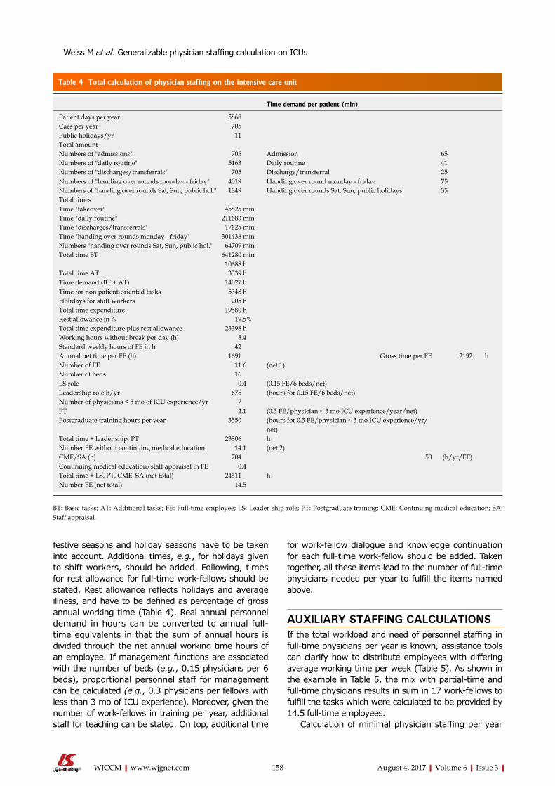
158 August 4, 2017|Volume 6|Issue 3|WJCCM|www.wjgnet.com
festive seasons and holiday seasons have to be taken into account. Additional times, e.g., for holidays given to shift workers, should be added. Following, times for rest allowance for full-time work-fellows should be stated. Rest allowance reflects holidays and average illness, and have to be defined as percentage of gross annual working time (Table 4). Real annual personnel demand in hours can be converted to annual full-time equivalents in that the sum of annual hours is divided through the net annual working time hours of an employee. If management functions are associated with the number of beds (e.g., 0.15 physicians per 6 beds), proportional personnel staff for management can be calculated (e.g., 0.3 physicians per fellows with less than 3 mo of ICU experience). Moreover, given the number of work-fellows in training per year, additional staff for teaching can be stated. On top, additional time
for work-fellow dialogue and knowledge continuation for each full-time work-fellow should be added. Taken together, all these items lead to the number of full-time physicians needed per year to fulfill the items named above.
AUXILIARY STAFFING CALCULATIONSIf the total workload and need of personnel staffing in full-time physicians per year is known, assistance tools can clarify how to distribute employees with differing average working time per week (Table 5). As shown in the example in Table 5, the mix with partial-time and full-time physicians results in sum in 17 work-fellows to fulfill the tasks which were calculated to be provided by 14.5 full-time employees.
Calculation of minimal physician staffing per year
Table 4 Total calculation of physician staffing on the intensive care unit
Time demand per patient (min)
Patient days per year 5868Caes per year 705Public holidays/yr 11Total amountNumbers of "admissions" 705 Admission 65Numbers of "daily routine" 5163 Daily routine 41Numbers of "discharges/transferrals" 705 Discharge/transferral 25Numbers of "handing over rounds monday - friday" 4019 Handing over round monday - friday 75Numbers of "handing over rounds Sat, Sun, public hol." 1849 Handing over rounds Sat, Sun, public holidays 35Total timesTime "takeover" 45825 minTime "daily routine" 211683 minTime "discharges/transferrals" 17625 minTime "handing over rounds monday - friday" 301438 minNumbers "handing over rounds Sat, Sun, public hol." 64709 minTotal time BT 641280 min
10688 hTotal time AT 3339 hTime demand (BT + AT) 14027 hTime for non patient-oriented tasks 5348 hHolidays for shift workers 205 hTotal time expenditure 19580 hRest allowance in % 19.5%Total time expenditure plus rest allowance 23398 hWorking hours without break per day (h) 8.4Standard weekly hours of FE in h 42Annual net time per FE (h) 1691 Gross time per FE 2192 hNumber of FE 11.6 (net 1)Number of beds 16LS role 0.4 (0.15 FE/6 beds/net)Leadership role h/yr 676 (hours for 0.15 FE/6 beds/net)Number of physicians < 3 mo of ICU experience/yr 7PT 2.1 (0.3 FE/physician < 3 mo ICU experience/year/net)Postgraduate training hours per year 3550 (hours for 0.3 FE/physician < 3 mo ICU experience/yr/
net)Total time + leader ship, PT 23806 hNumber FE without continuing medical education 14.1 (net 2)CME/SA (h) 704 50 (h/yr/FE)Continuing medical education/staff appraisal in FE 0.4Total time + LS, PT, CME, SA (net total) 24511 hNumber FE (net total) 14.5
BT: Basic tasks; AT: Additional tasks; FE: Full-time employee; LS: Leader ship role; PT: Postgraduate training; CME: Continuing medical education; SA: Staff appraisal.
Weiss M et al . Generalizable physician staffing calculation on ICUs
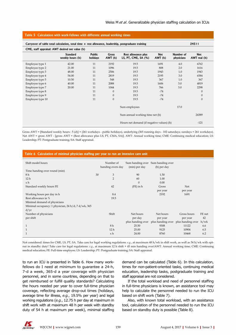
159 August 4, 2017|Volume 6|Issue 3|WJCCM|www.wjgnet.com
to run an ICU is presented in Table 6. How many work-fellows do I need at minimum to guarantee a 24-h, 7-d a week, 365-d a year coverage with physician personnel, and in some countries, depending on that to get reimbursed or fulfill quality standards? Calculating the hours needed per year to cover full-time physician coverage, reflecting average drop-out times (holidays, average time for illness, e.g., 19.5% per year) and legal working regulations (e.g., 12.75 h per day at maximum in shift work with at maximum 48 h per week with standby duty of 54 h at maximum per week), minimal staffing
demand can be calculated (Table 6). In this calculation, times for non-patient-oriented tasks, continuing medical education, leadership tasks, postgraduate training and staff appraisal are not considered.
If the total workload and need of personnel staffing in full-time physicians is known, an assistance tool may help to calculate the personnel needed to run the ICU based on shift work (Table 7).
Also, with known total workload, with an assistance tool, calculation of the personnel needed to run the ICU based on standby duty is possible (Table 8).
Table 5 Calculation with work-fellows with different annual working times
Carryover of table total calculation, total time + rest allowance, leadership, postgraduate training 24511
CME, staff appraisal: AWT desired net value (h)
Standardweekly hours (h)
Public holidays
GrossAWT (h)
Rest allowance plusLS, PT, CME, SA (%)
NetAWT (h)
Number ofphysicians
NetAWT real (h)
Employee type 1 42.00 11 2192 19.5 1691 4.0 6762Employee type 2 21.00 11 1096 19.5 808 2.0 1616Employee type 3 48.00 11 2506 19.5 1943 1.0 1943Employee type 4 54.00 11 2819 19.5 2195 3.0 6584Employee type 5 10.50 11 548 19.5 367 1.0 367Employee type 6 40.00 11 2088 19.5 1606 3.0 4819Employee type 7 20.00 11 1044 19.5 766 3.0 2298Employee type 8 11 0 19.5 -74 0Employee type 9 11 0 19.5 -74 0Employee type 10 11 0 19.5 -74 0
Sum employees 17.0
Sum annual working time net (h) 24389
Hours net demand (if negative values) (h) -121
Gross AWT = [Standard weekly hours : 5 (d)] × (261 workdays - public holidays), underlying (365 running days - 102 saturdays; sundays = 261 workdays). Net AWT = gross AWT - [gross AWT × (Rest allowance plus LS, PT, CMA, SA)]. AWT: Annual working time; CME: Continuing medical education; LS: Leadership; PT: Postgraduate training; SA: Staff appraisal.
Table 6 Calculation of minimal physician staffing per year to run an intensive care unit
Shift model hours Number of handing overs day
Sum handing over (min) per day
Sum handing over (h) per day
Time handing over round (min)8 h 30 3 90 1.5012 h 2 60 1.00x h 0 0.00Standard weekly hours FE 42 (FE) in h Gross Net
per year per yearWorking hours per day in h 8.4 2192 1691Rest allowance in % 19.5Minimal demand of physiciansMinimal occupancy: 1 physician, 24 h/d, 7 d/wk, 365 d/yrNumber of physicians Shift Net hours Net hours Gross hours FE netper shift per day per year per year 42
plus handing over plus handing over plus handing over h/wk1 8 h 25.50 9308 11122 6.61 12 h 25.00 9125 10904 6.51 x h 24.00 8760 10468 6.2
Not considered: times for CME, LS, PT, SA. Take care for legal working regulations: e.g., at maximum 48 h/wk in shift work, as well as 54 h/wk with opt-out in standby duty! Take care for legal regulations: e.g., at maximum 12 h shift + 45 min handing over!AWT: Annual working time; CME: Continuing medical education; FE: Full-time employee; LS: Leadership; PT: Postgraduate training; SA: Staff appraisal.
Weiss M et al . Generalizable physician staffing calculation on ICUs
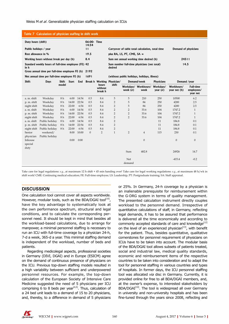
160 August 4, 2017|Volume 6|Issue 3|WJCCM|www.wjgnet.com
DISCUSSIONOne calculation tool cannot cover all aspects worldwide. However, modular tools, such as the BDA/DGAI tool[10], have the key advantage to systematically look at the own performance spectrum, structural and legal conditions, and to calculate the corresponding per-sonnel need. It should be kept in mind that besides all the workload-based calculations, due to arrange for manpower, a minimal personnel staffing is necessary to run an ICU with full-time coverage by a physician 24-h, 7-d a week, 365-d a year. This minimal staffing demand is independent of the workload, number of beds and patients.
Regarding medicolegal aspects, professional societies in Germany (DIVI, DGAI) and in Europe (ESICM) agree on the demand of continuous presence of physicians on the ICU. Previous top-down staffing models resulted in a high variability between sufficient and underpowered personnel resources. For example, the top-down calculation of the European Society of Intensive Care Medicine suggested the need of 5 physicians per ICU comprising 6 to 8 beds per year[4,5]. Thus, calculation of a 24 bed unit leads to a demand of 15 to 20 physicians, and, thereby, to a difference in demand of 5 physicians
or 25%. In Germany, 24-h coverage by a physician is an inalienable prerequisite for reimbursement within the G-DRG system in terms of quality management. The presented calculation instrument directly couples workload to the personnel demand. Irrespective of quantitative calculations of staff, in Germany, reflecting legal demands, it has to be assured that performance is delivered all the time economically and according to commonly accepted standards of care and knowledge[12] on the level of an experienced physician[13], with benefit for the patient. Thus, besides quantitative, qualitative cornerstones for personnel requirement of physicians on ICUs have to be taken into account. The modular basis of the BDA/DGAI tool allows subsets of patients treated, social and industrial law, medical quality standards, economic and reimbursement items of the respective countries to be taken into consideration and to adapt the tool for personnel staffing in various countries and types of hospitals. In former days, the ICU personnel staffing tool was allocated via disc in Germany. Currently, it is provided online for free to all BDA/DGAI members, and, at the owner’s expense, to interested stakeholders by BDA/DGAI[10]. The tool is widespread all over Germany in university and non-university hospitals and has been fine-tuned through the years since 2008, reflecting and
Table 7 Calculation of physician staffing in shift work
Duty hours (shift) 06:00-14:54
Time
Public holidays / year 11 Carryover of table total calculation, total time Demand of physicians
Rest allowance in % 19.5 plus RA, LS, PT, CME, SA =
Working hours without break per day (h) 8.4 Sum net annual working time desired (h) 24511
Standard weekly hours of full-time employee (FE) in h
42 Sum number full-time physicians (net total) desired
14.5
Gross annual time per full-time employee FE (h) 2192
Net annual time per full-time employee FE (h) 1691 (without public holidays, holidays, illness)
Shift Days Shiftmodel
Start End Break h Working hours
without break h
Physician/shift
Demand/week Physicians Demand /year
Workdays/week (n)
Workhours/week
Workdays/ year (n )
Workhours/ year net (h)
Full-time employees/
year net
a. m. shift Weekday 8 h 6:00 14:54 0.5 8.4 5 5 210 250 10500 6.2p. m. shift Weekday 8 h 14:00 22:54 0.5 8.4 2 5 84 250 4200 2.5night shift Weekday 8 h 22:00 6:54 0.5 8.4 2 5 84 250 4200 2.5a. m. shift Weekday 8 h 6:00 14:54 0.5 8.4 2 2 33.6 104 1747.2 1p. m. shift Weekday 8 h 14:00 22:54 0.5 8.4 2 2 33.6 104 1747.2 1night shift Weekday 8 h 22:00 6:54 0.5 8.4 2 2 33.6 104 1747.2 1a. m. shift Public holiday 8 h 6:00 14:54 0.5 8.4 2 11 184.8 0.1p. m. shift Public holiday 8 h 14:00 22:54 0.5 8.4 2 11 184.8 0.1night shift Public holiday 8 h 22:00 6:54 0.5 8.4 2 11 184.8 0.1Senior physician
weekend/Public holiday
8:00 10:00 0 2 1 2 4 115 230 0.1
Inhouse special duty
0:00 0:00 0 0 0
Sum 482.8 24926 14.7
Net demand
-415.4 -0.2
Take care for legal regulations: e.g., at maximum 12 h shift + 45 min handing over! Take care for legal working regulations: e.g., at maximum 48 h/wk in shift work! CME: Continuing medical education; FE: Full-time employee; LS: Leadership, PT: Postgraduate training; SA: Staff appraisal.
Weiss M et al . Generalizable physician staffing calculation on ICUs

161 August 4, 2017|Volume 6|Issue 3|WJCCM|www.wjgnet.com
integrating the feedback of the users. However, studies reflecting improved outcomes or better productivity have not been performed. Feedback to BDA/DGAI revealed that personnel calculations were effectuated
in around 1/3 of the users, transposed partially in 1/3, and not accepted in 1/3. Unfortunately, there is no in total or representative scientific evaluation of personnel staffing in non-university and university hospitals all
Table 8 Calculation of physician staffing in standby duty
Duty hours (shift) 07:15-16:09
Time
Public holidays/year 11 Carryover of table total calculation, total time plus
Demand of physicians
Rest allowance in % 19.5 RA, LS, PT, CME, SA =
Working hours without break per day (h) 8.4 Sum net annual working time desired (h) 24511
Standard weekly hours of full-time employee (FE) in h
42 Sum number full-time physicians (net total) desired
14.5
Gross annual time per full-time employee FE (h)
2192
Net annual time per full-time employee FE (h)
1691 (without public holidays, holidays, illness)
Shift Days Type Start End Break h
Working hours without break h
Physician/shift
Demand physicians/wk Demand physicians/yr
Workdays/wk (n)
Workhours/wk
Workdays/yr (n )
Workhours/ yr net
Core time full-time employee/ yr net
Standby duty full-time employees/yr net
a. m. shift Weekday 7:15 16:09 0.5 8.4 3 5 126 250 6300 3.7p. m. shift Weekday 13:30 22:24 0.5 8.4 2 5 84 250 4200 2.5x shift Weekday 0:00 0:00 5 0 250 0 0a. m. shift Weekday 7:15 16:09 0.5 8.4 0 2 0 104 0 0p. m. shift Weekday 0:00 0:00 2 0 104 0 0x shift Weekday 0:00 0:00 2 0 104 0 0a. m. shift Public
holiday 7:15 16:09 0.5 8.4 2 11 184.8 0.1
p. m. shift Public holiday
0:00 0:00 2 11 0 0
x shift Public holiday
0:00 0:00 2 11 0 0
Standby duty
Weekday 1 0:00 0:00 0 5 0.0 250 0 0
Standby duty
Weekend 1 0:00 0:00 0 2 0.0 104 0 0
Standby duty
Public holiday
1 0:00 0:00 0 11 0 0
Standby duty
Weekday 2 16:09 8:00 0 15.85 2 5 158.5 250 7925 4.7
Standby duty
Weekend 2 7:15 8:00 0 24.75 2 2 99 104 5148 3
Standby duty
Public holiday
2 7:15 8:00 0 24.75 2 11 544.5 0.3
Senior physician
Weekend /public holiday
8:00 10:00 0 2 1 2 4 115 230 0.1
Inhouse special duty
0:00 0:00 0 0 0
Sum 261.5 24532.3 6.3 8.2Sum core time, standby duty + special duties full-time employees net
14.5
Net demand
-21.7 0
CME: Continuing medical education; FE: Full-time employee; LS: Leadership; PT: Postgraduate training; SA: Staff appraisal.
Weiss M et al . Generalizable physician staffing calculation on ICUs
162 August 4, 2017|Volume 6|Issue 3|WJCCM|www.wjgnet.com
over Germany which could reflect the gap between the calculations done by the tool and the actual personnel staffing of the ICUs. Moreover, whether staffing differences from basic and regular care up to maximal care hospitals result in better productivity or improved outcome in Germany is still a matter of debate. However, quality of care, length of stay and mortality in ICUs has been reported to be highly dependent on organisational structures, personnel staffing and qualification of physicians[9,14,15]. Reductions in personnel staffing are counterproductive if safety for patients and staff, and efficiency of processes decline[16-19], and/or the costs for materials increase[18,20]. Furthermore, it has to be taken into account that optimal reduction in errors is expected with a 85% average utilisation of an ICU with 100% of personnel staffing[19]. To achieve optimal quality, physician staffing has been claimed as follows[5,21]: The ICU has to be under a qualified, uniform, physician organised guidance, e.g., by a physician of a specialty which has intensive care medicine as an integrated part, such as anaesthesia, surgery, internal medicine, and who has special certification in intensive care medicine. The leader of the ICU should not be in other duties in his hospital, devoted full-time or at least 75% of time to intensive care[5,21].
To find out whether timings for tasks are realistic, in the ICU personnel staffing tool, we proceeded as follows. To determine duration of tasks to be performed, estimations by experts’ opinion (10 leaders of ICUs), a survey in 200 ICUs in Germany (practitioning ICU physicians), and real time measurements on a surgical and a medical university and a non-university interdisciplinary ICU of a basic and regular care hospital have been compared[22]. In 20%, expert opinion survey and measured times were consistent. Differences, such as higher values for daily routine in the basic care non-university hospital, may be explained by different process operations on the various wards. Thus, necessary time requirements depend on the comparability of basic prerequisites, process operations, structural and legal conditions. Therefore, cited timings for tasks can serve as an indication for time requirements, however, have to be verified, at best with real time measurements in the own structural conditions and process operations.
Tasks beyond the ICU, such as initial trauma care, care for in-hospital emergencies or engagement as external emergency physician, should not be incorporated in the staffing calculation of the ICU, but calculated separately. Quantitative and qualitative cornerstones for personnel requirement of physicians in anaesthesia reflecting recent legal rights of patients in Germany, meeting legal demands of therapeutic quality, and, thus, serving patient safety, have been published in 2015 by the German Society of Anesthesiologists (BDA) and the German Society of Anesthesiology and Intensive Care Medicine (DGAI)[23]. Subsequently, the current Excel-based calculation tool version (2015) regarding physician staffing in anaesthesia has been published, especially reflecting recent laws governing physician’s working conditions and competence
in the field of anaesthesia, as well as demands of strengthened legal rights of patients, patient care and safety[24].
CONCLUSIONWorkload-oriented models of physician staffing with generalizable items taking into account quality, efficiency of processes, legal, educational, con-trolling, local, organisational and economic aspects, differentiating basic effort, additional effort, and non patient-oriented tasks, may help to justify realistic personnel staffing demands. Modular calculation models may serve to individualise generalizable aspects to various types of hospitals, process operations, structural and legal conditions, as well as funding and refunding systems, resulting in broadly use and acceptance by various stakeholders all around the world. In the future, it should be evaluated whether this model may lead to improvement of patient safety and quality of management.
ACKNOWLEDGMENTSWe thank Vagts DA, Schleppers A, Leidinger W, Sehn N and Klöss T of the working group “personnel management of BDA and DGAI” for their constructive contribution to develop and update the workmanship oriented modular calculation model for personnel staffing of physicians in the ICU in 2008 and in 2012. We thank Clair Hartmann, MD, working in our Department of Anesthesiology in Ulm, for checking the manuscript as a native speaker.
REFERENCES1 Engel C, Brunkhorst FM, Bone HG, Brunkhorst R, Gerlach H, Grond
S, Gruendling M, Huhle G, Jaschinski U, John S, Mayer K, Oppert M, Olthoff D, Quintel M, Ragaller M, Rossaint R, Stuber F, Weiler N, Welte T, Bogatsch H, Hartog C, Loeffler M, Reinhart K. Epidemiology of sepsis in Germany: results from a national prospective multicenter study. Intensive Care Med 2007; 33: 606-618 [PMID: 17323051 DOI: 10.1007/s00134-006-0517-7]
2 Halpern NA, Pastores SM, Greenstein RJ. Critical care medicine in the United States 1985-2000: an analysis of bed numbers, use, and costs. Crit Care Med 2004; 32: 1254-1259 [PMID: 15187502]
3 Weißauer W. Ausstattung und Organisation interdisziplinärer operativer Intensiveinheiten. Gemeinsame Empfehlungen der Deutschen Gesellschaft für Anästhesiologie und Intensivmedizin und des Berufsverbandes Deutscher Anästhesisten sowie der Deutschen Gesellschaft für Chirurgie und des Berufsverbandes der Deutschen Chirurgen. Deutsche Gesellschaft für Chirurgie - Mitteilungen 2005; 232-235. Available from: URL: https://www.dgai.de/publikationen/vereinbarungen
4 Ferdinande P. Recommendations on minimal requirements for Intensive Care Departments. Members of the Task Force of the European Society of Intensive Care Medicine. Intensive Care Med 1997; 23: 226-232 [PMID: 9069011]
5 Valentin A, Ferdinande P; Improvement EWGoQ. Recommendations on basic requirements for intensive care units: structural and organizational aspects. Intensive Care Med 2011; 37: 1575-1587 [PMID: 21918847 DOI: 10.1007/s00134-011-2300-7]
6 Haupt MT, Bekes CE, Brilli RJ, Carl LC, Gray AW, Jastremski MS, Naylor DF, PharmD MR, Md AS, Wedel SK, Md MH. Guidelines
Weiss M et al . Generalizable physician staffing calculation on ICUs
163 August 4, 2017|Volume 6|Issue 3|WJCCM|www.wjgnet.com
on critical care services and personnel: Recommendations based on a system of categorization of three levels of care. Crit Care Med 2003; 31: 2677-2683 [PMID: 14605541 DOI: 10.1097/01.CCM.0000094227.89800.93]
7 Dorman T, Angood PB, Angus DC, Clemmer TP, Cohen NH, Durbin CG, Falk JL, Helfaer MA, Haupt MT, Horst HM, Ivy ME, Ognibene FP, Sladen RN, Grenvik AN, Napolitano LM. Guidelines for critical care medicine training and continuing medical education. Crit Care Med 2004; 32: 263-272 [PMID: 14707590 DOI: 10.1097/01.CCM.0000104916.33769.9A]
8 Bundesärztekammer. Fallpauschalen-System wird zum Glücksspiel. Pressemitteilung zum 107 Deutschen Ärztetag Bremen 2004 vom 04.11.2005. Available from: URL: http://www.baek.de/home/themena-z/Ärztetag/107.dÄt/presse/drg
9 Weiss M, Marx G, Vagts D, Leidinger W, Sehn N, Iber T. Personnel requirement planning in intensive care medicine in the age of DRGs – A new performance-oriented calculation model. Anästh Intensivmed 2008; 49 Suppl 4: S41-S51. Available from: URL: http://www.ai-online.info/aionline/Supplements;jsessionid=CFC6779ABB90C0692A2027498412B0F8
10 Weiss M, Marx G, Vagts DA, Schleppers A, Leidinger W, Sehn N, Klöss T, Iber T. Calculation of personnel requirement “intensive care medicine 2012-Revision of the 2008 calculation tool for the medical profession. Anästh Intensivmed 2012; 53 Suppl 3: S50-S62 [ISSN0170-5334102330]. Available from: URL: http://www.ai-online.info/aionline/Supplements;jsessionid=CFC6779ABB90C0692A2027498412B0F8
11 BGBl. Gesetz über Medizinprodukte (Medizinproduktegesetz – MPG) in der Fassung der Bekanntmachung vom 7. August 2002, BGBl I S. 3146, geändert am 25. November 2003, BGBl I S. 2304. Available from: URL: https://www.gesetze-im-internet.de/bundesrecht/mpg/gesamt.pdf
12 Federal Ministry of Justice and Consumer Protection. SGB V §§ 2, 70 Abs. 1. Available from: URL: https://www.gesetze-im-internet.de/sgb_5/__70.html
13 OLG Düsseldorf. Urteil vom 20.10.1985-8 U 100/83, 198514 Knaus WA, Wagner DP, Zimmerman JE, Draper EA. Variations in
mortality and length of stay in intensive care units. Ann Intern Med 1993; 118: 753-761 [PMID: 8470850]
15 Pronovost PJ, Angus DC, Dorman T, Robinson KA, Dremsizov TT, Young TL. Physician staffing patterns and clinical outcomes in critically ill patients: a systematic review. JAMA 2002; 288: 2151-2162
[PMID: 12413375]16 Iapichino G, Gattinoni L, Radrizzani D, Simini B, Bertolini G, Ferla L,
Mistraletti G, Porta F, Miranda DR. Volume of activity and occupancy rate in intensive care units. Association with mortality. Intensive Care Med 2004; 30: 290-297 [PMID: 14685662 DOI: 10.1007/s00134-003-2113-4]
17 Tibby SM, Correa-West J, Durward A, Ferguson L, Murdoch IA. Adverse events in a paediatric intensive care unit: relationship to workload, skill mix and staff supervision. Intensive Care Med 2004; 30: 1160-1166 [PMID: 15067503 DOI: 10.1007/s00134-004-2256-y]
18 Tarnow-Mordi WO, Hau C, Warden A, Shearer AJ. Hospital mortality in relation to staff workload: a 4-year study in an adult intensive-care unit. Lancet 2000; 356: 185-189 [PMID: 10963195]
19 Friesdorf W, Göbel M. Safety and Reliability of clinical work processes. In: Strasser H, Kluth K, Rausch H, Bubb H. Quality of Work and Products in Enterprises of the Future. Stuttgart: Ergonomia Verlag, 2003: 669-672
20 Reinhart K. [Genuine progress in intensive care medicine must be payable also in future--a plea for a public debate]. Anasthesiol Intensivmed Notfallmed Schmerzther 2004; 39: 187-190 [PMID: 15098165 DOI: 10.1055/s-2004-814413]
21 Young MP, Birkmeyer JD. Potential reduction in mortality rates using an intensivist model to manage intensive care units. Eff Clin Pract 2000; 3: 284-289 [PMID: 11151525]
22 Miller FK, Iber T, Weiss M, Marx G, Uhlig E, Noky T, Henschel J, Vagts DA. Comparison of medical services based on expert opinion, survey and actual measurements to determine human resources. Intensive Care Med 2008; 34 Supp 1: S72
23 BDAktuell. | DGAInfo. Aus den Verbänden. Geschäftsführung BDA/DGAI: Behandlungsqualität und Patientensicherheit: Eckpunkte zur ärztlich-personellen Ausstattung anästhesiologischer Arbeitsplätze in Krankenhäusern. Beschluss des Erweiterten Präsidiums der DGAI vom 20.11.2014 sowie von Präsidium und Ausschuss des BDA vom 15.12.2014. Anästh Intensivmed 2015; 56: 145-154. Available from: URL: http://www.dgai.de/publikationen/shop
24 Weiss M, Rossaint R, Iber T. Calculation of personnel requirements in the field of anaesthesia for 2015 – Revision of the 2009 calculation basics applicable to medical services. Anästh Intensivmed 2015; 56: 1-18. Available from: URL: http://www.dgai.de/publikationen/shop
P- Reviewer: Krishnan T, Lin JA S- Editor: Song XX L- Editor: A E- Editor: Lu YJ
Weiss M et al . Generalizable physician staffing calculation on ICUs
Lorena Fernandez-Restrepo, Lauren Shaffer, Bravein Amalakuhan, Marcos I Restrepo, Jay Peters, Ruben Restrepo
EVIDENCE-BASED MEDICINE
164 August 4, 2017|Volume 6|Issue 3|WJCCM|www.wjgnet.com
Effects of intrapulmonary percussive ventilation on airway mucus clearance: A bench model
Lorena Fernandez-Restrepo, Lauren Shaffer, Bravein Am-alakuhan, Marcos I Restrepo, Jay Peters, Ruben Restrepo, Division of Pediatric Critical Care, Division of Pulmonary and Critical Care, and Department of Respiratory Care, University of Texas Health Science Center and the South Texas Veterans Health Care System, San Antonio, TX 78240, United States
Author contributions: All authors contributed equally to the literature search, data collection, study design and analysis, manuscript preparation and final review.
Conflict-of-interest statement: None of the authors have any conflicts of interest.
Data sharing statement: There is no technical appendix for this manuscript because the study was extensively described within the manuscript itself. The statistical codes and the raw dataset were also included within the manuscript.
Open-Access: This article is an open-access article which was selected by an in-house editor and fully peer-reviewed by external reviewers. It is distributed in accordance with the Creative Commons Attribution Non Commercial (CC BY-NC 4.0) license, which permits others to distribute, remix, adapt, build upon this work non-commercially, and license their derivative works on different terms, provided the original work is properly cited and the use is non-commercial. See: http://creativecommons.org/licenses/by-nc/4.0/
Manuscript source: Invited manuscript
Correspondence to: Dr. Bravein Amalakuhan, MD, Division of Pediatric Critical Care, Division of Pulmonary and Critical Care, and Department of Respiratory Care, University of Texas Health Science Center and the South Texas Veterans Health Care System, 7400 Merton Minter Blvd, San Antonio, TX 78240, United States. [email protected]: +1-210-5675792Fax: +1-210-9493006
Received: May 7, 2017 Peer-review started: May 10, 2017 First decision: May 23, 2017
Revised: June 1, 2017 Accepted: June 30, 2017Article in press: July 3, 2017Published online: August 4, 2017
AbstractAIMTo determine the ability of intrapulmonary percussive ventilation (IPV) to promote airway clearance in spon-taneously breathing patients and those on mechanical ventilation.
METHODSAn artificial lung was used to simulate a spontaneously breathing patient (Group 1), and was then connected to a mechanical ventilator to simulate a patient on mechanical ventilation (Group 2). An 8.5 mm endotracheal tube (ETT) connected to the test lung, simulated the patient airway. Artificial mucus was instilled into the mid-portion of the ETT. A filter was attached at both ends of the ETT to collect the mucus displaced proximally (mouth-piece filter) and distally (lung filter). The IPV machine was attached to the proximal end of the ETT and was applied for 10-min each to Group 1 and 2. After each experiment, the weight of the various circuit components were determined and compared to their dry weights to calculate the weight of the displaced mucus.
RESULTSIn Group 1 (spontaneously breathing model), 26.8% ± 3.1% of the simulated mucus was displaced proximally, compared to 0% in Group 2 (the mechanically ventilated model) with a P-value of < 0.01. In fact, 17% ± 1.5% of the mucus in Group 2 remained in the mid-portion of the ETT where it was initially instilled and 80% ± 4.2% was displaced distally back towards the lung (P < 0.01). There was an overall statistically significant amount of mucus
World Journal ofCritical Care MedicineW J C C M
Submit a Manuscript: http://www.f6publishing.com
DOI: 10.5492/wjccm.v6.i3.164
World J Crit Care Med 2017 August 4; 6(3): 164-171
ISSN 2220-3141 (online)
165 August 4, 2017|Volume 6|Issue 3|WJCCM|www.wjgnet.com
Fernandez-Restrepo L et al . IPV and airway mucus clearance
movement proximally towards the mouth-piece in the spontaneously breathing (SB) patient. There was also an overall statistically significant amount of mucus movement distally back towards the lung in the mechanically ventilated (MV) model. In the mechanically ventilated model, no mucus was observed to move towards the proximal/mouth piece section of the ETT.
CONCLUSIONThis bench model suggests that IPV is associated with displacement of mucus towards the proximal mouthpiece in the SB patient, and distally in the MV model.
Key words: Mucus; Sputum; Mechanical ventilators; Percussion; Respiratory drainage; Breathing exercises
© The Author(s) 2017. Published by Baishideng Publishing Group Inc. All rights reserved.
Core tip: Many respiratory conditions result in increased respiratory secretions and poor clearance, and are associated with poor patient outcomes. Intrapulmonary percussive ventilation (IPV) is an airway clearance technique that has become increasingly used over the last few years, however there is a paucity of data to support its efficacy. Using a simulated bench model, we found that IPV is associated with movement of mucus towards the mouth in the spontaneously breathing patient and thus supporting airway clearance. Interestingly, in patients on mechanical ventilation, IPV mainly displaced mucus distally into the lungs and thus may be harmful in this patient population.
Fernandez-Restrepo L, Shaffer L, Amalakuhan B, Restrepo MI, Peters J, Restrepo R. Effects of intrapulmonary percussive ventilation on airway mucus clearance: A bench model. World J Crit Care Med 2017; 6(3): 164-171 Available from: URL: http://www.wjgnet.com/2220-3141/full/v6/i3/164.htm DOI: http://dx.doi.org/10.5492/wjccm.v6.i3.164
INTRODUCTIONMany chronic conditions such as bronchiectasis, cystic fibrosis (CF), neuromuscular disease and chronic obstructive pulmonary disease (COPD) are associated with an increase in both the quantity and viscosity of respiratory secretions. Other conditions are associated with a decreased ability to clear secretions, such as those with impaired ciliary function or cough, with the latter being very common during mechanical ventilation, after strokes or surgical procedures, and in neuromuscular disorders[1]. Previous studies have shown that when these secretions are not adequately cleared, complications arise such as atelectasis, mucus plugging, and recurrent pneumonia[1]. Inadequate mucus clearance in patients in the intensive care unit (ICU) can lead to poor clinic outcomes such as prolonged time on mechanical ventilation, increase in need for tracheostomies, de
creased quality of life, overall worsening lung function and an increase in mortality[26]. Administration of airway clearance therapies (ACTs) involve the use of manual techniques coupled with postural drainage, breathing exercises and mechanical devices to improve patient outcomes and optimize recovery after acute illnesses[7]. However, there are few studies on the optimal ACT and under which clinical settings they are most effective[8,9].
Intrapulmonary percussive ventilation (IPV) is one such ACT that has recently become increasingly utilized in hospitalized patients. During IPV treatments, the patient breathes through an accessory device called a Phasitron®, which delivers rapid, high flow, mini-bursts (percussions) of tidal volumes into the lungs while simultaneously delivering therapeutic aerosols. In the clinical setting, IPV can be administered by mouthpiece, mask or endotracheal tubes (ETTs). This technique is also thought to improve expiratory flow by opening collapsed airways, thus promoting mucus clearance.
Several reports have suggested that IPV facilitates airway clearance and improves ventilation in patients with conditions such as cystic fibrosis[1014], neuromuscular disorders[15,16], atelectasis[1719], inhalation injury[2022], and COPD[2326]. To our knowledge there has only been one study that has evaluated the efficacy of IPV as an ACT in spontaneously breathing patients, and it illustrated a positive benefit[27]. When oscillating devices such as IPV were compared to conventional physiotherapy for airway clearance in people with cystic fibrosis, the most recent Cochrane metaanalysis found little evidence to support the use of any particular oscillating device for airway clearance over any other ACT modality[28]. The handful of other studies that exist have compared IPV to conventional chest physical therapy and showed no additional benefit with IPV. These results raise the question of whether IPV indeed is able to act as an effective ACT or whether its benefit is simply theoretical[14,29]. Furthermore, to our knowledge only one study has evaluated the efficacy of IPV in mechanically ventilated patients, and although it showed some benefit, it was completed only in a specific population of eight patients with neuromuscular disease[15]. Despite IPV’s widespread use as an ACT across numerous clinical settings, there are a paucity of data to document its benefit.
The objective of this study was to evaluate the ability of IPV to promote airway clearance in both spontaneously breathing patients and those on mechanical ventilation using a controlled simulated bench model.
MATERIALS AND METHODSArtificial test lungThe artificial test lung was obtained from “Michigan Instruments” (Grand Rapids, MI) and utilized in both the spontaneously breathing (SB) group/model and the mechanically ventilated (MV) group/model. This artificial test lung has been validated and mimics the human lung in many ways. First, the artificial test lung
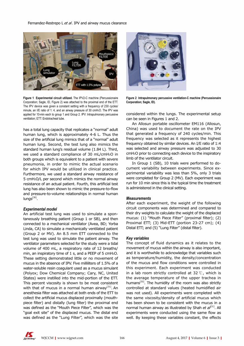
166 August 4, 2017|Volume 6|Issue 3|WJCCM|www.wjgnet.com
has a total lung capacity that replicates a “normal” adult human lung, which is approximately 46 L. Thus the size of the artificial lung mimics that of a “normal” adult human lung. Second, the test lung also mimics the standard human lung’s residual volume (1.84 L). Third, we used a standard compliance of 30 mL/cmH2O in both groups which is equivalent to a patient with severe pneumonia, in order to mimic the actual scenario for which IPV would be utilized in clinical practice. Furthermore, we used a standard airway resistance of 5 cmH2O/L per second which mimics the normal airway resistance of an actual patient. Fourth, this artificial test lung has also been shown to mimic the pressure-to-flow and pressuretovolume relationships in normal human lungs[30].
Experimental modelAn artificial test lung was used to simulate a spontaneously breathing patient (Group 1 or SB), and then connected to a mechanical ventilator (Avea, BD; Yorba Linda, CA) to simulate a mechanically ventilated patient (Group 2 or MV). An 8.5 mm ETT connected to the test lung was used to simulate the patient airway. The ventilator parameters selected for the study were a tidal volume of 400 mL, a respiratory rate of 12 breaths/min, an inspiratory time of 1 s, and a PEEP of 5 cmH2O. These setting demonstrated little or no movement of mucus in the absence of IPV. Five milliliters of 1.5% of a watersoluble resin coagulant used as a mucus simulant (Polyox; Dow Chemical Company; Cary, NC, United States) were instilled into the midportion of the ETT. This percent viscosity is shown to be most consistent with that of mucus in a normal human airway[31]. An anesthesia filter was attached at both ends of the ETT to collect the artificial mucus displaced proximally (mouth-piece filter) and distally (lung filter) the proximal end was defined as the “Mouth Piece Filter”, which was the “goal exit site” of the displaced mucus. The distal end was defined as the “Lung Filter”, which was the site
considered within the lungs. The experimental setup can be seen in Figures 1 and 2.
An Allosun portable oscillometer EM116 (Allosun, China) was used to document the rate on the IPV that generated a frequency of 240 cycles/min. This frequency was selected as it represents the highest frequency obtained by similar devices. An I/E ratio of 1:4 was selected and airway pressure was adjusted to 30 cmH2O prior to connecting each device to the inspiratory limb of the ventilator circuit.
In Group 1 (SB), 10 trials were performed to document variability between experiments. Since experimental variability was less than 5%, only 3 trials were completed for Group 2 (MV). Each experiment was run for 10 min since this is the typical time the treatment is administered in the clinical setting.
MeasurementsAfter each experiment, the weight of the following circuit components was determined and compared to their dry weights to calculate the weight of the displaced mucus: (1) “Mouth Piece Filter” (proximal filter); (2) Proximal ETT; (3) MidETT (portion 2327 cm); (4) Distal ETT; and (5) “Lung Filter” (distal filter).
Key variablesThe concept of fluid dynamics as it relates to the movement of mucus within the airway is also important, and it is worthwhile to acknowledge that variables such as temperature/humidity, the density/concentration of the mucus and flow conditions were controlled in this experiment. Each experiment was conducted in a lab room strictly controlled at 32 ℃, which is the average temperature of the upper trachea in humans[32]. The humidity of the room was also strictly controlled at standard values (heated humidified air was not used). All experiments were completed with the same viscosity/density of artificial mucus which has been shown to be consistent with the mucus in a normal human airway as illustrated by Shah et al[31]. All experiments were conducted using the same flow as well. By keeping these variables constant, the effects
Figure 1 Experimental circuit utilized. The IPV2-C machine (Percussionaire Corporation; Sagle, ID, Figure 2) was attached to the proximal end of the ETT. The IPV device was given a constant setting with a frequency of 230 cycles/minute, an I/E ratio of 1: 4, and an airway pressure of 30 cmH2O. The IPV was applied for 10-min each to group 1 and Group 2. IPV: Intrapulmonary percussive ventilation; ETT: Endotracheal tube.
Figure 2 Intrapulmonary percussive ventilation-C machine (Percussionaire Corporation; Sagle, ID).
Fernandez-Restrepo L et al . IPV and airway mucus clearance
MouthpieceFILTER
IPV
ETT with 1.5% polyox
LungFILTER
Lungsimulator

167 August 4, 2017|Volume 6|Issue 3|WJCCM|www.wjgnet.com
of the intervention (use of IPV) in both groups could be evaluated within a constant/replicable environment/context.
Statistical analysisDue to the limited sample size used in this study, the distribution of the data were not distributed normally, and for this reason only nonparametric tests were used[33]. All data are presented as medians with interquartile ranges (IQR), or means with standard deviations (SD) as appropriate. Paired nonparametric MannWhitney U test, Kruskal Wallis test or twotailed Student’s t-test were used to compare data at different tube distances. All statistical calculations were performed by a trained and expert biostatistician, using Prism 5 software (GraphPad) and SPSS 21.0. P values < 0.05 were considered statistically significant.
RESULTSIn Group 1 (spontaneously breathing model), 26.8% ± 3.1% of the simulated mucus was displaced proximally, compared to 0% in Group 2 (the mechanically ventilated
model). In fact, 17% ± 1.5% of the mucus in Group 2 remained in the midportion of the ETT where it was initially instilled and 80% ± 4.2% was displaced distally back towards the lung. There was an overall statistically significant amount of mucus movement proximally to-wards the mouthpiece in the spontaneously breathing patient. There was also an overall statistically significant amount of mucus movement distally back towards the lung in the mechanically ventilated model. The amounts of mucus measured within each section of the circuit is shown in Table 1 and Figure 3.
DISCUSSIONThe results from this study suggest that when IPV is used in a simulated model of a spontaneously breathing patient, it is associated with a statistically significant amount of mucus movement proximally, and thus supports airway clearance. In contrast, when IPV is used in the simulated model of a mechanically ventilated patient, it was found to be associated almost exclusively with the displacement of mucus distally back towards the lung, and thus did not support airway clearance.
100
80
60
40
20
0
% D
ista
nce
chan
ge
e
ns a
Distal Center Proximal
100
80
60
40
20
0
% D
ista
nce
chan
ge
Distal Center ProximalTest tube position
f
f b
Test tube position
100
80
60
40
20
0
% D
ista
nce
chan
ge
f
f f
ff
Lung Distal Center Proximal MouthTest tube position
100
80
60
40
20
0
% D
ista
nce
chan
ge
f
f ns
ff
Lung Distal Center Proximal MouthTest tube position
Figure 3 Percent of distance of mucus movement in the different portion of the endotracheal tube in both simulated intrapulmonary percussive ventilation models: mechanical ventilation (A and B) and spontaneous breathing (C and D). There was an overall statistically significant amount of mucus movement proximally towards the mouth-piece in the spontaneously breathing patient. There was also an overall statistically significant amount of mucus movement distally back towards the lung in the mechanically ventilated model. Statistical significant P values: aP < 0.05, bP < 0.01, eP < 0.001, fP < 0.0001. ns: No statistical significance.
Fernandez-Restrepo L et al . IPV and airway mucus clearance
A B
C D

168 August 4, 2017|Volume 6|Issue 3|WJCCM|www.wjgnet.com
IPV utilizes tidal volumes delivered at high oscillatory frequencies to loosen mucus and help with expectoration. Previous bench models have shown that highfrequency oscillations not only dislodge bronchial secretions from the walls of the airway but also reduce its actual viscosity making it easier to clear[34]. In a way these oscillations act like a “physical mucolytic”. One clinical study has shown that applying highfrequency oscillations directly to the airway opening of spontaneously breathing patients does indeed enhance secretion clearance compared to no therapeutic intervention[27]. Our study confirms the results from this study that IPV indeed improves mucus clearance in a simulated model of a spontaneously breathing patient. However, it is worth noting that when IPV has been compared to conventional chest physical therapy in spontaneously breathing patients, there appears to be no difference in efficacy of mucus clearance. In a study of 20 clinically stable CF patients, IPV was not associated with increased sputum clearance compared to conventional chest physical therapy[14].
Another study of 22 stable patients with bronchiectasis found similar results[29]. Taking the results from this current study and the study by George et al[27], IPV indeed improves sputum clearance; however, it may not provide added benefit compared to conventional chest physical therapy. However, the major value of IPV when compared to conventional chest physical therapy appears to be its increased tolerability, patient preference, replicability of its benefits and greater adherence to therapy, considering that conventional chest physical therapy is often uncomfortable and requires an experienced/trained individual to aid the patient for optimal results[29]. Since the benefit of chest physical therapy depends on the patient and the therapist, the benefits seen in the studies by Van Ginderdeuren et al[14] and Paneroni et al[29] may be difficult to replicate, making IPV more advantageous. Thus IPV may be a more effective clinical alternative for airway clearance in spontaneously breathing patients, although this requires further clinical studies to validate.
Similarly, there are few studies evaluating the efficacy of IPV as an ACT in mechanically ventilated patients. In one small observational study conducted in 8 patients with Duchenne Muscular Dystrophy who were ventilator dependent and had tracheostomies, IPV was shown to increase the quantity of mucus
clearance[15]. Our study showed the opposite findings, and in fact showed that more than 80% of the mucus was displaced distally back towards the lung. The biologic plausibility of why IPV may be detrimental in patients on invasive MV is important to understand considering these negative consequences. It is possible that the interplay between the positive pressure from the MV and the percussive oscillatory pressure waves from the IPV machine created a flow that was directed distally rather than proximally towards the opening of the airway. While this finding was noted in our study, it is not clear if this occurs in vivo. But considering that the majority of the mucus (approximately 80%) was displaced distally in the MV group, and that our artificial test lung and bench model has been validated and extensively used by prior studies, it raises valid concerns about its safety that requires further testing[3537]. IPV is currently being used in mechanically ventilated patients at variable driving pressures and oscillatory frequencies (our study chose the most common setting used clinically) and its use is growing exponentially. We hope the results of this bench study will result in additional future studies.
There are several limitations of this study. One of which is that in vitro models do not perfectly reflect the flow characteristics of a spontaneously breathing patient. Our in-vitro model did not measure the impact of the treatment effects within a chest cavity where recoil of the chest plays a significant role in increasing expiratory flows. If active expiration were to be simulated as happens in the spontaneously breathing patient, higher expiratory flows could have enhanced mucus transport to either the proximal end of the ETT or the filter representing the mouthpiece. However, there are many advantages to studying IPV in this simplified bench model that would be difficult to evaluate in an actual clinical setting. For example, using this simulated model, we can directly measure the amount of mucus in the airway and directly determine the amount displaced in either direction. In patients, we cannot control for the actual amount of mucus in the airway at time zero because this will vary from hourtohour and daytoday. Furthermore, different patients will differ in their baseline amount of mucus production based on complex physiological mechanisms and differences in their disease processes. This simplified model allows control of many factors that cannot be controlled in an in-vivo model. One of the main reasons for performing this study is that many Health Care Professionals accept that IPV is beneficial in both spontaneously breathing and mechanically ventilated patients with almost no data to document or substantiate its actual benefit. Our goal was to raise awareness through a bench study and create interest in furthering clinical research in this area.
Another potential limitation worth mentioning is that our model of the human lung did not contain cilia. An important question is whether IPV may interact with human cilia and in a manner our model could
Table 1 Amount of mucus within each section of the circuit
Location Group 1 (%) Group 2 (%)
Mouth piece/proximal filter 0 0Proximal ETT 26.8 ± 0.63 0Mid-ETT (portion 23-27cm) 0 17 ± 1.04Distal ETT 51.2 ± 1.75 80 ± 4.2Lung/distal filter 22 ± 0.55 3 ± 1.04
ETT: Endotracheal tube.
Fernandez-Restrepo L et al . IPV and airway mucus clearance
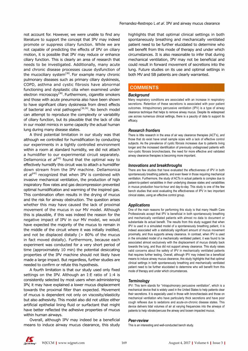
169 August 4, 2017|Volume 6|Issue 3|WJCCM|www.wjgnet.com
not account for. However, we were unable to find any literature to support the concept that IPV may indeed promote or suppress ciliary function. While we are not capable of predicting the effects of IPV on ciliary motion, it is possible that IPV may reduce or enhance ciliary function. This is clearly an area of research that needs to be investigated. Additionally, many acute and chronic disease processes cause dysfunction of the mucociliary system[38]. For example many chronic pulmonary diseases such as primary ciliary dyskinesis, COPD, asthma and cystic fibrosis have abnormal functioning and dysplastic cilia when examined under electron microscopy[38]. Furthermore, cigarette smokers and those with acute pneumonia also have been shown to have significant ciliary dyskinesia from direct effects of bacterial and viral pathogens[38,39]. No bench model can attempt to reproduce the complexity or variability of ciliary function, but its plausible that the lack of cilia in our model mimics in some capacity the actual human lung during many disease states.
A third potential limitation in our study was that although we controlled for humidification by conducting our experiments in a tightly controlled environment within a room at standard humidity, we did not attach a humidifier to our experimental circuit separately. Dellamonica et al[40] found that the optimal way to effectively humidify this circuit was to attach a humidifier down stream from the IPV machine. Dellamonica et al[40] recognized that when IPV is combined with invasive mechanical ventilation, the production of high inspiratory flow rates and gas decompression prevented optimal humidification and warming of the inspired gas. This combination often results in the drying of mucus and the risk for airway obstruction. The question arises whether this may have caused the lack of proximal movement of the mucus in our MV model. Although this is plausible, if this was indeed the reason for the negative impact of IPV in our MV model, we would have expected the majority of the mucus to remain in the middle of the circuit where it was initially instilled, and not be displaced distally (> 80% of the mucus in fact moved distally). Furthermore, because each experiment was conducted for a very short period of time (approximately 10 min) the potential desiccating properties of the IPV machine should not likely have made a large impact. But regardless, further studies are needed to confirm or refute this hypothesis.
A fourth limitation is that our study used only fixed settings on the IPV. Although an I:E ratio of 1:4 is consistently selected by most users when administering IPV, it may have explained a lower mucus displacement towards the proximal filter than expected. Movement of mucus is dependent not only on viscosity/elasticity but also adhesivity. This model also did not utilize either artificial epithelial lining fluid or surfactant that might have better reflected the adhesive properties of mucus within human airways.
Overall, although IPV may indeed be a beneficial means to induce airway mucus clearance, this study
highlights that that optimal clinical settings in both spontaneously breathing and mechanically ventilated patient need to be further elucidated to determine who will benefit from this mode of therapy and under which circumstances. It is also reasonable to infer that during mechanical ventilation, IPV may not be beneficial and could result in forward movement of secretions into the lung. Future studies on its use and optimal settings in both MV and SB patients are clearly warranted.
COMMENTSBackgroundMany respiratory conditions are associated with an increase in respiratory secretions. Retention of these secretions is associated with poor patient outcomes. Intrapulmonary percussive ventilation (IPV) is a type of airway clearance technique that helps to remove airway mucus. Despite its widespread use across numerous clinical settings, there is a paucity of data to support its efficacy.
Research frontiersThere is little research in the area of air way clearance therapies (ACT’s), and those that do exist have small sample sizes with a lack of effective control subjects. As the prevalence of cystic fibrosis increases due to patients living longer and the increased identification of previously undiagnosed patients with non-cystic fibrosis bronchiectasis, the need for more effective and validated airway clearance therapies is becoming more important.
Innovations and breakthroughsThere are few studies that have evaluated the effectiveness of IPV in both spontaneously breathing patients, and even fewer in those requiring mechanical ventilation. Furthermore, the study of ACTs in actual patients is complex due to patient-to-patient variabilities in their underlying disease states and variabilities in mucus production hour-to-hour and day-to-day. This study is one of the few bench studies that exist evaluating the effectiveness of IPV in two important clinical states, using an effective control group.
ApplicationsOne of the main reasons for performing this study is that many Health Care Professionals accept that IPV is beneficial in both spontaneously breathing and mechanically ventilated patients with almost no data to document or substantiate its actual benefit. The results from this study suggest that when IPV is used in a simulated model of a spontaneously breathing patient, it is indeed associated with a statistically significant amount of mucus movement proximally, and thus supports airway clearance. In contrast, when IPV is used in the simulated model of a mechanically ventilated patient, it was found to be associated almost exclusively with the displacement of mucus distally back towards the lung, and thus did not support airway clearance. This study raises valid concerns about the safety of IPV in mechanically ventilated patients that requires further testing. Overall, although IPV may indeed be a beneficial means to induce airway mucus clearance, this study highlights that that optimal clinical settings in both spontaneously breathing and mechanically ventilated patient need to be further elucidated to determine who will benefit from this mode of therapy and under which circumstances.
TerminologyIPV: This term stands for “intrapulmonary percussive ventilation”, which is a mechanical device that is widely used in the United States to help patients clear their secretions. It is especially used in those with bronchiectasis and those on mechanical ventilation who have particularly thick secretions and have poor cough reflexes due to sedations and acute-on-chronic disease states. This device delivers tidal volumes of air at varying frequencies into the airways of patients to help vibrate/percuss the airway and loosen impacted mucus.
Peer-reviewThis is an interesting and well-conducted bench study.
COMMENTS
Fernandez-Restrepo L et al . IPV and airway mucus clearance
170 August 4, 2017|Volume 6|Issue 3|WJCCM|www.wjgnet.com
REFERENCES1 Van der Schans CP. Bronchial mucus transport. Respir Care 2007;
52: 1150-1156; discussion 1150-1156 [PMID: 17716383]2 Prescott E, Lange P, Vestbo J. Chronic mucus hypersecretion in
COPD and death from pulmonary infection. Eur Respir J 1995; 8: 1333-1338 [PMID: 7489800 DOI: 10.1183/09031936.95.08081333]
3 Kim V, Han MK, Vance GB, Make BJ, Newell JD, Hokanson JE, Hersh CP, Stinson D, Silverman EK, Criner GJ. The chronic bronchitic phenotype of COPD: an analysis of the COPDGene Study. Chest 2011; 140: 626-633 [PMID: 21474571 DOI: 10.1378/chest.10-2948]
4 Seemungal TA, Donaldson GC, Paul EA, Bestall JC, Jeffries DJ, Wedzicha JA. Effect of exacerbation on quality of life in patients with chronic obstructive pulmonary disease. Am J Respir Crit Care Med 1998; 157: 1418-1422 [PMID: 9603117 DOI: 10.1164/ajrccm.157.5.9709032]
5 Vestbo J, Prescott E, Lange P. Association of chronic mucus hypersecretion with FEV1 decline and chronic obstructive pulmonary disease morbidity. Copenhagen City Heart Study Group. Am J Respir Crit Care Med 1996; 153: 1530-1535 [PMID: 8630597 DOI: 10.1164/ajrccm.153.5.8630597]
6 Combes A, Costa MA, Trouillet JL, Baudot J, Mokhtari M, Gibert C, Chastre J. Morbidity, mortality, and quality-of-life outcomes of patients requiring & gt; or=14 days of mechanical ventilation. Crit Care Med 2003; 31: 1373-1381 [PMID: 12771605 DOI: 10.1097/01.CCM.0000065188.87029.C3]
7 Andrews J, Sathe NA, Krishnaswami S, McPheeters ML. Nonpharmacologic airway clearance techniques in hospitalized patients: a systematic review. Respir Care 2013; 58: 2160-2186 [PMID: 24222708 DOI: 10.4187/respcare.02704]
8 Flume PA, Robinson KA, O’Sullivan BP, Finder JD, Vender RL, Willey-Courand DB, White TB, Marshall BC. Cystic fibrosis pulmonary guidelines: airway clearance therapies. Respir Care 2009; 54: 522-537 [PMID: 19327189]
9 Warnock L, Gates A. Chest physiotherapy compared to no chest physiotherapy for cystic fibrosis. Cochrane Database Syst Rev 2015; 12: CD001401 [PMID: 26688006 DOI: 10.1002/14651858.CD001401.pub3]
10 Natale JE, Pfeifle J, Homnick DN. Comparison of intrapulmonary percussive ventilation and chest physiotherapy. A pilot study in patients with cystic fibrosis. Chest 1994; 105: 1789-1793 [PMID: 8205878 DOI: 10.1378/chest.105.6.1789]
11 Homnick DN, White F, de Castro C. Comparison of effects of an intrapulmonary percussive ventilator to standard aerosol and chest physiotherapy in treatment of cystic fibrosis. Pediatr Pulmonol 1995; 20: 50-55 [PMID: 7478782 DOI: 10.1002/ppul.1950200110]
12 Newhouse PA, White F, Marks JH, Homnick DN. The intrapulmonary percussive ventilator and flutter device compared to standard chest physiotherapy in patients with cystic fibrosis. Clin Pediatr (Phila) 1998; 37: 427-432 [PMID: 9675436 DOI: 10.1177/000992289803700705]
13 Varekojis SM, Douce FH, Flucke RL, Filbrun DA, Tice JS, McCoy KS, Castile RG. A comparison of the therapeutic effectiveness of and preference for postural drainage and percussion, intrapulmonary percussive ventilation, and high-frequency chest wall compression in hospitalized cystic fibrosis patients. Respir Care 2003; 48: 24-28 [PMID: 12556258]
14 Van Ginderdeuren F, Verbanck S, Van Cauwelaert K, Vanlaethem S, Schuermans D, Vincken W, Malfroot A. Chest physiotherapy in cystic fibrosis: short-term effects of autogenic drainage preceded by wet inhalation of saline versus autogenic drainage preceded by intrapulmonary percussive ventilation with saline. Respiration 2008; 76: 175-180 [PMID: 18042976 DOI: 10.1159/000111818]
15 Toussaint M, De Win H, Steens M, Soudon P. Effect of intrapulmonary percussive ventilation on mucus clearance in duchenne muscular dystrophy patients: a preliminary report. Respir Care 2003; 48: 940-947 [PMID: 14525630]
16 Clini EM, Antoni FD, Vitacca M, Crisafulli E, Paneroni M, Chezzi-Silva S, Moretti M, Trianni L, Fabbri LM. Intrapulmonary percussive ventilation in tracheostomized patients: a randomized controlled trial.
Intensive Care Med 2006; 32: 1994-2001 [PMID: 17061020 DOI: 10.1007/s00134-006-0427-8]
17 Birnkrant DJ, Pope JF, Lewarski J, Stegmaier J, Besunder JB. Persistent pulmonary consolidation treated with intrapulmonary percussive ventilation: a preliminary report. Pediatr Pulmonol 1996; 21: 246-249 [PMID: 9121855 DOI: 10.1002/(SICI)1099-0496(199604)21: 4<246: : AID-PPUL8>3.0.CO; 2-M]
18 Deakins K, Chatburn RL. A comparison of intrapulmonary percussive ventilation and conventional chest physiotherapy for the treatment of atelectasis in the pediatric patient. Respir Care 2002; 47: 1162-1167 [PMID: 12354335]
19 Yen Ha TK, Bui TD, Tran AT, Badin P, Toussaint M, Nguyen AT. Atelectatic children treated with intrapulmonary percussive ventilation via a face mask: clinical trial and literature overview. Pediatr Int 2007; 49: 502-507 [PMID: 17587276 DOI: 10.1111/j.1442-200X.2007.02385.x]
20 Reper P, Wibaux O, Van Laeke P, Vandeenen D, Duinslaeger L, Vanderkelen A. High frequency percussive ventilation and conventional ventilation after smoke inhalation: a randomised study. Burns 2002; 28: 503-508 [PMID: 12163294 DOI: 10.1016/s0305-4179(02)00051-7]
21 Hall JJ, Hunt JL, Arnoldo BD, Purdue GF. Use of high-frequency percussive ventilation in inhalation injuries. J Burn Care Res 2007; 28: 396-400 [PMID: 17438509 DOI: 10.1097/BCR.0B013E318053D2D6]
22 Chung KK, Wolf SE, Renz EM, Allan PF, Aden JK, Merrill GA, Shelhamer MC, King BT, White CE, Bell DG, Schwacha MG, Wanek SM, Wade CE, Holcomb JB, Blackbourne LH, Cancio LC. High-frequency percussive ventilation and low tidal volume ventilation in burns: a randomized controlled trial. Crit Care Med 2010; 38: 1970-1977 [PMID: 20639746 DOI: 10.1097/CCM.0b013e3181eb9d0b]
23 Nava S, Barbarito N, Piaggi G, De Mattia E, Cirio S. Physiological response to intrapulmonary percussive ventilation in stable COPD patients. Respir Med 2006; 100: 1526-1533 [PMID: 16490350 DOI: 10.1016/j.rmed.2006.01.010]
24 Vargas F, Boyer A, Bui HN, Guenard H, Gruson D, Hilbert G. Effect of intrapulmonary percussive ventilation on expiratory flow limitation in chronic obstructive pulmonary disease patients. J Crit Care 2009; 24: 212-219 [PMID: 19327288 DOI: 10.1016/j.jcrc.2008.02.006]
25 Vargas F, Bui HN, Boyer A, Salmi LR, Gbikpi-Benissan G, Guenard H, Gruson D, Hilbert G. Intrapulmonary percussive ventilation in acute exacerbations of COPD patients with mild respiratory acidosis: a randomized controlled trial [ISRCTN17802078]. Crit Care 2005; 9: R382-R389 [PMID: 16137351 DOI: 10.1186/cc3724]
26 Antonaglia V, Lucangelo U, Zin WA, Peratoner A, De Simoni L, Capitanio G, Pascotto S, Gullo A. Intrapulmonary percussive ventilation improves the outcome of patients with acute exacerbation of chronic obstructive pulmonary disease using a helmet. Crit Care Med 2006; 34: 2940-2945 [PMID: 17075375 DOI: 10.1097/01.CCM.0000248725.15189.7D]
27 George RJ, Johnson MA, Pavia D, Agnew JE, Clarke SW, Geddes DM. Increase in mucociliary clearance in normal man induced by oral high frequency oscillation. Thorax 1985; 40: 433-437 [PMID: 4024002 DOI: 10.1136/thx.40.6.433]
28 Morrison L, Agnew J. Oscillating devices for airway clearance in people with cystic fibrosis. Cochrane Database Syst Rev 2009; (1): CD006842 [PMID: 19160305 DOI: 10.1002/14651858.CD006842.pub2]
29 Paneroni M, Clini E, Simonelli C, Bianchi L, Degli Antoni F, Vitacca M. Safety and efficacy of short-term intrapulmonary percussive ventilation in patients with bronchiectasis. Respir Care 2011; 56: 984-988 [PMID: 21352670 DOI: 10.4187/respcare.01098]
30 Instruments M. Dual Adult TTL Training/Test Lung. User’s Manual 2016; 1: 1-25
31 Shah S, Fung K, Brim S, Rubin BK. An in vitro evaluation of the effectiveness of endotracheal suction catheters. Chest 2005; 128: 3699-3704 [PMID: 16304336 DOI: 10.1378/chest.128.5.3699]
32 McFadden ER, Pichurko BM, Bowman HF, Ingenito E, Burns S, Dowling N, Solway J. Thermal mapping of the airways in humans. J Appl Physiol (1985) 1985; 58: 564-570 [PMID: 3980358]
Fernandez-Restrepo L et al . IPV and airway mucus clearance

171 August 4, 2017|Volume 6|Issue 3|WJCCM|www.wjgnet.com
33 Zhang Z. Univariate description and bivariate statistical inference: the first step delving into data. Ann Transl Med 2016; 4: 91 [PMID: 27047950 DOI: 10.21037/atm.2016.02.11]
34 Tomkiewicz RP, Biviji A, King M. Effects of oscillating air flow on the rheological properties and clearability of mucous gel simulants. Biorheology 1994; 31: 511-520 [PMID: 7833454]
35 Boukhettala N, Porée T, Diot P, Vecellio L. In vitro performance of spacers for aerosol delivery during adult mechanical ventilation. J Aerosol Med Pulm Drug Deliv 2015; 28: 130-136 [PMID: 25050644 DOI: 10.1089/jamp.2013.1091]
36 Ari A, Areabi H, Fink JB. Evaluation of aerosol generator devices at 3 locations in humidified and non-humidified circuits during adult mechanical ventilation. Respir Care 2010; 55: 837-844 [PMID: 20587094]
37 Berlinski A, Willis JR. Albuterol delivery by 4 different nebulizers
placed in 4 different positions in a pediatric ventilator in vitro model. Respir Care 2013; 58: 1124-1133 [PMID: 23107173 DOI: 10.4187/respcare.02074]
38 Tilley AE, Walters MS, Shaykhiev R, Crystal RG. Cilia dysfunction in lung disease. Annu Rev Physiol 2015; 77: 379-406 [PMID: 25386990 DOI: 10.1146/annurev-physiol-021014-071931]
39 Konrad F, Schiener R, Marx T, Georgieff M. Ultrastructure and mucociliary transport of bronchial respiratory epithelium in intubated patients. Intensive Care Med 1995; 21: 482-489 [PMID: 7560491 DOI: 10.1007/BF01706201]
40 Dellamonica J, Louis B, Lyazidi A, Vargas F, Brochard L. Intrapulmonary percussive ventilation superimposed on conventional ventilation: bench study of humidity and ventilator behaviour. Intensive Care Med 2008; 34: 2035-2043 [PMID: 18592212 DOI: 10.1007/s00134-008-1190-9]
P- Reviewer: Inchauspe AA, Shen HN S- Editor: Ji FF L- Editor: A E- Editor: Lu YJ
Fernandez-Restrepo L et al . IPV and airway mucus clearance

Jörg Peter, Wilfried Klingert, Kathrin Klingert, Karolin Thiel, Daniel Wulff, Alfred Königsrainer, Wolfgang Rosenstiel, Martin Schenk
EVIDENCE-BASED MEDICINE
172 August 4, 2017|Volume 6|Issue 3|WJCCM|www.wjgnet.com
Algorithm-based arterial blood sampling recognition increasing safety in point-of-care diagnostics
Jörg Peter, Wolfgang Rosenstiel, Department of Computer Engineering, University of Tübingen, 72076 Tübingen, Germany
Wilfried Klingert, Karolin Thiel, Daniel Wulff, Alfred Königsrainer, Martin Schenk, Department of General, Visceral and Transplant Surgery, University Hospital Tübingen, 72076 Tübingen, Germany
Kathrin Klingert, Department of Anesthesiology and Intensive Care Medicine, University Hospital Tübingen, 72076 Tübingen, Germany
Author contributions: All authors contributed to this manuscript.
Conflict-of-interest statement: There are no conflicts of interest arising from this work.
Data sharing statement: No further data are available.
OpenAccess: This article is an open-access article which was selected by an in-house editor and fully peer-reviewed by external reviewers. It is distributed in accordance with the Creative Commons Attribution Non Commercial (CC BY-NC 4.0) license, which permits others to distribute, remix, adapt, build upon this work non-commercially, and license their derivative works on different terms, provided the original work is properly cited and the use is non-commercial. See: http://creativecommons.org/licenses/by-nc/4.0/
Manuscript source: Invited manuscript
Correspondence to: Dr. Martin Schenk, Department of General, Visceral and Transplant Surgery, University Hospital Tübingen, Paul-Ehrlich-Straße 36, 72076 Tübingen, Germany. [email protected]: +49-7071-2982968
Received: February 16, 2017Peerreview started: February 17, 2017First decision: April 14, 2017Revised: May 2, 2017Accepted: May 12, 2017Article in press: May 15, 2017
Published online: August 4, 2017
Abstract AIMTo detect blood withdrawal for patients with arterial blood pressure monitoring to increase patient safety and provide better sample dating.
METHODSBlood pressure information obtained from a patient monitor was fed as a real-time data stream to an ex-perimental medical framework. This framework was connected to an analytical application which observes changes in systolic, diastolic and mean pressure to determine anomalies in the continuous data stream. Detection was based on an increased mean blood pressure caused by the closing of the withdrawal three-way tap and an absence of systolic and diastolic measurements during this manipulation. For evaluation of the proposed algorithm, measured data from animal studies in healthy pigs were used.
RESULTSUsing this novel approach for processing real-time measurement data of arterial pressure monitoring, the exact time of blood withdrawal could be successfully detected retrospectively and in real-time. The algorithm was able to detect 422 of 434 (97%) blood withdrawals for blood gas analysis in the retrospective analysis of 7 study trials. Additionally, 64 sampling events for other procedures like laboratory and activated clotting time analyses were detected. The proposed algorithm achieved a sensitivity of 0.97, a precision of 0.96 and an F1 score of 0.97.
CONCLUSIONArterial blood pressure monitoring data can be used to
World Journal ofCritical Care MedicineW J C C M
Submit a Manuscript: http://www.f6publishing.com
DOI: 10.5492/wjccm.v6.i3.172
World J Crit Care Med 2017 August 4; 6(3): 172-178
ISSN 2220-3141 (online)
173 August 4, 2017|Volume 6|Issue 3|WJCCM|www.wjgnet.com
Peter J et al . Algorithm-based arterial blood sampling recognition
perform an accurate identification of individual blood samplings in order to reduce sample mix-ups and thereby increase patient safety.
Key words: Blood withdrawal detection; Sample dating algorithm; Arterial blood gas analysis; Patient monitoring; Pointofcare diagnostics
© The Author(s) 2017. Published by Baishideng Publishing Group Inc. All rights reserved.
Core tip: Blood samplings for point-of-care analysis are essential procedures performed in large quantities in hospital wards every day. Whereas many guidelines and good practices exist, human error may still occur and additional safeguards are needed to avoid mix-ups. Using data from arterial blood pressure monitoring, which regularly is present in critical patients for whom errors would be most severe, different features, even the absence of information, may be used for analysis. We developed a novel approach accounting for lack of data in arterial blood pressure monitoring to determine the exact time of blood withdrawal for better sample dating and patient identification.
Peter J, Klingert W, Klingert K, Thiel K, Wulff D, Königsrainer A, Rosenstiel W, Schenk M. Algorithm-based arterial blood sampling recognition increasing safety in point-of-care diagnostics. World J Crit Care Med 2017; 6(3): 172-178 Available from: URL: http://www.wjgnet.com/2220-3141/full/v6/i3/172.htm DOI: http://dx.doi.org/10.5492/wjccm.v6.i3.172
INTRODUCTIONPre-analytical procedures in point-of-care diagnostics are prone to different types of safety relevant errors. Most of them are caused by human failure the “often interrupted” and stressful surrounding of intensive care units. The most common error in the process of handling and analyzing venous blood samples is patient misidentification[1]. Critical patient identification errors are observed in up to 1 per 1000 procedures or specimens[2-4]. The correct dating and matching of blood samples is significant for an adequate patient care and helps to avoid life threatening situations caused by mix-ups[1,5,6].
Precise sample labeling (e.g., barcode labels) is the most obvious way of prevention. Establishing guidelines for sample handling might be another. However, non-compliance with these guidelines, introducing additional effort of documentation and cross-checking, remains a major problem[1,7,8].
Technical safeguards against human failure would be a forward-looking strategy in this setting. The estimation of additional parameters for patient identification in the analytical phase may help to identify sampling mix-ups[9]. But those all face the limitations of post-analytical data analysis. Approaches to prevent mixed-up samples
therefore have to be technical solutions becoming effective in the pre-analytical phase[10].
Arterial blood sampling from the arterial line leads to an unavoidable and characteristic pattern (e.g., artificial blood pressure, no pulsation) in blood pressure monitoring[11,12]. With respect to vital sign monitoring, this pattern is useless and is often overlooked. But the missing of blood pressure data contains useful information. Monitoring data may be missing not at random (MNAR) or be missing at random (MAR)[13]. Data MAR like the missing of a single measurement in a defined sequence may reduce sample size or degree of freedom for analysis but probably will not bias the result. Data MNAR, like missing data in specific conditions, could add a strong bias to analyses[14]. Dealing with missing data can be performed in different ways: Missing data can just be excluded, the last value carried forward, related information can be used to estimate the missing value, imputation can be performed on logical rules or indicator variables can be used to represent the state of missing information. However, mean imputation is still the most common method to process missing data[15]. In the case of blood withdrawal, missing ABP information is NMAR caused by the specific procedure. Therefore, analyzing this pattern may help to identify the patient- and time-dependent blood sampling procedure and thus provide a valuable marker for pre-analytical safety approaches.
MATERIALS AND METHODSObjectiveThe aim of this study was to use available and missing information from arterial pressure monitoring to detect the exact time of blood withdrawal. Those calculated points in time can be used as a reference for dating blood samples and improve patient safety by validating a blood sample with a performed blood withdrawal from the selected patient.
A proof-of-concept approach is implemented to demonstrate the capabilities of the developed algorithm in real-time.
Measurement setup and data acquisitionTest and validation data were obtained from studies performed at the Department of General, Visceral and Transplant Surgery at the University Hospital Tübingen, Germany, in an experimental ICU setting for animal studies.
The arterial blood pressure was measured with a PiCCO-catheter system (MAQUET Holding B.V. and Co. KG, Germany) placed in the femoral artery using Seldinger technique and connected to a medical three-way tap allowing for blockage of the blood flow and attaching a syringe or vacutainer for blood withdrawal. For ABP measurements, a pressurized saline infusion bag was used to provide a standing fluid pillar for the sensor.
The mean (ABPm), diastolic (ABPd) and systolic

174 August 4, 2017|Volume 6|Issue 3|WJCCM|www.wjgnet.com
(ABPs) arterial pressure were displayed with an Intel-liVue MP50-Monitor (Koninklijke Philips Electronics N.V., Netherlands), exported via a serial connection and processed with the TICoMS monitoring and control framework and stored in a PostgreSQL database at 1 Hz[16].
Blood withdrawal processThe detection of blood withdrawals using pressure monitoring was tailored to a usual blood withdrawal process from an arterial access. During blood with-drawal, the three-way tap was rotated and pressure measurement via the standing fluid pillar was blocked off. Then the sample was collected. Afterwards the three-way tap was returned to its measurement position and the catheter was flushed. Blood withdrawals were performed by physicians, scientists, medical students and lab technicians. The blood sample was measured in a blood gas analysis device (ABL800 FLEX, Radiometer Medical ApS, Denmark).
Detection algorithmBy rotating the three-way tap to obtain a blood sa-mple, the pressure measurement was decoupled from the physiological state and set to an artificial level. Therefore, a static mean pressure ABPm with no physiological pulsation was observed on the patient monitor, leading to an absence of measurements for ABPs and ABPd.
Using this observation, the detection algorithm was designed and implemented with Matlab 2016a (The MathWorks, Inc., United States). The direct connection to the database and integration into the processing pipeline allowed real-time application.
In a first step, raw data from the patient monitor were processed and analyzed to detect deviations from the current state and to calculate indicator tags for each parameter at 1 Hz. If multiple observations were detected within this timeframe, the most recent measurement was used for further processing.
The detection was based on a scoring function that was normalized between 0 and 1. To calculate the scoring function all used parameters (ABPm, ABPs, ABPd) can be weighted individually, thus adapting their influence to the total score. To obtain the normalized score the sum of all parameters weights used (w↓i) (must be 1. For detection of blood withdrawals, the weight of all parameters was chosen to be the same and the scoring was therefore based on the normalized sum of the individual scores for each parameter:
S = (SABPm + SABPs + SABPd)/3.The scoring function therefore represents a score
between 0 and 1, whereas 1 means that all indicators for a blood withdrawal are present and 0 that no indication for such an event was given. This allows an easy adaption of the algorithm and including additional parameters.
When the scoring function was calculated, a sim-
ple threshold was used to determine if a blood withdrawal event was present. The threshold for the scoring function was set to STh = 0.7, and if S > STh a manipulation was assumed.
To obtain a more robust result and avoid false detections for cases where only a single measurement exceeds the threshold, a series of 10 successive points in time must reach the threshold STh to be accounted for as a blood withdrawal event.
For ABPm, the score SABPm was based on a deviation from the mean observation of the last 10 min, ignoring missing data. If the deviation was more than 10 mmHg from the mean the individual score was 1 otherwise 0.
For ABPs and ABPd the availability of the values was used for the scores SABPs and SABPd, respectively. Therefore, the tags from the first algorithmic step can be used and accounted for in the score as 1 if the measurement currently was missing or 0 if the measurement was present.
Due to multiple points in time where the threshold may be reached, a consensus time was calculated. The first point in a successive order of 10 scores exceeding the threshold was used.
ValidationFirst, existing data sets from previous study trials were used to calculate the exact times of blood withdrawals retrospectively. The arterial pressure measurements from the trials were read from the database and pro-cessed in successive order for each stored point in time. Each detected event was automatically plotted with a Matlab script as a graphic chart of a 10-min window to perform a visual inspection and validation of the variables and the scoring function.
Because the used blood gas analyzer was connected to the TICoMS infrastructure as the used medical framework as well, the analysis results and their dates were also processed and stored during the performed trials and thus available for retrospective analysis. The exact times of blood gas analyses were extracted and used as a reference for the blood withdrawal times. For each known analysis point the event was processed in the same way as the events detected by the algorithm: Plotting the corresponding pressure measurements and calculating a detection score. The resulting points in time and plots where then used to determine correct hits by the algorithm and match the detections correctly to the blood gas analysis and other events.
Second, a real-time version of the algorithm was tested alongside two study trials performed with the experimental setup described above. This gave the opportunity to observe the blood withdrawal events and the analytic algorithm concurrently without delay. The integration and real-time processing capabilities of the proposed method within the used medical software framework was evaluated.
For evaluation of the algorithm’s performance, all known blood gas measurements are matched to the
Peter J et al . Algorithm-based arterial blood sampling recognition

175 August 4, 2017|Volume 6|Issue 3|WJCCM|www.wjgnet.com
detected events and counted as hits, missed blood gas analysis events are counted as misses. Additional detections were visually inspected and compared to other performed procedures to determine if an actual manipulation is present. Such events are additional blood withdrawals for laboratory and activated clotting time analysis. These events are accounted for as additional hits by the algorithm and denoted as other, whereas all other disturbances erroneously detected by the proposed algorithm are classified as false positives (FP) and denoted as errors. The performance of the detection is evaluated in terms of sensitivity or true positive rate, which represents the fraction of detected events of the total number of events:
TPR = (hits + other)/(hits + other + misses).Precision or positive predictive value: PPV = (hits + other)/(hits + other + errors).And the F1 score as the harmonic mean of sensitivity
and precision: F1 = [2 × (hits + other)]/[2 × (hits + other +
misses + errors)]
RESULTSThe retrospective analysis was performed by calculating the scoring function and thus the exact times of blood withdrawal events from the numerical values stored in the database. Independently, the timestamps in the database for the results of the blood gas analysis were exported.
Each event detected by the algorithm was processed to store the exact times in a text file, containing all dates and times of the events and as an image where the ABPm, ABPs, ABPd measurements and the
calculating scoring function are plotted below each other. An example for such a detection event plot is shown in Figure 1. In this figure 10 min windows of the three measured parameters ABPs, ABPm, ABPd and the calculated scoring function are displayed. For ABPs and ABPd time frames with missing data are highlighted with a blue box in their respective graphs. For ABPm the calculated moving average pressure is plotted with a red line. Exceeding the defined deviation from the mean pressure is highlighted with a red box in the graph. Scoring function threshold exceedance for a consecutive order of 10 measurements is highlighted with green boxes.
Additionally, after processing all data from a single trial, a summarized plot for all events was generated to provide an overview of the performed blood gas analyses in comparison to the detected events. Such an overview is shown in Figure 2 for trial number 3. The plot displays the detected events and the performed blood gas analyses along the time axis of the study trial. In the upper row, red bars are used to show the points of blood withdrawal detections. The lower row represents the time of known blood gas analysis measurements. The blood gas analyses were performed regularly and except for one analysis at the beginning, all events were successfully detected by the algorithm. An additional detection is shown around hour 51 of the trial. Furthermore, the delay between blood withdrawal and the storing of the analysis results can be observed, as the red bars precede the black bars slightly.
The results of the retrospective analyses for all trials and the algorithmic performance for each individual trial and in total are shown in Table 1. Each column represents a single trial with the number of performed
Systolic arterial pressure
Mean arterial pressure
Diastolic arterial pressure
75
70
mm
Hg
150
100
50
mm
Hg
50
48
46
mm
Hg
1.0
0.5
0.0
Scor
e
0 100 200 300 400 500 600
0 100 200 300 400 500 600
0 100 200 300 400 500 600
0 100 200 300 400 500 600t /s
Figure 1 Example plot of the estimation process from a single detection during trial 2. A ten minute window for the measured blood pressures and the calculated score (bottom) is shown. Absence of data for ABPs and ABPd is shown as blue boxes, exceeding ABPm and scoring threshold are shown with red and green boxes, respectively.
Peter J et al . Algorithm-based arterial blood sampling recognition
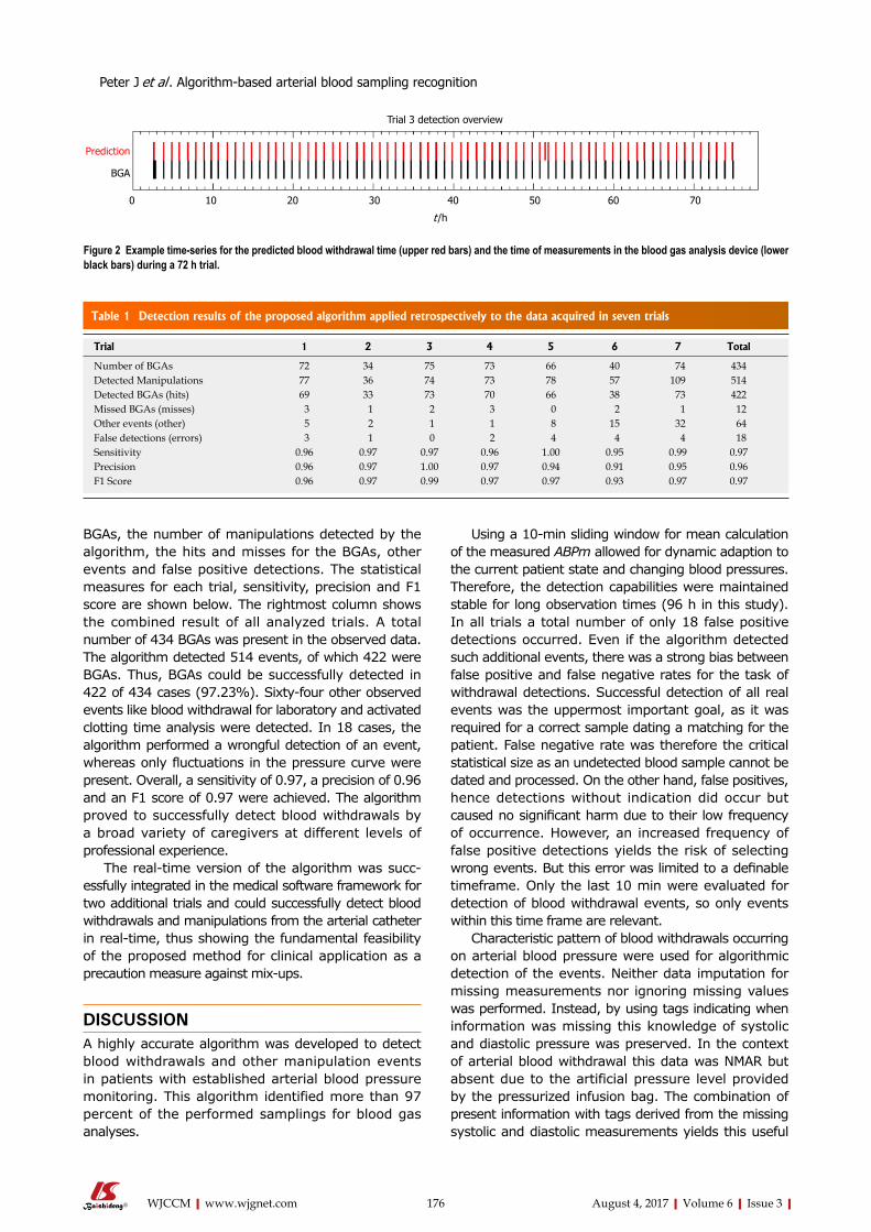
176 August 4, 2017|Volume 6|Issue 3|WJCCM|www.wjgnet.com
BGAs, the number of manipulations detected by the algorithm, the hits and misses for the BGAs, other events and false positive detections. The statistical measures for each trial, sensitivity, precision and F1 score are shown below. The rightmost column shows the combined result of all analyzed trials. A total number of 434 BGAs was present in the observed data. The algorithm detected 514 events, of which 422 were BGAs. Thus, BGAs could be successfully detected in 422 of 434 cases (97.23%). Sixty-four other observed events like blood withdrawal for laboratory and activated clotting time analysis were detected. In 18 cases, the algorithm performed a wrongful detection of an event, whereas only fluctuations in the pressure curve were present. Overall, a sensitivity of 0.97, a precision of 0.96 and an F1 score of 0.97 were achieved. The algorithm proved to successfully detect blood withdrawals by a broad variety of caregivers at different levels of professional experience.
The real-time version of the algorithm was succ-essfully integrated in the medical software framework for two additional trials and could successfully detect blood withdrawals and manipulations from the arterial catheter in real-time, thus showing the fundamental feasibility of the proposed method for clinical application as a precaution measure against mix-ups.
DISCUSSIONA highly accurate algorithm was developed to detect blood withdrawals and other manipulation events in patients with established arterial blood pressure monitoring. This algorithm identified more than 97 percent of the performed samplings for blood gas analyses.
Using a 10-min sliding window for mean calculation of the measured ABPm allowed for dynamic adaption to the current patient state and changing blood pressures. Therefore, the detection capabilities were maintained stable for long observation times (96 h in this study). In all trials a total number of only 18 false positive detections occurred. Even if the algorithm detected such additional events, there was a strong bias between false positive and false negative rates for the task of withdrawal detections. Successful detection of all real events was the uppermost important goal, as it was required for a correct sample dating a matching for the patient. False negative rate was therefore the critical statistical size as an undetected blood sample cannot be dated and processed. On the other hand, false positives, hence detections without indication did occur but caused no significant harm due to their low frequency of occurrence. However, an increased frequency of false positive detections yields the risk of selecting wrong events. But this error was limited to a definable timeframe. Only the last 10 min were evaluated for detection of blood withdrawal events, so only events within this time frame are relevant.
Characteristic pattern of blood withdrawals occurring on arterial blood pressure were used for algorithmic detection of the events. Neither data imputation for missing measurements nor ignoring missing values was performed. Instead, by using tags indicating when information was missing this knowledge of systolic and diastolic pressure was preserved. In the context of arterial blood withdrawal this data was NMAR but absent due to the artificial pressure level provided by the pressurized infusion bag. The combination of present information with tags derived from the missing systolic and diastolic measurements yields this useful
Table 1 Detection results of the proposed algorithm applied retrospectively to the data acquired in seven trials
Trial 3 detection overview
0 10 20 30 40 50 60 70
t /h
Prediction
BGA
Figure 2 Example time-series for the predicted blood withdrawal time (upper red bars) and the time of measurements in the blood gas analysis device (lower black bars) during a 72 h trial.
Trial 1 2 3 4 5 6 7 Total
Number of BGAs 72 34 75 73 66 40 74 434Detected Manipulations 77 36 74 73 78 57 109 514Detected BGAs (hits) 69 33 73 70 66 38 73 422Missed BGAs (misses) 3 1 2 3 0 2 1 12Other events (other) 5 2 1 1 8 15 32 64False detections (errors) 3 1 0 2 4 4 4 18Sensitivity 0.96 0.97 0.97 0.96 1.00 0.95 0.99 0.97Precision 0.96 0.97 1.00 0.97 0.94 0.91 0.95 0.96F1 Score 0.96 0.97 0.99 0.97 0.97 0.93 0.97 0.97
Peter J et al . Algorithm-based arterial blood sampling recognition
177 August 4, 2017|Volume 6|Issue 3|WJCCM|www.wjgnet.com
detection algorithm. Due to general anesthetics, patient movement was
not present in study conditions but should be evaluated for a general clinical application. However, explicitly using the information obtained from the absence of the systolic and diastolic pressures, random events like movements should not lead to such a loss of data, thus not exceeding the threshold for the scoring function.
With a known exact time of blood withdrawal, the blood sample can automatically be dated back to the moment of withdrawal if the measurement was processed by a hospital information system. As shown in the analysis, for the chosen 10 min window the event can be dated back from the exact measurement times to obtain the withdrawal point. If longer times for handling the blood samples are needed, the observed timeframe should be enlarged or even shifted, for example ignoring the most recent minutes for the analysis at all, as processing and transporting time of the blood sample provides a lower boundary.
Additional parameters can easily be included in the detection algorithm with an individual scoring weight to adapt for specific procedures or additional detection performance. If for example an arterial temperature was measured as well, the temperature change resulting from flushing the catheter with saline solution, can be detected as a characteristic event as well and be included in the scoring function.
For improved versions of the detection algorithm and additional studies there are several directions further research might be headed to. Instead of using the observed numerical values for ABPm, ABPs and ABPd, the pressure curve itself may be analyzed. Using such high resolution raw data, calculation of the numerical values for systolic, diastolic and mean pressure may be specifically tailored to process the state of missing data, therefore refining the detection by improving the decision when a systolic and diastolic measurement can be calculated. This might be further extended by integrating models for heart beat detection and to establish a better estimate for deviations from the expected heart rate and variations in to decide if a physiological measurement was present.
The application of the provided solution to a hospital ward with multiple patients could be performed by integration in point-of-care devices like a blood gas analyzer. As suggested by Huijsmans et al[9], usage of additional parameters in the analytical phase should be considered for better sample identification and reducing mix-ups. Using the novel information of automatically calculated times for blood withdrawals, a list of admitted patients can be limited to those, where such an event was detected. This would reduce the error potential of wrong data entries or mix-ups in patient identification from the entire patient database. A full list of all patients should still be available in a second menu, providing an override if detection was not possible. However, this additional step would force attention of the caregiver, being aware of currently performing an override and
focusing on the correct patient selection in this no longer standard procedure. This can significantly improve patient identification as the most commonly observed problem in different studies by Wallin, Wagar and Vallenstein et al[1-3].
Additionally, after successful patient identification, a selection of the detected points of blood withdrawal could be performed and directly be stored with the analysis result, allowing for a more accurate timing of the blood sample. The time between blood withdrawal and analysis is known when automated detection is performed. A warning could then be shown if a predefined time between sampling and measurement is exceeded.
In this paper, we provide a sample application and a first step on the way to automated systems in clinical care settings interacting with point-of-care devices. Detection of blood withdrawal is just an example for the broad range of possibilities that may lead to solutions which will help caregivers and reduce their workload or provide additional safeguards in stressful situations addressing the major problem of noncompliance with guidelines observed by Iboje et al[7].
Major challenges are still to be faced in terms of data access, interconnection of medical devices and dealing with consequently overwhelming big data of patient information. These tasks need to be solved in cooperation with established knowledge from computer science, tailored to the specific needs of the medical sector. By unleashing such great potential, many repetitive or standardized tasks can be automated or computer-assisted checks and protection systems against mistakes could be implemented to increase patient safety and reduce the risk of potential errors for caregivers in stressful situations caused by high workload.
COMMENTSBackgroundMonitoring of arterial blood pressure with arterial catheters is commonly performed in critical patients. Regular arterial blood withdrawals are performed to assess the patient state.
Research frontiersInterconnection of medical devices and automated systems in the medical sector are still experimental. Combining computer science and medicine is still a challenge. Whereas automation and guidance and advanced safeguards are common in other fields of application.
Innovations and breakthroughsA novel approach for the detection of blood withdrawal in patients with an arterial catheter for arterial blood pressure monitoring is described. By interconnecting a patient monitor to a point-of-care diagnostic device, the selection of patient data can be narrowed to patients with plausible sampling events.
ApplicationsDetection of blood withdrawal may be a useful feature integrated in medical monitors and blood gas analysis devices to increase patient safety by allowing a better, automated sample dating and the reduction of the risk for sample
COMMENTS
Peter J et al . Algorithm-based arterial blood sampling recognition
178 August 4, 2017|Volume 6|Issue 3|WJCCM|www.wjgnet.com
mix-ups in hospital wards. However, for a practical implementation additional validation steps are required and medical devices like the blood gas analyzer must be adapted by the manufacturer to allow a pre-selection for patients with recently detected blood withdrawals.
Terminology Arterial blood pressure (ABP), Mean Arterial blood pressure (MAP or ABPm), systolic ABPs, diastolic ABPd, blood gas analysis BGA, arterial blood gas (ABG).
Peer-reviewThe authors prepared and evaluated the algorithm of automatic detection of blood withdrawals by using data from continuous direct blood pressure monitoring. The algorithm may be useful to recognize and eliminate errors in recording blood withdrawal events from the specific patients. The algorithm provides reasonable precision and prediction rates. The methods used were adequate. The manuscript is well-written and will be interesting for intensive care specialists.
REFERENCES1 Wallin O, Söderberg J, Van Guelpen B, Stenlund H, Grankvist K,
Brulin C. Blood sample collection and patient identification demand improvement: a questionnaire study of preanalytical practices in hospital wards and laboratories. Scand J Caring Sci 2010; 24: 581-591 [PMID: 21050248 DOI: 10.1111/j.1471-6712.2009.00753.x]
2 Wagar EA, Tamashiro L, Yasin B, Hilborne L, Bruckner DA. Patient safety in the clinical laboratory: a longitudinal analysis of specimen identification errors. Arch Pathol Lab Med 2006; 130: 1662-1668 [PMID: 17076528 DOI: 10.1043/1543-2165(2006)130[1662: PSITCL]2.0.CO; 2]
3 College of American Pathologists, Valenstein PN, Raab SS, Walsh MK. Identification errors involving clinical laboratories: a College of American Pathologists Q-Probes study of patient and specimen identification errors at 120 institutions. Arch Pathol Lab Med 2006; 130: 1106-1113 [PMID: 16879009 DOI: 10.1043/1543-2165(2006)130[1106: IEICL]2.0.CO; 2]
4 Nutting PA, Main DS, Fischer PM, Stull TM, Pontious Ma. Problems in laboratory testing in primary care. J of the American Medical Association 1996; 275: 635-639 [DOI: 10.1001/jama.275. 8.635]
5 Plebani M, Carraro P. Mistakes in a stat laboratory: types and frequency. Clin Chem 1997; 43: 1348-1351 [PMID: 9267312]
6 Aslan B, Stemp J, Yip P, Gun-Munro J. Method precision and frequent causes of errors observed in point-of-care glucose testing: a proficiency testing program perspective. Am J Clin Pathol 2014; 142: 857-863 [PMID: 25389340 DOI: 10.1309/AJCPP5YS2MVSKBYY]
7 Ibojie J, Urbaniak SJ. Comparing near misses with actual mistransfusion events: a more accurate reflection of transfusion errors. Br J Haematol 2000; 108: 458-460 [PMID: 10691882]
8 van Dongen-Lases EC, Cornes MP, Grankvist K, Ibarz M, Kristensen GB, Lippi G, Nybo M, Simundic AM; Working Group for Preanalytical Phase (WG-PRE), European Federation of Clinical Chemistry and Laboratory Medicine (EFLM). Patient identification and tube labelling - a call for harmonisation. Clin Chem Lab Med 2016; 54: 1141-1145 [PMID: 26816400 DOI: 10.1515/cclm-2015-1089]
9 Huijsmans CJ, Heilmann FG, van der Zanden AG, Schneeberger PM, Hermans MH. Single nucleotide polymorphism profiling assay to exclude serum sample mix-up. Vox Sang 2007; 92: 148-153 [PMID: 17298578 DOI: 10.1111/j.1423-0410.2006.00871.x]
10 Aron R, Dutta S, Janakiraman R, Pathak PA. The Impact of Automation of Systems on Medical Errors: Evidence from Field Research. ISR 2011; 22: 429-446 [DOI: 10.1287/isre.1110.0350]
11 Aleks N, Russell SJ, Madden MG, Morabito D, Staudenmayer K, Cohen M, Manley GT. Probabilistic detection of short events, with application to critical care monitoring. Proceedings of the Advances in Neural Information Processing Systems 2009: 49-56
12 Bauer M, Gather U, Imhoff M. The identification of multiple outliers in online monitoring data. Dortmund: University of Dortmund, Technical Report SFB 475, 1999
13 Gelman A, Hill J. Missing-data imputation. Data Analysis Using Regression and Multilevel/Hierachical Models. Cambridge: Cambridge University Press, 2006: 529-544
14 Osborne JW. Dealing with Missing or Incomplete Data: Debunking the Myth of Emptiness. Best Practices in Data United States: Cleaning SAGE Publications, Inc., 2013: 105-138 [DOI: 10.4135/9781452269948]
15 Johnson AE, Ghassemi MM, Nemati S, Niehaus KE, Clifton DA, Clifford GD. Machine Learning and Decision Support in Critical Care. Proc IEEE Inst Electr Electron Eng 2016; 104: 444-466 [PMID: 27765959 DOI: 10.1109/JPROC.2015.2501978]
16 Peter J, Klingert W, Königsrainer A, Rosenstiel W, Bogdan M, Schenk M. TICoMS - A modular and message-based framework for monitoring and control of medical devices. Comp Med Sy 2014: 473-474 [DOI: 10.1109/Cbms.2014.96]
P- Reviewer: Beltowski J S- Editor: Song XX L- Editor: A E- Editor: Lu YJ
Peter J et al . Algorithm-based arterial blood sampling recognition

© 2017 Baishideng Publishing Group Inc. All rights reserved.
Published by Baishideng Publishing Group Inc7901 Stoneridge Drive, Suite 501, Pleasanton, CA 94588, USA
Telephone: +1-925-223-8242Fax: +1-925-223-8243
E-mail: [email protected] Desk: http://www.f6publishing.com/helpdesk
http://www.wjgnet.com

World Journal of Critical Care MedicineWorld J Crit Care Med 2017 November 4; 6(4): 179-189
Published by Baishideng Publishing Group Inc
ISSN 2220-3141 (online)

World Journal ofCritical Care MedicineW J C C M
EDITOR-IN-CHIEFBart Van Rompaey, Wilrijk
GUEST EDITORIAL BOARD MEMBERSHsing I Chen, HualienSheng-Hsien Chen, Yong-KangYih-Sharng Chen, TaipeiYung-Chang Chen, TaipeiDer-Yang Cho, TaichungCheng-Keng Chuang, TaoyuanHow-Ran Guo, TainanBang-Gee Hsu, HualienChien-Wei Hsu, KaohsiungWen-Jinn Liaw, TaipeiYan-Ren Lin, ChanghuaJiunn-Jye Sheu, Kaohsiung Hsien
MEMBERS OF THE EDITORIAL BOARD
ArgentinaEduardo Chuluyan, Buenos AiresAdrian A Inchauspe, Berazategui
AustraliaZsolt J Balogh, NewcastleZoltan H Endre, SydneyNam Q Nguyen, South AustraliaAlistair D Nichol, VictoriaGeorg M Schmolzer, VictoriaAndrew T Slack, Brisbane
Ravindranath Tiruvoipati, Frankston
AustriaLars-Peter Kamolz, ViennaSylvia Knapp, Vienna
BangladeshSaidur R Mashreky, Dhaka
BelgiumTeresinha Leal, BrusselsManu LNG Malbrain, AntwerpJean-Louis Vincent, Brussels
BrazilLuciano CP Azevedo, São PauloPatricia RM Rocco, Rio de JaneiroMarcos A Rossi, Ribeirao PretoRenato Seligman, Porto Alegre
CanadaDouglas D Fraser, OntarioPierre A Guertin, QuebecMarc G Jeschke, TorontoConstantine J Karvellas, EdmontonWolfgang M Kuebler, TorontoXi Yang, Winnipeg
ChinaXiang-Dong Chen, ChengduXu-Lin Chen, HefeiWong Tak Chuen, Hong KongMing-Xu Da, LanzhouHuang-Xian Ju, NanjingTing-Bo Liang, Hangzhou Peng-Lin Ma, BeijingChung-Wah D Siu, Hong KongYong-ming Yao, BeijingJia-Ping Zhang, ChongqingWei-Dong Zhou, Beijing
CroatiaAlan Sustic, Rijeka
CubaJesús Pérez-Nellar, La Habana
DenmarkDan S Karbing, Aalborg East
EgyptIbrahim Abouomira, CairoHanan Ibrahim, CairoAmr M Moghazy, IsmailiaAyman A Yousef, Tanta
I
Editorial Board2016-2019
The World Journal of Critical Care Medicine Editorial Board consists of 235 members, representing a team of worldwide experts in critical care medicine. They are from 44 countries, including Argentina (2), Australia (7), Austria (2), Bangladesh (1), Belgium (4), Brazil (4), Canada (6), China (23), Croatia (1), Cuba (1), Denmark (1), Egypt (4), Finland (1), France (6), Germany (9), Greece (9), Hungary (1), India (10), Iran (2), Israel (6), Italy (13), Japan (6), Jordan (1), Mexico (1), Morocco (1), Netherlands (4), New Zealand (3), Norway (1), Poland (1), Portugal (4), Russia (1), Saudi Arabia (2), Singapore (2), Slovenia (1), South Africa (1), Spain (6), Sweden (1), Switzerland (3), Thailand (1), Tunisia (1), Turkey (3), United Kingdom (7), United States (70), and Uruguay (1).
February 26, 2016WJCCM|www.wjgnet.com

FinlandAsko A Riutta, Tampere
FranceJean-Marc Cavaillon, ParisBruno Mégarbane, ParisSaad Nseir, LilleNicolas Terzi, CaenJean-Francois Timsit, La Tronche CedexBenoit Vallet, Lille
GermanyHendrik Bracht, UlmMichael E Czaplik, AachenGerrit Grieb, AachenTobias Keck, FreiburgPhilipp Kobbe, AachenAlexander Koch, AachenMarc Maegele, CologneAndrzej A Piatkowski, AachenArmin R Sablotzki, Leipzig
GreeceIoanna Dimopoulou, AthensDimitrios Karakitsos, AthensPetros Kopterides, AthensGregory Kouraklis, AthensAthanasios D Marinis, PiraeusGeorge Nakos, IoanninaPapaioannouE Vasilios, AlexandroupolisTheodoros Xanthos, AthensSpyros G Zakynthinos, Athens
HungaryZoltan Rakonczay, Szeged
IndiaRitesh Agarwal, ChandigarhRachna Agarwal, DelhiMohammad F Butt, SrinagarMohan Gurjar, LucknowDeven Juneja, New Delhi Farhad N Kapadia, MumbaiVikram Kate, PuducherryPramod Kumar, ManipalMedha Mohta, DelhiSrinivas Rajagopala, Bangalore
IranHemmat Maghsoudi, TabrizHomayoun Sadeghi-Bazargani, Tabriz
IsraelAlexander Becker, AfulaYoram Kluger, HaifaYona Kosashvili, ZerrifinKobi Peleg, Tel Hashomer Ilan Sela, RehovotPierre Singer, Petah Tikva
ItalyGiacomo Bellani, MonzaGiovanni Camussi, TurinAnselmo Caricato, RomePiero Ceriana, PaviaAntonio Chiaretti, RomeDavide A Chiumello, MilanoAlfredo Conti, MessinaPaolo Cotogni, TurinDaniele M De Luca, RomaVincenzo De Santis, RomeLuca La Colla, ParmaRaffaele Scala, LuccaGiovanni Vento, Roma
JapanKeishiro Aoyagi, Kurume citySatoshi Hagiwara, OitaYuichi Hattori, ToyamaHideo Inaba, KanazawaEisuke Kagawa, HiroshimaChieko Mitaka, Tokyo
JordanFeras I Hawari, Amman
MexicoSilvio A Ñamendys-Silva, Mexico City
MoroccoRedouane Abouqal, Rabat
NetherlandsWim A Buurman, MaastrichtMartin CJ Kneyber, GroningenPatrick Schober, AmsterdamArie Barend V Vugt, Enschede
New ZealandSultan Al-Shaqsi, DunedinArman A Kahokehr, WhangareiJohn W Pickering, Christchurch
NorwayUlf R Dahle, Oslo
PolandMaciej Owecki, Poznań
PortugalErnestina R Gomes, PortoCristina Granja, MatosinhosJosé A Lopes, LisbonPedro Póvoa, Lisbon
RussiaKonstantin A Popugaev, Moscow
Saudi ArabiaRitesh G Menezes, DammamMohamed T Suliman, Tabuk
SingaporeSanjay H Chotirmall, SingaporeDevanand Anantham, Singapore
SloveniaŠtefek Grmec, Maribor
South AfricaDamian Clarke, Pietermaritzburg
SpainDavid Jimenez, MadridJuan A Llompart-Pou, Palma de MallorcaAntonio T Martí, BarcelonaJuan C Montejo-González, MadridEnrique A Piacentini, TerrassaAlonso M Rodriguez, Madrid
SwedenMihai Oltean, Gothenburg
SwitzerlandDieter Cadosch, ZurichMihael Potocki, BaselJohn F Stover, Zurich
II February 26, 2016WJCCM|www.wjgnet.com

III February 26, 2016WJCCM|www.wjgnet.com
ThailandViroj Wiwanitkit, Bangkok
TunisiaMabrouk Bahloul, Sfax
TurkeyYusuf K Coban, MalatyaBensu Karahalil, AnkaraAli Nayci, Mersin
United KingdomSammy Al-Benna, NottinghamGiles N Cattermole, OrpingtonFrantisek Duska, NottinghamJames N Fullerton, LondonChristina Jones, PrescotSameer Khan, MiddlesbroughGeorge Ntoumenopoulos, London
United StatesEdward Abraham, Winston-SalemBernard R Bendok, ChicagoMichael Blaivas, AtlantaCharles D Boucek, PittsburghRonald Bronicki, Houston
Robert C Cantu, ConcordMarylou Cardenas-Turanzas, HoustonArchana Chatterjee, OmahaPaul A Checchia, St. LouisRubin I Cohen, New Hyde ParkStephen Cohn, San AntonioDonald E Craven, BurlingtonRuy J Cruz Jr, PittsburghFrancis C Dane, RoanokeMarc A de Moya, BostonSteven M Donn, Ann ArborChristopher P Farrell, WynnewoodMarcos A Fernandez, NashvilleKevin N Foster, PhoenixBarry D Fuchs, PhiladelphiaRichard P Gonzalez, MobileAlan H Hall, LaramieJijo John, GilbertJason N Katz, Chapel HillSalah G Keyrouz, Little RockImran Khalid, JeddahDeborah A Kuhls, Las VegasGregory L Larkin, New HavenChristos Lazaridis, CharlestonJames A Lin, Los AngelesYahia M Lodi, SyracuseRoger M Loria, RichmondAigang Lu, CincinnatiRudolf Lucas, AugustaO. John Ma, PortlandRobert T Mallet, Fort WorthWilliam T McGee, MiamiMark McKenney, MiamiMichael Moussouttas, PhiladelphiaOliver HJ Muensterer, Bronx
Rahul Nanchal, MilwaukeeMichael Steven Niederman, MineolaGary F Nieman, SyracuseJames M O’Brien, ColumbusMartin Oudega, MiamiCatherine M Preissig, DuluthVirginia Prendergast, PhoenixRamesh Raghupathi, PhiladelphiaMiren A Schinco, JacksonvilleCarl I Schulman, MiamiL Keith Scott, ShreveportKevin N Sheth, BaltimoreJenni Short, SalinaRonald F Sing, CharlottePhilip C Spinella, St. LouisRobert M Starke, CharlottesvilleStanislaw PA Stawicki, ColumbusDavid C Stockwell, WashingtonStanislav Svetlov, AlachuaMaged A Tanios, Long BeachNeal J Thomas, HersheyNancy M Tofil, BirminghamBalagangadhar R Totapally, MiamiSteven N Vaslef, DurhamJoseph C Watson, Falls ChurchJohn S Wilgis, OrlandoDavid C Willms, San DiegoHao-Dong Xu, RochesterXiao-Ming Xu, IndianapolisMidori A Yenari, San Francisco
UruguayWilliam Manzanares, Montevideo

World Journal ofCritical Care MedicineW J C C M
Contents
IWJCCM|www.wjgnet.com November 4, 2017|Volume 6|Issue 4|
Quarterly Volume 6 Number 4 November 4, 2017
ORIGINAL ARTICLE Retrospective Study
179 Intensivist-baseddeepsedationusingpropofolforpediatricoutpatientflexiblebronchoscopy
Abulebda K, Abu-Sultaneh S, Ahmed SS, Moser EAS, McKinney RC, Lutfi R
Prospective Study
185 Reproducibilityofdiaphragm thicknessmeasurementsbyultrasonography inpatientsonmechanical
ventilation
Dhungana A, Khilnani G, Hadda V, Guleria R
REVIEW
ContentsWorld Journal of Critical Care Medicine
Volume 6 Number 4 November 4, 2017
EDITORS FOR THIS ISSUE
Responsible Assistant Editor: Xiang Li Responsible Science Editor: Jin-Xin KongResponsible Electronic Editor: Ya-Jing Lu Proofing Editorial Office Director: Xiu-Xia SongProofing Editor-in-Chief: Lian-Sheng Ma
EDITORIALOFFICEXiu-Xia Song, DirectorWorld Journal of Critical Care MedicineBaishideng Publishing Group Inc7901 Stoneridge Drive, Suite 501, Pleasanton, CA 94588, USATelephone: +1-925-2238242Fax: +1-925-2238243E-mail: [email protected] Desk: http://www.f6publishing.com/helpdeskhttp://www.wjgnet.com
PUBLISHERBaishideng Publishing Group Inc7901 Stoneridge Drive, Suite 501, Pleasanton, CA 94588, USATelephone: +1-925-223-8242Fax: +1-925-223-8243E-mail: [email protected] Desk: http://www.f6publishing.com/helpdeskhttp://www.wjgnet.com
PUBLICATIONDATENovember 4, 2017
COPYRIGHT© 2017 Baishideng Publishing Group Inc. Articles published by this Open-Access journal are distributed under the terms of the Creative Commons Attribution Non-commercial License, which permits use, distribution, and reproduction in any medium, provided the original work is properly cited, the use is non commercial and is otherwise in compliance with the license.
SPECIALSTATEMENTAll articles published in journals owned by the Baishi-deng Publishing Group (BPG) represent the views and opinions of their authors, and not the views, opinions or policies of the BPG, except where otherwise expli-citly indicated.
INSTRUCTIONSTOAUTHORShttp://www.wjgnet.com/bpg/gerinfo/204
ONLINESUBMISSIONhttp://www.f6publishing.com
IIWJCCM|www.wjgnet.com
ABOUT COVER
AIM AND SCOPE
INDExING/ABSTRACTING
FLYLEAF
November 4, 2017|Volume 6|Issue 4|
NAMEOFJOURNALWorld Journal of Critical Care Medicine
ISSNISSN 2220-3141 (online)
LAUNCHDATEFebruary 4, 2012
FREQUENCYQuarterly
EDITOR-IN-CHIEFBart Van Rompaey, BSc, MSc, PhD, Associate Professor, Nurse, Faculty of Medicine and Health Sciences, Department of Nursing and midwifery, Cen-tre for Research and Innovation in Care, University of Antwerp, Wilrijk 2610, Antwerp, Belgium
EDITORIALBOARDMEMBERSAll editorial board members resources online at http://www.wjgnet.com/2220-3141/editorialboard.htm
EditorialBoardMemberofWorldJournalofCriticalCareMedicine ,MarcG
Jeschke,MD,PhD,Director,AssociateProfessor,SunnybrookHealthSciences
Centre,DepartmentofSurgery,DivisionofPlasticSurgery,Universityof
Toronto,Toronto,ONM4N3M5,Canada
World Journal of Critical Care Medicine (World J Crit Care Med, WJCCM, online ISSN 2220-3141, DOI: 10.5492) is a peer-reviewed open access academic journal that aims to guide clinical practice and improve diagnostic and therapeutic skills of clinicians.
WJCCM covers topics concerning severe infection, shock and multiple organ dysfunc-tion syndrome, infection and anti-infection treatment, acute respiratory distress syndrome and mechanical ventilation, acute kidney failure, continuous renal replacement therapy, rational nutrition and immunomodulation in critically ill patients, sedation and analgesia, cardiopulmonary cerebral resuscitation, fluid resuscitation and tissue perfusion, coagulant dysfunction, hemodynamic monitoring and circulatory support, ICU management and treatment control, and application of bronchofiberscopy in critically ill patients.
We encourage authors to submit their manuscripts to WJCCM. We will give priority to manuscripts that are supported by major national and international foundations and those that are of great clinical significance.
World Journal of Critical Care Medicine is now indexed in PubMed, PubMed Central.
I-III EditorialBoard

Kamal Abulebda, Samer Abu-Sultaneh, Sheikh Sohail Ahmed, Elizabeth A S Moser, Renee C McKinney, Riad Lutfi
ORIGINAL ARTICLE
179 November 4, 2017|Volume 6|Issue 4|WJCCM|www.wjgnet.com
Intensivist-based deep sedation using propofol for pediatric outpatient flexible bronchoscopy
Kamal Abulebda, Samer Abu-Sultaneh, Sheikh Sohail Ahmed, Renee C McKinney, Riad Lutfi, Department of Pediatrics, Section of Critical Care, Indiana University School of Medicine, Riley Hospital for Children at Indiana University Health, Indianapolis, IN 46202, United States
Elizabeth A S Moser, Department of Biostatistics, Indiana University School of Medicine and Richard M Fairbanks School of Public Health, Indianapolis, IN 46202, United States
Author contributions: Abulebda K and Lutfi R contributed equally to this work; Abulebda K, Abu-Sultaneh S, Ahmed SS and Lutfi R designed the research; McKinney RC collected the data; Moser EAS contributed to the statistical analysis; Abulebda K drafted the first draft of the paper; the entire author group critically reviewed and approved the manuscript.
Institutional review board statement: The study was reviewed and approved by the Institutional Review Board of Indiana University.
Informed consent statement: Patients were not required to give informed consent as the study was retrospective and all clinical patient data was de-identified before data analysis.
Conflict-of-interest statement: The authors have no conflict of interest or financial relationships to disclose.
Data sharing statement: Data was not shared amongst institutions.
Open-Access: This article is an open-access article which was selected by an in-house editor and fully peer-reviewed by external reviewers. It is distributed in accordance with the Creative Commons Attribution Non Commercial (CC BY-NC 4.0) license, which permits others to distribute, remix, adapt, build upon this work non-commercially, and license their derivative works on different terms, provided the original work is properly cited and the use is non-commercial. See: http://creativecommons.org/licenses/by-nc/4.0/
Manuscript source: Unsolicited manuscript
Correspondence to: Dr. Kamal Abulebda, Department of
Pediatrics, Section of Critical Care, Indiana University School of Medicine, Riley Hospital for Children at Indiana University Health, Dr, Phase 2, Room 4900, Indianapolis, IN 46202, United States. [email protected]: +1-317-9449674Fax: +1-317-9443442
Received: February 6, 2017Peer-review started: February 12, 2017First decision: May 17, 2017Revised: June 30, 2017Accepted: September 3, 2017Article in press: September 4, 2017Published online: November 4, 2017
AbstractAIMTo evaluate the safety and efficacy of sedating pediatric patients for outpatient flexible bronchoscopy.
METHODSA retrospective chart review was conducted for all chil-dren, age 17 years or under who underwent flexible bronchoscopy under deep sedation in an outpatient hospital-based setting. Two sedation regimens were used; propofol only or ketamine prior to propofol. Patients were divided into three age groups; infants (less than 12 mo), toddlers (1-3 years) and children (4-17 years). Demographics, indication for bronchoscopy, sedative dosing, sedation and recovery time and adverse events were reviewed.
RESULTSOf the total 458 bronchoscopies performed, propofol only regimen was used in 337 (74%) while propofol and ketamine was used in 121 (26%). About 99% of the procedures were successfully completed. Children in the propofol + ketamine group tend to be younger
World Journal ofCritical Care MedicineW J C C M
Submit a Manuscript: http://www.f6publishing.com
DOI: 10.5492/wjccm.v6.i4.179
World J Crit Care Med 2017 November 4; 6(4): 179-184
ISSN 2220-3141 (online)
Retrospective Study
180 November 4, 2017|Volume 6|Issue 4|WJCCM|www.wjgnet.com
Abulebda K et al . Pediatric deep sedation for outpatient bronchoscopy
and have lower weight compared to the propofol only group. Adverse events including transient hypoxemia and hypotension occurred in 8% and 24% respectively. Median procedure time was 10 min while the median discharge time was 35 min. There were no differences in the indi-cation of the procedure, propofol dose, procedure or recovery time in either sedative regimen. When compared to other age groups, infants had a higher incidence of hypoxemia.
CONCLUSIONChildren can be effectively sedated for outpatient flexible bronchoscopy with high rate of success. This procedure should be performed under vigilance of highly trained providers.
Key words: Pediatric flexible bronchoscopy; Propofol; Deep sedation; Procedural sedation; Sedation time; Hypoxemia
© The Author(s) 2017. Published by Baishideng Publishing Group Inc. All rights reserved.
Core tip: In this retrospective study “Intensivist-based deep sedation using propofol for pediatric outpatient flexible bronchoscopy”, we are presenting our center data on pediatric patients who underwent flexible bronchoscopy under deep sedation using propofol. The study outlines our experience with intensivist-based procedural sedation as an effective strategy to facilitate successful completion of flexible bronchoscopy. This is the largest retrospective study describing the use of propofol-based procedural sedation in the outpatient settings for pediatric flexible bronchoscopy.
Abulebda K, Abu-Sultaneh S, Ahmed SS, Moser EAS, McKinney
RC, Lutfi R. Intensivist-based deep sedation using propofol for pediatric outpatient flexible bronchoscopy. World J Crit Care Med 2017; 6(4): 179-184 Available from: URL: http://www.wjgnet.com/2220-3141/full/v6/i4/179.htm DOI: http://dx.doi.org/10.5492/wjccm.v6.i4.179
INTRODUCTIONIn the last two decades, flexible bronchoscopy (FB) has become an increasingly important outpatient tool used in the evaluation of pulmonary abnormalities in children[1,2]. As FB allows direct visualization of the patient’s upper and lower airway larynx[3], it has been used as a diagnostic and therapeutic tool for chronic cough, wheezing, cystic fibrosis and infection etiologies in immunocompetent and immunocompromised patients[4] to diagnose various congenital or acquired pediatric airway anomalies/abnormalities.
While the need for appropriate sedation for FB is controversial in adults[5], deep sedation is generally needed in children due to their developmental capabilities and airway anatomy in order to blunt the airway protective
reflexes and suppress the cough stimulus. Using deep sedation not only decreases a child’s distress and discomfort but also significantly increases the chance of a successful completion of the procedure[1,6]. Multiple sedation regimens and route had been evaluation including nasal, oral, intravenous and topical anesthetic[7]. Commonly used drugs for sedation for FB include a benzodiazepine and opioids combination or ketamine with or without benzodiazepine[6,8,9].
Propofol is an iv sedativehypnotic agent that is used for induction and maintenance of deep sedation and general anesthesia[10]. Propofol has many properties, including a rapid onset, short duration of action with rapid recovery time and minimal adverse events, which makes it an ideal agent for pediatric sedation in the outpatient setting[11]. Emerging data support the safety and efficacy of using propofol outside the operating room for pediatric outpatient procedures by qualified physicians trained in sedation with advanced airway management[1,12]. Additionally, with increasing numbers of pediatric patients undergoing diagnostic and therapeutic FB and the relative shortage of anesthesiologists and operating room availability, other pediatric subspecialists, such as pediatric critical care physicians, have stepped in to provide pediatric procedural sedation[1315].
Ketamine is a dissociative agent that has analgesic, sedative and amnestic properties. It has been frequently used to facilitate painful procedures in children and has proven to be safe and effective in numerous studies[16]. However, despite the reported safety of ketamine in these studies, it had been reported that high dose ketamine could result in respiratory depression and excessive salivary secretions leading to adverse respiratory events[17].
The combination of propofol and ketamine for pediatric sedation had been reported to provide optimal hemodynamic stability and reduced adverse effects when compared to propofol alone[1820]. Additionally, the combination of propofol and ketamine had been shown to be beneficial in other medical fields because of allowing lower doses of propofol resulting in the reduction of the undesirable adverse effects[20]. Many authors reported the advantages of propofolketamine combination in terms of hemodynamic profile and pain control in cancer patients undergoing painful procedures[19].
The purpose of this study is to review the author institution’s experiences using propofolbased deep, procedural sedation regimens for pediatric flexible bronchoscopy in an outpatient setting.
MATERIALS AND METHODSThis retrospective study was approved by the institutional review board of the Indiana University. All pediatric patients between the ages of two months to seventeen years of age undergoing deep sedation for flexible bronchoscopy from March 2007 to August 2012 were included. Patients were divided into three age groups; infants (less than 12 mo), toddlers (13
181 November 4, 2017|Volume 6|Issue 4|WJCCM|www.wjgnet.com
years) and children (417 years). Patients in whom flexible bronchoscopy evaluation was performed in the Pediatric Intensive Care Unit through tracheostomy or endotracheal tube were excluded. All bronchoscopies were performed at the Riley Hospital for Children at Indiana University Health System by a pediatric pulmonologist with the assistance of a respiratory therapist at our dedicated outpatient sedation room.
History and physical exam were performed and documented according to the American Academic of Pediatrics (AAP) guidelines for sedation[21]. Written consent was obtained from the parents or guardian prior to the procedure. Sedation was performed by a sedation team consisting of a pediatric intensivist and two sedation nurses with a pediatric critical care background who monitored the patient during and after each procedure. Guidelines have been laid down by the AAP regarding monitoring, management and discharging children during procedural sedation[21]. All patients were either classified as ASAPS Ⅱ or Ⅰ per American Society of AnesthesiologistsPhysical Status classification system. Patients were without any oral intake for at least 6 h prior to the procedure and had an intravenous catheter placed by sedation team. Physiologic parameters such as heart rate, respiratory rate, oxygen saturation and noninvasive blood pressure were measured every 5 min throughout the procedure and every 15 min after its completion until the patient was fully awake. Supplemental oxygen (2 L per min) was administered via nasal cannula to the majority of the patients (92%) before and during the procedure. Prior to sedation, each patient received viscous lidocaine to the nare, a transnasal approach was used for all procedures. Additional doses of lidocaine were applied to the vocal cords, trachea and major bronchi as required per pulmonologist.
Two sedation strategies were used; intravenous (iv) propofol only (PO) and iv ketamine prior to iv propofol (KP), solely based on the intensivist preference. When ketamine was used, it was administered as an initial bolus of 0.5 mg/kg for patients who weigh less than 20 kg and 0.25 mg/kg for patient’s weight more than 20 kg. Propofol was administered as an initial bolus of 12 mg/kg with additional boluses of 1 mg/kg as needed to achieve deep sedation (level 3) based on University of Michigan sedation scale[22].
Adverse events were recorded including development of hypoxemia (oxygen saturation of less than 90% for more than 30 s), hypotension (drop in systolic blood pressure below expected for age or a drop of 20% from baseline), worsening stridor from baseline, and bleeding (hemoptysis or epistaxis). Serious adverse events such endotracheal intubation, respiratory or cardiac arrest or failure to complete the procedure were also recorded.
Procedure time was defined as the time between the first bolus of sedation until the bronchoscopy procedure completed. Recovery time (RT) was defined as the interval between the completions of the procedure until the patient’s level of conscious was back to baseline. Discharge time (DT) was defined as the interval between
the start of sedation until the patient was discharged home.
Outcomes analyzed included: Propofol dose, hypoxemia, hypotension, procedure and recovery times, and time to discharge. For the two sedation strategies, bivariate analyses were conducted using χ 2 and Wilcoxon Sum Rank Tests. For the three age groups, bivariate analyses were conducted using χ 2 and KruskalWallis Tests.
RESULTSDuring the study period, a total of 458 bronchoscopies were performed, of which 454 (99.1%) were successfully completed. Patients’ demographics and indications for bronchoscopy are summarized in (Table 1). Of the 458 flexible bronchoscopies performed, 337 patients (73.6%) were sedated using propofol only strategy and 121 patients (26.4%) using propofol and ketamine. Children in the (KP) group tend to be younger and have lower weight compared to the (PO) group. Four cases (< 1%) (3 in the PO group, 1 in the KP group) were terminated early. Two patients (< 0.5%) were admitted to the pediatric intensive care unit; one toddler in the PO group and one child in the KP group. One of the four patients required endotracheal intubation; two other patients required fluid resuscitation and one patient had a brief bradycardic episode. Both admitted patients were discharged home in the first 24 h of admission. Transient hypoxemia occurred in 8.3% of patients while hypotension in 23.6%. Prolonged hypoxemia necessitating the need for bag/mask ventilation happened in 5.1% of all patients (Table 2). There was no significant difference in propofol dosage, adverse effects or sedation times using the two sedation strategies (PO or KP) (Table 2). Analysis of the three age groups showed significantly higher hypoxemia in infants compared to toddlers and children (Table 3). A logistic regression of age groups predicting hypoxemia showed that infants have significantly higher odds of hypoxemia compared to toddlers (P < 0.0001, OR: 13.56, 95%CI: 3.92, 46.91), and compared to children (P < 0.0001, OR: 10.96, 95%CI: 3.65, 32.91). However, children and toddlers do not have significantly different odds of hypoxemia (P = 0.62).
DISCUSSION FB is an essential diagnostic and therapeutic modality commonly used in various congenital and acquired pediatric pulmonary disorders[9,23].
To the best of our knowledge, this is the largest retrospective study describing the use of propofol with or without ketamine for procedural sedation in the outpatient settings for pediatric FB.
Between 2007 and 2012, we have used propofol as the main intravenous sedative agent for pediatric outpatient for FB. Propofol was well tolerated in the majority of pediatric patients undergoing the FB. Compared with the study of
Abulebda K et al . Pediatric deep sedation for outpatient bronchoscopy

182 November 4, 2017|Volume 6|Issue 4|WJCCM|www.wjgnet.com
Table 2 Average doses, sedation times and adverse events
Hasan and Reddy, our RT and discharge time DT were significantly shorter (26.7 ± 14.3 min vs 40 ± 18 min) and (37.6 ± 16.1 min vs 80 ± 44 min) respectively[24]. These findings can be due to the variability in indications and the practice of FB in pediatrics. Additionally, our propofol dose used is in line with the findings in another study to evaluate the use of propofol in pediatric FB[1] with no significant difference between three age groups or sedation regimens.
The routine administration of small dose ketamine prior to propofol has been shown in some studies to be beneficial in maintaining hemodynamic stability and decreasing side effect profile of propofol[18,19]. We used ketamine prior to propofol in only one fourth of our patient population but we did not observe significant difference in the adverse events between two groups. Also, we observed no difference in the average propofol dose between the groups. Additionally, RT and DT were similar in both groups. It is unclear whether there is
truly no difference when adding ketamine to propofol or if it was due to small sample size or could be related to the fact that ketamine dose is too low to achieve anesthetic effect.
In term of adverse events and comparing to the data from the Pediatric Sedation Research Consortium on propofol sedation, we observed higher incidence of transient hypoxemia, hypoxemia required bag/mask ventilation and unexpected hospital admission in our study (8% vs 1.4%, 5% vs 1%, 0.4% vs 0.07% respectively)[14]. The higher incidence of these adverse events could be related to the nature of the procedure. Additionally, the pediatric research consortium data did not include pediatric patients who undergo this category of procedures. However, our findings are consistent with other reported data of complications of FB in children[3,25]. Our infants group had a significantly higher incidence of transient hypoxemia in infants compared to toddlers
Table 1 Demographics and indications of bronchoscopy in patients
Variable Overall (n = 458) Propofol only (n = 337) Propofol ketamine (n = 121) P value
Age, yr 5.0 (2.5, 9.1) 5.6 (2.8, 9.8) 3.4 (1.9, 6.6) < 0.0001Age group, n (%) Infant (< 12 mo) 15 (3.3) 6 (1.8) 9 (7.4) < 0.0001 Toddler (1-3 yr) 132 (28.8) 84 (24.9) 48 (39.7) Child (4-17 yr) 311 (67.9) 247 (73.3) 64 (52.9)Weight (kg) 18.1 (13.1, 31.8) 20.0 (14.4, 33.0) 14.7 (11.2, 26.0) < 0.0001Female gender, n (%) 198 (43.2) 143 (42.4) 55 (45.5) 0.57Diagnosis, n (%) Cystic fibrosis 38 (8.3) 29 (8.6) 9 (7.4) Cough 93 (20.3) 62 (18.4) 31 (25.6) 0.38 Wheezing 108 (23.6) 87 (25.8) 21 (17.4) Stridor 56 (12.2) 41 (12.2) 15 (12.4) Pneumonia 57 (12.4) 42 (12.5) 15 (12.4) Tachypnea 106 (23.1) 76 (22.6) 30 (24.8)
Variable Overall (n = 458) Propofol only (n = 337) Propofol ketamine (n = 121) P value
Propofol dose (mg/kg) 4.1 (2.7, 5.6) 4.2 (2.7, 5.6) 3.7 (2.8, 5.2) 0.3Procedure time (min) 10 (6, 15) 10 (7, 12) 10 (5, 15) 0.3Recovery time (min) 25 (20, 30) 25 (20, 30) 25 (20, 35) 0.63Time to discharge (min) 35 (30, 43) 35 (30, 40) 35 (30, 45) 0.31Respiratory eventsProphylactic use of O2 supplementation prior to bronchoscopy, n (%)
423 (92.4) 311 (92.3) 112 (92.6) 0.92
Hypoxemia, n (%) 38 (8.3) 29 (8.6) 9 (7.4) 0.69BMV/significant desaturation1, n (%) 0.58 Neither 413 (91.2) 302 (90.7) 111 (92.5) BMV + significant desaturation 23 (5.1) 16 (4.8) 7 (5.8) Significant desaturation only 16 (3.5) 14 (4.2) 2 (1.7) BMV use only 1 (0.2) 1 (0.3) 0 (0)Cardiac eventsStart MBP 77.7 (70.3, 86.7) 78.3 (71.3, 86.3) 76.3 (68.0, 88.7) 0.58End MBP 70.3 (63.0, 78.7) 71.0 (64.0, 79.3) 68.7 (61.7, 76.3) 0.04Difference in MBP -7.5 (-17.0, 2.0) -6.7 (-16.0, 2.7) -8.7 (-19.0, 0.7) 0.12% Change MBP from start of procedure -9.8 (-20.0, 3.0) -9.2 (-18.8, 4.3) -10.5 (-22.8, 0.7) 0.12Blood pressure drop more than 20% from the baseline (hypotension)
108 (23.6%) 76 (22.6%) 32 (26.4%) 0.4
1Significant desaturation defined as oxygen saturation of less than 90% for more than 30 s. BMV: Bag mask ventilation; MBP: Mean arterial blood pressure.
Abulebda K et al . Pediatric deep sedation for outpatient bronchoscopy
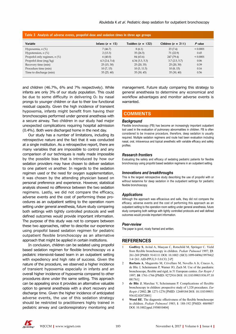
183 November 4, 2017|Volume 6|Issue 4|WJCCM|www.wjgnet.com
and children (46.7%, 6% and 7% respectively). While infants are only 3% of our study population. This could be due to some difficulty in delivering O2 by nasal prongs to younger children or due to their low functional residual capacity. Given the high incidence of transient hypoxemia, infants might benefit from having their bronchoscopies performed under general anesthesia with a secure airway. Two children in our study had major unexpected complications requiring hospital admission (0.4%). Both were discharged home in the next day.
Our study has a number of limitations, including its retrospective nature and the fact that it was conducted at a single institution. As a retrospective report, there are many variables that are impossible to control and any comparison of our techniques is really made impossible by the possible bias that is introduced by how our sedation providers may have chosen to deliver sedation to one patient vs another. In regards to the sedation regimen used or the need for oxygen supplementation, it was chosen by the attending physician based on personal preference and experience. However, statistical analysis showed no difference between the two sedation regimens. Lastly, we did not compare the efficacy, adverse events and the cost of performing these procedures as an outpatient setting to the operation room setting under general anesthesia, future study comparing both settings with tightly controlled protocols and well defined outcomes would provide important information. The purpose of this study was not to compare between these two approaches, rather to describe our experience using propofol based sedation regimen for pediatric outpatient flexible bronchoscopy as an alternative approach that might be applied in certain institutions.
In conclusion, children can be sedated using propofol based sedation regimen for flexible bronchoscopy vs a pediatric intensivistbased team in an outpatient setting with expediency and high rate of success. Given the nature of the procedure, we observed a higher incidence of transient hypoxemia especially in infants and an overall higher incidence of hypoxemia compared to other procedures done under the same setting. This approach can be appealing since it provides an alternative valuable option to general anesthesia with a short recovery and discharge time. Given the higher incidence of anticipated adverse events, the use of this sedation strategy should be restricted to practitioners highly trained in pediatric airway and cardiorespiratory monitoring and
management. Future study comparing this strategy to general anesthesia to determine any economical and workflow advantages and monitor adverse events is warranted.
COMMENTSBackgroundFlexible bronchoscopy (FB) has become an increasingly important outpatient tool used in the evaluation of pulmonary abnormalities in children. FB is often considered to be invasive procedure, therefore, deep sedation is usually required. Multiple sedation regimens and route had been evaluation including nasal, oral, intravenous and topical anesthetic with variable efficacy and safety profiles.
Research frontiers Evaluating the safety and efficacy of sedating pediatric patients for flexible bronchoscopy using propofol based sedation regimens in an outpatient setting.
Innovations and breakthroughs This is the largest retrospective study describing the use of propofol with or without ketamine for deep sedation in the outpatient settings for pediatric flexible bronchoscopy.
Applications Although the approach was efficacious and safe, they did not compare the efficacy, adverse events and the cost of performing this approach as an outpatient setting to the operation room setting under general anesthesia, future study comparing both settings with tightly controlled protocols and well defined outcomes would provide important information.
Peer-reviewThe paper is good, nicely framed and written.
REFERENCES1 Godfrey S, Avital A, Maayan C, Rotschild M, Springer C. Yield
from flexible bronchoscopy in children. Pediatr Pulmonol 1997; 23: 261-269 [PMID: 9141111 DOI: 10.1002/ (SICI) 1099-0496(199704)23:4<261: AID-PPUL3>3.0.CO; 2-P]
2 Barbato A, Magarotto M, Crivellaro M, Novello A Jr, Cracco A, de Blic J, Scheinmann P, Warner JO, Zach M. Use of the paediatric bronchoscope, flexible and rigid, in 51 European centres. Eur Respir J 1997; 10: 1761-1766 [PMID: 9272916 DOI: 10.1183/09031936.97.10081761]
3 de Blic J, Marchac V, Scheinmann P. Complications of flexible bronchoscopy in children: prospective study of 1,328 procedures. Eur Respir J 2002; 20: 1271-1276 [PMID: 12449184 DOI: 10.1183/09031936.02.02072001]
4 Wood RE. The diagnostic effectiveness of the flexible bronchoscope in children. Pediatr Pulmonol 1985; 1: 188-192 [PMID: 4069807 DOI: 10.1002/ppul.1950010404]
Table 3 Analysis of adverse events, propofol dose and sedation times in three age groups
Variable Infants (n = 15) Toddlers (n = 132) Children (n = 311) P value
Hypoxemia, n (%) 7 (46.7) 8 (6.1) 23 (7.4) < 0.0001Hypotension, n (%) 2 (13.3) 35 (26.5) 71 (22.9) 0.45Propofol only regimen, n (%) 6 (40.0) 84 (63.6) 247 (79.4) < 0.0001Propofol dose (mg/kg) 4.3 (2.4, 5.4) 4.34 (3.3, 5.3) 3.7 (2.5, 5.7) 0.06Recovery time (min) 25 (15, 30) 25 (20, 35) 25 (20, 30) 0.39Procedure time (min) 10 (7, 15) 10 (5, 11.5) 10 (8, 15) 0.16Time to discharge (min) 35 (25, 40) 35 (30, 45) 35 (30, 40) 0.56
COMMENTS
Abulebda K et al . Pediatric deep sedation for outpatient bronchoscopy
184 November 4, 2017|Volume 6|Issue 4|WJCCM|www.wjgnet.com
5 José RJ, Shaefi S, Navani N. Sedation for flexible bronchoscopy: current and emerging evidence. Eur Respir Rev 2013; 22: 106-116 [PMID: 23728864 DOI: 10.1183/09059180.00006412]
6 Tobias JD. Sedation and anesthesia for pediatric bronchoscopy. Curr Opin Pediatr 1997; 9: 198-206 [PMID: 9229156 DOI: 10.1097/00008480-199706000-00002]
7 Somu N, Vijayasekaran D, Ashok TP, Balachandran A, Subramanyam L. Flexible fibreoptic bronchoscopy in 582 children--value of route, sedation and local anesthetic. Indian Pediatr 1995; 32: 543-547 [PMID: 8613312]
8 Slonim AD, Ognibene FP. Amnestic agents in pediatric bronchoscopy. Chest 1999; 116: 1802-1808 [PMID: 10593809 DOI: 10.1378/chest.116.6.1802]
9 Berkenbosch JW, Graff GR, Stark JM. Safety and efficacy of ketamine sedation for infant flexible fiberoptic bronchoscopy. Chest 2004; 125: 1132-1137 [PMID: 15006978 DOI: 10.1378/chest.125.3.1132]
10 Steinbacher DM. Propofol: a sedative-hypnotic anesthetic agent for use in ambulatory procedures. Anesth Prog 2001; 48: 66-71 [PMID: 11515950]
11 Vardi A, Salem Y, Padeh S, Paret G, Barzilay Z. Is propofol safe for procedural sedation in children? A prospective evaluation of propofol versus ketamine in pediatric critical care. Crit Care Med 2002; 30: 1231-1236 [PMID: 12072673 DOI: 10.1097/00003246-200206000-00010]
12 Larsen R, Galloway D, Wadera S, Kjar D, Hardy D, Mirkes C, Wick L, Pohl JF. Safety of propofol sedation for pediatric outpatient procedures. Clin Pediatr (Phila) 2009; 48: 819-823 [PMID: 19483136 DOI: 10.1177/0009922809337529]
13 Havidich JE, Cravero JP. The current status of procedural sedation for pediatric patients in out-of-operating room locations. Curr Opin Anaesthesiol 2012; 25: 453-460 [PMID: 22732423 DOI: 10.1097/ACO.0b013e32835562d8]
14 Kamat PP, McCracken CE, Gillespie SE, Fortenberry JD, Stockwell JA, Cravero JP, Hebbar KB. Pediatric critical care physician-administered procedural sedation using propofol: a report from the Pediatric Sedation Research Consortium Database. Pediatr Crit Care Med 2015; 16: 11-20 [PMID: 25340297 DOI: 10.1097/PCC.0000000000000273]
15 Cravero JP, Beach ML, Blike GT, Gallagher SM, Hertzog JH. Pediatric sedation research consortium. The incidence and nature of adverse events during pediatric sedation/anesthesia with propofol for procedures outside the operating room: a report from the Pediatric Sedation Research Consortium. Anesth Analg 2009; 108: 795-804
[PMID: 19224786 DOI: 10.1213/ane.0b013e31818fc334]16 Grunwell JR, Travers C, McCracken CE, Scherrer PD, Stormorken
AG, Chumpitazi CE, Roback MG, Stockwell JA, Kamat PP. Procedural Sedation Outside of the Operating Room Using Ketamine in 22,645 Children: A Report From the Pediatric Sedation Research Consortium. Pediatr Crit Care Med 2016; 17: 1109-1116 [PMID: 27505716 DOI: 10.1097/PCC.0000000000000920]
17 Morton NS. Ketamine for procedural sedation and analgesia in pediatric emergency medicine: a UK perspective. Paediatr Anaesth 2008; 18: 25-29 [PMID: 18095962 DOI: 10.1111/j.1460-9592.2007.02395.x]
18 Alletag MJ, Auerbach MA, Baum CR. Ketamine, propofol, and ketofol use for pediatric sedation. Pediatr Emerg Care 2012; 28: 1391-1395; quiz 1396-1398 [PMID: 23222112 DOI: 10.1097/PEC.0b013e318276fde2]
19 Chiaretti A, Benini F, Pierri F, Vecchiato K, Ronfani L, Agosto C, Ventura A, Genovese O, Barbi E. Safety and efficacy of propofol administered by paediatricians during procedural sedation in children. Acta Paediatr 2014; 103: 182-187 [PMID: 24138461 DOI: 10.1111/apa.12472]
20 David H, Shipp J. A randomized controlled trial of ketamine/propofol versus propofol alone for emergency department procedural sedation. Ann Emerg Med 2011; 57: 435-441 [PMID: 21256626 DOI: 10.1016/j.annemergmed.2010.11.025]
21 Coté CJ, Wilson S. American academy of pediatrics; american academy of pediatric dentistry. Guidelines for Monitoring and Management of Pediatric Patients Before, During, and After Sedation for Diagnostic and Therapeutic Procedures: Update 2016. Pediatrics 2016; 138: e20161212 [PMID: 27354454]
22 Malviya S, Voepel-Lewis T, Tait AR, Merkel S, Tremper K, Naughton N. Depth of sedation in children undergoing computed tomography: validity and reliability of the University of Michigan Sedation Scale (UMSS). Br J Anaesth 2002; 88: 241-245 [PMID: 11878656 DOI: 10.1093/bja/88.2.241]
23 Field-Ridley A, Sethi V, Murthi S, Nandalike K, Li ST. Utility of flexible fiberoptic bronchoscopy for critically ill pediatric patients: A systematic review. World J Crit Care Med 2015; 4: 77-88 [PMID: 25685726 DOI: 10.5492/wjccm.v4.i1.77]
24 Hasan RA, Reddy R. Sedation with propofol for flexible bronchoscopy in children. Pediatr Pulmonol 2009; 44: 373-378 [PMID: 19274622 DOI: 10.1002/ppul.21013]
25 Schnapf BM. Oxygen desaturation during fiberoptic bronchoscopy in pediatric patients. Chest 1991; 99: 591-594 [PMID: 1995213 DOI: 10.1378/chest.99.3.591]
P- Reviewer: Nenna R, Aggarwal D S- Editor: Cui LJ L- Editor: A E- Editor: Lu YJ
Abulebda K et al . Pediatric deep sedation for outpatient bronchoscopy
Ashesh Dhungana, Gopi Khilnani, Vijay Hadda, Randeep Guleria
ORIGINAL ARTICLE
185 November 4, 2017|Volume 6|Issue 4|WJCCM|www.wjgnet.com
Reproducibility of diaphragm thickness measurements by ultrasonography in patients on mechanical ventilation
Ashesh Dhungana, Department of Medicine, National Academy of Medical Sciences, Pulmonary Medicine, Kantipath, Kathmandu 44600, Nepal
Gopi Khilnani, Vijay Hadda, Randeep Guleria, Department of Pulmonary Medicine and Sleep Disorders, All India Institute of Medical Sciences, New Delhi 110029, India
ORCID number: Ashesh Dhungana (0000-0003-2656-4405); Gopi Khilnani (0000-0003-0820-0624); Vijay Hadda (0000- 0001-5820-3685); Randeep Guleria (0000-0002-9234-602X).
Author contributions: Dhungana A, Khilnani G and Hadda V are guarantors of the paper, be responsibility for the integrity of the work; Khilnani G and Hadda V conceived the idea; Dhungana A was involved in performing ultrasonography, data collection, manuscript drafting and revision; Hadda V contributed to performing ultrasonography, manuscript drafting and revision; Guleria R was involved in drafting and revising the manuscript.
Institutional review board statement: This study was reviewed and approved by institutional review board of All India Institute of Medical Sciences.
Informed consent statement: All study participants, or their legal guardian, provided written consent prior to enrollment into the study.
Conflict-of-interest statement: None of the authors have any conflict of interest.
Data sharing statement: There is no additional data available.
Open-Access: This article is an open-access article which was selected by an in-house editor and fully peer-reviewed by external reviewers. It is distributed in accordance with the Creative Commons Attribution Non Commercial (CC BY-NC 4.0) license, which permits others to distribute, remix, adapt, build upon this work non-commercially, and license their derivative works on different terms, provided the original work is properly cited and the use is non-commercial. See: http://creativecommons.org/licenses/by-nc/4.0/
Manuscript source: Unsolicited manuscript
Correspondence to: Dr. Ashesh Dhungana, MD, DM, Assistant Professor, Department of Medicine, National Academy of Medical Sciences, Pulmonary Medicine, Kantipath, Kathmandu 44600, Nepal. [email protected] Telephone: +977-98-41860457
Received: April 20, 2017Peer-review started: April 21, 2017First decision: July 18, 2017Revised: July 26, 2017Accepted: September 3, 2017Article in press: September 4, 2017Published online: November 4, 2017
AbstractAIMTo prospectively evaluate the reproducibility of diaphragm thickness measurement by ultrasonography at the bed-side by critical care physicians in patients on invasive mechanical ventilation.
METHODSIn a prospective observational study of 64 invasively ventilated patients, diaphragmatic thickness measurement was taken by 2 different observers at the same site. Three measurements were taken by each observer and averaged. The intraobserver and interobserver variability was assessed by estimation of intraclass correlation coe-fficient. The limits of agreement were plotted as the difference between two observations against the average of the two observations in Bland and Altman analysis.
RESULTSThe mean diaphragm thickness at the functional residual capacity was 2.29 ± 0.4 mm and the lower limit of the normal, i.e. , the 5th percentile was 1.7 mm (95%CI: 1.6-1.8). The intraclass correlation coefficient for intra-observer variability was 0.986 (95%CI: 0.979-0.991)
World Journal ofCritical Care MedicineW J C C M
Submit a Manuscript: http://www.f6publishing.com
DOI: 10.5492/wjccm.v6.i4.185
World J Crit Care Med 2017 November 4; 6(4): 185-189
ISSN 2220-3141 (online)
Prospective Study
186 November 4, 2017|Volume 6|Issue 4|WJCCM|www.wjgnet.com
Dhungana A et al . Diaphragm USG during mechanical ventilation
with a P value of < 0.001. The intraclass correlation coefficient for interobserver variability was 0.987 (95%CI: 0.949-0.997) with a P value of < 0.001. In Bland and Altman analysis, both intraobserver and interobserver measurements showed high limits of agreement.
CONCLUSIONOur study demonstrates that the measurement of dia-phragm thickness by ultrasound can be accurately per-formed by critical care physicians with high degree of reproducibility in patients on mechanical ventilation.
Key words: Diaphragm; Ultrasonography; Mechanical ventilation
© The Author(s) 2017. Published by Baishideng Publishing Group Inc. All rights reserved.
Core tip: Ultrasonography (USG) is a cheap, cost effective and non-invasive bedside tool for evaluation of diaphragm thickness during mechanical ventilation. Measurement of diaphragm thickness by USG can be accurately per-formed by critical care physicians with high degree of reproducibility. USG should be used more often by the physicians in the intensive care unit for the assessment of the diaphragm.
Dhungana A, Khilnani G, Hadda V, Guleria R. Reproducibility of diaphragm thickness measurements by ultrasonography in patients on mechanical ventilation. World J Crit Care Med 2017; 6(4): 185-189 Available from: URL: http://www.wjgnet.com/2220-3141/full/v6/i4/185.htm DOI: http://dx.doi.org/10.5492/wjccm.v6.i4.185
INTRODUCTIONInvasive mechanical ventilation causes progressive decline in diaphragm bulk and strength in a phenomenon called ventilator induced diaphragm dysfunction[1]. Diaphragm movement and function can be assessed by various methods which include chest X-ray, supine vital capacity, maximum inspiratory pressure, electromyography and magnetic phrenic nerve stimulation. Ultrasonography (USG) is a cheap, cost effective and non-invasive bedside tool for evaluation of diaphragm thickness. It has been used successfully to measure diaphragm thickness and movement in ambulatoryin dividuals[2,3]. Diaphragm thickness is a surrogate of its strength and helps to predict the outcome of extubation in patients on mechanical ventilation[4,5]. However, localization and measurement may be more difficult in critically ill ventilated patients in the intensive care unit (ICU) due to significant subcutaneous edema and supine position. The variability may also be due to variation in image acquisition and interpretation.
MATERIALS AND METHODSThis was a prospective observational study done in mech-
anically ventilated patients admitted to the Pulmoanry Medicine ICU, All India Institute of Medical Sciences, New Delhi. Ethical clearance was obtained from the Institute Ethics Committee and written informed consent was obtained in all patients. Diaphragm measurements were taken within the 1st 24 h of ICU admission.
Inclusion criteriaThe inclusion criteria including: (1) patients aged > 18 years and requiring endotracheal intubation and mechanical ventilation; and (2) admitted to the ICU within 72 h of initiation of mechanical ventilation.
Exclusion criteriaThe exclusion criteria including: (1) mechanical ventilation for more than 72 h before admission; (2) any form of mechanical ventilation in the preceding 3 mo or those who are on home non-invasive or invasive ventilation; (3) surgical dressings over the right lower rib cage; and (4) surrogates of the patient not willing for consent.
Observer trainingBoth observers who conducted the ultrasonography were provided training in ultrasonographic measurement of diaphragm thickness by a radiologist in 3 sessions, each session lasting 30 min.
Measurement of diaphragm thicknessAll ultrasound examinations were done with Sonosite Micromaxx Portable Ultrasound Machine (Sonosite, Inc. United States) using the B-mode and a 5-10 MHz linear transducer. Patients were put in a supine position at 0 ℃ of incline. The same incline was used for all subsequent measurements for a given patient. Diaphragm thickness was measured in right hemi diaphragm in the zone of apposition. USG probe was positioned at the 8th or 9th right intercostal space with vertical orientation in the mid-axillary line and adjusted until the diaphragm was properly visualised. The distal end of the transducer was marked with permanent ink. The diaphragm was identified as the last set of parallel lines, the pleural and peritoneal membranes overlying the less echogenic muscle. Figure 1 shows an USG sample image of a patient taken at end expiration. Three measurements of the diaphragm thickness were taken and averaged to report the mean. In 10 randomly selected patients, diaphragm thickness was re-measured on the same day by 2nd observer who was blinded to the results of the 1st
observer. The results of diaphragm measurements were not revealed to the treating physician nor it was taken into consideration in any clinical decision-making or management of the patients.
Statistical analysisThe primary outcome was intraobserver and interobserver variability of the measurements. The intraobserver variability was assessed by estimation of intraclass correlation coefficient using the three observations in the same patient by the 1st observer. Interobserver variability

187 November 4, 2017|Volume 6|Issue 4|WJCCM|www.wjgnet.com
was tested between observations made by the 1st and the 2nd observers in the same subjects. The limits of agreement were plotted as the difference between two observations against the average of the two observations in Bland and Altman analysis. Data was analysed using International Business Machine (IBM) SPSS Statistics for Windows, Version 20.0. Armonk, NY: IBM Corp.
RESULTSBaseline charactersA total of 106 patients admitted to the ICU were assessed for eligibility and inclusion into the study. Forty two of the 106 were excluded as they did not meet the eligibility criteria. Right hemidiaphragm localisation for measurement of thickness was successful in 64 out of 66 (97%) subjects. The flow of the patients enrolled into the study is shown in Figure 2.
The mean age of the study population was 54.5 ± 15.3 years. The mean diaphragm thickness at the functional residual capacity was 2.29 ± 0.4 mm and the lower limit of the normal, i.e., the 5th percentile was 1.7 mm (95%CI: 1.6-1.8).The baseline characteristic of the study population is depicted in Table 1.
Intraobserver variabilityThe intraclass correlation coefficient was 0.986 (95%CI: 0.979-0.991) with a P value of < 0.001. In Bland and Altman plots, 2 out of 64 observations were outside the limits of agreement when first and second measurements were compared. Similarly 1 out of 64 observations was outside the 95% limit of agreement when the second and third or first and third measurements were compared.
Interobserver variabilityThe intraclass correlation coefficient of interobserver variability was 0.987 (95%CI: 0.949-0.997) with a P value of < 0.001. In Bland and Altman analysis, no mea-surements were outside the limit of agreement. Bland and Altman plots of intraobserver and interobserver agreement are shown in Figure 3.
DISCUSSIONDiaphragm is the principal muscle of respiration and its proper functioning is the critical determinant of the ability of a patient to be successfully weaned from mechanical ventilation. Assessment of diaphragm thickness and function is relevant to clinical practice because diaphragm dysfunction is an important cause of complications in mechanically ventilated patients[1,4]. We were able to successfully measure diaphragm thickness in 64 of the 66 (97%) patients who were eligible to participate in the study. This finding is important as
Figure 1 Ultrasonography image of a patient taken at end expiration. Diaphragm identified as the last set of parallel line, pleural and peritoneal membranes overlying the less echogenic muscle.
Screened: 106
Excluded(1) Mechanical ventilation for more than 72 h: 24(2) Receiving home non-invasive ventilation: 12(3) Right intercostal tube in situ : 3(4) Poor acoustic window: 2(5) Declined to participate: 1
Eligible for the study: 64
USG measurement of diaphragm thickness by 2nd observer on the same day
USG measurement of diaphragm at end expiration by 1st observer within 24 h of inclusion: 64
10 randomly selected patients
Figure 2 Flow of the patient enrolled into the study. USG: Ultrasonography.
Table 1 Baseline characters of the study population
Classification Quantity, n (%)
Mean age, yr 54.5 ± 15.3 Male sex 45 (70)Diagnoses COPD 20 (31) Post tuberculosis sequelae 11 (17) Interstitial lung disease 8 (13) Asthma 5 (8) Lung cancer 5 (8) Others1 15 (23)Mean apache Ⅱ score at admission 15.5 ± 5.3 Mean diaphragm thickness at FRC (mm) 2.29 ± 0.4
1Other diagnoses included chronic obstructive pulmonary disease, obstructive sleep apnea overlap syndrome, aspiration pneumonia, diabetic ketoacidosis and acute respiratory distress syndrome. COPD: Chronic obstructive pulmonary disease; FRC: Functional residual capacity.
Dhungana A et al . Diaphragm USG during mechanical ventilation

188 November 4, 2017|Volume 6|Issue 4|WJCCM|www.wjgnet.com
measurement of diaphragm thickness by USG is an easy to learn, non-invasive bedside tool and is hazard free. It also avoids the hassle of shifting the patients out of the ICU and the associated complications.
Previous studies have shown that USG measurements of diaphragm thickness and movement have high degree of reproducibility in both spontaneously breathing and mechanically ventilated patients[6-8]. In the study by DiNino et al[5] diaphragm thickness was measured by an intensivist after an initial training of three to five sessions lasting ten to 15 min each. The intra-observer variability after such training was less than 10%. Similarly, in the study by Schepens et al[9] the coefficient of reproducibility was high (0.945 for intra-observer and 0.971 for inter-observer variability). Francis et al[10] also demonstrated both to be greater than 0.95. The intraclass correlation coefficients of both intra and inter observer variability in our study was high. Our study demonstrates that the measurement of diaphragm thickness by ultrasound can be accurately performed by critical care physicians after a short training with high degree of reproducibility.
The mean diaphragm thickness in our cohort was 2.29 ± 0.4 mm and the lower limit of normal was 1.7 mm (95%CI: 1.6-1.8). Prior studies have reported a diaphragm thickness in the range of 1.5 to 3.2 mm in normal healthy population[6,11,12]. The diaphragm thickness and contractility
are minimally affected by age, body habitus and smoking history and may differ in different population. Majority of the patients in our study had underlying chronic respiratory disorder, as the most common diagnoses were chronic obstructive pulmonary disease (COPD), post tuberculosis sequelae, interstitial lung disease, asthma and lung cancer. The mean diaphragm thickness in COPD patients, as reported by Baria et al[12] was 2.8 mm and the lower limit of normal was 1.4 mm. The diaphragm thickness in COPD population was lesser than the normal controls. There was also a wider deviations of diaphragm thickness from the mean in those with COPD as compared to the controls (SD = 1.6 vs 1.3 mm for COPD and controls respectively).
Our study also has some limitations. Though we analysed the intraobserver variability of diaphragm thickness measurements in all included patients, inter-observer variability was only evaluated in 10 randomly selected patients in the study cohort. This was due to technical difficulties in performing ultrasonography twice in all patients. Hence, the results of interobserver agreements may need to be replicated in a larger cohort. All the measurements were taken by physicians trained in critical care ultrasonography and the radiologist was only involved in the initial training of the observers. Another limitation of the study is that we only used B mode for the measurement of diaphragm thickness. M
Agreement between 1st and 2nd measurement2/64 = 3.13% outside the limits of agreementMean difference 0.00295% limits of agreement (-0.027, 0.031)Averages lie between 0.185 and 0.415
Agreement between 1st and 3rd measurement1/64 = 1.56% outside the limits of agreementMean difference 0.00195% limits of agreement (-0.031, 0.033)Averages lie between 0.185 and 0.415
Agreement between observer 1 and observer 20/10 = 0.00% outside the limits of agreementMean difference -0.00295% limits of agreement (-0.026, 0.022)Averages lie between 0.220 and 0.395
Agreement between 2nd and 3rd measurement1/64 = 1.56% outside the limits of agreementMean difference -0.00195% limits of agreement (-0.026, 0.024)Averages lie between 0.180 and 0.410
0.04
0.02
0.00
-0.02
-0.04
Average of 1st and 2nd measurement0.19 0.22 0.25 0.28 0.31 0.34 0.37 0.40
0.04
0.02
0.00
-0.02
-0.04
Average of 1st and 3rd measurement0.19 0.22 0.25 0.28 0.31 0.34 0.37 0.40
0.02
0.01
0.00
-0.01
-0.02
-0.03
Average of observer 1 and observer 20.19 0.22 0.25 0.28 0.31 0.34 0.37 0.40
0.02
0.01
0.00
-0.01
-0.02
-0.030.19 0.22 0.25 0.28 0.31 0.34 0.37 0.40
Average of 2nd and 3rd measurement
A B
C D
Figure 3 Bland and Altman plots of intraobserver agreement in diaphragm measurement. The result of three occasions (A-C) and between two observers (D).
Dhungana A et al . Diaphragm USG during mechanical ventilation
189 November 4, 2017|Volume 6|Issue 4|WJCCM|www.wjgnet.com
mode USG has also been suggested by some authors as an alternative modality to assess diaphragmatic excursions[2,8]. Reproducibility compared to a radiologist derived measurement would have added more value to the results.
In conclusion, the results of our study indicate that the measurement of diaphragm thickness by ultrasound can be accurately performed by critical care physicians with high degree of reproducibility. Hence, USG should be used more often by the physicians in the ICU for the assessment of the diaphragm.
COMMENTSBackground Ultrasonography (USG) is a cheap, cost effective and non-invasive bedside tool for evaluation of diaphragm thickness and function during mechanical ventilation. However, there may be variability in the measurement of diaphragm thickness by USG due to variation in image acquisition and interpretation.
Research frontiers The reproducibility of diaphragm thickness measurement by critical care physicians at bedside needs to be further explored. The results from this study suggest that the intraobserver and interobserver agreements of the measurements by critical care physicians after adequate training is high.
Innovations and breakthroughs This study adds to the current literature of evidence that USG can be used at the bedside to measure diaphragm thickness during mechanical ventilation even by critical care physicians, and can be used as a guide to assess weaning outcomes.
Applications USG should be used more often by the physicians in the intensive care unit for the assessment of the diaphragm.
TerminologyUSG: A technique using echoes of ultrasound pulses to delineate objects or areas of different density in the body. Diaphragm: The principal muscle of inspiration muscle that separates the chest (thoracic) cavity from the abdomen. Mechanical ventilation: The technique through which gas is moved toward and from the lungs through an external device connected directly to the patient.
Peer-reviewThe authors describe a study to evaluate the interobserver agreement of
sonographic measurement of the diaphragm thickness in 64 ventilated patients.
REFERENCES1 Vassilakopoulos T, Petrof BJ. A Stimulating Approach to Ventilator-
induced Diaphragmatic Dysfunction. Am J Respir Crit Care Med 2017; 195: 281-282 [PMID: 28145756 DOI: 10.1164/rccm.200304-489CP]
2 Gerscovich EO, Cronan M, McGahan JP, Jain K, Jones CD, McDonald C. Ultrasonographic evaluation of diaphragmatic motion. J Ultrasound Med 2001; 20: 597-604 [PMID: 11400933 DOI: 10.7863/jum.2001.20.6.597]
3 Wait JL, Nahormek PA, Yost WT, Rochester DP. Diaphragmatic thickness-lung volume relationship in vivo. J Appl Physiol 1989; 67: 1560-1568 [PMID: 2676955]
4 Kim WY, Suh HJ, Hong SB, Koh Y, Lim CM. Diaphragm dysfunction assessed by ultrasonography: influence on weaning from mechanical ventilation. Crit Care Med 2011; 39: 2627-2630 [PMID: 21705883 DOI: 10.1097/CCM.0b013e3182266408]
5 DiNino E, Gartman EJ, Sethi JM, McCool FD. Diaphragm ultrasound as a predictor of successfulextubation from mechanical ventilation. Thora 2014; 69: 423-427 [PMID: 24365607 DOI: 10.1136/thoraxjnl-2013-204111]
6 Cohn D, Benditt JO, Eveloff S, McCool FD. Diaphragm thickening during inspiration. J Appl Physiol 1997; 83: 291-296 [PMID: 9216975]
7 Vivier E, Mekontso Dessap A, Dimassi S, Vargas F, Lyazidi A, Thille AW, Brochard L. Diaphragm ultrasonography to estimate the work of breathing during non-invasive ventilation. Intensive Care Med 2012; 38: 796-803 [PMID: 22476448 DOI: 10.1007/s00134-012-2547-7]
8 Boussuges A, Gole Y, Blanc P. Diaphragmatic motion studied by m-mode ultrasonography: methods, reproducibility, and normal values. Chest 2009; 135: 391-400 [PMID: 19017880 DOI: 10.1378/chest.08-1541]
9 Schepens T, Verbrugghe W, Dams K, Corthouts B, Parizel PM, Jorens PG. The course of diaphragm atrophy in ventilated patients assessed with ultrasound: a longitudinal cohort study. Crit Care 2015; 19: 422 [PMID: 26639081 DOI: 10.1186/s13054-015-1141-0]
10 Francis CA, Hoffer JA, Reynolds S. Ultrasonographic Evaluation of Diaphragm Thickness During Mechanical Ventilation in Intensive Care Patients. Am J Crit Care 2016; 25: e1-e8 [PMID: 26724302 DOI: 10.4037/ajcc2016563]
11 Boon AJ, Harper CJ, Ghahfarokhi LS, Strommen JA, Watson JC, Sorenson EJ. Two-dimensional ultrasound imaging of the diaphragm: quantitative values in normal subjects. Muscle Nerve 2013; 47: 884-889 [PMID: 23625789 DOI: 10.1002/mus.23702]
12 Baria MR, Shahgholi L, Sorenson EJ, Harper CJ, Lim KG, Strommen JA, Mottram CD, Boon AJ. B-mode ultrasound assessment of diaphragm structure and function in patients with COPD. Chest 2014; 146: 680-685 [PMID: 24700122 DOI: 10.1378/chest.13-2306]
P- Reviewer: He HW, Muensterer OH, Zhang ZH S- Editor: Cui LJ L- Editor: A E- Editor: Lu YJ
COMMENTS
Dhungana A et al . Diaphragm USG during mechanical ventilation

© 2017 Baishideng Publishing Group Inc. All rights reserved.
Published by Baishideng Publishing Group Inc7901 Stoneridge Drive, Suite 501, Pleasanton, CA 94588, USA
Telephone: +1-925-223-8242Fax: +1-925-223-8243
E-mail: [email protected] Desk: http://www.f6publishing.com/helpdesk
http://www.wjgnet.com