(with Binder, M., Roberts, C., Antoine, D. & Cartwright, C). 2014. “On the Antiquity of Cancer:...
11
On the Antiquity of Cancer: Evidence for Metastatic Carcinoma in a Young Man from Ancient Nubia (c. 1200BC) Michaela Binder 1 *, Charlotte Roberts 1 , Neal Spencer 2 , Daniel Antoine 2 , Caroline Cartwright 3 1 Department of Archaeology, Durham University, Durham, United Kingdom, 2 Department of Ancient Egypt and Sudan, British Museum, London, United Kingdom, 3 Department of Conservation and Scientific Research, British Museum, London, United Kingdom Abstract Cancer, one of the world’s leading causes of death today, remains almost absent relative to other pathological conditions, in the archaeological record, giving rise to the conclusion that the disease is mainly a product of modern living and increased longevity. This paper presents a male, young-adult individual from the archaeological site of Amara West in northern Sudan (c. 1200BC) displaying multiple, mainly osteolytic, lesions on the vertebrae, ribs, sternum, clavicles, scapulae, pelvis, and humeral and femoral heads. Following radiographic, microscopic and scanning electron microscopic (SEM) imaging of the lesions, and a consideration of differential diagnoses, a diagnosis of metastatic carcinoma secondary to an unknown soft tissue cancer is suggested. This represents the earliest complete example in the world of a human who suffered metastatic cancer to date. The study further draws its strength from modern analytical techniques applied to differential diagnoses and the fact that it is firmly rooted within a well-documented archaeological and historical context, thus providing new insights into the history and antiquity of the disease as well as its underlying causes and progression. Citation: Binder M, Roberts C, Spencer N, Antoine D, Cartwright C (2014) On the Antiquity of Cancer: Evidence for Metastatic Carcinoma in a Young Man from Ancient Nubia (c. 1200BC). PLoS ONE 9(3): e90924. doi:10.1371/journal.pone.0090924 Editor: Michael D. Petraglia, University of Oxford, United Kingdom Received October 23, 2013; Accepted February 6, 2014; Published March 17, 2014 Copyright: ß 2014 Binder et al. This is an open-access article distributed under the terms of the Creative Commons Attribution License, which permits unrestricted use, distribution, and reproduction in any medium, provided the original author and source are credited. Funding: Funding for the project was provided by a research grant (Grant number F/00 052/C) of the Leverhulme Trust http://www.leverhulme.ac.uk/. Additional funding was obtained from the Institute for Bioarchaeology at the British Museum. The funders had no role in study design, data collection and analysis, decision to publish, or preparation of the manuscript. Competing Interests: The authors have declared that no competing interests exist. * E-mail: [email protected] Introduction Today, cancer represents one of the leading causes of death worldwide [1], with numbers more than doubling over the past thirty years. It is a particular feature of the 2 nd epidemiological transition where industrialisation developed, living conditions improved, there was a decline in mortality from infectious diseases, and a corresponding rise in chronic non-infectious diseases such as cancer. Cancer’s global increase has largely been blamed on environmental and lifestyle related factors such as smoking, dietary constituents and pollution, as well as a longer life expectancy [1,2,3]. While the world now finds itself in the 3 rd epidemiological transition (the re-emergence of infectious diseases and new infections along with resistance to antibiotics), our human population has also experienced a transition to agriculturally based societies from hunting and gathering (1 st transition). At each transition it is well known that socioeconomic, political, and environmental factors all contribute to health and well-being, but also ill health [4]. However, very little is known about the antiquity, epidemiology and evolution of cancer in past human populations. Nevertheless, ancient medical documents indicate pathological conditions, tentatively identified as cancer, were known both to the Ancient Egyptians and Greeks [5,6]. The Edwin Smith Papyrus (c. 1600BC but assumed to be a copy of a document dating to c. 3000BC) provides the earliest known reference to a tumor-like swelling of the breast [7,8] and is generally believed to be also the earliest known description of cancer per se [9]. Increasingly, evolutionary approaches are being taken to the understanding many health problems today, cancer being one [10,11]. It has also been highlighted that of all other species, humans are more likely to contract cancer because we live a lot longer, especially now, and humans are challenged with biolog- ically adapting to rapidly changing factors that were introduced in the 1 st and 2 nd epidemiological transitions. These include a changing diet with high sugar and fat content, increased alcohol and tobacco consumption, and environmental pollutants. Fur- thermore, it has been noted that a genetic predisposition may increase a person’s risk of cancer [12] and that pathogens can be important in the development of cancer. This has implications for the many co-morbidities seen today, but one of the key features of the 3 rd epidemiological transition, an increase in infectious disease due to newly emerging and re-emerging infectious disease in combination with a resistance to antibiotic therapy (and cancer therapies), is particularly relevant for this paper [13]. Whilst understanding this evolutionary framework for cancer, and in spite of a long history of palaeopathological study of human remains globally [14], the direct evidence of cancer from ancient human remains is still very rare. This remains the case despite the constantly growing number of remains available for study, and an increase in numbers of bioarchaeologists. This makes the rigorous study of early evidence for cancer particularly important if PLOS ONE | www.plosone.org 1 March 2014 | Volume 9 | Issue 3 | e90924
Transcript of (with Binder, M., Roberts, C., Antoine, D. & Cartwright, C). 2014. “On the Antiquity of Cancer:...

On the Antiquity of Cancer: Evidence for MetastaticCarcinoma in a Young Man from Ancient Nubia (c.1200BC)Michaela Binder1*, Charlotte Roberts1, Neal Spencer2, Daniel Antoine2, Caroline Cartwright3
1 Department of Archaeology, Durham University, Durham, United Kingdom, 2 Department of Ancient Egypt and Sudan, British Museum, London, United Kingdom,
3 Department of Conservation and Scientific Research, British Museum, London, United Kingdom
Abstract
Cancer, one of the world’s leading causes of death today, remains almost absent relative to other pathological conditions, inthe archaeological record, giving rise to the conclusion that the disease is mainly a product of modern living and increasedlongevity. This paper presents a male, young-adult individual from the archaeological site of Amara West in northern Sudan(c. 1200BC) displaying multiple, mainly osteolytic, lesions on the vertebrae, ribs, sternum, clavicles, scapulae, pelvis, andhumeral and femoral heads. Following radiographic, microscopic and scanning electron microscopic (SEM) imaging of thelesions, and a consideration of differential diagnoses, a diagnosis of metastatic carcinoma secondary to an unknown softtissue cancer is suggested. This represents the earliest complete example in the world of a human who suffered metastaticcancer to date. The study further draws its strength from modern analytical techniques applied to differential diagnoses andthe fact that it is firmly rooted within a well-documented archaeological and historical context, thus providing new insightsinto the history and antiquity of the disease as well as its underlying causes and progression.
Citation: Binder M, Roberts C, Spencer N, Antoine D, Cartwright C (2014) On the Antiquity of Cancer: Evidence for Metastatic Carcinoma in a Young Man fromAncient Nubia (c. 1200BC). PLoS ONE 9(3): e90924. doi:10.1371/journal.pone.0090924
Editor: Michael D. Petraglia, University of Oxford, United Kingdom
Received October 23, 2013; Accepted February 6, 2014; Published March 17, 2014
Copyright: � 2014 Binder et al. This is an open-access article distributed under the terms of the Creative Commons Attribution License, which permitsunrestricted use, distribution, and reproduction in any medium, provided the original author and source are credited.
Funding: Funding for the project was provided by a research grant (Grant number F/00 052/C) of the Leverhulme Trust http://www.leverhulme.ac.uk/. Additionalfunding was obtained from the Institute for Bioarchaeology at the British Museum. The funders had no role in study design, data collection and analysis, decisionto publish, or preparation of the manuscript.
Competing Interests: The authors have declared that no competing interests exist.
* E-mail: [email protected]
Introduction
Today, cancer represents one of the leading causes of death
worldwide [1], with numbers more than doubling over the past
thirty years. It is a particular feature of the 2nd epidemiological
transition where industrialisation developed, living conditions
improved, there was a decline in mortality from infectious
diseases, and a corresponding rise in chronic non-infectious
diseases such as cancer. Cancer’s global increase has largely been
blamed on environmental and lifestyle related factors such as
smoking, dietary constituents and pollution, as well as a longer life
expectancy [1,2,3]. While the world now finds itself in the 3rd
epidemiological transition (the re-emergence of infectious diseases
and new infections along with resistance to antibiotics), our human
population has also experienced a transition to agriculturally based
societies from hunting and gathering (1st transition). At each
transition it is well known that socioeconomic, political, and
environmental factors all contribute to health and well-being, but
also ill health [4].
However, very little is known about the antiquity, epidemiology
and evolution of cancer in past human populations. Nevertheless,
ancient medical documents indicate pathological conditions,
tentatively identified as cancer, were known both to the Ancient
Egyptians and Greeks [5,6]. The Edwin Smith Papyrus (c. 1600BC
but assumed to be a copy of a document dating to c. 3000BC)
provides the earliest known reference to a tumor-like swelling of
the breast [7,8] and is generally believed to be also the earliest
known description of cancer per se [9].
Increasingly, evolutionary approaches are being taken to the
understanding many health problems today, cancer being one
[10,11]. It has also been highlighted that of all other species,
humans are more likely to contract cancer because we live a lot
longer, especially now, and humans are challenged with biolog-
ically adapting to rapidly changing factors that were introduced in
the 1st and 2nd epidemiological transitions. These include a
changing diet with high sugar and fat content, increased alcohol
and tobacco consumption, and environmental pollutants. Fur-
thermore, it has been noted that a genetic predisposition may
increase a person’s risk of cancer [12] and that pathogens can be
important in the development of cancer. This has implications for
the many co-morbidities seen today, but one of the key features of
the 3rd epidemiological transition, an increase in infectious disease
due to newly emerging and re-emerging infectious disease in
combination with a resistance to antibiotic therapy (and cancer
therapies), is particularly relevant for this paper [13]. Whilst
understanding this evolutionary framework for cancer, and in spite
of a long history of palaeopathological study of human remains
globally [14], the direct evidence of cancer from ancient human
remains is still very rare. This remains the case despite the
constantly growing number of remains available for study, and an
increase in numbers of bioarchaeologists. This makes the rigorous
study of early evidence for cancer particularly important if
PLOS ONE | www.plosone.org 1 March 2014 | Volume 9 | Issue 3 | e90924

palaeopathology is to contribute to a better understanding of its
evolution and increasing presence today.
While primary bone cancer is very rare even in modern
populations, secondary skeletal involvement due to metastatic
spread of a soft tissue cancer is very common [15]. Consequently,
the dearth of evidence from ancient skeletons has led to the
common conception that cancer was very rare in antiquity [3].
This is usually explained by two main factors: shorter life spans
and a healthier living environment. Even though the underlying
pathological processes of cancer are still far from being fully
understood, it is clear that mutations during growth and division of
cells represent the initial step in cancer genesis [16]. With
increasing age, the risk of mutations naturally increases as the
reproductive cycle of cells becomes more prone to error.
Therefore, the more common types of malignant cancer,
particularly those causing secondary bone involvement, show a
predilection for older age ranges [17]. Prior to the onset of modern
medical care and improved sanitary conditions, average life
expectancies are assumed not to have exceeded 30-50 years due to
infectious diseases [18]. People in the past simply did not, in
general live long enough to develop cancer [19]. The rise in cancer
prevalence today therefore is seen as a consequence the 2nd
epidemiological transition when significantly higher life expectan-
cies were experienced [20]. However, it has been recognised that
this absence of old individuals in past populations may in fact be a
misconception created by inadequate methods for accurately
estimating adult age-at-death in human remains, particularly with
regard to older age groups [21,22]. Textual evidence from the
Egyptian New Kingdom (c. 1500-1070BC) [23] or Roman period
[24] does, however, provide ample evidence that some individuals
did indeed live into their 60s and 70s. Other types of cancer,
including most primary bone cancers, predilect for younger ages
[17], but these are almost completely absent from the palaeo-
pathological literature [25].
The second explanation for the apparent absence of cancer in
antiquity is related to the fact that the main causes for cancer,
estimated to account for up to 80% of cancer-related deaths today,
are associated with a modern life style such as smoking, dietary
habits, and a lack of physical activity [26]. The sharp increase of
palaeopathological cancer evidence over the past centuries in the
wake of increasingly modern living conditions provides ample
support for this claim.
Palaeopathological evidence of malignanciesTo date, only around 200 skeletons and mummified individuals
from around the world have been reported with different primary
and secondary malignancies [3,25]. However, diagnosis of cancer
in human remains is not straightforward [27]. Particularly if
comprising lytic lesions, differentiation from other pathologies or
post-depositional damage is not always possible upon macroscopic
examination alone, and often requires additional analytical
techniques such as radiography or SEM [27,28]. Moreover, many
palaeopathological reports of cancer derive from the early days of
bioarchaeological research and diagnosis is often solely based on
morphological appearance. Due to the often inadequate descrip-
tions in publications, precluding a reliable re-evaluation of
previous analyses, combined with the fact that many of these
skeletons and mummies are not available for examination any
more, the majority of these reports should be considered tentative
at best [29]. Equally problematic is the fact that due to research
and excavation strategies skeletal collections are often confined to
skulls or selected pathological bones. Thus, in most early reports
diagnosis is based on isolated skeletal elements, preventing an
examination of the full range of pathological changes.
The earliest generally accepted example of a malignant
neoplasm was reported in a Neolithic skeleton (c. 4000BC) from
Austria displaying signs of multiple myeloma [30]. Further often
cited early examples of malignant neoplasm are reported from the
Czech Republic [31] and Russia [32] (Table 1); again, the lack of
adequate publication leaves doubts as to their diagnosis. The vast
majority of palaeopathological evidence only dates to the past 500
years of human history, whereas evidence for cancer before
modern era remains very sporadic [25,33,34]. With regard to
geographic distribution, the majority of evidence is from Europe
and Egypt, undoubtedly biased by the large number of skeletal
assemblages recovered from these areas. Nonetheless, examples
are also known from Australia, North and South America [25].
The relatively high number of reports of cancer in human
remains from ancient Egypt when compared to the rest of the
world cannot only be ascribed to the wealth of excellently
preserved mummified and skeletal human remains but also its
longstanding history of palaeopathological research [35]. To date,
around 50 individuals with primary and secondary malignancies
have been described in the literature [29,34,36,37,38,39,40,41,42]
even though, again, reports are often unconvincing and inade-
quate publication makes their reassessment impossible [43].
The earliest Egyptian example is found on an Old Kingdom
skull (c. 3000 BC) from Giza in Egypt [41]. Lytic lesions were
identified as metastatic carcinoma, thought to originate from a
nasopharyngeal tumor, though this diagnosis is disputed [44].
With four more tentative examples ranging in date between
2300BC and 300AD [34,41], nasopharyngeal carcinoma is the
most commonly reported type of cancer in Ancient Egyptian
collections. A further 14 individuals with malignant primary and
secondary neoplasm dated from the Archaic to New Kingdom
periods (c. 3000-1000BC), are cited in the early palaeopathological
literature [36], but again doubts remain as to their accurate
diagnosis. More convincing, recently published early examples
were found in tombs at Thebes (modern Luxor) dating to between
1500 and 500BC [29]. The vast majority however, again date
from the 1st millennium BC onwards. While most of the evidence
so far comes from skeletal remains, evidence of soft tissue tumors
from mummified remains, to date, very rare, to date [45]. Only
recently, the first convincing evidence of prostate cancer was
detected through computerised tomography of a Ptolemaic (285–
230BC) mummy [42].
Despite the large number of skeletal human remains available,
reports of malignancies from Nubia are rare up until now. One
case of metastatic carcinoma was described in a Meroitic period
male (350BC-350AD) from Wadi Halfa [46] and a second
Meroitic individual has recently been identified at Sai Island [47].
Amara WestThis paper presents the human skeletal remains of an individual
from the archaeological site of Amara West in modern Sudan,
situated on the left bank of the Nile, 750km downstream of the
country’s modern capital Khartoum (Figure 1). The settlement is
understood to have been founded around 1300 BC as a new
administrative capital for the region of Kush (Upper Nubia), on
the basis of administrative titles inscribed within a formal building
in the town [48]. The region had been controlled by the pharaonic
state from around 1500 BC, through the construction of planned
settlements. A British Museum research project, directed by Neal
Spencer ( Department of Ancient Egypt and Sudan), is investi-
gating the lived experience of the people buried within the ancient
town [49,50], through renewed excavations in both the town and
its cemeteries [51,52], complemented by a range of bioarchaeo-
logical and environmental analyses. Archaeological fieldwork has
Metastatic Carcinoma in Ancient Nubia (c. 1200BC)
PLOS ONE | www.plosone.org 2 March 2014 | Volume 9 | Issue 3 | e90924

been undertaken by the British Museum since 2008, focussing on
the walled town [49] and two associated cemeteries [51,52]. The
town’s occupation, which continues for several centuries after the
pharaonic state lost control of Upper Nubia, coincided with a time
of drastic environmental deterioration affecting the entire Nile
valley region [53].
Individuals were being interred until around 800BC in the two
cemeteries, on the basis of 14C dates (human bone and linen
associated with the burials) and the ceramic assemblages.
Archaeological and botanical evidence indicates a largely agricul-
tural community with a subsistence based on grain cultivation and
livestock [54,55], although integrated into the trading framework
of pharaonic Egypt, which includes, for example, the import of
luxury Mycenaean pottery. Epigraphic evidence from the site [48]
indicates the presence of a number of administrative officials and
priests present in the town.
The individual (skeleton 244-8), discussed here was recovered in
2013 from tomb G244, located in the north-eastern cemetery (C)
of Amara West. Based on tomb architecture and aspects of
funerary ritual, this burial ground appears to have been used for
the sub-elite population of the town [51]. In contrast, elite funerary
monuments with pyramid superstructures are found in a second,
contemporary, cemetery (D) on a desert escarpment to the north-
west of the town [52]. Tomb G244 is marked by a substantial
burial mound (tumulus), representing a distinctive hallmark
feature of indigenous Nubian funerary customs. In contrast, its
internal layout, comprising of five large underground burial
chambers used for multiple burials, is entirely Egyptian in nature
and consistent with contemporary examples in Egypt proper. As
such, the tomb’s architecture attests to a unique, hybrid culture
which developed in Amara West and similar settlements over the
course of several episodes of Pharaonic domination over Nubia
[56,57]. Similar to the eight other burials in the first western
chamber, individual 244-8 was buried extended, within a badly
Table 1.
Site Country Dating Diagnosis Preservation Site of involvement Reference
Primary bone tumors
Bassa Padana Italy Neolithic osteosarcoma skeletal ulna [93]
? Egypt 1500-1070BC osteosarcoma skeletal humerus** [93]
Munsingen Switzerland 800-600BC osteosarcoma skeletal humerus [19]
Secondary bone tumors
Mauer Austria c. 4000BC multiple myeloma skeletal skull [94]
Giza Egypt 3000BC metastatic carcinoma –nasopharyngeal*
skeletal skull** [41]
Naga-ed-Deir Egypt 2300-1800BC metastatic carcinoma –nasopharyngeal
skeletal skull** [34,95]
? Czech Republic 2200-800BC metastatic carcinoma* skeletal skull [31]
? Russia 1500BC metastatic carcinoma* skeletal skull [32]
Thebes West Egypt 1500-500BC 3 individuals with of metastaticcarcinoma, 2 with multiplemyeloma
skeletal various sites [29]
Arzhan Russia 700BC metastatic carcinoma – prostate skeletal skull, axial skeleton,humerus, femur
[96]
? Egypt 285–230BC metastatic carcinoma – prostate mummified spine, sacrum [42]
Abusir/Saqqara Egypt 664-332BC 4 individuals with of metastaticcarcinoma or multiple myeloma
skeletal various sites [94]
Early evidence of cancer (* indicates commonly cited but a dubious diagnosis or criticised in later publications, ** indicates isolated skeletal element, ? site unknown).doi:10.1371/journal.pone.0090924.t001
Figure 1. Location of Amara West. Map of modern Sudan showingthe location of the archaeological site (Map drawn by M. Binder, source:ESRI).doi:10.1371/journal.pone.0090924.g001
Metastatic Carcinoma in Ancient Nubia (c. 1200BC)
PLOS ONE | www.plosone.org 3 March 2014 | Volume 9 | Issue 3 | e90924

deteriorated painted wooden coffin, and provided with a faience
scaraboid (Figure 2). The position of the skeletal elements suggests
tight wrapping of the burial. Although there is no evidence to
suggest the elite status of the individual, funerary architecture and
the grave good assemblage do indicate a certain degree of wealth
of the individuals buried in this tomb. The well preserved ceramic
assemblage recovered from the tomb provides a date within the
20th Dynasty (1187-1064BC).
Methods
Skeleton 244-8 has been accessioned into the collection of the
Department of Ancient Egypt and Sudan of the British Museum,
London (EA 83132), through the generosity of the National
Corporation for Antiquities & Museums (Sudan). All necessary
permits were obtained for the described study, which complied
with all relevant regulations. The skeletal remains were analysed
applying standard anthropological and bioarchaeological methods
[58,59]. Estimation of sex was carried out based on morphological
markers on the pelvis and skull [58,60]. Age-at-death was
estimated based on age-related changes in the pubic symphysis
[61] and markers of skeletal maturation [62]. Examination of
pathological changes was first carried out macroscopically and
with the use of a hand lens whereby all detected abnormal lesions
were described and mapped according to their anatomical region.
All elements of the skeleton were investigated radiographically
(Seifert Isovolt DS1 X-ray tube). Selected lesions were further
examined using an digital microscope (DinoLite AM7013MT
Premier) and through SEM (Hitachi S-3700N variable pressure
scanning electron microscope). Radiography and SEM were both
carried out within the facilities of the Department of Conservation
& Scientific Research at the British Museum.
Results
Sexual dimorphic features in the skull, mandible and pelvis
suggest the individual is male. Age estimation, based on the pubic
symphysis of the pelvis [61] as well as remnants of epiphyseal
union in the distal tibiae visible upon radiographic examination,
indicates an age at death between 25 and 35 years [62].
The skeleton is almost complete with largely intact bone
surfaces (Figure 3). The long bones and skull of the skeleton
suffered little to moderate post-mortem breakage. In contrast, the
elements of the axial skeleton are very friable and fragmentary due
to the pathological conditions present, and are described below.
Taphonomic damage, mainly due to salt precipitation from the
surrounding soil, led to some erosion on the skull vault. A
multitude of small round to oval-shaped osteolytic lesions ranging
in size from between 3 and 30 mm in size are observable in the
scapulae, clavicles, sternum, vertebrae and pelvis. In the skeletal
elements with large amounts of cancellous bone, in particular the
bodies of thoracic and lumbar vertebrae as well as the sacrum and
pelvis, a high degree of fragmentation made detection and
description of individual lesions difficult.
Figure 2. The skeleton Sk244-8. Skeleton Sk244-8 in its originalburial position in the western chamber of G244. The insert showsfaience amulet F9273 found associated with the individual from bothsides. The Egyptian god Bes (right side) is depicted on the reverse side.doi:10.1371/journal.pone.0090924.g002
Figure 3. Preservation of Sk244-8. Preserved elements of theskeleton and elements affected by pathological changes. Dark areasindicate full preservation, light areas indicate fragmented areas.Hatched areas are the bones affected by lytic lesions.doi:10.1371/journal.pone.0090924.g003
Metastatic Carcinoma in Ancient Nubia (c. 1200BC)
PLOS ONE | www.plosone.org 4 March 2014 | Volume 9 | Issue 3 | e90924
In the clavicles, pinpoint-sized perforations are only visible on
the superior margin of the lateral end of the right clavicle (Figure
4). Upon radiographic examination, these lesions present them-
selves as a considerably larger sub-circular cavitation (9 mm). In
addition, the radiograph shows a second clear circular lesion in the
mid-shaft. Both scapulae display a multitude of round to irregular
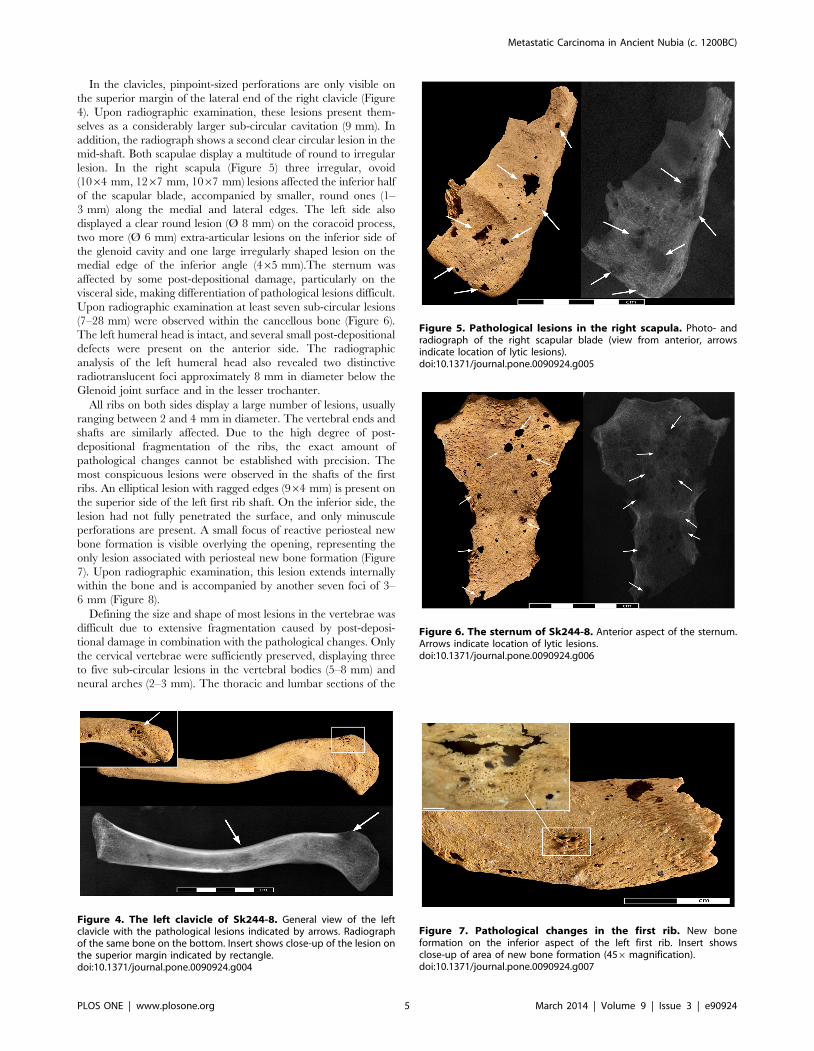
lesion. In the right scapula (Figure 5) three irregular, ovoid
(1064 mm, 1267 mm, 1067 mm) lesions affected the inferior half
of the scapular blade, accompanied by smaller, round ones (1–
3 mm) along the medial and lateral edges. The left side also
displayed a clear round lesion (Ø 8 mm) on the coracoid process,
two more (Ø 6 mm) extra-articular lesions on the inferior side of
the glenoid cavity and one large irregularly shaped lesion on the
medial edge of the inferior angle (465 mm).The sternum was
affected by some post-depositional damage, particularly on the
visceral side, making differentiation of pathological lesions difficult.
Upon radiographic examination at least seven sub-circular lesions
(7–28 mm) were observed within the cancellous bone (Figure 6).
The left humeral head is intact, and several small post-depositional
defects were present on the anterior side. The radiographic
analysis of the left humeral head also revealed two distinctive
radiotranslucent foci approximately 8 mm in diameter below the
Glenoid joint surface and in the lesser trochanter.
All ribs on both sides display a large number of lesions, usually
ranging between 2 and 4 mm in diameter. The vertebral ends and
shafts are similarly affected. Due to the high degree of post-
depositional fragmentation of the ribs, the exact amount of
pathological changes cannot be established with precision. The
most conspicuous lesions were observed in the shafts of the first
ribs. An elliptical lesion with ragged edges (964 mm) is present on
the superior side of the left first rib shaft. On the inferior side, the
lesion had not fully penetrated the surface, and only minuscule
perforations are present. A small focus of reactive periosteal new
bone formation is visible overlying the opening, representing the
only lesion associated with periosteal new bone formation (Figure
7). Upon radiographic examination, this lesion extends internally
within the bone and is accompanied by another seven foci of 3–
6 mm (Figure 8).
Defining the size and shape of most lesions in the vertebrae was
difficult due to extensive fragmentation caused by post-deposi-
tional damage in combination with the pathological changes. Only
the cervical vertebrae were sufficiently preserved, displaying three
to five sub-circular lesions in the vertebral bodies (5–8 mm) and
neural arches (2–3 mm). The thoracic and lumbar sections of the
Figure 4. The left clavicle of Sk244-8. General view of the leftclavicle with the pathological lesions indicated by arrows. Radiographof the same bone on the bottom. Insert shows close-up of the lesion onthe superior margin indicated by rectangle.doi:10.1371/journal.pone.0090924.g004
Figure 5. Pathological lesions in the right scapula. Photo- andradiograph of the right scapular blade (view from anterior, arrowsindicate location of lytic lesions).doi:10.1371/journal.pone.0090924.g005
Figure 6. The sternum of Sk244-8. Anterior aspect of the sternum.Arrows indicate location of lytic lesions.doi:10.1371/journal.pone.0090924.g006
Figure 7. Pathological changes in the first rib. New boneformation on the inferior aspect of the left first rib. Insert showsclose-up of area of new bone formation (456magnification).doi:10.1371/journal.pone.0090924.g007
Metastatic Carcinoma in Ancient Nubia (c. 1200BC)
PLOS ONE | www.plosone.org 5 March 2014 | Volume 9 | Issue 3 | e90924

spine were the most affected parts of the entire skeleton. Damage
was particularly extensive in the vertebral bodies, with discernible
lesions ranging in size from between 5 and 12 mm. The neural
arches were similarly affected, displaying smaller sub-circular
lesions (3–5 mm). A large lesion affecting almost the entire body of
7th thoracic vertebra (Figure 9), as well as the spinous process of
the 5th thoracic vertebra feature new bone formation within the
cancellous bone along the lesion margins (Figure 10).
Both innominate bones again suffered from heavy fragmenta-
tion. Within the fragments, distinctive circular cavitations within
the cancellous bone were observed in the better preserved iliac
crest and ischial tuberosity of both sides. Circular erosions were
also present on the inner cortical side of the fragments of the iliac
blades even though, due to heavy fragmentation, it is difficult to
ascertain whether they are due to pathology or post-mortem
damage. Formation of new bone within the trabecular structures
was observed in one lesion in the right iliac crest (Ø 6.5 mm)
(Figure 11) and one in the right ischium (Ø 7.5 mm). Despite post-
mortem damage, several cavitations were observed on the femoral
heads, with the right one displaying 7 cortical defects (5–8 mm)
and a lesion on the anterior side of the greater trochanter (Figure
12).
Discussion
Several differential diagnostic options can account for the
observed changes in skeleton Sk244-8.
Metastatic CarcinomaMetastatic organ cancer is the most common source of
metastasizing tumors affecting the skeleton [15]. Bone tissue is
one of the preferential sites for metastatic expansion [63]. Tumor
cells spread to the bones either through direct extension of a
primary soft tissue tumor, through the lymphatic system, cerebro-
spinal fluid or most importantly through haematogenous dissem-
ination. Thus, predilected sites for metastasis formation are those
bone structures rich in haematopoietic bone marrow and the
vertebrae, pelvis, ribs, sternum, skull, clavicles, scapulae, humeral
and femoral heads (in decreasing order of frequency) are the most
commonly affected elements [63]. The elements distal to the elbow
and knee are generally very rarely involved [17].
Skeletal response to metastatic tumours can either be osteolytic
(75%), osteoblastic (15%) or a combination of both (10%) [17].
The type, distribution and density of metastatic lesions are
dependent on the primary source of the tumour as well as on
the duration of the disease. Carcinomas with purely osteolytic
lesions are those of the thyroid, kidneys, adrenal glands, uterus and
gastrointestinal tract. Exclusively osteosclerotic lesions are pro-
duced by carcinoma of the prostate gland. Mixed lesions can occur
in carcinomas of the lung, breast, cervix, ovaries and testicles,
though the ratio of formation to resorption is highly variable.
However, none of these patterns is without exception [63]. In
osteolytic lesions, bone resorption due to growth of tumour cells
commences in cancellous bone characterised by internal scallop-
ing. Only in advanced stages does destruction progress into the
cortical bone [19]. The resulting lesions can vary considerably
both in size and shape. Depending on the aggressiveness of the
disease process, they can range between well circumscribed
(geographic) to poorly defined (motheaten or permeative) bone
destruction, the latter reflecting a more aggressive type [63,64].
Due to the mechanism of metastasis formation, the number and
size of lesions visible upon radiographic examination usually
exceeds the number of lesions visible externally, making the
radiographic appearance of lesions one of the key differential
diagnostic features of metastatic carcinoma [64]. Single metastases
are rare; in the majority of cases multiple lesions are present [15].
Multiple MyelomaMultiple myeloma is a neoplastic condition of the plasma cells of
the bone marrow [15]. Originating in the haematopoietic marrow
and cancellous structures of bone, multiple myeloma produces
numerous destructive lesions that can be very similar to those
caused by a carcinoma type cancer [64]. Differentiating between
multiple myeloma and metastatic carcinoma in dry bone is
Figure 8. Lytic lesions in the first rib. Photo- and radiograph of theleft first rib (superior surface. Arrows indicate location of the lesions.doi:10.1371/journal.pone.0090924.g008
Figure 9. Destructive lesion in vertebral body of the 7th
thoracic vertebrae. Detail of the pathological changes in the 7th
thoracic vertebra. Rectangle indicates area of new bone infill of thespongiosa. Close-up of new bone formation indicated by arrows isshown in the insert; arrows indicate new bone formation.doi:10.1371/journal.pone.0090924.g009
Metastatic Carcinoma in Ancient Nubia (c. 1200BC)
PLOS ONE | www.plosone.org 6 March 2014 | Volume 9 | Issue 3 | e90924

considered a major challenge and may not always be possible
[19,28].The main differential diagnostic features are size and
shape of the lesions. In contrast to metastatic carcinoma, the
lesions are usually small, uniform in size, spherical with effaced
edges, and are much denser and regular in distribution than
carcinoma lesions. Suppressed osteoblast formation in tumorous
foci is one of the hallmark pathophysiological features of multiple
myeloma [65,66]. Therefore, remodelling along the edges of
lesions or new bone formation as is seen in the individual from
Amara West is absent in multiple myeloma. [64].
MycosisSeveral fungal infections can produce lytic lesions in the skeleton
which may mimic the appearance of metastatic carcinoma [67]
even though, with the exception of African histoplasmosis, skeletal
involvement is generally rare [43: 217]. Bone infection occurs
secondary to haematogenous dissemination. Skeletal lesions
caused are almost exclusively lytic in nature with only little new
bone formation [43: 213]. Key features in differentiating between
metastatic carcinoma and fungal infections are the appearance of
the lytic lesions and bone remodelling. In mycoses lytic lesions
appear as fronts of resorption, in contrast to the space-occupying
lesions produced by bone metastases. Newly formed bone is rare in
fungal infections but if present it usually appears as characteristic
blunt spiculae [67]. In contrast to metastatic carcinoma, skeletal
lesions produced by mycotic infections occur throughout the
skeleton and also affects the distal portions of the long bones and
small hands of the hand and feet. The lesions observed in the
individual from Amara West do not conform to the features
associated with fungal infection, thus leaving it an unlikely
differential diagnosis.
Taphonomic damagePost-depositional processes could also produce damage to the
bone mimicking the osteolytic lesions caused by a tumour [27].
Breakage and surface erosion are generally common problems in
archaeological human remains. Small round holes similar to
metastatic lesions can be caused by a variety of factors including
roots, water, and termites [68] or dermatid beetles [69]. While
root or water damage is uncommon at Amara West, destruction
Figure 10. Lytic lesion in the spinous process of the 5th thoracic vertebra. A) shows a close-up of bone formation at 35x magnificationlocated within the lytic focus, B) SEM image of the lytic focus C) shows the complete spinous process with the location of the lytic focus highlightedin the rectangle.doi:10.1371/journal.pone.0090924.g010
Figure 11. Detail of new bone formation in the iliac crest. Theclose-up shows a focus of new bone formation indicated by arrows in alytic lesion in the iliac crest (40x magnification).doi:10.1371/journal.pone.0090924.g011
Metastatic Carcinoma in Ancient Nubia (c. 1200BC)
PLOS ONE | www.plosone.org 7 March 2014 | Volume 9 | Issue 3 | e90924

likely caused by osteophageous insects is frequently encountered
(Figure 13). However, even though these holes may appear similar
on the bone surface, they do not expand within the bone but
rather appear to be regular punched out tunnels through the entire
bone. In addition, insects do not show any preference for
particular elements but, if present, tend to affect all parts of the
skeleton. Further supporting evidence was obtained through
targeted SEM analyses in order to better characterise the nature
of the lesion margins. While in some examined lesions, post-
mortem damage was established as the cause (Figure 14a), others
show clear evidence of osteoclastic activity (Figure 14b). Therefore,
even though some post-mortem damage is certainly present in the
bones of Sk244-8, the radiographic appearance of the lesions, in
combination with the observation of associated periosteal new
bone formation, provides sufficient evidence to confirm that the
majority of the observed lesions are indeed of pathological origin.
The size, shape and distribution of the irregularly shaped, often
poorly defined to circular, lesions affecting the ribs, vertebrae,
clavicle, scapulae, pelvis, sternum, humeral and femoral heads of
skeleton 244-8 from Amara West are thus most probably the result
of a metastatic carcinoma. The irregularly distributed lesions of
varying shape and size as well as their subcortical appearance,
strongly argue against ascribing them to multiple myeloma. The
origin of the metastases, i.e. the primary tumour location, is not
possible to ascertain. Based on the examination of dry bone alone,
the origin of the metastases and the location of the primary tumour
is difficult to ascertain, if not impossible [19]. Generally, organ
cancers most commonly producing bone involvement are those of
the breast and the prostate gland, followed by the lung, thyroid
gland and kidneys [70]. While prostate cancer can be excluded
based on the type of metastatic lesions, cancer of the lung and
associated structures, or the thyroid gland, gastrointestinal tract or
liver, are possible sites for the primary tumour. Breast cancer does
occur in men [71], though rarely (,1% of cancers), thus remains a
possible source of the lesions.
Cancer in antiquityThis 25–35 year old man from Amara West, buried around
1200 BC, further evidence, provides another piece of evidence that
cancer is in fact not a modern phenomenon [25,29]. The apparent
absence of cancer in archaeological remains may also partly be an
illusion created by issues of bone preservation, and due to the fact
that methods of analysis are inadequate to detect initial changes
within bone. Due to financial, time and logistical reasons, human
remains are usually not systematically radiographed, and bone
metastases originating in cancellous tissue only penetrate the bone
surface in their advanced stages. If the immune system was already
compromised by other negative influences in a person’s life, people
may not have survived long enough to develop full skeletal
metastases. Thus, evidence for a large proportion of tumours could
be missed when skeletal remains are analysed [72]. Another
challenge in detecting cancer in ancient human remains is the
poor preservation of bone which often prevents the clear
identification of lytic lesions and precludes the diagnosis of
incomplete remains [27]. With increasing numbers of skeletal
collections and more detailed analysis, as well as more readily
available standard radiographic equipment, the evidence for
cancer in antiquity could increase significantly.
Recovered as part of an archaeological research project, this
individual can be set within a cultural and historical framework,
but also within the context of changing environmental conditions,
and specific diet and subsistence strategies. The potential exists,
therefore, to explore possible underlying causes of cancer in an
ancient population, before the onset of modernity. As such it could
provide important new insights into cancer aetiology and
epidemiology in the past. Even though today’s leading causes of
cancer are all products of modern living conditions and
industrialisation, a large number of environmental carcinogens
also occur naturally (e.g. asbestos), and would have affected our
ancestors in the same way [73]. The carcinogenic effects of smoke
from wood fires, particularly when indoors are well known [1,74].
The houses at Amara West typically feature hearths, but also
bread ovens [50], often within roofed spaces without windows,
where smoke would have dissipated slowly. In modern Sudan, the
common usage of fires in poorly ventilated rooms of small
Figure 12. Pathological changes in the right femoral head. Photo- and radiograph of lytic lesions in the right femoral head (arrows indicateareas of pathological lesions).doi:10.1371/journal.pone.0090924.g012
Figure 13. Post-mortem damage caused by insects. Tibia of askeleton from Amara West showing extensive damage caused bydermestid beetles.doi:10.1371/journal.pone.0090924.g013
Metastatic Carcinoma in Ancient Nubia (c. 1200BC)
PLOS ONE | www.plosone.org 8 March 2014 | Volume 9 | Issue 3 | e90924

mudbrick huts is still considered one of the major factors leading to
lung cancer [75]; similar conditions are common across early
societies, including those where other evidence of cancer has been
identified [25]. Bitumen, known also to cause cancer in individuals
occupationally exposed to bitumen fumes [76] was already used by
ancient Egyptians for waterproofing or embalming [77].
Infectious diseases can also lead to cancer. Schistosomiasis has
plagued inhabitants of Egypt and Nubia since at least 1500BC
[78,79,80] and is now recognized as a common cause of bladder
cancer [81]. In addition, it has been associated with an increased
risk of male breast cancer as a consequence of the hormonal
disturbances resulting from liver cirrhosis secondary to schistoso-
miasis infection [82]. This may also account for the fact that the
male to female breast cancer ratio in Egypt today is far greater
than anywhere else in the world [83]. Detecting evidence for
schistosomiasis infection in ancient human remains can be
achieved through immunobiological testing of mummified soft
tissue [84] or secondary through recovery of parasite eggs from soil
samples from graves or latrines as well as from coprolites [78].
Despite attempts to find parasite evidence in a selected number of
soil samples from other graves, no such evidence has yet been
identified at Amara West. Even though the disease is absent from
the Amara West area today, palaeoenvironmental data suggests
that the habitat would likely have been favourable for sustaining
populations of gastropods transmitting schistosomiasis during the
time of New Kingdom occupation. Though unproven, an
underlying schistosomiasis infection leading to breast cancer in
the man from Amara West as well as other Nubian and Egyptian
individuals [46] represents a plausible cause. The link between
gastrointestinal cancer and infection by helicobacter pylori, which is
recognized to have affected human populations since prehistory
[85] is also well established [86]. The individual from Amara
West, only 25–35 years old at death, further underlines that cancer
was restricted to neither old age nor elite social status. From a
modern clinical point of view, the young age-at-death of the man
from Amara West may seem unusual for the onset of skeletal
metastases, but it remains unknown whether the underlying causes
of cancer affected people in the same way and at the same speed as
they do today.
The lack of evidence for cancer in antiquity may to a large
extent, be the result of reduced life expectancy, and thus less time
to develop skeletal lesions if the immune system is already
compromised by an inadequate supply of nutrients and diseases.
This represents one of the major problems in inferring the absence
or presence of disease in the past in general [87]. The
archaeological and historical record certainly provides plenty of
evidence for possible causes of developing cancer. Despite recent
advances, the genetic background for cancer predisposition is still
far from being understood today [88,89]. Even though it may
perhaps remain unknown, there is no reason to assume that
predisposing genetic factors were not present in the past. The man
from Amara West does indicate that it was indeed possible to
develop skeletal lesions of cancer, provides a glimpse into one
individual’s life experience, and cautions against claims for the
absence, or presence, of any disease based on skeletal evidence
alone.
Conclusions
Dating to c. 1200 BC, the individual from Amara West in Sudan
represents one of the earliest people identified in the bioarchaeo-
logical record anywhere in the world, who suffered secondary
malignant neoplasm. This provides further support for the claim
that cancer is a disease of considerable antiquity. It may not have
been as prevalent as today, and there is little doubt that the main
factor accounting for the increased prevalence of cancer is
undoubtedly modern living. Environmental carcinogens deriving
from wood smoke (amongst others) have been present in the
human living environment for a long time, yet cancer is still very
rare in the palaeopathological record. However, with increasing
numbers of human remains available for study, and technical
advances and better availability of analytical equipment, in
combination with increased attention to detail by those conducting
palaeopathological research the number of ancient individuals
with cancer may increase significantly. Increasing scientific
research on mummified remains facilitated through improved
and more readily available imaging techniques such as computed
tomography-scanning, will provide an additional dataset, allowing
for detection of cancer in the soft tissues of preserved bodies.
Gaining a better understanding of the disease’s history and
epidemiology in the past may significantly contribute to further
investigate and understand the underlying mechanisms leading to
cancer today.
Increasingly, scholars working in palaeopathology are thinking
more about the value of using their data to contribute to
knowledge of disease today [90]. It is well known that people with
cancer today are developing resistance to the chemotherapy used,
similar to antibiotic resistance to the treatment of infectious disease
[10], and there have also been recommendations for future
research into the evolution of cancer to improve the management
Figure 14. Comparison of SEM-images of taphonomic and pathological lesions. A) shows the margin of a defect caused by post-mortemdamage. B) shows pathological changes on the margin of a lytic lesion in the cortical surface (bar on the bottom of each image indicates 100 mm).doi:10.1371/journal.pone.0090924.g014
Metastatic Carcinoma in Ancient Nubia (c. 1200BC)
PLOS ONE | www.plosone.org 9 March 2014 | Volume 9 | Issue 3 | e90924

of this common disease. For example, by using biomolecular
approaches to individual skeletons and mummies with evidence of
cancer (ancient DNA analysis), it might be possible to show
changes in their genome which can be mapped against the very
evolution of the human population, and detect mutations in
specific genes that are known to be associated with particular types
of cancer. In turn, and by linking these data to contextual
considerations i.e. the living environment that the person
experienced, it may be possible to understand better why and in
what ways cancer therapies need to be developed. Furthermore,
exploring the reasons for a population’s susceptibility to develop-
ing cancer may be aided by examining the nature of the immune
system of peoples affected by cancer in the past, via biomolecular
analysis, to see how cancer may have changed throughout the long
history of evolution. Research in these areas [91,92] has indicated
the potential for this approach. Consequently, by taking an
evolutionary approach to cancer, this knowledge may prove a
crucial element in finding ways to address one of the world’s major
health problems of the 21st century.
The presence of the disease in pre-modern populations further
raises questions about the theoretical concept of epidemiological
transitions undergone by human populations [46]. Even though
cancer represents one of the key features of the 2nd epidemiological
transition, its presence in a pre-2nd transitional population
highlights the fact that cancer as such is not only linked to
longevity but also to infectious diseases. The boundaries between
the 1st and 2nd transition may not be as clear-cut as previously
thought [46].
Acknowledgments
Fieldwork and export of human remains was carried out with the
permission and support of the National Corporation of Antiquities and
Museums (NCAM) of Sudan, particular thanks are due to the director
general, Abdelrahman Ali and the project inspector, Shadia Abdu Rabo
Thanks are due to A. Mongiatti (radiography) of the Department of
Conservation & Scientific Research at the British Museum; further thanks
are due to D. Saunders and C. Higgitt. M. Millet (Louvre, Paris) provided
information on the dating of the tomb.
Author Contributions
Conceived and designed the experiments: MB CR. Performed the
experiments: MB CR DA CC. Analyzed the data: MB CR DA.
Contributed reagents/materials/analysis tools: NS DA CC. Wrote the
paper: MB CR NS.
References
1. Boyle P, Levin P, editors (2008) World Cancer Report 2008. Lyon: International
Agency for Research on Cancer.
2. WHO (2013) Fact sheet Nu297: Cancer.
3. David AR, Zimmerman MR (2010) Cancer: an old disease, a new disease or
something in between? Nature Reviews: Cancer 10: 728–733.
4. McKeown RE (2009) The Epidemiologic Transition: Changing Patterns of
Mortality and Population Dynamics. American Journal of Lifestyle Medicine 3:
19S–26S.
5. Karpozilos A, Pavlidis N (2004) The treatment of cancer in Greek antiquity.
European Journal of Cancer 40: 2033–2040.
6. Nunn JF (2002) Ancient Egyptian Medicine. Norman: University of Oklahoma
Press.
7. Breasted JH (1930) The Edwin Smith Surgical Papyrus: Hieroglyphic
transliterations, translations and commentary. Chicago: University of Chicago
Press.
8. Sanchez GM, Meltzer ES (2012) The Edwin Smith Papyrus: Updated
Translation of the Trauma Treatise and Modern Medical Commentaries:
Updated Translation of the Trauma Treatise and Modern Medical Commen-
taries. Atlanta: Lockwood Press.
9. Mukherjee S (2010) The Emperor of All Maladies: A Biography of Cancer:
Scribner.
10. Stearns SC (2012) Evolutionary medicine: its scope, interest and potential.
Proceedings of the Royal Society of Biological Science 279: 4305–4321.
11. Nesse RM, Williams GC (1994) Why we get sick. The new science of Darwinian
medicine. New York: Vintage Books.
12. Manderson L (2011) Anthropologies of cancer and risk uncertainty and
disruption. In: Singer M, Erickson PI, editors. A companion to medical
anthropology 1st Edition. Chichester, West Sussex: Blackwell Publishing
Limited. pp. 323–338.
13. Brown PJ, Armelagos GJ, Maes KC (2011) Humans in a world of microbes: the
anthropology of infectious diseases. In: Singer M, Erickson PI, editors. A
companion to medical anthropology 1st Edition. Chichester, West Sussex:
Blackwell Publishing Limited. pp. 253–270.
14. Buikstra J, Roberts C, editors (2012) The Global History of Paleopathology:
Oxford University Press.
15. Dorfman HD, Czerniak B (1998) Bone Tumors. St. Louis: Mosby, Inc.
16. Bertram JS (2000) The molecular biology of cancer. Molecular Aspects of
Medicine 21: 167–223.
17. Greenspan A, Remagen W (1998) Differential Diagnosis of Tumors and Tumor-
like Lesions of Bones and Joints. Philadelphia, New York: Lippincott-Raven.
18. Barrett R, Kuzawa CW, McDade T, Armelagos GJ (1998) Emerging and Re-
emerging Infectious Diseases: The Third Epidemiologic Transition. Annual
Reviews in Anthropology 27: 247–271.
19. Ortner DJ (2003) Identification of Pathological Conditions in Human Skeletal
Remains. London: Academic Press. 645 p.
20. Franceschi S, Wild CP (2013) Meeting the global demands of epidemiologic
transition – The indispensable role of cancer prevention. Molecular Oncology 7:
1–13.
21. Cox M (2000) Ageing Adults from the Skeleton. In: Cox M, Mays S, editors.
Human Osteology: In Archaeology and Forensic Science. London: Greenwich
Medical Media. pp. 61–81.
22. Chamberlain A (2006) Demography in Archaeology. Cambridge: Cambridge
University Press. 235 p.
23. Gabler K (2009) Die Medja - dein Lieferant und Helfer. Untersuchungen zu
medja von Deir el-Medine anhand von Ostraka und Papyri [in German,
unpublished master thesis]. Munchen: Ludwig-Maximilians-Universitat
24. Parkin TG (2003) Old Age in the Roman World: A Cultural and Social History.
Baltimore: John Hopkins University Press.
25. Capasso LL (2005) Antiquity of Cancer. International Journal of Cancer 113: 2–
13.
26. Doll R, Peto R (1981) The Causes of Cancer: Quantitative Estimates of
Avoidable Risks of Cancer in the United States Today. Journal of the National
Cancer Institute 66: 1192–1308.
27. Brothwell DR (2012) Tumors: Problems of Differential Diagnosis in Paleopa-
thology. In: Grauer AL, editor. A Companion to Paleopathology. Oxford:
Wiley-Blackwell. pp. 420–433.
28. Marks MK, Hamilton MD (2007) Metastatic Carcinoma: Palaeopathology and
Differential Diagnosis. International Journal of Osteoarchaeology 17: 217–234.
29. Nerlich AG, Rohrbach H, Bachmeier B, Zink A (2006) Malignant tumors in two
ancient populations: An approach to historical tumor epidemiology. Oncology
Reports 16: 197–202.
30. Strouhal E, Kritscher H (1990) Neolithic case of a multiple myeloma from
Mauer (Vienna, Austria). Anthropologie 28: 78–97.
31. Gladykowska-Rzeczycka J (1991) Tumors in antiquity in East and Middle
Europe. In: Ortner DJ, Aufderheide AC, editors. Human Paleopathology -
Current Syntheses and Future Options. Washington, London: Smithsonian
Institution Press. pp. 251–256.
32. Rokhlin D (1966) Disease in ancient man. Moskov: Nauka Ed.
33. Strouhal E (2001) Malignant tumours in past populations in Middle Europe In:
La Verghetta M, Capasso L, editors. Proceesings of the XIIIth European
Meeting of the Paleopathology Association. Teramo: Edigrafical. pp. 265–272.
34. Strouhal E (1976) Tumors in the remains of Ancient Egyptians. American
Journal of Physical Anthropology 45: 613–620.
35. Baker BJ, Judd M (2012) Development of Paleopathology in the Nile Valley. In:
Buikstra J, Roberts C, editors. The Global History of Paleopathology: Oxford
University Press. pp. 209–234.
36. Pahl WM (1986) Tumors of bone and soft tissue in ancient Egypt and Nubia: a
synopsis of the detected cases. International Journal of Anthropology 1: 267–
275.
37. Strouhal E, Vyhanek L (1981) New cases of malign tumours from Late Period
cemeteries at Abusir and Saqqara (Egypt). Ossa 8: 165–189.
38. Strouhal E (1993) A case of metastatic carcinoma from Christian Sayala
(Egyptian Nubia). Anthropologischer Anzeiger 51: 97–115.
39. Strouhal E, Vyhnanek L (1982) New cases of malignant tumors from late period
cemeteries at Abusir and Saqqara (Egypt). Ossa 8: 165–189.
40. Strouhal E (1991) A case of primary carcinoma from Christian Sayala (Egyptian
Nubia). Journal of Paleopathology 3: 51–65.
41. Wells C (1963) Ancient Egyptian Pathology. Journal of Laryngology and
Otology 77: 261–265.
42. Prates C, Sousa S, Oliveira C, Ikram S (2011) Prostate metastatic bone cancer in
an Egyptian Ptolemaic mummy, a proposed radiological diagnosis. International
Journal of Paleopathology 1: 98–103.
Metastatic Carcinoma in Ancient Nubia (c. 1200BC)
PLOS ONE | www.plosone.org 10 March 2014 | Volume 9 | Issue 3 | e90924

43. Aufderheide AC, Rodrıguez-Martın C (1998) The Cambridge Encyclopaedia of
Human Paleopathology. Cambrigde, New York: Cambridge University Press.578 p.
44. Ho JHC (1972) Nasopharyngeal Carcinoma. In: Klein G, Weinhouse S, editors.
Advances in Cancer Research. London: Academic Press, Inc. pp. 57–92.
45. Aufderheide AC (2003) The Scientific Study of Mummies. Cambridge:Cambridge University Press.
46. Esche E, Mummert A, Robinson J, Armelagos GJ (2010) Cancer in Egypt and
Nubia. Anthropologie 48: 33–39.
47. Dupras T, de Voogt A, Francigny V, Williams L, Lacey J (2014) Advanced
Metastatic Carcinoma in the Paleopathological Record: A Case Study from theSudan. 41th Annual Meeting of the Paleopathology Association. Calgary,
Alberta, Canada.
48. Spencer P (1997) Amara West I. The architectural report. London: The EgyptExploration Society.
49. Spencer N (2012) Insights into Life in occupied Kush during the New Kingdom:
New Research at Amara West. Der Antike Sudan 23: 21–28.
50. Spencer N (forthcoming) Amara West: considerations on urban life in occupiedKush. In: Welsby D, Anderson JR, editors. Proceedings of the 12th International
Conference for Nubian Studies. Leuven: OLA.
51. Binder M (2011) The 10th-9th century BC - New Evidence from Cemetery C ofAmara West. Sudan & Nubia 15: 39–53.
52. Binder M, Spencer N, Millet M (2011) Cemetery D at Amara West: the
Ramesside Period and its aftermath. British Museum Studies in Ancient Egyptand Sudan.
53. Spencer N, Macklin MG, Woodward JC (2012) Reassessing the abandonment ofAmara West: the impact of a changing Nile? Sudan & Nubia 16: 37–43.
54. Ryan P, Cartwright C, Spencer N (2012) Archaeobotanical research in a
pharaonic town in ancient Nubia. The British Museum Technical ResearchBulletin 6: 97–107.
55. Binder M, Spencer N (In Press) The bioarchaeology of Amara West in Nubia:
Investigating the impacts of political, cultural and environmental change onhealth and diet. In: Fletcher A, Antoine D, Hill JD, editors. Regarding the Dead.
London: British Museum Press.
56. Van Pelt WP (forthcoming) Revising Egypto-Nubian Relationss in NewKingdom Lower Nubia: From Egyptianization to Cultural Entanglement.
Cambridge Archaeological Journal 23.
57. Smith ST (2003) Wretched Kush. London, New York: Routledge.
58. Buikstra JE, Ubelaker DH (1994) Standards for Data Collection from Human
Remains. Lafayetteville, Arkansas: Arkansas Archaeological Survey. 206 p.
59. Brickley M, McKinley JI, editors (2004) Guidelines to the Standards forRecording Human Remains. Reading: Institute of Field Archaeologists Paper
Number 7.
60. Bruzek J (2002) A method for visual determination of sex, using the human hipbone. American Journal of Physical Anthropology 117: 157–168.
61. Brooks S, Suchey JM (1990) Skeletal age determination based on the os pubis: a
comparison of the Acsadi-Nemeskeri and Suchey-Brooks methods. HumanEvolution 5: 227–238.
62. Scheuer L, Black S (2000) Developmental juvenile osteology. San Diego:
Academic Press.
63. Resnick D (1995) Diagnosis of Bone and Joint Disorders. St. Louis, MO: W. B.
Saunders.
64. Rothschild BM, Hershkovitz I, Dutour O (1998) Clues Potentially DistinguishingLytic Lesions of Multiple Myeloma From Those of Metastatic Carcinoma.
American Journal of Physical Anthropology 105: 241–250.
65. Yaccoby S (2010) Advances in the understanding of myeloma bone disease andtumour growth. British Journal of Haematology 149: 311–321.
66. Sezer O (2009) Myeloma Bone Disease: Recent Advances in Biology, Diagnosis,
and Treatment. The Oncologist 14: 276–283.
67. Hershkovitz I, Rothschild BM, Dutour O, Greenwald C (1998) Clues torecognition of fungal origin of lytic skeletal lesions. American Journal of Physical
Anthropology 106: 47–60.
68. Huchet JB, Deverly D, Gutierrez B, Chauchat C (2011) Taphonomic Evidenceof a Human Skeleton Gnawed by Termites in a Moche-Civilisation Grave at
Huaca de la Luna, Peru. International Journal of Osteoarchaeology 21: 92–102.
69. Huchet JB, Le Mort F, Rabinovich R, Blau S, Coqueugniot H, et al. (2013)
Identification of dermestid pupal chambers on Southern Levant human bones:
inference for reconstruction of Middle Bronze Age mortuary practices. Journalof Archaeological Science 40: 3793–3803.
70. Layer D (2005) Skelettmetastasen. In: Freyschmidt J, Stabler A, editors.
Handbuch diagnostische Radiologie - Muskoloskelettales System 2: Springer.pp. 327–338.
71. Anderson WF, Jatoi I, Tse J, Rosenberg PS (2010) Male Breast Cancer: A
Population-Based Comparison With Female Breast Cancer. Journal of ClinicalOncology 28: 232–239.
72. Rothschild BM, Rothschild C (1995) Comparison of Radiologic and Gross
Examination for Detection of Cancer in Defleshed Skeletons. American Journalof Physical Anthropology 97: 357–363.
73. Hueper WC (1963) Environmental Carcinogenesis in Man and Animals. Annalsof the New York Academy of Sciences 108: 963–1038.
74. Delgado J, Martinez LM, Sanchez TT, Ramirez A, Iturria C, et al. (2005) Lung
cancer pathogenesis associated with wood smoke exposure. Chest 128: 124–131.75. Awadelkarim KD, Mariani-Costantini R, Elwali NE (2012) Cancer in the
Sudan: An overview of the current status of knowledge on tumor patterns andrisk factors. Science of the Total Environment 423: 214–228.
76. Binet S, Pfohl-Leszkowicz A, Brandt H, Lafontaine M, Castegnaro M (2002)Bitumen fumes: review of work on the potential risk to workers and the present
knowledge on its origin. Science of the Total Environment 300: 37–49.
77. Serpico M, White R (2000) Resins, amber and bitumen. In: Nicholson PT, ShawI, editors. Ancient Egyptian Materials and Technology. Cambridge: Cambridge
University Press.78. Bouchet F, Harter S, Le Bailly M (2003) The State of the Art of
Paleoparasitological Research in the Old World. Memorias do Instituto
Oswaldo Cruz 98: 95–101.79. Miller RL, Armelagos GJ, Ikram S, De Jonge N, Krijger FW, et al. (1992)
Palaeoepidemiology of schistosoma infection in mummies. British MedicalJournal 304: 355-356.
80. Reyman TA, Zimmerman MR, Lewin PK (1977) Autopsy of an Egyptianmummy. 5. Histopathologic investigation. Canadian Medical Association
Journal 117: 470–472.
81. Sitas F, Parkin DM, Chirenje M, Stein L, Abratt R, et al. (2008) Part II: Cancerin Indigenous Africans - causes and control. Lancet Oncology 9: 786–795.
82. Buzdar AU (2003) Breast cancer in men. Oncology (Williston Park) 17: 1361–1364.
83. Mustacchi P (2003) Schistosomiasis. In: Kufe DW, Pollock RE, Wechselbaum
RR, editors. Holland-Frei, Cancer Medicine 6th edition. Hamilton (ON): BCDecker.
84. Hibbs CA, Secor WV, Van Gerven D, Armelagos GJ (2011) Irrigation andinfection: The immunoepidemiology of schistosomiasis in ancient Nubia.
American Journal of Physical Anthropology 145: 290–29885. Linz B, Balloux F, Moodley Y, Manica A, Liu H, et al. (2007) An African origin
for the intimate association between humans and Helicobacter pylori. Nature
445: 915–918.86. Polk DB, Peek RM (2010) Helicobacter pylori: gastric cancer and beyond. Nat
Rev Cancer 10: 403–414.87. Wood JW, Milner GR, Harpending HC, Weiss KM (1992) The Osteological
Paradox - Problems of Inferring Prehistoric Health from Skeletal Samples.
Current Anthropology 33: 343–370.88. Bartsch H, Dally H, Popanda O, Risch A, Schmezer P (2007) Genetic risk
profiles for cancer susceptibility and therapy response. Recent Results in CancerResearch 174: 19–36.
89. Frank SA (2004) Genetic predisposition to cancer - insights from populationgenetics. Nature Reviews: Genetics 5: 764–772.
90. Zuckerman MK, Turner BL, Armelagos GJ (2012) Evolutionary thought in
paleopathology and the rise of the biocultural approach. In: Grauer A, editor. Acompanion to paleopathology. Oxford: Wiley Blackwell.
91. Fornaciari G, Marchetti A, Pellegrini S, Ciranni R (1999) K-ras mutation in thetumour of King Ferrante I of Aragon (1431–1494) and environmental mutagens
at the Aragonese court of Naples. International Journal of Osteoarchaeology 9:
302–306.92. Schlott T, Eiffert H, Schmidt-Schultz T, Gebhardt M, Parzinger H, et al. (2007)
Detection and analysis of cancer genes amplified from bone material of aScythian royal burial in Arzhan near Tuva, Siberia. Anticancer Research 27:
4117–4119.
93. Strouhal E (1994) Malignant Tumors in the Old World. PaleopathologyNewsletter 85 (supplement): 1–6.
94. Strouhal E (1991) Myeloma Multiplex versus Osteolytic Metastatic Carcinoma:Differential Diagnosis in Dry Bones. International Journal of Osteoarchaeology
1: 219–224.95. Strouhal E (1978) Ancient Egyptian case of carcinoma. Bulletin of the New York
Academy of Medicine 54: 290–302.
96. Schultz M, Parzinger H, Posdnjakov DV, Chikisheva TA, Schmidt-Schultz TH(2007) Oldest known case of metastasizing prostate carcinoma diagnosed in the
skeleton of a 2,700-year-old Scythian King from Arzhan (Siberia, Russia).International Journal of Cancer 121: 2591–2595.
Metastatic Carcinoma in Ancient Nubia (c. 1200BC)
PLOS ONE | www.plosone.org 11 March 2014 | Volume 9 | Issue 3 | e90924