Volumetric neuroimage analysis extensions for the MIPAV software package
22
Volumetric Neuroimage Analysis Extensions for the MIPAV Software Package Pierre-Louis Bazin a , Jennifer L. Cuzzocreo b , Michael A. Yassa b , William Gandler c , Matthew J. McAuliffe c , Susan S. Bassett b , and Dzung L. Pham a,1 a Laboratory of Medical Image Computing, Neuroradiology Division, Department of Radiology and Radiological Science, Johns Hopkins University, Baltimore, MD, 21218, USA b Divisionof Psychiatric Neuroimaging, Department of Psychiatry and Behavioral Sciences, Johns Hopkins University, Baltimore, MD, 21218, USA c Biomedical Imaging Research Services Section, Center for Imaging Technology, National Institutes of Health, Bethesda, MD, 20892, USA Abstract We describe a new collection of publicly available software tools for performing quantitative neuroimage analysis. The tools perform semi-automatic brain extraction, tissue classification, Talairach alignment, and atlas-based measurements within a user-friendly graphical environment. They are implemented as plug-ins for MIPAV, a freely available medical image processing software package from the National Institutes of Health. Because the plug-ins and MIPAV are implemented in Java, both can be utilized on nearly any operating system platform. In addition to the software plug-ins, we have also released a digital version of the Talairach atlas that can be used to perform regional volumetric analyses. Several studies are conducted applying the new tools to simulated and real neuroimaging data sets. Keywords segmentation; magnetic resonance imaging; Talairach atlas 1 Introduction Segmentation of structural magnetic resonance (MR) brain images is a critical step in many neuroscientific applications such as in the morphological analyses of different diseases, characterization of the relationship between brain structure and function, and in treatment monitoring and planning (Pham et al. (2000)). Most publicly available software tools for performing segmentation tasks, however, are hindered by the lack of cross-platform compatibility, limited support of file formats, complex user interfaces, and an inability to manually edit the results. As a result, it is not uncommon for a segmentation analysis to require several different software packages for performing file conversion, image processing, manual editing, and volumetric measurements. 1 Corresponding author, 600 North Wolfe Street, Phipps B100, Baltimore, MD 21287, USA. Tel.:+1-410-614-3249; Fax: +1-410-614-1577 E-mail address: [email protected] (D. L. Pham). Publisher's Disclaimer: This is a PDF file of an unedited manuscript that has been accepted for publication. As a service to our customers we are providing this early version of the manuscript. The manuscript will undergo copyediting, typesetting, and review of the resulting proof before it is published in its final citable form. Please note that during the production process errors may be discovered which could affect the content, and all legal disclaimers that apply to the journal pertain. NIH Public Access Author Manuscript J Neurosci Methods. Author manuscript; available in PMC 2008 September 15. Published in final edited form as: J Neurosci Methods. 2007 September 15; 165(1): 111–121. NIH-PA Author Manuscript NIH-PA Author Manuscript NIH-PA Author Manuscript
-
Upload
independent -
Category
Documents
-
view
4 -
download
0
Transcript of Volumetric neuroimage analysis extensions for the MIPAV software package

Volumetric Neuroimage Analysis Extensions for the MIPAVSoftware Package
Pierre-Louis Bazina, Jennifer L. Cuzzocreob, Michael A. Yassab, William Gandlerc, MatthewJ. McAuliffec, Susan S. Bassettb, and Dzung L. Phama,1a Laboratory of Medical Image Computing, Neuroradiology Division, Department of Radiology andRadiological Science, Johns Hopkins University, Baltimore, MD, 21218, USA
b Divisionof Psychiatric Neuroimaging, Department of Psychiatry and Behavioral Sciences, Johns HopkinsUniversity, Baltimore, MD, 21218, USA
c Biomedical Imaging Research Services Section, Center for Imaging Technology, National Institutes ofHealth, Bethesda, MD, 20892, USA
AbstractWe describe a new collection of publicly available software tools for performing quantitativeneuroimage analysis. The tools perform semi-automatic brain extraction, tissue classification,Talairach alignment, and atlas-based measurements within a user-friendly graphical environment.They are implemented as plug-ins for MIPAV, a freely available medical image processing softwarepackage from the National Institutes of Health. Because the plug-ins and MIPAV are implementedin Java, both can be utilized on nearly any operating system platform. In addition to the softwareplug-ins, we have also released a digital version of the Talairach atlas that can be used to performregional volumetric analyses. Several studies are conducted applying the new tools to simulated andreal neuroimaging data sets.
Keywordssegmentation; magnetic resonance imaging; Talairach atlas
1 IntroductionSegmentation of structural magnetic resonance (MR) brain images is a critical step in manyneuroscientific applications such as in the morphological analyses of different diseases,characterization of the relationship between brain structure and function, and in treatmentmonitoring and planning (Pham et al. (2000)). Most publicly available software tools forperforming segmentation tasks, however, are hindered by the lack of cross-platformcompatibility, limited support of file formats, complex user interfaces, and an inability tomanually edit the results. As a result, it is not uncommon for a segmentation analysis to requireseveral different software packages for performing file conversion, image processing, manualediting, and volumetric measurements.
1 Corresponding author, 600 North Wolfe Street, Phipps B100, Baltimore, MD 21287, USA. Tel.:+1-410-614-3249; Fax:+1-410-614-1577 E-mail address: [email protected] (D. L. Pham).Publisher's Disclaimer: This is a PDF file of an unedited manuscript that has been accepted for publication. As a service to our customerswe are providing this early version of the manuscript. The manuscript will undergo copyediting, typesetting, and review of the resultingproof before it is published in its final citable form. Please note that during the production process errors may be discovered which couldaffect the content, and all legal disclaimers that apply to the journal pertain.
NIH Public AccessAuthor ManuscriptJ Neurosci Methods. Author manuscript; available in PMC 2008 September 15.
Published in final edited form as:J Neurosci Methods. 2007 September 15; 165(1): 111–121.
NIH
-PA Author Manuscript
NIH
-PA Author Manuscript
NIH
-PA Author Manuscript

We have developed three software tools that cover some of the most common needs ofquantitative neuroimage analysis: 1) brain extraction - isolation of the brain region and removalof external structures, 2) tissue classification - identification of the gray matter, white matter,and cerebrospinal fluid (CSF) tissues, and 3) Talairach alignment - alignment to a standardizedcoordinate system and quantification using digital atlases. The methods upon which the toolsare based have been previously published and validated but until now, have not been availablein an integrated, user-friendly, and cross-platform software package.
All of the software tools have been implemented as plug-ins for the Medical Image Processing,Analysis, and Visualization (MIPAV) software package developed at the Center forInformation Technology, National Institutes of Health (McAuliffe et al. (2001)). MIPAVenables quantitative analysis and visualization of medical images from multiple modalitiessuch as PET, MRI, CT, and microscopy. It includes basic and advanced computational methodsto analyze and quantify biomedical data, supports all major medical image formats, andprovides many visualization and data manipulation tools for 2D and 3D images or image series.Because MIPAV is Java-based, it can be utilized on nearly any operating system, such asWindows, Linux, and MacOS. MIPAV also provides an intuitive graphical user interface,providing researchers a user-friendly tool to visualize and analyze their imaging data. Theentire software package and documentation may be downloaded from the internet (http://mipav.cit.nih.gov).
When applied in conjunction with our provided digital atlas, our tools can be used forperforming regional volumetric analyses. This approach is loosely based on the approach ofAndreasen et al. (1996). In their work, the unlabeled brain image is first placed into Talairachspace using a standard piecewise affine transformation. Selected labels from the Talairach-Tournoux brain atlas (Talairach and Tournoux (1988)) are then superimposed on thetransformed brain, and the volume of brain tissues inside each of the labeled regions ismeasured. It was shown that these measurements provide accurate volumetrics of grossanatomical brain structures, such as lobes, and the procedure is very fast. In our version of themethod, the studied brain image may be first segmented into gray matter, white matter, andCSF, and this segmentation is then transferred into Talairach coordinates. Labels of the regionsof interest are selected and transferred to the image, and the volume of tissues included in eachregion may be computed separately for each tissue. Before quantification, the labels may alsobe manually refined using the various editing tools already incorporated into MIPAV.
In this paper, we describe the software tools, as well as our Talairach digital atlas, and showhow these tools can be used for performing regional volumetric analysis. We have made boththe software tools and atlas data available on the internet (http://medic.rad.jhu.edu). Discussionforums are also provided on the same website for technical support and a detailed tutorial onthe usage of these tools is available as a technical guide.
2 Methods2.1 The MIPAV Software Package
To support the National Institutes of Health (NIH) intramural research program, the BiomedicalResearch and Services Section (BIRSS) has developed MIPAV, a multifaceted, platform-independent, quantification, and visualization application for biomedical images. MIPAV is aJava-enabled application that encompasses the functionality to process, quantify and visualizedata sets from micro (eg. crystallography, microscopy) to macro (eg. CT, MRI). MIPAVsupports numerous medical and research file formats including Zeiss, Biorad, TIFF, DICOM3.0, NIFTI, AFNI, MINC, GE, Siemens, Philips, AVI, Interfile, MIPAV XML, and newformats are added as needed. Figure 1 shows the graphical user interface and some of thevisualization and manual editing capabilities offered by MIPAV.
Bazin et al. Page 2
J Neurosci Methods. Author manuscript; available in PMC 2008 September 15.
NIH
-PA Author Manuscript
NIH
-PA Author Manuscript
NIH
-PA Author Manuscript

From a system perspective, MIPAV was designed to support multiple levels of user needs:core, scripts, and plug-ins. Core functionality addresses basic user needs includingcomprehensive VOI support, manual annotation and delineation, reading/writing all supportedfile formats, and various image processing, filtering, segmentation, and registration algorithms.Core visualization tools include tri-planar visualization and editing, image and movie capture,3D surface and volume rendering, and 2D/3D fusion of multichannel datasets. In addition,MIPAV interfaces with the Insight Segmentation and Registration Toolkit (ITK) (Ibanez andSchroeder (2005)). To facilitate repetitive processing, MIPAV incorporates scripting abilitiesand supports command line execution. The scripting mechanism may be used to record asequence of image processing and analysis operations and then apply the recorded operationsto a series of datasets. User specific needs can be met by developing plug-ins using MIPAV’sopen application programming interface.
The software tools described in this paper were developed as Java plug-ins for MI-PAV. Duringinitialization, MIPAV automatically loads installed plug-ins and builds the plug-in menu,allowing them to be utilized as if they were native algorithms. The plug-ins that do not directlyrequire user interaction (eg. tissue classification) may also be used with MIPAV’s scriptinginterface, thereby facilitating batch processing.
2.2 The Brainstrip ToolIt is often desirable to remove unwanted structures from MR brain images as a preliminarystep prior to volumetric analyses. These structures include regions corresponding to the skull,eyes, skin, and dura. This step is unfortunately difficult, typically requiring several hours tomanually segment the brain tissue, depending on the desired quality for the result. As analternative to manual segmentation, fully automated techniques are available (Smith (2002);Shattuck and Leahy (2002);Segonne et al. (2004);Rex et al. (2004);Rehm et al. (2004)), butthese methods still benefit from subsequent editing (Fennema-Notestine et al. (2006)).
We have implemented a semiautomatic approach that substantially reduces manual processingtimes and is based on the method employed and validated by Goldszal et al. (1998). Thephilosophy of this approach is to interactively obtain a reasonable estimate of the brain maskthat can be refined manually. Given some simple input information from the user (the minimumand maximum intensity of the gray and white matter, a seed point defined inside the brain, anda bounding box for the brain), a sequence of automatic processing routines extract anapproximate brain volume. The first step in the sequence thresholds the image based on theprovided intensity values. A morphological erosion using a spherical structuring element(typically set to 5mm) is then applied to the thresholded volume to remove any bridges thatoccur between the brain tissue and dura. Next, the provided seed point is used to perform aregion growing algorithm to extract the central portion of the brain volume. Two dilations withthe same structuring element are then applied to recover the lost tissue and smooth the overallsurface. Finally, holes within the mask are removed by performing a 2-D region growing oneach slice within the background and retaining the complement. Optionally, a three-dimensional bounding box input may be defined by the user to decide where to cut thebrainstem, or select a region of the brain. Fig. 2 shows the actual brainstrip user interface. Theextracted region is superimposed on the original image and can then be manually edited toattain the accuracy of fully manual methods.
Selection of the inputs will typically require less than one minute for a trained operator. Theoutput result is generated within a few seconds on a typical PC after the inputs have beenprovided. Because the method is simple and fast, the operator has the ability to quickly andeasily make adjustments should the initial input parameters yield a poor result. Because allaspects of the processing are provided through MIPAV, the need to switch to separate softwareprograms for performing any manual refinement is eliminated. Note also that MIPAV natively
Bazin et al. Page 3
J Neurosci Methods. Author manuscript; available in PMC 2008 September 15.
NIH
-PA Author Manuscript
NIH
-PA Author Manuscript
NIH
-PA Author Manuscript

provides its own implementation of two popular algorithms for fully automatic brain stripping:Brain Surface Extractor (BSE) (Shattuck and Leahy (2002)) and Brain Extraction Tool (BET)(Smith (2002)).
2.3 The FANTASM ToolTissue classification algorithms typically segment the brain into its three major tissue types:gray matter, white matter, and CSF. Many algorithms have been proposed in the literature thatperform tissue classification in the presence of MR imaging artifacts, such as noise and intensityinhomogeneities (Wells et al. (1996); Leemput et al. (1999);Pham and Prince (1999);Shattucket al. (2001);Smith (2002). Our tool is based on the Fuzzy And Noise Tolerant AdaptiveSegmentation Method (FANTASM) (Pham (2001a);Han et al. (2004)). Given the number oftissue classes, the technique automatically classifies each pixel of the image as one of thetissues, estimates the inhomogeneity field, and also attributes a membership value from 0 to 1for each class.
We briefly describe FANTASM in the following. Our implementation in this work differsslightly from the implementation described in Han et al. (2004) in that we explicitly model thegain field parametrically as a polynomial field. Let Ω be the set of voxel indices, C be thenumber of tissue classes, and yj, j ∈ Ω be the observed (preprocessed) MR image values, whichmay be a vector in the case of multichannel images. The goal of FANTASM is to find intensitycentroids vk, k = 1, . . . , C, a gain field gj, j ∈ Ω, and membership values ujk, j ∈ Ω, k = 1, . . . ,C that will minimize the following objective function:
JFANTASM = ∑j∈Ω
∑k=1
Cu jk
q ∥ y j − g jvk∥2 + β
2 ∑j∈Ω ∑k=1
Cu jk
q ∑l∈N j
∑m≠k
ulmq (1)
Note that the membership values are constrained to be greater than zero and satisfy∑k=1
C u jk = 1.
The first term in (1) is simply the fuzzy c-means clustering objective function (Bezdek et al.(1993)) with a gain field term that allows the centroids to spatially vary throughout the image.The parameter q, which must satisfy q > 1 (and is set to 2 in this work), determines the amountof “fuzziness” of the resulting classification (Bezdek et al. (1993)). The parameter C is thenumber of classes and is assumed to be known. For brain images, this typically is set equal to3, corresponding to gray matter, white matter, and CSF. The second term in (1) is a penaltyfunction that forces the tissue memberships to be smooth, where the parameter β is a weightthat determines the amount of smoothness (Pham (2001b)). In practice, we use a normalizedsmoothness parameter that is multiplied by the square of the intensity range of the image. Thesymbol Nj represents the set of first order neighbors of pixel j.
We make the simplifying assumption that the gain field g can be represented by a low-degreethree-dimensional polynomial function. Although this assumption is somewhat morerestrictive than that of Han et al. (2004), it is usually sufficient to model the intensity variations(cf. Leemput et al. (1999)), is computationally more efficient, and requires fewer parametersto be specified. Mathematically, this can be expressed as:
g j = ∑n=1
Nf nPn( j)
where Pn are polynomial basis functions and fn are the coefficients. We use Chebyshevpolynomial basis functions for their numerical stability and other advantageous properties forfunction approximation (Davis (1975)). The number of Chebyshev coefficients are 20 for the
Bazin et al. Page 4
J Neurosci Methods. Author manuscript; available in PMC 2008 September 15.
NIH
-PA Author Manuscript
NIH
-PA Author Manuscript
NIH
-PA Author Manuscript

3rd degree case, and 35 for 4th degree polynomial fields. Because the gain field is assumed tobe smooth and the equations are overdetermined, we subsample the images by a factor of 3along each dimension when computing the coefficients fn to increase the computationalefficiency.
An iterative algorithm for minimizing (1) can be derived by solving for a zero gradientcondition with respect to the memberships and the centroids and can be described as follows:
1. Obtain initial estimates of the centroids, vk.
2. Compute membership functions:
u jk =( ∥ y j − g jvk∥
2 + β ∑l∈N j
∑m≠k
ulmq )−1/(q−1)
∑i=1
C ( ∥ y j − g jvi∥2 + β ∑
l∈N j∑
m≠iulm
q )−1/(q−1). (2)
3. Compute centroids:
vk =∑
j∈Ωu jk
q g jy j
∑j∈Ω
u jkq g j
2 , k = 1, … , C. (3)
4. Compute gain field by solving the following matrix equation for the coefficients fn:u jkvkPn( j) · f n = u jky j . (4)
5. Go to Step 2 and repeat until convergence.
We assume that the algorithm has converged when the maximum change in membership valuesbetween iterations is less than 0.01. In order to decrease the possibility of convergence to alocal minimum, the degree of the polynomial gain field is initially set to zero and theneventually increased to the desired value as convergence is achieved. Fig. 3 shows the actualFANTASM plug-in user interface. The additional tabs allow specification of multichannel datasets and various algorithm parameters.
2.4 The Talairach ToolTo draw comparisons between brains belonging to different subjects, it is often beneficial todefine a common coordinate system. The brain atlas and coordinate system defined byTalairach and Tournoux (1988) is a commonly accepted standard in the neuroscientificcommunity. The classical Talairach normalization procedure can be described as follows: 1)align the anterior and posterior commissures (AC and PC, respectively), and 2) scale the brainto align its boundaries to those of the atlas.
Although the transformation utilized in the Talairach transformation is relatively low-dimensional, particularly when compared to the more recently developed nonlineardeformation methods (Maintz and Viergever (1998); Christensen et al. (1997);Shen andDavatzikos (2002)), it is computationally fast and easy to control. For example, if afteralignment the mid-sagittal plane is not vertical within the axial view or if the gross atlas labelsdeviate substantially from the underlying image, one can easily update the input parameters tocorrect the results. It is clear that a Talairach alignment will be of limited use for small sub-structures of the brain, but it has been shown to accurately and reliably align some largerregions, such as lobes (Andreasen et al. (1996)). In the following, we briefly describe theoperation of our Talairach tool.
Bazin et al. Page 5
J Neurosci Methods. Author manuscript; available in PMC 2008 September 15.
NIH
-PA Author Manuscript
NIH
-PA Author Manuscript
NIH
-PA Author Manuscript

The first alignment within the Talairach transformation, often referred to as ’AC-PCalignment’, is a rigid-body Euclidean transformation (i.e. 3-D rotation and translation). It alignsthe AC-PC coordinate systems of brains defined by their origins and axes. We therefore requirea minimum of three points for AC-PC alignment: the AC, PC, and a mid-sagittal point (SG).With them, the entire new coordinate system is known, and the brain image can be transformedinto this new space.
Our AC-PC alignment follows the same procedure as the AFNI software (Cox (1996)). Whilethree point landmarks are required in theory, there can be problems when the AC or PC arelarger than one voxel, or when the mid-sagittal plane is oblique with respect to the voxel grid.The AFNI method requires the operator to pick several additional landmarks, such as the limitsof AC and PC, and points for accurately finding the mid-sagittal plane. It uses five pointlandmarks:
1. the AC superior edge: the top limit of the anterior commissure,
2. the AC posterior margin: the rear limit of the anterior commissure,
3. the PC inferior edge: the bottom limit of the posterior commissure,
4. a first mid-sagittal point (SG1) : any point in the mid-sagittal (inter-hemispheric)plane, above the corpus callosum,
5. a second mid-sagittal point (SG2): another point, if possible away from the first, ofthe mid-sagittal plane.
Finding these landmarks may require some training and familiarization with neuroanatomy.Once all landmarks are determined, it is straightforward to compute a rigid-bodytransformation such that the conditions of the AC-PC coordinate system are satisfied (Bazinet al. (2005)).
The second part of the Talairach alignment consists of scaling the brain to match its boundarieswith those of the atlas. It is a 12 degrees-of-freedom, piecewise linear transform that bringsthe AC, PC, and anterior, posterior, left, right, inferior, and superior boundaries of the brain tonormalized positions. To perform the Talairach scaling, the 6 extremal points (most anterior,posterior, left, right, inferior, and superior points), or alternatively the bounding box enclosingthe brain, must be determined. Along with the AC and PC points, these landmarks define a setof 12 boxes. In each box, the image is independently scaled along the three dimensions to fitthe normalized dimensions of the Talairach coordinates. Continuity is guaranteed as the boxesshare common faces. Our tool can save and reload the transform information (position of AC,PC, AC-PC orientation and scale, and cortex boundaries) in a readable text file. This allowsthe transformation to be applied to additional co-registered images (eg. functional MR images,the output of tissue classification algorithms).
Figure 4 shows the user interface for performing the Talairach transformation. The user canselect the various landmark points, compute the transformation, save the transformation, aswell as compute the inverse transformation. This latter functionality can be used fortransforming labels from a normalized template into the subject space.
2.5 Atlas LabelsTwo general purpose brain atlases that have become popular due to their availability are theTalairach atlas (Talairach and Tournoux (1988); Lancaster et al. (2000);Nowinski and Belov(2003)) and the MNI/ICBM atlas ( Holmes et al. (1998)). Although the Talairach atlas (alsoknown as the Talairach-Tournoux atlas) is the actual atlas from which the Talairach alignmentprocedure was created (Talairach and Tournoux (1988)), the atlas and the coordinate system
Bazin et al. Page 6
J Neurosci Methods. Author manuscript; available in PMC 2008 September 15.
NIH
-PA Author Manuscript
NIH
-PA Author Manuscript
NIH
-PA Author Manuscript

are two separate entities. Other atlases, such as the ICBM atlas, can also be used within thecoordinate system.
The Talairach atlas consists of 27 histological slices in the axial orientation on which manydifferent structures have been manually delineated. In our atlas, we have included labeledregions for use in MIPAV, as well as a set of volumetric label images that can be used in anyother imaging software. The atlas is based on manually corrected labels obtained from theTalairach Daemon database and therefore possesses a hierarchy of structures of different scale,from left and right cerebrum to individual Brodmann areas (see Fig. 5). The spatial resolutionof the atlas is 1mm x 1mm x 4mm. The groups and structures are listed in Lancaster et al.(2000) and Bazin et al. (2005). Once images are aligned into a common coordinate system,they may be labeled using the atlas, thereby providing an indication of the location, size, andshape of the particular structure of interest. Most small structures, such as those at theBrodmann level, will unlikely be accurate after Talairach alignment, but larger regions (eg.lobes, ventricles) are often reasonably delineated and can be further refined manually usingthe core tools provided within MIPAV. The MNI/ICBM atlas is also available in the MIPAVformat for download from our website.
2.6 Volume measurementsOur combined tools may be used to obtain regional volumetric measurements in the brain. Thiscan be accomplished using the following protocol:
1. Brainstrip: the images of interest are pre-processed in order to isolate the brain tissuesin the image.
2. FANTASM: the voxels of the original image are classified into white matter, graymatter, or CSF.
3. Talairach: the brain image is aligned within the Talairach coordinate system.
4. Atlas Labels: the volumes of interest (VOIs) for the study are loaded from theTalairach atlas onto the aligned brain.
5. Atlas inverse transform: the aligned brain with VOIs is transformed back into itsoriginal image space.
6. Volume measurements: the number of voxels within the studied tissue class is countedfor each VOI, using MIPAV VOI analysis tools.
Because the Talairach alignment is non-linear, any direct measurement in this coordinatesystem is irrelevant; voxels have a different resolution in different boxes. For this reason, Step4 is required to bring back any Talairach-aligned image into the original or the AC-PC alignedcoordinates.
Between steps (5) and (6), the operator has the additional option to perform manual editing ofthe VOI boundaries. Statistics from VOIs (eg. volume, mean intensity) are easily generatedwithin MIPAV, as shown in Figure 6. The volume of gray or white matter within each VOImay be computed, as well as the total brain volume. For gross lobar measurements, the providedTalairach labels are often sufficient for many applications. Even if further refinement isrequired, the labels will likely yield substantial time savings over completely manualdelineation. A technical instruction guide is available from the MIPAV and plug-in websitesthat provides a detailed description of the entire procedure (McAuliffe (2005)).
Bazin et al. Page 7
J Neurosci Methods. Author manuscript; available in PMC 2008 September 15.
NIH
-PA Author Manuscript
NIH
-PA Author Manuscript
NIH
-PA Author Manuscript

3 Results3.1 Brainstrip validation
The Brainstrip tool was validated on ten MR head scans against a previously validated manualprotocol performed using an in-house developed software package called “Measure” (Barta etal. (1997)). Images were acquired from a GE Signa 1.5 Tesla MR scanner using a T1-weightedMPRAGE imaging protocol with a voxel size of 0.9375 × 0.9375 × 1.5mm. The imagesincluded in this analysis were from healthy adults, ages 50 and older, who are participants inan on-going longitudinal study of aging and cognition (Bassett et al. (2006)). Raters wereinstructed to set the lower threshold in the Brainstrip tool high enough such that the CSF in thelateral ventricles was removed but not so high that it resulted in the erosion of temporal lobes.The upper threshold was set as low as possible without removing any white matter tissue. Thebrain region was then manually edited using MIPAV’s integrated paint tools.
Figure 7 shows a comparison of the total brain volumes computed semi-automatically usingour plug-in vs. the manual results using the Measure program. Brain volume was defined astotal gray and white matter as computed using FANTASM. The mean total brain volumecomputed manually was 1212.40 cubic centimeters (cc) with a standard deviation (SD) of120.73 and the mean volume computed using Brainstrip was 1188.17cc with a SD of 106.37.The mean error was −24.23cc with a 95% confidence interval of −47.97cc to −0.48cc, equatingto less than 4% error. Although there appears to be a slight bias in the Brainstrip results towardlower volumes, the differences were not statistically significant under a paired T-test. The meanabsolute percent error was 2.85% (SD=1.81). A Pearson correlation analysis yielded an R2 of0.93, indicating a high level of agreement between the two approaches. Processing times forbrain extraction were reduced from 4 to 8 hours using manual editing to 30 minutes to 1 hourusing the Brainstrip plug-in with manual postprocessing.
3.2 FANTASM validationThe original FANTASM implementation has been previously validated on simulated and realimages (Pham (2001a);Pham and Prince (2004)). The MIPAV plug-in implementation differsfrom the original mainly with respect to the gain field estimation. In this section, we comparethe results of applying the new implementation to FMRIB’s Automated Segmentation Tool(FAST), another freely available, cross-platform tissue classification software tool (Zhang etal. (2001)). The two methods are validated on the Brainweb phantom from the MontrealNeurological Institute (Collins et al. (1998)).
Table 1 shows the results of applying both FANTASM and FAST to the Brainweb phantomsimulated with various noise levels (indicated by ‘N’), and inhomogeneity levels (indicated by‘I’). The smoothness parameter setting for FANTASM was set at 0.3 for all images and a 3rddegree polynomial function was used to model the gain field. Different smoothness parametersettings were tested for FAST but because this value seemed to have little effect on the resultingperformance, it was left at its default setting. From the results, it is apparent that FANTASMperformed well at low noise levels and seemed to be very robust to inhomogeneity artifacts.FAST did not perform as well at low noise levels but was more robust to increases in noise.
The last three rows of Table 1 show the mean error between the true partial volume and theFANTASM membership functions, FAST partial volume estimates, and FAST posteriorprobability values, respectively. The FANTASM membership functions appear to better modelthe partial volume effects within the Brainweb image. The FAST partial volume estimatestended to provide an overly fuzzy representation relative to the true values while the posteriorprobabilities appeared to be too binarized. This is illustrated in Figure 8.
Bazin et al. Page 8
J Neurosci Methods. Author manuscript; available in PMC 2008 September 15.
NIH
-PA Author Manuscript
NIH
-PA Author Manuscript
NIH
-PA Author Manuscript

FANTASM typically requires approximately four minutes of computation time to segment anMR image with 1mm cubic resolution on a typical PC workstation (3GHz Intel processor). Inour tests, FAST required approximately 8 minutes on average. Although the Java programminglanguage is sometimes criticized for slow execution times, in practice we have found negligibledifferences in speed when compared to equivalent C code during floating-point intensiveoperations.
3.3 Talairach validationA sample of 20 scans from the same study described in Section 3.1 was used to evaluate thevalidity of frontal lobe measurements using the new tools. The images first were processedwith the Brainstrip tool to isolate brain tissue. Left and right frontal lobes were manuallysegmented in the Measure software, according to a previously published methodology that hasan inter-rater reliability of 0.99 (Aylward et al. (1997)). Figure 9 shows a typical result ofmanual vs. Talairach parcellation and Figure 10 shows plots of the resulting volumes for thefrontal lobes in each hemisphere. The mean left frontal lobe volume was estimated to be 145.4cc(SD=30.7) using manual delineation. The mean volume of the same region computed usingour Talairach tool was 146.2cc (SD=29.3). The mean right frontal lobe volume computed usingthe same two methods was 151.4cc (SD=34.1) and 154.9cc (SD=32.0), respectively. The intra-rater reliability between these methods was assessed using Intra-class correlation (ICC = 0.95on the right, ICC = 0.97 on the left). A Pearson correlation model was also estimated withR2 = 0.964 for the left and 0.975 for the right frontal lobe. This suggests that the Talairach-labeled volumes are good predictors of the actual volumes as determined by manualsegmentation.
To test the sensitivity of the Talairach tool in detecting differences in volume, we split thesample into two groups of 10 according to the volumes estimated using the manual method. Acomparison of large and small volumes was conducted using an independent samples t-testand showed a significant difference on the left (T = 13.4, p < 0.0001) and on the right (T =10.8, p < 0.0001). We conducted the same comparison using the volumes estimated using theMIPAV automated method and also detected a significant difference on the left (T = 10.3, p< 0.0001), and on the right (T = 10.3, p < 0.0001). Even though there are small differences inthe levels of significance, this comparison shows that the semi-automated approach possessedenough power to detect statistically significant group differences with small samples.
Processing times were approximately 1.5 to 2 hours to manually delineate the left and rightfrontal lobes in one subject. This time was reduced to approximately half an hour using theTalairach tool (which itself requires less than 10 minutes) followed by manual refinement.
4 ConclusionsWe have described three software tools and a digital brain atlas for assisting neuroanatomicalstudies requiring volumetric quantification and/or regional delineation. The tools are user-friendly, freely available to the public, well-documented, and may be used on multipleoperating system platforms. Robustness to different data types is provided by implementingsimple and fast processing and allowing the user to make changes to the inputs when necessary.Furthermore, by employing the MIPAV software package, a wide variety of visualization andediting tools are provided in a unified environment for multiple image processing tasks. Ourown experience has indicated that these tools may be used to substantially reduce the amountof manual interaction required in a number of neuroimaging applications.
Acknowledgements
This work was supported in part by NIH/NIA grant AG016324 (PI: S.S. Bassett), NIH/NIDA grant DA017231-01(PI: N.A. Honeycutt), and NIH/NINDS grant R01NS054255-01 (PI: D.L. Pham).
Bazin et al. Page 9
J Neurosci Methods. Author manuscript; available in PMC 2008 September 15.
NIH
-PA Author Manuscript
NIH
-PA Author Manuscript
NIH
-PA Author Manuscript

ReferencesAndreasen NC, Rajarethinam R, Stephan Arndt TC II, VWS, Flashman LA, O’Leary DS, dt JCE, Yuh
WT. Automatic atlas-based volume estimation of human brain region s from mr images. Journal ofComputer Assisted Tomography 1996;20(1):98–106. [PubMed: 8576490]
Aylward E, Augustine A, Li Q, Barta P, Pearlson G. Measurement of frontal lobe volume on magneticresonance imaging scans. Psychiatry Research: Neuroimaging Section 1997;71:23–30.
Barta P, Dhingra L, Royall R, Schwartz E. Improving stereological estimates for the volumes of structuresidentified in three-dimensional arrays of spatial data. Journal of Neuroscience Methods 1997;75:111–118. [PubMed: 9288642]
Bassett S, Yousem D, Cristinzio C, Kusevic I, Yassa M, Caffo B, Zeger S. Familial risk for Alzheimer’sdisease alters fMRI activation patterns. Brain 2006;129:1229–1239. [PubMed: 16627465]
Bazin P-L, McAuliffe M, Gandler W, Pham DL. Free software tools for atlas-based volumetricneuroimage analysis. Proceedings of SPIE Medical Imaging 2005: Image Processing2005;5747:1824–1833.
Bezdek J, Hall L, Clarke L. Review of MR image segmentation techniques using pattern recognition.Medical Physics 1993;20:1033–1048. [PubMed: 8413011]
Christensen GE, Joshi SC, Miller MI. Volumetric transformation of brain anatomy. IEEE Transactionson Medical Imaging December;1997 16(6):864–877. [PubMed: 9533586]
Collins, D.; Zijdenbos, A.; Kollokian, V.; Sled, J.; Kabani, N., et al. Design and construction of a realisticdigital brain phantom; IEEE Trans Med Imag. 1998. p. 463-468.see http://www.bic.mni.mcgill.ca/brainweb/
Cox, R. Afni: Software for analysis and visualization of functional magnetic resonance neuroimages;Computers and Biomedical Research. 1996. p. 162-173.see http://afni.nimh.nih.gov/
Davis, PJ. Interpolation and Approximation. Dover; 1975.Fennema-Notestine C, Ozyurt IB, Clark C, Morris S, Bischoff-Grethe A, Bondi M, Jernigan T, Fischl B,
Segonne F, Shattuck D, Leahy R, Rex D, Toga A, Zou K, Birn M, Brown G. Quantitative evaluationof automated skull-stripping methods applied to contemporary and legacy images: effects ofdiagnosis, bias correction, and slice location. Human Brain Mapping 2006;27:99–113. [PubMed:15986433]
Goldszal AF, Davatzikos C, Pham DL, Yan MXH, Bryan RN, Resnick SM. An image processing systemfor qualitative and quantitative volumetric analysis of brain images. Journal of Computer AssistedTomography 1998;22(5):827–837. [PubMed: 9754125]
Han X, Pham D, Tosun D, Rettmann M, Xu C, Prince J. Cruise: Cortical reconstruction using implicitsurface evolution. Neuroimage 2004;23(3):997–1012. [PubMed: 15528100]
Holmes, C.; Hoge, R.; Collins, L.; Woods, R.; Toga, A.; Evans, A. Enhancement of MR images usingregistration for signal averaging; J Computer Assisted Tomography. 1998. p. 324-333.see http://www.loni.ucla.edu/ICBM/ICBMBrainTemplate.html
Ibanez, L.; Schroeder, W. The ITK Software Guide. Kitware, INc; 2005.Lancaster, J.; Woldorrf, M.; Parsons, L.; Liotti, M.; Freitas, C.; Rainey, L.; Kochunov, P.; Nickerson, D.;
Mikiten, S.; Fox, P. Automated talairach atlas labels for functional brain mapping; Human BrainMapping. 2000. p. 120-131.http://ric.uthscsa.edu/projects/tdc
Leemput K, Maes F, Vandermulen D, Seutens P. Automated model-based tissue classification of MRimages of the brain. IEEE Trans Med Imag 1999;18(10):897–908.
Maintz JBA, Viergever MA. A survey of medical image registration. Medical Image Analysis 1998;2(1):1–36. [PubMed: 10638851]
McAuliffe, M. Using MIPAV to label and measure brain components in Talairach space. Tech rep,National Institutes of Health. 2005. see http://mipav.cit.nih.gov/documentation/howto.html
McAuliffe, M.; Lalonde, F.; McGarry, D.; Gandler, W.; Csaky, K.; Trus, B. Medical image processing,analysis and visualization in clini cal research. Proceedings of the 14th IEEE Symposium onComputer-Based Medi cal Systems (CBMS 2001); 2001. p. 381-386.
Nowinski WL, Belov D. The cerefy neuroradiology atlas: a talairach-tournoux atlas-b ased tool foranalysis of neuroimages available over the internet. NeuroImage 2003;20:50–57. [PubMed:14527569]
Bazin et al. Page 10
J Neurosci Methods. Author manuscript; available in PMC 2008 September 15.
NIH
-PA Author Manuscript
NIH
-PA Author Manuscript
NIH
-PA Author Manuscript

Pham, D. Robust fuzzy segmentation of magnetic resonance images. Proceedings of the 14th IEEESymposium on Computer-Based Medical Systems; Bethesda, MD. July 26–27. 2001a p. 127-131.
Pham D. Spatial models for fuzzy clustering. Computer Vision and Image Understanding 2001b;84(2):285–297.
Pham D, Prince J. Adaptive fuzzy segmentation of magnetic resonance. IEEE Trans Med Imag1999;18:737–752.
Pham, D.; Prince, J. Robust unsupervised tissue classification in mr image. Proceedings of the IEEEInternational Symposium on Biomedical Imaging; Arlington. april. 2004 p. 109-112.
Pham DL, Xu C, Prince JL. Current methods in medical image segmentation. Annual Review ofBiomedical Engineering 2000;2:315–337.
Rehm K, Schaper K, Anderson J, Woods R, Stolzner S, Rottenberg D. Putting our heads together: aconsensus approach to brain/non-brain segmentation in T1-weighted MR volumes. Neuroimage2004;22:1262–1270. [PubMed: 15219598]
Rex D, Shattuck D, Woods R, Narr K, Luders E, Rehm K, Stolzner S, Rottenberg D, Toga A. A meta-algorithm for brain extraction in MRI. Neuroimage 2004;23:625–637. [PubMed: 15488412]
Segonne F, Dale A, Busa E, Glessner M, Salat D, Hahn H, Fischl B. A hyrbrid approach to the skullstripping in MRI. Neuroimage 2004;22:1060–1075. [PubMed: 15219578]
Shattuck D, Leahy R. Brainsuite: An automated cortical surface identification tool. Med Im Anal 2002;8(2):129–142.
Shattuck D, Sandor-Leahy S, Schaper K, Rottenberg D, Leahy R. Magnetic resonance image tissueclassification using a partial volume model. Neuroimage 2001;13:856–876. [PubMed: 11304082]
Shen D, Davatzikos C. Hammer: Hiearachical attribute matching mechanism for elastic registration. IEEETransactions on Medical Imaging 2002;21(11)
Smith SM. Fast robust automated brain extraction. Human Brain Mapping 2002;17(3)Talairach J, Tournoux P. Co-Planar Stereotaxic Atlas of the Human Brain. Thieme. 1988Wells W, Grimson W, Kikins R, Jolesz F. Adaptive segmentation of MRI data. IEEE Trans Med Imag
1996;15:429–442.Zhang, Y.; Brady, M.; Smith, S. Segmentation of brain MR images through a hidden Markov random
field model and the expectation-maximization algorithm; IEEE Trans Med Imag. 2001. p. 45-57.seehttp://www.fmrib.ox.ac.uk/fsl/fast/index.html
Bazin et al. Page 11
J Neurosci Methods. Author manuscript; available in PMC 2008 September 15.
NIH
-PA Author Manuscript
NIH
-PA Author Manuscript
NIH
-PA Author Manuscript

Fig 1.The MIPAV graphical user interface. Painting in a tri-planar view, volume rendering, andcontour-based segmentation tools are a few of the capabilities offered by the software package.
Bazin et al. Page 12
J Neurosci Methods. Author manuscript; available in PMC 2008 September 15.
NIH
-PA Author Manuscript
NIH
-PA Author Manuscript
NIH
-PA Author Manuscript

Fig 2.The skull stripping plug-in: original image, interface, and stripped image mask overlaid onoriginal image (from left to right).
Bazin et al. Page 13
J Neurosci Methods. Author manuscript; available in PMC 2008 September 15.
NIH
-PA Author Manuscript
NIH
-PA Author Manuscript
NIH
-PA Author Manuscript
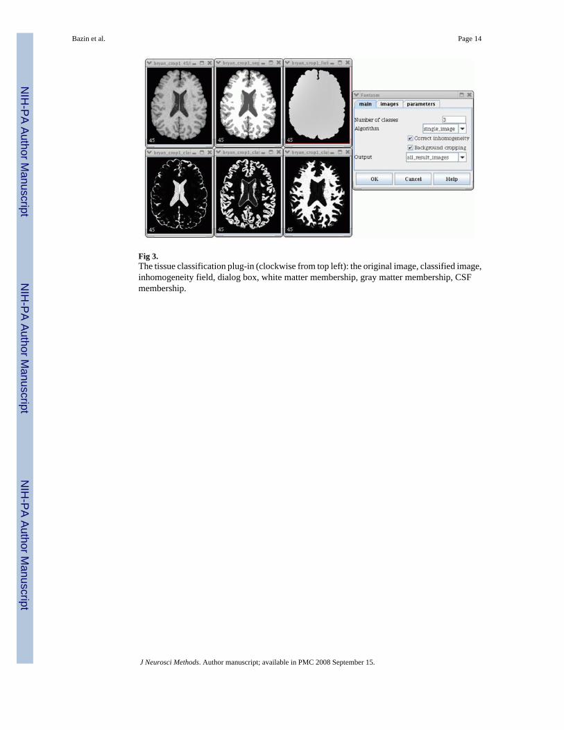
Fig 3.The tissue classification plug-in (clockwise from top left): the original image, classified image,inhomogeneity field, dialog box, white matter membership, gray matter membership, CSFmembership.
Bazin et al. Page 14
J Neurosci Methods. Author manuscript; available in PMC 2008 September 15.
NIH
-PA Author Manuscript
NIH
-PA Author Manuscript
NIH
-PA Author Manuscript

Fig 4.The Talairach alignment plug-in: (clockwise from top left) main dialog box, original image,AC-PC aligned image, image after full Talairach transformation, triplanar view for AC-PCalignment landmark selection, AC-PC alignment dialog box.
Bazin et al. Page 15
J Neurosci Methods. Author manuscript; available in PMC 2008 September 15.
NIH
-PA Author Manuscript
NIH
-PA Author Manuscript
NIH
-PA Author Manuscript

Fig 5.The Talairach atlas for MIPAV: (a) the volumetric images with the structure labels (b) thecorresponding VOIs, over a Talairach aligned brain.
Bazin et al. Page 16
J Neurosci Methods. Author manuscript; available in PMC 2008 September 15.
NIH
-PA Author Manuscript
NIH
-PA Author Manuscript
NIH
-PA Author Manuscript
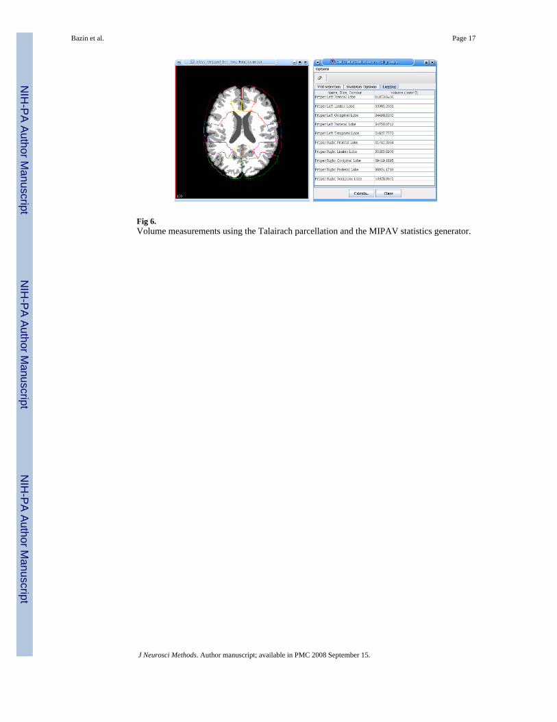
Fig 6.Volume measurements using the Talairach parcellation and the MIPAV statistics generator.
Bazin et al. Page 17
J Neurosci Methods. Author manuscript; available in PMC 2008 September 15.
NIH
-PA Author Manuscript
NIH
-PA Author Manuscript
NIH
-PA Author Manuscript

Fig 7.Plot of total brain volumes in cubic centimeters (cc) estimated using the semi-automatedapproach with our Brainstrip plug-in against the manual approach. The red line plots the resultof a linear regression.
Bazin et al. Page 18
J Neurosci Methods. Author manuscript; available in PMC 2008 September 15.
NIH
-PA Author Manuscript
NIH
-PA Author Manuscript
NIH
-PA Author Manuscript

Fig 8.FANTASM and FAST applied to the Brainweb phantom: (a) phantom image with 3% noiseand 20% inhomogeneity, (b) true classification, (c) FANTASM classification (d) FASTclassification, (e) true gray matter partial volume, (f) FANTASM gray matter membership, (g)FAST gray matter partial volume, (h) FAST gray matter probability map.
Bazin et al. Page 19
J Neurosci Methods. Author manuscript; available in PMC 2008 September 15.
NIH
-PA Author Manuscript
NIH
-PA Author Manuscript
NIH
-PA Author Manuscript

Fig 9.Frontal lobe delineations performed manually (left), using Talairach labels (center), and usingTalairach labels after transforming back into the original image space (right).
Bazin et al. Page 20
J Neurosci Methods. Author manuscript; available in PMC 2008 September 15.
NIH
-PA Author Manuscript
NIH
-PA Author Manuscript
NIH
-PA Author Manuscript

Fig 10.Plot of frontal lobe volumes measured using Talairach and manual protocols: (a) lefthemisphere, (b) right hemisphere. The red line plots the result of a linear regression.
Bazin et al. Page 21
J Neurosci Methods. Author manuscript; available in PMC 2008 September 15.
NIH
-PA Author Manuscript
NIH
-PA Author Manuscript
NIH
-PA Author Manuscript

NIH
-PA Author Manuscript
NIH
-PA Author Manuscript
NIH
-PA Author Manuscript
Bazin et al. Page 22
Table 1Classification errors for FANTASM and FAST. “Mem” refers to membership values, “Pve” refers to partialvolume estimates, and “Prob” refers to the posterior probability values.
Method 3%N, 0%I 3%N,20%I 3%N,40%I 5%N,20%I 7%N,20%I
FANTASM 4.36% 4.48% 4.51% 5.36% 6.49%FAST 6.22% 6.39% 7.44% 6.30% 7.01%FANTASM-Mem 0.055 0.056 0.056 0.066 0.078FAST-Pve 0.086 0.085 0.089 0.091 0.097FAST-Prob 0.070 0.070 0.075 0.067 0.069
J Neurosci Methods. Author manuscript; available in PMC 2008 September 15.