Housing Alternatives to Promote Holistic Health of the Fragile Aged
Upload
independentCategory
view
2download
0
Volumetric brain changes in femaleswith fragile X-associated tremor/ataxiasyndrome (FXTAS)
J.S. Adams, BAP.E. Adams, BSD. Nguyen, PhDJ.A. Brunberg, MDF. Tassone, PhDW. Zhang, MDK. Koldewyn, BAS.M. Rivera, PhDJ. Grigsby, PhDL. Zhang, MD, PhDC. DeCarli, MDP.J. Hagerman, MD,PhD
R.J. Hagerman, MD
ABSTRACT
Background: Fragile X-associated tremor/ataxia syndrome (FXTAS) is a late-onset neurodegen-erative disorder occurring in male and rare female carriers of a premutation expansion (55 to 200CGG repeats) of the fragile X mental retardation 1 (FMR1) gene.
Methods: Volumetric MRI studies, clinical staging, cognitive testing, and molecular analysis wereconducted in 15 female premutation carriers affected by FXTAS (age 59.5 � 10.3 years), 20unaffected female carriers (43.3 � 11.2 years), 11 genetically normal female controls (51.0 �
10.3 years), 36 affected male carriers (65.0 � 5.6 years), 25 unaffected male carriers (53.5 �
12.5 years), and 39 male controls (58.0 � 15.0 years). Female and male carriers with FXTASwere matched on duration of disease.
Results: We found less pronounced reductions of cerebellar volume and a lower incidence of in-volvement (symmetric high T2 signal) of the middle cerebellar peduncles (MCP sign) in femalesaffected by FXTAS (13%) compared with affected males (58%). We found reduced brain volumesand increased white matter disease associated with the presence of FXTAS in females comparedwith female controls. We also observed significant associations between reduced cerebellar vol-ume and both increased severity of FXTAS symptoms and increased length of the CGG repeatexpansion in male premutation carriers, but not in females.
Conclusions: Females affected by fragile X-associated tremor/ataxia syndrome (FXTAS) demon-strated milder brain changes than affected males, although they showed a similar pattern ofradiologic findings consistent with brain atrophy and white matter disease. FXTAS should beconsidered (by ordering fragile X DNA testing) in females who present with late-onset ataxia,action tremor, or neuropathy, particularly in those with a family history of mental retardation,autism, or premature ovarian failure. Neurology® 2007;69:851–859
Trinucleotide (CGG) repeat expansions in the 5= untranslated region of the fragile Xmental retardation 1 (FMR1) gene have recently been associated with clinical presenta-tions other than fragile X syndrome, a common cause of mental retardation and autism.Individuals with CGG expansions in the premutation range (55 to 200 repeats), previ-ously thought to be clinically unaffected, are now known to sometimes display forms ofclinical involvement, including neurocognitive deficits,1-4 emotional problems,5-7 prema-ture ovarian failure (POF),8,9 and the fragile X-associated tremor/ataxia syndrome(FXTAS), a late-onset neurodegenerative disorder.10-13
Core features of FXTAS include intention tremor and gait ataxia, typically with onsetafter age 50 years.10,11 Other features include memory and executive function deficits thatmay progress to cognitive impairment and dementia3,4,14; peripheral neuropathy11; andMRI findings of brain atrophy, often with subcortical and periventricular white matter
From the M.I.N.D. Institute (J.S.A., P.E.A., K.K., S.M.R., P.J.H., R.J.H.) and the Department of Pediatrics (J.S.A., R.J.H.), Public HealthSciences, Division of Biostatistics (D.N.), Radiology (J.A.B.), Biochemistry and Molecular Medicine (F.T., W.Z., P.J.H.), Neuroscience(K.K.), Psychology (S.M.R.), and Neurology (C.D., L.Z.), University of California, Davis, CA; and the Department of Medicine, Universityof Colorado at Denver and Health Sciences Center, Denver, CO (J.G.).
Supported by National Institute of Child Health and Development grants HD036071 and HD02274, National Institute of NeurologicalDisorders and Stroke grants NS044299 and NS43532, National Institute of Aging grant P30 AG10129, the Alzheimer Disease Center, theCenters for Disease Control and Prevention, the IDeA Laboratory, and the M.I.N.D. Institute.
Disclosure: The authors report no conflicts of interest.
Address correspondence andreprint requests to Dr. RandiHagerman, M.I.N.D. Institute,University of California–DavisMedical Center, 2825 50thStreet, Sacramento, CA [email protected]
Copyright © 2007 by AAN Enterprises, Inc. 851

disease, including white matter disease inthe middle cerebellar peduncles (the MCPsign, present in approximately 59% of af-fected males).15-18 Neuropathologic studiesof FXTAS have documented intranuclearinclusions in neurons and astroglial cellsthroughout the cerebrum and brain-stem.19,20 Although FXTAS affects approx-imately 40% of male carriers older than 50years in known fragile X families, it isthought to be rare in females, with only afew case studies reported.21-25 The secondX chromosome in females may be protec-tive for involvement with FXTAS.23,24
Neither POF nor FXTAS is seen in indi-viduals with fragile X syndrome, who haveCGG expansions in the full mutation range(�200 repeats). In contrast to fragile Xsyndrome, which involves silencing of theFMR1 gene, premutation carriers havenormal or slightly reduced levels of theFMR1 protein product (FMRP), but dra-matically (twofold to eightfold) elevatedlevels of FMR1 messenger RNA(mRNA).26 It is hypothesized that the pres-ence of elevated levels of expanded-repeatFMR1 mRNA has a toxic “gain-of-function” effect,27 akin to the pathogenicmechanism associated with myotonic dys-trophy.28 The presence of a pathogenicmechanism involving the premutation al-lele is of great importance given the highprevalence of the allele in the general popu-lation (1 in 813 males and 1 in 259 fe-males).29,30 Further studies of the effects ofthe premutation allele in both male and fe-male carriers are therefore warranted.
METHODS Subjects. Patients and controls were re-cruited through families with known fragile X syndrome orFXTAS in family members via a comprehensive cascade test-ing mechanism. Two additional male controls and one addi-tional female control were recruited to match the age andeducation level of our affected premutation samples. All studyparticipants signed informed consent for this study, which wasapproved by the institutional review boards at the University ofCalifornia at DavisMedical Center and the University of Colo-rado at Denver and Health Sciences Center.
Our analysis included 35 female premutation carriers (15affected by FXTAS and 20 unaffected), 61 male premutationcarriers (36 affected by FXTAS and 25 unaffected), and 11female and 39 male controls (normal-range CGG repeats).Of the male premutation carriers, 59% (n � 36) were previ-ously reported.18 The main comparisons of interest were be-
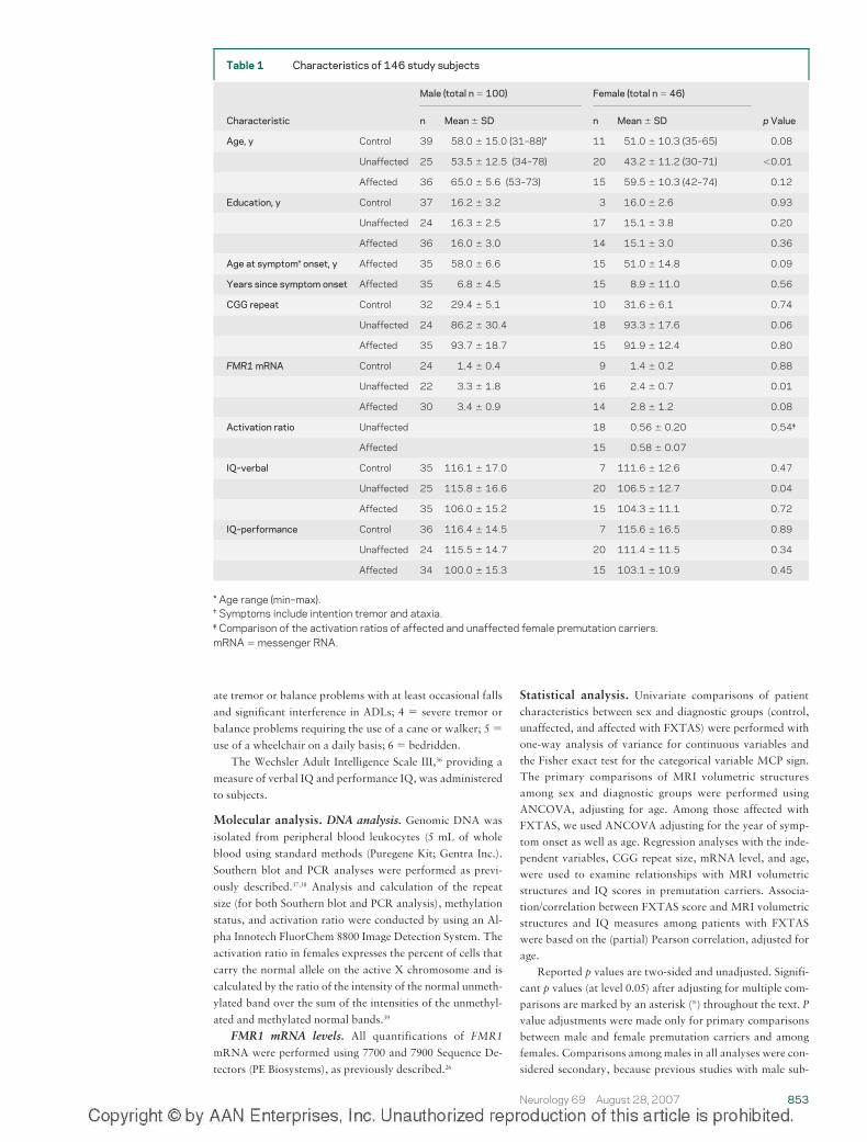
tween diagnostic groups of females (affected vs unaffectedcarriers, affected vs normal CGG repeat controls), and be-tween females and males affected by FXTAS. There was nodifference in age between the affected female and affectedmale groups (p � 0.12), nor was there a difference in the timebetween the onset of symptoms related to FXTAS and thedate of neuroimaging (p � 0.56). Unaffected female carriers(43.2 � 11.2 years) were younger than affected females(59.5 � 10.3 years; p � 0.01; table 1). This age difference islikely due to an increased incidence of FXTAS in aging pre-mutation carriers, particularly those older than 50 years,which would diminish the pool of unaffected premutationcarriers in this age range. Thus, in subsequent analyses weadjusted for the effect of age in regression analysis and anal-ysis of covariance (ANCOVA). Among female premutationcarriers, there was no difference in activation ratio betweenthose with FXTAS and unaffected groups (p � 0.54). Amongall premutation carriers, there was no difference in CGG re-peat length between sexes or diagnostic groups (FXTAS orclinically uninvolved). There was also no significant differ-ence in education level between any of our groups (table 1).
Neuroimaging. Structural magnetic resonance imageswere acquired using a 1.5-T GE Signa Horizon LX Echo-speed system. The acquisition parameters were as follows:
Coronal three-dimensional spoiled gradient recalled echo(inversion recovery–prepped spoiled gradient recalledecho) acquisition, T1 weighted: coronal plane, three-dimensional acquisition, gradient recalled echo, radio-frequency spoiled, repetition time 9.1 msec, spatialresolution 0.9375 � 0.9375 � 1.5 mm thickness.
High-resolution fluid-attenuated inversion recovery (sameorientation as axial spin echo): oblique axial plane,two-dimensional acquisition, inversion recovery spinecho, echo time 144 msec, repetition time 11,000 msec,inversion time 2,250 msec, 14 slices/acquisition, two in-terleaved acquisitions, resolution 0.9375 � 0.9375 � 3mm thickness, 0-mm interslice on reconstructed image.
MRI quantification was performed using a custom-written computer program operating on a UNIX, Solarisplatform (Quanta 6.1), as previously described.18 All vol-umes were normalized to total cranial volume (TCV).
The TCV was segmented into CSF, brain matter, andwhite matter increased T2 signal intensity volume accordingto previously published methods,31-34 and then further subdi-vided into regions of interest, including cerebrum, cerebel-lum, lateral ventricles, and third ventricle. Lateral and thirdventricular volumes were summed for a total ventricular re-gion of interest. Hippocampal volumes were quantified byoperator-guided tracing as described previously.33
MRIs were reviewed with a neuroradiologist (J.A.B.),and the MCP sign was scored as present or absent.
Clinical evaluation. The diagnosis of FXTAS was madeafter a thorough medical, neurologic, and radiologic exami-nation.35 Patients met criteria for probable or definiteFXTAS, as previously described.15 After examination, aFXTAS clinical staging score was given to each patient withthe premutation. This 7-point staging scale, as previouslyreported,3,14 measures functional impairment, as follows: 0 �
normal functioning; 1 � subtle or questionable tremor orbalance problems with no interference in activities of dailyliving (ADLs); 2 � minor but clear tremor or balance prob-lems producing minor interference with ADLs; 3 � moder-
852 Neurology 69 August 28, 2007
ate tremor or balance problems with at least occasional fallsand significant interference in ADLs; 4 � severe tremor orbalance problems requiring the use of a cane or walker; 5 �
use of a wheelchair on a daily basis; 6 � bedridden.The Wechsler Adult Intelligence Scale III,36 providing a
measure of verbal IQ and performance IQ, was administeredto subjects.
Molecular analysis. DNA analysis. Genomic DNA wasisolated from peripheral blood leukocytes (5 mL of wholeblood using standard methods (Puregene Kit; Gentra Inc.).Southern blot and PCR analyses were performed as previ-ously described.37,38 Analysis and calculation of the repeatsize (for both Southern blot and PCR analysis), methylationstatus, and activation ratio were conducted by using an Al-pha Innotech FluorChem 8800 Image Detection System. Theactivation ratio in females expresses the percent of cells thatcarry the normal allele on the active X chromosome and iscalculated by the ratio of the intensity of the normal unmeth-ylated band over the sum of the intensities of the unmethyl-ated and methylated normal bands.39
FMR1 mRNA levels. All quantifications of FMR1mRNA were performed using 7700 and 7900 Sequence De-tectors (PE Biosystems), as previously described.26
Statistical analysis. Univariate comparisons of patientcharacteristics between sex and diagnostic groups (control,unaffected, and affected with FXTAS) were performed withone-way analysis of variance for continuous variables andthe Fisher exact test for the categorical variable MCP sign.The primary comparisons of MRI volumetric structuresamong sex and diagnostic groups were performed usingANCOVA, adjusting for age. Among those affected withFXTAS, we used ANCOVA adjusting for the year of symp-tom onset as well as age. Regression analyses with the inde-pendent variables, CGG repeat size, mRNA level, and age,were used to examine relationships with MRI volumetricstructures and IQ scores in premutation carriers. Associa-tion/correlation between FXTAS score and MRI volumetricstructures and IQ measures among patients with FXTASwere based on the (partial) Pearson correlation, adjusted forage.
Reported p values are two-sided and unadjusted. Signifi-cant p values (at level 0.05) after adjusting for multiple com-parisons are marked by an asterisk (*) throughout the text. Pvalue adjustments were made only for primary comparisonsbetween male and female premutation carriers and amongfemales. Comparisons among males in all analyses were con-sidered secondary, because previous studies with male sub-
Table 1 Characteristics of 146 study subjects
Male (total n � 100) Female (total n � 46)
Characteristic n Mean � SD n Mean � SD p Value
Age, y Control 39 58.0 � 15.0 (31–88)* 11 51.0 � 10.3 (35–65) 0.08
Unaffected 25 53.5 � 12.5 (34–78) 20 43.2 � 11.2 (30–71) �0.01
Affected 36 65.0 � 5.6 (53–73) 15 59.5 � 10.3 (42–74) 0.12
Education, y Control 37 16.2 � 3.2 3 16.0 � 2.6 0.93
Unaffected 24 16.3 � 2.5 17 15.1 � 3.8 0.20
Affected 36 16.0 � 3.0 14 15.1 � 3.0 0.36
Age at symptom† onset, y Affected 35 58.0 � 6.6 15 51.0 � 14.8 0.09
Years since symptom onset Affected 35 6.8 � 4.5 15 8.9 � 11.0 0.56
CGG repeat Control 32 29.4 � 5.1 10 31.6 � 6.1 0.74
Unaffected 24 86.2 � 30.4 18 93.3 � 17.6 0.06
Affected 35 93.7 � 18.7 15 91.9 � 12.4 0.80
FMR1 mRNA Control 24 1.4 � 0.4 9 1.4 � 0.2 0.88
Unaffected 22 3.3 � 1.8 16 2.4 � 0.7 0.01
Affected 30 3.4 � 0.9 14 2.8 � 1.2 0.08
Activation ratio Unaffected 18 0.56 � 0.20 0.54‡
Affected 15 0.58 � 0.07
IQ–verbal Control 35 116.1 � 17.0 7 111.6 � 12.6 0.47
Unaffected 25 115.8 � 16.6 20 106.5 � 12.7 0.04
Affected 35 106.0 � 15.2 15 104.3 � 11.1 0.72
IQ–performance Control 36 116.4 � 14.5 7 115.6 � 16.5 0.89
Unaffected 24 115.5 � 14.7 20 111.4 � 11.5 0.34
Affected 34 100.0 � 15.3 15 103.1 � 10.9 0.45
* Age range (min–max).† Symptoms include intention tremor and ataxia.‡ Comparison of the activation ratios of affected and unaffected female premutation carriers.mRNA � messenger RNA.
Neurology 69 August 28, 2007 853
jects have been reported18 and are not of direct (primary)interest in this study. P value adjustment was performed us-ing the Sidak step-down method.
Because age is significantly associated with volumetricstructures among diagnostic and sex groups, it was necessaryto compare volumetric structures among these groups at spe-cific ages in the ANCOVA model. Thus, we chose to exam-ine volumetric differences among groups at ages 50, 60, and70 years, covering a broad range of ages for the FXTASpatients.
All volumetric structures were normalized by total cra-nial volume before statistical analyses. All tests were per-formed at a significance level of 0.05. Statistical analyseswere performed with the use of SAS software, version 9.1(SAS Institute).
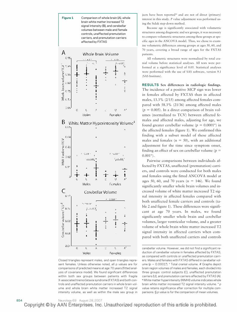
RESULTS Sex differences in radiologic findings.The incidence of a positive MCP sign was lowerin females affected by FXTAS than in affectedmales, 13.3% (2/15) among affected females com-pared with 58.3% (21/36) among affected males(p � 0.005). In a direct comparison of brain vol-umes (normalized to TCV) between affected fe-males and affected males, adjusting for age, wefound greater cerebellar volume (p � 0.0001*) inthe affected females (figure 1). We confirmed thisfinding with a subset model of these affectedmales and females (n � 50), with an additionaladjustment for the time since symptom onset,finding an effect of sex on cerebellar volume (p �0.001*).
Pairwise comparisons between individuals af-fected by FXTAS, unaffected (premutation) carri-ers, and controls were conducted for both malesand females using the fitted ANCOVA model atages 50, 60, and 70 years (n � 146). We foundsignificantly smaller whole brain volumes and in-creased volume of white matter increased T2 sig-nal intensity in affected females compared withboth unaffected female carriers and controls (ta-ble 2 and figure 1). These differences were signifi-cant at age 70 years. In males, we foundsignificantly smaller whole brain and cerebellarvolumes, larger ventricular volume, and a greatervolume of whole brain white matter increased T2signal intensity in affected carriers when com-pared with both unaffected carriers and controls
Figure 1 Comparison of whole brain (A), wholebrain white matter increased T2signal intensity (B), and cerebellarvolumes between male and femalecontrols, unaffected premutationcarriers, and premutation carriersaffected by FXTAS
-
Closed triangles represent males, and open triangles repre-sent females. Unless otherwise noted, all p values are forcomparisons of predicted means at age 70 years (fitted anal-ysis of covariance model). We found significant differenceswithin both sex groups between patients with fragileX-associated tremor/ataxia syndrome (FXTAS) and both con-trols and unaffected premutation carriers in whole brain vol-ume and whole brain white matter increased T2 signalintensity volume, as well as within the male sex group in
cerebellar volume. However, we did not find a significant re-duction of cerebellar volume in females affected by FXTAS,as compared with controls or unaffected premutation carri-ers. Males and females with FXTAS differed in cerebellar vol-ume (p � 0.0001*). † Total cranial volume. # Graphs depictbrain region volumes of males and females, each divided intothree groups: control subjects (C), unaffected premutationcarriers (U), and premutation carriers affected by FXTAS (A).‡ White matter hyperintensity (WMHI) volume indicates wholebrain white matter increased T2 signal intensity volume. * pvalue retains significance after correction for multiple com-parisons. § p value is for the comparison of mean volumes.
854 Neurology 69 August 28, 2007

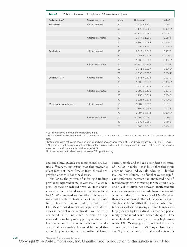
(table 3 and figure 1). These differences were gen-erally significant at older age levels (e.g., at ages60 and 70 years). The differences in whole brain,cerebellar, and ventricular volume between af-fected males and male controls were also evidentat the youngest age level (age 50 years). In bothmales and females, no differences in hippocampalvolume were found between the affected groupand either the unaffected group or the controlgroup. When comparing unaffected premutationcarriers with controls, no difference was found inany measured volumetric parameter in males orfemales.
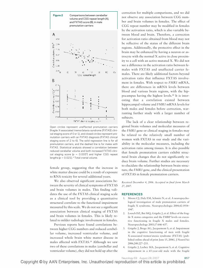
Correlation of clinical staging and quantitative find-ings. Among affected males (n � 36), we found anassociation between more advanced clinical stage(using the FXTAS clinical staging scale) and re-duced cerebellar volume (p � 0.005*; figure 2),reduced hippocampal volume (p � 0.009*), in-creased ventricular volume (p � 0.007*), de-creased performance IQ (p � 0.002*), anddecreased verbal IQ (p � 0.004*). No significantassociation was found between clinical stage andeither regional brain volumes or IQ scores in af-fected female premutation carriers (n � 15).
Correlation of molecular measures of FMR1 and ra-diologic findings. Regression analyses, with CGGrepeat number, FMR1 mRNA level, and age asindependent variables, were used to examine therelationship of molecular measures to brain vol-
umes in male premutation carriers (n � 51). Sim-ilar analyses were used for female premutationcarriers (n � 29), with the addition of activationratio as a covariate.
CGG repeat length was associated with re-duced cerebellar volume (p � 0.021; figure 2) andincreased ventricular volume (p � 0.047) in malecarriers, but not in female carriers (p � 0.82 forcerebellar volume; p � 0.69 for ventricular vol-ume). Elevated levels of FMR1mRNA were asso-ciated with reduced hippocampal volume in bothmale (p � 0.0035) and female carriers (p � 0.041).However, these associations dropped out of sig-nificance after adjusting p values for multipletesting.
DISCUSSION These results demonstrate milderradiologic findings in females affected by FXTASas compared with males. We document a signifi-cantly lower incidence of the MCP sign in femalesaffected by FXTAS (13.3%) compared with af-fected males (58.3%). In a direct comparison ofradiologic findings in females and males affectedby FXTAS, we report less pronounced cerebellarvolume loss in females after correcting for bothage and time since symptom onset. It is likely thatfemales are buffered from neurodegeneration as-sociated with the premutation allele by a dilutingeffect of the second, normal X chromosome.However, we do not see significant sex differ-
Table 2 Volumes of several brain regions in 46 female study subjects
Brain structure† Comparison group Age, y Difference‡ p Value§
Whole brain Affected–control 50 �0.587 � 1.398 0.6755
60 �2.524 � 1.297 0.0537
70 �4.462 � 1.552 0.0047
Affected–unaffected 50 �0.202 � 1.271 0.874
60 �2.640 � 1.283 0.0415
70 �5.079 � 1.671 0.0028*
White matter hyperintensity� Affected–control 50 0.155 � 0.273 0.5708
60 0.535 � 0.253 0.0365
70 1.226 � 0.303 �0.0001*
Affected–unaffected 50 0.111 � 0.248 0.6557
60 0.604 � 0.251 0.0172
70 1.319 � 0.326 �0.0001*
Plus–minus values are estimated difference � SE.† All brain volumes were expressed as a percentage of total cranial volume in our analysis to account for differences in headsize.‡ Differences were estimated based on a fitted analysis of covariance model at three different ages (50, 60, and 70 years).§ All reported p values are raw values taken before correction for multiple comparisons. P values that retained significanceafter the correction are marked with an asterisk (*).� Indicates whole brain white matter increased T2 signal intensity.
Neurology 69 August 28, 2007 855
ences in clinical staging due to functional or adap-tive differences, indicating that this protectiveeffect may not spare females from clinical pro-gression once they have the disease.
Similar to the pattern of radiologic findingspreviously reported in males with FXTAS, we re-port significantly reduced brain volumes and in-creased white matter disease in females affectedby FXTAS compared with unaffected female car-riers and female controls without the premuta-tion. However, unlike males, females withFXTAS did not demonstrate significant differ-ences in cerebellar or ventricular volume whencompared with unaffected carriers or age-matched controls, again suggesting milder or dif-ferent structural alteration of the brain in femalescompared with males. It should be noted thatgiven the younger age of our unaffected female
carrier sample and the age-dependent penetranceof FXTAS in males,35 it is likely that this groupcontains some individuals who will developFXTAS in the future. The fact that we see signifi-cant differences between the unaffected and af-fected groups after correcting for the effect of ageand a lack of difference between unaffected andcontrols suggests that the radiologic changes ob-served are due to the presence of FXTAS ratherthan a developmental effect of the premutation. Itshould also be noted that the increased white mat-ter disease observed among affected females waslargely driven by two individuals that had partic-ularly pronounced white matter changes. Theseindividuals did not have particularly high scoreson the FXTAS staging scale (both had a score of3), nor did they have the MCP sign. However, atage 74 years, they were the oldest subjects in the
Table 3 Volumes of several brain regions in 100 male study subjects
Brain structure† Comparison group Age, y Difference‡ p Value§
Whole brain Affected–control 50 �2.237 � 1.221 0.069
60 �4.175 � 0.802 �0.0001*
70 �6.113 � 0.890 �0.0001*
Affected–unaffected 50 �1.744 � 1.260 0.1686
60 �4.183 � 0.924 �0.0001*
70 �6.622 � 1.111 �0.0001*
Cerebellum Affected–control 50 �0.846 � 0.313 0.0077
60 �0.955 � 0.205 �0.0001*
70 �1.065 � 0.228 �0.0001*
Affected–unaffected 50 �0.845 � 0.323 0.0099
60 �0.941 � 0.237 0.0001*
70 �1.038 � 0.285 0.0004*
Ventricular CSF Affected–control 50 0.541 � 0.415 0.1951
60 1.238 � 0.273 �0.0001*
70 1.936 � 0.303 �0.0001*
Affected–unaffected 50 0.390 � 0.429 0.3642
60 1.158 � 0.314 0.0003*
70 1.925 � 0.378 �0.0001*
White matter hyperintensity� Affected–control 50 �0.387 � 0.238 0.1071
60 0.304 � 0.157 0.0544
70 0.994 � 0.174 �0.0001*
Affected–unaffected 50 �0.385 � 0.246 0.1202
60 0.330 � 0.180 0.0693
70 1.045 � 0.217 �0.0001*
Plus–minus values are estimated difference � SE.† All brain volumes were expressed as a percentage of total cranial volume in our analysis to account for differences in headsize.‡ Differences were estimated based on a fitted analysis of covariance model at three different ages (50, 60, and 70 years).§ All reported p values are raw values taken before correction for multiple comparisons. P values that retained significanceafter the correction are marked with an asterisk (*).� Indicates whole brain white matter increased T2 signal intensity.
856 Neurology 69 August 28, 2007
female group, suggesting that the increase inwhite matter disease could be a result of exposureto RNA toxicity for several additional years.
We also observed significant associations be-tween the severity of clinical symptoms of FXTASand brain volumes in males. This finding vali-dates the use of the FXTAS clinical staging scaleas a clinical tool by providing a quantitativestructural correlate to the functional impairmentmeasured by this scale. We do not see a significantassociation between clinical staging of FXTASand brain volumes in females. This is likely re-lated to milder radiologic involvement in females.
Previous reports have found correlations be-tween higher CGG numbers and reduced cerebel-lar volume, increased ventricular volume, andincreased whole brain white matter disease inmales affected with FXTAS.18 Although we sawtwo of these correlations in males (cerebellar andventricular volume), they did not hold up after the
correction for multiple comparisons, and we didnot observe any association between CGG num-ber and brain volumes in females. The effect ofCGG repeat number may be modified in femalesby the activation ratio, which is also variable be-tween blood and brain. Therefore, a correctionfor activation ratio obtained from blood may notbe reflective of the status of the different brainregions. Additionally, the protective effect in thebrain may be enhanced by having a neuron or as-trocyte with the normal X active in close proxim-ity to a cell with an active mutated X. We did notsee a difference in the activation ratio between fe-males with FXTAS and unaffected carrier fe-males. There are likely additional factors beyondactivation ratio that influence FXTAS involve-ment in females. With respect to FMR1 mRNA,there are differences in mRNA levels betweenblood and various brain regions, with the hip-pocampus having the highest levels.40 It is inter-esting that a correlation existed betweenhippocampal volume and FMR1mRNA levels forboth males and females before correction, war-ranting further study with a larger number ofsubjects.
The lack of a clear relationship between re-gional brain volumes and molecular measures ofthe FMR1 gene or clinical staging in females maybe related to the relatively small number ofwomen with FXTAS in our study and the vari-ability in the molecular measures, including theactivation ratio among tissues. It is also possiblethat female premutation carriers exhibit struc-tural brain changes that do not significantly re-duce brain volume. Further studies are necessaryto elucidate the relationship between brain struc-ture, the FMR1 gene, and the clinical presentationof FXTAS in female premutation carriers.
Received December 4, 2006. Accepted in final form March27, 2007.
REFERENCES1. Moore CJ, Daly EM, Schmitz N, et al. A neuropsycho-
logical investigation of male premutation carriers offragile X syndrome. Neuropsychologia 2004;42:1934–1947.
2. Loesch DZ, Bui MQ, Grigsby J, et al. Effect of the frag-ile X status categories and the FMRP levels on execu-tive functioning in fragile X males and females.Neuropsychology 2003;17:646–657.
3. Grigsby J, Brega AG, Jacquemont S, et al. Impairmentin the cognitive functioning of men with fragileX-associated tremor/ataxia syndrome (FXTAS) [pub-lished online ahead of print June 15, 2006]. J Neurol Sci2006;248:227–233.
4. Grigsby J, Leehey MA, Jacquemont S, et al. Cognitiveimpairment in a 65-year-old male with the fragile
Figure 2 Comparisons between cerebellarvolume and CGG repeat length (A),and FXTAS score (B), in malepremutation carriers
Open circles represent unaffected premutation carriers(fragile X-associated tremor/ataxia syndrome [FXTAS] clini-cal staging score of 0 or 1), and closed circles represent pre-mutation carriers with an FXTAS diagnosis (FXTAS clinicalstaging score of 2 to 6). The solid regression line is for allpremutation carriers, and the dashed line is for males withFXTAS. Statistical analysis showed a correlation betweenreduced cerebellar volume and both increased FXTAS clini-cal staging score (p � 0.005*) and higher CGG repeatlengths (p � 0.021). † Total cranial volume.
Neurology 69 August 28, 2007 857
X-associated tremor-ataxia syndrome (FXTAS). CognBehav Neurol 2006;19:165–171.
5. Cornish KM, Kogan C, Turk J, et al. The emergingfragile X premutation phenotype: evidence from thedomain of social cognition. Brain Cogn 2005;57:53–60.
6. Franke P, Leboyer M, Gansicke M, et al. Genotype-phenotype relationship in female carriers of the premu-tation and full mutation of FMR-1. Psychiatry Res1998;80:113–127.
7. Hessl D, Tassone F, Loesch DZ, et al. Abnormal eleva-tion of FMR1 mRNA is associated with psychologicalsymptoms in individuals with the fragile X premuta-tion. Am J Med Genet B Neuropsychiatr Genet 2005;139:115–121.
8. Murray A, Ennis S, MacSwiney F, Webb J, MortonNE. Reproductive and menstrual history of femaleswith fragile X expansions. Eur J Hum Genet 2000;8:247–252.
9. Sullivan AK, Marcus M, Epstein MP, et al. Associationof FMR1 repeat size with ovarian dysfunction. HumReprod 2005;20:402–412.
10. Hagerman RJ, Leehey M, Heinrichs W, et al. Intentiontremor, parkinsonism, and generalized brain atrophyin male carriers of fragile X. Neurology 2001;57:127–130.
11. Jacquemont S, Hagerman RJ, Leehey M, et al. FragileX premutation tremor/ataxia syndrome: molecular,clinical, and neuroimaging correlates. Am J HumGenet 2003;72:869–878.
12. Loesch DZ, Churchyard A, Brotchie P, Marot M, Tas-sone F. Evidence for, and a spectrum of, neurologicalinvolvement in carriers of the fragile X pre-mutation:FXTAS and beyond. Clin Genet 2005;67:412–417.
13. Leehey MA, Munhoz RP, Lang AE, et al. The fragile Xpremutation presenting as essential tremor. Arch Neu-rol 2003;60:117–121.
14. Bacalman S, Farzin F, Bourgeois JA, et al. Psychiatricphenotype of the fragile X-associated tremor/ataxiasyndrome (FXTAS) in males: newly described fronto-subcortical dementia. J Clin Psychiatry 2006;67:87–94.
15. Jacquemont S, Farzin F, Hall D, et al. Aging in individ-uals with the FMR1mutation. Am JMent Retard 2004;109:154–164.
16. Loesch DZ, Litewka L, Brotchie P, Huggins RM, Tas-sone F, Cook M. Magnetic resonance imaging study inolder fragile X premutation male carriers. Ann Neurol2005;58:326–330.
17. Brunberg JA, Jacquemont S, Hagerman RJ, et al. FragileX premutation carriers: characteristic MR imaging find-ings in adult males with progressive cerebellar and cogni-tive dysfunction. Am JNeuroradiol 2002;23:1757–1766.
18. Cohen S, Masyn K, Adams J, et al. Molecular and im-aging correlates of the fragile X-associated tremor/ataxia syndrome. Neurology 2006;67:1426–1431.
19. Greco C, Hagerman RJ, Tassone F, et al. Neuronalintranuclear inclusions in a new cerebellar tremor/ataxia syndrome among fragile X carriers. Brain 2002;125:1760–1771.
20. Greco CM, Berman RF, Martin RM, et al. Neuropa-thology of fragile X-associated tremor/ataxia syn-drome (FXTAS) [published online ahead of printDecember 5, 2005]. Brain 2006;129:243–255.
21. O’Dwyer JP, Clabby C, Crown J, Barton DE, Hutchin-son M. Fragile X-associated tremor/ataxia syndrome
presenting in a woman after chemotherapy. Neurology2005;65:331–332.
22. Hagerman RJ, Leavitt BR, Farzin F, et al. Fragile-X-associated tremor/ataxia syndrome (FXTAS) in fe-males with the FMR1 premutation. Am J Hum Genet2004;74:1051–1056.
23. Berry-Kravis E, Potanos K, Weinberg D, Zhou L,Goetz CG. Fragile X-associated tremor/ataxia syn-drome in sisters related to X-inactivation. Ann Neurol2005;57:144–147.
24. Jacquemont S, Orrico A, Galli L, et al. Spastic parapa-resis, cerebellar ataxia, and intention tremor: a severevariant of FXTAS? J Med Genet 2005;42:e14.
25. Zuhlke C, Budnik A, Gehlken U, et al. FMR1 premuta-tion as a rare cause of late onset ataxia: evidence forFXTAS in female carriers. J Neurol 2004;251:1418–1419.
26. Tassone F, Hagerman RJ, Taylor AK, Gane LW, God-frey TE, Hagerman PJ. Elevated levels of FMR1mRNA in carrier males: a new mechanism of involve-ment in fragile X syndrome. Am J Hum Genet 2000;66:6–15.
27. Hagerman PJ, Hagerman RJ. The fragile-X premuta-tion: a maturing perspective. Am J Hum Genet 2004;74:805–816.
28. Ranum LP, Day JW. Dominantly inherited, non-coding microsatellite expansion disorders. Curr OpinGenet Dev 2002;12:266–271.
29. Dombrowski C, Levesque ML, Morel ML, RouillardP, Morgan K, Rousseau F. Premutation andintermediate-size FMR1 alleles in 10 572 males fromthe general population: loss of an AGG interruption isa late event in the generation of fragile X syndromealleles. HumMol Genet 2002;11:371–378.
30. Rousseau F, Rouillard P, Morel ML, Khandjian EW,Morgan K. Prevalence of carriers of premutation-sizealleles of the FMRI gene–and implications for the pop-ulation genetics of the fragile X syndrome. Am J HumGenet 1995;57:1006–1018.
31. DeCarli C, Maisog J, Murphy DG, Teichberg D, Rap-oport SI, Horwitz B. Method for quantification ofbrain, ventricular, and subarachnoid CSF volumesfrom MR images. J Comput Assist Tomogr 1992;16:274–284.
32. DeCarli C, Murphy DG, Teichberg D, Campbell G,Sobering GS. Local histogram correction of MRI spa-tially dependent image pixel intensity nonuniformity. JMagn Reson Imaging 1996;6:519–528.
33. Wu CC, Mungas D, Petkov CI, et al. Brain structureand cognition in a community sample of elderly Lati-nos. Neurology 2002;59:383–391.
34. Jeerakathil T, Wolf PA, Beiser A, et al. Stroke risk pro-file predicts white matter hyperintensity volume: theFramingham Study [published online ahead of printJune 24, 2004]. Stroke 2004;35:1857–1861.
35. Jacquemont S, Hagerman RJ, Leehey MA, et al. Pen-etrance of the fragile X-associated tremor/ataxia syn-drome in a premutation carrier population. JAMA2004;291:460–469.
36. Wechsler D. Wechsler Adult Intelligence Scale–thirdedition: Administration and scoring manual. San Anto-nio: Harcourt Assessment, 1997.
37. Tassone F, Hagerman RJ, Garcia-Arocena D, Khand-jian EW, Greco CM, Hagerman PJ. Intranuclear inclu-
858 Neurology 69 August 28, 2007
sions in neural cells with premutation alleles in fragileX associated tremor/ataxia syndrome. J Med Genet2004;41:e43.
38. Saluto A, Brussino A, Tassone F, et al. An enhancedpolymerase chain reaction assay to detect pre- and fullmutation alleles of the fragile X mental retardation 1gene. J Mol Diagn 2005;7:605–612.
39. Tassone F, Hagerman RJ, Ikle DN, et al. FMRP ex-pression as a potential prognostic indicator in fragile Xsyndrome. Am J Med Genet 1999;84:250–261.
40. Tassone F, Iwahashi C, Hagerman PJ. FMR1 RNAwithin the intranuclear inclusions of fragileX-associated tremor/ataxia syndrome (FXTAS). RNABiol 2004;1:103–105.
Experience the 2007 Annual Meeting from theComfort of Your Home
Much of this year’s top programming is now available to you through the AAN VirtualAnnual Meeting Products:
� Audio MP3 Files—starting at $10Conveniently listen to some of the best talks on today’s most significant neurological topics.
• Webcasts-on-Demand—several program hours FREEInstant online access to the slides, audio, and video of more than 240 hours of programs andpresentations, including five Plenary Sessions. Enjoy several hours of free programming inaddition to other programs available for purchase ($399 members, $199 Junior members,$499 non-members).
• 2007 Syllabi on CD-ROM—only $199Complete syllabi for more than 160 education programs ($199 members, $299 non-members).
Check out the Virtual Annual Meeting Programming at www.aan.com/virtualAM today!
Neurology 69 August 28, 2007 859
Copyright © 2022 FDOKUMEN