Volume 12, Supplement 1 (2011) Sudhansu Chokroverty
146
Official Journal of the World Association of Sleep Medicine & the International Pediatric Sleep Association Volume 12, Supplement 1 (2011) Editor-in-Chief Sudhansu Chokroverty Please access the instructions to authors via the world wide web: www.elsevier.com/locate/sleep Cover image: Absolute cerebral blood flow (CBF) using arterial spin labeling (a special magnetic resonance imaging technique) in a patient with congenital central hypoventilation syndrome (CCHS) and an age-matched control. This shows average of the two subjects’ normalized images. The mean whole-brain map of CBF (not shown) is higher in CCHS than control. Amsterdam – Boston – London – New York – Oxford – Paris – Philadelphia – San Diego – St. Louis
-
Upload
khangminh22 -
Category
Documents
-
view
1 -
download
0
Transcript of Volume 12, Supplement 1 (2011) Sudhansu Chokroverty
Official Journal of the World Association of Sleep Medicine & the InternationalPediatric Sleep Association
Volume 12, Supplement 1 (2011)
Editor-in-Chief
Sudhansu Chokroverty
Please access the instructions to authors via the world wide web: www.elsevier.com/locate/sleep
Cover image: Absolute cerebral blood flow (CBF) using arterial spin labeling (a special magnetic resonance imaging technique) in apatient with congenital central hypoventilation syndrome (CCHS) and an age-matched control. This shows average of the two subjects’normalized images. The mean whole-brain map of CBF (not shown) is higher in CCHS than control.
Amsterdam – Boston – London – New York – Oxford – Paris – Philadelphia – San Diego – St. Louis

© 2011 Elsevier B.V. All rights reserved.
This journal and the individual contributions contained in it are protected under copyright by Elsevier B.V., and the following terms and conditions apply to their use:
Photocopying
Single photocopies of single articles may be made for personal use as allowed by national copyright laws. Permission of the publisher and payment of a fee is required for allother photocopying, including multiple or systematic copying, copying for advertising or promotional purposes, resale, and all forms of document delivery. Special rates areavailable for educational institutions that wish to make photocopies for non-profit educational classroom use.
Permissions may be sought directly from Elsevier via their homepage (http://www.elsevier.com) by selecting ‘‘Customer support’’ and then ‘‘Permissions’’. Alternatively youcan send an e-mail to: [email protected], of fax to: (+44) 1865 853333.
In the USA, users may clear permissions and make payment through the Copyright Clearance Center, Inc., 222 Rosewood Drive, Danvers, MA 01923, USA; phone: (978)7508400, fax: (978) 7504744, and in the UK through the Copyright Licensing Agency Rapid Clearance Service (CLARCS), 90 Tottenham Court Road, London W1P OLP, UK;phone: (44) 171 631 5555; fax: (44) 171 631 5500. Other countries may have a local reprographic rights agency for payments.
Derivative Works
Subscribers may reproduce tables of contents or prepare lists of articles including abstracts for internal circulation within their institutions. Permission of the publisher isrequired for resale or distribution outside the institution.
Permission of the publisher is required for all other derivative works, including compilations and translations.
Electronic Storage or Usage
Permission of the publisher is required to store or use electronically any material contained in this journal, including any article or part of an article.
Except as outlined above, no part of this publication may be reproduced, stored in a retrieval system or transmitted in any form or by any means, electronic, mechanical,photocopying, recording or otherwise, without prior written permission of the publisher.
Address permissions requests to: Elsevier Global Rights Department, at the mail, fax and e-mail addresses noted above.
Notice
No responsibility is assumed by the publisher for any injury and/or damage to persons or property as a matter of products liability, negligence or otherwise, or from any useor operation of any methods, products, instructions or ideas contained in the material herein. Because of rapid advances in the medical sciences, in particular, independentverification of diagnoses and drug dosages should be made.
Although all advertising material is expected to conform to ethical (medical) standards, inclusion in this publication does not constitute a guarantee or endorsement of thequality or value of such product or of the claims made of it by its manufacturer.
Publication information: Sleep Medicine (ISSN 1389-9457). For 2011, volume 12 is scheduled for publication. Subscription prices are available upon request from thePublisher or from the Regional Sales Office nearest you or from this journal’s website (http://www.elsevier.com/locate/sleep). Further information is available on thisjournal and other Elsevier products through Elsevier’s website (http://www.elsevier.com). Subscriptions are accepted on a prepaid basis only and are entered on a calendaryear basis. Issues are sent by standard mail (surface within Europe, air delivery outside Europe). Priority rates are available upon request. Claims for missing issues shouldbe made within six months of date of dispatch.
USA mailing notice: Sleep Medicine (ISSN 1389-9457) is published ten times a year, monthly except July and November by Elsevier BV (Radarweg 29, 1043 NX Amsterdam,the Netherlands). Periodical postage paid at Rahway NJ and additional mailing offices.USA POSTMASTER: Send address changes to Sleep Medicine, Elsevier Customer Service Department, 3251 Riverport Lane, Maryland Heights, MO 63043, USA.AIRFREIGHT AND MAILING in the USA by Mercury International Limted, 365 Blair Road, Avenel, NJ 07001.
Instructions to authors: Please access the instructions to authors via the world wide web: www.elsevier.com/locate/sleep
Author enquiries
For enquiries relating to the submission of articles (including electronic submission where available) please visit the journal’s homepage at www.elsevier.com/locate/sleep.Contact details for questions arising after acceptance of an article, especially those relating to proofs, will be provided by the publisher. You can track accepted articles athttp://www.elsevier.com/trackarticle. You can also check our Author FAQs at http://www.elsevier.com/authorFAQ and/or contact Customer Support via http://support.elsevier.com.
Advertising Information: Advertising orders and enquiries can be sent to: USA, Canada and South America: Mary Anne Arbolado, Elsevier Inc., 360 Park Avenue South,New York, NY 10010-1710, USA; phone: (+1) (212) 633 3974; e-mail: [email protected]. Europe and ROW: Martin Sibson, Business Development Executive,Elsevier Pharma Solutions, 32 Jamestown Road, London, NW1 7BY, UK. Tel.: +44 (0) 207 424 4963; fax: +44 (0) 207 424 4433; e-mail: [email protected]
Orders, claims, and journal enquiries: please contact the Elsevier Customer Service Department nearest you:St. Louis: Elsevier Customer Service Department, 3251 Riverport Lane, Maryland Heights, MO 63043, USA; phone: (800) 6542452 [toll free within the USA]; (+1) (314)
4478871 [outside the USA]; fax: (+1) (314) 4478029; e-mail: [email protected]: Elsevier Customer Service Department, The Boulevard, Langford Lane, Kidlington, Oxford OX5 1GB, UK; phone: (+44) (1865) 843434; fax: (+44) (1865) 843970;
e-mail: [email protected]: Elsevier Customer Service Department, 4F Higashi-Azabu, 1-Chome Bldg, 1-9-15 Higashi-Azabu, Minato-ku, Tokyo 106-0044, Japan; phone: (+81) (3) 5561 5037;
fax: (+81) (3) 5561 5047; e-mail: [email protected]: Elsevier Customer Service Department, 3 Killiney Road, #08-01 Winsland House I, Singapore 239519; phone: (+65) 63490222; fax: (+65) 67331510; e-mail:
Funding Body Agreements and Policies
Elsevier has established agreements and developed policies to allow authors whose articles appear in journals published by Elsevier, to comply with potential manuscriptarchiving requirements as specified as conditions of their grant awards. To learn more about existing agreements and policies please visit http://www.elsevier.com/fundingbodies
Printed by Polestar Wheatons Ltd., Exeter, UK
The paper used in this publication meets the requirements of ANSI/NISO Z39.48-1992 (Permanence of Paper)
Abstracts of4th International Congress of the Association of Sleep Medicine (WASM)
&5th Conference of the Canadian Sleep Society (CSS)
September 10–14, 2011, Quebec City, Canada

Official Journal of the World Association of Sleep Medicine &the International Pediatric Sleep Association
Contents
Abstracts of4th International Congress of the Association of Sleep Medicine (WASM) &5th Conference of the Canadian Sleep Society (CSS)
September 10–14, 2011, Quebec City, Canada
Welcome Addresses vi
Abstracts
ORAL PRESENTATIONS
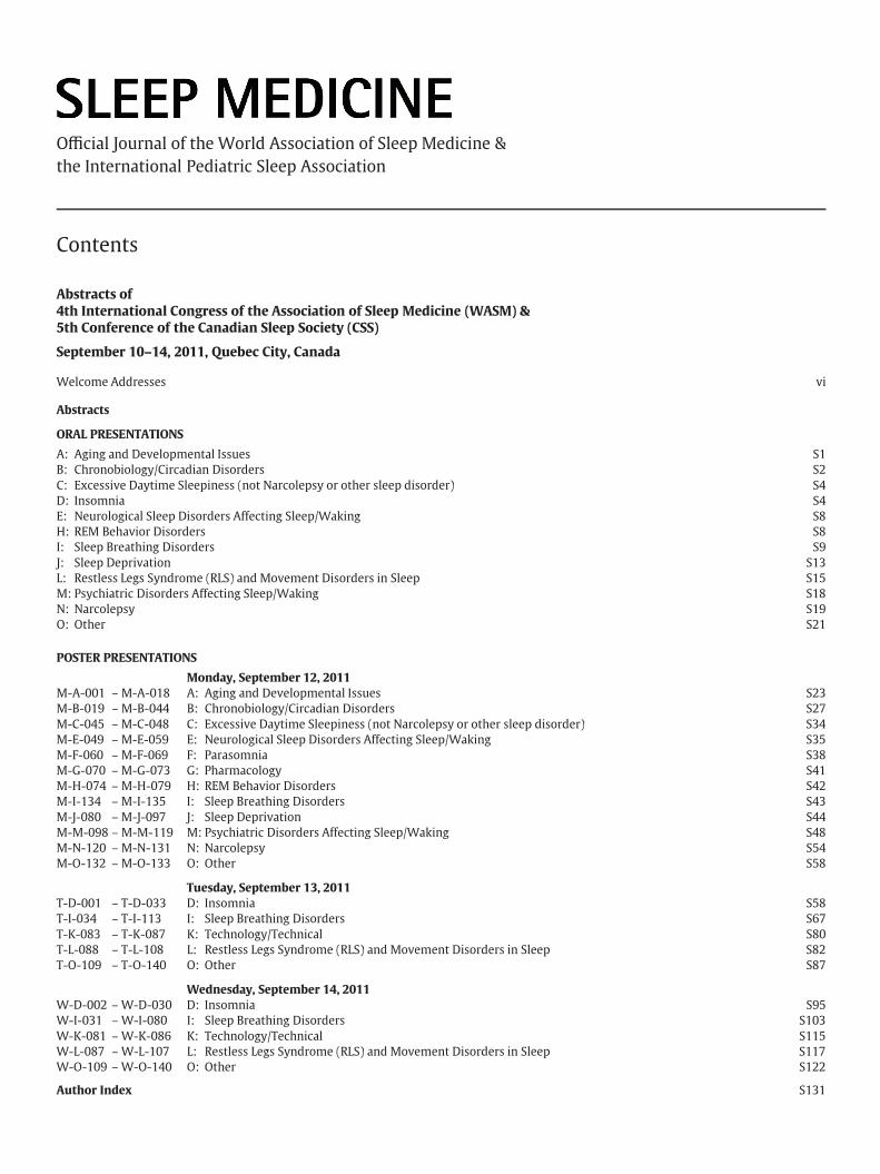
A: Aging and Developmental Issues S1B: Chronobiology/Circadian Disorders S2C: Excessive Daytime Sleepiness (not Narcolepsy or other sleep disorder) S4D: Insomnia S4E: Neurological Sleep Disorders Affecting Sleep/Waking S8H: REM Behavior Disorders S8I: Sleep Breathing Disorders S9J: Sleep Deprivation S13L: Restless Legs Syndrome (RLS) and Movement Disorders in Sleep S15M: Psychiatric Disorders Affecting Sleep/Waking S18N: Narcolepsy S19O: Other S21
POSTER PRESENTATIONS
Monday, September 12, 2011M-A-001 – M-A-018 A: Aging and Developmental Issues S23M-B-019 – M-B-044 B: Chronobiology/Circadian Disorders S27M-C-045 – M-C-048 C: Excessive Daytime Sleepiness (not Narcolepsy or other sleep disorder) S34M-E-049 – M-E-059 E: Neurological Sleep Disorders Affecting Sleep/Waking S35M-F-060 – M-F-069 F: Parasomnia S38M-G-070 – M-G-073 G: Pharmacology S41M-H-074 – M-H-079 H: REM Behavior Disorders S42M-I-134 – M-I-135 I: Sleep Breathing Disorders S43M-J-080 – M-J-097 J: Sleep Deprivation S44M-M-098 – M-M-119 M: Psychiatric Disorders Affecting Sleep/Waking S48M-N-120 – M-N-131 N: Narcolepsy S54M-O-132 – M-O-133 O: Other S58
Tuesday, September 13, 2011T-D-001 – T-D-033 D: Insomnia S58T-I-034 – T-I-113 I: Sleep Breathing Disorders S67T-K-083 – T-K-087 K: Technology/Technical S80T-L-088 – T-L-108 L: Restless Legs Syndrome (RLS) and Movement Disorders in Sleep S82T-O-109 – T-O-140 O: Other S87
Wednesday, September 14, 2011W-D-002 – W-D-030 D: Insomnia S95W-I-031 – W-I-080 I: Sleep Breathing Disorders S103W-K-081 – W-K-086 K: Technology/Technical S115W-L-087 – W-L-107 L: Restless Legs Syndrome (RLS) and Movement Disorders in Sleep S117W-O-109 – W-O-140 O: Other S122
Author Index S131
vi Welcome Addresses / Sleep Medicine 12, Suppl. 1 (2011) vi–vii
Welcome Addresses
Dear Colleagues and Friends,
On behalf of the World Association of Sleep Medicine (WASM) and the Canadian Sleep Society (CSS), we are delightedto welcome you to our joint congress of the 4th International World Sleep Congress and 5th Conference of the CSS inQuebec City, Canada from September 10–14, 2011.
The congress is an international forum of professionals advancing current thinking to improve sleep health, andencourage prevention and treatment of sleep disorders. The congress brings together leading experts to discuss, debate,and disseminate knowledge amongst sleep professionals, clinicians, researchers, technologists and trainees for theadvancement of sleep health worldwide.
Your involvement in this congress is greatly valued. We hope that you’ll enjoy the science, collegiality and social eventsat our world sleep conference in this charming city.
Welcome to Quebec City!
Best regards,
Christian Guilleminault, MD, DM, DBiol
President, WASM
Helen S. Driver, PhD, RPSGT, DAB
President, CSS
Welcome Addresses / Sleep Medicine 12, Suppl. 1 (2011) vi–vii vii
Dear Colleagues and Friends,
On behalf of the World Association of Sleep Medicine (WASM) and Canadian Sleep Society (CSS), we are delightedto welcome you to this international sleep meeting in Quebec City, Canada. The scientific committee has puttogether an outstanding program with courses and workshops, keynote lectures, symposia, and nearly 500 oral andposter presentations covering all areas of sleep medicine from basic sciences to technological advances and clinicalapplications.
With its theme on Sleep, Health, and Society, this international event brings together more than 1000 participants from44 countries. Leading experts from around the world will present the latest developments in the field about sleep andits disorders with content that should be of interest to clinicians, researchers, technologists, students and trainees, andeven the lay public.
We trust this educational forum provides an ideal opportunity to meet colleagues and share new ideas on the mostrecent advances in the field to promote healthy sleep worldwide and improve the prevention, diagnosis, and treatmentof sleep disorders.
We wish to thank you for attending this meeting and are grateful to all of those who have contributed to its contentand organization. We hope that you have a pleasant learning experience and that you enjoy Quebec City and its richculture. We look forward to greeting you personally during the meeting.
Best regards,
Charles M. Morin, PhDRichard Allen, PhD
WASM/CSS 2011Program Co-Chairs

Sleep Medicine 12, Suppl. 1 (2011) S1–S130
Abstracts of4th International Congress of the Association of Sleep Medicine (WASM) &5th Conference of the Canadian Sleep Society (CSS)
September 10–14, 2011, Quebec City, Canada
ORAL PRESENTATIONS
A: Aging and Developmental Issues
SPINDLES AND SLOW WAVES ARE ASSOCIATED TO VERBAL LEARNING INOLDER SUBJECTS
Marjolaine Lafortune, Jean-François Gagnon, Véronique Latreille,Jacques Montplaisir, Julie Carrier. Center for Advanced Research in SleepMedicine, Hôpital du Sacré-Cœur de Montréal, Canada
Introduction and Objectives: Sleep spindles and slow waves (SW; <4Hz;>75uV) are involved in declarative sleep-dependent memory consolidation.We aimed to evaluate the association between neural synchronizationduring N-REM sleep and verbal learning performances in older healthysubjects.Materials and Methods: Healthy volunteers (49-79 years) without sleepdisorders participated in a baseline polysomnographic (PSG) sleep recordingfollowed by a neuropsychological assessment, including a verbal memorytest: The Rey Auditory Verbal Learning Test (RAVLT). Spindles (25 subjects;20M) and SW (34 subjects; 18M) were automatically detected on artefactfree N-REM sleep on F3, F4, C3, C4, P3, P4, O1 and O2 (linked-ears). Allnight spindles and SW mean density (nb/min), duration (s), amplitude (μV)and frequency (Hz) were analysed. Regression analyses were performedbetween spindles, SW characteristics and raw scores on RAVLT.Results: Spindles density and spindles amplitude were positively correlatedwith RAVLT learning performance and delayed recall performance (R>0.41,p<0.05 in all cases). SW amplitude was positively correlated with RAVLTfirst trial performance and delayed recall performance (R>0.40, p<0.05). Nosignificant correlation was found between age and spindles characteristics,between age and SW characteristics or between age and RAVTL scores.Conclusion: Older subjects who showed higher spindles density, largerspindles and larger SW in frontal areas performed better on a verbal learningtask. The mechanisms underlying the association between neural synchro-nization during N-REM sleep and verbal learning in aging still have to bedetermined.Acknowledgements: This research was supported by scholarships from theFonds de la recherche en santé du Québec (FRSQ) and grants from theCanadian Institutes of Health Research (CIHR) and Canadian sleep society(CSS).
1389-9457/$ – see front matter © 2011 Elsevier B.V. All rights reserved.
THE EPIDEMIOLOGY OF SLEEP AND ITS DISORDER IN CHINESE CHILDRENAGED 0-5 YEARS
Xi-cheng Liu1, Xiaona Huang2, Hui-shan Wang2, Jing-xiong Jiang2, Lin An3.1Bronchoscopy Center, Affiliated Children Hospital of Beijing Capital MedicalUniversity, Beijing, China; 2Dept. of Children Health, National Ccenter forMaternal and Children Health, Chinese Disease Prevention Control Center, China;3Dept. of Public Health, Peking University Health Science Center, Beijing, China
Introduction and Objectives: This study was to investigate the sleep pat-terns and the prevalence and types of sleep problems among urban childrenunder 5 years of age.Materials and Methods: Data were gathered on 14,883 children selectedfrom 12 cities, using the Children’s Sleep Status Questionnaire (CSSQ). Theparents were asked about their children’s average sleep time in a twenty-four-hour period and common sleep problems noted during the past month.Results: Of 14778 subjects over 1 month old, 20.3% were reported as suffer-ing from at least one of sleep disturbances studied. The five most commonsleep problems were difficulty falling asleep (7.5%), nocturnal awakening(4.9%), bruxism (4.7%), snoring (4.4%) and mouth breathing (3.0%). Excludingthe cases with sleep disorders, the average daytime sleep significantlydecreased with increasing age, from an average of 6.86 hours in newborn,3.77 hours at 6 months, 2.76 hours at 12 months, 2.28 hours at 2 years and1.96 hours at 5 years-old. As to nighttime sleep, there was no differencebetween age groups, except that sleep was slightly shorter at 1 and 2 monthsthan other age groups. Overall, the average sleep time over a 24-h periodprogressively shortened with age, from 15.81 hours at 1 month to 12.56hours at 12 months, 12.02 hours at 2 years, and 11.31 hours at 5 years. Thenumber of naps and nocturnal awakenings, respectively, decreased from 4and 2 per day at 1 month of age to 1 and 0 per day at the 24 and 18 months.Most children continued to nap up to the age of 5 years.Conclusion: Our findings suggest that sleep problems are common in Chi-nese children aged 0-5 years. Also, with the development of society, certainchanges in sleep time of the children have occurred, there being about0.5-1.0 hr. less sleep than is commonly reported in medical textbooks.
INCREASED SLOW-WAVE SLEEP IN RESPONSE TO PROLONGED EXERCISEAFTER 4 MONTHS OF ENDURANCE TRAINING IN OLDER MEN
Michel O. Melancon1, Dominique Lorrain2, Isabelle J. Dionne1. 1ResearchCenter on Aging, Sherbrooke Geriatric University Institute, and Department ofKinanthropology, University of Sherbrooke, Canada; 2Research Center on Aging,Sherbrooke Geriatric University Institute, and Department of Psychology,University of Sherbrooke, Canada
Introduction and Objectives: Sleep in older individuals is characterized by
S2 Abstracts of 4th International Congress of WASM & 5th Conference of CSS / Sleep Medicine 12, Suppl. 1 (2011) S1–S130
an increased fragmentation and by decreases in slow-wave sleep (SWS).Electrophysiological studies investigating the effects of exercise on SWS arescarce and produced equivocal results. The current study examined the ef-fects of acute exercise and chronic endurance training on sleep architecturewith special attention to SWS.Materials and Methods: Thirteen community-dwelling healthy older men(64±3 y) with no sleep complaints served as their own controls forassessment of body composition, cardiopulmonary exercise testing, andpolysomnographic recordings of 6 nights according to the following condi-tions: 1) familiarization night, 2) ‘sedentary-night’ (SED), and 3) ‘exercise-night’ (EXR). These 3 nights were evaluated both before- and after 4 monthsof endurance exercise training. The supervised training program consistedof 45-min sessions of inclined treadmill exercise @ 80% of max. heart rate,thrice weekly during 4 months. The rules of Rechtschaffen & Kales, includinga 75 μV criterion for delta waves, were used for scoring sleep stages.Results: Fitness was improved by training as indicated by increases in thetwo submaximal ventilatory thresholds (VT1: +9.8%, p<0.05; VT2: +6.5%,p<0.01). Whereas sleep efficiency only tended to increase in response toEXR (p=0.076), a significantly higher amount of SWS (expressed as % totalsleep time) was observed in the post-training EXR-night compared to thepre-training SED-night (p<0.05). Finally, the number of minutes of non-rapid eye movement sleep was higher during the pre-training EXR-night ascompared to the SED-night after training (p<0.05).Conclusion: These results suggest that the increased physical demand gen-erated by prolonged exercise in healthy older men is compensated, in thetrained state, by an increase in restorative slow-wave sleep.Acknowledgements: This study was funded by a grant from the ResearchCenter on Aging, Sherbrooke, Quebec, CANADA.
ASSOCIATION BETWEEN SUBJECTIVE SLEEP QUALITY AND INCIDENTCOGNITIVE IMPAIRMENT IN COMMUNITY-DWELLING OLDER MEN ANDWOMEN
Olivier Potvin1, Dominique Lorrain1, Hélène Forget2, Micheline Dubé3,Sébastien Grenier4, Michel Préville1, Carol Hudon5. 1Université deSherbrooke, Canada; 2Université du Québec en Outaouais, Canada; 3Universitédu Québec à Trois-Rivières, Canada; 4Centre de recherche Institut universitairede gériatrie de Montréal, Canada; 5Université Laval, Canada
Introduction and Objectives: Neuropsychiatric symptoms may be earlysigns of dementia and might precede cognitive decline. This study in-vestigates whether poor sleep quality, in older men and women withintact cognitive functioning, is associated with one-year incident cognitiveimpairment.Materials and Methods: The sample comprises 1665 community-dwellingolder adults aged 65 to 96 with no cognitive impairment. Participants wererandomly recruited in the province Québec, Canada. Data were obtainedfrom the participants during two in-home interviews separated by 12months. Sleep quality was measured by the Pittsburgh Sleep Quality Index(PSQI). Incident cognitive impairment was defined as a loss of at least twopoints on the Mini-Mental State Examination (MMSE) between baselineand follow-up interviews in addition to a follow-up score lower than the15th percentile according to normative data. Amnestic and non-amnesticcognitive impairment were further determined based on MMSE delayedrecall performance. The association between sleep quality at baseline andincident cognitive impairment were assessed by odds ratios adjusted forage, education level, baseline MMSE score, psychotropic drug use, anxiety,depression, cardiovascular conditions, and chronic diseases.Results: Global PSQI score was significantly linked with general incidentcognitive impairment (odds ratio: 1.16, 95% CI: 1.05-1.29) in men, but notin women. In women, sleep disturbance score (2.57, 1.39-4.75) and longsleep duration (3.60, 1.45-8.93) were linked to non-amnestic and amnesticincident cognitive impairment, respectively. In men, habitual sleep effi-ciency score (1.95, 1.42-2.66) and short sleep duration (4.92, 1.71-14.17)were associated with general and amnestic incident cognitive impairment,respectively.Conclusion: Sleep quality in elders should receive particular attention byclinicians since poor sleep quality can be an early sign of cognitive decline.Further studies should examine whether the poor sleep quality precedingcognitive decline is the consequence of particular sleep disorders and/or anunderlying neurodegenerative disorder.Acknowledgements: This study was supported by research grants from the
Canadian Institutes of Health Research (CIHR) and the Fonds de rechercheen santé du Québec. Olivier Potvin is supported by a postdoctoral fellowshipaward from the CIHR.
ASSOCIATIONS BETWEEN SLEEP PROBLEMS AND INTERNALIZINGTROUBLES: A LONGITUDINAL STUDY OF THE FRENCH TEMPO COHORT
Evelyne Touchette1, Aude Chollet1, Cédric Galéra2, Eric Fombonne3,Bruno Falissard4, Michel Boivin5, Maria Melchior1. 1CESP, INSERM U1018,Epidemiology of Occupational and Social Determinants of Health, France;2Service de Pédopsychiatrie universitaire, Hôpital Charles-Perrens, UniversitéVictor Ségalen Bordeaux 2, France; 3Montreal Children’s Hospital, Departmentof Psychiatry, Canada; 4Paris Sud Innovation Group in Adolescent MentalHealth, France; 5Groupe de Recherche en Inadaptation Psychosociale, Ecole dePsychologie, Laval University, Canada
Introduction and Objectives: Sleep problems during infancy were associ-ated with an increased risk of presenting depressive and anxiety problemslater in life (Gregory et al., 2008). We aimed to evaluate the associationbetween sleep problems and internalizing problems during an 18-yearperiod.Materials and Methods: The study sample is composed of 674 Frenchadults aged 22 to 35 years in 2009 (mean age=28.8±3.6) who had previ-ously participated in a study on children’s mental health in 1991 (meanage=10.3±3.6) and in 1999 (mean age=18.8±3.6). At all assessments, par-ticipants’ internalizing and sleep problems were assessed using the ASEBAsystem. The presence of internalizing problems was defined as a score abovethe 85th percentile of the distribution. The association between childhoodsleep problems and longitudinal internalizing problems was estimated usingmultinomial regression models controlling for sex, age, childhood external-izing problems, parental depression, and childhood cumulative negative lifeevents.Results: Compared to participants who did not have sleep problems inchildhood, those who did were 1.98 times (95% CI = 1.05–3.76, P=0.04)more likely to present internalizing problems in adolescence. Compared toparticipants who did not have internalizing problems in childhood, thosewho did were 2.22 times (95% CI = 1.08–4.57, P=0.04) more likely to presentinternalizing problems in adolescence, 4.73 times (95% CI = 2.02–11.11,P<0.001) more likely to suffer from chronic internalizing problems thatlasted through adulthood, and 2.38 times (95% CI = 1.21–4.69, P=0.01) morelikely to suffer from chronic sleep problems that lasted through adulthood.Conclusion: Children who have sleep problems appear vulnerable to inter-nalizing symptoms later on in adolescence, which should be brought to theattention of clinicians and public health specialists.Acknowledgements: Research supported by France’s National ResearchAgency (grant to M. Melchior) and “Faculty of Social Sciences, Laval Univer-sity” (postdoctoral fellowship to E. Touchette).
B: Chronobiology/Circadian Disorders
NIGHT SHIFT WORK AND THEIR ASSOCIATION WITH METABOLICSYNDROME
Juan Carrillo1, Jacqueline Peters2, Gisella Arellano2, Mariana Dastres1,Claudio Morales2, Jecar Neghme1. 1Occupational Health Unit and SleepStudies Unit, Felix Bulnes Clinical Hospital, Chile; 2Clinical Laboratory, FelixBulnes Clinical Hospital, Chile
Introduction and Objectives: The metabolic syndrome is the clustering ofcardiovascular risk factors including abdominal obesity, high blood pressure,elevated triglycerides, low HDL cholesterol and high fasting glucose. Thereis evidence of an adverse association between shift work and the metabolicsyndrome in both men and women. We wanted to explore the associationbetween shift work with the prevalence of metabolic syndrome in healthworkers.Materials and Methods: We conducted a cross sectional study as part of aprogram of occupational health in health care workers of a public hospitalin the city of Santiago. This included medical assessment, measurement
Abstracts of 4th International Congress of WASM & 5th Conference of CSS / Sleep Medicine 12, Suppl. 1 (2011) S1–S130 S3
of weight, height, waist circumference, the realization of fasting glycemia,lipid profile and blood pressure were measured for those diagnosed withmetabolic syndrome who met the current criteria of NCEP-ATPIII. Weperformed a chi-square test and calculated the odds ratio (OR).Results: We studied 118 subjects with mean age of 45.9 (±12.0) years, ofwhich 87 (73.7%) were women, 57 (48.3%) were working in rotating shiftssystem. 49 had metabolic syndrome (41.5%). Workers, of which 33 (67.35%)were employed in rotating night shift work and 16 (32.65%) did so duringthe day. Workers who work in shifts had an OR = 3.87 (95% CI, 1.78 to 8.4) ofhaving metabolic syndrome (p <0.000).Conclusion: In the group studied, workers who were employed in shiftshad a 3.8 times higher risk of metabolic syndrome than those who workedduring the day.Acknowledgements: Workers and managers of the Félix Bulnes ClinicalHospital
NATURAL CIRCADIAN PHASE-SHIFTS DURING SUMMER NIGHTWORK INPOLICE OFFICERS
Jeanne Sophie Martin, Alexandre Sasseville, Joëlle Lavoie, Jérôme Houle.Centre de recherche Université Laval Robert-Giffard, Canada
Introduction and Objectives: It is often reported that night shift workersdo not usually adapt to the night schedule due to the resynchronisingeffect of light during the commute home combined with low light exposureat night in the workplace. However, police officers who are patrolling atnight do experience natural light towards the morning especially in summertime. It is unclear if this light which may start as early as 4AM may causephase-delay or if it would be counterbalanced by light received later inthe morning. This study investigated the phase-shift experienced by patrolpolice officers during night shifts in summer time.Materials and Methods: Salivary samples were obtained before and after 4consecutive night shifts in 13 officers (mean age 28.5±2.67). Pre-night shifts(hourly) collection went from 19h00 to 23h00 whereas post-night shiftscollection went from 21h00 to 04h00 (in order to detect an expected phase-delay). Elisa was used to assay melatonin concentration. Police officers wereassessed between May and October and wore a wrist photometer. Workschedule was 23h00-07h00.Results: DLMO before the night shift ranged from 20h00 to 23h00 with amean of 21h10±0h41. Phase-shift ranged from 1 to 7 hours with a mean of3h02±1h46. There was no correlation between the amount of phase-shiftexperienced and sunrise which varied (during the experiment) from 04h50(June) and 07h15 (October). Interestingly the police officer tested in Octobershowed a 3.5hr phase-delay.Conclusion: Preliminary analysis revealed substantial phase-shifts in somepatrol police officers whereas very small phase-shifts were observed inothers. Light exposure analysis shall provide more information regardingthe possibility that light intensity received in morning or evening (beforethe night shift) could explain this variability.Acknowledgements: This research was funded by the Canadian Institutesof Health Research. We thank the police officers of Quebec City.
CORTISOL AND MELATONIN RHYTHMS DISSOCIATION DURING ANANTARCTIC SUMMER EXPEDITION: EVIDENCE FOR TWO DISTINCTCIRCADIAN OSCILLATORS
Nathalie Pattyn1, Aisha Cortoos2, Olivier Mairesse2, Elke De Valck3,Raymond Cluydts3, Pierre-Francois Migeotte2, Xavier Neyt2. 1Royal MilitaryAcademy, Belgium & Vrije Universiteit Brussel, Belgium; 2Royal MilitaryAcademy, Belgium, Belgium; 3Vrije Universiteit Brussel, Belgium
Introduction and Objectives: Sleep complaints are consistently cited as themost prominent problem in Arctic and Antarctic expeditions. Continuousbright light exposure in the summer, and continuous darkness in the wintersuggest a fundamental disturbance of circadian sleep-wake regulation in thisenvironment. However, there is no clear evidence to date of a consistent cir-cadian disruption, nor of an established relationship to the sleep complaints.Sleep-wake regulation and circadian rhythmicity of cortisol and melatoninwere investigated during a 4 month summer expedition in Antarctica.Materials and Methods: After an habituation night and acclimatization tothe environment, polysomnography was performed in 21 healthy malesubjects, free of medication. Circadian rhythms were determined with a
18 hours profile (saliva sampling every 2 hr) of cortisol and melatonin. Alldata collection was performed during the continuous illumination of theAntarctic summer.Results: Polysomnography results showed, in addition to high sleep frag-mentation, a dramatic decrease in slow wave sleep (SWS) and an increase inREM sleep. Furthermore, SWS occurred at the end of the night, rather thanthe beginning. Autonomic activation showed a concurrent variation, with ahigh proportion of low frequency heart rate variability, and a delayed oc-currence of the high frequency component. Cortisol rhythmicity and serumlevels were preserved, and secretion profiles were remarkably synchronizedamong participants. Melatonin secretion however, showed a severe phasedelay, with no secretion onset as late as 24.00 and peak values around 06.00.Conclusion: The present results show a complete dissociation of cortisol andmelatonin secretion profiles. The delayed and decreased SWS could be sub-tended by the phase delay in melatonin secretion. The modified autonomicregulation is related to the disturbed pattern of sleep stages. These findingssuggest two distinct oscillators regulating cortisol, being more sensitive tosocial schedule, and melatonin, being more sensitive to photoperiod.Acknowledgements: The present research was supported by a DoD grantERM HF-13
BODY TEMPERATURE REGULATION ACROSS MENSTRUAL, CIRCADIANAND SLEEP-WAKE STATES
Ari Shechter, Philippe Boudreau, Diane Boivin. Centre for Study andTreatment of Circadian Rhythms, McGill University, Canada
Introduction and Objectives: Menstrual cycle-associated changes in repro-ductive hormones affect body temperature in women, which may influencesleep. We aimed to characterize the interaction between the menstrual,circadian and sleep-wake cycles on body temperature regulation.Materials and Methods: Eight healthy women were studied during theirmid-follicular (MF) and mid-luteal (ML) phases with an ultradian sleep-wakecycle (USW) procedure. The 72-hour USW consists of 36 cycles of 60-minutewake episodes alternating with 60-minute nap opportunities. Constantconditions, including maintained semi-recumbent position, small iso-caloricsnacks, time-isolation, and dim ambient light (<10 lux) were maintainedthroughout wake episodes, and nap episodes occurred in darkness (<0.03lux). Core body temperature (CBT) and distal skin temperature (DT) wererecorded and used to calculate a distal-core gradient (DCG).Results: DT and DCG circadian rhythms were not affected by menstrualphase, though the circadian CBT amplitude was significantly reduced duringML compared to MF. DT and DCG showed rapid, large nap episode-dependent increases, whereas CBT showed slower, smaller nap episode-dependent decreases. Nap episode-dependent decreases in CBT were furthermodulated as a function of both circadian- and menstrual factors with napepisode-dependent deceases occurring more prominently during the lateafternoon/evening in ML, whereas nap episode-dependent DT and DCG in-creases were not significantly affected by menstrual phase but only circadianphase.Conclusion: This study explored how the thermoregulatory system is in-fluenced by an interaction between circadian phase and sleep-wake state,and how this is further modulated by the menstrual cycle. These resultshelp clarify the role of thermoregulation in sleep initiation. Specifically, thesimilarity in the timing of DT and DCG increases following the transitioninto nap episodes may explain the similarity in circadian sleep propensitywe previously reported in MF and ML, and lend indirect support to thehypothesis that vasodilation of distal skin regions is more closely related tosleep initiation than decreased CBT.Acknowledgements: Research was supported by the Canadian Instituteof Health Research (CIHR). AS is supported by a McGill University Fac-ulty of Medicine fellowship. PB is supported by the Institut de rechercheRobert-Sauvé en santé et en sécurité du travail (IRSST).
LIGHT THERAPY FOR TREATMENT OF FATIGUE AND SLEEPINESSFOLLOWING TRAUMATIC BRAIN INJURY
Kelly Sinclair, Jennie Ponsford, Steven W. Lockley, ShanthaM.W. Rajaratnam. Monash University, Australia
Introduction and Objectives: Fatigue and sleep disturbances are some ofthe most pervasive complaints following Traumatic Brain Injury (TBI). They
S4 Abstracts of 4th International Congress of WASM & 5th Conference of CSS / Sleep Medicine 12, Suppl. 1 (2011) S1–S130
occur chronically, across the range of injury severity, and negatively im-pact on acute recovery, long term rehabilitation, mood and quality of life.Effective treatment is not well established. Recent research investigatingshort wavelength (blue) light exposure has demonstrated potency for acutesubjective and physiological alerting effects, circadian phase shifting andenhancement of mood. The current study aimed to investigate the effectsof 4 weeks of short-wavelength light therapy (LT) on fatigue and excessivedaytime sleepiness (EDS) in individuals with TBI.Materials and Methods: Using a randomised, placebo-controlled design, weinvestigated a 4-week, 45min/day ‘at-home’ treatment with short wave-length (blue) light therapy (goLITE®, Philips Lighting) (465nm, 85 μW/cm2)compared with yellow light therapy (574nm, 19 μW/cm2) designed specif-ically to be deficient in short wavelength blue light or treatment as usualin 25 individuals (8, 9, 8) with TBI who self reported fatigue and/or EDS.Subjective assessments of fatigue and EDS were conducted at baseline (week0), mid-way through and at the end of LT (weeks 4 & 6), and again 4 weeksfollowing cessation of LT (week 10).Results: We observed a trend for the mean decrease in fatigue (frombaseline) across the 4-week treatment phase to be highest in the blue group(-1.14±1.25), compared with yellow (-0.17±0.64) or treatment as usual(-0.24±0.74), (p=0.07). The mean decrease in EDS was also highest in theblue group (-4.00±5.25) compared with yellow (-2.61±2.50) and treatmentas usual (-0.56±2.57), although group differences were not significant(p=0.19).Conclusion: Preliminary findings suggest that short wavelength light maybe effective in alleviating fatigue and possibly EDS following TBI. The trial isongoing.Acknowledgements: Supported by grants from: The Jack Brockhoff Foun-dation, Centre for Integrated Research and Understanding Sleep (CIRUS,Woolcock Institute of Medical Research), RACV Sir Edmund Herring Memo-rial Scholarship, and Victorian Neurotrauma Initiative. goLITE®
CIRCADIAN MISALIGNMENT AS AN ENDOPHENOTYPE FOR DEPRESSION
Nevin Zaki1, Katharina Wuff2, Russel Foster2, Guy Goodwin2. 1Faculty ofMedicine, Mansoura University, Egypt; 2Oxford University, United Kingdom
Introduction and Objectives: Introduction: Alterations have been describedin the circadian rhythms of several biological markers in depressed patients.Thus, taking aspects of the circadian system into consideration is fundamen-tal for studying depression. Objectives: To compare the circadian profilesof control subjects and high risk participants to discover whether circadianrhythm misalignment can be considered as a reliable endophenotype fordepression.Materials and Methods: 20 high-risk participants with a clinically depressedfirst degree relative and 20 healthy control subjects age and sex matchedwere studied for 4 consecutive weeks in their normal environment. TheGeneral Health Questionnaires (GHQ), CAGE, Mood Disorder Questionnaire(MDQ), Morningness and Eveningness (M/E),and Pittsburgh sleep qualityindex (PSQI) were completed. Participants were asked to wear Actigraphson their non dominant wrist, daily routines were recorded in a diary.The subjects collected sequential four-hourly urine samples over 48hs formeasuring MT6s.Results: Controls had a morningness preference, while the high risk groupwere more evening types (p=0.017 for chronotypes). The high risk groupscored higher on the (MDQ). The T-test done for the actigraphy sleepparameters showed statistically significant difference, high risk individualshad a poorer sleep quality (subjective sleep quality P=0.036 and Global scoreP=0.037). A trend of higher M10 onset (p=0.087) indicated that high riskgroup have a phase shift. 1/3 of the sample had a sleep onset which waslater than the acrophase of melatonin secretion while 2/3 of the sample hada sleep onset which was earlier than the acrophase. The magnitude of timedifference was higher in the high-risk subjectsConclusion: Results imply that circadian phase misalignment could beconsidered a possible endophenotype for depression. It plays a strong rolein predicting depression in subclinical cases, high risk subjects, and duringthe prodromal phase; thus further confirming the need of chronobiologictreatments.Acknowledgements: We Would Like To Acknowledge The Egyptian Min-istry Of Higher Education And The Egyptian Cultural Office In London ForFunding The Study. Furthermore, We Would Like To Acknowledge the EffortsOf: Dr Osama El Boriae (Professor Of Psychiatry) & Dr Wafaa Elb
C: Excessive Daytime Sleepiness (not Narcolepsyor other sleep disorder)
SILENT SNORING – AN ENIGMA!
Vijayakrishnan Paramasivan. Madras ENT Research Foundation, India
Introduction and Objectives: Upper Airway Resistance Syndrome (UARS)is a recent entity in the spectrum of sleep-disordered breathing wheretransient increases in upper airway resistance result in partial airway blockwith repetitive EEG arousals. UARS is not associated with apnea, althoughsnoring and excessive daytime somnolence (EDS) is common. This studyinvestigates the prevalence of UARS in patients with snoring and a subsetof patients with hyper somnolence who do not manifest snoring, defined assilent upper airway resistance syndrome (SUARS).Materials and Methods: A retrospective case study of parameters inPolysomnographies performed in the Dept. of Snoring & Sleep Disordersbetween Apr 2008 and Mar 2009 (1 year).Results: A total of 224 Polysomnographies were analyzed with respect toobjective parameters like AHI, Basal SPO2 and EEG arousals. Obstructivesleep apnea was diagnosed in 159 patients (71.3%), and 24 patients (9.3%)were found to have UARS. In five patients with UARS (2.2%) ironicallysnoring was absent by history and during polysomnography leading us to adiagnosis of SUARS.Conclusion: UARS may occur in the absence of clinically significant snoringand may be an occult cause of EDS. We report a prevalence of 20.8% in silentvariety of UARS patients and nearly 2.2% of all patients studied for hypersomnolence.Acknowledgements: I thank Prof. Mohan Kameswaran for his valuableguidance and help. I thank all the patients without whom this study wouldnot have been possible.
D: Insomnia
IS A SELF-HELP BOOK BETTER THAN SLEEP HYGIENE ADVICE?A RANDOMIZED CONTROLLED TRIAL OF INSOMNIACS
Bjørn Bjorvatn, Eldbjørg Fiske, Ståle Pallesen. Norwegian Competence Centerfor Sleep Disorders, Haukeland University Hospital, Norway
Introduction and Objectives: Insomnia is a very prevalent condition, andlow-threshold treatment options are needed. This study compares theeffects of two types of written material for insomnia.Materials and Methods: A randomized controlled trial with follow-up afterthree months, using intention-to-treat analyses. Insomniacs were recruitedthrough newspaper advertisements to a web-based survey. In total, 77 and78 participants were randomized to either a self-help book or standard sleephygiene advice, respectively. Outcome measures were scores on validatedquestionnaires (Bergen Insomnia Scale, BIS; Pittsburgh Sleep Quality Index,PSQI; Dysfunctional Beliefs and Attitudes about Sleep, DBAS-16; HospitalAnxiety and Depression Scale, HADS). Use of sleep medications was alsorecorded.Results: Response rate was 81.9%. Mean age was 50 years. The self-helpbook gave significantly better scores on BIS (p=0.006), PSQI (p=0.012), andDBAS-16 (p=0.010) compared to sleep hygiene advice. The proportion usingsleep medications was reduced in the self-help book group, whereas itwas increased in the sleep hygiene group. Compared to pre-treatment, theself-help book improved scores on BIS and PSQI with effect sizes of 0.61and 0.62, and scores on depression were reduced with an effect size of 0.18.Compared to pre-treatment, the sleep hygiene advice improved scores onBIS and PSQI (effect sizes of 0.24 and 0.28), but worsened DBAS-16 (effectsize of -0.36) and increased the number of sleep medications taken per week(effect size of -0.50).Conclusion: In this randomized controlled trial, the self-help book improvedsleep and reduced the proportion using sleep medications compared to sleephygiene advice. The self-help book appears as an efficient low-threshold
Abstracts of 4th International Congress of WASM & 5th Conference of CSS / Sleep Medicine 12, Suppl. 1 (2011) S1–S130 S5
intervention, which is cheap and easily available for patients sufferingfrom insomnia. Sleep hygiene advice also improved sleep at follow-up, butincreased sleep medication use. Thus, caution is warranted when sleephygiene advice is given as a single treatment.
TEEN SLEEP, MEDIA EXPOSURES, AND PHYSICAL ACTIVITY: RESULTSFROM THE 2007 AND 2009 YOUTH RISK BEHAVIOR SURVEYS
Caris Fitzgerald1, Erick Messias1, Daniel Buysse2. 1University of Arkansas forMedical Sciences, United States; 2University of Pittsburgh Sleep MedicineInstitute, United States
Introduction and Objectives: To quantify the association between differentmedia exposures, vigorous physical activity, and self-reported sleep time inteens.Materials and Methods: All Youth Risk Behavioral Surveys (YRBS) withsleep data were analyzed to produce a nationally representative sample ofUS high-school students (2007 N=14,041, 2009 N=16,410). Media exposurewas evaluated with two questions on average school day use: “How manyhours of TV do you watch?” and “How many hours do you play video orcomputer games or use a computer for something that is not school work?”Media exposure was dichotomized into light (1hr or less/day) or heavy(3hrs or more/day). Physical activity was assessed with the question “Onhow many of the past 7 days did you exercise or participate in physicalactivity for at least 20 minutes that made you sweat and breathe hard?”categorized as light (less then 3 days/week) or heavy (3 days or more/week).The outcome variable of self-reported sleep duration was assessed withthe question: “On an average school night, how many hours of sleep doyou get?” Logistic regression models were used to adjust for age, gender,race/ethnicity, presence of sadness, and substance abuse.Results: Compared to teens who reported sleeping 8 hrs/night, those re-porting 4 or less hrs/night of sleep were more likely to report heavyvideogame/computer use (2007 adjusted odd ratio 2.3 (95% C.I. 1.7-3.0),2009 2.0 (95% C.I. 1.5-2.6)) while being less likely to meet recommendedphysical activity levels (2007 0.7 (95% C.I. 0.6-0.9), 2009 0.5 (95% C.I. 0.4-0.6)). TV exposure did not display significant associations with self-reportedsleep in these samples.Conclusion: In these large samples of US teens, self-reported short sleepduration was associated with higher gaming/computer use, lower vigorousphysical activity, and was unrelated to television watching.Acknowledgements: Center for Disease Control
DEPRESSIVE SYMPTOMATOLOGY, MEDICATION PERSISTENCE, ANDASSOCIATED HEALTH CARE COSTS IN OLDER ADULTS WITH INSOMNIA
Duru Golden Uzoma. Mon Oil and Gas Medical Clinic, Nigeria
Introduction and Objectives: The effect of insomnia along with the de-creased cognitive functioning associated with aging is a serious concernwithin the elderly (65 years and older) population. We examined the as-sociation of patient health care utilization and depressive symtomatologywith medication adherence in insomnia in Medicare-HMO enrolled elderlypatients.Materials and Methods: This was a retrospective, longitudinal cohort studywhich included elderly patients (65 and older) enrolled continuously for1-5 years in the Medicare HMO. Medication possession ratio was used toestimate the adherence in insomnia medication. Different MPR thresholds(0.8, 0.6, 0.4 and 0.2) were used to determine non adherence. Associationsbetween depressive symptoms, medication adherence and health care costswere assessed using ordinary least square multiple regressions.Results: A total of 2068 patients with a primary diagnosis of insomniawere included in the study. Sixty percent of these patients had depressivesymptomatology. The severity of comorbidity (Charlson index) was 4 andthe patient perception of quality of life (Short Form-12 scores) were between79 and 82. The prevalence of non adherence was 70% even with a low MPRof 0.2. Insomnia patients with depressive symptoms were 92% less likelyto be adherent to their insomnia medications (p<0.05). After controllingother variables, we found MPR was a good predictor of total health carecosts (10% increases in MPR for every 2% decrease in total health care costs,p<0.001)Conclusion: We found strong associations between depressive symtomatol-ogy, medication adherence, and health care costs in elderly patients with
insomnia. Disease and risk management programs in managed care settingsshould be used to optimize the medication adherence in the elderly.Acknowledgements: Elisha Stephen
MONTHLY FLUCTUATIONS OF SLEEP AND INSOMNIA SYMPTOMS OVERTHE COURSE OF A YEAR IN A POPULATION-BASED SAMPLE
Mélanie LeBlanc, Charles Morin, Lynda Bélanger, Hans Ivers,Marie-Andrée Côté. Université Laval, Canada
Introduction and Objectives: The longitudinal course of insomnia is notwell documented. Most studies examining temporal fluctuations of symp-toms have used yearly assessment intervals. The objective of this studywas to document the course of insomnia symptoms and sleep quality byexamining their fluctuations over shorter (i.e., monthly) intervals for oneyear.Materials and Methods: Participants were 100 adults (mean age = 49.9years; 66% women) selected from a larger sample enrolled in a longitudinalstudy of insomnia. They completed 12 monthly telephone interviews as-sessing sleep and insomnia symptoms, use of sleep aids, stressful life events,and physical and mental health problems for the previous month. Of apotential 1200 interviews, 1121 (94.3%) were completed. Participants wereclassified in one of three groups based on data collected at each assessment:good sleepers (GS; n= 42 at baseline), insomnia symptoms (SYMP; n= 34 atbaseline), and insomnia syndrome (SYND; n= 24 at baseline).Results: There were significant fluctuations of sleep/insomnia symptomsover time, with 66% of the participants changing status at least once overthe 12 assessments (GS, 50%, SYND, 58.3%, and SYMP, 91.2%). On average,the sleep status of an individual changed 2.58 times over the 12 monthlyassessments. Individuals with SYMP changed status significantly more fre-quently (3.41) than GS (1.93), but not more than SYND (2.54). Moreover, 83%of individuals with SYMP at baseline reported improved sleep (i.e., becameGS) at least once over the year, compared to 29.4% who reported sleepworsening (i.e., became SYND). Among GS, risks of developing insomniasymptoms and syndrome over the subsequent months were respectively14.4% and 3.2%.Conclusion: Repeated assessment of sleep and insomnia symptoms showedsignificant variability over monthly intervals. These findings highlight theimportance of conducting assessment at shorter than the usual yearlyinterval in order to capture more reliably the course of insomnia over time.Acknowledgements: Research supported by Canadian Institutes of HealthResearch grant (#42504)
COMPARATIVE EFFICACY OF BEHAVIOR THERAPY AND COGNITIVETHERAPY AS SINGLE THERAPIES FOR INSOMNIA: A PRELIMINARY REPORT
Charles M. Morin1, Allison Harvey2, Lynda Bélanger1,Simon Beaulieu-Bonneau1, Émilie Fortier-Brochu1, Polina Eidelman2,Lisa Talbot2. 1Université Laval, Canada; 2University of California, United States
Introduction and Objectives: Considerable evidence speaks for the efficacyof cognitive-behavior therapy (CBT) for insomnia. Yet, the unique contri-bution of its components remains poorly understood. This presentationsummarizes preliminary results from a randomized controlled trial assess-ing the relative efficacy and contribution of cognitive therapy (CT) andbehavior therapy (BT), compared to full CBT, for improving nighttime sleepand daytime functioning.Materials and Methods: 186 adults with chronic insomnia were recruited.This report comprises the first 100 participants (63% women; age = 38.8years, insomnia duration = 12.8 years). They were randomly assigned to oneof three 8-week treatment conditions: BT (n=32), CT (n=33), or CBT (n=35).Main end points were insomnia severity, measured by the Insomnia SeverityIndex (ISI), completed at baseline, mid, and end of treatment, and remissiondefined as an ISI score below 8.Results: Between-group differences were assessed using mixed model anal-yses. A significant interaction effect was observed for insomnia severity.Simple effects for treatment conditions were significant only at post-treatment (p=0.003). Pairwise comparisons revealed significant differencesbetween CBT (M=6.58) and the other conditions (M=9.46 for each), p=0.0007,but there was no significant difference between BT and CT. Remission rateswere also significantly different at post-treatment, F(2,79) = 4.34, p=0.02;there was a higher remission rate in CBT (72.4%) relative to CT (33.3%) and
S6 Abstracts of 4th International Congress of WASM & 5th Conference of CSS / Sleep Medicine 12, Suppl. 1 (2011) S1–S130
BT (49.7%), but these latter conditions did not differ significantly from eachother (p=0.20).Conclusion: These results provide evidence about the efficacy of CT andBT as single therapies for chronic insomnia. Further results on their uniquecontribution with regard to nighttime and daytime functioning parametersare awaited. This dismantling study should improve our understanding ofthe mechanisms underlying the efficacy of CBT for insomnia and enhance itsefficacy and efficiency.Acknowledgements: Research supported by the National Institute of MentalHealth (MH79188)
EFFECTS OF COGNITIVE BEHAVIORAL THERAPY FOR STRESS-INDUCEDSLEEP DISTURBANCE AND HYPERAROUSAL
Shun Nakajima1, Isa Okajima2, Masaki Nakamura2, Akira Usui3,Shingo Nishida2, Kenichi Hayashida4, Yuichi Inoue1. 1Development ofSomnology, Tokyo Medical University, Japan; 2Japan Somnology Center,Neuropsychiatric Institute, Japan; 3The Faculty of Health Science Technology,Bunkyo Gakuin University, Japan; 4Sleep and Stress Clinic, Japan
Introduction and Objectives: Stress-induced hyperarousal is thought toplay a role on the onset and relapse of insomnia. Although effectivenessof Cognitive Behavioral Therapy for Insomnia (CBT-I) has been established,there has been no research to clearly demonstrate whether or not CBT-I iseffective for stress-induced hyperarousal. This study was planned to clarifythis issue.Materials and Methods: There were 63 participants with primary insom-nia who visited the outpatient clinic of Japan Somnology Center. All theparticipants were assigned to (1) a six-sessions of CBT-I simultaneouswith pharmaceutical treatment (CBT-I group; n= 34, 70% female, meanage: 49.4±14.7 years) or (2) treatment as usual (TAU; n=29, 62% female,mean age: 43.2±16.1 years) by sleep disorder physicians in an open-labelmanner. Both groups completed questionnaires assessing the stress-inducedsleep disturbance and hyperarousal (Ford Insomnia Response to Stress Test:FIRST) and the insomnia symptom (Pittsburgh Sleep Quality Index: PSQI;Athens Insomnia Scale: AIS) both at the baseline and at the end of thetreatment.Results: There were significant group x time interaction on all scales byusing a repeated-measures ANOVA (FIRST: F(1,55) = 4.95, p<0.05, PSQI:F(1,58) = 22.91, p<0.001, AIS: F(1,60) = 13.16, p<0.001, respectively). Thescores of the FIRST decreased at the end of the treatment in both groupsand between groups at the end of the treatment. Significant decrease in thescore of the FIRST was observed only in CBT-I group. The rates of changeafter the treatment of CBT-I were larger than that of TAU (FIRST: d = 0.59,PSQI: d=1.23, AIS: d=0.92).Conclusion: These results showed that CBT-I improves not only insom-nia symptoms but also stress-induced hyperarousal. Intervention targetingmaladaptive coping with insomnia is likely to reduce the stress-inducedhyperarousal (Mendoza et al., 2010), possibly leading to preventing therelapse of insomnia.
THE NATURE AND PREVALENCE OF MIDDLE-OF-THE-NIGHT USE OFPRESCRIPTION HYPNOTICS
Thomas Roth1, Patricia Berglund2, Victoria Shahly3, Alicia C. Shillington4,Judith J. Stephenson5, Denise Cooke6, Nikhilesh Singh6, Ronald Kessler3.1Henry Ford Hospital, United States; 2Institute for Social Research, University ofMichigan, United States; 3Department of Health Care Policy, Harvard MedicalSchool, United States; 4Epi-Q, Inc., United States; 5HealthCore, Inc., UnitedStates; 6Transcept Pharmaceuticals, Inc., United States
Introduction and Objectives: Middle-of-the-night (MOTN) awakening withdifficulty returning to sleep is common and associated with significantadverse consequences. Given that there are no FDA-approved hypnoticswith an indication for MOTN use and no data on safety and efficacy ofmedications when used in the MOTN, a study was undertaken to assess theprevalence and nature of off-label MOTN hypnotic use.Materials and Methods: 1,927 subjects, ages 18-64yrs, who received a hyp-notic prescription in the past year, were randomly sampled from 120,000eligible members of a large commercial health plan (>34 million lives).Respondents with a history of MOTN hypnotic use who reported neverusing a hypnotic twice in the same night (n=209), plus a weighted sample
of at-bedtime only users (n=303), were studied further with additionalquestions.Results: 20.1% of the 1,927 subjects reported using hypnotics indicated foruse during MOTN awakening at least some of the time – 11.5% never usedhypnotics twice in the same night and 9.0% did – and 79.5% used them atbedtime only. Of those studied further, 43.0% of MOTN and 28.0% of bedtimeusers reported MOTN awakening as their biggest sleep problem. Of thosereporting MOTN insomnia as their biggest sleep problem, 51.5% reportedMOTN use. 81.7% used hypnotics MOTN on their own initiative, 12.1% underdoctors’ direction, and 5.2% were not sure. 69.5% of MOTN users had a MOTNdosing rule to determine when during the night to take the medication:average MOTN use time was 6 hours before having to arise. Hypnoticstaken MOTN and at-bedtime were essentially the same with the three mostcommonly used drugs being zolpidem, eszopiclone and temazepam.Conclusion: Despite the absence of any clinical information supportingMOTN use, a number of non-geriatric patients with a prescription hypnoticuse it off-label in the middle of the night.Acknowledgements: Funded by Transcept Pharmaceuticals, Inc., Pt. Rich-mond, CA.
ENHANCED USE-DEPENDENT PLASTICITY IN PRIMARY INSOMNIA
Rachel Marie E. Salas, Joseph Galea, Gabriela Cantarero, Richard Allen,Charlene Gamaldo, Michael Smith, Barbara Lam, Pablo Celnik. Johns Hopkins,United States
Introduction and Objectives: Healthy sleep provides favorable conditionsfor neuroplasticity and memory consolidation. Primary Insomnia patients(PIP) report cognitive and memory impairment. Investigations, focused onneuropsychological and behavioral measures, showed inconsistent deficitsin memory and learning. It is unknown if PIP are able to exhibit usedependent plasticity (UDP) changes resulting from motor training, oneof the initial steps in the formation of motor memories and motor skilldevelopment. Here, we tested the ability of PIP to sustain UDP using a TMSparadigm. Furthermore, because an imbalance of excitatory and inhibitoryinfluences are likely in PI, we also studied GABA and glutamate. Additionally,we evaluated sleep spindles on polysomnography.Materials and Methods: PIP (n=18; 57.8±1.5;12F) and Good Sleepers (GS)(n=10; 60.4±3.4;7F), underwent training consisting of thumb movementsat 1 Hz for 30min. TMS was applied over M1 to elicit consistent thumb-movements before and after training. A training target zone (TTZ) wasdefined as a window of ±20° centered on the mean direction of train-ing movements. The percentage of TMS-evoked thumb movements fallingwithin the TTZ was calculated for baseline, p1, and p2. In addition, glutamate(ICF) and GABA (SICI and LICI) were assessed. An algorithm detected fast andslow spindles and quantified their characteristics.Results: PIP showed increased UDP relative to GS as reflected by increasedmovements falling within the TTZ during p1 (p=0.02). There were no differ-ences in SICI or LICI between groups. However, PIP demonstrated increasedICF compared to GS. PIP also demonstrated increased fast spindle densities.Conclusion: The main finding of this study is that PIP experienced anenhanced ability to sustain UDP resulting from motor training which was as-sociated with increased intracortical facilitation in PIP. Results suggest thatPIP may have an enhanced capacity to undergo UDP changes possibly due toincreased glutamatergic mechanisms. PIP also demonstrated increased fastspindles, potentially related to UDP.Acknowledgements: Supplement to Novel Strategies to Enhance MotorFunction After Stroke (RO1 HD053793-SA1)
THE ROLE OF ANDROGEN-DEPRIVATION THERAPY AND HOT FLASHES INTHE EVOLUTION OF INSOMNIA IN PATIENTS WITH PROSTATE CANCER
Josée Savard, Séverine Hervouet, Hans Ivers. Université Laval, Canada
Introduction and Objectives: Androgen-deprivation therapy leads tonocturnal hot flashes and nocturia which may both disturb sleep. As partof a larger longitudinal study, this investigation aimed to: (1) compare theevolution of rates of insomnia in patients receiving ADT + radiotherapy(ADT-RTH) to patients receiving RTH only; and (2) assess the mediating roleof hot flashes and urinary symptoms in the relationship between ADT andinsomnia.Materials and Methods: Sixty men scheduled to receive RTH for prostate
Abstracts of 4th International Congress of WASM & 5th Conference of CSS / Sleep Medicine 12, Suppl. 1 (2011) S1–S130 S7
cancer, with (n=28) or without (n=32) ADT, were assessed prior to receivingany treatment (baseline) and at seven additional times over a period of 16months (1, 2, 4, 6, 8, 12, and 16 months) using the Insomnia Severity Indexand a measure of physical symptoms assessing the frequency of hot flashesand night sweats (2 items) and urinary symptoms (3 items). Linear mixedmodels using a factorial (2 groups x 8 times) design tested main and simpleeffects. Physical symptoms were added as covariates to test mediation.Results: After controlling for age and physical activity frequency, a signifi-cant time effect was found in ADT-RTH patients, F(7,354)=2.16, p=0.04, butnot in RTH only patients, F(7,354) =0.89, p=0.51. In ADT-RTH patients, signif-icant differences between the baseline assessment (M=4.4) and evaluationsat 2 months (M=6.0; t(354)=2.13, p=0.03), 4 months (M=6.0; t(354)=2.17,p=0.03), and 6 months (M=6.9; t(354)=3.25, p=0.001) were found. A signifi-cant mediating role of night sweats (p=0.006) was found in the relationshipbetween ADT and insomnia symptoms, while the mediating role of hotflashes frequency (p=0.07) and excessive urinary frequency (p=0.07) wasmarginally significant.Conclusion: ADT appears to be associated with an increased risk for insom-nia through the influence of nocturnal hot flashes and to a lesser extent ofurinary symptoms.Acknowledgements: NARSAD and the Canadian Institutes of Health Re-search.
EFFECT OF MIDDLE-OF-THE-NIGHT DOSES OF ZOLPIDEM SUBLINGUALTABLET 3.5 MG ON NEXT-MORNING DRIVING PERFORMANCE
Annemiek Vermeeren1, Tim R.M. Leufkens1, Cees Van Leeuwen1, Anita VanOers1, Eric Vuurman1, Nikhilesh N. Singh2, Frank Steinberg2,Salvador Rico2, Eugene Laska3, Thomas Roth4. 1Maastricht University,Netherlands; 2Transcept Pharmaceuticals, Inc., United States; 3New YorkUniversity School of Medicine, United States; 4Sleep Disorders and ResearchCenter, Henry Ford Health System, United States
Introduction and Objectives: Zolpidem sublingual tablet 3.5 mg (ZST) hasbeen developed for as needed use when middle-of-the-night (MOTN) awak-ening in insomnia patients is followed by difficulty returning to sleep. It isrecommended that it be taken at least 4 hours before the patient must beactive. The aim of this study was to assess its effect on driving performance3 and 4 hours after MOTN dosing.Materials and Methods: The study was conducted as a 4-way double-blindcrossover design in 40 healthy volunteers (20 females), mean (±SD) age37 (±15) years, possessing a valid driver’s license. Treatments were ZSTadministered MOTN 3 and 4 hours before driving, zopiclone 7.5 mg atbedtime 9 hours before driving and placebo. Effects were evaluated usinga one-hour standardized driving test on the highway in normal traffic. Thetest measures Standard Deviation of Lateral Position (SDLP in cm), an indexof weaving. Increases in SDLP from placebo of ≥2.5 cm were considered toreflect clinically relevant impairment of driving.Results: For ZST, symmetry analyses of the proportion of drivers exceedingthe 2.5 cm threshold showed a significant increase in risk of impaired drivingat 3 hours after dosing (p=0.0117), but not at 4 hours after dosing. Meanincreases in SDLP from placebo, although statistically significant, were small(1.5cm [p<0.0001] at 3 hours and 0.8cm [p=0.0174] at 4 hours after intake).Symmetry analysis for zopiclone showed a significant risk of impaireddriving. Mean increase in SDLP for zopiclone was 2.5cm [p<0.0001], whichwas significantly worse than for ZST administered 4 hours before driving.Conclusion: ZST appears to have a minimal effect on driving performancewhen taken in the middle of the night at least 4 hours before driving.Acknowledgements: Financially supported by Transcept Pharmaceuticals,Inc.
SKP-1041, A NOVEL MODIFIED-RELEASE FORMULATION OF ZALEPLON,SIGNIFICANTLY IMPROVES SLEEP IN PATIENTS WITH MIDDLE-OF-THE-NIGHT AWAKENING: RESULTS OF A PHASE II, DOUBLE-BLIND,CROSSOVER, PLACEBO-CONTROLLED, DOSE-RANGING TRIAL
James K. Walsh1, David Seiden2, Beth Safirstein3, D. Alan Lankford4,Gary Zammit5, Jon Freeman6, Steven Hull7, Russell Rosenberg8. 1SleepMedicine & Research Center, St Luke’s Hospital, United States; 2BrowardResearch Group, United States; 3MD Clinical, United States; 4Sleep DisordersCenter of Georgia, United States; 5Sleep Disorders Institute, United States;6Clinilabs, Inc., United States; 7Vince & Associates Clinical Research, UnitedStates; 8NeuroTrials Research, Inc, United States
Introduction and Objectives: Most chronic insomnia patients reportmiddle-of-the-night (MOTN) sleep difficulty, with or without sleep on-set complaints. Bedtime ingestion of current hypnotics provide a sleepinitiation effect whether or not it is needed. SKP-1041 is a novel formulationof zaleplon designed to release active drug over a 2-hour period beginning2 hours after ingestion. This release profile provides zaleplon during thehours needed to reduce MOTN wake time, while allowing physiologic sleepinitiation.Materials and Methods: This phase II, double-blind, crossover study en-rolled adult, non-elderly patients with primary insomnia characterized byMOTN awakening. Participants received placebo and SKP-1041 10 mg, 15mg, and 20 mg before bedtime for 2 consecutive nights with 4-7 days ofwashout between treatments. Previous reports have confirmed an identicalzaleplon release profile with all 3 doses, and dose-proportional Cmax andAUC. Sleep/wake episodes were recorded by polysomnography for 8 hoursafter lights-out. The primary endpoint was Wake After Sleep Onset duringhours 3-7 (WASO 3-7); secondary endpoints included Total Sleep Time 3-7(TST 3-7) and Number of Awakenings After Sleep Onset 3-7 (NAASO 3-7).Residual effects were evaluated within 1 hour of awakening using the DigitSymbol Substitution Test (DSST) and Digit Span Test (DST).Results: 62 patients were evaluated for efficacy. WASO 3-7 was significantly(p≤0.01) decreased and TST 3-7 was significantly (p≤0.009) increased withall SKP-1041 doses compared to placebo; TST analyzed by hour was signif-icantly (p<0.0001 to p=0.0259) greater at hours 3, 4, and 5 compared toplacebo. NAASO 3-7 was significantly (p≤0.02) decreased with SKP-1041 15mg and 20 mg compared to placebo. Residual drug effects were not detectedon the DSST and DST. All doses were well-tolerated.Conclusion: All doses of SKP-1041 significantly reduced MOTN wakefulness,relative to placebo, without evidence of residual effects.
PREVALENCE, COURSE AND LONG-TERM IMPACT OF NON-RESTORATIVESLEEP: A FIVE-YEAR COMMUNITY-BASED FOLLOW-UP STUDY
Jihui Zhang1, Siu-Ping Lam2, Shirley Xin Li2, Mandy Wai-Man Yu2, AlbertMartin Li3, Yun-Kwok Wing2. 1National Institute of Mental Health, NIH,United States; 2Department of Psychiatry, The Chinese University of Hong Kong,China; 3Department of Pediatrics, The Chinese University of Hong Kong, China
Introduction and Objectives: There is limited data on the longitudinalcourse of non-restorative sleep (NRS) and its long-term impacts on health.We aimed to explore the longitudinal course of NRS, its predictors andhealth impact in a community-based cohort in Hong Kong Chinese.Materials and Methods: A total of 2291 middle-aged adults (46.3+5.1 years,50.0% males, response rate: 41.3% at follow-up) were recruited into a 5-yearfollow-up study. NRS was defined as feeling of unfreshness after gettingup in the morning for at least 3 times/week in the past 12-month. Socio-demographics, other concurrent sleep complaints and daytime symptomswere measured at baseline. Chronic medical problems in recent one yearwere additionally assessed at follow-up.Results: The prevalence of NRS was 7.4% at baseline and 7.2% at follow-up. NRS had considerable persistence (31.9%) and incidence (5.2%) rates.Incidence of NRS was predicted by female gender (AOR (95%CI) = 1.67(1.11-2.52)), unwilling to get up in the morning (AOR (95%CI) = 1.96 (1.18-3.25)), and daytime fatigue (AOR (95%CI) = 2.18 (1.24-3.84)); while thepersistence of NRS could only be predicted by difficulty in initiating sleep(AOR (95%CI) = 2.36 (1.13-4.92)). NRS at baseline was significantly associatedwith multiple medical disorders at follow-up including frequent episodesof allergic rhinitis (AOR (95%CI) =1.62 (1.02-2.57)) and laryngopharygngitis(AOR (95%CI) = 2.47 (1.17-5.25)), diabetes mellitus (AOR (95%CI) =2.63 (1.23-5.63)), gastroesophageal reflux disease (AOR (95%CI) = (2.03 (1.05-2.31)), eye

S8 Abstracts of 4th International Congress of WASM & 5th Conference of CSS / Sleep Medicine 12, Suppl. 1 (2011) S1–S130
diseases (AOR (95%CI) = 2.45 (1.16-5.12)) and eczema (AOR (95%CI) = 2.18(1.28-3.69)) even after controlling for socio-demographics, other subtypesof insomnia, habitual snoring and short sleep duration.Conclusion: NRS is a common problem that tends to persist in middle-agedadults. The independent association of NRS with pervasive medical disordersargues for its distinct nosological status and need for medical intervention.Acknowledgements: This study was part of the epidemiological studyfunded by Health and Health Services Research Fund (HHSRF) grant (refer-ence number 08090011) of the Hong Kong SAR, China.
E: Neurological Sleep Disorders AffectingSleep/Waking
ANTIEPILEPTIC THERAPY IN NFLE PATIENTS: EFFECTS ONMACROSTRUCTURAL AND MICROSTRUCTURAL PSG PARAMETERS
Fernando De Paolis, Giulia Milioli, Andrea Grassi, Silvia Riccardi,Elena Colizzi, Liborio Parrino, Mario Giovanni Terzano. Sleep Disorder Centerof Parma, Italy
Introduction and Objectives: Nocturnal Frontal Lobe Epilepsy (NFLE) is asleep-related epilepsy characterized by a wide range of ictal paroxysmalmotor events ranging from smaller events to paroxysmal awakenings, tonic-dystonic seizures or hyperkinetic complex phenomena. Seizures can recurmany times during the night and antiepileptic therapy (especially withcarbamazepine) seems to be effective in reducing the frequency of seizuresin about two thirds of patients. In this paper we analyze the modifications ofmacrostructural and microstructural polysomnographic (PSG) parameters ina group of 20 patients with NFLE after 6 months of antiepileptic therapy.Materials and Methods: The baseline PSG recordings of 20 patients (14males and 6 females, mean age 34 years ±13.3) with a diagnosis of NFLEwere compared with the PSG recordings carried out after 6 months ofregular AED. Only patients in monotherapy were selected for the study. Car-bamazepine was used in 14 patients, topiramate in 5 patients, levetiracetamin 1 patient. Conventional sleep measures and Cyclic Alternating Pattern(CAP) parameters were analyzed and compared.Results: Baseline PSG conventional findings included: increased REM latencyand WASO and decreased sleep efficiency. CAP analysis revealed a significantincrease in sleep instability (mean CAP rate 76%) and the emergence of ictalphenomena in phase A1 of CAP. Pharmacological treatment is clinicallyaccompanied by a reduction in the percentage of seizures and improvedsubjective sleep quality. On PSG findings, we observed the normalizationof macrostructural alterations and a partial reduction of microstructuralinstability (mean CAP rate 62%).Conclusion: Antiepileptic therapy, in spite of a good clinical efficacy and anormalization of polysomnographic conventional abnormalities, only par-tially attenuated the microstructural instability, which remained abovethe normal values. Although interictal epileptiform abnormalities are lessfrequently followed by major seizures, they probably continue to act as anintrinsic sleep disturbance factor.
SLEEP AND WAKE DISORDERS ASSOCIATED WITH TRAUMATIC BRAININJURY: IMPACT OF SUCCESSFUL MANAGEMENT ON RECOVERY OFCOGNITION AND COMMUNICATION
Catherine Wiseman-Hakes1, Angela Colantonio1, Nora Cullen2,Chanth Seyone3, Marc Narayansingh4, Brian Murray5. 1University of Toronto,Canada; 2University of Toronto and Toronto Rehabilitation Institute, Canada;3UHN Toronto Western Hospital, Canada; 4Sunnybrook Health Sciences Centre,Canada; 5University of Toronto and Sunnybrook Health Sciences Centre, Canada
Introduction and Objectives: Although sleep and wake disturbances such asexcessive daytime sleepiness, and insomnia are among the most commonlyreported neuropsychiatric sequelae following traumatic brain injury (TBI),the impact of these disorders on aspects of recovery has received limited sci-entific attention to date. Disturbances in sleep and wakefulness are reportedto exacerbate other trauma related disorders in cognition, communication,and mood, as well as compromise the rehabilitation process and community
reintegration. The objective of this study was to longitudinally evaluate theimpact of successful management of sleep and wake disorders on recoveryof aspects of cognition (primarily sustained attention, speed and capacity oflanguage processing, and working memory), as well as communication andmood in adults with moderate-severe TBI.Materials and Methods: A sample of 12 adults with TBI underwent evalu-ation of neuropsychological/mood status and communication specifically atbaseline, and following optimization of sleep and wakefulness. The evalua-tion included a formal neuropsychological battery and self report measures,including the Daily Cognitive-communication and Sleep Profile. Sleep wasassessed by clinical interview, polysomnography and MWT (or MSLT whereindicated). Treatment included behavioral interventions, pharmacologicalmanagement or continuous positive airway pressure, depending on theidentified disorder.Results: Clinically and statistically significant improvements using t-testswere found across several key domains of cognition (as measured by theRepeatable Battery of Neuropsychological Status (p<0.05), communication(as measure by the Latrobe Communication Questionnaire p<0.01), andmood (as measured by the Beck Depression Inventory p<0.01) in responseto treatment of the sleep/wake disorder. Functional improvements werealso reported including return to work, and increased social interaction.Conclusion: Our results underscore the importance of the systematicand comprehensive management of sleep/wake disorders following TBI.Furthermore, our results illustrate that successful management may opti-mize cognitive and communication outcomes that are essential underlyingcomponents of successful community reintegration.
H: REM Behavior Disorders
HIPPOCAMPAL PERFUSION PREDICTS THE EMERGENCE OFNEURODEGENERATIVE DISEASE IN IDIOPATHIC REM SLEEP BEHAVIORDISORDER
Thien Thanh Dang-Vu1, Jean-François Gagnon1, Mélanie Vendette1,Jean-Paul Soucy2, Ron Postuma1, Jacques Montplaisir1. 1Centre d’EtudesAvancées en Médecine du Sommeil, Hôpital du Sacré-Cœur de Montréal,Canada; 2Montreal Neurological Institute, McGill University, Canada
Introduction and Objectives: REM sleep behavior disorder (RBD) is a riskfactor for the development of neurodegenerative diseases, particularly foralpha-synucleinopathies such as Parkinson’s disease (PD) and Lewy bodydementia (LBD). In the present study, we aimed at identifying biologicalmarkers predicting the short-term emergence of alpha-synucleinopathies inRBD patients, using functional brain imaging.Materials and Methods: 18 idiopathic RBD patients were scanned using Sin-gle Photon Emission Computed Tomography (SPECT) with 99mTc-EthyleneCysteinate Dimer (ECD) in the morning in an awake resting state. After thescanning procedure, patients had a yearly clinical follow-up for two to fouryears. Patients were then subdivided in 2 groups: those who developed aneurodegenerative disease (PD or LBD) within the follow-up period (n=9),and those who remained stable (n=9). SPECT data analysis was performedusing Statistical Parametric Mapping (SPM8) implemented in Matlab (ver-sion 7.11), and compared regional cerebral blood flow (rCBF) between thetwo groups using a 2-sample t test. Regression analysis was also performedbetween rCBF and UPDRS-III at the time of scanning, across all patients.Results: Regional CBF was higher in the right hippocampus of patients whodeveloped a neurodegenerative disease compared to those who remainedstable (p<0.05 after correction for multiple comparisons, small volumecorrection (SVC)). There was a significant positive correlation betweenUPDRS-III and rCBF in the right hippocampus (p<0.05 corrected, SVC).Conclusion: It is possible to predict the short-term development of neu-rodegenerative disease in RBD patients, using SPECT imaging. Diseaseprogression can be predicted by abnormal perfusion in the right hippocam-pus at baseline. Perfusion within this brain structure is also correlatedwith scores of motor impairment, further suggesting its involvement in thedevelopment of full-blown neurodegenerative disorder such as PD or LBD.Acknowledgements: Supported by Fonds de la Recherche en Santé duQuébec, Canadian Institutes of Health Research, Fonds National de la
Abstracts of 4th International Congress of WASM & 5th Conference of CSS / Sleep Medicine 12, Suppl. 1 (2011) S1–S130 S9
Recherche Scientifique (Belgium), Wallonie-Bruxelles International, FondsLéon Frédéricq, and University of Liege.
DECISION MAKING IN IDIOPATHIC RAPID EYE MOVEMENT SLEEPBEHAVIOR DISORDER
Birgit Högl, Laura Ehrmann, Margarete Delazer, Thomas Mitterling,Viola Gschliesser, Laura Zamarian, Johanna Wenter, Birgit Frauscher,Werner Poewe. Department of Neurology, Austria
Introduction and Objectives: Patients with idiopathic REM sleep disorder(iRBD) have a 12 year risk of 52.4% to develop a neurodegenerative diseasesuch as Parkinson disease, Lewy body dementia, or Alzheimer disease (Pos-tuma 2009). Studies reported neuropsychological deficits in iRBD. No studyso far has assessed decision making in iRBD patients. The present studyaims to investigate decision making under ambiguity as well as severalexecutive functions by means of computerized neuropsychological tests iniRBD patients.Materials and Methods: 17 iRBD patients were compared with 30 age- andeducation-matched control subjects in an extensive test battery assessingdecision making under ambiguity (Iowa Gambling Task, IGT), informationsampling, reflection impulsivity, intra- and extradimensional set shifting,working memory and planning, as well as response inhibition and control.Results: Compared with controls, iRBD patients showed impaired decisionmaking in the IGT and did not learn over the task. iRBD patients did notdevelop an advantageous decision strategy and frequently shifted betweendifferent choices. An analysis of the last two blocks of the task revealed that62.5% of iRBD patients selected randomly between advantageous and dis-advantageous alternatives, while 25% showed an advantageous and 12.5.%a disadvantageous decision pattern. This distribution significantly differedfrom the control group (66.7% advantageous performance). iRBD patientsperformed as controls in most of the other executive tasks. Disadvantageousdecision making was not related to high risk taking or impulsivity.Conclusion: As suggested by the present investigation, iRBD patients mayshow difficulties in decision making under ambiguity in a stage when othercognitive functions are well preserved. Whether the presence of decisionmaking deficits has a predictive value for developing more pervasive cog-nitive difficulties and/or a neurodegenerative disease has to be assessed byfollow-up investigations.
IMPAIRED DECISION-MAKING IN IDIOPATHIC REM SLEEP BEHAVIORDISORDER
Taeko Sasai1, Takashi Abe1, Tomoyuki Miyamoto2, Yuichi Inoue1. 1JapanSomnology Center, Neuropsychiatric Research Institute, Japan; 2Department ofNeurology, Center of Sleep Medicine, Dokkyo Medical University School ofMedicine, Japan
Introduction and Objectives: REM sleep behavior disorder (RBD) occursidiopathically (iRBD) or secondarily to synucleinopathies including Parkin-son disease (PD). Previous studies have shown that patients with PDshowed impaired decision-making ability. Given this, we investigated thedecision-making ability in patients with RBD.Materials and Methods: Consecutive thirty-eight patients with idiopathicRBD (iRBD group) and 34 age-matched healthy control subjects (HC group)were enrolled. The Iowa Gambling Task (IGT) and the Sniffin’s Stick Test wereconducted for all subjects. Performance in the both IGT and Sniffin’s StickTest were compared between the two groups. In the iRBD group, proportionsof the patients with impaired decision-making ability or olfactory dysfunc-tion were compared. In addition, the correlations between performances ofthese two test or other clinical RBD variables were also investigated.Results: Both total IGT score and scores in Sniffin’s Stick Test were signif-icantly lower in the iRBD group than in the HC group. In the HC group,the IGT score for final block was significantly higher than that for first andsecond blocks. In contrast, the iRBD group showed no significant differencein the score among the five blocks. The proportion of the patients withiRBD with olfactory dysfunction (78.4%) was higher than that with impaireddecision-making (40.5%). However, the proportion of the patients with im-paired decision-making ability was not different between the patients withand without olfactory dysfunction. Furthermore, total IGT score showedcorrelation neither with the score of Sniffin’s Stick Test nor with any clinicalRBD variables.
Conclusion: The present results revealed that the impaired decision-makingability as well as olfactory dysfunction in patients with iRBD could be aherald of PD. However, considering the lack of association between theabove functions or clinical RBD variables, these dysfunctions in iRBD couldnot necessarily follow ascending disease process of PD.
REM SLEEP BEHAVIOR DISORDER IN A LARGE COHORT OF PARKINSON’SDISEASE PATIENTS: FREQUENCY AND ASSOCIATED FACTORS
Friederike Sixel-Döring, Ellen Trautmann, Brit Mollenhauer.Paracelsus-Elena-Klinik, Germany
Introduction and Objectives: REM sleep behaviour disorder (RBD) is con-sidered a preclinical marker for Parkinson’s disease (PD). Data on thefrequency, phenomenology, course of the syndrome and associated riskfactors in clinically apparent PD are limited, a definition of RBD in treatedPD patients is lacking and requires modification of ICSD criteria. We inves-tigated the frequency, phenomenology and associated risk factors of REMsleep behaviour disorder (RBD) in different stages of Parkinson’s disease(PD).Materials and Methods: An unselected cohort of sleep disturbed PD patients(n=457) was investigated with video-supported polysomnography. RBD wasdiagnosed according to the International Classification of Sleep Disorders,2nd edition (ICSD 2), with one modification as historical information was notincluded. We determined the frequency of RBD and analyzed the influenceof age, clinical disease features, disease duration, cognitive and physicalimpairment, medication, comorbidity, and sleep architecture.Results: RBD was found in 210/457 patients, amounting to an overall fre-quency of 46%. Hoehn & Yahr stage was determined at median 3 (range 0-5).According to our cohort and modified definition, there was no preferredPD subtype for RBD (p=0.142). There was no gender preference (p=0.770).RBD was associated with older age (p=0.000). Adjusted for age and gender,PD patients with RBD had longer disease duration (p=0.024), higher Hoehn& Yahr stages (p=0.002), more falls (p=0.018), more fluctuations (p=0.005),more psychiatric comorbidity (p=0.026) and a higher dose of levodopa(p=0.002). The presence of RBD was related to slightly increased sleepefficiency (p=0.007), a higher amount of REM sleep (p=0.000) and moreperiodic limb movements in sleep (p=0.019).Conclusion: RBD is a frequent and clinically relevant nocturnal disturbancefor all stages of PD. It increases with age and disease duration, does not seemto be influenced by dopaminergic medication and may clinically significantlycontribute to the nocturnal problems of PD patients and their bed partners.
I: Sleep Breathing Disorders
SLEEP BREATHING DISORDERS AT PATIENTS WITH ACROMEGALY
Michail Agaltsov, Irena Ilovayskaya, Irina Trigolosova, Anna Vinogradova,Svetlana Fedorova, Alexander Dreval. Moscow Regional Scientific ResearchClinical Institute named by M.F. Vladimirsky, Russian Federation
Introduction and Objectives: Acromegaly is rare endocrine disease charac-terized by elevated levels of growth hormone (GH) and insulin-like growthfactor type1 (IGF-1) and associated with many co-morbidities includingsleep breathing disorders (SBD). However detailed data about SBD in pa-tients with acromegaly are limited. Evaluation of frequency and types of SBDin patients with active acromegaly was the aim of this study.Materials and Methods: We examined 10 patients with de novo activeacromegaly, age 57 (28; 75) y., GH levels 27 (5; 53) μg/l, IGF-1 levels 766(360; 1250) μg/l, duration of acromegaly 8 (5; 16) years. Sleep breathingstudy was conducted using the cardiorespiratory monitoring for all patients.Numbers of sleep obstructive and central apnea, (IAH), were estimated.Three patients were also examined after 1, 3 and 6 months of treatment ofOctreotideLAR 20 mg i/m every 4 week. Data are expressed in Median (min;max).Results: Severe or moderate SBD were found in 7/10 patients. Number ofsleep obstructive and central apnea was 253 (11; 593) and 9 (1; 46) respec-tively. index of apnea-hypopnea (IAH) was 44 (12.2; 79.2) ep/hour. Medium
S10 Abstracts of 4th International Congress of WASM & 5th Conference of CSS / Sleep Medicine 12, Suppl. 1 (2011) S1–S130
levels of saturation were 92 (85; 95)% and peak of desaturation was 71 (49;88)%. Central sleep apnea were compounded 5-18% from all SBD. Strongcorrelations were found between IGF-1 levels (which reflected the activityof acromegaly) and frequency of central apnea episodes (r=0.78, p=0.036),medium level of saturation (r=-0.93, p=0.001) and desaturation peak (r=-0.75, p=0.05). Severity of central and obstructive apnea was also stronglycorrelated with each other (r=0.89, p=0.006). OcreotideLAR treatment wasassociated with significant improvement of sleep breathing parameters evenafter 1 month of therapy.Conclusion: Sleep breathing disorders were found at the majority of ob-served patients with acromegaly. Activity of acromegaly was associated withhigher severity of SBD and development of central sleep apnea. Medicaltreatment with OctreotideLAR improved sleep breathing parameters.
ELEVATED PANCREATIC POLYPEPTIDE (PP) LEVELS IN OBSTRUCTIVESLEEP APNEA
Fanny Delebecque, Lisa Morselli, Rachel Leproult, Karla Temple,Harry Whitmore, Jameese Sykes, David Ehrmann, Eve Van Cauter. Universityof Chicago, United States
Introduction and Objectives: PP is a hormone that plays a role in appetiteregulation, promoting satiety. Its secretion is stimulated by food intake andproportional to caloric content. Since its release relies on vagal cholinergicmechanisms, PP is thought to be an indicator of the vagal tone at the levelof the pancreas. There is evidence that cardiac sympatho-vagal balanceis altered in Obstructive Sleep Apnea (OSA), with a decrease in daytimepara-sympathetic activity. We therefore hypothesized that OSA may beassociated with reduced levels of PP which may have an adverse impact onappetite regulation.Materials and Methods: Following a night of polysomnography, obese menand women (n=78, BMI: 38±2 kg/m2) with (n=48) or without OSA (n=30)underwent a 2-h oral glucose tolerance test (OGTT) with sampling at 30-min intervals. Fifteen subjects were then enrolled for a 24-h study withblood sampling at 20-min intervals in the laboratory, including identicalcarbohydrate-rich meals presented at 5-hour intervals at 0900h, 1400h,and 1900h. Plasma PP levels were measured by radioimmunoassay (ALPCO,Salem, NH).Results: All statistical analyses were controlled for sex, age and BMI. Con-trary to expectations, the presence of OSA was associated with significantlyhigher basal PP levels (p=0.023). This impact of OSA tended to be larger inwomen than in men (p=0.11 for sex x presence of OSA interaction). Thepresence of OSA was also associated with a larger PP response to glucose(p=0.007) that was similar in men and women but greater in older thanyounger individuals (P<0.0001). Similar findings of elevated PP levels wereobtained during 24h blood sampling with the OSA group having highercirculating PP levels (68.0 pmol/L) than the non-OSA group (40.5pmol/L;p=0.01).Conclusion: These findings demonstrate for the first time a dysregulation ofPP release in OSA that may reflect a resistance to PP and its effects on satiety.Acknowledgements: This work was supported by the NHLBI grant,R01HL75025, by the NIDDK grant P60DK-020595 and by the NIH grantsMO1-RR-00055 and P50 HD057796.
NEW INSIGHTS INTO MONOCYTE DIFFERENTIATION IN SLEEP APNEAPATIENTS
Larissa Dyugovskaya, Andrey Polyakov, Slava Berger, Peretz Lavie,Lena Lavie. The Lloyd Rigler Sleep Apnea Research Laboratory, Faculty ofMedicine, Technion - Israel Institute of Technology, Israel
Introduction and Objectives: Blood monocytes are multipotent cells andcan differentiate into macrophages and dendritic cells (DCs) which promoteatherogenesis, or into endothelial-like cells (ELCs) promoting neovascu-larization. Hypoxia profoundly affects monocyte differentiation. Thus, weinvestigated the effects of obstructive sleep apnea (OSA) and intermittenthypoxia (IH) in-vitro on monocyte differentiation.Materials and Methods: Peripheral blood monocytes from 15 healthy con-trols (age = 40±10.8 years, BMI=27.7±4.9 kg/m2, RDI = 7.9±2.9 events/hr)and 17 co-morbidity free OSA patients (age=45±13.3 years, BMI=28.4±3.7kg/m2, RDI=33.2±17.7 events/hr) were cultured for 9 days. Oxidized-LDL(OxLDL) was added for the last 24 hours of culture. Control cultures were
also exposed to IH (BioSpherix-OxyCycler-C42-system). Flow cytometry,light- and Time-Lapse microscopy were used.Results: Macrophages, DCs and ELCs developed in OSA and in controlcultures maintained for 9 days, but in different proportions. The percent-age of stellar-shaped DCs was significantly higher in OSA (44.1±22.8% vs.23.6±14%, respectively, p=0.01). OxLDL activation further increased DCsgeneration (OSA: 61.6±24% vs. Control: 37.8±23.7, p=0.01). Moreover,OSA, but not controls DCs, were transformed into foam cells. The numberof macrophage-derived foam cells was also higher in OSA (12±4.9% vs5.4±4.5%, p=0.02). However a dichotomy was noted in the development ofspindle-shaped ELCs - all subjects could be sub-divided into two distinctgroups with low- (5-15%) and high-ELC development (>20%). High-ELCslevels were detected in 8 out of 17 OSA and in 6 out of 15 controls. Exposureof control cultures to 24 cycles of IH in the last 24 hrs or to 10 cycles of IHfor 9 consecutive days induced DC development.Conclusion: Increased monocyte differentiation into macrophages and DCsfollowed by foam cell formation may promote atherogenesis in OSA. OxLDLcan further exacerbate these processes. Yet, ELC development, which wasan individual trait, could potentially differentiate between OSA with good orbad prognosis for cardiovascular morbidity.
TUMOR GROWTH AND CIRCULATING VEGF INDUCED BY INTERMITTENTHYPOXIA IN A MOUSE MODEL OF SLEEP APNEA
Ramon Farre1, Isaac Almendros2, Josep Montserrat3, Marta Torres2,Daniel Navajas1. 1Unit of Biophysics & Bioengineering, School Medicine,University of Barcelona-IDIBAPS, Spain; 2CIBER de Enfermedades Respiratorias,Spain; 3Servei Pneumologia, Hospital Clínic, Facultat de Medicina, IDIBAPS,Spain
Introduction and Objectives: Data from an animal model of obstructivesleep apnea (OSA) have recently shown that a pattern of intermittenthypoxia similar to the one experienced by OSA patients enhances tumorgrowth (Almendros et al.; ATS Congress, 2011). Moreover, it is also knownthat cancer progression is associated with the levels of circulating vascularendothelial growth factor (VEGF). The aim of this study was to test thehypothesis that tumor growth rate and concentration of circulating VEGFshow parallel increases in a mouse model of cancer where the animals weresubjected to intermittent hypoxia mimicking OSA.Materials and Methods: A melanoma tumor was induced in ten maleC57BL/6NCrl mice (Charles River) by subcutaneous injection of B16F10 cells(ATTC) in their left flank region. Five of these animals were chronicallysubjected to intermittent hypoxia with a pattern mimicking OSA: 20 s of 5%O2 and 40 s of room air (6 h/day for 17 days). The other 5 animals were keptunder normoxia (control). At day 17, the tumors were excised and weighed,and VEGF concentration in plasma was measured by ELISA.Results: Tumor weight was 3-fold greater (p=0.008) in the intermittenthypoxia group (1.94±0.39 g) than in the normoxia group (0.63±0.07 g).VEGF plasma concentration was significantly increased (p=0.016) from63.2±2.4 pg/mL in control animals to 115.0±14.3 pg/mL in those subjectedto intermittent hypoxia. Moreover, VEGF values were strongly correlatedwith those obtained from tumor weight (r=0.78, p=0.005).Conclusion: The increased levels of VEGF observed and their positive cor-relation with tumor weight suggest that VEGF plays an important role inthe enhancement of tumor progression induced by intermittent hypoxiamimicking OSA.Acknowledgements: Ministerio de Ciencia e Innovación (SAF2009-02991,PI081908, PI080277).
OBSTRUCTIVE SLEEP APNEA AND ALLERGIC RHINITIS: IMPACT OFTREATING UPPER AIRWAY INFLAMMATION ON SLEEP PARAMETERS
Veronique-Isabelle Forest1, Basil Petrof2, Najat Binotham2,Qutayba Hamid2, François Lavigne3. 1Université de Montréal, Canada; 2McGillUniversity, Canada; 3Insitut ORL de Montreal, Canada
Introduction and Objectives: Nasal obstruction has been associated withobstructive sleep apnea (OSA). Symptoms present in OSA, such as daytimesleepiness, are also common in patients with allergic rhinitis (AR). OSA hasbeen shown to be associated with a variable degree of nasal inflammation,uvula congestion and airway hyperreactivity, all of which may exacerbatethe disease. The purpose of this study was to determine the effects of in-
Abstracts of 4th International Congress of WASM & 5th Conference of CSS / Sleep Medicine 12, Suppl. 1 (2011) S1–S130 S11
tranasal corticosteroid on sleep parameters and upper airway inflammationin OSA patients with AR versus non-AR.Materials and Methods: Patients in the AR (n=34) and non-AR (n=21)groups both received mometasone, 200 mcg twice daily for a period of12 weeks. Before and after treatment, each patient underwent a full nightpolysomnography, maintenance of wakefulness testing (MWT), subjectivesleepiness assessment (Epworth and Quebec Sleep questionnaires), and tis-sue biopsies from the inferior turbinate, nasopharynx and uvula. All biopsieswere processed for the detection of immunoreactivity of EG2 (eosinophils),CD4 T cells, CD8 T cells, CD68 (macrophages) and Elastase (neutrophils).Results: Patients reported less sleepiness after treatment as measured bythe ESS and QSQ. Treatment improved the supine AHI in allergic subjectsmore than in non-allergic subjects even after adjusting for age, body massindex and sex (p=0.05). AR group showed higher oxygen saturation nadirafter treatment. These sleep parameters improvements were associated withsignificant reduction of the eosinophil counts (P< 0.0001), and to a lesserextend of the CD4 counts (P<0.05), in the tissue biopsies from patients withallergic rhinitis.Conclusion: Inflammation plays a role in OSA. Controlling inflammationof the upper airway is useful in improving clinical symptoms associatedwith this condition. Allergic rhinitis probably impacts on OSA through bothobstructive and inflammatory phenomenon.
OBJECTIVE OUTCOMES AND USE OF CPAP FOLLOWINGMAXILLOMANDIBULAR ADVANCEMENT SURGERY FOR TREATMENT OFOBSTRUCTIVE SLEEP APNEA
Reginald Goodday, Susan Bourque. Department of Oral and MaxillofacialSurgery, QEII Health Sciences Center, Canada
Introduction and Objectives: Obstructive episodes during sleep can bemeasured by the apnea hypopnea index (AHI). This index is used to assessthe severity of sleep apnea. Excessive daytime sleepiness can be measuredby the Epworth Sleepiness Scale (ESS). In the literature, there are conflictingstudies as to whether or not the apnea hypopnea index is correlated in anyway with the subjective symptoms of daytime sleepiness. Continuous pos-itive airway pressure (CPAP) and maxillomandibular advancement (MMA)surgery are two options to treat OSA. The purpose of this study was todetermine the success rate of MMA surgery for OSA, determine if there is acorrelation between AHI and ESS and if there is a need for CPAP followingsurgery.Materials and Methods: All patients undergoing maxillomandibular ad-vancement surgery to treat obstructive sleep apnea between February 2000and August 2009 were asked to participate in the study. They were evalu-ated before and approximately six months after surgery using an overnightpolysomnograph and a self administered questionnaire which included boththe Epworth Sleepiness Scale and documentation of CPAP use. Criteria forsuccessful treatment of OSA was a postoperative AHI<10, or a 50% reductionin AHI.Results: Out of one hundred seventy-one patients, 100 pre- and post-operative AHIs were available. The average preoperative AHI was 46.2 andthe average postoperative AHI was 9.1 (p<0.001, paired T test). In thispatient population, MMA surgery was successful in 87% of cases. The Pear-son correlation coefficient shows a moderate positive correlation betweenthe AHI and ESS that is statistically significant (R 0.361, p<0.001). Out ofone hundred sixteen patients, CPAP use was eliminated in 93.7% followingsurgery.Conclusion: In this patient population: 1. The success rate of MMA was 87%2. There was a moderate positive correlation between AHI and ESS. 3. MMAsurgery eliminated the use of CPAP in most patients (93.7%).
THREE-YEARS OF RECALL: CLINICAL PROFILE OF OSA TREATMENT
Luciane Mello-Fujita1, Camila Furtado Rizzi1, ElisangelaTrevisan Mendonca1, Fatima Cintra1, Terri E. Eaver2, Sergio Tufik1,Dalva Poyares1. 1Universidade Federal de Sao Paulo, Brazil; 2University ofIllinois, United States
Introduction and Objectives: There is limited investigation of the real con-sequences of patients‘ decision not to receive CPAP treatment. Our objectivewas to compare those who were treated with CPAP therapy (G1) with whodid not receive any treatment (G2); and those who received treatment
and continued (G1A) and who abandoned treatment (G1B). We sought toevaluate the consequence of voluntary discontinuation of CPAP treatmentwith regard to several clinical and laboratory salient outcomes after 3 yearsof follow-up.Materials and Methods: Data from 673 participants (459 OSA patients and214 non-OSA) underwent clinical BP measurements, laboratory analysis andpulmonary function tests. After 3 years, participants completed question-naires by telephone to identify possible treatment complaints regarding theuse of CPAP.Results: 329 participants completed the telephone interview; 229 wereOSA patients and 100 controls. The mean age of G1 (N=30) was 54.3±9.6years and of G2 (N=199) was 54.3±8.7years,p=0.4. There were differ-ences in neck circumference at baseline (G1:38.6±3.9 cm, G2:36.0±4.2;p=0.001). The baseline PSG showed differences between G1 and G2in AHI (41.7±19.1vs.23.9±19.2;p<0.001) and N3 (14.0±7.4vs.19.0±9.8;p<0.001). The hematocrit (G1:40.9±8.8, G2:44.1±3.8;p=0.001) and cre-atinine (G1:0.7±0.1, G2:0.8±0.1;p=0.01) were higher in G2. The ISSL-24h (G1:1.4±1.6, G2:2.7±2.2;p=0.03), maximal volunteer ventilation(G1:105.1±26.6, G2:124.3±42.9;p=0.03) and FEV1/FVC% (G1:91.2±30.0,G2:99.5±11.0;p=0.01) were also different. The multiple regression showedthat AHI, ISSL-24h, hematocrit, gender and FEV1/FVC% were independentlyassociated to treatment with CPAP. Despite the low number of subjects inG1B group (N=2), we observed higher baseline IDATE-state/trace (G1A:37.1,G1B:59.1) and triglycerides (G1A:102.3, G1B:348.5).Conclusion: Patients who complied with CPAP were more severe and hadless slow wave sleep. Interestingly, patients who did not accept CPAP treat-ment were more acutely stressed as seen in the ISSL-24h scale, while thosewho discontinued CPAP presented higher values on the anxiety inventory.This study suggests that compliance and acceptance of CPAP is associated tomultifactorial aspects including health and mental status.Acknowledgements: AFIP/FAPESP, CNPq, CAPES
TARGETED HYPOGLOSSAL NEUROSTIMULATION (THN) FOR THETREATMENT OF OBSTRUCTIVE SLEEP APNEA: SLEEP DATA FROM A PHASE1 SAFETY AND EFFICACY STUDY
Gimbada Benny Mwenge. Cliniques Universitaires Saint-Luc, Center for SleepMedicine, Belgium
Introduction and Objectives: Treatment of obstructive sleep apnea withCPAP is efficient but some patients are not compliant. Alternative treatmentsshould be developed Objective: Describe the effects on sleep parameters oftargeted hypoglossal neurostimulation for the treatment of OSA in a safetyand efficacy study.Materials and Methods: Eleven patients (one female) with moderate to se-vere OSA and non-compliant with CPAP therapy volunteered to participatein the study. Stimulators were implanted subcutaneously in the upper chestwall and a dedicated multi-contact electrode was tunneled subcutaneouslyto the main homolateral Hypoglossal nerve, in the submandibular triangle,unilaterally. The electrode’s soft silicone cuff housing 6 independent con-tacts was rolled over the nerve. Patients remained in the ward overnight.Stimulation started 2 to 4 weeks after surgery; patients were titrated duringwakefulness, according to tolerance and bulk tongue movement assessedduring pharyngeal endoscopy. Full night Polysomnography (PSG) was per-formed immediately after, and stimulation was adjusted until correction ofOSA or patients awakened. PSG was repeated after three months treatment.Paired t-test was used for analysisResults: Mean age of the patients was 50+12 years (mean, SD), BMI 30.7+3.5kg/m2. After three months of daily use there were no significant changesin total sleep time (425.5+89.2 min to 429+66 min), stage 2 (64.7+8.1 to67.7+12), deep stages (8.9% to 15.3%) non REM, or REM sleep (16.8%+4.4to 15.3%+5.6). By contrast, sleep efficiency showed a tendency to increase(73.3%+13.8 to 80%+13; p=0.08), whereas stage 1 (9.8% to 6.4%; p = 0.01) andsleep fragmentation significantly decreased (MAI 38.5/hour to 21.1/hour (p= 0.005)). The mean AHI had decreased by 59%, from 47.4+16.9 to 19.4+12.6per hour slept p< 0.001.Conclusion: THN therapy does not impair sleep duration or continuity. Itimproves sleep quality by decreasing stage1 and sleep fragmentation andimproving sleep efficiency.
S12 Abstracts of 4th International Congress of WASM & 5th Conference of CSS / Sleep Medicine 12, Suppl. 1 (2011) S1–S130
VALIDATED QUESTIONNAIRES AND AN AMBULATORY MONITOR IN THEDIAGNOSIS OF OBSTRUCTIVE SLEEP APNEA
Effie Pereira, Helen Driver, Steven Stewart, Michael Fitzpatrick. Queen’sUniversity, Canada
Introduction and Objectives: Screening questionnaires have been validatedin populations at risk for obstructive sleep apnea (OSA). Simplified ambu-latory monitors have become accepted for confirmation of OSA in patientswith a high pre-test probability. Advantages of an alternative algorithmto in-lab polysomnography (PSG) include timely access to diagnosis andreduced costs.This study compared the diagnostic utility of three validated questionnairesand a Level III portable monitor (PM), in the diagnosis and exclusion of OSA.Materials and Methods: One hundred and fifty patients recruited from theKingston General Hospital Sleep Clinic completed (i) three validated ques-tionnaires (Sleep Apnea Clinical Score, Stop-Bang Questionnaire, and BerlinQuestionnaire], (ii) wore a Level III PM device (MediByte®, Braebon, Kanata),and (iii) underwent in-lab polysomnography. Downloaded Medibyte* data,including oximetry, nasal pressure airflow, and respiratory inductanceplethysmography, were manually scored by an experienced scorer, blind toin-lab PSG results. Questionnaire and PM data were then compared withLevel I in-laboratory overnight PSG.Results: Of 150 patients recruited, data for 117 patients (77 M, 40 F), meanage 50 ± SD 12.6 years, mean BMI 31 ± SD 6.6 kg/m2, is available. At a diag-nostic threshold AHI of 10 (based on in-lab polysomnography) the PM hadslightly lower sensitivity (79%) but greater specificity (88%) than any of thequestionnaires. The sensitivity and specificity for each of the questionnaireswas as follows: Sleep Apnea Clinical Score 89%, 52%; Stop-Bang 89%, 24%;Berlin 89%, 28%. For a diagnostic threshold AHI of 15, the sensitivity andspecificity were as follows: MediByte 79%, 94%; Sleep Apnea Clinical Score91%, 50%; Stop-Bang 91%, 28%; Berlin 91%, 28%.Conclusion: Our findings underscore the importance of objective mea-surement of respiration in the assessment of OSA, rather than reliance onvalidated questionnaires.Acknowledgements: This research was funded by the Innovation Fund, On-tario Ministry of Health and the WM Spear Foundation, Queen’s University.The authors thank Braebon Medical Corporation for providing 3 MediByte®
units.
DIFFERENCES BETWEEN INTERMITTENT USERS AND ADHERENTPATIENTS OF CPAP TREATMENT DURING THE INITIAL 6 MONTHS OFTREATMENT
Rute Sampaio1, M. Graça Pereira1, João Carlos Winck2. 1Minho University,Portugal; 2Department of Pulmonology, Oporto University, Portugal
Introduction and Objectives: The night-to-night variability combined withthe patterns of CPAP use suggest the existing of consistent and intermittentusers.This study aimed to identify differences between intermittent users andadherent patients of CPAP treatment at a first evaluation (1 to 2 months)and a second evaluation (3 to 6 months) during CPAP treatment.Materials and Methods: 125 OSAS patients who received CPAP treatment atthe sleep disordered breathing clinic of a university hospital (Mean age=53,2SD= 9.83) participated in the study. Assessment took place at the beginningof treatment (1-2 months) during witch 63 patients were intermittent users(defined as CPAP use ≥4h/night and <90% of the total treatment days) and54 adherent patients (defined as CPAP use ≥4h/night and >90% of the totaltreatment days). At the second evaluation (3-6 months) 75 patients wereintermittent users and 50 adherent patients. All patients underwent a stan-dardized protocol that included quality of life (SAQLI), illness perceptions(IPQ-b) and anxiety (HADS).Results: The results for MANOVA tests revealed significant differences be-tween intermittent and adherent OSAS patients. At 1-2 months assessment,intermittent CPAP users had significant levels of anxiety (F=8.61, p<0.005),higher impairment in emotional functioning (F=5.39, p<0.05), treatmentside effects (F=9.74, p<0.005) and higher subjective somnolence (F=6.08,p<0.05) when compared with adherents. At 3- 6 months CPAP treatment,intermittent users were younger (F=5.10, p<0.05), presented more impair-ment in quality of life (total) (F=9.37, p<0.005) and perceived OSAS as amore threatening illness affecting their lives (F=8.66, p<0.005).Conclusion: The results emphasize the need to include illness perceptions
and address patient’s anxiety during CPAP treatment in order to improveadherence and patients’ quality of life.
SLEEP DISORDERED BREATHING AND COGNITION: THE MEDIATING ROLEOF WEIGHT
Karen Spruyt, David Gozal. The University of Chicago, United States
Introduction and Objectives: Sleep disordered breathing has long beenassociated with a wide range of cognitive deficits. Alternatively, whilstconfronted with an obesity epidemic, the validity of a straightforwardassociation between overweight-obesity and increased prevalence of sleepdisordered breathing remains pertinent, and well debated. A sleep disor-dered breathing mediation model was previously tested, and in order toimprove our understanding of the cognition, sleep disordered breathing andobesity relationships we contrasted that model.Materials and Methods: Children aged 6 to 10 years were recruited fromthe community, and underwent an overnight sleep study and physicalexamination of height and weight. The next morning a neuropsychologicalbattery including the Differential Abilities Scale (DAS), assessing cognitiveabilities, was administered. A recursive mediating model was fitted in acomplete dataset of 351 children’s verbal, nonverbal and spatial abilities.Indicators were for weight the Body Mass Index (BMI=weight/meters2) andfor sleep disordered breathing the Apnea Hypopnea Index (AHI/hrTST).Results: Mean age of the sample was 7.9±0.8 years and had a medianAHI/hrTST of 0.8 (Q1: 0.3 and Q3: 1.7) and BMI of 17.5 (Q1: 15.6 andQ3: 20.8). Children’s general cognitive abilities fell in normal range be-ing 100.8±13.5 (Q1: 91 and Q3: 109). The model fit was very good:Chi-square(9) = 13.5, p=0.143 with RMSEA = 0.038 and AIC: 67.5. Sleepdisordered breathing is predictive of weight problems (p=0.036), and weightproblems (p<0.001) affected adversely cognitive abilities. The direct effectwas -0.60 (p<0.001) whilst when mediated by sleep disordered breathing a0.40 fold increased risk was found.Conclusion: Poor cognitive outcomes in children with sleep disorderedbreathing is partially mediated by weight problems. Weight management ofchildren with sleep disordered breathing should be part of treatment.
SLEEP DISORDERED BREATHING, OBJECTIVE SLEEP QUALITY ANDINCIDENT CARDIOVASCULAR DISEASE IN OLDER MEN: THE MROS SLEEPSTUDY
Katie Stone1, Terri Blackwell1, Paul Varosy2, Sonia Ancoli-Israel3,Douglas Bauer4, Kristine Ensrud5, Jane Cauley6, Reena Mehra7,Elizabeth Barrett-Connor3, Andrew Hoffman8, Susan Redline9. 1CaliforniaPacific Medical Center, United States; 2University of Colorado, Denver, UnitedStates; 3University of California, San Diego, United States; 4University ofCalifornia, San Francisco, United States; 5University of Minnesota, Minneapolis,United States; 6University of Pittsburgh, United States; 7Case School ofMedicine, United States; 8Stanford University, United States; 9Brigham andWomen’s Hospital, United States
Introduction and Objectives: Sleep disordered breathing (SDB) has beenassociated with cardiovascular outcomes, but these relationships amongolder adults remain unclear. We tested the association between SDB andother objectively measured sleep characteristics and incident cardiovascularoutcomes in older men.Materials and Methods: In-home polysomnography (PSG) was performedin 2,911 men, ages 67 and older. Incident cardiovascular events wereadjudicated during 5.8 years of follow-up. Exposures included the apnea-hypopnea index (AHI: number of apneas and hypopneas with ≥ 3% oxygendesaturation per hour of sleep); oxygen desaturation index (ODI: numberof oxygen desaturations of 3% or more per hour of sleep); percent timein each stage of sleep (REM and stages 1, 2, and 3/4); and arousal index(AI). The primary outcome (CVD) is a composite event. Secondary outcomesinclude incident coronary heart disease (CHD), stroke, and heart failure (HF).Multivariate analyses were adjusted for age, race, study site, body massindex, and lifestyle variables.Results: There were 634 CVD, 433 CHD, 120 stroke, and 130 HF eventsconfirmed. There were no significant associations between sleep exposuresand CVD or CHD. Those in the highest quartile of ODI had nearly a 2-foldincrease in risk of HF (hazard ratio [HR]=1.91; 1.07 – 3.39) compared tothose in the lowest quartile. AHI ≥ 15 was also associated with greater
Abstracts of 4th International Congress of WASM & 5th Conference of CSS / Sleep Medicine 12, Suppl. 1 (2011) S1–S130 S13
risk of HF (HR=1.50; 1.04-2.16). However, these associations were no longersignificant after excluding men with self-reported history of HF. Older menin the highest quartile of %REM sleep had significantly lower risk of stroke,even after excluding men with history of stroke.Conclusion: SDB is associated with incident HF, but not after excludingthose with history of HF. Possible explanations are reverse causality orearlier susceptibility to HF among those with SDB. High %REM sleep isassociated with decreased risk of stroke.Acknowledgements: Supported by the National Institutes of Health (NHLBI)
J: Sleep Deprivation
NEW DISCOVERIES IN THE IN VIVO INFLUENCE OF CORTICOTHALAMICFEEDBACK IN SLEEP SPINDLES
Maxime Bonjean1, Maxime Lemieux2, Igor Timofeev2, Terrence Sejnowski1,Maxim Bazhenov3. 1University of California, San Diego and Salk Institute,United States; 2School of Medicine, Laval University, Canada; 3University ofCalifornia, Riverside, United States
Introduction and Objectives: Sleep spindles have been tied to memoryconsolidation. Emerging research helps us to better understand the under-lying mechanisms for spindle oscillations. Current evidence from thalamicslice experiments supports a mechanism for termination mediated byupregulation of the hyperpolarization activated depolarizing current (Ih)dependent on intracellular calcium concentration. In this study, we usean unprecedented combination of in vivo and modeling experiments andfind significant and compelling evidence that cortical feedback indeed playsan active role in initiating and terminating spindles by desynchronizingthalamocortical neurons - effectively controlling the oscillation duration.Materials and Methods: Electrophysiological experiments were conductedin cats (n=5) under general anaesthesia. We performed juxtacellular record-ings in motor and somatosensory areas of the cortex. Also, we carriedout simultaneous dual intracellular recordings (in parallel with local fieldpotential recordings) from cortical (motor cortex) and thalamic (VL nucleus)recordings. Furthermore, we constructed a biologically realistic neuronalnetwork of the thalamocortical loop to investigate the experimental ev-idence, modeling thalamocortical, reticular, pyramidal, and inhibitory in-terneuron populations, with each neuron embedded with specific ion chan-nels following Hodgkin-Huxley kinetics. We also used models for AMPA,NMDA, GABAa, GABAb synaptic receptors. The spatial afferent pattern tothese neurons was stochastically distributed as anatomically observed.Results: The primary mechanism for spindle termination was desynchro-nization of firing between thalamic and cortical neurons leading to corticalspiking after rebound burst in the thalamus, which prevented T-channelsde-inactivation and promoted spindle termination. Cortical feedback alsocaused spindle initiation through the summation of mini-EPSPs that eventu-ally recruited reticular neurons.Conclusion: The cortical feedback plays an unexpected active role in themechanism of spindle generation. By acting as an initiation and terminationmechanism, the cortex actively controls the duration of sleep spindles invivo, which sharply contrasts with the view held so far that this sleeprhythm was purely controlled by intrinsic thalamic properties.
PARADOXICAL SLEEP DEPRIVATION POTENTIATES THE DEVELOPMENTOF ORAL DYSKINESIA IN MICE: ROLE OF OXIDATIVE STRESS
Juliana Castro, Vanessa Abilio, Sergio Tufik, Roberto Frussa-Filho.Universidade Federal de Sao Paulo - UNIFESP, Brazil
Introduction and Objectives: The pathophysiology of tardive dyskinesia isrelated to nigrostriatal dopaminergic supersensitivity and an increase inoxidative stress. Our aim was to investigate if the effects of sleep deprivation(SD) on the development of reserpine-induced oral dyskinesia in mice (ananimal model of tardive dyskinesia) would be involved with dopaminergicsupersensitivity and striatal oxidative.Materials and Methods: Adult male mice were treated with two injectionsof saline (SAL) or of reserpine (RES) 0.25 mg/kg, separated by 48h. After
the first injection, in the next 96h they were submitted either to 1) SDwith the method of multiple platforms in a water tank (SAL-SD and RES-SDgroups), 2) stress control when kept in their home-cages, but daily exposedto the tank (SAL-ST and RES-ST groups), or 3) control when maintainedin their home-cages (SAL-CTRL and RES-CTRL groups). Then animals weretaken to wire mesh cages for orofacial dyskinesia quantification. Later, theywere observed for stereotyped behavior after treatment with 1.5 mg/kg ofapomorphine. In a second experiment, animals were euthanized and striatalcatalase was quantified.Results: In experiment 1, oral dyskinesia was significantly higher in bothRES+ST and RES+SD compared to their reference groups (SAL+ST andSAL+SD), whereas RES+SD differed from all groups [F(5,77)=6.77, p<0.0001].Apomorphine-induced stereotyped behavior did not differ among groups.In experiment 2, RES+SD presented a decrease of striatal catalase activitycompared to SAL+CTRL and to SAL+SD groups [F(5,56)=2.86, p<0.05].Conclusion: Our results show that SD potentiates the development ofreserpine-induced oral dyskinesia. This potentiation does not seem to be re-lated to nigrostriatal dopaminergic supersensitivity, because apomorphine-induced stereotyped behavior was not modified. Moreover, increased striataloxidative stress was suggested by a decrease in striatal catalase activity inmice sleep deprived during reserpine treatment.Acknowledgements: This study was supported by CEPID-FAPESP/AFIP
THE ASSOCIATION BETWEEN SHORT SLEEP DURATION AND WEIGHT GAINIS DEPENDENT ON DISINHIBITED EATING BEHAVIOR IN ADULTS
Jean-Philippe Chaput1, Jean-Pierre Després2, Claude Bouchard3,Angelo Tremblay4. 1Children’s Hospital of Eastern Ontario Research Institute,Canada; 2Hôpital Laval Research Center, Canada; 3Pennington BiomedicalResearch Center, United States; 4Laval University, Canada
Introduction and Objectives: Little is known about the possible mech-anisms underpinning the association between short sleep duration andweight gain. The objective of this study was to investigate whether therelationship between short sleep duration and subsequent body weight gainis influenced by disinhibited eating behavior.Materials and Methods: Two hundred seventy-six adults aged 21 to 64years from the Quebec Family Study were involved in this 6-year longitu-dinal study. Body composition measurements, self-reported sleep duration,and disinhibition eating behavior trait (Three-Factor Eating Questionnaire)were determined at both baseline and after 6 years. For each sleep durationgroup [short- up to 6 h), average- (7-8 h), and long- 9 h or more) durationsleepers], differences in weight gain and waist circumference were testedby comparing the lowest (score no more than 3) versus the highest (score 6or more) disinhibition eating behavior tertiles using analysis of covariance,with adjustment for potential confounding factors.Results: Individuals having both short sleep duration and high disinhibi-tion eating behavior were more likely to gain weight and increase theirabdominal circumference over time (P<0.05); however, short-durationsleepers having a low disinhibition eating behavior trait were not morelikely to increase their adiposity indicators than average-duration sleep-ers. Over the 6-year follow-up period, the incidence of overweight/obesityfor short-duration sleepers with a high disinhibition eating behavior traitwas 2.5 times more frequent than for short-duration sleepers with a lowdisinhibition eating behavior trait. Energy intake was significantly higherin short-duration sleepers with a high disinhibition eating behavior trait(P<0.05 versus all other groups).Conclusion: We observed that having a high disinhibition eating behaviortrait significantly increases the risk of overeating and gaining weight inadults characterized by short sleep duration. This observation is novel andmight explain the inter-individual differences in weight gain associated withshort sleep duration.Acknowledgements: The authors acknowledge the financial support over20 years of the Canadian Institutes of Health Research through several grantsfor the Quebec Family Study as well as other agencies from the governmentsof Quebec and Canada.
S14 Abstracts of 4th International Congress of WASM & 5th Conference of CSS / Sleep Medicine 12, Suppl. 1 (2011) S1–S130
SHORT-TERM PARTIAL SLEEP DEPRIVATION: EFFECTS ON EMOTIONREGULATION OF HEALTHY ADULTS
Nanette S. Danielsson1, Shane MacDonald1, Markus Jansson-Fröjmark1,Steven J. Linton1, Allison G. Harvey2. 1Örebro University, Sweden; 2Universityof California, United States
Introduction and Objectives: Previous research has shown that sleep de-privation has negative effects on memory, cognitive, and physiologicalfunction. Sleep-deprived people also often show signs of emotionality. Thisstudy investigated risk posed by short-term partial sleep deprivation on theemotional regulation of healthy adults.Materials and Methods: We randomized 80 adults, ages 18-68, into sleepdeprivation (3-nights with 5-hours in bed) and a control group. We usedexperimental emotion elicitation to examine group differences dependenton sleep conditions. We elicited negative emotions with music (MCI), vi-sualization, and pictures (IAPS). We measured emotion regulation by levelsof positive and negative emotion and affect; scores on discrete emotions(PANAS-X); levels of arousal, pleasure, emotion dominance (SAM, AffectGrid). We assessed within and between group differences with MANOVA,and group differences on discrete emotions with t-tests.Results: Following sleep-deprivation, people scored significantly lower onarousal, positive emotionality and affect compared to controls. In addition,they exhibited a negativity bias on discrete emotions, scoring positive (e.g.,delighted, joyful) and neutral items (e.g., concentrating, attentive) lower andnegative items (e.g., irritable, hostile) higher than controls. After emotionelicitation, both groups significantly reported increased negative emotion,arousal, displeasure, emotion dominance, and reduced positive emotion.The only significant differences between groups were on discrete emotions.The sleep-deprived group reported feeling significantly less fearless, moresluggish, tired, sleepy, and drowsy.Conclusion: Sleep-deprivation appears to pose risk to emotion regulationthrough reductions in positive emotionality and affect. This study supportsprior research showing a negativity bias following sleep deprivation withparticipants scoring lower on positive and neutral items and higher onnegative items. The results of this study provide an analog for how peoplewith long-term sleep problems may be at increased risk for mood relateddisorders.Acknowledgements: Funding provided in part by Lars Hiertas MemorialFund.
ASSOCIATION BETWEEN SLEEP HOMEOSTASIS AND A SYNAPTICADHESION MOLECULE
Janine El Helou1, Erika Belanger Nelson1, Stéphane Dorsaz2, Thomas Curie2,Paul Franken2, Valerie Mongrain3. 1Center for Advanced Research in SleepMedicine, Hôpital du Sacré-Coeur de Montréal, Canada; 2Center for IntegrativeGenomics, University of Lausanne, Switzerland; 3Center for Advanced Researchin Sleep Medicine, Hôpital du Sacré-Coeur de Montréal, PsychiatryDepartments, Université de Montréal, Canada
Introduction and Objectives: Sleep need builds up during wakefulnessand decays during sleep and this has been linked to changes in synapticstrength. Accordingly, sleep deprivation was shown to decrease NMDAreceptors (NMDAr) functioning in different brain areas. NMDAr functioningis regulated by synaptic adhesion molecules (SAM), and one of these wasshown to determine NMDAr synaptic localization. Here, we assessed theeffect of sleep pressure on the expression of this SAM and its role in sleepregulation.Materials and Methods: 1) Male mice from 3 inbred strains were submittedor not to a 6h sleep deprivation (SD) by gentle handling starting at ZT0(Zeitgeber time 0: lights on), and sacrificed at ZT6 for brain sampling. TotalRNA was extracted and expression levels measured by microarray or qPCR.2) AKR/J male mice were submitted to a 6h SD at ZT0, followed by proteinextraction of brain cortical tissue and Western blot. 3) The EEG of mutantmale mice not expressing the targeted SAM was recorded during 24h ofbaseline, during a 6h SD starting at ZT0 and 18h of recovery.Results: We observed that the mRNA expression of the targeted SAM wasconsistently decreased by SD in all inbred strains, whereas the SD-dependentdecrease in its protein level was not significant for total cortical proteinsamples. Preliminary observations indicate that mice homozygote for theSAM mutation are much more difficult to keep awake during SD. Also,mutant and heterozygote mice had increased non-rapid eye movement
sleep time during baseline compared to wild-type litter mates. A similartrend was observed for recovery.Conclusion: Additional analyses are underway to assess the effect of thisSAM on the dynamics of EEG markers of sleep pressure. Our preliminarydata suggest a role for this SAM in sleep regulation.Acknowledgements: University of Lausanne, FNS (3100A0-111974), NSERCgrants (386623-2010 and 390478-2010).
NIGHT-WORK SHIFTS AND INFLAMMATORY MARKERS
Khosro Sadeghniiat-Haghighi, Omid Aminian. Center for Research onOccupational Diseases, Iran
Introduction and Objectives: There are some certain side effects for shiftworkers. Sleep deprivation has been shown to be associated with an el-evation in inflammatory markers such as IL-6, TNF-α and CRP; and alsoinflammation is associated with increased risk of cardiovascular disorders,inflammatory disorders, cerebrovascular disorders, and mortality which isseen in shift workers too. The purpose of the present study was investigationof the relationship between night work and inflammatory markers.Materials and Methods: After selecting 50 workers according to inclusionand exclusion criteria, we designed a cross over study and a specific shiftschedule for them. They were divided randomly to 2 groups. Group 1 (25persons) went on a schedule of 3 days as day-worker, one day off, and 3days as night-worker. Group 2 were vice versa, 3 days as night-worker, oneday off, and 3 days as day-worker.(cross over) Blood samples were obtainedbetween 7 and 8 AM after the periods of day-work and night-work. IL6,TNF-α and CRP were assayed by ELISA, and WBC was measured by cellcounter H1.Results: Night-work increased IL6, WBC, Neutrophils, Lymphocytes sig-nificantly, compared with day-work. TNF-α was increased but it wasnot statistically significant, and also changes in monocyte count was notsignificant.Conclusion: This study showed increased inflammatory markers after nightwork, which have been reported in some previous studies on sleep depriva-tion. No significant changes in Monocyte count were observed. The resultsshowed the elevation in blood levels of inflammatory markers is due toincrease in gene expression, not monocyte count. These results support thehypothesis that increase in inflammatory markers may have a relationshipwith night work side effects.
MANIPULATING SLEEP DURATION ALTERS COGNITIVE AND EMOTIONALFUNCTIONING IN CHILDREN
Jennifer Vriend1, Fiona Davidson2, Sunny Shaffner1, Penny Corkum1,Ben Rusak1. 1Dalhousie University, Canada; 2Mount Saint Vincent University,Canada
Introduction and Objectives: Daytime consequences of sleep loss havebeen well documented in adults, but the pediatric literature includes mostlycorrelational studies, with few well-controlled, experimental studies. Somestudies manipulated sleep experimentally in school-aged children to ex-amine effects on cognitive functioning, but none has examined effects onemotional functioning.Materials and Methods: In this study, thirty 8-12 year-old children woreactigraphs for 3 weeks. During the baseline week, actigraphic data wereused to estimate the child’s typical daily sleep duration. During the secondweek, the child was randomly assigned to go to bed either one hour earlier(sleep optimization) or one hour later (sleep restriction) than their typicalbedtime. Each child then completed the opposite schedule during the thirdweek of the study. After each of the 3 weeks, cognitive and emotionalfunctioning were assessed using both objective and subjective measures.Results: A paired samples t-test revealed that the sleep manipulation waseffective; the children slept longer in the sleep-optimized (M=9.29, SD=0.61)versus -restricted (M=8.06, SD=0.67) condition (t(29)=11.51, p<0.001).MANOVA revealed significant differences in performance between the sleepconditions (F(1, 29)=4.65, p=0.003), which authorized running paired sam-ples t-tests. Results revealed that when sleep was restricted, comparedto optimized, children showed poorer performance on verbal (t(29)=2.73,p=0.011) and visual (t(29)=2.20, p=0.036) memory tasks and expressedfewer positive emotions (t(29)=2.05, p=0.049) on an affective response task.Measures of parent-reported attention (t(29)=4.02, p<0.001) and emotion
Abstracts of 4th International Congress of WASM & 5th Conference of CSS / Sleep Medicine 12, Suppl. 1 (2011) S1–S130 S15
regulation (t(29)=2.48, p=0.019) were also worse under sleep-restricted ver-sus -optimized conditions. There were no significant differences in measuresof attention or emotion regulation as self-reported by children.Conclusion: These results suggest that even a modest degree of chronicsleep restriction can have negative consequences for children’s daytimefunctioning. These findings support emphasizing the importance of pro-moting healthy sleep habits for children, and they have implications forunderstanding how inadequate sleep can affect cognitive and emotionaldevelopment.
L: Restless Legs Syndrome (RLS) and MovementDisorders in Sleep
IRON DEFICIENT ANEMIA POPULATION: PRELIMINARY REPORT ONPREVALENCE AND CHARACTERISTICS OF RESTLESS LEGS SYNDROME
Michael Auerbach1, Richard Allen2, Christopher Earley2. 1AuerbachHematology Oncology Associates, United States; 2Johns Hopkins University,Dept. of Neurology, United States
Introduction and Objectives: Iron deficiency plays a major role in RestlessLegs Syndrome (RLS) and in two small studies has been reported to occurfor about 40% of the patients with iron deficiency anemia (IDA). This studydocuments prevalence, severity and morbidity of RLS in a clinical IDApopulation referred for iron treatment. It also contrasts the iron and IDAmorbidity (sleep and energy) for those with and without IDA.Materials and Methods: All patients referred for treatment of IDA at alarge clinical practice completed clinical forms including the CHRLSq-13(validated for RLS diagnosis) and questions about sleep and energy.Results: Preliminary data from 38 consecutive cases included 15 (38%) withdefinite RLS, and 13 (34%) not RLS. Ten reported some but not all RLS symp-toms. Definite RLS compared to not RLS were younger (avr±SD: 47±12.6vs. 62±19.2, t=2.3, p=0.03), and had higher EPO (avr±SD 125.7±73.9 vs.57.1±48.7, t=2.1, p=0.058), greater difficulty returning to sleep (mean rank10-point scale 7.5 vs. 3, U=0, p=0.005) and shorter total sleep times (avr±SDhours 6.0±0.8 vs. 7.4±0.5). There were no other significant differences. Defi-nite RLS reported symptoms occurring more than twice a week, 66% reporteddaily occurrence. The symptoms were moderate to severely distressing for73%.Conclusion: RLS occurs commonly with IDA (39%) causing moderate tosevere distress in 28% of the IDA population. RLS was associated with shortersleep and more difficulty returning to sleep. These patients may benefit fromRLS treatments, but none were being treated. The iron status in general didnot differ between RLS and not RLS except for an elevated EPO possiblyindicating greater hypoxic pathway activation for the RLS patients.
THALAMO-CORTICAL AND SUBCORTICAL CONNECTIVITY IN RESTLESSLEGS SYNDROME: A RESTING STATE CONNECTIVITY STUDY USING FMRI
Yong Won Cho1, Do Hyung Kim1, HyukWon Chang2, Jeonghun Ku3.1Department of Neurology, Dongsan Medical Center, Keimyung UniversitySchool of Medicine, Korea (South); 2Department of Radiology, Dongsan MedicalCenter, Keimyung University School of Medicine, Korea (South); 3Department ofBiomedical Engineering, Dongsan Medical Center, Keimyung University Schoolof Medicine, Korea (South)
Introduction and Objectives: There have been few fMRI studies of RLS. Weconducted a resting state connectivity study using fMRI to evaluate thefunctional connectivity in RLS.Materials and Methods: Resting state fMRIs were obtained from 15 idio-pathic RLS patients not on RLS medications (10 female; mean age ± sd53.27±9.94, 10 (67%) female) and 13 controls (mean age ± sd 52.38±9.06,8 (61.5%) female), both during the night when RLS symptoms are expressedand in the morning when RLS symptoms are generally absent. Resting stateconnectivity was measured by a seed based method using AFNI software.Bilateral thalamus (ventral posterolateral nucleus) were selected as a seed.Connectivity characteristics of RLS patients with and without symptomswere compared to controls.
Results: RLS Patients showed higher connectivity than controls in the thala-mus, basal ganglia and red nucleus regardless of symptom period (night ormorning), while less connectivity was observed in the parietal lobe, frontaland the temporal gyrus. The connectivity of the nigrostriatal dopaminergicpathway was increased.Conclusion: RLS patients may have hyper-connections among dopaminergicmotor-control circuits, but also hypo-connections between sensory integra-tion areas in cortex. This is consistent with putative dopamine dysfunctionin RLS patients and could explain why RLS patients show unconscioussensations that are hard to describe. The results indicate RLS patients mayhave deficits in controlling and managing sensory information.Acknowledgements: Many thanks to Dr. Richard P. Allen and Christopher J.Earley, who helped our study.
BEHAVIORAL CHARACTERIZATION OF BTBD9 KNOCKOUT MICE - APOTENTIAL MODEL OF RESTLESS LEGS SYNDROME
Mark DeAndrade1, Li Zhang1, Thomas van Groen2, Russell Johnson2,Karen Gamble2, Yuqing Li1. 1University of Florida, United States; 2Universityof Alabama at Birmingham, United States
Introduction and Objectives: Restless Legs Syndrome (RLS) is a disorderthat is manifested at rest with unpleasant sensations deep inside the legsthat are relieved partially with movement. Family and twin studies havestrongly support a genetic contribution. Two independent studies publishedrecently suggest that the BTBD9 gene plays a role in RLS. The goal of thisproject is to understand the function of BTBD9 protein and model RLS inmice.Materials and Methods: We have generated a knockout of the Btbd9 gene,the mouse homolog of the BTBD9 gene, using a gene-trap. Furthermore, wehave characterized the Btbd9 knockout mice using an array of behavioraland physiological experiments, including open field observation for totalactivity, wheel running for measurement of voluntary activity across a 24hour period, tail flick for sensation, polysomnography for sleep analysis, andserum iron analysis.Results: In heterozygous interbreeding Btbd9 knockout mice were notproduced as predicted by Mendelian ratio suggesting the knockout is par-tially lethal. The adult Btbd9 knockout mice showed several interestingphenotypes including alterations in pain sensitivity, iron metabolism, sleeppattern, and open field activity.Conclusion: The KO mice hold the promise to be the first mammaliangenetic model of RLS having several key characteristic of an RLS phenotypein mouse. Experiments will be conducted in the future to test the responseof these mice to dopaminergic agonist, which are common treatments forrestless legs syndrome patients.Acknowledgements: Mark P. DeAndrade, Li Zhang, Thomas van Groen, Rus-sell Johnson Jr., Karen L. Gamble, and Yuqing Li. Funding by NIH (NS37409,NS47466, NS47692, NS54246, NS57098, NS65273, NS72872, and NS74423)and startup funds from the Department of Neurology (UAB).
RELATIONSHIP BETWEEN INTERLEUKIN, IRON LEVELS AND RESTLESS LEGSYNDROME IN MARATHON RUNNERS
Sayonara Beatriz Ranciaro Fagundes, Derlei João Leite Fagundes, AndréL.L. Bachi, Luciane Bizari Coin Carvalho, João Eduardo C. Carvalho, Lucila B.Fernandes Prado, Mauro Vaisberg, Gilmar Fernandes Prado. UNIFESP, Brazil
Introduction and Objectives: Introduction Long distance street running isemerging as a popular sport in Western industrialized countries. Hours afteran inflammatory stimulus, hepcidin levels greatly increase, and studies indi-cate that hepcidin inhibits intestinal iron absorption, placental iron transportand release of recycled iron from macrophages, decreasing the delivery ofiron to maturing erythrocytes in the bone marrow. The acute induction ofhepticidin is mediated in large part by interleukin-6 (IL-6). Restless LegsSyndrome (RLS) is a sensorimotor disorder and studies have indicated thatlow brain iron concentrations and dysfunction of iron metabolism andintracellular iron may play key role in the pathogenesis of RLS. Objective Ourgoal was to describe iron and IL-6 concentrations in RLS marathon runnersvs no RLS marathon runners and its association with RLS.Materials and Methods: Participants included in this study were participat-ing in the São Paulo Marathon. We investigated 33 recreational runners whofinished the marathon (42.195Km) in less than 4 h. We collected venous
S16 Abstracts of 4th International Congress of WASM & 5th Conference of CSS / Sleep Medicine 12, Suppl. 1 (2011) S1–S130
blood basal, immediate and post 72h and measured IL-6 and iron levels.The researchers applied The International RLS Study Group (IRLSG) fourcriteria to diagnosis RLS and questions about physical activity (marathon,half-marathon, running, walking, aerobic, swimming and bike).Results: IL-6 correlated positively with RLS. After the marathon, IL-6showed slight increase (basal=16.46; Pos=43.93), not returning to baseline(72h=52.95). There were no significant associations between iron levels(basal=97.12 SD=35.79; post=109.62 (SD=34.15); 72h=86.70 (SD=26.83) andRLS.Conclusion: IL-6 more than other cytokine is produced in large amountsin response to exercise and declined 72h afterward. The retriever kineticsof IL-6 is important for the recovery process of an athlete. Damage of thispattern may be associated with an altered pattern of sleep and could beresponsible for the increased number of runners with RLS in our sample.This indicates that IL-6 may represent an important link with RLS.Acknowledgements: The authors gratefully acknowledge the athletes fortheir patience as well as Gianni M.S. dos Santos for help with statisticanalysis.
RELATIONSHIP BETWEEN CLINICALLY SIGNIFICANT AUGMENTATION OFRESTLESS LEGS SYNDROME (RLS) AND DOSAGE OF ROTIGOTINETRANSDERMAL SYSTEM: POST HOC ANALYSIS OF A 5-YEAR PROSPECTIVE,MULTINATIONAL, OPEN-LABEL STUDY
Luigi Ferini Strambi1, Ralf Kohnen2, Heike Beneš3, Birgit Högl4,Wolfgang Oertel5, Claudia Trenkwalder6, Andreas Fichtner7,Erwin Schollmayer7, Diego García-Borreguero8. 1University Vita-Salute SanRaffaele, Italy; 2ReSearch Pharmaceutical Services Inc, Fort Washington, PA, USAand Psychology Department, Germany; 3Somni Bene Institute for MedicalResearch and Sleep Medicine, Germany; 4Innsbruck Medical University, Austria;5Philipps-Universität, Germany; 6University of Göttingen and Paracelsus-ElenaKlinik, Germany; 7UCB BioSciences GmbH, Germany; 8Sleep Research Institute,Spain
Introduction and Objectives: A 5-year study showed sustained improve-ment in RLS symptoms with optimal-dose rotigotine (0.5-4 mg/24h).However, the EMA-approved dose range is 1-3 mg/24h. The relationshipbetween rotigotine dose and clinically significant augmentation was evalu-ated to provide additional information on dosing for optimal long-term RLSmanagement.Materials and Methods: SP710 (NCT00498186) was a 5-year, prospective,open-label follow-up of a placebo-controlled trial with rotigotine trans-dermal system. Patients were titrated to optimal rotigotine dose (0.5-4.0mg/24h) and periodically evaluated for safety and efficacy. A computerizedalgorithm pre-selected patients who either met the Max Planck Institute(MPI) diagnostic criteria for augmentation (García-Borreguero et al. SleepMed. 2007;8:520-30) or had discontinued the study following loss of ef-ficacy. These cases were examined by an international expert panel fordefinite confirmation and evaluation of clinical significance.Results: Of 295 patients enrolled, 290 entered open-label maintenanceand 126 (43%) completed the 5-year follow-up. In 44% of patients themodal (most frequently applied) dose during open-label maintenance was4 mg/24h. In 39 (39/295 [13.2%]) patients augmentation was confirmedas clinically significant; 15 (15/295 [5.1%]) were taking EMA-approveddoses (1-3 mg/24h). The first clinically significant episode occurred morefrequently when patients were taking higher doses (4 mg/24h [24 cases],3 mg/24h [7 cases], 2 mg/24h [8 cases]). The majority of patients whowithdrew due to clinically significant augmentation, as assessed by theexpert panel, did so while on 4 mg/24h (8 patients; 3 mg/24h [3 patients], 2mg/24h [1 patient]).Conclusion: These data represent the first long-term assessment of aug-mentation outcomes by dose of any RLS treatment. Long-term risk ofclinically significant augmentation increased when rotigotine was titratedabove 1 mg/24h, with no further increase observed until dose reached 4mg/24h. These data suggest that the low incidence of augmentation withrotigotine can be further mitigated by maintaining RLS patients within therecommended dose range.Acknowledgements: This study was supported by UCB Pharma SA, del-egated by Schwarz Pharma Ltd, Ireland. K. Moran (UCB Pharma Inc) isacknowledged for her contribution to the development of this abstract
IS RESTLESS LEGS SYNDROME AN IMPORTANT PREDICTOR OFSUBCORTICAL STROKE? – A PROSPECTIVE STUDY ON 117 STROKEPATIENTS
Anupama Gupta, Garima Shukla, Afsar Mohammed, Vinay Goyal,Achal Srivastava, Madhuri Behari. All India Institute of Medical Science, India
Introduction and Objectives: One epidemiological study suggests the pos-sibility that RLS may also lead to stroke. The objective of this study was toassess the prevalence of RLS among patients with stroke and examine theanatomical correlation reported in the few studies published.Materials and Methods: We administered a pre-structured sleep question-naire to consecutive stroke patients seen in our Neurology services over 1½ years. Unconscious (Glasgow coma scale <15) or aphasic, renal impairedor neuropathic patients were excluded. Diagnosis of RLS was established bythe international RLS study group (IRLSSG) criteria and polysomnographywas conducted.Results: Out of 117 stroke patients, 14 (11.96%) were identified to fulfillIRLSSG diagnostic criteria for RLS, which had existed for an average of 60 +40 months prior to stroke. The mean age of onset was 51 (±16.49). Eleven(78%) had the RLS symptoms corresponding to the hemisphere involvedby the stroke (4 with unilateral, and 7 others with grossly asymmetricalRLS). Nine patients had basal ganglia strokes (3 hemorrhages, 6 infarcts),one with internal capsule infarct, 2 thalamic infarct, 1 cerebellar infarct andhad a parenchymal hematoma. On PSG only one patient had periodic limbmovements, with an index of 16.4 and 7 had obstructive sleep apnea (AHI>5). Nerve conduction was normal in 13/14 patients.Conclusion: Restless legs syndrome, especially unilateral or asymmetricalmight commonly pre-exist in patients presenting with subcortical stroke.The common laterality may suggest an important predictive value for RLSand may form an important point for future research.
RANDOMIZED, DOUBLE-BLIND, PLACEBO-CONTROLLED STUDY ON THEEFFICACY AND SAFETY OF GABAPENTIN ENACARBIL IN JAPANESEPATIENTS WITH PRIMARY RESTLESS LEGS SYNDROME
Yuichi Inoue1, Naohisa Uchimura2, Kenji Kuroda3, Koichi Hirata4,Nobutaka Hattori5. 1aJapan Somnology Center, Neuropsychiatric ResearchInstitute, Japan; 2Department of Neuropsychiatry, Kurume University School ofMedicine, Japan; 3Hannan Hospital, Japan; 4Department of Neurology, DokkyoMedical University School of Medicine, Japan; 5Department of Neurology,Juntendo University School of Medicine, Japan
Introduction and Objectives: This randomized, double-blind, placebo-controlled study was designed to demonstrate the efficacy and safetyof gabapentin enacarbil, a gabapentin pro-drug, for patients with moderateto severe primary Restless Legs Syndrome (RLS).Materials and Methods: A total of 469 Japanese adult patients who met thestudy criteria initially received daily doses of 600 mg/day gabapentin enacar-bil or placebo for three days, and thereafter were maintained with 600,900, or 1200 mg/day gabapentin enacarbil or placebo until the end of thetrial period (12 weeks). Efficacy evaluations included the International RLSStudy Group Rating Scale (IRLS) as primary endpoint, the investigator-ratedClinical Global Impression of Improvement (CGI), and the patient-rated CGIas secondary endpoints. Safety was documented by Adverse Events (AEs)reported in >5% of the studied patients.Results: Treatment with 1200 mg/day of gabapentin enacarbil was asso-ciated with a statistically significant improvement in the IRLS total scorecompared to placebo based on the primary analysis by last observationcarried forward (LOCF). Statistically significant improvements over placebowere also observed for each of the 600, 900, and 1200 mg/day dosingcohorts on both the investigator-rated and patient-rated CGI. Mixed-effectModel Repeated Measures (MMRM) approach was also applied in order tominimize the bias of LOCF, and the analysis revealed that treatment with600, 900, or 1200 mg/day of gabapentin enacarbil was associated with astatistically significant improvement in the IRLS total score compared toplacebo. The most frequent AEs were somnolence and dizziness, but therewere no serious AEs during the study period.Conclusion: Gabapentin enacarbil is effective and well tolerated for thetreatment of RLS.
Abstracts of 4th International Congress of WASM & 5th Conference of CSS / Sleep Medicine 12, Suppl. 1 (2011) S1–S130 S17
IRON DEFICIENCY PRODUCES PERIODIC LEG MOVEMENTS DURING WAKEAND SLEEP IN RATS
Yuan-Yang Lai1, Darian Nguyen2, Kung-Chiao Hsieh2, Jerome Siegel1.1UCLA/VAGLAHS Sepulveda, United States; 2VAGLAHS Sepulveda, United States
Introduction and Objectives: Restless legs syndrome (RLS) appears closelyrelated to iron deficiency (ID). ID rodents have been used as a biologicalmodel to explore the neurobiology of RLS. A critical question we addresshere for the first time is whether ID in the rodent also produces the motorsign for RLS, i.e. periodic leg movements (PLM) in wake and sleep.Materials and Methods: ID rats (n=2) were developed by feeding the wean-ling (21-day old) pups with a low iron diet (4-ppm) for 2 months. Electrodeswere implanted for sleep recording. After recording sleep parameters for4-days, ID rats were then given the regular diet (iron, 35-ppm) and sleeprecordings were conducted for an additional 3 days a week for 4 weeks.Control rats (n=5) were fed regular diet throughout the experiment. Theindex of PLM was calculated based on the 24-h recording.Results: We found that ID rats had lower hematocrit levels than controlrats (normal rats: 47.1±0.2; ID rats: 25.6±1.4). We also found that IDrats have less sleep time (min/24h; SWS: control rats: 714.1±43.49, IDrats: 608.3±146.59; REM: control rats: 106.5±5.69, ID rats: 71.9±12.74)and higher occurrence of PLM in wake (PLMWI, control rats: 0, ID rats:121.1±34.22) and in SWS (PLMSI, control rats: 1.8±1.66, ID rats: 58.6±7.82)than control rats. Hematocrit levels (47.2±0.1), sleep time (min/24h; SWS:644.2±112.29, REM: 112.5±31.73), and motor activity (PLMWI: 11.0±8.41,PLMSI: 6.7±4.49) were improved in ID rats by week 4 of iron replacement.Conclusion: Reduced sleep time and PLM in wake and SWS seen in ourID rats resembles that of RLS patients. The ID rat can serve as an animalmodel of RLS. This model will enable CNS evaluations during PLM to betterunderstand the causes of PLM and RLS and facilitate the testing of treatmentsfor these diseases.Acknowledgements: This work is supported by the NIH grants, NS14610and MH64109, and the Medical Research Service of the Department ofVeterans Affairs, VAGLAHS Sepulveda.
ARE RESTLESS LEGS SYNDROME AND PERIODIC LEG MOVEMENTSDURING SLEEP ASSOCIATED WITH CARDIOVASCULAR ABNORMALITIES?
Marie-Helene Pennestri1, Jacques Montplaisir1, Robert Amyot2,Dominique Petit1, Paola A. Lanfranchi1. 1Centre d’études avancées enmédecine du sommeil, Canada; 2Cardiology Division, Universite de Montreal,Canada
Introduction and Objectives: Patients with restless legs syndrome (RLS)show a high prevalence of periodic leg movements during sleep (PLMS).Significant increases of systolic and diastolic blood pressure (SBP, DBP)were described in association with PLMS, whereas epidemiological studiesshowed an association between RLS and cardiovascular diseases. Howeverthe relationship between RLS/PLMS and cardiovascular function has beenpoorly investigated. The aim of the present study was to assess whetherpatients with RLS and/or PLMS have cardiovascular abnormalities, comparedwith control subjects.Materials and Methods: We recruited 16 RLS patients (12W/4M; 44.2±8.3years), 11 controls without PLMS (9W/2M; 42.7±8.8 years) and 11 controlswith PLMS (5W/6M; 53.4±5.6 years). Subjects had normal BP at rest andwere screened for hypertension and risk factors for cardiovascular diseases.Standard PSG and BP were recorded during 24 hours to assess nocturnalBP dipping ((night-day)/dayX100)). Intima–media thickness (IMT) of thecarotid artery and flow-mediated dilation (FMD) at the brachial artery werealso measured. The 3 groups were compared on cardiovascular measuresby one-way ANOVAs. Relationships between PSG measures (sleep efficiency,total sleep time, PLMS index) and cardiovascular measures were assessed inall 38 subjects by Pearson correlation coefficients.Results: No between-group differences were observed on SBP and DBPdipping, IMT or FMD. PLMS index was not correlated to any cardiovascularmeasure. The only significant correlations observed were between nocturnalBP dipping and total sleep time (SBP: r= -0.34;p=0.04; DBP: r= -0.41;p=0.01)and sleep efficiency (SBP trend: r= -0.29;p=0.08; DBP: r= -0.37;p=0.02).Conclusion: These results suggest that in a population carefully screened forcardiovascular diseases, RLS and PLMS are not associated with cardiovascu-lar abnormalities. However, poor sleep is associated with a lack of nocturnalBP dipping.
Acknowledgements: RLS patients and controls with PLMS were recorded ina pilot study funded by Boehringer Ingelheim.
EXOME SEQUENCING OF TWO INDIVIDUALS WITH EARLY ONSETFAMILIAL EKBOM SYNDROME
Jacinda Sampson1, Lisa Baird2, Jeff Stevens2, Nori Matsunami2,Mark Leppert2. 1Department of Neurology, United States; 2Department ofHuman Genetics, United States
Introduction and Objectives: Familial Ekbom Syndrome, or Familial Rest-less Legs Syndrome (FRLS), is a heterogeneous genetic disorder with multiplerisk loci (RLS1-7). Our objective was to search for the gene(s) responsible inan early onset, autosomal dominant FRLS in a large Utah family.Materials and Methods: DNA samples from 91 individuals of Kindred7150 have been archived; 33 individuals underwent additional phenotyp-ing, including survey, neurologic exam, lab testing for secondary causes(iron deficiency, diabetes, thyroid function, B12 deficiency). Two individ-uals meeting early onset, strict criteria for FRLS were selected for exomesequencing. DNA from the proband (onset age 9) and great niece (on-set age 14) underwent Agilent Illumina 50 Mb whole exome capture(50 bp paired end, including intron/exon boundaries) (by Hudson Al-pha, AL). Sequences were aligned using BWA and NovoAlign (referenceNCBI36/hg18). Functional candidate variants were identified by ANNOVARprogram (www.openbioinformatics.org/annovar/) and manually reviewedin IGV 1.5 for sequence quality. Expression profiles and gene function weresearched using NCBI (www.ncbi.nlm.nih.gov) and UCSC Genome Browsers(www.genome.ucsc.edu/). LightScanner (Idaho Technologies, ID) melt curvescreening of 3 K7150 affected, 2 K7150 unaffected individuals, and 1 unre-lated unaffected individual are ongoing for molecular validation of sequencevariants. SNPs of interest will be sequenced for confirmation and tested inthe entire kindred.Results: Exome capture produced 1x coverage of 97% of the exome; 30xcoverage was 74%. Only 116 functional candidates identified by ANNOVARwere shared by both individuals: 87 heterozygous and 27 homozygous.There were 52 nonsynonymous, 1 synonymous, 50 indel, 8 splice site, and2 missense mutations detected. None of the reported RLS loci (RLS1-RLS7),SNPs (Winkelmann 2007), MAOa or NOS1 were among the mutationsdetected by exome sequencing in this kindred.Conclusion: FRLS is a genetically heterogeneous disorder - this family mayhave a unique or private mutation.Acknowledgements: NINDS supported by K23 NS55713-01. The project de-scribed was supported by Award Number UL1RR025764 from the NationalCenter for Research Resources. The content is solely the responsibility of theauthors and does not necessarily represent the offici
IRON INFUSION IN RESTLESS LEGS SYNDROME IN THE THIRD TRIMESTEROF PREGNANCY
Juliane Schneider1, Alexander Krafft1, Annika Bloch1, Astrid Huebner1,Monika Raimondi2, Christian Baumann2, Esther Werth1, Claudio Bassetti2.1University Hospital Zuerich, Switzerland; 2Neurocenter of SouthernSwitzerland, Ospedale Civico, Lugano, Switzerland
Introduction and Objectives: Restless legs syndrome (RLS) affects around25% of women during pregnancy. No safe treatment options are available.Ferric carboxymaltose (Ferinject®) is approved in Switzerland for treatmentof iron-deficiency anemia in pregnancy. The objective of this open-labelnon-controlled exploratory double centre study is to assess the effect of aninfusion of Ferinject® in pregnant women with RLS and iron deficiency.Materials and Methods: Pregnant women in the third trimester of preg-nancy with moderate-severe RLS (IRLS > 20) with iron deficiency (Ferritin<35μg/l) or anemia (Hb <11g/dl) can be included in this study. Dependingon Hb, iron substitution is performed with an infusion of 500–900mg offerric carboxymaltose. Assessment of treatment effects is done by question-naires (international RLS Study Group questionnaire (IRLS), fatigue severityscale, Epworth sleepiness scale, Pittsburgh sleep quality index), blood tests(iron, Ferritin, CRP) at the screening visit, 28 days after therapy, as well as 2weeks and 6 weeks after ferric carboxymaltose delivery. Foot actigraphy isadditionally performed 1 week before and 1 week after the treatment.Results: So far 11 patients were included. All but one patient reported fromthe first night following a marked decrease in RLS. Four weeks after treat-
S18 Abstracts of 4th International Congress of WASM & 5th Conference of CSS / Sleep Medicine 12, Suppl. 1 (2011) S1–S130
ment the RLS score was reduced from 25±5 to 7±6 (p<0.01). A reduction inPLMS was also documented in 7 out of 11 patients (from 34±22 to 25±19)(p<0.02). Ferritin levels increased from 16±10μg/l to 75±50μg/l. Ferriccarboxymaltose was well tolerated without severe adverse.Conclusion: Preliminary results of this ongoing study suggest that infusionof ferric carboxymaltose is (rapidly) effective in improving moderate-severeRLS in pregnant women with iron deficiency or anemia.
IV IRON ISOMALTOSIDE INCREASES TOTAL VENTRAL MIDBRAIN (VMB)IRON IN THE IRON-DEPRIVED MURINE MODEL OF RLS
Erica Unger1, Lars Thomsen2, Daniela Lawton1, Chrisopher Earley3,Richard Allen3. 1Penn State University, United States; 2Pharmacosmos,Denmark; 3Johns Hopkins Univ., United States
Introduction and Objectives: Brain iron status has been closely linked toRLS. IV iron treatment has been shown to reduce RLS symptoms presumablyby iron transport across the blood brain barrier. But the extent if any of brainiron changes after IV iron has never before been evaluated.Materials and Methods: Forty female strain 40 mice on a low iron diet for100 days post weaning were divided into 4 groups for IV iron (1,000 mg/kgof iron isomaltoside (Monofer, Pharmacosmos) vs. saline sacrificed at 3 or10 days post injection. Microdialysis was used to assess free iron in the VMBfrom 1 day before to 3 days after injection for the group sacrificed 3-dayspost injection.Results: Free iron in VMB (Ventral midbrain) increases rapidly after injectionto a peak level at about 6 hours after injection and then decreases to normalby 12 – 14 hours after injection. Total iron determined by atomic absorptionwas increased at 3-days post treatment for the VMB and the NA (nucleusaccumbens) but not for other areas including pons, striatum, pre-frontalcortex and cerebellum. At 10-days post treatment the total iron remainedhigh for the VMB and NA but was now also increased for the pons withoutchanges in other areas. There were no post-injection changes in VMB ferritinor transferrin receptor. The IV iron normalized the hemoglobin values at 3and 10 days after injection.Conclusion: IV iron isomaltoside increases total VMB iron, the area asso-ciated with decreases in RLS. Other studies indicate this would normalizethe iron-deficiency changes in striatal dopamine levels. Proteins binding theiron remain to be determined. Regional regulation of iron uptake shows acomplex pattern with an interesting uptake in the NA and a delayed uptakein the PONS of uncertain significance.Acknowledgements: Research supported by an unrestricted grant fromPharmacosmos
M: Psychiatric Disorders Affecting Sleep/Waking
QUANTITATIVE EEG IN REM SLEEP AND NREM SLEEP IN COMBAT OEF/OIFVETERANS
Daniel Cohen1, Jennifer Alman2, Amy Begley1, David Cashmere3,Jean Miewald3, Anne Germain1. 1University of Pittsburgh School of Medicine,United States; 2Washington and Jefferson College, United States; 3UPMC, UnitedStates
Introduction and Objectives: Sleep disturbances are a core feature of post-traumatic stress disorder (PTSD)and have been associated with the onsetand persistence of this pervasive disorder. In this study, we used quantitativeEEG to compare the sleep of recent combat veterans with and without PTSD.We hypothesized that PTSD would be associated with greater beta activityand lower slow-wave activity during REM and NREM sleep.Materials and Methods: Twenty-nine non-medicated combat-exposed vet-erans from recent conflicts (M age = 28.4±4.3 years) were recruited fromthree studies of sleep in veterans. Sixteen met diagnostic criteria for PTSD,13 did not. EEG power from 0.5-50 Hz was computed on artifact-free4-second epochs for both REM and NREM sleep. Using test and repeatedmeasures mixed-effects models, we compared groups on EEG power bandsfor the whole night, and across the first 4 periods of REM or NREM sleep,respectively.
Results: Groups did not differ on whole-night total power. During REMsleep, there was a significant interaction between PTSD diagnosis andsleep period over the course of the night for delta power (F(3,27)=4.47,p<0.02), but no difference was found for beta power. No significant groupby period interaction was observed during NREM sleep for beta or deltapower. Of note, group by sleep period interactions were observed for sigmapower during both REM sleep and NREM sleep (F(3,27)=6.54, p<0.002and F(3,27)=4.21), p<0.02, respectively) and for theta power during REM(F(3,27)=4.12, p<0.02).Conclusion: Contrary to our hypothesis, veterans with PTSD did not exhibitthe expected global pattern of heightened arousal during REM and NREMsleep compared to veterans without PTSD. Exploratory analyses suggest thatthe pattern across the night of activity in the delta, theta, and sigma bands isdifferent between the PTSD and non-PTSD groups. These changes may relateto altered information processing and integration.Acknowledgements: This research was supported by the US Departmentof Defense (PR059043 & PT073961: Germain) and the National InstitutesHealth (MH083035; Germain; UL1RR024153; PI: Reis)
ATYPICAL TOPOGRAPHICAL DISTRIBUTION AND DENSITY OFK-COMPLEXES: A POSSIBLE CAUSE OF POOR SLEEP IN AUTISM
Sabine Michaëlle Duplan, Élyse Chevrier, Laurent Mottron, Roger Godbout.Hôpital Rivière-des-Prairies, Canada
Introduction and Objectives: Sleep EEG recording of adults with AutismSpectrum Disorders (ASD) show signs of altered sleep, resembling insomnia.K-complexes are sleep EEG phasic events, mainly prevalent during stage 2,and are thought to reflect brain mechanisms involved in sleep protection.K-complexes can be used as EEG markers to quantify sleep maintenancecapacity. To evaluate the sleep maintenance capacity of autistic adultsthrough the quantification and scalp distribution of K-complexes, using afull EEG montage.Materials and Methods: Sixteen autistic adults (ASD; 22.1±1.3 years) andnormal IQ and a comparison group of 18 typically developed participants(TD; 21.1±1.0 years) were recorded for two consecutive nights. Stage 2K-complexes were visually identified and quantified for 14 recording elec-trodes according to the following criteria: a negative-going biphasic wave,lasting 0.5 to 1.5 seconds, with an amplitude of at least 75 μV. Spectralanalysis (0.5-30.0 Hz) of the EEG 2 seconds before and after K-complexeswas performed and spectral power was calculated.Results: The ASD group generated less K-complexes per hour of stage 2 overall recording sites compared to the TD group but a significant differencewas reached only over parietal cortex bilaterally (P3 electrode: ASD =49.6±6.5 vs. TD = 75.9±10.2, p<0.05; P4 electrode: ASD = 44.8±6.0 vs. TD= 78.8±9.8, p<0.01). Preliminary analysis of EEG spectral power showed asignificant increase in relative delta activity after vs. before K-complexes inTD participants.Conclusion: 1) The decrease density of K-complexes in ASD adults suggestthat neural substrates of sleep stage 2 K-complexes are atypical. 2) Impair-ment of this cortical sleep protective mechanism may be responsible forfragmented sleep in ASD. 3) The analysis of EEG activity in the TD groupshows that K-complexes are indeed followed by cortical hyperpolarization,a sleep protective mechanism that will be assessed in further analyses in theASD group.Acknowledgements: Supported by the Canadian Institutes of Health Re-search and the “Fonds de la recherche en santé du Québec”.
SELF-REPORTED SLEEP PROPLEMS AND NEUROPSYCHOLOGICALPERFORMANCE IN ADHD
Barbara Fisher1, Danielle Garges1, Stephany Fulda2. 1United PsychologicalServices, United States; 2Max Planck Institute of Psychiatry, Germany
Introduction and Objectives: To explore the impact of self-reported sleepproblems (disturbed sleep and hypersomnia/sleepiness) on neuropsycholog-ical performance in subjects with ADHD without a diagnosed sleep disorder.Materials and Methods: Retrospective chart review of 607 adults andadolescents (15-73 years, 229 females, 378 males) with diagnosed ADHD(Inattentive Type) and without diagnosed sleep disorder or brain in-jury/insult. Neuropsychological tests used the Trail Making Tests, the Strooptest and the SDMT-W. Self-reported problems initiating or maintaining sleep
Abstracts of 4th International Congress of WASM & 5th Conference of CSS / Sleep Medicine 12, Suppl. 1 (2011) S1–S130 S19
(sleep disturbance) and hypersomnia or sleepiness were extracted from thePersonal History Checklist for Adults. Univariate ANOVAs were used toassess the effect of sleep problems while controlling for age, sex, depression,anxiety, and pre-morbid intelligence level (NART-R).Results: Self-reported sleep problems had no influence on performance inthe Trails Making-B, Stroop test or SDMT-W. Subjects with self-reportedproblems initiating or maintaining sleep performed worse on the TrailMaking-A (F(1,601)=8.77, p=0.003).Conclusion: Self-reported sleep problems in ADHD had an impact only ona speeded sequencing measure. There was no impact seen on a measure ofwhole brain functioning that is also dependent upon speed. Similarly, therewas no influence seen on a task of divided attention measuring distractibil-ity or a task of cognitive flexibility that assesses executive function whencontrolling for age, gender, depression, anxiety and pre-morbid intelligence.While we have found that more ADHD subjects report sleep problems, theseproblems were found to influence performance on only one of the testmeasures assessed, which is a task highly dependent upon speed.Acknowledgements: Maria Gambino, Katie Maguire, Danielle Zipay, CarissaMichael, Cherise Zanetti, Kimberley Danner.
INSOMNIA AND DAYTIME SLEEPINESS ARE RISK FACTORS FORDEPRESSIVE SYMPTOMS IN THE ELDERLY
Isabelle Jaussent1, Jean Bouyer2, Marie-Laure Ancelin1, Tasnime Akbaraly1,Karine Pérès3, Karen Ritchie1, Alain Besset1, Yves Dauvilliers4. 1INSERMU1061, France; 2CESP-INSERM U 1018, France; 3INSERM U897, France; 4CHUMontpellier, France
Introduction and Objectives: Some studies reported that insomnia and ex-cessive daytime sleepiness (EDS) may predict depression in adults. However,none of them have examined these associations specifically in community-dwelling elderly taking into account insomnia symptoms, EDS, and sleepmedication.Materials and Methods: Analyses were carried out on 3824 subjects agedabove 65 years randomly recruited from 3 French cities and free of depres-sive symptoms at baseline. Questionnaires were used to evaluate insomniasymptoms, EDS and sleep medication at baseline. Depressive symptoms(DEP-s) were assessed using the Center for Epidemiologic Studies–De-pression scale at baseline, at 2- and 4-year follow-up. Logistic regressionmodels controlling for potential confounders were generated to determinewhether sleep disturbances were associated with depressive symptoms andto determine the effect of individual insomnia symptoms.Results: Insomnia symptoms and EDS increased independently the risk ofincident DEP-s (Odds Ratio (OR) = 1.23, 95% Confidence Interval (CI) =1.01-1.49 and OR = 2.05, 95% CI = 1.30-3.23, respectively). Poor sleep quality,difficulty in initiating and in maintaining sleep but not early morningawakening were identified as risk factors of DEP-s, with risk increasing withthe frequency of IS. Prescribed sleep medication was also a risk factor forDEP-s independently of insomnia symptoms (OR = 1.62, 95% CI = 1.26-2.09).Conclusion: Insomnia symptoms, EDS and the use of medication increaseindependently the risk of subsequent depression in elderly. Physiciansshould carefully consider sleep disorders or durable prescription of sleepmedications to prevent late depression.Acknowledgements: Sanofi-Synthélabo, FRM, CNMTS, DGS, MGEN, ANRproject 07 LVIE 004, Fondation de France, the Ministry of Research-Inserm
N: Narcolepsy
NEURAL SUBSTRATES OF AWAKENING PROBED WITH GENETICALLYTARGETED OPTICAL CONTROL OF HYPOCRETIN NEURONS
Antoine Adamantidis. McGill University, Canada
Introduction and Objectives: The neural underpinnings of sleep involveinteractions between sleep promoting areas such as the anterior hypothala-mus, and arousal systems located in the posterior hypothalamus, the basalforebrain and the brainstem. Hypocretin (Hcrt, also known as Orexin orOx)-producing neurons in the lateral hypothalamus (LH) are important for
arousal stability too since loss of Hcrt function has been linked to narcolepsy.However, it is unknown if electrical activity arising from Hcrt neurons issufficient to drive awakening from sleep states, or is simply correlated.Materials and Methods: Here we directly probed the impact of Hcrt neuronactivity on sleep-wake transitions with in vivo optogenetics. We geneticallytargeted the expression of the light activated protein Channelrhodopsin-2(ChR2) to Hcrt cells and used an optical fiber to deliver light deep in thebrain, directly into the LH, of freely-moving mice. We further tested whetherHcrt-mediated sleep-to-wake transitions are affected by light/dark periodand sleep pressure.Results: We found that direct, selective photostimulation of Hcrt neuronsincreased the probability of transition to wakefulness from either Slow WaveSleep (SWS) or Rapid-Eye Movement (REM) sleep. Interestingly, photostim-ulation using 5-30 Hz light pulse trains reduced latency to wakefulness,whereas 1 Hz trains did not. We found that optically induced SWS and REMsleep-to-wake transitions were blocked by Hcrt receptor antagonists andincreasing sleep pressure.Conclusion: These results demonstrated that in vivo optical activation ofHcrt neurons genetically targeted with ChR2 was sufficient to increase theprobability of an awakening event during both slow-wave sleep and rapideye movement sleep. Importantly, these experiments establish a causal rela-tionship between frequency-dependent activity of genetically defined Hcrtneurons, and a specific sleep-wake behavior central to clinical conditionsand neurobehavioral physiology.Acknowledgements: FRS-FNRS, NARSAD, NIH, NIDA.
POSTPRANDIAL BLOOD GLUCOSE CONCENTRATIONS ARE INCREASED INPATIENTS WITH NARCOLEPSY WITH CATAPLEXY AS COMPARED WITHHEALTHY CONTROLS
Paul C. Baier1, Sarah L. Weinhold1, Sarah Burkert1,Mareen Seeck-Hirschner1, Robert Göder1, Dunja Hinze-Selch1,Manfred Hallschmid2. 1Center for Sleep Medicine, Germany; 2Dept.Neuroendocrinology, Germany
Introduction and Objectives: Narcolepsy is caused by deficient orexin Asignaling and is associated with obesity and an increased risk of diabetesmellitus. Accordingly, orexin A has been shown to be involved not only insleep regulation but also in energy homeostasis. We characterized the glu-coregulatory response to ad libitum food intake in patients with narcolepsywith cataplexy (N) compared to healthy control subjects (C).Materials and Methods: We assessed the effect of food intake on bloodglucose levels in 8 patients with N and 8 gender-, age- and BMI-matchedC. After a nocturnal fast of 12 hours, subjects were offered a 30 minutead libitum breakfast. In blood samples drawn before and after food intake,blood glucose and insulin concentrations were determined. Measures ofinsulin resistance (HOMA and QUICKI) were calculated. Group differenceswere tested with a paired Wilcoxon test. Results are given as means ±standard deviation.Results: There was no difference in fasting QUICKI (p=0.263) and fastingHOMA (p=0.263). Total food intake (N vs. C, 812.0±263.2 vs. 992.8±251.3kcal; p=0.123) and carbohydrate intake (349.9±158.3 vs. 455.4±166.6 kcal;p=0.161) was non-significantly reduced in N. In contrast, patients showedincreased postprandial blood glucose concentrations (133±21mg/dl) incomparison to C (106±9 mg/dl; p=0.036), while postprandial insulin con-centrations (64±34 vs. 41±15 uU/ml; p=0.161) did not differ significantly.Conclusion: Our results indicate an elevation in postprandial blood glucoseconcentrations in N vs. C against the background of unchanged postprandialinsulin levels and comparable measures of insulin sensitivity. These find-ings provide evidence for a disturbed regulation of glucose metabolism innarcolepsy with cataplexy that may be due to deficient orexin A signaling.Further studies are necessary to verify this assumption.Acknowledgements: Supported by the Deutsche Forschungsgemeinschaft(SFB 654).
EXECUTIVE FUNCTIONS IN NARCOLEPSY
Sophie Bayard, Valérie Decock Cochne, Muriel Croisier Langenier,Sabine Scholz, Yves Dauvilliers. Centre de référence national maladies rares(narcolepsie, hypersomnie idiopathique, syndrome de Kleine Levin), France
Introduction and Objectives: The objective of the present study was to
S20 Abstracts of 4th International Congress of WASM & 5th Conference of CSS / Sleep Medicine 12, Suppl. 1 (2011) S1–S130
characterize executive functions in drug-free patients with narcolepsy-cataplexy (NC) and to determine whether the executive deficits observedin patients with NC are specific to the disease itself through hypocretindeficiency or whether they reflect performance changes due to the severityof excessive daytime sleepiness.Materials and Methods: Twenty-two subjects with NC, 22 with narcolepsywithout cataplexy (NwC) matched for age, gender, intellectual level, objec-tive daytime sleepiness, and number of sleep onset REM periods (SOREMPs),and 32 healthy controls matched for sex, age, and intellectual level. Partic-ipants underwent a standardized interview, completed questionnaires (de-pressive symptoms and self-reported cognitive complaint), and neuropsy-chological tests (alertness and executive processes: updating, inhibition,and shifting). All patients and 18 controls underwent a polysomnographyfollowed by multiple sleep latency tests (MSLT), with neuropsychologicalevaluation performed the same day.Results: Irrespective of diagnosis, patients reported higher cognitive com-plaints associated with the intensity of depressive symptoms. NC performedslower and more variably on simple reaction time tasks than NwC, who weresimilar to controls. This general cognitive slowing was related to the severityof objective sleepiness in NC. NC and NwC performed slower, reacted morevariably, and made more errors than controls on all executive functioningtests. Individual profile analyses pinpointed a clear heterogeneity of theseverity of executive deficit related to objective sleepiness, greater numberof SOREMPs, and lower intelligence quotient. The nature and severity ofexecutive deficits were unrelated to NC and NwC diagnosis.Conclusion: We demonstrated that drug-free patients with NC and NwCpresented large cognitive complaints associated with higher intensity ofdepressive symptoms, and altered executive functioning which may beexplained by the severity of objective sleepiness and global intellectuallevel.
LONG TERM USE OF SODIUM OXYBATE IN THE TREATMENT OFCHILDHOOD NARCOLEPSY-CATAPLEXY
Meghna Mansukhani, Suresh Kotagal. Mayo Clinic, Rochester, MN, UnitedStates
Introduction and Objectives: To evaluate the long term efficacy, adverseeffects and discontinuation rates for sodium oxybate (gamma hydroxybu-tyrate) used in the off-label treatment of childhood narcolepsy-cataplexy.Materials and Methods: A retrospective chart review was conducted ata multidisciplinary sleep center. This report expands on our previouslyreported experience in eight patients on sodium oxybate (SO) for pediatricnarcolepsy-cataplexy. The current series includes fifteen patients diagnosedwith severe narcolepsy-cataplexy before the age of 18 years based on clinicalhistory and objective testing.Results: Subjects were followed for 3-90 (mean 33) months, while beingmaintained on additional medications for sleepiness (n=14) and/or cataplexy(n=6). Sleepiness improved with the addition of SO in 13 patients; medianEpworth Sleepiness Scores fell from 18 to 12 (n=10, p=0.01) where thisinformation was available. Cataplexy episodes decreased from a medianof 38 to <1/week post treatment (n=14, p<0.001). Cataplexy severity,measured on an arbitrary scale, fell from median of 3 (severe) to 1 (mild)(n=15, p<0.001). Adverse effects occurred in 6/15 (40%) individuals; 2/15patients (13%) discontinued SO, one due to insurance reasons and the otherdue to constipation and dissociative feelings. A third patient stopped themedication temporarily due to body aches and dizziness. Side effects inthree other patients included tremor, blurry vision, nightmares and nightawakenings. Improvement in social/academic spheres was noted in 11/15(73%) subjects after commencement of SO. Median BMI was 23 before andafter treatment (p=0.92). The mean dose was 5 (±2) grams. Dose escalationowing to development of tolerance was not noted.Conclusion: Sodium oxybate remains effective in alleviating sleepinessand cataplexy over the long term in children with narcolepsy-cataplexy.It is relatively well tolerated with fairly low discontinuation rates and nodevelopment of tolerance.
CLINICAL DIFFERENCES BETWEEN CHILDHOOD AND ADULTHOODNARCOLEPSY
Sona Nevsimalova1, Jitka Buskova1, David Kemlink1, Iva Prihodova1,Jelena Skibova2, Karel Sonka1. 1Department of Neurology, Charles University,1st Faculty of Medicine, Czech Republic; 2Unit of Statistics, Institute of Clinicaland Experimental Medicine, Czech Republic
Introduction and Objectives: Diagnosis of childhood narcolepsy is fre-quently delayed. Our study can help to clarify the reasons.Materials and Methods: Over the past decade, 148 narcoleptic patients (66men, 82 women) underwent clinical face-to face interview, HLA-DQB1*0602typing, nocturnal polysomnography (PSG) followed by multiple sleep la-tency test (MSLT) and filled-in Epworth Sleepiness Scale (ESS). The groupwas divided into childhood and adolescent cases (31 patients) examinedbefore turning 18 years (mean age 15.3±3.3 years, age of the first symptom13.4±3.8 years), and adult cases (117 patients) whose diagnosis was esti-mated later (mean age 47.6±17.2 years, age of the first symptom 23.0±12.4years). For statistic evaluation chi-square, Fisher’s and two sample t-testswere used.Results: Narcolepsy-cataplexy (N-C) was found in 93 adult patients (79.5%)compared with 16 pediatric patients (51.6%), p<0.01. No difference wasfound in the presence of hypnagogic/hypnopompic hallucinations and sleepparalysis. Adult patients had more sleep comorbidities: sleep apnea syn-drome (p<0.01), periodic limb movements (p<0.05), restless leg syndrome(p<0.05) and a mildly, but non-significantly REM behavior disorder. Nodifferences were found in the sleep latency and number of SOREMs tested byMSLT or in ESS. While N-C groups showed equal results in HLA-DQB1*0602positive cases (93.7 versus 92.6%), narcolepsy without cataplexy (Nw/oC)had many more positive cases in childhood (78.6 versus 52.9%), p<0.001,suggesting a possible later appearance of cataplexy in children. In bothgroups, the body mass index (BMI) was higher in N-C cases than in Nw/oC(p<0.01).Conclusion: The nearly 50% absence of cataplexy as the main distinctivefeature of childhood narcolepsy can explain the diagnostic delay if cataplexyis taken as the salient clinical symptom.Acknowledgements: Supported by MSM 0021620849 and PHEA nEUroped2007122.
THE EFFECT OF INTRANASAL HYPOCRETIN-1 ON GLUCOSE TOLERANCE INNORMAL WEIGHTED AND OBESE NARCOLEPSY PATIENTS
Sarah Lena Weinhold, Mareen Seeck-Hirschner, Alexander Nowak,Robert Göder, Paul Christian Baier. Christian-Albrechts-University of Kiel,Germany
Introduction and Objectives: Narcolepsy is an intrinsic sleep disordertypically caused by a deficiency of hypocretin-1, also called orexin-A.Hypocretin-1 is essentially involved in not only sleep regulation, but alsoenergy homeostasis. There is evidence that its absence might lead to obesityand a diabetogenic state. Aim of this study was to investigate the effect ofintranasal hypocretin-1 on oral glucose tolerance in patients with narcolepsywith cataplexy with normal weight and obesity.Materials and Methods: We investigated, in a double blind, placebo-controlled experiment, the effect of hypocretin-1 on glucose tolerance in12 patients with narcolepsy with cataplexy. All patients received in ran-domized order hypocretin-1 and placebo intranasally with an interval ofapproximately two weeks. Immediately after substance administration theparticipants were subjected to a standard procedure oral glucose tolerancetest (OGT; administration of 75 g glucose in solution, measurement of bloodglucose before and 30, 60, 90 and 120 minutes after glucose intake). Patientswere divided into a group with normal weight (n=5, BMI<26) and withobesity (n=7, BMI≥26).Results: In addition to the expected main effect of body weight on theblood glucose level in every measuring time, an ANOVA showed a significantinteraction effect of body weight and substance on glucose in the OGT(p=0.039). Post hoc t-tests show a significantly increased blood glucose levelin OGT in obese narcoleptics 90 minutes after hypocretin-1 and glucoseadministration (M=160.43, SD=34.34) compared to the placebo condition(M=125.86, SD=20.47; p=0.021), whereas in the normal weight group nodifferences were detectable (placebo: M=122.60, SD=21.48; hypocretin-1:M=110.20, SD=21.32; p=0.220).Conclusion: Our results indicate that exogenous hypocretin-1 has an im-
Abstracts of 4th International Congress of WASM & 5th Conference of CSS / Sleep Medicine 12, Suppl. 1 (2011) S1–S130 S21
pact on oral glucose tolerance in obese but not normal weight patientswith narcolepsy. Further studies are necessary to elucidate the underlyingmechanisms.Acknowledgements: This study was supported by the Deutsche Forschungs-gemeinschaft (DFG, SFB 654).
O: Other
THE AUSTRALIAN CENTRE FOR EDUCATION IN SLEEP (ACES) PROGRAM:SLEEP EDUCATION TRIALS FOR MIDDLE SCHOOL STUDENTS INAUSTRALIA AND NEW ZEALAND
Sarah Blunden1, Geoff Kira2, Michelle Hull2, Ralph Maddison2. 1University ofSouth Australia, Australia; 2University of Auckland, New Zealand
Introduction and Objectives: Sleep duration and quality is associated witha range of neuropsychological and psychosocial outcomes in adolescents.Sleep education programs, although few, have endeavoured with varyingsuccess to educate adolescents through school based programs, to increasesleep knowledge and duration. The Australian Centre for Education in Sleep(ACES) has developed and trialled a sleep education program for adolescents.This paper presents data from three trials of the same ACES programs.Materials and Methods: ACES sleep education was delivered to 69 Aus-tralian adolescents (mean age 15.2 y) and 29 New Zealand adolescents(mean age 14.8y). The Australian trials were cross sectional designs, the NZtrial a randomised control trial. The ACES high school program of 4 x 50minutes classroom sessions, with accompanying student workbook resourcewere used.Results: Where sleep knowledge was evaluated (Australian trials), signif-icant improvements were shown in all trials (All p <0.005). Where sleepduration was assessed (New Zealand trial) significant improvements werefound in weekend sleep duration [F(1, 27)=4.26, p=0.04). Qualitative evalu-ation suggested that both students and teachers thought the program wasfeasible, interesting, and educational.Conclusion: Findings suggest that ACES sleep education programmes canimprove both sleep knowledge and sleep duration in adolescents. Futuredirections, how to improve the programme and how to optimise the po-tential that sleep knowledge attained during the ACES program, equates toactual behaviour change is discussed. This study provides encouraging signsthat adolescents can improve their sleep which bodes well for sleep-relatedhealth and psycho-social issues.
SLEEP BRUXISM AND HEADACHE IN ADOLESCENTS
Maria Clotilde Carra, Gilles Lavigne, Pierre Rompré, Gilles Lavigne. Universityof Montreal, Canada
Introduction and Objectives: Sleep bruxism (SB) is a sleep-related move-ment disorder with a high prevalence in pediatrics. It is frequently reportedin association with other sleep disorders (e.g. sleep disordered breathing,SDB) and daytime complaints (e.g. headache, jaw muscle pain). Moreover,recent data in a pediatric orthodontic population reported a higher fre-quency of SB in children and adolescents with retrognathia, a recognizedrisk factor for the development of SDB. Recently it has been questionedwhether SB is actually a “protective” mechanism that develops in an effortto maintain or restore upper airway patency during sleep. This study aimedat investigating the relationship between SB and headache in adolescents,prior orthodontic treatment.Materials and Methods: Twelve adolescents (mean age 15.3±0.66) withno previous orthodondic treatments were selected based on reports of SB,snoring, and frequent headaches. Participants underwent 4 non-consecutiveambulatory polysomnographic recordings for data collection of sleep andrespiratory parameters at baseline, as well as when a mandibular advance-ment appliance (MAA) was worn during sleep. The MAA was used in 3different positions (A-free splints; B-central occlusion; C-50% advancement),each in a randomized order (A-B-C or B-C-A) with a wash out periodbetween each position.Results: Overall, sleep variables were not different between the 4 nights. SB
index (episodes/h of sleep) was reduced with the MAA, up to 71% decrease inthe advanced position (p=0.009; ANOVA). Headache intensity was decreaseby 57%, reaching significant values only with the MAA in central occlusion(p=0.04) and advanced (p=0.05) positions.Conclusion: SB is frequently associated with frequent headaches, espe-cially in the morning. Short-term treatments with a MAA reduced both SBand headaches in adolescents. However, the interaction between SB andheadaches, and the assessment of long-term efficacy and safety of using aMAA need further investigations.Acknowledgements: The study was supported by the Canadian Institutes ofHealth Research (CIHR grant MOP-11701), the Ministère de l’Éducation, duLoisir et du Sport du Quebec (scholarship for M.C.Carra) and ResMed-ORM(for the oral appliances).
POWER NAPS IN NIGHT TIME DRIVING: FIRST RESULTS OF ANINVESTIGATION UNDER NATURAL CONDITIONS
Doris Moser1, Gerhard Kloesch2, Marion Seidenberger-Wutzl3,John Dittami4, Josef Zeitlhofer5. 1Department of Neurology, Vienna GeneralHospital, Austria; 2Department of Neurology, Medical University of Vienna,Austria; 3Austrian Automobile, Motorcycle and Touring Club, Austria;4Department of Anthropology, University of Vienna, Austria; 5Department ofNeurology, Medical University of Vienna, Bahamas
Introduction and Objectives: Several studies have shown the restorativeeffect of power naps on performance and wakefulness. A power nap isa short sleep (< 30 minutes), which terminates before the occurrence ofdeep sleep (N3) to avoid sleep inertia. Investigations on sleep breaks duringnight-time driving when sleep pressure is high (between 03:00 and 06:00a.m.) are rare. Thus, the aim of the present study was to examine the impactof a short power break on nocturnal driving behaviour.Materials and Methods: Participants were driving their own cars at adriving test center between 02:00 and 04:00 a.m. After one and a halfhours all drivers had the possibility of taking a 30-minutes break, duringwhich they could take a nap. Afterwards all participants had to drive foranother 30 minutes. Power naps were polygraphically recorded. Before andafter night-time driving all participants performed tests assessing alertness,concentration, psychomotoric performance, mental state and sleepiness.Results: Polygraphic recordings were performed in 20 out of 28 drivers (11women). Objective analysis: 12 participants (63%) fell asleep (6 women)with a mean sleep latency of 10 minutes. The naps of two participants werecharacterized by only sleep stage N1, those of eight participants by sleepstage N2 and two naps by sleep stage N3. Subjective ratings: Most of thedrivers (89%) subjectively fell asleep, only three participants stated that theydidn’t. No significant differences were found between the drivers who sleptin the break and those who did not (n= 7).Conclusion: According to these first analyses of PSGs a power nap duringnight-time driving seems advisable. However, the duration of nocturnalnaps must be limited to a maximum of 20 minutes.Acknowledgements: This study was funded by ASFINAG and realisedwith the technical assistance of B.E.S.T.-medical Dr. Grossegger and SOM-NOmedics.
IMPORTANCE OF THE SLEEPINESS AND FATIGUE PREVENTION FORREDUCING OCCUPATIONAL ACCIDENTS IN A BRAZILIAN MININGCOMPANY
Felipe Vieira, Sergio Barros. UNISONO, Brazil
Introduction and Objectives: Due to the significant number of incidentsand near misses involving sleepiness and fatigue in mine operations, itis crucial that the mining companies implement a preventive program forreducing the risks of occupational accidents involving sleepiness and fatigue.The aim of this article is to describe a program implemented in the year of2009 in the “Mineração Rio do Norte” which is a mining company located inBrazil specialized in aluminum extraction.Materials and Methods: A specialized multidisciplinary team conducted aninitial diagnosis to determine the critical points and establish prioritiesand necessities. The employees were evaluated through Morningness–Eveningness Questionnaires (MEQs), alertness and attention tests, theEpworth Sleepiness Scale and then were submitted to polysomnography.Those individuals who had problems indicated by these assessments were
S22 Abstracts of 4th International Congress of WASM & 5th Conference of CSS / Sleep Medicine 12, Suppl. 1 (2011) S1–S130
evaluated clinically by a doctor specialized in Sleep Medicine and a dentist.Also, other preventive measures were adopted such as: development ofStimulation Rooms; evaluation of the nutritional content of the meals;educational lectures; visits to home and living quarters.Results: From January to December of 2009, 281 employees were clinicallyevaluated, and 132 (47%) of them showed some form of sleep disorderthat needed some kind of intervention. Out of these, 64 presented clinicalsymptoms of sleep apnea. In all, 39 patients were prescribed intra-oraldevices and 13 were prescribed the use of CPAP. The final result was as fol-lows: equipment down time due to drowsiness-related incidents/accidents:2008 – 7,160 and 2009 – 458; unmet production due to drowsiness-relatedincidents/accidents: 2008 - 224,702 tons and 2009 - 13,737 tons.Conclusion: In general, every excessive sleepiness and fatigue managementprogram has some challenges, however, understanding the need for a scien-tific approach to the problem helps the program to achieve its objectives ofreducing costs and saving lives.Acknowledgements: Barros, S; Vieira, FS; Mendonça, P
SLEEP DURATION DOES NOT PREDICT MAJOR ADVERSE CARDIAC EVENTSIN THE SWEDISH NATIONAL MARCH COHORT STUDY
Anna Westerlund1, Rino Bellocco1, Madeleine Svensson1,Johan Sundström2, Torbjörn Åkerstedt3, Ylva Trolle Lagerros1. 1theKarolinska Institutet, Sweden; 2Uppsala University, Sweden; 3StockholmsUniversitet, Sweden
Introduction and Objectives: Experimental research suggests that sleepdeprivation may alter physiological factors associated with an increasedrisk for cardiovascular diseases (CVDs). Prior observational studies exam-ining the effects of sleep duration have focused on narrowly defined CVDoutcomes, such as myocardial infarction or stroke only. A more comprehen-sive measure of CVDs is lacking. Therefore, we examined the relationshipbetween sleep duration and Major Adverse Cardiac Events (MACE).Materials and Methods: In 1997, 39,047 Swedish residents (women: 64%,age: 18-94 years) were enrolled in the National March cohort study andasked to self-report their habitual sleep duration in a questionnaire. Theywere followed-up over approximately 7 years to study incidents of MACE.Events were defined as death from all CVDs, nonfatal myocardial infarction,stroke, or heart failure. The relationship between sleep duration and MACEwas analyzed using Cox proportional hazards models.Results: A total of 1,730 events were observed during a median follow-upperiod of 7.25 years. We found 665 nonfatal myocardial infarctions, 641nonfatal strokes, 212 nonfatal heart failures, and 198 deaths from all CVDs.Age- and sex-adjusted hazard ratios (95% confidence intervals) of MACE(with 7 hours of sleep/day as the reference group) for individuals reporting≤5, 6, and ≥ 8 hours of sleep were 1.24 (1.05-1.47), 1.03 (0.91-1.16), and1.09 (0.97-1.23), respectively. Adjusting for BMI and physical activity did not
change the hazard ratios. When adjusting for additional confounders, e.g.,depressive symptoms, sleep apnea, and smoking, the association between≤5h of sleep and MACE was attenuated (HR: 1.22, 95% CI: 0.98-1.52).Conclusion: Sleep duration was not associated with the risk of MajorAdverse Cardiac Events. Sleep duration, however, may not in itself explainthe effects of inadequate sleep on cardiovascular diseases. Yet, it may serveas an essential component in the understanding of cardiovascular diseases.
EFFECT OF PREGABALIN ON QUANTITATIVEELECTROENCEPHALOGRAPHY (QEEG) DURING NON-REM SLEEP INPATIENTS WITH FIBROMYALGIA AND SLEEP MAINTENANCE DIFFICULTIES
Frederick J. Wilson1, E. Malca Resnick2, Jon Freeman3, Verne Pitman2,Pritha Bhadra2, Thomas Roth4. 1Pfizer Inc., United Kingdom; 2Pfizer Inc.,United States; 3Clinilabs Inc., United States; 4Henry Ford Health System, UnitedStates
Introduction and Objectives: Alpha-delta sleep, which is characterized byincreased fast (alpha and beta) EEG signals during Non Rapid Eye Movement(NREM) sleep and particularly during Slow Wave Sleep (SWS), has beenreported previously in fibromyalgia patients. Pregabalin, approved in the USfor the treatment of fibromyalgia, has been seen to increase the duration ofSWS but modulation of the EEG has not been previously investigated. Thisanalysis sought to explore the effect of pregabalin during NREM sleep infibromyalgia patients.Materials and Methods: 119 patients with an ACR-diagnosis for fibromyal-gia and sleep maintenance difficulties were randomized to receive prega-balin (150-450 mg/d) and placebo in a double-blind, 2-period crossoverstudy (4 weeks treatment per period). Polysomnography (PSG), incorporat-ing EEG, was performed during 2 consecutive nights at the end of Period1 and 2. EEG spectral power during NREM sleep was evaluated in 109patients across both nights using Fourier transform-based techniques. Theaverage power from the standard 6 PSG electrodes was analyzed in the delta(0.5-4.76 Hz), alpha (8.0-11.76 Hz), and beta (12.0-35.76 Hz) EEG bands.Results: Data were collected from 108 patients when on pregabalin (99.1%)and 109 (100%) on placebo. Treatment with pregabalin vs. placebo decreasedthe relative alpha band EEG power during NREM sleep (0.053±0.003 vs.0.058±0.003; P<0.002), decreased the relative beta band EEG power(0.047±0.003 vs. 0.053±0.003; P<0.006), and increased the relative deltaband EEG power (0.815±0.007 vs. 0.804±0.007; P<0.02).Conclusion: In fibromyalgia patients treated with pregabalin, relative fastEEG activity was reduced and slow activity was enhanced during NREMsleep. The percentage decreases in alpha and beta band relative power (8.6%and 11.3%, respectively) were larger than the percentage increase in thedelta band relative power (1.0%), implying specific drug modulation of thehigher EEG frequencies.Acknowledgements: Supported by Pfizer Inc.
Abstracts of 4th International Congress of WASM & 5th Conference of CSS / Sleep Medicine 12, Suppl. 1 (2011) S1–S130 S23
POSTER PRESENTATIONS
Monday, September 12, 2011
A: Aging and Developmental Issues
M-A-001 A CRITICAL REVIEW OF NON-PHARMACOLOGICAL SLEEPINTERVENTIONS FOR PERSONS WITH DEMENTIA
Cary Brown, Robyn Berry, Maria Tan. University of Alberta, Canada
Introduction and Objectives: Rationale: Disordered sleep in persons withdementia is a contributing factor for cognitive, emotional and psycho-socialdysfunction, risk taking behaviors and a range of other health problems(Ancoli-Israel, 2009; Bloom et al., 2009). The relationship between disor-dered sleep and decreased cognitive and physical functioning, substancemisuse, and a number of mental health problems is highly relevant tooccupational therapy (OT). Research also now suggests that this may be abi-directional relationship. This exciting proposition means that interven-tions for disordered sleep may reduce the risk for, or lessen the severity of,cognitive disorders like dementia, other mental and physical health prob-lems, and facilitate continued independent community living (Bloom et al.,2009). However, the evidence-base for non-pharmacological interventions(NPI) has not yet been evaluated, synthesized, and clearly presented in theliterature. As such, rehabilitation therapists lack the information requiredto guide practice. Objective: To evaluate the quality of evidence for NPIs toimprove restorative sleep in persons with dementia.Materials and Methods: Approach: A Critical Review based on the McMas-ter Protocols (Law & MacDermid, 2008) of published research has beenundertaken and will be completed by May 2011.Results: Preliminary findings indicate a range of intervention techniqueswithin the scope of rehabilitation therapists’ practice. A sleep health liter-acy website for caregivers of persons with dementia has been developedas a knowledge translation vehicle. The website (www.sleep-dementia-resources.ualberta.ca) will provide evidence-based information about prac-tical, non-pharmacological sleep interventions for persons with dementia.Conclusion: Implications: The results of this Critical Review will providebest-practice information for therapists and other front line healthcareproviders working with persons who have dementia. Additionally the find-ings will provide guidance to focus future activity in this under-addressedbut high need area of disordered sleep and dementia management research.Acknowledgements: This research was funded by the Canadian DementiaKnowledge Translation Network (www.lifeandminds.ca)
M-A-002 A LONGITUDINAL EVALUATION OF SLEEP DURATION ANDQUALITY AS A FUNCTION OF ATTACHMENT STYLE INCHILDREN
Marie-Helene Pennestri1, Michael Meaney1, Meir Steiner2, Reut Gruber1,Klaus Minde3, Ellen Moss4, Leslie Atkinson5, Helene Gaudreau1. 1McGillUniversity, Douglas Hospital, Canada; 2Women’s Health Concerns Clinic, St.Joseph’s Hospital, Canada; 3Montreal Children Hospital, Canada; 4PsychologyDepartment, Université du Québec à Montréal, Canada; 5CAMH, Canada
Introduction and Objectives: As bedtime involves a separation from themother, attachment style appears to be an important factor in the estab-lishment of the sleep-wake cycle during infancy. A few studies found anassociation between sleep patterns and attachment, reporting more sleepproblems in insecure infants. The aim of the present study was to evaluatesleep duration and quality in children with a secure or insecure attachmentstyle at different stages of development.Materials and Methods: These data are part of the MAVAN study (MaternalAdversity, Vulnerability and Neurodevelopment) a longitudinal study aimedto measure the effects of the environment on infant development. Attach-ment was assessed with the Strange Situation procedure at 36 months(n=63; secure=45, insecure=18). Sleep duration and quality were based onmaternal reports at 6, 12, 24 and 36 months (nocturnal sleep duration,number of awakenings per night, sleep latency and bedtime). Sleep variables
were compared with two-way ANOVAs with one independent factor (secureversus insecure attachment) and one repeated measure (age).Results: We found a significant interaction between attachment and age(p=0.02) on nocturnal sleep duration. At 6 months, insecure children sleptone hour less than secure children (9.5±2.3 vs 10.5±1.1 hours; p=0.02).A main effect of attachment (p=0.008) on the number of awakenings wasobserved, indicating that insecure children had more awakenings during thenight than secure children (1.4±0.9 vs 0.7±0.7). A main effect of attachmenton bedtime (p=0.03) showed that insecure children were going to bed laterthan secure children. Finally, sleep latency was longer for insecure childrenthan secure children, but this difference failed to reach the significancelevel.Conclusion: These results show that children with insecure attachmentsleep less during the night, have more awakenings and go to bed later. Asecure attachment appears as a key factor in the establishment of children’ssleep pattern.
M-A-003 A LONGITUDINAL STUDY OF FEEDING METHODS AND SLEEPPATTERNS AMONG CHINESE INFANTS IN THE FIRST 4MONTHS OF LIFE
Xiao-na Huang1, Hui-shan Wang1, Xi-cheng Liu2, Jing-xiong Jiang1, Lin An3.1Department of Children Health, National Center for Maternal and ChildrenHealth Care, China CDC, China; 2Bronchoscopy Center, Affiliated ChildrenHospital of Beijing Capital Medical University, Beijing, China; 3Department ofPublic Health, Peking University Health Science Center, Beijing, China
Introduction and Objectives: Many studies regarding the relationship ofinfant feeding methods to maternal sleep have been conducted, whereas fewhave focused on the impact of feeding methods on infant sleep patterns. Thepurpose of this study was to longitudinally explore infants’ sleep patternsdifferences related to feeding methods under 4 months of age.Materials and Methods: 524 healthy term infants from 9 urban districtswere recruited in this study. 24-hour sleep diaries recording infants’ sleepingand feeding status were administered in the following occasions, i.e., Day2-4 after birth, the first 3 consecutive days every week at 2-4 weeks of age,and the first 7 consecutive days every month at 2-12 months old. Differencesin infants’ sleep variables (day, night, and 24-hour sleep percentages, num-bers of naps and nighttime awakenings, and nocturnal longest sleep time)by feeding types were determined by using general linear model (GLM)analyses.Results: In the first 4 months of age, there were 47.4-63.2% of infantsreported to be breastfed, 12.0-27.3% for partial breastfed, and 22.1-28.9% forformula-fed. As shown by GLM analysis, percentages of sleeping time duringdaytime and 24-hour, as well as frequency of nighttime awakening, differedsignificantly by feeding methods, yet this was not observed in the restparameters. Compared with breastfed babies, after controlling for age effect,the means of partial breastfed infants have 4.2% and 1.6% less percentagetime of sleep in daytime and 24-hour day, but no significant differences werefound in formula-fed infants. Formula-fed (1.5±0.0) babies showed fewernocturnal awakenings in contrast to breastfed babies (1.6±0.0), whereas nodifferences existed between partial breastfed and breastfed infants.Conclusion: Partial breastfeeding is suspected to decrease infant’s sleep timeand increase night awakenings. The difference in frequency of night awaken-ings between formula-fed and breastfed infants seemed to be demonstrated,which was inconsistent with the results from majority previous studies.
M-A-004 AGING, SLEEP SPINDLES AND DECLARATIVE MEMORY
Carlyle Smith, Kevin Peters, Laura Ray. Trent University, Canada
Introduction and Objectives: Previous studies, mostly involving youngadults, have demonstrated a link between sleep spindles and memoryconsolidation. The purpose of this study was to examine changes in spindledensity following a declarative learning task in young and older adults.Materials and Methods: There were 4 young female (20-21 yrs) and 4 olderfemale participants (60-75 yrs). Participants underwent polysomnographicrecording for three consecutive nights in the sleep lab; the data reportedhere are from the second (Baseline) and third (Post-Acquisition) nights only.Stage 2 spindle densities were computed from the Fz, Cz, and Pz electrodesites, and for the whole spindle frequency band (12-16Hz) as well as for slow(12-14Hz) and fast (14-16Hz) spindles. 2 (Group: Young, Older) x 2 (Night:
S24 Abstracts of 4th International Congress of WASM & 5th Conference of CSS / Sleep Medicine 12, Suppl. 1 (2011) S1–S130
Baseline, Post-Acquisition) mixed ANOVAs were computed for each spindletype and electrode site. On the third evening, participants learned 15 wordpairs (the A-B list). The next morning, participants learned word pairs thatcomprised the same first word as before but a new associate word (the A-Clist). Following a 15-minute delay, participants were shown the A-wordsand asked to recall the corresponding B- and C-words.Results: As expected, the density values of all spindle types (whole, slow,and fast) were significantly greater at the Fz and Cz sites in young rather thanin older adults, p<0.05 for all. The main effect of night for fast spindles at Pzapproached significance, F(1,5)=5.39, p=0.059: spindle density increased inboth age groups following learning. The young adults recalled more B-wordsthan the older adults, t(6)=2.40, p=0.053.Conclusion: Although preliminary, the present findings suggest that thedensity of fast spindles at Pz increases following learning in both young andolder adults.Acknowledgements: This research was funded by the Canadian Institutesof Health Research (CIHR).
M-A-005 ASSOCIATIONS BETWEEN PHYSICAL ACTIVITY AND SLEEPIN YOUNG AND OLDER ADULTS
Rébecca Robillard1, Naomi L. Rogers2, Timothy Lambert3, François Prince4,Julie Carrier1. 1Center for Advanced Research in Sleep Medicine, Canada;2Chronobiology & Sleep, Australia; 3Psychiatry Clinic, Brain & Mind ResearchInstitute & Psychiatry Clinic Concord Hospital, University of Sydney, Australia;4Département de kinésiologie, Université de Montréal, Canada
Introduction and Objectives: Several epidemiological studies have sug-gested an association between self-reported physical activity levels andsubjective sleep measures. Conversely, interventional studies using acutephysical training have provided inconsistent results. To our knowledge, thepossible association between habitual physical activity levels and sleep-wake patterns has not been investigated with objective measures. Thecurrent study aimed to assess this association with ambulatory monitorsand to identify whether polysomnographic sleep variables correlate withphysical activity levels in young and older adults.Materials and Methods: Twelve young healthy adults underwent 5 to 9 daysand nights of continuous actigraphy monitoring, wearing two actimeterssimultaneously to measure active energy expenditure (EE) during the mainwake episode and rest efficiency during nocturnal rest episodes. A secondsample of 11 young and 12 older adults wore an EE monitor for 7 daysbefore a polysomnographic recording night. Two-tailed Pearson correlationswere conducted between EE and ambulatory/sleep variables. Significancethreshold was set at p≤0.05.Results: EE in the vigorous activity class (r=0.64, p=0.03) and the timespent in the vigorous activity class (r=0.67, p=0.02) correlated signifi-cantly with rest efficiency measured with ambulatory sleep monitoring.Polysomnographic data showed that, in young adults, EE parameters reflect-ing sustained activity correlated with REM sleep (r=0.61, p≤0.04). In olderadults, locomotion cadence correlated with sleep efficiency (r=0.58, p<0.05)and slow wave sleep (r=0.60, p=0.04).Conclusion: These objective measures indicate that physical activity levelsduring the day are associated with more consolidated sleep during the night.Furthermore, in the elderly, daily locomotion activity is linked with deepersleep. Compared to acute interventions, habitual physical activity patternsintegrated over long periods may facilitate sleep promotion mechanisms.This provides empiric data for the therapeutic use of physical activity toimprove sleep.
M-A-006 CHANGES IN THE DURATION OF SLOW (11 – 13.5HZ) ANDFAST (13.51 – 16HZ) SPINDLES IN ADOLESCENTS
Rebecca Nader1, Carlyle Smith2, Mark Sabbagh3. 1Trent University/Queen’sUniversity, Canada; 2Trent University, Canada; 3Queen’s University, Canada
Introduction and Objectives: Spindle durations were compared in youngerand older adolescents.Materials and Methods: Thirty-three adolescents (17 female) between theages of 12 and 18 years were sleep EEG recorded (C3, C4, FZ, PZ) fortwo consecutive nights at home using a portable monitoring system. Slow(11–13.5Hz) and fast (13.51–16Hz) spindles were counted in Stage 2 sleepusing PRANA software. The EEG was filtered for frequencies between 11 and
18.5 Hz for duration analysis. Thirty spindles were randomly chosen fromStage 2, 15 in the first half of the night and 15 from the second half.Results: A 2 (age: younger, older) x 4 (electrode location: C3, C4, FZ, PZ)ANOVA on slow spindle duration showed a main effect of age [F(1,28) =6.88, p=0.014], and a main effect of location on duration [F(3,84] = 7.07,p<0.001]. Tukey post hocs showed that younger adolescents (M = 1.82 sec)had significantly longer slow spindles than older adolescents (M = 1.66 sec).Also, spindles at PZ (M = 1.81) were significantly longer than at all otherlocations. A 2 (age: younger, older) x 4 (C3, C4, FZ, CZ) ANOVA on fast spindleduration showed a main effect of location on duration [F(3,84] = 48.617,p<0.00001]. A Tukey post hoc showed that spindles at PZ (M = 1.57 sec)were significantly longer than at all other locations; fast spindles at FZ (M =1.38 sec) were also significantly shorter than fast spindles at C4 (M = 1.44sec).Conclusion: Younger adolescents exhibit longer slow spindles than olderadolescents, suggesting a developmental trend. Both fast and slow spindlesare longest in the parietal region.
M-A-007 CHILD AND ADOLESCENT SLEEP CHECKLIST (CASC):DEVELOPMENT AND VALIDATION OF A CHILD SLEEPSCREENING QUESTIONNAIRE
Yasunori Oka1, Fumie Horiuchi2. 1Department of Sleep Medicine, EhimeUniversity Graduate School of Medicine, Japan; 2Department ofNeuropsychiatry, Ehime University Graduate School of Medicine, Japan
Introduction and Objectives: Assessment of sleep using a single question-naire for children with a wide range of ages requires special considerationsas characteristics of sleep change dramatically both in quality and in quan-tity during childhood. The aim of this study was to develop a child sleepscreening questionnaire: Child and Adolescent Sleep Checklist (CASC) for thescreening of sleep problems among children and adolescents.Materials and Methods: CASC consisted of 36 questions commonly usedfrom infants up to high-school students. CASC has three versions; for care-givers (common for all age groups), for elementary school children (2-11years of age), and for high-school students (12-18 years of age). 53 children(29 community sample and 24 clinical sample) were recruited for thevalidation study of CASC. Subjects were asked to answer CASC twice with2 weeks interval, and the responses were compared. CASC sleep problemscores were also compared with other sleep questionnaires.Results: CASC sleep problem score showed good correlation between thefirst and second responses (r=0.787, p<0.001). Parental reports (preschool-ers and elementary school children, n=26) were compared with Childrens’Sleep Habits Questionnaire (CSHQ), and the CASC sleep problem scoresshowed good correlation with CSHQ total scores (r=0.770, p<0.001). Selfreports (high school students, n=19) were compared with Pittsburgh SleepQuality Index (PSQI) and CASC sleep problem scores showed good correla-tion with PSQI scores (r=0.599, p=0.007).Conclusion: CASC has its advantage in making cross sectional screeningof sleep problems in wide range of ages by using both parental and selfreport. CASC can be especially useful in interventional or cohort study asthis questionnaire allows to use same question items throughout the studyperiod.
M-A-008 DEVELOPMENT OF SLEEP PATTERNS IN CHINESE INFANTSDURING THE FIRST 12 MONTHS OF LIFE
Xiao-na Huang1, Hui-shan Wang1, Xi-cheng Liu2, Jing-xiong Jiang1, Lin An3.1Department of Children Health, National Center for Maternal and ChildrenHealth Care, China CDC, China; 2Bronchoscopy Center, Affiliated ChildrenHospital of Beijing Capital Medical University, Beijing, China; 3Department ofPublic Health, Peking University Health Science Center, Beijing, China
Introduction and Objectives: This longitudinal research aimed to describethe developmental characteristics of infants’ sleep patterns in householdenvironments during the first 12 months of life.Materials and Methods: 524 healthy term infants from 9 urban districtswere enrolled in the study. 24-hour sleep diaries were administered in thefollowing occasions: Day 2-4 after birth, the first 3 consecutive days everyweek at 2-4 weeks of age, and the first 7 consecutive days every month at2-12 months old. The age-dependent distribution of sleep variables wereanalyzed with repeated measures data analysis of variance, and the 1st,
Abstracts of 4th International Congress of WASM & 5th Conference of CSS / Sleep Medicine 12, Suppl. 1 (2011) S1–S130 S25
10th, 50th, 90th, 99th centile curves of sleep time over 24h was constructedby polynomial models.Results: Infant daytime sleep changed significantly at 0-2, 3-4, 5-6, and8-9 months after birth. The mean percentage of daytime sleep decreasedfrom 82.4% at Day 2 to 62.8%, 56.6%, 48.2%, 42.7%, 35.7% at 1, 2, 4, 6 and 9months of age. Also, the mean number of naps reduced from 3.7 to 2 acrossthe first year. The nighttime sleep percentage increased significantly at 4and 9 months of age, from 55.8% on Day 2 to 64.3% and 71.2% at 4 and 9months old. The nocturnal longest sleep duration extended from 3.9 hoursat 1 month to 4.6, 6.3 and 7.5 hours at 2, 4 and 9 months old. After 6 months,nighttime awakening frequency was less than 0.5. In general, a downwardtrend with age was shown in 24-hour sleep time, from 69.1% on Day 2 to59.9% at 1 month old. This was followed by monthly 1% -2% decline till 5months old, and since then the changes were no longer significant up to 12months of age.Conclusion: First six months and 8-9 months after birth were the criticalperiods for infant sleep development.
M-A-009 EFFECTS OF HYPNOTICS ON SLEEP AND VIGILANCE INELDERLY PEOPLE
Marilyn Vigneault1, Isabelle Viens2, Lisandre Danis3,Alexandre Gagné-Deland3, Julien Fortier-Chicoine3, Sarah Massicotte3,Marie-Pier Normand3, Tijana Ceklic3, Denis Bélisle3, Dominique Lorrain3.1Centre universitaire de formation en gérontologie, Université de Sherbrooke,Canada; 2Research Centre on aging, Canada; 3Université de Sherbrooke, Canada
Introduction and Objectives: Among common health complaints, sleepdisorders are the most frequent and tend to increase with aging. Hypnoticdrugs are often used as solution to insomnia because of their positive effectson sleep. However, hypnotic drugs can also be associated with cognitiveimpairments, especially in elderly people. This study examined the effectsof hypnotic drugs on subjective and objective sleep measures and also onvigilance.Materials and Methods: Eighteen participants with a mean age of 69.3 yearsold (SD=6.8) complaining of sleep maintenance insomnia (9 hypnotics users,9 non-users) were matched for age and gender. Participants had to sleep3 consecutive nights in laboratory for polysomnographic (PSG) recordingand complete a sleep diary. Vigilance was evaluated upon awakening in themorning using an event-related potentials (ERPs) oddball paradigm.Results: Group comparison revealed no significant difference between themin PSG measures except in the percentage of time spent awake during thenight which was lower for hypnotics users compared to non-users (11.6%vs 23.4%, p<0.05). While the subjective evaluation of sleep efficiency forthe third night in laboratory was similar for both groups (hypnotics users= 72.4% and non-users = 73.5%) there was a significant difference (p<0.05)between their perception and the PSG measures which showed a higherefficiency for hypnotics users (82.6%) compared to non-users (74.3%). At thevigilance task, hypnotics users showed a significantly longer latency to theP3 component than the non-users (p<0.05).Conclusion: As expected, hypnotics uptake provoked a decrease of wakeful-ness during the night. However, users still underestimate these effects sincesubjective evaluation of sleep efficiency was much lower than what PSGrevealed. Moreover, the use of hypnotic drugs in these elderly individualsdid also affect the information processing underlying the act of decisionmaking as measured by the P3 component of the ERP task in the morning.
M-A-010 HOW DO QUEBEC PARENTS ORGANIZE THEIR CHILDREN’SSLEEP AND WHAT MEANING DO THEY GIVE TO IT?
Taís Araújo, Évelyne Marquis-Pelletier, Stefanie Salazar-Delgadillo,Annie Vallières, Yvan Leanza. Université Laval, Canada
Introduction and Objectives: Research on children sleep is attracting grow-ing interest with the recognition that several sleep disorders are particularlyprevalent in this age group (Tse & Hall, 2007). Parenting styles with regardto sleep in children vary within and between cultures and are influencedby parents’ representations about the child’s general development (Stork,2000). This study explores the sociocultural aspects of parents’ percep-tions and interventions in the sleep arrangements of their children inQuebec/Canada.Materials and Methods: A structured interview divided into four main
themes (place for sleeping/night feeding/sleep routines/difficulties asso-ciated with sleep) was used. Parents of children aged two years old oryounger from Quebec and with no immigration history in the family wereincluded. One-hundred-ninety-seven parents, mainly from Quebec City areaand the surrounding areas, were interviewed (79 responded as a couple).A frequency statistics analysis to unveil parents’ behavior patterns in orga-nizing children sleep, and a content analysis based on the Merleau-Ponty’sphenomenology method to find the meaning associated with these parents’behavior patterns, were conducted.Results: Eighty-six percent of children had been sleeping in their own bed-room since birth or by 6 months of age, regardless of socioeconomic status.Most parents accepted and/or sought advice regarding their children sleepmainly from relatives. However, they reported that their final decision wasguided by their "intuition" and children’s needs. Furthermore, they preferedto portray themselves as autodidactic, self-reliant, and independent.Conclusion: Quebec parenting organization regarding children’s sleep con-firms a strong behavior trend in North-America, in which separating thechild from the parents is a common practice (Morelli et al., 1992). Fur-thermore, it is consistent with the early individuation and the autonomyenhancement, which is characteristic of individualistic societies. This high-lights the need Quebec parents feel for compliance with the cultural norm ofthe society in which they are born, live, and raise their children.
M-A-011 MORNING BLOOD TESTOSTERONE LEVELS ARE ASSOCIATEDWITH SLOW WAVES IN MIDDLE-AGED MEN
Zoran Sekerovic, Catherine Lord, Julie Carrier. Centre d’études avancées enmédecine du sommeil (CÉAMS), Canada
Introduction and Objectives: Starting in the thirities, testosterone levels inmen generally decreased at a rate of 1 to 2% per year. Aging is also associatedwith detrimental effects on sleep architecture. Since it appears that there areboth an age and testosterone-related sleep deterioration occurring at aboutthe same period in men’s life, this study aimed to evaluate the associationbetween morning blood testosterone levels and sleep among young andmiddle aged men.Materials and Methods: Laboratory polysomnographic (PSG) nocturnalsleep recordings of 72 healthy men without sleep complaints were used inthe analysis. 48 were young men (mean = 23.3 y, SD =2.6) and 24 weremiddle aged (mean = 53.3 y, SD = 5.9). Spectral analysis (1 Hz frequencybins from 1 to 32 Hz) and automatic slow oscillation (SO) detection wereperformed on artefact-free NREM sleep from C3 (linked-ear). SO density wasdefined as number of SO per minute of NREM sleep. Blood testosterone lev-els were evaluated in the morning after PSG recording. Partial correlationscontrolled for age were calculated between testosterone levels and sleepvariables in both groups separately.Results: Partial correlations controlled for age revealed that in the middle-aged group only, higher testosterone levels were related to a larger amountof slow wave sleep (r=0.45, p< 0.05), measured by the percentage of stages3 and 4 combined. Spectral analysis revealed a positive association betweentestosterone levels and total spectral power between 1-2 and 2-3 Hz (respec-tively r=0.41, p<0.05 and r=0.44, p<0.05) in the middle-aged group only.Furthermore, higher testosterone levels were associated with higher SOdensity within middle-aged subjects (r=0.41, p<0.05). No relationship wasfound between testosterone levels and sleep variables in the young group.Conclusion: We propose that, in middle-aged men, testosterone levels couldact as a modulating factor of slow wave sleep by potentially protecting oldermen with higher testosterone levels against age-related deep sleep loss.
M-A-012 NORMATIVE VALUES OF POLYSOMNOGRAPHICPARAMETERS IN CHILDHOOD
Sabine Schoelle. Zentrum für Schlaf- und Beatmungsmedizin,Robert-Koch-Krankenhaus Apolda GmbH, Germany
Introduction and Objectives: To define normal values for polysomno-graphic sleep parameters in the age from 1 year to 18 years of age and toinvestigate how sleep is affected by age, stage of maturation and gender.Materials and Methods: In a prospective study, single night polysomnogra-phy was performed at the subjects’ habitual bedtimes in 16 German sleeplabs according to the same standards. The examinations were conducted inaccordance with AASM rules for technical performance and scoring of sleep.
S26 Abstracts of 4th International Congress of WASM & 5th Conference of CSS / Sleep Medicine 12, Suppl. 1 (2011) S1–S130
209 healthy German children of Caucasian ancestry were grouped by gender(112 females, 97 males), age and Tanner stage. There were 8 groups: (1) 1year old n=22; (2) 2 to 3 years old n=23; (3) 4 to 5 years old n=25; (4) 6 to 12years old, including Tanner 1 n=34; (5) Tanner 2 n=33; (6) Tanner 3 n=23;(7) Tanner 4 n=24 and (8) Tanner 5 n=25. Sleep was staged in accordancewith AASM rules. Visual scoring of polysomnography was performed by oneobserver, to exclude inter-rater variabiltity.Results: There is a strong age dependency of sleep parameters. Only sleeplatency, % N1/TST, number of hyponeas, mean duration of apneas and arousalindex are not age dependent. Awakening index, R latency, sleep efficiency,% N2/TST, mean sleep cycle duration and number of stage shifts increasewith age, the other sleep architecture parameters decrease. There is no sexdifference of all parameters beside mean heart rate and arousal index.Conclusion: This study presents normal values of polysomnographic pa-rameters considering AASM rules in the range from 1 year of age to 18 yearsof age in large and representative groups. As shown by age dependence ofthe parameters, it is helpful to consider more precisely the age of pediatricpatients in describing deviations from normal measures.Acknowledgements: Scholle, S. Zentrum für Schlaf- und Beatmungsmedi-zin, Robert-Koch-Krankenhaus Apolda GmbH, Apolda, Germany
M-A-013 OVERESTIMATION OF CHILDREN’S SLEEP QUALITY BYMOTHERS OF INSECURE-AVOIDANT CHILDREN
Valerie Simard1, Marie-Ève Bélanger2, Annie Bernier2, Julie Carrier2,Jean Paquet3. 1Université de Sherbrooke, Canada; 2Université de Montréal,Canada; 3Hôpital du Sacré-Coeur de Montréal, Canada
Introduction and Objectives: It has been suggested that felt security isa prerequisite to good sleep in children. Attachment theory offers awell-defined framework for investigating security in the parent-child rela-tionship. Studies that have investigated child sleep in relation to attachmenthave yielded inconsistent findings, possibly due to methodological differ-ences in sleep assessments (actigraphy versus mother reports). The objectiveof this study was to investigate the relation between child attachment andsleep quality using both actigraphy and maternal reports.Materials and Methods: Fifty-two children (23 boys) aged 25 to 48 monthstaking part in a broader longitudinal study wore an actigraph (Mini-Mitter;Respironics, Oregon) for a 72-hour period. Data were analyzed using a high(80) sensitivity threshold. Then, nocturnal wake periods were smoothedfollowing a validated procedure for reducing wake bouts/duration, artifi-cially inflated by high motor activity in young children’s sleep. A sleep diarywas completed by mothers during actigraphic data collection. Attachmentsecurity was assessed (n=36) when children were 18 months with theStrange Situation procedure, which consists of a series of separations andreunions designed to trigger child attachment behaviours. Expert codersrated child behaviour on four scales (avoidance, proximity seeking, contactmaintenance, resistance).Results: Actigaphy-derived sleep parameters were unrelated to attachmentscores. However, better sleep (longer sleep duration, greater sleep efficiency)according to maternal reports was associated with more avoidance duringthe Strange Situation (r(36)=0.46 and 0.51, p<0.01). Overestimation of sleepquality by the mother (mother reports – actigraphy values) with respect towake duration and sleep efficiency was related to increased child avoidance(r(36)=0.39 and 0.35, p<0.05).Conclusion: Mothers of avoidant children may be unreliable observersof their child’s sleep quality, possibly because avoidant children do notmanifest their distress at night. The present results suggest that parentalreports of child sleep might be systematically biased in certain subgroups ofthe population.
M-A-014 PERCEPTION OF SLEEP QUALITY AND SEVERITY OFSYMPTOMS IN THE ELDERLY POPULATION IN QUÉBEC
Dominique Lorrain1, Sophie Desjardins2, Marilyn Vigneault1,Isabelle Viens3, Michel Préville1, Denis Bélisle1. 1Université de Sherbrooke,Canada; 2Université du Québec à Trois-Rivières, Canada; 3Research centre onaging, CSSS-IUGS, Canada
Introduction and Objectives: As sleep modifications appears normallywithin the senescence process, primary sleep disorders and mental healthproblems also contributes to decrease its quality resulting of more sleep
complaints with aging. The goal of the present study was to estimate theprevalence of sleep complaints and its relation to psychological distress inthe elderly population of Québec.Materials and Methods: Participants were 2663 adults with a mean age of74 (SD=6.1) (40.9% men, 59.1% women) selected from a probabilistic samplecomposed of aging individuals living at home in the province of Québec.The inclusion criteria’s were: being older than 65 years old, understandingand speaking French and having no diagnostic of cognitive disorders. Theinterview was held at the residence of the participants and had an averagelength of 90 minutes. The DIS (Diagnostic Interview Schedule) was usedto evaluate the presence of depression or anxiety symptoms in the last 12months whereas the PSQI was used to measure sleep quality (Buysse, 1989).Results: While more than 40% of the participants had a score higher than 5on the PSQI, 9.4% of those met the criteria of a probable diagnostic of anxietyor affective disorders and 15% met the diagnosis of insomnia according to theDSM-IV. Mean score on the PSQI was significantly higher (p<0.05) for theparticipants with a mental health problem (7.2) compare for those without adiagnostic (5.0). Use of hypnotics was also significantly more frequent (37%vs 17%) in participants meeting a diagnostic of mental health. A multipleregression revealed that the factors related to mood state, anxiety, use ofhypnotics, sex and age contributed to explain 33.6% of the variance.Conclusion: The prevalence of sleep complaints is high in the elderlypopulation and the severity of symptoms increases with the co-occurrenceof mental health problems.Acknowledgements: Research supported by the Canadian Institute ofHealth Research.
M-A-015 RISK FACTORS INFLUENCING SLEEP QUALITY IN ELDERLYPATIENTS WITH DIABETES MELLITUS, HYPERTENSION ANDHYPERLIPIDEMIA
Grace Chiang, Brenda Sim. National University of Singapore, Singapore
Introduction and Objectives: The elderly are known to have poor qualitysleep. In individuals with diabetes mellitus, hypertension and hyperlipi-daemia, this might be a result of obstructive sleep apnoea, which they aremore susceptible to developing. The objective of this study was to evaluatethe various demographic factors, co-morbidities (other than diabetes, hy-pertension and hyperlipidaemia), and lifestyle factors so as to identify riskfactors and protective factors of poor sleep quality.Materials and Methods: This cross-sectional study was conducted in theprimary healthcare setting (Outram polyclinic) in Singapore. Individualsaged 65 and above who had at least one of the three co-morbiditieswere identified. The responders’ sleep quality were then assessed with thePittsburgh Sleep Quality Index (PSQI) questionnaire. Additional informationon demographics, co-morbidities and lifestyle practices were collected. Thestudy population was then divided into those with good quality sleep andthose with poor quality sleep, based on the PSQI score. We then applied uni-variate, bivariate and multivariate analyses on the risk factors and protectivefactors studied.Results: 199 patients out of the 226 patients identified responded to thequestionnaire, obtaining a response rate of 88.1%. Using cox regressionanalysis, nocturia (prevalence rate ratio 1.542, 95% confidence interval 1.056– 2.251) was found to be associated with an increased risk of poor sleepquality.Conclusion: Nocturia is a prevalent problem in the geriatric population andit has been found to be associated with poor sleep quality in our study.Hence it is imperative to address this issue, whether by means of lifestylemodification or good control of co-morbidities, especially diabetes.Acknowledgements: We would like to thank Dr Daniel J. Buysse for per-mission to use the Pittsburgh Sleep Quality Index questionnaire in ourstudy.
M-A-016 SLEEP PATTERNS AND THEIR SOCIODEMOGRAPHIC ANDMENTAL HEALTH CORRELATES IN A NATIONALLYREPRESENTATIVE SAMPLE OF U.S. ADOLESCENTS
Jihui Zhang, Jian-ping He, Femke Lamers. Genetic Epidemiology ResearchBranch, Intramural Research Program, National Institute of Mental Health,United States
Introduction and Objectives: Insufficient sleep is associated with a broad
Abstracts of 4th International Congress of WASM & 5th Conference of CSS / Sleep Medicine 12, Suppl. 1 (2011) S1–S130 S27
range of adverse consequences in adolescents, including mood disturbances,inattention, daytime sleepiness and poor school performance. However, fewstudies have provided normative data on sleep patterns in a large andnationally representative sample that also included information on sociode-mographic and health correlates. The goal of this presentation is to describethe patterns of sleep duration and its sociodemographic and mental healthcorrelates in a nationally representative sample of U.S. adolescents.Materials and Methods: The National Comorbidity Survey Adolescent Sup-plement (NCS-A) is a nationally representative, survey of adolescents aged13 to 18 years in the U.S. Information on sociodemographic factors, physicalhealth, mental health disorders and sleep patterns (weekday and week-end) was collected via a direct interview with adolescents and a parentalquestionnaire about adolescent health.Results: The average sleep duration decreased with increasing age for bothweekdays and weekends. Females had shorter weekday but not weekendsleep duration than males. Black youth had significantly less sleep durationon both weekends and week days than white and Hispanic youth. In terms ofmental health, short sleep duration was significantly associated with moodand behavior disorders. Weekend bedtime and weekend bedtime delay werestrongly associated with mood disorders and substance use disorders, withadjusted odds ratios approximating 10 for those at the extreme levels ofweekend bedtime (later than 3 am) and weekend delay (greater than 4 hourdifference from usual weekday bedtime).Conclusion: These findings reveal that a substantial proportion of adoles-cents in the U.S., particularly females and black youth, have short sleepduration. The substantial increase in short sleep duration and weekendsleep time delay across adolescence and their associations with mentaldisorders raises serious public health concern and warrants further studyinto mechanisms and targets for prevention.Acknowledgements: This work was supported by the Intramural ResearchProgram, National Institute of Mental Health. The NCS-A is supported bygrants Z08-MH002808 and U01-MH60220 from the National Institute ofMental Health.
M-A-017 SUBJECTIVE AND OBJECTIVE MEASURES OF SLEEP INHEALTHY OLDER ADULTS
Laura Ray, Carlyle Smith, Kevin Peters. Trent Univeristy, Canada
Introduction and Objectives: Although subjective sleep quality is knownto decline with age, the relationship between subjective and objectivemeasures of sleep perception has not been thoroughly examined to date.Most current research focuses on sleep in older adults with other medicalconditions. The objective of this study was to examine the association be-tween actual (objectively estimated) and perceived (subjectively estimated)sleep characteristics among a sample of healthy older adults.Materials and Methods: Ten older adults’ (adults: 60-82yr, 5 females) sub-jective sleep [Epworth Sleepiness Scale, Pittsburgh Sleep Quality Index, dailylog] and objective actigraphy were collected over a 7-day period. Globalmeans and standard deviations were calculated across the 7-day period.Bivariate correlations were calculated for all variables of interest.Results: Subjective sleep latency correlated significantly with variability(standard deviation) of sleep efficiency (r=-0.68, P=0.03). Subjective sleepduration correlated significantly with mean sleep efficiency (r=0.77, P=0.01),mean wake after sleep onset (r=-0.66, P=0.04), and mean number of awak-enings (r=-0.68, P=0.03). Subjective habitual sleep efficiency significantlycorrelated with mean total sleep time (r=-0.73, P=0.02), mean sleep effi-ciency (r=-0.67, P=0.04), and mean wake after sleep onset (r=0.64, P=0.04).Conclusion: Overall, we found a number of interesting correlations betweensubjective and objective sleep measures. These results also suggest that itis important to look at measures of central tendency and variability whenexamining the associations between subjective and objective measures ofsleep quality. Perceived sleep onset latency is related to an individual’svariability in night-to-night sleep quality. Perceived sleep duration is relatedto an individual’s overall sleep quality, time awake after sleep onset andaverage number of awakenings. Perceived habitual sleep efficiency is relatedto an individual’s average amount of sleep, overall sleep quality and timeawake after sleep onset.Acknowledgements: Supported by the Canadian Institutes of Health Re-search.
M-A-018 WHERE PARENTS TURN FOR HELP FOR PRESCHOOL-AGECHILDREN WITH SLEEP PROBLEMS
Graham J. Reid1, Moira A. Stewart1, Evelyn R. Vingilis1, David J.A. Dozois1,Stephen Wetmore1, Gordon Dickie1, John Jordan1, Ted Osmun1, TerranceJ. Wade2, Judith Belle Brown1, Gregory S. Zaric1. 1The University of WesternOntario, Canada; 2Brock University, Canada
Introduction and Objectives: Sleep problems are common amongstpreschool-age children. However, little is known about where parents turnfor help, or the type of help received. This study aimed to document whereparents turn to for help their preschool-age children with sleep problems.Materials and Methods: Secondary analyses of data from a randomizedclinical trial were conducted. The intervention group received a bookletwith two calls from a telephone coach over a 6-week treatment periodalong with usual care. Parents (N=146; 94% birth mothers) with concernsabout their 2-to-5 year old’s sleep or bedtime behavior were recruitedat a routine appointment with a family physician (FP) from 24 practicesin Southwestern Ontario, Canada. Parents reported help received from (a)FPs and other professionals/ agencies, (b) informal help (e.g., family) anduse of self-help (e.g., books, Internet) during 2-months pre-, and 8-monthspost-randomization.Results: Help received rarely differed between the treatment and usualcare groups. In the 2 months before randomization, 10% of parents in theintervention group and 13% in usual care had one or more visits withtheir FP related to their child’s sleep issues. During the entire study period,FPs commonly gave advice (3% intervention; 8% usual care) and suggestedprescriptions and/or referrals for 3-4% of children. Across groups and time,4-11% of parents sought help from other professionals/agencies, and manyparents asked friends/family for advice (47-65% across groups and timeperiods) or used self-help (text resources - 41-67%, internet - 16-24%). Afterrandomization, self-help increased in both groups (p<0.05).Conclusion: Many parents of preschool-age children with sleep problemsaccess informal help and use self-help resources, but few receive profes-sional help. When they do, FPs are a common place to turn for help.Interventions that are easily accessible and complement help provided inprimary care are needed.Acknowledgements: Supported by a grant from the Canadian Institutes ofHealth Research.
B: Chronobiology/Circadian Disorders
M-B-019 A TYPICAL CASE OF ASYNCHRONIZATION
Jun Kohyama. Tokyo Bay Urayasu/Ichikawa Medical Center, Japan
Introduction and Objectives: In Japan, more than half of students com-plained of daytime sleepiness, while about one quarter of junior high andhigh school students suffer from insomnia. Most of these youngsters mightbe diagnosed as having behavioral-induced insufficient sleep syndrome dueto inadequate sleep hygiene. If this diagnosis is correct, they must becured easily by following adequate sleep hygiene. However, this therapeuticapproach often fails. To explain the reason for this failure, a new clinicalentity - asynchronization - was proposed. Here I reported a typical patientwith asynchronization.Case Report: A 14-year-old girl visited my clinic because of difficulty inwaking-up in the morning. She complained of both insomnia during thenight and hypersomnia during the daytime. Her sleep duration variesmarkedly day by day. Some days she had only 1 or 2 hours sleep, while sheslept more than 12 or 13 hours on the other day. In addition to instructionson regular meals, exercise during the daytime, and to avoid excessive mediaexposure, zolpidem was prescribed. However, her irregular rhythm has re-mained unchanged. To avoid missing regular examinations, she sometimeswas admitted to our hospital. During the admission, she can manage to go toschool in the morning, however, she returned to her irregular rhythm aftergoing back to her home. She has exhibited this pattern for four years.Conclusion: According to the current criteria, patients with circadianrhythm sleep disorder of both delayed sleep phase and irregular sleep-waketypes should reveal normal age defined sleep duration. I failed to diagnose
S28 Abstracts of 4th International Congress of WASM & 5th Conference of CSS / Sleep Medicine 12, Suppl. 1 (2011) S1–S130
this girl as having circadian rhythm sleep disorder. The major triggers forasynchronization are hypothesized to be a combination of light exposureduring the night, as well as a lack of light exposure in the morning.Presumable diagnostic criteria would be proposed.
M-B-020 ACADEMIC MOTIVATION PLAYS A KEY ROLE ON IRREGULARSLEEP SCHEDULE IN SENIOR HIGH SCHOOL STUDENTSDURING LONG VACATION
Ya-Wen Jan, Chien-Ming Yang. Department of Psychology, National Cheng-ChiUniversity, Taiwan, Taiwan
Introduction and Objectives: It is not uncommon that teenagers get insuf-ficient sleep during the school term due to early class schedule. Therefore,they typically extend sleep duration on non-school days. This practice mightlead to a delay of circadian phase. Consequently, it usually needs a greateffort to accommodate the intrinsic circadian rhythm to the school scheduleafter the end of a long vacation. The current study explored the factors thatare associated with irregularity of sleep schedule among senior high schoolstudents during long vacation.Materials and Methods: A questionnaire regarding sleep pattern, sleepquality, achievement motivation, parent’s supervisory behavior, and theMorningness-Eveningness preference were administered to 608 studentsfrom the 10th grade to 12th grade. The participants were recruited fromsenior high schools in Taipei using stratified cluster sampling method. Therewere 510 valid questionnaires obtained.Results: Academic motivation (r= -0.150, p=0.001) and the discrepancyin bed time (r=0.235, p<0.001) between school day and holiday weresignificantly correlated to difficulty in falling asleep when students wentback to school. Moreover, academic motivation significantly correlatedwith discrepancy in bedtime (r= -0.237, p=0.001) and rise-time (r= -0.167,p<0.001) between school day and holiday sleep. One-way ANOVA revealedlow academic motivation groups went to bed (F=14.6, p<0.001) and wokeup (F=6.12, p=0.002) significantly later than average and high academicmotivation groups on holiday but not during school days.Conclusion: The current study found that academic motivation, ratherthan circadian preference, correlate more with the discrepancy of sleepschedule between school and non-school days. In addition to the effectof physiological factor (circadian preference) that was well-established inprevious studies, psychological factor (achievement motivation) might havesignificant impact on sleep during the transition from vacation schedule toschool schedule among senior high school students.
M-B-021 AFTERNOON "NAP ZONE" REFLECTS THE REVERSAL OFPROCESS-S BY A LIGHT-SENSITIVE CIRCADIAN AROUSALSYSTEM
Susanne Krupa. University of Ottawa, Canada
Introduction and Objectives: Objective was to test the hypothesis(Broughton 1998) that the afternoon nap zone represents the time when theincreasing sleep tendency after morning awakening (process-S) is reversedby a light-sensitive circadian arousal system (process-C).Materials and Methods: Eight normal males, 20-30 years, were monitoredunder low ambient light (150 lux). Night sleep was from 2300-0600h.PSG monitoring included EEG, submental EMG and core body temperaturerecorded by an Oxford Medilog 9000 system and Minilogger temperaturerecorder with rectal probe. Following a baseline 24-hour day, bright lightstimulation (10,000 lux) was given on two consecutive days either in theevening (2000-2200h) or the morning (0600-0800h) in counterbalancedfashion. Other than during bright light stimulation, the level of daytimealertness/arousal was assessed every 60 min by quantified EEG followed bya 10-minute simple reaction time test.Results: In the baseline condition both the Q-EEG and performance mea-sures confirmed a transient afternoon period of decreased alertness indexedby greater spectral EEG power (theta and delta bands) plus a period ofmost variable RTs. Evening light stimulation significantly delayed the corebody temperature nadir (mean delay of 1hr 46 min, p<0.001), and morninglight stimulation advanced it (mean 53 min, p<0.01). Evening bright lightproduced an equivalent delay in both the Q-EEG and performance nap zonemeasures; and morning bright light produced a phase advance of thesemeasures.
Conclusion: That both the Q-EEG and performance parameters of thetransient afternoon nap zone can be phase delayed by evening and phaseadvanced by morning bright light supports the proposed mechanism ofthe the nap zone. The results also confirm the existence in man of alight-sensitive active circadian arousal system similar to that described byEdgar et al. (1993) in sub-human primates.Acknowledgements: Supported by a grant from the Canadian Institutes forHealth Research (to RB)
M-B-022 ANALYSIS OF STRESS AND CHRONOTYPE FOR STUDENTS,RELATION TO DAY AND NIGHT SHIFT
Carla Andreoli. University of the State of Campinas, Brazil
Introduction and Objectives: There is a strong tendency in modern timesfor increasing time and demand of studying and working. Night shift is areality in favor of the people who seek qualifications and who have a desireto occupy a better position in the labor market. So there was interest ininvestigating stress and its relation to diurnal preference in this populationthat presents favorable conditions for the proposed type of study.Materials and Methods: This study analyzed the presence of Stress andChronotype among college students in the University Center HerminioOmetto, UNIARARAS Two questionnaires were used: the Identification ofChronotype and the Inventory of Stress’ Symptoms of Lipp (ISSL), byconsidering a total of (n = 116) with a mean age of 24.11.Results: The chronotypes were classified as the following ones: the MorningOnes 14.7% (n = 17), the Evening Ones 26.7% (n = 31) and 56.9% (n = 66)as the Indifferent Ones. The results showed statistically significant data(Fisher exact test) for the variables “work” and “stages of stress” with higherfrequency of “alert” 7.04% (n = 5) and “nearly exhaust/exhaust” 16.9% (n= 12) among the students that work and higher frequency of “non stress”32.56% (n = 14) and “resistance” 62.79% (n = 27) among those students thatdo not work. It was concluded that 56.9% (n = 66) of these students areat the stage of resistance and 53.4% (n = 62) present more exacerbatingpsychological symptoms.Conclusion: The results show that the psychological symptoms of stressprevailing in different chronotypes. The stages of alert and near-exhaustionand depletion shows significant results among students who study andwork. The information collected in this study cam be used for developingthoughts on favorable conditions for students who work and study indifferent shifts.
M-B-023 ARMODAFINIL FOR THE TREATMENT OF EXCESSIVESLEEPINESS ASSOCIATED WITH SHIFT WORK DISORDER:EFFECT ON PATIENT-REPORTED FUNCTIONALIMPAIRMENT, TREATMENT SATISFACTION, AND QUALITYOF LIFE
Milton Erman1, David Seiden2, Ronghua Yang3, Ryan Dammerman3. 1PacificSleep Medicine Services, United States; 2Broward Research Group, UnitedStates; 3Cephalon, Inc., United States
Introduction and Objectives: Patients with shift work disorder (SWD) haveexcessive sleepiness and/or insomnia and exhibit functional impairmentat work and in their personal lives. The current study examined whetherarmodafinil improved patient-reported functioning and quality of life inpatients with SWD and whether patients were satisfied with this treatment.Materials and Methods: This was a 6-week randomized, double blind,multi-center study in which patients received 150 mg armodafinil orplacebo on nights worked. Patients with diagnosed SWD, worked at leastfive 6-12 hour night shifts per month, had late shift sleepiness (0400 to0800), and clinician-rated functional impairment. Patients were adminis-tered the Sheehan Disability Scale-Modified [SDS-M]), Treatment Satisfac-tion Questionnaire for Medication [TSQM], and Functional Outcomes of SleepQuestionnaire [FOSQ-10]. Final visit data included using last observationcarried forward. Tolerability was assessed.Results: A similar proportion of patients in both groups completed the study(82% armodafinil [N=158]; 88% placebo [N=167]). The armodafinil groupshowed greater improvement in composite SDS-M scores at final visit (-6.8vs. -4.5; p=0.0027). Also, more armodafinil patients were satisfied with theirtreatment based on composite TSQM score (61% vs. 46%; p<0.0001). Thearmodafinil group showed a greater improvement in total FOSQ-Q score
Abstracts of 4th International Congress of WASM & 5th Conference of CSS / Sleep Medicine 12, Suppl. 1 (2011) S1–S130 S29
at final visit (+3.4 vs. +2.7; p=0.0775), although a statistically significantdifference was only observed in the completer population at Week 6 (+3.6vs. +2.7; p=0.0351). Headache and nausea were the most common adverseevents and no serious events were observed with armodafinil treatment.Conclusion: Consistent with previous findings, patients receiving ar-modafinil had greater improvements in functional impairment. Additionally,more patients receiving armodafinil were satisfied with treatment thanthose who received placebo and quality of life was only significantly im-proved in armodafinil patients who completed the study. Adverse eventswith armodafinil were similar to those observed in a previous SWD studywith armodafinil.Acknowledgements: This research was sponsored by Cephalon, Inc., Frazer,PA.
M-B-024 CIRCADIAN VARIATION OF HEART RATE DURINGDIFFERENT SLEEP STAGES
Philippe Boudreau1, Guy Dumont2, Diane B. Boivin1. 1McGill University,Canada; 2University of British Colombia, Canada
Introduction and Objectives: Using forced desynchrony and constant rou-tine protocols, heart rate (HR) has been shown to be influenced by thecircadian system during wakefulness. Whereas it is often favorable to ex-clude sleep as a masking variable while studying circadian rhythms, thecircadian variation of HR during sleep is an important process to understand.Therefore, we utilized a 72-hour ultradian sleep-wake cycle (USW) proce-dure to determine if HR maintains a measurable circadian rhythm duringnon-REM sleep.Materials and Methods: Nine healthy participants (7 men, 2 women; meanage ± SD: 26.5±4.8 years) entered a time isolation suite to undergo a72-hour USW consisting of 36 cycles of alternating 60-minute wake and60-minute nap episodes. The USW procedure required that participantsmaintain a semi-recumbent position with low activity levels in dim light(<10 lux) during wake episodes or in total darkness during nap oppor-tunities. Participants received iso-caloric snacks every 2 hours. HR (200Hz) and core body temperature (CBT; 4x/min) were recorded. Sleep waspolysomnographically recorded, and scored according to standard criteria.HR data were binned according to circadian phase (30° bins relative to CBTminimum) and sleep stage.Results: RR intervals were progressively longer with deeper sleep stages. Asignificant circadian rhythm was observed for RR intervals during wake afterlights out (WALO), stage 1, stage 2 and slow wave sleep (SWS; p≤0.0011).Circadian amplitude of RR intervals was significantly increased during WALOcompared to other sleep stages. Circadian phase of RR interval during stage1, stage 2 and SWS were significantly advanced ( 2.5 hours) compared toWALO (p≤0.0145).Conclusion: The circadian variation of HR is preserved and measurableduring sleep. Surprisingly, circadian phase was advanced when RR intervalswere measured during non-REM sleep compared to WALO. This observationsuggests an interaction between the circadian and sleep systems.Acknowledgements: Canadian Institutes of Health Research (CIHR), Institutde recherche Robert-Sauvé en santé et en sécurité du travail (IRSST) andFond de la Recherche en Santé du Québec (FRSQ)
M-B-025 CIRCADIAN VARIATION OF PLASMA MELATONIN ACROSSTHE MENSTRUAL CYCLE IN WOMEN WITH PREMENSTRUALDYSPHORIC DISORDER
Ari Shechter1, Paul L’Espérance2, N.M.K. Ng Ying Kin3. 1Centre for Study andTreatment of Circadian Rhythms, Douglas Mental Health University Institute,McGill University, Canada; 2Department of Psychiatry, Centre Hospitalier del’Université de Montréal, Université de Montréal, Canada; 3Clinical ResearchDivision, Douglas Mental Health University Institute, McGill University, Canada
Introduction and Objectives: Women with premenstrual dysphoric dis-order (PMDD) experience severe mood deterioration and possibly alteredcircadian rhythms during the symptomatic luteal phase (LP) of theirmenstrual cycles. Disturbed circadian rhythms may be involved in thedevelopment of these clinical mood states. Our aim was to conduct achronobiological characterization of melatonin secretion in PMDD womenduring their asymptomatic follicular phase (FP) and symptomatic LP, andcompare it to that of healthy women.
Materials and Methods: Participants included six women meeting DSM-IVcriteria for PMDD diagnosis, based on at least 2 months of prospectivelyrated daily mood assessment and psychiatric evaluations, and five age-matched controls with no evidence of menstrual-related mood disorders.All participants entered the laboratory for two 24-hour visits, scheduledduring FP and LP. Laboratory procedures were conducted under constantposture (CP) conditions, which included dim ambient light (<10 lux), smalliso-caloric snacks (1x/hour), semi-recumbent posture, time-isolation, andan 8-hour nocturnal sleep episode in complete darkness. Measures includedvisual analogue scales for mood assessments, plasma ovarian hormones, and24-hour plasma melatonin.Results: Mood significantly worsened during LP in PMDD compared totheir own FP value and controls. Progesterone was significantly increasedduring LP compared to FP, with no between-group differences. Comparedto controls, PMDD women had significantly decreased plasma melatoninthroughout the nocturnal secretion period, and reduced amplitude of secre-tion during LP. The timing of the circadian melatonin profile was found to beunchanged between menstrual phases and groups.Conclusion: PMDD women, who experience worsened mood during LP,also show affected circadian melatonin rhythms, with reduced nocturnalsecretion and lower amplitude during the symptomatic phase comparedto controls. These alterations in melatonin may be related to a disruptedserotonergic system. Current findings may be useful in the development andapplication of novel chronotherapies targeting mood disturbances withinfemales.Acknowledgements: Research was supported by the Canadian Institute ofHealth Research (CIHR). AS is supported by a McGill University Faculty ofMedicine fellowship.
M-B-026 CORRELATION OF AGE AND MOOD DISORDERQUESTIONNAIRE (MDQ) SCORE WITHMORNINGNESS-EVENINGNESS QUESTIONNAIRE (MEQ)SCORE IN PATIENTS WITH BIPOLAR DISORDER
Piyush Das, Rahul Kashyap, Mark Frye, Simon Kung, Timothy Lineberry,Christine Galardy, Kathleen Poppe, Robert Auger. Mayo Clinic College ofMedicine, United States
Introduction and Objectives: Circadian rhythm disturbances have been im-plicated in etiopathogenesis of bipolar disorder. Chronotype is an importantconcept in field of sleep medicine. The chronotype of an individual isindicative of his/her predilection for an “early bird” or “night owl” circadianpreference, which correlates with physiological measures of circadian phaselike core body temperature minimum. There is paucity of literature oncircadian preference in bipolar patients. This study aimed at investigatingif the Horne-Ostberg Morningness-eveningness Questionnaire (MEQ) Scorehas any correlation with the Mood Disorder Questionnaire (MDQ) Score andage.Materials and Methods: Adult inpatients with a well established diagnosisof Bipolar disorder (type I, II and NOS) were identified over a period of 5months and recruited into an IRB approved study. Patients with pregnancy,cognitive impairment, affective instability impairing questionnaire comple-tion and age <18 years were excluded. A total of 43 consecutive patientswere eligible for analyses. Chronotype preferences were assessed with theMEQ. MEQ scores >58 correlate with morningness, and <42 correlate witheveningness. Scores outside of these ranges indicate neutral preference. Outof 43 patients, 40 also completed the MDQ, a screening instrument forbipolar disorder.Results: Out of 43 patients, based on MEQ scores, 11 (25.6%) were evening-types, 24 (55.8%) were neither-types and 8 (18.6%) were morning-types.In our study population, the MEQ Score frequency formed a normal “bell-shaped” distribution curve. On bivariate analyses, MEQ score was not foundto have any correlation with MDQ score (Spearman coefficient, rho (P) =0.06, p-value= 0.7). A modest but statistically significant correlation wasfound between age and MEQ score (Spearman coefficient, rho (P) = 0.31,p-value= 0.04).Conclusion: MDQ score does not correlate with MEQ score. Statisticallysignificant correlation of age with MEQ suggests that morningness tendsto increase with age in bipolar patients as in healthy individuals withoutbipolar disorder.
S30 Abstracts of 4th International Congress of WASM & 5th Conference of CSS / Sleep Medicine 12, Suppl. 1 (2011) S1–S130
M-B-027 DOES TIME OF TESTING AFFECT EMOTION PERCEPTION?
Louise Beattie1, Markus Bindemann2, Miriam Holm1, Nicola Forsberg1,Stephany M. Biello1. 1University of Glasgow, United Kingdom; 2University ofKent, United Kingdom
Introduction and Objectives: Vigilance and mood are known to be af-fected by sleep, with circadian phase modulating performance and affectiveexperience. These are also affected by sleep loss, with increased labilityto emotional images. Time of testing could therefore affect emotion taskperformance. The perception of emotion from faces has special significance,as faces can be used to communicate information like threat and danger.Materials and Methods: In a first study, participants (n=55, mean age 25,43 females) completed an emotion recognition task. Normal sleepers andpoor sleepers were identified, and two time of day groups were created. In afollow-up study designed to control for time since awakening, good sleepers(n=36, mean age = 22, 25 females) were asked to categorize emotional facesand images as angry, fearful, happy or sad. Performance was assessed at 3, 6,9, or 12 hours since waking, based on sleep diary responses.Results: Pilot data indicated a significant interaction of emotion, time ofday and sleep group (P<0.05). Follow up tests showed normal sleeperswere less sensitive to angry and fearful faces after 3pm (for both, P<0.05).Results from the current study indicate that participants tested at 12 hoursare more sensitive towards happy facial expressions than those tested at 3hours (P<0.05). Participants tested at 9 and 12 hours could also be moresensitive towards angry facial expressions than those tested at 6 hours (forboth, P<0.1). Ongoing work aims to clarify these effects.Conclusion: These results indicate that time of day could affect sensitivitytowards angry faces in particular, suggesting that sensitivity towards dif-ferent emotions varies over the course of the day. This finding adds to theliterature linking sleep and emotion, and learning how sleep and emotionperception are linked may help inform models of insomnia development.Acknowledgements: This research was supported by an ESRC studentshipto L. Beattie, an EPS research bursary to M. Bindemann and N. Forsberg, anda Carnegie Trust vacation scholarship to M. Holm.
M-B-028 EXCESSIVE SLEEPINESS AND PERCEIVED HEALTH IN SHIFTWORKERS
Annie Vallières, Vincent Moreau, Mélanie LeBlanc, Charles M. Morin.Université Laval, Canada
Introduction and Objectives: Few studies to date have assessed the im-pact of sleepiness on physical and psychological health in shift workers.This study examines the influence of sleepiness on perceived health andpsychological variables in day, shift, and night workers.Materials and Methods: 276 adults (55% women; mean age = 41.8) includ-ing 27 night workers, 111 rotating night shift workers, and 138 day workerswere selected from an epidemiological study. All participants were classifiedaccording to presence or absence of excessive daytime sleepiness (ES) asdefined by an Epworth Sleepiness Scale score higher than 10. Each rotatingshift or night worker was paired with a day worker based on gender,age, income, and ES. Only participants who reported working the preced-ing week were included. Participants completed self-report questionnairesabout sleep and psychological variables.Results: Workers with ES showed higher fatigue (F(1,269) = 7.8, p=0.006),more role limitations due to physical health (F(1,267) = 5.5, p=0.02) andpoorer functioning (F(1,266) = 7.5, p=0.008) independently of work sched-ule. Interactions between work schedule and sleepiness were significantfor sleep quality (F(2,270) = 3.5, p=0.03), fatigue (F(2,269) = 4.2, p=0.02),and health-related impairments of activity (F(2,269) = 3.9, p=0.02), sug-gesting that shift workers with ES are more severely impaired on thesethree variables. Significant interactions were also found for insomnia anddysfunctional beliefs and attitudes about sleep. These results suggest thatin rotating shift and day workers, but not in night workers, ES is associatedwith more severe insomnia and endorsement of dysfunctional beliefs.Conclusion: Excessive sleepiness seems to negatively impact fatigue andquality of life, and some negative consequences of excessive sleepinesswere more pronounced in rotating shift workers. Taken together, theseresults underline the clinical importance of addressing excessive sleepinessparticularly in the context of rotating shift work.Acknowledgements: This research was supported by the Canadian Insti-tutes of Health Research (MT42504).
M-B-029 EXCESSIVE TIME SPENT IN BED AND IRREGULAR SLEEPPATTERN IN SHIFT WORK SLEEP DISORDER SUFFERERSAND GOOD SLEEPERS
Delphine Saey1, Emmanuelle Bastille-Denis1, Annie Vallières2. 1UniversitéLaval, Québec, Canada, and Centre de recherche Université Laval Robert-Giffard,Canada; 2Université Laval, Québec, Canada, Centre de recherche UniversitéLaval Robert-Giffard, and Centre de recherche du Centre hospitalieruniversitaire de Québec, Canada
Introduction and Objectives: Few studies address psychosocial variablesinvolved in shift work sleep disorder (SWSD) despite its important con-sequences on well-being. While it is accepted that excessive time in bedand irregular sleep schedules contribute to maintain insomnia, nothing isknown about the contribution of similar behaviours on SWSD’s course. Thepresent study examines excessive time in bed and sleep schedule in bothgood sleepers and SWSD sufferers who work on night shift.Materials and Methods: The sample included 21 participants (mean age: 36years old; 85% women), 15 with SWSD and six good sleepers. Participantswore an actigraph during two weeks. Sleep variables were calculated usingthe main diurnal sleep period from the actigraph’s report. An averagesleep efficiency (SE) was calculated for each participant in order to assessexcessive time spent in bed. The irregularity in sleep pattern was based onthe standard deviation of differences in times to go to bed (SD-TB) for eachsleep period and the standard deviation between total sleep time (SD-TST)for each sleep period.Results: Mean total sleep time was 288,4 minutes for SWSD group and 375,2minutes for good sleepers. Mean total wake time was about 50 minutes forboth groups. A Mann-Withney test showed no difference between subjectswith SWSD and good sleepers for sleep efficiency (SE) (Z = -0.585, p = 0.569),standard deviation of differences in times to go to bed (SD-TB) (Z = -0,389, p= 0.733) and the standard deviation between total sleep time (SD-TST) (Z =-0,156, p = 0.876).Conclusion: Sleeping during the day, while personal and familial activitiesmay be more appealing can restrain participants from spending excessivetime in bed. Behaviours such as those related to sleep hygiene should befurther investigated. Dysfunctional thinking about sleep should be assessedto draw a complete picture of SWSD.
M-B-030 FACTORS ASSOCIATED WITH DIFFICULTY INREADJUSTMENT OF SLEEP-WAKE SCHEDULE AFTERLONG-VACATION IN COLLEGE STUDENTS
Chih-Ying Hung, Ya-Wen Jan, Chien-Ming Yang. Department of Psychology,National Cheng-Chi University, Taiwan, Taiwan
Introduction and Objectives: College students tend to have irregular sleeppatterns due to academic and social reasons, and developmental changes ofendogenous circadian rhythms. Previous studies showed that the averagesleep time for college students on weekdays is short, and they tend toincrease sleep period on weekends and during vacation to make up forthe sleep loss. Therefore, some students may have difficulty readjusting toschool schedule after a long vacation. The purpose of the study was to ex-plore the association between circadian preference, Internet usage, parents’supervisory behavior, stress and sleep quality in the first week of a new term.Materials and Methods: 824 college students in Taiwan participated inthe study. Those with psychiatric and sleep disturbances history were ex-cluded. They had to fill out a package of questionnaires, including questionsregarding sleep pattern (on weekdays, weekends, and summer vacation),Morningness-Eveningness questionnaire (ME), Chinese Internet AddictionScale-Revised (CIAS-R), parent’s supervisory behavior (PSB) and Ford Insom-nia Response to Stress Test (FIRST). 370 valid questionnaires were obtained.Pearson’s correlation and stepwise linear regression were used to examinethe relationship among the variables.Results: Difficulty falling asleep was found to correlate with CIAS-R (r=0.245,p<0.001), FIRST (r=0.316, p<0.001), PSB (r= -0.109, p<0.001)and ME(r=0.199, p<0.001); difficulty waking up was associated with ME (r=0.317,p<0.001) and CIAS-R (r=0.213, p<0.001). Moreover, PSB (β=0.243, p=0.007)and FIRST (β=0.207, p=0.028) can significantly predict difficulty fallingasleep; ME (β=0.413, p<0.001) and CIAS-R (β=0.409, p<0.001) can signifi-cantly predict difficulty waking up.Conclusion: The current study found two factors that were associated withdifficulty readjusting to school schedule after a long vacation: 1) sleep
Abstracts of 4th International Congress of WASM & 5th Conference of CSS / Sleep Medicine 12, Suppl. 1 (2011) S1–S130 S31
related behaviors such as Internet usage and parent’s supervisory behavior,and 2) individual traits including circadian preference and stressful life eventbefore bed. These measures can be applied as a screening tool to identifystudents who are at a higher risk for sleep problem and then provide sleephygiene education.
M-B-031 INFLUENCE OF NAP OPPORTUNITY TIMING ON HEART RATEVARIABILITY
Philippe Boudreau1, Guy Dumont2, Diane B. Boivin1. 1McGill University,Canada; 2University of British Colombia, Canada
Introduction and Objectives: Most shift workers do not completely adaptto their atypical schedules, leading to a misalignment between circadianphysiology and their sleep behaviors. Because shift workers sleep during thedaytime and experience a higher risk of cardiovascular events, we aimed toexplore how heart modulation varies with sleep timing across the day.Materials and Methods: After 3 consecutive weeks of a regular sleep-wakeschedule, 9 healthy subjects (mean age ± SD: 26.3±4.6 years) entered thelaboratory for an 8-hour baseline night of sleep. Upon awakening, theyunderwent a 72-hour ultradian sleep-wake cycle (USW) procedure consist-ing of 60-minutes wake episodes in dim light (<10 lux) alternating with60-minute nap opportunities in total darkness. Participants were kept intime isolation suites, maintained semi-recumbent position and were servediso-caloric snacks throughout the procedure. Heart rate was monitored(200Hz) and RR intervals, high and low frequencies (HF, LF), and the LF:HFratio were calculated. Data were binned according to circadian phase (30°bins) and time since lights out (10-minute bins).Results: Nap opportunities scheduled during the habitual night (i.e. between300° to 60°) were associated with rapid increases in RR interval (+10%) andHF power (+37%) within 10 minutes after lights out, whereas changes weremore progressive when nap opportunities were scheduled during the day(i.e. between 90° and 240°). Similarly, sudden decreases in LF power (-16%)and the LF:HF ratio (-47%) were associated with nap opportunities scheduledduring the habitual night, whereas changes were slower for nap opportuni-ties scheduled during the habitual day. Moreover, RR interval and HF powerrecorded at the end of daytime nap opportunities were lower than thoseobtained 10 minutes after lights-out during nighttime nap opportunities.Conclusion: Our results suggest that naps during habitual night could bemore beneficial than those during habitual day. These observations haveimplications for night shift workers.Acknowledgements: Canadian Institutes of Health Research (CIHR), Institutde recherche Robert-Sauvé en santé et en sécurité du travail (IRSST) andFond de la Recherche en Santé du Québec (FRSQ)
M-B-032 MORNINGESS-EVENINGNESS AND MENSTRUAL DISTRESSMEDIATING THE EFFECTS OF SHIFT PATTERN ON SLEEPHYGIENE PRACTICE
Min-hurey Chung. Graduate Insitiute of Nursing, Taipei Medical University,Taiwan
Introduction and Objectives: Morningness-eveningness has influence onhabitual sleep patterns and women with menstrual-related problems re-ported more frequently smoking and drinking heavily. However, the rela-tionship among circadian type, menstrual distress, and sleep hygiene remainunknown for nurses under shift work system. This study was to investigatewhether morningness-eveningness and menstrual distress would statisti-cally explain the relationship between shift patterns and sleep hygienepractice, but not to sleep hygiene awareness.Materials and Methods: The present study included 339 female nursesbetween the ages of 20-51. Nurses completed the questionnaires includingthe Chinese versions of the Sleep Hygiene Awareness and Practice Scale(SHAPS), Morningness-Eveningness Questionnaire (MEQ), and the Chineseversions of Short-Form Menstrual Distress Questionnaire (MDQ-SF).Results: The mediation regression and path analysis was conducted. Thepresent study provided one appropriated path model to indicate that themorningness-eveningess and menstrual distress mediating the effects ofshift pattern on sleep hygiene. Our results also addressed sleep hygieneawareness is not directly associated with sleep hygiene practice.Conclusion: This study provided one appropriate path model to suggestcircadian type is important in predicting sleep hygiene practice for nurses.
Acknowledgements: This study was supported by a grant (NSC-97-2314-B-038-046-MY2) from National Science Council, Taiwan.
M-B-033 PRESERVED CIRCADIAN RHYTHM DESPITE OF D2ANTAGONIST RACLOPRIDE-RELATED MOTOR ACTIVITYSHIFTS
Walter Paulus, Florian Klinker, Kenan Hasan, Michael A. Nitsche,David Liebetanz. Department of Clinical Neurophysiology, UniversityMedicalCentre Göttingen, Georg August University, Germany
Introduction and Objectives: Both disturbed night sleep and daytimesleepiness are associated with – especially advanced – Parkinson’s Disease(PD). This raises the question of a disturbance of the circadian rhythm, e.g.by decreased dopaminergic signaling. The aim of this study was to assessthe effect of the preferential dopamine D2 receptor antagonist raclopride onthe circadian activity pattern in a running wheel mouse model.Materials and Methods: The experiment was conducted on 3 groups ofC57/Bl6 mice (each group n=6). All animals were housed in single cages andhad continuous and voluntary access to a running wheel. Running wheel rev-olutions were recorded and the distance run was calculated by an automatedsoftware. The mice were injected intraperitoneally with different doses ofraclopride (1,5 or 3 mg/kg) or vehicle on three consecutive days prior to theactive period and the effect on wheel running behavior was assessed.Results: Raclopride led to a dose-dependent shift of the activity peak to latertime-points within the activity phase. Interestingly, the mice compensatedthe lower running distance during the early active period by an increasedrunning wheel usage in the second half of the night. Thus, the overalldistance run during the night remained unchanged in all groups. As well, theactive period itself remained unchanged, as activity was not postponed intothe resting period.Conclusion: In our mouse model, acute D2 antagonism did not lead to adisturbance of the circadian rhythm as the general timing and duration ofthe activity phase remained unchanged. The observed altered distributionof running wheel activity within the active phase can most probably beexplained by both altered motivation and pharmacogenic hypokinesis. Fu-ture studies should address the effect of chronic dopaminergic deficits oncircadian activity levels.
M-B-034 SEX DIFFERENCES IN THE CIRCADIAN VARIATION OF BODYTEMPERATURE
Wei Hsien Yeh1, Ari Shechter1, Philippe Boudreau1, Diane Boivin2. 1Centrefor Study and Treatment of Circadian Rhythms, Douglas Mental HealthUniversity Institute, Integrated Program in Neuroscience, McGill University,Montreal, Quebec, Canada; 2Centre for Study and Treatment of CircadianRhythms, Douglas Mental Health University Institute, Department of Psychiatry,McGill University, Montreal, Quebec, Canada; Integrated Program inNeuroscience, McGill University, Montreal, Quebec, Canada
Introduction and Objectives: Studies have shown that body temperature isinfluenced by various parameters including metabolic rate and subcutaneousblood flow, which is related to heat loss at the extremities. Interestingly, sexdifferences and circadian variations were found in both metabolic rate andthe subcutaneous blood flow. The aim of the present study is to examine sexdifferences in the circadian variation of body temperature regulation.Materials and Methods: After two consecutive weeks of a regular sleep-wake schedule, 10 healthy men (mean age ± SD: 25.77±4.48 years) and 10healthy women in their mid-follicular phase (26.01±3.06 years) entered thelaboratory to participate in a 72-hour ultradian sleep-wake cycle (USW) pro-cedure. During the USW, participants alternated between 60-minute wakeepisodes in dim light (<10 lux) and 60-minute nap episodes in total dark-ness. Throughout the procedure, participants remained in time-isolation,maintained a semi-recumbent position, and were served iso-caloric snacks(1x/2 hours). Measures included core body temperature (CBT), distal skintemperature (DT), and a calculated distal-core temperature gradient (DCG).Data were binned according to circadian phase (30° bins).Results: Neither significant interactions nor main effects of sex were foundfor CBT. As for DT, the main effect of sex approached, but did not reach,significance (p=0.056), with males having lower values compared to females.DCG showed a main effect of sex, with women having significantly highervalues than men (p<0.05) throughout all circadian phases.
S32 Abstracts of 4th International Congress of WASM & 5th Conference of CSS / Sleep Medicine 12, Suppl. 1 (2011) S1–S130
Conclusion: Parameters that could affect body temperature, such as activitylevel, food intake, and posture were controlled for by using the USWprocedure. Women presented higher DCG throughout circadian phases eventhough statistical tests showed that both CBT and DT were comparable tomen. Our results suggest a sex difference in thermoregulatory mechanisms.Acknowledgements: Research was supported by the Canadian Institute ofHealth Research (CIHR). AS is supported by a McGill University Facultyof Medicine fellowship. PB is supported by a fellowship from Institut derecherche Robert-Sauvé en santé et en sécurité du travail
M-B-035 SLEEP IN MT2 MELATONIN RECEPTOR KNOCKOUT MICE
Rafael Ochoa-Sanchez, Stefano Comai, Gabriella Gobbi. McGill University,Canada
Introduction and Objectives: Melatonin (MLT) is implicated in the regu-lation of sleep and circadian rhythms. The physiological actions of MLT inthe brain are mediated by two high-affinity G-protein-coupled receptors,denoted MT1 and MT2, whose respective roles in sleep regulation remain tobe defined. Here we performed a 24 h polisomnographic study in MT2 mela-tonin receptor knockout mice with the aim to fill the gap in knowledge on theinvolvement of melatonin receptors in the control and promotion of sleep.Materials and Methods: These experiments were carried out in adult maleC3H/He wildtype mice (WT), and mice of the same genetic backgroundlacking the MT2 receptor (MT2KO). Electroencephalogram/Electromyogram(EEG/EMG) recordings were performed across 24 h in animal kept under12:12 h light/dark cycle. EEG/EMG signals were digitized using a CED 1401Plus interface and analyzed using Spike2 software (CED, Cambridge, UK)according to the classical vigilance states: Non Rapid Eye Movement Sleep(NREMS), Rapid Eye Movement Sleep (REMS), and wakefulness.Results: The analysis on NREMS showed a significant interaction betweenmice strain and phase of the day (Two-way ANOVA, F(1,16)=4.73,p=0.04).Student-Newman-Keuls Post-hoc test (SNK) indicated that during the lightphase, NREMS duration was significantly lower in MT2KO mice, comparedto WT (p=0.01). During the dark phase NREMS was significantly lowerthan the light phase for both strains. Two-way ANOVA on REMS showeda significant difference for phase of the day (F(1,16)=18.52,p=0.001) butno for strain (F(1,16)=0.6,p=0.4). SNK analysis indicated that REMS wassignificantly lower during the dark phase for both WT and MT2KO mice.Conclusion: The lack of MT2 receptor significantly affects NREMS, sug-gesting that the MT2 receptor play a key role in sleep-wake regulation bycontrolling NREMS duration without affecting REMS.Acknowledgements: Funding Support: CIHR; #PPP-81421; #PP2-90159 toG.G., MUHC Postdoc fellowship to S.C., and CONACYT (Mexico government)fellowship to R.O.S.
M-B-036 SLEEP-WAKE SCHEDULES AND SUBJECTIVE SLEEP QUALITYOF GEORGIAN UNIVERSITY STUDENTS
Lia Maisuradze, Nani Lortkipanidze, Nikoloz Oniani. Tengiz Oniani laboratoryof Sleep-Wakefulness, Ilia State University, Georgia
Introduction and Objectives: Problems in sleeping are widely prevalent inmodern society and are often one of the presenting complaints of universitystudents worldwide. There is very little known, however, about sleep habitsand schedules of Georgian university students. We therefore conducted aquestionnaire-based study in the Ilia State University, Tbilisi.Materials and Methods: 102 undergraduate students (68 males, 34 females)between the ages of 19-21 years completed comprehensive self-responsequestionnaire on sleep-wake patterns on weekdays and weekends, sleepduration needs, nocturnal sleep complaints and daytime sleepiness over theprevious month.Results: Most of the students (70.7%) sleep for 6-8 h on weekdays. Thesesubjects went to bed on an average at 24:30 h; get-up time was 07:30 h (onan average). Time in bed (TIB) increased to 9-11 h on weekend days. Femalesreported spending a longer TIB compared to males on weekend. “SleepInitiation Difficulty” was identified as most prevalent sleep complaint. Ofthe students, 74.6% usually fell asleep within 10-20 min; 25.4% reportedtaking at least 50 min to fall asleep. 2.4% of males and 4.5% of females tooka nap (1.5 h in average). The complaints for nocturnal awakenings and earlymorning awakenings were reported by less than 12% of the responders. Themajority of the students (70% of both sexes) felt that their sleep is insuffi-
cient; however, these students had no response on the question “What isa cause for your insufficient sleep”. 3.2% of the sample reported excessivedaytime sleepiness.Conclusion: Male and female Georgian university students are getting in-sufficient sleep on weekday nights and are trying to satisfy the need forsleep during the weekend. They do not follow a regular routine both duringthe day and bedtime. It would be reasonable to provide Georgian universitystudents with a special course on sound sleep significance for mental andphysical health in relation to lifestyle.
M-B-037 STUDY OF CIRCADIAN VARIABILITY OF BODYTEMPERATURE AND SLEEP-WAKE CYCLE OF THE STUDENTNIGHT SHIFT WORKERS
Luciane Ferreira. State University of Campinas, Brazil
Introduction and Objectives: Currently there has been a growing interestin health in the developing research on sleep disturbances of the workerwho wakes up too early or works at night. Considering this, we developedan interest in analyzing the patterns of sleep - wake cycle and circadianvariation of body temperature in nursing night shift workers who studyduring the day.Materials and Methods: Data was collected during 30 days in two terms(school term and school holidays), between nursing technicians of nightshift, daytime students. We used the MEQ of Horne and Ostberg, EpworthSleepiness Questionnaire, Sleep Log and a thermistor for verification of wristtemperature every 30 minutes.Results: 27 individuals participated in the survey, and as for sleep, it wasfound they had, on average, 3 hours less on days on duty in the school term,compared to days-off; 66.7% had scores higher than 9.0 on the ESS, whichfeatures excessive daytime sleepiness. We observed phase relation betweenthe acrophase and the half-sleep phase, in the morning subjects in theschool term on working days (r=0.8244), and on vacation on working days(r=0.8843) and on vacation on days-off (r=0.6004). There were significantdifferences in the time that the acrophase occurred, when comparing theschool term on days-off and days of work (p<0.0001)and days-off on schoolvacation with working days on school vacation (p<0.0001).Conclusion: It can be observed in the thermogram, the temperature varia-tion over the day, and on days-off the highest temperatures are in the earlyevening and early hours on working-days the temperature tends to rise late,close to noon, which shows that in this sample, even with routine shiftson alternate days, the temperature rhythm of the wrist accompanies thesleep-wake cycle.
M-B-038 STUDY OF MEMORY, ATTENTION AND SLEEP-WAKE CYCLEOF THE NURSING STAFF IN DIFFERENT WORK SHIFTS
Beatriz De Oliveira, Milva Maria Figueiredo De Martino. State University ofCampinas - SP, Brazil
Introduction and Objectives: The shift work can affect sleep and cognitivefunctions such as attention and short-term memory. Objectives: Evaluatethe effect of shift work on cognitive functions, short-term memory andattention, through the sleep-wake study cycle and chronotype nursing.Materials and Methods: The study was approved by the State University ofCampinas Research Ethics Committee. A nursing group from a hospital in in-ner Minas Gerais state distributed by professional categories such as: nurses,technicians and nursing assistants participated working in a 12-36 hourresting shifts, n=109 (88 women and 21 men), average age 34, distributed onday shift (n = 68) and night shift (n = 41). Instruments: identification sheet,sleep diary, Questionnaire of Horne & Östberg, WAIS III (subtest Digit Spanand Digit Symbol). Statistical analysis set the significance level at 5%.Results: The sleep quality in the day shift: 7.4 (2.2), night shift: 4.9 (2.6).Sleep time in day shift 07h07m (01h30m), night shift 12h31m (01h59m),nap time: 80.4 minutes the night shift, day shift 73.4 minutes. Most chrono-types was classified as indifferent. The Digit Symbol was better: if you donot have children (p = 0.0021), if studying (p = 0.0154) (Mann-Whitney’stest), number of children (p = 0.0104, Kruskal-Wallis’s test), age (p <0.0001),Digit Symbol and professional groups, p = <0.001 (Kurskal-Wallis’s test),nurses and nursing assistants, p = 0.001, and technical and nursing assistant,p = 0.0001 (Mann-Whitney’s test). Digit Span and other work variable, p =0.0314 (Mann-Whitney’s test).
Abstracts of 4th International Congress of WASM & 5th Conference of CSS / Sleep Medicine 12, Suppl. 1 (2011) S1–S130 S33
Conclusion: The sleep quality of those who work day shift is better and theones who work night shift sleep and nap more. The attention measured byDigit Symbol showed the influence of age, family and study while in DigitSpan short-term memory retention improved in those who work more.Acknowledgements: Coordination for the Improvement of the Higher LevelPersonnel - CAPES
M-B-039 STUDY OF VARIABILITY CIRCADIAN BODY TEMPERATUREAND SLEEP-WAKE CYCLE OF THE STUDENT NIGHT SHIFTWORKERS
Luciane Carmona. UNICAMP, Brazil
Introduction and Objectives: Currently there has been growing interest inhealth in developing research on sleep disturbances of the worker whowakes up too early or working at night. Thus, there developed an interest inanalyzing the patterns of sleep–wake cycle and circadian variation of bodytemperature in night shift workers of nursing who studies during the day.Materials and Methods: Data were collected during 30 days in two periods(and school holidays), among nursing works of night shift, daytime students.Used the Questionnaire for Individuals Morning and Evening (HO) andJournal of Sleep and a thermistor for verification wrist temperature every30 minutes, which is characterized as a safe measurement of temperature,related to the circadian rhythm.Results: 17 subjects participated in the survey, with a mean age of 28 years.As for sleep, it was found they had less after 2:56 pm for days on duty inthe school year, compared to rest days. It was possible to observe a uniformphase shift in temperature in all subjects according to the presence of nightwork or not. Individuals have routines that alternate due to workload, whichis 12 hours per 36 hours rest. The phase relation between the acrophase andthe half-sleep phase was observed. There were significant differences in thetime that the acrophase occurred, when comparing the school year withoutwork and school to work and work-free vacation with work.Conclusion: It was believed that would not be possible to identify thetemperature rhythm in most subjects, and if that were possible it wouldbe of low amplitude and low significance. These did not happen. It wasobserved that the wrist temperature shifted, on average every 24 hours,trying to maintain a phase relationship with the sleep-wake cycle, andconsequently with the work schedule.Acknowledgements: We thank CAPES for a PhD scholarship.
M-B-040 SUBJECTIVE SLEEP QUALITY (SSQ) AND SLEEP HABITS(SH) IN PATIENTS WITH DELAYED SLEEP PHASEDISORDER
Ulises Jimenez, Irma Hernandez, Alberto Labra, Selene Verde, Reyes Haro.Sleep Disorder Clinic, School of Medicine, National Autonomus University ofMexico, Mexico
Introduction and Objectives: Delayed Sleep Phase Disorder (DSPD) ismainly characterized by habitual sleep-wake times which are delayed usu-ally more than 2 hours, according to conventional or socially acceptabletimes; it has been reported that patients complain of difficulty in fallingasleep, but once sleep is set, it is reported as normal. The aim was to describeSSQ and SH in the studied sample.Materials and Methods: With a structured clinical interview, Athens Insom-nia Scale (AIS) and Epworth Sleepiness Scale (ESS) we studied 30 patients(mean age of 21.3 ± s.d. 2.68 years old, men = 76.7%) whose met DSPDcriteria according to ICSD-II; the aim was to describe SSQ and SH in thestudied sample.Results: In our sample, the patients reported remaining in bed from 3:00hrs. through noon. Regarding SSQ, we identified an increase in subjectivesleep latency (92.5 min), number of awakenings (2.5 events per night) andpremature awakenings (4 events per week); as well as decrease in subjectivesleep time (5.9 hours) and insufficient sleep quality (5.1/10). In spite of thesedata, ESS and AIS were within normal range (6.43 and 5.7 respectively).Related to SH we observed that 83% daily used caffeinated drinks, 26.7%had alcohol and 6.7% tobacco abuse, 76.7% watched TV or had computeractivities lying down in bed before sleep onset and 86.7% used mobile phoneduring the night while remaining awake. The patients reported taking 2naps per week, performed athletic activity 5.5 days per week and 20% ofthem reported psychological stress.
Conclusion: The presence of inadequate sleep hygiene and low sleep quality(with no pathological level of sleepiness and insomnia), generates doubtsabout a unique Delayed Sleep Phase Diagnosis as has been proposed inICSD-II; therefore, further research about DSPD subtypes such as secondaryto Inadequate Sleep Hygiene should be important.
M-B-041 THE ASSOCIATION OF SLEEPINESS AND DIURNALPREFERENCE WITH SALIVARY AMYLASE ACTIVITY
Maria Gardani1, Christopher Miller1, Jason Ellis2, Malcolm Von Schantz3,Simon Archer3. 1University of Glasgow, United Kingdom; 2NorthubriaUniversity, United Kingdom; 3University of Surrey, United Kingdom
Introduction and Objectives: Salivary amylase activity has been associatedwith extended waking periods and homeostatic sleep drive in both humansand flies (Seugnet et al, 2006). The present study investigated the effective-ness of salivary amylase activity as a biomarker of sleepiness and exploredthe association of amylase with diurnal preference.Materials and Methods: Visitors (18 years old and over) to the GlasgowScience Centre were asked to participate in the study regardless of countryof origin. Consented participants (n=1,138; 526 males and 614 females)completed a computer-based version of the Horne-Ostberg diurnal prefer-ence questionnaire and a short questionnaire about their sleep duration andquality for the previous night. Additionally, they completed the StanfordSleepiness Scale and were also asked to give a saliva sample for amylaseactivity analysis.Results: Amylase activity was not found to be associated with sleepiness(r=0.03, P>0.05). Diurnal preference was associated with both age (r=0.47,P<0.05) and with the participants’ subjective reports of time since wake forthe day of testing (r=0.25, P<0.01). Examined independently, participantsthat reported an extreme evening diurnal preference were found to havesignificantly lower (P<0.05) levels of amylase compared to all other groups.Conclusion: Salivary amylase was not found to be associated with statespecific measures of sleepiness. A marked variation in salivary amylaseactivity was found for individuals with extreme evening preference.Acknowledgements: Supported by Wellcome Trust Public EngagementAward (087978/Z/08/Z)
M-B-042 THE EFFECT OF ARMODAFINIL ON LATE-IN-SHIFT CLINICALCONDITION AND EXCESSIVE SLEEPINESS AS WELL ASOVERALL DAILY FUNCTIONING IN PATIENTS WITH SHIFTWORK DISORDER: A RANDOMIZED, DOUBLE-BLIND,PLACEBO-CONTROLLED STUDY
Milton Erman1, David Seiden2, Ronghua Yang3, Ryan Dammerman3. 1PacificSleep Medicine Services, United States; 2Broward Research Group, UnitedStates; 3Cephalon, Inc., United States
Introduction and Objectives: For patients with shift work disorder (SWD),the impact of excessive sleepiness is most profound at the end of thenight shift and morning commute home. This study examined whetherarmodafinil improved clinical condition and wakefulness late in the nightshift. Patient functioning was also examined.Materials and Methods: Patients received 150 mg armodafinil or placeboon nights worked in this 6-week multi-center study. Patients with SWD(DSM-IV and ICSD-2 criteria), who worked at least five 6-12 hour night shiftsa month, had mean Karolinska Sleepiness Scale (KSS) score >6, and GlobalAssessment of Functioning (GAF) score <70 were included. The primaryefficacy assessment was improvement in Clinical Global Impression-Change(CGI-C) related to excessive sleepiness late in the shift, including the com-mute home (0400 to 0800) at final visit. Secondary efficacy assessmentsincluded mean change in GAF and late shift KSS from baseline to final visit.Final visit data included last observation carried forward. Tolerability wasassessed.Results: A total of 383 patients were enrolled and a similar proportionof patients in both groups completed the study (82% armodafinil; 88%placebo). At the final visit, more armodafinil-treated patients demonstratedimproved late-in-shift CGI-C scores versus placebo (77% vs. 57%; p<0.0001).Armodafinil-treated patients also had a greater mean change in GAF (+9.5vs. +5.2; p<0.0001) and late-in-shift KSS (-2.8 vs. -1.8; p<0.0001) versusplacebo. Common adverse events were headache and nausea with no seriousevents observed with armodafinil.
S34 Abstracts of 4th International Congress of WASM & 5th Conference of CSS / Sleep Medicine 12, Suppl. 1 (2011) S1–S130
Conclusion: To our knowledge, this is the largest interventional trial thathas ever been conducted in SWD. Armodafinil significantly improved late-in-shift clinical condition during the critical circadian nadir period of 0400 to0800 in patients with SWD. Armodafinil improved patient functioning andreduced late shift sleepiness. Adverse events were similar to those observedin a previous SWD study with armodafinil.Acknowledgements: This research was sponsored by Cephalon, Inc., Frazer,PA.
M-B-043 THE “FORBIDDEN ZONE FOR SLEEP” MIGHT BE CAUSED BYTHE EVENING THYROTROPIN SURGE AND ITS BIOLOGICALPURPOSE IS TO ENHANCE SURVIVAL: A HYPOTHESIS
Jose Carlos Pereira Jr.1, Rosana Cardoso Alves2. 1Faculdade de Medicina deJundiai - Brazil, Brazil; 2Dept. of Neurology, Faculdade de Medicina daUniversidade de São Paulo, Brazil
Introduction and Objectives: For normal subjects, propensity for daytimesleep (SP) varies, being high in mid- afternoon and lower two or threehours before overnight sleep onset. This lowering of SP in the early eveningis termed "forbidden zone for sleep" (SFZ). During the day, thyrotropinlevels (TL) oscillate: lower levels during afternoon and higher levels dur-ing the early evening. The steep increase of TL in the evening is called"thyrotropin-evening surge” (TES), with the higher levels just before sleeponset. Thereafter, TL decrease reaching their low daytime levels. Consideringthe role of thyroid in alertness, this negative correlation between TL andSP is not a coincidence. Assuming that the circadian rhythms (CR) of anyspecies are turned to facilitate survival, we propose that the SFZ enhancessurvival. Humans rely mainly on vision to interact with environment; whennight came, men were more vulnerable than during day-light. To enhancesurvival, it was necessary for evolution to create a physiological conditionthat kept us alert if we were caught by the night. FSZ is the evolution’s solu-tion that increased our survival in the evening. FSZ enhances the sharpnessof all human’s senses which helps to counteract the weakening of the sightsense due to diminished daylight. Objectives: To posit that this increase inour general performance capacity, synchronized with the early evening, issecondary to the TES.Materials and Methods: Medical literature pertinent to the theme. Logicalreasoning applied on data pertinent to the theme.Results: The forbidden zone for sleep is caused by the Thyrotropin-eveningsurge.Conclusion: During mankind evolution, SFZ enhanced our survival.Acknowledgements: We are much indebted to Prof Mônica Andersen, ofUniversidade Federal Paulista, São Paulo, Brasil, for her helpful critique ofthis manuscript and kind attention.
M-B-044 THE PREVALENCE AND PATTERNS OF SLEEP DISORDERSAND CIRCADIAN RHYTHM DISRUPTIONS IN CHILDREN ANDADOLESCENTS WITH FETAL ALCOHOL SPECTRUMDISORDERS (FASD)
Shery Goril1, Colin Shapiro2. 1University of Toronto and Youthdale Child andAdolescent Sleep Centre, Canada; 2Youthdale Child and Adolescent Sleep Centre,Canada
Introduction and Objectives: Sleep disorders have been poorly described inchildren and adolescents diagnosed with FASD. The objective of this study isto describe the sleep and circadian rhythm characteristics of children withFASD using overnight polysomnography, sleep questionnaires, and the DimLight Melatonin Onset (DLMO) test. To our knowledge, no comprehensivestudy of this nature has been conducted.Materials and Methods: Children aged 6-18 years diagnosed with Fetal Al-cohol Spectrum Disorder (FASD) were recruited from various FASD clinics tothe Youthdale Child and Adolescent Sleep Centre in Toronto. After a medicalconsultation, each participant had 1 night of overnight polysomnography, aswell as an additional night of DLMO. Participants completed various sleep,alertness, and mood questionnaires. The results of the polysomnography,subjective questionnaire data, and DLMO were analyzed using SPSS.Results: Descriptive pilot data shows the mean percentage values for stage4 slow wave sleep (SWS) were slightly elevated compared to the normativevalues. The onset of REM sleep was slightly delayed. Mean percentage valuesof wakefulness were also increased, suggesting increased sleep fragmenta-
tion during the night. The DLMO results obtained to date in this group areabnormal of typical melatonin secretion profiles.Conclusion: Polysomnographic analysis provided evidence for disruptedsleep patterns in children with FASD including decreased sleep efficiency,increased night awakenings, increased sleep fragmentation, and decreasedREM sleep. Abnormal DLMO results suggest possible deregulation of circa-dian rhythms, specifically with the secretion of the hormone, melatonin. Theincreased prevalence of sleep and circadian disturbances in this populationsuggests the need for a more proactive approach to sleep.
C: Excessive Daytime Sleepiness (not Narcolepsyor other sleep disorder)
M-C-045 COMPARATIVE UTILITY OF NEW PICTORIAL EPWORTHSLEEPINESS SCALE (ESS) QUESTIONNAIRE ANDTRADITIONAL WORDED ESS QUESTIONNAIRE
Siti Raudha Senin, Shuhui Xu, Thun How Ong, Nancy Lew. Singapore GeneralHospital, Singapore
Introduction and Objectives: To Assess the Utility of a New Pictorial Ep-worth Sleepiness Scale (ESS) Questionnaire versus the Traditional WordedESS Questionnaire in a Local Population.Materials and Methods: Patients attending Sleep Disorders and ENT clinicsin a public tertiary hospital were asked to complete the Pictorial ESS (pESS)and worded ESS (wESS) consecutively; in the following week, a differentgroup of patients were asked with reverse order of ESS. Patients ratedthe ease of completion on a scale from 1 (easiest) to 10 (hardest) andindicate their questionnaire preference. Main language of communication,educational level and help needed (indicated as Yes when there were two ormore questions asked pertaining to each ESS) were also compiled.Results: 124 responses (83 males and 41 females) were collected. Medianage was 46 years old, racial profile was similar to the national level and71% had at least tertiary education. Average score of the ease of completionfor the pESS was 1.97 and 2.73 for wESS. A slim majority of patients withpre-tertiary education (50% vs 44%) preferred the pESS. Among patientswith tertiary education, the wESS was preferred (57% vs 42%). However,these differences were not statistically significant. There was no significantdifference in preference for either questionnaire based on whether the mainlanguage of communication was English or not. Patients with pre-tertiaryeducation needed more help with wESS (70% vs 30%, p0.002), but the sameproportion of these patients needed help with the pESS as well.Conclusion: The pESS was preferred by patients with pre-tertiary educa-tion. Generally, pESS was perceived to be easier than wESS, however theproportion of patients needing help with either questionnaire was the same.Interpretation of pictorial images in the pESS may be subjected to samecultural limitations as the traditional wESS.
M-C-046 KLEINE-LEVIN SYNDROME: A 18F-FDG PET/CTSTUDY
Haba-Rubio Jose1, John O. Prior2, Eric Guedj3. 1Centre d’Investigation et deRecherche sur le Sommeil (CIRS), CHUV, Lausanne, Switzerland; 2Service deMédecine Nucléaire, CHUV, Switzerland; 3Service Central de Biophysique etMédecine Nucléaire, Hôpital de la Timone, France
Introduction and Objectives: Kleine–Levin syndrome (KLS) is a rare dis-order characterized by recurrent episodes of hypersomnia, cognitive andbehavioral disturbances. Etiology and pathogenesis underlying KLS remainsenigmatic, and functional imaging studies have been inconsistently reported.Materials and Methods: We performed 18F-FDG PET/CT in two patients(a 17 years-old man and 18 years-old woman, right-handed, with typi-cal clinical presentation, fulfilling the International Classification of SleepDisorders II revised criteria of KLS), while symptom free and during anacute KLS episode. Exams were performed during quiet wakefulness, underEEG monitoring. Increases and decreases in brain glucose metabolism weredetermined during symptomatic period, in comparison to asymptomaticperiod using a 5%-threshold. Concordant changes separately found in eachpatient are presented.
Abstracts of 4th International Congress of WASM & 5th Conference of CSS / Sleep Medicine 12, Suppl. 1 (2011) S1–S130 S35
Results: While symptomatic, patients exhibited significant decreases inmetabolism in the orbitofrontal and frontal parasagittal areas, and occip-itally, while increase in metabolic activity was observed in the premotorcortex particularly on the dominant side, as compared to the asymptomaticperiod. Striatal and diencephalic areas had unchanged metabolic rates.Conclusion: This is, to the best of our knowledge, the first 18F-FDG PETstudy in KLS investigating variations in cerebral glucose metabolism accord-ing to the presence of symptoms. During the symptomatic period, focal brainmetabolic changes were observed in frontal, occipital and left premotorcortex, but no variations were seen in diencephalic regions. We hypothesizethat areas of decreased metabolism may correlate with motivational andperceptive symptoms, while regions of increased metabolism could reflect atentative of recruitment of compensatory mechanisms. Further studies arenecessary to confirm these hypotheses.
M-C-047 LEVELS OF SLEEPINESS AND SLEEP PATTERNS OF NIGHTNURSING STUDENT WORKER
Luciane Carmona, Milva De Martino. UNICAMP, Brazil
Introduction and Objectives: Due to the increasing industrialization ofsociety, night work has become increasingly common. Nurses provide carein a 24 hour work setting and so are frequently shift workers. This routinecan have pronounced negative effects on sleep, performance and health. Theaim of this study was to investigate levels of sleepiness and sleep patterns ofstudent nurses who are night shift workers.Materials and Methods: 10 subjects participated in the survey for 30 days,divided into two periods (holidays and school) by completing a sleep diaryand Epworth Sleepiness Scale.Results: Most students were female (n = 7) with a mean age of 29.6 years,while men have a longer sleep duration (402.8 min) compared to females(376.5 min). Results showed: sleeping longer on vacation when compared toschool period (408.8 x 358.7 min, p = 0.0187); on non-working days (453.7x 273.9 min, p = <0, 0001),; and did not sleep in the days immediately afterthe night shift (424.5 x 292.2, p = <0.0001). Results also showed increasedperception of well-being on days when there was no nap (p = 0.0013) andnocturnal sleep after the holiday period (p = 0.0206). Regarding the levelsof sleepiness, excessive daytime sleepiness has been identified, mainly inpost-shift days, which was statistically significant (p = 0.0002).Conclusion: The data corroborate with other research and lead us to con-clude that the night worker suffers from sleep deprivation because theirworking hours causes increased waking hours, especially in females. Impair-ing the quality of sleep and causing high levels of sleepiness during the classperiod, may damage health and expose the subject to the risk of accident,especially when traveling from one place to another and also impact schoolperformance.Acknowledgements: Thanks for the Coordination of Improvement of HigherEducation Personnel
M-C-048 SLEEP AND WAKING PATTERNS IN PROFESSIONAL DRIVERSIN CHILE
Walter Avdaloff. Universidad de Chile, Chile
Introduction and Objectives: In the last 10 years there is an increasingnumber of accidents with high casualties on Chilean highways. Most of theaccidents involve bus and truck drivers. The objective of this study was toevaluate and assess the sleep and waking characteristics of a professionaldrivers cohort.Materials and Methods: We studied a sample of professional drivers ofa bus company that has more that 3000 professional drivers. From thatwe successfully studied 300 drivers. The company has all the Psychomotorskills technology to asses drivers. We applied an interview with items ofgeneral health, sleep habits, morphological data. We applied the Epworthtest modified by Rosenthal. In 30% of the people (100) we performed PSG.We studied the drivers schedules, distance driven, rest time, and accidentrate.Results: The mean age was 35 years (25-60). 3% were Diabetics under treat-ment. 5% were on treatment for high blood pressure. 50% were overweight,and 30% were obese. The Epworth test showed a high index, specially at midday and in the evening.The apnea and snoring test shows high probabilityof that condition. The PSG showed in 50% of the persons snoring and in
20% apneas (moderates) The Psychomotor tests, trial making A and B wereslightly slowed specially after 4 hours drive.Conclusion: The Chilean professional drivers licence don’t have any ques-tion or examination about sleep and wakefulness condition. The interest ofthis study was to show that a high number of professional drivers, workingin a Bus company, have a high sleepiness index, snoring and apneas. As wellas a high percentage was overweight or obese. We conclude that it is veryimportant to asses professional drivers in the sleep and waking studies.
E: Neurological Sleep Disorders AffectingSleep/Waking
M-E-049 AGRYPNIA EXCITATA: POLYSOMNOGRAPHIC FINDINGS INLIMBIC AUTOINMUNE ENCEPHALOPATHY (LAE) AND FATALFAMILIAL INSOMNIA (FFI)
Arturo Garay1, Susana Blanco2, Diego Castro1, Paola Fassano1,Rivero Alberto1. 1CEMIC, Argentina; 2CONICET, Argentina
Introduction and Objectives: “Agrypnia Excitata” is a term coined origi-nally by Lugaresi and Provini (Sleep Medicine Reviews 2001) to describea syndrome caused by a dysfunction in thalamolimbic circuits producingsevere insomnia, mental confusion, dream enactment, motor and auto-nomic activation. Of conceptual importance for the understanding of theneurophysiopathology of mechanisms of wakefulness and sleep, this syn-drome is observed in fatal familial insomnia, delirium tremens, limbicautoimmune encephalopaties and the Mulvihill-Smith syndrome. We anal-ized polysomnographic findings of a proven case of LAE-associated withVGKC antibodies (LAE-VGKC) and performed its comparisons with findingspreviously reported in our case of IFF (Garay A. et al., Neurology 1996).Materials and Methods: We analized polisomnograms (PSGs) of LAE-VGKC(PSGs, n=3) using digital video-polysomnographic recordings. Sleep-wakepatterns were scored in 30 seconds epochs and in 5 seconds epochs bytime-frequency analysis thorough continuous wavelet transform (CWT). Forthe analysis of our results we performed simulations using a mathematicalmodel of sleep and wake of Rempe et al. (J. Math. Biol. 2010).Results: LAE-VGKC showed during microepochs analysis of the motor seg-mental overactivity a cyclic alternating pattern (CAP) with cycles of restand activity of 100-130 sec. (p< 0.05, Kruskall-Wallis NP Test, Dunn’s NCTest). Previous data of IFF showed CAP during “intra-atypical REM sleep”fragmentation near to 60-90 sec. During modeling, modifications of circa-dian and homeostatic variables were able to produce a cyclic behaviour likeLAE-VGKC and IFF.Conclusion: This study demonstrates patterns of severe abnormality duringcycles of wakefulness-sleep and activity-rest with CAP behavior duringmotor overactivity (LAE-VGKC) and during fragmentation of atypical REMsleep (IFF). These findings and the mathematical simulations performedsuggest that the expression of this short-term arousability of rhythms ofnear 1-2 min. (CAP) could be related to a loss of the inhibitory controlof corticothalamic-limbic circuits upon the hypothalamic and reticular-activating-systems.
M-E-050 ANALYSES OF SLEEP CHARACTERISTICS IN POST-POLIOSYNDROME PATIENTS
Gustavo Antonio Moreira, Tatiana Mesquita e Silva,Marcia Pradella-Hallinan, Abrahao Augusto Juviniano QUadros, AcarySouza Bulle Oliveira. Universidade Federal de Sao Paulo, Brazil
Introduction and Objectives: Study of 60 PPS patients in 2010 (1) (ages 32to 59 years) analyzed their sleep patterns. Median male age was 47, medianfemale age 48. Patient evaluations: scales for fatigue; sleepiness; pain; anda sleep exam. Observations: high sleep latency and increased arousals re-duced sleep efficiency. Higher index numbers for apnea and hypopnea (AHI)and PLM were noted. No other abnormalities were seen. Conclusion: Thenewly observed PPS sleep disturbances were isolated symptoms caused byintensive dysfunctions of surviving motor neurons in patients’ brainstems.Abnormal dopamine production could also be inferred in these findings. In
S36 Abstracts of 4th International Congress of WASM & 5th Conference of CSS / Sleep Medicine 12, Suppl. 1 (2011) S1–S130
2011, findings in our double-blind (as yet unpublished) study noted effectsof MIG3 bioceramic mattresses on PPS patient sleep quality. Objective:Assessment of four week use of MIG3 bioceramic mattresses and PPS patientimprovements in pain; daytime sleepiness; daily activities; quality of life;and sleep quality.Materials and Methods: Random; controlled; and double-blind. Two groupsof PPS patients were compared over four weeks. One group used mattresseslined with MIG3 bioceramic (TG) (n=25), and a control group (CG), usedcommon mattresses i.e. no MIG3 linings (n=27). Patient evaluations used:Epworth sleepiness scale; a sleep exam the night before the first use of theMIG3 mattress and another the night after 4-weeks use of MIG3 mattress;McGill pain questionnaire; analogic visual pain scale; Barthel index for dailyactivities; and WHOQOL-BREF for life quality.Results: After four weeks MIG3 patients had reduced sleep latency (28.2±15vs. 58.4±45 minutes, p=0.003), reduced duration of stage II (p=0.04), and im-proved life quality (30.2±8.4 vs. 24.1±9.6, p<0.05), and patients presentedimproved pain scores (p=0.001).Conclusion: After 4 weeks MIG3 bioceramic mattress use PPS patientsafflicted with poor sleep quality showed marked sleep quality improvement,decreased pain, and dramatic improvement in life quality.Acknowledgements: Instituto do Sono; Instituto Invel de Tecnologia ePesquisa; Universidade Federal de Sao Paulo
M-E-051 CHIDHOOD EPILEPSY!... WHAT ABOUT YOUR CHILD’SSLEEP?
Al de Weerd, Yvette Geerts. Sleep and Epilepsy Center SEIN Zwolle,Netherlands
Introduction and Objectives: Sleep disturbances are common in adults withepilepsy. For children sleep is often abnormal too, but exact data are notavailable. The aim of the study is to provide more insight in the interactionepilepsy and sleep during childhood.Materials and Methods: The parents of 4-10 y/o children with partialepilepsies (N=140) were asked to participate. The children visited normalschool or one level below. They used no more than two anti-epilepticdrugs (AEDs). These requirements precluded participation of severely hand-icapped children. All were patients in our specialized epilepsy center. TheWHO epilepsy questionnaire, Medical Outcome Score-Sleep (MOS), SleepDisturbance Scale for Children (Bruni list) and a severity scale for epilepsyin children (HSSC) were used. Every child brought in a healthy control ofsimilar age whose parents filled in the same questionnaires on the aspectsof sleepResults: Patients and controls differed substantially (p<0.05 - p<0.001):initiating and maintaining sleep was abnormal in 40% of the patients andin 15% of controls, For parasomnia’s and sleepiness during the day thesefigures were 32-21% and 38-18%, respectively. There were no differences forthe respiratory parameters. The total score on the Bruni list was abnormal in35% of the patients and in 17% of controls with similar data for the MOS.Conclusion: In children with epilepsy about twice as much sleep dis-turbances were encountered when compared to controls, in particularregarding initiating and maintaining sleep, parasomnia and sleepinessduring the day.
M-E-052 EFFECT OF GABAPENTIN ON SLEEP PATTERNS IN A PATIENTWITH NOCTURNAL EPILEPSY
Fructuoso Ayala-Guerrero. Universidad Nacional Autónoma de México, Mexico
Introduction and Objectives: There are complex physiologic mechanismsthat support the interaction of sleep and epilepsy. Certain epileptic seizurestend to occur predominantly during sleep such as in supplementary sen-sorimotor area epilepsy or benign focal epilepsy of childhood. Numerousresearchers have described that alert wakefulness and REM sleep inhibitthis kind of nocturnal epilepsy. In contrast, transitional states betweenwaking and NREM sleep and also between NREM sleep and REM sleephave a promoting effect of epilepsy. On the other hand, administrationof anti-epileptic drugs (AEDs) can induce both detrimental and beneficialeffects on sleep. Most of the AEDs result in a normalization of the sleep ar-chitecture. Objective. The objective of this work was to analyze the effect ofgabapentin, a GABA analogue, on sleep patterns in a patient with nocturnalepilepsy.
Materials and Methods: Nocturnal polysomnographic studies were carriedout on a patient before and after gabapentin treatment. EEG was obtainedfrom frontal and central regions of the brain. Additional electrodes wereplaced to obtain activity of different physiological variables such as EOG,EMG, respiratory activity and oximetry.Results: Sleep efficiency increased from 40.76% to 86.50% after gabapentinadministration. Concomitantly, sleep fragmentation was reduced showing44 awakenings before and 15 after gabapentin treatment. Number of REMsleep episodes throughout the night increased from 1 to 5. REM sleeppercentage in relation to total sleep time increased from 6.41% to 27.50%.Conclusion: Administration of gabapentin resulted in an improvement inthe sleep efficiency as well as in a decrease in the number of awakenings. Inaddition, there was a decrease in the shifting of sleep stages, improving thesleep stability.
M-E-053 EFFECTS OF TRANSDERMAL ROTIGOTINE ON SLEEP ANDNOCTURNAL SYMPTOMS OVER A 1-YEAR PERIOD INPARKINSON’S DISEASE: AN OPEN-LABEL EXTENSION OFTHE RECOVER STUDY
Claudia Trenkwalder1, Marco Zucconi2, Eduardo Tolosa3, JosephH. Friedman4, Erwin Surmann5, John Whitesides6, Babak Boroojerdi5, K.Ray Chaudhuri7. 1University of Göttingen and Paracelsus-Elena Klinik,Germany; 2Instituto Scientifico H San Raffaele, Italy; 3Universitat de Barcelona,Spain; 4Alpert Medical School of Brown University, United States; 5UCBBioSciences GmbH, Germany; 6UCB BioSciences Inc., United States; 7NPF Centreof Excellence, United Kingdom
Introduction and Objectives: In RECOVER (Randomized Evaluation of the24-hour Coverage: Efficacy of Rotigotine; NCT00474058), a double-blind,placebo-controlled trial, 24h transdermal delivery of rotigotine was as-sociated with statistically significant improvements in sleep quality andcontrol of early morning motor function in subjects with Parkinson’s disease(PD). Long-term efficacy and safety of rotigotine were assessed in a 1-yearopen-label extension (NCT00519532).Materials and Methods: After de-escalation of the double-blind mainte-nance dose, rotigotine was up-titrated at 2mg/24h/week to its optimal dose(≤16mg/24h) which was maintained for a 10-month maintenance period.The primary outcome measure was the Unified Parkinson’s Disease RatingScale Part III (Motor Examination; assessed at any time of the day) with themodified Parkinson’s Disease Sleep Scale (PDSS-2) as a co-primary measure.Results: Of 84 subjects enrolled, 79% (n=66) completed 1 year of treatment.(Note: enrollment was lower than anticipated due to a change in themanufacturing process of rotigotine). At end of maintenance (EOM) themean ± SD UPDRS Part III score had improved by 5.8±9.4 points from theopen-label baseline score of 28.1±13.7. The improvement of 5.9±7.6 pointsin PDSS-2 total score seen for the rotigotine subjects (n=178) at EOM of thedouble-blind phase (from baseline of 19.3±9.2) was sustained for up to 1year (improvement from double-blind baseline of 5.8±7.8 points [n=78] atEOM of the open-label study). Improvements from double-blind baseline toEOM of the open-label study were observed for all 3 PDSS-2 domain scores:(1) disturbed sleep (change of -2.5±4.1 points); (2) motor symptoms atnight (-2.2±3.5 points); and (3) PD symptoms at night (-1.3±3.1 points). Asin RECOVER, all individual item scores of the PDSS-2, except for “distressinghallucinations”, were improved relative to double-blind baseline at EOM ofthe open-label study.Conclusion: The beneficial effects of 24h transdermal rotigotine on sleepquality and nocturnal symptoms seen in RECOVER were maintained overthis 1-year extension.
M-E-054 EVIDENCE THAT POSTSYNAPTIC NMDA RECEPTORSUNDERLIE ENHANCED EXCITABILITY OF THALAMICNEURONS DURING EEG SOWING FOLLOWING TETANICSTIMULATION OF THE SCIATIC NERVE
Peter Soja, Niwat Taepavarupruk, Raul Sanoja, Elke Huber. Faculty ofPharmaceutical Sciences, Canada
Introduction and Objectives: Insomnia and fragmented sleep are commoncomplaints of chronic pain patients. In particular, EEG “slowing”, reflected bypredominant delta and theta wave EEG activity manifests in chronic pain pa-tients during relaxed wakefulness. This EEG slowing phenomenon has been
Abstracts of 4th International Congress of WASM & 5th Conference of CSS / Sleep Medicine 12, Suppl. 1 (2011) S1–S130 S37
coined thalamocortical dysrhythmia (TCD) (PNAS, 1999, 96: 15222-7). Werecently reported that high-intensity-tetanic stimulation (HFS) of the sciaticnerve in isoflurane-anesthetized rats produces a long-term-potentiation(LTP) of thalamic neuron excitability in conjunction with sustained deltawaves in the cortical EEG (Sanoja and Soja. SFN, 2010). It is not clearwhether thalamic LTP following HFS is due to alterations in the propertiesof somatosensory thalamo-cortical neurons. The present study addressedthis issue by comparing the glutamate (GLU)-evoked responsiveness ofindividual thalamic neurons around HFS.Materials and Methods: Multibarrel micropipettes were employed to ion-tophoretically eject glutamate (GLU, 0.5M, pH8.0, 60-80nA, 15s, 20 sintervals) juxtacellularly onto individual ventro-postero-lateral (VPL) thala-mic neurons in isoflurane-anesthetized Sprague Dawley rats. Five to eightconsecutive GLU-evoked responses of individual wide-dynamic-range VPLneurons (n=10) obtained before and after HFS were computer-averaged(0.25 s binwidth) and analyzed offline for response onset and magnitude.A Mann Whitney Rank Sum test was used to detect significance betweencontrol and test responses in the sample population.Results: The mean (± SEM) magnitude of GLU-evoked responses measured9.6±3.3 and 26.2±6.5 Hz before and after HFS, respectively (P<0.05). Theonset of the GLU responses, i.e., time to 50% maximum and 10-90% slope,measured 7.2±0.9 s and 1.07±0.53 before HFS while after HFS, these param-eters measured 4.6±0.9 s and 2.1±0.4, respectively (P<0.05). In three VPLneurons, the NMDA selective antagonist CPP abolished HF-induced changesin GLU responsiveness.Conclusion: Our findings indicate that marked alterations in postsynapticNMDA receptor responsiveness may occur during LTP of thalamic VPLneurons and TCD following HFS.Acknowledgements: Supported by CIHR
M-E-055 SLEEP INVESTIGATION OF TEENAGERS (13-18 YEARS OLD)
Fatiha Bradai, Abderrazak Baba Ahmed, Reda Baghdad, Zahia Ardjoun,Souad Boukhris. CHU Sidi-Bel-Abbès (Algeria), Algeria
Introduction and Objectives: It concern a descriptive sleep’s investigationof 1005 teenagers (13-18 years old) realised in colleges of the city ofSidi-Bel-Abbès (Algeria).Materials and Methods: A questionnaire was elaborated to evaluate thequality and the quantity of teenagers’s sleep, in order to detect the troubleswitch they encounter the fatigue, diurn drowsiness, low yield in classroomand, delayed development.Results: Of 1005 teenagers, 82,78% are good sleepers and, 17,22% are badsleepers, according to this following index, like latency of sleep, time of sleep,wake up time and, nocturn awakening frequency. The comparative analysisof our results revealed: Good sleepers (82,78%) – excited drinks 51,01%,wake up quality 15,62% (fatigue), noise 4% (disturbed), nocturn awakening9,24%, school attendance 2,28% (negligence). Bad sleepers (17,22%) – exciteddrinks 89,1%, wake up quality 54,91% (fatigue), noise 35,9% (disturbed),nocturn awakening 35,9%, school attendance 12,13% (negligence).Conclusion: According to these results of our study, we can confirm thatsleeping is vital, a specialy in this category of age. Teenagers needs a goodsleeping, because a deficiency of sleep can cause a consequentials effects invigilence.
M-E-056 SLEEP SLOW WAVES IN PARKINSON’S DISEASE
Véronique Latreille1, Julie Carrier1, Marjolaine Lafortune1,Jacques Montplaisir1, Ronald Postuma2, Jean-François Gagnon1. 1Centre forAdvanced Research in Sleep Medicine, Hôpital du Sacré-Coeur de Montréal,Canada; 2Department of Neurology, Montreal General Hospital, Canada
Introduction and Objectives: Parkinson’s disease (PD) is clinically charac-terized by motor symptoms including resting tremor, rigidity and bradyki-nesia. Non-motor symptoms, such as sleep disorders, are frequent andcentral features of PD. Indeed, abnormalities of the electroencephalogram(EEG) activity have been reported in PD, notably a slowing of wakingEEG. This study aimed to compare slow waves (SW; >75μV and <4 Hz)characteristics during non-rapid-eye-movement (N-REM) sleep in PD andhealthy control subjects.Materials and Methods: SW detection was automatically performed in 77subjects, including 38 PD patients without dementia (27 men; mean age,
62.2±9.0 years) and 39 controls (29 men; mean age, 63.3±8.1 years) onparasagittal derivations F3, C3, P3 and O1. SW parameters included: SWdensity (number per minute), SW amplitude (μV), SW mean frequency (Hz),SW slope between positive and negative phase (μV/sec) and SW negativeand positive phase durations (sec).Results: PD patients showed lower SW amplitude (PD: 113.7 vs CTR: 119.2;p=0.04), faster SW frequency (PD: 0.89 vs CTR: 0.83; p<0.001) and shorterSW positive phase duration (PD: 0.62 vs CTR: 0.67; p<0.0001) in parietaland occipital regions compared to healthy controls. No between-groupdifferences were observed for age, sex and sleep architecture.Conclusion: Our results show EEG abnormalities during N-REM sleep inPD patients and suggest disturbances in neurophysiological mechanismsresponsible for N-REM sleep EEG activity.Acknowledgements: This study was supported by the Fonds de la Rechercheen Santé du Québec and the Canadian Institutes of Health Research.
M-E-057 THE IMPACT OF A DEFICIENCY OF SLEEP ON THE TYPE 2DIABETES
Fatiha Bradai, Abderrazak Baba Ahmed, Senouci Bradai,Abdelkader Boudjemaa, Fatima Benchohra, Soumia Selouani. CHUSidi-Bel-Abbès (Algeria), Algeria
Introduction and Objectives: A repeated restrictions on sleep time cancause alterations in glucose metabolism, frequently found in patients withtype 2 diabetes (DT2). The aim of the study is to detect the sleep troubles inpateints with DT2.Materials and Methods: A group of 300 patients (G1) with DT2 (55% F, 45%M) with a history of 1 to 15 years of DT2, was compared with a group ofyoung normal persons (G2). A questionnaire was developed to evaluate thequality and the quantity of sleep and completed with a specific questionsconcerning the DT2, and an auto-questionnaire to evaluate the daytimedrowsiness using Epworth scale. We also used a French criterion in order toclassify our two groups in good and bad sleepers.Results: The bad sleepers representes 6% of G1 vs 3% of G2. Respiratorytroubles of sleep are more frequent in G1 vs G2. Among the respiratorytroubles we can encounter a sleep apnea syndrom detected with the clinicalcriteria: a daily snoring, a respiratory break (reported from the joint orentourage), and a daytime drowsiness. This symptoms are more frequent inG1 than G2 (50% vs 36%). The daily snoring is three times more frequent inG1 than G2. The sleepless nights are two times more frequent in G1 than G2.The duration of sleep which is a risk factor for DT2 is less in G1 than G2.Conclusion: According to these results we can confirm that DT2 is the causeof sleep’s troubles. The lack of sleep may cause enhancement of DT2. Thissituation was observed in the most of the countries including our country.This investigation has permitted to answer for the cited aim of the study,that in any patient with DT2, we must suspect sleep troubles.
M-E-058 WHAT DO WE KNOW ABOUT PEDIATRIC SLEEP POSTTRAUMATIC BRAIN INJURY? A SYSTEMATIC REVIEW OF THELITERATURE IDENTIFIES GAPS IN RESEARCH ANDKNOWLEDGE
Yair Sadaka1, Ryan Hung2, Tamsin Adams-Webber1, Shelly Weiss1. 1TheHospital for Sick Children, Canada; 2Holland Bloorview Kids RehabilitationHospital, Canada
Introduction and Objectives: Sleep disturbance is one of the most commonyet least studied sequelae of patients post traumatic brain injury (TBI). In thisstudy we systematically review the current literature regarding prevalenceand types of sleep disturbance in children post TBI.Materials and Methods: A search based on Cochrane methodology was con-ducted evaluating available literature in the MEDLINE, EMBASE, PsycINFOand Web of Science databases. Relevant articles were classified for thestrength of evidence based on type of study, the presence of a controlgroup, and the use of objective measurements to evaluate sleep disorders(polysomnography or actigraphy).Results: Our search yield 1618 articles. To date 1000 articles have beenscreened and only seven meet criteria to be included in the final analysis.In six studies, the TBI is mild. Symptoms were evaluated at a variety offollow-up intervals (less than 1 month post TBI in 3 studies, 1-6month in 2studies and >6 month in 6 studies). Only two of the studies used objective
S38 Abstracts of 4th International Congress of WASM & 5th Conference of CSS / Sleep Medicine 12, Suppl. 1 (2011) S1–S130
measurements to evaluate sleep disorders, and in most studies there was nocontrol group. Sleep disturbance was commonly reported in all the studies.However, when objective measurements were included (in two studies)only one of these found a significant difference in the subjects v.s. controls.Conclusion: This review emphasizes that there is a lack of understanding ofthe relationship between TBI in children and associated sleep disturbance.Further conclusions will be reported with the completion of the final analysisof the available literature.
M-E-059 WHAT IS CAUSING SLEEP PROBLEM? RESTLESS LEGSSYNDROME OR BENIGN MUSCLE CRAMPS!
Abdul Qayyum Rana. Parkinson’s Clinic of Eastern Toronto, Canada
Introduction and Objectives: Benign cramps and symptoms of RLS arecommonly reported by otherwise healthy elderly individuals as well as bypatients with Parkinson’s disease, particularly in the wearing off periods.There are no diagnostic investigations to confirm and differentiate thesetwo conditions which can cause a diagnostic dilemma as the treatment isdifferent for each of these conditions. We report some exemplary cases withdifferentiating features of these two conditions.Materials and Methods: Mrs. R.S. was a 75 year old female with pullingsensation in calves, thighs and occasionally her arms, worse in the evenings,interfering with her sleep and not relieved by walking. Massaging did nothelp her symptoms. Mr. K.K. was a 63 year old male with DM-II who hadcramping of his left leg involving the mid calf and mid thigh area at nighttime from 1:30 a.m. and would continue until 7:00 a.m. and would go awayif he would get up and walk around. The symptoms would reoccur if hewould take a nap in the day time. He had no sensory symptoms involvinghis legs or arms.Results: Symptoms of Mrs. R.S. were improved with pramipexole, whereassymptoms of Mr. K.K. were improved with application of a heating pad.Conclusion: Restless legs syndrome and benign cramps have overlappingfeatures. In the case of cramps, sometimes the muscle involved may beseen contracting; only a single or group of muscles may be involved. Thesymptoms may involve only one leg whereas symptoms of RLS usuallyinvolve both legs and sometimes arms as well. Careful history taking is theonly tool currently available in to differentiate these conditions and furtherresearch is needed in this area.
F: Parasomnia
M-F-060 CEREBRAL DYSFUNCTIONS IN RAPID EYE MOVEMENT SLEEPBEHAVIOUR DISORDER: AN EVENT-RELATED POTENTIALSTUDY
Pierre-Olivier Gaudreault, Jacques Y. Montplaisir, Jean-François Gagnon,Mélanie Vendette, Nadia Gosselin. Center for Advanced Research in SleepMedicine, Hôpital du Sacré-Cœur de Montréal, Canada
Introduction and Objectives: Rapid eye movement (REM) sleep behaviourdisorder (RBD) is a parasomnia characterized by a loss of muscular atoniaand the presence of elaborate motor activities during REM sleep. RBD isoften seen as a preclinical stage of Parkinson’s disease and dementia withLewy bodies. The aim of this study was to verify the presence of cerebraldysfunctions in RBD patients using visual event-related potentials (ERP).Materials and Methods: ERP were recorded in 13 RBD patients (age:62.9±10.5 years) and 15 healthy control subjects (age: 69.3±6.7 years).They performed a visual attention task in which three types of stimuli werepresented (i.e. standard, target, and distractor). The amplitude and latencyof sensory (N100 & P200) and attention (P3a & P3b) ERP components weremeasured. Subjects also underwent neuropsychological and neurologicaltesting. Groups were compared on neuropsychological and neurologicaltests, as well as on amplitude and latency of ERP components. Moreover,correlations were performed between scores obtained in neurological andneuropsychological tests and ERP characteristics.Results: No between-group differences were found for the behavioural re-sponses. A trend for a group difference was observed for the P200 amplitude
on Oz (p=0.05) where RBD patients had higher amplitudes compared tocontrol subjects for the target and distractor stimuli. Significant differenceswere found for the cognitive and neurological tests (i.e. MMSE, UPSIT,WAIS-III Block design, and FM-100, p<0.05) where RBD patients had poorerperformance than control subjects. Correlations were also observed betweenP200 amplitude and cognitive and neurological impairments (MMSE, UPSIT,ROCF, reaction times, p<0.05), showing higher impairments in patients withhigher P200.Conclusion: Our results suggest an occipital dysfunction characterized byhigher P200 amplitude in RBD patients. This occipital dysfunction wasassociated with other markers of cognitive and neurological decline in thispopulation.Acknowledgements: This study was supported by a grant from the CanadianInstitutes of Health Research.
M-F-061 CHILDREN DIFFICULT TEMPERAMENT, MOTHERS’DEPRESSION AND NIGHTMARES: A MAVAN STUDY
Andrée-Anne Bouvette-Turcot1, Marie-Hélène Pennestri1,Michael Meaney1, Alison Fleming2, Meir Steiner3, John Lydon1,Reut Gruber1, Klaus Minde1, Lestlie Atkinson4, Ashley Wazana1,Hélène Gaudreau5. 1McGill University, Canada; 2University of Toronto atMississauga, Canada; 3St. Joseph’s Hospital, McMaster University, Canada;4Ryerson University, Canada; 5Douglas Hospital Research Institute, Canada
Introduction and Objectives: Maternal well-being associates with childrentemperament. For instance, children of women who suffer from depres-sion and/or anxiety tend to display behavioural, social, and emotionalimpairments such as increased negative emotionality. Children with difficulttemperament also show an increased prevalence of parasomnias such asnightmares or night terrors. With the present study, we aimed to determinewhether maternal depression and children nightmares and/or night terrors,predict differences in negative emotionality at 36 months of age.Materials and Methods: Our representative community sample consistedof 138 mother/children dyads recruited in Montreal and Hamilton (Ontario)in the prenatal period and that were part of a large longitudinal study: theMAVAN (Maternal Adversity, Vulnerability, and Neurodevelopment) study.At 36 months, mothers were asked to complete questionnaires pertaining totheir own mood and mental states (CES-D) and their children’s temperament(ECBQ-R). The presence of nightmares and night terrors was assessed usingmaternal reports.Results: Multivariate models including maternal depression, frequency ofnightmares and/or night errors, and child negative emotionality, revealed asignificant interaction. Child negative emotionality increased as a functionof their mothers’ depression scores only if they were subject to doingnightmares and/or night terrors.Conclusion: Child negative emotionality at 36 months was associated withmothers’ depressive state only if children experienced nightmares and/ornight terrors. Considering the association between nightmares and emo-tional well-being, the role of chronic nightmares and night terrors in thedevelopment of early signs of psychopathology will also be presented.
M-F-062 FREQUENCY OF PARASOMNIAS IN PATIENTS WITHNON-EPILEPTIC SEIZURES
Mitchell Miglis, Michael Boffa, Alcibiades Rodriguez, Anuradha Singh. NYUSchool of Medicine, United States
Introduction and Objectives: Psychogenic Non-Epileptic Seizures (PNES)have been associated with a history of psychosocial stressors, an associationthat has also been suggested for certain types of parasomnias. There is littledata on frequency of parasomnias in patients with PNES. Our objective wasto assess the frequency of self-reported parasomnias in patients with PNES.Materials and Methods: We selected a cohort of patients (n=9) with video-EEG (vEEG) confirmed PNES from our Epilepsy Monitoring Unit database andadministered sleep questionnaires (Epworth Sleepiness Scale, Munich Para-somnia Scale) on follow-up visits and phone interviews. An age-matchedgroup of patients with vEEG-confirmed epilepsy (n=9) were interviewed forcomparison. Participants were scored based on frequency of responses toquestions relating to twenty-one distinct parasomnias. Results were con-verted to categorical data and responses of PNES patients were compared toepilepsy patients using Chi square analysis (SPSS v16).
Abstracts of 4th International Congress of WASM & 5th Conference of CSS / Sleep Medicine 12, Suppl. 1 (2011) S1–S130 S39
Results: PNES patients reported a higher frequency of NREM parasomniaswhen compared to epilepsy patients, notably hypnic jerks (77.8% vs. 11.1%,p=0.004), rhythmic foot movements (55.6% vs. 0%, p=0.023), exploding headsyndrome (44.4% vs. 0% p=0.023), and bruxism (66.7% vs. 0%, p=0.003).No patients in either group reported symptoms of REM Behavior Disorder.There was no significant difference in subjective sleepiness as determinedby Epworth scores.Conclusion: Patients with PNES in our study population had a higher report-ing frequency of NREM parasomnias when compared to epilepsy patients,and a much higher frequency when compared to general population preva-lence estimates. This may be due to a higher prevalence of psychosocialstressors in this population. Parasomnias should always be considered in thedifferential of paroxysmal nocturnal events in these patients.
M-F-063 SLEEP RELATED VIOLENCE – A CASE REPORT OF A NREMPARASOMNIA TRIGGERED BY FEVER
Colin Shapiro, Miqdad Bohra. University Health Network, Canada
Introduction and Objectives: Sleep related violence has a prevalence of 2%in the general population.1 This may result from NREM or REM parasomnias.There is limited evidence on febrile illness being a risk factor for NREMparasomnias.(1) We describe a case of violent NREM parasomnias associatedwith a febrile illness.Materials and Methods: A 16 year old boy presented with three episodes ofconfusional arousals and sleepwalking on two consecutive nights when hehad a high-grade fever. This occurred between midnight and 2 am. On thethird occasion, he repeatedly struck his face on a mirror and whilst bleeding,went to his father’s room, repeatedly stating that he was dying and assaultedhis father. His father restrained him till he awakened confused and unawareof what had happened. He has a past history of confusional arousals withabnormal behaviour during febrile illnesses in the first third of the night on3-4 four occasions since the age of four. No other triggers were identified inthe assessment.Results: Two consecutive nights of full EEG sleep study with video recordingwere conducted. The study reported arousals from SWS associated withheart rate acceleration and sudden awakening from stage 2. This is foundin a high percentage of young people with NREM parasomnias. Increasedspontaneous arousals were recorded. No seizure activity was noted. Theboy was successfully treated with clonazepam 0.5 mg. His current manage-ment involves using prophylactic clonazepam in situations that trigger theparasomnias in his case i.e. fever.Conclusion: Disorders of arousal maybe triggered by several risk factorsincluding exercise, sleep deprivation, fever and stress. To our knowledgethis is the first reported case of violent parasomnias triggered by fever.The predisposing, priming, precipitating factors in this case can be the SWSarousals and past history, fragmented sleep and fever respectively.References:1. Poyares, D. et al. Violent behaviour during sleep. Rev Bras Psiquiatr. 2005;
27(Supl I).2. Pressman, M.R. Factors that predispose, prime, and precipitate NREM
parasomnias in adults: clinical and forensic implications. Sleep Med Rev.2007.
M-F-064 SLEEP TEXTING: A NEW VARIATION ON AN OLD THEME
Sandra Horowitz. Sleep HealthCenters Harvard University Division of SleepMedicine, United States
Introduction and Objectives: Sleep walking, the classic NREM parasomnia[1] is sometimes associated with complex behaviors, including wandering,conversing, and at times driving. We recently evaluated a patient with aclassic history of somnambulism, but in addition she sent a text messageto a friend while asleep. This manifestation of sleep activity has not beendescribed, but may be increasingly common as the sending of text over thephone has become part of our daily routine.Materials and Methods: A 25 year old woman had somnambulism in child-hood, recurring as an adult, when her schedule was more variable andsleep deprivation frequent. Episodes happened at least weekly, and resultedin sleep disruption with daytime sleepiness. She climbed a ladder on abunk bed, walked down stairs and conversed. Clonazepam, improved thesymptoms but was not tolerated. She sent a detailed text message to a
friend during sleep and retained it on her phone. It was patient initiated andupsetting to the recepient. A polysomnogram was done.Results: Patients with insomnia and delayed sleep phase syndrome maysend text messages from bed after "lights out" but before sleep onset. Inthis patient the message sent after sleep onset, was agrammatic but thethoughts were coherent. She was asking to be picked up but could not tellthe recipient where she was located. Lorazepam controlled sleep textingalthough she still walks occasionally.Conclusion: Part of the assessment of sleep walking should include ques-tions about texting and other complex technology based activity. Securingportable devices in an inaccessible place and use of benzodiazepines areeffective therapies and should be considered in our young tech savvypatients.Reference:1. Gastaut H. and Broughton R.J: Biol Psychiatry 7:197-221, 1965.
M-F-065 TEMPORAL RELATIONSHIP BETWEEN EEG AROUSAL,BRUXISM AND NOCTURNAL GROANING (CATATHRENIA)
Hisae Muraki1, Mutsumi Okura1, Rie Imai1, Manami Tanaka1,Hideko Sugita1, Etsuko Takeda1, Takafumi Kato2, Mitsutaka Taniguchi1.1Osaka Kaisei Hospital Sleep Medical Center, Japan; 2Department of OralAnatomy and Neurobiology, Osaka University Graduate School of Dentistry,Japan
Introduction and Objectives: Sleep related groaning (catathrenia) is charac-terized by episodes of monotonous vocalization during sleep and is classifiedinto a parasomnia. A few reports suggest that arousal mechanisms play animportant role in triggering nocturnal groaning (NG), and that NG canoccur concomitantly with sleep bruxism (SB), which is associated with sleeparousal. On the other hand, some groups have reported that catathreniashould be classified in the sleep disordered breathing. We will present apatient with NG and SB, and describe the temporal relationship between NG,SB and EEG arousal.Materials and Methods: A 39-year-old man with complaints of NG,snoring and excessive daytime sleepiness underwent a full-night video-polysomnography. Sleep variables, and NG and SB episodes were scored andanalyzed.Results: Sleep efficiency was 88.2%, and the proportions of the varioussleep stages as a percentage of total sleep time were as follows: stage N18.1%; stage N2 66.2%; stage N3 0.7%; and REM sleep 25.0%. Arousal andapnea-hypopnea index were 6.6 and 12.0/hour, respectively. One hundredand seventy NG episodes (index 22.1/hour) and 51 concomitant SB episodes(index 6.6/hour) were scored. Both NG and SB episodes occurred duringstage N1, N2 and the Wake stage. Successive NG episodes occurring within15 seconds were defined as a cluster type otherwise isolated type. In 62cluster and isolated types, EEG arousal occurred before the onset of 92%(57/62) of NG events with (46.8%) or without (45.2%) SB. When NG and SBco-occurred, NG events were always preceded by SB.Conclusion: In this patient, a stereotyped sequence from EEG arousals toNG events was observed with or without the intervening SB, suggestingthat sleep arousal is closely related to the trigger mechanism for NG. Fur-ther studies are needed to clarify the pathophysiological associations thatorganize a stereotyped sequence between EEG arousal and NG.
M-F-066 THE APPLICATION OF POLYSOMNOGRAPHIC RECORDING INNONREM PARASOMNIAS: IMPLICATIONS FOR RESEARCHAND CLINICAL UNDERSTANDING
Tatyana Mollayeva. University of Toronto, Canada
Introduction and Objectives: A continuum of parasomnias arises from non-rapid eye movement (NonREM) sleep: from brief confusional arousals tosleep walking, sleep talking, and night terrors. While presenting complaintsrelate mostly to behavioural disturbances, such events can lead to self-harm and disturb sleep continuity. The occurrence of NonREM parasomniasdepends on predisposing (genetic susceptibility), priming (quality of slowwave sleep (SWS) and arousal threshold), and precipitating factors (youngerage, sleep deprivation, irregular sleep schedule, fever, etc.). 1,2 Coinci-dentally, density of delta activity and its power dominates in young age,significantly decreasing in older adults; sleep deprivation has been shown toincrease delta wave activity during sleep recovery, and following irregular

S40 Abstracts of 4th International Congress of WASM & 5th Conference of CSS / Sleep Medicine 12, Suppl. 1 (2011) S1–S130
sleep schedules.3 While a diagnosis of REM sleep behavioural Disorderrequires polysomnographic validation with confirmed markers of increasedbursts of muscle activity during REM, the utility of polysomnography fordiagnosis of NonREM parasomnias has been questioned, mainly due to thefact that episodes are often isolated, and may not occur during a singleovernight sleep study. Searching for specific markers of NREM parasomniasin a paediatric population, a pilot study was performed.Materials and Methods: The observation of confusional arousals from SWSwith cardiac acceleration was studied in a sample of 200 pediatric patientswho underwent overnight polysomnography at a Toronto sleep centre.Parents of 100 children with confusional arousals from SWS with cardiacacceleration and parents of 100 children without confusional arousals fromSWS were asked if their children ever experienced episodes of sleep walking,talking, or night terrors.Results: 84% of parents of children with confusional arousals from SWSwith cardiac acceleration reported NREM parasomnia events in the pastcompared to only 22% in the second group.Conclusion: We conclude that further studying microstructure of sleep,specifically arousals from SWS with accelerated heart rate and the deltawaves power in sleep can contribute to the understanding and assessmentof NREM parasomnias.
M-F-067 THE EFFECT OF HYPNOTHERAPY ON SPECTRAL POWER INSLEEPWALKERS
Pauline Brayet, Régine Denesle, Sylvie Rompré, Dominique Petit,Jacques Montplaisir. Université de Montréal, Canada
Introduction and Objectives: Hypnotherapy is considered effective forreducing sleepwalking episodes and increasing the control over agitated be-haviors. The aim of the study was to investigate the effect of hypnotherapyon spectral power during normal sleep and during recovery sleep after 25hours of sleep deprivation. The latter was shown to increase the frequencyand the complexity of sleepwalking episodes.Materials and Methods: Participants were eight sleepwalkers (5 males, 3 fe-males; mean age: 44±12.5 years) referred to our Sleep Disorders Clinic andwho reported improvement of their condition one year post-hypnotherapy.Each subject was investigated for four nights: one normal night (N1) andone recovery night following 25 hours of sleep deprivation (N2), before andafter a four to six weeks hypnotherapy (N3 and N4). Spectral power in delta,theta, alpha, sigma and beta frequency bands was calculated on the C3-A2,C3, C4, F3 and F4 leads.Results: Trends for a Night (pre/post treatment) x Cycle interaction in deltapower were found for frontal leads for the N1-N3 (p=0,054-0,08) and theN2-N4 (p=0,07-0,08) comparisons. T-tests performed on cycle 2 revealed ahigher delta power (or trends) in post-treatment (N4) than in pre-treatment(N2) recovery nights in frontal (p=0,028-0,014) and central (p=0,046-0,014)leads. Main Night effects were also found for higher sigma (p=0,017) andbeta (p=0,023) power in the recovery night after treatment (N4) comparedto that prior to treatment (N2).Conclusion: Our data suggest that hypnotherapy could take effect by in-creasing delta power, especially in sleep cycle 2 as well as sigma andbeta frequencies. Results also suggest that the physiological effects of atreatment may be more prominent when looking at recovery nights aftersleep deprivation. These results should be interpreted with caution due tothe small sample.Acknowledgements: This study was supported by the Canadian Institutesof Health Research.
M-F-068 THE TREATMENT OF SLEEPWALKING WITH HYPNOSIS
William C. Kohler. Florida Sleep Institute, United States
Introduction and Objectives: Hypnosis has been successfully applied intreating parasomnias including sleepwalking, nightmares, night terrors andsleep related eating disorders. Hypnosis with a unique post hypnoticsuggestion was utilized successfully in treating sleepwalking in a sleepmedicine practice.Materials and Methods: Twelve patients seen for treatment of sleep walk-ing were treated with hypnosis. Hypnotherapy was performed by the authorwho is a board certified sleep specialist and an Approved Consultant inClinical Hypnosis by the American Society of Clinical Hypnosis. Hypnosis
was performed using visual imagery induction with images chosen by thepatient prior to the hypnotherapy. The patient was seated with his feeton a stool. Post hypnotic suggestions included ego strengthening and thesuggestion that “whenever your feet touch the floor you will imme-diately be awake and will go back to sleep as soon as you want to.”Following the initial session the patient was asked to evaluate how he feltand to suggest changes in the wording of the post-hypnotic suggestion. Asecond session was held immediately thereafter and a tape was made. Thepatients were asked to practice the tape at least once a day.Results: Twelve patients, 6 females and 6 males ranging in age from 11½ years to 77 years, with onset of sleepwalking ranging from pre-school to age 64 underwent hypnotherapy. The number of sessions rangedfrom 1 to 3. Over the short term, all 12 patients showed cessation ormarked reduction in sleepwalking. Eight patients which were followed frombetween 3 and 42 months reported complete cessation in sleepwalking.Conclusion: Hypnotherapy is an effective and safe treatment for sleepwalk-ing.
M-F-069 SHAKEN BUT NOT STIRRED – EVIDENCE TOWARDS THEONGOING DEBATE OF TRIGGERS FOR NREM PARASOMNIAS
Shawn Vasdev, Miqdad Bohra, Colin Shapiro. Department of Psychiatry,University of Toronto; Sleep and Alertness Clinic and Youthdale Child andAdolescent Sleep Centre, Toronto, Canada
Introduction and Objectives: Sleep deprivation, psychoactive drugs, exer-cise and fever are among the various precipitants of NREM parasomnias.Alcohol, previously considered an important trigger, has recently beendisputed as a risk factor. We carried out a clinical exercise to evaluate thetriggers for parasomnias, with a particular focus on alcohol consumption.Materials and Methods: 50 consecutive adult patients with documentedNREM parasomnias and 50 sleep disorder patients without parasomniasmatched for age, sex and weight (±10lbs) were asked to identify frompersonal experience about four behaviours (that have been reported totrigger parasomnias) i.e. alcohol consumption, sleep deprivation, exerciseand sexual intercourse.Results: Among the parasomnia group, 21 patients identified at least oneof the four triggers. Eight (7 men) clearly recalled at least one parasomniaepisode “out of the blue” after more than 3 glasses of wine, 3 beers or 3 shots.Five men who consumed alcohol frequently reported parasomnia events butcould not clearly indicate that these were related to alcohol consumption.Seven (5 men) identified heavy exercise, four (3 men) identified sexualintercourse and two (1 man) identified long distance driving (>18hrs sleepdeprivation) as triggers for parasomnias. Of the non-parasomnia group, onlyone individual described enuresis after inebriation as a behaviour akin toparasomnia and none identified any other trigger for parasomnias.Conclusion: This clinical exercise provides evidence on a more idiographicbasis and complements to the nominal data available on effects of alcoholon NREM parasomnias. It is suggestive that alcohol is a precipitant of para-somnia behaviour as identified by susceptible individuals in a non-forensicclinical setting. It also points towards the prospect that alcohol may be atrigger in less frequent drinkers rather than more regular users. It wouldseem premature to eliminate alcohol from the list of parasomnia risk factorsand emphasises the need of further clinical research in this area.Acknowledgements: Sleep and Alertness Clinic and Youthdale Child andAdolescent Sleep Centre, Toronto
Abstracts of 4th International Congress of WASM & 5th Conference of CSS / Sleep Medicine 12, Suppl. 1 (2011) S1–S130 S41
G: Pharmacology
M-G-070 AGE AND CAFFEINE HABITUAL CONSUMPTION PATTERNSMODULATE THE EFFECTS OF CAFFEINE ON SLEEP
Caroline Reinhardt1, Rébecca Robillard1, Frédéric Lupien1,Ariane Jacob-Lessard2, Julie Carrier3. 1Center for Advanced Research in SleepMedicine, Hôpital du Sacré-Cœur de Montréal and Département de psychologie,Université de Montréal, Montréal, QC, Canada; 2Center for Advanced Researchin Sleep Medicine, Hôpital du Sacré-Cœur de Montréal, Montréal, QC, Canada;3Center for Advanced Research in Sleep Medicine, Hôpital du Sacré-Cœur deMontréal, Département de psychologie, Université de Montréal and InstitutUniversitaire de Gériatrie de Montréal, Montréal, QC, Canada
Introduction and Objectives: Patterns of caffeine consumption may altersleep sensitivity to caffeine. Up to now, age-related effects of caffeine weremostly studied in moderate caffeine users. Older subjects who are moresensitive to age-related effects of caffeine may reduce/stop their caffeineconsumption, which will then exclude them from studies on moderateconsumers. This study aimed to compare the effect of 200 mg of caffeine onsleep in young (20-30 years old) and middle-aged (40-60 years old) subjectsin relation to their habitual caffeine consumption.Materials and Methods: The sleep of 26 moderate consumers (100-300mg/day; 12 young and 14 older) and 24 light consumers (<50 mg/day; 12young and 12 older) was recorded. Each participant received a caffeine cap-sule (100 mg) or a placebo (lactose) three hours and one hour before sleep ina double-blind counterbalanced crossover design. Three-way ANOVAs withtwo independent factors (age group: young and middle-aged, habitual caf-feine consumption: light and moderate) and one repeated measure (condi-tion: placebo and caffeine) were performed on polysomnographic variables.Results: Caffeine increased wake after sleep onset (WASO) and decreasedsleep efficiency and total sleep time more strongly in light consumers thanin moderate consumers (all p<0.05). Caffeine decreased slow-wave sleeponly in young subjects (p<0.01) and increased WASO in the last third ofthe night only in older subjects (p=0.03). No interaction between age andcaffeine consumption were found.Conclusion: These results suggest that moderate caffeine consumptionhabits lead to lower sensitivity to caffeine effects on sleep. Importantly,ageing amplifies sleep maintenance disturbances following caffeine intake.Acknowledgements: Natural Sciences and Engineering Research Council ofCanada (NSERC)
M-G-071 FORMALIN-INDUCED THETA WAVE EEG ACTIVITY:RESISTANCE TO CLASSICAL ANESTHETICS
Elke Huber, Raul Sanoja, Peter Soja. Faculty of Pharmaceutical Sciences,Canada
Introduction and Objectives: Asynchronous nociceptive inputs induced byintraplantar injection of formalin produces a sustained enhancement ofthalamic ventro-postero-lateral (VPL) neuron excitability in conjunctionwith sustained theta wave cortical EEG activity (Sanoja et al., SFN, 2011)The theta wave EEG entrainment is reminiscent of the phenomenon of tha-lamocortical dysrhythmia (TCD) (PNAS, 1999, 96: 15222-7). While generalanesthetics produce unconsciousness, analgesia, and atonia, it is not knownif such agents protect thalamocortical networks from the induction of TCD.Accordingly, the present study addressed this issue.Materials and Methods: Experiments were conducted in male SpragueDawley rats under 1% isoflurane (ISO) anesthesia. The animal’s head wasfixed in a stereotaxic frame. Left and right cortical parietal EEG activity wasmonitored before and after each animal was paralyzed and artificially ven-tilated; as well as before and after a single subcutaneous (s.c.) intraplantarinjection of formalin (5%, 0.1 ml). Separate control experiments (s.c. saline)were also performed. EEG activity was monitored for 1 hr before and >3 hrsafter a specific anesthetic agent was administered. Power spectra of corticalEEG were calculated between 1-100 Hz using a fast Fourier transformation(FFT, 512 point). Frequency/power plots across standard EEG bandwidths(i.e., delta, theta, alpha, beta, and gamma) were constructed for all controland test conditions.
Results: EEG activity during stable ISO anesthesia (1%) consisted of a re-current “burst-pause” signature. Following formalin, the EEG shifted to apattern of repetitively occurring large-amplitude slow waves in the thetabandwidth; a phenomenon that is akin to TCD. Deep anesthesia afforded byeither isoflurane (4%), diazepam (5 mg/kg, i.v.) and pentobarbital (50 mg/kg,i.v.) rapidly diminished all cortical activity, which eventually returned asthe ISO concentration was re-adjusted to 1%, or diazepam and pentobarbitalwere metabolized.Conclusion: These data indicate that formalin-induced cortical theta waveactivity is resistant to common anesthetics.Acknowledgements: Supported by CIHR
M-G-072 THE PREVALENCE AND NATURE OF STOPPEDON-THE-ROAD DRIVING TESTS AND THE RELATIONSHIPWITH OBJECTIVE PERFORMANCE IMPAIRMENT (SDLP)
Joris Verster. Utrecht University, Netherlands
Introduction and Objectives: The on-the-road driving test in normal trafficis applied to examine the impact of drugs on driving performance. Althoughparticipants are accompanied by a licensed driving instructor, under Dutchlaw, the driver is primarily responsible for safe driving and is not permit-ted to continue driving when experiencing drug compromised safety. Thisreview examined the prevalence and nature of stopped driving tests, andthe relationship with Standard Deviation of Lateral Position (SDLP), i.e. the“weaving of the car”.Materials and Methods: A literature search was conducted to gather allpublications on clinical trials that applied the on-the-road driving test.Results: 48 papers reported on 51 clinical trials in which 1,077 subjectsparticipated (921 healthy volunteers and 156 patients). A total of 7,448driving tests were performed; 5,212 after drug treatment and 2,096 afterplacebo. 3.02% of all driving tests were terminated before completion: 4.0%after drug treatment, and 0.76% after placebo. The decision to stop a drivingtest was 3 to 4 times more often made by the driving instructor than thesubject. Most common reasons for stopping were feeling tired and drowsy.Although SDLP values of stopped driving tests are sometimes high, there isno clear relationship between SDLP (changes from placebo) and the decisionto stop a driving test. Based on 8 studies that reported exact data, 39.6% ofstopped drivers had a lower and 60.4% had a higher SDLP than 35 cm, i.e. thecut-off point of safe driving. This confirms that perception of driving to be"unsafe" differs between individuals.Conclusion: Driving tests are sometimes stopped after drug treatment orplacebo. When SDLP is below 35 cm, the decision to stop driving is a poorcorrelate of objective performance (SDLP).
M-G-073 XYLARIA NIGRIPES WULING MITIGATES SPATIAL MEMORYIMPAIRMENT INDUCED BY RAPID EYE MOVEMENT SLEEPDEPRIVATION IN THE RAT
Zhengqing Zhao, Yanpeng Li, Zhongxin Zhao. Department of Neurology,Changzheng Hospital, China
Introduction and Objectives: To investigate the effects of Xylaria NigripesWuling on the rapid eye movement sleep deprivation (REMSD)-inducedmemory impairment and find out the mechanism.Materials and Methods: Male Sprague Dawley rats were randomly dividedinto six groups [cage control (CC)- NaCl group, tank control (TC)-NaCl group,SD-NaCl group, CC-Wuling group, TC- Wuling group and SD-Wuling group],each consisting of eight animals. The rats in SD group were deprived ofREM sleep for 72 h using the modified multiple platform method. Weused the Morris water maze (MWM) to determine the effects of XylariaNigripes Wuling on spatial learning and memory in rats after 72 h REMSD.We also investigated the change of CREB and p-CREB expression in the rathippocampus in all the groups.Results: In Morris water maze test, the rats in SD-NaCl group had signif-icantly longer mean escape latencies in finding the platform compared tothe control rats (p<0.05). The rats treated with Xylaria Nigripes Wuling hadslightly shorter mean latencies in finding the platform compared with therats in SD-NaCl group. The SD-NaCl group spent significantly less percenttime in the goal quadrant compared to the control group and SD-Wulinggroup. We also measured the expression of CREB and p-CREB using westernblot. There was no difference in CREB expression among all the groups. After
S42 Abstracts of 4th International Congress of WASM & 5th Conference of CSS / Sleep Medicine 12, Suppl. 1 (2011) S1–S130
72-h REMSD, the expression of p-CREB in the hippocampus significantlydecreased, which was reversed by Xylaria Nigripes Wuling.Conclusion: The results suggest that Xylaria Nigripes Wuling mitigatesspatial memory impairment in rats induced by 72 h REMSD and CREB.Phosphorylation in the hippocampus maybe one of the mechanisms.
H: REM Behavior Disorders
M-H-074 TONIC AND PHASIC MUSCLE ACTIVITY IN REM SLEEPBEHAVIOR DISORDER
Mutsumi Okura, Hideko Sugita, Yoko Fujii, Noriko Yasumuro,Mitsutaka Taniguchi, Motoharu Ohi. Sleep Medical Center Osaka KaiseiHospital, Japan
Introduction and Objectives: REM sleep behavior disorder (RBD) is char-acterized by the loss of the normal REM sleep skeletal muscle atonia,resulting in complex motor behaviors associated with dream mentation.The polysomnographical hallmarks of RBD include tonic/phasic loss of themuscle atonia of REM sleep (REM sleep without atonia; RWA). However,scoring RWA is not well established. We will describe our methods ofscoring RWA and will discuss some unsolved issues of RWA scoring.Materials and Methods: The diagnosis of RBD was made based on theInternational Classification of Sleep Disorders 2nd criteria. The subjects were20 consecutive patients (19 male and 1 female, mean age: 67.9±7.8 years)who underwent full PSG. In our RWA scoring based on “The AASM Manualfor Scoring 2007”, increased EMG activity was counted separately accordingto the EMG activity patterns; tonic EMG, phasic pattern, and combinedEMG activities. If chin EMG activity was present for more than 50% of each30-second epoch, that epoch was scored as tonic. Phasic EMG density wasscored from the chin EMG and represented the percentage of 3 secondmini-epochs containing EMG activity lasting 0.1 to 5 seconds. We calculatedthe percentage of RWA, tonic REM, phasic and REM density.Results: The mean values of the proportions of REM and RWA as a per-centage of total sleep time were 19.7±6.3% and 7.2±4.6%. The mean valuesof tonic REM percentage and phasic EMG activity during REM sleep were26.0±19.8% and 18.7±7.9%.Conclusion: Some groups have reported cut off or mean values of RWA todiagnose RBD or to identify a predictor for the development of neurode-generative diseases. Compared to this data, our tonic REM percentage andphasic activity was low. The causes of differences were unclear. Furtherinvestigation including a large scale study population will be necessary toclarify the adequate values.
M-H-075 COGNITIVE PERFORMANCE AND MILD COGNITIVEIMPAIRMENT IN REM SLEEP BEHAVIOUR DISORDER
Daphné Génier Marchand, Jacques Montplaisir, Josie-Anne Bertrand, RonaldB. Postuma, Jean-François Gagnon. Hôpital du Sacré-Coeur de Montréal,Canada
Introduction and Objectives: Idiopathic REM sleep behaviour disorder(iRBD) is a parasomnia considered as a risk factor for Parkinson’s disease(PD) and Lewy body dementia (LBD). Studies have reported cognitive dys-functions and mild cognitive impairment (MCI) in iRBD. However, thesestudies have been performed on relatively small samples of participants. Theaim of this study is to evaluate cognitive performance and the frequency ofMCI in a large cohort of patients with iRBD.Materials and Methods: One hundred and twenty participants, including60 iRBD patients confirmed by polysomnography (46M; age, 66.68±8.50years; education, 12.87±3.81 years) and 60 healthy subjects (38M; age,64.58±11.4 years; education, 13.55±3.69 years) underwent a comprehen-sive neuropsychological evaluation. Three cognitive domains were defined:attention/executive functions, episodic verbal memory, and visuospatialabilities. MCI was defined as 1) a subjective cognitive complaint; 2) objectiveevidence of cognitive decline defined as any two scores, in a same cognitivedomain, ≥ 1.5 standard deviations below the standardized mean; and 3)preserved activities of daily living. The Chi Squared test was used to compare
the proportion of participants with MCI in each group. Independent-samplest-test or the nonparametric Mann-Whitney U was used for between-groupcomparisons. Statistical significance was set at p<0.05.Results: No between-group difference was found for age, gender and ed-ucation. MCI was more frequent in iRBD patients than in controls (60%versus 15%; p< 0.001). Moreover, iRBD patients performed worse than con-trols in neuropsychological tests that evaluate attention/executive functionsand episodic verbal memory. The visuospatial domain was relatively wellpreserved in iRBD.Conclusion: MCI and cognitive dysfunctions affecting attention/executivefunctions and episodic verbal memory are frequent features in iRBD.Prospective studies will allow us to determine the utility of MCI andneuropsychological testing in predicting PD or DLB in iRBD.Acknowledgements: This study was supported by the CIHR and the FRSQ.
M-H-076 DISCORDANCE BETWEEN DOPAMINE TRANSPORTERDEFICIT AND MOTOR SYMPTOMS IN A CASE OF REMBEHAVIOUR DISORDER
Monica Puligheddu1, Gioia Gioi1, Patrizia Congiu1, Michela Figorilli1,Ilaria Laccu1, Alessandra Serra2, Gianni Orofino1, Antonino Cannas1,Paolo Solla1, Matteo Fraschini1, Francesco Marrosu1. 1Department ofNeurological and Cardiovascular Sciences, University of Cagliari, Italy; 2NuclearMedicine, University of Cagliari, Italy
Introduction and Objectives: It has been shown that nigrostriatal dopamin-ergic degeneration could play a role in the pathogenesis of RBD. On the otherhand abnormal striatal dopamine transporter levels have been detected2 yrs before Parkinson Disease (PD) motor onset. Previous studies showthat timing and lateralization of clinical symptoms are likely correlated toasymmetric metabolic changes at the putaminal node of this network. Inthis study we present a paradigmatic case of idiopathic RBD who showa DAT-SCAN SPECT with asymmetric putamen alteration without motorsymptoms.Materials and Methods: A 69-year aged man was admitted to our outpa-tient Service for sleep disturbance mostly characterized by dream-enactingbehaviours with a clinical history of 8 years. Patient underwent all-nightrepeated with continuous audiovisual monitoring (VPSG BQ-Micromed ®).Sleep stages were scored according to standard criteria with allowancefor REM sleep completed by neuropsychological examination with specificaggressivity scale evaluation. Brain MRI (1.5 Tesla Phylips Gyroscan®) andcerebral DAT-SCAN SPECT (fanbean Varicam®) were acquired.Results: Neurological examination failed to show extrapiramidal symptomsas well as sleep breathing disorder at VPSG, while reduced muscle ato-nia was detected during REM. It is worth noting that during this sessionthree consecutive episodes of abnormal motor behaviour were recorded,though after the following awakening the patient was unable to accountfor the dreams content. Neuropsychological assessment was normal and theagressivity scale showed no significant score. Morphological brain MRI wasnormal, while DAT-SCAN-SPECT showed a asymmetric putaminal defect ofDA transporter. Three years of follow-up clinical evaluation didn’t showmotor extrapiramidal alteration.Conclusion: This case report, atypical in the instrumental results and clinicalfeatures, highlights the fact that, the use of DAT-SCAN, though highly sensi-tivity for nigrostriatal dysfunction, cannot predict the time course of motorPD manifestations and confirms the possibility of different interindividualdependent occurrences.
M-H-077 OCCIPITAL DYSFUNCTION IN PATIENTS WITH PARKINSON’SDISEASE AND REM SLEEP BEHAVIOUR DISORDER: ANEVENT-RELATED POTENTIAL STUDY
Pierre-Olivier Gaudreault, Jean-François Gagnon, Jacques Y. Montplaisir,Ronald Postuma, Nadia Gosselin. Center for Advanced Research in SleepMedicine, Hôpital du Sacré-Cœur de Montréal, Canada
Introduction and Objectives: Rapid eye movement (REM) sleep behaviourdisorder (RBD) is a parasomnia characterized by a loss of muscular atoniaand the presence of elaborate and undesirable motor activities during REMsleep. RBD is often a precursor of Parkinson’s disease (PD) and dementiawith Lewy bodies. The presence of RBD in PD was previously associatedwith a waking electroencephalographic slowing. The aim of this study was
Abstracts of 4th International Congress of WASM & 5th Conference of CSS / Sleep Medicine 12, Suppl. 1 (2011) S1–S130 S43
to compare the cerebral activity in PD patients with and without RBD usingvisual event-related potentials (ERP).Materials and Methods: Fourteen PD patients without RBD (PD-NRBD; age:62.3±5.4 years; PD duration: 5.5±4.0 years; H&Y stage: 2.1±0.8), 16 PD pa-tients with RBD (PD-RBD; age: 64.7±8.0; PD duration: 5.4±3.5; H&Y stage,2.1±0.8) and 15 healthy control subjects (age: 69.3±6.7) were tested withERP. Subjects performed a visual attention task, where 3 types of stimuliwere presented (i.e. standard, target and distractor). The amplitude andlatency of sensory (N100 & P200) and cognitive (P3a & P3b) ERP componentswere measured.Results: The two PD groups did not differ for disease duration or severity.PD-NRBD patients were younger than controls (p<0.05); however, age wasnot correlated with ERP characteristics. No between-group differences werefound for reaction times or accuracy. Significant between-group differenceswere observed for P200 amplitude on Oz where PD-RBD patients had higheramplitudes than control subjects for distractor (p<0.05), target, and stan-dard stimuli (p<0.01). Higher P200 amplitude was associated with longerdisease duration in PD patients (p<0.05).Conclusion: Higher occipital P200 amplitude in PD is related to the presenceof RBD. In PD patients, higher occipital amplitude of P200 was associatedwith longer PD duration. 1Gagnon JF et al. Neurology 2004.Acknowledgements: This study was supported by a grant from the Cana-dian Institutes of Health Research and the Fonds de la Recherche en Santédu Québec.
M-H-078 OCCURRENCE AND DAERGIC THERAPY CORRELATES OFREM SLEEP BEHAVIOUR DISORDER IN A LARGEPOPULATION OF PATIENTS WITH PARKINSON’S DISEASE
Monica Puligheddu, Patrizia Congiu, Gioia Gioi, Ilaria Laccu,Michela Figorilli, Gianni Orofino, Antonino Cannas, Paolo Solla,Matteo Fraschini, Francesco Marrosu. Department of Neurological andCardiovascular Sciences, University of Cagliari, Italy
Introduction and Objectives: Patients with Parkinson’s disease usuallycomplain of a wide range of parasomnias most of them related to rapid eyemovement sleep behaviour disorder (RBD) or to a spectrum of symptomsranging from vivid dreaming to psychosis. The present study is aimed atinvestigating RBD using the validated 10-items self-rating questionnaire(maximum total score 13 points), which includes the clinical features ofRBD. This approach allows for investigating the occurrence of RBD andwhether DAergic therapy plays a role in its physiopathology.Materials and Methods: 261 subjects (mean age 68.7±13.4) affected byParkinsonism were consecutively recruited. Among them, 86 patients wererecruited as first line diagnosis without any DAergic treatment. Clinicalsleep scoring was realized by using standardized and translated PDSS,the RBD screening questionnaire, finally UPDRS and H&Y scores. Accord-ing to treatment, patients were grouped in non-DAergic therapy (n=86);only L-dopa (n=87); exclusively Dopamine-agoniste (n=30) and combinedL-dopa + Da-agonist therapy (n=58). Non-parametric Kruskal-Wallis testwith Dunn’s Multiple Comparison post hoc test were computed between thefour categories.Results: Parasomnias were evident in 70.2% of the total patients and specifi-cally 62.45% were positive to RBD score. No significant correlation was foundbetween Daergic group and non-Daergic therapy in the RBD group, neithersignificant correlation was found in respect to H&Y score. As expected RBDis not related to stage of PD. Nonetheless, therapy stratification surprisinglyshowed direct effect connected with L-dopa therapy, with a significanthighest RBD score in respect to no-therapy subjects (p=0.019).Conclusion: The role of L-DOPA treatment seems crucial in this observationalstudy with an enhancing/inducing effect; however, da-agonist seems to mod-ulate and attenuate clinical manifestations of RBD. Further studies, longitu-dinal follow up monitoring and possible investigations on animal models arerequired for the adequate comprehension of the subtended mechanism.
M-H-079 REM-SLEEP BEHAVIOUR DISORDER SCREENINGQUESTIONNAIRE: USEFUL IN PARKINSON’S DISEASE?
Friederike Sixel-Döring, Ellen Trautmann, Claudia Trenkwalder.Paracelsus-Elena-Klinik, Germany
Introduction and Objectives: The REM sleep behaviour disorder screening
questionnaire (RBD-SQ) was primarily developed to screen for possible REMsleep behaviour disorder (RBD) in the population. Sensitivity was good with0.96 and specificity was 0.56 in idiopathic RBD patients (1). As idiopathicRBD may represent a preclinical stage of Parkinsonism, the RBD-SQ isconsidered a useful tool aiding identification of the population at risk.We investigated whether the RBD-SQ is also applicable to patients withdiagnosed Parkinson’s disease (PD) to identify those at risk for developingpotentially endangering nocturnal behaviours. We tested sensitivity andspecificity of the RBD-SQ in PD patients.Materials and Methods: 75 consecutive sleep disordered PD patients com-pleted the RBD-SQ. Patients were then investigated with video-supportedpolysomnography (vPSG). The presence of RBD was established accordingto ICSD 2 criteria by identification of REM without atonia (RWA) anddream enacting behaviours visible on time-synchronized video during REMsleep. Using the pre-defined cut-off score of 5 or greater on the RBD-SQ aspredictive for RBD, sensitivity and specificity was calculated.Results: Mean age of the patients was 66±9 yrs., mean disease durationwas 7.5±5.3 yrs. and mean Hoehn&Yahr stage was 2.7±1. 38 of 45 patientsscoring 5 and greater on the RBD-SQ showed RBD in vPSG. 18 of 30 patientsscoring <5 on the RBD-SQ showed RBD in vPSG. Sensitivity was calculatedat 0.678 and specificity at 0.63.Conclusion: In comparison to idiopathic RBD, sensitivity of the RBD-SQ wassignificantly poorer in PD patients. Therefore, the RBD-SQ is not an adequatescreening instrument for RBD in PD. Poor self-perception and additionalnocturnal motor activity besides RBD may account for this result.
I: Sleep Breathing Disorders
M-I-134 A SURVEY ON THE MANAGEMENT OF OBSTRUCTIVE SLEEPAPNEA IN EUROPE
Thomas Penzel, Ingo Fietze.
Charité Universitätsmedizin Berlin, Interdisciplinary Sleep Medicine Center,GermanyIntroduction and Objectives: In Europe, services for the investigation andmanagement of obstructive sleep apnea (OSA) vary from country to country.The aim of this questionnaire-based study was to investigate the currentstatus of diagnostic pathways, and therapeutic approaches applied in thetreatment of OSA in Europe, qualification requirements of physicians in-volved in diagnosis and treatment of OSA, and reimbursement of theseservices.Materials and Methods: Two questionnaires were sent to 39 physiciansin 22 countries in Europe. In order to standardize the responses, thequestionnaire was accompanied by a completed example.Results: Sleep centers from 21 countries (38 physicians) participated. Abroad consistency among countries with respect to the following was found:pathways included referral to sleep physicians/sleep laboratories, necessityfor objective diagnosis (primarily by polysomnography), usage of portablemonitoring techniques, analysis of polysomnography (PSG), indications forpositive airway pressure (PAP) therapy, application of standard continuousPAP (CPAP) therapy (100% with an CPAP/APAP ratio of 2.24:1), and theneed (90.5%) and management of follow-up. Differences were apparent inreimbursement of the diagnostic procedures and follow-up, in the proce-dures for PAP titration from home APAP titration with portable sleep apneamonitoring (38.1%) up to hospital monitoring with PSG and APAP (85.7%), aswell as the qualification requirements of sleep physicians.Conclusion: Management of OSA in different European countries is sim-ilar except the reimbursement rules, qualification of sleep specialists andprocedures for titration of the CPAP treatment. A European network can im-plement the findings into health service in order to standardize managementin a cost effective perspective.Acknowledgements: The study was supported by travel grants from COST(European Cooperation in Science and Technology) Action B26 funded bythe European Union.References:1. Fietze I et al. Management of obstructive sleep apnea in Europe. Sleep
Med 12: 190-7 (2011).

S44 Abstracts of 4th International Congress of WASM & 5th Conference of CSS / Sleep Medicine 12, Suppl. 1 (2011) S1–S130
M-I-135 COMPARISON OF EFFICACY, ADHERENCE AND FUNCTIONALOUTCOMES WITH AUTOMATICALLY-ADJUSTED POSITIVEAIRWAY PRESSURE WITH A-FLEX®
Thomas Penzel1, Polina Dimitrova1, Richard B. Berry2, Alexander Blau1,Tami Crabtree3, Ingo Fietze1, Clete A. Kushida4, Meir H. Kryger5, SamuelT. Kuna6, G. Vernon Pegram7. 1Charité - Universitätsmedizin BerlinInterdisciplinary Center of Sleep Medicine, Berlin, Germany, Germany; 2Shandsand University of Florida Sleep Disorders Center, Gainesville, FL, United States;3Medical Devices Consultant and Contractor, Santa Rosa, CA, United States;4Stanford Sleep Medicine Center, Stanford, CA, United States; 5Gaylord Hospital,Wallingford, CT, United States; 6University of Pennsylvania Medical Center,Philadelphia, PA, United States; 7Sleep Disorders Center of Alabama,Birmingham, AL, United States
Introduction and Objectives: Positive airway pressure (PAP) treatment forobstructive sleep apnea (OSA) can be limited by suboptimal adherence. TheA-Flex® technology (Philips Respironics, PA, USA) combines autoadjustingalgorithm (APAP) and the Bi-Flex® pressure relief feature, aiming to increasecomfort levels and therefore adherence. The purpose of this study wasto assess A-Flex® in terms of efficacy, adherence and functional outcomescompared to APAP-derived and manually titrated optimal pressure for CPAP.Materials and Methods: The study was conducted as a prospective, ran-domized, double-blinded, three-arm, multicenter trial, the reported dataconcern the 164 patients with moderate to severe OSA enrolled. Patientswere randomly treated for six months with A-Flex®, CPAP or APAP for 14days and then switched to fixed pressure (A-CPAP).Results: Anthropometric and polysomnographic data were comparable inthe three groups at baseline. After six months of therapy the correctionof sleep respiratory disturbance and of sleep structure was satisfactoryin all groups. There was a similar adherence with CPAP, A-CPAP andA-Flex® as determined by average machine use (4.63±1.75; 4.44±1.98;4.40±2.02 h/night, p = 0.8). There were no significant differences betweenimprovement in subjective sleepiness, objective vigilance, blood pressure,and quality of life during the study for participants in A-Flex vs. the CPAPgroups.Conclusion: In patients with moderate to severe OSA A-Flex®, APAP andCPAP resulted in substantial improvements of AHI and sleep fragmentation.The use of A-Flex® did not result in greater adherence but appeared aseffective also in terms of functional outcomes.Acknowledgements: We gratefully acknowledge the support of PhilipsRespironics.
J: Sleep Deprivation
M-J-080 A REVIEW OF SLEEP PROMOTING INTERVENTIONS INFAMILY CAREGIVERS
Krista Keilty, Robyn Stremler. University of Toronto, Canada
Introduction and Objectives: There is a global reliance on family caregiversto care for recipients with chronic illness and disability ’around the clock’.Sleep disturbance in family caregivers is associated with negative physio-logical, psychological, and emotional outcomes. This review describes theevidence for sleep promoting interventions in family caregivers.Materials and Methods: A computerized search of: CINAHL, EMBASE, MED-LINE, and PsychINFO was undertaken in December 2010. English languagestudies were reviewed that included the use of an outcome sleep measurein family caregivers with recipients >21 years of age. The identified studieswere diverse with respect to the methods, nature of the intervention, andoutcomes, precluding a meta-analysis. Therefore this presentation providesa systematic summary of the findings in a narrative review.Results: 12 empirical studies and no systematic reviews were examined.Major findings included: 1) dementia caregivers have been investigatedmost often, 2) randomization was applied infrequently, 3) behavioral inter-ventions appeared to be significantly helpful, 4) improvements in at leastone sleep variable was seen in studies of physiological (eg. Exercise) comple-mentary (eg. Massage therapy) and respite interventions, 5) elevated refusaland attrition rates were frequently reported, 6) individual home-based
treatments were deemed acceptable to most caregivers, 7) sleep variableswere not consistently described, and 8) and objective outcome measuressleep were seldom employed.Conclusion: This review offers insight into which interventions are showingthe most promise, which delivery methods are most acceptable, and whichmethodological issues require particular attention in sleep medicine andresearch with family caregivers. There is good evidence for the utility ofbehavioral therapies in the treatment of sleep problems in family caregivers.Considerable bias exists within most of the sleep and family caregiver stud-ies and thus further investigation is merited that pays particular attentionto the recruitment and retention of diverse participants and objective sleepmeasurement.Acknowledgements: Canadian Nurses Foundation, Canadian Lung Associa-tion, and the Ontario Lung Association.
M-J-081 ACUTE ALERTING EFFECTS OF DAYTIME EXPOSURE TOSPECIFIC WAVELENGTHS OF LIGHT
Ahuva Y. Segal1, Tracey L. Sletten1, Jennifer R. Redman1, Steven W. Lockley2,Shantha M.W. Rajaratnam1. 1Sleep and Chronobiology Group, School ofPsychology and Psychiatry, Monash University, Victoria, Australia; 2Division ofSleep Medicine, Department of Medicine, Brigham and Women’s Hospital andDivision of Sleep Medicine, Harvard Medical School, Boston, MA, United States
Introduction and Objectives: The non-image forming (NIF) effects of lighthave been shown to be partly mediated by intrinsically photosensitiveretinal ganglion cells that express the short wavelength–sensitive photopig-ment melanopsin (lambda max 480 nm). There is evidence that the classicalrod and cone photoreceptors may also contribute to these effects in humans.The objective of this study was to assess the acute alerting effects of daytimeexposure to 3 h of monochromatic light [either blue (lambda max 460 nm,n=14), or green light (lambda max 555 nm, n=16), matched for photondensity (2.8×10¹³ photos/cm2/sec)] following two nights of sleep restrictionunder controlled laboratory conditions.Materials and Methods: Thirty young adults (15 M, 15 F; 22.84±3.01 mean± SD years) maintained a regular sleep-wake schedule (8:16 h) for atleast three weeks prior to the laboratory visit. Participants were then sleeprestricted for two consecutive nights: 5 h on the last night of the baselineperiod and 3 h on the night of their laboratory stay. Participants attendedthe sleep laboratory (<2 lux) for 26 h, during which they were placed ina 8 h modified constant posture protocol (beginning 1 h after wake time).Subjective sleepiness and psychomotor vigilance performance was assessedevery 30-60 minutes.Results: Participants in the longer-wavelength condition experienced fewerlapses of attention when compared to the short-wavelength condition im-mediately after, but not during light exposure. In contrast, the PsychomotorVigilance Task did not reveal a significant main effect or interaction effectof light on reaction time. Subjective sleepiness scores also did not differsignificantly between the two light conditions.Conclusion: These results demonstrate a possible role of visual photorecep-tors for the acute alerting effects of light exposure during the daytime. Acontrol (no light) condition is currently being evaluated.Acknowledgements: Supported by NHMRC, Compumedics, Philips Lightingand Postgraduate Research Scholarship.
M-J-082 ALPHA EEG POWER SHOWS INATTENTION PRIOR TOIMPULSIVE RESPONSES IN A SIMPLE RT TASK FOLLOWINGONE NIGHT OF SLEEP DEPRIVATION
Tamara Speth, Kimberly Cote. Brock University, Canada
Introduction and Objectives: Sleep deprivation leads to slower, more vari-able reaction time. To understand the brain basis of performance instability,we investigated EEG prior to slow and fast reaction time (RT) bins during asimple RT task following one night of sleep deprivation.Materials and Methods: Following screening and baseline nights in thelaboratory, healthy good sleepers were randomly assigned to a sleep de-privation (SD, n=16) or control (C, n=16) group. A 6-minute RT task wasadministered at 10:30h and 14:00h; data were combined for analysis.Auditory tones (50ms) were delivered at random (2-10s); participants wereto respond as quickly as possible. EEG from 64-channels was quantifiedusing FFT analysis in 2-sec windows preceding tones. RTs were binned into
Abstracts of 4th International Congress of WASM & 5th Conference of CSS / Sleep Medicine 12, Suppl. 1 (2011) S1–S130 S45
discrete categories of 10% slowest and 10% fastest artifact-free trials; meanRT differed significantly across bins for both groups.Results: The SD group was significantly slower and more variable for: meanRT, standard deviation, fastest 10%, slowest 10%, lapses (>500ms), andmissed responses (p’s<05). A Group x Bin (fast, slow) x Site (Fz, Cz, Pz, Oz)x Band (theta, alpha, beta) ANOVA yielded a significant Group x Bin x Bandinteraction, p=0.037. Follow-up tests indicated the SD group had greatertheta and beta power than controls for both slow and fast RT bins; SDs hadgreater alpha than controls for fast, but not slow bins (p’s<05).Conclusion: As expected, a night of sleep deprivation led to slower, morevariable RT. The SD group had more theta (physiological sleepiness) andbeta EEG power (effort to maintain performance on task). The greateralpha power prior to fast, but not slow RTs, reflects inattention associatedwith impulsive responding. Further examination of EEG predictors of RTinstability, from multiple scalp sites and with different performance tasks,will enhance understanding of mechanisms underlying performance failureduring sleepiness.Acknowledgements: The Brock University Sleep Research Laboratory isfunded by the Natural Science and Engineering Research Council (NSERC) ofCanada.
M-J-083 CONFIRMATION OF THE U-SHAPED ASSOCIATIONSBETWEEN SLEEP DURATION AND CARDIOVASCULARDISEASES AND BIOMARKERS IN A NATIONALLYREPRESENTATIVE SAMPLE OF U.S. ADULTS
Jihui Zhang. Genetic Epidemiology Branch, Intramural Research Program,National Institute of Mental Health, United States
Introduction and Objectives: Emerging evidence suggests U-shaped asso-ciations between sleep duration and cardiovascular diseases (CVD) but itsunderlying mechanisms are unclear. The aim of the present study was toexamine the potential U-shaped associations of sleep duration with CVD andbiomarkersMaterials and Methods: A total of 10,896 individuals aged 20 years or olderin the 2005 to 2008 National Health and Nutrition Examination Surveys(NHANES) were included in this study. Subjects were divided into 3 groupsaccording to their usual sleep duration on weekdays, namely < 6 hours(14.1%), 6- 9 hours (78.8%), and ≥ 9 hours (7.0%).Results: There were significant U-shaped associations of sleep duration withthe risk quintiles of systolic blood pressure, C-reactive Protein (CRP), andurinary albumin-to-creatinine ratio, congestive heart failure (CHF), heartattack, stroke and any CVD (range ORs = 1.32-2.04 for short sleep durationand 1.35- 2.65 for long sleep duration) in univariate analyses. However,most associations found in long sleep duration did not maintain statisticalsignificance after controlling for demographic and clinical factors, except forCRP. By contrast, majority of significant associations found in short sleepduration persisted in the fully adjusted models (range ORs = 1.20-1.69),except for CRP and CHF. The associations of short sleep duration with riskquintile of SBP, stroke and any CVD occurred in females only.Conclusion: The results directly support the hypothesis that long sleep du-ration represents undiagnosed co-morbidity whereas short sleep durationmay be considered as an independent risk for cardiovascular morbidity.Females are more vulnerable to sleep loss than males.Acknowledgements: This study was funded by an intramural grant(1Z011Z01MH002870) from the National Institute of Mental Health
M-J-084 EFFECT OF SLEEP FRAGMENTATION IN CONGENITALDEAF
Lígia Ferreira1,2,3,4, Teresa Paiva3,4. 1Laboratório de EEG/Sono, Faculdade deMedicina de Lisboa, Lisboa, Portugal; 2CHLN, Hospital Santa Maria, Lisboa,Portugal; 3Instituto de Medicina Molecular, Lisboa, Portugal; 4Centro de Estudosde Neurofisiologia Clínica, Lisboa, Portugal
Introduction and Objectives: We hypothesize that congenital deaf are moresensitive to sleep fragmentation due to their more unstable sleep. The objec-tive was to analyze Cyclic Alternating Pattern (CAP) density (no.events/min)in deaf and normal-hearing subjects before and after a provoked awakening.Materials and Methods: 8 congenital deaf and 8 control subjects underwent2 consecutive nocturnal video-PSGs in the lab, during which subjects wereawakened after 5 minutes of each stable REM sleep period for dream
recording. CAP A, B and NonCAP phases were identified, measured anddensity calculated in the periods before (BA) and after (AA) the provokedawakenings.Results: Both groups seem to have lower density of A1 and higher A3 densityin the AA period. Deaf subjects have a statistically significant increase in A1density in AA periods compared to BA periods (p=0.015). Comparing the twogroups in AA periods, normal-hearing subjects seem to have higher densityof A1 subtype (p=0.057) and deaf subjects have higher density of A3 subtype(p=0.037).Conclusion: This study showed that sleep fragmentation increases sleepinstability in normal-hearing and deaf subjects, although the effect on themis not similar. After provoked awakenings, A1 subtype (which is related tomechanisms of stability and maintenance of sleep) showed an importantdensity decrease in the deaf; while A3 subtype (related to sleep instabilityand arousal mechanisms) have an equally great density increase when com-pared with control subjects. These results indicate that sleep fragmentationis more disruptive in the deaf.
M-J-085 EFFECTS OF PARADOXICAL SLEEP DEPRIVATION ONHYPERALGESIA AND ITS RELATIONSHIP WITHDOPAMINERGIC SYSTEM IN PERIQUEDUCTAL GRAY MATTER
Gabriela Skinner, Fabio Damasceno, Olga Almeida. Rio de Janeiro StateUniversity, Brazil
Introduction and Objectives: Paradoxical sleep deprivation (PSD) leads tochanges in pain sensitivity, but the mechanisms that govern these changesare poorly understood. Because dopaminergic neurons in the periaqueductalgray matter (PAG) are involved in pain modulation and opioid inducedanti-nociception, we evaluated the effects of PSD on thermal pain sensi-tivity, morphine- and L-DOPA-induced antinociception and dopaminergicfunctionality in the PAG.Materials and Methods: Adult male rats (250g-300g) were submitted or notto 96h of PSD through the single platform method. After 96h of PSD they re-ceived vehicle, morphine (2.5, 5 or 10 mg/kg, i.p.), L-DOPA (50 or 100 mg/kg,i.p.) or L-DOPA (50 mg/kg) + morphine (2.5 and 5 mg/kg) and were testedwith a 46°C hot plate one hour after the injections. TH immunoreactivitywas evaluated in the ventrolateral PAG through immunohistochemistrytechniques.Results: The paw withdrawal latency response to the hot plate was de-creased in the PSD rats and was modified by the highest dose of morphine,L-DOPA and L-DOPA+morphine. The analgesic effects were observed incontrol groups for all morphine doses, 100 mg/kg of L-DOPA and L-DOPA(50 mg/kg) + morphine (5 mg/kg). The number of cell bodies that wereimmunopositive for TH in the PAG was reduced in PSD rats.Conclusion: In conclusion, the increased thermal sensitivity was reversedby L-DOPA and could be caused by a reduction in PAG TH level. Our dataalso suggests a relationship between central dopaminergic network andopiate-induced analgesia in rats.Acknowledgements: CNPq, UERJ, FAPERJ
M-J-086 EFFECTS OF PERCEIVED DRIVING SIMULATORPERFORMANCE AND PROLONGED WAKEFULNESS ONSELF-RATED SLEEPINESS
Alistair MacLean1, Amber Harvey1, David Davies2. 1Queen’s University,Canada; 2Royal Ottawa Mental Health Centre, Canada
Introduction and Objectives: Self-assessment of sleepiness is probably thecommonest action leading drivers to decide whether to continue driving.Relatively little is known about the factors influencing this self-assessment.We hypothesised that individuals rely on outside cues, including the as-sessment of their own behaviour, when formulating a self-assessment ofsleepiness. In this study of simulated driving behaviour, deterioration inperformance was produced by intermittent wind gusts, intended to be un-detected by the participants. It was predicted that self-assessed sleepinesswould increase as a function of wind-gust frequency.Materials and Methods: Participants (16F; 16M) completed four simulated,30-minute driving sessions at 2400, 0200, 0400 and 0600. Each participantexperienced: no wind, low frequency of gusts, and high frequency of gusts.Self-ratings of sleepiness (Stanford Sleepiness Scale) and performance werecompleted before and after each condition.
S46 Abstracts of 4th International Congress of WASM & 5th Conference of CSS / Sleep Medicine 12, Suppl. 1 (2011) S1–S130
Results: In accordance with the prediction, as wind frequency increased,so did participants’ ratings of sleepiness, F(2,60) = 67.50, p<0.005, whileself-ratings of performance decreased, F(2,60) = 77.32, p<0.001. Accordingto a post-experiment debriefing, whether or not participants reported beingaware of the presence of wind gusts had no effect on their self-ratings ofsleepiness or performance.Conclusion: The results are consistent with the hypothesis that individ-uals use outside cues, including their perception of their own drivingperformance, to aid them in assessing their level of sleepiness.Acknowledgements: This research was supported by a grant from Queen’sUniversity to A.W. MacLean. We are grateful to Martin York for support ofthe York Driving Simulator.
M-J-087 ERROR DETECTION IS REDUCED AND EMOTIONALEVALUATION OF ERRORS IS HEIGHTENED FOLLOWING ANIGHT OF TOTAL SLEEP DEPRIVATION
Ryan Renn, Kimberly Cote. Brock University, Canada
Introduction and Objectives: Total sleep deprivation leads to deficits inreaction time, attention, and frontal lobe functioning. Sleep loss also impairserror detection and evaluation as reflected in event-related brain potentials(ERPs). The error-related negativity (ERN; index of error detection) and errorpositivity (Pe; index of error evaluation) are ERP waveforms elicited aftermaking an error; imaging studies reveal they originate from the anteriorcingulate cortex (ACC) located in the frontal lobe. Previous research on theERN and Pe during sleep loss have been mixed, possibly due to small samplesizes, varying levels of sleep loss, and the type of tasks employed.Materials and Methods: Following PSG screening and a baseline night,participants were randomly assigned to a Sleep Deprivation (SD) (n=10, 4female, mean age 20.8) or a Control group (n=11, 4 female, mean age 19.7).EEG was recorded from 64-channels during a Flanker task (at 27 hoursawake); participants responded to the middle letter in a series (e.g., HHHHH,HHSHH).Results: Although accuracy did not differ between groups on either congru-ent or incongruent trial types, the SD group performed significantly sloweron congruent error trials, t(19) = -2.61, p=0.02 and incongruent correct trials,t(19) = -0.75, p=0.04. The SD group had significantly smaller ERN amplitudesat FCz, t(19) = -2.58, p=0.02 and a trend towards an increased Pe at Pz t(19)= -1.74, p=0.10 (1-tailed test, p=0.05).Conclusion: The smaller ERN is consistent with previous studies and indi-cates the detection of errors is reduced during sleep deprivation. A larger Pein the SD group is contrary to prior research, and suggests that a heightenedemotional response to making errors may occur following this degree ofsleep loss.Acknowledgements: The Sleep Research Laboratory at Brock University isfunded by the Natural Science and Engineering Research Council (NSERC) ofCanada.
M-J-088 I DON’T WANT TO GO TO BED YET: THE IMPACT OF SLEEPMANIPULATION ON ACADEMIC AND BEHAVIOURALFUNCTIONING IN ELEMENTARY SCHOOL-AGED CHILDREN
Fiona Davidson1, Penny Corkum2, Jennifer Vriend2. 1Mount Saint VincentUniversity, Canada; 2Dalhousie University, Canada
Introduction and Objectives: Sleep is an important component of childdevelopment, yet a growing number of children are sleeping fewer hoursthan recommended. Correlational research shows a significant relationshipbetween shortened sleep and difficulties with academic performance, atten-tion and behaviour, however, very few studies experimentally manipulatechildren’s sleep to evaluate the daytime consequences.Materials and Methods: This study was an experimental within-subjectsdesign, examining the impact of sleep restriction on attention, behaviour,and academic functioning in twenty typically developing children aged 8 to12 years. Sleep duration was restricted and extended by one hour in relationto baseline sleep for 4 nights. The impact of these sleep conditions wasassessed through both objective (one-to-one tests) and subjective (parent,teacher, RA, child questionnaires) measures. We hypothesized that all infor-mants would report a decrease in behavioural functioning (e.g., increasedopposition, impulsivity and hyperactivity) in the sleep restricted conditioncompared to sleep extended condition, and that children would perform
less well on tasks of academic functioning during the sleep restrictioncondition.Results: A paired samples t-test was used to determine that the experi-mental manipulation was successful and children slept significantly less,73 minutes, in the restricted condition than in the extended condition(p<0.001). Using repeated measures MANOVA, no significant differenceswere found on teachers’ ratings in the extended versus restricted sleepcondition; however, parents reported significant changes in both hyperac-tivity (p=0.042) and opposition (p=0.012). The research assistant, blind toexperimental condition, reported significant changes in attention (p=0.038).Conclusion: Results indicate that even modest amounts of sleep restrictioncan affect daytime behaviour in children; however, the changes may besubtle and not observable in a classroom context. The results of this studywill be used to inform educators and parents about the implications of lessthan optimal sleep on children’s school functioning.Acknowledgements: Penny Corkum (Supervisor), Jennifer Vriend (Co-investigator), Nova Scotia Health Research Foundation (Funding)
M-J-089 IMPACT OF SLEEP RESTRICTION ON THE REGULATION OFAPPETITE IN MIDDLE-AGED OBESE SUBJECTS
Lisa L. Morselli1, Marcella Balbo2, Eve Van Cauter1, Aurore Guyon3,Karine Spiegel3. 1University of Chicago, Chicago, IL, United States; 2AziendaOspedaliera SS Antonio e Biagio e Cesare Arrigo, Italy; 3Université ClaudeBernard 1, Lyon, France, France
Introduction and Objectives: Sleep curtailment has been linked to in-creased obesity risk. We aimed to evaluate the effect of sleep restriction, ascompared to sleep extension, on appetite regulation in middle-aged, obeseadults.Materials and Methods: Seven men and 4 women (age 38±6years, BMI34±3kg/m2) underwent, in a randomized cross-over design, 4 nights ofrestriction or extension of their habitual sleep schedule by 2-3 hours/night.After 3 or 4 nights of sleep intervention, blood was sampled for 24 hoursat 10-30 min intervals for the measurement of leptin, an anorexigenichormone. Caloric intake consisted of 3 identical carbohydrate-rich meals. Onthe next day, subjects were served an ad libitum buffet and caloric intakewas evaluated.Results: Energy intake during this buffet was 15±6% higher after bedtimerestriction (p=0.04), mainly due to an increase in protein consumption(17±6%, p=0.04). This overall increase in energy intake translates into anexcess 400 Kcal/day, resulting in a higher risk of weight gain. After sleeprestriction, leptin levels were decreased in 5 out of the 11 subjects andincreased in the remaining 6 subjects, such that leptin levels were similar inboth bedtime conditions. The difference in caloric intake between bedtimeconditions tended to be correlated to the difference in daytime leptin levels(r=0.54, p<0.11); paradoxically, the stronger the increase in leptin levels,the stronger the increase in food intake. These results demonstrate thatsleep restriction promotes food intake, extending previous observations ofincreased hunger after sleep loss. However, the present findings obtained inobese subjects differ from our report of consistently decreased leptin levelsin healthy sleep-restricted lean subjects. The role of leptin-resistance knownto be associated with excess weight needs to be evaluated to elucidate thesefindings.Conclusion: Recurrent sleep restriction in middle-aged, obese subjectsresulted in increased energy intake, possibly via an alteration in leptinregulation and/or efficacy of action.Acknowledgements: Research supported by US National Institute of Healthgrants R01 HL-075025 and M01 RR000055, by US National Institute ofDiabetes, Digestive and Kidney Disease grant P60DK-0205 by INSERMU1028-UMR 5292, and by Universite
M-J-090 MILD COGNITIVE IMPAIRMENT IN A SAMPLE OF PATIENTSUNDERGOING CLINICAL SLEEP STUDY
Glenn Legault. Laurentian University, Dept. of Psychology, Canada
Introduction and Objectives: Chronically disrupted sleep has negativeconsequences for patients. Deficits in attention, alertness, vigilance, andmemory may arise. Similar dysfunction is seen in patients with mildcognitive impairment (MCI). Patients presenting with cognitive concernsmay have untreated sleep pathology or early dementia. To investigate the
Abstracts of 4th International Congress of WASM & 5th Conference of CSS / Sleep Medicine 12, Suppl. 1 (2011) S1–S130 S47
relationship between MCI and sleep disruption, we assessed patients beinginvestigated for sleep pathology. We sought to determine if these patientshad evidence of MCI using the Montreal Cognitive Assessment (MoCA)and cognitive dysfunction consistent with head injury/concussion using thePaced Auditory Serial Addition Test (PASAT).Materials and Methods: Fifty three patients attending a sleep disordersclinic completed the MoCA and PASAT. The MoCA was administered duringan interview and the PASAT had patients listening to the digit sequencesfrom a computer audio file with manual transcription.Results: Analyses showed that this sample demonstrated neurocognitivedysfunction. MoCA scores were significantly lower than what would bepredicted from normative data (z -9.83, p<0.001). This sample had 73.6%of the patients scoring in the diagnostic range for MCI (p<0.001). PASATresults showed a similar pattern. Patients scored significantly lower than thepopulation norms for both the 3 and 2-second versions (z= -9.4, p <0.001and z= -5.57, p<0.001 respectively). 66.7% of patients were in the lowerhalf of the distribution (p<0.05) for the 3-second version. For the 2-secondversion, 67.6% scored below μ (p<0.001).Conclusion: We assumed that sleep disruption over many nights wouldresult in neuropsychological deficits. Our analyses suggest that: 1. A sig-nificant proportion of patients with sleep pathology have neurocognitivedysfunction consistent with a diagnosis of MCI. 2. Our patients exhibiteddecreased vigilance, sustained attention ability, and information processingspeeds much like those with some types of head injury.Acknowledgements: Ontario Work Study Program.
M-J-091 PARADOXICAL SLEEP DEPRIVATION AND HYPERALGESIA INRATS: ROLE OF NITRIC OXIDE IN DORSOLATERALPERIAQUEDUCTAL GRAY MATTER
Fabio Damasceno, Gabriela Skinner, Olga Almeida. Rio de Janeiro StateUniversity, Brazil
Introduction and Objectives: Paradoxical Sleep Deprivation (PSD) has beenfound to promote hyperalgesia in animals and human beings. Pain hypersen-sitivity following both PSD and sciatic nerve injury seem to share the samespinal pain mechanisms, since it can be reversed by nitric oxide synthase(NOS) inhibition. Given that both Nitric Oxide (NO) and the dorsolateralPeriaqueductal Gray Matter (dlPAG) area of the brainstem are thought to beinvolved in hyperalgesia, we evaluated the NOS activity on the dlPAG andthe pain-related behavior response after PSD.Materials and Methods: Adult male rats (250 - 300 g) were randomly sepa-rated into Control and PSD groups. After 96 hs of regular sleep or paradoxicalsleep deprivation using the flower pot technique, animals were submittedto formalin test (n=8/group), to von Frey filaments test (n=8/group) orto transcardial perfusion and subsequent NADPH-d histochemical staining(n=5/group), which served as an indicator of NOS activity.Results: Data revealed that PSD led to increased pain-related behavior(+27%, p≤0.05) in phase I of the chemical test (formalin) and a lower pawwithdrawal threshold (-47%, p≤0.05) in the mechanical test (von Frey fila-ments), confirming its hyperalgesic effect. The number of NADPH-d positivecells in dlPAG was higher after PSD than that in control animals (+59%,p≤0.05), indicating an increased NOS activity in dlPAG.Conclusion: Taken together, our data suggest that the hyperalgesia ob-served in PSD rats after mechanical and chemical noxious stimuli might beassociated with increased NOS activity in dlPAG. These nitrergic neuronalchanges in dlPAG is presumably influencing the descending antinociceptivepathway.Acknowledgements: CNPq, UERJ
M-J-092 SLEEP HABITS AND SLEEP QUALITY IN A PORTUGUESEPOPULATION-BASED SAMPLE
Marta Gonçalves1, Teresa Paiva2, Jorge Maltez3, Elisabete Ramos4. 1Institutodo Sono, Porto, Portugal; 2CENC, Portugal; 3Hospital Santa Maria, Portugal;4Department of Hygiene and Epidemiology, Porto Medical School, Portugal
Introduction and Objectives: Sleep deprivation has a significant health im-pact. There is a general belief that this behavior has been increasing aroundthe world but specific data regarding Portuguese people is not known sincethe last decade. Our aim was to describe the sleep habits and quality on theadult mainland Portuguese population.
Materials and Methods: During 2009 we evaluated a national sample ofnon-institutionalized Portuguese inhabitants. The sample was weighted byage, gender and region. A total of 3706 individuals (51.9% female) were in-cluded with a mean (standard deviation) age of 45 (17.7) years. Demographicand clinical data were obtained by trained interviewers using a structuredquestionnaire. Pittsburgh Sleep Quality Index was used to evaluated sleepquality and duration. Epworth Sleepiness Scale was used as a measure ofsleepiness. Sleep deprivation was defined as sleeping 6h or less.Results: The mean of sleep duration was 7h10min (1h16min), the meanbedtime was 23h41min (0h28min) and the awake up time was 8h09m(0h27min). The prevalence of sleep deprivation was 27.7% in the global sam-ple and 26.2% in individuals with less than sixty years old. The prevalenceof sleep deprivation increased significantly from 18.0% among those aged<25 years to 32.0% among those aged <60 years (p<0.001). We also founda significant higher prevalence of sleep deprivation in females (34.0 vs. 24.8,p<0.001). Approximately 10% of the participants reported being dissatisfiedor very dissatisfied with their sleep. The daily use of sleep-enhancing med-ication was reported by 10.3% of the total population. This medication wassuggested in 75% cases by doctors and in 12% by friends.Conclusion: Portuguese data suggest similar sleep durations to those de-scribed in other European countries. Sleep deprivation was reported in arelevant proportion of the sample.Acknowledgements: This study was promoted by the Portuguese SleepAssociation and sponsored by Sanofi-Aventis.
M-J-093 SLEEP PATTERN, QUALITY AND SLEEPINESS AMONGPATIENTS WITH CHRONIC PULMONARY PARENCHYMADISEASE IN NIGERIA
Olufemi Adewole, Greg Erhabor, Olawale Oguntola. Obafemi AwolowoUniversity Teaching Hospitals Complex, Nigeria
Introduction and Objectives: Sleep is very important for optimal dailyactivities and contributes significantly to health and quality of life. Patientswith pulmonary diseases have various sleep related problems which havenot been well documented in our environment. We aim to study sleeppattern and sleepiness in patients with pulmonary tuberculosis.Materials and Methods: PTB patients seen at the Chest clinic of OAUTHC, IleIfe were recruited. Subjects were obtianed through an interviewer admin-istered questionnaire looking into sleep pattern, clinical presentation andsocial characteristics. The questionnaire contains items from the PittsburghSleep Questionnaires.Results: Overall 100 subjects were studied: 64 cases and 36 controls. Themean age of the cases and control were 41.5+ 5years and 34.0 + 5.6years respectively. 15% of the cases was smokers. Overnight mean sleepduration was not significantly different between the cases and the controls.The predominant sleep problem among the cases was frequent night timeawakenings which was present in 31 (48%), while problems initiating sleepwas a predominant problem among the cases, 4/36 (11%). More cases haveproblems initiating sleep, increased nightmares and snoring than controls,which were all statistically significant, p <0.05. Sleepiness was reported in20/64 (31%) and none (0/36%) of the cases and controls respectively. Overall48% of the cases and 50% of the controls described their sleep as goodrespectively. Among the cases, sleep quality is significantly influenced bythe extent of involvement on CXR. Mean duration of symptoms in thosewith good sleep was 2.5 months compared with 6.4 months among thosewith poor sleep. This is statistically significant <0.05. Smear negative resultsis significantly associated with good/fair sleep qualityConclusion: This study shows that PTB patients have poor sleep pattern,quality and have sleepiness compared with general population. Extent ofpulmonary disease and duration of symptoms have significant impact ontheir sleep.
M-J-094 THE ASSOCIATION BETWEEN SLEEP DURATION ANDTEACHERS’ REPORTS OF INATTENTION AND BEHAVIORPROBLEMS IN HEALTHY SCHOOL-AGED CHILDREN
Cassandra Monette, Sarah Hanafi, Bianca Brunetti, Lisa Montecalvo,DaHee Shon, Reut Gruber. Douglas Mental Health University Institute, Canada
Introduction and Objectives: Sleep deprivation is a prevalent problemaffecting approximately 61% of students. This reduction in sleep duration
S48 Abstracts of 4th International Congress of WASM & 5th Conference of CSS / Sleep Medicine 12, Suppl. 1 (2011) S1–S130
has been shown to lead to cognitive and behavioural impairments such asinattention and hyperactivity. Sleep problems in children with ADHD arethought to exacerbate these problems, but the nature of this associationin typically developing children is unclear. The goal of this study was,therefore, to examine the association between children’s sleep duration andteacher-reported cognitive impairment/inattention. It is hypothesized thatshort sleep duration will be associated with poorer attention and behaviourwithin the school context.Materials and Methods: Thirty-five healthy participants, aged 7 to 11 years(M=8.7, SD=1.1), kept their natural bedtimes and wake times for five consec-utive weekdays. The first 4 nights, parents documented children’s bedtimeand wake time using sleep logs. On the fifth night, sleep duration wasmeasured using ambulatory assessment of sleep architecture with portablepolysomnography (PSG) equipment (TEMEC Vitaport-3). Teachers weregiven the Conner’s Teacher Rating Scale (CTRS-R) to evaluate behaviouralproblems and inattention.Results: Partial correlations were carried out to examine the relationshipbetween PSG-measured sleep duration and teachers’ report of cognitivedifficulty/inattention and hyperactivity among typically developing chil-dren. A significant negative correlation was found between sleep durationand scores of cognitive problems/inattention (r= -0.47, p≤0.01), such thatchildren with shorter sleep durations had more problems.Conclusion: Short sleep duration was associated with higher teachers’ratings of inattention among typically developing school-aged children. Assleep deprivation is highly prevalent in school-aged children, these findingsemphasize the need for interventions aimed at improving sleep in order toreduce inattention and increase chances for academic success.Acknowledgements: We thank our volunteers and the participating fam-ilies. This work was supported by grants to Dr. Reut Gruber from theCanadian Institutes of Health Research and the Fonds de la recherche ensanté.
M-J-095 THE EFFECT OF CHAIR MASSAGE ON SLEEP QUALITY OFPROFESSIONALS OF AN URGENCY AND EMERGENCY SERVICE
Ermeson Campos. Faculdade Leão Sampaio, Brazil
Introduction and Objectives: Sleep disturbances are common in society andthe costs are considerable. The causes of disturbed sleep are varied, and oneof them is the effects of shift work, which is well established as a cause of dis-turbed or shortened sleep. Urgency and emergency service needs shift work-ers and this professional probably has decreased sleep quality. It has beensuggested that massage therapy can lead to a good sleep quality and anxietyreduction in several populations, including nurses and doctors. Objective:To evaluate the effectiveness of eight sessions of 15 minutes chair massageon the sleep quality of professionals of urgency and emergency service.Materials and Methods: After signature of Informed Consent, Twelve (M =9; F = 3) professionals of an urgency and emergency service, participatedin this study to determine the effect of eight sessions of 15-min on-sitechair massage (n =12) had on their sleep quality. Using the PittsburghSleep Quality Index (PSQI), sleep quality was assessed in an experimentalpre-test/post-test design and analyzed using t-tests for dependent samplesResults: Sleep quality was significantly lower in the sample, after the eightsessions of chair massage there was a significant change in the sleep quality(PSQI: before MTS = 7,5; after MTS = 5; p=0,01).Conclusion: The chair massage for professionals of an urgency and emer-gency service in a 15 min session increases sleep quality in this populationand can be a good tool to reduce the shift work effects.Acknowledgements: Thanks to SAMU Patos.
M-J-096 THE EFFECT OF SLEEP DEPRIVATION ON CORTICALEXCITABILITY: A
Alessandra Del Felice, Silvia Savazzi, Antonio Fiaschi. Dipartimento di ScienzeNeurologiche, Neuropsicologiche, Psicologiche e Motorie, Università degli Studidi Verona, Italy
Introduction and Objectives: Electroencephalography-transcranial mag-netic stimulation (EEG-TMS) co-registration is an innovative techniqueto study cortical reactions to external perturbations and their modulation.TMS evoked potentials (TEPs) have been described during both wake andsleep, but the effect of sleep deprivation on TEPs is unknown. The objective
of this study was to investigate vigilance-related modifications of corticalreactivity by measuring variability in TEP.Materials and Methods: In twelve healthy subjects, EEG-TMS co-registration was performed with compatible equipment (BrainVisionRecording System, 32 electrodes) and the dominant motor area wasstimulated in a standard wake condition, after partial sleep deprivation,and during sleep. Of the initial twelve subjects, nine fell asleep duringthe experiment and qualified for analysis. EEG traces were scored off-lineaccording to Rechschaffen-Kales sleep stages; segments were analysed from100 msec before to 500 msec after the TMS artifact.Results: TMS induced clear-cut EEG activity lasting up to 300 msec afterthe TMS artefact and consisting of alternating positive and negative polaritydeflections. Sleep deprivation caused a statistically significant amplitudeincrease of the whole EEG response to TMS, with a prominent anteriordistribution.Conclusion: Our results showed increased cortical reactivity after sleepdeprivation, with a tendency for a more anterior topographical distributionof this effect. Neurophysiologically, this finding underpins the notion thatthe frontal-prefrontal areas are more susceptible to the effects of sleep de-privation. Moreover, while no effect of sleep deprivation in healthy subjectshas been detected with TMS alone, TMS-EEG co-registration seems to be amore sensitive tool for studying vigilance-induced modulations.
M-J-097 SHORT SLEEP DURATION IS ASSOCIATED WITH INSULINRESISTANCE INDEPENDENT OF ADIPOSITY IN CHINESEADULT TWINS
Rong Liu. Children’s Memorial Hospital, Northwestern University, United States
Introduction and Objectives: To investigate the association between sleepduration and insulin resistance in rural Chinese adults and examine whetherany such associations are independent of adiposity.Materials and Methods: This is a cross-sectional analysis of 854 men and640 women aged 20 to 70 years from the Anqing Twin Cohort. The followingmeasures were obtained for each subject: Body mass index (BMI) andpercentage of trunk fat (%TF), fasting plasma glucose, homeostatic modelassessment of insulin resistance index (HOMA-IR), self-reported sleep du-ration, and measures of snoring and sleep disturbance from the PittsburghSleep Quality Indices (PSQI) questionnaire modified for a Chinese popula-tion. Multivariate linear regressions were applied to examine the associationof sleep duration with HOMA-IR, with and without adjustment for adiposityvariables, along with other relevant covariates.Results: In this sample of relatively lean rural Chinese adults, short sleepduration was associated with HOMA-IR in women but not in men. In women,short (≤7 hrs/night) sleep duration was associated with a higher HOMA-IR(p=0.003) compared with normal sleep duration (>7 to ≤8 hrs/night) afteradjustment for all the covariates except adiposity. Further adjustment forBMI or %TF attenuated the sleep-HOMA-IR association, but the associationremained significant upon adjustment for BMI (p=0.013); and upon ad-justment for %TF (p=0.026). Long sleep duration (>8 hrs/night) was notsignificantly associated with HOMA-IR.Conclusion: In this rural Chinese cohort, short sleep duration is indepen-dently associated with increased insulin resistance among women only,even after adjusting for adiposity and other potential confounders.
M: Psychiatric Disorders Affecting Sleep/Waking
M-M-098 ARE THERE BIOMARKERS FOR DEPRESSION INADOLESCENTS? A REVIEW
Jura Augustinavicius1, Colin Shapiro2. 1University of Toronto, Canada;2University Health Network, Canada
Introduction and Objectives: Sleep dysfunction is characteristic of majordepressive disorder (MDD) in adults and adolescents. Although sleep mark-ers for depression, based on electroencephalographic (EEG) recordings, inadults have been relatively well established, there continues to be variabil-ity in the types of proposed sleep markers for depression in adolescents.

Abstracts of 4th International Congress of WASM & 5th Conference of CSS / Sleep Medicine 12, Suppl. 1 (2011) S1–S130 S49
Further, additional biomarkers such as sleep wake cycle changes, circadianrhythm abnormalities, and alterations in the hypothalamic pituitary adrenal(HPA) axis function may be associated with depression in adolescents. Thishas not been extensively studied in this population. The primary objectiveof this study was to systematically identify biological variables that indicateMDD in adolescent populations.Materials and Methods: The literature on depression was reviewed witha focus on studies investigating biological factors consistently present indepressed adolescents.Results: Sleep architecture, specifically sleep spindle activity, circadianrhythm disturbances, abnormal sleep wake patterns, and hyperactive HPAfunctioning were identified as potential biomarkers of depression in theadolescent depression literature. Greater sleep latency is currently the pri-mary differential characteristic marking distinct adolescent sleep changesin MDD as compared to adults. The sleep architecture in adolescent MDDis marked by decreased temporal coherence and decreased sleep spindleactivity in depressed adolescents, particularly in females. Other reportedbiomarkers include lower activity levels in the sleep wake cycle, dampedcircadian amplitude, and increased peri-sleep cortisol levels in adolescents.Conclusion: There are several proposed biomarkers of depression in adoles-cents in the depression literature, including sleep specific markers, ultradianand circadian rhythm abnormalities, and HPA axis cortisol secretion. Furtherwork is needed to ascertain the precise nature, sensitivity, and specificity ofthese markers in predicting depression in adolescent populations.
M-M-099 CHARACTERISTICS OF SLEEP IN AUTISTIC CHILDREN
Fructuoso Ayala-Guerrero1, Sarahí Huicochea2. 1Universidad NacionalAutónoma de México, Mexico; 2Universidad de Morelos, Mexico
Introduction and Objectives: Pervasive developmental disorders (PDD) re-fer to a group of neurodevelopmental disorders, autism being the bestknown, that are characterized by a triad of behavioral symptoms: (1) severedeficits in social interaction, (2) communication deficits, and (3) presenceof restricted, repetitive behaviors (American Psychiatric Association, 1994).Although these areas define PDD diagnostically, many other symptoms oftencoexist, such as cognitive impairment, and sleep problems (Johnson, 1996).Although sleep problems are not part of the diagnostic criteria for autism,these problems are commonly present in children with the disorder (Hering,Epstein, Elroy, Iancu, & Zelnik, 1999). There are reports that most childrenwith autism are affected by difficulties with sleep (Arbelle & Ben-Zion, 2001;Didden & Sigafoos,2001;Richdale, 1999; Schreck & Mulick, 2000; Sweeney,Hoffman, Ashwal, Downey & Stolz, 2003 Research is needed to furtherclarify the extent and character of the sleep problems experienced by thesechildren compared to children who do not have this disorder. There hasbeen a limited amount of information regarding the characteristics of thechildren studied or of the severity of their disorders included in empiricalreports to date (e.g., Durand, 2002). Objective. The aim of this work is todescribe the sleep patterns displayed by autistic childrenMaterials and Methods: Nocturnal polysomnographic studies were carriedout during two consecutive nights in control and autistic children of bothsexes. EEG was obtained from frontal and central regions of the brain. Ad-ditional electrodes were placed to obtain activity of different physiologicalvariables such as EOG, EMG, and respiratory activity.Results: Significant sleep differences were obtained when comparing con-trol and autistic children. Longer sleep latency, sleep fragmentation, lowersleep efficiency were observed in the autistic children. Epileptiform Elec-troencephalographic (EEG) activity was also observed during the lightsleep.Conclusion: Autistic children present sleep patterns disorganization, re-duced sleep efficiency and EEG alterations.
M-M-100 CIRCADIAN TENDENCIES AND BEHAVIOURAL FACTORS ASPREDICTORS OF BEDTIME RESISTANCE AND SLEEP ONSETINSOMNIA AMONG CHILDREN WITH ADHD
Lana Bergmame, Cristina Banu, Sonia Michaelsen, Megan Wood,Ronald Yeung, Reut Gruber. Douglas Mental Health University Institute,Canada
Introduction and Objectives: Studies indicate a higher prevalence of sleepproblems among children with ADHD compared to controls; however, the
specific association between sleep and ADHD remains unclear. Biologicaland behavioural factors are proposed as potential mechanisms for “going-to-sleep” issues among children with ADHD. The objective of this study was,therefore, to investigate the relative contributions of behavioural problems(externalization) and circadian tendencies to bedtime resistance and SleepOnset Insomnia among children with ADHD and controls.Materials and Methods: Participants with ADHD (N = 26) were asked todiscontinue stimulant medication, and all participants (N = 75; aged 7 to11) were asked to avoid caffeinated products 48 hours before evaluation.Parents completed the Child Sleep Habits Questionnaire, and documentedtheir child’s bedtime routine over four nights using sleep logs. On the fifthnight, sleep was recorded using ambulatory assessment of sleep architec-ture afforded by portable polysomnograph (PSG) equipment (Vitaport-3System). Circadian tendency was evaluated with the Child Morning-EveningPreference Scale and externalizing problems were assessed using the ChildBehaviour Check List.Results: Regression analyses revealed that externalizing problems con-tributed significantly to bedtime resistance (Beta = 0.35, p=0.01), whereasevening circadian tendency contributed to both parental reports of sleeponset delay (Beta = -0.49, p<0.001), and PSG-measured sleep-onset latency(Beta = -0.46, p<0.001) in both groups.Conclusion: The results demonstrated that children’s behavioural problemsand circadian tendencies contributed to different bedtime issues, suggestingthat bedtime problems vary in etiology. Moreover, they emphasize the needfor accurate diagnoses of sleep problems. A better understanding of thesefactors is needed to further the development of more effective interventionstrategies for such children.Acknowledgements: We appreciate all the volunteers who helped withthe data collection, as well as the participating families. This work wassupported by grants to Dr. Reut Gruber from the Canadian Institutes ofHealth Research and the Fonds de la recherche en santé.
M-M-101 EARLY CIRCADIAN ABNORMALITIES AND NEUROPEPTIDEDEGENERATION WITHIN THE CIRCADIAN PACEMAKER AREPREDICTIVE OF FUTURE ALZHEIMER’S DISEASEPATHOLOGY
Roxanne Sterniczuk, Richard Dyck, Michael Antle. University of Calgary,Canada
Introduction and Objectives: Patients with Alzheimer’s disease (AD) of-ten exhibit circadian abnormalities early on in the progression of thedisease, such as increased daytime napping, disrupted nighttime sleep,and sundowning. Alterations to the mammalian circadian pacemaker, thesuprachiasmatic nucleus (SCN), has been speculated to play a role in theseabnormalities. The current study examined whether any circadian changesare present pre- and post-AD pathology in a triple transgenic model of AD,as well as whether the SCN is affected by the disease.Materials and Methods: 3xTg-AD mice (n=20) were compared to non-transgenics on several measures. General activity and wheel-running be-haviour, as well as light-induced behavioural phase shifting at circadiantime 16 and 22, were evaluated prior to and following the onset of plaquepathology. Immunohistochemical evaluation of cells expressing vasoactiveintestinal polypeptide, gastrin-releasing peptide, and vasopressin, withinthe SCN, was conducted prior to the onset of tangle pathology.Results: Significant changes were found in daytime activity levels prior tothe time of plaque development. When compared to age-matched controls,AD mice exhibit greater overall activity levels, with much higher activityobserved both during the subjective day and the subjective night, as ADpathology progresses. A reverse in activity levels was observed in femalemice exhibiting both plaque and tangle pathology, with general activitydropping dramatically when compared to pre-tangle formation as wellas age-matched controls. Prior to tangle pathology, these mice were alsoshown to have significantly diminished vasoactive intestinal polypeptide-and vasopressin- containing cells within the SCN.Conclusion: These findings correspond to the irregular sleep/wake patternsof patients in the early stages of AD, as well as the neurodegeneration thatoccurs over the course of the disease in humans. This model demonstratesthe potential for circadian abnormalities to act as a predictive tool fordiagnosing the development of AD pathology.
S50 Abstracts of 4th International Congress of WASM & 5th Conference of CSS / Sleep Medicine 12, Suppl. 1 (2011) S1–S130
M-M-102 EVALUATION OF DREAM CONTENT AMONGSCHIZOPHRENIC PATIENTS
Firozbakhsh Habibi. Yasooj University of Medical Sciences, Iran
Introduction and Objectives: Schizophrenia is a chronic psychotic disorderwith unknown etiology that causes cognitive impairment and influencesthinking, behavior and social function. This study considered the dreamcontent of patients with schizophrenia. The study had two goals.Materials and Methods: Participants were 36 inpatients with schizophreniawhose medications were stable for at least four weeks. A 14-item dreamcontent questionnaire was administered for all the participants and thePositive and Negative Symptoms Scale (PANSS) was administered for thetwo groups of hospitalized patients.Results: Results showed that the dream content of the schizophrenia hadthe most complexity. No significant differences were found between thepositive and negative subscales of PANSS and any of the dream question-naire subscales. The dream content of patients with schizophrenia includedmore familiar acquaintances, more females, more joyful content and lesserinnocuous components.Conclusion: Our results suggest that there were a few changes in the dreamcontent of the schizophrenic patients.Acknowledgements: The authors wish to express gratitude to all those whoparticipated in this study.
M-M-103 EXAMINING SLEEP AND SLEEPINESS IN CLINICAL (ADHD)AND NON-CLINICAL PEDIATRIC POPULATIONS
Sabrina T. Wiebe1, Julie Carrier2, Sonia Frenette3, Reut Gruber4. 1McGillUniversity, Canada; 2Centre du Sommeil et des Rythmes Biologiques, Hôpital duSacré-Coeur de Montréal; Département de Psychologie, Université de Montréal,Canada; 3Centre du Sommeil et des Rythmes Biologiques, Hôpital duSacré-Coeur de Montréal, Canada; 4McGill University; Douglas Mental HealthUniversity Institute, Canada
Introduction and Objectives: Many children with ADHD suffer from ex-cessive daytime sleepiness, which may exacerbate already problematicsymptoms. Furthermore, sleepiness measures (objective or subjective) havenot been adequately validated within pediatric populations. Thus, the ob-jective of the present study was to assess the association between objectivemeasures of sleepiness with both objective measures of sleep (cumulativelyand immediately preceding sleepiness assessment) and subjective measuresof sleepiness for children with ADHD and controls, in order to examinepossible differences between these groups, as well as reliability within apediatric sample.Materials and Methods: Eighty-three children (26 ADHD, 57 Controls), 7 to11 years of age, underwent nocturnal sleep recording, using actigraphy, for 4nights and polysomnography for one night, within their natural home envi-ronments. Daytime sleep latencies were examined using the multiple sleeplatency test (MSLT) within a controlled laboratory setting the day followingnocturnal polysomnography. Using Spearman correlations, MSLT latencieswere compared with scores on a modified Epworth Sleepiness scale (ESS),polysomnographic and mean actigraphic sleep duration, latency, efficiency,total wake time, and mean actigraphic activity scores and fragmentationindices.Results: A negative correlation between ESS scores and MSLT latencieswas found for both children with ADHD and controls. Polysomnographicnocturnal sleep latencies were positively related to MSLT latency in controls,while mean measures of sleep disturbance (actigraphic time awake, activityand fragmentation), as well as time in stages 3 and 4, were positively relatedto sleep latencies in children with ADHD.Conclusion: These findings suggest that the ESS adequately measuressleepiness in pediatric populations. The MSLT, however, may be more ameasure of general arousal levels, at least for a subset of children, as moredisturbed sleep in both children with ADHD and controls was related tolonger latencies on the MSLT within this pediatric population.Acknowledgements: We thank our volunteers that helped with data collec-tion and the families that participated in the study. This work was supportedby grants to Dr. Reut Gruber from the Canadian Institutes of Health Researchand the Fonds de la recherche en santé.
M-M-104 EXAMINING SLEEP ARCHITECTURE IN CHILDREN WITHADHD, AND THEIR TYPICALLY DEVELOPING PEERS
Andre Benoit, Jennifer Richards, Penny Corkum. Dalhousie University, Canada
Introduction and Objectives: Over the last 30 years, various polysomnogra-phy (PSG) sleep studies have found inconsistent results when comparing theEEG sleep architecture profiles of children diagnosed with ADHD and theirtypically developing (TD) peers (e.g., Sadeh, 2007). Nevertheless, method-ological issues (e.g., participant medication status, co-morbidities) couldhave accounted for the published studies’ non-significant group differences.Therefore, the present study used PSG technology to analyze sleep qualityand sleep architecture variables of a medication-naïve, rigorously diagnosedsample of children aged 6 – 12 years with ADHD, comparative to their TDpeers.Materials and Methods: The (n=15) children who comprised the ADHDsample had (1) a new diagnosis of ADHD based on a comprehensive clinicaldiagnostic assessment, (2) were medication-naïve, and (3) had not beendiagnosed with another primary mental health disorder known to impactsleep (e.g., depression, anxiety disorder). The (n=15) children who com-prised the TD sample were not diagnosed with any mental health disorder-confirmed through parent-completed rating scales. All PSG testing wasperformed at the Chronobiology Laboratory in the Department of Psychiatryat the QEII HSC in Halifax.Results: Multivariate analysis (MANOVA) revealed significant differences inboth sleep quality and sleep architecture between ADHD and TD groups.Specifically, children in the ADHD group had a significantly longer latency toREM sleep, significantly fewer REM cycles, as well as significantly less Stage1 sleep than TD peers. Also, children in the ADHD sample had significantlylarger sleep onset values, but also had significantly shorter sleep durationvalues than their TD peers.Conclusion: These results provide evidence to suggest children with ADHDmay have underlying etiological issues regulating sleep architecture patternscompared to TD peers.Acknowledgements: Dalhousie University; Social Sciences and HumanitiesResearch Council (SSHRC)
M-M-105 FACE PERCEPTION AND REM SLEEP IN CHILDREN WITHAUTISM
Sophie Tessier1, Andréane Lambert1, Annie-Claude Rochette1,Élyse Chevrier1, Peter B. Scherzer2, Laurent Mottron1, Roger Godbout1.1Centre de Recherche Fernand-Séguin, Canada; 2Université du Québec àMontréal, Canada
Introduction and Objectives: Autistics (AUT) show atypical REM sleepcharacteristics, including less rapid eye movements (rems). Since remsare generated by a limbic network and since children with ASD show anatypical pattern of neural responses to emotional stimuli, including faces,we measured rems density REM sleep and tested whether this measurewould correlate with face recognition performance.Materials and Methods: Thirteen male autistic high functioning children(AUT; 10.3±1.9 years) and thirteen comparison children (COM; 9.6±2.2years) were recorded for two consecutive nights. Data from night 2 werecomputed. An immediate (evening) and delayed (morning) recognitiontask of unfamiliar face (positive, negative or neutral emotions) was used.Statistical analysis includes Mann-Whitney U-tests and Spearman’s rhocorrelation.Results: Thirteen male autistic high functioning children (AUT; 10.3±1.9years) and thirteen comparison children (COM; 9.6±2.2 years) wererecorded for two consecutive nights. Data from night 2 were computed. Animmediate (evening) and delayed (morning) recognition task of unfamiliarface (positive, negative or neutral emotions) was used. Statistical analysisincludes Mann-Whitney U-tests and Spearman’s rho correlation.Conclusion: Although correlations do not allow drawing causative relation-ships, these findings indicate that the neural networks dedicated to therecognition of faces with positive emotions relate differently with sleepvariables in typically developing and in HFA children.Acknowledgements: Supported by the CIHR and the FRSQ.
Abstracts of 4th International Congress of WASM & 5th Conference of CSS / Sleep Medicine 12, Suppl. 1 (2011) S1–S130 S51
M-M-106 POLYSOMNOGRAPHIC ASSESSMENT OF SOMATOFORMDISORDER PATIENTS COMPARED TO PATIENTS WITHMAJOR DEPRESSION: A STUDY IN AN EGYPTIAN SAMPLE
Tarek Asaad1, Mohammed Elwan2, Nabil Rashed2, Lamiaa El Hamrawy3.1Institute of Psychiatry, Ain Shams University, Cairo, Egypt; 2NeurologyDeprtment, School of Medicine, Menoufiya, Egypt; 3Psychiatry Deprtment,School of Medicine, Menoufiya, Egypt
Introduction and Objectives: There is a diagnostic overlap between theclinical presentations of both major depressive disorder (MDD), and so-matoform disorder (SD). Like MDD patients, SD patients commonly have adisturbed sleep pattern. The aim of the present study was to evaluate therole of sleep assessment as a possible biological correlate, differentiating SDfrom MDD patients.Materials and Methods: We investigated the sleep of 40 SD patients, incomparison to 40 MDD patients,(diagnosed according to DSM-IV TR cri-teria), and 40 healthy matched controls, by means of polysomnographicrecordings of their sleep after subjective assessment using Structured SleepQuestionnaire (in Arabic), Taylor Anxiety Scale, Beck Depression Inventory,and Toronto Alexithymia Scale.Results: Both MDD and SD patients had, in comparison to healthy controls,significantly decreased sleep efficiency, increased sleep latency, increasedarousal index, increased stage 1, and decreased SWS. Although REML wasreduced in both MDD and SD groups, the changes in REM sleep were morerobust in MDD patients regarding short REML, increased REM % and longerduration of first REM period.Conclusion: Some similarities of sleep changes in both MDD and SD patientsmight suggest a common or shared biological basis of both conditions.Quantitative rather than qualitative differences in sleep profile seem to bepossibly differentiating parameters, which is in need of further studying.
M-M-107 RELATIONSHIP BETWEEN SLEEP DISTURBANCES, PTSD,DEPRESSION AND ALCOHOL USE AMONG VETERANSREFERRED FOR TREATMENT AT AN OPERATIONAL STRESSINJURY CLINIC
Anik Gosselin, Carrie Robertson. Royal Ottawa Operational Stress Injury Clinic,Canada
Introduction and Objectives: Among combat veterans, sleep complaintsare ubiquitous and significantly higher for veterans with PTSD (Mellmanet al, 1995). Although treatments such as CBT are effective at reducingPTSD symptoms, almost half of treatment responders still report insomnia(Zayfert & De Viva, 2004). Apneas are also highly prevalent among traumasurvivors (e.g., Douglass, Commandant, & De Koninck, 2011). Germain et al.(2004) found that severity of PTSD paralleled severity of sleep disturbances(SD), while psychiatric comorbidity had little impact on SD. However, SD,particularly insomnia, is common among individuals diagnosed with de-pression (Riemann, 2009) and substance abuse (Roth, 2009). The focus ofthis study was therefore to examine whether PTSD symptoms are related toa greater number of SD than symptoms of depression or substance abuse.Materials and Methods: A sample of 153 combat veterans referred to theOSI clinic completed the BDI-II, AUDIT, PCL-M and items from the SleepDisorder Questionnaire (SDQ). SDQ items were clustered to form 5 differentSD indicators (sleep-related anxiety; apnea; insomnia, sleepiness and sleep-related substance use) and correlations with scores on the BDI-II, AUDIT, andPCL-M were calculated.Results: While all measures were significantly correlated with insomnia in-dicators (p’s <0.05); specific SD differed by symptom type: PTSD symptomswere significantly correlated with sleep related anxiety, somnolence, andapnea indicators (p’s <0.05); depressive symptoms were correlated withsleep related anxiety (p<0.05); and alcohol use symptoms were correlatedwith sleep related substance use (p<0.01).Conclusion: These data suggest that PTSD symptoms are associated witha greater number of SD than are symptoms of depression or alcohol use,and may imply that the underlying cause of SD differs by diagnosis. Thus,interventions aimed at improving sleep disruptions among trauma survivorsshould be tailored to address the specific sleep difficulties experienced bythose with various OSI’s.Acknowledgements: Funded by Veteran’s Affairs Canada
M-M-108 SLEEP ATYPICALITIES AND BEHAVIORAL MEASURES INCHILDREN WITH AUTISM
Andréane Lambert1, Sophie Tessier1, Annie-Claude Rochette1,Élyse Chevrier1, Peter B. Scherzer2, Laurent Mottron1, Roger Godbout1.1Hôpital Rivière-des-Prairies, Canada; 2Université du Québec à Montréal,Canada
Introduction and Objectives: Disrupted sleep in children is associated to be-havioral problems during daytime. Children with autism present with highprevalence of sleep disorders as reported by parents, including long sleeplatency and nocturnal awakenings. Laboratory polysomnographic recordingssupport these observations. Poor sleep in children with autism may thusbe associated with disturbances in daytime functioning and modulate coresymptoms of autism. The aim of our study was 1- To compare sleep charac-teristics of children with autism, as measured with questionnaires filled byparents and laboratory polysomnography; 2- To evaluate the relationshipbetween sleep characteristics in children with autism and core symptoms ofautism.Materials and Methods: Thirteen boys diagnosed with autism accordingto DSM-IV criteria (ASD: 10.7±1.9 years) and 21 typically-developing boys(10.7±1.7) spent 2 consecutive nights in a sleep laboratory. All had anormal IQ and were free of psychiatric or neurologic conditions. Nonereported difficulties with sleep. Sleep was evaluated in two ways: 1)The Children’s Sleep Habits Questionnaire (CSHQ), filled by parents; 2)Polysomnographic recordings. The ADI-R (current scores) measured daytimefunctioning. Groups were compared using t-tests for independent samples.Pearson’s correlation coefficients evaluated the association between sleepand daytime functioning.Results: The CSHQ showed that ASD children have more problems thanTD children with sleep onset delay and sleep duration. Compared tothe TD group, polysomnographic recordings of ASD children showed alonger sleep latency (30.4±7.2 min, vs. 13.5±4.6; (p=0,006), more awak-enings (15.54±2.47 min, vs. 13.97±2.31; p=0.055), less slow-wave sleep(18.4±0.8%, vs. 24.0±1.3%; p=0.002), but the same amount of REM sleep(18,9±0,9%, vs. 17,0±1,0; p=0,186). The density of EEG sleep spindles perhour of stage 2 was the same over the central electrodes (294.4±32.0, vs.288.4±26.7; p=0.70) but inferior over the frontal electrodes (147.1±19.6vs. 213.0±26.9; p=0.08). In the ASD group, CSHQ sleep latency showed apositive correlation with ADI-R on the communication (r=0.72; p=0.008) andthe repetitive behavior scale (r=0.665; p=0.018) sum scores; CSHQ daytimesleepiness showed a positive correlation with the ADI-R socialization sumscore (r= 0.590; p=0.043); PSG sleep efficiency showed a negative correlationwith the ADI-R on the communication sum score (r= -0.742, p=0.006).Conclusion: Objective and subjective measures show signs and symptomsof alterated sleep in children with autism. Sleep atypicalities in childrenwith autism is associated with prototypicality of the autism phenotype: themore the sleep is modified in comparison to typical individuals, the higherare the scores indexing the atypicality of the autism phenotype.Acknowledgements: Supported by the Canadian Institutes of Health Re-search and the “Fonds de la recherche en santé du Québec”.
M-M-109 SLEEP DISORDERS IN ADULTS WITH ATTENTION-DEFICIT/HYPERACTIVITY DISORDER OF THE PREDOMINANTLYINATTENTIVE AND COMBINED SUBTYPES
Rosalia Sun Young Yoon1, Colin Shapiro2, Umesh Jain1. 1Centre for Addictionand Mental Health, Canada; 2University Health Network, Canada
Introduction and Objectives: Attention-deficit/hyperactivity disorder(ADHD) is a condition characterized by inattention and/or impulsivitythat starts in early childhood and persists into adulthood in 60% of cases.The aim of this study is to investigate the nature and frequency of sleepdisturbances in adult ADHD.Materials and Methods: Subjective data on sleepiness, sleep quality, alert-ness, circadian preference, and fatigue were collected from ADHD patients.Patients with significant daytime sleepiness and/or poor sleep quality wereinvited to participate in the second phase of our study, in which sleep-wakecycle and sleep architecture were objectively assessed by polysomnographyand a dim light melatonin onset test.Results: Subjective data have been collected from 102 patients with ADHD.Approximately 90% of patients with ADHD reported either excessive day-time sleepiness and/or poor sleep quality. While correlation studies revealed
S52 Abstracts of 4th International Congress of WASM & 5th Conference of CSS / Sleep Medicine 12, Suppl. 1 (2011) S1–S130
no relationship between daytime sleepiness and sleep quality, there appearsto be a correlation between sleep quality and fatigue. This is clinicallyimportant as it implies that fatigue, rather than daytime sleepiness, is amarker of sleep quality. It also raises the question of whether sleepinessis a pathological condition rather than a symptom of poor sleep quality.Regarding objective methods, data collected from 28 patients indicate thata large percentage of patients suffer from sleep apnea, initial and middleinsomnia not associated with a circadian sleep disorder, reduced total sleeptime, and increased REM sleep.Conclusion: Given the high incidence of sleep problems in ADHD, under-standing sleep in ADHD will lead to better understanding of the complexitiesof ADHD and, consequently, to the development of alternative and effectivetreatment options. Although the data presented here represent the prelim-inary results, this is one of the largest objective studies of sleep in adultADHD conducted to date, and the results of this study are expected to opennew avenues of investigation for the understanding of ADHD.Acknowledgements: We would like to thank the Ontario Mental HealthFoundation for financial support
M-M-110 SLEEP DURING REMISSION PERIODS OF BIPOLARDISORDER
Julie St-Amand, Martin Provencher, Charles Morin. Université Laval, Canada
Introduction and Objectives: Bipolar disorder is a mental illness charac-terized by periods of depression and mania/hypomania, interspersed withperiods of remission. Even during these periods, many individuals continueto experience significant symptoms, including sleep difficulties. This studyaims to describe the nature and severity of sleep disturbances of individualswith bipolar disorder.Materials and Methods: Following an initial interview, where participantscompleted a series of questionnaires evaluating sleep and mood, they wereasked to keep a sleep diary, to wear an actigraph, and to complete otherinstruments measuring sleep related issues for two consecutive weeks.Results: The total sample consists of 40 participants including 14 with abipolar disorder, 13 with primary insomnia, and 13 healthy controls withoutinsomnia or psychiatric disorders. The three groups were not different interms of age, sex, marital status or occupation. Sleep diary data showed thatparticipants in the bipolar disorder and good sleeper groups were similar,with both groups reporting less sleep disturbances relative to the insomniagroup on measures of sleep onset latency, time awake after sleep onset,total sleep time and sleep efficiency (all ps less than or equal to 0.01).A similar pattern of results was also observed, although less striking, onthe actigraphic measures. Nevertheless, participants with a bipolar disorderperceived their sleep as more disturbed than participants without mentalhealth or sleep problems (p<0.001), whereas their daytime functioning wasmore similar to the insomnia participants.Conclusion: These results indicate that sleep difficulties are quite variableacross individuals during remission periods of bipolar disorder. On the otherhand, daytime functioning data are more consistent and suggest increasedsleepiness and more irregular scheduling of activities. Interventions aimedat increasing daytime activity and maintaining a routine could have apositive impact on daytime functioning, including sleepiness, and could alsohave an indirect impact on sleep.
M-M-111 SLEEP IN MAJOR DEPRESSION: EFFECTS OFPHARMACOLOGICAL REM SLEEP SUPPRESSION ONPERFORMANCE IN NEUROPSYCHOLOGICAL TASKS
Robert Göder, Mareen Seeck-Hirschner, Jakob Koch, Karoline Stingele,Sarah Weinhold. Department of Psychiatry and Psychotherapy, UniversityHospital Schleswig-Holstein, Germany
Introduction and Objectives: Earlier findings suggest that non-rapid eyemovement (NREM) sleep facilitates declarative memory consolidation andrapid eye movement (REM) sleep is particularly important in promotingprocedural learning. The aim of this study was to examine the effectsof pharmacological REM sleep suppression on performance in differentneuropsychological tasks.Materials and Methods: Pre-treatment polysomnography was examined in41 moderately depressed patients (age range 19–44 years). In the morningafter polysomnography, we tested memory recall and cognitive flexibility
by assessment of verbal and figural fluency, a shift of attention task and theTrail Making Test B. After recording baseline values, patients were assignedrandomly to one of three treatment groups: medication with citalopram orreboxetine or exclusive treatment with psychotherapy. Retesting took placeone week after onset of treatment.Results: The main results were (1) an association of slow wave sleep withverbal memory performance at baseline, (2) a suppression of REM sleepin patients taking citalopram and reboxetine, (3) no differences regardingneuropsychological performance within the treatment groups, and (4) noassociation of REM sleep diminution with decreases in memory performanceor cognitive flexibility in patients treated with citalopram or reboxetine.Conclusion: In line with other studies, our results suggest that there areno negative effects of a decrease in REM sleep on memory performance inpatients taking antidepressants.
M-M-112 SLEEP INFLUENCE ON CARDIAC ACTIVITY IN ADULTS WITHAUTISM
Martin Pelletier1, Bianca D’Antono2, Tommy Chevrette1, Laurent Mottron3.1Hospital riviere-des-Prairies, Canada; 2Health Institute of Montreal, researchcenter, Canada; 3Centre d’Excellence en Troubles Envahissants duDéveloppement de l’Université de Montréal, Canada
Introduction and Objectives: Poor sleep is a frequent finding in individualswith autism spectrum disorder (ASD) and it has been shown to interferewith daytime functioning, using either behavioral (Limoges et al., 2005) orEEG measures (Daoust et al. 2004). Literature in typically developing individ-uals (TD) shows that sleep also influences the regulation of the autonomicnervous system so that the sympathovagal tone is normally higher in themorning compared to evening values. Studies of electrocardiographic (ECG)recordings suggest that there might be disequilibrium between sympatheticand parasympathetic activity in autism (Ming et al., 2005) but it is notknown whether this observation is related to sleep or not.Materials and Methods: Fifteen men with ASD (22.3±3.5 years) and 18men TD (21.0±4.2 years) were evaluated over two consecutive nights ina sleep laboratory. ECG samples were taken for 5 minutes at bedtime andjust before final rise time in the morning. Spectral analysis of the ECGsignal was performed using a commercial software and the following fourvariables were extracted: total spectral power, low frequency power (LF:sympathetic tone), high frequency spectral power (HFabs: parasympathetictone), normalized values of high frequency spectral power (HFnu). Groupswere compared with Student’s t-tests.Results: Significant differences between evening and morning values werefound only in the TD group, with lower evening values for total spectralpower (p=0.008), LF (p=0.007) and HFabs (p=0.040). In the morning, signifi-cantly lower HFabs (p=0.043) and HFnu (p=0.027) values were found in ASDvs. TD groups.Conclusion: These results suggest that the effect of nocturnal sleep differsin TD and ASD individuals and that the parasympathetic tone is lower inASD at rise time. Further analyses will focus on ECG activity during sleep, foreach of the sleep stages.Acknowledgements: This project was supported by the Canadian Institutesof Health Research and the “Fonds de la recherche en santé du Québec”.
M-M-113 SLEEP PROBLEMS IN YOUNG ADULTS WITH ATTENTION-DEFICIT-HYPERACTIVITY DISORDER (ADHD)SYMPTOMATOLOGY
Tonya Bauermann. Trent University, Canada
Introduction and Objectives: The current study examined the relationshipbetween sleep problems, sleep hygiene and Attention-Deficit-HyperactivityDisorder (ADHD) symptoms in a sample of young adults with elevated ADHDsymptomatology. ADHD has been linked to sleep problems in the literature;however, the nature of the relationship is not yet fully understood. Previousresearch has also failed to take into account lifestyle issues related to poorsleep hygiene (e.g., substance abuse) which have also been associated withADHD in adults.Materials and Methods: Sleep symptoms were assessed using the SleepProblems Inventory (SPI) and ADHD was assessed using the DSM items fromthe Conners Adult ADHD Rating Scales (CAARS). Data from 713 undergradu-ates were analyzed, and “ADHD symptom groups” were identified based on
Abstracts of 4th International Congress of WASM & 5th Conference of CSS / Sleep Medicine 12, Suppl. 1 (2011) S1–S130 S53
CAARS scores: High inattentive (n= 63); high hyperactive/impulsive (n= 55);high combined (n=29), and non-ADHD controls (n=222).Results: ADHD symptoms were associated with insomnia and sleepiness,and also substance abuse and poor sleep scheduling. However, poor sleephygiene did not account for the sleep problems. Young adults with elevatedADHD symptoms reported higher insomnia and sleepiness levels, even aftercontrolling for substance abuse and poor sleep scheduling.Conclusion: Findings suggest a genuine association between ADHD andsleep symptoms that may reflect the underlying presence of sleep distur-bances in young adults with elevated ADHD symptoms.
M-M-114 SLEEP PROFILE IN POST-TRAUMATIC DISORDER PATIENTS:A POLYSOMNOGRAPHIC EVALUATION IN AN EGYPTIANSAMPLE
Tarek Asaad, Hisham Sadek, Soheir El Ghonemy. Ain Shams University, Cairo,Egypt
Introduction and Objectives: Although sleep disturbance is considered ahallmark of post-traumatic stress disorder (PTSD), objective evidence forsleep disturbance in those patients has been equivocal. The aim of this studyis to explore the subjective and objective sleep disturbance in PTSD patients,and its correlation with severity of PTSD symptoms.Materials and Methods: The study included 20 patients fulfilling DSM-IV-TRcriteria of PTSD, recruited from outpatient clinics of the Institute of Psychi-atry, Ain Shams University, Cairo (aged 18-45 years, no medical or mentalcomorbidity and medication free, for at least two weeks). A control group of10 healthy volunteers (age and sex matched) were slected from emploeesof the same Institute of Psychiatry. All patients were subjected to completemedical and neurological examination, Structured Clinical Interview forDSM-IV axis-I diagnosis (SCID-I), PTSD Checklist (PCL), Standardized SleepQuestionnaire (SSQ) (in Arabic), and overnight polysomnographic (PSG) as-sessment. Control group completed General Health Questionnaire (GHQ)andgeneral physical examination to exclude any medical or mental morbidity aswell as SSQ and PSG.Results: Subjective sleep assessment revealed more initial and middle in-somnias in the patients group with higher prevalence of nightmares andsleep talking. Significant differences in PSG revealed longer sleep latency,lower sleep efficiency and higher arousal index in PTSD patients. Increasedstages I and II, with decreased SWS, were obtained in patients, with nosignificant difference regarding REM sleep parameters, and sleep relatedbreathing disturbance. Periodic limb movement index was significantlyhigher in PTSD patients. Correlation between sleep changes and severity ofPTSD was not significant.Conclusion: Our study confirmed both subjective and objective sleep abnor-mality in patients with PTSD. Despite the higher prevalence of nightmares,the nature of PSG abnormality is more related to NREM parameters, withno significant correlation between sleep changes and severity of PTSDsymptoms.
M-M-115 SLEEP-WAKE PATTERNS AND MOOD DISTURBANCES INPATIENTS WITH PSYCHOTIC DISORDERS: A CONTROLLEDSTUDY
Rébecca Robillard1, Timothy Lambert2, Bradley G. Whitwell3, Tony K.C. Ip3,Ian B. Hickie4, Naomi L. Rogers5. 1Center for Advanced Research in SleepMedicine, Canada; 2Psychiatry Clinic, Brain & Mind Research Institute &Psychiatry Clinic Concord Hospital, University of Sydney, Australia;3Chronobiology and Sleep Group, Brain & Mind Research Institute, University ofSydney, Australia; 4Clinical Research Unit, Brain & Mind Research Institute,University of Sydney, Australia; 5Chronobiology & Sleep, School of Management& Marketing, Central Queensland University, Australia
Introduction and Objectives: In the general population, sleep and moodhave been shown to influence each other. The prevalence of comorbidmood and sleep disruptions is especially high in people with psychoticdisorders, but little is known about their interaction in this population.This study aimed to investigate the relationship between sleep and mooddisturbances in young adults with psychotic disorders, and to evaluate howthese disturbances relate to subjective quality of life.Materials and Methods: Twelve outpatients with psychotic disorders and12 age-matched healthy controls underwent up to 14 consecutive nights
of actigraphy recording. Symptoms of depression were determined usingthe Hamilton Depression Rating Scale (HAM-D), which was used to sub-divide the psychotic group into two groups: minimal depression and milddepression. Subjective quality of life was assessed with the World Health Or-ganisation Quality of Life questionnaire. Sleep and mood data were analysedwith Kruskal-Wallis ANOVAs and Pearson correlations.Results: Compared to the control group and the minimal depression psy-chosis group, the mild depression psychosis group had significantly highervariability in rest efficiency and in wake after rest onset (p≤0.05). More-over, the variability in rest efficiency was significantly correlated withHAM-D total score (r=0.70, p=0.04). Objective sleep variables and depressivesymptoms were associated with different domains of quality of life; sleepwas related to the social, physical and environmental domains whereasdepression was linked to the social and psychological domains (r= -0.64,p≤0.03).Conclusion: These results suggest that comorbid mild depression may bea possible aggravating factor for sleep problems in people with psychoticdisorders, and that sleep and mood difficulties may be independently linkedto lower quality of life in this population. These findings reinforce theimportance of evaluating and treating both sleep and mood disturbances inpatients suffering from psychotic disorders.
M-M-116 SLOW-WAVE EEG ACTIVITY DURING NREM SLEEP INADULTS WITH AUTISTIC SPECTRUM DISORDERS
Annie-Claude Rochette1, Élyse Chevrier1, Laurent Mottron2. 1SleepLaboratory & Clinic, Hôpital Rivière-des-Prairies, Montréal, Canada; 2AutismExcellence Center (CETEDUM), Hôpital Rivière-des-Prairies, Montréal, Centre derecherche Fernand-Seguin, Hôpital Rivière-des-Prairies, Montréal, Departmentof Psychiatry, Université de Montréal, Canada
Introduction and Objectives: Sleep of autistic adults is characterized by lowamounts of slow-wave sleep (SWS: stages 3+4). The main polysomnographicfeature of SWS is Delta activity, a low frequency EEG signal peaking overfrontal recording sites. Our objective was to map quantified EEG Deltaactivity in adults with autism using a full EEG montage in order to verify itsamplitude and distribution vs. a group of typically developed individuals.Materials and Methods: Sixteen autistic adults (ASD:15M, 1W, 22.0±3.8years) and 18 comparison participants (COM: 17 M, 1 W, 21.0±4.2 years)were recorded for two consecutive nights using Fp1, Fp2, Fz, F3, F4, F7,F8, Cz, C3, C4, T7, T8, Pz, P3, P4, P7, P8, O1, and O2 electrodes. Spectralamplitude of Delta activity (0.75-3.75Hz) was computed in nonREM sleep(stages 2+3+4) for the first seven hours after sleep onset, and in SWS for thefirst hour of sleep after sleep onset. Groups were compared for a) the sevenfrontal electrodes and b) the remaining electrodes, with one-way ANOVAs.Results: a) Frontal Delta activity was significantly *decreased* for ASDcompared to controls for Fz (ASD=2.7±0.06, COM=2.9±0.06) and F3(ASD=2.7±0.06, COM=2.8±0.06) during nonREM sleep. A stage-by-stageanalysis showed that differences were restricted to stage 2; moreover,this analysis disclosed *increased* Delta activity for F7 in the ASD group(ASD=296.13±52.06, COM=158.22±11.43) and a comparable trend at T7. b)More posterior recording sites showed a *decreased* activity over C3, C4, Pz,P3, P4, O1 and O2 both during stage 2 and SWS.Conclusion: Delta activity during nonREM sleep is *decreased* over mostof the recording sites of the ASD group while it is *increased* over theleft temporal lobe (F7, P7). These results further support the hypothesis ofan atypical cortical connectivity in ASD, possibly due to altered synapticdensity, maturity, or cortical metabolic rate during nonREM sleep.Acknowledgements: This study was supported by the CIHR and the FRSQ.
M-M-117 THE RELATIONSHIP BETWEEN SLEEP AND MEMORY INPTSD
Malgorzata Lipinska, Kevin Thomas, Debbie Kaminer, Ridwana Timol.University of Cape Town, South Africa
Introduction and Objectives: Research has shown that in normal individ-uals sleep is critical to the formation of memories. Successful memoryconsolidation during sleep is contingent on the presence of slow-wavesleep (SWS), REM sleep and the successful transition of stages acrossthe night. In PTSD, both sleep and memory processes are disrupted, butno previous study has examined whether these two variables are inter-
S54 Abstracts of 4th International Congress of WASM & 5th Conference of CSS / Sleep Medicine 12, Suppl. 1 (2011) S1–S130
related. This study aimed at determining whether disrupted sleep was amechanism underlying declarative memory deficits in PTSD, investigatingwhether memory consolidation during sleep is disrupted in PTSD diagnosedindividuals in comparison with controls.Materials and Methods: Participants were recruited to one of four groups– PTSD (n = 16), trauma-exposed non-PTSD (n = 15), depression (n = 15)and healthy controls (n = 14). After a screening interview, participantsattended the Vincent Pallotti sleep laboratory for one night. On arrival,they completed several tasks measuring declarative and procedural memoryperformance. Declarative memory was assessed using a story recall task.Procedural memory was measured using a finger tapping task. After memorytasks were completed, participants prepared for bed and went to sleep. Sleepvariables such as total sleep time, sleep latency, number of awakenings, andpercentage spent in REM and SWS were measured using sleep adapted EEG.Results: Results were analysed using one-way ANOVA for sleep and mem-ory variables as well as regression analysis with memory variables as theoutcomes. PTSD participants retained significantly less information on adeclarative memory task than healthy controls after sleep, despite the factthat PTSD diagnosed individuals did not show the worst encoding or delayedrecall scores. Further disruptions in SWS, which is directly implicated indeclarative memory consolidation during sleep, predicted poor memoryperformance in at least one domain of declarative memory.Conclusion: Overall results show some support for the disruption ofmemory consolidation during sleep in PTSD.Acknowledgements: The A.W. Mellon Foundation, ACSENT LaboratoryUniversity of Cape Town, Supervisors Kevin Thomas and Debbie Kaminer
M-M-118 THE REM SLEEP ABNORMALITIES OF 90 CHINESEDEPRESSED PATIENTS
Xueyu Lv, Weidong Wang, Fang Wang. Guang’anmen Hospital, ChinaAcademy of Chinese Medical Sciences, China
Introduction and Objectives: REM sleep activity is reported abnormal indepressed patients, such as shortened REM latency, increased REM density.In our study, we observed the REM sleep variances of 90 Chinese depressedpatients. We aimed to detect the characteristics of REM sleep in depressionand to check if degree of severity of depression contributes to REM sleepvariance.Materials and Methods: The study was conducted at psychological depart-ment and sleep medicine center of Guang’anmen Hospital, China Academyof Chinese Medical Sciences, from January 2008 to May 2010. Accordingto depression diagnostic criterion of ICD-10, 90 depressed patients agedbetween 20 and 60 years were enrolled after diagnosed by 1 psychiatristwith more than 10 years’ working experience. Montgomery-Asberg Depres-sion Rating Scale (MADRS) was used to evaluate the degree of severity ofdepression. Polysomnography (PSG) were used to evaluate the REM sleepvariance. 50 healthy people were enrolled to do PSG only as the controlgroup. The PSG data was analyzed by 2 technologists.Results: Compared to healthy controls, REM sleep of depressed patientsmanifested a series of variances: shortened RL (79.27±20.44), increasedREM sleep percentage (24.81±6.11), REM sleep numbers (5.20±1.10), andREM activity (123±37.25). However, in our study, no significant correlationbetween REM variance and degree of severity of depression was found whenrelevant data was analyzed by Spearman’s correlation.Conclusion: The changes of REM sleep of 90 Chinese depressed patients aresimilar to other reports; however, whether degree of severity of depressioncontributes to REM sleep variances still remain unclear. More studies areneeded before a definite conclusion can be made.Acknowledgements: Researchers was funded by the following project: TheMechanism of TCM Nondrug Treatment to Treat Depression with Insomnia:National Nature Science Fund Project (National Natural Science Foundationof China): grant number 30772845.
M-M-119 ANXIETY DISORDERS IN PATIENTS WITH UNEXPLAINEDCHEST PAIN: INVESTIGATING INSOMNIA AND FAULTYBELIEFS ABOUT SLEEP
Myriam Plamondon1, Alenka Potocnik1, Leslie-Ann Boily1,Guillaume Foldes-Busque2, André Marchand3, Jean-Marc Chauny4, JeanG. Diodati4, Julien Poitras5, Richard Fleet2. 1School of Psychology, UniversitéLaval, Québec, Canada; 2Research Centre, Centre Hospitalier Affilié UniversitaireHôtel-Dieu de Lévis, Québec, Canada; 3Department of Psychology, Université duQuébec à Montréal, Centre de Recherche Fernand-Séguin, Québec, Canada;4Centre de Recherche de l’Hôpital du Sacré-Cœur de Montréal, Québec, Canada;5Department of Family Medicine and Emergency Medicine, Université Laval,Québec, Canada
Introduction and Objectives: The relationship between sleep and anxietyin patients with unexplained chest pain (UCP) remains unexplored. Thisstudy aims at: (1) estimating the prevalence of insomnia in this population;(2) evaluating the relationship between anxiety disorders and insomnia,considering the possible mediating effect of depression; and, among indi-viduals reporting insomnia, (3) comparing beliefs and attitudes about sleepin anxious and non-anxious patients.Materials and Methods: The sample includes 305 patients who consultedfor UCP in emergency departments of two hospitals in the province ofQuébec. Inclusion criteria were: (1) UCP as chief complaint, (2) age 18 andolder, and (3) ability to speak and write in English or French. Exclusioncriteria were the presence of: (1) a physical cause explaining chest pain or(2) an unstable medical condition. The Anxiety Disorders Interview Schedulewas administered, and participants completed the Insomnia Severity Indexand the Dysfunctional Beliefs and Attitudes about Sleep Questionnaire.Results: More than half (54%) of participants reported clinically significantinsomnia. Hierarchical linear regressions showed a significant associationbetween insomnia and a diagnosis of anxiety disorder (p<0,001), with adiagnosis of depression being a non significant mediator (p=0,509). Amongpatients reporting insomnia, those with an anxiety disorder showed: (1) adiminished perception of control and predictability of sleep (t(147) = 2,99,p=0,003), (2) greater amplification of consequences of insomnia (t(155) =2,80, p=0,006), and (3) more faulty beliefs about sleep-promoting practices(t(143) = 3.165, p=0,002).Conclusion: Insomnia is frequent among emergency department patientswith UCP and is associated with anxiety disorders. Examining beliefs andattitudes about sleep exposes a thinking pattern typically found in anxietythat is characterized by intolerance of uncertainty and need for control.
N: Narcolepsy
M-N-120 ENVIRONMENTAL RISK FACTORS FOR NARCOLEPSY IN ASERIES OF 32 NARCOLEPTIC-CATAPLECTIC PATIENTS
Rosa Peraita-Adrados1, Rafael Del Río Villegas2. 1Sleep Unit-ClinicalNeurophysiology-Gregorio Marañón University Hospital, Spain; 2SleepUnit-Clinical Neurophysiology-Hospital Universitario "La Paz", Spain
Introduction and Objectives: Epidemiological studies emphasize the impor-tance of environmental risk factors in the aetiology of narcolepsy-cataplexy(NC) in genetically predisposed patients. The aim is to replicate literaturefindings in a case-control study using disease controls matched for age atonset.Materials and Methods: All narcoleptic patients were recruited throughour outpatient department, and the diagnosis of NC fulfilled the criteriaof the ICSD. Picchioni’s questionnaire was administered to 32 NC patients,and we specifically determined the stressor and infectious factors one yearbefore the onset of the first potentially related symptom (sleep episodes orcataplectic attacks). The questionnaire consisted of 54 environmental riskfactor items and 42 infectious disease items. Data from questionnaires werematched with 32 control participants recruited from non-related familymembers (e.g. spouses) and local community members. We asked the con-trol group about the risk factors appearing in early adulthood. The cases andcontrols were matched for current age.Results: The questionnaire was answered by 32 patients (20 males [16
Abstracts of 4th International Congress of WASM & 5th Conference of CSS / Sleep Medicine 12, Suppl. 1 (2011) S1–S130 S55
sporadic cases, and 7 cases from the same family]) with a mean age atthe onset of sleep episodes of 22.19±9.1 yrs (range, 6-36) and mean ageat diagnosis of NC of 39.2±13.2 yrs (range, 18-61). The most frequentenvironmental risk factor was a major change in sleeping habits (31.3%), andthe difference was statistically significant compared with the control group(p=0.029). This was followed by death of a close friend or family member(31.3%) and major change in working hours or conditions (21.9%), althoughthe differences were not significant. Common cold was the most frequentinfectious disease (48.3%), although, once again, the difference between thegroups was not significant.Conclusion: In line with previous studies, the only significant difference inrisk factors in our NC series was major change in sleeping habits.
M-N-121 FAVORABLE EFFECT OF STEROID THERAPY FOR AN11-YEARS-OLD GIRL WITH ACUTE ONSET NARCOLEPSY
Rie Miyata1, Makoto Honda2, Takashi Kanbayashi3, Jun Kohyama4.1Department of Pediatrics, Tokyo-Kita Social Insurance Hospital, Japan;2Department of the Sleep Research Project, Tokyo Metropolitan Institute ofMedical Science, Japan; 3Department of Neuropsychiatry, Akita UniversitySchool of Medicine, Japan; 4Department of Pediatrics, Tokyo BayUrayasu-Ichikawa Medical Center, Japan
Introduction and Objectives: Introduction: The autoimmune mechanismhas been hypothesized to underlie the pathogenesis of narcolepsy. Herewe reported favorable effect of steroid on a girl with very recent onset-narcolepsy with cataplexy.Case Report: An eleven-years-old girl visited our clinic because of sleepi-ness and cataplexy. On the day when her second menstruation occurred,she abruptly developed difficulty walking due to frequent knee bucklings.Soon severe daytime sleepiness followed and she could not keep awakefor 15 minutes during daytime, sometimes became stuporous in schoolor at home. She collapsed on the floor with her jaw sagging when shelaughed. Frequent nocturnal awaking together with muscle twitches andsleep talking also started. Her medical histories were unremarkable exceptfor mild bronchial asthma from childhood. Initial nocturnal polysomnog-raphy (nPSG) revealed seven sleep cycles and 4 slow wave sleep (SWS)periods distributed throughout the night, predominantly in the morning.The hypocretin-1 level in her cerebrospinal fluid (CSF) was undetectableand other inflammatory cytokines were not elevated. HLA-typing revealedshe carried HLA-DQB1*0602 allele. From the 32nd day of her illness, shewas medicated with full dose of predonisolone for two weeks with threeweeks tapering off period. Her frequent knee bucklings decreased and shecould walk by herself. She could stay awake longer in daytime. Her EpworthSleepiness Score reduced from 19 to 9, though sleepiness and cataplexyremained. Second measurement of CSF hypocretin-1 and cytokine levelsshowed no change.Discussion: Steroid therapy reduced her cataplexy frequency and sub-jective daytime sleepiness, though CSF cytokines and hypocretin-1 levelswere not changed. Change in nocturnal sleep structure might contributeto the improvement. The steroid therapy could be effective for symptomsof narcolepsy. Steroid could be one therapeutic option for acute onsetnarcolepsy.
M-N-122 MONOZYGOTIC TWINS AFFECTED WITH KLEINE-LEVINSYNDROME
Rosa Peraita-Adrados1, José-Luis Vicario2, Manuel García de León3,Mehdi Tafti4. 1Sleep Unit - Clinical Neurophysiology Department, GregorioMarañón University Hospital, Madrdid, Spain; 2Histocompatibility, BloodCenter of the Community of Madrid, Spain; 3Sleep Unit, Milagrosa Hospital,Spain; 4Center for Integrative Genomics (CIG), University of Lausanne,Lausanne, Switzerland
Introduction and Objectives: A recent review article on recurrent hyper-somnia indicated that the percentage of familial cases of Kleine-Levinsyndrome (3.7%) was in the same range as that of narcolepsy with cataplexy.Aim. To report a unique pair of Spanish monozygotic twins who wereaffected with Kleine-Levin syndrome.Materials and Methods: Both patients underwent a complete physical ex-amination and laboratory work-up including EEG, PSG, MRI, and HLA class IIantigen typing
Results: – Case No. 1 was the second-born twin. His first attack occurredat the age 17, after breaking up with his first girlfriend, and his last attackoccurred at the age of 31. Clinical features included recurrent bouts ofhypersomnia associated with compulsive eating, increased libido, cognitivesymptoms, depression and anxiety, profuse sweating, and transient elationand insomnia at the end of episodes. The patient was diagnosed withKleine-Levin syndrome.– Case No. 2 was the first-born twin. His first attack occurred at the age of 16,in the context of rhinitis, and his last attack at the age 29. Clinical features in-cluded recurrent hypersomnia, anorexia, odd behavior, cognitive symptoms,depression and anxiety, transient partial amnesia, and insomnia at the endof episodes. His diagnosis was Kleine-Levin syndrome without compulsiveeating. The twin brothers looked very much like each other and geneticmarkers (HLA and 7 additional highly polymorphic markers on chromosome11 and 15) revealed identical results, confirming monozygosity.Conclusion: To our knowledge this is the first published report of monozy-gotic twins with Kleine-Levin syndrome. Although familial cases of KLS areextremely rare, our observation raises the possibility of genuine geneticforms of KLS.
M-N-123 MULTICOMPONENT COGNITIVE BEHAVIORAL TREATMENTEFFICACY FOR NARCOLEPSY (MCBT-N)
Hernán Andrés Marín Agudelo. Universidad Cooperativa de Colombia,Colombia
Introduction and Objectives: Narcolepsy treatment includes pharmacolog-ical strategies, with high recommendation levels and strong studies thatdemonstrate the effectiveness. So far there are few studies on the effective-ness of CBT for the treatment of narcolepsy. The objective of this study wasto evaluate the effect of a multicomponent cognitive-behavioral treatmentof 12 weeks for narcolepsy.Materials and Methods: Randomized study sample of 11 patients, assignedMCBT-N (sleep satiation, scheduling naps, cognitive restructuring, problemsolving, systematic desensitization and training in social support), andcontrols. Care was taken to confirm the diagnosis of narcolepsy and thatthey were not receiving pharmacological treatment when assigning theintervention protocol. Multiple Sleep Latency, Epworth Sleepiness Scale, theSF36, Ullanlina Narcolepsy scale and the Stanford cataplexy scale (modified)were used at pre-treatment, 6 months and 12 months of treatment.Results: Differences were significant for patients compared to controls (p<0.001) with improvement in TCC-MN at six months and one year afterfinishing treatment. Dimensions of quality of life related to health (SF-36),excessive daytime sleepiness and symptoms of narcolepsy improved alongwith improvement in the areas of social function (p <0.005), vitality androle limitations, physical problems both in emotional problems (P <0.001).These areas were more abormal in the patients before the measurementsand their improvement has been reported in previous studies. Importantly,the reduction of episodes of excessive daytime sleepiness, the TMLS means(p <0.005), and episodes of cataplexy (p <0.005), were significant at theone year of treatment, without significant differences compared to the sixmonths treatment.Conclusion: Multicomponent cognitive behavioral therapy applied system-atically produces appropriate levels of efficacy intervention in narcolepsy,results that significantly impact the improvement of quality of life of pa-tients treated with this protocol. However, it is necessary to study largersamples support the conclusions of this study.
M-N-124 PSYCHOPATHOLOGICAL PROFILE IN PATIENTS WITHPRIMARY HYPERSOMNIA
Ana Teijeira-Azcona1, Carmen Rosa-Aguilar2, Jose Corredera-Rodriguez3,Fernando Ayuga-Loro4, Rafael Cuena-Boy5, Jose Teijeira-Alvarez1. 1Serviciode Neurofisiologia Clinica, Complejo Hospitalario de Toledo, Spain; 2DirecciónGeneral de Atención Sanitaria y Calidad, SSCC SESCAM, Spain; 3ClinicalNeurophysiology Department, Complejo Hospitalario de Toledo, Spain; 4Serviciode Neurología, Hospital Nacional de Paraplejicos, Spain; 5Unidad deInvestigación, Complejo Hospitalario de Toledo, Spain
Introduction and Objectives: Psychopathological profile in patients withPrimary Hypersomnia has not been systematically studied, probably due tothe complexity and chronicity of this disease and to the possible side-effects
S56 Abstracts of 4th International Congress of WASM & 5th Conference of CSS / Sleep Medicine 12, Suppl. 1 (2011) S1–S130
of the drugs used for treatment. The Minnesota Multiphasic PersonalityInventory (MMPI) is the clinical test used most frequently to identifypersonality profiles and for diagnosing different psychopathologies. It iscomposed of multiple scales, including six critical variables with a greaterinfluence in the treatment decision.Materials and Methods: We have performed a comparative and observa-tional study using the latest version of the MMPI-2RF in 10 patients in oursleep laboratory diagnosed with Primary Hypersomnia. The range of age was18 to 40 years. 7 of them had Narcolepsy, 3 of them with Cataplexy, and 3had Idiophatic Hypersomnia. None of them had a history of psychiatric orsomatic problems. The education level of the patients was at least the highschool level. We compared the results with 10 healthy controls with similardemographic characteristics but without sleep disorder. Previous informedconsent was obtained from all patients.Results: Preliminary results showed in patients a high level of demoral-ization, somatization, absence of positive emotions, hopelessness and nomotivation for change. Many patients showed a propensity for violence,familial conflicts and impulsivity. None of these characteristics were identi-fied in the control group. We performed a statistical analysis with the data.In a discriminant analysis between the critical variables, we found that acombination of two factors, specifically anxiety (AXY) and defenselessness(HLP), were capable of separating patients from the control subjects.Conclusion: Based on preliminary data we have found a specific psy-chopathological profile in patients with Primary Hypersomnia but a moredetailed and extensive study is needed to provide consistent data. In ourexperience, we suggest that these patients need specific psychologicalsupport.
M-N-125 REACTION TIME IN A MONETARY INCENTIVE TASK DOESNOT DIFFER BETWEEN NARCOLEPSY PATIENTS ANDHEALTHY CONTROLS
Armand Mensen1, Rositsa Poryazova2, Nina Zollinger2,Christopher Tsatsaris2, Tanja Eberle1, Christian Baumann2,Ramin Khatami1. 1Sleep Center, Klinik Barmelwed, Switzerland; 2UniversityHospital Zurich, Switzerland
Introduction and Objectives: Hypocretin deficiency in narcolepsy –cataplexy patients has been linked to disturbed emotional processing,especially reward. We aimed to assess reaction time in narcolepsy pa-tients and healthy controls in an incentive monetary task and to compareoutcomes to those in healthy controls.Materials and Methods: Nine HLA positive, hypocretin-deficient (7/7 tested)unmedicated (8/9) narcolepsy patients with cataplexy (mean age 39 years)and nine healthy controls (mean age 31 years) performed an incentivemonetary task using different value and valence cues. The participants hadto press a button as fast as possible while a picture of a landscape waspresented on the screen in order to gain or not lose money. On each trial, thepicture was preceded by one of four possible cues: potential gains (+1/+5points) or losses (-1/-5 points). The duration of the target presentation wasadapted online based on the participant’s performance on the previous trialsto ensure a balanced amount of won and lost trials in each participant. Datawas analyzed using repeated measures ANOVA (with group as a betweensubject factor and valence – positive versus negative, and value – smallversus big cues, as within subject factors) and unpaired t-tests.Results: Reaction time for successful trials did not differ between patients(mean ± SD, ms 313±66) and controls (277±61) in general. There wereno differences in reaction times neither for small versus large cues nor forpositive versus negative cues.Conclusion: Narcolepsy patients achieve normal reaction times in highlymotivational game-like tasks.Acknowledgements: The study is supported by a Swiss National Foundation(SNF) Grant.
M-N-126 RETROSPECTIVE EVALUATION OF PREGNANCIES INEUROPEAN NARCOLEPTIC WOMEN
Eszter Maurovich Horvat1, David Kemlink1, Birgit Högl2, Birgit Frauscher2,Laura Ehrmann2, Peter Geisler3, Katharina Ettenhuber3, Geert Mayer4,Rosa Peraita-Adrados5, Elena Calvo5, Gert Jan Lammers6, Astrid van derHeide6, Luigi Ferini Strambi7, Francesca Poli8, Yves Dauvilliers9, FranciscoJavier Puertas Cuesta10, Poul Jennum11, Helle Leonthin11,Mathis Johannes12. 11st Faculty of Medicine, Department of Neurology, CharlesUniversity, Czech Republic; 2Sleep Disorders Unit, Department of Neurology,Innsbruck Medical University, Austria; 3University Medical Center Regensburg,Department of Psychiatry, Germany; 4Hephata-Klinik, Germany; 5Sleep andEpilepsy Uni-Clinical Neurophysiology Department Gregorio MarañónUniversity Hospital, Spain; 6Leiden University Medical Center, Department ofNeurology, Netherlands; 7Sleep Disorders Center, Universitr Vita-Salute SanRaffaele, Italy; 8Department of Neurological Sciences, University of Bologna,Italy; 9Service de Neurologie, Hôpital Gui-de-Chauliac, France; 10Sleep DisordersUnit University Hospital La Ribera, Alzira, Department of Physiology, Universtyof Valencia, Spain; 11Danish Center for Sleep Medicine, Department of ClinicalNeurophysiology, Center of Healthy Ageing Faculty of Health, University ofCopenhagen Glostrup Hospital, Denmark; 12Zentrum für Schlafmedizin, Abt.Klin. Neurophysiologie, Bern, Switzerland
Introduction and Objectives: Narcolepsy is a chronic disease that causespatients many complications in everyday life. Although there have beenadvancements in recent years in understanding this disorder, its effects onpregnancy and childbirth are still largely unknown. To address this problemwe ran an international study.Materials and Methods: We carried out a retrospective questionnaire studywith patients suffering from narcolepsy in 10 European countries. The datawere collected from 245 mothers with a total of 373 childrenResults: 213 patients were diagnosed with narcolepsy and cataplexy (NC)and 32 with narcolepsy without cataplexy (Nw/outC). The patients wereaged during pregnancy on average 27.4±6.2 years old. The mean BMIbefore pregnancy was 28,2±5,6, the patients gain on average 14,7±7,8 kg.Women with NC gained significantly more weight than women withoutcataplexy. Ten percent of patients suffered from complications as impairedglucose tolerance, type 2 diabetes mellitus, hypertension or preeclampsy.Twenty women took medications against the symptoms of narcolepsy(Ephedrin, Fenmetrazin, Modafinil, Sodium Oxybate) without any reportedcomplication. Duration of pregnancy was 39.3±5.1 weeks, 78% of patientsdelivered spontaneously. Two patients with NC experienced cataplexy dur-ing the delivery. Women with NC reported significantly higher rate ofCaesarean sections, than patients without cataplexy. Weight of newbornswas 3328±826 g and their height was 50.7±3.0 cm. From 373 newborns,there were 207 boys and 166 girls. 64% of babies endured reduced levels ofcare due to mothers afflicted with one or more symptoms of narcolepsyConclusion: Overall, no clinically relevant adverse effects of narcolepsy onpregnancy, childbirth or the newborn were disclosed. Coping with baby careis more difficult due to symptoms of narcolepsy.
M-N-127 SUBJECTIVE RATING OF ATTENTION AND MOOD INPATIENTS WITH NARCOLEPSY-CATAPLEXY
Birgit Högl, Johanna Wenter, Birgit Frauscher, Laura Zamarian,Laura Ehrmann, Viola Gschliesser, Elisabeth Brandauer, Margarete Delazer.Department of Neurology, Austria
Introduction and Objectives: Narcolepsy-cataplexy is a neurological dis-ease due to hypocretin deficiency. Hypocretin is a neurotransmitter which isespecially important for sleep wake regulation, motor control in sleep andfood intake. Findings about cognitive functioning in narcolepsy-cataplexyare inconsistent. Deficits have been described in demanding tasks assessingattention or executive functions. Patients themselves frequently report diffi-culties in cognitive domains including attention and memory. Furthermore,a high proportion of anxiety disorders (Droogleever Fortuyn et al., 2010)or of depressive symptoms (Dauvilliers et al., 2009) has been reported.The present study investigates how subjective ratings of attention relate toobjectively measured cognitive functions and to ratings of depression andanxiety.Materials and Methods: Thirty consecutive patients with narcolepsy-cataplexy according to ICSD-2 criteria completed the FEDA questionnaireassessing everyday attention in three different domains (distractibility and

Abstracts of 4th International Congress of WASM & 5th Conference of CSS / Sleep Medicine 12, Suppl. 1 (2011) S1–S130 S57
slowing in mental processes, fatigue and slowing in practical activities,drive; Zimmermann et al., 1991), standard neuropsychological tests of at-tention and executive functions, as well as ratings of depression, anxiety andsleepiness.Results: No correlations were found between subjectively perceived at-tention and objectively measured neuropsychological functions. Significantcorrelations appeared between attention ratings and depression scores.Furthermore, the self rating of subjective attention scores correlated withsleepiness.Conclusion: Subjective and objective measures of attention may dissoci-ate. Self reported deficits in attention are not always related to objec-tively measured performance decrease, but may be related to depressivesymptoms. These results are of importance for clinical practice and forpatients’counseling.
M-N-128 THE AMYGDALA IS NECESSARY FOR TRIGGERINGCATAPLEXY IN OREXIN KO MICE
Christian Burgess1, John Peever1, Thomas Scammell2. 1University of Toronto,Canada; 2Harvard Medical School, United States
Introduction and Objectives: Narcolepsy, a sleep disorder that results froma loss of orexin/hypocretin signaling, is characterized by cataplexy and ex-cessive daytime sleepiness. Cataplexy is the sudden loss of postural muscletone during waking and is often elicited by positive emotions in both humanpatients and animal models of narcolepsy. The neural circuitry responsiblefor triggering cataplexy is not well understood, but the amygdala helpsregulate positive emotions and amygdala neurons fire in association withcataplexy. To investigate whether the amygdala is necessary for triggeringcataplexy, we ablated amygdala neurons in orexin KO.Materials and Methods: Excitotoxic lesions were made using ibotenic acidand mice were recorded using EEG, EMG, and video to identify cataplexyunder both baseline and cataplexy-inducing conditions.Results: We found that compared to baseline conditions, sham-lesionedorexin KO mice had a 100% increase in the occurrence of cataplexy whengiven a running wheel and a 218% increase when given palatable food plusa running wheel. Bilateral amygdala lesions had little effect on baselinelevels of cataplexy, but they reduced the frequency of cataplexy by 54%and 63% (for wheel running and palatable food/wheel running, respectively)when compared to the sham-lesioned group. Interestingly, lesioned micehad increased amounts of REM sleep when compared to the sham group.Conclusion: This data suggest that the amygdala is a necessary part of theneural circuitry responsible for cataplexy induced by positive emotions.
M-N-129 THE NARCOLEPSY AND SCHIZOPHRENIA IN PEDIATRICCASES
Yu-Shu Huang. Department of Child Psychiatry and Sleep center, Chang Gungmemorial Hospital, Taiwan
Introduction and Objectives: Narcolepsy is a sleep disorder with daytimesleepiness, cataplexy and other manifestations of REM sleep. There is nolarge sample size study looking at the simultaneous presence of narcolepsyand schizophrenia. Therefore, we performed a retrospective case controlledstudy based on a prospectively recruited group investigating this question.Materials and Methods: We collected 158 total narcolepsy patients with 100narcolepsy with cataplexy patients meeting all ICSD-2 criteria for diagnosis.Ten of the narcolepsy with cataplexy had both narcolepsy and schizophrenia.For the 90 subjects with narcolepsy without schizophrenia, 37 matchedfor age and gender at time of study with the 10 narcoleptics with schizo-phrenia. Moreover, 13 young schizophrenia patients could also be matchedfor age and gender with these narcoleptic with schizophrenia group.Results: The age of onset of narcolepsy with schizophrenia group is11.25±2.92 years old and 12.59±3.41 years old in narcolepsy withoutschizophrenia group. At entry mean BMI of narcoleptic with schizophreniagroup is 27.39±6.17 (p=0.016). The comobidity data showed there is 80%(n=8) of the narcolepsy with schizophrenia group had BMI >25 comparedto 37.8% (n=14) of narcoleptic without schizophrenia group and 15.4% (n=2)schizophrenia group (p=0.028).There was a significantly higher percentagesof major depression using DSM criteria in narcoleptic- schizophrenia (50%).They also had significantly lower total sleep time and stage 2 NREM sleep innarcolepsy with schizophrenia group.
Conclusion: At entry narcoleptic- schizophrenia had abnormal BMI andsubjective more important PANSS, CGI, PDSS and VAS score. Our geneticanalysis did not show significant results between these three group.
M-N-130 TIME PERCEPTION IN NARCOLEPSY PATIENTS ANDHEALTHY CONTROLS
Armand Mensen1, Rositsa Poryazova2, Nina Zollinger2,Christopher Tsatsaris2, Tanja Eberle1, Christian Baumann2,Ramin Khatami1. 1Sleep Center, Klinik Barmelweid, Switzerland; 2UniversityHospital Zurich, Switzerland
Introduction and Objectives: Prefrontal cortex plays an important rolein cognitive time processing, and time perception depends on sustainedattention. Narcolepsy-cataplexy patients are unable to maintain sustainedattention, probably due to deficient hypocretin signaling. We aimed toassess time perception in patients with narcolepsy-cataplexy and comparethe outcome that of healthy control subjects.Materials and Methods: Nine HLA positive, hypocretin-deficient (7/7 tested)unmedicated (8/9) narcolepsy patients with cataplexy (mean age 39 years)and nine healthy controls (mean age 31 years) performed a time estimationtask, where they had to estimate one, two or five seconds. A picture waspresented on the screen and the participants had to press a button afterthe above mentioned time periods. If the time estimation was right, theinitial picture turned funny, if not a mirror image of the initial picture waspresented. Data was analyzed using unpaired t-tests and repeated measuresANOVA (with group as a between subject factor and standard deviation ofthe time estimation as a within subject factor).Results: No significant differences were observed in time estimation be-tween patients and controls. However responses of narcolepsy patientsvaried significantly more than those of healthy controls (main effect forgroup, F=5.2, p=0.041). In both groups the responses varied significantlymore for longer periods to be estimated (main effect for standard deviationof the time estimation, F=12.2, p<0.001). There was no interaction betweenthe two factors.Conclusion: Time perception appears to be normal in narcolepsy patientsalthough response times were more variable than the response times ofhealthy controls.Acknowledgements: The study is supported by a Swiss National Foundation(SNF) Grant.
M-N-131 COULD ORAL IMMUNOTHERAPY TO COW MILK INDUCENARCOLEPSY-CATAPLEXY?
Rosa Peraita-Adrados1, Joan Santamaría2, Pilar López-Esteban3,Juan-José García-Peñas4, Carmelo Escudero-Díaz5, José-Luis Vicario6,Roser Casamitjana7. 1Sleep Unit-Clinical Neurophysiology Department,Gregorio Marañón University Hospital, Spain; 2Neurology Service, ClinicUniversity Hospital, Spain; 3Neurophysiology Unit, Niño Jesús UniversityHospital, Spain; 4Child Neurology Division, Niño Jesús University Hospital,Spain; 5Allergy Department, Niño Jesús University Hospital, Spain;6Histocompatibility, Blood Center of the Community of Madrid, Spain;7Biochemistry Laboratory, Clinic University Hospital, Spain
Introduction and Objectives: The autoimmune hypothesis of narcolepsy-cataplexy is based on the close association with HLA. The possibility thatnarcolepsy-cataplexy is a side effect of vaccination against H1N1 influenzahas been raised. Hypotheses include specific immune response or gener-alized stimulation of the immune system. Aim: To present a unique caseof narcolepsy-cataplexy that appeared during oral immunotherapy to cowmilk (CMOIT).Materials and Methods: Case Report. A 6-year-old girl with cow-milk allergyand asthma since the age of 3 was referred due to sleep episodes at schooland cataplexy attacks triggered by laughter 3 months after starting a 6-month CMOIT protocol. She was not vaccinated against H1N1. Cataplexy con-sisted of tongue protrusion and complete loss of muscle tone. She also pre-sented frightening hypnagogic hallucinations, headache, and binge eating.Results: Physical and neurological examinations (including EEG) were nor-mal. Cranial MRI showed a hyperintense periatrial signal. Video-PSG showeddisturbed nocturnal sleep with a sleep onset REM episode, increased N-1,sleep fragmentation, WASO, low sleep efficiency, bruxism, and central ap-noeas. Mean sleep latency was 7.5 minutes and 4 SOREMPs were observed
S58 Abstracts of 4th International Congress of WASM & 5th Conference of CSS / Sleep Medicine 12, Suppl. 1 (2011) S1–S130
on MSLT. She was DRB1*1501-DQB1*0602–positive and her CSF Hcrt-1level was undetectable (<10 pg/ml). She was treated with intravenousimmunoglobulin 2 g/kg/d for 2 days repeated 4 times at 4-week intervalsfollowed by 1 g/kg/d (2 days) and 400 mg/kg/d (every month). Before thefourth cycle, sleep episodes were less frequent, but cataplexy persisted. Shedid not gain weight and the binge eating disappeared. PSG showed increasedWASO and sleep fragmentation. MSLT showed sleep latency of 9 minutes and2 SOREMPs. A new lumbar puncture revealed undetectable levels of Hcrt-1.Conclusion: To our knowledge, this is the first report of pediatric narcolepsy-cataplexy induced by oral immunotherapy to cow milk. Larger epidemiolog-ical studies are needed to obtain more reliable data.
O: Other
M-O-132 ASSESSING UNDERGRADUATE SLEEP KNOWLEDGE,ATTITUDES, AND CHARACTERISTICS USING A MODIFIEDDARTMOUTH SLEEP SURVEY
Maria Braileanu, Kodi Lowman, Malinda McPherson, Jennifer Whitehead,Richard Allen. Johns Hopkins University, Homewood Campus, United States
Introduction and Objectives: American college students reportedly cava-lierly neglect sleep as much as possible. However, their sleep attitudes,knowledge, and practices have not been well studied, despite its importancefor planning any intervention. This study assesses these factors by develop-ing a questionnaire and using it to survey a large undergraduate population.Materials and Methods: The Dartmouth Sleep Knowledge and AttitudeSurvey was modified for a general undergraduate population and itemswere added about undergraduate sleep related practices. All undergradu-ates at Johns Hopkins University were invited via general university-wideannouncements to take the new survey. 655 students participated (averageage 20.6±2.6 years), 503 surveys were completed.Results: Students reported an average nightly sleep of 6.45±1.12 hours dur-ing the academic weekday, 8.38±1.23 hours during the academic weekend,and 6.34±1.36 hours during exam week. 80% strongly agreed that sleep isimportant to people’s health, 69% strongly agreed it was important to indi-vidual health, while 71% felt sleep loss was unavoidable for undergraduates,and 63% felt it was possible to adapt to chronic sleep loss. 28% reportedbeing involved in a “close call” due to sleepiness while driving. Weekdaysleep was more likely to be correlated to personal perspectives on sleep thanto sleep knowledge.Conclusion: This survey provides a cheap and simple way to monitor collegestudent’s sleep habits and attitudes. It documented undergraduate sleep lossand associated adverse consequences, but also revealed subtle disconnectsbetween sleep knowledge and attitudes. Sleep was viewed as importantbut something that could be sacrificed despite possible risks. Campus in-terventions should address personal attitudes and individual needs, morethan increasing general sleep knowledge, to improve the sleep habits ofundergraduates.
M-O-133 FATIGUE IN MULTIPLE SCLEROSIS: WHICH PATIENTSHOULD BE REFERRED TO A SLEEP SPECIALIST?
Christian Veauthier. Hanse-Klinikum Stralsund, Neurology/Sleep laboratory,Germany
Introduction and Objectives: Fatigue is among the most frequent symp-toms in multiple sclerosis (MS) patients with substantial negative impacton quality of life and employment status. The enormous personal andsocioeconomic burden of this symptom is in striking contrast to our sparseknowledge on aetiopathogenesis and the insufficient treatment options. In aprevious study (Veauthier et al., Multiple Sclerosis Journal 2011) we found asignificant relationship between sleep disorders and fatigue in MS, measuredwith the Modified Fatigue Impact Scale (MFIS), but there was no signifi-cant relationship between sleep disorders and the values in the EpworthSleepiness Scale (ESS). Fatigue is usually not associated with the propensityto fall asleep. In order to find a screening instrument for sleep disordersin MS-related fatigue we investigated the self-rating questionnaires applied
in our previously reported polysomnographic investigation to predict thepresence of a sleep disorder.Materials and Methods: A Receiver Operating Characteristic (ROC) analysiswas performed (SPSS 13, SPSS Inc., Chicago, IL, USA).Results: Here, a MFIS score of > 34 yielded a sensitivity of 71.4% and a speci-ficity of 82.4% for predicting sleep disorders as detected by polysomnography(positive predictive value 92.1%; negative predictive value 50%). A thresholdvalue of > 5 in the Pittsburgh Sleep Quality Index (PSQI) yielded 75%sensitivity, 64.7% specificity (positive predictive value 85.7%; negative pre-dictive value 47.8%). By jointly applying the MFIS-cut-off of 34 and thePSQI-cut-off of 5 (either MFIS > 34 or PSQI > 5), a higher sensitivity of 89.8%was achieved (specificity 58.8%, positive predictive value 86.3%, negativepredictive value 66.7%).Conclusion: Our data show that the combination of two self-rating ques-tionnaires which can easily be filled out within a few minutes is a goodscreening instrument for sleep disorders in MS-related fatigue: the MFISand the PSQI together can predict sleep disorders with a good sensitivity infatigued MS-patients.
Tuesday, September 13, 2011
D: Insomnia
T-D-001 ABSENCE OF TOLERANCE, DEPENDENCE POTENTIAL, ANDREBOUND DURING PRN TREATMENT OFMIDDLE-OF-THE-NIGHT AWAKENING WITH ZOLPIDEMTARTRATE SUBLINGUAL TABLET 3.5MG
Thomas Roth1, Nikhilesh N. Singh2, Frank J. Steinberg3, Russell Rosenberg4,Andrew Krystal5. 1Henry Ford Hospital, United States; 2TransceptPharmaceuticals, Inc., United States; 3Chief Medical Consultant, TransceptPharmaceuticals, United States; 4Atlanta School of Sleep Medicine, UnitedStates; 5Duke University Medical Center, United States
Introduction and Objectives: Zolpidem tartrate sublingual tablet 3.5mg(ZST), with buccal absorption promoting buffers, was developed for MOTNawakening. Data from a 4-week prn outpatient study were analyzed to eval-uate persistence of efficacy (tolerance potential), weekly tablet utilization(dependence potential), and sleep on non-dosing nights (rebound).Materials and Methods: Dependence potential was determined by assess-ing: 1) repeated measures ANCOVA of post-MOTN sleep latency (LSOMOTN)across 4 weeks of treatment, 2) to determine tolerance; weekly medicationuse across weeks; and 3) change in sleep onset and total sleep time relativeto baseline on the first non-dosing night after one or more nights ofmedication as a measure of rebound insomnia.Results: There was no evidence of development of tolerance for the sleep-promoting properties of ZST. LSOMOTN improved at week 1 and efficacystabilized by week 3 for both the ZST and placebo, suggesting a non-specificinitial trend toward improvement over time. There was no significant dif-ference in frequency of weekly tablet utilization between ZST and placebothroughout the treatment period. Interestingly, average week 4 medicationuse was significantly lower than week 1 for both groups. There were norebound effects on sleep initiation or total sleep time, regardless of thenumber of nights of continuous use before the first non-use night.Conclusion: In placebo-controlled clinical studies, ZST has been shown tobe efficacious in treating MOTN insomnia on a prn basis without producingresidual effects in primary insomnia patients. The data showing absence oftolerance, no increase in medication use, and no rebound insomnia, threemeasures associated with abuse potential, further demonstrate the utilityof prn treatment with ZST for MOTN awakening followed by difficultyreturning to sleep.Acknowledgements: Funded by Transcept Pharmaceuticals, Inc., Pt. Rich-mond, CA.
Abstracts of 4th International Congress of WASM & 5th Conference of CSS / Sleep Medicine 12, Suppl. 1 (2011) S1–S130 S59
T-D-002 ACUPUNCTURE AS A COMPLEMENTARY MEDICINE INTREATING INSOMNIA
Shapour Badiee Aval Baghyahi1, Xueli Zhang2, Hamid Reza Badiee AvalBaghsiahi3, Shatila Torabi4, Mojgan Badiee Aval5. 1Mashhad MedicalUniversity, Iran; 2Beijing University, China; 3Torbatejam Amuzesh, Iran;4Birjand University, Iran; 5Torbate Jam Azad University, Iran
Introduction and Objectives: Acupuncture, as complementary medicine,has been used for many years in China and many research studies haveproved its effect separately or combined with other treatment methods.In this study we tried to compare therapeutic effects of Acupuncture plusestazolam and Cognitive - Behavioral Therapies plus estazolam on insomniato determine the efficacy of acupuncture as complementary medicine intreating insomnia.Materials and Methods: 64 insomnia patients were randomly divided intoan acupuncture group and a behavioral group, 30 cases in acupuncture groupand 18 cases in behavioral group completed the research. The acupuncturegroup was treated by administration of oral estazolam before sleeping eachday and needling, three times a week, the behavioral group received Stim-ulus control as Cognitive - Behavioral Therapy plus oral estazolam beforesleeping each day. Treatment course was 3 weeks. The insomnia severityindex (ISI) scores before and after treatment were observed in the bothgroups.Results: The total effective rate was 86.7% in the acupuncture group and 50%in the behavioral group with statistically significant difference (P < 0.05).The cured rate of 30% in the acupuncture group was significantly higherthan 16.7% in the behavioral group (both P < 0.05).Conclusion: The therapeutic effect of Acupuncture as a complementarymedicine on insomnia is better than stimulus therapy as a Cognitive -Behavioral Therapy.Acknowledgements: shapour badiee aval baghyahi,zhang xueli,hamidrezabadieeaval baghsiahy,mojgan badiee aval
T-D-003 ADHERENCE TO COGNITIVE BEHAVIORAL THERAPY FORINSOMNIA (CBTI) IN BREAST CANCER SURVIVORS
Ellyn Matthews1, Ann Berger2, Paul Cook1, Michaela McCarthy1,Mark Aloia3. 1University of Colorado College of Nursing, United States;2University of Nebraska Medical Center, United States; 3National Jewish Health,United States
Introduction and Objectives: Previous studies have demonstrated adher-ence to CBTI is associated with positive sleep outcomes, but few haveidentified adherence as a continuous variable or explored intent to changebehavior. This study aims to examine selected predictors of CBTI adherence.Indices of adherence included: prescribed bedtime hour (TIB), prescribedarising time from bed (TOB), and total time spent in bed (TST).Materials and Methods: Twenty -three breast cancer survivors with in-somnia (Mean age = 53.5) received CBTI for 6 weeks. Measures includeda single baseline rating of intent to change sleep behavior, HADS and ISI.Daily sleep diaries were completed during CBTI, 2 weeks post-CBTI, 3 and 6months. Adherence measures including days non-adherent, average minutesof non-adherence, and proportion of adherent days/week to prescribed TIB,TOB, and TST.Results: Data indicates that adherence varies from week to week, and tendsto diminish over time. Adherence to recommended TIB, TOB, TST was 83%,47%, and 64% of the nights/week, respectively. Hierarchical linear modelanalysis indicated that older women were more likely to go to bed earlierthan recommended (t Ratio = 2.221, p=0.04) and those with higher anxietyhad a longer TST (t Ratio = 2.074, p=0.05). Rating of intent to change sleepbehaviors was significantly associated with multiple measures of adherence,but demographic variables (e.g., education, marital status), depression level,insomnia severity did not predict adherence to prescribed TIB, TOB, or TST.Conclusion: Preliminary results suggest that breast cancer survivors receiv-ing CBTI adhere to the recommended bedtime; adhere to TST restriction, yetless than half adhere to the prescribed TOB. Only intent to change behavior,age and anxiety predicted adherence to recommended sleep times. If theseresults are supported in larger samples, studies aimed at identifying andincreasing adherence are warranted.
T-D-004 ATTITUDES AND PRACTICES OF NORTH GLASGOW GENERALPRACTITIONERS TOWARDS PATIENTS RECEIVING OPIATESUBSTITUTION THERAPY WHO REPORT SLEEPDISTURBANCES
Ivan Julio Fernández Cabrera. Glasgow Addiction Services, United Kingdom
Introduction and Objectives: Introduction: Insomnia has been linked todetrimental physical and mental consequences, increased mortality ratesand poor quality of life. Drug misuse, which typically alters sleep archi-tecture, increases the incidence of insomnia. The goal of MMT (MethadoneMaintenance Treatment), the most researched and widely available treat-ment for opiate dependence, is to reduce and even eliminate heroin useamong drug misusers by stabilizing them on methadone for as long as isnecessary to help them avoid returning to previous patterns of drug use.Research has shown that 4 out of 5 patients on MMT complain of poorsleeping patterns. Successful treatment of insomnia has been shown toincrease therapeutic retention, as well as diminish the chances of relapse.At present, there is no guidance available to General Practitioners (GP’s)regarding insomnia in opiate dependent patients. Objectives: 1. To explorethe attitudes of North Glasgow GPs towards patients receiving MMT whoreport sleep disturbances. 2. To explore local practice in the context ofcurrent research and provide recommendations for improvement.Materials and Methods: A qualitative design has been chosen to generatedata rich in information due to the lack of previous studies about thisaspect of care. A series of digitally recorded semistructured interviews wereconducted. Thematic analysis followed Colaizzi’s seven step method.Results: Findings suggest that GPs often face complaints of insomnia frompatients on MMT. GPs experience a degree of mistrust towards thosepatients which makes consultations arduous and successful treatment in-frequent. GPs firmly believe that the socioeconomic background of theirpatients is the main cause behind their drug misuse and, as a consequence,of their sleeping problems.Conclusion: In this context, education regarding sleep disorders and formalguidance for the treatment of this specific group of patients could potentiallybenefit both patients and practitionersAcknowledgements: Mairead MCcann, UNOPS, Copenhaguen; MichaelSimpson, Glasgow Addiction Services, Glasgow; Paul Ryan, North Glas-gow Community Healt and Care Partnership, Glasgow.
T-D-005 CHRONIC INSOMNIA: WHICH TREATMENT? A DESCRIPTIVESTUDY IN A FRENCH SLEEP UNIT IN PSYCHIATRY
Isabelle Poirot. Universite Lille Nord de France, France
Introduction and Objectives: Insomnia is the most frequent sleep disorder,affecting 19% of the adult population in France. Causes include psychiatricand medical conditions but this problem is often under-diagnosed andunder-treated. Insomnia is a major public health problem because of its highprevalence and quality of life impairment. Despite scientific progress, toomany patients are treated with hypnotic or other psychotropic agents for along time, in contrast with non-pharmacological techniques.Materials and Methods: We tried to understand what sort of treatment weproposed to insomniac patients in our sleep units in psychiatry.Results: 500 patients, 17 to 90 years old, identified by their general practi-tioner (70%), came into our sleep units between 2006 and 2010, for chronicinsomnia (ICSD’s criteria). Many causes were diagnosed: 36% depressivedisorders, 33% anxiety, 14% addiction, 14% primary insomnia (Mini DSMIV).We observed 12% with sleep initiation disturbance, 40% with mixed insom-nia (sleep initiation and maintenance trouble) and 48% with early morningawakening. 30% of our population presented with a use of hypnotics:70% received benzodiazepine, Zolpidem or Zopiclone. First, we proposed awithdrawal of benzodiazepines, and an etiologic treatment. 25% of our pop-ulation received no pharmacological treatment. Sleep hygiene is reviewedsystematically. Sleep hygiene and etiologic treatment were sufficient for onethird of our patients. Behavioural therapy was sufficient for one third andcognitive and behavioural therapy was necessary for one third, dependingon dysfunctional beliefs (Dysfunctional Beliefs About Sleep Scale (DBAS)).Conclusion: Despite the belief that cognitive and behavioural therapies arethe gold standard treatment in chronic insomnia, it seems that Sleep hygieneand behavioural therapy can constitute a complete treatment in many cases.These non pharmacological techniques would constitute an alternative incommunity medicine.
S60 Abstracts of 4th International Congress of WASM & 5th Conference of CSS / Sleep Medicine 12, Suppl. 1 (2011) S1–S130
T-D-006 CLINICAL EFFECTIVENESS TRIAL OFMEDICAL-ADMINISTERED SMALL-GROUP COGNITIVEBEHAVIOR THERAPY FOR PRIMARY INSOMNIA IN GENERALPRACTICE
Márcio W. Soares1, Julie C.N. Soares1, Fernando M.J. Vieira1, LucianoH. Lehmkuhl2, Pablo Moritz2, Lucila B.F. Prado1, Luciane B.C. Carvalho1,Gilmar F. Do Prado1. 1UNIFESP, Brazil; 2SOMED, Brazil
Introduction and Objectives: Insomnia is very common in general practicehowever it has a difficult management. This study investigates the clinicaleffectiveness and adherence of applying cognitive behavior therapy (CBT)methods for insomnia in primary care.Materials and Methods: A prospective, multicentric design was employed.It used general medical practice patients. Sixty eight adults (mean age, 47years) were assigned to receive CBT (n=18; 50 women). CBT comprised3 sessions which included sleep hygiene education delivered individuallyby a sleep medical doctor. The aim of the CBT treatment was the use ofweariness to prevent hyper-alert status. There were two options of inducingweariness: Ironing and phone book memorizing. They should be used beforesleep time.Results: Assessments were completed at baseline, two weeks after treat-ment, and six weeks follow-up visits. Sleep outcomes were accessed by sleepdiary, and clinical visits. CBT was associated with improvements in self-reported sleep latency, wakefulness after sleep onset, and sleep efficiency.Improvements were partly sustained at follow up. Absenteeism was not acommon issue among patients. CBT was associated with significant positivechanges in mood and sleep satisfaction. Comorbid mental health difficultieshad no impact in the sleep improvement after CBT.Conclusion: This study suggests that it is possible and useful to deliverCBT for insomnia using weariness as an easy tool in general medical prac-tice. Further research is required to consider the possibilities of increasingadherence to CBT in primary healthcare.Acknowledgements: To Gilmar Fernandes do Prado and his family forfriendship and scientific support
T-D-007 COGNITIVE-BEHAVIORAL THERAPY FOR INSOMNIACO-MORBID WITH HEARING IMPAIRMENT: A RANDOMIZEDCONTROLLED TRIAL
Markus Jansson-Fröjmark, Steven Linton, Ida Flink, Annika Norell-Clarke.Center for Health and Medical Psychology, School of Law, Psychology, and SocialWork, Sweden
Introduction and Objectives: Based on research showing that sleep com-plaints are common among individuals with hearing impairment, thepurpose was to examine the effects of cognitive behavior therapy forinsomnia in patients with insomnia co-morbid with hearing impairment.Materials and Methods: A randomized controlled design was used with athree-month follow-up. Thirty-two patients with insomnia co-morbid withhearing impairment were recruited from audiology clinics and randomizedto either cognitive behavior therapy (CBT-I) or a wait-list condition (WLC).The primary outcome was insomnia severity. Secondary outcomes weresleep diary parameters, dysfunction, anxiety, and depression.Results: While neither CBT nor WLC led to improvements on anxiety anddepression, CBT was superior to WLC on the remaining outcomes. Comparedto WLC, CBT resulted in lower insomnia severity at post-treatment and atfollow-up (d = 1.18-1.56). Relative to WLC, CBT also led, at both assessmentpoints, to reduced sleep onset latency (d = 1.07-1.18) and wake after sleeponset (d = 0.94-1.05) and increased sleep restoration (d = 1.03-1.07) andsleep quality (d = 0.91-1.16). Both groups increased their total sleep time,but no significant group difference emerged. Compared to WLC, CBT resultedin higher function at both assessment points (d = 0.81-0.96). Based on theInsomnia Severity Index, more CBT (53-77%) than WLC participants (0-7%)were treatment responders. Also, more CBT (24%) than WLC participants(0%) remitted.Conclusion: In patients with insomnia co-morbid with hearing impairment,cognitive behavioral therapy aimed at reducing insomnia symptomatologywas effective in decreasing insomnia severity, subjective sleep parameters,and dysfunction.Acknowledgements: We would like to express our appreciation to theSwedish funding agency for funding and to the two clinics for recruitment.
T-D-008 COMMON PSYCHOLOGICAL SYMPTOMS OF 1320 PATIENTSWITH INSOMNIA FROM GUANG’ANMEN HOSPITAL, CHINAACADEMY OF CHINESE MEDICAL SCIENCES
Lan Hong, Yingna Lin, Fang Wang. Guang’anmen Hospital, China Academy ofChinese Medical Sciences, China
Introduction and Objectives: To analyze the common psychological symp-toms of 1320 patients with insomnia from Guang’anmen Hospital, ChinaAcademy of Chinese Medical Sciences.Materials and Methods: The results of Minnesota Multiphasic PersonalityInventory (MMPI) of 1320 cases with insomnia enrolled in the Guang’anmenHospital, China Academy of Chinese Medical Sciences from 2002 to 2009were analyzed retrospectively.Results: The factor scores of depression, anxiety, neuroticism, hysteria,hypochondriasis, epileptic personality, distress, worry, stress, psychasthe-nia, ill health, complaints of health, impotence, fatigue, headache, autoblame,autopunition, autotomy, work worry, social withdrawal, school discomfort,seeking instruction, traffic accident, ominous prognosis were higher thannormal values for China. The factor scores of optimism, academic success,social responsibility and self-strength were lower than normal values forChina.Conclusion: Depression, anxiety, hypochondriasis, hysteria and epilepticpersonality, low self-strength and high dependence are the common per-sonality basis of Chinese patients with insomnia. Stress from social, school,work and physical diseases are the causes. The risk for suicide of insomniaincreased significantly, a matter for serious attention. Besides sleep disor-ders, insomnia often accompanies many somatic symptoms, such as fatigue,headache and sexual dysfunction, etc. These symptoms may contribute tocausing the insomnia. Insomnia can also cause these symptoms.Acknowledgements: The Mechanism of TCM Relaxation Induction to TreatInsomnia: National Nature Science Fund Project (National Natural ScienceFoundation of China): grant number 81072854
T-D-009 COMORBID DEPRESSION AND INSOMNIA: WHO SEEKSWHICH TREATMENT?
Andrea Harris1, Taryn Moss1, Colleen Carney1, Joey Friedman1,Colin Shapiro2. 1Ryerson University, Canada; 2Toronto Western Hospital,Canada
Introduction and Objectives: Individuals presenting with comorbid Ma-jor Depressive Disorder (MDD) and insomnia comprise a diverse group.Whereas insomnia was originally considered to be a mere symptom ofdepression, it is now regarded as a common independent disorder that war-rants its own treatment. Given this previously held bias towards exclusivetreatment of depression, it is unclear who is motivated to receive treatmentprimarily for their depression or their sleep. This study examines differ-ences amongst those with MDD and insomnia who present for depressiontreatment (CBT-D) or for insomnia treatment (CBT-I).Materials and Methods: Participants (N=28) met diagnoses of MDD andresearch diagnostic criteria for insomnia, and were matched on sex andage. One group presented for a CBT-D study, and the other presented for astudy of CBT–I for those with MDD. We examined group differences amongdepression severity, maladaptive sleep beliefs, sleep medication status,antidepressant status, and the presence/absence of comorbid conditions.Results: CBT-D patients had significantly higher depression scores (p=0.048),and were more likely to be taking antidepressants (p=0.042) than CBT-I pa-tients. Individuals seeking CBT-I had significantly greater maladaptive sleepbeliefs than those in CBT-D (p=0.022). There were no group differences onsleep medication status or the presence of other disorders.Conclusion: Individuals seeking depression treatment were more depressedand more likely to receive antidepressant medication, whereas insomniatreatment seekers reported elevated levels of maladaptive sleep beliefs. Thissuggests some degree of appropriate patient self-selection for depression-focused versus sleep-focused treatment amongst this group. Since cliniciansmay make diagnostic errors in favour of treating depression, perhaps pa-tients have something to tell us about which problem is more troublesomefor them. Future studies should investigate whether this apparent self-selection has any treatment utility (i.e., whether those who perceive theirmain problem as sleep actually do better in sleep-focused treatment).Acknowledgements: R01 MH76856-02
Abstracts of 4th International Congress of WASM & 5th Conference of CSS / Sleep Medicine 12, Suppl. 1 (2011) S1–S130 S61
T-D-010 COMPARISON OF TWO ASSESSMENT TOOLS THAT MEASUREINSOMNIA: THE INSOMNIA SEVERITY INDEX ANDPOLYSOMNOGRAPHY
Khosro Sadeghniiat-Haghighi1, Zohreh Yazdi2, Marjan Firoozeh3. 1Center forResearch on Occupational Disordere, Iran; 2Qazvin University of MedicalSciences, Iran; 3Shahid Beheshti University of Medical Sciences, Iran
Introduction and Objectives: Insomnia is a common sleep disorder, char-acterized by difficulties in sleep initiation, sleep maintenance or earlymorning awakening. Although polysomnography is a standard method inthe evaluation of sleep disorders, it is not recommended for routine usein the clinical assessment of insomnia. Instead, standard questionnairescould be used in the primary evaluation of insomnia. Objective: The mainobjective of this study was to compare the type and severity of patientssubjective complaints of insomnia gathered from Insomnia Severity Index(ISI) questionnaire with the result of their polysomnographic evaluation.Materials and Methods: In this cross-sectional study conducted during2006 to 2009 in Iran, all of the patients with sleep disorders were eval-uated. The study consisted of self administered questionnaires completedby participants to provide information on demographic characteristics, andISI questionnaire. After completing the questionnaire, all the participantsunderwent standard overnight polysomnography.Results: Subjects were 151 patients (47.2±10.8 years old). The average ISSwas 6.1±4. There was significant relationship between subjective complaintof difficulty in sleep initiation and sleep onset latency in PSG (r=0.5)Conclusion: Our findings suggest that objective insomnia measured withInsomnia Severity Index is related with PSG variables and ISI could be usefultools to quantify perceived insomnia severity. Further studies are needed todetermine sensitivity and specificity of this questionnaire.
T-D-011 DIFFERENCES BETWEEN GOOD SLEEPERS AND INSOMNIASUFFERERS IN NAPPING CHARACTERISTICS AFTER AMENTALLY EXHAUSTING BATTERY OF COGNITIVE TESTING
Alexandra D. Pérusse, Isabelle Turcotte, Célyne H. Bastien. École dePsychologie, Université Laval, Canada
Introduction and Objectives: It has been shown that good sleepers (GS)sleep better than insomnia sufferers (INS) while napping. The objectiveof this study was to examine if sleep during napping differs between GSand INS (subdivided in paradoxical ‘IPA’ and psychophysiological ‘IPS’) afterindividuals underwent a mentally exhausting battery of cognitive testing.Materials and Methods: 14 IPS (Mean age = 36.0), 11 IPA (Mean age = 37.2)and 14 GS (Mean age = 34.8) completed four consecutive PSG nights in thelaboratory. Upon awakening on mornings 2 and 3, cognitive testing (lastingbetween 90 to 120 minutes) was administered and then followed by a 20minute nap. Sleep onset latency (SOL), total sleep time (TST), total waketime (TWT) and sleep efficacy (SE) were computed for each napping session.Results: One way ANOVAs revealed significant group differences (≥0.01) forTST, SE and TWT. GS had a longer TST (Mean = 7.15, SD = 7.21) than IPS(Mean = 2.80, SD = 4.60) and IPA (Mean = 2.80, SD = 4.37) and a better SE(Mean = 34.25, SD = 34.24) than IPS (Mean = 14.46, SD = 23.51) and IPA(Mean = 13.3, SD = 20.86). Finally, INS were awake significantly longer thanGS (IPS: Mean = 16.93, SD = 4.87; IPA: Mean = 17.93, SD = 4.21; and GS:Mean = 12.70, SD = 7.04). However, only a marginal significant differencewas observed for SOL (p=0.307). Still, GS were seemingly falling asleep morerapidly than both groups of INS.Conclusion: Results show that GS sleep better than INS during naps fol-lowing prolonged cognitive testing, suggesting that, in INS, hyper arousalpredominates over exhaustion. These results may parallel what is observedat night when INS experience cognitive loading but are unable to fall asleep.Acknowledgements: Supported by the Canadian Institute of Health Re-search.
T-D-012 DOES CBT-I DECREASE CONCOMITANT ANXIETY?A META-ANALYTIC REVIEW
Geneviève Belleville1, Héloïse Cousineau2, Katia Levrier2,Marie-Ève St-Pierre-Delorme2. 1Université Laval, Canada; 2Université duQuébec à Montréal, Canada
Introduction and Objectives: Anxiety and insomnia are prevalent and
frequently comorbid disorders. It is unclear whether cognitive-behaviortherapy for insomnia (CBT-I) has an impact on concomitant anxiety symp-toms. The impact of CBT-I on anxiety has neither been a primary focus ofefficacy studies nor the subject of a review. The present systematic reviewand meta-analysis represents an effort to fill this gap. The objectives are (1)to assess the impact of CBT-I on associated anxiety; (2) to determine whichcharacteristics of studies, samples, treatments, and measures influence themagnitude of the impact; and (3) to document methods of assessing anxietyin CBT-I trials.Materials and Methods: Systematic search for clinical trials of CBT-I inPsycInfo, Medline, and Proquest Dissertations and Theses.Results: Of the 216 CBT-I trials reviewed, 72 (33.3%) reported data onanxiety. The combined effect size (ES) of CBT-I on anxiety was 0.406 [95% CI0.318 – 0.493], indicating a small to moderate effect of CBT-I on concomitantanxiety. Use of a between-group experimental design and inclusion ofa hypnotic withdrawal program significantly decreased ES. Presence of acomorbid condition (including anxiety disorders) and inclusion of anxietymanagement strategies did not impact ES. Anxiety and anxiety-relatedconstructs were measured with 31 different questionnaires or questionnairesubscales, the majority of which were used only once.Conclusion: Available data suggest that CBT-I has a positive moderateimpact on associated anxiety. However, magnitude of this effect may beoverestimated by the inclusion of findings from less rigorous researchprotocols. Greater consensus about the relevant aspects of anxiety andthe most appropriate instruments for anxiety assessment will facilitateimproved integration of research findings in this area. Further research onthe impact of CBT-I on anxiety, and particularly on the impact of addinganxiety management strategies to CBT-I for populations with or withoutanxiety disorders, is needed.
T-D-013 DON’T WORRY, BE CONSTRUCTIVE: A RANDOMIZEDCONTROLLED FEASIBILITY STUDY COMPARING BEHAVIORTHERAPY SINGLY AND COMBINED WITH CONSTRUCTIVEWORRY FOR INSOMNIA
Markus Jansson-Fröjmark, Marcus Lind, Rikard Sunnhed. Center forHealth and Medical Psychology, School of Law, Psychology, and Social Work,Sweden
Introduction and Objectives: Based on the lack of research on interventionstargeting intrusive and worrisome thinking for insomnia, the aim was toexamine whether a constructive worry intervention (Carney and Waters,2006) adds to the effects of behavior therapy.Materials and Methods: A randomized, controlled design was used. Thedesign included a two-week baseline, a four-week intervention phase [sleeprestriction and stimulus control (BT) or sleep restriction and stimulus controlplus constructive worry (BT+CW)], and a two-week follow-up. Twenty-twopatients with primary insomnia participated. The primary outcome was theAnxiety and Preoccupation about Sleep Questionnaire to tap sleep-relatedworry, and secondary endpoints were subjective sleep estimates, the In-somnia Severity Index, and the Work and Social Adjustment Scale to indexdysfunction.Results: Although both conditions produced significant improvements insubjective sleep estimates, no significant group differences over time wereshown for total wake time and total sleep time. Both interventions resultedin reductions over time in insomnia severity, worry, and dysfunction. Com-pared to BT, BT+CW led to a larger decrease in insomnia severity at allthree time points (controlled d = 1.10-1.68). In comparison with BT, BT+CWresulted in a larger reduction in worry at two of the time points (controlledd = 0.76-1.64). No significant differences between the two conditions weredemonstrated for dysfunction. While more participants responded positivelyto treatment in the BT+CW (80-100%) than in the BT condition (18-27%),none of the participants remitted.Conclusion: The findings suggest that, compared to behavior therapy alone,constructive worry might result in additional improvements in insomniaseverity and worry. Given the small sample size and short follow-up, futurestudies are warranted.Acknowledgements: We would like to express our appreciation to MikaelBermås and Andreas Kjellén for collaboration on recruiting the participants.
S62 Abstracts of 4th International Congress of WASM & 5th Conference of CSS / Sleep Medicine 12, Suppl. 1 (2011) S1–S130
T-D-014 HEALTH ANXIETY AMONG POOR SLEEPERS
Angela Lachowski, Taryn Moss, Colleen Carney, Andrea Harris, Stacey Hart,Jenna Belanger. Ryerson University, Canada
Introduction and Objectives: There is evidence to support an MMPI som-atization profile in those with insomnia (e.g., Kales et al., 1976; Levinet al., 1984). Indeed, the belief that insomnia will have serious adverseconsequences on one’s health discriminates good sleepers from those withclinical insomnia, and modifying this belief with Cognitive Behaviour Ther-apy (CBT-I) is associated with improvement of wakefulness after sleep onset(Carney & Edinger, 2006). It is possible that some people with insomnia havesignificant levels of health-related anxiety, but this has yet to be examined.This study investigated the relationship between sleep and health anxietyby comparing good and poor sleepers on health anxiety.Materials and Methods: Participants (N = 119, 85.7% females; Mean age =20.39 years old) were undergraduate students recruited from Ryerson Uni-versity. Participants completed a battery of online questionnaires includingthe Short Health Anxiety Index (HAI; Salkovskis et al., 2002) to assess forhealth anxiety and the Insomnia Severity Index (ISI; Morin, Vallières, & Ivers,2007) to assess for insomnia symptoms. Participants who scored ≤7 on theISI were classified as good sleepers, and participants who scored ≥8 wereclassified as poor sleepers.Results: Of the sample, 55% of participants were classified as poor sleepers.Health anxiety scores were found to be significantly higher among poor (M =17.64, SD = 8.10) versus good sleepers (M = 13.85, SD = 7.50, t(103) = -2.461,p<0.05).Conclusion: These findings suggest that relative to good sleepers, healthanxiety is significantly elevated in poor sleepers. Should future investiga-tions of health anxiety in clinical insomnia sufferers replicate this finding,it could lead to possible treatment add-ons to CBT-I. CBT for health anxietyis an empirically supported treatment (Barsky & Ahern, 2004) that mayinclude tools that could be used in CBT-I.
T-D-015 IMMUNOLOGIC ALTERATIONS IN INSOMNIA
Sayonara Beatriz Ranciaro Fagundes, Derlei João Leite Fagundes, AndréL.L. Bachi, Lucila Bizari Fernandes Prado, João Eduardo Coin Carvalho,Luciane Bizari Coin Carvalho, Mauro Vaisberg, Gilmar Fernandes Prado.UNIFESP, Brazil
Introduction and Objectives: Insomnia is defined as the complaint of notbeing able to fall asleep or to maintain sleep, and/or nonrestorative sleep,accompanied by impaired daytime functioning on a social, emotional orprofessional level. Sleep loss has an impact on the expression of cytokinesgenes. Objectives. The aim of the present study was to investigate whetherthere is a difference in daytime interleukin-6 (IL-6) and TNF serum excretionin runners with insomnia vs without insomnia.Materials and Methods: We studied IL-6 and TNF in 33 marathon runnersparticipating in the São Paulo Marathon (42.195Km), who finished the race.One runner was excluded and we investigated IL-6 and TNF basal, immediateand post 72h. We compared insomniac vs non insomniac runners.Results: We found 21.21% of runners with insomnia. IL-6 showed elevationin non insomniac (basal = 24.49±40.58; immediate = 124.96±92.907),compared to insomniac immediately after the race (basal = 28.841±31.294;immediate = 71.17±47.598), and all returned to basal levels (72h = 10.57±18.73 vs 72h = 19.614±39.461). TNF showed negative association withinsomnia: insomniac runners: basal = 6.54±4.73; immediate = 10.55±7.36;72h = 2.87±1.425 and no insomniac: basal = 7.26±13.32; immediate =36.75±43.55; 72h = 5.69±13.84.Conclusion: Studies suggest that IL-6 is a putative ‘sleep factor’ and itscircadian secretion correlates with sleep/sleepiness. Following sleep depri-vation or in disorder of sleep disturbance, e.g. insomnia, IL-6 is elevatedduring the day. Our study demonstrated that higher systemic inflammation,as demonstrated by serum IL-6 immediately after the race is associated withgood sleep. TNF is elevated in disorders associated with excessive daytimesleepiness (EDS) and studies suggest that TNF are a possible mediator ofEDS. Our findings confirm this association. Runners may be effective forimproving the quality of sleep. Sleep is gradually regarded as a restorativeprocess important for proper immune system functioning. Recognizing theextremely heavy burden of insomnia and related complications in the pop-ulation, more aggressive action to survey patients with sleep disorders andadequate intervention is increasingly important.
Acknowledgements: The authors gratefully acknowledge the athletes fortheir patience as well as Gianni M.S. dos Santos for help with statisticanalysis and FAPESP who sponsor these study.
T-D-016 INSOMNIA AND COMORBIDITIES IN THE ELDERLY
Fernando Andrés-Pretel1, Beatriz Navarro1, Jesús López-Torres2,Ignacio Párraga2, Pedro Abizanda Soler3, Pedro Sanchez Jurado3. 1Healthand Social Care Fundation of Castilla-La Mancha, Spain; 2Primary Care HeadOffice of Albacete, Spain; 3University General Hospital of Albacete, Spain
Introduction and Objectives: Insomnia complaints in the elderly are asso-ciated with other health problems, the consumption of medication, poorself-perceived health and a higher use of health care services. The mainobjective of this study has been to analyze which illnesses are more oftenassociated with insomnia due to a general medical condition (DSM-IV-TRcriteria).Materials and Methods: Cross-sectional, observational study performed inthe city of Albacete (Spain). Participants were randomly selected from a listof persons over 65 years of age on the register of public health cardholders.The sample size was 926 subjects. Participants were diagnosed throughan interview conducted by psychiatrists. They were also questioned abouttheir sociodemographic characteristics and their health problems (ICPC-2)by nurses or psychologists.Results: The mean age was 74.38 years (45.2% men). Health problems moreprevalent were those related to the circulatory system (74.0%), endocrinesystem, metabolism and nutrition (55.7%), locomotive system (34.9%), di-gestive system (11.2%) and respiratory system (10.3%). A prevalence of 7.0%of insomnia due to a general medical condition was observed. Using logisticregression, it was determined that there was a statistically significant associ-ation between the presence of insomnia due to a general medical conditionand the following variables: presence of any illness of the locomotive system(OR:1.808; p=0.037), presence of any illness of the male genital system(OR:3.398; p=0.001) and presence of 4 or more health problems (OR:2.009;p=0,000). Of these illnesses, among those who presented insomnia due to ageneral medical condition, the more prevalent were osteoarthritis (31,75%),signs or symptoms related to the prostate (15,87%), osteoporosis (14,29%),back pain (4,76%) and rheumatoid arthritis (4,76%).Conclusion: Among those people who present with a diagnosis of insomniadue to a general medical condition, the illnesses associated with that diag-nosis are those related with the locomotive system and the male genitalsystem.Acknowledgements: This research has been funded through a grant fromthe Castilla-La Mancha Health Research Foundation (FISCAM).
T-D-017 INSOMNIA AND PERIODIC LIMB MOVEMENTS WITH NO RLS.ACTORS OR BYSTANDERS?
Al de Weerd. Sleepcenter SEIN Zwolle, Netherlands
Introduction and Objectives: Periodic Limb Movements in Sleep (PLMS)have a prevalence up to 25% of the population, increasing with age. Theyoften occur together with other disorders as Parkinsons disease, renalfailure, use of anti-depressants, etc. Eighty percent of patients with RestlessLegs Syndrome (RLS) have PLMS. Still, a high percentage of patients withPLMS have no causative or related co-morbidity. In these cases the PLMSare often thought to be just concurrent with another disorder, for whichpolysomnography (PSG) was performed, for example insomnia. Some stud-ies suggest that PLMS and the accompanying arousals from sleep may causetiredness and excessive sleep during daytime, which still may introduceclinical significance for these events in the night. The presentation is meantto summarize the ongoing discussion if PLMS have clinical significance incases with no RLS and if PLMS should be treated as a separate entity, inparticular when seen in insomnia patients.Materials and Methods: The literature search will be discussed, but givesno clear answers. There is a tendency that PLMS should be considered asnonspecific events during the night with no need for therapy except forpatients with RLS. As a challenge to this theory, patients with insomnia andPLMS but no RLS were treated with dopamine agonists (DA).Results: Recent studies (Ferri et al., de Weerd et al.) suggest that subgroupsof patients with insomnia can be delineated, in whom the PLMS have welldefined characteristics (strict periodicity, highest prevalence in the first
Abstracts of 4th International Congress of WASM & 5th Conference of CSS / Sleep Medicine 12, Suppl. 1 (2011) S1–S130 S63
hours of sleep). The efficacy of treatment with DA is higher in this subgroupwhen compared to similar patients who do not have these features of PLMS.Conclusion: PLMS remain to be seen as unspecific phenomena during thenight. The exceptions are concurrent RLS and possibly insomnia with specificcharacteristics of the accompanying PLMS.
T-D-018 THE RELATIONSHIP BETWEEN PSYCHOPHYSIOLOGICALREACTIVITY TO STRESS AND MUSIC AND SLEEP IMPROVINGEFFECT OF MUSIC
Tsai Hanyin1, Yang Chien-Ming2. 1Department of Psychology, Fu-Jen CatholicUniversity, Taiwan; 2Department of Psychology, National Cheng-Chi University,Taiwan
Introduction and Objectives: Studies in the past have yielded inconsistentresults regarding the sleep-improving effect of music. Therefore, individualdifferences may exist in the reaction to music. This study is to evaluate theeffect of music on sleep in daily life environment and to examine whetherindividual difference in psychophysiological reactivity to music can predictits sleep-enhancing effect.Materials and Methods: 37 young adults (28 females) who complained ofmild sleep difficulties and did not meet the criteria for insomnia participatedin the study. The study procedure consists of two parts: 1) a daytimeevaluation session to measure psychophysiological reactivity to music. Elec-trocardiogram (ECG), skin conductance level (SCL), and finger temperature,were conducted during the process of experimentally induced stress anda period followed with music listening; 2) three in-home experimentalconditions: one music condition (adagio classical music) and two controlconditions (distracting story and no-manipulation control). Each conditionlasted for 4-5 days and actigraphy was used to record their sleep at home.Results: One-way repeated measures were conducted to compare the sleepparameters among the three conditions. Post-hoc comparisons revealedsignificant shorter SOL and higher SE under music condition in compar-ison to “no-manipulation control condition". Stepwise logistic regressionrevealed that SCL slope reacting to music during daytime session can predictthe improvement in SE at night with music. LF/HF can best discriminatedresponders and non-responders to music in SOL and SE.Conclusion: These findings supported that music has a sleep-improvingeffect as measured by actigraphy at home, although the effect sizes werewithin small-effect range. Individual differences of psychophysiologicalreactivity toward music can predict the effect of sleep improvement.
T-D-019 MULIMODAL TRAINING IN PATIENTS WITH CHRONICPRIMARY INSOMNIA
Antje Buettner(-Teleaga). Woosuk University, Korea (South)
Introduction and Objectives: For treatment of chronic insomnia hypnoticsor sleep-inducing “psychotropic” drugs are used most often. Patients withprimary insomnia typically complain of long Sleep Latency and/or NighttimeAwakenings, and often also a limited quality of life. The multimodal therapyis a treatment for such difficulties. In the present study its effectiveness willbe checked, including the quality of life (QOL).Materials and Methods: The multimodal therapy involves e.g. writing in asleep log, the calculation of sleep efficiency and the establishment of a sleepwindow. Further, numerous practical exercises to modify sleep behaviourare performed and theoretical and practical backgrounds are provided. Thestudy was carried out so far in 46 patients with chronic primary insomnia(23 males, 23 females, age: 50.9±12.1 years).Results: In the study, Therapy improved both sleep parameters (decreasein Sleep Latency, increase in the Total Sleep Time, reducing NighttimeAwakenings, and 20% increase in Sleep Efficiency) and various aspects ofquality of life. The results were almost values of sleep-healthy people. Theaverage Total Sleep Time (TST) increased during the 6-week training from301 minutes of sleep to 321 minutes) The Time in Bed (TIB) decreased from497 minutes to 400 minutes). Sleep Efficiency increased as a result of theimproved Sleep Latency and Total Sleep Time from 61.8% to 84.5% (normalhealthy sleep: > 85%). Subjectively the nighttime parameters improved,relating to Sleep Quality (p = 0.003), Sleep Latency (p = 0.002), Total SleepTime (p = 0.001) and Nighttime Awakenings (p < 0.001). The treatmentpositively improved the quality of life (QoL) in its 4 subscales (p < 0.001).Conclusion: The multimodal therapy seems to be an appropriate therapeutic
agent for the treatment of chronic primary insomnia and its consequences(e.g. QoL), especially because it is easy to perform, cost effective and free ofside effects.
T-D-020 PASSIVE COPING STRATEGIES AS A MEDIATOR BETWEENPERFECTIONISM AND INSOMNIA IN CANCER PATIENTS
Claudia Trudel-Fitzgerald, Lisa-Maria Slim, Renée-Claude Roy, Hans Ivers,Josée Savard. School of Psychology, Université Laval and Laval UniversityCancer Research Center, Canada
Introduction and Objectives: A few studies conducted in the general pop-ulation have shown that perfectionism and avoidant coping strategies areindependently associated with sleep difficulties. Moreover, avoidance hasbeen found to be a mediator between perfectionism and insomnia symptomsin recent studies. However, these associations have yet to be investigated inthe context of cancer.Materials and Methods: As part of a larger epidemiological research, thisstudy investigated associations between perfectionism, coping strategiesand insomnia symptoms in patients scheduled to undergo surgery for cancer(N = 962). The patients completed the Multidimensional Perfectionism Scale,the Coping with Health Injuries and Problems and the Insomnia SeverityIndex a few days before or after surgery (T1) and six months later, duringadjuvant treatments (T2).Results: At T1, the association between perfectionism and insomnia symp-toms was mediated in totality by passive coping strategies (total mediation;p≤0.0001). A similar pattern was found at T2 (total mediation; p≤0.0001).Consequently, the direct association between perfectionism and insomniasymptoms was not significant at both time assessments (p=0.16 and p=0.28,respectively).Conclusion: These findings suggest that perfectionism may increase therisk of insomnia symptoms through a greater utilization of passive copingstrategies. It might be beneficial to help perfectionist cancer patients changetheir passive coping strategies (e.g., avoidance, rumination) in favour ofmore active strategies, such as cognitive restructuring, to prevent them fromdeveloping sleep difficulties. In order to better understand the associationsbetween perfectionism and insomnia symptoms, other potential mecha-nisms than coping strategies should also be investigated, such as intrusivethoughts.Acknowledgements: This study is supported by a training award held bythe first author and by a grant held by the fifth author from the CanadianInstitutes of Health Research.
T-D-021 PHYSICAL ACTIVITY IS ASSOCIATED WITH IMPROVED SLEEPIN CANCER PATIENTS
Joanie Mercier1, Hans Ivers2, Josée Savard2. 1School of Psychology, UniversitéLaval, Canada; 2Laval University Cancer Research Center, Canada
Introduction and Objectives: Both in the general population and amongpeople with cancer, some studies have reported that physical activity ap-pears to be helpful to improve sleep quality. The objective of this studywas to assess whether there is an association between the frequency ofphysical activity and insomnia symptoms in a large population of patientswith cancer.Materials and Methods: As part of a larger population-based longitudinalstudy, 991 cancer patients completed various self-report scales on 6 occa-sions over a period of 18 months following the peri-operative period. Theseevaluations included a validated measure of insomnia (Insomnia SeverityIndex) and a self-reported measure of the frequency of moderate physicalactivity of the past month (i.e., leading to an increased heart rate for at least20 minutes). All participants were divided into four groups according to thefrequency of their physical activity per week: 1) no activity; 2) 1-2 times; 3)3-5 times; and 4) 6 times or more.Results: An ANOVA revealed a significant difference between the fourgroups F (3,1015) = 5.57, p = 0.0009. Compared to sedentary individuals, adecrease of 0.42, 0.39 and 1.05 points on the ISI was found in patients whoreported doing physical activity once to twice a week, three to five times aweek and at least six times a week, respectively.Conclusion: These findings suggest that there is a first beneficial effect onsleep by simply doing any physical activity and a second gain when exer-cising more regularly (≥ 6 times per week). Thus, although this hypothesis
S64 Abstracts of 4th International Congress of WASM & 5th Conference of CSS / Sleep Medicine 12, Suppl. 1 (2011) S1–S130
will need to be investigated in a clinical study, a more regular practice ofphysical activity may be a suitable recommendation for cancer patients whohave sleep difficulties.Acknowledgements: This study was supported by a research grant held bythe third author from the Canadian Institutes of Health Research (MOP –69073).
T-D-022 RELATIONSHIP BETWEEN COGNITIVE FUNCTION ANDHIPPOCAMPAL VOLUMES IN PATIENTS WITH PRIMARYINSOMNIA
Eun Yeon Joo, Pamela Song, Hyun Jin Noh, So-Young Byun, Dae-Lim Koo,Seung Bong Hong. Sleep Center, Department of Neurology, Samsung MedicalCenter, Korea (South)
Introduction and Objectives: To investigate the relationship between cog-nitive function and hippocampal volumes in patients with primary insomnia(PI).Materials and Methods: We enrolled 20 female patients with PI and 20good sleepers (GS) (mean age 50.8 yrs) matched by age and educationalstatus. Patients were diagnosed with PI according to ICSD-2 criteria and allof them reported subjective memory decline. All subjects had undergonenight polysomnography, neuropsychological tests, and brain MRI (1.5T). Thehippocampal volumes (HV) were manually measured bilaterally. HV wascorrected for intracranial volume.Results: Mean insomnia duration was 7.2 yrs, mean education period was11.8 yrs, mean number of sleep-aid pills was 2.1, and mean duration oftaking sleep pills was 3.7 yrs in PI. Mean HV of right hemisphere was3265.2±255.1 mm3 in PI and 3151.3±295.7 mm3 in GS (p=0.185) and thatof left side was 3000.3±214.7 mm3 in PI and 2897.4±390.5 mm3 in GS(p=0.265). Right HV was significantly correlated with insomnia duration (r=-0.885) and duration of taking sleep medication (r=- 0.488) and left HV wasnegatively correlated with insomnia duration (r=- 0.536) and duration oftaking sleep medication (r=- 0.539). PI showed significant impairment forattention, frontal lobe function, and non-verbal/verbal memory comparedto GS. Recognition in the non-verbal memory test was positively correlatedwith right (r=0.913) and left HV (r=0.969). Short delay free recall in theverbal memory test was positively correlated with left HV (r=0.759).Conclusion: Although there was no significant difference in HV and ICVbetween PI and GS, long insomnia duration had negative influence on HV.PI had impaired cognitive function compared to GS and HV was positivelycorrelated with memory function in PI. Chronic insomnia may deterioratedaytime cognitive impairment in PI, which was related to the HV.
T-D-023 RELIABILITY AND VALIDITY OF THE CHINESE TRANSLATIONOF INSOMNIA SEVERITY INDEX (C-ISI) IN CHINESE PATIENTSWITH INSOMNIA
Shapour Badiee Aval Baghyahi1, Xueli Zhang2, Hamid Reza Badiee AvalBaghsiahi3, Shatila Torabi3, Mojgan Badiee Aval4. 1Mashhad MedicalUniversity, Iran; 2Beijing University, China; 3Birjand University, Iran;4Torbatejam Azad University, Iran
Introduction and Objectives: The Insomnia Severity Index is a Question-naire with 7-self rated items that briefly screens patients with insomnia inclinics and also is a tool to easily assess treatment outcome in research.This research plans to determine the reliability and validity of the ChineseTranslation of Insomnia Severity Index (C-ISI) in patients with Insomnia(clinical group) and compare it with the Chinese version of the PittsburghSleep Quality Index (C-PSQI).Materials and Methods: English version of Insomnia Severity Index (ISI) wastranslated into Chinese based on standard guidelines then Chinese versionof Insomnia Severity Index was filled in by 83 patients with insomnia as aclinical group and 45 persons without sleep complaint as the control groupby themselves. For finding Test-Retest reliability they refilled ISI question-naire 2 weeks later. Study was done in Neuropsychiatery Department ofDongzhi Men hospital affilated with Beijing University of Chinese MedicineResults: Cronbach-alpha; coefficient of C-ISI for the clinical group was 0.72and for control groups was 0.75 and for both of them was 0.91. The C-ISIcomponent and total scores in test were significantly correlated with theirrelated components and total scores in re-test (P < 0.05). Mean Ranks forAll C-ISI components, total scores were significantly higher in clinical group
than control group that presents low sleep quality in clinical group. Thereare Significant correlations between C-ISI component and total scores andC-PSQI components and Total scores in related Items.Conclusion: C-ISI is an adequate scale with acceptable reliability, C-ISIcomponents and global score have a good sensitivity for insomnia patientsand C-ISI can evaluate the insomnia patients similarly to the PSQI-C so it canbe used as a good scale to measure sleep quality in Chinese speakersAcknowledgements: Dr.caokegang
T-D-024 SCALE BASED ON DSM-IV-TR CRITERIA FOR DETECTION OFINSOMNIA IN THE ELDERLY (IES)
Beatriz Navarro1, Fernando Andrés-Pretel1, Jesús López-Torres2,Ignacio Párraga2, José Miguel Latorre3, Juan Téllez2. 1Health and Social CareFoundation of Castilla-La Mancha, Spain; 2Primary Care Head Office of Albacete,Spain; 3Faculty of Medicine, University of Castilla-La Mancha, Spain
Introduction and Objectives: Even though the complaints about sleep arecommon in the elderly, insomnia disorders are under-diagnosed by familyphysicians. It is necessary to develop instruments to support the insomniadiagnosis and, although scales can be found in the literature, they are notspecifically designed and validated in people over 65 years using psychiatricdiagnosis as the gold standard. The main objective of this study has been thedesign and validation of a scale to help detect Insomnia in the Elderly (IES).Materials and Methods: Cross-sectional observational study of diagnostictests validation carried out in Albacete (Spain), with a sample of 926people over the age of 65. Different health professionals (psychologists,psychiatrists and family physicians) collaborated in the design of the scale.It has 9 items with 4 response options about frequency of symptoms.Participants attended 2 interviews, the first one for the insomnia psychiatricdiagnosis, following DSM-IV-TR criteria, and the second one with a nurse orpsychologist to answer the IES.Results: The mean age was 74.38 years (45.2% male). The scale was dividedinto 2 sub-scales to independently detect the A and B criteria of insomniaas specified in the DSM-IV-TR. Intraclass correlation coefficients were 0.929and 0.766, and a Cronbach’s alpha of 0.85 and 0.80 respectively. The areasunder the ROC curves for the sub-scales A and B were 0.868 and 0.832respectively. For sub-scale A, a cut-off point=3 (Se=86.4%; Sp=69.5%) isrecommended and a cut-off point=2 for sub-scale B (Se=86.3%; Sp=66.4%).Conclusion: IES has a high reliability and adequate psychometric properties.The division into 2 sub-scales to individually evaluate insomnia criteriaallows separately verifying the presence of symptoms for category A andB insomnia criteria, helps symptom differentiation and matches the usualdiagnostic procedure. The scale has been validated in people over 65 years,the same target population for its later use.Acknowledgements: This research has been funded through a grant fromthe Castilla-La Mancha Health Research Foundation.
T-D-025 SLEEP DISTURBANCES IN PATIENTS WITH STAGE III-IVCANCER
Marie Solange Bernatchez, Josée Savard, Hans Ivers. Laval University CancerResearch Center, Canada
Introduction and Objectives: A few cross-sectional studies have revealedhigh rates of sleep difficulties among advanced cancer patients. However,these studies did not differentiate between subsyndromal and clinical levelsof insomnia. Moreover, the evolution of insomnia over time after cancerdiagnosis has yet to be investigated in advanced cancer patients. The aimof this study was to assess the prevalence and the evolution of insomniasymptoms and insomnia syndrome in patients with a stage III-IV cancer.Materials and Methods: In the context of a larger epidemiological research,200 patients with an advanced cancer scheduled to undergo surgery wererecruited. The patients completed the Insomnia Interview Schedule duringthe peri-operative period, as well as 2, 6, 10, 14 and 18 months later. Ateach time point, patients were categorized into the following three groups:insomnia syndrome, insomnia symptoms or good sleepers.Results: The prevalence of insomnia symptoms and insomnia syndrome atthe peri-operative period were respectively 30% and 19.8% (total of 49.8%).Dependent t tests showed a significant reduction of insomnia symptomsbetween the baseline and the 2-month follow-up, ((30.0% to 21.2%), t(3636)= 2.06, p≤0.05) and also between the 6- and 10-month follow-ups, (19.1%
Abstracts of 4th International Congress of WASM & 5th Conference of CSS / Sleep Medicine 12, Suppl. 1 (2011) S1–S130 S65
to 11.7%), t(3636) = 1.97, p≤0.05. The prevalence of insomnia syndrome wasfairly stable over time.Conclusion: The high rates of insomnia symptoms and syndrome found inthis study emphasize the need to develop appropriate interventions for thetreatment of insomnia in this specific population.Acknowledgements: This study was funded by the Canadian Institutesof Health Research by a subvention (BO507974) according to the secondauthor. The first author would like to thank the Canadian Institutes of HealthResearch and the Psychosocial Oncology Research Training
T-D-026 SLEEP PERCEPTION IN INSOMNIA AND OTHER DIAGNOSTICENTITIES
Teresa Paiva1, Sofia Rebocho2, Alberta Leão3. 1Faculty of Medicine Universityof Lisbon; CENC-Sleep Center Lisboa – Portugal, Portugal; 2CENC-Sleep Center,Portugal; 3Santo Tirso, Portugal
Introduction and Objectives: The perception of sleep has two main compo-nents: the ability to known whether one is asleep or awake and the capacityto assess the duration of sleep. It’s currently considered that insomniacs havea deficient perception of their sleep sub estimating its duration; furthermoresleep apneas misperceive their sleepiness with consequent accident risks.The objective was to study the ability of sleep duration assessment in severaldiagnosis (insomnia, sleep apnea and PLM).Other variables such as gender,age and recording type were also evaluatedMaterials and Methods: In 200 patients of both genders, the total sleeptime (TTS) and the perceived sleep duration (PSD) after the final awakeningduring PSG were collected. Two measures were derived: Perception (Per%):the ratio (PSD/TTS*100) and the Difference (Dif): (PSD-TTS).Results: In the all sample the average values were: TTS=6h:46; PSD=5h:56;Per%=88.69; Dif=49min. Patient’s age (Males: 51.2; Females 52.3), TTS,PSD,Per% and Dif were identical in both genders. Patient number acrossdiagnosis was: Insomnia: 53; Apnea: 107; PLMS: 43.Insomnia patients hada lower TTS and a higher perception than the remaining group; genderdid not influence perception; the average Per% was 92.8%, min 23%; max162%, skewed to the lower values. No differences were found for sleepapnea or PLM patients.The presence of alpha delta sleep had no influencein perception, while those with a fragmented sleep had a higher perceptionof sleep duration. In ambulatory recordings the PSD, Per% were significantlyhigher than laboratory.Conclusion: Insomniacs, in spite of the large range in perception and signif-icant extremes, either in the pessimistic or in the optimistic side, in averageperceive sleep better than apnea or PLM patients. Diagnosis and alphadelta sleep have no major influence in the perception of sleep duration butambulatory recordings and sleep fragmentation improve perception. Theseare however preliminary results and other variables must be evaluated.
T-D-027 SLEEP-REGULATING TECHNIQUE UNDER THETRANQUILIZATION STATE OF QIGONG: A NOVEL TECHNIQUEFOR PRIMARY INSOMNIA
Weidong Wang, Fang Wang, Xueyu Lv. Guang’anmen Hospital, ChinaAcademy of Chinese Medical Sciences, China
Introduction and Objectives: To introduce a sleep-regulating techniqueunder the tranquilization state of Qigong which is based on Chinese cultureand psychological characteristics of Chinese people for primary insomnia.Materials and Methods: Primary insomnia is sleeplessness that cannot beattributed to a medical, psychiatric, or environmental cause. Sleep hygieneplays an important role in the treatment of primary insomnia. Other tradi-tional therapies include behavioral therapy, stimulus control therapy, sleeprestriction therapy and relaxation therapies, etc. Sleep-regulating techniqueunder the tranquilization state of Qigong is a novel technique inventedby a Chinese expert in traditional Chinese medicine psychology and sleepmedicine on the basis of his 20 years’ clinical experience. It consists of threemajor techniques. The first technique is sleep stimulate adaptive techniquewith two operation points. They are “language induction” and “process ofstimulate-wake up-quiet-falling into sleep again”. The second techniqueis emotions and sleep dissection technique. The third technique is sleepconfidence strengthening technique. The three operation points for the thirdtechnique are “induction at the beginning of tranquilization state”, “induc-tion after sleep stimulate adaptive technique” and “induction at the close”.
Results: Sleep-regulating technique under the tranquilization state ofQigong is a specific and practical technique, which has been graduallystandardized. Clinical studies about it have been carried out.Conclusion: As a novel technique, sleep-regulating technique under thetranquilization state of Qigong may have great clinical value. More clinicalstudies are needed to establish the efficacy and safety of it, and its potentialto be used as an intervention for primary insomnia.Acknowledgements: The Mechanism of TCM Nondrug Treatment to TreatDepression with Insomnia: National Nature Science Fund Project (NationalNatural Science Foundation of China): grant number 30772845.
T-D-028 THE CLOCK AS A FOCUS OF ATTENTION IN INSOMNIA: HOWDOES DAY DIFFER FROM NIGHT?
Heather Cleland Woods1, Dionysia Mexa1, Stephany M. Biello1, ColinA. Espie2. 1School of Psychology, United Kingdom; 2University of Glasgow SleepCentre, United Kingdom
Introduction and Objectives: Research in recent years has demonstratedselective attention to sleep in insomnia (Marchetti et al, 2006). Woods etal (2009) demonstrated a delayed disengagement from sleep related timesusing a modified Posner paradigm. The data presented here is from twoexperiments, one presenting the clock cue for 100ms and the other for250ms.Materials and Methods: The methodology follows that of Woods et al(2009) with two significant alterations. Firstly, the clock cue presentationtimes are 100ms or 250ms. Secondly, day times were also presented in bothexperiments as well as sleep times.Results: The presentation time of the clock cue and time displayed influ-enced reaction time. With 100ms cue presentation, an interaction was foundbetween sleep quality and trial type [F(1,31)=4.97, p<0.05. The largest effectwas seen in PI and validly cued day times (d’=0.5). Furthermore, sleepquality and daytime impairment measures accounted for nearly 20% of thevariance on valid day time trials only. With 250ms cue presentation, PIshowed a general delay in performance compared to GS over all trial typesand times presented on the cue [F(1,46)=18.7, p<0.000). The largest effectsize was found on valid sleep trials (d’=1.23). Sleep quality and insomniaseverity accounted for between 14% and 15% of performance variance on alltrial types. Error rates could not account for these group differences withPI making significantly fewer errors than GS [F(1,51)=7.8, p<0.0005] in the250ms experiment.Conclusion: The delayed disengagement to sleep times seen previously(Woods et al, 2009) is not replicated here as it would appear that day timesare the more salient cue at 100ms but, when cue presentation time increasesto 250ms, this differential pattern of attention allocation disappears and amore general performance deficit is seen. This is relevant in the context ofdaytime impairment in insomnia as well as neurocognitive impairment.
T-D-029 THE EFFECTIVENESS OF COGNITIVE BEHAVIORAL THERAPYFOR CHRONIC INSOMNIACS IN KOREA
Eun Yeon Joo1, Su Jung Choi1, Sung Ik Lee2, Sun Jung Han2, SeungBong Hong1. 1Sleep Center, Department of Neurology, Samsung Medical Center,Sungkyunkwan University School of Medicine, Seoul, Korea (South); 2InamNeuroscience Research Center, Department of Neurology, Sanbon MedicalCenter, College of Medicine, Wonkwang University, Korea (South)
Introduction and Objectives: Use of nonpharmacological behavioral ther-apy has been suggested for treatment of chronic insomnia. Since cognitivebehavioral therapy for insomnia (CBT-i) has not been popular in SouthKorea until now, there was few data of the effectiveness of CBT-i in Koreaninsomnia patients.Materials and Methods: Thirty (25 women; 5 men, mean age, 57.8 years,range 32-64 years) who were diagnosed with primary or secondary chronicinsomnia participated in an 8-week, 4-session, multi-component group in-tervention. On sleep diary sleep latency, total sleep time, wakefulness aftersleep onset (WASO), presumed numbers of awakening during sleep, sleepefficiency were reported before and after CBT-i. Questionnaire measures ofglobal insomnia symptoms, sleep related self-efficacy, and mood status werealso assessed before and after every session.Results: Mean duration of insomnia history was 7.3 yrs (1.5-30 yrs, mean5.3 days/week). Six patients had obstructive sleep apnea syndrome (mean
S66 Abstracts of 4th International Congress of WASM & 5th Conference of CSS / Sleep Medicine 12, Suppl. 1 (2011) S1–S130
11.4/hr). Another 10 had periodic limb movement during sleep (mean totalindex, 35.1/hr) and others had no specific findings on night polysomnogra-phy except higher arousal index (mean 15.3/hr). After CBT sessions, meanglobal insomnia symptoms was significantly improved (16.5 ->10.6) (p <
0.001). Patients achieved a mean 51% decrease in sleep latency (0.84 -> 0.4hr), a mean 35% reduction in WASO (1.48->0.95 hr), and increased sleepefficiency (70.0->85.1%) (p < 0.05). Total sleep time (4.8 ->6.5 hr) andnumber of awakening (1.8->1.2) showed a trend towards an improvementafter CBT-i session.Conclusion: CBT-i for insomnia produced significant improvements acrossthe majority of sleep outcome measures, which suggested that CBT-i forinsomnia was effective for the treatment of primary or secondary chronicinsomniacs in Korea.
T-D-030 THE RELIABILITY AND VALIDITY OF THE JAPANESE VERSIONOF ATHENS INSOMNIA SCALE
Isa Okajima1, Shun Nakajima2, Mina Kobayashi1, Yuichi Inoue2. 1JapanSomnology Center, Neuropsychiatric Research Institute, Japan; 2Tokyo MedicalUniversity, Japan
Introduction and Objectives: The Athens Insomnia Scale (AIS) was devel-oped as a brief self-assessment questionnaire for the evaluation of theseverity of insomnia, and has been confirmed to have highly reliability andvalidity. However, the reliability and validity of the Japanese version ofAIS (AIS-J) has not been examined. This study aims to confirm the internalconsistency and factorial, convergent, and diagnostic validity of the AIS-J.Materials and Methods: One hundred and seventy nine insomniacs whomet the insomnia criteria of the ICSD-2 and 160 healthy control subjectshaving less than 30-min of sleep onset latency and/or wake after sleeponset were enrolled. At the first visit to the outpatient clinic of the JapanSomnology Center, participants completed the following self-rating scalesof insomnia; the AIS-J, the Pittsburgh Sleep Quality Index (PSQI), InsomniaSeverity Index (ISI), and Dysfunctional Beliefs and Attitudes about SleepQuestionnaire (DBAS).Results: Results of exploratory factor analysis showed that the AIS-J has aone factor structure. The Cronbach’s alpha was high in both group (insomnia:0.70, healthy: 0.70). The correlation analysis showed that the AIS-J showedsignificantly positive correlation with the PSQI (insomnia: r = 0.57, healthy:r = 0.64), ISI (insomnia: r = 0.26, healthy: r = 0.70), and DBAS (insomnia: r= 0.47, healthy: r = 0.21) in both groups. A result of the ROC curve showedthat individuals having 7 points or more of the AIS-J score were highly likelyto be positive for insomnia (AUC = 0.99, sensitivity: 0.94, specificity: 0.95,positive likelihood ratio: 17.78, negative likelihood ratio: 0.06).Conclusion: It was revealed that the AIS-J has high reliability and validity,and that the AIS-J can be utilized not only as an instrument to measurethe severity of sleep-related problems but also as a screening tool for thediagnosis of insomnia.
T-D-031 THREE ITEMS PREDICT INSOMNIA DIAGNOSIS
Dorrie Rizzo, Sally Bailes, Catherine S. Fichten, Laura Creti, Eva Libman.Jewish General Hospital, Canada
Introduction and Objectives: The goal was to validate the Insomnia Sub-scale of the Sleep Symptom Checklist (SSC) as a proxy for researchdiagnostic criteria for insomnia (i.e. ≥ 31 minutes undesired wake time,≥3nights/week, ≥6 months).Materials and Methods: 108 adults (52 females, 56 males, aged 28 - 84 (M= 57, SD = 13) were recruited from primary care. While waiting to see theirdoctor, participants completed the SSC, an 18-item screening instrumentwhich includes 3 subscales: Sleep Disorder, Daytime Distress, Insomnia. Theinsomnia subscale is comprised of: 1) Waking up and trouble getting backto sleep; 2) Insomnia; 3) Trouble falling asleep; 4) Poor sleep quality; 5)Waking up too early in the morning; 6) Waking often to urinate; and 7)Daytime sleepiness. Participants rate each symptom from 0 (not severe) to3 (very severe). Participants also completed a Sleep Questionnaire: a briefretrospective measure that inquires about usual sleep experiences duringthe past typical month. This allows us to diagnose the presence of insomniaaccording to research criteria.Results: A stepwise discriminant function analysis was carried out to predictInsomnia Diagnosis Yes/No from all 7 items from the SSC Insomnia Subscale.
The following predictor variables: Insomnia, Trouble falling asleep, andWaking up and trouble getting back to sleep, discriminated between the In-somnia/No Insomnia groups at the 0.0001 level with a canonical correlationof 0.603, accounting for 100% of the variance. Classification accuracy for thetwo insomnia groups was 78.7% overall, with the Insomnia group at 70% andthe No Insomnia group at 82.1% accuracy.Conclusion: The SSC is validated for screening for sleep disorders in primarycare. Here we show that 3 items from the Insomnia subscale can be usedas a proxy, with significant accuracy, for diagnosis of insomnia according toaccepted research criteria.Acknowledgements: This project is funded by the Canadian Institutes ofHealth Research.
T-D-032 TREATMENT OF MIDDLE-OF-THE-NIGHT INSOMNIA:CURRENT MEDICAL PRACTICE
Thomas Roth1, Denise Cooke2, Nikhilesh N. Singh2. 1Henry Ford Hospital,United States; 2Transcept Pharmaceuticals, Inc., United States
Introduction and Objectives: Epidemiological studies indicate that middle-of-the-night (MOTN) awakening with difficulty resuming sleep affects 15% ofthe U.S. population, and about 16% of these patients seek medical attention.The purpose of this study was to assess current therapeutic practices, giventhe absence of FDA approved drugs for the treatment of MOTN awakenings.Materials and Methods: 178 physicians (45 psychiatrists/133 primary care[PCP]) each provided data for 4 general insomnia patients and 1 patientspecifically with MOTN insomnia, all on a hypnotic prescription currently,or within the last 3 months. This yielded a total of 712 (178x4) generalinsomnia records (62 with MOTN insomnia) and 178 MOTN records foranalysis.Results: On average, psychiatrists and PCPs were treating similar numbersof insomnia patients (approximately 137/month), issuing a similar numberof prescriptions (approximately 133/month), and directing use of MOTNdosing at a similar rate (5%). 14% of MOTN-identified patients and 5% of thegeneral insomnia patients were instructed to dose in the middle of the night.A variety of hypnotic drugs were prescribed for off-label use as an MOTNtherapy, 61% of which were zolpidem and eszopiclone, which are approvedfor at-bedtime use. Importantly, 78% of MOTN patients did not experienceMOTN awakenings every night of the week and the less frequent the MOTNawakenings, the more likely physicians were to instruct patients to dose atthe time of awakening rather than at bedtime.Conclusion: Physicians are treating MOTN insomnia with medications in-dicated for use at bedtime. Given that currently available hypnotics arefrequently prescribed off label with no data on dose-related safety andefficacy for MOTN use, there is a clear need for medications that are safe andeffective when used prn for MOTN insomnia.Acknowledgements: Funded by Transcept Pharmaceuticals, Inc., Pt. Rich-mond, CA.
T-D-033 WHAT ARE YOU LOOKING AT? MOVING AWAY FROMSNAPSHOTS OF ATTENTION IN INSOMNIA: A NOVEL EYETRACKING STUDY
Heather Cleland Woods, Kerry Ann Ross, Christoph Scheepers, StephanyM. Biello, Colin A. Espie. School of Psychology, United Kingdom
Introduction and Objectives: Research has documented that individualswith insomnia selectively attend to sleep stimuli compared to good sleep-ers (Marchettti et al 2006, MacMahon et al 2006, Woods et al 2009,Spiegelhalder et al 2010). The objective of the current study was to estab-lish a timeline of approach and avoidance to sleep words using a novelmethodology in insomnia research, eye tracking.Materials and Methods: A sleep positive, sleep negative or neutral word(Marchetti et al, 2006) was presented simultaneously with a pseudoword toinsomnia and good sleeper groups. Recordings were made using an EyelinkII (SR Research, Ontario, Canada) eye-tracking system set at 500Hz.Results: On all parameters measured, significant differences (p<0.05) werefound between insomnia and good sleepers: onset of gaze began laterF(1,8654) = 8.32, p<0.005, speed of processing was slower F(1,39)=5.0,p<0.05 and interpretation strength was poorer F(1,39)=4.6, p<0.05 in thosewith insomnia suggesting poorer vigilance. Duration of first fixation on thetarget word was shorter F(1,3167) = 75.8, p<0.005 and time to home in on
Abstracts of 4th International Congress of WASM & 5th Conference of CSS / Sleep Medicine 12, Suppl. 1 (2011) S1–S130 S67
the target word was longer F(1,39)=3.9, p<0.05 for those with insomnia.However, duration of first fixation provides the only effect of word conditionF(2,3167)= 3.22, p<0.05.Conclusion: This study has two main findings. Firstly, word salience had lesseffect than found in Marchetti et al (2006) with only one parameter, durationof first fixation, being influenced. Secondly, although this experiment wasfirst thought to be a simple measure of attention allocation, it now appearsthat the nature of the task, target versus distractor, is complex for those withinsomnia and a general delay in discrimination of the target word is foundover all parameters. This suggests an online performance deficit rather thana selective processing bias leading to questions on the neuropsychology af-fected by insomnia. These results are comparable to current neurocognitivedeficit work (Altena 2008, Edinger 2008).
I: Sleep Breathing Disorders
T-I-034 A ONE-YEAR, RANDOMIZED, DOUBLE BLIND, 6-MONTHSHAM CONTROLLED STUDY EVALUATING THE EFFECT OFCPAP IN OSA PATIENTS
Camila Furtado Rizzi, Luciane Mello-Fujita, Elisangela Trevisan Mendonca,Lais F. Rios, Fatima Cintra, Sergio Tufik, Dalva Poyares. Universidade Federalde Sao Paulo, Brazil
Introduction and Objectives: Most CPAP trials are not SHAM controlledor randomized. We sought to evaluate, in a randomized, Sham-CPAPcontrolled-fashion, the effects of 6-months and one-year effective CPAPusage upon blood pressure (BP), blood and anthropometric variables in OSApatients.Materials and Methods: 72 recently-diagnosed OSA patients with AHI>20were randomized to CPAP or Sham-CPAP. They all underwent two full PSG:one baseline and another for CPAP or Sham-CPAP titration. At titration PSG,CO2 was measured. Forty-three patients completed 6-months treatment(23 CPAP, 20 Sham-CPAP). Codes were opened after 6 months trial. AllCPAP users were then followed for 6 more months, in an open label study.Their mean age was (CPAP:53.7±8.4,Sham-CPAP:56.9±8.0;p=0.8). 24-hourBP, blood parameters, anthropometric variables and compliance data werecollected each 3 months. Statistical analysis: T Test and repeated measuresANOVA.Results: Twenty-nine patients dropped CPAP or Sham-CPAP (p>0.05). Noneof the variables analyzed changed after 6 months of CPAP or Sham-CPAPuse. However, at 12-months of CPAP the following variables changed sig-nificantly, in the open label arm of the study: Reduction in abdominalcircumference (p=0.01); hip circumference (p<0.01); hemoglobin (p<0.01);hematocrit (p<0.01); uric acid (p=0.01), 24-h systolic BP (p<0.01), and 24-hdiastolic BP (p<0.01). BMI, neck circumference, cholesterol, triglycerides,glycemia, CRP and urea did not change after 1-year.Conclusion: Only one-year, but not 6 months of CPAP effective use, wasable to significantly reduce abdominal and hip circumference, as well ashemoglobin, hematocrit, uric acid, and 24-h BP. Surprisingly metabolic bloodvariables did not change in our sample. Our results suggest that long-termCPAP trials are warranted. We question whether Sham-CPAP as a controlwould be useful in clinical trials, due to the long-term nature of the resultsobtained.Acknowledgements: AFIP, FAPESP.
T-I-035 A RATIONAL APPROACH TO THE MANAGEMENT OFOBSTRUCTIVE SLEEP APNEA SYNDROME
Vijayakrishnan Paramasivan. Madras ENT Research Foundation, India
Introduction and Objectives: Snoring & Obstructive Sleep Apnea Syndrome(OSAS) is a globally prevalent problem which is on the rise in recenttimes. The treatment modalities include Medical Appliances & Surgery. Itis mandatory to have a rational approach in the management of OSAS, bymeticulously analyzing both anatomical & physiological parameters. Aim isto define a rational approach for the management of OSAS, by devising acomprehensive protocol with assessment of anatomical level of obstruction
by Dynamic MRI and physiological factors by Epworth sleepiness scale (ESS)and Polysomnography.Materials and Methods: A prospective study in 110 patients was conductedover a period of two years. Surgery was advocated in 46 patients (Group1) and remaining 64 patients (Group 2) were provided Continuous PositiveAirway Pressure support (CPAP). Successful outcomes among these 110patients were analyzed. A few patients required multimodal therapy.Results: Among 46 patients, surgical treatment proved successful in 41patients in whom AHI reduced from 46.96 to 12.88 (improved by 62%) andESS improved by almost 10 points. Among 64 patients in CPAP group, AHIreduced from 54.2 to 11.3 (improved by 79%) and ESS improved by 11 pointsin all the patients, but 6 of them had poor compliance. 5 patients among thesurgical group had persistence of symptoms. Inferences derived from theabove results proved the success of formulating a rational approach in themanagement of OSAS.Conclusion: Critical analysis of the anatomical & physiological factors induc-ing obstructive episodes & an appropriate treatment plan is vital, to producesuccessful outcomes in patients with OSAS. Failure of surgical procedures,are often due to improper case selection. A small group of patients mayrequire multimodal therapy with surgery and CPAP.
T-I-036 ADVERSE HEALTH IN INDIVIDUALS WITH AND AT HIGH RISKOF SLEEP APNEA
Jessica Evans1, Corneliu Rusu1, Helen Driver2, Brian Graham3,Irvin Mayers4, Louise McRae1, John J. Reisman5, Robert Skomro6,Teresa To7, John Fleetham8. 1Public Health Agency of Canada, Canada; 2SleepDisorders Lab, Canada; 3Lung Association, Canada; 4University of Alberta,Canada; 5Children’s Hospital of Eastern Ontario and University of Ottawa,Canada; 6University of Saskatchewan, Canada; 7The Hospital for Sick Children,Research Institute, Canada; 8The Lung Centre, Canada
Introduction and Objectives: Sleep apnea (SA) is associated with reducedquality of life. We assessed the adverse health risks related to general health,restriction of activity, pain, cognitive function, emotion, sleep-related symp-toms, and injuries in Canadians with and at high risk of SA.Materials and Methods: The Public Health Agency of Canada developedthe 2009 Sleep Apnea Rapid Response survey, a supplement to StatisticsCanada’s Canadian Community Health Survey. A sub-sample of 8647 respon-dents aged ≥18 years was included in this study from a total sample of 9523respondents aged ≥12 years. Log-linked binomial regression modelling wasemployed to assess prevalence rate ratios for 22 health outcomes amongindividuals with versus without self-reported diagnosed SA (excluding thoseat high risk of OSA), and in those at high risk versus not at risk of OSA(excluding those reporting diagnosed SA). High risk of OSA was defined bythe presence of ≥2 out of 4 indicators for obstructive sleep apnea: Snoring,Tiredness, Observed apnea, and hyPertension (STOP tool).Results: Compared to those without SA, individuals reporting SA were sig-nificantly (p<0.05) more likely to report poor general health and worsenedhealth compared to one year ago. They were also more likely (p<0.05)to report being unhappy with little interest in life, experiencing difficultyremembering things, thinking clearly, or solving problems, and more likelyto report sleep-related symptoms such as extreme sleepiness, difficultyfalling or staying asleep, and waking suddenly with the feeling of gaspingor choking. They were less likely to report being free of pain or discomfort(p<0.05). Individuals at high risk of OSA were more likely than those not atrisk to report similar adverse health outcomes (p<0.05).Conclusion: Individuals with diagnosed SA and individuals at high risk ofOSA were at increased risk of reporting adverse health outcomes.
T-I-037 ANATOMICAL REVIEW OF HYOID SURGERY FOROBSTRUCTIVE SLEEP APNOEA
Pon Poh Hsu, Song Tar Toh, Kah Leong, Alvin Tan, Wen Li Howe. ChangiGeneral Hospital, Singapore
Introduction and Objectives: The role of Hyoid surgery in treatment of OSAis not well understood. This study is to review the changes in anatomical pa-rameters in patients with thyroid-hyoid approximation and Hyoid expansionMaterials and Methods: Patients who underwent modified Thyroid-HyoidApproximation (12 patients) and hyoid expansion (2 patients) were re-viewed with lateral Cephalometry, Computer assisted endoscopic measure-
S68 Abstracts of 4th International Congress of WASM & 5th Conference of CSS / Sleep Medicine 12, Suppl. 1 (2011) S1–S130
ments and CT scan. Linear parameters such as posterior airway space (PAS),MPH-Mandibular plane to hyoid (MPH), Hyoid-posterior pharyngeal (HPP)distance were reviewed. Hypo-pharyngeal airway surface areas at epiglottisand hyoid levels were also reviewed in the group of hyoid expansion.Results: There is less significant change in linear parameters in patientswith modified thyroid hyoid approximation despite improvement in RDI andsymptoms. There is more significant change in linear parameter and surfacearea at hyoid level in patients with hyoid expansion with improvement inRDI and symptoms. The distance from epiglottis to posterior pharyngeal wallincreased from 4.09mm to 6.89mm, Hypo-pharyngeal airway at epiglottislevel (65.54mm2 to 90.65mm2), hypo-pharyngeal airway at hyoid level(227.86mm2 to 370.66mm2)Conclusion: Our observations from this small group of patients suggestthat modified thyroid hyoid approximation surgery treats patient with OSAby stabilizing the muscles of hypo-pharyngeal airway rather a significantincrease in these airway parameters. Observations from Hyoid expansionsurgery suggest that it may increase the tension of Hypo-pharyngeal andHyoid muscles and ligaments and result in some improvement of the airwayparameters. Larger study is certainly needed to assess the role and outcomeof Hyoid surgery in OSA treatment. Hyoid surgery should be considered aspart of upper airway reconstructive procedures, together with other softtissue and skeletal surgery to enlarge and stabilize the upper airway of OSApatients.
T-I-038 BRAZILIAN STATE PUBLIC PROGRAM OF REFERENCE INRESPIRATORY MONITORING OF PATIENTS WITH DUCHENNEMUSCULAR DYSTROPHY WITH SLEEP BREATHINGDISORDERS
Márcia Maria Leonardo1, Leonardo Augusto Fogaça Tavares1, Maria TeresaMohallem Fonseca2. 1John Paul II Child Hospital/Hospitalar Foundation ofMinas Gerais State (FHEMIG), Brazil; 2Federal University of Minas Geras(UFMG), Brazil
Introduction and Objectives: In Duchenne muscular dystrophy (DMD) mo-tor unit of the respiratory muscles suffer impairments in their dutiesbecause of genetics, so variable. The clinical manifestations usually begin inearly childhood, insidiously, with motor disorders. In Brazil, in most cases,confirmation of diagnosis is still delayed. In DMD can be present even ifthe disorder swallowing, sleep, and the progressive motor impairment withthe risk of chronic respiratory failure in adolescence. The clinical evaluationshould be related to the assessment of respiratory function as a measure ofvital capacity (VC). As the disease worsens, it may require the institutionof ventilatory support (VS) non-invasive (NIV) or invasive (IV) associatedwith features of respiratory therapy for patients with chronic respiratoryfailure. Besides the cost of the device for mechanical ventilation (rather rep-resentative for developing countries), you must count on a multidisciplinaryassistance, caregiver well-trained and multidisciplinary care. In Brazil, untilin 2001 no well-structured institutional program of home ventilation wasavailable. In August 2002, was implemented in the State of Minas Gerais,through the Muscular Dystrophy Association of Mining and Hospital Foun-dation of Minas Gerais (FHEMIG), a public assistance program for patientswith DMD (Program Ventilar-VP) in John Paul II Child Hospital/HospitalarFoundation of Minas Gerais State (JPIICH/HFMGS) that feasible, the NationalHealth System (SUS), multidisciplinary assistance at home. Objective: Todescribe the main features of the VP/JPIICH/HFMGS.Materials and Methods: Cohort study. Data collected from all patients whowere followed by VP from August 2002 to December 2010 in JPIICH/HFMGS.Kruskal-Wallis test for analysis of the medians between group A (SV users)and B (non-users VS).Results: 62 patients with DMD were followed by VP (16 group A, 46group B). One patient died in group B. No patient had respiratory diseaseunrelated to the disease, with no exclusions. Median age (years) the entiresample: onset of symptoms: 4 (00-10), diagnosis: 7.4 (2.8 to 14.7), loss ofambulation: 9 (5-15), last visit: 15.6 (6.4 to 30.2), followed at VP: 4.5 (0to 6.5). Data from all sample: Caregiver: 4 (6.5) parents, 42 (67.7) mother,16 (25.8) others. Family income (minimum wages): 4 (8.2), 8 (16.3) 31(63.3) 6 (12.4). Extremes of maternal schooling: 4 (8.2) incomplete highereducation, 6 (12.4) illiterate. Brothers with DMD: 46 (74.2) none 10 (16.1)1, 6 (9.7) 2. Deviation of the spine to the panoramic radiograph: 21 (42.9)without deviation, 15 (30.6) lightweight, 8 (16.3) moderate 5 (10.2) severe.Most of the relatives felt the first symptom of the disease and the frequent
occurrence of falls (p=0.04) for group B. Higher frequency of swallowingdisorder (p=0.001) and greater number of hospitalizations (p=0.01) for groupA. Cognitive impairment (p=0.08) occurred more frequently in group B. CV(% predicted) at first visit was lower in group A (p=0.001). Daytime arterialoxygen saturation was initially similar in both groups (p=0.001). Medianbody mass index was low in both groups (p=0.01).Conclusion: Is it possible in a developing country with a population oflow socioeconomic status, provide monitoring for patients with DMD andsleep breathing disorders, efficiency, especially for those with severe clinicalpresentation of disease.Acknowledgements: Ventilar Program - John Paul II Child Hospital/Hospi-talar Foundation of Minas Gerais State (FHEMIG)
T-I-039 BREATHING ROUTE AND SEVERITY IN OSAS WITHOUT NASALOBSTRUCTION
Hiroyuki Tada1, Kiyoshi Togawa2. 1Makuhari Sleep center, Japan; 2AkitaUniversity School of Medicine, Japan
Introduction and Objectives: Nasal obstruction may cause sleep apnea andchanges in breathing route. But it remains unclear how breathing routechanges effect obstructive sleep apnea syndrome (OSAS). We evaluated therelationship between breathing route and OSAS severity.Materials and Methods: We evaluated the relationship between breathingroute and OSAS severity. We conducted polysomnography on 45 subjectswith OSAS but no nasal obstruction using simultaneous multipoint pressuremeasurement. Pressure sensors were placed in the pharyngeal cavity suchat the epipharynx, the oropharynx immediately below the soft palate, thehypopharynx next to the posterior tongue and in the esophagus.Results: Subjects were divided into 3 groups based on negative pressure inepipharynx - nasal breathing with nasal obstruction (n=13), normal nasalbreathing (n=16) and oral breathing (n=16). The apnea-hypopnea index(AHI) of the oral breathing group was 52.0/h - significantly higher than inthe nasal obstruction group at 32.1/h and the normal nasal breathing groupat 24.4/h. The pressure study indicated oral breathing obstructs the upperairway in the oral cavum and at tongue bases and nasal obstruction in turngenerates negative pressure in the epipharynx collapsing the lower upperairway. In contrast nasal breathing normally obstructs the upper airwayonly via the uvula in OSAS during sleep.Conclusion: Severity thus clearly differs between oral and nasal breathingduring sleep. Subjects with OSAS breathing orally during sleep tend to havea higher AHI than subjects with nasal breathing due to severe hypopharynxairway obstruction.
T-I-040 BRUXISM IN CHILDREN: POLYSOMNOGRAPHICCHARACTERISTICS AND OROFACIAL CHANGES
Maria Rita Giovinazzo. UNIFESP, Brazil
Introduction and Objectives: This study was to compare orofacial andpolysomnographic variables in a group of children, aged 6 to 12 years, withand without bruxism.Materials and Methods: We selected those who met the criteria for bruxismbased on a questionnaire (n=87), and controls without bruxism (n=41). Bothgroups underwent comprehensive dental, upper airway and orofacial evalu-ations. In the bruxism group, polysomnography was performed to evaluatethe presence and distribution of bruxism episodes, and other associatedsleep disturbances.Results: The mean weight of the children with bruxism was 32.57±9.98kg, compared with 36.41±10.45 kg for the control group (p=0.048). Thefrequency of morning headaches among the bruxism group was higher(p<0.001). Children with bruxism were more brachycephalic (p=0.008).The facial muscle pattern of children with bruxism significantly varied intype, whereas in the control group, the majority of children had long facialmuscles (p<0.001). The dentition of the bruxism group significantly variedin type, whereas the control group had only mixed dentition (p=0.003). Thesupero-inferior relationship in the bruxism group was also more variable,in the control group only deep and open bite were found (p<0.001). Thelatero-lateral relation varied in the bruxism group, in the control group onlysharp bite and cross bite side were found (p<0.001). Children belongingto the test group presented with smaller pharyngeal tonsils (p=0.007). Thecreak scores in the test group were significantly higher (p<0.001). In chil-
Abstracts of 4th International Congress of WASM & 5th Conference of CSS / Sleep Medicine 12, Suppl. 1 (2011) S1–S130 S69
dren with bruxism, the number of tonic bruxism episodes was significantlyhigher in S2 (p=0.002) and REM (p<0.001) sleep stages, whereas phasicbruxism occurred more frequently in S2 (p=0,002) and S4 (p<0.001) NREMsleep.Conclusion: Bruxism was more common in children with lower bodyweight, better breathing pattern indices, more complaints of morningheadaches. The tonic and phasic events are distributed differently amongthe sleep stages. The tonic episodes tend to occur more in younger andthinner children.
T-I-041 CHANGES IN LEFT VENTRICULAR MECHANICS IN PATIENTSWITH OBSTRUCTIVE SLEEP APNEA
Ruzena Tkacova1, Gabriel Valocik2, Miroslav Brusik1, Zuzana Sopkova1.1Department of Respiratory Medicine, Medical Faculty, PJ Safarik University,Slovakia; 2Department of Cardiology, Medical Faculty, PJ Safarik University,Slovakia
Introduction and Objectives: Obstructive sleep apnea (OSA) is associatedwith increased cardiovascular morbidity and mortality, and may predisposeto left ventricular (LV) remodelling, hypertrophy and heart failure. Our aimwas to determine relationships between OSA and myocardial mechanicsreflected by myocardial deformation and LV diastolic function.Materials and Methods: Thirteen patients with severe OSA [41.9 12.6 years;apnea-hypopnea index (AHI) 68.8 27.1 events/h; lowest SaO2 63.9 17.9%]and 15 control subjects OSA [45.5 12.0 years; AHI 7.5 2.7 events/hour;lowest SaO2 88.3 5.0%] were studied. Speckle-tracking echocardiographyand tissue doppler imaging - powerful novel modalities to identify reversiblemyocardial dysfunction - were used to determine the global longitudinalstrain (GLS), E wave (peak flow velocity in early diastole), A wave (peak flowvelocity at atrial contraction), and the E/A ratio.Results: Compared to control subjects, patients with OSA had significantlyreduced GLS (-18.0±3.7 vs -14.8±3.3%, p=0.024), and significantly higher Awave in association with reduced E/A ratio (57.7±9.9 vs 68.6±15.9 cm/s,p=0.036; 1.32±0.34 vs 0.96±0.22, p=0.023, respectively). No differenceswere observed between control subjects and patients in LV ejection fraction,LV mass and LV mass index (59.9±7.3 vs 56.5±5.5%, p=0.182; 189.2±36.1vs 193.1±39.8 g, p=0.789; 91.5±12.9 vs 87.4±18.1 g/m2, p=0.491, respec-tively).Conclusion: The present results suggest that OSA is related to impairmentsin LV relaxation and contractility. Further studies are needed to exploremechanisms contributing to changes in myocardial mechanics in patientswith OSA.Acknowledgements: VEGA 1/0348/09, Slovakia.
T-I-042 CLINICAL AND FUNCTIONAL SEVERITY IN DUCHENNEMUSCULAR DYSTROPHY: COMPARING VARIABLE BETWEENUSERS AND NON-USERS VENTILATORY SUPPORT
Márcia Maria Leonardo1, Leonardo Augusto Fogaça Tavares1, Rta CássiaGuedes Azevedo Barbosa1, Alberto Andrade Vergara1, Tatiana BarbosaReis Guimarães1, Mauro Resende Vidigal1, Maria TeresaMohallem Fonseca2, Laura Maria Lima Belisário Facury Lasmar2, CláudiaRibeiro Andrade2. 1John Paul II Child Hospital/Hospitalar Foundation of MinasGerais State (FHEMIG), Brazil; 2Federal University of Minas Geras (UFMG),Brazil
Introduction and Objectives: Duchenne muscular dystrophy (DMD) hasa high prevalence among neuromuscular diseases in infancy and higherprevalence among the muscular dystrophies. The respiratory impairment inDMD may be due to primary weakness of the diaphragm, the intercostaland expiratory muscle that causes impairment in pulmonary ventilation, thepatient’s ability to cough and chest wall deformities and scoliosis, whichfurther aggravates the restrictive process. Features such as physiotherapyair stacking, glossopharyngeal breathing, assisted cough in the presenceof hypersecretion in the airways and ventilatory support (VS) are used inthe prevention and treatment of respiratory disorders (sleep) in patientswith DMD. The literature demonstrates the efficacy of VS in improvingthe paradoxical breathing pattern on sleep, alveolar ventilation and gasexchange, with increased oxygen saturation (SpO2), vital capacity (VC) andpeak cough flow (PCF), and promote the reduction of dyspnoea, musculareffort, atelectasis and relieve symptoms of alveolar hypoventilation on sleep.
In the last two decades, several authors have reported the importance ofSV in the reversion of acute and chronic respiratory insufficiency, leadingto better quality of life and increased patient survival. The Ventilar Pro-gram (VP) in John Paul II Child Hospital/Hospitalar Foundation of MinasGerais State (JPIICH/HFMGS) assist DMD patients in a Brazilian state for 8years. Objective: To compare, among patients with DMD, changing user andnon-users of the SV in the VP of JPIICH/HFMGS.Materials and Methods: Cohort study. Data collected from August 2002to December 2010. Two groups: Group A: users of VS on sleep. GroupB: non-users of VS. Kruskal-Wallis test for comparison of medians. It wasconsidered statistically significant when p<0.05.Results: 62 patients with DMD were analyzed: 16 (25.8%) in group A and46 (74.2%) in group B. No patients with DMD showed respiratory diseaseunrelated to the disease, with no exclusions. Median age (years) for theentire sample monitored by the Program: 4.5 (0 to 6.5). Falls as the initialsymptom of DMD group A: 13 (86.7), group B 21 (56.7) (p=0.04). Presence ofdysphagia: group A: 7 (43.7), group B: 3 (6.5) (p=0.001). Oral corticosteroiduse: group A 11 (91.7), group B 22 (56.4) (p=0.01). Number of hospitaliza-tions during follow-up period: group A 9 (60), group B 11 (21.1) (p=0.01).Presence of moderate to severe scoliosis: Group A 6 (50), group B 6 (50)(p=0.03). Mild to moderate cognitive impairment: Group A 1 (6.3), group B12 (26.1) (p=0.08). Median age (years): last query: group A 17.7 (11.4-25.8),group B 15.4 (9.1-26.1) (p=0.08), onset of symptoms: group A 4 (2-8), groupB 5 (0-10) (p=0.53), diagnosis: group A 5.9 (2.8-14.7), group B 7.9 (3 0.5-13,3) (p=0.07), loss of ambulation: group A 8.1 (5-13), group B 10 (6.7-15)(p=0.05). Median: CV first visit (% predicted): group A 39 (14-101), group B71 (43-110) (p=0.001), SpO2 in the first visit (%): group A 97 (95-100), groupB 98 (94-100) (p=0.40), body mass index (BMI) at last visit: group A 11.2(10-12), group B 18.4 (10-31) (p=0.01). There were significant differencesbetween groups A and B in relation to falls as the initial symptom of DMD,the presence of dysphagia, use of oral steroids, number of hospitalizations,presence of scoliosis, age of loss of ambulation, CV and BMI.Conclusion: Clinical and functional variables indicate more severe impair-ment related to disease for DMD patients needing to use SV compared topatients with DMD who do not yet require SV.Acknowledgements: Ventilar Program - John Paul II Child Hospital/Hospi-talar Foundation of Minas Gerais State (FHEMIG).
T-I-043 CONCENTRIC NEEDLE ELECTROMYOGRAPHICCHARACTERISTICS OF THE ANTERIOR ANDPOSTERIORREGIONS OF THE GENIOGLOSSUS INOBSTRUCTIVE SLEEP APNEA PATIENTS AND NORMALSUBJECTS
Jingying Ye1, Hong Zhang1, Hua Lin2. 1Beijing Tongren Hospital, CapitalMedical University, China; 2Biomedical Engineering Institute of Capital MedicalUniversity, China
Introduction and Objectives: To compare the electromyogram differencesin the anterior and posterior regions of the genioglossus (GG) in obstructivesleep apnea (OSA) patients and normal subjects, we speculated the mechan-ical loads may be related to the electromyographic changes of the GG in OSApatients.Materials and Methods: The data obtained with concentric needle electrodein the anterior and posterior regions of the GG were compared in ninehealthy subjects and seventeen OSA patients.Results: All parameters obtained from the anterior and posterior regionsof the GG were not significantly different in normal subjects (P>0.05). InOSA patients, abnormal potentials were observed both in the anterior andposterior regions and the outliers in the posterior region were increasedsignificantly than that in the anterior region with the parameters of ampli-tude and area (P<0.05). Although there is no significant difference betweenthe anterior and posterior region with the parameter of duration in OSApatients, the duration in the posterior region is much longer than that in theanterior region. Some irregular discharging potentials were observed in theposterior region GG in OSA patients, while these potentials did not occur innormal subjects.Conclusion: Neural drive input to the genioglossus is significantly increasedin the posterior as compared to the anterior region of genioglossus in OSApatients. And some morphological changed motor units were observed inthe posterior region indicated the neurogenic lesions were more severe inthe posterior than that in the anterior of genioglossus.
S70 Abstracts of 4th International Congress of WASM & 5th Conference of CSS / Sleep Medicine 12, Suppl. 1 (2011) S1–S130
Acknowledgements: National Natural Science Foundation of China (3097-3295, 30730100), Natural Science Foundation of Beijing (KZ201010025020).
T-I-044 CT SCALE FOR EVALUATING THE COMBINED EFFECT OFUPPER AIRWAY ANATOMY IN OBSTRUCTIVE SLEEP APNEA
Ye Jingying. Department of Otorhinolaryngology-Head and Neck Surgery,Beijing Tongren Hospital, Affiliated to Capital University of Medical Science, KeyLaboratory of Otorhinolaryngology Head and Neck Surgery, Ministry ofEducation, Beijing, China
Introduction and Objectives: The previous studies confirmed the respectiverole of each anatomic feature which was associated with obstructive sleepapnea (OSA), the objective of the present study was to design a scalewhich could reflect the combined effects of the anatomic features as manyas possible, using three-dimensional Computer Tomography Scan (3-D CT)measurement.Materials and Methods: This study collected 1-D, 2-D, 3-D measurementsrelated with OSA first, then a few were chosen which were significantstatistically (correlation coefficients were more than or close to 0.500) orwere significant according to previous literature to form the initial scale, andselected the initial scale to form the final CT scale, finally tested the validityand the reliability of the scale.Results: 168 adult male subjects were enrolled in this study. Final CT scale(OSA UA-CT 19) consisted of 19 items, including the measurements ofRetropalatal Region (6 items), Retroglossal Region (5 items), Laryngophar-ynx (3 items) and the whole airway (5 items). The validity and reliabilityanalysis showed the final scale had good discriminate validity (p≤0.01),concurrent validity (r=0.749, p≤0.01) and reliability (Cronbach α coefficient=0.880). The area under Receiver-operating-characteristic (ROC) curve was0.885, and two cut-off points were 33 and 40 for demonstration (withsensitivity of 67.7% and specificity of 92.1%, respectively). Linear regressionmodel between Total Score of the scale (Y) and AHI (X) in OSA group wasconducted as Y = 29.098 + 0.295X (R2=0.720, p≤0.001).Conclusion: OSA UA-CT 19 scale may provide a guide for phenotypic anal-ysis of etiology, assessment of surgical treatment, and choice of PositiveAirway Pressure (PAP) therapy about OSA.
T-I-045 DETERMINANTS OF POOR ADHERENCE TO CPAP TREATMENTFOR OBSTRUCTIVE SLEEP APNEA
Sat Sharma, Eleni Giannouli. University of Manitoba, Canada
Introduction and Objectives: Continuous positive airway pressure (CPAP)is the treatment of choice for patients with moderate-to-severe obstructivesleep apnea (OSA). Randomized controlled trials have demonstrated thatPAP can effectively reduce the apnea-hypopnea index and improve subjec-tive and objective sleepiness. In Manitoba, PAP equipment is fully coveredby the Health Services, a large number of patients either don’t use PAP oruse it suboptimally.Materials and Methods: We developed a social cognitive model that hasbeen applied to the treatment of chronic disease including CPAP treatment.The patient’s perception of the relative weight of costs (side effects, incon-venience) vs. benefits (symptom improvement) is viewed as determiningadherence. This study enrolled patients prescribed CPAP for OSA but discon-tinued its use and returned the machine. Demographic data was collected,we conducted a telephone interview of these individuals to assess theirunderstanding of the disease process and difficulties encountered.Results: A total of 123 patients were enrolled in the study. Only 84 patientshad complete data and were available for phone interviews. Average agewas 51.5±11.15 years, 68% were female; BMI was 33.21±16.42 kg/m2.Epworth sleepiness scale score was 15.5±5.79 at baseline and decreased to12.52±6.40 (p=0.23) post treatment. SAQLI score was 3.95±1.86 (p=0.18)and increased to 4.21±2.54 on treatment. The major reasons for referralwere: snoring 68%, awakening with a choking sensation 34% excessivedaytime sleepiness 54%, restless sleep 43%, impaired ability to maintainattention, concentration, and memory 23%. The reasons for discontinuationof CPAP were: no benefit from using CPAP 87%, claustrophobia 62%, did notlike the CPAP pressure 53%, mask uncomfortable 49%, mask too tight 44%.Conclusion: Our model of social cognitive theory confirmed the followingto be relevant to CPAP adherence: (1) Poor perception of risk from un-
treated sleep apnea, and (2) High expectations regarding treatment outcome(outcome expectancies) and (3) Lack of perceived benefit from therapy.
T-I-046 DIFFERENCE OF CORTICAL EXCITABILITY IN OBSTRUCTIVESLEEP APNEA SYNDROME ACCORDING TO THE SEVERITY:TRANSCRANIAL MAGNETIC STIMULATION STUDY
Won Chul Shin, Kun Hee Lee, Minji Sung, Seunghyun Lee. KyungHee MedicalCenter, Korea (South)
Introduction and Objectives: It is known that severe OSAS patients haveimbalanced cortical excitabilities that enhanced inhibition or decreasedbrain excitability in TMS-based study. The aim of this study was to in-vestigate cortical excitability in obstructive sleep apnea syndrome (OSAS)according to the severity.Materials and Methods: The authors recruited 54 untreated OSAS patients(14 mild, 16 moderate, 24 severe) and 10 healthy volunteers. All patientsand volunteers underwent polysomnography and TMS. The TMS parametersmeasured were resting motor threshold (RMT), motor evoked potential(MEP) amplitude, cortical silent period (CSP), and short-interval intracorticalinhibition (SICI) and intracortical facilitation (ICF).Results: Severe OSAS patients had a higher SICI than normal and mild OSASpatients. However, there were no significant difference on the RMT, MEP,CSP, and ICF.Conclusion: Our study shows that cortical inhibition is enhanced in moresevere OSA patients. It suggests that fragmented sleep by recurrent sleepapnea-hypopnea events result in enhanced cortical inhibition in OSAS group.
T-I-047 EFFECTS OF CPAP THERAPY ON HYPOKINETIC ARRITHMIASIN AN OSAS PATIENT
Fernando De Paolis, Giulia Milioli, Andrea Grassi, Silvia Riccardi,Elena Colizzi, Liborio Parrino. Sleep Disorder Center of Parma, Italy
Introduction and Objectives: It’s well known that in OSAS patients theincreased parasympathetic tone observed during the apnea events coulddetermine significant bradiarrhythmhias during sleep. We analyzed theeffectiveness of CPAP therapy on hypokinetic arrhythmias in a young obesepatient with severe obstructive sleep apnea syndrome.Materials and Methods: A 39 years old male patient, overweight (BMI: 36.4kg/m2),with an history of neurogenic syncope and ECG finding of nocturnalpause > 3 sec associated to junctional escape rhythm and atrio-ventricularblock, underwent a polysomnographic study (PSG) for suspected OSAS. Heperformed a second PSG control study after beginning of CPAP treatment.Results: The baseline PSG showed a severe obstructive sleep apnea syn-drome (AHI: 64/h, ODI: 65/h) associated with 20 episodes of long third-degree AV block during the apnea periods (max pause of 3.6 sec). Acardiological indication for implantation of cardiac pacemaker was takeninto consideration. A PSG recording after ten days of CPAP therapy showedan optimal control of sleep breathing disorder (AHI: 0.3/h) and the absenceof bradiarrhythmhias in the sleep ECG. Holter ECG monitoring in the next 24hours confirmed the absence of bradyarrhythmias, so it ruled out the needfor implantation of cardiac pacemakers.Conclusion: In a young patient, overweight, with a picture of severe ob-structive sleep apnea and episodes of third-degree AV block in the absence ofheart disease, CPAP therapy was effective not only in the optimal resolutionof obstructive sleep breathing disorder but also in the normalization ofcardiac arrhythmias. This effect is probably mediated by the attenuationof the increased parasympathetic tone secondary to the overstimulation ofcardio-pulmonary vagal afferent fibers during the apnea periods.
Abstracts of 4th International Congress of WASM & 5th Conference of CSS / Sleep Medicine 12, Suppl. 1 (2011) S1–S130 S71
T-I-048 EFFICACY OF MAXILLOMANDIBULAR ADVANCEMENT IN THETREATMENT OF OSAS: A SYSTEMATIC REVIEW ANDMETA-ANALYSIS OF CASE SERIES
Rafael Balsalobre1, Marco Machado2, José Fausto Moraes3, MariaLigia Juliano2, Luciane Bizari Coin Carvalho2, Lucila B.F. Prado2, GilmarF. Prado2. 1Nero Sono Team, Discipline of Traslational Medicine, Dept.Medicine; UNIFESP - Federal University of São Paulo, Brazil; 2Nero Sono Team,Discipline of Neurology, Dept. Neurology/Neurosurgery; UNIFESP - FederalUniversity of São Paulo, Brazil; 3Faculty of Mathematic, Federal University ofUberlândia, Brazil
Introduction and Objectives: Obstructive sleep apnea syndrome (OSAS) ischaracterized by repetitive pauses in breathing during sleep due to ob-struction of the upper airway (nasopharynx, oropharynx, and hypopharynx).Maxillomandibular advancement (MMA) surgery has been indicated in thetreatment of OSAS, as it pulls forward the maxilla and mandible and thusenlarges the upper airway. The present study assessed the efficacy andsafety of MMA-based surgical treatment of OSAS.Materials and Methods: A systematic review of the literature, open toall study designs, was performed. Studies assessing apneic patients whounderwent MMA as the sole surgical intervention for treatment of OSASwere included. Patients’ pre- and postoperative polysomnographic recordswere reviewed. The primary endpoint was change in the apnea–hypopneaindex (AHI). The secondary endpoints were safety of the surgical procedure,postoperative complications, changes in facial appearance, and subjectivedaytime sleepiness scores.Results: Two case series including a total of 51 apneic patients treatedsolely with MMA were included in the review, and suggested substantialreductions in AHI values (Hochban, 1997, 36 subjects: mean pre-op AHI,43.5±15.9; mean post-op AHI, 1.8±2.5; Dattilo and Drooger, 2004, 15 sub-jects: mean pre-op AHI, 76.2±45.7; mean post-op AHI, 12.6±12.1). Dattiloand Drooger also employed the Epworth Sleepiness Scale and found a meanreduction to 4.7 points postoperatively, down from 17.8 at baseline.Conclusion: This systematic review suggests that MMA can be an effectivetreatment option for OSAS. However, this conclusion is based on case seriesdata, and studies with higher levels of evidence are required.Acknowledgements: The authors would like to thank the Neuro-Sonoteam at the UNIFESP -Federal University of São Paulo.- Dept. of Medicine;Discipline of Translational Medicine.
T-I-049 FACTORS ASSOCIATED WITH A POSITIVE STOP-BANG SCREENIN THE PREOPERATIVE CLINIC POPULATION
Kamran Kabolizadeh, Kimberly N. Mims. Virginia Commonwealth University,Neurology Department, United States
Introduction and Objectives: Introduction: Many preoperative clinics usethe STOP-Bang Questionnaire as a screening tool for obstructive sleep ap-nea considering data that suggests obstructive sleep apnea predisposes toincreased postoperative complications. Factors associated with obstructivesleep apnea include but are not limited to age, gender, Body Mass Index(BMI) > 35 kg/m2, hypertension, hyperlipidemia, and diabetes. Factorsassociated with positive STOP-Bang screening need clarification.Materials and Methods: We analyzed STOP-Bang scores collected on 996patients in the Virginia Commonwealth University (VCU) preoperative clinicover a 6 week period. A score greater than or equal to three defined a positiveSTOP-Bang screening. In addition to the STOP-Bang score, we collected dataregarding age, gender, race, BMI and other co-morbidities including thosemost commonly associated with obstructive sleep apnea.Results: We found that 43.3% of preoperative patients scored positive onthe STOP-Bang Questionnaire. Increasing body mass index demonstrateda direct, linear correlation of 0.982 with increasing STOP-Bang, ultimatelycontributing to STOP-Bang positive patients averaging 38.9 kg/m2. A positiveSTOP-Bang screen yielded an average age of 57.6 years compared with 48.9years seen in those with a negative screen. Among co-morbid conditions,renal disease exhibited the highest STOP-Bang positivity with 67.9%, fol-lowed by diabetes (66.9%) and hypertension (65.3%). Sixty-seven percent ofpreoperative patients presenting for gastric bypass surgeries scored positiveon STOP-Bang.Conclusion: STOP-Bang positivity demonstrates associations similar to co-morbid conditions identified in obstructive sleep apnea. Specifically, theseinclude increased body mass index, average age and the presence of renal
disease, diabetes and hypertension. Based on the above findings, one couldspeculate that patients scoring positive on STOP-Bang, regardless of a sleepapnea diagnosis, exhibit similar increased risks of associated postoperativecomplications.Acknowledgements: Kabolizadeh K1, Mims KN1, Price-Stevens L2, Lesz-czyszyn D1.
T-I-050 FULL RESPIRATORY ASSISTANCE ON SLEEP OFMUCOPOLYSACCHARIDOSIS DISORDERS: A REFERENCE ANDINNOVATIVE STATE PROGRAM THAT IMPROVES THEQUALITY OF LIFE IN BRAZIL
Márcia Maria Leonardo, Leonardo Augusto Fogaça Tavares. John Paul II ChildHospital/Hospitalar Foundation of Minas Gerais State (FHEMIG), Brazil
Introduction and Objectives: The organization of a service to assist patientswith mucopolysaccharidosis disorders (MPS) is not simple. In early 2007,there was implemented in Belo Horizonte, Minas Gerais state, Brazil, forFHEMIG by a government agency of the State of Minas Gerais, the MPSProgram (MPSP) of John Paul II Child Hospital/Hospitalar Foundation ofMinas Gerais State (JPIICH/HFMGS) with a multidisciplinary care for patientswith MPS. This allows a monitoring sleep system within the NationalHealth System (NHS). Although there are specific etiological treatment forsome types of MPS, the multidisciplinary approach with neurologists, car-diologists, orthopedists, pulmonologists, nurses, physical rehabilitation andpsychological counseling, nutritional and social, allows the relief of symp-toms resulting from systemic involvement, improving the quality of life andreducing morbidity and mortality of patients. Prior to implementation ofPMPS, patients diagnosed with MPS had no connection with the NHS, whichfound itself unable to meet patient needs for a multidisciplinary approach.In Brazil, assistance programs for patients with MPS and home care is stillpoor and scarce. Objective: To describe the main features of respiratoryassistance during sleep at the MPSP of JPIICH/HFMGS.Materials and Methods: A descriptive, retrospective, observational study.Data collected between January 2007 and december 2010. We included allpatients in the MPSP/JPIICH/HFMGS.Results: 25 patients were evaluated by MPSP (7 MPS I, MPS II 4, 1 MPS IIIa,13 MPS VI). Only two of Belo Horizonte. Median follow up: 3.5 years. 13meandering. 4 brother with MPS. Median age at 1st assessment in PMPS:7 years. 20 do (ram) ERT for at least 10 weeks in HIJPII. Deaths: 1 patienton ERT and ventilatory support (VS), an ERT in SV, without a ERT andwithout SV. Median hospitalizations per year per patient: 2 from respira-tory infections. 2 with a swallowing disorder and 2 with serious cognitiveimpairment. 2 with tracheotomy (one with continuous VS). Oxygen Users:2 before-post-MPSP and 0 after-MPSP. Sleep obstructive and central apnea(SAS) undiagnosed, before-MPSP: 25, after-MPSP: 12 with SAS treated withSV. Median age-of-onset for commitment of cardio-respiratory: 10 years. Inmultidisciplinary follow: before-MPSP: 10, after-MPSP: 15. The patients withtrained caregivers for acute respiratory problems: 2 with home care and 23outpatient weekly. 11 had VS (10 noninvasive and 1 invasive continuous).Conclusion: With the installation of MPSP, there was a qualitative leapin the monitoring of patients with MPS. You can effectively perform amultidisciplinary follow up care appropriate for patients with MPS, even indeveloping countries.Acknowledgements: MPS Program - John Paul II Child Hospital/HospitalarFoundation of Minas Gerais State
T-I-051 FUNCTIONAL IMPROVEMENT OF THE STOMATOGNATHICSYSTEM BY MEANS OF MAXILLAE FUNCTIONAL ORTHOPEDICTREATMENT AND PREVENTION OF RESPIRATORY DISORDERSRELATED TO SLEEP DISORDERS – CASE REPORT
Denise Fernandes Barbosa, Aline Jung, Maria Ligia Juliano, Luciane BizariCoin Carvalho, Lucila Bizari Fernandes do Prado, Marco Antônio CardosoMachado, Gilmar Fernandes Prado. UNIFESP - Neuro-Sono, Brazil
Introduction and Objectives: A precondition for the bone remodeling anddental movement activities is the occurrence of an inflammatory processtriggered by the neural stimulus by means of the Functional Jaw Orthopedic(FJO) appliance, captured by the orofacial proprioception. Inflammatorymediators, growth factors and neuropeptides play roles in the periodontaltissues plasticity with varied degree of connectivity through synaptic mech-
S72 Abstracts of 4th International Congress of WASM & 5th Conference of CSS / Sleep Medicine 12, Suppl. 1 (2011) S1–S130
anisms that act in a vast neural net that is interconnected by feed-forwardand feedback structures projections in the transmission of neural stimuluswith high neuroplasticity potential. They are closely related with dentaleruption, occlusion and mastication, suggesting that the mechanical stimulidue to the dental eruption are a necessary condition to the differentiationand maturation of the dentoalveolar process. Together with the mouthfunctions, nasal breathing is also a factor of equal importance in the con-formation of the dental arches for both the masticatory function and thedental aesthetics to occur in a balanced way. This neuromolding interface isobserved in the functional priority octagon, where the head, neck, jaw andtongue position is intimately connected to the air space, inframandibularregion, and spinal cord. The intention of presenting two clinical cases isto show how the FJO treatment stimulates the growth direction and thedevelopment of the bone bases with the correction of oral functions.Materials and Methods: Two male children, with mouth-breathing andalteration on functional priority octagon with skeletal and dental open bitewere treated with Simões Network and Planas appliance.Results: After the treatment, we observe the recovery of the functionalpriority octagon, with alteration of growth direction of mandible and theinterrelationship with the maxilla, increase in the air space with reductionin the soft palate length.Conclusion: Additional studies must be performed with objective datarelated to sleep, comparing the restoration of the functional skeletal patternand the quality and architecture of sleep.
T-I-052 HILBERT-HUANG TRANSFORMATION ANALYSIS OFPOLYSOMNOGRAPHIC DATA OF OBSTRUCTIVE SLEEP APNEAPATIENTS RECEIVING CPAP TREATMENT
Chia-Mo Lin. Shin Kong Memorial Hospital, Taipei, Taiwan, Taiwan
Introduction and Objectives: A recently developed method, Hilbert-HuangTransform (HHT), is examined in this study. The previous studies demon-strated the key step of the HHT algorithm, called Empirical Mode De-composition (EMD), can reconstruct the time function with a particularfrequency-amplitude modulation related to the specific physiologic pro-cesses.Materials and Methods: The EMD is further modified into Ensemble Em-pirical Mode Decomposition (EEMD), a noise-assisted EMD algorithm, toprevent the potential problem of mode mixing. The EEMD obtains an ensem-ble of decompositions of data with added white noise, and uses the resultantmeans as the final result of each corresponding IMF, making it more reliable.The chi-square test or Fisher exact test were performed. The continuousvariables were represented as mean value ± SD and Student’s paired t testwas calculated between baseline and CPAP treatment. Correlation betweendifferent parameters were estimated by Pearson’s correlation. The statisticalsignificance was set at p<0.05.Results: The correlation analysis revealed that the OSAamp increased as AHIindexes increased (p<10-4) and LF was weakly correlated with AHI indexes(p<0.05) while AHI indexes had no relationship to VLF (p=0.081) or HFpower (p=0,387). The AHI indexes were significantly reduced during CPAPtreatment (baseline:34.3±22.9; CPAP:2.3±1.5, p<10-11). Although VLFpower was the highest during baseline, all spectral indices were markedlyreduced during successful treatment of OSA. OSAamp derived from modifiedEMD was also significantly diminished during CPAP treatment (p<10-9).Conclusion: Although AHI remains the gold standard in assessing theseverity of OSA, the modified EMD may provide an alternative index forclinicians with the advantage of using only heart rate signals. Moreover, thismethod may serve as a simple and inexpensive home monitoring tool for theeffects of OSA treatments. Removal of the OSA oscillation may also provide apotential solution for spectral analysis during sleep.
T-I-053 HYPERGLYCEMIA IN ANIMAL MODEL OF SLEEP APNEA:EFFECT OF MELATONIN AND N-ACETYLCYSTEINE
Renata Kaminski, Cintia Fiori, Alicia Carissimi, Diego Baronio,Márcia Fischer, Darlan da Rosa, Daniela Massierer, Norma Marroni,Denis Martinez. UFRGS, Brazil
Introduction and Objectives: Repeated episodes of sleep apnea causeintermittent hypoxia (IH). Pathophysiological effects similar to hypoxia-reperfusion lead to the formation of free radicals, oxidative stress, and
changes in the glycolipid metabolism. We investigated the ability of the an-tioxidants melatonin and N-acetylcysteine (NAC) to reverse hyperglycemiacaused by IH in a sleep apnea animal model.Materials and Methods: Seventy-two Balb/c mice, divided in six groups,were exposed during 35 days to IH (n=36) and to sham IH (SIH; n=36). Thedaily IH procedure lasted 8 hours during which the rodents underwent atotal of 480 cycles of 30 seconds of progressive hypoxia to a nadir FIO2of 6%, followed by 30 seconds of normoxia. After day 21, each mouse wasinjected daily with vehicle (n=24), melatonin (n=24), or N-acetylcysteine(n=24) intra-peritoneally. Glucose, total cholesterol, and triglycerides levelswere measured by enzymatic colorimetric assay tests.Results: Glucose levels were higher in the group exposed to IH (141±38mg/dL) than to SIH (75±17 mg/dL; p<0.05). The group receiving NACalso exhibited the effect of IH, increasing the glucose concentration to131±39mg/dL. Administration of the antioxidant melatonin preventedincrease of glucose levels by IH (74±13 mg/dL). Total cholesterol andtriglycerides levels did not show significant changes under IH.Conclusion: These results suggest that melatonin prevents increase of glu-cose levels after chronic exposure to intermittent hypoxia simulating sleepapnea. Since the antioxidant NAC was ineffective, it is more likely thatmelatonin acts through its direct action on insulin release than through anantioxidant effect that would compensate for the detrimental influences ofoxidative stress on glucose metabolism.Acknowledgements: Research was financed by FIPE-HCPA
T-I-054 INTERIM ANALYSIS OF LONG-TERM PATIENT FEEDBACK ON ANOVEL AUTOMATIC RAMP FEATURE IN FIXED PRESSURECPAP
Eric D. Powell, Danielle R. Overstreet, Matthew L. Uhles, Mark J. Muehlbach,Joseph M. Ojile. Clayton Sleep Institute, United States
Introduction and Objectives: Patients using CPAP therapy commonlyarouse or wake from sleep during the night1 and during these timesthey are most likely to perceive high pressure2. Some CPAP devices havea timed ramp feature to relieve pressure during wakefulness. Using Sen-sAwake™ technology3,4 to automatically detect the transition from sleep towake, this process has been automated. A fixed pressure is maintained whenthe patient is asleep and the pressure is rapidly reduced when the patientarouses or awakes (CPAPSA). This is an interim (30-day) analysis of patientpreference and compliance with this novel algorithm.Materials and Methods: Ethics approval was obtained and adult OSA pa-tients were enrolled to receive CPAPSA for 90 days. Machine usage wasdownloaded and patients were asked to fill out a questionnaire about theirperceptions at 30 days.Results: 94 patient reports have been collected and 90 questionnairesreturned. Subjectively, 78% of all responders slept well with the device and14% of patients found the device disrupted their sleep regardless of CPAPexperience. Objective data from the machine (30 day reports) is listed belowand separated by user experience. Notably, 4 patients (4%) accidentallyturned SensAwake™ off and 1 withdrew from the study due to pressureintolerance. The average time on CPAP for experienced users was 3.5 years.Values are mean and SD. Naïve (47%); Experienced (53%); Combined (100%)AHI: 3 (2); 4 (3); 3 (3) Therapeutic Pressure: 9 (3); 10 (3); 10 (3) 90%Pressure: 9.5 (2.6); 10.2 (2.7);9.9 (2.7) SensAwake™ events per night: 11.9(5.0); 13.6 (6.4); 12.8 (5.8) Hours used per night: 5.51 (1.8); 5.71 (1.99); 5.6(1.9)Conclusion: CPAPSA controlled the AHI well in this group and patientsresponded positively to the algorithm. However, raising the SensAwake™pressure may be necessary for optimal comfort in some patients. CPAP andSensAwake™ was preferred by most patients and offers noticeable relieffrom high pressures.References:1. McArdle N et al. Thorax 2010; 65: 606-11.2. Engleman HM et al. Chest 1996; 109(6): 1470-6.3. Ayappa I et al. Sleep 2009; 32(1): 99-104.4. Dungan G et al. Journal of Clinical Sleep Medicine 2011; [Ahead of print].

Abstracts of 4th International Congress of WASM & 5th Conference of CSS / Sleep Medicine 12, Suppl. 1 (2011) S1–S130 S73
T-I-055 IS DAYTIME SLEEPINESS MEASURED BY THE EPWORTHSLEEPINESS SCALE ASSOCIATED WITH LONG TERMCONSEQUENCES OF UNTREATED OSA (CARDIO-VASCULAREVENTS AND DEATH)?
Tetyana Kendzerska. University of Toronto, Canada
Introduction and Objectives: The Epworth Sleepiness Scale (ESS) is acommonly-used scale in sleep laboratories and the only one that measuresa person’s average sleep propensity in daily life. This study examined thepublished evidence to (a) assess the psychometric properties of the ESSfor describing the level of daytime sleepiness among adult patients withobstructive sleep apnea (OSA) and (b) assess the predictive validity of theESS, defined here as the extent to which ESS scores predict cardiovascularevents.Materials and Methods: Articles describing psychometric properties andusing the ESS for predictive purposes were located through a MEDLINEand EMBASE search. Psychometric properties were appraised using themethodological principles of item development, sensibility, reliability andvalidity.Results: The ESS was developed in a diverse sleep disorder population,and later tested for uni-dimensionality and cumulative, hierarchical pat-tern of sleep-related situations within the items in a large OSA sample.However due inadequate item development, unclear items (e.g., #2, #3,#8), possible redundancy between pairs of items (e.g. #6 and #8, #3 and#7) and vagueness around time frames for questions were reported. TheESS was found to form an ordinal scale with between four to six levels ofsomnificity and not all true levels were represented. ESS has good sensibilityand reliability (between-item correlations 0.33-0.57; Rho=0.79). Uncertaintyexisted around convergent construct validity: correlations between the ESSand the multiple sleep latency test ranged from medium to weak, possiblydue to distinct underlying conceptions (average versus situational sleeppropensity). Known-group construct validity was well established. Somestudies have shown predictive validity for ad hoc measures of sleepiness,but predictive validity of the ESS for cardiovascular events has not beenevaluated. No predictive study was of good quality.Conclusion: The ESS is an acceptable instrument for descriptive purposeswith good psychometric properties but its predictive validity has not yetbeen studied.
T-I-056 LONGITUDINAL STUDY OF SLEEP BREATHING DISORDER(SBD) IN A GENERAL POPULATION SAMPLE
Seung Chul Hong. St. Vincent’s Hospital, Korea (South)
Introduction and Objectives: Evolution of SBD has been insufficiently doc-umented in a general population. Longitudinal information can be gatheredby interviewing the same individuals at different times but it can alsobe obtained by measuring a population on different occasions. This studypresents the results related to SBD in South Korea assessed in a 7-yearinterval.Materials and Methods: The first study was carried out in 2001 with 3719individuals aged 15 years or older representative of the general populationof South Korea. The second study was performed in 2009 using a representa-tive sample of 2537 individuals in the same age range. The methodology wasthe same for both studies. The participants were interviewed by telephoneusing the Sleep-EVAL system. The interviews covered sleep habits, sleepsymptomatology,and physical and psychiatric illnesses. DSM-IV sleep andpsychiatric disorder diagnoses were also assessed.Results: 1) Men: In 2001 8.1% of the sample reported snoring; the prevalenceincreased to 13.7% in 2009. In 2001 2.9% of the sample reported OSAS; theprevalence increased to 4.7% in 2009. 2) Women: In 2001 2.8% of the samplereported snoring; the prevalence increased to 6.2% in 2009. In 2001 1.7% ofthe sample reported OSAS; the prevalence was increased to 2.6% in 2009.Conclusion: Overall, prevalence of SBD has increased over a 7-year periodmainly in young adults. The increase of SBD is strongly correlated withweight gain among young adults in Korea.
T-I-057 MEDICATION IS NOT ASSOCIATED WITH HYPOVENTILATIONIN PATIENTS WITH SUSPECTED SLEEP APNEA
Barbara Capozzolo, Marc Baltzan, Paul Verschelden. OSR Medical SleepDisorders Centre, Canada
Introduction and Objectives: Hypoventilation is considered both a seriousco-morbidity and consequence of sleep apnea, especially in obese patients.It is substantially under-diagnosed prior to the advent of clinical respira-tory failure. We evaluated the association of concomitant medication withthe incidence of a new diagnosis of hypoventilation using the systematicmonitoring of main stream end-tidal CO2 (ETCO2).Materials and Methods: We surveyed 518 community-based who were re-ferred for clinically suspected sleep apnea. Participants were evaluated by asleep medicine specialist, questionnaire and one night of polysomnography.All patients were monitored with main stream ETCO2 during diagnos-tic nocturnal polysomnography. All medications were coded for opiates,antipsychotics, benzodiazepines, benzodiazepine receptor antagonists (BZ-DRA), antidepressants and respiratory drugs.Results: Patients were a mean (SD) age of 49.9 (SD 12.1) years and 69% weremen. The mean apnea-hypopnea index was 45.4 (SD 36.2) and 398 (77%)of patients met diagnostic criteria for at least moderate obstructive sleepapnea. Of all patients, 4 were treated with opiates, 8 with antipsychotics,37 (7.1%) with benzodiazepines, 17 (3.3%) with BZDRA, 77 (14.9%) withantidepressants and 42 (8.1% of 518) with respiratory medications. Overall,101 (19%) patients spent at least 10% of sleep time in hypoventilation, while24 (4.6%) had at least 10% of total sleep time with an ETCO2 of 50mmHgor higher. None of these patients were known for hypoventilation prior topolysomnography. The odds ratios (95% confidence intervals) for associationwith hypoventilation were: benzodiazepines 0.96 (0.41-2.25), BZDRA 0.55(0.12-2.41), antidepressants 0.48 (0.20-0.93), and respiratory drugs 0.56(0.20-1.40).Conclusion: We conclude that hypoventilation is frequent is patients withsuspected sleep apnea, and that medications do not appear to be a usefulrisk factor to aid in predicting hypoventilation.
T-I-058 NEED FOR VENTILATORY SUPPORT ON SLEEP AND LOSS OFWALKING IN PATIENTS WITH DUCHENNE MUSCULARDYSTROPHY
Márcia Maria Leonardo1, Leonardo Augusto Fogaça Tavares1, Maria TeresaMohallem Fonseca2, Laura Maria Lima Belisário Facury Lasmar2. 1John PaulII Child Hospital/Hospitalar Foundation of Minas Gerais State (FHEMIG), Brazil;2Federal University of Minas Geras (UFMG), Brazil
Introduction and Objectives: Duchenne muscular dystrophy (DMD) is arecessive genetic disease that affects male children. In adolescence comesthe cardiorespiratory impairment on sleep, marked by the loss of ambula-tion. This restriction is related to respiratory muscle weakness caused bydiaphragmatic, intercostal and accessories, which can lead to: respiratoryfailure with hypoventilation, sleep disorders and respiratory effort. At thattime, the institution may be required ventilatory support (VS). The VS canincrease survival by 85% in the 1st year of use up to 73% after 5 years ofdecreasing death rate. Death is often related to cardiopulmonary compli-cations: sleep disorders breathing, chronic respiratory failure, pulmonaryhypertension and cardiomyopathies, around 25 years old. Despite the clini-cal outcome following a well-defined sequence of events (falls, loss of gait,respiratory failure and cardiac), there is significant variability in age andintensity with which these events occur. In recent decades, several studiessupport and strengthen the institution of the VS in improving lung function,reducing the need for intubation and/or tracheostomy in patients withDMD. The Ventilar Program (VP) in John Paul II Child Hospital/HospitalarFoundation of Minas Gerais State (JPIICH/HFMGS) assist DMD patients bythe National Health System in a Brazilian state. Objective: To evaluate thecumulative probability of the need to VS in patients with DMD in theVP/JPIICH/HFMGS.Materials and Methods: Cohort study. Data collected between August 2002and December 2010 in VP/JPIICH/HFMGS. Group A: VS users and group B:nonusers of VS. The likelihood of ventilatory support during the observationperiod was estimated by survival analysis of Kaplan-Meier. The responsevariable studied was the need for introduction of ventilatory supportregarded as binary variable.Results: 62 patients with DMD were assessed at VP. To date, 16 patients
S74 Abstracts of 4th International Congress of WASM & 5th Conference of CSS / Sleep Medicine 12, Suppl. 1 (2011) S1–S130
(25.8%) required ventilatory support and 46 patients (74.2%) have not yetneeded. No patients with DMD showed respiratory disease unrelated tothe disease, with no exclusions. Medians for the entire sample (years): lastvisit: 15.6 (6.4 to 30.2), monitored by the VP: 4.5 (0 to 6.5) performedbefore-physical therapy VP: 13 (20, 6). Median age (years) loss of ambu-lation: group A 8.1 (5-13), group B 10 (6.7 to 15) (p=0.05). It is observedthat the majority of patients in group A lost early ambulation. There was asignificant difference between the two groups (p=0.05). The survival curveof cumulative probability of VS according to age showed that age below 11years of age no patient needed VS. At 16, the probability of using SV wasabout 20% at 23 years and 50% at 26 years was almost 100%. Establishing acutoff point for the loss of march to 10 years, we could study two groupsof patients. There is a statistically significant difference in the cumulativeprobability (p<0.001) in need of SV among patients with loss of ambulationbefore 10 years of age (group C) and with loss of ambulation after that age(group D). With 15 years of age, approximately 12% of patients in group Crequired SV. While in Group C 100% of patients needed to SV 20, SV in groupD was necessary only after 23 years.Conclusion: Age of loss of ambulation at age 10 is a marker for estimatingthe need of early VS on sleep in patients with DMD.Acknowledgements: Ventilar Program - John Paul II Child Hospital/Hospi-talar Foundation of Minas Gerais State (FHEMIG).
T-I-059 OROPHARYNGEAL EXERCISES AS THERAPY OF OBSTRUCTIVESLEEP APNEA IN A PATIENT WITH CHRONIC OBSTRUCTIVEPULMONARY DISEASE
Glaucylara Geovanini, Vanessa Ieto, Fabiane Kayamore, Pedro Genta,Geraldo Lorenzi-Filho. Sleep Laboratory, division of Pneumology at HeartInstitute, College of Medicine of Sao Paulo, Brazil, Brazil
Introduction and Objectives: Obstructive Sleep Apnea (OSA) is a seriouspublic health problem. The adherence to the gold standard treatment withContinuous Positive Airway Pressure is variable among patients with mildOSA. Oropharyngeal exercises are a promising alternative treatment (AM JRespir Crit Care Med 2009;179:962-966). However it has not been tested inpatients with OSA and severe comorbidities. Objective: Describe the effectsof therapy in a patient with mild OSA and severe comorbidities.Materials and Methods: Case report of a patient 59 years old male, bodymass index = 40 kg/m2, edentolous, severe chronic obstructive pulmonarydisease and on use of continuous oxygen. The patient was referred witha history of loud snoring, excessive daytime sleepiness and dry mouthawakening. Full polysomnography (PSG) showed: Apnea Hypopnea In-dex (AHI)=11 events/hour, minimum oxygen saturation of 64% on oxygen2L/min. Patient underwent oropharyngeal therapy for 8 weeks. Each sessiontakes 30 to 45 minutes, once a week and he did the same exercises threetimes a day, sessions of 5 minutes, everyday.Results: Oropharyngeal examination showed that the patient was: tooth-less, tonsil grade II, large tongue, swollen and elongated uvula. During thesessions, the following were performed isotonic and isometric exercises. Inthe fourth session, he reported improvement of symptoms and at the endof therapy he did another full PSG that showed: AHI = 5.5 events/hour andminimum oxygen saturation of 71% on oxygen 2L/min.Conclusion: Although the patient presented severe comorbidities and wastoothless, the oropharyngeal exercises proved to be efficient, evidenced bydecrease in AHI and symptoms.Therefore, oropharyngeal exercises are apromising treatment for patients with mild OSA and severe comorbidities.
T-I-060 PREDICTING USE OF PORTABLE MONITORING VERSUSPOLYSOMNOGRAPHY IN THE PREOPERATIVE CLINIC
Kamran Kabolizadeh, Kimberly N. Mims. Virginia Commonwealth University,Neurology Department, United States
Introduction and Objectives: Obstructive sleep apnea is highly prevalent inthe preoperative population and evidence suggests that it causes postop-erative complications. One can use the simple STOP-Bang questionnaire todifferentiate between preoperative patients at high or low risk for obstruc-tive sleep apnea. However, even those found to be at high risk of obstructivesleep apnea need a sleep study to confirm the diagnosis. Most sleep centerscannot perform in-laboratory, preoperative, polysomnograms in a timelymanner, but portable monitoring is more accessible in this setting.
Materials and Methods: We analyzed STOP-Bang scores collected on 996patients in the Virginia Commonwealth University (VCU) preoperative clinicover a 6 week period. We stratified patients determined to be at high riskon STOP-Bang as to whether they should receive portable monitoring orpolysomnograms based on recommendations from the American Academyof Sleep Medicine (AASM).Results: We found that 43.3% of preoperative patients scored positive onthe STOP-Bang questionnaire, a percentage higher than any previouslypublished. Based on the American Academy of Sleep Medicine guidelines,78.8% were eligible for portable monitoring and only 21.2% required in-laboratory polysomnograms. Clinical history and not the STOP-Bang scoremost strongly supported the need for a full polysomnogram. Although anear perfect correlation between rising body mass index (BMI) and risingSTOP-Bang values exists, only when the body mass index was 50 or higherdid a significant percentage (30.8%) of the patients require polysomnogram.Conclusion: Seventy-eight percent of preoperative patients scoring positiveon the STOP-Bang questionnaire meet criteria for portable sleep apnea test-ing. Other than morbid obesity, no predictors exist for delineating betweena high risk patient’s need for portable monitoring or polysomnography.Acknowledgements: Kabolizadeh K1, Mims KN1, Price-Stevens, L2,Leszczyszyn D1
T-I-061 PREVALENCE OF SLEEP ABNORMALITIES AND THEIRASSOCIATION AMONG HYPOTHYROID PATIENTS IN INDIANPOPULATION
Vijayakrishnan Paramasivan. Madras ENT Research Foundation, India
Introduction and Objectives: To estimate prevalence of sleep abnormalitiesand their association with Hypothyroidism and metabolic risk factors in arelatively lean urban South Indian population.Materials and Methods: This population-based, cross-sectional study wascarried out in 250 subjects aged 20-70 years (58% male) with a mean bodymass index (BMI) of 22.7±3.2, randomly selected from the Chennai Urbanpopulation. A validated questionnaire assessing various sleep abnormalities(snoring, daytime sleepiness, lack of refreshing sleep and number of hoursof sleep) was administered. Anthropometric and biochemical measurementswere obtained to assess metabolic risk factors including thyroid status.Results: The overall prevalence of snoring and daytime sleepiness was 52%and 64%, respectively. Snorers were more often male, older, smokers andhad higher BMI, Neck circumference, blood pressures, and hypothyroidism.Subjects with daytime sleepiness had higher BMI and Neck obesity. Bothsleep measures were associated with hypothyroid status. Metabolic syn-drome was significantly associated with snoring even after adjusting forage, sex, family history of Hypothyroidism, physical activity, smoking andalcohol.Conclusion: The prevalence of snoring and daytime sleepiness is highamong urban South Indians who are relatively lean. Both disorders areassociated with hypothyroidism, although these associations were strongerin those with obesity.Acknowledgements: I thank Prof. Mohan Kameswaran for his valuableguidance and help. I thank all the patients without whom this study wouldnot have been possible.
T-I-062 PREVALENCE OF SLEEP-DISORDERED BREATHING INKOREAN PATIENTS WITH ISCHEMIC STROKE
Jee Hyun Kim, Yong Joo Lee, Chang Min Lee, Young Mok Song, Geun-ho Lee,Jae-il Kim. Department of Neurology, Dankoo University College of Medicine,Korea (South)
Introduction and Objectives: The aim of this study is to evaluate theprevalence of OSA and snoring history in Korean patients with ischemicstroke.Materials and Methods: Patients with acute to subacute ischemic strokewere evaluated for the presence of OSA or other sleep-disordered breathingby portable polysomnogram (PSG) or in-lab night PSG. Berlin question-naire (BQ) was asked to investigate previous history of snoring or apnea.Their medical history, demographic data, neurologic examination includingdysarthria and dysphagia were reviewed as well as lesion location.Results: Eighty-seven patients with ischemic stroke were enrolled (meanage 65.5±11.4yrs, 52 male). Mean BMI was 23.9±3.4. 57.1% and 50% of
Abstracts of 4th International Congress of WASM & 5th Conference of CSS / Sleep Medicine 12, Suppl. 1 (2011) S1–S130 S75
59 patients who answered BQ had a history of persistent snoring andexperienced apnea, respectively. 87.7% (50/57) of patients with ischemicstroke who underwent PSG within a month after the onset of stroke werediagnosed with OSA. The prevalence of mild, moderate and severe sleepapnea was 30%, 20%, and 50%;, respectively. The rates of dysarthria ordysphagia, prevalence of hypertension, diabetes mellitus, ischemic heartdisease, age, BMI and lesion location were not different between patientswith OSA and those without OSA. Mean AHI was 30.9±19.5/hr. One patientsout of 57 showed Cheyne-Stokes respiration combined with OSA. 11 patientsunderwent nasal CPAP titration. Four agreed to a trial of nasal CPAP but onlyone continued using it.Conclusion: Our study showed that OSA is very prevalent in patients withischemic stroke. High prevalence of snoring history suggests that OSA mightbe a pre-existing condition rather than the result of stroke. Due to lowacceptance and adherence of nasal CPAP, alternative treatment other thanCPAP might be feasible although more education and encouraging usingCPAP in patients with ischemic stroke should be tried.
T-I-063 PROLONGED APNEA AND APNEA ACCOMPANIED BYSIGNIFICANT DESATURATIONS AND BRADYCARDIA ININFANTS WITH AND WITHOUT BRONCHOPULMONARYDYSPLASIA
Natalia Petrova. Federal Specialized Perinatal Centre under V.A. AlmazovFederal Heart, Blood and Endocrinology Centre, Russian Federation
Introduction and Objectives: Apnea is a common breathing pattern inpremature infants especially those with bronchopulmonary dysplasia (BPD).Our objective was to describe the occurrence of apnea, desaturations andbradycardia in infants with and without BPD.Materials and Methods: We performed pneumography on 25 prematureinfants with BPD and 25 non-BPD prematures comparable in gestation age(26-30 weeks). Infants were examined 1–3 times at ages of less then 29days, 29-50 days, more than 50 days. Percent of prolonged apnea ≥20 s orevents accompanied by oxygen saturation (SatO2) ≤80%, bradycardia ≤80beats per minute among all apnea ≥10 s were analyzed. The total amountof 717 episodes was revealed. The main neurologic abnormalities appearednot to differ among groups.Results: During the first 28 days infants with mild BPD had less prolongedapnea than non-BPD infants (49% vs. 59%, P=0,08) and more events withSatO2 ≤80% (43% vs. 27%, P<0,05). During the second age interval therewas no difference between these groups. Infants with moderate to severeBPD had more apnea with SatO2 ≤80% compared to infants with mild BPDand without BPD at the second (59%, 41% and 41%) and third (50%, 18%,0, P<0,05) age interval. Bradycardia ≤80 beats per minute accompaniedmore apnea in infants with BPD (9% before 29 days, 11% at 29-50 days, 15%after 50th day) than in patients without chronic lung disease (7%, 1%, 0,respectively).Conclusion: Infants with moderate to severe BPD are characterized bydeeper SatO2 falls while infants with mild illness has similar accidence ofprolonged apnea ≥20 s and events with oxygen saturation (SatO2) ≤80%as prematures without chronic lung disease. Apnea in BPD infants are oftenaccompanied by significant bradycardia.
T-I-064 QUALITY OF LIFE IN PATIENTS WITH OBSTRUCTIVE SLEEPAPNEA AND DEPRESSION
David Summers, Ruzica Jokic, Roumen Milev. Queen’s University, Kingston,Ontario, Canada
Introduction and Objectives: The clinical and neuropsychological implica-tions of comorbid depression and obstructive sleep apnea (OSA) are not wellunderstood. This study aimed to: 1) investigate the prevalence of comor-bid OSA in patients with treatment resistant depression (TRD) who werereferred to a tertiary care mood disorders clinic; 2) determine the effectof comorbid OSA on daytime function: quality of life, mood and anxietysymptoms, and sleep in patients with TRD.Materials and Methods: Seventy-two patients (52 females; mean age46.7±9.7) with TRD participated in the study. Detection of OSA wasperformed by ambulatory overnight polysomnography. Variables assessedincluded standardized scales for sleep (Pittsburgh Sleep Quality Index, Ep-worth Sleepiness Scale), depression and anxiety (Profile of Mood States;
POMS, Montgomery-Asberg Depression Rating Scale, Beck Anxiety Index),and health perception (General Health Questionnaire, SF-36 Health Survey).Results: Thirty patients (42%) were found to suffer from OSA (RespiratoryDisturbance Index > 10, M = 30.7, SD = 27.7). POMS fatigue subscale scoreswere significantly higher in patients with TRD and OSA than in patientswith TRD alone (p<0.05). Patients with TRD and OSA were significantlymore impaired in physical and social functioning, role limitations due toemotional concerns, and mental health subscales on the SF-36 than patientswith no OSA. There were no significant differences found between groupsregarding subjective sleep quality, daytime somnolence or anxiety anddepressive symptoms.Conclusion: This preliminary study demonstrates a high prevalence of pre-viously undiagnosed OSA in patients with TRD. Comorbid OSA is associatedwith more impaired daytime function in patients with depression. The POMSis an important tool for assessing the symptom severity in patients with TRDwith suspected OSA. Screening for OSA in patients with TRD may enhanceour understanding of the relationship between these common disorders andtheir treatment.Acknowledgements: Supported By ResMed Corp. Research Grant.
T-I-065 RANDOMIZED CROSSOVER EVALUATION OF A NOVELIMPLEMENTATION OF PRESSURE RELIEF TECHNOLOGY –SENSAWAKE™ AND FIXED PRESSURE CPAP
David Cumin1, David Whiting1, Amol Malla1, Andrew Gerred1,George Dungan2. 1Fisher & Paykel Healthcare, New Zealand; 2NHMRC CCRE forInterdisciplinary Research into Understanding Sleep, University of Sydney; Sleepand Circadian Research Group, Woolcock Institute of Medical Research, Sydney,Australia, Australia
Introduction and Objectives: Pressure intolerance during wakefulness isone potential source of poor compliance with CPAP therapy1,2. Sen-sAwake™, currently available in some autoCPAP devices, uses the flowsignal to detect the transition from sleep to wake and rapidly reduces thepressure in a similar manner to a ramp activation3,4. A novel algorithm(CPAPSA) has been developed whereby a fixed therapeutic pressure is main-tained when the patient is asleep and, at the onset of wakefulness, rapidlyreduced to a preset minimum (4 cmH2O in this study) using SensAwake™.The objective of this study was to investigate the efficacy of this algorithmfor treating OSA.Materials and Methods: Ethics approval was obtained and CPAP-experienced, adult OSA patients without co-morbidities received eitherCPAPSA or conventional CPAP at the same therapeutic pressure for halfof a split night PSG in a randomized, crossover design. Sleep parametersand AHI were calculated for each patient following standard techniques.Additionally, patients were asked which therapy they preferred in themorning.Results: A total of 29 patients were recruited but 2 were excluded for a totalsleep time of less than 2 hours with either therapy. There was no significantdifference in AHI between treatments (CPAPSA v CPAP: 4.6±4.6 hr-1 v3.9±6.5 hr-1, p=0.32). Furthermore, there were no significant differences inPSG-calculated variables (SpO2 < 90%, sleep efficiency, sleep architecture).The majority of patients who expressed a preference preferred CPAPSA(CPAPSA 12, CPAP 3 patients, p=0.039).Conclusion: This study shows that CPAPSA controls sleep disordered breath-ing in OSA patients as effectively as conventional CPAP and is generallypreferred by patients. This technology may offer improved comfort to CPAPusers through reduction of pressure during wakefulness (when they arelikely to experience pressure intolerance) leading to improved compliance.Further long-term studies are required to clinically verify these findings.References:1. Weaver TE & Grunstein RR. Proceedings of the American Thoracic Society
2008; 5(2): 173-8.2. Nilius G et al. Chest 2006; 130(4): 1018-24.3. Ayappa I et al. Sleep 2009; 32(1): 99-104.4. Dungan G et al. Journal of Clinical Sleep Medicine 2011.
S76 Abstracts of 4th International Congress of WASM & 5th Conference of CSS / Sleep Medicine 12, Suppl. 1 (2011) S1–S130
T-I-066 RELATIONSHIP BETWEEN SYMPATHETIC AND CORTICALREACTIVITY IN RESPONSE TO RESPIRATORY EVENTS DURINGSLEEP
Florian Chouchou1, Vincent Pichot2, Jean-Claude Barthélémy2,Hélène Bastuji1, Frédéric Roche1. 1Unité "intégration centrale de la douleurchez l’Homme", Inserm U1028 - CNRS, UMR 5292, Centre de Recherche enneurosciences de Lyon, Université Lyon 1, France; 2Service de PhysiologieClinique, Pole NOL, CHU Nord, Equipe SNA-EPIS, Université Jean Monnet, PRESde Lyon, France
Introduction and Objectives: Several studies have suggested a tight andreciprocal relationship between autonomic and cortical reactivity to differ-ent types of stimuli during sleep, with concomitant cortical arousal andsympathetic activation after the stimulus and an increase in sympatheticbasal activity when the stimulus gives rise to an arousal. However, thisrelationship remains unclear in case of respiratory events. Thus, we studiedautonomic reactivity after respiratory events according to sleep stages,type of respiratory events, presence of cortical arousal (CA) and autonomicactivity during respiratory events.Materials and Methods: Fourteen untreated patients with obstructive sleepapneas/hypopneas syndrome underwent laboratory polysomnographicrecordings. RR intervals (RR) and spectral analysis of RR by wavelettransform were used to study sympathetic (LFWV and LFWV/HFWV ratio)and parasympathetic (HFWV) activities before and after the end of respira-tory events. These RR variability indexes were compared according to sleepstages (stage 2, slow wave sleep and paradoxical sleep), to the severityof respiratory events (hypopnea with or without oxygen desaturation, andobstructive apnea) and presence cortical arousal. The possible relationshipbetween RR variability indexes before the end of respiratory events and theoccurrence of subsequent CA were studied by adjusted multivariate statisticanalysis.Results: After respiratory events, RR (p<0.001) and HFWV (p=0.033) de-crease whereas LFWV (p=0.006) and LFWV/HFWV ratio (p=0.001) increase,without any statistical difference according to sleep stages and severity ofrespiratory events. Decrease in RR (p=0.001) and increase in LFWV/HFWV ra-tio (p=0.008) were significantly higher when respiratory events gave rise toCA. Multivariate analysis showed that high level of LFWV and LFWV/HFWVratio during respiratory events were related to CA (p<0.001).Conclusion: These results suggest that the sympathetic cardiac reactiv-ity to respiratory events is modulated by arousal process rather than bysleep stages or severity of respiratory events, and reciprocally, sympatheticdominance during respiratory events could facilitate the occurrence ofsubsequent cortical arousal.
T-I-067 RELATIONSHIP BETWEEN WEIGHT AND SLEEPARCHITECTURE IN CHILDREN
Carlos Sendon, Jose Chocano. Children Hospital of the Kings Daughters, UnitedStates
Introduction and Objectives: Overweight has been associated with sleepproblems in children. It is important to identify if weight gain changes thesleep architecture. OBJECTIVE: Evaluate overweight as a risk factor for sleeparchitecture disturbances.Materials and Methods: This is a retrospective review of 224 polysomno-grams (PSG) performed between January and December 2006 in a populationbetween 2 and 20 years of age. The patients were classified in three groups:normal weight, overweight and obese. Underweight subjects were excluded.Total Sleep Time, Sleep Efficiency, Sleep Latency, REM duration, slow waveduration (stage 3), light sleep duration (sleep 1 and stage 2), daytimesleepiness, arousal index, age, gender, ethnicity, height, weight, BMI, and theApnea Hypopnea Index (AHI) were evaluated.Results: 123 patients were male and 101 female, 120 Caucasians and92 African American; 100 patients had normal weight, 26 patients wereoverweight and 98 patients were obese. No significant relationship wasfound between weight and Apnea-Hypopnea index (0.266 Kruskal Wallistest). The normal weight group showed normal sleep stages durationsand normal sleep latency. Overweight group showed greater durations ofstage 3 and REM duration and a tendency to have decreased sleep latency.The obese group showed more severe OSAS, decreased sleep latency, de-creased REM duration, decreased stage 3 duration and increased daytimesleepiness.
Conclusion: Maintaining a normal weight helps to conserve normal sleeparchitecture. Overweight produces increased deep sleep and decreased sleeplatency, which are the earliest changes noticed in the sleep architecture.Obesity produce a significant disturbance in the sleep architecture: de-creased sleep latency, decreased REM duration (ANOVA 0.013), decreasestage 3, increased daytime sleepiness (p 0.01) and more severe OSAS.This data shows that obesity produces significant disturbance in the sleeparchitecture adding further support to the importance of weight control.
T-I-068 RISK OF OBSTRUCTIVE SLEEP APNEA (OSA) SYNDROME INGENERAL POPULATION: A CROSS-SECTIONAL STUDY
Masood Bagherian, Habibolah Khazaie. 1Lorestan University of MedicalSciences, Iran; 2Sleep Research Center, Department of Psychiatry, KermanshahUniversity of Medical Sciences (KUMS), Kermanshah, Iran
Introduction and Objectives: Obstructive Sleep Apnea (OSA) syndrome isone of the most common sleep breathing disorders. The aim of the presentstudy was to determine prevalence of symptoms and risk of OSA in generalpopulation of Dorood City, Iran.Materials and Methods: From 2008-2009 by random-cluster-sampling, 260adult subjects were selected from the urban region of Dorood. The age rangeof the sample was from 20 to 87 years. Assessment was carried-out usingthe Berlin questionnaire, a valid scale, dissecting "high risk" and "low risk"peoples for OSA symptoms. Common symptoms were later defined.Results: 66 (25%) of the subjects with a mean age of 46.6±14.6 year andBMI of 24.1±4.3 were at high risk for OSA (men 18% and Women 7%) and131 (50%) were suffering from snoring. From those snoring during sleep, 26(10%) reported a breathing pause more than once per week.Conclusion: Prevalence of symptoms, risk of OSA and associated factors inthis city are noticeable. Considering the adverse effects of this condition onquality of life, further research, effort to early diagnose, and treatment arerecommended.Acknowledgements: The authors wish to express gratitude to all those whoparticipated in this study.
T-I-069 SCREENING FOR OBSTRUCTIVE SLEEP APNEA: ARE THEEPWORTH SLEEPINESS SCALE AND MUELLER MANOEUVREREALLY WORTHWHILE?
Amanda Fanous1, Veronique-Isabelle Forest2, John Kimoff1,Tamara Mijovic1, Richard J. Payne1. 1McGill University, Canada; 2Universitede Montreal, Canada
Introduction and Objectives: Obstructive sleep apnea (OSA) is a prevalentcondition with potential serious consequences. The objective of this studyis to assess the ability of three different clinical screening modalities atpredicting OSA. These modalities are: The Epworth Sleepiness Scale (ESS),Mueller Manoeuvre (MM), and specific signs/symptomatology.Materials and Methods: This is a retrospective case series study of 280patients diagnosed with obstructive sleep apnea by polysomnography be-tween 2006 and 2010. Each patient underwent OSA screening with both theESS and digitally recorded MM. As well, a sleep apnea focused history andphysical assessment was performed.Results: The severity of obstructive sleep apnea, as established by the AHI,correlated significantly with both the MM examination of the base of thetongue (r =0.45 with p<0.03) and with the ESS (r=0.31 with p<0.05). Thevelopharyngeal and hypopharyngeal MM examinations, although correlatingweakly with the AHI, were not statistically significant. Finally, the "historyand physical" examination data analysis showed both an unrefreshed sleepand uvular hypertrophy correlated weakly with OSA, but these were notstatistically significant.Conclusion: To our knowledge, this is the first study to examine the corre-lation between OSA severity and popular clinical screening tools availableto clinicians. The MM examination of the base of the tongue showed an ex-cellent correlation with AHI. ESS showed statistically significant correlationas well. MM of the velopharynx and hypopharynx, an unrefreshing sleep,and uvular hypertrophy had a correlation with OSA, though weak and notstatistically significant.
Abstracts of 4th International Congress of WASM & 5th Conference of CSS / Sleep Medicine 12, Suppl. 1 (2011) S1–S130 S77
T-I-070 SEVERITY OF DEPRESSION AND ANXIETY IN OBSTRUCTIVESLEEP APNEA SYNDROME
Fatemeh Mohammadi1, Alimohamad Asghari2, Seyed Kamran Kamrava2,Saman Tavakoli3. 1Department & Research Center of Otolaryngology, Head andNeck Surgery, Hazrat Rasoul Akram Hospital, Tehran University of MedicalSciences, Tehran, Iran; 2Assistant professor of otolaryngology, Department &Research Center of Otolaryngology, Head and Neck Surgery, Hazrat RasoulAkram Hospital, Tehran University of Medical Sciences, Tehran, Iran;3Psychiatrist, Iran
Introduction and Objectives: Obstructive sleep apnea (OSA) is a commonsleep disorder which induces nocturnal hypoxemia, hypercapnia and sleepfragmentation. It subsequently results in excessive daytime sleepiness,mood problems, poor neurocognitive performance as well as serious organsystem dysfunction. The aim of this study was to evaluate the severityof depression and anxiety symptoms as the most prevalent psychologicaldisturbances present in different severity of OSA.Materials and Methods: We performed a cross-sectional study of 649 re-cently diagnosed sleep-disordered patients over 18 years of age, referred toNoor Sleep Lab from August 2008 to November 2010. The participants filledthe Beck Depression Inventory-II (BDI-II) and the Beck Anxiety Inventory(BAI) to assess depressive and anxiety symptoms. We collected other char-acteristics of subjects such as age, sex, body mass index (BMI) and EpworthSleepiness Scale (ESS). Apnea Hypopnea Index (AHI) was determined by anovernight polysomnography.Results: Mean age of the participants was 47.19 years (SD = 11.66). Morethan 40% of patients had normal levels of depression and anxiety. AHIshowed no significant correlation with BDI or BAI (p>0.05). Obesity wasnot correlated with depression or anxiety (p>0.05). Nonetheless, ESS wasweakly correlated with depression and anxiety scores (p=0.01, r2=0.11).BMI and ESS means were significantly higher in patients with severe OSA(p<0.05). In comparison with men, the severity of depressive and anxietysymptoms were significantly higher in women (p=0.000).Conclusion: In this cross- sectional study of patients with sleep problems,a majority of patients had normal to mild levels of depressive and anxietysymptoms and OSA was not associated with severity of these symptoms.
T-I-071 SHORT SLEEP AND DYSFUNCTIONAL BELIEFS AND ATTITUDESTOWARD SLEEP AMONG BLACK MEN
Abhishek Pandey, Dmitriy Gekhman, Prince Odigie, Girardin Jean-Louis.Brooklyn Health Disparities Center, Department of Medicine, SUNY DownstateMedical Center, Brooklyn, NY, United States, United States
Introduction and Objectives: Barbershops are an ideal location for healthscreening and wellness promotion among black men. It is used successfullyto screen for the presence of hypertension, diabetes, and prostate cancer.The present study assessed associations of short sleep with dysfunctionalbeliefs and attitudes toward sleep among black men in the barbershop.Materials and Methods: Respondents were black barbershop clients (n=120;mean age=42±15 years) in Brooklyn, NY. They provided sociodemographicdata and estimated habitual sleep time. Apnea Risk Evaluation System(ARES) was used to identify men at high Obstructive Sleep Apnea (OSA)risk; recommended for populations with large pretest probability for OSA.Dysfunctional Beliefs & Attitudes about Sleep Scale (DBAS) was used toquantify strength of endorsed attitudes/beliefs toward sleep. DBAS is aLikert-type scale from 0 (strongly disagree) to 10 (strongly agree); higherscores indicated more dysfunctional attitudes/beliefs about sleepResults: Of the sample, 25% reported hypertension, 11%, diabetes, and 3%,heart disease; 68% were overweight/obese. They also reported caffeine in-take (22%) and alcohol consumption (29%). Estimated rates of sleep-relatedproblems were: nap=36%, DIS=23%, DMS=24%, and sleep medicine=6.2%.Rate of short sleep (≤6h) was 57%; 34% were satisfied with their sleep. ARESdata showed 29% were at high OSA risk (cut-off: >5). The mean DBAS scorewas 4.26±1.99; log-transformed values were used in ANCOVA, adjustingfor effects of age, BMI, HTN, DM, mood, and sleep variables. Short sleepersdid not have greater DBAS scores than average sleepers (7-8h) [F1,92=0.89,NS]. Rather, men at high OSA risk had greater DBAS scores [F1,92=13.68,p<0.001] and tended to report greater rate of sleep dissatisfaction [36% vs.21%, p=NS].Conclusion: Findings suggest that ARES can be used to screen black menin the barbershop. That black men at high risk for OSA have dysfunc-
tional beliefs about sleep might explain low adherence rates to physician-recommended sleep assessment in that population.Acknowledgements: Brooklyn Health Disparities Center, Department ofMedicine, SUNY Downstate Medical Center, Brooklyn, NY, United States
T-I-072 SLEEP APNEA ANIMAL MODEL ELEVATES BRAIN WATERCONTENT AND DECREASES AQP 1 IN MOUSE CEREBELLUM
Diego Baronio. UFRGS, Brazil
Introduction and Objectives: Patients with sleep apnea syndrome exhibitcerebral morphological changes. Aquaporins are membrane proteins re-sponsible for the transport and the balance of water content in the brain.Changes in aquaporin 1 expression in the cerebellum have been reportedin pregnancy. Little attention has been dedicated to the role of aquaporinsin sleep apnea-related brain alterations. The present study aims to quantifyaquaporin 1 levels in mouse hippocampus and cerebellum as well as todetermine the brain water content.Materials and Methods: We exposed C57BL/6 mice to 35 days of inter-mittent hypoxia (IH; n=17) or sham intermittent hypoxia (SIH; n=18),alternating 30 seconds of progressive hypoxia to a nadir of 6% FIO2 with30 seconds of room air insuflation. During 8 hours of the light period,the rodents underwent a total of 480 cycles of hypoxia/reoxygenation,equivalent to an apnea index of 60/hour. Brains of 6 HI and 6 SHI animalswere dissected, weighted while wet and placed in an oven at 95°C during48 hours. Percent brain water content was calculated. Levels of aquaporin 1were measured using an ELISA test.Results: The mean brain wet weight and brain water content were higherin the IH group (respectively 0.48±0.004 g and 79.7±0.11%) than the SIHgroup (respectively 0.46±0.008 g and 76.8±0.52%; p<0.01). The averageaquaporin 1 level in the cerebellum was higher in the SIH group (4.5±0.79ng/mL) than in the IH group (3.9±0.48 ng/m; p=0.01). No significant dif-ference was found between the two groups for the aquaporin 1 in thehippocampus.Conclusion: Exposure to intermittent hypoxia during 35 days in a micemodel of sleep apnea increases wet weight and water content of the brainwhile reducing cerebellar aquaporin 1 levels. The relationship betweensleep apnea and central nervous system structural alterations may involvedownregulation of aquaporin 1 activity.Acknowledgements: Medical Sciences Graduated Program - UFRGS andresearch incentive fund (FIPE- Hospital de Clínicas de Porto Alegre).
T-I-073 SLEEP DISORDERED BREATHING AND SHIFT WORK:ASSOCIATION WITH IMPAIRED GLUCOSE METABOLISM
Juan Carrillo, Gisella Arellano, Jacqueline Peters, Mariana Dastres,Claudio Morales, Jecar Neghme. Felix Bulnes Clinical Hospital, Chile
Introduction and Objectives: Obesity and diabetes are epidemic diseaseswith increasing rates around the world. Sleep disordered breathing (SDB)are also a public health problem due to its high prevalence and associationwith obesity, metabolic and cardiovascular diseases. The severity of obstruc-tive sleep apnea has been associated with the degree of insulin resistance.Shift work (SW) has been also related with alterations in lipid and glucosemetabolism.Our aim was to assess the effects of the combination of SDB and SW onglucose metabolism.Materials and Methods: We conducted a cross sectional study as part of aprogram of occupational health in workers of a public hospital in the cityof Santiago, Chile. The Berlin Questionnaire was utilized, as measurementof height and weight to calculate BMI. A battery of tests including fastingglucose and insulin to calculate HOMA-IR were also performed.Results: We studied 102 workers with an age average of 45.5 (± 12.3)years, in which 75 (73.5%) were women. 47 (46.1%) were working in rotatingnight shifts, and 47 (46.1%) were scored at high risk for OSAS in BerlinQuestionnaire. The workers were classified into four groups: Under normalschedule and low risk (NS/LR) (n = 35), normal schedule and high risk(NS/HR) (n = 20), rotating shifts and low risk (RS/LR) (n = 20), and rotatingshifts and high risk (RS/HR) (n = 27). Results were analyzed by ANOVA forHOMA-IR, resulting in higher values for groups with high risk for OSA: NS/LR= 2.55 (±2.05), NS/HR = 5.8 (±5.33), RS/LR = 3.53 (±2.39), and RS/HR = 7.67(±6.52); (p<0.000).
S78 Abstracts of 4th International Congress of WASM & 5th Conference of CSS / Sleep Medicine 12, Suppl. 1 (2011) S1–S130
Conclusion: Our results show that insulin resistance was significantly higherin workers with high risk of OSAS, and it is even greater in those who workrotating night shifts.Acknowledgements: Workers and managers of the Felix Bulnes Hospital.
T-I-074 SLEEP ILLNESS REPRESENTATION AS A POTENTIAL BARRIERTO TREATMENT ADHERENCE IN OBSTRUCTIVE SLEEP APNEA:A PILOT STUDY
Annie Mathieu1, Matthieu Gaudet1, Marie-Elise Labrecque2,Anne-Marie Laurin1, Pierre Mayer1, Vincent Jobin1. 1Clinique du sommeil,Hôpital Hôtel-Dieu du CHUM, Université de Montréal, Canada; 2Centre d’actionen prévention et réadaptation de l’incapacité au travail, Université deSherbrooke, Canada
Introduction and Objectives: Sleep apnea is a chronic disease with severecomorbidities and CPAP adherence is often suboptimal. Behavioral strategieshave been shown to help patients accept their disease and encourage themto seek medical attention. Patient perceptions (e.g. cognition, emotion) areviewed as psychological correlates amenable to intervention. The impacts ofthese perceptions in sleep apnea remain unexplored. The aim of our studywas to assess the patient’s sleep illness perceptions and to correlate themto the level of sleepiness, a determinant of disease severity and of CPAPadherence, in patients investigated for sleep apnea.Materials and Methods: Eleven patients (56±14 years; seven men) par-ticipated in our study. Sleepiness was measured by the ESS and illnessrepresentation by the Brief Illness Perceptions Questionnaire (BIPQ). Ques-tionnaires were filled after the sleep studies. Elevated scores suggest anegative impact in the patient’s life.Results: In terms of cognition, our results suggest that patients believethey will live with the disease all their lives (8/10) and had a low per-ception of future treatment efficacy (4/10). In terms of emotion, they feelnegatively affected by their illness (6.5/10) and their understanding for thelatter is rather low (4.5/10). The results of the BIPQ (mean of 6/10±1) andthe ESS (11/24±6) correlated positively (r=0.5, p=0.1). We also observedthat patients without pathological sleepiness (6 patients, ESS 7/24±3) hadbetter results on the BIPQ (4.6±7.6) compared to those with pathologicalsleepiness (ESS 16/24±5; BIPQ 5.5±9) (T-test p=0.01).Conclusion: Our study suggests that sleep illness perceptions are interestingavenues to understanding the coping behaviour of our patients and possiblytreatment adherence. Further studies are needed to confirm these findingsin patients with diagnosed and treated sleep apnea.Acknowledgements: Biron Soins du Sommeil for their constant support
T-I-075 SLEEP STRUCTURE AND HEMATOLOGICAL ABNORMALITIESIN PATIENTS WITH SLEEP-RELATED BREATHINGDISTURBANCES
Aleksi Chikadze, Ioseb Burduladze, Lia Khuchua, Tornike Chikadze,Ana Ivaniadze, Roman Shakarishvili. Sleep Medicine Center, GeorgianAssociation of Sleep Medicine & Clinical Electrophysiology, Georgia
Introduction and Objectives: Sleep-related breathing disturbances (SRBD)is a frequent sleep pathological condition. Multiple researches revealed therole of hematological abnormalities (HA) in those patients in development ofprothrombotic states that predispose to stroke. The purpose of the presentstudy was to investigate the HA and night sleep structure in patients withSRBD using Polysomnography method and blood tests monitoring.Materials and Methods: 15 male patients (mean age 48.0 year) with clinicalmanifestation of Dyssomnia, Obstructive Sleep Apnea/Hypopnea Syndrome(OSAHS), Habitual Snoring and Periodic Limb Movements (PLM) were in-vestigated. All of them underwent a Polysomnography. Sleep questionnaireswere completed and sleep parameters calculated in all cases. The followinghematological parameters were evaluated during the night sleep: erythro-cyte aggregability index (EAI), prothrombin index and blood fibrinolyticactivity.Results: In patients with hypopnea, accompanied by less snoring (group I),high percentage of sleep III stages and less of REM sleep, were found aswell as a long sleep latency period. In patients with hypopnea, accompaniedby less mixed apnea and high snoring percentage (group II), were found adefragmentation of the sleep II and III stages and high percentage of REMsleep, compared with group I. In the group III (patients with hypopnea,
accompanied by the highest percentage of snoring, central, obstructive andmixed apnea indexes), were revealed defragmentation of the sleep II stageand high percentage of REM sleep, compared with group II. In those patients,there were not found sleep III stage but high frequency of PLM weredetermined. We found that in patients with SRBD trustworthy changes ofhematological data were in evidence in cases of OSAHS, than in other typesof SRBD.Conclusion: Investigation of night sleep using polysomnography can helpresearchers to reveal variability of sleep architecture during the differentclinical presentation of SRBD. We conclude, that HA represents a testing forprothrombotic states in SRBD patients.
T-I-076 THE ACCURACY OF AUTOTITRATING CPAP DETERMINEDRESIDUAL APNEA-HYPOPNEA INDEX
Aykut Cilli, Rusen Uzun, Ugur Bilge. Akdeniz University Hospital Dept ofRespiratory Diseases, Turkey
Introduction and Objectives: Although home continuous positive airwaypressure (CPAP) titration is an optional recommendation, there is not enoughdata about the auto-CPAP determined residual apnea-hyopnea index (AHI).The aim of the present study was to determine the accuracy of auto-CPAPdetermined residual AHI.Materials and Methods: We studied 137 consecutive patients (72.3% men)with obstructive sleep apnea (OSA) from January 2008 to December 2010who underwent in-laboratory overnight PSG using auto-CPAP. We excludedpatients with comorbidities like CHF, COPD or hypoventilation syndromesand patients with central sleep apnea. Residual AHI obtained from theauto-CPAP device through smart card (CPAP-AHI) was compared with AHIfrom an overnight PSG on auto-CPAP (PSG-AHI) using Bland-Altman analysisand Paired Samples T-test.Results: The mean AHI on the diagnostic study was 45.08±1.8. During thetitration, auto-CPAP markedly suppressed the respiratory events (PSG-AHI,3.40±0.20). On the other hand, CPAP-AHI was 3.35±0.17. Blant-Altmananalysis showed good agreement between auto-CPAP-AHI and PSG-AHI (AHImean difference of 0.05, and the limits of agreement for the AHI were from+4.9 to -4.8). Two methods have also been compared with Paired SamplesT-test and no statistically significant difference was found (p>0.05).Conclusion: Auto-CPAP derived residual AHI may be used to determineaccurately the PSG-directed AHI.
T-I-077 THE CHARACTERISTICS OF SLEEP OR SLEEP DISORDERBREATHING WITH AGE IN A PRE-MENOPAUSE WOMEN
Chan-Soon Park. the Catholic university of Korea, Korea (South)
Introduction and Objectives: Age and body weight are significant risk fac-tors of SDB. The aim of the present study was to identify the characteristicsof sleep or sleep disorder breathing with age in pre-menopausal women.Materials and Methods: A total of 89 subjects (33.0±7.8 years, median 34,range 20 - 45 years) referred to a sleep medicine center, Stanford University,CA, USA for diagnostic PSG between January, 2010 and February, 2011 wereenrolled retrospectively. PSG, physical examinations, and complete sleep,medical and surgical history were obtained and statistically analyzed in allsubjects.Results: Although BMI was significant different between younger group(aged 20 to 30) and older group (aged 31 to 45), the distribution of each agegroup according to OSA severity was similar without significant differences.Compared to younger group, older group reported more frequently snoring,witnessed apnea, and excessive movement during sleep but both groupswere approximately of similar daytime symptoms without significant dif-ferences. In comparison of PSG parameters between 2 groups, in olderfemale group, the percentage of S3 and REM decreased but the percentageof S2 increased, which showed their sleep was more hindered than youngergroup. Mean oxygen saturation, but not minimal oxygen saturation, in eachstage was lower in an older female group with significant difference andolder female group showed AHI, Non-REM AHI, REM AHI, Supine AHI werewell correlated with ODI and DI.Conclusion: Our results demonstrated that daytime symptoms and minimalarterial oxygen saturation show no difference between 2 groups, though theincrease of BMI, nighttime symptoms, and correlation of ODI and DI withAHIs is more definite with age in premenopause women. So it is possible
Abstracts of 4th International Congress of WASM & 5th Conference of CSS / Sleep Medicine 12, Suppl. 1 (2011) S1–S130 S79
that less female subjects be aware of the progression of SDB because ofthe lack of their daytime symptoms despite objective aggravations in PSGparameters with age.
T-I-078 THE EFFECT OF HEAD POSITION ON OBSTRUCTIVE APNEAAND HYPOPNEA: DO WE NEED ANOTHER POSITION SENSOR?
Dae Jin Kim1, Jae Wook Cho1, Sung Min Lee1, Ji Yeon Jeong1,Changhoon Kim2. 1Department of Neurology, School of Medicine, PusanNational University, Yangsan Hospital, Korea (South); 2Office of Public HealthService, Pusan National University Hospital, Korea (South)
Introduction and Objectives: A lateral position during sleep is effectivein reducing apnea and hypopnea in obstructive sleep apnea. However theeffect of head posture in supine position on reducing apnea has not beenreported. Usually only one body position sensor on the chest is used duringpolysomnography. In this study, the match rate between head position andbody position was evaluated, and we compared the apnea hypopnea index(AHI) of different head posture in the supine position.Materials and Methods: Nineteen patients with OSA were studied by usingfull standard overnight polysomnography. Sixteen patients were male andthree patients were female. Patients with OSA were naive to treatment.We used two position sensors, one on the chest and the other one onthe forehead. Each epoch was classified into 4 categories according to theposition of head and body; supine-supine, lateral-lateral, supine-lateral, andlateral-supine. The proportion of each state was calculated by epoch number.The numbers of apnea and hypopnea in each state were also evaluated.Results: Their mean age and body mass index (BMI) were 48.8±11.3 yearsand 27.3±4.9 respectively. Ten patients were mild OSA, eight patients weresevere OSA, and the other one patient was moderate OSA. In supine position,the epoch number of supine head posture was similar to the lateral headposture (156.5±107.4 versus 187.1±139.6). In supine position, apnea andhypopnea were much more with supine head posture than lateral headposture (60.2±68.8 versus 38.5±31.2). This tendency was more prominentin NREM sleep.Conclusion: This results show that apneic events occurring in the supineposition are more severe with supine head posture. Further evaluation withanatomical analysis would be necessary.
T-I-079 THE RELATIONSHIP BETWEEN THE CYCLIC ALTERNATINGPATTERN (CAP) AND RESPIRATORY RATE IN OBSTRUCTIVESLEEP APNEA (OSA)
Yanko Yankov. Alamo Sleep Disorders Center, United States
Introduction and Objectives: The Cyclic Alternating Pattern (CAP, Terzano,et al, 1996) is a well-known pattern of arousal instability observed in theelectroencephalogram (EEG) during NREM sleep. The pattern has been asso-ciated with a variety of clinical conditions, including insomnia, fibromyalgia,and nonrestorative sleep. Less is known, however, concerning the relation-ship between the CAP pattern and other physiological parameters, includingrespiration, as observed in common clinical conditions such as OSA.Materials and Methods: Three minute segments of NREM (stage N2) sleepwere reviewed and analyzed from a series of 34 consecutive patientsreferred for evaluation of obstructive sleep apnea. The CAP period (A-Ainterval, seconds) was correlated with the respiratory rate for each patient.Results: CAP EEG patterns were readily identified in patients with OSA,and were notably more prominent when compared to patients with normalsleep. CAP A-A intervals varied considerably among the patient population,ranging from 18 to 40 seconds. The CAP interval was observed to have astrong inverse correlation with respiratory rate (Pearson r = -0.72).Conclusion: The relationship between EEG patterns of arousal stabil-ity/instability (CAP) and respiration may become more distinctly observablein disease states, such as OSA. CAP may represent a common pathway inthe loss of the restorative function of sleep that can be observed across avariety of pathophysiological conditions, including restless legs syndrome,insomnia, and sleep-disordered breathing. Treatments for OSA that are asso-ciated with decreasing the respiratory rate, such as positive airway pressuremodalities (CPAP and bilevel therapy) may exert a therapeutic benefit bymodulating the CAP interval, and thereby enhancing the restorative functionof sleep.Acknowledgements: The authors gratefully acknowledge the support of the
technical and professional staff of the Alamo Sleep Disorders Center, SanAntonio, Texas.
T-I-080 TREATING RESTRICTIVE VENTILATORY LIMITATION ANDSLEEP DISORDERS IN MUCOPOLYSACCHARIDOSIS
Márcia Maria Leonardo, Leonardo Augusto Fogaça Tavares, JoséSemionato Filho. John Paul II Child Hospital/Hospitalar Foundation of MinasGerais State (FHEMIG), Brazil
Introduction and Objectives: In the advanced stage of mucopolysacchari-dosis (MPS), there is cardiorespiratory compromise. The respiratory involve-ment in MPS could be due to the primary weakness of the diaphragm, theintercostal and expiratory muscle that causes impairment in pulmonaryventilation, the patient’s ability to cough and chest wall deformities and sco-liosis, which exacerbate the restrictive process. Associated with respiratoryfailure and alveolar hypoventilation and respiratory effort has been sleepingdisorders. Resources respiratory therapy unit with respiratory maneuverssuch as manual assisted cough in the presence of hypersecretion in theairways and mechanical ventilation support (MVS) non-invasive (NIV) orinvasive (IV) are employed in the treatment of chronic respiratory disordersand sleep MPS patients. These features are associated with the MVS partof treatment adjunct to enzyme replacement therapy (ERT) of chronicrespiratory failure, sleep disorders and decrease the incidence of atelectasis,infections, hospitalizations, endotracheal intubation and tracheostomy. Thesurvival rate can be increased. Objective: To describe the use of MVS inthe MPS Program (MPSP) in the John Paul II Child Hospital/HospitalarFoundation of Minas Gerais State (JPIICH/HFMGS).Materials and Methods: A retrospective study. Data collection: 2007-2010.Included were all patients evaluated by the program in JPIICH/HFMGS.Results: Patients after MPSP: evaluated: 25, ERT, 16, began MVS: 12 (NIV:3 MPS I, 7 MPS VI and 1 MPS II, IV 24 hours: 1 MPS VI). Median age atthe time of MSV: 9 years. MVS for chronic respiratory failure: 4. WeatherMonitoring Program: 3.5 years. Fatalities: follow-up after 3 years of MVS:MVS without an MPS VI of heart failure in a MPS VI ERT and with NIV forat post-surgery intracranial hypertension patients outside of the Program:acute insufficiency respiratory (AIR) 4. Median: number of hospitalizationsper patient per AIR: before-MVS: 3, 1 year after-MVS: zero; vital capacity (%predicted spirometry): MVS users: before-MVS: 37%, after-MVS: 48% non-users of MVS: 70%. Polysomnography altered: before-MVS: 9, after-MVS:zero. None for the patients using MVS prevention.Conclusion: The MVS is an adjuvant treatment modality to ERT for thetreatment of chronic respiratory insufficiency and sleep disorders in patientswith MPS in JPIICH/HFMGS.Acknowledgements: MPS Program - John Paul II Child Hospital/HospitalarFoundation of Minas Gerais State
T-I-081 TREATMENT OF OBSTRUCTIVE SLEEP APNEA IN INFANTSWITH SUPPLEMENTAL OXYGEN
Piyush Das1, Subhash Chandra2, Suresh Kotagal3. 1Department of Psychiatry,Mayo Clinic College of Medicine, United States; 2Department of EmergencyMedicine, Mayo Clinic College of Medicine, United States; 3Department ofPediatric Neurology and Center for Sleep Medicine, Mayo Clinic College ofMedicine, United States
Introduction and Objectives: Adenotonsillectomy and continuous positiveairway pressure are generally not suitable options for treating infants withobstructive sleep apnea (OSA). Given the limited therapeutic options inthis age group, we aimed at evaluating the efficacy of low-flow oxygen intreatment of OSA by comparing polysomnographic (PSG) findings in roomair and during inhalation of low concentrations of oxygen.Materials and Methods: This was a retrospective study. The charts of allthe infants who underwent a therapeutic trial of oxygen supplementation(0.25-1 liter per minute) during PSG performed between 2004 and 2010were reviewed. Sleep related parameters on diagnostic PSG with roomair were compared with PSG with low-flow oxygen. Twenty patients hadundergone a split-night study, and four had two nights of study within 1month period. Matched-pairs t-test was used for comparison.Results: Twenty four infants were studied. Their mean age (SD) was 5 (2.8)months, 50% being male, with mean awake SaO2 (SD) being 94.5% (4.5).Low-flow oxygen use was associated with significant decrease (baseline on
S80 Abstracts of 4th International Congress of WASM & 5th Conference of CSS / Sleep Medicine 12, Suppl. 1 (2011) S1–S130
room air versus on oxygen) in central apneas (1.9/hour of sleep vs 0.4,p=0.0008), obstructive/mixed apneas (7.8/hour of sleep vs 3.0, p=0.0053),hypopnea index (15.3 vs 7.3, p=0.0013), combined apnea/hypopnea index(24.7 vs 11.0, p=0.0001). There was no significant improvement in sleepefficiency (84.7% at baseline vs 81.0% on oxygen, p=0.137). Average SaO2improved (93% at baseline vs 96% on oxygen, p=0.0392) as did SaO2 nadir(79% at baseline vs 85% on oxygen, p=0.0434). Oxygen use was associatedwith increased periodic leg movement (PLM) index (12.2 vs 23.4, p=0.005)and PLM arousal index (2.4 vs 6.9, p=003).Conclusion: Low-flow oxygen is effective in reducing central, obstructiveand mixed apneas in infants with OSA. Proposed mechanisms includealleviation of hypoxemia-induced hypotonia of the upper airway dilatorsand improved central ventilatory control. Prospective studies are needed toconfirm these findings.
T-I-082 VENUS VS. MARS - PAP COMPLIANCE AND GENDER
Denise Sharon, Cindy Mack. Comprehensive Sleep Medicine Center, UnitedStates
Introduction and Objectives: Compliance with nasal PAP devices is over-estimated by subjective reports. PAP manufacturer software allows forobjective compliance measurements. Previous studies reported PAP compli-ance ranging from 55-65% versus 30-80% based on subjective information.Gender was infrequently assessed as a variable in compliance and resultswere mixed. We performed a retrospective study to objectively assessdifferences in PAP compliance between men and women.Materials and Methods: Charts were reviewed for compliance monitoringreports. Patients were included after having a diagnostic and therapeuticpolysomnograms and received a PAP unit with compliance monitoring. Thepatient group consisted of 56 adults: 36 males, 20 females. Compliance wasmeasured using downloads from PAP manufacturer software and defined asusage of greater-than or equal to 4 hours per sleep period. Statistics includedt-tests performed on the percentage of compliance by gender. Analyses onfactors such as BMI, AHI, type of PAP unit and lower (CPAP less-than or equalto 10 cwp) versus higher pressure (CPAP >10 cwp) were performed.Results: The total mean compliance was 77.79±22.2. Males’ mean com-pliance was 76.72±22.9. Females’ mean compliance was slightly higher at79.72±21.3. The gender difference was not statistically significant (P=0.316).There were no significant differences when the groups were sorted by BMI,AHI, type of unit and lower versus higher pressures. The mean number ofdays of usage was 183±41.Conclusion: Our data suggests a relatively high compliance in both femalesand males that at 78% is close to the higher subjective compliance reportedin the literature. Compliance in females seemed to be slightly higher, butwas not statistically significant. The small sample is a considerable limitationof this study. Our patient group presented a gender ratio of 1.8:1 (m:f) andis higher than the reported 2:2.7 ratio. Therefore it is possible that increasedfemale representation in a larger group of patients will lead to differentresults.
T-I-111 INTRACEREBRAL PRESSURE WAVES AND SLEEP DISORDEREDBREATHING
Aruna Rao. Neurology, Johns Hopkins University, United States
Introduction and Objectives: Increases in Intracerebral pressure (ICP) havebeen reported to occur in patients with sleep disordered breathing (SDB) inassociation with the periods of oxygen desaturation. These studies however,involved very small sample sizes. Objective: The purpose of our study was toassess the relationship between alterations in ICP and oxygen desaturationsusing a large cohort of patients undergoing continuous spinal fluid pressuremonitoring for evaluation of hydrocephalus.Materials and Methods: Patients admitted to the CSF disorders unit forcontinuous spinal fluid pressure monitoring were selected for analysis. Thepressure waves were recorded via pressure transducer attached to an in-dwelling Codman spinal catheter. Oxygen saturation was recorded by pulseoximetry. The spinal fluid pressure waves and oxygen saturation waveformswere converted to digital format for analysis. Our outcome variables were: 1.Oxygen desaturation defined as 4% below or 96% of the mean; 2. Elevated ICPdefined as values above the median value for each patient. We dichotomizedthe Oxygen data into normal or desaturations and ICP data into above or
below the median ICP. We computed the association between DichotomizedOxygen saturation versus dichotomized ICP using the chi square test. Weanalyzed desaturations with ICP as a continuous variable using the t testResults: A consecutive series of 100 patients (median age was 61.5 years;58% were women) were analyzed. We found that oxygen saturation wasmore likely to be low immediately before an instance of elevated ICP and ICPwas significantly higher immediately before periods of oxygen desaturation.This finding was statistically significant association (p < 0.05) in 39 (39%) ofpatients. A diagnosis of NPH did not differ between those with and withoutoxygen saturations (p=0.2) but a confirmed diagnosis of SDB did (p=0.07).Conclusion: Small but consistent elevations in spinal fluid pressure areassociated with Oxygen desaturations in patients with SDB.
T-I-113 DIFFERENCE IN THE IMPACT OF SLEEP APNEA SYNDROMEON NOCTURIA ACCORDING TO AGE
Suk-Hoon Kang1, In-Young Yoon1, Sang Done Lee1, Jeong-Whun Kim2.1Department of Neuropsychiatry, Korea (South); 2Department ofOtorhinolaryngology-Head and Neck Surgery, Korea (South)
Introduction and Objectives: The relationship between nocturia and sleepapnea syndrome (SAS) may change with aging because many factors areinvolved in the occurrence of nocturia in the elderly. This study is to assessthe association between nocturia and SAS according to age, and to determinefactors related to nocturia.Materials and Methods: We included 1755 male subjects who had beenreferred to a sleep laboratory for evaluation of suspected sleep disorders.All the subjects underwent polysomnography, and nocturic frequency wasassessed using a self-report question. Pathologic nocturia was defined asvoiding twice or more per night. We took a medical history and evaluatedsubjective sleep disturbance and depressive symptoms using the Pittsburgsleep quality index (PSQI) and Beck depression inventory (BDI).Results: In subjects aged 65 years or more, nocturic frequency was signifi-cantly associated with BDI and PSQI without any relation between nocturicfrequency and SAS parameters. However, in subjects less than 65 yearsold, significant correlations were found between nocturic frequency andage, apnea-hypopnea index (AHI), BDI, PSQI, benign prostatic hypertrophy(BPH) and hypertension. ANOVA of three groups based on SAS severity (con-trol, mild-to-moderate and severe degree) showed differences in nocturicfrequency (p=0.046) and pathologic nocturia (p=0.002). In the multiple re-gression analysis, age (ß=0.303, p<0.001), AHI (ß=0.107, p<0.001) and BPH(ß=0.069, p=0.005) were associated with nocturia, and pathologic nocturiawas also predicted by age (ß=0.217, p<0.001), AHI (ß=0.106, p<0.001) andBPH (ß=0.085, p=0.001).Conclusion: SAS had modest impacts on nocturia in young and middle-agedmen, but SAS may not play any role as a risk factor for nocturia in theelderly. The mechanism underlying the relationship between nocturia andSAS needs to be clarified, and age-related voiding dysfunction and othermedical factors could override influences of SAS on nocturia with aging.
K: Technology/Technical
T-K-083 AUTOMATIC DETECTION OF A PHASES OF THE CYCLICALTERNATING PATTERN
Andrea Grassi1, Sara Mariani2, Fernando De Paolis1, Giulia Milioli1,Silvia Riccardi1, Elena Colizzi1, Sergio Cerutti2, Liborio Parrino1, MarioGiovanni Terzano1. 1Sleep Disorder Center of Parma, Italy; 2BioengineeringDepartment, Politecnico di Milano, Italy
Introduction and Objectives: The aim of this study is to develop an algo-rithm to detect the activation phases (A phases) of the Cyclic AlternatingPattern (CAP) during non-REM (NREM) sleep.Materials and Methods: An EEG central trace (C3-A2 or C4-A1) from eightpolysomnographic recordings of healthy adult subjects were examined. Fiveband descriptors (Delta, Theta, Alpha, Sigma and Beta), an activity descrip-tor and a variance descriptor were extracted and used to train differentmachine-learning techniques. A visual CAP scoring was provided by an
Abstracts of 4th International Congress of WASM & 5th Conference of CSS / Sleep Medicine 12, Suppl. 1 (2011) S1–S130 S81
expert clinician and used as the desired output. Four alternative mathe-matical machine-learning algorithms were implemented using the LeaveOne Out technique: 1) Discriminant Classifier, 2) Support Vector Machines,3) Adaptive Boosting technique (AdaBoost), 4) supervised Artificial NeuralNetwork.Results: The statistics on the second by second classification showed averageaccuracies equal to 84.9% and 81.5% for the Discriminant Classifier and theNeural Network respectively, while AdaBoost had a slightly lower accuracy,equal to 79.4%. The SVM leaded to accuracy of 81.9%.Conclusion: The results obtained are encouraging, since an efficient auto-matic classifier would benefit the practice in everyday clinics, preventingthe physician from the time-consuming activity of manual CAP scoring.Finally, the classification based on learning algorithms would provide anobjective criterion capable of overcoming the actual problems of inter-scorervariability.
T-K-084 MEASUREMENT OF SLEEP PROPENSITY WITH THEEPWORTH SLEEPINESS IS NOT INVARIANT TO WORKSCHEDULES
Olivier Mairesse1, Daniel Neu2, Nathalie Pattyn1. 1Vital signs andperformance, Royal Military Academy, Belgium; 2Sleep laboratory and unit forchronobiology, Brugmann University Hospital, Belgium
Introduction and Objectives: In comparison to day workers, shift and nightworkers report higher levels of sleep propensity as measured by the EpworthSleepiness Scale (ESS; Johns, 1991, Drake et al., 2004). A requirement ofmeasurement, however, is that the values attributed to variables are inde-pendent of persons, instruments and contexts (Bond & Fox, 2007). Therefore,to make useful comparisons between groups of individuals performingdifferent work schedules, it is crucial that the psychometric properties ofthe instrument remain invariant across these groups. In this study, weinvestigate whether the ESS meets this criterion.Materials and Methods: The ESS results of 12 night workers, 10 shift work-ers and 9 day workers (mean age= 40.47, SD= 9.43), were subjected to aRasch-analysis.Results: Person and item reliability are well within acceptable range (0.81and 0.99 respectively). Rating scale diagnostics show that the 4-point re-sponse scale is used adequately (struct. calibr. = n/a; -2.32; 0.16; 2.16).Overall, item analysis show satisfactory INFIT and OUTFIT measures (all>0.50 and <2.0) and unidimensional measurement is achieved (unexpli-aned variance in 1st contrast <3). However, Differential Item Functioning-analysis shows that, for similar levels of global sleep propensity, shift work-ers report a significantly higher sleep propensity "in a car, while stoppedfor a few minutes in traffic" than night workers (Mantel-Haenszel p=0.01)Unsurprisingly, night workers report a significantly higher chance of dozing"lying down to rest in the afternoon when circumstances permit" (p=0.001)than day workers. Finally, day workers report a higher probability of dozingoff "as a passenger in a car for an hour without a break" as opposed to nightand shift workers (p=0.009; p=0.02).Conclusion: The ESS used as such may induce a severe psychometric biasin comparing sleep propensity between groups of individuals performingdifferent work schedules, as items are perceived differently.Acknowledgements: Olivier Mairesse is supported by a grant from theEuropean Space Agency. We are grateful to R. Vandersmissen for helpingwith the data collection.
T-K-085 SUCCESSFUL CPAP TRIALS SECONDARY TO LEVEL III TESTINGIN THE PRIMARY CARE POPULATION
Laree Fordyce, AnDrea Siemens, Rod Rousseau, Linda Sutherland.Respiratory Homecare Solutions Canada, Canada
Introduction and Objectives: In the province of Alberta, there are 8 Level IPolysomnography (PSG) laboratories (5 public and 3 private). PSG wait timesvary throughout the province, but may exceed one year. Level III sleep diag-nostic testing in the community provides an alternative to Polysomnographybased management. The purpose of this study was to determine if patientscould be successfully managed using level III sleep diagnostic testing in acommunity based setting. Based on the results, many patients may requireCPAP treatment. The study purpose is to determine how many patients weresuccessful on CPAP therapy after Level III testing.
Materials and Methods: Patients were referred to private sleep diagnostictesting facilities (Respiratory Homecare Solutions (RHS)) by their primarycare physician. Qualified professionals (RRTs or RPSGTs) completed LevelIII instruction and review of results (interpreted by a Board Certified SleepPhysician). A recommendation was made to the primary care physician;who would then determine appropriate treatment. If CPAP was prescribed,the patient would be given a month, auto-CPAP trial. At the end of the trial,depending on how the patients did, they were converted to fixed CPAP.The fixed CPAP pressure was determined by the P90/P95 on the auto-CPAPdownload.Results: The preliminary results revealed that there were 170 female and399 male patients (N=569). The average Epworth Sleepiness Scale (ESS) was10. The mean RDI was 20.9/hr. There were 123 (21.6%) patients with severeOSA (RDI > 30), 115 (20.2%) with moderate (RDI 15-30) and 190 (33.4%)with mild (RDI 5–15). There were 179 (31.5%) that were given a trial of CPAPtherapy.Conclusion: The preliminary results revealed that 104/179 (58.1%) of pa-tients having a Level III study did purchase CPAP after initial CPAP trial. Onone month follow-up post CPAP trial (81%) of these patients show continueduse of minimum 4 hours/day.
T-K-086 VALID, SENSITIVE, INTERPRETABLE: A NOVEL APPROACH TOEEG ANALYSIS
Armand Mensen1, Ramin Khatami2. 1University of Zurich, Switzerland; 2ClinicBarmelweid, Switzerland
Introduction and Objectives: Relative to other neuroimaging methods inhuman research, EEG has gradually gained its complexity. Over the pastyears, advances in EEG technology have allowed for an increased numberof sensors to be recorded from, a higher sampling rate, and a broaderrange of experimental manipulations. Despite such depth in data acqui-sition, methods with which to analyze evoked potentials have remainedrelatively primitive and border on statistically invalid, difficult to interpret,and insensitive.Materials and Methods: Here, arguments are made for a non-parametric,permutation approach to EEG analysis which: uses information from allchannels and every time point; can incorporate any statistic; is statisticallyvalid; combines intensity and cluster information optimally; and eliminatesthe need for user assumptions and interference. In order to maximize sen-sitivity, threshold-free cluster-enhancement (TFCE) is used which is basedon a novel algorithm to calculate channel neighbours in time and space.This novel analysis method is compared with previously used methods boththeoretically and with relevant practical examples for single case and groupstudies. As an example, a tutorial dataset for EEG analysis from the StatisticalParametric Mapping (SPM) software is taken and 100% of trials and 20% oftrials used in separate analyses.Results: For all datasets the TFCE approach generally outperforms theclassical maximal intensity approach, parametric SPM approach, mostcluster-based methods, and is comparable to even the most finely tunedcluster approach using TFCE’s default parameters. Moreover, the TFCE ap-proach retains information on the local maxima within significant clustersand provides an individual p-value for each channel-time pair.Conclusion: In conclusion, the non-parametric permutation approach guar-antees statistical validity while the TFCE approach maximizes sensitivitywith minimal opportunity for user bias. Since each channel-time pair has aunique p-value, the structure of results is identical to the structure of theoriginal data and open to any method of interpretation.
T-K-087 VALIDATING ACTIGRAPHY AS A MEASURE OF SLEEP FORPRESCHOOL CHILDREN
Marie-Ève Bélanger1, Annie Bernier1, Valérie Simard2, Jean Paquet3,Julie Carrier1. 1University of Montreal, Canada; 2University of Sherbrooke,Canada; 3Hôpital du Sacré-Cœur de Montréal, Canada
Introduction and Objectives: While polysomnography (PSG) is the gold-standard measure of sleep quality, it requires significant resources. Actigra-phy, which uses a watch-size movement sensor, constitutes a less invasivealternative. However, the algorithms to derive sleep parameters weredeveloped with adults, and therefore have unclear validity with children.Accordingly, this study aimed to evaluate the validity of actigraphy as a sleep
S82 Abstracts of 4th International Congress of WASM & 5th Conference of CSS / Sleep Medicine 12, Suppl. 1 (2011) S1–S130
measure for young children, by comparing it to PSG, while also consideringthe location of the monitor (wrist vs. ankle).Materials and Methods: 12 children (8 G) aged 2 to 5 years simulta-neously wore an actigraph to the ankle and to the wrist (Actiwatch,Mini-Mitter/Respironics) during a night of PSG recording in the homesetting. Two types of concordance analyses were performed to comparePSG with different algorithms of actigraphic analyses (thresholds of 20, 40,80, and regression-based). First, 30-sec epoch-by-epoch comparisons wereperformed to evaluate sensitivity and specificity. Intra-class correlationswere also computed on 4 sleep parameters.Results: Actigraphy showed good sensitivity (>95%; detection of sleep) butlow specificity (±50%; detection of wake). Intra-class correlations betweenPSG and actigraphy variables were strong (>0.80) for sleep latency, sleepduration and sleep efficiency, but weak for number of awakenings (<0.40).The algorithm based on a threshold of 80 showed higher concordance withPSG than the 20 and 40 threshold-based algorithms when estimating wakeduration, total sleep time and sleep efficiency. Regression-based algorithmstended to overestimate sleep duration whereas those based on thresholdsoverestimated awakenings. Finally, wrist and ankle actigraphy showedsimilar sensitivity and specificity.Conclusion: In conclusion, actigraphy is a promising measure to evaluatesleep among young children. However, while a threshold of 40 is often usedwith adults, the threshold of 80 appears to be more accurate with children.
L: Restless Legs Syndrome (RLS) and MovementDisorders in Sleep
T-L-088 ALTERED CYTOKINES IN MARATHON RUNNERS WITHRESTLESS LEG SYNDROME
Sayonara Beatriz Ranciaro Fagundes, Derlei João Leite Fagundes, AndréL.L. Bachi, Lucila Bizari Fernandes Prado, João Eduardo Coin Carvalho,Luciane Bizari Coin Carvalho, Mauro Vaisberg, Gilmar Fernandes Prado.UNIFESP, Brazil
Introduction and Objectives: Introduction Restless Legs Syndrome (RLS) isa sensorimotor disorder characterized by urge to move the legs when at restthat is worse in the evening or night and relieved by movement. Severalarticles indicated in the literature that cytokines are important in sleep andthat exercises changes the production and secretion of cytokines. We studiedIL-6 and IL-8 in RLS marathon runners vs no RLS marathon runners, showingstrong alteration with RLS. Objectives The aim of the present investigationwas to determine cytokines basal, immediate and post 72h in RLS vs no RLSmarathon runners.Materials and Methods: The researchers interviewed 34 runners partici-pating in the São Paulo marathon, was excluded 1 and investigated theconcentration of IL-6, IL8 basal, immediate and post 72h who finished therace. We compared with and without RLS.Results: We found 27.27% of athletes that meet criteria for RLS. Plasmaconcentration of IL-6 showed a smaller increase immediately after themarathon (basal=16,46; immediate=43,93) and didn’t return to basallevels (72h=52,95). IL-8 showed very important increase after the race(basal=35,39; pos=151,26).Conclusion: After a marathon a large increase of IL-6 with a return to basallevels after 72h is expected. This kinetic is important for metabolic andinflammatory control of internal milieu of athletes. The loss of this patterncan be associated with sleep alteration that might be speculated responsiblefor the increased number of runners with RLS seen in our study. The highlevel of IL-8 immediately after the marathon also can be associated withsleep disturbance. The results of IL-6 secretion pattern altered in RLS runnerscould be fundamental to understand the RLS because it may be associatedwith altered metabolism and inflammatory mechanism mainly in sleep. IL-8also is important to sleep regulation. Further studies would be necessary tobetter understand RLS and cytokines.Acknowledgements: The authors gratefully acknowledge the athletes fortheir patience as well as Gianni M.S. dos Santos for help with statisticanalysis.
T-L-089 CHANGE OF RLS IN PATIENTS WITH EARLY PARKINSONSDISEASE AFTER ONE YEAR OF DOPAMINERGIC TREATMENT
Michaela Gjerstad1, Ole Bjørn Tysnes2, Jan Petter Larsen1. 1The NorwegianCentre for Movement Disorders, Stavanger University Hospital, Norway;2Department of Neurology, Haukeland University Hospital, Bergen, Norway,Norway
Introduction and Objectives: The clinical response to dopaminergic drugsin idiopathic RLS and Parkinson’s disease (PD) has been considered tosupport a relationship between these two disorders. A recent study suggeststhat long-term dopaminergic treatment rather than the disease itself maybe a major risk factor for the development of RLS in PD. This study exploresthe development of RLS in drug-naive patients with PD after one year ofdopaminergic treatment.Materials and Methods: 200 drug-naive patients with early PD derivedfrom a population-based incident cohort and 173 age- and gender-matchedcontrol subjects were assessed for leg restlessness before initiating dopamin-ergic treatment and after one year on medication. 150 of the patients and164 control subjects were available for a follow-up assessment. RLS wasdiagnosed according to the essential diagnostic criteria using the JohnHopkins telephone interview.Results: Thirty-one (15.5%) of the drug-naive PD patients and 16 (9.2%)control subjects met RLS criteria (p=0.07) at baseline. After one year withdopaminergic treatment 18 (13.6%) of the remaining 150 PD patients and11 (7.2%) control subjects met RLS criteria (p=0.157). 15 (10%) of the PDpatients no longer experienced RLS, 11 (7.3%) new cases were reported and7 (4.7%) patients still experienced RLS. There were no major differences inmotor or cognitive function or the type of dopaminergic treatment given.Conclusion: RLS is inconsistent over time in PD, even though the prevalenceof RLS remained rather unchanged during the first year of dopaminergictreatment. More extensive longitudinal studies are necessary for furtherunderstanding of RLS in PD.
T-L-090 DESCRIBING RESTLESS LEG SYNDROME (RLS) AS A CAUSE OFCIRCADIAN RHYTHM SLEEP DISORDERS (CRSD): VIDEOSTUDIES IN THE HOME SETTING: FROM ETHNOGRAPHY TOQUANTITATIVE ANALYSES
O.S. Ipsiroglu1, A. Black2, A. Vilela-Barbosa3, E. Vatikiotis-Bateson3,William McKellin4. 1UBC/British Columbia Children’s Hospital/Sunny HillHealth Centre for Children, Canada; 2Shriners Gait Lab, British ColumbiaChildren’s Hospital, Canada; 3UBC Department of Linguistics, Canada; 4UBCDepartment of Anthropology, Canada
Introduction and Objectives: Home-based over-night-video-sleep-studiesstudies may be helpful for the diagnosis of RLS among children withneurodevelopmental conditions (NDC) who have difficulties expressing RLSassociated sensations. Reproducibility of individual symptoms and theirdescription is a major challenge in clinical diagnosis, particularly when thepatients themselves are not able to express verbally discomfort/urge-to-move/pain. Often, movement patterns and challenging behaviour remainsunrecognized as an indication of possible RLS, and challenging behaviourand/or CRSD are treated with various medications.Materials and Methods: We initially used an ethnographic approachadapted from medical anthropology to explore parent(s)/caregiver(s) per-ceptions of the sleep problem leading to CRSD and associated behaviour;then developed and piloted home-based over-night-video-sleep-studieswith which we could describe sleep efficiency, restful/restless sleep, po-sitioning and movement patterns; out of these reports we developedstandardized descriptions. The periods of interest were analyzed quali-tatively and quantitatively with Optical Flow computing horizontal andvertical pixel displacements between consecutive frames of a video. Specificregions of interest with respect to each other were chosen and the amountof motion (over time) quantified, magnitudes and frequency of movementswere plotted on a time graph.Results: Along with medical history, collaborating with parent(s)/care-giver(s) in collecting narratives and an ethnographic analysis of the child’sfavourite daytime activities as well as challenging behaviour and movementpatterns during restful daytime activities and falling asleep has helped us toidentify RLS as an often overseen clinical diagnosis for CRSD in children andyouth with NDC. Standardized qualitative and quantitative analyses makethese descriptions reproducible.
Abstracts of 4th International Congress of WASM & 5th Conference of CSS / Sleep Medicine 12, Suppl. 1 (2011) S1–S130 S83
Conclusion: The strategy of using narratives, along with ethnography andvideos opens the floor for a new ‘narrative sleep medicine’ useful in describ-ing discomfort/urge-to-move/pain related behavioural movement patternsin non-verbal patients.Acknowledgements: Victoria Foundation; BCCH Telethon Award 2007
T-L-091 DIFFERENCES IN RLS SYMPTOMATOLOGY, ATTITUDES, ANDBELIEFS BETWEEN ADULTS WITH TREATED AND UNTREATEDRLS
Hochang Lee1, Christine Ramsey1, Adam Spira2, Allison Carlson1,Christopher Earley1, Richard Allen1. 1Johns Hopkins University School ofMedicine, United States; 2Johns Hopkins University, the Bloomberg School ofPublic Health, United States
Introduction and Objectives: Restless Legs Syndrome (RLS) is common,but remains an under-recognized and under-treated condition in the com-munity. Differences in attitudes toward RLS treatment and symptoms andRLS symptomatology might explain low rates of recognition and treatment.Objective: (a) compare attitudes and beliefs regarding RLS and treatmentin no RLS (NRs), treated RLS (TRs), and untreated RLS (URs) subjects; (b)compare severity and symptomatology in TRs and URs.Materials and Methods: 41 NRs (age: 69.1±9.5; female: 75.6%) and 29 URs(age: 72.6±14.1; female: 89.7%) were recruited from the RLS in BaltimoreECA (RiBECA) study, a community-based epidemiologic study, and 35 TRs(age: 61.1±13.0; female: 68.6%) were recruited from the Johns HopkinsCenter for RLS. Diagnosis of RLS was ascertained based on the RLS-ClinicalDiagnostic Interview and physical and neurological examination by a neu-ropsychiatrist. Attitudes and beliefs towards RLS and RLS treatment wereassessed based on the RLS Attitude Scale (modified from Diabetes AttitudeScale) in four domains: need for specialized physician training in RLS, sever-ity of RLS, psychosocial impact of RLS and patient autonomy. Subjects werealso assessed based on Medical Skepticism Scale, and Trust in PhysiciansScale. RLS severity was measured in participants with RLS using the IRLSSGRating Scale and RLS Quality of Life Instrument. Participants indicated theextent to which they agreed with phrases used to describe RLS symptomson the RLS Symptoms Survey.Results: TRs indicated less medical skepticism, less trust in physicians, andbelief in greater severity and psychosocial impairment in RLS than theNRs and URs. Also, compared to URs, TRs had more severe symptoms andworse quality of life and frequently endorsed different descriptors of RLSsymptoms.Conclusion: Differences in attitudes and beliefs toward RLS and RLSsymptom severity and symptomatology appear to have a role in treatment-seeking behavior in individuals with RLS.Acknowledgements: This Investigator-Initiated Study was funded by Glax-oSmithKline
T-L-092 DIFFERENTIATING COEXISTING RESTLESS LEGS SYNDROMEAND POLYNEUROPATHY CAUSING INSOMNIA
Abdul Qayyum Rana. Parkinson’s Clinic of Eastern Toronto, Canada
Introduction and Objectives: Symptom of RLS sometimes may be confusedwith polyneuropathy. NCS may help to differentiate these two conditionshowever in many cases of small fiber sensory dysfunction or early polyneu-ropathy NCS may be normal. In these cases it may be difficult to differentiatethese two conditions. However in some patients these two conditions mayco-exist. We present three of our patients who had co-existing RLS andpolyneuropathy contributing to insomnia.Materials and Methods: We had three patients who presented with symp-toms of RLS overlapping with polyneuropathy. Therefore, the two conditionswere difficult to differentiate with certainty. Since the diagnosis of two sepa-rate conditions with somewhat overlapping symptoms in any one patient isdiscouraged, the management of these patients was challenging. There wasa controversy about the diagnosis and decision of which treatment shouldbe initiated could not be reached. Hence, nerve conduction studies wererequested and confirmed the diagnosis of polyneuropathy. Their symptomswere only partially improved after treating for polyneuropathy and theycontinued to manifest symptoms of RLS. Hence they required the treatmentof each condition individually to improve insomnia.Results: NCS showed distal symmetric axonal sensory polyneuropathy.
These patients continued to suffer with symptoms of RLS in spite oftreatment for polyneuroapthy and required dopamine agonists resulting inresolution of their symptoms. These patients have insomnia and both ofthese conditions were responsible for this.Conclusion: RLS may coexist with polyneuropathy and should be carefullydifferentiated with the help of detailed history and NCS. Both of theseconditions need to be treated to improve insomnia.
T-L-093 EARLS 2010 RLS – EUROPEAN PATIENT SURVEY
Sten Sevborn. EARLS, Sweden
Introduction and Objectives: During September 2010 EARLS conducteda survey in Austria, Belgium, Finland, France, Great Britain, Italy, Spain,Sweden and The Netherlands of patients suffering from RLS. The aim was toget a picture of the situation for RLS patients 2010 in a major part of Europe.Materials and Methods: We created a very detailed questionnaire whichwas sent, in their local language, by letter to 6,359 members of EuropeanRLS Patient Advocacy Groups. 1,908 questionnaires were returned to EARLS.Questions were asked about age, gender, familial RLS, RLS symptoms, sickleave and early retirement due to RLS, diagnostic data, now used and earlierused RLS medications, the reason for change in RLS medication, co-morbiditymedications efficacy, side effects, sleep disorders, hypnotics and sleep clinic.It is the first time that nine European countries were surveyed with thesame questionnaire for RLS.Results: Major differences between countries, but also similarities. Dataconcerning RLS symptom severity and heredity. Time for diagnosis is shorterbut still too long. Many patients on sick leave or early retired due to RLS. RLSmedication is safe and effective. Name (trade names and generic names),strengths, doses and daily intake frequencies for medication for RLS andco-morbidities. Side effects Causes of sleep disturbances. Hypnotics andtheir functions. Frequency of visits to sleep clinics.Conclusion: The situation for RLS patients is becoming better, but verymuch has still to be done, mainly educational activities to health care staffand to RLS families to use the health care resources more optimally.Acknowledgements: EARLS (European Alliance for Restless Legs Syndrome)is a new founded European patient organiza-tion for people suffering fromRLS. www.earls.eu.
T-L-094 IMBALANCE BETWEEN THYROID HORMONES AND THEDOPAMINERGIC SYSTEM MIGHT BE CENTRAL TO THEPATHOPHYSIOLOGY OF RESTLESS LEGS SYNDROME: AHYPOTHESIS
Jose Carlos Pereira Jr.1, Marcia Pradella-Hallinan2, José Hugo Lins Pessoa1.1Faculdade de Medicina de Jundiai - Brazil, Brazil; 2IIDisciplina de Medicina eBiologia do Sono, Departamento de Psicobiologia, Universidade Federal de SãoPaulo, Brazil
Introduction and Objectives: Introduction: Data from medical literatureindicate that dopaminergic (DA) agonists ameliorate Restless Legs Syndrome(RLS), while DA antagonists aggravate it. DA is a physiologycal regulatorof thyroid hormone (TH): DA infusion diminishes the levels of TH. TH canprovoke restlessness, hyperkinetic states, tremors and insomnia. Conditionswith higher levels of TH, such as hyperthyroidism, have a higher prevalenceof RLS symptoms. Low iron (LI) levels can cause secondary RLS or aggravatesymptoms of primary disease as well as diminish enzymatic activities in-volved in DA production and the degradation of TH. As a result of LI levels,DA diminishes and TH increases. The daily profile of thyroid-stimulating-hormone (TSH) levels resembles the daily circadian intensity profile ofsymptoms of an RLS patient. Levels of TSH increase in the evening, as doesthe severity of RLS symptoms. One mechanism by which TH is modulated bythe DA system is through enhancement of the biochemical functions of thecomplex family of the cytochrome P450 (CYP450) enzymes. In addition, DAinhibits TSH secretion. The CYP450 superfamily of enzymes, heme proteins(all have iron), is important for the degradation of TH, and LI levels diminishthe quantity of CYP450 available to degrade TH. Several drugs that alleviateRLS symptoms are inducers of CYP450 activity. Objectives:To present evi-dence that RLS is, in idiopathic cases, secondary to an imbalance betweentwo physiological forces, TH and DA.Materials and Methods: Medical literature pertinent to the theme. Thoughtresearch: logical reasoning applied on data existing on the theme.
S84 Abstracts of 4th International Congress of WASM & 5th Conference of CSS / Sleep Medicine 12, Suppl. 1 (2011) S1–S130
Results: TH, not counterbalanced by DA, induces RLS symptoms. As THincreases in early evening RLS symptoms are higher at that time.Conclusion: Imbalance between TH and DA is central to RLS pathophysiol-ogy.Acknowledgements: We are much indebted to Dr Arthur S. Walters, Profes-sor of Neurology Vanderbilt School of Medicine, Nashville, Tennesee, USA,for helpful criticism of the manuscript and kind attention.
T-L-095 MULTIPLE SCLEROSIS AND RESTLESS LEGS SYNDROME INMIDDLE-AGE WOMEN
Yanping Li1, Kassandra L. Munger2, Salma Batool-Anwar3,Alberto Ascherio2, Xiang Gao2. 1The Channing Laboratory, Department ofMedicine, Brigham and Women’s Hospital and Harvard Medical School, UnitedStates; 2Departments of Nutrition, Harvard School of Public Health, UnitedStates; 3Division of Sleep Medicine, Department of Medicine, Brigham andWomen’s Hospital and Harvard Medical School, United States
Introduction and Objectives: Significant associations between multiplesclerosis (MS) and restless legs syndrome (RLS) have been reported inprevious case-controls studies, but these studies have been small. We,therefore, assessed the relationship between MS and RLS in a large cohort ofmiddle-aged women.Materials and Methods: This is a cross-sectional study including 65,544women (aged 41-58 years) participating in Nurses Health Study II. Womenwith diabetes and arthritis were excluded as these conditions can mimicRLS. Participants were considered to have RLS if they met four RLS diagnosticcriteria recommended by the International RLS Study Group and had restlesslegs ≥5 times/month. Severe RLS was defined as having RLS more than 15times per month. MS was self-reported and confirmed by their neurologistor by our study neurologist after medical record review. Multivariable logis-tic regression models were used to analyze the relation between MS and RLS,with adjustment for age, ancestry, latitude of residence, current BMI andBMI at age 18, physical activity, menopausal status, smoking, use of vitaminD supplement, analgesics, alcohol, folate, oral contraceptive, antidepressant,and history of stroke, MI and hypertension.Results: After adjusting for potential confounders, women with MS werefound to have higher odds of RLS (odds ratio [OR]=2.67, 95% confidentinterval [CI]: 1.85-3.86) and severe RLS (OR=4.0, 95%CI: 2.58-6.22) comparedto those without MS. We did not observe significant associations betweenMS duration and likelihood of RLS or severe RLS. The adjusted OR for RLS was2.99 (95% CI 1.57-5.68) for women with MS for 0.1-5.0 years, 2.39 (95%CI:1.30-4.41) for 5.1-10.0 years, and 2.73 (95%CI: 1.44-5.17) for 10+ years ascompared to participants without MS. The associations between MS and RLSpersisted in subgroup analysis according to age, obesity, and sleep duration.Conclusion: Women with MS are more likely to have RLS, especially severeRLS, relative to those without MS.
T-L-096 NOCTURNAL MOVEMENT STUDY DURING SLEEP WITHCLASSIC ANALYSIS SYSTEM AND WITH A METHOD BASED ONVIDEO ANALYSIS
Giacomo Della Marca1, Serena Dittoni1, Michele Scatena1,Gioacchino Mennuni2. 1Neuroscience Departement Catholic University ofRome, Italy; 2Sleep Laboratory of Complesso Integrato Columbus Rome, Italy
Introduction and Objectives: Study nocturnal movements during sleepusing a combined polysomnography and video analysis method.Materials and Methods: Five patients were studied with nocturnalpolysomnography in sleep laboratory and actigraphy. Movements werestudied using offline video analyses with a software system composed by:Manycam, WebcamXP, Zoneminder, Zoneminder Analyzer (ZMA), ActiwatchActivity & Sleep Analysis 7. The sleep stages and the major body movementswere classified according to the AAMS Criteria. The time spent in eachposition and the sleep hours index were calculated. The video analisys wasperformed with combined use of Manycam and WebcamXP using an IPvirtual camera. The ZMA provided data by video analysis performed withZoneminder to obtain a format compatible with Actigraphy Analysis soft-ware. Zoneminder reveals the differences of video frame input, quantifiesthem and then stores them in an SQL database. ZMA divides the time inepochs of X seconds and calculates for these epochs values compatible withthose obtained through actigraphic analysis using the database data. The
obtained data are transformed in a legible format by SW Actiwatch Activity& Sleep Analysis 7.Results: The video analysis offline offers an approach to study the motionduring sleep, not always visible with the classic video system and withsurface EMG electrodes. This system permits an analysis of body movementsof the limbs and the face. The Manycam and WebcamXP system is a morecomplete characterization of the macrostructure of sleep, visual analysis ofbody movements, spectral analysis of EMG and Actigraphy Analyses.Conclusion: This method has been used in studies of: patients recoveringin intensive care unit and with altered consciousness; patients affected bymovements disturbances during sleep; and animals.
T-L-097 PERIODIC LEG MOVEMENTS, NASAL CPAP AND EXPIRATORYLOAD
Woohee Seo1, Christian Guilleminault2. 1Stanford University Sleep MedicineDiv and South Korea, United States; 2Stanford University School of Medicine,Department of Psychiatry and Behavior Science, Division of Sleep Medicine,United States
Introduction and Objectives: Periodic limb movements during sleep (PLMS)appear during nasal CPAP titration. We studied 97 successively monitoredpatients during polysomnography performed during nasal PAP titration withpresence of PLMS.Materials and Methods: PLMS were monitored and scored following AASMguidelines. Simultaneously with monitoring of leg EMG we recorded otherEMG including those of inspiratory and expiratory muscles. The relationshipbetween apnea and hypopneas and any periodic EMG discharge duringnasal-PAP titration was also temporally determined. The temporal relation-ship of periodic EMG bursts seen in inspiratory and expiratory musclesand legs was determined: To be considered as “related” the two EMG (legand abdominal muscles) bursts must have been starting in same time andoverlapping in duration.Results: PLM analyses showed that the mean peak interval between twoEMG leg discharges was 24±4 seconds for the total group. Leg EMGdischarges were associated with a sympathetic activation (finger plethys-mography curve) in 95±2.2% and in 95±3% with change of the sleepEEG. Expiratory muscle EMG discharges were related to increase in PAPpressure and PLMS were associated with simultaneous expiratory muscleEMG discharge. 55 subjects treated with nasal CPAP (n=82/97) and 17/17subjects treated with bilevel were still presenting PLM despite disappear-ance of AASM defined hypopneas, but PLMS disappeared with eliminationof “flow limitation”, and cycling-alternating-pattern phase A2 and A3 whichrequested higher PAP pressure.Conclusion: 1) Disappearance of AASM defined hypopneas does not controlabnormal breathing and PLMS. 2) PLMS are part of a muscle activationinvolving active contraction of expiratory muscles. This active contractionof expiratory and leg muscles disappeared with PAP pressure needed tocontrol not hypopneas but flow limitation associated with active expirationduring sleep. 3) Are “isolated” PLMS related to periodic muscle discharges ofun-studied muscle groups particularly respiratory?
T-L-098 POLYSOMNOGRAPHICALLY VALIDATED REM SLEEPBEHAVIOUR DISORDER IN RESTLESS LEGS SYNDROME:FREQUENCY AND ASSOCIATED FACTORS
Friederike Sixel-Döring, Ellen Trautmann, Monica Canelo.Paracelsus-Elena-Klinik, Germany
Introduction and Objectives: Due to leg movements in sleep, patients withRLS may show abnormal motor behaviours during sleep which may be dif-ficult to differentiate from RBD by taking the patient’s and/or bedpartner’shistory. We investigated the frequency of REM sleep behaviour disorder(RBD) in Restless Legs Syndrome (RLS) and analyzed associated factors.Materials and Methods: Video-supported polysomnography (vPSG), com-parative statistical analysis of PSG, demographic, clinical and medicationdata. RBD was defined according to polysomnographic ICSD-2 criteria.Results: 286 consecutive RLS patients were investigated with vPSG. 20patients were excluded from further analysis as they showed no REM sleepduring vPSG recording. RBD was diagnosed in 12 of the remaining 266 RLSpatients (5%). Violent behaviour was not diagnosed in any case, in somecases motor behaviour could not be differentiated from PLMS by history.
Abstracts of 4th International Congress of WASM & 5th Conference of CSS / Sleep Medicine 12, Suppl. 1 (2011) S1–S130 S85
Disease duration was 11.5±11 years. 160 patients (60%) were female. Familyhistory was available in 211 cases and positive for RLS in 88 cases (42%).RLS+RBD patients were older (68±14 years) compared to RLSnonRBD with(61±13 years) (p<0.05). In 65 RLSnonRBD patients (26%) concomitant sleepapnea was diagnosed, compared to 7 RLS+RBD patients (58%) (p<0.05).There was no significant difference in gender distribution, disease duration,RLS medication, RLS severity measured with the International Restless LegsSeverity Scale (IRLSS), occurrence of reduced ferritin levels and other PSGparameters between RLS patients with and without RBD.Conclusion: The prevalence of RBD in the general population is approx-imated at 0.8%. In this study, RBD affected 5% of RLS patients and wasassociated with older age and a higher frequency of sleep apnea. RBD mayoccur in RLS patients, but is not a clinically relevant feature and may be dueto older age and respiratory problems in those patients.
T-L-099 PREVALENCE OF SLEEP DISORDERS AND SLEEP WAKEPATTERN IN AN OUTPATIENT PEDIATRIC POPULATIONATTENDING A TERTIARY TEACHING HOSPITAL IN CHENNAI
Suresh Kumar1, Nithya Franklyn2, Sheetal V. Mehta3. 1Sree Balaji MedicalCollege & Hospitial, India; 2Vijaya Health Centre, Vadapalani, India; 3ChennaiEpilespy and Sleep Disorders Centre, India
Introduction and Objectives: Sleep disorders are encountered in pediatricpopulation and are unrecognized and underreported unless the patientsare subjected to specific questionnaires relating to sleep disorders. Thisstudy was performed to assess the prevalence of sleep disorders in childrenattending the outpatient department in a tertiary teaching hospital.Materials and Methods: 195 children in the age group of 2 months to 15years attending the pediatric OPD were assessed for their sleep pattern usinga standardized sleep questionnaire from an on going study.Results: Out of 30 children in the age group 2- 12 mths (30) OSA and RLSwas seen in (0.3%), 33% had some form of sleep problems. In the age groupof 1-2 yrs (41) 4% had RLS,14% had OSA and 41% sleep problems. In the 2-5yrs group (76) the prevalence of RLS was 11%, OSA 6% and sleep problems39%.In children aged 5- 15 yrs (48), 29% had RLS, 14% had osa, and 41% hadsleep problems. The average sleep time (ST) and sleep latency (SL) of thechildren were as follows 2-12mths (ST- 12 hrs 30 mins, SL -19 mins), 1-2 yrsST- 11hrs 40 mins, SL -25mins), 2-5 yrs (ST- 10hrs 45 mins, SL -27mins) andin 5-15 yrs (ST-9hrs 50 mins, SL 23 mins).Conclusion: Overall 40% of the children had some form of sleep problemslike nightmares, sleep talking and waking frequently. Symptoms of RLS andOSA were found to be more prevalent in the age group of 5- 15 yrs. Theprevalence of sleep problems in family members were found to be 31%. Theresults are further discussed in the paper.
T-L-100 RESTLESS LEGS SYNDROME IN SUBJECTS WITH KNEEARTICULATION PROSTHESIS. EVIDENCE THAT THESYMPTOMS ARE GENERATED IN THE PERIPHERY
José Carlos Pereira Jr.. Faculdade de Medicina de Jundiaí, Brazil
Introduction and Objectives: There are no data on the prevalence of restlesslegs syndrome (RLS) in subjects who have knee prostheses. This encouragedus to carry out a cross sectional survey of such a group, and to investigate iftheir RLS symptoms are unilateral, or bilateral.Materials and Methods: 107 subjects (30 male, 77 female) submitted toa knee prosthetic surgery (KPS) were interviewed over the telephone re-garding RLS symptoms. When RLS was positive, we conducted face to faceinterviews (FFI) and queried if the existence of RLS was prior or posterior toKPS, if the symptoms were unilateral or bilateral, along with any associatedfamily history of RLS. Lastly, a therapeutic trial with pramipexole wasproposed for each subject.Results: In our cohort, 7 males (23%) and 30 females (39%) had RLS. Of these,6 males and 23 females were submitted to FFI. From the males, 5 (83%) hadRLS after KPS - exclusively in the operated leg - and highlighted no familyRLS history. One man had prior bilateral RLS, a positive family history andclaimed exacerbation of symptoms in the operated leg. Among the females,16 (69%) had RLS prior to surgery, 14 of whom had relatives with thissyndrome. Ten signaled bilateral RLS symptoms with no change after KPS orpresented fewer symptoms in the operated leg, with 6 displaying a worseoutcome in the operated leg. The 7 females (31%) without RLS prior to KPS
and without family history experienced symptoms only in the operated leg.All subjects responded favorably to the therapeutic trial.Conclusion: Our results lead us to conclude that unilateral RLS may ensuefrom KPS; and to posit that the RLS symptoms are generated in the sensorialperipheral nervous system.
T-L-101 RESTLESS LEGS SYNDROME IN PATIENTS ON CHRONICDIALYSIS TREATED WITH ERYTROPOIETIN
Fernando Stelzer1, Ana Paula Johnson Duarte Bartmann2, Denise Neme daSilva Crestani2, Roberta Diehl Rodriguez2, Cristiane d’Aviz2, Vanise Grassi2,Paula Fernandes Moretti2, João Carlos Goldani2. 1University of HealthSciences of Porto Alegre, Brazil; 2UFCSPA, Brazil
Introduction and Objectives: There is a high prevalence rate of RLS inpatients undergoing dyalisis therapy for end-stage renal disease (ESRD),usually related to iron deficiency. The aim is to evaluate the prevalence andrisk factors of RLS in patients on chronic dyalisis, on erytropoietin therapy.Materials and Methods: In a cross-sectional study, including all patients onchronic dyalisis, we analyszed the relationship between RLS, its risk factorsand quality of life (QoL). We collected demographic, clinical and laboratorydata. QoL was measured with SF-36. The diagnosis and severity of RLS wereestablished using the IRLSSG criteria. We compared patients diagnosed withRLS (group I) to those who did not fulfill the four minimal IRLSSG criteria forRLS (group II).Results: Mean age was 52.6±15.8 years. 50.9% were female. The mean timeof dialysis therapy was 5.4±4.4 years. RLS was diagnosed in 14 of a 102patients included in this study (13.7%). The two groups did not differ as toage, gender, race, life style, family history of RLS, etiology of ESDR, QoL andlaboratory data. Mean score of the IRLSSG was 18.3±9.1. Patients in groupI refer to sleep less during nightime (5.64±2.17 hours vs. 6.5±2.1 hours;p=0.032). There was a trend for higher prevalence of initial insomnia amonggroup I patients (78.6% vs. 54.5%; p=0.091). Movements of the legs duringsleep was a more common complaint among patients of group I (64.3% vs.29.5%; p = 0.011). Therapy with antidepressants was more common in groupI (42.8% vs. 17%; p=0.027).Conclusion: Prevalence of RLS was 13.7%. The most important risk factorrelated to RSL was antidepressant therapy. RLS patients reported sleepingless hours each night and complain more of leg movements during sleep.
T-L-102 RESTLESS LEGS SYNDROME IN SUBJECTS WITH KNEEARTICULATION PROSTHESIS. EVIDENCE THAT THESYMPTOMS ARE GENERATED IN THE PERIPHERY
José Carlos Pereira Jr.1, Marcia Pradella-Hallinan2. 1Faculdade de Medicina deJundiaí. Brasil, Brazil; 2Dept. of Psychobiology - Sleep Medicine, Unifesp, SãoPaulo, Brasil, Brazil
Introduction and Objectives: There are no data on the prevalence of restlesslegs syndrome (RLS) in subjects who have knee prosthesis. This encouragedus to carry out a cross-sectional survey of such a group, and to investigate iftheir RLS symptoms are unilateral, or bilateral.Materials and Methods: One hundred and seven subjects (30 male, 77female) submitted to a knee prosthetic surgery (KPS) were interviewedover the telephone regarding RLS symptoms. When RLS was positive, weconducted face-to-face interviews (FFI) and queried if the existence of RLSwas prior or posterior to KPS, if the symptoms were unilateral or bilateral,along with any associated family history of RLS. Lastly, a therapeutic testwith pramipexole was proposed for each subject.Results: In our cohort, 7 males (23%)) and 30 females (39%) had RLS. Ofthese, 6 males and 23 females were submitted to FFI. From the males, 5(83%) had RLS after KPS - exclusively in the operated leg - and highlightedno family RLS history. One man had prior bilateral RLS, a positive familyhistory and claimed exacerbation of symptoms in the operated leg. Amongthe females, 16 (69%) had RLS prior to surgery, 14 of whom had relativeswith this syndrome. Ten signaled bilateral RLS symptoms with no changeafter KPS or presented fewer symptoms in the operated leg, with 6 dis-playing a worse outcome in the operated leg. The 7 females (31%) withoutRLS prior to KPS and without family history experienced symptoms onlyin the operated leg. All subjects responded favorably to the pramipexoletherapeutic test.Conclusion: Our results lead us to conclude that unilateral RLS may en-
S86 Abstracts of 4th International Congress of WASM & 5th Conference of CSS / Sleep Medicine 12, Suppl. 1 (2011) S1–S130
sue from KPS; and to posit that the RLS symptoms are generated in theperipheral sensorial nervous system.Acknowledgements: We are much indebted to Dr Arthur S. Walters, Profes-sor of Neurology Vanderbilt School of Medicine, Nashville, Tennesee, USA,for helpful criticism of the manuscript and kind attention.
T-L-103 RESTLESS LEGS SYNDROME: THE RESPONSE OF RLS TOCOMPREHENSIVE VARICOSE VEIN CARE
Jeffrey Gibson, J. Theodore King. Vein Clinics of America, United States
Introduction and Objectives: Restless Legs Syndrome (RLS) is a progressivedisorder of uncomfortable sensations in the legs that leads to unwantedmotor restlessness. Recent research has suggested a correlation betweenRLS and chronic venous disorders (CVD). The purpose of this study is todetermine the effect that comprehensive vein treatment has in relieving RLSsymptoms in those patients who have both RLS and CVD.Materials and Methods: This study is a case series report of a physician’sdetailed account of 54 consecutive patients (mean age: 46±13 years) whopresented to a specialty phlebology practice with CVD (CEAP C1 to C5) andwho also had RLS conforming to the International Restless Legs SyndromeStudy Group (IRLSSG) diagnostic criteria. The IRLSSG Rating Scale (RLSRS)ten question survey instrument (score: 0-40) was administered to eachpatient prior to treatment. Each patient was treated with sclerotherapy,with or without endovenous laser treatment, according to the presentationof their CVD. Each patient’s RLS symptoms were subsequently re-evaluatedat predetermined intervals within their treatment course, using the RL-SRS survey instrument. The study end points were defined as completeRLS symptom relief (score = 0) or completion of vein treatment protocol,whichever came first. No patients were prescribed any other pharmacologicintervention for their RLS.Results: Fifty patients completed the study. Four patients did not completethe study and/or their treatment and were lost to follow-up. The meanRLSRS baseline score was 22±6 (range: 9–36). Forty-eight of the 50 patients(96%) experienced complete relief of their restless legs symptoms (RLSRS= 0, p<0.001). The mean RLSRS score at the end of the study was 0.5±1(p<0.01). Two patients (4%) experienced significant but incomplete relief.Conclusion: While the pathology of RLS is unclear, treating a patient’sconcomitant chronic venous insufficiency provided them with completeor significant relief of their RLS symptoms. Patients having RLS should beevaluated for concomitant CVD and appropriately treated.Acknowledgements: JS Gibson, JT King, RC Guptan, Vein Clinics of Americaand Venous Research Foundation, IL, USA
T-L-104 SAFETY AND EFFICACY OF LONG-TERM TREATMENT WITHROTIGOTINE IN JAPANESE PATIENTS WITH RESTLESS LEGSSYNDROME
Yuichi Inoue1, Koichi Hirata2, Kenichi Hayashida3,Diego García-Borreguero4, Nobutaka Hattori5. 1Department of Somnology,Tokyo Medical University, Japan; 2Department of Neurology, Dokkyo MedicalUniversity, Japan; 3Sleep & Stress Clinic, Japan; 4Sleep Research Institute,Spain; 5Department of Neurology, Juntendo University School of Medicine,Japan
Introduction and Objectives: The aims of this trial were to assess safetyand efficacy of long-term treatment with rotigotine transdermal patch inpatients with restless legs syndrome (RLS), and to investigate incidenceof clinically significant persistent augmentation, the most problematiccomplication of dopaminergic treatment.Materials and Methods: This open-label extension trial consisted of 6-weekdose-titration and 48-week dose-maintenance period. Rotigotine was ad-ministered to 185 patients enrolled from the preceding double-blind phase2b trial. The dosage was initiated from 1 mg/24h with a weekly optimaltitration up to 3 mg/24h, and dose adjustment was not permitted afteroptimal dose was determined. Efficacy was evaluated with the severityscale (IRLS) developed by the International Restless Legs Syndrome StudyGroup. Augmentation was diagnosed retrospectively based on the Max PlankInstitute criteria by expert panels. Clinical significance of augmentation wasevaluated from clinical course as assessed by IRLS, Augmentation SeverityRating Scale (ASRS) and SF-36 scores.Results: 133 patients (72%) completed and 52 patients (28%) prematurely
discontinued the trial. Change of IRLS total score from the baseline in thephase 2b trial to the end of treatment was -15.4, and 134 patients (73%)were judged as treatment responder (50% or more of IRLS reduction from thebaseline). The clinically significant persistent augmentation was observed in5 patients (2.7%). The first episode of augmentation occurred between Week16 and Week 24 in these patients. Doses at the onset of episode were 3mg/24h (4 patients) and 1 mg/24h (1 patient). The other common adverseevents in this trial were application site reaction (52.4%), nausea (28.6%),somnolence (15.7%) and headache (13.5%).Conclusion: Long-term treatment with rotigotine improved RLS symptomsin Japanese patients with RLS, with an acceptable safety profile.
T-L-105 STUDY OF RESTLESS LEG SYNDROME IN RUNNERS
Sayonara Beatriz Ranciaro Fagundes, Derlei João Leite Fagundes, AndreL.L. Bachi, Lucila Bizari Fernandes Prado, João Eduardo Coin Carvalho,Luciane Bizari Coin Carvalho, Mauro Vaisberg, Gilmar Fernandes Prado.UNIFESP, Brazil
Introduction and Objectives: Long distance running is emerging as a pop-ular sport in Western industrialized countries. It also has beneficial effectssuch as preventing diabetes, obesityand cardiovascular disease,but it doeshave adverse effects such as cramping and gastrointestinal bleeding. Objec-tive The purpose of this study was to determine the relationship betweenRestless Legs Syndrome (RLS) and hematological-biochemical parameters inmarathon runners; to estimate the prevalence of RLS in runners and if wasassociated with alterations in serum biochemical parameters.Materials and Methods: We investigated the biochemical pre race, immedi-ate and post 72 hour in 73 recreational runners participating in 3 marathons(42.195Km) (São Paulo, Porto Alegre and Florianopolis), who finished therace. We excluded 7 (3m hypertensive, 1m and 2fwith hypertension anddiabetics and 1 m refused to participate). We compared with presence orno of restless leg syndrome. Iron deficiency was defined as a plasma ferritin(PF) concentration <15μmol/l (iron depletion).Results: RLS was found in 22.45% of runners who finished the marathonin less than 4 h. There were 1.5% of runners had iron <50 mcg/dl and theremaining had normal values, (mean basal = 97.12 (SD 35.79) pos = 109.62(SD 34.15) 72h = 86.70 (SD 26.83) and 1.5% had ferritin concentration <15mcg/dl (mean basal = 119.64 (SD 79.41) Pos = 148.18 (SD 89.72) 72h = 139.41(SD 87.58) in the group with RLS. The marathon runners loss iron during therace, but maintain normal values. The mucoprotein was abnormal in 27.27%in runners without RSL and in 12.12% with RLS.Conclusion: The incidence of RLS in marathon runners is higher thanexpected for the Brazilian general population and it is not linked to irondepletion. This sample related worsening of symptoms after long aerobicwork. The relationship of long aerobic effort and RLS was demonstrated.Further studies would be necessary to better understand mechanism of RLSAcknowledgements: The authors gratefully acknowledge the athletes fortheir patience as well as Gianni M.S. dos Santos for help with statisticanalysis and FAPESP who sponsor these study.
T-L-106 THALAMIC MR SPECTROSCOPY EVALUATION OF RESTLESSLEGS SYNDROME
Richard Allen, Peter Barker, Alena Horskå, Christopher Earley. Johns HopkinsUniversity, United States
Introduction and Objectives: Evaluate thalamic neurochemistry of restlesslegs syndrome (RLS) in relation to controls and polysomnographic (PSG)sleep. RLS produces a profound increased arousal (hyperarousal) with bothsignificant wake during sleep times and wakefulness reducing expecteddaytime sleepiness. Dopamine treatments reduce periodic limb movementsin sleep (PLMS) and RLS sensory symptoms but fail to correct sleep loss. Anon-dopaminergic abnormality may contribute to RLS sleep problems.Materials and Methods: Thalamic MR spectroscopy glutamate and glu-tamine (Glx) measures relative to creatine (Cr) were obtained in 28 primaryRLS patients off RLS medications, and 20 matched controls. Most had twonights of polysomnography. We hypothesized Glx/Cr would be greater forRLS patients and would correlate with wake time during sleepResults: RLS vs. controls did not differ for Cr but showed significantlyincreased Glx/Cr for RLS (t=2.2, p=0.008). The Glx/Cr correlated significantlywith wake during sleep (r=0.61, p=0.007) and with all sleep variables that
Abstracts of 4th International Congress of WASM & 5th Conference of CSS / Sleep Medicine 12, Suppl. 1 (2011) S1–S130 S87
differed significantly for RLS vs. controls except for PLMS. Other standardMRS neurochemical measures neither differed significantly from controlsnor correlated with sleepConclusion: In brain, the Glx signal is mostly ( 80%) glutamate. Increaseslikely indicate increased glutamate. A primary insomnia study with sleepdisruption similar to these RLS patients found reduced GABA with normalglutamate. The glutamate increases, therefore, unlikely result from butrather cause the sleep disruption. If confirmed, this study would providea missing part of the neurobiological abnormalities of RLS: dopamine, ironand glutamate. Dopamine related to PLMS and sensory symptoms, gluta-mate related to hyperarousal. Brain iron compromise causes both of theseabnormalities in animals.Acknowledgements: Study supported by: NIH grants P01-AG21190, R01-NS38704, M01-RR02719.
T-L-107 THE PREVALENCE OF RESTLESS LEGS SYNDROME INHORMONAL CONTRACEPTIVE USE
Lyla Kamsheh1, Ilana Ambrogi2, Cherridan Rambally3, Hrayr Attarian2.1University of Chicago, United States; 2Northwestern University, United States;3Methodist Diagnostic Sleep Center, United States
Introduction and Objectives: The prevalence of Restless Legs Syndrome(RLS) in pregnancy is around 30%. The etiology, however, of this associationis not yet known. Changes in sex hormones are postulated to play a crucialrole. We hypothesize that combined hormonal contraceptive therapy (OCP)creates a hormonal environment similar to pregnancy, which may confera similar increased risk of RLS. The objective of our study is to deter-mine the prevalence rate of RLS among women taking combined hormonalcontraceptive therapy compared to controls.Materials and Methods: A survey was distributed to women on the North-western Campus of Chicago. The survey included demographic information,contraceptive use history, other medication use, and an International RLSseverity index (IRLSS). The prevalence rate of RLS was determined in 145women taking hormonal contraceptive therapy, who met inclusion criteriaand compared to a 169 matched controlled women not taking OCP.Results: A significant relationship between combined OCP use and RLSsymptoms was not found (p=0.53). RLS severity between users and non-users of OCP was not significant either (p=0.2127). OCP users were signif-icantly lighter than non-users (p=0.0040) as were RLS subjects comparedwith those without RLS (p=0.0015); RLS severity weakly correlated with BMI(R=0.26, p=0.044) as well.Conclusion: These findings suggest that hormonal contraceptive therapydoes not increase the risk of developing RLS symptoms and that RLS in preg-nancy is not purely due to hormonal changes. Examining a larger sampleof women subdividing by estrogen content to determine if there exists adifference between very low vs. low estrogen containing pills and RLS couldprovide further insight into the hormonal hypothesis. The correlation ofhigher BMI and RLS has been reported before but the correlation betweennot using OCPs and increased BMI needs to be perhaps explored further.
T-L-108 WHICH RECEPTOR SUBTYPE IS THE TARGET OFDOPAMINE-AGONISTS IN RESTLESS LEGS SYNDROME?
Mauro Manconi1, Raffaele Ferri2, Marco Zucconi3, Alessandro Oldani3,Claudio Bassetti1. 1Sleep and Epilepsy Centre, Neurocenter of the SouthernSwitzerland, Switzerland; 2Sleep Research Centre, Department of Neurology I.C.,Oasi Institute (IRCCS), Italy; 3Sleep Disorders Center, Department of Neurology,Scientific Institute and University Ospedale San Raffaele, Vita-Salute University,Institute and Ospedale San Raffaele, Italy
Introduction and Objectives: The aim of this study was to compare theefficacy of equivalent low dosages of the most selective D3 receptor subtypedopamine-agonist available pramipexole and that of the most selectiveD2 receptor subtype bromocriptine on restless legs syndrome (RLS) andperiodic leg movements during sleep.Materials and Methods: A placebo-controlled prospective investigation wascarried out on 45 drug naïve patients with idiopathic RLS. Each patientunderwent two consecutive full night polysomnographic studies, the firstrecording was performed without pre-medication, before the second record-ing one group received a single oral dose of 0.25 mg pramipexole and asecond group a single oral dose of 2.5 mg bromocriptine, the remaining
patients received placebo. Subjective evaluation of the severity of RLSsymptoms was also assessed the morning after each polysomnography.Results: Subjective symptoms were improved by both pramipexole andbromocriptine, with pramipexole inducing the most important improve-ment. Side effects were preponderant after bromocriptine. Only pramipexoleinduced a mild reduction in REM sleep and an increase in sleep stage 1.Pramipexole was more effective than bromocriptine in reducing PLMS inpatients with a high level of PLMS index at baseline. Typical PLMS (inter-LMintervals 10-40) disappeared completely after pramipexole treatment butpersisted, even if reduced, after bromocriptine treatment.Conclusion: This study shows the superior efficacy of a drug targeting D3receptor subtypes than a drug targeting preferentially D2 subtypes in RLS;establishing the specific target of DA in RLS has scientific relevance andimportant clinical implications.
O: Other
T-O-109 SLEEP DISORDERS IN PRIMARY SCHOOL CHILDREN INKERMANSHAH, IRAN
Shapour Karimi. Kermanshah, Iran, Iran
Introduction and Objectives: Sleep disorders are common in children; theyalso have a significant impact on the parents. Few studies in this field havebeen performed in Iran; hence the present study was carried out in orderto determine sleep patterns and the prevalence of sleep disorders in schoolaged children in Kermanshah.Materials and Methods: This cross sectional study was done on 200 stu-dents (100 males and 100 females) of elementary schools in Kermanshah,selected from cluster random samples. The duration of the study was fromSeptember 2009 to April 2010, and data was acquired by means of a standardpediatric sleep questionnaire.Results: The prevalence of sleep disorder among subjects was 40.3% (43.3%in males, and 37.3% in females). Sleep patterns during the school daysdiffered significantly from what was observed on Fridays (p=0.000). Inaddition, there was a significant difference between males and females interms of duration of sleep on Fridays (p=0.014).Conclusion: Considering the high prevalence of sleep disorders in children,their harmful impact on the development of the nervous system and thechild’s learning and psychological health as well as on parental competency,and again bearing in mind the low level of awareness among the gen-eral population about sleep hygiene, we strongly recommend educationalprograms via public media and via meetings with the parents in schools.
T-O-110 A MOBILE SLEEP UNIT IS FEASIBLE AND COST EFFECTIVE
Tiina Aalto1, Adel Bachour2. 1Sleep Unit, Finland; 2Sleep Unit, Finland
Introduction and Objectives: Patients with reduced mobility or those dur-ing acute hospitalization experience difficulties in moving to the Sleep Unitfor a sleep study. We developed a mobile sleep unit where the nursemoves to the patient’s bed-side to conduct the unattended sleep study. Weevaluated the feasibility of this approach.Materials and Methods: Patients were referred for suspicion of sleep apnea.We selected patients hospitalized elsewhere and patients with reducedmobility, for a bed-side sleep study. The sleep nurse moved by a public taxiand the sleep device Embletta Gold (Embla, Denver, CO) was sent back bytaxi the next day. A clinical and technical evaluation was performed (0 = nosignal, 100 = good signal).Results: We studied 23 subjects (12 females), mean age 59 (range 24-83years) and BMI 31 (20-45 kg/m2). Eight patients were hospitalized elsewhereduring the time of the study. Seven patients had tetraplegy; three patientsexhibited no communication ability. Two studies failed; one because no datawas found, and the other due to patient readiness. The failure rate was 8%.The best signal quality was for the ECG channel 100% and the poorest forthe oximetry at 83%. The cost for one bed-side sleep study was €340 (€200for the routine sleep study, €115 for taxi, and €25 for 75 minutes of extraworking time for the sleep nurse) and for a technically successful study
S88 Abstracts of 4th International Congress of WASM & 5th Conference of CSS / Sleep Medicine 12, Suppl. 1 (2011) S1–S130
€370. The cost of the same sleep study at the Sleep Unit was €580. The totalcost benefit for one bed-side study was €210.Conclusion: In special indications the bed-side sleep study is practical andgives useful information
T-O-112 ADAPTATION AND VALIDATION OF CHILDREN’S SLEEPHABITS QUESTIONNAIRE AND SLEEP SELF REPORT FORPORTUGUESE CHILDREN – PRELIMINARY RESULTS
Helena Loureiro1, Helena Rebelo Pinto2, Teresa Paiva1. 1Medical FacultyLisbon, CENC, Portugal; 2CENC, Portugal
Introduction and Objectives: Sleep is a critical issue in child development.Surveys are a recognized method to evaluate sleep, in large populations.The aims of this study are: 1- To provide a cultural adaptation of Children’sSleep Habits Questionnaire (CSHQ), a parent report about a child’s sleep, andof Sleep Self Report (SSR), a self questionnaire, for Portuguese children; 2-To describe sleep habits and sleep disturbances in a group of Portuguesechildren. The validation of both questionnaires is currently being donecomparing a community with a clinical sample.Materials and Methods: Both questionnaires were translated, adapted anddistributed in 4 schools. Parents of children from 4-10 years old answeredCSHQ and children from 7-10 years old answered the SSR. A descriptiveanalysis was performed. A test-retest was done in the community sam-ple to assure reliability. Internal consistency was assessed by Cronbachalpha. Answers from parents and children were compared using correlationanalysis.Results: In the community sample (n=574), aged 4 to 10 (Mean 7.29; SD 1.5),299 male (52.1%), parents answered CSHQ. Cronbach alpha was 0.77.Test-retest reliability was 0.80. From these 310 children answered SSR, 50.6%were male (mean age 8.2; SD 0.9) with a Cronbach alpha of 0.68; test-retestreliability was 0.65. A Spearman correlation was calculated for each item ofCSHQ and SSR, r values ranged 0.05-0.48. Social score was analyzed in 463children. Average sleep duration was 9.8h and 92.8% of children go to bed inaverage 57 min later at weekend. From SSR, 79.7% of children like to sleepbut 42.3% think they sleep too much and 51% feel usually sleepy.Conclusion: The reliability of the Portuguese adaptation of the CSHQ andthe SSR has been proven; the correlations between parents and childrenanswers are high. Average sleep duration is lower than the original studybut reveals a similar tendency.
T-O-114 ASSESSMENT OF PRINTED MATERIAL ON SLEEP DISORDERSFOR AN EDUCATIONAL OPERATION IN PEDIATRIC HEALTH
Eleida Camargo, Luciane Carvalho, Yandra Mello, Fabiana Ruotolo,Lucila Prado, Gilmar Prado. UNIFESP, Brazil
Introduction and Objectives: The quality of sleep directly influences thephysical and emotional development of children. Many printed materialshave been prepared for actions of health education, but we have littlesystematic statistical data on the effectiveness of Comic Strips with children.This study assessed the use of the “Snory Sleeps at Home” (“Ronco Dormeem Casa”) Comic Strips (CS) in a pediatric health educational operation with548 children aged 6 to 10 years who were enrolled in public and privateelementary schools in the city of São Paulo.Materials and Methods: The subjects of the sample responded to 3 ques-tions on sleep disorders (test). According to the Salomon 3 group model, thestudents were divided into 2 groups: Post-CS Group (children who answeredthe test only after reading the Comic Strip) and the Pre & Post-CS (childrenwho answered the test before and after reading the Comic Strip).Results: Statistically there was a very significant difference (p<0.05) amongthe correct answers the children provided before and after reading theCS. (Question 1 p<0.000001; Question 2 p<0.000001 and Question 3p<0.000001). In the answers given after reading the CS there was nosignificant statistical difference between school origin, gender and age, withthe exception of the more complex question 3, where older children (10years) performed better.Conclusion: The results showed the effectiveness of the “Snory Sleepsat Home” (“Ronco Dorme em Casa”) CS as a support in pediatric healtheducation.Acknowledgements: We would like to thank the Federal research sup-porting agencies CAPES (Camargo EP, #3300915) and CNPq (Carvalho LBC,
#559187; Prado LBF, #312587; Prado GF, #312584); Roberto Vedolin andMaria Tereza Garcia for the suggestions and Luciano Palma for statistics.
T-O-115 CAN A ONE-TIME SLEEP SPECIALTY CONSULTATIONIMPROVE SLEEP DISORDER MANAGEMENT IN PRIMARYCARE?
Jack Edinger1, Janet Grubber1, Christi Ulmer1, Jennifer Zervakis2. 1VA andDuke University Medical Centers, United States; 2VA Medical Center, UnitedStates
Introduction and Objectives: Sleep disorders often are not adequately ad-dressed in the primary care setting. The current study was conducted todetermine the effects of a one-time consultation with a sleep specialist onsleep management patterns and outcomes in a primary care setting.Materials and Methods: The study entailed a prospective, randomized, clin-ical intervention trial. Participants were 137 veterans (Mage=55.4years; 29women) enrolled in the primary care clinics of the Durham VA Medical Cen-ter. Eligible participants had a sleep complaint > 1 month duration, greateror equal to 6 on the Pittsburgh Sleep Quality Index-PSQI, greater or equal to24 on the Folstein exam, no unstable medical or psychiatric disorders, andno previous sleep specialist treatment. Participants were randomized to anintervention (INT; N=68) or wait-list control (WLC; N=69). INT consisted ofone meeting with a sleep specialist who administered structured interviewsassessing sleep and psychiatric disorders, and then provided manualizedtreatment recommendations to patients and their respective healthcareproviders. Providers’ referral patterns and patient outcomes (sleep diaries,PSQI, Epworth Sleepiness) were then monitored for a subsequent 10-monthperiod.Results: Provider-initiated sleep-focused interventions were significantlymore frequent for the INT group than for the WLC group including PSGreferrals (p<0.0001), mental health clinic referrals (p<0.05), and medicationfor restless legs (p<0.05). INT recipients showed greater improvements indiary total wake time (p<0.05) and sleep efficiency (p<0.03) and marginallylarger PSQI improvements (p<0.08; ES=0.26) than did WLC recipients at 10-month follow-up. Moreover, 56.14% of INT recipients had normal EpworthSleepiness Scale scores ≤10 at the 10-month follow-up whereas only 43.3%of the WLC group fell below this clinical cutoff.Conclusion: A one-time sleep consultation significantly increased health-care providers’ attention to sleep problems and resulted in benefits topatients’ sleep/wake symptoms.Acknowledgements: Funded by US Department of Veterans Affairs HealthServices Research and Development Grant # IIR 05-213.
T-O-116 DIFFERENCES BETWEEN PREGNANT AND NON PREGNANTWOMEN IN DREAM AND DYSPHORIC DREAM RECALL ANDIN DREAMED MATERNAL MENTAL REPRESENTATIONS
Jessica Lara-Carrasco1, Kadia Saint-Onge1, Vickie Lamoureux-Tremblay1,Tania Guilbault-Lapointe1, Tore Nielsen2. 1Département de psychologie,Université de Montréal, Canada; 2Département de psychiatrie, Université deMontréal, Canada
Introduction and Objectives: Clinicians describe pregnant women as re-calling more dreams and dysphoric dreams (bad dreams, nightmares) thanat any other period in life. Research also indicates that pregnant women’sdreams differ in content from those of nulliparous women. However, [1]dream (DR) and dysphoric dream (DDR) recall and [2] dreamed mater-nal mental representations (MMRs) have never been prospectively andcomparatively assessed during pregnancy, which we aimed to do in thisstudy.Materials and Methods: Dreams of 12 primiparous (29.08±3.68 yrs;33.08±4.08 wks of gestation) and 12 nulliparous (29.00±3.52 yrs) healthywomen with similar marital status were collected using 14-day home logs.Women qualified each dream as either a bad dream (a very disturbing dreamwhich though unpleasant did not awaken you) or a nightmare (a very dis-turbing dream in which the unpleasant visual imagery/emotions woke youup). Dreams were blindly rated by three judges for the presence of MMRs:babies, dreamer as mother/spouse/daughter, spouse, maternal figures, andfamily. MMRs totals were divided by the #dreams recalled, except for thedreamer as mother/spouse/daughter representations which were dividedby the #dreamer representations. Independent t-tests compared groups in
Abstracts of 4th International Congress of WASM & 5th Conference of CSS / Sleep Medicine 12, Suppl. 1 (2011) S1–S130 S89
DR and DDR (p<0.017). A MANOVA with MMRs as dependent variablesassessed group differences in dream content.Results: Women did not differ in #dreams (p=0.28) or #bad dreams(p=1.00), but primipara tended to report more #nightmares (t(22)=2.32,p=0.03). Groups differed on MMRs [Trace = 0.52, F(7,16) = 4.30, p=0.007],with more babies (F(1,22) = 9.84, p=0.005), dreamer as spouse (F(1,22) =20.54, p<0.001), spouse (F(1,22) = 26.61, p<0.001), and maternal figures(F(1,22) = 13.00, p=0.002) in primiparous dreams.Conclusion: Women recall more nightmares and dreamed MMRs duringpregnancy. Differences may reflect the need to remodel mental representa-tions and to regulate associated emotions during this major life transition.Acknowledgements: Research was supported by the Canadian Institutes ofHealth Research.
T-O-117 DIFFERENT INTERVENTIONS ON TURBINATE HYPERTROPHYAND COMPLIANCE OF CPAP ON PATIENTS WITH OSAS
Shuchen Sun, Yan Ma, Li Yang, Yingying Zhao. Guang’anmen Hospital, China
Introduction and Objectives: Many OSAS patients have complaints of nasaldiseases, therefore, treatments to keep the nose ventilated are vital parts toimprove CPAP compliance.Materials and Methods: 17 patients (25-52yr, 14 men and 3 women) di-agnosed as severe OSAS and using CPAP for over 2 months with variouscomplaints of discomfort. Their pressures had been adjusted several times,but to no avail. They could not sleep over 3 hours. Patients were randomlydivided into two groups: 10 of them were treated by orally taking Cetirizinehydrochloride qd for 14 days, 7 of them were treated by nasally taking Nitro-furazone ephedrine tid for 7 days. Clinical data before intervention were col-lected to show compliance and pressure of CPAP, and medication (Cetirizinehydrochloride or Nitrofurazone ephedrine). Nasal ventilation conditionswere valued by VAS. Patients continued with CPAP treatment two weeksafter RF, and we recorded compliance and pressure of CPAP, as well as VAS.Results: For patients with turbinate swelling, it is vital to improve their nasalventilation. According to cases without deviated septum, orally applyingH1 receptor antagonist, using decongestants or radiofrequency treatment ofturbinate hypertrophy can all be effective in improving CPAP compliance.Comparatively speaking, orally taking H1 receptor antagonist brings theleast improvement and topical application of decongestant brings bettereffects in nasal ventilation, CPAP compliance and pressure change.Conclusion: Turbinate hypertrophy and poor ventilation have negative im-pact on CPAP compliance. RF shows the best therapeutic effects on turbinatehypertrophy among the three interventions. Besides, at the beginning stageof CPAP treatment, conditions of nasal ventilation should be considered as akey determinant to be treated to improve patient compliance.
T-O-118 DISCREPANCY BETWEEN COMPLAINTS AND DIAGNOSIS MAYPROVIDE UNSATISFACTORY INFORMATION ABOUT SLEEPDISORDERS
Eleida Camargo, Luciane Carvalho, Vanessa Ferreira, Clarisse Potasz,Lucila Prado, Gilmar Prado. UNIFESP, Brazil
Introduction and Objectives: This study aims to measure the correlationbetween patient complaints and medical diagnosis by comparing priorknowledge about the investigated population’s sleep disorders and thepatients’ ability to report their disorders adequately to health professionals.Materials and Methods: We analyzed 208 patient records and extractedthe following information: medical diagnostic hypotheses, the patients’primary complaint, date of birth, and gender. We considered 95% confidenceintervals for the proportions between the complaint and medical diagnostichypotheses and determined agreement using a Kappa test.Results: When comparing the complaint and medical diagnostic hypotheses,we found strong correlations for bruxism. The correlations were moderatefor snoring, insomnia, nightmares, somniloquy, and restless legs syndrome(RLS). Correlations were fair for excessive movement during sleep (EMDS)and obstructive sleep apnea syndrome (OSA). In men, the correlation waspronounced for insomnia and nightmares; in women, the correlation wasmarked for somniloquy and RLS. In general, correlations were lower forpediatric patients.Conclusion: We conclude that the observed correlations were heteroge-neous, but important diseases such as OSA and EMDS in children showed
fair and weak correlations. This suggests an unsatisfactory knowledge levelamong the population about these disorders, despite their high prevalenceand impact on patients’ overall health.Acknowledgements: We would like to thank the Federal research sup-porting agencies CAPES (Camargo EP, #3300915) and CNPq (Carvalho LBC,#559187; Prado LBF, #312587; Prado GF, #312584); Yandra Mello and PaulaAndrade Barreto for helping on data collecting.
T-O-119 DREAMING AND COGNITION IN PATIENTS WITHFRONTO-TEMPORAL DYSFUNCTION
Teresa Paiva1, Paulo Bugalho2, Carla Bentes3. 1CENC, Centro do Sono - FMUL,Portugal; 2Neurology - Hospital Egas Moniz, Portugal; 3Neurology - HospitalSanta Maria, Portugal
Introduction and Objectives: Parkinson’s disease (PD) and Temporal LobeEpilepsy (TLE) share similarities in hallucinations and mild cognitive dys-functions; therefore considering dreams as a model for hallucination, dreamsimilarities are expected. The objective was the study of dreams in these twoneurological disorders (PD and TLE) considering for both cases a commonfunctional model of dream production, namely the involvement of limbicand paralimbic structuresMaterials and Methods: We characterized the dreams in early stage PD (19male) and in TLE patients (52) by dream diaries obtained at morning recalland classified by the Hall van de Castle system; in both groups matchedcontrols were used as reference.Results: Patient’s ages and gender were as follows: PD: 71.6±7.9 years;controls: 71.3±7.8 years; all males. TLE (26 female, 26 male), mean age of37.9±11.1; controls (21 female, 20 male), mean age of 42.1±12.4 years.Dream recall was lower in patients then in matched controls. TLE had evena lower recall then PD. comparison of Patients’ dreams contained signifi-cantly more aggression, animal and negative emotion related features thancontrols. PD patients with frontal dysfunction and more advanced motorstages showed more aggressive features. In the Hall and van de Castle scalethere were statistically significant differences between patient’s dreams andthose of the control group, regarding the following items: animals, physicalaggression, befriender (higher in the patient group) and aggressor and bodilymisfortunes (lower in the patient group). The largest difference found wasin physical aggression. Right Temporal lobe epilepsy had higher proportionof dreams with animals and self negativity, while in Left TLE aggression wasmore frequent.Conclusion: Patients of both groups with higher levels of frontal dysfunctionhad significantly more aggression in their dreams. Dreams of PD and TLEpatients share important common features, suggesting a defective interplaybetween limbic structures and prefrontal cortex.
T-O-120 EEG MAPPING OF DEAF’ DREAMS
Joana Pires1, Ana Rita Peralta1, Teresa Paiva2. 1Hospital Santa Maria LabSleep/EEG, Portugal; 2Centro Neurofisiologia Clinica - Cenc, Portugal
Introduction and Objectives: Several theories consider that dreams are theresult of memories of the dreamer and can be activated by an internal stimu-lus. The study of dreams in congenitally deaf can bring relevant informationabout auditory sensory processing of dreams, and electroencephalographiccorrelates of dreaming, can provide relevant data regarding the underlyingbrain processes. In this study we assess the topographical and temporalvariation of spectral EEG components during dream recall in REM sleepawakenings in order to: 1) Evaluate brain areas involved in the processing ofspecific sensory components; 2) Determine the temporal profile associatedwith dream recall or non recall.Materials and Methods: 8 congenital deaf, (aged 25-50), half men, and 8normal hearing. Dream content was assessed with Hall and Van de Castlescale. Mapping of the EEG spectral bands was done in two situations; 5minutes prior to the forced REM awakenings and in stable REM periods. Therelative power of the conventional spectral frequency bands using the FFTwas calculated The 5 minutes periods were subdivided in 30 seconds epochs,numbered consecutively from 1 to 10, 1 being the closest to awake and 10the one 5 minutes apart. The influence of dream reporting topography andstudy on the temporal evolution of each frequency power was analyzed bymultivariate analysis.Results: There were significant changes over the 5 minutes prior to the
S90 Abstracts of 4th International Congress of WASM & 5th Conference of CSS / Sleep Medicine 12, Suppl. 1 (2011) S1–S130
awakenings in every spectral frequency studied, in relation to the group andpresence or absence of recall. The various spectral bands evolved accordingto an underlying rhythm, which is attenuated in the presence of dreamrecall; this was more evident in Normal subjects. Verbal activities werenegatively correlated with high frequency bands in C3, T5 and P3 electrodesin controls.Conclusion: Differences between recall and no recall were detected. Thespectral frequency bands powers were attenuated with dream recall. Acyclic variation in spectral power was detected in association with recall.Verbal activities correlated with high frequencies in left centro-temporo-parietal areas.
T-O-121 ESCITALOPRAM REDUCES INFLAMMATION AND IMPROVESBEHAVIOR WITHOUT AFFECTING SLEEP IN A RAT MODEL OFPOST CARDIAC INFARCT DEPRESSION
Thierno Madjou Bah1, Mohamed Benderdour2, Sevan Kaloustian3,Ramy Karam4, Guy Rousseau3, Roger Godbout1. 1Centre de biomédecine,Hôpital Sacré-Cœur de Montréal, Département de psychiatrie, Université deMontréal, Canada; 2Centre de biomédecine, Hôpital Sacré-Cœur de Montréal,Département de chirurgie, Université de Montréal, Canada; 3Centre debiomédecine, Hôpital Sacré-Cœur de Montréal, Département de pharmacologie,Université de Montréal, Canada; 4Centre de biomédecine, Hôpital Sacré-Cœurde Montréal, Université de Montréal, Canada
Introduction and Objectives: Acute myocardial infarction (MI) in rats isfollowed by apoptosis in the limbic system, depressive behavior and sleepdisturbances compatible with human post-MI depression. We tested theeffects of escitalopram, a selective serotonin reuptake inhibitor two weeksafter MI.Materials and Methods: Myocardial ischemia was induced for 40 minutes in19 young adult Sprague-Dawley rats (MI rats) and compared to 20 Sham rats.Fifteen minutes after the onset of reperfusion, escitalopram (10 mg/kg/day,i.p.) or saline was continuously infused through osmotic minipumps. Aftertwo weeks of treatment, rats were tested for behavioral despair and an-hedonia using the Forced Swim Test (FST) and the Sucrose Preference Test(SPT) paradigm respectively. Animals were then sacrificed and blood levelsof proinflammatory cytokines (IL-1, IL-6, TNF), prostaglandin E2 (PGE2) andcorticosterone were measured. In a separate cohort of 24 rats (12 MI and12 Sham), sleep was recorded after two weeks of post-MI treatment withescitalopram or saline.Results: In MI rats, behavioral despair and anhedonia were blocked byescitalopram but prolonged sleep latency, short total sleep time and shortlatency to Paradoxical Sleep (PS) were not; escitalopram decreased PS inSham rats. TNF, PGE2 and corticosterone plasma levels were higher in MIrats compared to controls. Escitalopram decreased TNF, IL-1, and PGE2 levelsin both groups of rats while IL-6 showed no differences whatsoever.Conclusion: Escitalopram reverses the rat post-MI behavioral syndromethrough a mechanism that could involve a reduction of pro-inflammatorycytokines. Escitalopram has only limited effects on sleep disorders in MI ratsbut reduces PS in control rats.Acknowledgements: We are grateful to Natural Sciences and EngineeringResearch Council of Canada and H. Lundbeck A/S for financial support.
T-O-122 MATERNAL MENTAL REPRESENTATIONS AND EMOTIONSCONTEXTUALIZING IMAGES IN NIGHTMARES OF PREGNANTAND NON PREGRANT WOMEN: IS THERE A DIFFERENCE?
Kadia Saint-Onge1, Jessica Lara-Carrasco1, Vickie Lamoureux-Tremblay1,Gabrielle Lapointe-Quenneville1, Tania Guilbault-Lapointe1, Tore Nielsen2.1Psychology Department, Université de Montréal, Québec, Canada; 2PsychiatryDepartment, Université de Montréal, Québec, Canada
Introduction and Objectives: Nightmares are more frequent during preg-nancy, possibly reflecting the abrupt emotional transitions underway atthis time. However, no research has prospectively and comparatively ex-amined nightmare content among pregnant and non-pregnant women. Wecompared maternal mental representations (MMRs; i.e., imagery reflectingmaternal relationships) and contextualizing images (CIs; i.e., an image thatstands out as especially powerful, vivid, bizarre, or detailed) in prospectivelycollected nightmares of pregnant and non-pregnant women.Materials and Methods: Dreams of 12 primiparous (M = 29.08±3.68 yrs;
33.08±4.08 wks of gestation) and 12 nulliparous (M = 29.00±3.52 yrs)healthy women with similar marital status were collected using 14-dayhome logs. For each dream, women rated (yes/no) if it was a nightmare (i.e.a disturbing dream triggering an awakening). Each nightmare was blindlyrated by three judges for #CIs (maximum two images), and for CI valenceand intensity (valence: 1 = negative to 9 = positive; intensity: 1 = not at all to9 = very intense). Nightmares were also evaluated for the presence of MMRs:babies, dreamer as mother/spouse/daughter, spouse, maternal figures, andfamily. Independent t-tests compared nightmares from 22 primiparous and9 nulliparous mothers.Results: Primiparous nightmares contained fewer CIs (t(29)= -2.83, p=0.008),but were more negative in valence (t(26)= -2.05, p=0.05) than nulliparousnightmares. No differences were found for image intensity or MMRs.Conclusion: The occurrence of fewer, but more dysphoric, CIs in pregnantwomen’s nightmare, suggests an intensified process that focuses dreamingintensively on a more limited set of emotional concerns.Acknowledgements: Research was supported by the Canadian Institutes ofHealth Research.
T-O-123 IMPACT OF LONG TERM MEDITATION PRACTICE ON SLEEP: AMATCHED CONTROLLED TRIAL
Suhas Vinchurkar1, Shirley Telles1, Naveen KV1, Bhavani Balakrishnan2.1ICMR Center for Advanced Research in Yoga and Neurophysiology, India; 2IshaInstitute of Inner Sciences, Isha Yoga Foundation, India
Introduction and Objectives: Practice of meditation has shown to have apositive influence on an individual’s health and well being. Various medi-tation practices exist, however very few have been studied for their effectsover sleep architecture. Isha yoga is a technique which combines breathingpractices, sun salutations (postures), mudras and shoonya (meditation).Materials and Methods: Whole night polysomnography measures wererecorded in 21 male meditators (group mean age ± S.D., 35.14±7.98)with a minimum experience of 3 years in Isha yoga techniques. Thesemeditators were compared to 21 male non meditators (group mean age± S.D., 35.19±8.18) matched for their age and education. Meditatorsand non-meditators underwent independent polysomnographic recordings.Recordings were from the F4, C4 and O1 electrode sites (International 10-20system of electrode placement) referenced to linked earlobes and bipolarelectroculography and electromyography sites.Results: The percentage of time spent in REM sleep was significantly higherin meditators as compared to the non-meditators (Independent samplest-test, t= -5.346, p<0.001). The REM latency (t=3.800, p<0.001) and sleeponset latency (t=2.677, p<0.05) were significantly lower in meditators com-pared to the non–meditators. Sleep efficiency however not significant washigher in meditators indicative of a better quality of sleep in meditators ascompared to non-meditators. The 95% confidence intervals for the differencein REM sleep, REM latency and sleep onset latency between meditatorsand non-meditators were [-63.42, -28.61], [21.90, 72.02] and [1.57, 12.22]respectively.Conclusion: These results suggest that consistent practice of Isha yogatechniques appeared to have a positive impact on sleep as signified by bettersleep onset latency. Also the higher percentage of REM sleep in meditatorssignifies better memory consolidation. However, this needs to be furtherinvestigated.Acknowledgements: The authors’ gratefully acknowledge the active partic-ipation of meditators of Isha yoga in this study.
T-O-124 IMPACT OF PRENATAL MATERNAL BELIEFS ON INFANTS’NIGHTTIME SLEEP ARRANGEMENTS IN THE FIRST 3 MONTHSOF LIFE
Hui-shan Wang1, Xiaona Huang1, Xi-cheng Liu2, Hui Ding3, Lijing Zhang1,Fan Jiang4, Odio Mauricio5. 1Department of Children Health, National Centerfor Maternal and Children Health, Chinese Center for Disease Control andPrevention, China; 2Bronchoscopy Center, Affiliated Children Hospital of BeijingCapital Medical University, Beijing, China; 3General Hospital of Chinese People’sArmed Police Forces, China; 4Shanghai Xin Hua Hospital Affiliated withShanghai Jiaotong University School of Medicine, Shanghai, China; 5The Procter& Gamble Company, Cincinnati, Ohio, United States
Introduction and Objectives: This study was to explore the correlation of
Abstracts of 4th International Congress of WASM & 5th Conference of CSS / Sleep Medicine 12, Suppl. 1 (2011) S1–S130 S91
prenatal beliefs about infant sleep with postnatal nighttime sleep practices,during the first 3 months after birth.Materials and Methods: 347 eligible women were enrolled at approxi-mately 34 weeks of pregnancy; after delivery 260 healthy infants meetinginclusion criteria were enrolled as study subjects. Prior to delivery, allexpectant mothers completed a questionnaire on attitudes towards fouraspects of infant sleep. Following delivery, sleep diaries were administeredover 3 consecutive nights starting at 10 and 28 days and 2 and 3 monthspostnatally.Results: On prenatal survey, 79% of women expressed a preference forthe child to sleep in a separate crib in the same room with an adult and11% anticipated co-sleeping with the child. On post-natal survey, only 38%reported their child slept in an independent crib, whereas 57% reportedco-sleeping (prenatal to post-natal kappa = 0.06). Regarding sleeping po-sition, 55% preferred the supine position and 36% preferred putting theinfant on the side; postnatally, the respective proportions were 71% and29% (kappa = 0.16). On when to put the child to sleep, 63% indicated thisshould happen when the child was drowsy but not yet asleep, whereas 31%indicated the infant should be fully asleep; postnatally, 31% put the childto sleep when drowsy, whereas 64% waited until he/she was fully asleep(kappa = 0.02). Regarding night awakenings, 65% stated they should wait afew minutes before taking action while 33% anticipated taking immediateaction; postnatally, the proportions reversed with 76% taking immediateaction and only 18% waiting a few minutes before action (kappa = 0.00).Conclusion: With the one favorable exception of sleep position practices,we conclude that prenatal beliefs are poor predictors of postnatal sleeprelated practices and that opportunities exist for educational interventionsto improve postnatal sleep practices among primiparous Chinese women.
T-O-125 INFANT SLEEP LOCATION: BED SHARING, ROOM SHARINGAND SOLITARY SLEEPING AT 6 AND 12 WEEKS POSTPARTUM
Robyn Stremler1, Ellen Hodnett1, Laura Kenton1, Kathryn Lee2,James Macfarlane3, Shelly Weiss4, Julie Weston1, Andrew Willan4.1Lawrence S. Bloomberg Faculty of Nursing, University of Toronto, Canada;2School of Nursing, UCSF, United States; 3MedSleep, Canada; 4The Hospital forSick Children, Canada
Introduction and Objectives: Parents may have their infant sleep with themin the same bed (bed sharing [BS]), in the same room, but a separate bed(room sharing [RS]), or a separate room in a separate bed (solitary sleeping[SS]); prevalence estimates of these choices are limited. Safe infant sleeprecommendations are to practice RS in the first six months, and avoid BS andSS. Given the negative sequelae of sleep loss, effects of infant sleep location(ISL) on sleep should be examined.Materials and Methods: Healthy first-time mothers (n=246) in a RCT of asleep intervention provided information on ISL in-hospital, and 6 and 12weeks postpartum when maternal and infant sleep were measured usingactigraphy. Intervention group mothers received training to improve sleep,but no advice related to ISL. There were no significant differences betweengroups in outcomes, so results were aggregated.Results: At baseline, 27% planned to BS, 88% planned to RS, and 6% plannedto SS to some extent. At 6 weeks, 17% were classified as usually BS, 46%usually RS, 21% usually SS; at 12 weeks rates were 12%, 39%, and 29%. At 6weeks, 51% reported any BS; this reduced to 41% at 12 weeks. At 6 weeks,usually BS mothers had shorter stretches of sleep than those usually SS (130minutes vs 156 minutes; p=0.01) and more awakenings than usually RS andSS (11 vs 9 vs 8; p=0.001). At 12 weeks, usually RS mothers had shorterstretches of sleep than those usually SS (163 minutes vs 192 minutes;p=0.04).Conclusion: BS and SS rates are high across the first three postpartummonths and ISL location has effects on maternal sleep. More research isneeded to examine ISL across the postpartum year, reasons for ISL choice,and relationship of ISL to objective sleep outcomes for infant, mother, andpartner.Acknowledgements: Funding for the study was provided by the CanadianInstitutes of Health Research (CIHR) (MCT 84658). Dr. Stremler is recipientof a CIHR New Investigator Award.
T-O-126 INTERACTIONS WITH BABIES AND TEMPORALITY INPREGNANT DREAMERS
Vickie Lamoureux-Tremblay, Kadia Saint-Onge. Université de Montréal,Canada
Introduction and Objectives: In absence of the subject in previous litera-ture, this study focuses on the level and the valence of interactions withbabies in dreams of pregnant compared to nulliparous women along withits temporality (past vs. present features).Materials and Methods: 12 healthy primipara (29.08±3.68 yrs; 33.08±4.08weeks of gestation) and 12 healthy nullipara (M=29.00±3.52 yrs) completed14-day home logs. Three raters blindly evaluated dreams for the level ofdreamer/baby interaction (1=not at all vs. 9=very much) and its valence(1=negative vs. 9=positive), the level of characters/baby interaction and itsvalence, and whether baby was in danger (1=not at all vs. 9=very much).Groups were compared using independent t-tests, in which each variabletested represents sum of scores divided by the total # of dreams recalled.Raters also scored presence/absence of past vs. future features (1-3 scales;1=one reference vs. 3=the dream takes place in past/future).Results: Primipara tended to interact less with babies in dreams than didnullipara (t(22)=1,86, p=0.084), but no difference was found for charac-ters/baby interactions (p=0.148). Interactions tended to be more negativebetween other characters and babies in primiparous dreams (t(22)=2.68,p=0.055), whereas valence of dreamer/baby interactions was not different(p=0.337). Primipara also showed significantly greater danger for babies intheir dreams (t(22)=1.65, p=0.034). Though no difference was found for ref-erences to the past (p=0.39), primipara made significantly more referencesto the future (t(22) 2.78 p=0.01).Conclusion: Pregnant women’s dreams displayed fewer dreamer/baby inter-actions but more negative characters/baby interactions. They also includedgreater references to the future. Results may reflect the urgent need to dealwith preoccupations about the unborn baby and the future, and to regulateassociated emotions during this major life transition.Acknowledgements: Research was supported by the Canadian Institutes ofHealth Research and the Natural Sciences.
T-O-127 IRON DEFICIENCY ANEMIA (IDA) IN INFANCY MODIFIESNIGHTTIME PLASMA CORTISOL PATTERNS IN 10-YR-OLDCHILDREN
Rodrigo Chamorro Melo1, Cecilia Algarín Crespo1, Marcelo GarridoGonzález1, Manuel Maliqueo2, Betsy Lozoff3, Patricio Peirano Campos1.1Sleep Laboratory, INTA, University of Chile, Chile; 2Endocrinology Laboratory,Faculty of Medicine, University of Chile, Chile; 3Department of Pediatrics andCommunicable Diseases, CHGD, University of Michigan, United States
Introduction and Objectives: IDA in infancy is associated with long-lastingneurodevelopmental consequences including neuroendocrine regulationwhich are not reverted after successful iron therapy. We previously notedthat former IDA (FIDA) children showed slower nighttime increase of corti-sol plasma concentration. In order to characterize the dynamics of cortisolprofile, we assessed features of plasma cortisol secretory pulses throughoutthe sleep period time (SPT) in otherwise healthy FIDA and control children.Materials and Methods: We studied 102 (67 FIDA/35 control) children froman ongoing follow-up study initiated in infancy. All infants received irontherapy and there were no subsequent recurrences of IDA. At 10 years, anighttime polisomnographic sleep recording and simultaneous blood sam-pling every 20 min (via catheter) were performed in the laboratory. Plasmacortisol concentration was measured in duplicate by electroquimiolumines-cence. Secretory pulses were identified and measured throughout the SPTusing an autodeconvolution algorhythm (PulseXp®).Results: Children were 10.2±0.2 years old; 54.9% were males. There wereno differences in daytime waking episode duration, WASO, sleep onset time,and total sleep time between groups. The same was also true for frequencyand inter-pulse intervals of cortisol secretory pulses. However, their meanconcentration and area under the curve were reduced in the FIDA group(p=0.02 and =0.03, respectively).Conclusion: Our findings show reduced concentration and area under thecurve of cortisol secretory pulses in FIDA children in the absence of differ-ences in pulse frequency. We suggest that the lower amount of nighttimecortisol in FIDA children might be due to reduced cortisol secretion.Acknowledgements: Grants from NIH (HD33487) and Fondecyt (1070668).
S92 Abstracts of 4th International Congress of WASM & 5th Conference of CSS / Sleep Medicine 12, Suppl. 1 (2011) S1–S130
T-O-128 OBJECTIVE AND SUBJECTIVE BENEFITS OF PREGABALIN ONMEASURES OF SLEEP AND PAIN IN PATIENTS WITHFIBROMYALGIA AND SLEEP MAINTENANCE DISTURBANCE
E. Malca Resnick1, D. Alan Lankford2, Pritha Bhadra1, Ed Whalen1,Thomas Roth3. 1Pfizer Inc, United States; 2Sleep Disorders Center of Georgia,United States; 3Henry Ford Health System, United States
Introduction and Objectives: Sleep problems are common among fi-bromyalgia patients and contribute towards fibromyalgia-related fatigueand pain. This randomized, double-blind, placebo-controlled, 2-periodcrossover study sought to assess the effect of pregabalin on objective andsubject-rated measures of sleep and daily pain in patients with fibromyalgia.Materials and Methods: Subjects 18y and older with fibromyalgia were re-quired to satisfy subjective and objective sleep disturbance criteria prior torandomization. Eligible patients were randomized (1:1) to pregabalin (300-450 mg/d) or placebo for crossover Period 1; and vice versa for Period 2. Eachcrossover period comprised a dose-adjustment (to Day 14 of period) anddose maintenance (to Day 29 of period) phase, with a 2-week taper/washoutbetween periods. Total Sleep Time (TST) was recorded by polysomnographyduring 2 consecutive nights at end of Period 1 and 2, subject-rated measuresof sleep efficacy were recorded daily upon awakening via IVRS includingsubjective-TST (sTST) and Sleep Quality (from 0[“very poor”] to 10[“excel-lent”]), and tiredness (from 0[“not tired”] to 10[“extremely tired"]) and pain(from 0[“no pain”] to 10[“worst possible pain”]) due to fibromyalgia.Results: Of 119 subjects randomized (103[86.6%] female;104[87.4%] white;48.4y), 102 (85.7%) completed both Periods. Mean duration of fibromyalgiaat baseline was 4.2y. Baseline pain was 6.7±1.63 (sequence: placebo[Period1]–>pregabalin[Period 2]) and 6.6±1.62 (sequence: pregabalin [Period 1]–>placebo[Period 2]). Treatment with pregabalin increased PSG-recordedTST by >25 mins vs. treatment with placebo (LS mean 396.2 vs. 370.6mins;P<0.0001). During all treatment weeks, pregabalin treatment in-creased sTST vs. placebo treatment (Week 4 difference [95%CI]: +25.4[15.0,35.9];P<0.0001). Pregabalin treatment also improved Sleep Quality(Week 4 difference:+0.89 [0.51,1.26];P<0.0001), and reduced tiredness(Week 4 difference:-0.70[-1.08,-0.33];P=0.0003) vs. placebo treatment. Painscores improved (reduced) during all treatment weeks with pregabalin(Week 4 difference:-0.52[-0.90,-0.14];P=0.008) vs. placebo treatment.Conclusion: Fibromyalgia subjects treated with pregabalin recorded andreported clinically-meaningful improvements in measures of sleep, reportedreduced tiredness and pain.Acknowledgements: Supported by Pfizer Inc.
T-O-129 RACE/ETHNICITY, SLEEP DURATION, AND QUALITY OF LIFE:ANALYSIS OF THE BEHAVIORAL RISK FACTORSURVEILLANCE SYSTEM
Abhishek Pandey, Melaku Demede, Felicia McKoy, Ferdinand Zizi,Girardin Jean-Louis. Brooklyn Health Disparities Center, United States
Introduction and Objectives: Sleep disturbance is independently associatedwith declining physical and mental health-related quality of life (QOL). Weexamined whether sleep duration (short and long) is related to QOL andwhether short sleep among blacks augurs greater likelihood for dissatisfac-tion with life.Materials and Methods: Analysis was based on the Behavioral Risk FactorSurveillance System (BRFSS) data obtained in 2009. BRFSS is a CDC sponsoredproject and represents the world’s largest ongoing, state-specific, random-ized survey that measures behavioral risk factors among adults in the UnitedStates [mean age=56±16years, female=63%]. Analysis focused on telephoneinterviews conducted in six representative states, soliciting sociodemo-graphic, health, and sleep data, yielding data for 31,059 respondents. Dataanalysis was performed using SPSS 18.0.Results: Prevalence of short sleep (≤5hrs), long sleep (≥9hrs) and dis-satisfaction with quality of life among whites and blacks were: 9.8% vs.18.9%, 11.1% vs. 15.7%, and 5.1% vs. 7.7%, respectively (p<0.001). Multi-variate adjusted logistic regression analysis showed that short and longsleep durations were associated with dissatisfaction with QOL [OR=2.44, 95%CI=2.10-2.84, p<0.001; OR=1.28, 95% CI=1.07-1.54, p<0.001, respectively].Results of regression analysis testing interactions between race/ethnicityand short sleep showed that black short and long sleepers were morelikely to report dissatisfaction with QOL, relative to their white counterparts[OR=1.84, 95% CI=1.65-2.06, p<0.001 and OR=1.11, 95% CI=0.96-1.29, NS,
respectively]. Covariates adjusted in the analyses included age, sex, income,education, marital status, physical activity, smoking and/or alcohol con-sumption, depression, anxiety, and a history of diabetes, hypertension, heartdisease, cancer, or arthritis.Conclusion: Our analysis indicated that both short and long sleep durationswere associated with dissatisfaction with quality of life, but black shortsleepers were at increased risk of reporting dissatisfaction with qualityof life compared with white short sleepers. These findings are consistentwith data suggesting that blacks are at greater risk for metabolic disordersassociated with short sleep.Acknowledgements: Brooklyn Health Disparities Center, Department ofMedicine, SUNY Downstate Medical Center, Brooklyn, NY, United States
T-O-130 SLEEP AND QUALITY OF LIFE IN URBAN POVERTY: THEIMPACT OF SLUM-HOUSING UPGRADING
Guido Simonelli1, Daniel E. Vigo2, Martin R. Hyland1, Daniel Cardinali2,Alexandra Boilard3, Yvan Leanza3, Annie Vallières3, Daniel Perez Chada4.1Facultad de Medicina, Universidad de Buenos Aires, Argentina; 2Conicet -Pontificia Universidad Católica Argentina, Argentina; 3École de Psychologie,Université Laval, Canada; 4Hospital Universitario Austral, Argentina
Introduction and Objectives: Urban poverty is estimated to effect ap-proximately one third of all urban residents in the developing world. Indeveloping countries, more than 900 million people are living in slums.The Latin-American nonprofit organization Un Techo para mi País (UTPMP)defines slums as settlements of eight or more families occupying land andlacking at least one of three basic services: water, electricity or sewage.UTPMP focuses on construction of adequate housing through basic prefabri-cated 18m2 modular houses for families living in slums. This study evaluatesthe transitional impacts on sleep quality and quality of life of slum residentsin Buenos Aires, Argentina, where UTPMP works.Materials and Methods: A total of 30 (Men=36%, Mean age=30,5±9) slumresidents on the waiting list for the housing-program were interviewed. ThePittsburgh sleep quality index (PQSI) and WHO quality of life brief scalewere administered before and after upgrade. Data about housing conditions,income, education, sleeping conditions, and cardiovascular risk was alsocollected. Differences were assessed through the McNemar (categoricalvariables) and Wilcoxon test (numerical variables)Results: Results showed that sleep quality significantly increased after thehousing-program (z=-4.576, p<0.001). Sleep duration also improved andwas significantly increased by about one hour and a half (z=-3.47, p=0.001).Sharing the bed with at least one child decreased 27% (x2=6.125, p=0.013).Self rated life quality improved from “Poor” to “Good” (z=-4.476, p<0.001).Conclusion: The housing-program improved sleep and quality of life. How-ever this quantitative data does not inform about the meaning of thesebehavioral changes. More studies are needed, especially qualitative studies,in order to fully understand sleep and life conditions in the high riskpopulation of the developing countries, and determine what specific actionscan be performed to improve them at a basic level.
T-O-131 SLEEP DISTURBANCE IMPAIRS STROKE RECOVERY IN THERAT
Cristina Zunzunegui1, Bo Gao2, Ertugrul Cam2, Aleksandra Hodor2,Claudio Bassetti2. 1Neurocenter of Southern Switzerland, Switzerland;2Universitätspital Zürich, Switzerland
Introduction and Objectives: There is a lack of experimental evidencesupporting the hypothesis that sleep may modulate stroke outcome assuggested by clinical observations. We have previously shown that sleepdisturbance (SDis) over 3 days aggravates brain damage in a rat model offocal cerebral ischemia. The aim of this study is to further investigate effectsof SDis on long term stroke recovery and neuroplasticity as assessed byneural repairing axonal sprouting, neurogenesis and angiogenesis.Materials and Methods: 12 hours after initiation of focal cerebral ischemiaby permanent occlusion of the distal branches of Middle Cerebral Artery(MCAo), SDis was performed over 3 consecutive days by deprivation of 80%sleep during the 12-h light phase. Control groups included ischemia withoutSdis (nSDis), sham/SDis and sham/nSDis. The single pellet reaching test(SPR) was used for assessing sensorimotor function. Axonal sprouting wasassessed by anterograde tracing with biotinylated dextran amine (BDA), neu-
Abstracts of 4th International Congress of WASM & 5th Conference of CSS / Sleep Medicine 12, Suppl. 1 (2011) S1–S130 S93
rogenesis/angiogenesis was by bromodeoxyuridine (BrdU) labelling alongwith cell-type markers.Results: After MCAo the SPR performance dropped to 4% of baseline (100%).At day 35 the recovery in the SDis group was less than 50% whereas in thenSDis group was almost complete. Independent t-tests showed significantdifference at day 14 (p=0.031), 21 (p=0.008), 28 (p=0.045) and 35 (p=0.052).There was significant increase (p=0.035) in the damage area in the SDisgroup (18%±4) compared with the nSDis group (11%±4). The BDA stainedarea in the contralateral motor cortex and striatum was significantly smallerin the SDis than in the nSDis group. Furthermore there was significantdecrease in the number of BrdU positive cells in the peri-infarct area in theSDis group compared with the nSDis group.Conclusion: SDis has detrimental effects on functional and morpholog-ical/structural outcomes after stroke, suggesting a role of sleep in themodulation of brain injury-associated neuroplasticity.
T-O-132 SLEEP DURATION, QUALITY OF LIFE, HABITS AND ACADEMICSUCCESS IN ADOLESCENTS: RESULTS FROM A PORTUGUESENATIONAL SURVEY
Teresa Paiva1, Margarida Matos2. 1CENC -Centro do Sono, Portugal; 2FMH/UTL& CMDT-LA/UNL, Portugal
Introduction and Objectives: Adolescents sleep shows marked variation induration and variability. There are known relations between sleep durationand daytime sleepiness, BMI, race, gender, type II diabetes, insulin resis-tance, specific sleep disorders, health features, pain, cognitive functioning,socioeconomic status, habits and risk taking behaviors. Our aim was toevaluate the influence of sleep duration on adolescents’ features related towell being,i.e. age, gender, BMI, nutrition, health, high tech use, adolescents’well-being, life satisfaction, social interactions and academic success.Materials and Methods: This study is a component of the Health Behaviourin School-Aged Children (HBSC) study (Currie et al2004; Matos et al2006).The Portuguese HBSC included pupils in the 6th, 8th and 10th grades (highschool); the mean age was 13.98 years (SD = 1,798). The sample hereconsisted of 4622 students from randomly chosen Portuguese schools: 52.6%were girls.Results: Week days sleep duration is curtailed in 38.5% and increased in 5.8%of the students; during weekends there is a substancial increase of thosesleeping more than 10 hours (42.6%), together with a persistence of thosesleeping less than 7 hours (21.1%). Health is poor in 12.4%; 19.9% complain offrequent headaches and 33.9% of fatigue. Nervousness and irritability werecommon (29.3 and 26.8%) and depressive symptoms less frequent: feelinglow in 20.4%, unhapiness in 15.7% and sadness in 3.3%. Self injuries occurringonce or more occurred in15.5%. The rates of academic failure and missingclasses were low; 22.9% of the students do not like school.Conclusion: There were significant negative correlations between sleepduration and age, computer use, BMI, and positive correlations with aca-demic success, physical activity and life satisfaction. The predictors ofsleep duration were: physical activity, academic achievement, computeruse, life satisfaction, drinking coke and eating sweets. Life satisfaction is animportant issue in adolescent sleep studies
T-O-133 SLEEP INTERVENTION THERAPY IN CHILDREN WITHNEURODISABILITIES AFFECTED BY SLEEP DISTURBANCES: ASERVICE EVALUATION
Lisa Rodolico. University of East Anglia, United Kingdom
Introduction and Objectives: To evaluate the impact of a sleep interventiontherapy (SIT) in children under twelve years with neurodisabilities affectedby sleep disorders.Materials and Methods: Seventeen families were enrolled in the sleepintervention therapy programme run at the Norfolk and Norwich ScopeAssociation family centre, UK, by May 2010. Of these, four families wereexcluded: two children became too ill to complete SIT, one family wasnon-English speaking and one child was over the age of twelve. Twelvequestionnaires were posted of which nine were returned (eight families,nine children). The questionnaires’ primary aim was to establish parentsview of the impact of sleep intervention therapy both on their child’s sleepquality and their well being. Informed consent from parents and approvalfrom the University of East Anglia Faculty of Health Committee was obtained.
Results: Median time from completing sleep intervention therapy to an-swering the questionnaire was 12.4 months. Of the nine children, fivewere female and four were male. Mean age at enrollment was 3.9 years(range 1.1-9.7years). Post-intervention, eight children (88.8%) had an overallparent-reported improvement to their sleep. Six children improved in allpreviously affected sleep disturbance index categories. All parents reportedsome degree of improvement to personal well-being.Conclusion: This is the first sleep intervention therapy in Norfolk, UK. Theresults of this evaluation suggest that sleep intervention therapy providesan effective solution to the complex and diverse sleep problems present inthese children and these results are consistent with previous research. Itis therefore the recommendation of this evaluation that sleep interventiontherapy should be made widely available. Further research is required toexplore different methods of delivering this therapy.Acknowledgements: With thanks to Dr Anastasia Bem, Joy Bishop and DrChristopher Jeanes
T-O-134 SLEEPLESS AND MEDIA SAVVY: THE BI-DIRECTIONALRELATIONSHIP BETWEEN SLEEP AND MEDIA USAGE INAUSTRALIAN CHILDREN AND YOUNG PEOPLE
Sarah Blunden. University of South Australia, Australia
Introduction and Objectives: Reduced sleep quantity and quality are knownto affect behaviour, attention, learning, mood, and appetite and weight sta-tus both in the short and long term. One factor shown to reduce sleepquantity and quality of sleep is television viewing, particularly at bedtimeas it decreases sleep quality, increases nightmares and changes sleep archi-tecture (Owens, Maxim, McGuinn et al. 1999) The aim of this study was toassess these relationships in Australian children.Materials and Methods: In two separate studies, 58 children (mean age 8.05,3.81 range 5-12y), the effect of television viewing on subjectively (sleepdiaries) and objectively (actigraph) assessed sleep, school performance andbehaviour across weekday and weekend time zones.Results: In general, on the weekend, the greater the amount of televisionviewed the shorter the sleep duration (r= -0.52, p<0.01) and this wasparticularly true for television viewing at bedtime (r= -0.49, p<0.01). Schoolperformance and aggressive behaviour both had bivariate relationships withtelevision viewing and parental limit setting was a significant mediator inthese relationships.Conclusion: This study confirmed the relationship between sleep and excessmedia usage both in and out of the bedroom but also acknowledged theimportance of parental guidance and limit setting that may impact thisdetrimental relationship.References:1. Owens, J., R. Maxim, et al. (1999). "Television viewing habits and sleep
disturbance in school aged children." Pediatrics 104(3): e27-e41.
T-O-135 THE COSTS ASSOCIATED WITH SLEEP DIFFICULTIES AMONGPATIENTS WITH FIBROMYALGIA
Arthi Chandran1, Jan-Samuel Wagner2, Marco DiBonaventura2,Joseph Cappelleri1. 1Pfizer, Inc., United States; 2Kantar Health, United States
Introduction and Objectives: Difficulty sleeping is common among fi-bromyalgia patients; however, the associated costs are not well understood.This study aimed to assess the costs associated with sleep difficulties amongpatients with fibromyalgia.Materials and Methods: Data from the 2009 National Health and WellnessSurvey (N=75,000) was used. Only patients reporting physician-diagnosedfibromyalgia were included in the analyses (N=2196). Sleep difficulty symp-toms included: difficulty falling asleep, staying asleep, and waking up tooearly. Patients who regularly experienced ≥ 2 of the symptoms (severesleep difficulties; n=1353) or only one of the symptoms (general sleepdifficulties; n=574) were compared with patients reporting no symptoms(n=269) in the past year. Annual direct costs included costs from physicianvisits, emergency room visits, and hospitalizations. Time missed from work(absenteeism) and impairment at work due to health (presenteeism) wereincluded in annual indirect cost calculations using the Work Productivityand Activity Impairment questionnaire. Generalized linear regression mod-els specifying a negative binomial distribution were conducted, controllingfor several potential confounders.
S94 Abstracts of 4th International Congress of WASM & 5th Conference of CSS / Sleep Medicine 12, Suppl. 1 (2011) S1–S130
Results: The overall fibromyalgia sample (N=2196) had a mean age of53.32 (SD=12.63). Patients experiencing severe or general sleep difficul-ties were significantly more likely to be on disability (severe=28.01% vs.general=23.17% vs. none=14.87%; p<0.05) and less likely to possess in-surance coverage (87.09% vs. 90.07% vs. 93.68%; p<0.05) compared withpatients without sleep difficulties. After adjustment for demographic andhealth characteristics, patients experiencing severe or general sleep dif-ficulties reported higher mean direct costs ($5,947.34 vs. $5,951.51 vs.$4,205.72; p<0.05) than patients without sleep difficulties. Patients withsevere symptoms reported higher indirect costs ($15,043.48 vs. $11,505.72vs. $11,852.57; p<0.05) than patients without sleep difficulties.Conclusion: Among the fibromyalgia population, sleep difficulties wereindependently associated with higher direct and indirect costs. These resultssuggest effective treatment may be necessary to adequately manage sleepdifficulties among patients with fibromyalgia.Acknowledgements: This study was conducted by Kantar on behalf ofPfizer, which funded the study. Mr. Wagner and Dr. DiBonaventura arefull-time employees of Kantar, who were paid consultants to Pfizer inconnection with the analysis and development of this manuscript.
T-O-136 THE EFFECTS OF A HIGH SUGAR DIET ON SLEEP QUALITYAND ATTENTIONAL CAPACITY IN PREPUBESCENT GIRLS: APRELIMINARY STUDY
Sarah Blunden1, Emily Watson1, Gabby Rigney1, Siobhan Banks1,Scott Coussens2, Gilly Hendrie3, Mark Kohler1. 1University of South Australia,Australia; 2University of Adelaide, Australia; 3CSIRO, Australia
Introduction and Objectives: Poor sleep and high sugar intake are commonin adolescents. Dietary sugar intake is reportedly higher in children withsleep disturbance (1). This study aimed to objectify these findings anddetermine whether a high sugar diet affects objective sleep quality andsubsequent performance (attentional capacity) in prepubescent girls.Materials and Methods: Sleep was measured with actigraphy andpolysomnography and attention with a psychomotor vigilance task (PVT).The Victorian Cancer Council Food Frequency Questionnaire Diet assessedweekly dietary intake and food intake was strictly controlled during thelaboratory stay. Nine healthy female participants aged 10-12 years (meanage: 11years 8.4months) attended a sleep laboratory for two nights usingan experimental randomized within subjects design and were given either astandard diet (48g of sugar) or a high sugar diet (90g of sugar).Results: Sugar significantly decreased time spent in stage one (p=0.04) andstage two sleep (0.05) with a trend for more time is SWS (p=0.07). Furtheranalyses are underway to assess the cyclic alternating pattern (CAP) of theparticipants’ sleep. Increased dietary sugar initially decreased attentionalcapacity but increased it after sleep suggesting a mediating effect of sleep.Conclusion: A high sugar diet has a significant and immediate impact onsleep stages and attentional capacity in prepubescent girls. Further studiesneed to examine the finer detail of these relationships.
T-O-137 THE EVOLUTION, ROLE AND SCOPE OF COUNSELINGSERVICES AT THE SLEEP AND ALERTNESS CLINIC INTORONTO, CANADA
Dora Zalai. Sleep and Alertness Clinic, Canada
Introduction and Objectives: There is a strong relationship between sleepdisorders and mental health conditions: sleep disorders may precede, maybe the symptom of or may become co-morbid with psychiatric disorders,for example anxiety disorders, depressive disorders or trauma. Addition-ally, psychological (behavioral, cognitive and emotional) processes maycontribute to the onset and perpetuation of sleep disorders. Behavioralsleep medicine is a new, rapidly expanding discipline that encompasses thestudy and treatment of behavioral and emotional problems associated withsleep disorders. A decade after the foundation of behavioral sleep medicine,"state-of-the-art" sleep laboratories should provide counseling and be-havioral sleep medicine interventions for patients seeking comprehensivetreatment for their sleep problems.Results: The Sleep and Alertness Clinic is an independent health care fa-cility with a multidisciplinary team, including professors in psychiatry anda counselor with a medical and psychology background. An open-endedgroup for people with chronic insomnia and psychiatric disorders and an
open-ended support group for people with obesity and sleep problemshave been running at the Clinic for more than a decade. With a part-timecounselor on board, individual cognitive behavioral therapy for people withinsomnia and individual counseling for people with psychiatric disordershas been available since 2009. More recently a short-term insomnia group, amedication group for people with insomnia, CPAP coaching and a narcolepsysupport group program have been set up. The next steps of the programdevelopment will involve the improvement of the referring process, betterco-ordination of patient-flow within the counseling program and facilitationof information-flow between treatment providers. The counseling programfits in the practice that a comprehensive patient-centered care sleep clinicscould provide and it could ideally serve as a training center for sleepspecialists interested in psychological treatment of sleep disorders.
T-O-138 THE IMPACT OF SLEEP HYGIENE EDUCATION OF THECAREGIVERS IN INSTITUTIONAL CARE FACILITIES
Maha Abou-Khadra. Department of Pediatrics, Faculty of Medicine, CairoUniversity, Egypt
Introduction and Objectives: Sleep problems are common among childrenand implementation of sleep hygiene principles plays an important role intheir management. The objective of the study was to examine the impact ofsleep hygiene education of the caregivers in institutional care facilities onthe sleep of institutionalized children.Materials and Methods: Sleep patterns and problems were assessed beforeand after sleep hygiene education of the caregivers in six institutional carefacilities in Egypt using the Children’s Sleep Habits Questionnaire in theArabic language.Results: The sample consisted of 85 children with a mean age of 8.1±1.6years. There were significant differences in wake-up time (P<0.001), du-ration of nap (P<0.001), night-sleep duration (P<0.001), and total sleepduration (P<0.001) after sleep hygiene education. There was a reductionin the percentage of children who took daytime naps from 33.8% (n=33) to3.5% (n=3). Bedtime resistance, sleep-onset delay, sleep duration, and sleepanxiety and total scale scores were significantly lower after sleep hygieneeducation.Conclusion: Sleep hygiene education of the caregivers had significant pos-itive impact on the sleep of children living in institutional care facilitieswith improvement in several subscales and the total score and reduction ofdaytime napping.Acknowledgements: The author would like to thank the children and thecaregivers whose ongoing participation made this study possible.
T-O-139 THE RELATION BETWEEN CENTRAL ADIPOSITY, SLEEPDURATION AND SLEEP QUALITY IN YOUTH
Denise Christina Jarrin. Concordia University, Canada
Introduction and Objectives: The prevalence of childhood obesity has in-creased almost threefold over the past two decades. Mirroring these trends,total sleep duration has decreased among youth. Recent studies have founddecreased sleep duration is linked to higher body mass index (BMI) andobesity in youth. Few studies have obtained measures of central adiposity orsleep quality reports among a pediatric population. Given that central adi-posity is more strongly associated with increased metabolic consequencesthan BMI, it is important to better understand the nature of this associa-tion. The aim of the present study was to replicate and extend previousfindings on the link between objectively measured indicators of obesity andsubjective reports of sleep duration and sleep quality.Materials and Methods: Participants included 244 youth (46.3% females)aged 8 to 18 years (M=12.66, SD=2.03) who were part of the larger HealthyHeart Project at Concordia University. Central adiposity (waist circumfer-ence; M=72.12cm, SD=9.16), hip circumference (M=126cm, SD=90.73), BMIpercentile (M=63.37%ile, SD=26.65), and percent body fat (M=22%, SD=9.25)was collected by trained research assistants. Sleep duration was the averagedifference between self-reported bed- and wake-times over 3 days. Sleepquality was rated on a 10-point Likert scale (1=poor to 10=excellent).Results: After controlling for age and sex, multiple regression analysesrevealed that reported sleep duration was significantly associated withcentral adiposity (t=-2.78, p<0.01,) and BMI percentile (t=-2.07, p<0.05).Interestingly, sleep quality ratings was associated with hip circumference
Abstracts of 4th International Congress of WASM & 5th Conference of CSS / Sleep Medicine 12, Suppl. 1 (2011) S1–S130 S95
(t=-2.83, p<0.01), BMI percentile (t=-2.35, p<0.05), and percent body fat(t=-1.85, p=0.06).Conclusion: Results indicate that central adiposity in youth is linked withshorter sleep duration but not with sleep quality among healthy youth.Those who report poorer sleep quality show greater indices of obesity. Fu-ture research should aim to better understand the pathogenic mechanismsunderlying the link between sleep and obesity.Acknowledgements: This work was supported by funding from FQRSC,CIHR, and FRSQ.
T-O-140 WHAT COMPONENTS OF SLEEP EDUCATION ARE NEEDED TOBRING ABOUT BEHAVIOURAL CHANGE
Gabrielle Rigney, Sarah Blunden. Centre for Sleep Research, University of SouthAustralia, Australia
Introduction and Objectives: The average Australian child is not gettingenough sleep, which impacts upon their overall well-being. Sleep educationprograms aim to improve children’s attitudes and knowledge of sleep,therefore mediating changes in sleep behaviour. However, there is a paucityof research assessing sleep education interventions and it is unknown whichcomponents of an intervention are needed to result in behavioural changes.According to the Theory of Planned Behaviour, these need to include “intentto change”, motivation and salience, in order for behavioural changes tooccur. Therefore the current study aimed to assess components of our sleepeducation program, in order to ascertain if these conditions are met andwhich is the most influential in eliciting sleep behaviour changes.Materials and Methods: In a randomized controlled trial, repeated(baseline-post intervention), design, one Year 6/7 class of 11-12 yearolds from six schools participated. Four schools were intervention (N=120),two were control (N=60). Participants completed a battery of questionnairesmeasuring sleep knowledge, sleep hygiene, anxiety, motivation, sleepiness,quality of life and sleep attitudes and beliefs. Sleep duration was measuredwith actigraphy, as well as self report sleep diaries, each spanning sevendays.Results: Data analysis is currently being undertaken. Given previous studieswith ACES programs, it is proposed that participants in the interventiongroup will significantly increase their sleep knowledge, sleep duration andsleep hygiene compared to the control group. It is also proposed motivationwill be a major contributing factor to these increases.Conclusion: If motivation is found to be a major contributing factor toincreases in sleep knowledge, sleep duration and sleep hygiene, it suggeststhat all future sleep education interventions need to include a motivationalcomponent.
Wednesday, September 14, 2011
D: Insomnia
W-D-002 AUDITORY PROCESSING DURING THE TRANSITION TOSLEEP IN PATIENTS WITH SLEEP ONSET COMPLAINTS
Kimberly Cote1, Michael Perlis2, Catherine Milner1, Will Pigeon3,Sara Matteson-Rusby3. 1Brock University, Canada; 2University ofPennsylvania, United States; 3University of Rochester, United States
Introduction and Objectives: Chronic insomnia is thought to develop andpersist because of heightened sensory and information processing resultingfrom conditioned CNS activation. Event-related potentials (ERPs) are probesof brain processing which are ideal to investigate information processingduring sleep. ERP studies have been inconsistent, possibly because varioussubtypes of insomnia have been combined in samples. We aimed to investi-gate auditory processing during the transition to sleep for those with sleeponset complaints. It was expected that N1 would not decrease and P2 andN350 would not increase at sleep onset for patients with insomnia (PI) inthe same way as for good sleepers (GS).Materials and Methods: GSs (n=5) and PIs with difficulty falling asleep (n=4)spent a baseline and experimental night in the lab where a pitch oddball
task was delivered. Standard tones were 80% of trials (1000Hz, 50ms, 70dB);pitch targets were 2000Hz; ISI varied 1000-2000ms. Participants respondedto target tones as they fell asleep in 3-4 consecutive sleep onset blocks;10-minutes of wake separated blocks.Results: PIs had earlier and larger N1 peaks than GSs for both standard andtarget stimuli in Wake, early Stage1, late Stage 1, and Stage 2 sleep. PIs hadlarger N350s than GSs to standards in all states. PIs had larger P300s totargets in wake and early Stage1.Conclusion: PIs with sleep onset complaints showed faster and enhancedencoding of stimuli, as reflected in N1 which was present in wake throughStage 2 sleep. The presence of N350 in wake and its larger amplitude in sleepmay indicate PIs were sleep deprived or experiencing greater sleep pressure(diaries indicated lower sleep quality for PIs in the preceding week). A largerP300 in wake, and its presence in early Stage 1, indicates PIs exhibitedincreased attentiveness to auditory stimuli.Acknowledgements: Funded by National Institutes of Health, U.S.A, Grantno. 5 R21 MH076855-02
W-D-003 AUDITORY PROCESSING IN STAGE 2 SLEEP IN PATIENTSWITH SLEEP MAINTENANCE COMPLAINTS
Kimberly Cote1, Michael Perlis2, Catherine Milner1, Will Pigeon3,Sara Matteson-Rusby3. 1Brock University, Canada; 2University ofPennsylvania, Canada; 3University of Rochester, United States
Introduction and Objectives: Neurocognitive models of insomnia proposeit develops and is maintained because of heightened sensory and informa-tion processing. Event-related potentials (ERPs) are evoked brain responsesthat index information processing and attention. Previous ERP studies havebeen inconsistent, possibly because various subtypes of insomnia have beencombined in samples. We explored auditory processing in Stage 2 non-REMsleep across the night for those with sleep maintenance complaints. It wasexpected that patients with insomnia (PI) would show enhanced processing,i.e., larger N1, smaller P2, and smaller N350 ERPs compared to good sleepers(GS).Materials and Methods: GSs (n=5) and PIs with difficulty maintaining sleep(n=6) spent a baseline and experimental night in the lab where pitch andintensity auditory oddball tasks were delivered during sleep. Standard toneswere 95% of trials (1000Hz, 50ms, 70dB); pitch-deviants were 2000Hz;intensity-deviants were 90dB; ISI varied 1000-2000ms.Results: N1 was earlier and larger for PIs vs GSs to standard stimuli. P2 waslarger for PIs vs GSs for the intensity-deviants only. N350 was larger for PIsvs GSs for standards, but smaller for PIs vs GSs to deviants (more so forintensity-deviants).Conclusion: As expected, PIs showed some signs of enhanced attentionduring sleep, e.g., N1 reflected faster and greater encoding. However, PIs alsoexhibited greater inhibition in some circumstances, possibly due to sleepdeprivation (e.g., larger P2 to intensity-deviants, larger N350 to standards).Indeed, diaries indicated lower sleep quality for PIs in the preceding week.For more salient deviants, PIs had smaller N350s than GS, reflecting a failureto inhibit. Thus, both enhanced attention as well as sleep pressure may be atplay simultaneously, influencing ERPs in opposing ways. An understandingof cumulative sleep dept in PIs is needed to better understand how thisco-exists and interacts with conditioned cortical activation.Acknowledgements: Funded by National Institutes of Health, U.S.A, Grantno. 5 R21 MH076855-02
W-D-004 CHARACTERISTICS OF CHRONIC INSOMNIA:A POPULATION-BASED STUDY
Laura S. Castro, Rogerio Santos-Silva, Dalva Poyares, Luciano R. Pinto Júnior,Sergio Tufik, Lia R.A. Bittencourt. Universidade Federal de Sao Paulo, Brazil
Introduction and Objectives: This study aimed at investigating prevalence,clinical and polysomnographic characteristics of chronic insomnia in SaoPaulo, Brazil.Materials and Methods: A clustered sample from the EPISONO cohort,a study investigating sleep in Sao Paulo, included 1042 subjects (meanage 42.34+14.39) who completed validated questionnaires and visited thelaboratory for full-night polysomnography and other evaluations in 2007.Individuals were classified by symptom subtype (initial, maintenance orearly-morning), frequency and chronicity. Chronic insomnia was deter-
S96 Abstracts of 4th International Congress of WASM & 5th Conference of CSS / Sleep Medicine 12, Suppl. 1 (2011) S1–S130
mined by two or more symptoms, or by difficulty to maintain sleep, at least3 days/week and lasting for more than a year. A final fully adjusted modelwas used to identify main characteristics associated with chronic insomnia.Results: A total of 308 (29.6%) had no insomnia complaint. Chronic insomniawas found in 31.3% (N=327) of the total sample. The remaining 407 indi-viduals, who were excluded in the final model, had been classified in fourdifferent subgroups: those with symptoms occurring no more than once ortwice a week (N=128), individuals who had frequent difficulty to initiatesleep or early-morning awakening as their predominant complaint and werethought to present a circadian rhythm disorder (N=58), individuals who hadfrequent symptoms for no more than a year (N=158) and those who had achronic complaint absent in the last month (N=62). Mean duration of chronicinsomnia was 12.2±10.4 years, varying from 1.7 to 62.0. Compared to goodsleepers, chronic insomniacs had four times more chance to present psychi-atric symptomatology (OR:4.3, 95%CI:2.3-8.1) and two times more chance topresent hypertension (2.1, 1.3-3.6), renal problems (1.9, 1.1-3.3) and sleeplatencies of more than 30 minutes (2.4, 1.3-4.7) or wake-after-sleep-onsetgreater than 60 minutes (2.4, 1.2-4.8) in the PSG.Conclusion: The majority of insomnia complainers have a chronic condi-tion of more than ten years in average, which is associated with severelyimpaired health and with objective sleep alterations.Acknowledgements: This study was supported by AFIP, CNPQ, FAPESP(CEPID).
W-D-005 CLINICAL OUTCOMES OF GROUP COGNITIVE BEHAVIORALTHERAPY FOR INSOMNIA (CBT-I)
Vincenza Castronovo1, Tracy Kuo2, Laura Giarolli1, Matteo Anelli1,Sara Marelli1, Marco Zucconi1, Alessandro Oldani1, Mauro Manconi3,Fiammetta Bornatico3, Claudio Bassetti3, Luigi Ferini Strambi1. 1SleepDisorders Center, University Vita-Salute and San Raffaele Scientific Institute,Italy; 2Sleep Medicine Center, Stanford University, United States; 3Centro diMedicina del Sonno Servizio di Neurologia, Neurocentro della Svizzera Italiana,Switzerland
Introduction and Objectives: Chronic insomnia is common in sleep clinicpatients and it is associated with high degree of psychiatric and/or medicalco-morbidities. Although CBT-I is considered one of the first line treatments,it is still not widely available. Group format can represent a cost-effectiveapproach. We report clinical significance, patient satisfaction and mosthelpful components of a 7-session CBT-I delivered in group format.Materials and Methods: 304 consecutive sleep clinic patients (55.8% female;age 41.8±13.3 years) received 8-weeks group CBT-I. Patients com-pleted sleep log daily from baseline (BL; 1 week before Session 1) throughthe end of treatment (S7). Questionnaires were completed at S1 and at S7(including treatment satisfaction questionnaire and most helpful elements).Results: CBT-I resulted in significant improvement. Effect sizes (ES) were0.52 (18min) for SLL, 0.60 (40min) for WASO, 0.68 (14%) for SE, 0.18 (16min)for TST, and 0.75 for Sleep Quality. ES for measures of mood ranged from0.56 on POMS to 0.77 on BDI. All components of BAS significantly improvedwith ES ranging from 0.25 to 1.6. Based on Insomnia Severity Index, 75% ofpatients no longer had clinical insomnia. 85% of patients rated moderate orhigher level of treatment satisfaction. The most helpful components were:learning about sleep and insomnia, maintaining the prescribed rise time,being able to trust my treatment provider, feeling that insomnia is beingtaken seriously.Conclusion: Group format is an effective mechanism of treatment deliv-ery, and produces clinically meaningful improvement with high patientsatisfaction. Common therapeutic factors such as rapport with treatmentprovider and imparting hope were identified by patients as the most helpfulcomponents and highlight the importance that the therapeutic elementsof CBT-I go beyond instructions to change patient’s sleep behaviors andcognitions.
W-D-006 COGNITIVE MECHANISMS IN INSOMNIA: A COMPARISONOF COMORBID AND PRIMARY INSOMNIA
Lynda Bélanger1, Charles M. Morin1, Allison Harvey2,Émilie Fortier-Brochu1, Simon Beaulieu-Bonneau1, Lisa Talbot2,Polina Eidelman2, Hans Ivers1. 1Université Laval, Canada; 2University ofCalifornia, United States
Introduction and Objectives: Different cognitive mechanisms are hypoth-esized to play a critical role in maintaining insomnia. This study examinedwhether individuals with insomnia comorbid with a mood or anxiety disor-der differed from individuals with primary insomnia on different measuresof cognitive processes.Materials and Methods: Participants were 205 adults (mean age = 50.2years; 60% women) with an insomnia disorder, including 47 with a co-morbid mood or anxiety disorder and 158 without comorbidity. They wereenrolled in a CBT trial aimed at assessing mechanisms underlying therapeu-tic change. This study focused on baseline data and included the followingmeasures: Anxiety and Preoccupation about Sleep Questionnaire (APSQ),Dysfunctional Beliefs and Attitudes about Sleep Scale (DBAS), Thought Con-trol Questionnaire for Insomnia (TCQ-I), Sleep Associated Monitoring (SAMI),Sleep Behaviors Rating Scale (SBRS), and the Insomnia Severity Index (ISI).Results: The two insomnia groups did not differ with respect to gender,age, insomnia duration, or sleep-incompatible behaviors. However, theydiffered on the ISI, with the comorbidity group showing higher insomniaseverity (M= 19.5 vs. 17.2; p < 0.001). Significant group differences werealso observed on the APSQ (p < 0.001), DBAS (p < 0.001), TCQI worry (p< 0.001) and cognitive distraction (p=0.056) subscales, and most of theSAMI subscales (all ps < 0.05), with higher scores in the comorbidity group,suggesting these patients were more preoccupied, engaged more often inworry and monitoring and had more sleep-related dysfunctional beliefs. Alldifferences remained significant after controlling for insomnia severity. Onthe other hand, the SAMI calculation and TCQ-I suppression subscales werepositively correlated with insomnia severity but not related to comorbidity.Conclusion: These results suggest that the presence of a comorbid moodor anxiety disorder increases insomnia severity and support the hypothesisthat some cognitive mechanisms may be shared across these disorders.These findings have implications for tailoring CBT interventions.Acknowledgements: Research supported by a National Institutes of HealthResearch grant (MH079188).
W-D-007 CORRELATION AND CLUSTER ANALYSIS OF SLEEPQUESTIONNAIRES IN AN INSOMNIAC POPULATION
Teresa Rebelo Pinto, Helena Rebelo Pinto, Teresa Paiva. CENC Sleep Centre,Portugal
Introduction and Objectives: This study reports data collected in a sleepmedicine centre, as an individual counselling for insomniacs. The objectivewas to correlate scores obtained among different sleep questionnaires inorder to explore different indices of validity. Furthermore, cluster analysiswas used to identify sub-populations, including those who dropped out fromCognitive Behavioural Treatment (CBT).Materials and Methods: Insomniacs were selected for CBT after an estab-lished medical diagnosis of primary insomnia. The following tests wereadministered to all 166 participants: Insomnia Severity Index - ISI, Pitts-burgh Sleep Quality Index - PSQI, Dysfunctional Beliefs and Attitudes AboutSleep - DBAS 16, and Glasgow Sleep Scale - GSS. Subjective Sleep Efficiency,obtained from PSQI, was collected. Correlation and cluster analysis wereperformed, together with Chi Square analysis of the adherence to treatmentin the sub-population clusters.Results: The correlation coefficients were significant between all instru-ments (p=0.01), with values from 0.24 to 0.66. Only Subjective SleepEfficiency did not correlate with DBAS and GSS scores. Cluster analysisused 7 iterations; subjects were subdivided in two clusters which differedmainly with regard to the PSQI scores (F = 85.655, p=0.000), SubjectiveSleep Efficiency (F = 214.970, p=0.000) and insomnia severity (F = 19.904,p=0.000). Scores from other questionnaires did not differentiate the twoclusters. These clusters showed no significant differences in what concernstreatment adherence (χ2(1) = 0.276, N = 103, p=0.379).Conclusion: Significant correlations among insomnia scales were obtained.Cluster analysis provided interesting results but it remains to be testedwhether these clusters are associated with PSG values. With reference to
Abstracts of 4th International Congress of WASM & 5th Conference of CSS / Sleep Medicine 12, Suppl. 1 (2011) S1–S130 S97
treatment adherence, there were no differences between the two clus-ters, which leads us to conclude that other variables such as age, gender,occupation and socio-economic status are important in that regard.
W-D-008 DYSFUNCTIONAL BELIEFS AND ATTITUDES ABOUT SLEEPBOTH PREDISPOSE AND MAINTAIN INSOMNIA DURING THECANCER CARE TRAJECTORY
Caroline Desautels, Sophie Ruel, Hans Ivers, Josée Savard. School ofPsychology, Université Laval, Canada
Introduction and Objectives: Insomnia affects up to 60% of cancer patients.Dysfunctional beliefs and attitudes about sleep (DBAS) have been found to beassociated with insomnia in the general population and in cancer patients.The goal of this study was to assess to what extent DBAS contribute inpredisposing rather than perpetuating insomnia over time in cancer patients.Materials and Methods: Patients scheduled to undergo surgery for cancer(N = 962) completed the Dysfunctional Beliefs and Attitudes about Sleepscale (DBAS-16) at baseline (T1) and the Insomnia Interview Schedule (IIS) atT1 and 2, 6, 10, 14 and 18 months later. At T1, participants were categorizedin one of these three groups based on the IIS: good sleepers, insomniasymptoms or insomnia syndrome.Results: Chi-square analyses showed that, in good sleepers, a higher DBASscores at T1 were significantly associated with an increased incidence ofinsomnia throughout the cancer care trajectory, χ2(2) = 8.33, p=0.02. Inparticipants with insomnia symptoms at T1, greater baseline DBAS scoreswere significantly associated with a higher persistence of insomnia betweentwo time assessments throughout the 18-month follow-up, χ2(2) = 8.93,p=0.01. Conversely, lower baseline DBAS scores were significantly associatedwith higher remission rates, χ2(2) = 5.91, p=0.05. Finally, in participants whohad an insomnia syndrome at T1, greater DBAS scores were significantly as-sociated with greater insomnia maintenance between two time assessmentsthroughout the cancer care trajectory, χ2(2) = 6.68, p=0.04.Conclusion: DBAS appear to be involved both in the development andmaintenance of insomnia in the context of cancer, while lower levels ofDBAS appear to facilitate insomnia remission. These results highlight therelevance of integrating cognitive therapy in the treatment of insomnia.Acknowledgements: This study is supported by a training award and agrant from the Canadian Institutes of Health Research.
W-D-009 EFFECT OF CLONAZEPAM ON EXECUTIVE FUNCTION ININSOMNIA PATIENTS
Noe Contreras1, Bernarda Tellez2, Ulises Jimenez3, Reyes Haro3. 1Facultadde Estudios Superiores Zaragoza Universidad Nacional Autonoma de Mexico,Mexico; 2Facultad de Psicologia, Universidad Autonoma del Estado de Morelos,Mexico; 3Sleep Disorder Clinic, School of Medicine, National AutonomusUniversity of Mexico, Mexico
Introduction and Objectives: Executive functions (EF) are cognitive pro-cesses that allow one to transform thinkings and feelings into actions;and they are mainly related to success in task solving. Executive attention,inhibition, working memory, planning, mental flexibility and self monitoringare the most studied EF. Sleep is essential for central nervous system and anoptimal well being during awake; therefore insomnia and its treatment withclonazepam has been related to cognition and psychomotor impairment.Materials and Methods: The aim was to compare EF between 30 healthycontrols, 30 Inadequate Sleep Hygiene patients (ISHp) and 30 ClonazepamDependent Insomnia patients CDIp (subjects for each group, paired byage, escolarity and body mass index). Respectively, spatial Incompatibilityparadigm, stop/signal task, n-back paradigm and London Tower test wereused to evaluate executive attention, inhibition, working memory and plan-ning. Also, switch task paradigm was used to evaluate thinking flexibilityand mistakes in self monitoring.Results: We found that both insomnia groups showed significantly worseperformance in working memory, thinking flexibility, and mistakes in selfmonitoring. Also, ISHp had a significant decrease in inhibition. CDIp showeda significant deficiency in planning and executive attention.Conclusion: Our results suggest that despite the etiology, insomnia patientsshared common deficiencies in EF such as deficiencies in working memoryor thinking flexibility; but ISHp had specific deficiencies in inhibition thatcould be provoked by central nervous system hyperarousal that has been
related to impulsivity. CDIp showed specific deficiencies on executive atten-tion and planning that could be caused by amnesia and sedation related toclonazepam.Acknowledgements: Dr. Victor Mendoza, FES Zaragoza UNAM
W-D-010 EMPIRICAL VALIDATION OF THE INSOMNIA SEVERITYINDEX IN PRIMARY CARE SETTINGS: PRELIMINARYRESULTS
Gagnon Christine, Jennifer Hains, Thomas Tourigny-McLean,Magalie Vézina, Marie-Lyne Bédard, Charles M. Morin. École de Psychologie,Université Laval, Canada
Introduction and Objectives: Although insomnia is a prevalent complaintthat has important consequences on health and quality of life, it is stillundiagnosed and untreated in primary care settings. Brief, valid and reliableinstruments are needed to facilitate the screening of insomnia in generalpractices. This study examined the psychometric indices of the InsomniaSeverity Index (ISI) to identify individuals suffering of insomnia in a primarycare setting.Materials and Methods: A heterogeneous sample of 301 patients was re-cruited in six general practice clinics of Quebec and completed the ISI.Of those 301 participants, 42 also participated to a clinical interview ad-ministered by phone, which included the Insomnia Diagnostic Interview(IDI), generally used for the diagnosis of insomnia. Comparisons were madebetween ISI total score and the diagnosis derived from the IDI. Receiveroperator characteristic (ROC) analyses were used to determine the optimalcut-off score that correctly identifies individuals with insomnia.Results: Of the 42 participants, 31% received a diagnosis of insomnia. Theresults suggested that a cut-off score of 14 provided the best balancebetween sensibility (84.6%) and specificity (82.8%). The rate of correctlyclassified participants was 83.3%.Conclusion: These preliminary results suggest that the ISI could be aninteresting screening instrument with patients consulting in primary caresettings. Further research is necessary to continue the validation of the ISIin this context. A further step would be to promote the utilization of the ISIamong primary care clinicians.
W-D-011 EVALUATION OF THE EFFECTIVENESS OF MINDFULNESSBASED COGNITIVE THERAPY TO TREAT INSOMNIA
Michel Larouche, Dominique Lorrain, Guylaine Côté. Université deSherbrooke, Canada
Introduction and Objectives: Psychophysiological insomnia is character-ized by sleep difficulties and/or hypervigilance at bedtime. These mental andphysiological conditions may lead to sleep deprivation, quite independentlyfrom pathologies like anxiety or depression. Although cognitive behavioraltherapy has been shown effective in the treatment of psychophysiologicalinsomnia, some individuals do not respond to this type of treatment. Ittherefore seems important to explore new types of intervention that couldaugment the effectiveness of current accepted treatment. Mindfulness ap-proaches that encourage acceptance of the condition without judgment areincreasingly mentioned as being effective in treating diverse physical andpsychological health issues, by reducing stress associated with those con-ditions (Hofmann, Sawyer, Witt, & Oh, 2010). It is therefore suggested thatmindfulness based cognitive therapy (MBCT; Segal, Williams, & Teasdale,2002) could help diminish insomnia by focusing on certain cognitive factorsrelated to insomnia.Materials and Methods: A pre-experimental, pre-test protocol with a post-test and three month follow-up was used to measure the effect of a groupmindfulness based cognitive therapy intervention of eight sessions on 12participants (all female) with a mean age of 54. Self-monitoring, self-reportquestionnaires, and objectives sleep measures were used.Results: The intervention had a positive effect on subjective evaluationparticipants made of their sleep and identified gains were maintained afterthree months. Also, two factors related to the maintenance of insomnia:dysfunctional beliefs and attitudes about sleep and mental control strategiesevolved favourably during treatment and observed improvements were stillpresent at follow-up. However, the intervention had no effect on objectivemeasures of sleep.Conclusion: The results of this study suggest that mindfulness based cog-
S98 Abstracts of 4th International Congress of WASM & 5th Conference of CSS / Sleep Medicine 12, Suppl. 1 (2011) S1–S130
nitive therapy constitutes an interesting approach for the treatment ofinsomnia, because it decreases the impact of cognitive factors that tend tomaintain this condition.
W-D-012 IS INSOMNIA ASSOCIATED WITH THE OCCURRENCE OFINFECTIONS IN CANCER PATIENTS? RESULTS FROM ALONGITUDINAL STUDY
Sophie Ruel, Caroline Desautels, Josée Savard, Hans Ivers. Laval UniversityCancer Research Center, Canada
Introduction and Objectives: The psychoneuroimmunological model pro-poses that psychological factors, such as stress and depression, can havean impact on health through immune downregulation. Sleep impairmentshave also been found to be related to a greater vulnerability to developingrespiratory infections, but this question has yet to be investigated in cancerpatients, in whom insomnia is a significant problem, affecting up to 60%of them. The aim of this study was to evaluate the role of insomnia inpredicting the occurrence of infection episodes and symptoms in cancerpatients throughout their treatment trajectory.Materials and Methods: Patients scheduled to undergo surgery for cancer(N = 962) completed a structured interview to assess symptoms of infectiousillness as well as the Insomnia Interview Schedule at three time points:at baseline (T1), as well as 2 (T2) and 6 (T3) months later. At each timepoint, patients were categorized into the following three groups: insomniasyndrome, insomnia symptoms, good sleepers.Results: Mixed-model analyses revealed a significant between-groups dif-ference on the occurrence of infection episodes, F(2,1578) = 3.43, p=0.03,and symptoms, F(2,1578) = 3.23, p=0.04, at T3, but not at T1 and T2.Specifically, the presence of an insomnia syndrome was associated with asignificantly greater number of infection episodes and symptoms in patients,as compared to the other two groups.Conclusion: These findings suggest that there is a relationship betweenthe presence of an insomnia syndrome and the risk of infections in cancerpatients, but only during cancer treatments. Although more research isneeded on this issue, it would appear that insomnia potentiates the risk ofdeveloping infections during cancer treatments.Acknowledgements: This study was supported by a training award held byboth the first and the second authors from the PORT program and by a grantheld by the fourth author from the Canadian Institutes of Health Research(MOP – 69073).
W-D-013 LONGITUDINAL ASSOCIATIONS BETWEEN CORTISOLLEVELS AND INSOMNIA IN PATIENTS TREATED FORPROSTATE CANCER
Josée Savard1, Séverine Hervouet1, Hans Ivers2. 1Université Laval, Canada;2Université Laval, United States
Introduction and Objectives: In the general population, studies have founda relationship between insomnia and hyperactivity of the hypothalamo-pituitary-adrenocortical (HPA) system, but it is not clear whether hypercor-tisolemia is a risk factor or a consequence of insomnia. As part of a largerlongitudinal study, this investigation aimed to assess whether increasedcortisol levels are a better predictor of insomnia at a subsequent time pointthan the reverse path (insomnia predicting higher cortisol) in patients withprostate cancer.Materials and Methods: Sixty men scheduled to receive radiation therapyfor prostate cancer, with or without androgen deprivation therapy (ADT),were assessed prior to receiving any treatment (baseline) and at sevenadditional times over a period of 16 months (1, 2, 4, 6, 8, 12, and 16 months)using the Insomnia Severity Index and plasma levels of cortisol. Datawere analyzed using mixed modeling regressions controlling for temporaldependency and treatments received (with vs. without ADT).Results: Greater insomnia symptoms at one time point were significantlypredicted (beta=0.94) by insomnia symptoms, t(308)=46.91, p<0.001, andcortisol levels (beta=0.07), t(308)=3.36, p=0.001, at the previous assessment.Conversely, higher cortisol levels at one assessment were significantly pre-dicted by cortisol levels at the previous time point (beta = 0.25), t(293)=4.79,p=0.001, marginally predicted by insomnia symptoms assessed at the sametime (beta=0.11), t(293)=1.90, p=0.06, but not significantly predicted byinsomnia symptoms at the preceding time point, t(293)=0.38, p=0.70.
Conclusion: Longitudinally, cortisol levels better predicted insomnia sever-ity than insomnia severity predicted cortisol levels in patients treated forprostate cancer. Although replication of these findings is warranted, itwould appear that interventions aiming at reducing the stress levels whichare associated with HPA hyperactivity could contribute to preventing thedevelopment of insomnia in prostate cancer patients.Acknowledgements: NARSAD and the Canadian Institutes of Health Re-search.
W-D-014 MEMORY COMPLAINTS AND OBJECTIVE PERFORMANCEDEFICITS IN INDIVIDUALS WITH INSOMNIA
Émilie Fortier-Brochu, Charles M. Morin. Université Laval, Canada
Introduction and Objectives: While a significant proportion of individualswith insomnia report memory problems, studies examining the relation-ship between these complaints and objective memory performance arescarce. The aim of this study was to examine differences in objectivememory performance in individuals with insomnia with or without memorycomplaints.Materials and Methods: Participants were 25 adults (mean age = 44.4; 56.0%women) with primary insomnia who completed a battery of questionnairesand neuropsychological tests including the Multifactorial Memory Ques-tionnaire (MMQ) and the California Verbal Learning Test – II (CVLT-II). TheMMQ includes 3 scales assessing 1) contentment with memory, 2) subjectivememory ability, and 3) use of memory strategies. Using the median scoreof the whole sample for each MMQ subscale as a cut-off score, participantswere classified as being either satisfied (x>44; n=13) or dissatisfied (n=12)with their memory, having good (x>53; n=13) or poor (n=12) memory abil-ity, and using (x>30; n=12) or not using (n=13) memory strategies. T-testswere computed to compare individuals with and without each type of mem-ory complaint on CVLT-II scores normalized for age, gender and education.Results: Participants reporting poor memory ability performed significantly(p<0.05) worse than those reporting good memory ability on measuresof delayed recall, long-delay retention and recognition. Participants report-ing the use of more memory strategies performed significantly better onlong-delay retention than those reporting fewer strategies. There was nosignificant difference between individuals who were satisfied and those whowere dissatisfied with their memory.Conclusion: Distinct types of memory complaints were differentially asso-ciated with objective memory performance in individuals with insomnia.Complaints pertaining to memory ability were associated with lower per-formance in measures assessing the long-term storage process of newlylearned verbal information, whereas self-reported use of memory strategiesappeared related to improved performance on these measures.Acknowledgements: Supported by the Canadian Institutes of Health Re-search
W-D-015 NOCTURNAL INSOMNIA SYMPTOMS AND DAYTIMEFUNCTIONING IMPAIRMENTS
Lynda Bélanger1, Min Xu2, Hans Ivers1, Melanie LeBlanc1, Junjian Zhang2,Charles M. Morin1. 1Université Laval, Canada; 2Zhongnan Hospital of WuhanUniversity, China
Introduction and Objectives: The relationship between nocturnal insomniasymptoms and reported daytime consequences remains poorly understood.This study examined whether the type and severity of diurnal symptomsvaried according to the type of nocturnal insomnia complaint.Materials and Methods: Participants were 514 adults (mean age = 46.9; 70%women) selected from a larger population-based sample taking part in alongitudinal study of insomnia. Responses to the Insomnia Severity Index(ISI) and Pittsburgh Sleep Quality Index (PSQI) were used to classify partici-pants into one of five groups based on the nature of insomnia symptoms: (1)difficulty initiating sleep (early; N=75), (2) difficulty maintaining sleep (mid-dle; N=55), (3) early morning awakenings (late; N=75), (4) mixed (N=206),or (5) non-restorative sleep (NRS; N=103). Participants completed measuresof daytime fatigue and sleepiness, depression and anxiety symptoms, andquality-of-life. Group differences were tested using ANOVAS with multiplerange tests for post-hoc comparisons.Results: Individuals with middle and late insomnias did not differ signif-icantly from each other on any of the daytime indicators. Few significant
Abstracts of 4th International Congress of WASM & 5th Conference of CSS / Sleep Medicine 12, Suppl. 1 (2011) S1–S130 S99
differences were observed between the NRS, mixed, and early insomnia sub-groups, but these subgroups were generally more impaired during the daythan individuals with middle or late insomnia. For example, they reportedbeing more fatigued than the middle and late subgroups and reported moreanxiety and depression symptoms than the middle subgroup. In general,individuals with NRS reported poorer quality of life (e.g., energy and mentalhealth subscales) than other subgroups, which did not differ from eachother. No group difference was observed with respect to sleepiness.Conclusion: These findings indicate that the nature and severity of daytimeimpairments vary as a function of insomnia symptoms, with NRS andmixed insomnia complaints being associated with more severe impair-ments. Future research should examine whether treating different nocturnalsymptoms produce different outcomes on daytime symptoms.Acknowledgements: Support: Canadian Institutes of Health Research grant(#42504)
W-D-016 PATIENTS’ ATTRIBUTIONS ABOUT THE CAUSES OFINSOMNIA
Lynda Bélanger1, Allison Harvey2, Charles Morin1, Tania Lombrozo2, JamieB. Rifkin2, Simon Beaulieu-Bonneau1, Émilie Fortier-Brochu1, Lisa Talbot2,Polina Eidelman2. 1Université Laval, Canada; 2University of California, UnitedStates
Introduction and Objectives: This study investigated the possibility that the‘folk theories’ or attributions a patient holds about their disorder may impacttheir treatment preference. This is important because treatment preferencemay influence engagement and compliance with treatment which, in turn,will impact outcome. Insomnia is the focus as it is prevalent and associatedwith significant impairment of quality of life. Moreover, there are varioustreatment options.Materials and Methods: Participants were 69 patients with chronic in-somnia. They completed the ‘Causal Attributions of My Insomnia Ques-tionnaire’ (CAM-I), a questionnaire evaluating potential causal explana-tions/attributions about insomnia on Likert-type scales (1 = very likely; 7 =very unlikely). Several other assessment instruments were administered aspart of a larger treatment study (e.g., Duke Interview for Sleep Disorders,SCID-I).Results: Of 12 factors rated, emotions and thinking patterns were mostcommonly endorsed as ‘contributing to insomnia’ (60% and 58% of patients,respectively, endorsed these factors as ‘very likely’). Patients were reason-ably confident that targeting these factors in treatment would alleviatetheir insomnia (emotions M = 5.96; SD = 1.38; thinking patterns M =6.00; SD = 1.25). Percentage of patients who rated that a psychologicaltreatment could help varied from 30% for genetic factors to 59% for emo-tions. Patients who rated that a biological treatment could help variedfrom 19% for environmental factors to 40% for biochemical factors. Thosewho endorsed thinking patterns (t=6.57, p<0.001), environment (t=4.50,p<0.001), scheduling (t=4.29, p<0.001) and emotions (t=5.76, p<0.001) aslikely contributors were more confident that a psychological than biologicaltreatment would help. Surprisingly, no contributing factor assessed (hor-mones, arousal, genetics) were endorsed as more likely to be alleviated by abiological treatment.Conclusion: Patient’s attributions influenced treatment choice. Patientswere more confident in psychological treatments over biological treatments.Acknowledgements: 5R01MH079188-02.
W-D-017 PHARMACOKINETIC PROFILE OF A MODIFIED RELEASEFORMULATION OF ZALEPLON (SKP-1041) 10.0 MG, 15.0MG, AND 20.0 MG IN ADULTS WITH PRIMARY SLEEPMAINTENANCE INSOMNIA
James K. Walsh1, David J. Greenblatt2, Jerold S. Harmatz2. 1Sleep Medicine &Research Center, St Luke’s Hospital, United States; 2Program in Pharmacology &Experimental Therapeutics, Tufts University School of Medicine, United States
Introduction and Objectives: A novel formulation of zaleplon (SKP-1041)that releases active drug via proprietary Geoclock® technology has a phar-macokinetic profile consistent with drug release during the middle hoursof the night. The present study evaluated the pharmacokinetics of a singledose of SKP-1041 10.0, 15.0, and 20.0 (2x10.0) mg in adults with insomniacharacterized by middle-of-the-night awakenings.
Materials and Methods: This was a phase II, multicenter, randomized,double-blind, double-dummy, placebo-controlled study. For pharmacoki-netic testing, patients received a single dose of SKP-1041 10.0, 15.0, or 20.0(2x10.0) mg at bedtime; to maintain the double-blind, patients who receivedthe 10.0- or 15.0-mg dose also received a placebo capsule. Blood was drawnfor determination of plasma zaleplon concentrations predose and hourlyto 10 hours postdose. Pharmacokinetic parameters were determined bystandard non-compartmental methods. Statistical analyses were done usingSAS General Linear Models.Results: Pharmacokinetics were evaluated for 45 patients (15 males, 30females; age 24 – 64 years). Dose distribution was 10.0 mg, n=17; 15.0 mg,n=13; 20.0 mg, n=15. Median time to maximum zaleplon plasma concen-tration (Tmax) was 4 hours postdose and was not different among doses.Mean elimination half-life of zaleplon was 1.52, 1.65, and 1.47 hours for the10.0, 15.0, and 20.0 mg doses, respectively. Maximum plasma concentration(Cmax) was dose-proportional: 17.9, 25.3, and 34.4 ng/mL for the 10.0, 15.0,and 20.0 mg doses, respectively. Total area under the plasma concentrationcurve (AUC) was also dose-proportional (56.5, 77.4, and 135.3 ng/mL x hr).Normalization of Cmax and AUC for dosage eliminated these differences.Conclusion: All three doses of SKP-1041 produced essentially identical de-layed release of zaleplon, with median Tmax at 4 hours postdose. Cmax andAUC were dose proportional, indicating linear pharmacokinetics of zaleplonfrom SKP-1041 in this dose range.
W-D-018 POLYSOMNOGRAGHY IN CHRONIC INSOMNIA: SHOULD ITBE ROUTINELY PERFORMED?
Teresa Paiva. CENC, Portugal
Introduction and Objectives: The diagnosis of insomnia is mainly clinicaland therefore PSG is not considered in the routine evaluation of chronic in-somnia (Littner et al 2003; Kushida et al 2005). Furthermore, PSG recordingin insomnia faces two important limitations: 1) the requirement for onehabituation night with the consequent increase in costs and waiting lists;2) the interference of medication upon sleep macro and microstructure andthe possibility of rebound insomnia. The objectives of this study were: 1)toperform a single night PSG without medication modification; 2) to evaluatesleep perception and apply general and specific sleep questionnaires; and 3)to evaluate the benefits of PSG in chronic insomnia in terms of diagnosticimprovement and treatment recommendation.Materials and Methods: 250 patients (70% females) presenting to theSMC with chronic insomnnia complaints were recorded at the sleeplab with type I PSG; medication, whenever present, was not discontin-ued. Patients completed standard sleep and insomnia questionnaires. PSGrecordings evaluation included macro and microstructure analyses, conven-tional cardio-respiratory-EMG, and temporal evolution, sleep cycle duration,patterns of PSG abnormality. The benefits obtained by PSG were categorizedin perception evaluation, diagnostic change/improvement, and treatmentimpact. Descriptive statistics and cluster analysis were used.Results: PSG was normal in 0.5% of the patients; increased fast activityand sleep stages abnormalities occurred in 20% of the cases and temporalprofiles were abnormal in 30%. The occurrence of other sleep disorders wasfrequent: Periodic limb movements: 36%; parasomnias: 15% and apneas:12%; other disorders were also detected: cardiac (7%). PSGs could be mainlydifferentiated by severity; however, six subtypes of PSG abnormalities weredetected. The impact of PSG upon treatment occurred in 70% of the patients.Conclusion: The benefits obtained with PSG in chronic insomnia are highand clinically relevant, even when a single night recording is performed, andshould be considered as routine test.
W-D-019 POTENTIAL PROTECTIVE FACTORS IN INDIVIDUALSVULNERABLE TO STRESS-RELATED INSOMNIA
Cristina Perozzo, Melanie LeBlanc, Ivy Y. Chen, Hans Ivers, Charles M. Morin.Centre d’étude des troubles du sommeil, École de psychologie, Université Laval,Québec, QC, Canada
Introduction and Objectives: Previous studies have suggested individualdifferences in vulnerability to experience insomnia under stressful condi-tions. This vulnerability has been associated with maladaptive stress copingstrategies and hyperarousal among good sleepers but not necessarily amongindividuals with insomnia. The present study examined potential factors
S100 Abstracts of 4th International Congress of WASM & 5th Conference of CSS / Sleep Medicine 12, Suppl. 1 (2011) S1–S130
that might differentiate individuals with and without insomnia among thosewith a high vulnerability to stress-related insomnia.Materials and Methods: Participants were 847 adults (mean age = 47.3years; 73.3% women) selected from a larger population-based sample en-rolled in a longitudinal study of insomnia. They were selected for the presentanalysis on the basis of their high vulnerability to stress-related insomniaas assessed with the Ford Insomnia Response to Stress Test (score higherthan 21). Among those, two groups were formed based on their sleep status:Good Sleepers (n = 509) and Insomnia Syndrome (n = 338). Dependentvariables were trait anxiety, arousal predisposition, stress perception andcoping strategies, and beliefs and attitudes about sleep.Results: A MANCOVA controlling for age and a past episode of insomniarevealed that Good Sleepers with high vulnerability to stress-related in-somnia were significantly less anxious (eta2 = 0.22), less predisposed toactivation (eta2 = 0.07), appraised life’s events as less stressful (eta2 = 0.18),and endorsed fewer dysfunctional beliefs about sleep (eta2 = 0.15) com-pared to individuals with an Insomnia Syndrome and a high vulnerabilityto stress-related insomnia. The Good Sleepers group also relied less onemotion-focused strategies (eta2 = 0.06), but more on task- (eta2 = 0.04) andavoidance-oriented strategies (eta2 = 0.02).Conclusion: Lower levels of anxiety, perception of stress, and arousal pre-disposition, and less dysfunctional beliefs about sleep might be protectiveagainst chronic insomnia among individuals otherwise vulnerable to stress-related insomnia. A longitudinal design is needed to test more rigorouslythis hypothesis.Acknowledgements: This research was supported by the Canadian HealthResearch Institutes (#42504).
W-D-020 PREDICTORS OF CHRONIC USE OF HYPNOTICS AMONGCANCER PATIENTS
Marie Solange Bernatchez, Josée Savard, Hans Ivers. Laval University CancerResearch Center, Canada
Introduction and Objectives: Hypnotic are by far the most commonly usedtreatment for insomnia. A Canadian study showed that the use of hypnoticand tranquillizers was approximately three times higher among cancer pa-tients than in the general population. However, little is known about chronicuse of hypnotics in the cancer population and factors that are associatedwith it. The aim of this study was to identify predictors of chronic use ofhypnotic among cancer patients.Materials and Methods: In the context of a larger epidemiological research,962 cancer patients scheduled to undergo surgery were recruited and 921completed all relevant information for this study. During the peri-operativeperiod (T1), the patients completed a demographic and medical question-naire, the Insomnia Interview Schedule, the Insomnia Severity Index, thePhysical Symptoms Questionnaire, the Hospital Anxiety and DepressionScale, the List of Life Events, and the EORTC Quality of Life Questionnaire-C30. Also, the patients gave information about their hypnotic consumptionon 6 occasions: at T1, as well as 2, 6, 10, 14, and 18 months later. The patientswere categorized into the following four groups: no hypnotic use during thecourse of the study, hypnotic users at 1 to 2 time points, hypnotic users at3 to 5 time points, and hypnotic users at all time points. The patients whoused a hypnotic at 3 time points or more were considered chronic users.Results: Multiple regressions identified the following predictors: older age(p=0.0002), prostate cancer (p=0.01), difficulties falling asleep (p=0.002),early awakenings (p=0.02), anxiety (p=0.02), and past psychological difficul-ties (p=0.003).Conclusion: Patients who are older, who have difficulties falling asleep orwaking up too early, who are more anxious, and who had psychologicaldifficulties have a higher risk to use hypnotic on a chronic basis, whileprostate cancer patients have a lower risk than other cancer patients.Acknowledgements: This study was supported by training awards held bythe first author from the Canadian Institutes of Health Research (CIHR) andthe Psychosocial Oncology Research Training and by a research grant heldby the second author from the CIHR (MOP-69073).
W-D-021 PROFILING THE SIDE-EFFECTS OF SLEEP RESTRICTIONTHERAPY FOR INSOMNIA
Simon Kyle, Megan Crawford, Chris Miller, Colin Espie. University of Glasgow,United Kingdom
Introduction and Objectives: Sleep Restriction Therapy (SRT) is one of themost effective components of cognitive behavioural therapy for insomnia.It is also one of the most challenging; previous work from our grouprevealed the presence of potent side-effects and implementation difficultiesduring week one of treatment. Here, we report the number and nature ofside-effects experienced by patients on a weekly basis over the course of a4-week SRT intervention.Materials and Methods: Seventeen patients (12 female; Mean age=44.2)meeting criteria for primary insomnia have completed the SRT protocol. Theintervention involves one main session to deliver rationale/SRT instructions,and four subsequent brief interactions to review sleep efficiency. Patientscompleted the Epworth Sleepiness Scale (ESS) at baseline, weeks 1, 2, 3, and4, and a side-effects inventory at the end of weeks 1, 2, 3 and 4, askingthem to check whether they had experienced 14 specified symptoms as aconsequence of SRT.Results: Insomnia severity, measured with the ISI, significantly decreasedfrom baseline to post-treatment [Medians=19 v. 8, p<0.01]. ESS scoresdemonstrated a main effect of time (p<0.05), significantly increasing frombaseline (Median=4) during weeks 1 (Median=6, p<0.05) & 2 (Median=7,p<0.05), while showing a non-significant decrease at weeks 3 (Median=6)& 4 (Median=5). For number of side-effects, there was a trend towards aneffect of time (p=0.066). Side-effects were common during weeks 1 (Me-dian=7) & 2 (Median=8), tending to dissipate during weeks 3 (Median=6) & 4(Median=5, p<0.05). The five most commonly reported side-effects (week 1)were fatigue/exhaustion (91%), extreme sleepiness (89%), impaired memory(73%), reduced energy/motivation (73%) and difficulty concentrating (73%).Conclusion: This preliminary work tentatively indicates that SRT is asso-ciated with negative side-effects and increased sleepiness, both of whichappear maximal within the first two weeks. It is also clear that side-effects,while reduced in number, are still present four weeks into treatment.Acknowledgements: This work is supported in part by the Chief ScientistOffice, Scottish Government.
W-D-022 SLEEP PERCEPTION INDEX IN INSOMNIA COMPLAINTASSOCIATED TO DEPRESSION SYMPTOM IN APOPULATION-BASED SAMPLE
Glicia Santana, Luciano Ribeiro Pinto Júnior, Laura Castro, Rogerio Silva,Lia Bittencourt, Sergio Tufik. Sao Paulo Federal University, Brazil
Introduction and Objectives: The mechanism involved in sleep perceptionis not widely known, especially regarding the participation of depression. Inthis study we evaluated the perception of sleep in patients with and withoutthe complaint of insomnia, presenting or not with symptoms of depression,in a population-based sample from Sao Paulo, Brazil.Materials and Methods: We studied 1042 volunteers aged 42±14ys (55%women) who participated in a survey on sleep habits, complaints and dis-orders. Questionnaires were conducted face-to-face and a polysomnography(PSG) was performed in the laboratory. The sleep perception index wasdefined by the subjective sleep perception in the morning after PSG dividedby the total sleep time obtained in the exam. Insomnia diagnosis was de-fined by an algorithm based on the DSM-IV, and including items from threequestionnaires: The Pittsburgh Sleep Quality Index, the Insomnia SeverityIndex and the UNIFESP Sleep Questionnaire. Depression was determined bya cut-off of twenty in the Beck Depression Inventory (BDI).Results: We found a modest but significant negative correlation betweenthe sleep perception index and the BDI (r= -0.08; p=0.021). A total of 126individuals classified with insomnia adequately fulfilled the BDI and 25% ofthem were categorized with depression. Insomniacs independent of beingdepressed presented the same sleep perception on average (0.94; p=0.89).We did observe, however, that the perception of sleep in non-insomniacswas lower among depressed individuals than in non-depressed (0.93 versus1.05; p=0.07).Conclusion: Depression is a factor that by itself may interfere with sleepperception. Since insomniacs presented with lower sleep perception, de-pression was not important to worsen perception in these individuals.Acknowledgements: AFIP, CNPQ, FAPESP (CEPID).
Abstracts of 4th International Congress of WASM & 5th Conference of CSS / Sleep Medicine 12, Suppl. 1 (2011) S1–S130 S101
W-D-023 THE CLINICAL RELEVANCE OF REDUCING NIGHT-TO-NIGHTSLEEP VARIABILITY IN PRIMARY AND COMORBIDINSOMNIA
María Montserrat Sánchez-Ortuño1, Jack D. Edinger2. 1University of Murcia,Spain; 2VA Medical Center and Duke University Medical Center, United States
Introduction and Objectives: Sleep variability across nights presumablycauses frustration and distress among insomnia sufferers. However, thereare few studies quantifying this construct and investigating its clinicalrelevance. Herein we developed an index of night-to-night sleep variabilityusing sleep diary (SD) and actigraphy (ACT) outcomes and examined itsrelationship to perceived sleep quality.Materials and Methods: Participants were 40 primary (PI) and 41 comorbid(CMI) insomnia sufferers (11 women, Mage = 54.2 years) enrolled in a treat-ment study. SD and ACT measures of onset latency, wake after sleep onset,total sleep time and sleep efficiency were computed daily for 2-weeks atbaseline, post-treatment and 6-month follow-up. For each measure, an acutechange was defined as a decrease or increase in successive observations thatequalled or exceeded the value for the 90th percentile of the distributionof the night-to-night changes for all participants. Within the individual,the number of acute changes per assessment period was divided by thetotal number of observations, so that a probability of acute change (PAC)was derived for each individual at baseline, post-treatment and follow-up.Participants also completed the Pittsburgh Sleep Quality Index (PSQI) at eachtime period. Correlations between changes in PAC and PSQI scores werethen examined within each diagnostic group.Results: Among PI sufferers, only changes in SD-derived sleep onset latencyvariability were associated with changes in PSQI scores across assessmentperiods, r=0.41, Ps<0.01. Within the CMI group, all SD-derived sleep vari-ability measures were associated with PSQI scores (all rs>0.33, Ps<0.01).None of the ACT-derived variability indexes was associated with the PSQIscores.Conclusion: Reductions in subjective sleep variability across time are asso-ciated with improvements in perceived sleep quality, and these associationsseem stronger within the CMI group. Reducing night-to-night sleep vari-ability may itself be seen as an improvement by individuals suffering frominsomnia, especially for patients with comorbid mental disorders.
W-D-024 THE SLEEP PERCEPTION INDEX IN INSOMNIA COMPLAINTASSOCIATED WITH ANXIETY: A POPULATION-BASED STUDY
Luciano Ribeiro Pinto Júnior, Laura Castro, Glicia Santana, Rogerio Silva,Lia Bittencourt, Leonardo Goulart, Sergio Tufik. Sao Paulo Federal University,Brazil
Introduction and Objectives: The perception of sleep is an important bio-logic parameter especially in insomnia. The participation of anxiety in sleepperception is not known. The objective of our study was to evaluate theperception of sleep in patients with and without the complaint of insomnia,presenting or not with symptoms of anxiety, in a population-based samplefrom Sao Paulo, Brazil.Materials and Methods: We studied 1042 volunteers aged 42±14ys (55%women) who participated in a survey on sleep habits, complaints anddisorders. Questionnaires were conducted face-to-face and a full-nightpolysomnography (PSG) was performed in the laboratory. The sleep percep-tion index was defined by the ratio between the total sleep time perceived bythe patient and the total sleep time obtained by PSG. Insomnia diagnosis wasdefined by an algorithm based on the DSM-IV and including items from threequestionnaires: The Pittsburgh Sleep Quality Index, the Insomnia SeverityIndex and the Sao Paulo Federal University Sleep Questionnaire. Anxiety wasdetermined by a cut-off of twenty in the Beck Anxiety Inventory (BAI).Results: A total of 132 individuals classified with insomnia adequatelyfulfilled the BAI and 29% of them were categorized with anxiety. Anxiousinsomniacs tended to present worse sleep perception than insomniacs with-out anxiety (0.84 versus 0.97; p=0.035). On the other hand, in individualswithout insomnia, we found no significant difference between those withand without anxiety (1.01 versus 1.06; p=0.66).Conclusion: Even though individuals with insomnia and with increasedanxiety symptomatology seem to frequently underestimate the amountthey sleep, our study shows that anxiety symptomatology by itself it is notdeterminant of sleep perception.Acknowledgements: AFIP, CNPQ, FAPESP (CEPID).
W-D-025 THE SLEEP-IMPROVING EFFICACY OFMIND-TRANQUILIZATION CARING EXERCISE ON FEMALEPATIENTS WITH INSOMNIA
Xueyu Lv, Shuchen Sun, Weidong Wang. Guanganmen Hospital ChinaAcademy of Chinese Medical Sciences, China
Introduction and Objectives: Based on the health preserving role of merid-ian, we explore the sleep-helping massage called mind-tranquilizationcaring exercise. In order to make clear whether the efficacy of the exercise isconfirmed. We designed a cross-over study described below.Materials and Methods: The study was conducted at the sleep medicinecenter of Guang’anmen Hospital, CACMS. 20 female patients who are diag-nosed as primary insomnia according to ICSD (2005) are included into ourstudy. They are divided randomly into 2 groups. The first group did exercisefor the first week and then only listened to soft music for the second week.The other group listened to the music only for the first week and thendid the exercise for the second week. The cross-over time point is afterthe first week. Sleep condition are evaluated with PSQI and SPIEGEL sleepquestionnaire at the beginning, after 1 week and after 2 weeks. Then datawere analyzed with SPSS 13.0.Results: Statistics demonstrated that mind-tranquilization caring exercisehas a definite improving effect on insomnia: after 1 week, compared to onlymusic group, the sleep quality improves significantly (P<0.05), PSQI scoreof exercise group (Mean 10.4; SD 1.65), only music group (Mean 13.5, SD2.3); SPIEGEL score of exercise group (Mean 11.2; SD 1.3); Only music group(Mean 14.5, SD 2.7). After 2 weeks, there is no significant difference betweentwo groups.Conclusion: From the statistics of the first week, we can find that comparedto music only group, the efficacy of mind-tranquilization caring exercise isclear. However, after 2 weeks, there is no significant difference between twogroups. We suppose that lack of the wash-out before measure exchange maybe influencing the result. On the other hand, to draw further conclusions,the enlargement of the sample size is required.Acknowledgements: Thanks to the 20 participants who help us accomplishthe study.
W-D-026 THE TIMING OF SLEEP DISTURBANCES RELATIVE TO HOTFLASHES IN BREAST CANCER PATIENTS: WHAT PRECEDESWHAT?
Marie-Hélène Savard1, Josée Savard2, Aude Caplette-Gingras1, Hans Ivers2,Célyne Bastien3. 1Laval University Cancer Research Center and Unité derecherche en santé des populations, Canada; 2Laval University Cancer ResearchCenter and School of Psychology, Université Laval, Canada; 3School ofPsychology, Université Laval and Centre de recherche Université LavalRobert-Giffard, Canada
Introduction and Objectives: Hot flashes (HFs) and insomnia are commonamong breast cancer patients. Although a number of women complain ofbeing awakened by HFs, available studies conducted among healthy womenshow that awakenings tend to preceed HFs rather than follow them, thusquestioning the role of HFs as a trigger for sleep disturbances among breastcancer patients. This study aimed to explore the most typical timing of sleepdisturbances relative to HFs in women who had completed their primarytreatments for a first diagnosis of breast cancer within the previous threemonths.Materials and Methods: Fifty-six participants wore an ambulatory measureof sternal skin conductance and polysomnography for a home-based night-time recording of HFs and sleep. Sleep indices collected in the 5-minuteperiod before HFs (baseline), as well as during (HF onset) and after (HFplateau) the increase in sternal skin conductance were compared usinglinear mixed models.Results: The probability that an awakening or a stage change to a lightersleep occurred was significantly greater during the HF onset (11%) ascompared to the HF plateau (6%), t(343) = 2.78, p=0.02, while differenceswith the baseline period (9%) were non-significant. There were significantlygreater sleep disturbances during the HF onset or plateau as compared to thebaseline: greater percentage of wake time, F(2,351) = 114, p=0.000, lowerpercentage of stage 2, F(2,351) = 75, p=0.000, and lower percentage of REMsleep, F(2,351) = 4.3, p=0.01.Conclusion: Sleep disturbances tend to occur concurrently with the HFonset among breast cancer patients. These findings are consistent with
S102 Abstracts of 4th International Congress of WASM & 5th Conference of CSS / Sleep Medicine 12, Suppl. 1 (2011) S1–S130
the hypothesis that these two nocturnal symptoms are manifestations of ahigher-order mechanism involving the central nervous system.Acknowledgements: Canadian Institutes of Health Research and Fonds pourla recherche en santé du Québec.
W-D-027 THE UNDERLYING MECHANISMS OF COGNITIVEBEHAVIOURAL THERAPY FOR INSOMNIA: SLEEPRESTRICTION AS AN IN VIVO EXPOSURE PROCEDURE
Elke De Valck1, Aisha Cortoos2, Sonia De Weerdt3, Sofie De Staercke1,Frans Van Hee4, Walter Vincken3. 1Research Unit of Biological Psychology,Vrije Universiteit Brussel, Belgium; 2Dept. Pneumology - Sleep Unit, UniversityHospital Brussels and Royal Military Academy, Belgium; 3Dept. Pneumology -Sleep Unit, University Hospital Brussels, Belgium; 4Department of ClinicalPsychology, Sint-Jan Hospital Brugge, Belgium
Introduction and Objectives: Cognitive behavioural interventions are con-sidered the primary treatment of choice in chronic insomnia. Studying theunderlying working mechanisms will be helpful in further optimizing theeffectiveness of the treatment. Here, we investigate whether the positiveeffect of sleep restriction, as part of the program, is in essence the result ofexposure to the situation patients fear the most: not being able to sleep asmuch as they consider sufficient.Materials and Methods: 11 people with insomnia (IN, mean age = 48y) and8 good sleepers (GS, mean age = 25y) participated. All joined an educationalsession and a session in which they were instructed sleep restriction (SR),based on their personal average total sleep time the week before (IN group)or on the average total sleep time in the insomnia group (GS group). Anxietyand mood scales and sleep diaries were used.Results: The educational session resulted in a decrease in anxiety in the INgroup (p<0.05), not in the GS group. The SR session, was followed by a sig-nificantly higher level of anxiety (p<0.05) and somatic arousal (p<0.01) inthe IN as compared to the GS group. One week later, no significant differencein mood and anxiety between the two groups was present anymore. Therewas no significant correlation between the reduction in anxiety and the finaltreatment effect.Conclusion: The hypothesis that sleep restriction acts as a form of exposurein vivo to the anxiety of not getting sufficient sleep, seemed to be confirmed.Indeed, the initially higher levels of anxiety following sleep restriction inthe insomnia as opposed to the control group, normalised one week later.However, the reduction in anxiety after one week of sleep restriction, areflection of desensitisation after exposure, was not significantly related tothe therapeutic effect on sleep.
W-D-028 TYPES OF INSOMNIA AND COGNITIVE EVENT-RELATEDPOTENTIALS (ERPS): ACTIVATION AND INHIBITIONPROCESSES AT NIGHT
Celyne Bastien. School of psychology, Laval University, Canada
Introduction and Objectives: Chronic insomnia has been hypothesized toresult from conditioned arousal or the inability to initiate normal sleepprocesses. ERPs N1 and P2, reflecting respectively attention and inhibition,are useful index of arousal levels. The objective of this study is to compareN1 and P2 behaviours in two types of insomnia individuals (psychophys-iological ‘Psy-I’; paradoxical ‘Para-I’) and in good sleepers (GS) during thenight.Materials and Methods: Participants were 30 Psy-I, (mean age=40.9 y),28 Para-I (mean age=39.7 y) and 30 GS (mean age = 35.8 y). Followinga multi-step clinical evaluation, participants underwent four consecutivenights of PSG recordings (N1 to N4). ERPs were recorded all night on N4.Auditory stimuli consisted of ‘standard’ (70 dB, 2000 Hz, 0.85 probability)and ‘deviant’ auditory tones (90 dB, 1500 Hz, 0.15 probability). The ISI was 2s. Stage 2 was subdivided in early (2E) and late (2L) portions of the night.Results: One tailed T-tests were used to test dependent variables (ampli-tudes and latencies) in each stage of sleep. Significance levels were set at0.05. N1 amplitude differed significantly between Psy-I and Para-I in 2L,t(39) = 1.73 and REM sleep, t(36) = 1.68, N1 being larger in Para-I than Psy-Iat both times. The amplitude of P2 also significantly differed between GSand Para-I in 2E, t(47) = -2.36 and REM, t(39) = -1.85 as well as betweenPsy-I and Para-I in 2L, t(37) = -1.79 and REM, t (39) = -2.06, P2 being largerin Para-I than in GS and Psy-I on all occasions.
Conclusion: Paradoxical insomnia individuals are more disturbed by au-ditory stimulation than good sleepers and psychophysiological insomniaindividuals. Although hyperarousal appears to persist during sleep, inhibi-tion deficits, expressed by an inability to disregard irrelevant stimuli, aremore likely to affect paradoxical insomnia individuals than psychophysio-logical insomnia individuals.Acknowledgements: Research supported by the Canadian Institutes ofHealth Research (# 49500 and #86571 to CB).
W-D-029 USING RHYTHMIC STIMULATION IN BEHAVIORALTREATMENT OF CHILDREN SLEEP-ONSET INSOMNIA TYPE
Elmira Sagutdinova1, Svetlana Blokhina2, Olga Kovtun3. 1Ural ChildrenClinical Rehabilitation Hospital Scientific and Practical Center ""Bonum"",Russian Federation; 2Ural Children Clinical Rehabilitation Hospital Scientificand Practical Center, Russian Federation; 3Ural Medical Academy, RussianFederation
Introduction and Objectives: The main type of behavioral treatment ofchildren’s insomnia is "graduated extinction with minimal check". However,up to 30-40% of parents find this method unacceptable, difficult to imple-ment with a high frequency of relapses. It can be explained by the fact thatgraduated extinction only suppresses inappropriate behavior stereotypewithout a formation and consolidation of required behavioral model. Theaim: to propose intervention directed to formation of necessary behavioralmodel.Materials and Methods: Our method includes a rhythmic stimulation(metronome and surf sounds with/without repetitive night-lamp glare)during falling asleep, clear daily routine (fixed wake-up time and day sleepduration) in addition to extinction. Participants were 53 children aged 7-18months with sleep-onset insomnia type. We analyzed sleep onset durations,night wake-up rates and degree of parents interventions to soothe theirbabies during the night on third, 7th, 14th and 30th treatment days intotwo groups: first – 26 children, received graduated extinction; second – 27children, received rhythmic stimulation intervention.Results: On the third treatment day falling asleep durations were signifi-cantly decreased in both groups, but this parameter was less in the secondgroup (13,2±2,5 vs.29,9±4,4 min, p<0,05) and was on 30th day 8,1±2,5vs.4,6±3,1 min (p<0,05). Night wake-up rates were significantly decreasedin both groups on the 7th treatment day without differences between thegroups. But in the second group parents interventions were reduced from100% to 33,3%, 18,5% and 3,7% on third, 14th and 30th treatment days.Whereas in first group a reduction was from 100% to 65,4%, 53,8% and 14,3%on third, 14th and 30th treatment days (p<0.05). Failure (2,3% vs.21,3%) andrelapses (11,5% vs.38,4%) rates were significantly less in the second group.Conclusion: Using rhythmic stimulation significantly increases the efficacyand parents acceptability of behavioral treatment of insomnia in youngchildren.
W-D-030 WHAT TYPES OF HOT FLASHES ARE ASSOCIATED WITHSLEEP DISTURBANCES IN BREAST CANCER PATIENTS?
Marie-Hélène Savard1, Josée Savard2, Aude Caplette-Gingras1, Hans Ivers2.1Laval University Cancer Research Center and Unité de recherche en santé despopulations, Canada; 2Laval University Cancer Research Center and School ofPsychology, Université Laval, Canada
Introduction and Objectives: Insomnia is highly prevalent in breast cancerpatients and may in part be the result of the development of hot flashes(HFs) due to chemotherapy and hormone therapy. Inconsistent resultshave been found on the relationship between HF frequency and standardpolysomnographic (PSG) sleep parameters. However, it is possible that othercharacteristics of HFs such as their intensity and duration better account forPSG-assessed sleep impairments. Moreover, quantitative sleep EEG (spectralpower) may be more sensitive to HFs. This cross-sectional study aimedat assessing the relationship between objectively-recorded HFs and sleepdisturbances in women who had completed their primary treatments for afirst diagnosis of breast cancer within the previous 3 months.Materials and Methods: Fifty-six participants wore an ambulatory measureof sternal skin conductance and PSG for a home-based nighttime recordingof HFs and sleep. Partial Spearman correlations were computed between HFcharacteristics, and PSG and spectral measures of sleep.
Abstracts of 4th International Congress of WASM & 5th Conference of CSS / Sleep Medicine 12, Suppl. 1 (2011) S1–S130 S103
Results: HF frequency was not significantly associated with any PSG variable(r= -0.18 to 0.21), but was significantly associated with increased slow(r=0.28) and delta EEG activity (r=0.32). Slower HF onset and longer HFduration were significantly associated with a greater total wake time, apoorer sleep efficiency, a higher number of awakenings of more than 1 and3 minutes and a shorter duration of REM sleep during the first third of thenight (r= -0.50 to 0.48).Conclusion: Slower and longer HFs appear to contribute more to sleepalterations than HF frequency. It would be interesting to investigate whetherthese types of HFs are also perceived by the patients as being morebothersome.
I: Sleep Breathing Disorders
W-I-031 OSA SEVERITY PREDICTS PERFORMANCE IN THESIX-MINUTE WALKING TEST OF INDIVIDUALS WITHMORBID OBESITY
Roberto Pacheco da Silva1, Winston Isio Boff Pereira de Souza1,Christiane Carvalho Faria1, Daniela Massierer1, Nelson GuardiolaMeinhardt2, Manoel Roberto Maciel Trindade3, Luiz Alberto de Carli4,Rafael Beltrame1, Jorge P. Ribeiro1, Denis Martinez1. 1Graduate Program inCardiology and Cardiovascular Sciences, Federal University of Rio Grande do Sul(UFRGS), Porto Alegre, Brazil; Cardiology Unit, Hospital de Clinicas de PortoAlegre, Federal University of Rio Grande do Sul, Brazil; 2Grupo HospitalarConceição, Brazil; 3Hospital de Clinicas de Porto Alegre, Federal University ofRio Grande do Sul, Brazil; 4Centro de Tratamento da Obesidade (CTO). GrupoHospitalar Santa Casa de Misericórdia de porto Alegre (HSCMPA), Brazil
Introduction and Objectives: Both obstructive sleep apnea (OSA) and obe-sity may reduce exercise performance. The observed performance reductionin the 6-min walk test (6-WT) by OSA patients may be attributable tovarying degrees to the presence of obesity and OSA. We hypothesized thatOSA severity, besides obesity, influences the performance in the 6-WT. Theobjective of this study was to investigate the correlations between OSA,obesity, and distance covered in the 6-WT.Materials and Methods: We recruited individuals being evaluated forbariatric surgery in obesity treatment centers. Exclusion criteria were: age> 40 years, health-related changes or complaints in the last month, dia-betes mellitus, cardiovascular conditions, except controlled hypertension;nervous system diseases, and any condition interfering with locomotion, be-sides obesity. All participants underwent the 6-WT and unattended portabletype III polysomnography (SomnoCheck, Weinmann, Germany) at home.The severity of OSA was determined by the apnea-hypopnea index (AHI).Results: Twenty-seven volunteers, 12 men, age= 28±6 years, BMI= 53±8kg/m2, AHI= 22±23/h were included. The average distance covered onthe 6-WT was 435±75 m. AHI (Spearman’s rho= -0.44; p=0.022) and BMI(rho= -0.39; p=0.048) presented significant correlations with the distancecovered in 6-WT. Controlling for gender, age, and BMI, OSA severity was anindependent predictor of the distance covered in the 6-WT modeled in astepwise multiple linear regression (adjusted r2=0.23; p=0.007).Conclusion: These results, from a sample of morbidly obese individuals,disclose the considerable influence of OSA severity in the performance onthe 6-WT. The reduction of walked distance attributable to OSA should beconsidered, besides the effect of obesity, in the functional capacity assess-ment of patients with morbid obesity. Further studies in larger samples arenecessary to confirm these preliminary findings about the influence of OSAon exercise capacity.
W-I-032 18 MONTH ASSESSMENT OF 54 PATIENTS REFERRED FORTREATMENT OF ANTI-SOCIAL SNORING (AHI ≤30/HR) WITHA CUSTOM MANDIBULAR REPOSITIONING DEVICE (MRD)
Roy Dookun. Pirvate Dental Practice, United Kingdom
Introduction and Objectives: 54 patients referred by ENT/Respiratory de-partments of Medical Specialist Group, Guernsey, Channel Islands, UKfor treatment with novel, custom-made, adjustable, traction based MRD
(ResMed Narval CC® MRD). Primary objective: evaluation of snoring re-duction Secondary objective: effect on OSA related symptoms, treatmentcompliance and side-effects.Materials and Methods: Research conducted in private General DentalPractice in collaboration with ENTs/Respiratory physician according to bestclinical practice, including symptomatic based appliance titration and reg-ular communication with referring physician and patient’s General MedicalPractitioner. All underwent initial polygraphy by referring physician toexclude severe OSAS.Results: 39 patients (M:F ratio 1.6:1)completed the whole protocol. Aver-ages: Age 51 years - sd ±11. Pre –treatment BMI 27.7 Pre- treatment AHI11.8/hr After 18 months: Snoring measured by TSS reduced for all patients(11.8 (±3.2) to 2.8 (±2.8)) Daytime somnolence measured by ESS reducedfrom 7.6 (±4.2) to 4.7 (±3.0) ESS reduced for 33 patients, marginally in-creased in 5 patients and in 5 remained unaltered. 16 patients with high ESS≥10, ESS decreased from 12.3 (±4.4) to 6.3 (±3.1). Most commonly reportedOSA related symptom was violent snoring which woke the patient fromsleep (61%). This disappeared in 92%. Favourable effects on the frequency ofwitnessed apnoeas, choking during sleep, nocturia and morning headachewere noticed. Treatment compliance high. Average reported appliance wear6.3 nights/week. 82% of patients wore the device every night. Side Effects Nodetrimental effects upon TMJ detected. 80% had no occlusal changes.Conclusion: ResMed Narval CC® MRD effective both in terms of snoring andEDS reduction. Well tolerated with no impact on TMJ function and minimalimpact upon occlusion.Acknowledgements: Dr D Beaumont, Dr R Vowles, Dr W Anees, and Mr AZacher.
W-I-034 ADHERENCE TO CPAP AFTER 18-MONTHS TREATMENT INOBSTRUCTIVE SLEEP APNEA PATIENTS: EFFECTS ONCOGNITION AND QUALITY OF LIFE
Vincenza Castronovo1, Sara Marelli1, Mark Aloia2, Marco Zucconi1,Alessandro Oldani1, Mauro Manconi1, Luigi Ferini Strambi1. 1Sleep DisordersCenter, Vita-Salute San Raffaele University and San Raffaele Scientific Institute,Italy; 2Department of Medicine, National Jewish Medical and Research, UnitedStates
Introduction and Objectives: Obstructive Sleep Apnea (OSA) determinescognitive dysfunctions and diminished quality of life (QoL) mainly causedby sleep fragmentation and intermittent ipoxemia. Literature data on im-provement after treatment are still mixed. Adherence to PAP treatmentalthough effective, still remains a challenge for patients. Aims of the studywere to evaluate neurocognitive functions and QoL in a consecutive groupof OSA patients at baseline (BL) compared to age and education-matchednormal controls and changes after 18-months of fixed PAP treatment (FU)with C-flex (Philips/Respironics). Changes were correlated with objectiveadherence to treatment.Materials and Methods: 70 males (mean age 51.2±10.3) with severe OSA(mean AHI=55.3±22.6) and 15 healthy controls. Neurocognitive functions(attention, vigilance, memory, executive functions, visuo-constructionalabilities), sleepiness, mood and QoL (SF36 and FOSQ) were evaluated at BLand at FU. At FU objective compliance to PAP (hours of use per night and %of days of use) were assessed by EncorePro software.Results: 10 patients were excluded for low compliance to PAP within the18-months observation period. At BL patients showed significantly lowerscores than controls in all neurocognitive domains (p<0.001) as well asin FOSQ (p<0.001) and in the general health subscale of SF36. At FU anoverall significant improvement of cognition (all domains), QoL, sleepinessand mood was observed. At FU mean compliance to PAP was 6.1±1.1 hrsand % of days of use 88.5±15.1.Conclusion: Our data showed that cognitive functions and QoL, all impairedwhen compared to controls at BL, significantly improved at FU when pa-tients, if adherent to the device, were able to reach the same scores ofcontrols. Adherence to PAP can be considered the trigger of improvementboth in QoL and cognition but still remains a challenge for both patients andphysicians.Acknowledgements: Study supported by Philips Respironics.
S104 Abstracts of 4th International Congress of WASM & 5th Conference of CSS / Sleep Medicine 12, Suppl. 1 (2011) S1–S130
W-I-035 ADHERENCE TO CPAP TREATMENT: DIFFERENCES BETWEENCOMPLIANT AND NON-COMPLIANT PATIENTS
Juan Fernández-Lahera, Francisco García Río, Carlos Carpio, Isabel Fernández,Santiago Zudaire, Darwin Feliz, David Barros, Carlos Villasante,Rodolfo Álvarez-Sala. Universitary Hospital La Paz, IdiPAZ, Spain
Introduction and Objectives: Objetives: 1)To study the adherence to CPAPtreatment, analyzing the differences between compliant and non-compliantpatients. 2)To evaluate the degree of recognition of non-compliance bypatients.Materials and Methods: The study evaluated 102 patients diagnosed withOSAHS by polysomnography or respiratory polygraphy with indication ofCPAP. The required pressure was determined by an overnight automatictitration at home. Actual treatment time was recorded using the ratio(hours/day) for the last quarter and those who had a ratio greater than 3.5were considered compliant. In addition to anthropometric characteristics,somnolence (Epworth scale) and sleep parameters (AHI, SpO2 and CT90),we also considered the prescribed pressures, sleep hours, the declared useof CPAP, availability of humidifier, family support and use of CPAP for shorttrips or vacations.Results: A total of 74 patients (73%) were classified as compliant versus 28(27%) who were non-compliant. In contrast to the latter group, compliantpatients had greater AHI (50±23 h-1 vs. 38±21 h-1, p=0,015), lower nightlymean SpO2 (88±10% vs. 91±4%, p=0,046) and greater CPAP pressure (9±1cm3 H2O vs. 8±1 cm3 H2O, p=0,005) and habitual sleep time (7,5±1.6 h vs.6,7±1,1h p=0,012). Patients who lived with someone had a greater degreeof compliance than alone (80% vs. 40%; p=0.013). Using a multiple logisticregression model, living alone was the only determinant of non-compliancethat was retained (r2=0.169, p=0,035). Only 27% of the noncompliant pa-tients recognized their noncompliance, with a difference between actual anddeclared hours of use of 4,6±1,7 hours. Patients who lived alone recognizedtheir non-compliance more than those who lived with someone (67% vs.27%, p=0,031). No other variables related to non-compliance recognitionwere identified.Conclusion: Therapeutic compliance was greater in patients with moresevere OSAHS. Most noncompliant patients did not recognize their non-compliance. The lack of family support was a main determinant of non-compliance.
W-I-036 ADIPOCYTE FATTY ACID-BINDING PROTEIN SERUM LEVELIN PATIENTS WITH OBSTRUCTIVE SLEEP APNEA SYNDROMETREATED BY CONTINUOUS POSITIVE AIRWAY PRESSURE
Milada Hobzová1, Eliška Sovová2, David Stejskal3, Jana Zapletalova4.1Department of Pneumology, University Hospital Palacky University, Olomouc,CZ, Czech Republic; 2I. Internal Department, University Hospital PalackyUniversity, Olomouc, CZ, Czech Republic; 3Department of Medical Chemistryand Biochemistry, Palacky University, Olomouc, CZ, Czech Republic;4Department of Medical Biophysics, Palacky University, Olomouc, CZ, CzechRepublic
Introduction and Objectives: Adipocyte fatty acid-binding protein (A-FABP)is produced by adipocytes and macrophages and an increased level of A-FABP is connected with a higher incidence of metabolic syndrome andcardiovascular diseases. Obstructive sleep apnea (OSA) is a common sleep-related breathing disorder. A positive correlation between the severity ofOSA and the serum level of A-FABP has been shown. The aim of the studywas to find if treatment by continuous positive airway pressure (CPAP) leadsto a decrease in the AFABP level.Materials and Methods: 81 patients (70 men), average age 53.5±10.1 years,with OSA were treated by CPAP. Anthropometric and laboratory assessmentof A-FABP level was carried out on these patients at the beginning, after onemonth and after one year of CPAP treatment.Results: Before CPAP treatment BMI was 37.9±12.0, Epworth sleepinessscale 10.83±5.06, oxygen desaturation index (ODI) 58.15±23.61, apnea-hypopnea index (AHI) 54.06±22.26, average night saturation 88.71±4.58,saturation under 90% SaO2 (t 90) 38.03±25.29. The level of AFABP was39.8 mg/l. After one month of CPAP treatment ODI was 12.87±16.72, AHI7.06±9.26, average night saturation 92.23±8.59, t 90: 6.24±15.46, A-FABP32.0 mg/l (all Wilcoxon paired test values p<0,001). After one year ofCPAP treatment was ODI was 10.63±10.32, AHI 6.73±5.8, average nightsaturation 93.34±2.35, t 90: 7.10±18.27, A-FABP 25.1mg/l (all p<0,01).
Conclusion: The treatment of OSA patients by CPAP also positively affectsthe A-FABP level. Reduction of the A-FABP level can play a positive role in thereduction of cardiovascular complications of OSA (it affects the regulationof lipid metabolism, has a significant role in glucose homeostasis and thereis a positive correlation between A-FABP level and the index of insulinresistance).
W-I-037 ASSESSMENT OF UPPER AIRWAY DYNAMIC PROPERTIESUSING STERNAL PHRENIC NERVE MAGNETIC STIMULATIONIN AWAKE SUBJECTS AND IN OBSTRUCTIVE SLEEP APNEAPATIENTS
César Silva, Jean-Christian Borel, Simon Gakwaya, Frédéric Sériès. InstitutUniveritaire de Cardiologie et de Pneumologie de Québec, Canada
Introduction and Objectives: To assess upper airway (UA) dynamic proper-ties, magnetic stimulation of the phrenic nerve (MSPN) is usually performedat cervical level or bilaterally and anteriorly at the neck base (BAMPS). Wehypothesized that UA dynamic properties could be effectively assessed byMSPN performed at sternal level (a-MS).Materials and Methods: Instantaneous flow, pharyngeal and mask pres-sures were recorded in 12 healthy awake subjects and in 3 obstructive sleepapnea (OSA) patients in whom esophageal pressure was also recorded. Innormals, end-expiratory MSPN were applied randomly at the 7th cervicalvertebrae (C7-MS) and at ST. In OSA patients, twitch stimuli were appliedrandomly using BAMPS and a-MS.Results: In healthy, the percentage of flow-limited twitches was higher withMSPN at ST (a-MS: 33% and C7-MS: 2%; P<0.0001). For non flow-limitedtwitches, maximal inspiratory flow was 36% higher (P<0.001) and isoflowUA resistance was lower at ST (0.6±0.1 and 0.9±0.1 cmH2O.L-1.s respec-tively; P=0.02). Maximal inspiratory flow of flow-limited twitches was 78%higher (P=0.05) and isoflow UA resistance tended to be lower at ST (0.9±0.3and 1.8±0.7 cmH2O.L-1.s; P=0.09). In OSA patients, 70% of BAMPS and 77%of a-MS twitches were flow-limited. For the non flow-limited twitches,maximal inspiratory flow was similar between BAMPS (739.2±192 ml.s-1)and a-MS (676.2±185 ml.s-1) in spite of lower peak esophageal pressurewith the latter (-9.7±5.5 cmH2O and -4.7±1.4 cmH2O respectively). Thispattern was amplified for flow-limited twitches, the maximal inspiratoryflow being lower (819.7±141.5 ml.s-1 and 1026±150.7 ml.s-1) and peakesophageal pressure higher (-12.3±0.6 cmH2O and -8.2±1.6 cmH2O) forBAMPS compared to a-MS. UA theoretical closing pressure determined bythe flow/pressure relationship was -9.3±1 cmH2O for BAMPS and -7.9±1.6cmH2O for a-MS.Conclusion: a-MS could be a practical approach for assessing UA dynamicproperties in awake subjects and in OSA patients. The 2 procedures maydiffer in the generated upstream resistance.Acknowledgements: CIHR (MT 13768).
W-I-038 ATYPICAL CAUSES OF OBSTRUCTIVE SLEEP APNEA IN OTORHINO-LARYNGOLOGICAL PRACTICE
Marlit Veldi, Heisl Vaher, Raivo Ani, Priit Kasenõmm. Tartu University,Estonia
Introduction and Objectives: We present three cases that rarely causeobstructive sleep apnea (OSA); they occurred at the Sleep Disorders Centreof the Ear Clinic at Tartu University in 2006–2010.Case Report 1: Lingual thyroid. A 52-year-old male: progressive daytimesleepiness, snoring, and sleep apnea. The patient had already been diag-nosed with lingual thyroid years ago. The thyroid function tests were withinnormal limits. Midline mass, over 3×2 cm in size, could be palpated onthe posterior tongue base, with a high Mallampati score (Class 4). Thepatient’s Epworth Sleepiness Scale 14, BMI 37 kg/m2. The patient under-went polysomnography, AHI was 49. In order to evaluate the possibility ofsurgery, the patient underwent CT of the head and the neck and radionuclidescanning by means of 99m technetium pertechnetate to evaluate the activityof thyroid tissues. The study revealed that there were no normal thyroidtissues in the neck. AutoCPAP relieved obstructive events (average AHI: 1.4).Case Report 2: Tongue hemangioma. A 25-year-old male: snoring, andprogressive sleep apnea. He had a congenital hemangioma involvng theneck, oral cavity, and tongue with a high Mallampati score (Class 4). Thepatient’s ESS 7; BMI was 29 kg/m2. The patient underwent PSG, AHI was 81.
Abstracts of 4th International Congress of WASM & 5th Conference of CSS / Sleep Medicine 12, Suppl. 1 (2011) S1–S130 S105
aCPAP relieved obstructive events (average AHI: 32). The patient underwenttransarterial catheter embolization, followed by repeated PSG, AHI was 48.aCPAP relieved obstructive events (average AHI: 7.4).Case Report 3: Adenoma hypophysis. A 63-year-old male: snoring, andprogressive sleep apnea, macroglossia with a high Mallampati score (Class4). The patient’s ESS 16; BMI was 26 kg/m2. The patient underwent PSG,AHI was 83 per hour of sleep. aCPAP relieved obstructive events (averageAHI: 4.5). During the treatment the patient was diagnosed with adenomahypophysis and underwent surgery.Conclusion: In same cases OSA patients need multidisciplinary diagnosticsand treatment.
W-I-039 AUTOMATED SLEEP APNEA SYNDROME RECOGNITIONFROM ECG RECORDINGS IN HEART FAILURE PATIENTS
Vincent Pichot1, Frederic Roche1, Florian Chouchou1, Emilia Sforza1,Renaud Tamisier2, Jean-Louis Pépin2, Patrick Lévy2,Jean-Claude Barthélémy1. 1EA SNA-EPIS, CHU Nord, Université Jean Monnet,PRES Lyon, France; 2Laboratoire HP2, Inserm ERI17, France
Introduction and Objectives: Sleep Apnea Syndrome (SAS) is associatedwith cadiovascular morbidity. The prevalence of this syndrome in heart fail-ure population reaches 50%. The gold standard for diagnosis is polysomnog-raphy wich is an expensive procedure. Thus, efforts are made to developpalternative methods to detect SAS. A promising method, validated in subjectwithout cardiac pathologies, consists to analyse the ECG-derived respiration(EDR) recorded overnight. The goal of the present study was to evaluatethe possibility to detect the presence of SAS using EDR method in patientssuffering from cardiac failure.Materials and Methods: Thirty nine (n=39) heart failure subjects with com-plete overnight polysomnography diagnosis (AHI<15: n=11, 15≤AHI<30n=15, AHI≥30 n=13; central, obstructive and mixed apneas) with an ECGrecording, were included (age: 63.8±16.6 years, weight: 82.4±22 kg, height:172.1±6.3 m). An algorithm based on R-peak artefacts rejection and Fourierspectral analysis of the EDR signal was developped to determine the pres-ence of SAS (choosen thresold: AHI > 15 for SAS+). The method wasvalidated on the 70 recordings of the MIT apnea database and then appliedto the set of heart failure patients.Results: The developped method indicates an accuracy of 97.1% for the MITdatabase. For heart failure patients, the method permitted to recognize 27out of 28 SAS+ and 9 out of 11 SAS- (sensibility = 96.4%, specificity = 81.8%,accuracy = 92.3%). Interestingly enough, it was possible to diagnose 7 out of8 SAS+ patients with a pacemaker and 2 out of 2 SAS+ patients with atrialfibrillation. Also, detection was possible for central and obstructive apneasydrome.Conclusion: The EDR method to detect SAS is thus applicable in heart failurepatients, and could be easily implemented in routine 24h-ECG analysis forpreventive evaluation. Further studies in larger population should specifyexplanations for diagnosis mistakes to increase the accuracy.
W-I-040 BP VARIATION STUDY ON OSA PATIENTS WITH ANDWITHOUT HYPERTENSION
Yan Ma1, Shuchen Sun2, Weidong Wang2, Yeming Fang3, Yingna Lin2,Lan Hong2. 1Sleep Center, Eye Hospital, China Academy of Chinese MedicalSciences, China; 2Sleep Center, Guang’anmen Hospital, China Academy ofChinese Medical Sciences, China; 3Cardiovascular Dept., Guang’anmen Hospital,China Academy of Chinese Medical Sciences, China
Introduction and Objectives: Obstructive sleep apnea (OSA) and hyper-tension (HT) are conditions that coexist in many patients. Although theirassociation has been documented, most studies target OSA patients withHT. Our understanding of the implications of OSA, HT and the circadian BPvariation is still not clear enough. Our earlier study had shown abnormalBP variation among patients with OSA and HT, so this study was aimed atcomparing BP variations among OSA patients with and without HT.Materials and Methods: All subjects completed a full night PSG and 24-hambulatory blood pressure monitoring (24h-ABPM). Data were collected insubjects who met the inclusion criteria. A total of 51 participants (37 malesand 14 females) were enrolled in the study. They were divided into twogroups (no significant difference found between groups) according to HTdiagnosis, 24 in HT group and 27 in non-HT group. Basic information, BMI,
AHI, night-time SaO2min, average nocturnal SaO2, BP and 24h-ABPM datawere recorded.Results: Analysis of ABPM in the HT group showed that 33% of subjectshad dipping profile (i.e., 10-20% fall in nocturnal BP), 50% had non-dipping(i.e., less than 10% fall), and 17% had a reverse-dipping (i.e., 0% at mostfall in nocturnal BP); in the non-HT group, there were 33% dipping, 48%non-dipping and 19% reverse-dipping respectively. Statistic analysis showsno significant difference on the tendency of abnormal BP variation betweenthe two groups.Conclusion: Since ABPM assesses cardiovascular risk and target organ dam-age, and both groups are prone to abnormal BP variation, OSA may causeabnormal BP variation even before patients are diagnosed with HT. OSA canbe considered as an individual risk factor for abnormal BP variation andtarget organ damage. ABPM should be recommended to OSA patients withor without HT.
W-I-042 CUMULATIVE PARASYMPATHETIC SYSTEM ACTIVITY COULDBE USED IN EVALUATING OBSTRUCTIVE SLEEP APNEA
Min Yin, Lei Cheng. The First Affiliated Hospital of Nanjing Medical University,China
Introduction and Objectives: Although the autonomic nervous system playsa key role in mediating cardiovascular changes during obstructive sleep ap-nea (OSA), parasympathetic nervous system (PNS) activity during sleepapnea has not yet been sufficiently investigated. This study is to discuss therelationship between PNS activity and OSA.Materials and Methods: Polysomnography recording was carried out in76 patients (71 male and 5 female) with OSA. Cumulative PNS activ-ity during sleep for each patient was derived from time series data ofElectrocardiogram and analyzed by coarse graining spectral analysis ofheart rate variability. The correlation between cumulative PNS activity andapnea-hypopnea index (AHI) was then discussed.Results: Cumulative PNS activity and PNS peaks during sleep were low butsignificantly correlated with OSA severity (r = – 0.344, p < 0.005; and r =– 0.266, p < 0.05 respectively), and a linear regression equation could beestablished. Furthermore, significant correlation was also observed in theadult groups and in the moderate and severe groups, but not in the juvenileand the elderly and mild groups.Conclusion: These findings indicated that PNS function was obviously influ-enced by OSA during sleep. Cumulative PNS activity level might also serveas a useful parameter for the evaluation of OSA.Acknowledgements: This study was partially supported by the Program forDevelopment of Innovative Research Team in the First Affiliated Hospital ofNanjing Medical University.
W-I-043 DIAGNOSIS AND ASSESSMENT OF SURGICAL TREATMENT OFPATIENTS WITH CONGENITAL MALFORMATIONS OF THEMANDIBLE
Karachunskiy Grigory. Moscow Regional Scientific Reseasrch Clinical Institutenamed by M.F.Vladimirsky, Moscow, Russia, Russian Federation
Introduction and Objectives: Sleep breathing disorders like obstructivesleep apnea syndrome (OSA) in children with Pierre Robin sequence canbe extremely severe and require urgent surgical operation for eliminationto serious physical and psychoneurological complications. Objectives: Toevaluate the sleep respiratory disturbances in children with Pierre-Robinsequence with minor clinical symptoms of disease; To assess the influence ofsurgical treatment using the compression-distraction osteogenesis on sleepbreathing.Materials and Methods: 12 Children aged 6 to 11 months were investigated.The first group of 7 children had minor clinical symptoms. The function ofbreathing in sleep in pre-operative period was estimated in this group. In thesecond group of 5 children respiratory function after surgery was assessed.A sleep study was conducted using the cardiorespiratory monitoring andpolysomnography.Results: Moderate and severe obstructive sleep apnea was observed in 6of the 7 children of the first group. The average level of blood saturationwas 97±0,4%, but there were episodes of pronounced oxygen desaturationup to 84±6%. In the second group OSA was not observed in 3 of the 5children, mild OSA was observed in one child and anther child had severe
S106 Abstracts of 4th International Congress of WASM & 5th Conference of CSS / Sleep Medicine 12, Suppl. 1 (2011) S1–S130
OSA. The average blood saturation during sleep was 98.3±0.3%, the peakvalue of desaturation was not below 90%. The structure of sleep duringpolysomnography was characterized by the right balance of NREM and REMsleep and no problem of EEG arousal. A child with severe OSA continuedorthodontic treatment.Conclusion: All children with Pierre Robin sequence require examinationof sleep at night independently of the clinical manifestations. Postoperativeovernight sleep examination clearly demonstrates the efficiency of thesurgical treatment by removal of sleep respiratory disorders in children withPierre Robin sequence.
W-I-045 DISTRACTION OSTEOGENESIS (DO) WITHMAXILLOMANDIBULAR ADVANCEMENT AS A TREATMENTFOR OBSTRUCTIVE SLEEP APNEA (OSA)
Flavia Consens, Joseph Helman, John Palmisano, Louise O’Brien. University ofMichigan, United States
Introduction and Objectives: Positive Airway Pressure (PAP) is the mosteffective treatment for Obstructive Sleep Apnea (OSA), but is not toleratedby all patients. Maxillomandibular Advancement (MMA) is an alternativetreatment for OSA but allows a limited advancement of the bone (up to10mm). We concomitantly utilized Distraction Osteogenesis (DO) to furtheradvance the maxillary and mandibular arches by gradual surgical elongation.We hypothesized that DO would be a viable option for OSA treatment.Materials and Methods: A retrospective chart review of patients undergoingDO was conducted. Polysomnographic variables were obtained from studiesperformed at baseline as well as following maximum distraction. These vari-ables included AHI, in both REM (AHI-R) and NREM sleep (AHI-NR), minimalO2, body mass index (BMI) and Epworth Sleepiness Scale scores (ESS).Results: A total of 16 subjects (1 woman), mean age 44.1±11 years (range17.6-62.8) with a BMI at baseline of 37.0±6.5 (range 25.3-47.1) were in-cluded in the analysis. At baseline PSG, the mean AHI was 78.9±24.5 (range26.5-115), with a nadir SpO2 of 94.4±3.3% (range 84-97%) and a meanESS of 11.9±4.7 (range 5-21). The mean NREM-AHI was 77.9±19.9 (range42-107), and the mean REM-AHI was 55.8±33.7 (range 9-128). The post-opPSG was performed a mean of 54.4±47.7 days (range 24-217) after maxi-mum advancement. Post-operative BMI was 33.20±5.4 (range 23.25-42.39).The mean follow up AHI was 13.7±12.7 (range 1-45.1), SpO2 94.2±2.4%(range 89-98%) and ESS 9.6±6.8 (range 0-24). The mean NREM-AHI was11.7±13.43 (range 1-41.1), and the mean REM-AHI was 22.9±21.4 (range0-64.6). Post-operatively there was a significant decrease in AHI (mean13.7±12.7; p<0.0001), NREM-AHI (mean 11.7±13.43; p<0.0001). For 8subjects with pre and post REM-AHI there was a decrease with a p=0.59.There were no significant differences in SpO2 or ESS.Conclusion: DO is a viable option for the treatment of OSA, mainly due toimprovement of the NREM-AHI.
W-I-046 DOES UPPP HAS AN IMPACT ON METABOLIC SYNDROME INPATIENTS WITH OSAS?
Sohit Kanotra, Yosef Krespi. St. Luke’s Roosevelt Hospital Center, United States
Introduction and Objectives: Obstructive sleep apnea has been associatedwith metabolic syndrome (MS) and its various components. The aim of thepresent study was to study the prevalence of metabolic syndrome and itsvarious components in patients with obstructive sleep apnea and to evaluatethe impact of uvulopalatopharyngoplasty on metabolic syndrome.Materials and Methods: A three-year retrospective chart review of patientsreferred to an ENT department in a tertiary care setting for sleep disorderedbreathing. A total of 134 patients met the inclusion criterion and wereenrolled for the study. The diagnosis of MS was established according toNational Cholesterol Education Program Adult Treatment Panel III (NCEPATP III)Results: The mean age of the sample was 47±9 years, mean body massindex (BMI) and Epworth sleepiness scale (ESS) was 37.23±8.23 and 11,respectively. According to the AHI, the patients were divided in two groups,OSAS (AHI = 64±34.6, n= 91) and controls (AHI=4.46±1.34,n=43). A total of83 (61.9%) patients met criteria of MS. The incidence of MS was significantlyhigher in patients with OSAS (72.5%) vs controls (39.5%) (p<0.001). Usinglogistic regression analysis, the components of MS which showed statisticalsignificance were a fasting glucose of >100mg/dl (p=0.002) and a high blood
pressure (p<0.032). A total of 69 patients underwent UPPP. The prevalenceof MS in this subgroup of patients before and at 6 months follow up aftersurgery was 71.0% and 60.87% which was not statistically significant. Theonly component of MS which showed statistically significant improvementwas blood pressure (p<0.001).Conclusion: There is a high incidence of metabolic syndrome in patientswith OSAS. At 6 months follow up, UPPP did not have a significant effecton the prevalence of MS. The only component which showed a significantimprovement was blood pressure.
W-I-047 EFFECT OF CONTINUOUS AIRWAY PRESSURE TREATMENTON SYMPATHETIC NERVOUS ACTIVITY AND DAYTIMEFUNCTION IN PATIENTS WITH OBSTRUCTIVE SLEEP APNEA
Marlit Veldi, Heisl Vaher, Marika Tamm, Tiina Torma, Triin Eller,Veiko Vasar. Tartu University, Estonia
Introduction and Objectives: The aim of the present study was to evaluatesleep and life quality and night and daytime disorders in OSAS patientsbefore and after 3-month aCPAP treatment.Materials and Methods: 88 men and 22 women with OSAS (age 55.6±10.2;apnea-hypopnea index 49.3±24.7, oxygen saturation 89.8±5.3%, slow-wavesleep 8.2±7.9%, mean BMI 37.9±6.4 kg/m2, Effort sleepiness scale 11.5±4.7)and the same patients after 3-month aCPAP treatment. OSAS was diagnosedby polysomnography. Before aCPAP treatment the patients completed a ten-point visual scale questionnaire about how the sleep-related problems hadaffected them (no change – significantly impaired) for each of the followingactivity areas: 1) work-related activity; 2) social life activity; 3) family andhome related activity. The question about quality of life was evaluated ona ten-point scale before and after aCPAP treatment. After aCPAP treatmentthe patients completed a questionnaire on a three-point scale (decrease, nochange, increase), how the aCPAP treatment changed the night and daytimedisorders.Results: We found that sleep problems affect work-related (5.0±2.8) andfamily and home (5.0±3.0) related activities rather than social life activity(3.6±2.8) in patients. The quality of life of apnea patients improved afteraCPAP treatment (7.4±1.6) by comparison with the situation before aCPAPtreatment (3.6±1.3), p<0.01. AHI with aCPAP treatment was 7.3±7.9 inthe patients. Sleep and daytime disorders, such as: nocturnal urination(62%), nocturnal tachycardia (38%), nocturnal sweating (47%), snoring (88%),nocturnal breathing disorders (85%), nocturnal coughing (31%), headaches(38%), pain (24%), morning fatigue (91%) decreased, and daytime sleepiness(83%) and quality of sleep (94%) improved in patients after aCPAP treatment.Conclusion: aCPAP treatment decreased disorders related to sympatheticactivity and cognitive dysfunction in apnea patients.Acknowledgements: SF0180125s08.
W-I-048 EFFECTIVENESS OF TREATMENT OF SLEEP DISORDERSBREATHING IN PEDIATRIC PATIENTS
Artur Shukuryan1, Gegham Khandanyan1, Aida Matevosyan2,Parounak Zelveian3. 1Yerevan State Medical University, ENT Department,Armenia; 2Center of Sleep Medicine, Armenia; 3Center of Preventive Cardiology,Armenia
Introduction and Objectives: Deviation of nasal septum, tonsillar, adenoidand inferior turbinate hypertrophy is one of the most common causes fordisorder breathing. Medical treatment produces often very little improve-ment. The aim of this study was to evaluate the temperature-controlledradiofrequency submucosal volumetric tissue reduction (RFVTR) methodcombined with correction of nasal septum in children with snoring andsleep disorder breathing.Materials and Methods: We followed 82 children, 3 to 15 years of age,with a case history of oral breathing, snoring and restless sleep (PediatricSleep Questionnaire). All patients presented with tonsillar, adenoid andinferior turbinate hypertrophy, 20 of them had nasal septum deformity.The surgery procedure was performed with different combination. All thepatients underwent RFVTR. Adenoidectomy with RFVTR of inferior turbinesand correction of deviation nasal septum were performed in 20 children.Adenoidectomy and RFVTR of tonsils were performed in 30 children. Ade-noidectomy with RFVTR of tonsils and inferior turbines were performed in32 of 82 children.
Abstracts of 4th International Congress of WASM & 5th Conference of CSS / Sleep Medicine 12, Suppl. 1 (2011) S1–S130 S107
Results: All children were reviewed in the 7th and 14th days and 1 monthand 6 months after surgery. Assessment of results was based on the visualanalog scores (VAS) 0-10 score system. All the patients postoperative notedsignificant improvement of their nasal breathing. Outcomes showed snoringreduction in 79 (96.3%) patients, sustained increases in oropharyngeal sizein 75 (91.5%) patients. All these changes improved sleep quality in allpatients.Conclusion: The results of this study demonstrate that for recovery of thenasal breathing disorders it was necessary to perform combinative surgi-cal procedures and for avoiding postoperative complications in pediatricpatients to use RFVTR method is recommended. RFVTR is an effectivemethod for the reduction of turbinate and tonsillar hypertrophy and for thetreatment of sleep disorders breathing in children.
W-I-049 EFFECTS OF INTERMITTENT HYPOXIA IN MOUSEHIPPOCAMPUS AND CEREBELLUM AFTER 35 DAYS
Alicia Carissimi, Diego Baronio, Cíntia Fiori, Daniela Massierer,Luciana Vieira, Fernando Venero, Marcia Fischer, Marcelle Cerski,Denis Martinez. UFRGS, Brazil
Introduction and Objectives: Obstructive sleep apnea (OSA) causes mor-phological brain alterations. Acute exposure to mild levels of intermittenthypoxia (IH) during periods up to 30 hours damages more than 90% of thePurkinje cells. Reactive gliosis occurs in central nervous system degenerativediseases and after injury. Because OSA is a chronic condition, the reactions tolong-term exposure to IH should be investigated. We investigated the effectof IH simulating severe OSA during 35 days on neurons of mice hippocampusand Purkinje cells.Materials and Methods: We exposed Balb/c mice to 35 days of IH (n=8)or sham IH (SIH; n=8), alternating 30 seconds of progressive hypoxia to anadir of 6% FIO2, followed by 30 seconds of room air insufflation. During8 hours of the light period, the rodents underwent 480 cycles of hy-poxia/reoxygenation, equivalent to an apnea index of 60/hour. The animalswere euthanized and the brains were removed and placed overnight in 10%formalin at 4°C. Sagital 10μm thick brain sections were cut and stainedfor hematoxylin and eosin (HE). Distinguished features were: cariolisis,picnosis, cariorrexis, and gliosis. Two blinded observers evaluated all slides.Results: In the IH group, 88% of the examined Purkinje cells presentedirreversible lesions while in the SIH group similar findings were seen in47% of the Purkinje cells (p<0.01). Hippocampal irreversible lesions wereobserved in the dorsal hippocampal commissure next to the third ventricle.The IH group showed 36% of damaged cells in this area, while the SIH grouphad damage in 18% of the examined neurons. Preliminary analysis indicatesthat reactive gliosis predominated in the IH group with 20-30% more glialcells. No neurofibrillary or amyloid deposits were observed.Conclusion: In the first experiment on the consequences of intermittenthypoxia in mice lasting 35 days, the preliminary finding of a chronic naturewas perilesional reactive gliosis.Acknowledgements: Research was funded by FIPE-HCPA (Brazil).
W-I-050 EXPIRATORY THRESHOLD LOADING IN OBESITY ANDOBSTRUCTIVE SLEEP APNEA
Effie Pereira, Chen Tang, John Glew, Helen Driver, Michael Fitzpatrick.Queen’s University, Kingston, Ontario, Canada
Introduction and Objectives: Increasing lung volume protects against up-per airway obstruction, and increasing positive end-expiratory pressure(PEEP) has been shown to reduce obstructive sleep apnea (OSA) severity.Given that obesity may contribute to reduce lung volume, and that PEEPmay be particularly helpful in this subgroup with OSA, the purpose of thisstudy was to investigate the role of self-titrated expiratory threshold loadingon end-expiratory lung volume (EELV) and severity of OSA in obese patients.Materials and Methods: A prospective, randomized, single-blind, sham-controlled crossover study with twelve obese patients recruited from theKingston General Hospital Sleep Disorders Laboratory. Patients participatedin two overnight polysomnograms (threshold and sham). Lung volumechanges were determined by calibrated respiratory inductance plethysmog-raphy (RIP), and lung volume recruitment was achieved by having patientswear a fullface mask attached to a two-way non-rebreathing valve, with theexpiratory tubing immersed in a column of water. Prior to both overnight
studies, each patient’s expiratory threshold load was titrated to tolerancewith an increase in resting EELV of > 500mL.Results: Data is currently available for 7 patients (4M, 3F), mean age 52 ±SD 15.7 years, mean BMI 34 ± SD 5.9 kg/m2. Patients had a mean increase inEELV of 484 ± SD 58.4 mL. The mean AHI fell significantly with thresholdloading, (threshold 16 ± SD 7.8 events/hr versus sham 43 ± SD 21.6events/hr), p=0.015, and the percentage of REM sleep increased significantly,(threshold 11±SD 9.1% versus sham 4 ± SD 5.3%), p=0.021. There were nosignificant differences found in total recording time, total sleep time, andpercentage of time spent supine.Conclusion: Expiratory threshold loading, sufficient enough to increaseEELV by just 500mL, reduces OSA severity and is tolerated by obese patients.Lung volume recruitment is a potentially valuable therapeutic avenue forthe treatment of OSA in obese patients.Acknowledgements: This research was funded by the William M. SpearEndowment Fund, Queen’s University.
W-I-051 IMMEDIATE AND OVERNIGHT RECUMBENCE-DEPENDENTCHANGES OF NECK CIRCUMFERENCE IN OBESE ANDNON-OBESE SUBJECTS
Márcia Fischer1, Cristiane Cassol1, Luciana Vieira1, Daniela Massierer1,Alicia Carissimi1, Silvana Perini1, Thays Schaffer2, Cíntia Fiori1,Denis Martinez1. 1UFRGS, Brazil; 2Centro Universitário Metodista - IPA, Brazil
Introduction and Objectives: Studies have shown that overnight changesin neck circumference (NC) correlate with the reduction of leg fluid volumeand with severity of obstructive sleep apnea (OSA). Studies investigating therole of immediate postural changes of neck circumference in OSA were notfound. The present study aimed to evaluate the relationship of OSA severitywith recumbence-related changes in neck circumference, immediately andovernight, in obese and non-obese subjects.Materials and Methods: In 45 male patients undergoing polysomnographyfor suspected OSA, 23 non-obese and 22 obese, the following circumfer-ences were measured before and after sleep: neck, calves and ankles, whilestanding and supine. Circumference changes were calculated by the differ-ence between standing and recumbent positions (immediate change) andbetween before and after sleep (overnight change). These changes were cor-related with apnea-hypopnea index (AHI) and minimum oxygen saturation(SaO2min) obtained from full-night in-laboratory polysomnography.Results: The overnight change in the NC is larger in obese (0.7±0.6 cm)than in non-obese subjects (0.4±0.7 cm), but non-significant (p=0.2. Theimmediate change in the NC was significantly higher in obese (1.8±1.2cm)than in non-obese individuals (1.1±0.7cm; p=0.01). A quadratic correlationof immediate change in NC with AHI (r=0.51; p=0.02) was observed in obesesubjects. The overnight change in neck circumference was correlated neitherwith AHI nor with SaO2min. No significant correlations were seen betweenovernight changes in neck circumference and AHI or SaO2min. Neitherimmediate nor overnight changes were significant in non-obese subjects.The results remained unchanged after controlling for common confoundersin multivariate analysis.Conclusion: In obese subjects, OSA severity correlates with the increasein neck circumference occurring immediately after assuming recumbence,suggesting that immediate fat tissue displacement plays a role in the patho-genesis of OSA. This finding extends the complexity and implications of neckcircumference changes.Acknowledgements: Research was funded by FIPE - HCPA (Brazil).
W-I-052 IMPACT OF MANDIBULAR ADVANCEMENT DEVICEASSOCIATED WITH CPAP INTERFACE ON UPPER AIRWAYMECHANICAL PROPERTIES ASSESSED BY PHRENIC NERVESTIMULATION IN OBSTRUCTIVE SLEEP APNEA PATIENTS:PRELIMINARY RESULTS
Simon Gakwaya, Jean-Christian Borel, Jean-François Masse, Silva Cesar,Frédéric Sériès. Centre de Recherche, Institut Universitaire de Cardiologie et dePneumologie de Québec, Canada
Introduction and Objectives: During CPAP therapy, nasal mask (NM) is thefirst line interface but full-face mask (FF) can be used when oral leaks impairCPAP effectiveness. However, constraint on the chin and straps’ tractionare prone to push the mandible posteriorly and therefore alter upper
S108 Abstracts of 4th International Congress of WASM & 5th Conference of CSS / Sleep Medicine 12, Suppl. 1 (2011) S1–S130
airway (UA) mechanical properties. In contrast, mandibular advancementdevice (MAD) associated with nasal-CPAP may reduce UA resistance andcould become an alternative to FF. We hypothesize that MAD associatedwith NM could reduce UA resistance compared to NM or FF. Objective: Tocompare, during wakefulness, the effects of interface without/with MAD(NM, NM+MAD, FF) on UA mechanical properties during CPAP therapy usinga magnetic phrenic nerves stimulation (PNS) paradigm.Materials and Methods: Six OSAS patients (37±8 years, BMI=34.6±4.1kg.m-2, AHI=47.3±30/h) were enrolled. PNS-twitches were characterizedby measuring airflow, pharyngeal pressures (nasopharynx, velopharynx,hypopharynx) and pharyngeal resistance for each interface modality at 6, 8and 10 cmH2O CPAP levels in random order. UA collapsibility was estimatedby flow/driving pressure relationship analysis. Comparisons were performedusing two-way repeated measures ANOVA.Results: Maximal flow was significantly higher with NM+MAD (p=0.05)compared to FF and NM regardless of CPAP level. Hypopharyngeal resis-tances, assessed at maximal flow, tended to decrease with NM+MAD (p =0.1). At 8 cmH20, the percentages of twitches with flow limitation were100%, 54% and 93% with NM, NM+MAD and FF respectively. At 10 cm H2O,these percentages were 67%, 40% and 77%.Conclusion: MAD associated with NM reduces UA resistance and col-lapsibility during CPAP therapy. When associated with a NM, MAD couldsignificantly decrease the effective pressure level during CPAP therapy, thusreducing the risk for mask leaks.Acknowledgements: Funded by CIHR (MT 13768).
W-I-053 IMPACT OF OBSTRUCTIVE SLEEP APNEA ON GLUCOSEMETABOLISM AND FREE FATTY ACIDS: SEX DIFFERENCES
Karla Temple, Rachel Leproult, Lisa Morselli, Harry Whitmore,Jameese Sykes, Eve Van Cauter, David Ehrmann. University of Chicago, UnitedStates
Introduction and Objectives: Obstructive sleep apnea (OSA) is an indepen-dent risk factor for the metabolic syndrome, and is more prevalent in menthan women. The objective of this study was to determine if there are sexdifferences in the impact of OSA on glucose metabolism and free fatty acids.Materials and Methods: Seventy-eight non-diabetic men and women withBMI >27 kg/m2 had an overnight polysomnogram followed by a 2-hour75-gram oral glucose tolerance test with sampling every 30-min. Sampleswere assayed for glucose, insulin, C-peptide, and free fatty acids (FFA).Results: Twenty-eight men and 20 women had OSA (74% versus 50%;p<0.05). The presence of OSA was associated with prediabetes (impairedfasting glucose and/or impaired glucose tolerance) in 15% of women but43% of men (p<0.05). Fasting levels of glucose, insulin, C-peptide, FFA andHOMA-IR were not affected by the presence of OSA, after controlling for age,BMI and sex. In men, but not in women, the presence of OSA delayed thereturn to baseline of glucose levels (timing of maximum glucose: 61±4 minin men with OSA versus 36±7 min in men without OSA; p=0.015 for OSAx sex interaction). OSA had an adverse impact (p=0.028) on 60-min glucosevalues (women [adjusted mean±SE]: 137±8 versus 126±8mg/dl; men:162+6 versus 127±11 mg/dl) after controlling for age and BMI. HOMA-IRvalues tended to be higher in subjects with OSA, irrespective of sex. Theseverity of OSA, as quantified by the Apnea-Hypopnea Index (AHI) wasassociated with higher levels of FFA during the OGTT in men but not inwomen (interaction of sex x AHI: p<0.02).Conclusion: OSA has a significant adverse impact on glucose and FFAmetabolism which is more severe in men than in women, after controllingfor age and BMI.Acknowledgements: Supported by NIH grants MO1-RR-00055, P50-HD057796, R01-HL75025 and P60-DK020595.
W-I-054 IMPAIRMENT OF RENAL FUNCTION IN OVERWEIGHTPATIENTS WITH OBSTRUCTIVE SLEEP APNEA ANDHYPERTENSION
Lusine Dheryan1, Gevorg Podosyan2, Zoya Hakobyan2, Aida Matevosyan3,Parounak Zelveian2. 1YSMU after M. Heratsi, Armenia; 2Center of PreventiveCardiology, Armenia; 3Center of Sleep Disorders, Armenia
Introduction and Objectives: Microalbuminuria (MAU) and glomerular fil-tration rate (GFR) reveal a state of renal dysfunction; obstructive sleep
apnea (OSA) further promotes the development of hypertension (HP) andrenal impairment. The aim of the present study has been to investigate therelationship between renal dysfunction and HP in overweight patients withOSA.Materials and Methods: Twenty two overweight subjects were enrolledin this study from the outpatient OSA unit (86.4% male; apnea hypopneaindex [AHI] of all subjects was above 5 h-1). Patients were divided into twogroups according to the presence of HP: Group A-11 hypertensive patientswith office blood pressure (BP) values of 151±22/95±10 mmHg and GroupB-11 patients without HP with BP values of 121±7/78±4 mmHg. MAUand GFR were compared between A and B groups. OSA was documentedwith polysomnography. Urinary albumin excretion (UAE) was assessed byalbuminuria and MAU. Polysomnography and office BP measurement wereperformed on all patients. GFR was calculated according to the Cockcroft-Gault formula.Results: Groups were comparable by sex, age (46.5±12.3 years vs 43.8±14.2years, respectively) and body mass index (38.4±8.0 vs 34.1±4.9 kg/m2, re-spectively). Levels of mean UAE and GFR tended to be greater in Group A;76.6±122.2 vs 23.3±48.6 mg/L (p=0.193) and 120.9±34.7 vs 101.8±19.9ml/min*1.73m2 (p=0.129), respectively. In the partial correlation modelUAE related with diastolic BP in Group A (p=0.012). UAE and GFR werenegatively correlated in Group B (p=0.037), but not in Group A. BP level wasan independent predictor for UAE based on a linear regression analysis inGroup A (R2=0.493; p=0.016).Conclusion: The study suggests that hypertensive overweight OSA subjectsdemonstrated a significant interaction between UAE and diastolic BP. Theobserved association between BP levels and MAU may provide an insightinto UAE related risk evaluation in hypertensive overweight patients withOSA.
W-I-055 INFLUENCE OF THE RESPIRATORY PARAMETERS ONCARDIOVASCULAR AUTONOMIC MODULATION DURINGNREM SLEEP STAGE IN PATIENTS WITH ARTERIALHYPERTENSION AND OBSTRUCTIVE SLEEP APNEA
Ani Aleksanyan, Gevorg Podosyan, Aida Matevosyan, Parounak Zelveian.Center of Preventive Cardiology, Armenia
Introduction and Objectives: Sleep-disordered breathing is associated withan altered sleep stage-specific sympathovagal balance determined by noc-turnal respiratory disturbance and hypoxia. In patients with arterial hyper-tension (AH) and concomitant obstructive sleep apnea (OSA), disturbanceof the autonomic control may be potentiated. We investigated whetherspecific alterations in autonomic cardiac modulation during NREM sleep, isassociated with respiratory parameters in patients with OSA and AH.Materials and Methods: We studied overnight polysomnographies of 21untreated patients (95.2% male, mean age 46±11.3y, mean body mass index34.9±4.9 kg/m2) with OSA (average apnea-hypopnea index 54.9±29.6h-1)and AH (mean level of blood pressure 146.2±21.5/96.4±8.5 mmHg). Inorder to assess the effect of above mentioned parameters the oxygen de-saturation (OD), snore time (ST), apnea-hypopnea episodes during NREM(NREM index), average oxygen saturation (AOS) total, AOS NREM were esti-mated. Cardiovascular autonomic control during NREM sleep was estimatedby spectral analysis of heart rate variability – very-low-frequency (VLF0.003-0.04 Hz), low-frequency (LF 0.04-0.15 Hz), and high-frequency (HF0.15-0.4 Hz), the LF/HF ratio representing the sympathovagal balance. Rela-tionship between above mentioned parameters were estimated by Pearsoncorrelation analysis.Results: Were observed significantly negative correlation between AOStotal, AOS NREM and sympathovagal balance, but OD and NREM index weresignificantly correlated both with sympathetic activity and altered sympa-thovagal balance. The OD was significantly correlated with VLF. Results arepresented below. VLF LF HF LF/HF NREM index .332 .653(**) -.156 .597(**)AOS total -.330 -.381 .231 -.452(*) AOS NREM -.344 -.373 .230 -.441(*) ST-.135 -.244 -.133 -.150 OD .477(*) .827(**) -.158 .742(**) *p<0.05, **p<0.01Conclusion: The nighttime oxygen desaturation and apnea-hypopnea eventsduring NREM sleep stage may be considered as the most important determi-nants of autonomic disregulation in the direction of sympathetic activationduring NREM sleep in patients with AH and OSA.
Abstracts of 4th International Congress of WASM & 5th Conference of CSS / Sleep Medicine 12, Suppl. 1 (2011) S1–S130 S109
W-I-056 INTERMITTENT HYPOXIA AFFECTS UNCOUPLING PROTEIN 1EXPRESSION
Cíntia Fiori1, Diego Baronio1, Alicia Carissimi1, Thays Schaffer2,Daniela Massierer1, Nelson Kretzmann1, Silvana Perini1, Denis Martinez1.1UFRGS, Brazil; 2IPA, Brazil
Introduction and Objectives: Sleep apnea leads to intermittent hypoxia (IH)and is associated with weight gain. Mice and rats exposed to IH reduce bodyweight and decrease brown adipose tissue (BAT) wet weight. BAT dissipatesenergy as heat in the non-shivering thermogenesis. The uncoupling protein1 (UCP1) saves energy generating heat instead of synthesizing ATP at themitochondria. We hypothesized that weight loss under IH occurs due tochanges in BAT affecting UCP1 expression.Materials and Methods: During 35 days, Balb/c mice were exposed to IHand to sham IH (SIH). To simulate sleep apnea, the IH system alternates 30seconds of progressive cage hypoxia to a nadir of 6% in oxygen concentration,followed by 30 seconds of room air insufflation. In 8 hours daily, the animalswere exposed to a total of 480 cycles of hypoxia/reoxygenation. Twenty-three rodents in IH and SIH groups were anesthetized and euthanized after35 days of experiment. Eight mice were returned to their standard cageswithout intervention for 2 weeks of recovery from IH (RIH) before eu-thanasia. Interescapular BAT was dissected and immediately frozen. Housingtemperature varied between 22.5 and 24.5°C. Expression of UCP1 mRNA wasmeasured from the BAT specimens by real-time PCR.Results: Median (50th percentile) and percentiles (25th-75th percentiles)of the UCP1 mRNA levels in the IH, SIH, and RIH groups were 0.007(0.0002-0.65), 1.32 (0.49-11.94), and 0.25 (0.095-8.37), respectively. Differ-ence between groups IH and SIH was significant (p=0.006); no significancewas reached between IH vs. RIH (p=0.078) and SIH vs. RIH (p=0.242).Conclusion: IH during 35 days in a mice model of sleep apnea diminishesthe expression of UCP1 mRNA. Two weeks of recovery do not seem toreverse completely the expression of the UCP1. Weight loss under IH may berelated to energy unbalance caused by reduction in UCP1 activity.Acknowledgements: Research was supported by FIPE – HCPA (Brazil).
W-I-057 INTERMITTENT HYPOXIA ELEVATES BRAIN WATERCONTENT AND DECREASES AQUAPORIN 1 IN MOUSECEREBELLUM
Diego Baronio1, Cíntia Fiori1, Alicia Carissimi1, Daniela Massierer1,Lenise Kim2, Victorio Bambini Jr.1, Darlan da Rosa1, Luiz Felipe Forgiarini1,Denis Martinez1. 1UFRGS, Brazil; 2UFCSPA, Brazil
Introduction and Objectives: Patients with sleep apnea syndrome exhibitcerebral morphological changes. Aquaporins are membrane proteins re-sponsible for the transport and the balance of water content in the brain.Changes in aquaporin 1 expression in the cerebellum have been reportedin pregnancy. Little attention has been dedicated to the role of aquaporinsin sleep apnea-related brain alterations. The present study aims to quantifyaquaporin 1 levels in mouse hippocampus and cerebellum as well as todetermine the brain water content.Materials and Methods: We exposed C57BL mice to 35 days of intermittenthypoxia (IH; n=17) or sham intermittent hypoxia (SIH; n=18), alternating30 seconds of progressive hypoxia to a nadir of 6% FIO2 with 30 secondsof room air insufflation. During 8 hours of the light period, the rodentsunderwent a total of 480 cycles of hypoxia/reoxygenation, equivalent to anapnea index of 60/hour. Brains of 6 IH and 6 SIH animals were dissected,weighted while wet and placed in an oven at 95°C during 48 hours. Percentbrain water content was calculated. Levels of aquaporin 1 were measuredusing an ELISA test.Results: The mean brain wet weight and brain water content were higher inthe IH group (respectively 0.48±0.004 g and 79.7±0.11%) than the SIH group(respectively 0.46±0.008 g and 76.8±0.52%; p<0.01). The average aqua-porin 1 level in the cerebellum was higher in the SIH group (4.5±0.79 ng/mL)than in the IH group (3.9±0.48 ng/mL; p=0.01). No significant difference wasfound between the two groups for the aquaporin 1 in the hippocampus.Conclusion: Exposure to intermittent hypoxia during 35 days in a micemodel of sleep apnea increases wet weight and water content of the brainwhile reducing cerebellar aquaporin 1 levels. The relationship betweensleep apnea and central nervous system structural alterations may involvedownregulation of aquaporin 1 activity.Acknowledgements: Research was funded by FIPE-HCPA (Brazil).
W-I-058 INTERRELATIONSHIP BETWEEN SLEEP PARAMETERS ANDCARDIOVASCULAR AUTONOMIC MODULATION DURINGNREM SLEEP STAGE IN PATIENTS WITH ARTERIALHYPERTENSION AND OBSTRUCTIVE SLEEP APNEA
Ani Aleksanyan, Aida Matevosyan, Gevorg Podosyan, Parounak Zelveian.Center of Preventive Cardiology, Armenia
Introduction and Objectives: The sleep stage-specific shift of cardiac au-tonomic modulation such as decreased sympathetic tone and increasedparasympathetic tone during NREM sleep is strongly altered in patients withobstructive sleep apnea (OSA). In patients with OSA alteration of autonomiccontrol may be potentiated in the presence of arterial hypertension (AH).The aim of this study is to assess the interrelationship between the NREMsleep stage cardiovascular autonomic modulation and sleep parameters inOSA patients concomitant with AH.Materials and Methods: We studied overnight polysomnography from 21untreated patients (95.2% male, mean age 46±11.3y, mean body mass index34.9±4.9 kg/m2) with OSA (average apnea-hypopnea index (AHI) 54.9±29.6h-1) and AH (mean level of blood pressure 146.2±21.5/96.4±8.5 mmHg).In order to assess the effect of the above mentioned parameters: the totalsleep time (TST), total arousals index (TAI), respiratory arousals (RA), sleepefficiency (SE (%)), sleep structure (S1 (%), S2 (%), S3, S4 (%), REM (%) ofTST), was estimated. Based on recordings of brain waves and the analysisof their characteristic patterns it is possible to distinguish five sleep phases.Cardiovascular autonomic control during the NREM sleep was estimated byspectral analysis of heart rate variability – very-low-frequency (VLF) 0.003-0.04Hz, low-frequency (LF) 0.04-0.15Hz, and high-frequency (HF) range0.15-0.4Hz, the LF/HF ratio represented the sympathovagal balance. Rela-tionship between above mentioned parameters were estimated by Pearsoncorrelation analysis.Results: Significant correlation were observed between: S1 (%) TST andparasympathetic activity (HF) (r=0.509, p<0.05), S2 (%) TST and sympatheticactivity (LF) (r=0.471, p<0.05), TAI and sympathetic activity (LF) (r=0.459,p<0.05), sympathovagal balance (LF/HF) (r=0.457, p<0.05), TST and sym-pathetic activity (LF) (r=0.458, p<0.05). Between other parameters anysignificant correlation was not detected.Conclusion: NREM sleep stage cardiovascular autonomic dysregulation,especially sympathetic activation is closely related with transient arousals,but not with respiration related arousals.
W-I-059 OBJECTIVE ASSESSMENT OF SLEEP DISTURBANCES DURINGGESTATIONAL DIABETES: A CASE CONTROL STUDY
Isabelle Marc1, Frédéric Sériès2, Yves Giguère3, John Kimoff4,Alexandra Dufresne1, John Weisnagel5, Sylvie Marcoux6. 1Department ofpediatrics, Centre Hospitalier Universitaire du Québec, Laval University,Canada; 2Department of Pneumology, University Institute of Cardiology andPneumology of Quebec, Laval University, Canada; 3Department of MolecularBiology, Laval University, Canada; 4Department of Medicine, McGill UniversityHealth Centre, McGill University, Canada; 5Department of Social and PreventiveMedicine, Centre de recherche de l’Université Laval, Laval University, Canada;6Deparment of Social and Preventive Medicine, Laval University, Canada
Introduction and Objectives: Gestational diabetes (GDM) occurs in 2-9%of pregnancies increasing the risk of perinatal morbidity. While diabetes isassociated with snoring and sleep disorders, this case-control study exploreswhether GDM is associated with a higher prevalence of sleep disturbancesin pregnant women.Materials and Methods: One night of home complete polysomnography(PSG) is performed in cases and controls selected among healthy womenscreened for GDM by oral glucose tolerance test (OGTT) between 24 and 32wks of gestation. Subjective sleep quality is assessed via Pittsburgh SleepQuality Index (PSQI), which includes self-reported snoring and sleep time(RST).Results: To date, we have studied 13 cases (age=31.1±5.7 y; pre-pregnancyBMI=25.8±3.5 kg/m2; primiparous=92%) and 11 controls (age=31.5±3.4 y;pre-pregnancy BMI=27.1±4.3 kg/m2; primiparous=91%). 27% of cases and20% of controls reported snoring ≥;3 times/wk with apnea-hypopnea index(AHI) values of 2.6±2.3/h and 4.5±3.1/h respectively. Minimal O2 saturationwas 85.8±18.4% in cases and 86.4±6.7% in controls, while the desaturationindex was 1.9±2.4/h in cases and 1.0±1.0/h in controls. Recorded totalsleep time (TST) by PSG was 6.4±0.7 h and 6.8±1.2 h, while reported RST
S110 Abstracts of 4th International Congress of WASM & 5th Conference of CSS / Sleep Medicine 12, Suppl. 1 (2011) S1–S130
in the PSQ was 7.3±1.6 h and 7.3±1.2 h in cases versus controls respec-tively. Snoring, AHI, minimal O2 saturation and desaturation index, TST,RST, sleep efficiency, and total PSQI scores were not significantly differentbetween groups. Pre-pregnancy BMI but not gestational weight gain sincethe beginning of the pregnancy was negatively correlated with RST (r=-0.4;p=0.04).Conclusion: Our preliminary data suggest no significant differences in ei-ther sleep-disordered breathing or other objective or subjective measures ofsleep quality, between women with versus without GDM.Acknowledgements: Supported by the Réseau en Santé Respiratoire duFRSQ and Fondation des Étoiles.
W-I-060 POLYSOMNOGRAPHY VARIABLES, SYMPTOMS ANDMORBIDITY IN OBESE PATIENTS. A COMPARISON BETWEENOBESE WITH AND WITHOUT SLEEP APNEA
Victoria Santiago-Ayala1, Matilde Valencia-Flores2, Montserrat Reséndiz-García1, Arturo Vega-González2, Alejandra Castaño1, Sandra Juarez1,Sofia Sanchez1, Demian Gil1. 1Instituto Nacional de Ciencias Médicas yNutrición Salvador Zubirán, Mexico; 2Universidad Nacional Autonoma deMexico, Mexico
Introduction and Objectives: Frequent symptoms in OSAHS are habitualsnoring, sleepiness, fatigue, depression, nocturnal awakening and respira-tory pauses. The frequency of these symptoms in obese patients withoutOSAHS is unknown. In this study we established polysomnography variables,symptoms, and morbidity in obese patients with and without OSAHS.Materials and Methods: Fifty-six obese patients (20 men, 36 women) re-ferred by suspected OSAHS were included, if they gave the informed consentand have not drug addictions, hypothyroidism, or were under medicationthat may affect sleep. Medical status regarding hypertension and diabeteswas established. Patients were 32.2±9.6 y.o. with BMI=45.8±6.6 kg/m2,Polysomnography (PSG) was performed under two consecutive nights fol-lowed by Multiple Sleep Latency Test (MSLT). Patients completed: the SleepDisorder Questionnaire (SDQ), Epworth Sleepiness Scale (ESS), Beck Depres-sion Inventory (BDI) and Fatigue Severity Scale (FSS). Patients were classifiedaccording to the Apnea–Hypopnea Index (AHI) in two groups: OSAHS-Group (AHI ≥5, n=28) and non-OSAHS-Group (AHI <5, n=28). Groups werematched by sex, age and BMI.Results: The OSAHS-Group had AHI=33.0±25.3, whereas non-OSAHS-Groupwas 2.1±1.5. OSAHS-Group had more light sleep (stage N1%=15.4±7.8vs.10.6±3.8, P=0.005) and increased number of sleep transitions(187.0±45.7 vs.160.9±27.6, P=0.02). MSLT score for OSAHS-Group was5.5±4.3 vs. 6.2±4.3 for non-OSAHS-group, P=0.50. Also subjective sleepi-ness score (ESS) did not differ, OSAHS-Group=9.8±4.5 (50%, ESS>10) vs.8.9±4.8 (32%, ESS>10), P=0.50. OSAHS-Group reported higher snoringscore (SDQ21=4±1 vs. SDQ21=3±1, P=0.014), and a higher depression(BDI=18.7±10.5 vs. BDI=12.6±9.4, P=0.027) than non-OSAHS-group. Therewere not differences in the fatigue level. The OSAHS-Group had higherfrequency of hypertension (77.8%) than non-OSAHS-Group (43.5%), P=0.027.The proportion in diabetes was about the same between groups.Conclusion: MSLT sleepiness is highly prevalent in obesity, even withoutOSAHS. ESS underestimates sleepiness level in obese. OSAHS induces morelight sleep, and sleep transitions. Patients with OSAHS have more snoringfrequency and depression level. The hypertension is more frequent in theobese with OSAHS.Acknowledgements: This work was supported by grants from CONACYT46257-H and PAPIIT IN209109.
W-I-061 PORTABLE MONITORING FOR OSAS DIAGNOSIS
Edilson Zancanella1, Lucila Fernandes Prado2, Luciane B.C. Carvalho2,Agricio Crespo3. 1UNIFESP/UNICAMP, Brazil; 2UNIFESP, Brazil; 3UNICAMP,Brazil
Introduction and Objectives: OSAS is a prevalent disease and requires com-plementary test for the diagnosis. Portable Monitoring for OSAS diagnosiscan be performed at patient’s home and is less expensive than polissonog-raphy. The Portable Monitoring type 2 has few data in the literature. Theobjetive is comparing attended in the lab and unattended at home test forOSA diagnosis using a Portable Monitoring type 2 equipment.Materials and Methods: 15 patients were selected using one Protocol with
evaluation and physical examination directed for OSAS. All the patientswere had 02 Portable Monitoring with 02 channels EEG, EMG, EOG, nasalcannula, plesthimography thorax and abdomen belts, position, pulse andoxygen saturation in consecutive dates, one at home - non assisted - andother in the lab – assisted – using the same equipment – Embletta X100.We randomized the sample. We compared all the sleep summary data andthe AHI. The sleep analysis was performed by an observer blinded to studyconditions.Results: We analyzed data from 11 men and 4 women. The mean age was42 years old and the mean BMI was 26,6. 9 patients (6 men and 3 women)started at home and 6 in the lab (5 men and 1 woman). The mean sleepefficiency at home was 80% and in the lab 72%. We observed AHI 0-5: 5 athome and 4 in the lab, 05-15: 05 home and 6 lab, 15-30: 2 home and 3 laband over 30: 3 home and 2 lab, T student 0,31 inter samples. We calculatedthe Pearson’s Correlation 0,870798 and Kappa 0,64 for the AHI.Conclusion: In our preliminary results, we concluded that Portable Mon-itoring assisted in the lab compared to non assisted at home has a goodcorrelation and can be an option for OSAS diagnosis
W-I-062 PREDICTING EFFECTIVE CONTINUOUS POSITIVE AIRWAYPRESSURE (CPAP) FOR SLEEP APNEA
Gautam Soparkar. Adjunct Professor, University of Western Ontario, London,Ontario, Canada
Introduction and Objectives: Predicted effective CPAP can be used as astarting point for CPAP titration, or for initial treatment when titrationis impractical, unsuccessful or involves unacceptable delay. The predictedeffective CPAP can be derived from formulas, but these can be difficultto validate and require calculations. The simplest and most time-efficientapproach is to predict the effective CPAP based only on clinical informationand the results of diagnostic polysomnography, without using any formula.The objective of this study was to compare predicted effective CPAP obtainedfrom clinical data and diagnostic polysomnography results (without usingany formula), to effective CPAP determined by CPAP titration.Materials and Methods: Subjects of 28 consecutively reviewed adult CPAPtitration studies at a sleep laboratory were initially included. All subjects hadpreviously undergone clinical evaluation and diagnostic polysomnography.Four subjects were excluded due to inconclusive results. For each of the24 remaining subjects, the effective CPAP was predicted (Ppred) using datafrom the history and physical examination and results of the diagnosticpolysomnogram, relying only on clinical experience and familiarity withpolysomnographic techniques in the laboratory, without using any formula.The Ppred was correlated with the effective pressure determined by theCPAP titration study (Peff). The technicians involved with each study wereunaware of the Ppred, thus avoiding technician bias.Results: Peff ranged from 5 to 14 cm H2O. Ppred correlated significantlywith Peff (r=0.726, P≤0.0001). Ppred was equal to Peff for 8 (33.3%), within±1 cm H2O for 13 (54.2%) and within ±2 cm H2O for 17 (70.8%) of the 24subjects.Conclusion: Assuming sufficient clinical experience and familiarity withpolysomnographic techniques, effective CPAP can be predicted with reason-able accuracy from clinical data and the results of diagnostic polysomnogra-phy, without using any formula.
W-I-063 PREDICTORS OF MEDIUM-TERM ADHERENCE DURING CPAPTREATMENT: A STUDY WITH ADHERENTS ANDINTERMITTENT USERS
Rute Sampaio1, M. Graça Pereira1, João Carlos Winck2. 1Minho University,Portugal; 2Department of Pulmonology, Oporto University, Portugal
Introduction and Objectives: Adherence to CPAP treatment is a dynamicvariable which measurement needs to take in consideration the context,time and patient’s experience. This study aimed to identify predictors ofadherence to CPAP in Obstructive Sleep Apnea Syndrome (OSAS) patients ata second follow up time (between 3 to 6 months).Materials and Methods: 125 OSAS patients who received CPAP treatmentat a Sleep Disordered Breathing Clinic of a University Hospital (Age: M=53.2SD= 9.83) participated in the study. Seventy five intermittent users (definedas CPAP use ≥4h/night and <90% of the total treatment days) and fiftyadherent patients (defined as CPAP use ≥4h/night and >90% of the total
Abstracts of 4th International Congress of WASM & 5th Conference of CSS / Sleep Medicine 12, Suppl. 1 (2011) S1–S130 S111
treatment days) participated in the study and underwent a standardizedprotocol. Assessment included psychological evaluation to assess quality oflife (SAQLI) and determinants of nasal CPAP compliance, which includedvariables from the social cognitive theory (self-efficacy, results expectancies,social support and knowledge) and transtheoretical model (decision balanceindex and process of change). All the instruments had been adapted to thePortuguese population.Results: A logistic regression was performed. Results showed that adher-ence to CPAP was best predicted by higher quality of life and self-efficacyperception, age under 53, being referred to treatment by a physician or byspouse and frequent processes of change. The model explained 42.5% ofvariance (Nagelkerke R2=0.425).Conclusion: Results emphasize the need to include self-efficacy, to focuson quality of life as a result of CPAP treatment and also how importantphysician or spouse’s referrals are. It is therefore important that generalphysicians reinforce OSAS patients to CPAP treatment.
W-I-064 PREVALENCE OF SNORING AND APNEA EPISODES DURINGSLEEP IN YEREVAN ADULT POPULATION DERIVED FROMTELEPHONE INTERVIEW SURVEY
Aida Matevosyan1, Gevorg Podosyan2, Parounak Zelveian2. 1Center of SleepDisorders, Armenia; 2Center of Preventive Cardiology, Armenia
Introduction and Objectives: To determine the prevalence of snoring,breathing pauses during sleep in Yerevan adult population.Materials and Methods: The Berlin questionnaire was used to assess thepresence of snoring and apnea episodes. The survey was conducted bytelephone interview. Participants were selected by taking random numbers.The number of participants was elected by using sample size determinationfor estimating a proportion, and was also taken into account approximateestimated prevalence of apnea, which is 5% and probable error margins1%. The population quantity that had to be surveyed was 1825 people,from which 1503 [mean age 45.1±15.8 years, 521 (34.7%) male] were usedin the analysis, the rest (322) of them either refused to answer or theirquestionnaires were insufficient. So from all calls made the success rate wasabout 82.4%. We divided our sample into 6 groups in 10y intervals. Statisticalanalyses were estimated by Pearson “chi-square”.Results: In the analyzed population 37.2% reported regular snoring and 5.5%reported apnea episodes during sleep. In our study snoring is associated withmale rather than female gender (50.3% vs 30.2% respectively; p<0.0001)and apneas (13.7% vs 1.1% respectively; p<0.0001). Due to our results theregular snoring has correlation with age groups in both sex (X2=42.7 formale, X2=106.8 for female, p<0.0001), the prevalence of snoring is gettinghigher with the increase of age. There were not any correlations betweenapnea episodes and age groups, but in female population (X2=3.82; p=0.046)aged over 45 years we found high prevalence of apnea episodes, rather thanin age below 45 years.Conclusion: Snoring is highly prevalent in Yerevan male population. Despiteworld data regarding age and apnea interrelationship, we did not find anycorrelation between them.
W-I-065 RADIOTELEMETRY FOR STUDYING NEONATAL RESPIRATIONDURING SLEEP IN TERM AND PRETERM OVINE MODELS
Nathalie Samson, Jean-Philippe Gagné, Sylvain Dumont, Jean-Paul Praud.Neonatal Respiratory Research Unit/Université de Sherbrooke, Canada
Introduction and Objectives: Wireless equipment is being increasinglyused in research for monitoring multiple physiological signals in freely mov-ing animals. However, most studies on neonatal respiration are currentlyperformed in newborn mice or rats. While these sophisticated techniquesare being rapidly developed, simultaneous recording of numerous biopo-tentials are yet impossible in rodent models, due to size limitation. Thisled us to design and study unique term and preterm ovine models, us-ing radiotelemetry for recording numerous biopotentials. The aim of thepresent communication is to report our experience with our custom-maderadiotelemetry system in studying neonatal respiration in lambs.Materials and Methods: Chronic surgical instrumentation under generalanaesthesia is performed in all lambs in order to implant indwellingcatheters (for monitoring fluid or air pressures) and electrodes (for mon-itoring biopotentials). Following postoperative recovery, all catheters and
electrodes are connected to our radiotelemetry system housed in the lamb’sjacket.Results: Our current system is comprised of 3 different transmitters: 1) onetransmitter with 12 channels for EMG (x 8), ECG, EEG, EOG and ground; 2)one transmitter for abdominal and thoracic movements, nasal flow and 2respiratory or arterial pressures; 3) one transmitter for O2 saturation (SpO2,pulse wave). The combined radiotelemetry system allows us to perform pro-longed polysomnographic recordings, comprising up to 18 simultaneouslysignals over a period of 16 h/day.Conclusion: To our knowledge, our radiotelemetry system offers the highestnumber of physiological signals, which can be recorded simultaneously andwirelessly throughout the various states of alertness. Its contribution hasproven invaluable to the study of respiration in our models of prematurebirth, reflux laryngitis, postnatal exposure to cigarette smoke, respiratorysyncytial virus infection or nasal ventilation, which are all relevant toneonatal respiratory problems.Acknowledgements: Supported by the Canadian Institutes for Health Re-search and the Canada Research Chair in Neonatal Respiratory Physiology.
W-I-066 SLEEP APNEA ATTENUATES THE EFFICIENCY OF A LIFESTYLEINTERVENTION PROGRAM IN VISCERALLY OBESE MEN
Frédéric Sériès1, Anne-Laure Borel1, Xavier Leblanc1, Natalie Almeras1,Angelo Tremblay1, Jean Bergeron2, Paul Poirier1, Jean-Pierre Després1.1Centre de recherche de l’Institut universitaire de cardiologie et de pneumologiede Québec, Québec, QC, Canada; 2Lipid Research Center, CHUL Research Cnter,Canada
Introduction and Objectives: Excess visceral adiposity and sleep apneaare two conditions independently associated with cardiovascular diseases.The two conditions are often combined and are believed to interact in avicious circle. Objectives: 1) To test the relative contribution of changes invisceral adiposity and cardiorespiratory fitness to changes in sleep breath-ing disorders, 2) To compare the response of viscerally obese men withor without sleep apnea syndrome to a one-year healthy eating, physicalactivity/exercise intervention program.Materials and Methods: Seventy seven men, selected on the basis of in-creased waist circumference (≥90 cm), and dyslipidemia (triglycerides≥1.69and/or HDL-cholesterol<1.03 mmol/L), participated in this study. Bodycomposition and fat distribution were assessed by DEXA/computed tomog-raphy and sleep breathing disorders by home-based polygraphic recording.Cardiorespiratory fitness, plasma cardiometabolic risk markers, fastinglipoprotein-lipid profile and oral glucose tolerance test were assessed.Results: After the one-year lifestyle intervention, the oxygen desaturationindex decreased (-3±13 events/hours), in relation with improvement incardiorespiratory fitness (r=-0.40, p<0.05), but not with the reduction invisceral adiposity. The higher the baseline oxygen desaturation index andtime spent under 90% saturation, the lesser were the reductions in fatmass and visceral adiposity as well as the improvement in glucose/insulinhomeostasis indices after one year, despite similar compliance to theprogram.Conclusion: A one-year healthy eating, physical activity/exercise interven-tion program is efficient to manage nocturnal desaturations of viscerallyobese men through cardiorespiratory fitness improvement. However, hav-ing an elevated number of nocturnal desaturations at baseline impedesthe metabolic improvements associated with the lifestyle interventionprogram.Acknowledgements: Research funding source: CIHR grant 89985.
W-I-067 SLEEP DISORDERED BREAHING AND FERTILITY: A PUTATIVERELATIONSHIP?
Elliott Lee1, Spencer Gutcher2, Tinghua Zhang3. 1Royal Ottawa Mental HealthCenter, Canada; 2Department of Family Medicine, University of Ottawa,Canada; 3Ottawa Hospital Research Institute, Canada
Introduction and Objectives: Obstructive sleep apnea (OSA) is characterizedby collapse of the upper airway in sleep, resulting in sleep fragmentation,and hypoxemia. The apnea hypopnea index (AHI) and respiratory distur-bance index (RDI) are two measures used to quantify OSA severity. Affectingan estimated 11% of premenopausal women, studies show this conditionmay alter menstrual cycles and lower levels of estradiol, progesterone
S112 Abstracts of 4th International Congress of WASM & 5th Conference of CSS / Sleep Medicine 12, Suppl. 1 (2011) S1–S130
and 17 hydoxyprogesterone. Consequently, some have speculated this mayaffect fertility. Miscarriage, the abrupt spontaneous loss of a pregnancyusually before 12 weeks of pregnancy, is estimated to occur in 12-24% ofpregnancies. Frequently, the cause is unknown. The aim of this study was toassess whether OSA would increase the risk of having a miscarriage.Materials and Methods: 70 Female patients under the age of 52 years withat least one pregnancy referred to the Royal Ottawa Mental Health CenterSleep Disorders Clinic were assessed with a retrospective chart review.Clinical history was obtained regarding demographic information and ahistory of miscarriages. All women included also completed an overnightpolysomnography. Women were divided into two groups; those with a his-tory of miscarriages and those without; results were analysed to determinewhether those women with a history of at least one miscarriage would havea higher frequency of sleep disordered breathing events as measured by RDIand AHI.Results: Using a t-test, there was no significant difference in RDI for womenwith a history of miscarriage (n=35) compared to those without miscarriages(n=35, p=0.41). Results showed that while there was a slight trend towardsa higher AHI in patients with a history of miscarriage, the difference was notstatistically significant (p=0.18).Conclusion: The study suggests that OSA may not be strongly associatedwith miscarriages. The small sample size and the retrospective nature of theresearch, however, limits the generalisability of these findings.
W-I-068 SLEEP DISORDERED BREATHING AND SHIFT WORK: THEIRASSOCIATION WITH IMPAIRED QUALITY OF LIFE
Juan Carrillo1, Maria Massardo2, Mery Becerra3, Maria Mora4,Claudia Figueroa4, Hilda Marin5, Jecar Neghme6. 1Felix Bulnes ClinicalHospital, Chile; 2Occupational Health Unit, SSMOcc, Chile; 3Personnel Unit, FelixBulnes Clinical Hospital, Chile; 4Department of Quality of Care, Felix BulnesClinical Hospital, Chile; 5Children’s Emergency Service, Felix Bulnes ClinicalHospital, Chile; 6Occupational Health Unit and the Sleep Research Unit, Chile
Introduction and Objectives: Sleep disordered breathing (SDB), and in par-ticular obstructive sleep apnea syndrome (OSAS), have a known negativeimpact on mental health and quality of life. Shift work (SW) is increasinglybeing studied as an important factor of deterioration in the same areas.In the health workers combine both risk factors on a regular basis. Ourobjective was to evaluate the combination of both risk factors and theirassociation with quality of life.Materials and Methods: We conducted a cross-sectional study of healthworkers in a public hospital in the city of Santiago. As part of an occupa-tional health program, we applied the SF-36 health survey and the BerlinQuestionnaire. Workers were classified into four risk groups and evaluatedusing standard ANOVAs.Results: We studied 201 workers of both sexes, with an average age of 45.5(±12.1) years. In the study group 96 (47.8%) had high risk score of OSAS inBerlin Questionnaire, and 92 (43.8%) were employed in rotating shift system(RS) night. The groups formed were: normal schedule and low risk (NS/LR)(n = 63), normal schedule and high risk (NS/HR) (n = 46); rotating shift andlow risk (RS/LR) (n = 42), and rotating shift and high risk (RS/HR) (n = 50).The results show lower scores in all domains of quality of life in groupsat high risk of OSAS. The results for summary measures of SF-36 were asfollows. Health Physics: NS/LR = 74.15 (±16.23), NS/HR = 62.79 (±19.45),RS/LR = 69.66 (±18.41), RS/HR = 56.11 (±23.2); (p<0.000). Mental Health:NS/LR = 72.16 (±20.25), NS/HR = 61.43 (±23.39), RS/LR = 69.68 (±23.55),RS/HR = 57.98 (±26.63); (p<0.007).Conclusion: Our results show that in health workers SDB is highly prevalentand combined with RS night becomes associated with a further deteriorationin the quality of life.
W-I-069 SLEEP DISORDERED BREATHING IN TIA/ISCHEMIC STROKE:EFFECTS ON SHORT- AND LONG-TERM OUTCOME AND CPAPTREATMENT EFFICACY - SAS CARE STUDY
Mauro Manconi1, Carlo Cereda1, Andrea Azzola2, Alfonso Ciccone3,Urs Fischer4, Augusto Gallino5, Mathias Gugger2, Sandor Gyorik2,Ramin Khatami4, Lena Lavie6, Peretz Lavie6, Johannes Mathis4,Sebastian Ott2, Liliane Petrini1, Lino Nobili3, Marco Pons5, Claudio Bassetti1.1Neurocenter of Southern Switzerland, Switzerland; 2Department ofPneumology, Switzerland; 3Department of Neurology, Italy; 4Department ofNeurology, Switzerland; 5Department of Internal Medecine, Switzerland; 6TheLloyd Rigler Sleep Apnea Research Laboratory, Israel
Introduction and Objectives: The SAS CARE 1 study is planned to verifyif SDB has a detrimental 3 months effect on cardiovascular functions andmarkers after AIE; The SAS CARE 2 study is designed to address if treatmentof SDB with CPAP reduces the combined rate of mortality, stroke, car-diovascular events (myocardial infarction/revascularisation/instable angina/hospitalisation for heart insufficiency) over a 24 months period in patientsafter AIE.Materials and Methods: SAS CARE 1 (n=200) is an open, prospective trialin patients with AIE admitted in a Stroke Unit. SAS CARE 2 (preliminaryphase n=200) is an open, randomised, 5-arm parallel group, prospective,partially interventional, controlled study in patients with AIE within the last90 days or previously screened in SAS CARE 1. After baseline assessment(3 months) patients will be classified according to their AHI. All patientswith moderate-severe obstructive and sleepy SDB (AHI ≥20, ESS score ≥10)will receive CPAP treatment. Patients with moderate-severe obstructive andnon-sleepy SDB (AHI ≥20,ESS score < 10) will be randomised to receiveCPAP treatment or not.Results: So far 33 patients have been recruited in SAS CARE 1: 68% ofpatients presented an AHI >10, and 40% with AHI > 20; 52% had obstructiveSDB, 42% central and 40 mixed SDB; 18 patients were already includedin SAS CARE 2: 58% had an AHI > 10 and 52% with AHI > 20, 90% hadobstructive SDB and 10% had central SDB.Conclusion: Current recruitment status corresponds to expectations and letus anticipate by December 2011 the inclusion of n=100 in SAS CARE 1, n=100in SAS CARE 2 with n=30 randomized patients. Population characteristic ofSAS CARE reflects the previously reported high frequency of SDB in patientsin the acute phase of AIE, with persistence of primarily obstructive SDB inthe subacute phase of stroke.Acknowledgements: ClinicalTrials.gov number: NCT01097967 Study sup-ported by: Swiss National Science Foundation (SNSF), Swissheart foundationResmed Inc. Respironics Inc.
W-I-070 SLEEP HYPERHIDROSIS AS AN INDICATOR OF SLEEPBREATHING DISORDERS IN CHILDREN
Clarisse Potasz, Mayara Calabrez, Luma Oliveira, Luciane Bizari C. Carvalho,Lucila Fernandes Prado, Gilmar Fernandes Prado. universidade federal de sãopaulo, Brazil
Introduction and Objectives: Nowadays, sleep breathing disorders (SBD)are seen as a serious problem during childhood, mainly because they mayhave consequences for the entire life. Snoring or apnea may sometimes bedifficult to acknowledge since caretakers are also sleeping when childrenpresent the symptoms. We hypothesized that sleep hyperhydrosis could bean easier sign for caretakers to recognize, and might be a good indicator ofSBD, facilitating early diagnosis and intervention. Our objective was to verifyif children with SBD also presented sleep hyperhydrosis.Materials and Methods: We studied 21 children (mean age 8. 33±1. 52years, 15 boys) referred to our sleep service. Children’s inclusion followedall individuals admitted with complaints of sleep breathing disorders, andages varying from 4 to 16 years old. Patients with neurological or psychiatricdisorders, respiratory diseases, or using any kind of medications that couldinfluence sleep parameters were excluded. We used the Sleep DisturbanceScale for Children (validated in Brazil) to detect sleep hyperhydrosis andpolysomnography to detect SBD.Results: 18 children presented SBD and among them, 9 were referred tohave sleep hyperhydrosis. The 3 children without SBD did not present sleephyperhydrosis (p<0.05).Conclusion: Results seem to show that sleep hyperhydrosis may be anindicator of sleep breathing disorders in children and should be consid-
Abstracts of 4th International Congress of WASM & 5th Conference of CSS / Sleep Medicine 12, Suppl. 1 (2011) S1–S130 S113
ered when diagnosing respiratory problems during sleep. More studies areneeded with larger samples in order to confirm these data.
W-I-071 SLEEP LABORATORY TESTING REFERRALS IN CANADA
Jessica Evans1, Robert Skomro2, Helen Driver3, Brian Graham4,Irvin Mayers5, Louise McRae1, John J. Reisman6, Corneliu Rusu1, Teresa To7,John Fleetham8. 1Public Health Agency of Canada, Canada; 2University ofSaskatchewan, Canada; 3Sleep Disorders Lab, Canada; 4Lung Association,Canada; 5University of Alberta, Canada; 6Children’s Hospital of Eastern Ontarioand University of Ottawa, Canada; 7The Hospital for Sick Children, ResearchInstitute, Canada; 8The Lung Centre, Canada
Introduction and Objectives: Polysomnography is the gold standard diag-nostic test for sleep apnea. We sought to assess the predictors of sleeplaboratory testing referral in Canada.Materials and Methods: The Public Health Agency of Canada developedthe 2009 Sleep Apnea Rapid Response survey, a supplement to StatisticsCanada’s Canadian Community Health Survey, to provide national informa-tion on prevalence, risk (based on the STOP tool), and diagnosis of sleepapnea. A sub-sample of 8647 respondents aged 18 years or older wasincluded in this study from a total sample of 9523 respondents aged 12years or older. Predictors of self-reported sleep laboratory testing referralwere assessed using log-linked binomial regression modelling.Results: An estimated 77% of adult Canadians with reported diagnosed sleepapnea, and 5% of adults at high risk of obstructive sleep apnea (OSA) re-ported being referred to a sleep laboratory for overnight testing. Significantpredictors (p<0.05) of sleep laboratory testing referral were being male,being middle aged, a BMI in the overweight or obese range, 1 or more of5 chronic conditions (hypertension, heart disease, diabetes, stroke, mooddisorder), and having a regular medical doctor. Symptoms of sleep apnea,including snoring, tiredness, and observed apnea were significant predictors(p<0.05) of sleep laboratory testing referral, with observed apnea being thestrongest predictor. Individuals from regions other than Ontario were lesslikely to report testing referrals (p<0.05).Conclusion: Individuals at high risk of OSA and those reporting symptoms ofsleep apnea were more likely to report sleep laboratory referral compared tothose not at risk and not reporting symptoms; however, 23% of individualswith reported diagnosed sleep apnea and 95% of Canadians at risk of havingOSA did not report testing referral. The significance of region in predictingreported testing referral is consistent with the distribution of sleep testingfacilities across the country.
W-I-072 SLEEP-DISORDERED BREATHING IN A PEDIATRIC COHORTWITH ARNOLD-CHIARI MALFORMATIONS
Robin Pham, Colin Massicotte, Indra Narang, Reshma Amin. Sick KidsHospital, Canada
Introduction and Objectives: Data on Arnold Chiari (AC) malformationsand its significance in children are limited. Our objective was to describethe polysomnographic, clinical and radiologic features of children with ACmalformations.Materials and Methods: A retrospective review was performed of thepolysomnograms (PSG) of children with AC malformations at Sick KidsHospital Toronto, Canada between January 2006 and June 2011. All studieswere performed in a pediatric sleep laboratory and conducted and scoredaccording to the American Academy of Sleep Medicine 2007 guidelines.Children were excluded if PSGs were only performed after decompressionsurgery or the initiation of mechanical ventilation. The medical record wasreviewed and demographic data, neurological symptoms, sleep symptomsand MRI results were obtained. Patients were then grouped into the follow-ing categories: neurological symptoms or sleep symptoms, asymptomatic,and asymptomatic but with complex histories (co-morbidities in addition totheir AC malformation).Results: Baseline polysomnographic data was available for thirty-nine chil-dren (19 male). The mean (SD) age was 7±4.1 years. Thirty-one (79%)patients had Type I AC malformations. Eighteen (46%) children had eitherobstructive or central sleep apnea, 13 (33%) had obstructive sleep apnea,12 (31%) had central sleep apnea. Hypoventilation was observed in threechildren. Twenty-eight children had either neurological or sleep symptomsat the time of the polysomnogram. Of the 22 children with normal sleep
studies, 7 (32%) were asymptomatic; all seven of these children did not havemedical co-morbidities.Conclusion: This study confirms that sleep-disordered breathing occurs inalmost 50% of children with AC malformations referred for polysomnogram,with obstructive and central sleep apnea occurring in almost equal numbers.Although predicting the presence of sleep-disordered breathing is difficultwhen symptoms are present, our data suggests that in asymptomatic chil-dren without medical co-morbidities, polysomnograms may not be needed.However, larger studies are needed to confirm this finding.
W-I-073 STUDY OF THE RELATIONSHIP BETWEEN SERUM URIC ACIDAND SLEEP-DISORDERED BREATHING
Juan Fernández-Lahera, Francisco García Río, Isabel Fernández,Carlos Carpio, Darwin Feliz, David Barros, Santiago Zudaire. UniversitaryHospital La Paz, IdiPAZ, Spain
Introduction and Objectives: 1) Determine if there are correlations be-tween uric acid and sleep parameters. 2) Evaluate if uric acid readings differdepending on the severity of OSAHS and whether uric acid can be considereda marker of intermittent hypoxia in patients studied for suspected OSAHS.Materials and Methods: A sleep study was conducted (respiratory polyg-raphy or polysomnography) on 110 patients referred for the first timeto a sleep-disordered breathing clinic during which serum uric acid wasmeasured. In addition, anthropometric parameters and Epworth scale mea-surements were recorded.Results: Included 110 patients: 55±12 years of age, BMI 30,4±6,1 kg/m2,neck circumference 42±7 cm, Epworth scale 11±10, AHI 33,6±26.3h-1,mean SpO2 91±8%, CT90 23±29%, minimum SpO2 76±14%, DI 35±27 h-1.The mean value for uricemia was 5,85±1,31 mg/dl. In men (n=74), this valuewas 6,1±1,2 mg/dl, and in women (n=27) it was 5,1±1.3 mg/dl. There weresignificant differences between patients with OSAHS (6,5±1,2mg/dl) and noOSAHS (5,5±1,3 mg/dl), (p<0,001). Using a Pearson correlation analysis,significant correlations were found between uricemia with AHI (r=0.253),SpO2 med (r=-0.235), SpO2 min (r=-0.291), AI (r=0.263) and anthropometricmeasures: BMI (r=0.316) and neck circumference (r=0.252). To evaluatewhether uricemia varies according to the degree of severity, we differenti-ated three groups: 1) AHI ≤5, 2) IAH >5-30, and 3) AHI >30. The mean uricacid levels from the groups were: 1) 5,3±1,5 mg/dl), 2) 5,5±1,2 mg/dl and3) 6,3±1,2 mg/dl. Using an analysis of variance and a Bonferroni post hoctest, significant differences were found between groups 1 and 3 (p=0,035)and groups 2 and 3 (p=0,015).Conclusion: 1) Significant positive correlations were found betweenuricemia and AHI, AI, BMI and neck circumference and negative corre-lations with SpO2 med and SpO2 min. 2) Values of uricemia in plasmaincreased as the severity of OSAHS increased. Uric acid readings may beconsidered markers for intermittent hypoxia.
W-I-074 THE CHARACTERISTICS OF SLEEP-DISORDERED BREATHINGWITH AGE IN PRE-MENOPAUSAL WOMEN
Chan-Soon Park1, Christian Guilleminault2. 1Department of OL-HNS, theCatholic university of Korea, Korea (South); 2Department of Psychiatry andBehavior Science, Division of Sleep Medicine, Stanford University School ofMedicine, United States
Introduction and Objectives: The prevalence of SDB in pre-menopausalwomen is quite low but both age and BMI are significant risk factors forSDB. So authors tried to identify the characteristics of SDB with age inpremenopausal womenMaterials and Methods: A total of 89 subjects (mean 33 years, median 34,range 20-45 years) referred to Sleep medicine center, Stanford UniversityCA, USA for diagnostic PSG between June, 2010 and February, 2011 wereenrolled retrospectively. Physical examination, medical/surgical history andfull-night PSG were obtained from all subjects. All subjects were dividedinto 2 groups, younger group (age between 20 and 30 years) and older group(age between 31 and 45 years)Results: BMI was significantly different between the two groups but thedistribution of SDB according to severity of AHI was similar without signifi-cant difference. Compared to the younger group, the older women reportedmore frequently snoring, witnessed apnea, and excessive movement duringsleep but for daytime symptoms, both groups did not show any significant
S114 Abstracts of 4th International Congress of WASM & 5th Conference of CSS / Sleep Medicine 12, Suppl. 1 (2011) S1–S130
differences. In comparison of PSG parameters between 2 groups, olderwomen showed the percentage of S3 and REM decreased but the percentageof S2 sleep increased. The older group also showed mean arterial oxygen ineach stage was significant low and AHI, Non-REM AHI, Supine AHI were wellcorrelated with ODI and DI.Conclusion: Even in pre-menopausal women, night symptoms and PSGparameters continue to be aggravated with age. But pre-menopause womenare less likely to be aware of the progression of SDB because of lack of thechange in daytime symptoms with age.
W-I-075 THE IMPACT OF SLEEP CONSULTATION PRIOR TO ADIAGNOSTIC POLYSOMNOGRAM ON CPAP ADHERENCE
Sushmita Pamidi, Kristen Knutson, Farbod Ghods, Babak Mokhlesi. SleepDisorders Center, Section of Pulmonary and Critical Care, Department ofMedicine, University of Chicago, United States
Introduction and Objectives: Polysomnograms (PSGs) are routinely orderedby non-sleep specialists. However, it is unknown whether a sleep specialistconsultation prior to a diagnostic PSG influences adherence to continuouspositive airway pressure (CPAP) therapy. Thus, our goal was to quantify theimpact of the referring physician specialty — sleep specialist vs. non-sleepspecialist — on objective CPAP adherence.Materials and Methods: The study setting was an accredited outpatientsleep center (University of Chicago) in a large urban center in the UnitedStates. CPAP was set up at home and objective adherence was remotelymonitored during the first 30 days of therapy. Physicians who ordered PSGswere divided into 2 groups: sleep specialists and non-sleep specialists. Thisstudy included 403 patients with obstructive sleep apnea who had CPAPadherence data available.Results: Mean (± standard deviation) age was 52.5±14 years; 47% weremen and 54% were African American. Mean daily CPAP use was greater inpatients who were referred by sleep specialists (n=105; 279±179 min/day)compared to patients referred by non-sleep specialists (n=298; 219±152min/day, p=0.005). In the linear regression model adjusting for severalcovariates, only two predictors were significantly associated with CPAPadherence. A sleep specialist consultation prior to the diagnostic PSG wasassociated with 58.2 minutes more per day (p=0.002) and African Americanrace was associated with 56.0 minutes less per day (p=0.002) of CPAP use.Conclusion: In this cohort study, CPAP adherence was significantly higherwith a sleep specialist consultation prior to the diagnostic PSG. In addition,African American race was associated with worse adherence to therapy.A better understanding of predictors of CPAP adherence may be useful inidentifying patients that may benefit from a sleep specialist consultationprior to ordering a diagnostic PSG.
W-I-076 USE OF BILEVEL IN A PATIENT WITH OSAS - A CASE REPORT
Tania Doria, Rosa Hasan. Faculdade Medicina ABC, Brazil
Introduction and Objectives: The CPAP and BILEVEL are the principal ther-apy for Obstructive Sleep Apnea Syndrome (OSAS). The purpose of this studywas to evaluate the impact of BILEVEL treatment for OSAS on quality of life.Materials and Methods: Patient N.A., male, aged 72, was referred to FUABCfor OSAS diagnostic investigation. The FUABC Sleep lab includes a multidis-ciplinary team: neurologist, a respiratory doctor, ORL and physiotherapist.The patient had the following co-morbidities: IAM-ICO,DM,HAS,RGE,AVC.During the consultation, the patient’s spouse reported snorts of high inten-sity everyday, memory deficits, excessive daytime sleepiness, a sedentarylifestyle, tiredness, and apathy. The physical examination revealed Tonsilsgrade I, also Malampaty classification, Class IV, Medianizados, Uvula ThickPillars and long Tongue Demarcated. Total score on the Epworth scalewas 18, suggesting excessive daytime sleepiness. On 17/12/2008, baselinepolysomnography showed the following results: ES: 96.6% IA/h: 63.6, S1,S2: 3%: 17.8% S3/4: 0.7% 18.3%, REM, SAT MINO2: 60%. On 28/08/09,polysomnography with Bilevel showed the following results: ES: 92.9%,IA/h: 13.7, S1, S2: 5.3%: 33.1% S3/4: 30.9% 30.7%, REM, SAT O2: 70% MIN.,PRESSURE BIPAP: 22 x 17 cmh20.Results: On 24/09/09 patient was asked to use Bilevel with oronasal mask,with pressure of 18 × 14 cm H2O for better adaptation, as well as weeklyfollow-up with team physiotherapist. After a month of use of the instru-ment, the patient returned and reported important improvements of diurnal
symptoms with eight hours/night of Bilevel use. On 12/05/10, patient re-ported significant improvements in symptoms, and was well adjusted to theappliance, with improvement of AVD, practicing physical activity five daysweek.Conclusion: This case report highlights the usefulness of multidisciplinaryfollow-up and Bilevel treatment to improve quality of life in a patient withmajor OSAS.
W-I-077 USEFULNESS OF PHRENIC NERVE STIMULATION TO PREDICTEFFICIENCY OF MANDIBULAR ADVANCEMENT DEVICEWHEN ANALYSING CHANGES IN FLOW-LIMITATION SITES INSLEEP APNEA PATIENTS
Simon Gakwaya, Jean-François Masse, Frédéric Sériès. Centre de Recherche,Institut Universitaire de Cardiologie et de Pneumologie de Québec, Canada
Introduction and Objectives: The success rate of mandibular advancementdevices (MAD) is lower than that of CPAP in the treatment of obstructivesleep apnea. Furthermore, it is not possible to predict accurately patientswho will improve. Phrenic nerve stimulation (PNS) is a useful tool tocharacterize upper airway (UA) mechanical properties during wakefulness.This study aims at comparing the influence of MAD-induced changes inlimitation site assessed by PNS on treatment efficacy.Materials and Methods: 15 sleep apnea patients (14 males, age 51±9 years,BMI 28.4±4.0 kg/m2, AHI 28±13 n/h) participated in the study. PNS ex-periment took place during wakefulness before completing a conventionalMAD home titration procedure. Airflow, velo- and oro-pharyngeal pressureswere measured. PNS was applied at end expiration during nose breathingwithout MAD and following MAD progressive advancement until maximumtolerance and/or mandibular protrusion. MAD efficiency was assessed by apolysomnography at the end of the home titration.Results: Stimulation consistently induced inspiratory flow limitation (IFL) atthe oro- and/or velo-pharyngeal levels in all but one subject. Five subjectshad exclusive oro- IFL at baseline and this pattern did not change with MAD.Three of these improved their AHI with MAD (complete or partial success).Ten subjects had velo- IFL at baseline. In seven of them, PNS-inducedvelopharyngeal IFL was no more observed with MAD-induced protrusion. Inthese ones, treatment success was observed in five. MAD success was seenin only 1/3 subjects whose PNS-induced velo- IFL persisted with MAD.Conclusion: MAD can effectively modify the site of UA collapse as assessedby PNS. Subjects whose PNS-induced velopharyngeal IFL is abolished withMAD are prone to get improvement of sleep apnea with MAD.
W-I-078 USING CARDIAC VARIABLES AS AN INNOVATIVE METHODFOR SCREENING OSA
Adrian Aron1, Cody Dubose1, Ashley Taylor1, Amy Corbin1, Donald Zedalis2.1Radford University, United States; 2Sleep Disorders Network of SoutwestVirginia, United States
Introduction and Objectives: Obstructive sleep apnea (OSA) is a serioussleep disorder associated with an increased risk of morbidity and mortality.With all this recognition, OSA still remains undiagnosed and, consequentlylimiting the access to treatment for patients in need. Polysomnography(PSG) is the standard sleep examination procedure, although it is expensiveand requires longer waiting lists in limited sleep facilities. The goal of thisstudy was to assess the utility of a new algorithm in screening for OSA.Materials and Methods: Subjects were 15 healthy volunteers (Mean ± SD:age = 37.7±5.6 yr; BMI = 22.7±1.8; neck circumference = 37.9±2.1) and57 recently diagnosed OSA patients (age = 48.2±12.9 yr; BMI = 34.4±8.4;AHI = 31.8±26.3). Using a bioimpedance cardiac monitor, patients weretested alternating 3 periods of normal breathing with two-30 sec apneaepisodes to simulate OSA conditions. Subjects were receiving points basedon prevalence of the following: low resting myocardial contractility, lowstroke volume at 90 sec after apnea, high neck circumference, high BMI andincreased age. Subjects with a total score higher than 3 were “diagnosed” ashaving OSA. The investigator performing the scoring was blinded to the PSGdiagnosis performed by a certified sleep technician.Results: The assigned score was different between groups (OSA = 3.5±1.1;healthy = 0.8±0.9, p<0.0001) and correlated positively with the presence ofOSA (r=0.7, p<0.0001). The sensitivity and specificity of this new algorithmwas 0.80 and 0.93, respectively.
Abstracts of 4th International Congress of WASM & 5th Conference of CSS / Sleep Medicine 12, Suppl. 1 (2011) S1–S130 S115
Conclusion: Values from a non-invasive cardiac test, together with simpleanthropometric values, may represent a viable method for screening OSAand delineate high-risk patients that require immediate treatment. Futureresearch is needed to determine the score’s accuracy in a larger population.Acknowledgements: Research supported by NeuMeDx Inc.
W-I-079 WAIST CIRCUMFERENCE VS. WAIST-TO-HEIGHT RATIO FORTHE PREDICTION OF APNEA SEVERITY
Daniela Massierer1, Cíntia Zappe Fiori1, Alícia Carissimi2, Patrícia Mattes1,Roberto Pacheco da Silva1, Diego Baronio1, Gabriela Ramires de Oliveira3,Carolina Montanari1, Marcelo Fabris Vidal1, Denis Martinez1. 1LaboratórioInterdisciplinar de Pesquisa em Sono (LIPES-UFRGS), Brazil; 2Hospital deClínicas de Porto Alegre, Brazil; 3Porto Alegre, Brazil
Introduction and Objectives: Waist circumference (WC) is a predictor ofobstructive sleep apnea (OSA). Waist-to-height ratio (WHR) has been re-ported as a better predictor than WC for diabetes and cardiovascular risk inthe majority of studies. No study has compared the performance of WC andWHR in the prediction of the apnea-hypopnea index (AHI). Yhe objectivewas to compare the diagnostic performance of the WC and the WHR in theprediction of AHI greater or equal to 5/h and greater or equal to 30/h.Materials and Methods: We analyzed retrospectively data from 4009overnight polysomnographies performed in men and women aged 18-80 years. Before the full-night baseline polysomnography the measurementof the WC was done, at the midpoint between the lower costal rib andsuperior iliac crest, in men and women with suspected OSA.Results: Combining data obtained from 2597 male and 1412 female sub-jects, WC (r=0.53; p<0.001) and WHR (r=0.52; p<0.001) were moderatelycorrelated with the AHI. All areas under the receiver-operating characteristiccurve (AUC-ROC) tested were significant (p<0.001) and larger for womenthan for men. Among men, AUC-ROC to predict AHI ≥5/h for WC (0.67) wassimilar to the AUC-ROC for WHR (0.66); the AUC-ROC for WHR was slightly,but significantly greater than for WC to predict AHI ≥30/h. Among women,AUC-ROCs for WC and WHR to predict AHI ≥5/h (0.73 and 0.73) and IAH≥30/h (0.8 and 0.8) were equal. Cutoff points of WC to predict AHI ≥5/hand AHI ≥30/h, for men, were, respectively, 98cm and 100cm. Cutoff pointsof WC AHI ≥5/h and AHI ≥30/h, for women, were, respectively, 84cm and92cm.Conclusion: Diagnostic performance of WC and WHR is indistinguishable.Correcting WC for height seems to aggregate no advantage in the OSA inves-tigation. The cutoff points for mild and severe OSA are virtually identical formen (98cm and 100cm). For women, the difference (84cm and 92 cm) mayhave clinical usefulness.
W-I-080 WEIGHT AND COGNITION: THE MEDIATING ROLE OF SLEEPDISORDERED BREATHING
Karen Spruyt, David Gozal. The University of Chicago, United States
Introduction and Objectives: Academic success involves the ability touse cognitive skills in a school environment. Poor academic performancehas been linked to disrupted sleep such as that from sleep disorderedbreathing. Further, growing evidence suggests poor sleep is associated withthe risk for obesity. Weight management problems like binge eating arelinked to executive dysfunctions, which fosters the relationship betweenobesity and adverse neurocognitive outcomes. We hypothesize that theweight-cognition association is mediated by sleep disordered breathing.Materials and Methods: Children aged 6 to 10 years were recruited fromthe community, and underwent an overnight sleep study and physicalexamination of height and weight. The next morning a neuropsychologicalbattery including the Differential Abilities Scale (DAS), assessing cognitiveabilities, was administered. A recursive mediating model was fitted in acomplete dataset of 351 children’s verbal, nonverbal and spatial abilities.Indicators for weight were the Body Mass Index (BMI=weight/meters2) andfor sleep disordered breathing the Apnea Hypopnea Index (AHI/hrTST).Results: Mean age of the sample was 7.9±0.8 years, median AHI/hrTST was0.8 (Q1:0.3 and Q3: 1.7) and BMI was 17.5 (Q1: 15.6 and Q3: 20.8). Children’sgeneral cognitive abilities fell in normal range and were 100.8±13.5 (Q1:91and Q3:109). The model fit was very good: Chi-square (12)=11.2, p=0.512with RMSEA=0.00 and AIC=59.2. Increased weight predicts increased sleepdisordered breathing (p=0.012). Increased sleep disordered breathing had
adverse cognitive effects (p<0.001). The direct effect was -0.20 (p<0.001)whilst when mediated by sleep disordered breathing a 0.55 fold increasedrisk was found.Conclusion: Children with weight problems partially mediated by sleepdisordered breathing have a significant decrease in cognitive abilities. As aresult, obese children should be routinely monitored for sleep disorderedbreathing in order to prevent future learning deficits.
K: Technology/Technical
W-K-081 EFFICACY OF DETECTING SEIZURE ACTIVITY DURINGOVERNIGHT PSG USING AN EXTENDED EEG MONTAGEVERSUS STANDARD PSG MONTAGE
James Sayegh, Indra Narang, Reshma Amin, Shelly Weiss. The Hospital forSick Children, Canada
Introduction and Objectives: Sleep disorders are typically diagnosed usinga standard polysomnogram (PSG). When clinically indicated, a seizure mon-tage may be applied which includes additional EEG electrodes. At SickKidsthis involves 10 extra EEG leads, however the exact number may varybetween institutions. There is no current pediatric data available to assessthe added utility of a seizure montage in detecting seizure activity over andabove a standard montage. Given the increased discomfort to the patients,in addition to the extra preparation time required for the technologists toperform such a study, this question is particularly relevant. Objectives ofthis study: 1) To evaluate the prevalence of seizure activity on the PSGsperformed in a single institution. 2) To compare the standard montage to aseizure montage in detecting EEG abnormalities during sleep.Materials and Methods: PSGs performed with a seizure montage over thepast three years were reviewed and analyzed. Data collected includedpatient demographics, diagnosis at admission, indication for a sleep study,indication for a seizure montage, as well as all pertinent overnight PSG data.The presence or absence of epileptiform activity when viewing both the stan-dard and seizure montage was reviewed and compared. All PSGs were scoredby a registered sleep technologist according to the 2007 AASM guidelines.Results: 13 PSGs with seizure montages were reviewed. Of these, 10/13(77%) had some prior history of a seizure disorder, and 4/13 (31%) hadabnormal EEG data detected during their PSG with seizure montage. Ab-normalities on the EEG were detected on both the standard and seizuremontage in all of these patients.Conclusion: There is limited additional information from PSGs performedwith a seizure montage as abnormal EEG data can usually be obtained froma standard montage. More strict criteria are required for determining whento perform a seizure montage study in children.Acknowledgements: Tara Beattie, Research Assistant.
W-K-082 EVALUATING HEALTH CARE DELIVERY MODELS FOR SLEEPRELATED BREATHING DISORDERS (SRBD) USING HOMESLEEP TESTING (HST) AND AUTOMATIC SELF-ADJUSTINGPOSITIVE AIRWAY PRESSURE (APAP)
Max Hirshkowitz, Amir Sharafkhaneh. MED VAMC Sleep Center, United States
Introduction and Objectives: Sleep laboratory capacity usually representsthe rate-limiting step SRBD health care delivery. To evaluate the expenseand dynamics of different health care delivery models we introduced HSTand APAP into a real clinical setting and monitored results.Materials and Methods: Data from 4000 consecutively referred patients,half before and half after introducing HST and APAP into our clinical settingprovided input to models presented here. 88% of referrals were for sus-pected SRBD (98% male, mean age 58 years). We recorded polysomnograms(PSGs) with Grass digital systems and HSTs with Respironics Stardust car-diopulmonary devices; we used Respironics and ResMed APAPs. Nurses andrespiratory therapists provided HST and APAP patient education, training,setup, follow-up, and data extraction.Results: We modeled four SRBD-care paradigms: 1) PSG-diagnosis followedby full-night PSG-titration (traditional), 2) PSG-diagnosis and titration on

S116 Abstracts of 4th International Congress of WASM & 5th Conference of CSS / Sleep Medicine 12, Suppl. 1 (2011) S1–S130
a single night, 3) HST-diagnosis followed by full-night PSG-titration, and4) HST-diagnosis with home APAP-titration (excluding patients with lung,heart, neuromuscular, or stroke, morbid obesity, status-post ENT surgery,or sleep-SaO2s below 85%). Economic analysis used current reimbursementrates ($709 for PSG, $727 for PSG-titration, and $110 for HST). We used“splitability” as a severity surrogate measure. Using HST-diagnosis reducedcost (compared to traditional approach) by more than $500K per 2000referrals (even with 0%-splitability). If population was 50%-splitable, thesaving neared $1M. Adding judicious APAP-titration reduced 0%-splitablepopulation by >$300K and a 50%-splitable population by >$400K. However,as important is that waiting time for care delivery declined dramaticallywith HST and APAP use.Conclusion: Our SRBD care models predicted operational costs and waitingtime- two critical health care delivery factors. In most public institutions,capacity increase takes time; therefore, being able to project patient vol-ume, operating expense, and efficiency (as we introduced HST-diagnosisand APAP-titration) greatly helped us justify resource allocation as weredesigned our evolving sleep program.
W-K-083 ISCORE: AN SYSTEM FOR GRADING HOME SLEEP TESTINTERPRETABILITY
Max Hirshkowitz. MED VAMC Sleep Center, United States
Introduction and Objectives: As home sleep testing (HST) becomes morecommon for diagnosing sleep related breathing disorders (SRBD), we needquality control monitoring techniques. To this end, we developed an HSTinterpretability grading system.Materials and Methods: Key HST recording channels include airflow (AF),respiratory-effort (RE), oximetry (OX), and snoring-sounds (SN). HSTs areusually recorded unattended, without real-time monitoring; consequently,data loss occurs when sensors malfunctions or detach. To augment in-terpreter’ comments concerning HST technical quality, we developed an“interpretability” grading system. We tested it on 500 HSTs using AASMscoring (with 4%-desaturations hypopnea criteria).Results: An iScore-10 indicates AF-RE-OX-SN or AF-RE-OX are intact. Pat-terns from this array can diagnose obstructive (OSA) and central sleep apnea(CSA). iScore-9 indicates AF-OX-SN or AF-OX are present and patterns canreveal apnea and hypopnea, albeit of unknown type (SN can sometimes pro-vide an EF-surrogate allowing OSA recognition). iScore-8 indicates AF-RE-SNor AF-RE are present. If sufficient apnea (not hypopnea) occur, OSA or CSAdiagnosis is possible. iScore-7 indicates AF-SN presence and can diagnoseunknown-type SRBD if sufficient apnea occur (SN may help identify OSA).iScore-6 indicates AF-only is intact and can diagnose unknown-type SRBD ifsufficient apnea occur. iScores-5 and below may suggest SRBD but are notdiagnostic (details for these rules are omitted here due to word-limit). Toapply the system, one divides recordings hourly. If half (or more) of eachhour meets criteria, the appropriate iScore is assigned for that segment.Hourly segments are summed and averaged to calculate a final grade.Overall, 9.2 was our sample’s mean iScore and all records graded below 5.0were repeated (with repeat mean=7.7).Conclusion: The iScore grading system was easily applied and quantita-tively indexed HST technical adequacy. Indices also provided quality controlindicators for tracking program operation. iScores may be useful to assessstaff-performance, recording equipment, and diagnostic certainty.
W-K-084 LONG-TERM MANAGEMENT OF SNORING AND OSA
Dan Anker Hofsøy, Johannes Clauss, Bernhard Wolf. TU München, HeinzNixdorf Chair for Medical Electronics, Germany
Introduction and Objectives: Millions of bed partners are bothered byhabitual snoring every night. Snoring is also an indicator for obstructivesleep apnea (OSA), and up to 80% of OSA patients are yet to be diagnosed.Even after a correct diagnosis, a lack of motivation could lead to therapyabortion in both snorers and patients with a mild to moderate OSA. Morethan half of these are so-called positional patients with snoring and OSApredominately in the supine position. We are therefore developing an inno-vative long-term monitoring system for snoring and OSA, with an integratedpositional therapy.Materials and Methods: By only using an accelerometer attached to aheadband we were able to detect important parameters such as snoring,
breathing and sleeping position. In addition, a biofeedback signal wasintegrated to enable an intelligent positional therapy in those identifiedas positional patients. In a first trial we tested an automatic detection ofsnoring and obstructive events developed in Matlab.Results: Simultaneous measurements with headband and polysomnographywere conducted on seven patients in a sleep clinic so far. The results indicatethat an estimation of snoring and obstructive events is possible with theaccelerometer attached in a headband.Conclusion: Based on the collected data we will continue to optimize themeasurement system. Our goal is to enable a reliable and comfortablemultiple-night screening, as well as regular, on-going follow-ups at home.Those identified as positional patients can initially try out the integratedbiofeedback therapy, which has also shown positive results in the first trials.In that way, an objective monitoring of positional therapy, but also weightloss, oral appliances and other therapy approaches, can be done to motivatepatients and regularly report on therapy efficiency.Acknowledgements: This project has been supported in part by a grantfrom Bund der Freunde der TUM e.V.
W-K-085 TOWARDS CONTACTLESS SCREENING OFSLEEP-DISORDERED BREATHING
Adrienne Heinrich, Igor J. Berezhnoy, Gerard de Haan. Philips ResearchLaboratories, Netherlands
Introduction and Objectives: The prevalence of sleep-related breathing dis-orders (SRBD) in general practice attenders with a fatigue complaint is ratherhigh. In screening and analysis of SRBD, reliable monitoring of respirationplays a crucial role. The current medical practice in screening for SRBDinvolves a setup which combines respiration monitoring in the polysomnog-raphy (PSG) setup. PSG is regarded as the golden standard to objectivelymeasure sleep and screen for a wide variety of sleep disorders. However,this obtrusive monitoring requires attachment of highly uncomfortable setof sensors and wires to the skull and the body to ensure accurate and reliablemeasurements. In contrast to above mentioned setup we propose a mobile,contactless, fully automatic, video based, respiration monitoring system(VBRMS). The advantages of the proposed system include: (1) contactlessmeasurement of respiration, (2) possibility for long term monitoring ofrespiration patterns in the home situation.Materials and Methods: Eleven healthy sleepers (5 females, 6 males) weremonitored overnight with PSG including the piezo respiratory effort chestand abdominal belts. An infrared-sensitive camera and pattern projectorwere employed to monitor the chest movements of the sleeping subjects.A dedicated algorithm to extract the breathing waveform was designed andtested.Results: High agreement between the video-based system and the referencebelt derived respiration rates were achieved with an overall performanceof 96% and 95% for video-abdomen and video-chest belts respectively.This agreement compares favorably with the 97% agreement between thetwo reference signals abdomen-chest. A solid performance of the VBRMSthroughout all test subjects and different conditions such as thick blanketsand different body positions is observed.Conclusion: The proposed VBRMS can successfully compete with state-of-the-art respiratory belts to monitor a sleeping person’s breathing patternwhile reducing medical cost and allowing the patient to sleep in the naturalsleeping environment without sensors attached to the body.Acknowledgements: We would like to thank Tim Weysen, Henriette vanVugt and Roy Raymann for their support in this study.
W-K-086 USING HEALTH INFORMATION TECHNOLOGY AND THESTOP QUESTIONNAIRE AS A SELF-DIRECTED AND COSTEFFECTIVE STRATEGY TO SCREEN FOR OBSTRUCTIVE SLEEPAPNEA
Alexander Villareal, Robert Moser, Jonathan Darer, Peter Berger. GeisingerClinic, United States
Introduction and Objectives: Obstructive sleep apnea (OSA) is prevalentand under-diagnosed in obese patients. The “STOP” questionnaire is a simplevalidated self-administered screening tool consisting of 4 “yes/no” ques-tions. “Yes” answers to 2 questions or more identify individuals at high riskof having OSA. Geisinger is a health care system in rural Central Pennsylvania
Abstracts of 4th International Congress of WASM & 5th Conference of CSS / Sleep Medicine 12, Suppl. 1 (2011) S1–S130 S117
with integrated electronic medical records servicing 2.6 million people in 43counties.Materials and Methods: Using an internet-based portal through whichpatients can review their Electronic Health Record (EHR), schedule appoint-ments, and receive communication, ambulatory patients with a body massindex (BMI) >40 and without a diagnosis or prior evaluation for sleepapnea were invited once to complete the “STOP” questionnaire. Patientswho screened positive were then contacted to schedule a sleep medicineevaluation.Results: 143,303 adult users of the interactive electronic portal werescreened. Notifications were sent to 2,283 eligible patients. 279 (12%) com-pleted and submitted their answers. 122/279 (44%) were identified as beingat high risk for having OSA. The information technology (IT) staff spent atotal of 159.25 hours at a cost of $5,967.69 ($37.47/hr). The cost breakdownwas as follows: questionnaire $2,307.28 (39%); broadcast $208.86 (3%);self-scheduling $2,124.83 (36%); and project management $1,326.72 (22%).The cost was $0.03 to screen each electronic portal user and $2.61 for eacheligible patient. Each IT work hour did impact 900 portal users and 14eligible patients.Conclusion: This study suggests that the STOP questionnaire can be appliedas a valuable cost effective OSA screening tool in obese ambulatory patientsusing health information technology in institutions or health care systemswith large mature EMR. Efforts to increase patient response rate should befurther investigated.Acknowledgements: Division of Clinical Innovation and the Center ofClinical Studies at Geisinger Health System.
L: Restless Legs Syndrome (RLS) and MovementDisorders in Sleep
W-L-087 CHARACTERIZATION OF BTBD9 HOMOLOG KNOCKOUT INC.ELEGANS
Yuqing Li1, Atbin Doroodchi2, Youfeng Yang2, Mark DeAndrade1,Charles Amsler2, Michael Miller2. 1University of Florida, United States;2University of Alabama at Birmingham, United States
Introduction and Objectives: Restless legs syndrome (RLS) is a commonneurological movement and sleep disorder. Two genome wide associationstudies have associated a common gene, BTBD9, to this disorder. BTBD9is a protein of unknown function that contains two highly conserved pro-tein domans, a BTB/POZ domain and a BACK domain. BTBD9 has severalhomologous proteins across species, including C05C8.6 in C. elegans, witha 40% protein homology. C05C8.6 consists of 5 exons and 4 introns andis expressed in the head neurons, intestine, pharynx and seam cells. Thefunction of C05C8.6, like BTBD9, is unknown.Materials and Methods: A strain of worms containing a knockout of thisgene, tm3719, has been previously generated by the National BioResourceProject by removing part of the second intron and second exon. Afterbackcrossing, we examined the mutant strain for egg-laying deficiency andactivity.Results: We have found that the mutant strain have alterations in egg-layingbehavior and activity levels.Conclusion: The mutant strain of worm will help to develop an understand-ing of the function of BTBD9 and potentially the underlying pathophysiologyof RLS. Future studies will be conducted on the mutant worm’s synapticplasticity and chemotaxis.Acknowledgements: Atbin Doroodchi, YouFeng Yang, Mark P. DeAndrade,Michael A. Miller, Charles D. Amsler, and Yuqing Li. Funding by NIH(NS37409, NS47466, NS47692, NS54246, NS57098, NS65273, NS72872, andNS74423), and startup funds from the Department of Neurology (UAB).
W-L-088 COMPARISON OF THE MEDIBYTE® PORTABLE MONITORWITH POLYSOMNOGRAPHY FOR RECORDING LIMBMOVEMENTS IN SLEEP
Mary Jane O’Donovan1, Helen Driver2, Linda McLean1, Trisha Parsons1.1Queen’s University; 2Kingston General Hospital/Queen’s University, Canada
Introduction and Objectives: Objective assessment of Restless Legs Syn-drome (RLS) lacks convenient, efficacious methods to evaluate periodic limbmovements (PLMs) outside of the sleep laboratory. The purpose of this studywas to establish whether the auxiliary EMG channel of a type 3 portablemonitor validated for screening of obstructive sleep apnea (OSA) - BraebonMedibyte®, could be utilized to accurately record PLMs, and to assess thereliability in comparison with polysomnography (PSG).Materials and Methods: 1) Due to a lower than recommended EMG sam-pling frequency, the Medibyte® (250 Hz) was compared to standard surfaceEMG (1000Hz) for recording of repeated contractions of bilateral tibialisanterior in a sample of 5 healthy female adults, mean age 24.6 (±6.6). (2)The device was tested simultaneously with PSG in overnight sleep studies atKingston General Hospital with 17 participants, mean age 56.2 yrs (±11.8),referred for screening of sleep disorders.Results: (1) Medibyte®/EMG comparison demonstrated that the device ac-curately recorded 100% of tibialis anterior contractions (60/participant). (2)The mean PLM Index (wake and sleep) by PSG was 13.0±18.1; Medibytewas 17.0±16.0 (NS). Good association of PLM numbers recorded by the twomethods was shown by Pearson’s correlation (r = 0.76). Intraclass Corre-lation Coefficient between the two measures for absolute agreement was0.74. Bland-Altman analysis showed a mean difference between measuresof 4.1±12.1 with 95% limits of agreement in PLM Index of 27.7 to -19.6. Thislarge interval reflects the small sample size and the great variation in PLMIndex.Conclusion: The Medibyte® EMG channel accurately records muscle con-tractions. These preliminary data suggest that there may be sufficientagreement between PLM Index collected by Medibyte® and PSG to supportthe use of the Medibyte for measurement of PLMs. More participants arebeing recruited
W-L-089 DIFFERENCE IN SENSORY SYMPTOMS OF RESTLESS LEGSSYNDROME (RLS) RELATED TO TEMPERATURE CHANGE
Yasunori Oka1, Satsuki Tominaga2, Yoshiko Watari2. 1Department of SleepMedicine, Ehime University Graduate School of Medicine, Japan; 2HiroshimaSleep Center, Japan
Introduction and Objectives: Restless legs syndrome (RLS) is a sensori-motor disorder characterized by an urge to move the legs and most RLSpatients experience uncomfortable sensation of the legs. Worsening or reliefof RLS symptoms related to temperature change is sometimes experienced,however, this phenomenon has rarely been discussed. The aim of the studywas to elucidate whether the temperature or seasonal change could alterthe severity of RLS symptoms.Materials and Methods: Fifty-two consecutive RLS patients (21 males and31 females, mean age: 59.1 SD 16.0) who consulted Hiroshima Sleep Centerwere included in the study. Seasonal difference in sensory symptoms of RLS,temperature conditions that worsen or relieve the symptom, and dose ofmedical treatment for RLS were investigated.Results: RLS symptoms worsened at summer in 19% of the patients andworsened at winter in 17% of the patients. Patients with summertimeworsening experience relief of the symptom in cooler condition in 60%and worsening of the symptom in warmer condition in 40%. Patients withwintertime worsening experience relief of the symptom by bathing in 33%and worsening of the symptom in warmer condition in 44%, especiallywhen their legs got warmer with a heating device. Among the 14 patientswith seasonal worsening under medication for more than a year, 8 patientsrequired to increase the dose of medication when their symptoms wereworse due to seasonal change.Conclusion: Our study showed that severity of RLS symptom could alterwith seasonal or temperature change. Seasonal worsening of the symptomsmay mimic augmentation and should be differentiated.
S118 Abstracts of 4th International Congress of WASM & 5th Conference of CSS / Sleep Medicine 12, Suppl. 1 (2011) S1–S130
W-L-090 DISEASE-SPECIFIC QUALITY OF LIFE (QOL) IN RESTLESSLEGS SYNDROME (RLS): VALIDATION OF THE QOL-RLSQUESTIONNAIRE
Ralf Kohnen1, Heike Beneš2, Birgit Högl3, Birgit Högl3, Julia Hülsmann1,Wolfgang Oertel4, Karin Stiasny-Kolster5, Claudia Trenkwalder6. 1ReSearchPharmaceutical Services & University of Erlangen-Nuremberg, Germany;2Somni bene Institute for Medical Research and Sleep Medicine Schwerin &University of Rostock, Germany; 3University Hospital for Neurology, SleepLaboratory and Special Outpatient Department for Sleep Disorders, Austria;4Philipps-University Marburg, Clinic for Neurology, Germany; 5SomnomarInstitute for Medical Research and Sleep Medicine, Germany; 6Paracelsus-ElenaHospital, Center of Parkinsonism and Movement Disorders & NeurophysiologyDepartment, University of Goettingen, Germany
Introduction and Objectives: RLS is associated with functional impairmentsof activities of daily living and quality of life. New data on the validityand reliability of the disease specific QoL-RLS questionnaire for measuringquality of life (QoL) are presented.Materials and Methods: The QoL-RLS self-rating questionnaire includes 12items, using 6 categories from 0 = “no impairment” to 5 =”very severeimpairment" (total score: 0-60). One item requests global QoL. Further itemsare related to (a) consequences of the RLS symptoms on sleep, activities ofdaily living, mood and social interactions, (b) the consequences of disturbedsleep on everyday life, (c) consequences of pain and side effects as well as to(d) the evaluation of coping behavior.Results: Evaluation was performed with the pooled data of four clinicaltrials on efficacy of different dopamine agonists (893 subjects). Constructvalidity: A factor analysis resulted in a two factors solution (“impairment bysymptoms” and “burden of symptoms”) explaining 56.67% of the variance.Reliability: The QoL-RLS total score showed a high internal consistency(Chronbach’s alpha = 0.89). Convergent validity: Mean baseline correlationsto the IRLS total score (0.67) and its sub-scale “Symptom Impact” (0.73)were high. Change from baseline to end of treatment correlated with changein the IRLS total score at 0.75. Discriminant validity: The QoL-RLS total scorewas different (p<0.0001) for three severity classes according to the ClinicalGlobal Impression severity scale at baseline (moderate, severe, very severe).Conclusion: The QoL-RLS questionnaire is a valid and reliable instrumentwhich reflects RLS patients’ subjective impairment of well-being and activi-ties of daily living due to RLS symptoms and concomitant features. Becauseof high internal consistency and sensitivity for change over time the QoL-RLStotal score is qualified to be used as a patient report outcome endpoint inclinical trials.
W-L-091 DURING THE DAYTIME IN RESTLESS LEGS. A SURVEY ON 224PATIENTS
Desislava Tzonova1, Oscar Larrosa1, Juan José Granizo2, Elena Calvo1,Yolanda de la Llave1, Diego García-Borreguero1. 1Sleep Research Institute,Spain; 2GRANA DATOS S.L., Spain
Introduction and Objectives: One of the diagnostic criteria of Restless LegsSyndrome (RLS) is the worsening of the symptoms in the evening or night.The presence of daytime symptoms has been subject to limited investiga-tion. The objectives of this study were to evaluate the prevalence of daytimesymptoms and to characterize the clinical aspects of these symptoms intreated RLS patients.Materials and Methods: This is a descriptive epidemiological survey, with224 RLS patients. Demographic data, current RLS medication, presence ofsudden unexpected RLS symptoms (”break-through” crisis), their intensity,frequency, start time and the efficacy of the current medication in theprevention of this crisis was evaluated through a specific questionnaire,designed for this purpose. All of the patients were asked to perform aSuggested Immobilization Tests (SIT) at their home during the day andadditional information about the presence of symptoms at home after 1hour of immobility, was gathered.Results: Breakthrough crisis before 18:00 hours was registered in 67.4% ofthe patients. These episodes had a daily frequency in 41.3% of the cases. Themean intensity of the crisis in the Visual Analogue Scale was 6.8 (SD 2.12).During the home based SIT, 65% of the patients presented RLS symptomsbefore 16:00 hours, which is 35% of the total number of patients medicatedwith an additional dose of RLS medication before 18:00 hours.Conclusion: This is the first study to describe daytime RLS symptoms as a far
more prevalent clinical phenomenon than initially reported. Furthermore,administration of RLS medication during the afternoon was not sufficient toprevent these episodes. Due to the disease progression and/or to the med-ication used, there might be a circadian propensity for symptoms to occurearlier in the afternoon. The unexpected nature of the breakthrough symp-toms has important consequences for the management of RLS, suggestingthe convenience of a daily prophylaxis.
W-L-093 OBSERVATIONAL STUDY OF RESTLESS LEGS SYNDROME(RLS) AND SUBSEQUENT CARDIOVASCULAR (CV) RISKUSING US CLAIMS DATA
John Winkelman1, Lydia Makaroff2, Guilhem Pietri3, Elizabeth Moorcroft3,Florent Richy2. 1Brigham and Women’s Hospital, United States; 2UCB PharmaSA, Belgium; 3Heron Evidence Development Ltd, United Kingdom
Introduction and Objectives: Published data suggest an association be-tween RLS and CV risk. We investigated risk of major CV eventsamongst RLS patients using US observational administrative claims data(IMS Pharmetrics®).Materials and Methods: A 2-year matched retrospective cohort study wasperformed on a dataset containing longitudinal records on demographics,diagnoses, procedures, providers, prescriptions and claims. Patients withclinical diagnoses of RLS at two or more visits before December 2005were continuously enrolled between January 2006 and December 2007. RLSpatients were matched 1:1 by age, gender and comorbidities (includinghypertension) to patients without RLS. CV diagnoses were recorded for the2-year observation period. Relative risks (RR, 95% CI) for specific outcomesby study group were computed.Results: 3485 RLS patients were matched to 3485 healthy controls. Meanage was 55 years; female/male ratio was 2.34. 33% of the patients with RLSreceived ≥1 dopamine agonist. Overall, the most common CV diagnoses dur-ing follow-up were hypertension (48%), cardiac dysrhythmias (9.3%), chronicischemic heart diseases (8.5%), occlusion and stenosis of cerebral arteries(2.81%), cardiomegaly (2.50%), and hypertensive heart disease (2.43%). Therewas a 12% increase in RR versus healthy controls for developing any CV eventduring follow-up (p<0.01). RR (95% CI) for CV events by category were,in decreasing order: other forms (non-ischemic) of cardiovascular diseases,1.33 (1.20; 1.47); cerebrovascular diseases, 1.31 (1.12; 1.53); disease of thepulmonary circulation, 1.29 (0.86; 1.92); ischemic heart diseases, 1.20 (1.06;1.35); diseases of the arteries, 1.12 (1.01; 1.43); and hypertension, 0.99(0.84; 1.03).Conclusion: These data suggest that there is a significant association be-tween a diagnosis of RLS and subsequent records of major CV conditions.This study provides a basis to consider RLS as a distinct risk factor in additionto hypertensive disease, and confirms the need for early diagnosis of RLS tomonitor patients for subsequent development of serious CV events.Acknowledgements: This study was supported by UCB Pharma SA.
W-L-094 PREVALENCE OF RESTLESS LEG SYNDROME (RLS) ANDPHYSICAL THERAPY ASSOCIATION IN SAUDI POPULATION
Doaa AL-Sharif. King Abdulaziz University, Saudi Arabia
Introduction and Objectives: Restless Leg Syndrome (RLS) is a commonneurological sleep disorder but mostly misdiagnosed. Its prevalence andcharacteristics in the Saudi population aren’t well defined. The aims of thisresearch are to study the prevalence and characteristics of RLS in the Saudipopulation, in addition to investigate if there is a positive impact of PhysicalTherapy on RLS symptoms.Materials and Methods: Well-designed structured questionnaire was dis-tributed among 412 randomly selected individuals from Jeddah area. RLSdiagnosis was based on subjective agreement with four criteria proposedby the International Restless Leg Study Group. Other physical therapyawareness, previous treatment and demographic data were also collected.Results: The mean age of all participants was 31.80 years (± SD 12.04),216 (52.4%) of the participants were female. The mean body mass index(BMI) of RLS individuals was (27.45 kg/m2) (± SD 7.6), and the mean BMIin normal was 25.84 (± SD 9.7). The RLS group had significantly highermean weight (OR 4.41, (CI 0.07-8.7), p = 0.04). Seventy two (17.5%) ofthe participants had RLS with no significant differences in the mean age.Among patients with RLS,48.6% were practicing home exercises compared
Abstracts of 4th International Congress of WASM & 5th Conference of CSS / Sleep Medicine 12, Suppl. 1 (2011) S1–S130 S119
to 56.3% in normal individuals. 21% of RLS patients used cold packs andportable electrotherapy at home to alleviate RLS discomfort. 11 (15.5%) wentfor physical therapy (p=0.04) and 10 of these (90.9%) reported significantsubjective improvement (p=0.031). The majority (68.1%) of RLS individualsnever sought medical attention for this problem (P<0.0001).Conclusion: RLS symptoms seem to be more prevalent in our commu-nity (17.5%) than other communities. RLS symptoms are more commonin obese individuals, while anemia and diabetes are associated with thedisease prevalence. Physical therapy and exercises were shown to improveRLS symptoms. However, most RLS patients are not aware of its potentialpositive impact. Poor disease awareness is common and significantly delaysRLS treatment.
W-L-095 REPEATED SIT TESTS OVER 6 MONTHS OF TREATMENTWITH EITHER DOPAMINE AGONISTS OR OPIOID
Richard P. Allen1, Bradley V. Vaughb2, Jacques Montplaisir3,Michel Bornemann4, John Winkelman5, Markus Schmidt6. 1Johns HopkinsUniversity, Dept. of Neurology, United States; 2University of North Carolina,United States; 3University of Montreal, United States; 4University of Minnesota,United States; 5Harvard University, United States; 6Ohio State University,United States
Introduction and Objectives: Longer-term treatment of RLS has been foundto produce significant augmentation of the RLS symptoms with dopaminebut not opioid treatment. The suggested immobilization test (SIT) mayprovide an objective marker of RLS severity that might be an early indicationof augmentation.Materials and Methods: Patients on stable RLS treatment with eitherdopamine agonists or opioids entered the study provided they took onlyevening medications. SITs were scheduled at 4:00 and again at 5:45PMat baseline and then at months 1, 2, 4, 6, 9 and 12 after the baseline.Sensory scores from 1-10 were obtained every 5 minutes during the test.Diphenhydramine 25 mg orally was taken 45 minutes before the start ofthe second SIT on baseline, 6 months and 12 month. SIT test-retest wasdetermined by repeated days at baseline for half the subjectsResults: Preliminary data are from: 11 opioid and 25 dopamine patients for4 months, 23 dopamine and 9 opioid for 6 months. Repeated baseline scorescorrelated well (r=0.074) but were generally slightly lower on day 2 than 1(p=0.07). The average SIT Sensory scores showed continuous increases frommonth 1 to month 6 for dopamine but not opioid patients. The differencesfrom baseline were significantly greater for the dopamine than the opioidpatients at month 4 (p=0.024) but not for the smaller sample at month 6(p=0.12). The sensory scores consistently increased at months 2, 4 and 6(where available) for 8 dopamine and 1 opioid patient and was worse afterdiphenhydramine. Augmentation was noted for one of these 8 dopaminepatientsConclusion: The SIT sensory data indicate possible increased RLS severityover 4 to 6 months of treatment with dopamine agonists but not opioids.Acknowledgements: This research was supported by an unrestricted grantfrom GlaxoSmithKline.
W-L-096 RESTLESS LEG SYNDROME (RLS) IN CHILDREN AND YOUTHWITH NEURODEVELOPMENTAL CONDITIONS – ACLINICALLY MISSED DIAGNOSIS AGGRAVATING THECHALLENGING BEHAVIOUR OF UNDERLYING CONDITIONS?
O.S. Ipsiroglu1, F. Chan1, A. Vilela-Barbosa2, A. Black3, J. Garden1, D. Veer1,J.E. Jan4, E. Vatikiotis-Bateson2. 1UBC/British Columbia Children’sHospital/Sunny Hill Health Centre for children, Canada; 2UBC Department ofLinguistics, Canada; 3Shriner’s Gait Lab, British Columbia Children’s Hospital,Canada; 4UBC, Department of Pediatrics, Canada
Introduction and Objectives: Sleep related day- and nighttime symptomsmay not be recognized in children with neurodevelopmental conditions, asthe underlying condition is usually associated with developmental delayand challenging behaviour. Results of clinical sleep assessments suggestthat optimizing our understanding before triaging patients for furtherdiagnostic/therapeutic care would be helpful.Materials and Methods: We developed a reproducible inexpensive videomonitoring solution, which can be sent out via courier for home-basedover-night-video-sleep-studies. Prerequisites: 1) Low cost/physical bulki-
ness/weight, durability for infrared-light camera and netbook; 2) synchro-nized audio/video software with live time-stamp, constant frame-rates,automatic splitting of the recordings into multiple smaller files; 3) internetconnection for remote access from the research lab to facilitate home setup.Results: We are describing clinical symptoms, including behavioural pat-terns (facial expression and movements) during restful daytime activitiesand before falling asleep in 15 patients with autism spectrum disorder,fetal alcohol spectrum disorder, cerebal paresis, and/or diagnosis of geneticdisorders (Down syndrome, di-George, cri-du-chat etc.). All patients hadsignificant difficulties with falling asleep and maintaining sleep, and allpatients have developed movement related skills which are considered to beunusual for the underlying condition, e.g. dancing for a 7 year old boy withDown syndrome, gymnastics for a 4 year old girl with di-George syndrome.Conclusion: RLS related discomfort/urge-to-move/pain seems to be themain cause of difficulties falling asleep, aggravated by periodic limb move-ment related sleep disturbances. Children may develop movement basedabilities and/or extreme adaptive movement strategies (trampoline jumpinguntil collapsing) to overcome difficulties falling asleep, which are inter-preted as part of their challenging behaviour. History and analysis ofbehavioural patterns seems to be key in non-verbal patients and/or patientswith intellectual disability. Our observations open a new causality relateddiagnostic/therapeutic care option.Acknowledgements: Victoria Foundation; BCCH Telethon Award 2007.
W-L-097 RESTLESS LEGS SYNDROME IN THE GEORGIAN INDIVIDUALSHAVING EPILEPSY: RESULTS OF A QUESTIONNAIRE STUDY
Sofia Kasradze1, Lia Maisuradze2, Sofia Tsintsadze1, Eteri Togonidze1,Giorgi Lomidze1. 1Institute of Neurology and Neuropsychology, Georgia; 2IliaState University, Georgia
Introduction and Objectives: Restless legs syndrome (RLS) is a cause ofsleep disturbances that exacerbate epileptic seizures. The pilot study onthe prevalence of RLS among the Georgian general population showed theexpected prevalence of 11%. Here we report about the distribution of RLS inthe epilepsy patients living in Georgia.Materials and Methods: The questionnaire for RLS (Allen RP, 2003) wasfilled out by the 115 patients with epilepsy (56 males and 59 females, 18-76years of age). Epilepsy was diagnosed based on multidisciplinary investiga-tions (consultation of an epileptologist/neurologist, neuropsychological test-ing and EEG-recordings). The subjects were divided into two groups: treated(T) with antiepileptic drugs (AEDs) (n=55) and untreated - newly diagnosed(ND) epilepsy (n=60) never taken antiepileptic medications before. T-grouppatients had been taking various AEDs (Carbamazepine, n=41; Valproate,n=10; others, n=4) at admission to the Institute of Neurology and Neuropsy-chology (INN). The questionnaire is based on the current IRLSSG diagnosticcriteria (following appropriate onwards-backwards English-Georgian trans-lation). Data analysis was performed by SPSS statistical software, version13.0.Results: The overall prevalence of RLS among the epilepsy outpatientshaving all four screening questions positive was 15.7% (n=18) with nosignificant gender difference (women 16.9%, men 14.3%). Three screeningquestions positive (possible RLS) were distributed in 22.6% (26) of totalsample, more female 27.1% (16) than male 17.9% (10) patients, and sleepinitiation difficulty was reported by 14.8% of the patients. The differenceswere not statistically significant between the T and ND epilepsy patientssuffered from RLS.Conclusion: The distribution of the RLS symptoms in epilepsy patients ishigher than in the general Georgian population and is not depending onAEDs. It is important to prevent seizure exacerbation and sleep deteriorationthrough the correct diagnosis and treatment of the RLS in persons withepilepsy.
W-L-098 RESTLESS LEGS SYNDROME IS A COMMON FINDING INMULTIPLE SCLEROSIS AND DOES NOT CORRELATE WITHBRAIN DAMAGE
Jana Vavrova, Jan Krasensky, Karel Sonka, Eva Havrdova, David Kemlink.Department of Neurology, Charles University in Prague, 1st Faculty of Medicineand General Teaching Hospital, Czech Republic
Introduction and Objectives: Restless legs syndrome (RLS) is a frequent
S120 Abstracts of 4th International Congress of WASM & 5th Conference of CSS / Sleep Medicine 12, Suppl. 1 (2011) S1–S130
neurological disorder affecting up to 10% of the general population. Re-cently, multiple sclerosis (MS) was identified as a common cause forsecondary RLS, the prevalence ranged from 13.3 to 37.5%. The aim of ourstudy was to evaluate the prevalence of RLS among Czech patients withMS and to compare the extent of brain damage between patients with andwithout RLS using magnetic resonance imaging (MRI).Materials and Methods: Each patient underwent a semi-structured inter-view and brain MRI. A patient was considered to be affected by RLS if all fourstandard criteria had ever been met in their lifespan. Lesion load (LL), brainatrophy and brain parenchymal fraction (BPF) were assessed.Results: A total of 765 subjects (553 females, 211 males, mean age 36.54,± SD 9.5) with multiple sclerosis were included in the study. The diagnosisof RLS was confirmed in 245 subjects (32.1%, 95% CI 28.7-35.4%. Patientssuffering from both MS and RLS were significantly older (38.6 vs. 35.6 years),had longer durations of MS symptoms (11.0 vs. 8.2 years) and had higherEDSS score (2.9 vs. 2.3). Quantitative MRI data were obtained in 385 patientswithout RLS and 215 patients with RLS. We found no difference betweenthe two groups in the whole brain lesion load, brain atrophy and brainparenchymal fraction, despite the fact that we were able to replicate thecorrelation of these data with clinical parameters of MS.Conclusion: RLS is a common finding in patients with multiple sclerosis andMS should be considered amongst symptomatic forms of RLS. RLS is moreprevalent in advanced stages of MS, but does not correlate with MRI markersof brain damage. Therefore, further studies with the spinal cord MRI arenecessary to disclose the ethiopatogenesis.Acknowledgements: Supported by GACR 309/08/H079.
W-L-099 RESTLESS LEGS SYNDROME: IS IT A FUNCTIONALPERIPHERAL NEUROPATHY?
José Carlos Pereira Jr.1, Rosana Alves2. 1Pediatric Department, Jundiaí MedicalSchool, Brazil; 2Fleury Medical Center, Brazil
Introduction and Objectives: The current definition of Restless Legs Syn-drome (RLS) from the “International Restless Syndrome Study Group(IRLSSG)” is: 1) An urge to move the legs that is usually accompanied byor caused by uncomfortable and unpleasant sensations in the legs; 2) Theurge to move or unpleasant sensations begin or worsen during periods ofrest or inactivity, such as lying or sitting; 3) The urge to move or unpleasantsensations are partially or totally relieved by movements such as walkingor stretching, as long as the activities continue; 4) The urge to move orunpleasant sensations are worse in the evening or night than during theday or only occur in the evening or night. The main objective is to presentevidence that RLS may be a functional peripheral neuropathy.Materials and Methods: Review of the literature.Results: RLS pathogenesis coincides with a disequilibrium between thyroidhormones (TH) and dopamine (DA) and is supported by the followingevidence: 1) Hyperthyroidism can incite symptoms of RLS; 2) The thyroidstimulating hormone “evening-surge” (TES) coincides with worsening ofRLS symptoms in the evening; 3) DA is a well-recognized antagonist toTH; 4) DA agonists alleviate Restless Legs Syndrome symptoms. As there isno anatomic damage in patients with RLS, it is logical to assume that thedisease is functional in origin. As DA counteracts TH; as elevated TH canincite symptom of RLS; and as DA ameliorates RLS symptoms, it is logical toassume that symptoms of RLS ensue when TH is not sufficiently modulatedby DA. As TH levels increase in early evening, as the evening worsening ofRLS symptoms, it is possible that the fourth criterion, which helps to defineRLS diagnosis, is secondary to the TES.Conclusion: RLS may be considered a functional peripheral neuropathy.
W-L-100 RESTLESS LEGS SYNDROME: PATIENTS WITH EXCESSIVEDAYTIME SLEEPINESS
Desislava Tzonova, Danyal Fer, Oscar Larrosa, Elena Calvo, Yolanda de laLlave, Diego García-Borreguero. Sleep Research Institute, Spain
Introduction and Objectives: Sleep onset and sleep maintenance insomniabelong, together with Periodic Leg Movement during Sleep (PLMS), to themost frequent symptoms in Restless Legs Syndrome (RLS). Despite thereduced sleep time reported by these patients, excessive daytime sleepiness(EDS) is not a common complaint, as just one third of the patients report itspresence.
Materials and Methods: Twenty two patients diagnosed with primary orsecondary RLS underwent a clinical evaluation in a specialized clinic. Theclinical examination included a detailed sleep history and severity scales.The procedures performed during the study were a Suggested Immobiliza-tion Test (SIT) followed by polysomnography. Those patients with a scorehigher than 10 on the Epworth Sleepiness Scale (ESS), were also evaluatedby Multiple Sleep Latency Test (MSLT).Results: Two groups were defined depending on the Epworth scale score:Group A (ESS score >10 points, 9 patients) and group B (ESS <10 points,13 patients). No significant differences were found between both groupson sleep architecture or on any other polysomnographic variables (t-test,Mann-Whitney Test). Furthermore, for the group complaining of daytimesleepiness, the mean sleep latency on the MSLT was well within the normalrange (mean: 18,1min; SD: 7,3). The mean values of the sleep latency, totalsleep time and sleep efficiency were as follows for group A: 21,4min (SD28,9), 301,9min (SD 47,8), 75,7% (SD 12,5) and for group B: 33,8min (SD39,8), 228min (SD 97,7), 60,7% (SD 21,7).Conclusion: Despite the significantly reduced total sleep time, objectivedaytime sleepiness is a rare phenomenon in RLS probably due to a“hyperarousal” phenomenon. Patients complaining of subjective daytimesleepiness do not differ in the objective measurements of night and daytimesleep from RLS patients without daytime sleepiness.
W-L-101 RESULTS OF A GERMAN SURVEY OF SYMPTOMS ANDMEDICATION USE IN PATIENTS WITH RESTLESS LEGSSYNDROME (RLS)
Heike Beneš1, Nicole Reketat2, Reinhard Berkels2. 1Somni Bene Institute forMedical Research and Sleep Medicine, Germany; 2UCB Pharma GmbH, Germany
Introduction and Objectives: A survey of adults with idiopathic RLS wasconducted to gain a patient perspective of symptom occurrence and control,and treatment effectiveness.Materials and Methods: A patient questionnaire was developed to investi-gate duration of disease, occurrence of RLS symptoms and symptom control,as well as an individual assessment of treatment effectiveness. The question-naire was distributed to members of the German Restless Legs Foundationand also offered to German field-based neurologists for distribution amongtheir patients. Missing values were treated as missing: percentages werecalculated based on all returned questionnaires. Multiple answers wereallowed.Results: 741 questionnaires were completed and included in the analysis.Median treatment duration of participants was 6 years (median age: 69years, 72% female). Of 651 patients reporting use of at least one medica-tion, treatment comprised mainly dopamine receptor agonists (70%) andlevodopa preparations (48%); 59% took drugs from only one RLS treatmentclass and 41% received combination therapy using different classes. Daytimesymptoms were experienced by 69% of patients. If patients took medicationduring the day, administration was in the morning in 27%, at noon in 25%and in the afternoon in 24%. Although 80% of patients cited an improvementin their quality of life as a result of RLS medication, overall 70% regardedtheir current pharmacological therapy as not sufficient; 42% believed theyneeded additional medication at another point in time and 38% would liketo take higher doses.Conclusion: This patient survey indicates that there is an unmet needin RLS pharmacotherapy since most patients felt insufficiently treated orrequired more than one medication. A high proportion of patients reportedsymptoms also during the day, showing that RLS should not be considered adisease associated only with evening/night-time symptoms, and suggests aneed for long-acting pharmacological symptom control. These data need tobe confirmed by larger prospective, epidemiological studies.Acknowledgements: This study was supported by UCB Pharma GmbH.
Abstracts of 4th International Congress of WASM & 5th Conference of CSS / Sleep Medicine 12, Suppl. 1 (2011) S1–S130 S121
W-L-102 SEARCHING FOR SILENT STROKES IN MAGNETICRESONANCE IMAGING OF PATIENTS WITH RESTLESS LEGSSYNDROME AND CONTROLS
Raffaele Ferri1, Michael Moussouttas2, Filomena I.I. Cosentino1, Lily Wang3,Arthur S. Walters4. 1Department. of Neurology I.C., Oasi Institute for Researchon Mental Retardation and Brain Aging (IRCCS), Italy; 2Thomas JeffersonMedical School, United States; 3Department of Biostatistics, VanderbiltUniversity School of Medicine, United States; 4Department of Neurology,Vanderbilt University School of Medicine, United States
Introduction and Objectives: There is increasing indirect (epidemiological,clinical and neurophysiological) evidence that patients with restless legssyndrome (RLS) might be at increased risk of hypertension, heart disease,and stroke. For this reason, the aim of this study was to evaluate whetherRLS is an independent risk factor for cerebral microvascular ischemic disease(MVD) when other potential risk factors for stroke are controlled.Materials and Methods: Twenty-eight patients with RLS and 25 age- andsex-matched normal control subjects were included. All patients had normalneurological exam and no previous history of stroke; both patients and con-trols had no risk factors for stroke, including hypertension, hyperlipidemia,coronary artery disease, diabetes and excessive tobacco use. A Neurologystroke specialist (MM) blinded to the experiment scored the volume ofcerebral MVD (Digital Image Analysis, Image J program, version 1.37).Results: All parameters (total MVD area and volume, and number of MRIslices with MVD) tended to be higher in RLS patients; however, they ap-proached but did not reach statistical significance. In addition, we plottedeach variable against age and a positive correlation was found between ourmeasures and age in both groups of subjects. This type of plot also showedthat differences between the two groups might be more evident after theage of 55-60 years.Conclusion: The more pronounced age-related increase in MVD in RLSmight indicate that years of repeated transient heart rate/blood pressurerises accompanying periodic leg movements during sleep are needed in or-der to develop a cerebral MVD involvement exceeding the amount expectedfor age. The results of this exploratory study are encouraging because theyseem to confirm the initial hypothesis and deserve to be expanded to alarger group of patients and controls; a power analysis indicates that weneed approximately to double the number of subjects to reach statisticalsignificance in the older age group.
W-L-103 SENSITIVITY AND SPECIFICITY OF A SELF-ADMINISTEREDSURVEY IN SPANISH FOR RESTLESS LEGS SYNDROME (RLS)
Virginia Parisi1, Gabriel Persi1, Maria Clara Martín1, José Luis Etcheverry2,Emilia Gatto3. 1Sanatorio de la Trinidad Mitre, Argentina; 2Instituto deNeurociencias Buenos Aires (INEBA), Argentina; 3Instituto de NeurocienciasBuenos Aires, Argentina
Introduction and Objectives: The International Restless Legs SyndromeStudy Group sets four diagnostic criteria for restless legs syndrome (RLS).Nevertheless, RLS false positive diagnosis occurred in ≥10% of individualsreporting previously established criteria. Several studies of prevalence havebeen conducted in different populations using different tools for assessment.We previously published the first study of RLS prevalence in a series ofindividuals from Argentina, based on a self-administered survey. The aim ofthis study is to evaluate the sensitivity and specificity of the ad hoc survey.Materials and Methods: Surveys were distributed to literate individualswho visited our institution’s outpatient clinic as either patient or a patientcompanion. Once the survey was completed, each individual was evaluatedby a movement disorders expert who confirmed the diagnosis of RLS. Theexpert opinion was employed as the gold standard. An independent observerclassified the subjects based on expert opinion and the results of the survey.A third professional carried out the statistical analysis with the G-stat 2.0program.Results: Of 150 surveys returned, 143 were evaluated, 56.64% of the samplewere females, and mean age was 62.42±15.35 years (range 21-87 years).A positive self-administered RLS survey was reported by 36 individuals;however, 22 of them were diagnosed as RLS by the expert. No patient witha negative survey was determined as RLS by the expert. Survey sensitivitywas 100% and specificity was 88.43%.Conclusion: Considering the high sensitivity of this survey, it appears to bea useful tool for RLS screening in our population.
W-L-104 SLEEP IMPROVEMENT WITH ROTIGOTINE TRANSDERMALSYSTEM IN PATIENTS WITH MODERATE-TO-SEVEREIDIOPATHIC RESTLESS LEGS SYNDROME (RLS):RESULTS FROM TWO 6-MONTH PLACEBO-CONTROLLEDTRIALS
Art Walters1, Richard Bogan2, William Ondo3, Richard Allen4,Markku Partinen5, Erwin Schollmayer6, Andreas Fichtner6. 1VanderbiltUniversity School of Medicine, United States; 2SleepMed of South Carolina Inc,United States; 3Baylor College of Medicine, United States; 4Johns HopkinsUniversity, United States; 5Helsinki Sleep Clinic, Finland; 6UCB BioSciencesGmbH, Germany
Introduction and Objectives: Two placebo-controlled trials (SP790:NCT00136045; SP792: NCT00135993) demonstrated rotigotine efficacy inRLS, based on IRLS and CGI-1. Significant improvements in sleep qualitywere demonstrated at all rotigotine doses tested versus placebo, measuredby the Medical Outcomes Study (MOS) sleep scale (Trenkwalder et al. LancetNeurol 2008; Hening et al. Mov Disord 2010). The current analysis evaluatedtemporal aspects of sleep improvement and attainment of optimal sleep.Materials and Methods: Following titration (SP790: 3 weeks, SP792: 4weeks), patients (SP790 [n=447]; SP792 [n=494]) received transdermalrotigotine (0.5 [SP792 only], 1.0, 2.0 or 3.0 mg/24h) or placebo for up to 6months. Patients completed the MOS questionnaire at baseline and monthlymaintenance visits.Results: In both trials, mean change from baseline scores in MOS parame-ters most relevant to RLS symptoms showed improvement with rotigotineversus placebo following dose titration (sleep disturbance [items 1, 3, 7, 8][SP790/SP792: -22.3/-24.5 vs -10.7/-20.3 for placebo]; sleep adequacy [items4, 12] [20.6/16.1 vs 8.0/12.4]; sleep problems Index II [items 1, 3-9, 12][-18.0/-18.0 vs -9.2/-14.2]) and were maintained over 6-months’ mainte-nance. Sleep quantity (item 2) increased following rotigotine dose titrationversus placebo in SP790 (by 0.82 vs 0.41 hours/night) and was maintainedthereafter, but remained similar between SP792 treatment groups. Therewas little between-treatment difference in other MOS category scores.The proportion of rotigotine- and placebo-randomized patients achievingMOS-defined optimal sleep increased from baseline to end of maintenance(EOM) in SP790 (rotigotine, 18.9% [59/312] baseline vs 34.4% [101/294]EOM; placebo, 19.6% [21/107] vs 28.3% [28/99]) and SP792 (rotigotine, 32.1%[124/386] vs 49.8% [157/315]; placebo, 23.5% [23/98] vs 38.6% [32/83]). Alldata are descriptive.Conclusion: More patients achieved optimal sleep with rotigotine versusplacebo. For parameters of MOS most relevant to RLS symptoms, benefitsof rotigotine versus placebo were observed by the end of dose titration andmaintained consistently over 6 months.Acknowledgements: These studies were supported by UCB BioSciencesGmbH and UCB Biosciences Inc., delegated by Schwarz Pharma Ltd, Ireland.
W-L-105 SYMPTOM SEVERITY RATING IN RESTLESS LEGS SYNDROME(RLS): VALIDATION OF THE RLS-6 SCALES
Ralf Kohnen1, Heike Beneš2, Birgit Högl3, Julia Hülsmann1,Wolfgang Oertel4, Karin Stiasny-Kolster5, Claudia Trenkwalder6. 1ReSearchPharmaceutical Services & University of Erlangen-Nuremberg, Germany;2Somni bene Institute for Medical Research and Sleep Medicine Schwerin &University of Rostock, Germany; 3University Hospital for Neurology Innsbruck,Sleep Laboratory & Special Outpatient Department for Sleep Disorders, Austria;4Philipps-University Marburg, Clinic for Neurology, Germany; 5SomnomarInstitute for Medical Research and Sleep Medicine, Germany; 6Paracelsus-ElenaHospital, Center of Parkinsonism and Movement Disorders & University ofGoettingen, Germany
Introduction and Objectives: The RLS-6 scales have been used as efficacyendpoints to assess relief from symptoms in various clinical trials for dif-ferent RLS treatments. New data on the validity and reliability of the RLS-6scales are presented.Materials and Methods: The RLS-6 scales are 6 global self-rating scalesusing 11 numerical categories from 0 = “no symptoms” to 10 = “maximum”severity. Four scales assess the severity of RLS “at bedtime”, “during thenight”, “during daytime when at rest”, and “during daytime when en-gaged in activities”. Additionally, two scales request patients’ “satisfactionwith sleep”, and severity of “daytime tiredness/sleepiness”. All scales areevaluated separately, no total or sub-scale scores are intended.
S122 Abstracts of 4th International Congress of WASM & 5th Conference of CSS / Sleep Medicine 12, Suppl. 1 (2011) S1–S130
Results: Evaluation was performed with the pooled data of four clinicaltrials on efficacy of different dopamine agonists (893 subjects). Conver-gent validity: Mean baseline correlations between the RLS-6 items andthe International RLS Severity Rating Scale (IRLS) total score were highestfor “severity during the night” (0.65) and smallest for “severity duringthe day when engaged in activities” (0.38). For change from baseline toLOCF correlations were similar, ranging from 0.30 (severity during the daywhen engaged in activities) to 0.74 (severity during the night). Discriminantvalidity: All RLS-6 scores were different (p<0.0001 for all tests) for threeseverity classes according to the Clinical Global Impression severity scaleat baseline (moderate, severe, very severe). Reliability: There was a hightest-retest-reliability on the basis of a one day interval, ranging from 0.63(satisfaction with sleep) to 0.96 (severity at bedtime).Conclusion: The RLS-6 scales show a high convergent and discriminantvalidity. The scales are able to identify changes over time and their test-retest-reliability is high. In addition to the IRLS scale, the RLS-6 scales maypoint to special qualities of treatments, like influence on daytime symptomsand can be used in diaries.
W-L-106 UNILATERAL VS BILATERAL RESTLESS LEGS SYNDROME! ACOMPARATIVE OBSERVATIONAL STUDY OF 195 PATIENTS
Garima Shukla, Vinay Goyal, Achal Srivastava, R.M. Pandey. All India Instituteof Medical Sciences, New Delhi, India, India
Introduction and Objectives: RLS remains underdiagnosed, often due tothe variability in the description of symptoms by patients. Our anecdotalexperience suggested that patients with classical RLS, manifesting unilateralsymptoms, are occasionally seen. Objective: This study was planned tosystematically evaluate clinical, investigational features that would serve todifferentiate unilateral versus bilateral RLS.Materials and Methods: All patients diagnosed with RLS during an 8-yearperiod, attending the Sleep disorders clinic, AIIMS, were studied. Clinicalassessment was carried out by a structured and standardized data collectionformat that incorporates diagnostic criteria proposed by the IRRLSG of theNIH. Investigations included; a hemogram, serum Ferritin, liver and kidneyfunction tests and nerve conduction studies with sympathetic skin responserecording, for all patients. Other investigations were tailored according toclinical suspicion. To identify features that differentiate patients with uni-lateral RLS from those with bilateral RLS, the Mann Whitney or the Fisher’sexact tests were used, as applicable.Results: Among 195 patients, 161 (69M, 92F) with bilateral and 34 (12M,22F) with unilateral features of RLS (mean age of 42 years + 10-14) wereenrolled. Variables significantly differentiating unilateral from bilateral RLSgroup, were RLS being secondary versus primary (44 v/s 55% in unilateral;76% v/s 23% in bilateral) (OR 4.2, CI 2-9); positive family history (36% inbilateral, 11% in unilateral) (OR 2.6, CI 1.1-8) and abnormality on brain orspine MRI (10/15 patients- 66% unilateral, 1/9 patients -11% bilateral) (OR16, CI=1.5-166). All other features were similar in both groups.Conclusion: Unilateral RLS is not uncommon (17%) and although the clinicalfeatures resemble bilateral RLS, this entity is likely to be secondary, lesslikely to be familial, and more likely to be associated with an identifi-able structural abnormality on brain or spine imaging. Presentation withunilateral features of RLS should prompt a search for a secondary cause.Acknowledgements: Anupama Gupta, Jyoti Katoch and Md Afsar, for theiruntiring help. Dr. Kalaivani for assistance with statistical analysis.
W-L-107 VALIDATION OF THE MULTIPLE SUGGESTEDIMMOBILIZATION TEST (M-SIT): A SLEEP LABORATORY TESTFOR THE ASSESSMENT OF SEVERITY OF RESTLESS LEGS(WILLIS-EKBOM DISEASE)
Diego García-Borreguero1, Desislava Tzonova1, Oscar Larrosa1, Yolanda dela Llave1, Elena Calvo1, Ralf Kohnen2. 1Sleep Research Institute, Spain; 2RPSResearch Germany GmbH, Germany
Introduction and Objectives: The severity of restless legs syndrome (RLS)is normally evaluated by means of rating scales. However, their validity islimited due to the fact that they rely exclusively on subjective assessmentand memory recall, do not evaluate motor dysfunction, and do not controlon physical activity. The objective of this study was to validate the Multi-ple Suggested Immobilization Test (m-SIT), a new test that measures RLS
severity at different times during the day while the patient is awake andresting.Materials and Methods: The m-SIT consisted of seven 60-min SITs per-formed every 2 hours between noon and 1AM, followed by polysomnogra-phy. During each single SIT, the patient reclined for one hour, was instructedto remain quiet and move the legs only to alleviate symptoms. Periodic legmovements (PLMW) were recorded and a Numerical Symptoms SeverityScale (NSSS) on sensory discomfort (range 10-0) was completed every 10minutes. 19 RLS patients underwent the m-SIT on two consecutive dayswhile on treatment, and 1-3 days following discontinuation. The procedurewas performed once in 10 healthy controls.Results: The m-SIT showed adequate discriminant-, convergent- and cri-terion validities. It differentiated patients from controls (mean NSSS: 2.13vs. 0, p<0.001; PLMW: 62.28 vs 6.06, p<0.001) and discerned untreatedfrom treated patients both for sensory (NSSS mean score (SD): 15.90 (18.2);p<0.001) and motor symptoms (PLMW index: 30.1 (38.4), p<0.01). Fur-thermore, NSSS scores covariated well with other severity scales (IRLS, JHS,RLS-6, Pearson’s r>0.5), and proved reliable on retest. Interestingly, 26.3%of the patients who scored negative for daytime symptoms on RLS-6 or JHSwere symptomatic on m-SIT.Conclusion: This study shows that the m-SIT is valid and reliable to evaluateRLS severity and treatment response, and might become useful in clinicaltrials. Furthermore, it might be of particular use to identify break-throughcrises during the daytime.
O: Other
W-O-109 A CHINESE QUESTIONNAIRE-BASED SURVEY: SLEEPMEDICINE AWARENESS IN PRIMARY CARE
Shuchen Sun1, Lan Hong1, Yan Ma2, Yingying Zhao1, Jing Qiao1.1Guang’anmen Hospital, China Academy of Chinese Medical Sciences, China;2Eye Hospital, China Academy of Chinese Medical Sciences, China
Introduction and Objectives: Numerous published articles emphasizedsleep studies. However, it remains to be discussed whether sleep disordershave drawn enough attention from physicians in primary care. We con-ducted this survey, aiming to assess their knowledge, opinion and clinicalawareness regarding sleep medicine.Materials and Methods: The sample was from primary care centers andconsisted of 35 physicians (12 males and 23 females, aged 28 to 49 years).Data for the study were collected by means of questionnaires for sleepmedicine. One questionnaire included 5 questions, where they were askedto write down 1) the exact names and the definitions of sleep diseases thatthey know; 2) disciplines related to sleep disorders; 3) normal sleep cycleof adults; 4) PSG related knowledge and 5) national and/or internationalacademic associations of sleep medicine. A total of 34 participants returnedcompleted surveys.Results: Statistical analysis showed only 26.5% of the participants couldname at least 2 sleep disorders correctly, 41.2% only got one right answerand 32.4% didn’t know any correctly. The question about PSG showed thatonly 17.6% participants knew a little while 82.4% participants knew nothingcorrectly.Conclusion: Our study shows a severe lack of knowledge regarding sleepmedicine even at a basic level. It can be implied that community patientswith sleep disturbances may not be given medical tests or recommended toclinics accordingly. Alarmed by the lack of knowledge of sleep associations,it is necessary to arrange more academic activities and focus on propaganda.Compared with congresses, sleep studies, academic exchange and publica-tions, to generalize the knowledge of sleep medicine should be consideredequally important, so that sleep medicine can be better developed.
Abstracts of 4th International Congress of WASM & 5th Conference of CSS / Sleep Medicine 12, Suppl. 1 (2011) S1–S130 S123
W-O-110 ACTIGRAPHIC VARIABLES IN PATIENTS WITH CIRRHOSISAND ITS ASSOCIATION TO APATHY AND DEPRESSION
Matilde Valencia-Flores1, Montserrat Reséndiz-García2, Aldo Torre2, VioletaA. Castaño Meneses2, Andres Duarte-Rojo2, Victoria Santiago-Ayala2,Demian Gil-Aldeco2, Alejandro Mendoza-Pacheco2, Arturo Vega-González3.1INCMNSZ and UNAM, Mexico; 2INCMNSZ, Mexico; 3UNAM, Mexico
Introduction and Objectives: Sleep disturbances have been reported in cir-rhosis patients. Actigraphy is a method that utilizes a miniaturized comput-erized writswatch-like device to long-term monitor and collect data gener-ated by body movements and allows indirect estimation of sleep and wake-fulness based on motor activity. The aim of this exploratory study was estab-lish the relationship between apathy, depression, and sleepiness to circadianparameters of sleep-wake behaviors in patients diagnosed with cirrhosis.Materials and Methods: Cross-sectional study was conducted with a totalof 16 female patients diagnosed with cirrhosis. Patients using antidepres-sant drugs or any other drugs that could alter sleep, or had psychiatricdiagnosis were excluded. The patients answered the following question-naires: personal and demographic data, sleep hygiene index (SHI), HamiltonAnxiety Scale (HAS), Beck Depression Inventory (BDI), the Geriatric Depres-sion Apathy-Scale (GDAS) and Epworth Sleepiness Scale (ESS). Actigraphicrecordings were performed for seven consecutive days with the method ofzero crossing, in epoch of one minute.Results: The patients had a mean age of 52±11 years, with a BMI of 26±7kg/m2. Cosinor analysis showed for the circadian parameters a period of23.9±0.25 h, with mean activity=111.81±21.23 a.u., acrophase=15.75±1.83h, and amplitude=79.3±18.9 a.u. The sleep actigraphic parameters measuredwere as follows: total sleep time 7.1±2.3 h, sleep latency 13.2±7.5 min,sleep efficiency 80.5±11.8%, wake after sleep onset 98.2±47.4 min, nap time35.1±31.6 min. The amplitude of circadian cycle negatively correlated withGDAS (rho= -0.60, P=0.01), BDI (rho= -0.64, P=0.009), and SHI (rho= -0.66,P=0.004), but amplitude was not significant related to HAS or sleepiness.Conclusion: Patients with cirrhosis have a low level of activity associated tosymptoms of apathy and depression that involve alterations in sleep hygienebut not to sleepiness level.
W-O-111 ANTIOXIDANTS TO PREVENT LIVER DAMAGE IN MICEEXPOSED TO INTERMITTENT HYPOXIA
Dênis Martinez, Darlan Rosa, Renata Kaminski, Cíntia Fiori, LuizFelipe Forgiarini, Mariel Barbachan e Silva, Norma Possa Marroni.Universidade Federal do Rio Grande do Sul, Brazil
Introduction and Objectives: Sleep apnea causes intermittent hypoxia (IH).It has been demonstrated that IH causes oxidative stress and liver damage.Antioxidants can prevent oxidative damage but their effect has not beentested in a model of sleep apnea. Objective: To investigate the effect of theuse of the antioxidants melatonin (MEL- 200μL/kg) and N-acetylcysteine(NAC- 10mg/kg) to prevent liver damage and oxidative stress in micesubjected to IH during 35 days.Materials and Methods: 72 male mice, Balb-C, adults were divided into sixgroups (sham IH - SIH, SIH + MEL, SIH + NAC, IH, IH + MEL and IH+NAC). Dur-ing 35 days, a gas mixture reduced the FIO2 in the cages during 30 secondsto a nadir of 6%, followed by 30 seconds of room air insufflation, totaling480 cycles over eight hours. From day 21 on, the intra-peritoneal injectionof vehicle (VEH) and antioxidants was started. On the 35th day, the rodentswere euthanized and the following markers were measured: liver enzymesAST (U/L), ALT (U/L) and alkaline phosphatase (ALP; U/L), lipoperoxidation(LPO; nmol/mg prot), and liver antioxidant activity of superoxide dismutase(SOD-USOD/mg prot). The results are expressed as mean ± SE.Results: Liver enzymes increased significantly under IH+VEH (p<0.05). ASTand ALT in the IH+MEL group and only ALT in the IH+NAC group weresignificantly reduced. A significant increase in the LPO was seen in theIH+VEH group (0.71±0.2 - p <0.05) versus SIH+VEH group (0.4±0.1). LPOwas reduced in IH+MEL and IH+NAC groups (p <0.01). SOD, was reduced inthe IH+VEH group (p <0.001) and increased in IH+MEL and IH+NAC groups(p < 0.01).Conclusion: Mice exposed to intermittent hypoxia simulating severe sleepapnea, during 35 days, exhibit liver damage and oxidative stress. Adminis-tration of antioxidants reduces liver damage, suggesting a protective effect,in special, of melatonin.Acknowledgements: FIPE-HCPA, UFRGS, PIBIC/UFCSPA, ULBRA.
W-O-112 ARCHITECTURE OF NOCTURNAL SLEEP IN CHILDREN WITHFRONTO-TEMPORAL LOBE EPILEPSY
Aleksi Chikadze, Lia Khuchua, Ioseb Burduladze, Tornike Chikadze,Nana Lomashvili, Ana Ivaniadze, Maia Jibladze, Roman Shakarishvili. SleepMedicine Center, Georgian Association of Sleep Medicine & ClinicalElectrophysiology, Georgia
Introduction and Objectives: Polysomnographic investigation of night sleepis important for the differentiation of the disease and its optimal treatment,even more, it will enable us to study the influence of epilepsy on the nightsleep architecture.Materials and Methods: The study was done on 15 patients, aged 4-12years. EEG recordings were done in parallel with EOG, EMG, PLM, ECG,respiration SPO2. The night sleep was recorded for 9-13 hours.Results: The common pattern of EEG for all three groups is cluster-like highamplitude (300-400 mkv) sharp peak-waves and poly-peaks with bilaterallysynchronous discharges, also unilateral single events in the frontal andtemporal parts of hemispheres. This common background for all grouppatients with disturbed sleep structure varies in groups so that the degree ofdisturbance is lower in the first group, there is a tendency to normalizationin the second group, and the disturbance degree is the highest in the thirdone. The sleep structure has the following features in the group I: The stage 2and the stage 3 are weakly reduced, REM sleep is close to the norm margins.The group II is characterised by the slight reduction in REM sleep and thestage 3; the stage 2 is remarkably reduced, prolonged is the wake. The groupIII patients are characterised by the remarkable increase of the stage 2 andthe REM sleep (with REM behavioural disorders: leg jerks and screaming)while the stage 3 is omitted.Conclusion: The investigation shows that the night sleep disturbances inthe epileptic patients are side effects of the main disease and the propermanagement of epilepsy is required to normalize their sleep.
W-O-113 AUTOMATIC ANALYSIS OF DREAM REPORTS CAN DETECTDREAMER’S GENDER
Christina Wong, Reza Amini, Joseph De Koninck. University of Ottawa,Canada
Introduction and Objectives: This study aimed to improve current quan-titative techniques of dream analysis with a more efficient and objectiveartificial intelligence method for automatic analysis. Known as “machinelearning”, a text-mining software was developed to analyze dream reportsfor target and associated words that differentiated the dreams of males fromfemales. Hypothesized gender predictors were based on previous literatureconcerning both dream content and written language features.Materials and Methods: Dreams of 200 adolescent Anglophones (100 malesand 100 females) were drawn from an on-going normative study of Cana-dian dreams. From all of the dreams reported by these participants, 144male dreams were within a word limit of 60 to 500 words. These reportswere matched for length with 144 female dreams; as such, there was nosignificant mean difference in dream length. Following this, two male andtwo female undergraduate students were asked to read all 288 dreamsand to determine the dreamer’s gender to the best of their ability. Also, acomputer model was trained to score the gender of the dreamer using 66%of the dream reports. The remaining 34% of dream reports were used to testthe model’s accuracy.Results: Results showed that the average agreement between humanjudges’ rating of the dreamer’s gender was 74.6%. Pair wise percent agree-ment ranged from 70.6% (κ=0.41) to 77.9% (κ=0.557). In comparison, themachine learning model indicated a percentage of gender predictability of74.3% and a Cohen’s kappa score (κ) of 0.492 (chance is 50%, with κ=0.00).The best artificial intelligence model for gender classification involved wordassociation, linguistic inquiry and word count (LIWC), and word progression.Conclusion: As hypothesized, this model demonstrated that it can success-fully predict the gender of the dreamer from dream reports, with the humanjudges being slightly better on average.
S124 Abstracts of 4th International Congress of WASM & 5th Conference of CSS / Sleep Medicine 12, Suppl. 1 (2011) S1–S130
W-O-114 CLUSTER ANALYSIS OF EEG ASYMMETRIES AND CONTENTSOF DREAMS IN NORMAL SUBJECTS
Teresa Paiva1, Joana Pires2. 1CENC and FMUL, Portugal; 2Hospital Santa Maria,Portugal
Introduction and Objectives: Imaging motor, sensorial, emotional and cog-nitive activities in humans is a longstanding and unfinished quest (Maquet2000; Schwartz, Maquet 2002). In dreams analyses several EEG correlationswere found: a reduced coupling between frontal and perception regions(Corsi-Cabrera et al., 2003); visual activities and alpha attenuation (Bertoloet al., 2003), reduced delta activity and dream length (Palagini et al., 2004);reduced alpha power and mentation recall (Esposito et al., 2004); emotionsand alpha asymmetry (Daoust et al., 2008); sharing properties of dreams andlucid dreaming in the low frequency bands (Voss et al., 2009); frontal alphaasymmetries recognized as a measure of physical and emotional distressduring sleep (Flo et al., 2011). Objectives The objectives were 1) Dreams EEGmapping according to content; 2) EEG asymmetries in frequency bands anddream contents correlations; 3) Classification by cluster analysis the EEGasymmetries and the dream contents.Materials and Methods: Eight adult normal subjects (4 males) withoutmedical or psychiatric disorders and medication were recorded for 2 consec-utive nights; PSG included 21 EEG channels. REM awakenings dreams werecollected. Dream contents were evaluated by the Hall van Castle method andthe 5 minutes EEG prior to the awakening were selected and submitted toconventional frequency analysis. Asymmetries were evaluated in Fp2/Fp1,F4/F3, F8/F7, C4/C3, P4/P3, T4/T3, O2/O1. Analysis included: each subject,each dream and groups. Analysis of variance, correlation and cluster analysiswere used.Results: Forty four dreams were evaluated (5.5 per subject, min 1; max 10).Asymmetries in frequency bands varied among subjects and with dreamcontents: characters, emotions, success, visual, rational- fast and low bands;activities, verbal, objects - fast bands; failure – low bands. Cluster analysisof the EEG asymmetries provided 2 clusters, while cluster analysis of dreamcontents could only identify a single cluster.Conclusion: EEG frequency bands vary among subjects and according todream contents; the effect is enhanced by the corresponding left/rightasymmetries in the different electrode regions.
W-O-115 DEVELOPMENT OF A PATIENT TRACKING AND REPORTINGSYSTEM
Dora Zalai1, Eitan Rooz2. 1Sleep and Alertness Clinic, Canada; 2Hypesystems,Canada
Introduction and Objectives: Precise patient tracking – following the pa-tients through the whole treatment cycle from referrals, through sleepstudies to the follow-up appointments- is essential for the purposes ofachieving high quality care delivery, quality control, adherence to profes-sional regulations and informed management decisions making. We havedeveloped an electronic patient tracking and reporting system meeting theabove criteria.Results: The development of the tracking system has been a continuousprocess where the management, the project manager and the secretarieshave been working with a software developer in a team to create tailor-made software incorporating the specific pattern of the patient flow and theentire patient and treatment related information relevant to patient care andbusiness management in the sleep clinic. Everyone in contact with patients –secretaries, technicians, physicians- continuously update the system so thatit shows real-time information and generates up-to-date reports. In additionto the system displaying real-time numerical and graphical summary ofpatient history and status, administrators are able to generate and presentmonthly business reports to the management. The purpose of these analysesis to increase operational efficiency and to maintain strict adherence to theprofessional standards and regulations. Specifically, reports contain infor-mation and analyses on referrals, consults as well as sleep studies brokendown by referral month. Besides, ad hoc reports (e.g. physicians referringover X patient per month) help to increase operational efficiency.Conclusion: This poster will show the type of information generated andthe conclusion that can be drawn from such information as well as someinformed decisions that can be made to improve patient care and sleep clinicmanagement.
W-O-116 DIFFERENT 10-DAY TEMPORAL PATTERNS OF DREAMSABOUT SLEEP LABORATORY AND VIRTUAL REALITY MAZEEXPERIENCES: ASSOCIATIONS WITH TEMPORALLYPATTERNED CHANGES IN DREAMED LOCUS OF CONTROL
Elizaveta Solomonova1, Tyna Paquette2, Philippe Stenstrom3, Tore Nielsen4.1Psychology Department, Université de Montréal, Canada; 2Dream andNightmare Laboratory, Center for Advanced Research in Sleep Medicine, Hôpitaldu Sacré-Coeur de Montréal, Canada; 3Department of Psychiatry, HarvardMedical School, United States; 4Department of Psychiatry, Université deMontréal, Canada
Introduction and Objectives: Several studies have shown that target ex-periences are re-expressed as elements of dream content according to adistinct bimodal temporal pattern: appearance/day-residue, disappearance,reappearance/dream-lag. We attempted to replicate this pattern with twodistinct target experiences to determine if dream timing and content woulddifferentially reflect these events.Materials and Methods: 26 healthy volunteers slept 1 night in the labora-tory, where they underwent a virtual reality maze task and then kept homedream journals for 10 days. Dreams were scored for maze and laboratoryelements; and for the locus of control (LOC) of dreamed events.Results: Analyses were limited to subjects whose dreams had the highestincidences of maze/laboratory elements (N=13; median split). An ANOVAwith journal days (2-day totals) as a 4-level repeated measure revealed thatfrequency of laboratory elements followed a quadratic trend (p=0.039), withmost elements on days 1+2, fewest on days 5+6 and 7+8, and a resurgenceon days 9+10. Frequency of maze elements also followed a quadratic trend(p=0.082), with fewest elements on days 1+2 and days7+8, and most ondays 3+4 and days 5+6. LOC scores were more external for days withmost laboratory elements (day 1: M=6.13) compared with days with fewestelements (day 5: M=4.13, t=2.84, p=0.013). An opposite trend was obtainedfor maze elements (day 1: M=5.38, day 5: 4.00, t=2.06, p=0.066).Conclusion: Laboratory elements in dreams followed the typical patternwith both immediate and delayed incorporation effects. Maze elementsshowed an inverse pattern. That laboratory incorporations were paralleledglobally by more external LOC, but maze incorporations by more internalLOC, is consistent with the suggestion that two distinct memory mech-anisms underlie the processing of interpersonal (laboratory) and spatial(maze) experiences.Acknowledgements: The study was funded by a grant from SSHRC, E.S. wassupported by a FRSQ master’s scholarship.
W-O-117 DREAMING: NONSENSE OR NOT NONSENSE, THAT IS THEQUESTION – WHAT ANCIENT EGYPTIANS USED TO SEE INTHE MORNING (1)
Gotthard Tribl. SVA Zurich, Switzerland
Introduction and Objectives: Dreaming is a sleep related phenomenonconsisting predominantly of image perception. Free dream produc-tion/memorization is most prolific during the late sleep period/earlymorning, the peak of REM-sleep. Interest in a popularized Egyptian culture,termed Egyptomania, is widespread. Reasons of this prevalent phenomenonremain basically unexplained. The Egyptian culture highly relied on imageryexpression, including the extensively picture using hieroglyphic script.Appreciation of nonverbal, pictorial communication seems evident in thisculture. Relations between dreaming and Egyptian use of imagery areinvestigated.Materials and Methods: Egyptian linguistic structures, etymology, dreamreports, dream books and classical night-literature are examined for infor-mation about dreaming. The vast text-image corpus of the night-literatureextensively documents a highly complex nightly netherworld, called Duat.Delineated as a hidden afterlife existence among the gods, the dead andnumerous perils, it is also of utmost importance for daily regeneration ofhuman life.Results: The Egyptian concept Duat, a nightly netherworld, is etymolog-ically related to Duau, the morning. The highly elaborate assertions onthe life regenerating meta-world Duat seem to be inspired by dreamingas an early morning experience. They may be best explained as a tex-tual manifestation of individual and collective dream experience within adream-wake-continuum culture.Conclusion: Dreams have been of high interest and have been experienced
Abstracts of 4th International Congress of WASM & 5th Conference of CSS / Sleep Medicine 12, Suppl. 1 (2011) S1–S130 S125
as significant in the Egyptian culture. Analyzing Egyptian night-literatureand language structures may be helpful in improving awareness of dreamsemantics and pictorial logic. The extraordinary fascination of the Egyptianculture to the general audience may be explained by yet unconscious effectson these capacities. Intentional use of dream experience in the Egyptianculture is of further scientific interest in somnology. (1) Tribl-GG: Dream asa constitutive cultural determinant. IJoDR 2011,4,24-30 http://archiv.ub.uni-heidelberg.de/ojs/index.php/IJoDR/article/view/9075/2923
W-O-118 DYSPHORIC DREAM EMOTION IS ASSOCIATED WITH SLEEPDURATION AND RISE-TIME DIFFERENTIALLY FOR HEALTHYWOMEN AND MEN
Tyna Paquette1, Jessica Lara-Carrasco2, Tore Nielsen3. 1Center for AdvancedResearch on Sleep Medicine, Canada; 2Department of Psychology, Canada;3Department of Psychiatry, Canada
Introduction and Objectives: The finding that greater eveningness is asso-ciated with more nightmares among women but not men (Nielsen, 2010,J.Biol.Rhythms;25:53-62) suggests the more general possibility that dyspho-ric dream emotion is a function of either longer sleep or later rise-times. Weexamined this in the home-recorded dreams of healthy volunteers.Materials and Methods: Thirteen men and 24 women (Mage=25.0±4.4)kept sleep/dream logs for 4 days prior to and 10 days after sleeping inthe laboratory. They recorded bed- and rise-times, and rated quality ofsleep, morning mood and 10 distinct emotions in dream content. Sleepduration and mean negative dream emotion (NDE) were calculated fromthese ratings. Correlations were computed separately by sex for all dreamsand day-by-day across subjects (n=14 days maximum).Results: In both sexes, longer sleep correlated with both positive mood(M: r129=0.27, p=0.002; W: r329=0.12, p=0.026) and sleep quality (M:r141=0.20, p=0.016; W: r329=0.20, p=0.000). For women, longer sleep cor-related positively with NDE (r160=0.16, p=0.007), fear (r160=0.16, p=0.007)and disgust (r160=0.16, p=0.007). For men, longer sleep correlated nega-tively with fear (r68=-0.38, p=0.001). Partialing out bed- and rise-times didnot eliminate these effects. Day-by-day correlations for women producedsimilarly positive NDE x sleep duration correlations for 11 of 14 days; thesewere highest for pre-lab day 3 (r18=0.45, p=0.058) and post-lab days 1(r12=0.62, p=0.031), 7 (r15=0.76, p=0.001) and 10 (r13=0.56, p=0.045). Therewere too few men for this analysis.Conclusion: Results conceptually replicate the Nielsen (2010) findings inthat greater dysphoric emotion was associated with longer sleep and laterrise-times for women but not men. It is thus possible that women’s dys-phoric dreaming becomes more chronic when their sleep duration andrise-times become chronically entrenched in an eveningness chronotype. Insharp contrast, men’s dysphoric dreaming may be more likely with shortsleep and early rise-times.Acknowledgements: Funded by CIHR, SSHRC, and FRSQ.
W-O-119 FASTING BLOOD GLUCOSE AND SLEEP QUALITY IN NONOVERWEIGHT VERSUS OVERWEIGHT NON DIABETICPATIENTS PRESENTING WITH SLEEP COMPLAINTS
Miguel Meira e Cruz, Sofia Rebocho, Teresa Paiva. Center ofElectroencephalography and Clinical Neurophysiology, Portugal
Introduction and Objectives: There is a well known association betweenglucose metabolism impairment and sleep problems among overweightpatients but this relationship is not so clear with regard to non overweightpersons. The aim of this study was to evaluate the relationship betweenfasting blood glucose and sleep quality in non overweight versus overweightnon diabetic patients presenting with sleep complaints.Materials and Methods: The study included 20 non overweight (6 males)and 19 overweight (10 males) patients presenting to a sleep clinic inorder to perform a diagnostic PSG. Pittsburgh Sleep Quality Index (PSQI)and Epworth Sleepiness Scale (ESS) were applied. Sleep parameters wereobtained by polysomnography and blood glucose (FBG) levels were takenfrom a validated blood glucose monitoring system.Results: The mean age was 46.9±12.8 in the non overweight patients and54.7±8.9 in the overweight patients with a body mass index of 22.1±1.9and 30.7±4.9, respectively. FBG levels were higher in overweight group(111±19 mg/dl versus 99±16 mg/dl) although 1/4 of patients without over-
weight had FBG levels over 110 mg/dl. PSQI and ESS showed no differencesbetween the two groups. Both N1 sleep stage and sleep latency to REM wereincreased in overweight patients (16±16.8% and 182±125 min) comparedto those with normal weight (9.1±6.6% and 135±62 min). FBG levels werepositively associated with N1% (p=0.02) and negatively associated with N3%(p=0.03) but only for the non overweight group.Conclusion: The finding that 25% of non diabetic non overweight patientshave increased levels of fasting blood glucose and the correlations with amore superficial sleep makes plausible the hypothesis that impaired glucoseregulation can play a subliminal role in sleep dysfunction.
W-O-120 HORMONAL PROFILE, THE PROGINS POLYMORPHISM, ANDERECTILE DYSFUNCTION: DATA FROM A SLEEPPOPULATION-BASED SURVEY
Monica Andersen, Tathiana Alvarenga, Diego Mazzotti, Camila Guindalini,Renata Pellegrino, Karina Barrueco, Rogerio Santos-Silva, Lia Bittencourt,Sergio Tufik. Universidade Federal de São Paulo, Brazil
Introduction and Objectives: PROGINS is a progesterone receptor poly-morphic variant that acts as a risk-modulating factor in several disordersin women. Data regarding the influence of this polymorphism on thepathogenesis of male related disorders are limited. The aim of this studywas evaluated a potential association among the hormonal profile, PRO-GINS polymorphism, and erectile dysfunction (ED) complaints in a largepopulation-based sample in Sao Paulo, Brazil.Materials and Methods: 449 men were enrolled in this study. General in-formation was obtained through interviews and blood sample was collectedfor DNA extraction and PROGINS genotyping.Results: Progesterone, prolactin, testosterone, and estradiol levels did notdiffer between the genotype groups (T1/T1 and T1/T2+T2/T2). No significantgenotypic or allelic differences were found between individuals with EDcomplaints and controls. Multivariate logistic regression analyses includingage, body mass index, hypertension, diabetes, apnea-hypopnea index, andgenetic ancestry estimation, as well as the PROGINS polymorphism, con-firmed the lack of association between the T2 allele carriers and the risk ofED (odds ratio = 0.80; 95% confidence interval = 0.40-1.62).Conclusion: This is the first study to demonstrate the genotypic and allelicfrequencies of the PROGINS polymorphism in a large population-basedsample of men. The results do not support a direct role for the PROGINSpolymorphism in the risk of developing ED; however, further examinationof other variants within PR gene will be necessary to completely rule out aneffect.Acknowledgements: AFIP and FAPESP (no. 07/50525-1 to R.S-S., no. 09/01030-5 to T.A.A. and CEPID no. 98/14303-3 to S.T.). M.L.A., L.R.B., and S.T.are recipients of the CNPq fellowship.
W-O-121 HOW ARE SLEEP DIFFICULTIES EXPERIENCED IN COUPLERELATIONSHIPS IN QUEBEC (CANADA) AND IN FORTALEZA(BRAZIL)?
Taís Araújo, Annie Vallières, Yvan Leanza. Université Laval, Canada
Introduction and Objectives: Sleep is a natural, biological and individualphenomenon, and also a social one. The lifetime of most adults will includethousands of nights of shared sleeping with another adult (Rosenblatt,2006). However, bed sharing among couples is still poorly understood. Thepresent study explores the experience of sleep difficulties in everyday life ofCanadians and Brazilians couples focusing on possible cultural variations.Materials and Methods: Five heterosexual couples without children agedbetween 24-50 years were recruited in each cultural context (Quebec-Canada/Fortaleza-Brazil). At least one partner reported having sleep prob-lems. Participants attended two research meetings one week apart: 1)completion of self-report questionnaires (PSQI & DAS-16), and 2) in-depth,semi-structure couple interview. During the seven days between both meet-ings, partners wrote separately and each morning free reports about theirprevious night of sleep. This material was also discussed during the coupleinterview.Results: Preliminary interview results for couples from Quebec City showedthat bed sharing with a good night’s sleep is experienced as a “challenge”in the relationship of a couple. According to Quebecers, sleep difficultiescan be a daily source of individual and shared frustration in everyday
S126 Abstracts of 4th International Congress of WASM & 5th Conference of CSS / Sleep Medicine 12, Suppl. 1 (2011) S1–S130
life. However, strategies used to cope with sleep difficulties seem to varyfrom one couple to another. Preliminary interview results for couples fromFortaleza showed that anxiety/worries related to socio-economic issues andthe stress experienced in professional life intensified sleep difficulties ineveryday life. Nevertheless, they seem to get nearly used to the situationdespite negative impacts on their quality of sleep and global life.Conclusion: Suffering related to sleep difficulties can be experienced indi-vidually or shared in the couple relationship. However, these preliminaryqualitative results still need to be related, in both cultural contexts, to thequantitative results from the sleep quality index and the dyadic adjustmentscale.
W-O-122 LONGITUDINAL ASSOCIATIONS BETWEEN QUALITY OFMOTHER-INFANT INTERACTIONS AND CHILDREN’S SLEEPAT PRESCHOOL AGE
Annie Bernier, Stephanie Bordeleau, Julie Carrier. University of Montreal,Canada
Introduction and Objectives: In line with propositions that children’s sleepis dependent upon the emotional quality of parent-child relationships,the current study aims to examine the prospective and longitudinal linksbetween the quality of mother-infant interactions and preschoolers’ sleep.Materials and Methods: Three dimensions of maternal behavior were con-sidered with 60 mother-child dyads: maternal mind-mindedness assessed at12 months during a mother-infant free-play sequence, maternal sensitivityrated at 12 months based on observations performed throughout a 1.5 hourhome visit, and maternal autonomy support assessed at 15 months basedon a 10-minute problem-solving situation. These three maternal behavioraldimensions were coded with well-validated observational rating systems,and coders reached excellent inter-rater agreement. Children’s sleep wasassessed at 3 and 4 years using a sleep diary completed by mothers duringthree consecutive days. To focus on development of sleep patterns, we alsocontrolled for infant sleep, concurrent to the assessment of mother-infantinteractions.Results: Results indicated that after controlling for family SES and childdaycare attendance, the overall quality of maternal behaviors duringmother-infant interactions (composite score) was related to children gettinghigher proportions of night time sleep at preschool age (r=0.34, p<0.05).Maternal sensitivity, in particular, was related to an increase in proportionsof night time sleep between infancy and preschool age (r=0.31, p<0.05),indicating enhanced sleep consolidation in children initially exposed tohigher-quality mother-infant interactions.Conclusion: These findings add importantly to the literature in suggest-ing that early maternal behaviors are related to sleep consolidation inpreschoolers.Acknowledgements: Special thanks go to the participating families of theGrandir Ensemble project who generously opened their homes to us.
W-O-123 LONGITUDINAL LEARNING-DEPENDENT CHANGES IN REMSLEEP FOLLOWING AVOIDANCE LEARNING
Caitlin Higginson1, Stuart Fogel2, Carlyle Smith1. 1Trent University, Canada;2Institut Universitaire de Gériatrie de Montréal, Canada
Introduction and Objectives: Both animal and human studies have demon-strated learning-dependent sleep changes following the acquisition of avariety of tasks. The present study examined the longitudinal (1 week)changes in sleep architecture in rats following acquisition of a two-wayshuttle avoidance task.Materials and Methods: Male Sprague-Dawley rats (n=19) were trained inthe shuttle avoidance task in 4 blocks of 25 trials across 2 days (50 tri-als/day) and were retested 1 week later (25 trials). After an acclimatizationperiod, the rats were recorded polysomnographically (EEG, EMG) for 10consecutive days. Baseline recordings (Day 1) were taken for 24-hr prior totraining. Subjects were recorded on the training days (Day 2 & 3) and for1-week post-training (Days 4-10). At retest, the subjects with at least 60%avoidances were labeled Learners (n=5) and the remaining as Non-learners(n=14). Spearman’s rank correlation coefficients were computed betweenthe percent change from baseline in the proportion of REM/SWS and taskperformance (i.e., the number of correct trials/latency sum; 1 = poorestlearner, 19 = best learner) for the final recording day (1-week post training).
In addition, Mann-Whitney tests were performed to test group differencesin the proportions of REM/SWS change from baseline on the final recordingday.Results: A significant positive relationship was found between task perfor-mance at retest and the percent change from baseline in the proportion ofREM/SWS at 1-week post-training, rrank (17) = 0.48, p<0.05. Furthermore,the Mann-Whitney test revealed a significant difference in the change inproportion of REM/SWS at 1-week post training between the learning (M =47.31, SD = 65.59) and non-learning groups (M = -8.67, SD =35.55), U = 11,p<0.05.Conclusion: The results of this study suggest that learning-dependentchanges in sleep architecture are evident long after avoidance task acquisi-tion.
W-O-124 LUNG AND LIVER DAMAGE IN MICE SUBJECTED TOINTERMITTENT HYPOXIA - A SLEEP APNEA MODEL
Dênis Martinez1, Darlan Pase da Rosa1, Luiz Felipe Forgiarini1,Mariel Barbachan e Silva2, Diego Baronio1, Norma Possa Marroni1.1Universidade Federal do Rio Grande do Sul, Brazil; 2Universidade Federal deCiência da Saúde de Porto Alegre, Brazil
Introduction and Objectives: Sleep apnea promotes intermittent hypoxia,leading to oxidative stress and inflammation. Aim: To investigate the extentof pulmonary and hepatic injury in mice subjected to intermittent hypoxia(IH).Materials and Methods: We exposed two-month old male C57bl6 mice toIH (n=6) or to sham IH (SIH; n=6) in sealed acrylic chambers. The gas systemcontrolled by a timer insufflated a mixture of N2 (90%) and CO2 (10%) for30 seconds reducing oxygen concentration from 21 to 6%. Afterwards, roomair was insufflated during 30 seconds. The cycles were repeated duringeight hours daily, simulating sleep apnea. After 35 days of IH the animalswere euthanized. Lung and liver samples were collected for assessment oflipid peroxidation (nmol/mg prot), superoxide dismutase (SOD – USOD/mgprot), catalase (CAT – nmol/prot) and expression of NF-κB by p65 portion bywestern blotting (AU).Results: The IH group showed significantly higher lipid peroxidation in bothlung (mean±SD. IH: 5.1±0.3; SIH; 4.7±0.3) and liver (IH: 3.6±0.3; SIH:3.0±0.4). SOD was significantly lower in lung (IH, 4.6±0.55; SIH: 7.2±2.4)and higher in liver of the IH group (IH, 5.9±1.7; SIH, 3.1±1.3). CAT activitywas significantly higher in both lung (IH: 3.5±0.3; SIH: 2.6±0.5) and liver(SIH: 0.9±0.3; IH: 1.9±0.8). In the evaluation of NF-κB by its p65 portion,we found a significantly higher expression in IH in both lung (IH: 4.1±0.7,SIH: 0.5±0.5) and liver (IH: 0.7±0.1 SIH: 0.1±0.01).Conclusion: These results suggest that the IH can induce lung and liver dam-age possibly by the involvement of oxidative stress and activation of NF-κB.Acknowledgements: FIPE-HCPA, UFRGS, PIBIC/UFCSPA, ULBRA.
W-O-125 MONITORING DAILY SLEEP QUALITY ANDSELF-PERCEPTIONS OF COGNITION AND AFFECTIVEEXPERIENCES IN UNIVERSITY STUDENTS
Christina Fung, Michelle Nguyen, Catherine Wiseman-Hakes,Angela Colantonio. University of Toronto, Canada
Introduction and Objectives: Students have long been reported to haveinconsistent sleep patterns in response to academic demands and lifestyle.However, there is a paucity of research about the relationship between dailyfluctuations of sleep quality and self-perception of cognition and affectiveexperiences amongst university students. The goal of this study was to lon-gitudinally examine the relationship between daily subjective sleep qualityand daytime function amongst university students.Materials and Methods: The Daily Cognitive-Communication and SleepProfile (D-CCASP) was administered to a convenience sample of universitystudents for four weeks. Spearman rank (rs) correlation coefficient was usedto determine the relationship between daily ratings of sleep quality to otherdomains of the D-CCASP.Results: Fifty-six students participated in this study (46 females, 10 males,Mean age = 21.9). Mean duration of sleep was 6.38 hours. There weresignificant positive correlations between mean ratings of sleep quality andmood (rs=0.715, p<0.01); attention (rs=0.564, p<0.01); memory (rs=0.570,p<0.01); and language processing (rs=0.565, p<0.01) on the D-CCASP. Of
Abstracts of 4th International Congress of WASM & 5th Conference of CSS / Sleep Medicine 12, Suppl. 1 (2011) S1–S130 S127
note, preliminary analysis of self-reported external factors such as exams,assignments, and health issues negatively correlated with sleep quality andother domains on the D-CCASP.Conclusion: University students experience irregular sleep quality patternsand consequent negative effects on perceived cognitive and communicationfunctions when exams and assignments are due. Education is warranted toincrease awareness of the negative impact of poor sleep quality on daytimefunction. Specifically, sleep hygiene may optimize the quality of sleep, withthe goal of improving academic performance and reducing mood issues forstudents.Acknowledgements: This study was supported by a CIHR Fellowship inClinical Research and the Toronto Rehabilitation Institute.
W-O-126 NIGHT TIME DRIVING – AN UNDERESTIMATED RISK
Gerhard Kloesch1, Doris Moser1, Marion Seidenberger-Wutzl2,John Dittami3, Josef Zeitlhofer1. 1Department of Neurology, MedicalUniversity of Vienna, Austria; 2Austrian Automobile, Motorcycle and TouringClub, Austria; 3Department of Anthropology, University of Vienna, Austria
Introduction and Objectives: According to the statistics of the AustrianFederal Ministry of Interior, 548 persons died in road accidents in 2010;in 28 traffic casualties sleepiness was suspected to be the main reason.Nevertheless, the percentage of deadly road accidents caused by sleepinessmight be much higher. Studies on nocturnal driving behaviour are rare andin nearly all of them driving simulators were used. It is still unclear if resultsobtained in an artificial surrounding are representative for real-life driving.The current investigation examines nocturnal driving behaviour at testingsite under realistic conditions.Materials and Methods: The investigation addresses two basic questions:How frequent are driving lapses during night time and does a 30-minutebreak after 90 minutes of driving have positive effects on the driver’sperformance? Drivers were healthy subjects (n= 60; mean age: 40.8a ±7.03;26 females), with an advanced driving routine and no history of severe trafficaccidents, driving their own car on a test site between 2.00 and 4.00 am.Half of the group was allowed to take a break after 90 minutes of driving.Videometry, polygraphy and GPS-loggers were used to monitor drivingperformance. Subjective ratings of sleepiness and well-being were obtainedbefore and after driving, as were reaction-time, attention and concentration.Results: Ratings of sleepiness at 4.00 am were significantly higher only inthe non resting group. Attention and the number of errors increased signif-icantly in both groups, but reaction time showed no changes. Interestingly,the number of driving lapses was very low in the resting as well as in the noresting condition.Conclusion: Two hours of night time driving without a break increasessubjective ratings of sleepiness but does not result in severe driving lapses.One might speculate that night time driving under realistic conditions yieldsresults that differ from those obtained with driving simulators.Acknowledgements: The authors would like to thank Dr. Grossegger(B.E.S.T.-medical Systems) and SOMNOmedics for their technical assistancein the project.
W-O-127 PARENTING AT MIDNIGHT: PRELIMINARY VALIDATION OFTHE PARENTS’ NIGHT-WAKING THOUGHTS AND AFFECTQUESTIONNAIRE
Aimée Coulombe, Graham Reid. The University of Western Ontario, Canada
Introduction and Objectives: Measures of parents’ cognitions have ad-vanced our understanding of infants’ sleep. No comparable measures existspecifically for use with parents of preschool-aged children. The Parents’Night-waking Thoughts and Affect Questionnaire (PNTQ), a self-report mea-sure of what parents think and feel when their preschool-aged child wakesduring the night, was developed to address this need.Materials and Methods: Items were created based on literature review, par-ent interviews, expert review, and clinical experience. 203 mothers (M age= 32 years) of preschool-aged children (M age = 3.4 years, range 2-5) com-pleted the PNTQ and measures of: parents’ agreement with night-wakingstrategies, parenting stress, well-being, and night-waking. ConfirmatoryFactor Analyses (CFA) were conducted, convergent validity of the PNTQsubscales was examined, and associations between PNTQ subscales andchildren’s night-waking were explored.
Results: The 4 subscales of the PNTQ: positive thoughts about limit-setting,positive thoughts about active comfort, concerns about limit-setting, andnegative affect were an acceptable fit to the data [robust CFI = 0.90; robustRMSEA = 0.06 (90% C.I. = 0.05, 0.07)]. Internal consistency were solid (α =0.68-0.87, mean inter-item rs = 0.35-0.56). Support for convergent validityincluded: PTNQ negative affect subscale was correlated with parenting dis-tress (r=0.41, p<0.001), and the PTNQ positive thoughts about limit-settingsubscale was correlated with agreement with limit-setting in hypotheticalnight-waking vignettes (r=0.53, p<0.001). PTNQ subscales were correlatedwith parents’ reports of children’s night-waking (e.g., PTNQ negative af-fect correlated with greater night-waking frequency, r=0.20, and parents’perceptions of sleep as problematic, r=0.48; both p<0.001).Conclusion: The PNTQ is a promising measure of thoughts and affect relatedto night-waking experienced by parents of preschool-aged children.Acknowledgements: This research was funded through a research grantprovided by the Children’s Health Research Institute. Dr. Coulombe wassupported by a Social Sciences and Humanities Research Council of Canadadoctoral research award, and by CIHR.
W-O-128 PRELIMINARY VALIDATION OF A NEW INSTRUMENT FORMONITORING SLEEP, WAKEFULNESS AND DAYTIMEFUNCTION: DAILY COGNITIVE-COMMUNICATION ANDSLEEP PROFILE
Christina Fung, Michelle Nguyen, Catherine Wiseman-Hakes,Angela Colantonio. University of Toronto, Canada
Introduction and Objectives: There is a paucity of measures that are sen-sitive to daily fluctuations in sleep, wakefulness, and daytime function.The Daily Cognitive-Communication and Sleep Profile (D-CCASP) monitorsdaily fluctuations in cognitive and communication functions in relation tosubjective sleep quality. Despite its use in clinical practice, the psychometricproperties of the D-CCASP have not yet been evaluated. The objective ofthis study was to evaluate the reliability and validity of the D-CCASP in anormative sample.Materials and Methods: This study involved the administration of the D-CCASP to a convenience sample of young adults for two 2-week blocks,interspersed with a 2-week rest period. Upon completion, participants alsocompleted the Pittsburgh Sleep Quality Index (PSQI) to analyze criterionvalidity of the D-CCASP. Internal consistency, test-retest reliability, andcriterion validity were calculated by Cronbach’s alpha coefficient, IntraclassCorrelation Coefficient (ICC), and Spearman rank (rs) correlation coefficient,respectively.Results: Fifty-six university students participated in this study (46 females,10 males, Mean age = 21.9). Preliminary analysis showed moderate internalconsistency (Cronbach’s alpha = 0.706) and test-retest reliability (ICC =0.700, p < 0.001) among mean ratings of sleep quality on the D-CCASP.There were significant correlations between mean ratings of sleep qualityand all domains on the D-CCASP (rs = 0.564-0.730, p < 0.01). Criterionvalidity was established between mean sleep quality ratings in the D-CCASPand PSQI (rs = 0.410, p < 0.01).Conclusion: The D-CCASP is a reliable and valid tool to monitor daily sleepquality and related perceived mood and cognitive communication functionover time among a normative sample of young adults.Acknowledgements: This study was supported by a CIHR Fellowship inClinical Research and the Toronto Rehabilitation Institute.
W-O-129 PROVIDING SLEEP SERVICES TO THE INDIGENT: ANANALYSIS
Anthony Costrini, Don Causey. Costrini Sleep Services, United States
Introduction and Objectives: Patients may not receive sleep medicine ser-vices due to lack of insurance coverage which may result in devastatinghealth and work-related consequences. We reviewed our experiences afterinitiating an indigent sleep medicine service.Materials and Methods: We established a relationship with two local indi-gent healthcare clinics: J.C. Lewis and St. Mary’s Health Centers in SavannahGeorgia. We offered to evaluate patients for sleep disorders at no charge.From October 2009 to December 2010 we evaluated 17 well screenedpatients with formal sleep medicine consultation and home sleep testing.If the home study indicated significant sleep apnea, patients were titrated
S128 Abstracts of 4th International Congress of WASM & 5th Conference of CSS / Sleep Medicine 12, Suppl. 1 (2011) S1–S130
with AutoPAP for 3 weeks. Based on AutoPAP results CPAP was initiatedwith planned follow-up.Results: Seventeen patients were evaluated (71% male, age = 48.6±8.87).Eleven patients had evidence of sleep apnea (AHI = 50.7±29.49). CPAPwas initiated in 10 patients (pressure = 14±3.89 cmH2O). We assessedadherence in 8 patients (hrs/night = 3.59±1.58 on 67.6±26.43% of nights).Following consultation and workup, 8 patients returned for follow-up.Labor and professional costs were approximately $4,620. Since most PAPequipment given to patients was donated to us, our equipment costs wereminimal. Lost equipment costs were approximately $1,500 - three of ourAutoPAP devices were not returned. Expected revenue, had we been billing,would have been approximately $18,722.Conclusion: We believe it is feasible to provide sleep services at modest costto an indigent population. We believe the cost is worth the benefit derived.The most difficult aspect of this program has been patient follow-up. Thispopulation is often highly mobile and difficult to contact. Despite this,it would be of interest to see if this model could be replicated in othercommunities.Acknowledgements: We are grateful for the support of our equipmentvendors and patients who donated equipment and supplies to our program.
W-O-130 SLEEP AND HEALTH-RELATED QUALITY OF LIFE IN SOUTHAUSTRALIAN YEAR 6-7 CHILDREN BEFORE AND AFTER ASLEEP EDUCATION PROGRAM
Sarah Blunden, Hayley Etherton, Gabrielle Rigney. University of SouthAustralia, Australia
Introduction and Objectives: Initial studies have indicated school-basedsleep education programs (SEP) can increase sleep knowledge and outcomeswithin adolescent populations. Blunden (2007a & 2007b) found that sleepknowledge significantly increased and Kira et al. (2010) found sleep durationsignificantly improved (all p <0.05) following the ACES SEP. Whether thisSEP can also improve health-related quality of life (HRQoL) is yet to be es-tablished. This project aims to improve children’s sleep duration, efficiency(time in bed minus time awake) and HRQoL by implementing a school-basedSEP.Materials and Methods: This study is a randomised control trial. SouthAustralian metropolitan schools (N=4) were randomly selected and assignedas either intervention or control groups. Participants (N=120, aged 11-12years) will complete the Child Health Utility 9D (CHU9D) and those ran-domly selected (N=60) will also wear actigraphy monitors for one week tomeasure sleep duration and efficiency. Parents will complete a demographicquestionnaire for their child. The SEP is teacher delivered and sessions run1/week for four weeks with an optional fifth week on relaxation available.Groups will be assessed at baseline and then 5-6 weeks later following theSEP. Data collection is ongoing. Between groups analysis will evaluate theeffectiveness of the intervention.Results: It is expected that participants in the intervention groups willincrease their overall sleep time and efficiency after the sleep educationprogram. Subsequently, participants in the intervention groups will alsoreport improved HRQoL.Conclusion: Should significant improvements be found it would be recom-mended that sleep education programs be introduced into schools in orderto improve student’s sleep and HRQoL in a simple and cost-effective manner.
W-O-131 SLEEP COMPLAINTS AND QUALITY OF LIFE INGYNECOLOGICAL CANCER PATIENTS
Marta Gonçalves1, Rita Cruz2, Susana Almeida3, Rui Mota-Cardoso4.1Instituto do Sono, Portugal; 2Porto Universuty Medical Faculty and Hospital deSão João, Portugal; 3Porto University Medical Faculty and Portuguese Instituteof Oncology, Portugal; 4Porto University Medical Faculty, Portugal
Introduction and Objectives: Women diagnosed with gynecological canceroften present with sleep complaints. Moreover, it has been suggested byprevious work that these type of complaints are strongly correlated withuncontrolled pain. Sleep disturbance can influence patient’s well-beingself-awareness and quality of life, and we can anticipate a clinically rel-evant impact in a vulnerable population experiencing pain and distress.Furthermore, it may be helpful to know if sleep complaints precede orfollow the diagnosis or treatment stage of illness in order to better inform
management. The objectives of this study are to assess gynecological cancerpatients’ views about sleep and quality of life at the first gynecologicconsultation in a central hospital.Materials and Methods: Cross-sectional study. Sample of 36 women pre-senting to their first appointment in a central hospital gynecological canceroutpatients clinic, recruited consecutively. Individual brief interviews wereperformed. Two self-completion questionnaires were used: EORTC-QLQ 30scale for quality of life and Pittsburg Sleep Quality Index (PSQI).Results: Population’s age and education years (mean + SD) were 57+19,33and 6,1 +3,7 respectively. Fifty percent reported fairly bad or very badsleep quality (mean PSQI component 1 =1,44+0,9) and 30% were on sleepenhancing medication. Sample’s mean sleep efficiency was 75,6%+23%. Themore distressful sleep disturbances were having to get out of bed to usethe bathroom (component 5 question b score mean = 2,19+1,2) and pain(component 5 question i score mean= 1,58+1,4). PSQI total score mean was9,3+4,9. Global health status’ mean was fair.Conclusion: Pain and sleep disturbance are common complaints in womenpresenting with gynecological cancer. Future follow-up cohort studies mighthelp to clarify how these complaints progress and which patients maybenefit from early intervention.
W-O-132 SLEEP IN MIGRAINE AND TENSION TYPE HEADACHE INCHILDREN: PRELIMINARY STUDY
Marcin Zarowski1, Monika Cieslik1, Marta Studzinska1, Arkadiusz Garczyk1,Barbara Steinborn2. 1Polysomnography and Sleep Research Unit, Departmentof Developmental Neurology, Poznan University of Medical Sciences, Poznan,Poland, Poland; 2Select, Poland
Introduction and Objectives: A relationship between migraine headachesand sleep disturbances has been suggested in both adults and children. Clin-ical studies performed in adult populations indicate that certain headachetypes may be related to specific sleep stages, but there is a lack of researchexamining the relationship between primary headache and sleep in children.The aim of the study was to evaluate sleep disturbances in children withmigraine and tension type headache (TTH).Materials and Methods: We evaluated nocturnal polysomnography (PSG)in 20 Migraine children and 11 TTH subjects. PSG records were manuallyscored. We used Fast Fourier Transformation to deconstruct EEG signalsinto primary frequency bands of beta, alpha, theta, and delta frequencydomains. We analyzed central EEGs (C3-A2) and occipital EEGs (O2-A1) infirst episodes of N1, N2, N3 and REM sleep. Epochs containing artifact wereremoved. The spectral power of each frequency domain for each sleep statewas compared between Migraine and TTH children.Results: On Sleep Macrostructure, sleep latency was significantly longerin children with migraine (19.2min) than in TTH (11.6min) (p=0.048), N2latency was longer in migraine (21.6min) than in TTH (13.0min) (p=0.028).The REM sleep latency was significantly shorter in children with migraine(100.4min) than in TTH (153.2min) (p=0.011). The children with migrainehad a mean of 5 REM periods vs. 4 periods in TTH. In persons with Migraine,theta power was significantly higher in REM sleep, than in TTH patients.In central EEGs migraine children had 22.0% theta power vs. 14.5% for TTHchildren. In occipital EEGs migraine children had 18.9% theta power vs.12.6% for TTH children.Conclusion: It is necessary to conduct further research on a larger groupof patients with headaches in order to better correlate the prevalence ofsleep disorder symptoms with the type of primary headache and daytimefunctioning.
W-O-133 SYMPTOMS CLUSTER IN PERSONS WITH MULTIPLESCLEROSIS AND THEIR IMPACT ON SLEEP DISORDERS
Shahnaz Shahrbanian1, Nancy Mayo1, Pierre Duquette2, Ayse Kuspinar1,Shang Yuan Teng1. 1McGill University, Canada; 2Hôpital Notre-Dame duCHUM, Canada
Introduction and Objectives: Multiple sclerosis (MS) is one of the mostcommon neurological disabling conditions among young adults. Patientswith MS often experience several concurrent symptoms, called a clustersymptom, which is believed in comparison to a single symptom, to havea stronger effect on disease consequences such as sleep disturbance. Theobjective of this study was to examine the existence of symptom cluster of

Abstracts of 4th International Congress of WASM & 5th Conference of CSS / Sleep Medicine 12, Suppl. 1 (2011) S1–S130 S129
pain, spasticity, anxiety, and bladder dysfunction, and its association withsleep disturbance in individuals with MS.Materials and Methods: Study population was a centre-stratified randomsample of persons registered at the 3 MS clinics in Montreal including 139women and 49 men. To assess the average pain intensity 0-10 numericrating scale (NRS), to estimate spasticity the Ashworth Scale, to measuresleep disturbance Pittsburgh Sleep Quality Index (PSQI), and to determineanxiety the Hospital Anxiety and Depression Scale (HADS) were used. Tomeasure bladder dysfunction, participants were asked to respond to thequestion “How many times do you usually wake up to go to the bathroomafter you have fallen asleep at night?” SAS statistical software was used torun the analysis.Results: Forty one percent of the sample identified sleep disorder as asymptom (average age 43±10 yrs; 73% F; mean EDSS= 3±4; average yearof onset= 9±5). Cluster analysis initially identified four subgroups of MSpatients differing in experiences of pain, spasticity, anxiety, and bladderdysfunction. In addition, analysis of variance later indicated that the foursubgroups differed in sleep disturbance, unrelated to patients’ age, gender,and disability level. The subgroup with lowest scores on symptoms had thelowest sleep disturbance, whereas the subgroup with the highest scores onthe symptoms had the worst sleep disturbance.Conclusion: This study provides preliminary support for pain, spasticity,anxiety, and bladder dysfunction as a symptom cluster that correlates withsleep disorder in persons with MS.Acknowledgements: CIHR - The Frederick Banting and Charles Best DoctoralResearch Award.
W-O-134 THE PREDICTIVE VALUE OF PEDIATRIC SLEEP HEALTH ONPEDIATRIC BEHAVIOR
Heisl Vaher, Marlit Veldi, Triin Eller, Priit Kasenõmm, Veiko Vasar. TartuUniversity, Estonia
Introduction and Objectives: This study investigates the relation betweenparental reported sleep disorder symptoms and behavioral difficulties intheir children. It assesses the impact of poor sleep health on poor behavior.Materials and Methods: The Pediatric Sleep Questionnaire for parents andthe Rutter Behavior Scale A for parents were mailed to the families of795 children, aged 8-9 years who were second grade students in TartuCity and County, Estonia. Completed questionnaires for 607 students werereturned for a response rate of 76%: 262 children were from Tartu City; 345from Tartu County; 294 were female (48%), 313 male (52%). The study wasapproved by the Tartu University Review Committee on Human Research;all data was coded for privacy. Multiple regression analysis (the Statistica 10software package) analyzed the relation between sleep health and behaviorscores. Sleep health questions were scored for degree of sleep difficulty; ahigh Rutter score indicated behavioral difficulties.Results: The sleep health score accounted for 18.8% of the variance in thebehavioral score. R2 was 0.1879; the F test was 16.58. The sleep healthvariables with the greatest predictive power were: restless sleep (b*=0.190);child crying during the night (b*=0.145); snoring (b*=0.126); bed timeresistance (b*=0.107). The p-value = 0.00000005.Conclusion: The results indicate that sleep health has a clear, althoughperhaps not overwhelming impact on poor behavior. Poor sleep qualityaccounted for 18.8% of poor behavior, suggesting that other factors are atplay. Further, parental feelings may lead to under reporting of sleep andbehavioral difficulties. Nevertheless, the results suggest that better sleephealth will improve children’s social behavior, leading to improved child,and family health.
W-O-135 THE PSYCHOMETRIC DEVELOPMENT OF AN ATHLETESLEEP SCREENING QUESTIONNAIRE: PROCESS & METHODS
Charles Samuels, Brent Alexander. Centre for Sleep & Human Performance,Canada
Introduction and Objectives: The present study was designed to develop,validate and test the reliability of a subjective sleep screening questionnairefor elite athletes – the Athlete Sleep Screening Questionnaire (ASSQ).Materials and Methods: Sixty elite athletes from the Canadian Sport Centre– Calgary were randomly selected to participate in the study. Phase I:Thirty athletes completed a self-report sleep screen (SRSS): Pittsburgh Sleep
Quality Index (PSQI), Adjusted Neck Circumference (ANC), Athlete Morning-ness/Eveningness Scale (AMES) followed by a structured clinical interview(SCI)) with a sleep physician. Phase II: SRSS was revised (AMES and ANCwere replaced with the Composite Scale of Morningness and the MaislinApnea Risk Index, respectively; the Insomnia Severity Index was included).Clinical outcomes (referral to a sport or sleep physician or sleep education)were attached to the results of the SRSS and SCI.Results: Phase I revealed a concordance rate of 57% and 53% between theSRSS and the SCI for the AMES and PSQI, respectively; no better than chance.Adjustments were made to the SRSS and to the method and process oftesting the psychometric properties for Phase II. These adjustments includedcategorization of sleep factor domains and items of inquiry within thedomains.Conclusion: While the method for screening sleep disturbance and sleep-related impairment in athletes cannot rely on current screening tools, theprocess for developing a reliable screening tool must rely to some extent onthe standard domains of interest and items of inquiry found within thesetools. The process of validating and testing the reliability of the method isthe focus of our current research with the goal of developing an ASSQ thatwill guide clinical practice for this special population.Acknowledgements: The authors would like to thank the Canadian OlympicCommittee and Own The Podium for funding this project.
W-O-136 THE ROCKY SLEEP TRIAL: A BEHAVIORAL SLEEPINTERVENTION FOR 6-8-MONTH-OLD INFANTS
Wendy Hall1, Rollin Brant2, Jean-Paul Collet3, Eileen Hutton4,Roy Saunders5, Amiram Gafni6, Osman Ipsiroglu7. 1University of BritishColumbia School of Nursing, Canada; 2University of British ColumbiaDepartment of Statistics, Canada; 3Child & Family Research Institute, Canada;4McMaster University Department of Obstetrics and Gynecology, Canada;5University of British Columbia Faculty of Medicine, Canada; 6McMasterDepartment of Clinical Epidemiology and Biostatistics, Canada; 7Sunny HillCentre for Children, Canada
Introduction and Objectives: Infant behavioral sleep problems affect be-tween 20 and 30% of infants and can contribute to adverse developmentaloutcomes. Interventions directed at changing parents’ behaviors have re-duced infant night waking. Short-term group interventions to improveinfant sleep have not been tested. To assess effects of short-term groupinterventions, our primary question was: Are parents who have infants withbehavioral sleep problems and randomized to a cognitive-behavioral sleepintervention versus those randomized to a safety intervention more or lesslikely to have their child wake less frequently per night by actigraphy and toidentify their infants as having a severe sleep problem?Materials and Methods: Trial aimed at parents of 6-8-month old infants(waking and crying >2× at night, 4 days a week). Follow-up data collectionplanned at 6 and 24 weeks. 253 families were recruited through healthunits; 6-week data collection (questionnaires, sleep diaries, and actigraphy)concluded in April, 2011. Experimental and control groups received eithera 2-hour teaching session on sleep or safety and 2 weeks of bi-weeklytelephone support delivered by Public Health Nurses.Results: From baseline to 6 weeks both groups demonstrated significantdecreases (P<0.0001 - using paired t-tests) in numbers of wake episodesper night, with a mean decrease of 1.2 (95% C.I. 0.7 to 1.8) in control and1.3 (95% C.I. 0.7 to 2.0) in the experimental arm. There was no statisticallysignificant difference in between-group decreases (P=0.8, unpaired t-test)with a difference of means of 0.1 (95% C.I. -0.8 to 1.0).Conclusion: Lack of significant differences indicate further analyses shouldexplore parents’ perceptions of infants’ sleep problem severity (analysisforthcoming). Infants may have been waking and self-soothing, whichparents could interpret as sleep improvement.Acknowledgements: CIHR Funding (MCT-94836).
S130 Abstracts of 4th International Congress of WASM & 5th Conference of CSS / Sleep Medicine 12, Suppl. 1 (2011) S1–S130
W-O-138 EMOTION REGULATION AND SLEEP PHYSIOLOGY:BREAKING THE LINK BETWEEN NEGATIVE EVENTS ANDSLEEP DISTURBANCE
Marie Vandekerckhove1, Jenny Kestemont1, Rolf Weiss2,Vasilis Exadaktylos3, Bart Haex3, Johan Verbraecken4, Daniel Berckmans3,Raymond Cluydts1. 1Vrije Universiteit Brussel, Belgium; 2University of Cologne,Germany; 3Catholique University of Louvain, Belgium; 4University of Antwerp,Belgium
Introduction and Objectives: Despite a long history of interest in emotionregulation as well as in the mechanisms that regulate sleep, the relationshipbetween emotion regulation and sleep is not well described.Materials and Methods: To get insight in this relationship, we examined theeffects of pre-sleep painful emotion (i.e., a failure-experience), moderatingeffects of induced emotion regulation in the recovery from the experienceand the effects in shaping sleep physiology assessed by polysomnography(EEG).Results: As hypothesized, results indicate that participants who received ex-periential emotion regulation showed less fragmentation of sleep by a longerTotal Sleep Time, higher Sleep Efficiency with more % REM sleep and SlowWave Sleep than participants who received analytical emotion regulation.For the group of participants who received experiential emotion regulation,due to its regulatory effect, sleep parameters did not differ between thebaseline and the failure night. By contrast, analytical emotion regulation wasassociated with more Awakenings, more % Time Awake, more Waso (Awakeafter Sleep Onset), and a longer Latency to Slow Wave-Sleep in comparisonwith experiential emotion regulation.
Conclusion: In the recovery of a painful experience, a beneficial effect ofexperiential emotion regulation has been observed on sleep physiology,compared to cognitive analytical emotion regulation.
W-O-140 SLEEP CHARACTERISTICS OF CHINESE RURAL POPULATIONBY PSQI
Rong Liu. Children’s Memorial Hospital, Northwestern University, United States
Introduction and Objectives: To describe the sleep characteristics of Chi-nese adults in rural area.Materials and Methods: Pittsburgh Sleep Questionnaire Index (PQSI) wasapplied to evaluate the sleep quantity and quality in Chinese adults (1049men and 776 women) in rural area. PSQI global score and seven compo-nent scores were compared by gender and age groups. The association ofdemographic measurements with sleep quality were assessed.Results: Women were more likely to report poor sleep quality and sleepdisturbance than men. Older subjects scored higher in seven components ofsleep quality than younger. Low education level, Depressive and Restless legsymptoms were relevant to unfavorable sleep score. Use of Sleep medicinewas rare in this rural population.Conclusion: Gender, age, education, depressive and restless symptoms weresignificant factors related sleep quality.Acknowledgements: This study is supported in part by grants R01HD049059 from the National Institute of Child Health and Human De-velopment; R01 HL0864619 from the National Heart, Lung, and BloodInstitute; and R01 AG032227 from the National Institute of Aging.
Sleep Medicine 12, Suppl. 1 (2011) S131–S138
Author Index
Aalto, T., S87 (T-O-110)Abe, T., S9Abilio, V., S13Abizanda Soler, P., S62 (T-D-016)Abou-Khadra, M., S94 (T-O-138)Adamantidis, A., S19Adams-Webber, T., S37 (M-E-058)Adewole, O., S47 (M-J-093)Agaltsov, M., S9Akbaraly, T., S19Åkerstedt, T., S22AL-Sharif, D., S118 (W-L-094)Alberto, R., S35 (M-E-049)Alberto de Carli, L., S103 (W-I-031)Aleksanyan, A., S108 (W-I-055), S109 (W-I-058)Alexander, B., S129 (W-O-135)Algarín Crespo, C., S91 (T-O-127)Allen, R.P., S6, S15, S18, S58 (M-O-132),
S83 (T-L-091), S86 (T-L-106),S119 (W-L-095), S121 (W-L-104)
Alman, J., S18Almeida, O., S45 (M-J-085), S47 (M-J-091)Almeida, S., S128 (W-O-131)Almendros, I., S10Almeras, N., S111 (W-I-066)Aloia, M., S59 (T-D-003), S103 (W-I-034)Alvarenga, T., S125 (W-O-120)Álvarez-Sala, R., S104 (W-I-035)Alves, R., S120 (W-L-099)Ambrogi, I., S87 (T-L-107)Amin, R., S113 (W-I-072), S115 (W-K-081)Amini, R., S123 (W-O-113)Aminian, O., S14Amsler, C., S117 (W-L-087)Amyot, R., S17An, L., S1, S23 (M-A-003), S24 (M-A-008)Ancelin, M.-L., S19Ancoli-Israel, S., S12Andersen, M., S125 (W-O-120)Andrade, C.R., S69 (T-I-042)Andreoli, C., S28 (M-B-022)Andrés-Pretel, F., S62 (T-D-016), S64 (T-D-024)Anelli, M., S96 (W-D-005)Ani, R., S104 (W-I-038)Antle, M., S49 (M-M-101)Antônio Cardoso Machado, M., S71 (T-I-051)Araújo, T., S25 (M-A-010), S125 (W-O-121)Archer, S., S33 (M-B-041)Ardjoun, Z., S37 (M-E-055)Arellano, G., S2, S77 (T-I-073)Aron, A., S114 (W-I-078)Asaad, T., S51 (M-M-106), S53 (M-M-114)Ascherio, A., S84 (T-L-095)Asghari, A., S77 (T-I-070)Atkinson, L., S23 (M-A-002), S38 (M-F-061)
Attarian, H., S87 (T-L-107)Auerbach, M., S15Auger, R., S29 (M-B-026)Augustinavicius, J., S48 (M-M-098)Avdaloff, W., S35 (M-C-048)Ayala-Guerrero, F., S36 (M-E-052),
S49 (M-M-099)Ayuga-Loro, F., S55 (M-N-124)Azzola, A., S112 (W-I-069)
Baba Ahmed, A., S37 (M-E-055, M-E-057)Bachi, A.L.L., S15, S62 (T-D-015), S82 (T-L-088),
S86 (T-L-105)Bachour, A., S87 (T-O-110)Badiee Aval, M., S59 (T-D-002), S64 (T-D-023)Badiee Aval Baghsiahi, H.R., S59 (T-D-002),
S64 (T-D-023)Badiee Aval Baghyahi, S., S59 (T-D-002),
S64 (T-D-023)Baghdad, R., S37 (M-E-055)Bagherian, M., S76 (T-I-068)Bah, T.M., S90 (T-O-121)Baier, P.C., S19, S20Bailes, S., S66 (T-D-031)Baird, L., S17Balakrishnan, B., S90 (T-O-123)Balbo, M., S46 (M-J-089)Balsalobre, R., S71 (T-I-048)Baltzan, M., S73 (T-I-057)Bambini Jr., V., S109 (W-I-057)Banks, S., S94 (T-O-136)Banu, C., S49 (M-M-100)Barbachan e Silva, M., S123 (W-O-111),
S126 (W-O-124)Barbosa, R.C.G.A., S69 (T-I-042)Barker, P., S86 (T-L-106)Baronio, D., S72 (T-I-053), S77 (T-I-072),
S107 (W-I-049), S109 (W-I-056, W-I-057),S115 (W-I-079), S126 (W-O-124)
Barrett-Connor, E., S12Barros, D., S104 (W-I-035), S113 (W-I-073)Barros, S., S21Barrueco, K., S125 (W-O-120)Barthélémy, J.-C., S76 (T-I-066), S105 (W-I-039)Bartmann, A.P.J.D., S85 (T-L-101)Bassetti, C., S17, S87 (T-L-108), S92 (T-O-131),
S96 (W-D-005), S112 (W-I-069)Bastien, C.H., S61 (T-D-011), S101 (W-D-026),
S102 (W-D-028)Bastille-Denis, E., S30 (M-B-029)Bastuji, H., S76 (T-I-066)Batool-Anwar, S., S84 (T-L-095)Bauer, D., S12Bauermann, T., S52 (M-M-113)
Baumann, C., S17, S56 (M-N-125),S57 (M-N-130)
Bayard, S., S19Bazhenov, M., S13Beattie, L., S30 (M-B-027)Beaulieu-Bonneau, S., S5, S96 (W-D-006),
S99 (W-D-016)Becerra, M., S112 (W-I-068)Bédard, M.-L., S97 (W-D-010)Begley, A., S18Behari, M., S16Belanger, J., S62 (T-D-014)Bélanger, L., S5, S96 (W-D-006), S98 (W-D-015),
S99 (W-D-016)Bélanger, M.-È., S26 (M-A-013), S81 (T-K-087)Belanger Nelson, E., S14Bélisle, D., S25 (M-A-009), S26 (M-A-014)Belleville, G., S61 (T-D-012)Bellocco, R., S22Beltrame, R., S103 (W-I-031)Benchohra, F., S37 (M-E-057)Benderdour, M., S90 (T-O-121)Beneš, H., S16, S118 (W-L-090), S120 (W-L-101),
S121 (W-L-105)Benoit, A., S50 (M-M-104)Bentes, C., S89 (T-O-119)Berckmans, D., S130 (W-O-138)Berezhnoy, I.J., S116 (W-K-085)Berger, A., S59 (T-D-003)Berger, P., S116 (W-K-086)Berger, S., S10Bergeron, J., S111 (W-I-066)Berglund, P., S6Bergmame, L., S49 (M-M-100)Berkels, R., S120 (W-L-101)Bernatchez, M.S., S64 (T-D-025), S100 (W-D-020)Bernier, A., S26 (M-A-013), S81 (T-K-087),
S126 (W-O-122)Berry, R., S23 (M-A-001)Berry, R.B., S44 (M-I-135)Bertrand, J.-A., S42 (M-H-075)Besset, A., S19Bhadra, P., S22, S92 (T-O-128)Biello, S.M., S30 (M-B-027), S65 (T-D-028),
S66 (T-D-033)Bilge, U., S78 (T-I-076)Bindemann, M., S30 (M-B-027)Binotham, N., S10Bittencourt, L.R.A., S95 (W-D-004),
S100 (W-D-022), S101 (W-D-024),S125 (W-O-120)
Bizari Fernandes do Prado, L., S71 (T-I-051)Bjorvatn, B., S4Black, A., S82 (T-L-090), S119 (W-L-096)Blackwell, T., S12

S132 Author Index / Sleep Medicine 12, Suppl. 1 (2011) S131–S138
Blanco, S., S35 (M-E-049)Blau, A., S44 (M-I-135)Bloch, A., S17Blokhina, S., S102 (W-D-029)Blunden, S., S21, S93 (T-O-134), S94 (T-O-136),
S95 (T-O-140), S128 (W-O-130)Boffa, M., S38 (M-F-062)Bogan, R., S121 (W-L-104)Bohra, M., S39 (M-F-063), S40 (M-F-069)Boilard, A., S92 (T-O-130)Boily, L.-A., S54 (M-M-119)Boivin, D.B., S3, S29 (M-B-024), S31Boivin, M., S2Bonjean, M., S13Bordeleau, S., S126 (W-O-122)Borel, A.-L., S111 (W-I-066)Borel, J.-C., S104 (W-I-037), S107 (W-I-052)Bornatico, F., S96 (W-D-005)Bornemann, M., S119 (W-L-095)Boroojerdi, B., S36 (M-E-053)Bouchard, C., S13Boudjemaa, A., S37 (M-E-057)Boudreau, P., S3, S29 (M-B-024), S31Boukhris, S., S37 (M-E-055)Bourque, S., S11Bouvette-Turcot, A.-A., S38 (M-F-061)Bouyer, J., S19Bradai, F., S37 (M-E-055, M-E-057)Bradai, S., S37 (M-E-057)Braileanu, M., S58 (M-O-132)Brandauer, E., S56 (M-N-127)Brant, R., S129 (W-O-136)Brayet, P., S40 (M-F-067)Brown, C., S23 (M-A-001)Brown, J.B., S27 (M-A-018)Brunetti, B., S47 (M-J-094)Brusik, M., S69 (T-I-041)Buettner(-Teleaga), A., S63 (T-D-019)Bugalho, P., S89 (T-O-119)Bulle Oliveira, A.S., S35 (M-E-050)Burduladze, I., S78 (T-I-075), S123 (W-O-112)Burgess, C., S57 (M-N-128)Burkert, S., S19Buskova, J., S20Buysse, D., S5Byun, S.-Y., S64 (T-D-022)
Calabrez, M., S112 (W-I-070)Calvo, E., S56 (M-N-126), S118 (W-L-091),
S120 (W-L-100), S122 (W-L-107)Cam, E., S92 (T-O-131)Camargo, E., S88 (T-O-114), S89 (T-O-118)Campos, E., S48 (M-J-095)Canelo, M., S84 (T-L-098)Cannas, A., S42 (M-H-076), S43 (M-H-078)Cantarero, G., S6Caplette-Gingras, A., S101 (W-D-026),
S102 (W-D-030)Capozzolo, B., S73 (T-I-057)Cappelleri, J., S93 (T-O-135)Cardinali, D., S92 (T-O-130)Cardoso Alves, R., S34 (M-B-043)Carissimi, A., S72 (T-I-053), S107 (W-I-049,
W-I-051), S109 (W-I-056, W-I-057),S115 (W-I-079)
Carlson, A., S83 (T-L-091)Carmona, L., S33 (M-B-039), S35 (M-C-047)Carney, C., S60 (T-D-009), S62 (T-D-014)Carpio, C., S104 (W-I-035), S113 (W-I-073)Carra, M.C., S21Carrier, J., S1, S24 (M-A-005), S25 (M-A-011),
S26 (M-A-013), S37 (M-E-056),
S41 (M-G-070), S50 (M-M-103),S81 (T-K-087), S126 (W-O-122)
Carrillo, J., S2, S77 (T-I-073), S112 (W-I-068)Carvalho, J.E.C., S15, S62 (T-D-015),
S82 (T-L-088), S86 (T-L-105)Carvalho, L.B.C., S15, S60 (T-D-006),
S62 (T-D-015), S71, S82 (T-L-088),S86 (T-L-105), S88 (T-O-114), S89 (T-O-118),S110 (W-I-061), S112 (W-I-070)
Carvalho Faria, C., S103 (W-I-031)Casamitjana, R., S57 (M-N-131)Cashmere, D., S18Cassol, C., S107 (W-I-051)Castaño, A., S110 (W-I-060)Castaño Meneses, V.A., S123 (W-O-110)Castro, D., S35 (M-E-049)Castro, J., S13Castro, L.S., S95 (W-D-004), S100 (W-D-022),
S101 (W-D-024)Castronovo, V., S96 (W-D-005), S103 (W-I-034)Cauley, J., S12Causey, D., S127 (W-O-129)Ceklic, T., S25 (M-A-009)Celnik, P., S6Cereda, C., S112 (W-I-069)Cerski, M., S107 (W-I-049)Cerutti, S., S80 (T-K-083)Cesar, S., S107 (W-I-052)Chamorro Melo, R., S91 (T-O-127)Chan, F., S119 (W-L-096)Chandra, S., S79 (T-I-081)Chandran, A., S93 (T-O-135)Chang, H., S15Chaput, J.-P., S13Chaudhuri, K.R., S36 (M-E-053)Chauny, J.-M., S54 (M-M-119)Chen, I.Y., S99 (W-D-019)Cheng, L., S105 (W-I-042)Chevrette, T., S52 (M-M-112)Chevrier, É., S18, S50 (M-M-105),
S51 (M-M-108), S53 (M-M-116)Chiang, G., S26 (M-A-015)Chien-Ming, Y., S63 (T-D-018)Chikadze, A., S78 (T-I-075), S123 (W-O-112)Chikadze, T., S78 (T-I-075), S123 (W-O-112)Cho, J.W., S79 (T-I-078)Cho, Y.W., S15Chocano, J., S76 (T-I-067)Choi, S.J., S65 (T-D-029)Chollet, A., S2Chouchou, F., S76 (T-I-066), S105 (W-I-039)Christine, G., S97 (W-D-010)Chung, M.-h., S31 (M-B-032)Ciccone, A., S112 (W-I-069)Cieslik, M., S128 (W-O-132)Cilli, A., S78 (T-I-076)Cintra, F., S11, S67 (T-I-034)Clauss, J., S116 (W-K-084)Cleland Woods, H., S65 (T-D-028), S66 (T-D-033)Cluydts, R., S3, S130 (W-O-138)Cohen, D., S18Colantonio, A., S8, S126 (W-O-125),
S127 (W-O-128)Colizzi, E., S8, S70 (T-I-047), S80 (T-K-083)Collet, J.-P., S129 (W-O-136)Comai, S., S32 (M-B-035)Congiu, P., S42 (M-H-076), S43 (M-H-078)Consens, F., S106 (W-I-045)Contreras, N., S97 (W-D-009)Cook, P., S59 (T-D-003)Cooke, D., S6, S66 (T-D-032)Corbin, A., S114 (W-I-078)
Corkum, P., S14, S46 (M-J-088), S50 (M-M-104)Corredera-Rodriguez, J., S55 (M-N-124)Cortoos, A., S3, S102 (W-D-027)Cosentino, F.I.I., S121 (W-L-102)Costrini, A., S127 (W-O-129)Côté, G., S97 (W-D-011)Cote, K., S44 (M-J-082), S46 (M-J-087),
S95 (W-D-002, W-D-003)Côté, M.-A., S5Coulombe, A., S127 (W-O-127)Cousineau, H., S61 (T-D-012)Coussens, S., S94 (T-O-136)Crabtree, T., S44 (M-I-135)Crawford, M., S100 (W-D-021)Crespo, A., S110 (W-I-061)Crestani, D.N.d.S., S85 (T-L-101)Creti, L., S66 (T-D-031)Croisier Langenier, M., S19Cruz, R., S128 (W-O-131)Cuena-Boy, R., S55 (M-N-124)Cullen, N., S8Cumin, D., S75 (T-I-065)Curie, T., S14
D’Antono, B., S52 (M-M-112)d’Aviz, C., S85 (T-L-101)da Rosa, D., S72 (T-I-053), S109 (W-I-057)da Silva, R.P., S115 (W-I-079)Damasceno, F., S45 (M-J-085), S47 (M-J-091)Dammerman, R., S28 (M-B-023), S33 (M-B-042)Dang-Vu, T.T., S8Danielsson, N.S., S14Danis, L., S25 (M-A-009)Darer, J., S116 (W-K-086)Das, P., S29 (M-B-026), S79 (T-I-081)Dastres, M., S2, S77 (T-I-073)Dauvilliers, Y., S19, S56 (M-N-126)Davidson, F., S14, S46 (M-J-088)Davies, D., S45 (M-J-086)de Haan, G., S116 (W-K-085)De Koninck, J., S123 (W-O-113)de la Llave, Y., S118 (W-L-091), S120 (W-L-100),
S122 (W-L-107)De Martino, M.M.F., S32 (M-B-038),
S35 (M-C-047)De Oliveira, B., S32 (M-B-038)de Oliveira, G.R., S115 (W-I-079)De Paolis, F., S8, S70 (T-I-047), S80 (T-K-083)De Staercke, S., S102 (W-D-027)De Valck, E., S3, S102 (W-D-027)de Weerd, A., S36 (M-E-051), S62 (T-D-017)De Weerdt, S., S102 (W-D-027)DeAndrade, M., S15, S117 (W-L-087)Decock Cochne, V., S19Del Felice, A., S48 (M-J-096)Del Río Villegas, R., S54 (M-N-120)Delazer, M., S9, S56 (M-N-127)Delebecque, F., S10Della Marca, G., S84 (T-L-096)Demede, M., S92 (T-O-129)Denesle, R., S40 (M-F-067)Desautels, C., S97 (W-D-008), S98 (W-D-012)Desjardins, S., S26 (M-A-014)Després, J.-P., S13, S111 (W-I-066)Dheryan, L., S108 (W-I-054)DiBonaventura, M., S93 (T-O-135)Dickie, G., S27 (M-A-018)Dimitrova, P., S44 (M-I-135)Ding, H., S90 (T-O-124)Diodati, J.G., S54 (M-M-119)Dionne, I.J., S1Dittami, J., S21, S127 (W-O-126)

Author Index / Sleep Medicine 12, Suppl. 1 (2011) S131–S138 S133
Dittoni, S., S84 (T-L-096)Do Prado, G.F., S60 (T-D-006)Dookun, R., S103 (W-I-032)Doria, T., S114 (W-I-076)Doroodchi, A., S117 (W-L-087)Dorsaz, S., S14Dozois, D.J.A., S27 (M-A-018)Dreval, A., S9Driver, H., S12, S67 (T-I-036), S107 (W-I-050),
S113 (W-I-071), S117 (W-L-088)Duarte-Rojo, A., S123 (W-O-110)Dubé, M., S2Dubose, C., S114 (W-I-078)Dufresne, A., S109 (W-I-059)Dumont, G., S29 (M-B-024), S31 (M-B-031)Dumont, S., S111 (W-I-065)Dungan, G., S75 (T-I-065)Duplan, S.M., S18Duquette, P., S128 (W-O-133)Dyck, R., S49 (M-M-101)Dyugovskaya, L., S10
Earley, C., S15, S18, S83 (T-L-091), S86 (T-L-106)Eaver, T.E., S11Eberle, T., S56 (M-N-125), S57 (M-N-130)Edinger, J.D., S88 (T-O-115), S101 (W-D-023)Ehrmann, D., S10, S108 (W-I-053)Ehrmann, L., S9, S56Eidelman, P., S5, S96 (W-D-006), S99 (W-D-016)El Ghonemy, S., S53 (M-M-114)El Hamrawy, L., S51 (M-M-106)El Helou, J., S14Eller, T., S106 (W-I-047), S129 (W-O-134)Ellis, J., S33 (M-B-041)Elwan, M., S51 (M-M-106)Ensrud, K., S12Erhabor, G., S47 (M-J-093)Erman, M., S28 (M-B-023), S33 (M-B-042)Escudero-Díaz, C., S57 (M-N-131)Espie, C.A., S65 (T-D-028), S66 (T-D-033),
S100 (W-D-021)Etcheverry, J.L., S121 (W-L-103)Etherton, H., S128 (W-O-130)Ettenhuber, K., S56 (M-N-126)Evans, J., S67 (T-I-036), S113 (W-I-071)Exadaktylos, V., S130 (W-O-138)
Fagundes, D.J.L., S15, S82 (T-L-088),S86 (T-L-105)
Fagundes, S.B.R., S15, S82 (T-L-088),S86 (T-L-105)
Falissard, B., S2Fang, Y., S105 (W-I-040)Fanous, A., S76 (T-I-069)Farre, R., S10Fassano, P., S35 (M-E-049)Fedorova, S., S9Feliz, D., S104 (W-I-035), S113 (W-I-073)Fer, D., S120 (W-L-100)Ferini Strambi, L., S16, S56 (M-N-126),
S96 (W-D-005), S103 (W-I-034)Fernandes Barbosa, D., S71 (T-I-051)Fernandes Prado, G., S71 (T-I-051)Fernández, I., S104 (W-I-035), S113 (W-I-073)Fernández Cabrera, I.J., S59 (T-D-004)Fernández-Lahera, J., S104 (W-I-035),
S113 (W-I-073)Ferreira, L., S32 (M-B-037), S45 (M-J-084)Ferreira, V., S89 (T-O-118)Ferri, R., S87 (T-L-108), S121 (W-L-102)Fiaschi, A., S48 (M-J-096)Fichten, C.S., S66 (T-D-031)
Fichtner, A., S16, S121 (W-L-104)Fietze, I., S43 (M-I-134), S44 (M-I-135)Figorilli, M., S42 (M-H-076), S43 (M-H-078)Figueroa, C., S112 (W-I-068)Filho, J.S., S79 (T-I-080)Fiori, C.Z., S72 (T-I-053), S107 (W-I-049,
W-I-051), S109 (W-I-056, W-I-057),S115 (W-I-079), S123 (W-O-111)
Firoozeh, M., S61 (T-D-010)Fischer, M., S72 (T-I-053), S107 (W-I-049,
W-I-051)Fischer, U., S112 (W-I-069)Fisher, B., S18Fiske, E., S4Fitzgerald, C., S5Fitzpatrick, M., S12, S107 (W-I-050)Fleet, R., S54 (M-M-119)Fleetham, J., S67 (T-I-036), S113 (W-I-071)Fleming, A., S38 (M-F-061)Flink, I., S60 (T-D-007)Fogel, S., S126 (W-O-123)Foldes-Busque, G., S54 (M-M-119)Fombonne, E., S2Fonseca, M.T.M., S68 (T-I-038), S69 (T-I-042),
S73 (T-I-058)Fordyce, L., S81 (T-K-085)Forest, V.-I., S10, S76 (T-I-069)Forget, H., S2Forgiarini, L.F., S109 (W-I-057), S123 (W-O-111),
S126 (W-O-124)Forsberg, N., S30 (M-B-027)Fortier-Brochu, É., S5, S96 (W-D-006),
S98 (W-D-014), S99 (W-D-016)Fortier-Chicoine, J., S25 (M-A-009)Foster, R., S4Franken, P., S14Franklyn, N., S85 (T-L-099)Fraschini, M., S42 (M-H-076), S43 (M-H-078)Frauscher, B., S9, S56Freeman, J., S7, S22Frenette, S., S50 (M-M-103)Friedman, J., S60 (T-D-009)Friedman, J.H., S36 (M-E-053)Frussa-Filho, R., S13Frye, M., S29 (M-B-026)Fujii, Y., S42 (M-H-074)Fulda, S., S18Fung, C., S126 (W-O-125), S127 (W-O-128)
Gafni, A., S129 (W-O-136)Gagné, J.-P., S111 (W-I-065)Gagné-Deland, A., S25 (M-A-009)Gagnon, J.-F., S1, S8, S37 (M-E-056),
S38 (M-F-060), S42Gakwaya, S., S104 (W-I-037), S107 (W-I-052),
S114 (W-I-077)Galardy, C., S29 (M-B-026)Galea, J., S6Galéra, C., S2Gallino, A., S112 (W-I-069)Gamaldo, C., S6Gamble, K., S15Gao, B., S92 (T-O-131)Gao, X., S84 (T-L-095)Garay, A., S35 (M-E-049)García de León, M., S55 (M-N-122)García Río, F., S104 (W-I-035), S113 (W-I-073)García-Borreguero, D., S16, S86 (T-L-104),
S118 (W-L-091), S120 (W-L-100),S122 (W-L-107)
García-Peñas, J.-J., S57 (M-N-131)Garczyk, A., S128 (W-O-132)
Gardani, M., S33 (M-B-041)Garden, J., S119 (W-L-096)Garges, D., S18Garrido González, M., S91 (T-O-127)Gatto, E., S121 (W-L-103)Gaudet, M., S78 (T-I-074)Gaudreau, H., S23 (M-A-002), S38 (M-F-061)Gaudreault, P.-O., S38 (M-F-060), S42 (M-H-077)Geerts, Y., S36 (M-E-051)Geisler, P., S56 (M-N-126)Gekhman, D., S77 (T-I-071)Génier Marchand, D., S42 (M-H-075)Genta, P., S74 (T-I-059)Geovanini, G., S74 (T-I-059)Germain, A., S18Gerred, A., S75 (T-I-065)Ghods, F., S114 (W-I-075)Giannouli, E., S70 (T-I-045)Giarolli, L., S96 (W-D-005)Gibson, J., S86 (T-L-103)Giguère, Y., S109 (W-I-059)Gil, D., S110 (W-I-060)Gil-Aldeco, D., S123 (W-O-110)Gioi, G., S42 (M-H-076), S43 (M-H-078)Giovinazzo, M.R., S68 (T-I-040)Gjerstad, M., S82 (T-L-089)Glew, J., S107 (W-I-050)Gobbi, G., S32 (M-B-035)Godbout, R., S18, S50 (M-M-105),
S51 (M-M-108), S90 (T-O-121)Göder, R., S19, S20, S52 (M-M-111)Goldani, J.C., S85 (T-L-101)Golden Uzoma, D., S5Gonçalves, M., S47 (M-J-092), S128 (W-O-131)Goodday, R., S11Goodwin, G., S4Goril, S., S34 (M-B-044)Gosselin, A., S51 (M-M-107)Gosselin, N., S38 (M-F-060), S42 (M-H-077)Goulart, L., S101 (W-D-024)Goyal, V., S16, S122 (W-L-106)Gozal, D., S12, S115 (W-I-080)Graham, B., S67 (T-I-036), S113 (W-I-071)Granizo, J.J., S118 (W-L-091)Grassi, A., S8, S70 (T-I-047), S80 (T-K-083)Grassi, V., S85 (T-L-101)Greenblatt, D.J., S99 (W-D-017)Grenier, S., S2Grigory, K., S105 (W-I-043)Grubber, J., S88 (T-O-115)Gruber, R., S23 (M-A-002), S38 (M-F-061),
S47 (M-J-094), S49 (M-M-100),S50 (M-M-103)
Gschliesser, V., S9, S56 (M-N-127)Guardiola Meinhardt, N., S103 (W-I-031)Guedj, E., S34 (M-C-046)Gugger, M., S112 (W-I-069)Guilbault-Lapointe, T., S88 (T-O-116),
S90 (T-O-122)Guilleminault, C., S84 (T-L-097), S113 (W-I-074)Guimarães, T.B.R., S69 (T-I-042)Guindalini, C., S125 (W-O-120)Gupta, A., S16Gutcher, S., S111 (W-I-067)Guyon, A., S46 (M-J-089)Gyorik, S., S112 (W-I-069)
Habibi, F., S50 (M-M-102)Haex, B., S130 (W-O-138)Hains, J., S97 (W-D-010)Hakobyan, Z., S108 (W-I-054)Hall, W., S129 (W-O-136)

S134 Author Index / Sleep Medicine 12, Suppl. 1 (2011) S131–S138
Hallschmid, M., S19Hamid, Q., S10Han, S.J., S65 (T-D-029)Hanafi, S., S47 (M-J-094)Hanyin, T., S63 (T-D-018)Harmatz, J.S., S99 (W-D-017)Haro, R., S33 (M-B-040), S97 (W-D-009)Harris, A., S60 (T-D-009), S62 (T-D-014)Hart, S., S62 (T-D-014)Harvey, A., S5, S45 (M-J-086), S96 (W-D-006),
S99 (W-D-016)Harvey, A.G., S14Hasan, K., S31 (M-B-033)Hasan, R., S114 (W-I-076)Hattori, N., S16, S86 (T-L-104)Havrdova, E., S119 (W-L-098)Hayashida, K., S6, S86 (T-L-104)He, J.-p., S26 (M-A-016)Heinrich, A., S116 (W-K-085)Helman, J., S106 (W-I-045)Hendrie, G., S94 (T-O-136)Hernandez, I., S33 (M-B-040)Hervouet, S., S6, S98 (W-D-013)Hickie, I.B., S53 (M-M-115)Higginson, C., S126 (W-O-123)Hinze-Selch, D., S19Hirata, K., S16, S86 (T-L-104)Hirshkowitz, M., S115 (W-K-082),
S116 (W-K-083)Hobzová, M., S104 (W-I-036)Hodnett, E., S91 (T-O-125)Hodor, A., S92 (T-O-131)Hoffman, A., S12Hofsøy, D.A., S116 (W-K-084)Högl, B., S9, S16, S56, S118 (W-L-090),
S121 (W-L-105)Holm, M., S30 (M-B-027)Honda, M., S55 (M-N-121)Hong, L., S60 (T-D-008), S105 (W-I-040),
S122 (W-O-109)Hong, S.B., S64 (T-D-022), S65 (T-D-029)Hong, S.C., S73 (T-I-056)Horiuchi, F., S24 (M-A-007)Horowitz, S., S39 (M-F-064)Horskå, A., S86 (T-L-106)Houle, J., S3Howe, W.L., S67 (T-I-037)Hsieh, K.-C., S17Hsu, P.P., S67 (T-I-037)Huang, X.-n., S1, S23 (M-A-003), S24 (M-A-008),
S90 (T-O-124)Huang, Y.-S., S57 (M-N-129)Huber, E., S36 (M-E-054), S41 (M-G-071)Hudon, C., S2Huebner, A., S17Huicochea, S., S49 (M-M-099)Hull, M., S21Hull, S., S7Hülsmann, J., S118 (W-L-090), S121 (W-L-105)Hung, C.-Y., S30 (M-B-030)Hung, R., S37 (M-E-058)Hutton, E., S129 (W-O-136)Hyland, M.R., S92 (T-O-130)
Ieto, V., S74 (T-I-059)Ilovayskaya, I., S9Imai, R., S39 (M-F-065)Inoue, Y., S6, S9, S16, S66 (T-D-030),
S86 (T-L-104)Ip, T.K.C., S53 (M-M-115)Ipsiroglu, O.S., S82 (T-L-090), S119 (W-L-096),
S129 (W-O-136)
Isio Boff Pereira de Souza, W., S103 (W-I-031)Ivaniadze, A., S78 (T-I-075), S123 (W-O-112)Ivers, H., S5, S6, S63, S64 (T-D-025),
S96 (W-D-006), S97 (W-D-008), S98,S99 (W-D-019), S100 (W-D-020),S101 (W-D-026), S102 (W-D-030)
Jacob-Lessard, A., S41 (M-G-070)Jain, U., S51 (M-M-109)Jan, J.E., S119 (W-L-096)Jan, Y.-W., S28 (M-B-020), S30 (M-B-030)Jansson-Fröjmark, M., S14, S60 (T-D-007),
S61 (T-D-013)Jarrin, D.C., S94 (T-O-139)Jaussent, I., S19Jean-Louis, G., S77 (T-I-071), S92 (T-O-129)Jennum, P., S56 (M-N-126)Jeong, J.Y., S79 (T-I-078)Jiang, F., S90 (T-O-124)Jiang, J.-x., S1, S23 (M-A-003), S24 (M-A-008)Jibladze, M., S123 (W-O-112)Jimenez, U., S33 (M-B-040), S97 (W-D-009)Jingying, Y., S70 (T-I-044)Jobin, V., S78 (T-I-074)Johannes, M., S56 (M-N-126)Johnson, R., S15Jokic, R., S75 (T-I-064)Joo, E.Y., S64 (T-D-022), S65 (T-D-029)Jordan, J., S27 (M-A-018)Jose, H.-R., S34 (M-C-046)Juarez, S., S110 (W-I-060)Juliano, M.L., S71 (T-I-048)Jung, A., S71 (T-I-051)Juviniano QUadros, A.A., S35 (M-E-050)
Kabolizadeh, K., S71 (T-I-049), S74 (T-I-060)Kaloustian, S., S90 (T-O-121)Kaminer, D., S53 (M-M-117)Kaminski, R., S72 (T-I-053), S123 (W-O-111)Kamrava, S.K., S77 (T-I-070)Kamsheh, L., S87 (T-L-107)Kanbayashi, T., S55 (M-N-121)Kang, S.-H., S80 (T-I-113)Kanotra, S., S106 (W-I-046)Karam, R., S90 (T-O-121)Karimi, S., S87 (T-O-109)Kasenõmm, P., S104 (W-I-038), S129 (W-O-134)Kashyap, R., S29 (M-B-026)Kasradze, S., S119 (W-L-097)Kato, T., S39 (M-F-065)Kayamore, F., S74 (T-I-059)Keilty, K., S44 (M-J-080)Kemlink, D., S20, S56 (M-N-126),
S119 (W-L-098)Kendzerska, T., S73 (T-I-055)Kenton, L., S91 (T-O-125)Kessler, R., S6Kestemont, J., S130 (W-O-138)Khandanyan, G., S106 (W-I-048)Khatami, R., S56 (M-N-125), S57 (M-N-130),
S81 (T-K-086), S112 (W-I-069)Khazaie, H., S76 (T-I-068)Khuchua, L., S78 (T-I-075), S123 (W-O-112)Kim, C., S79 (T-I-078)Kim, D.H., S15Kim, D.J., S79 (T-I-078)Kim, J.-i., S74 (T-I-062)Kim, J.-W., S80 (T-I-113)Kim, J.H., S74 (T-I-062)Kim, L., S109 (W-I-057)Kimoff, J., S76 (T-I-069), S109 (W-I-059)King, J.T., S86 (T-L-103)
Kira, G., S21Klinker, F., S31 (M-B-033)Kloesch, G., S21, S127 (W-O-126)Knutson, K., S114 (W-I-075)Kobayashi, M., S66 (T-D-030)Koch, J., S52 (M-M-111)Kohler, M., S94 (T-O-136)Kohler, W.C., S40 (M-F-068)Kohnen, R., S16, S118 (W-L-090),
S121 (W-L-105), S122 (W-L-107)Kohyama, J., S27 (M-B-019), S55 (M-N-121)Koo, D.-L., S64 (T-D-022)Kotagal, S., S20, S79 (T-I-081)Kovtun, O., S102 (W-D-029)Krafft, A., S17Krasensky, J., S119 (W-L-098)Krespi, Y., S106 (W-I-046)Kretzmann, N., S109 (W-I-056)Krupa, S., S28 (M-B-021)Kryger, M.H., S44 (M-I-135)Krystal, A., S58 (T-D-001)Ku, J., S15Kumar, S., S85 (T-L-099)Kuna, S.T., S44 (M-I-135)Kung, S., S29 (M-B-026)Kuo, T., S96 (W-D-005)Kuroda, K., S16Kushida, C.A., S44 (M-I-135)Kuspinar, A., S128 (W-O-133)KV, N., S90 (T-O-123)Kyle, S., S100 (W-D-021)
L’Espérance, P., S29 (M-B-025)Labra, A., S33 (M-B-040)Labrecque, M.-E., S78 (T-I-074)Laccu, I., S42 (M-H-076), S43 (M-H-078)Lachowski, A., S62 (T-D-014)Lafortune, M., S1, S37 (M-E-056)Lai, Y.-Y., S17Lam, B., S6Lam, S.-P., S7Lambert, A., S50 (M-M-105), S51 (M-M-108)Lambert, T., S24 (M-A-005), S53 (M-M-115)Lamers, F., S26 (M-A-016)Lammers, G.J., S56 (M-N-126)Lamoureux-Tremblay, V., S88 (T-O-116),
S90 (T-O-122), S91 (T-O-126)Lanfranchi, P.A., S17Lankford, D.A., S7, S92 (T-O-128)Lapointe-Quenneville, G., S90 (T-O-122)Lara-Carrasco, J., S88 (T-O-116), S90 (T-O-122),
S125 (W-O-118)Larouche, M., S97 (W-D-011)Larrosa, O., S118 (W-L-091), S120 (W-L-100),
S122 (W-L-107)Larsen, J.P., S82 (T-L-089)Laska, E., S7Lasmar, L.M.L.B.F., S69 (T-I-042), S73 (T-I-058)Latorre, J.M., S64 (T-D-024)Latreille, V., S1, S37 (M-E-056)Laurin, A.-M., S78 (T-I-074)Lavie, L., S10, S112 (W-I-069)Lavie, P., S10, S112 (W-I-069)Lavigne, F., S10Lavigne, G., S21Lavoie, J., S3Lawton, D., S18Leanza, Y., S25 (M-A-010), S92 (T-O-130),
S125 (W-O-121)Leão, A., S65 (T-D-026)LeBlanc, M., S5, S30 (M-B-028), S98 (W-D-015),
S99 (W-D-019)

Author Index / Sleep Medicine 12, Suppl. 1 (2011) S131–S138 S135
Leblanc, X., S111 (W-I-066)Lee, C.M., S74 (T-I-062)Lee, E., S111 (W-I-067)Lee, G.-h., S74 (T-I-062)Lee, H., S83 (T-L-091)Lee, K., S91 (T-O-125)Lee, K.H., S70 (T-I-046)Lee, S., S70 (T-I-046)Lee, S.D., S80 (T-I-113)Lee, S.I., S65 (T-D-029)Lee, S.M., S79 (T-I-078)Lee, Y.J., S74 (T-I-062)Legault, G., S46 (M-J-090)Lehmkuhl, L.H., S60 (T-D-006)Leite Fagundes, D.J., S62 (T-D-015)Lemieux, M., S13Leonardo, M.M., S68 (T-I-038), S69 (T-I-042),
S71 (T-I-050), S73 (T-I-058), S79 (T-I-080)Leonthin, H., S56 (M-N-126)Leppert, M., S17Leproult, R., S10, S108 (W-I-053)Leufkens, T.R.M., S7Levrier, K., S61 (T-D-012)Lévy, P., S105 (W-I-039)Lew, N., S34 (M-C-045)Li, A.M., S7Li, S.X., S7Li, Y., S15, S41 (M-G-073), S84 (T-L-095),
S117 (W-L-087)Libman, E., S66 (T-D-031)Liebetanz, D., S31 (M-B-033)Ligia Juliano, M., S71 (T-I-051)Lin, C.-M., S72 (T-I-052)Lin, H., S69 (T-I-043)Lin, Y., S60 (T-D-008), S105 (W-I-040)Lind, M., S61 (T-D-013)Lineberry, T., S29 (M-B-026)Lins Pessoa, J.H., S83 (T-L-094)Linton, S.J., S14, S60 (T-D-007)Lipinska, M., S53 (M-M-117)Liu, R., S48 (M-J-097), S130 (W-O-140)Liu, X.-c., S1, S23 (M-A-003), S24 (M-A-008),
S90 (T-O-124)Lockley, S.W., S3, S44 (M-J-081)Lomashvili, N., S123 (W-O-112)Lombrozo, T., S99 (W-D-016)Lomidze, G., S119 (W-L-097)López-Esteban, P., S57 (M-N-131)López-Torres, J., S62 (T-D-016), S64 (T-D-024)Lord, C., S25 (M-A-011)Lorenzi-Filho, G., S74 (T-I-059)Lorrain, D., S1, S2, S25 (M-A-009),
S26 (M-A-014), S97 (W-D-011)Lortkipanidze, N., S32 (M-B-036)Loureiro, H., S88 (T-O-112)Lowman, K., S58 (M-O-132)Lozoff, B., S91 (T-O-127)Lupien, F., S41 (M-G-070)Lv, X., S54 (M-M-118), S65 (T-D-027),
S101 (W-D-025)Lydon, J., S38 (M-F-061)
Ma, Y., S89 (T-O-117), S105 (W-I-040),S122 (W-O-109)
MacDonald, S., S14Macfarlane, J., S91 (T-O-125)Machado, M., S71 (T-I-048)Mack, C., S80 (T-I-082)MacLean, A., S45 (M-J-086)Maddison, R., S21Mairesse, O., S3, S81 (T-K-084)Maisuradze, L., S32 (M-B-036), S119 (W-L-097)
Makaroff, L., S118 (W-L-093)Maliqueo, M., S91 (T-O-127)Malla, A., S75 (T-I-065)Maltez, J., S47 (M-J-092)Manconi, M., S87 (T-L-108), S96 (W-D-005),
S103 (W-I-034), S112 (W-I-069)Mansukhani, M., S20Marc, I., S109 (W-I-059)Marchand, A., S54 (M-M-119)Marcoux, S., S109 (W-I-059)Marelli, S., S96 (W-D-005), S103 (W-I-034)Mariani, S., S80 (T-K-083)Marin, H., S112 (W-I-068)Marín Agudelo, H.A., S55 (M-N-123)Marquis-Pelletier, É., S25 (M-A-010)Marroni, N., S72 (T-I-053)Marrosu, F., S42 (M-H-076), S43 (M-H-078)Martin, J.S., S3Martín, M.C., S121 (W-L-103)Martinez, D., S72 (T-I-053), S103 (W-I-031),
S107 (W-I-049, W-I-051), S109 (W-I-056,W-I-057), S115 (W-I-079), S123 (W-O-111),S126 (W-O-124)
Massardo, M., S112 (W-I-068)Masse, J.-F., S107 (W-I-052), S114 (W-I-077)Massicotte, C., S113 (W-I-072)Massicotte, S., S25 (M-A-009)Massierer, D., S72 (T-I-053), S103 (W-I-031),
S107 (W-I-049, W-I-051), S109 (W-I-056,W-I-057), S115 (W-I-079)
Matevosyan, A., S106 (W-I-048), S108 (W-I-054,W-I-055), S109 (W-I-058), S111 (W-I-064)
Mathieu, A., S78 (T-I-074)Mathis, J., S112 (W-I-069)Matos, M., S93 (T-O-132)Matsunami, N., S17Mattes, P., S115 (W-I-079)Matteson-Rusby, S., S95 (W-D-002, W-D-003)Matthews, E., S59 (T-D-003)Mauricio, O., S90 (T-O-124)Maurovich Horvat, E., S56 (M-N-126)Mayer, G., S56 (M-N-126)Mayer, P., S78 (T-I-074)Mayers, I., S67 (T-I-036), S113 (W-I-071)Mayo, N., S128 (W-O-133)Mazzotti, D., S125 (W-O-120)McCarthy, M., S59 (T-D-003)McKellin, W., S82 (T-L-090)McKoy, F., S92 (T-O-129)McLean, L., S117 (W-L-088)McPherson, M., S58 (M-O-132)McRae, L., S67 (T-I-036), S113 (W-I-071)Meaney, M., S23 (M-A-002), S38 (M-F-061)Mehra, R., S12Mehta, S.V., S85 (T-L-099)Meira e Cruz, M., S125 (W-O-119)Melancon, M.O., S1Melchior, M., S2Mello, Y., S88 (T-O-114)Mello-Fujita, L., S11, S67 (T-I-034)Mendonca, E.T., S11, S67 (T-I-034)Mendoza-Pacheco, A., S123 (W-O-110)Mennuni, G., S84 (T-L-096)Mensen, A., S56 (M-N-125), S57 (M-N-130),
S81 (T-K-086)Mercier, J., S63 (T-D-021)Mesquita e Silva, T., S35 (M-E-050)Messias, E., S5Mexa, D., S65 (T-D-028)Michaelsen, S., S49 (M-M-100)Miewald, J., S18Migeotte, P.-F., S3
Miglis, M., S38 (M-F-062)Mijovic, T., S76 (T-I-069)Milev, R., S75 (T-I-064)Milioli, G., S8, S70 (T-I-047), S80 (T-K-083)Miller, C., S33 (M-B-041), S100 (W-D-021)Miller, M., S117 (W-L-087)Milner, C., S95 (W-D-002, W-D-003)Mims, K.N., S71 (T-I-049), S74 (T-I-060)Minde, K., S23 (M-A-002), S38 (M-F-061)Mitterling, T., S9Miyamoto, T., S9Miyata, R., S55 (M-N-121)Mohammadi, F., S77 (T-I-070)Mohammed, A., S16Mokhlesi, B., S114 (W-I-075)Mollayeva, T., S39 (M-F-066)Mollenhauer, B., S9Monette, C., S47 (M-J-094)Mongrain, V., S14Montanari, C., S115 (W-I-079)Montecalvo, L., S47 (M-J-094)Montplaisir, J.Y., S1, S8, S17, S37 (M-E-056),
S38 (M-F-060), S40 (M-F-067), S42,S119 (W-L-095)
Montserrat, J., S10Moorcroft, E., S118 (W-L-093)Mora, M., S112 (W-I-068)Moraes, J.F., S71 (T-I-048)Morales, C., S2, S77 (T-I-073)Moreau, V., S30 (M-B-028)Moreira, G.A., S35 (M-E-050)Moretti, P.F., S85 (T-L-101)Morin, C.M., S5, S30 (M-B-028), S52 (M-M-110),
S96 (W-D-006), S97 (W-D-010), S98, S99Moritz, P., S60 (T-D-006)Morselli, L.L., S10, S46 (M-J-089), S108 (W-I-053)Moser, D., S21, S127 (W-O-126)Moser, R., S116 (W-K-086)Moss, E., S23 (M-A-002)Moss, T., S60 (T-D-009), S62 (T-D-014)Mota-Cardoso, R., S128 (W-O-131)Mottron, L., S18, S50 (M-M-105), S51 (M-M-108),
S52 (M-M-112), S53 (M-M-116)Moussouttas, M., S121 (W-L-102)Muehlbach, M.J., S72 (T-I-054)Munger, K.L., S84 (T-L-095)Muraki, H., S39 (M-F-065)Murray, B., S8Mwenge, G.B., S11
Nader, R., S24 (M-A-006)Nakajima, S., S6, S66 (T-D-030)Nakamura, M., S6Narang, I., S113 (W-I-072), S115 (W-K-081)Narayansingh, M., S8Navajas, D., S10Navarro, B., S62 (T-D-016), S64 (T-D-024)Neghme, J., S2, S77 (T-I-073), S112 (W-I-068)Neu, D., S81 (T-K-084)Nevsimalova, S., S20Neyt, X., S3Ng Ying Kin, N.M.K., S29 (M-B-025)Nguyen, D., S17Nguyen, M., S126 (W-O-125), S127 (W-O-128)Nielsen, T., S88 (T-O-116), S90 (T-O-122),
S124 (W-O-116), S125 (W-O-118)Nishida, S., S6Nitsche, M.A., S31 (M-B-033)Nobili, L., S112 (W-I-069)Noh, H.J., S64 (T-D-022)Norell-Clarke, A., S60 (T-D-007)Normand, M.-P., S25 (M-A-009)

S136 Author Index / Sleep Medicine 12, Suppl. 1 (2011) S131–S138
Nowak, A., S20
O’Brien, L., S106 (W-I-045)O’Donovan, M.J., S117 (W-L-088)Ochoa-Sanchez, R., S32 (M-B-035)Odigie, P., S77 (T-I-071)Oertel, W., S16, S118 (W-L-090), S121 (W-L-105)Oguntola, O., S47 (M-J-093)Ohi, M., S42 (M-H-074)Ojile, J.M., S72 (T-I-054)Oka, Y., S24 (M-A-007), S117 (W-L-089)Okajima, I., S6, S66 (T-D-030)Okura, M., S39 (M-F-065), S42 (M-H-074)Oldani, A., S87 (T-L-108), S96 (W-D-005),
S103 (W-I-034)Oliveira, L., S112 (W-I-070)Ondo, W., S121 (W-L-104)Ong, T.H., S34 (M-C-045)Oniani, N., S32 (M-B-036)Orofino, G., S42 (M-H-076), S43 (M-H-078)Osmun, T., S27 (M-A-018)Ott, S., S112 (W-I-069)Overstreet, D.R., S72 (T-I-054)
Pacheco da Silva, R., S103 (W-I-031)Paiva, T., S45 (M-J-084), S47 (M-J-092),
S65 (T-D-026), S88 (T-O-112), S89 (T-O-119,T-O-120), S93 (T-O-132), S96 (W-D-007),S99 (W-D-018), S124 (W-O-114),S125 (W-O-119)
Pallesen, S., S4Palmisano, J., S106 (W-I-045)Pamidi, S., S114 (W-I-075)Pandey, A., S77 (T-I-071), S92 (T-O-129)Pandey, R.M., S122 (W-L-106)Paquet, J., S26 (M-A-013), S81 (T-K-087)Paquette, T., S124 (W-O-116), S125 (W-O-118)Paramasivan, V., S4, S67 (T-I-035), S74 (T-I-061)Parisi, V., S121 (W-L-103)Park, C.-S., S78 (T-I-077), S113 (W-I-074)Párraga, I., S62 (T-D-016), S64 (T-D-024)Parrino, L., S8, S70 (T-I-047), S80 (T-K-083)Parsons, T., S117 (W-L-088)Partinen, M., S121 (W-L-104)Pase da Rosa, D., S126 (W-O-124)Pattyn, N., S3, S81 (T-K-084)Paulus, W., S31 (M-B-033)Payne, R.J., S76 (T-I-069)Peever, J., S57 (M-N-128)Pegram, G.V., S44 (M-I-135)Peirano Campos, P., S91 (T-O-127)Pellegrino, R., S125 (W-O-120)Pelletier, M., S52 (M-M-112)Pennestri, M.-H., S17, S23 (M-A-002),
S38 (M-F-061)Penzel, T., S43 (M-I-134), S44 (M-I-135)Pépin, J.-L., S105 (W-I-039)Peraita-Adrados, R., S54 (M-N-120),
S55 (M-N-122), S56 (M-N-126),S57 (M-N-131)
Peralta, A.R., S89 (T-O-120)Pereira, E., S12, S107 (W-I-050)Pereira, M.G., S12, S110 (W-I-063)Pereira Jr., J.C., S34 (M-B-043), S83 (T-L-094),
S85 (T-L-100, T-L-102), S120 (W-L-099)Pérès, K., S19Perez Chada, D., S92 (T-O-130)Perini, S., S107 (W-I-051), S109 (W-I-056)Perlis, M., S95 (W-D-002, W-D-003)Perozzo, C., S99 (W-D-019)Persi, G., S121 (W-L-103)Pérusse, A.D., S61 (T-D-011)
Peters, J., S2, S77 (T-I-073)Peters, K., S23 (M-A-004), S27 (M-A-017)Petit, D., S17, S40 (M-F-067)Petrini, L., S112 (W-I-069)Petrof, B., S10Petrova, N., S75 (T-I-063)Pham, R., S113 (W-I-072)Pichot, V., S76 (T-I-066), S105 (W-I-039)Pietri, G., S118 (W-L-093)Pigeon, W., S95 (W-D-002, W-D-003)Pinto Júnior, L.R., S95 (W-D-004),
S100 (W-D-022), S101 (W-D-024)Pires, J., S89 (T-O-120), S124 (W-O-114)Pitman, V., S22Plamondon, M., S54 (M-M-119)Podosyan, G., S108 (W-I-054, W-I-055),
S109 (W-I-058), S111 (W-I-064)Poewe, W., S9Poirier, P., S111 (W-I-066)Poirot, I., S59 (T-D-005)Poitras, J., S54 (M-M-119)Poli, F., S56 (M-N-126)Polyakov, A., S10Pons, M., S112 (W-I-069)Ponsford, J., S3Poppe, K., S29 (M-B-026)Poryazova, R., S56 (M-N-125), S57 (M-N-130)Possa Marroni, N., S123 (W-O-111),
S126 (W-O-124)Postuma, R., S8, S37 (M-E-056), S42 (M-H-077)Postuma, R.B., S42 (M-H-075)Potasz, C., S89 (T-O-118), S112 (W-I-070)Potocnik, A., S54 (M-M-119)Potvin, O., S2Powell, E.D., S72 (T-I-054)Poyares, D., S11, S67 (T-I-034), S95 (W-D-004)Pradella-Hallinan, M., S35 (M-E-050),
S83 (T-L-094), S85 (T-L-102)Prado, G.F., S15, S62 (T-D-015), S71 (T-I-048),
S82 (T-L-088), S86 (T-L-105), S88 (T-O-114),S89 (T-O-118), S112 (W-I-070)
Prado, L.B.F., S15, S60 (T-D-006), S62 (T-D-015),S71 (T-I-048), S82 (T-L-088), S86 (T-L-105),S88 (T-O-114), S89 (T-O-118),S110 (W-I-061), S112 (W-I-070)
Praud, J.-P., S111 (W-I-065)Préville, M., S2, S26 (M-A-014)Prihodova, I., S20Prince, F., S24 (M-A-005)Prior, J.O., S34 (M-C-046)Provencher, M., S52 (M-M-110)Puertas Cuesta, F.J., S56 (M-N-126)Puligheddu, M., S42 (M-H-076), S43 (M-H-078)
Qiao, J., S122 (W-O-109)
Raimondi, M., S17Rajaratnam, S.M.W., S3, S44 (M-J-081)Rambally, C., S87 (T-L-107)Ramos, E., S47 (M-J-092)Ramsey, C., S83 (T-L-091)Rana, A.Q., S38 (M-E-059), S83 (T-L-092)Ranciaro Fagundes, S.B., S62 (T-D-015)Rao, A., S80 (T-I-111)Rashed, N., S51 (M-M-106)Ray, L., S23 (M-A-004), S27 (M-A-017)Rebelo Pinto, H., S88 (T-O-112), S96 (W-D-007)Rebelo Pinto, T., S96 (W-D-007)Rebocho, S., S65 (T-D-026), S125 (W-O-119)Redline, S., S12Redman, J.R., S44 (M-J-081)Reid, G.J., S27 (M-A-018), S127 (W-O-127)
Reinhardt, C., S41 (M-G-070)Reisman, J.J., S67 (T-I-036), S113 (W-I-071)Reketat, N., S120 (W-L-101)Renn, R., S46 (M-J-087)Reséndiz-García, M., S110 (W-I-060),
S123 (W-O-110)Resnick, E.M., S22, S92 (T-O-128)Ribeiro, J.P., S103 (W-I-031)Riccardi, S., S8, S70 (T-I-047), S80 (T-K-083)Richards, J., S50 (M-M-104)Richy, F., S118 (W-L-093)Rico, S., S7Rifkin, J.B., S99 (W-D-016)Rigney, G., S94 (T-O-136), S95 (T-O-140),
S128 (W-O-130)Rios, L.F., S67 (T-I-034)Ritchie, K., S19Rizzi, C.F., S11, S67 (T-I-034)Rizzo, D., S66 (T-D-031)Roberto Maciel Trindade, M., S103 (W-I-031)Robertson, C., S51 (M-M-107)Robillard, R., S24 (M-A-005), S41 (M-G-070),
S53 (M-M-115)Roche, F., S76 (T-I-066), S105 (W-I-039)Rochette, A.-C., S50 (M-M-105), S51 (M-M-108),
S53 (M-M-116)Rodolico, L., S93 (T-O-133)Rodriguez, A., S38 (M-F-062)Rodriguez, R.D., S85 (T-L-101)Rogers, N.L., S24 (M-A-005), S53 (M-M-115)Rompré, P., S21Rompré, S., S40 (M-F-067)Rooz, E., S124 (W-O-115)Rosa, D., S123 (W-O-111)Rosa-Aguilar, C., S55 (M-N-124)Rosenberg, R., S7, S58 (T-D-001)Ross, K.A., S66 (T-D-033)Roth, T., S6, S7, S22, S58 (T-D-001),
S66 (T-D-032), S92 (T-O-128)Rousseau, G., S90 (T-O-121)Rousseau, R., S81 (T-K-085)Roy, R.-C., S63 (T-D-020)Ruel, S., S97 (W-D-008), S98 (W-D-012)Ruotolo, F., S88 (T-O-114)Rusak, B., S14Rusu, C., S67 (T-I-036), S113 (W-I-071)
Sabbagh, M., S24 (M-A-006)Sadaka, Y., S37 (M-E-058)Sadeghniiat-Haghighi, K., S14, S61 (T-D-010)Sadek, H., S53 (M-M-114)Saey, D., S30 (M-B-029)Safirstein, B., S7Sagutdinova, E., S102 (W-D-029)Saint-Onge, K., S88 (T-O-116), S90 (T-O-122),
S91 (T-O-126)Salas, R.M.E., S6Salazar-Delgadillo, S., S25 (M-A-010)Sampaio, R., S12, S110 (W-I-063)Sampson, J., S17Samson, N., S111 (W-I-065)Samuels, C., S129 (W-O-135)Sanchez, S., S110 (W-I-060)Sanchez Jurado, P., S62 (T-D-016)Sánchez-Ortuño, M.M., S101 (W-D-023)Sanoja, R., S36 (M-E-054), S41 (M-G-071)Santamaría, J., S57 (M-N-131)Santana, G., S100 (W-D-022), S101 (W-D-024)Santiago-Ayala, V., S110 (W-I-060),
S123 (W-O-110)Santos-Silva, R., S95 (W-D-004), S125 (W-O-120)Sasai, T., S9Sasseville, A., S3

Author Index / Sleep Medicine 12, Suppl. 1 (2011) S131–S138 S137
Saunders, R., S129 (W-O-136)Savard, J., S6, S63, S64 (T-D-025),
S97 (W-D-008), S98, S100 (W-D-020),S101 (W-D-026), S102 (W-D-030)
Savard, M.-H., S101 (W-D-026), S102 (W-D-030)Savazzi, S., S48 (M-J-096)Sayegh, J., S115 (W-K-081)Scammell, T., S57 (M-N-128)Scatena, M., S84 (T-L-096)Schaffer, T., S107 (W-I-051), S109 (W-I-056)Scheepers, C., S66 (T-D-033)Scherzer, P.B., S50 (M-M-105), S51 (M-M-108)Schmidt, M., S119 (W-L-095)Schneider, J., S17Schoelle, S., S25 (M-A-012)Schollmayer, E., S16, S121 (W-L-104)Scholz, S., S19Seeck-Hirschner, M., S19, S20, S52 (M-M-111)Segal, A.Y., S44 (M-J-081)Seiden, D., S7, S28 (M-B-023), S33 (M-B-042)Seidenberger-Wutzl, M., S21, S127 (W-O-126)Sejnowski, T., S13Sekerovic, Z., S25 (M-A-011)Selouani, S., S37 (M-E-057)Sendon, C., S76 (T-I-067)Senin, S.R., S34 (M-C-045)Seo, W., S84 (T-L-097)Sériès, F., S104 (W-I-037), S107 (W-I-052),
S109 (W-I-059), S111 (W-I-066),S114 (W-I-077)
Serra, A., S42 (M-H-076)Sevborn, S., S83 (T-L-093)Seyone, C., S8Sforza, E., S105 (W-I-039)Shaffner, S., S14Shahly, V., S6Shahrbanian, S., S128 (W-O-133)Shakarishvili, R., S78 (T-I-075), S123 (W-O-112)Shapiro, C., S34 (M-B-044), S39 (M-F-063),
S40 (M-F-069), S48 (M-M-098),S51 (M-M-109), S60 (T-D-009)
Sharafkhaneh, A., S115 (W-K-082)Sharma, S., S70 (T-I-045)Sharon, D., S80 (T-I-082)Shechter, A., S3, S29 (M-B-025), S31 (M-B-034)Shillington, A.C., S6Shin, W.C., S70 (T-I-046)Shon, D., S47 (M-J-094)Shukla, G., S16, S122 (W-L-106)Shukuryan, A., S106 (W-I-048)Siegel, J., S17Siemens, A., S81 (T-K-085)Silva, C., S104 (W-I-037)Silva, R., S100 (W-D-022), S101 (W-D-024)Sim, B., S26 (M-A-015)Simard, V., S26 (M-A-013), S81 (T-K-087)Simonelli, G., S92 (T-O-130)Sinclair, K., S3Singh, A., S38 (M-F-062)Singh, N.N., S6, S7, S58 (T-D-001), S66 (T-D-032)Sixel-Döring, F., S9, S43 (M-H-079),
S84 (T-L-098)Skibova, J., S20Skinner, G., S45 (M-J-085), S47 (M-J-091)Skomro, R., S67 (T-I-036), S113 (W-I-071)Sletten, T.L., S44 (M-J-081)Slim, L.-M., S63 (T-D-020)Smith, C., S23 (M-A-004), S24 (M-A-006),
S27 (M-A-017), S126 (W-O-123)Smith, M., S6Soares, J.C.N., S60 (T-D-006)Soares, M.W., S60 (T-D-006)
Soja, P., S36 (M-E-054), S41 (M-G-071)Solla, P., S42 (M-H-076), S43 (M-H-078)Solomonova, E., S124 (W-O-116)Song, P., S64 (T-D-022)Song, Y.M., S74 (T-I-062)Sonka, K., S20, S119 (W-L-098)Soparkar, G., S110 (W-I-062)Sopkova, Z., S69 (T-I-041)Soucy, J.-P., S8Sovová, E., S104 (W-I-036)Speth, T., S44 (M-J-082)Spiegel, K., S46 (M-J-089)Spira, A., S83 (T-L-091)Spruyt, K., S12, S115 (W-I-080)Srivastava, A., S16, S122 (W-L-106)St-Amand, J., S52 (M-M-110)St-Pierre-Delorme, M.-È., S61 (T-D-012)Steinberg, F.J., S7, S58 (T-D-001)Steinborn, B., S128 (W-O-132)Steiner, M., S23 (M-A-002), S38 (M-F-061)Stejskal, D., S104 (W-I-036)Stelzer, F., S85 (T-L-101)Stenstrom, P., S124 (W-O-116)Stephenson, J.J., S6Sterniczuk, R., S49 (M-M-101)Stevens, J., S17Stewart, M.A., S27 (M-A-018)Stewart, S., S12Stiasny-Kolster, K., S118 (W-L-090),
S121 (W-L-105)Stingele, K., S52 (M-M-111)Stone, K., S12Stremler, R., S44 (M-J-080), S91 (T-O-125)Studzinska, M., S128 (W-O-132)Sugita, H., S39 (M-F-065), S42 (M-H-074)Summers, D., S75 (T-I-064)Sun, S., S89 (T-O-117), S101 (W-D-025),
S105 (W-I-040), S122 (W-O-109)Sundström, J., S22Sung, M., S70 (T-I-046)Sunnhed, R., S61 (T-D-013)Surmann, E., S36 (M-E-053)Sutherland, L., S81 (T-K-085)Svensson, M., S22Sykes, J., S10, S108 (W-I-053)
Tada, H., S68 (T-I-039)Taepavarupruk, N., S36 (M-E-054)Tafti, M., S55 (M-N-122)Takeda, E., S39 (M-F-065)Talbot, L., S5, S96 (W-D-006), S99 (W-D-016)Tamisier, R., S105 (W-I-039)Tamm, M., S106 (W-I-047)Tan, K.L.A., S67 (T-I-037)Tan, M., S23 (M-A-001)Tanaka, M., S39 (M-F-065)Tang, C., S107 (W-I-050)Taniguchi, M., S39 (M-F-065), S42 (M-H-074)Tavakoli, S., S77 (T-I-070)Tavares, L.A.F., S68 (T-I-038), S69 (T-I-042),
S71 (T-I-050), S73 (T-I-058), S79 (T-I-080)Taylor, A., S114 (W-I-078)Teijeira-Alvarez, J., S55 (M-N-124)Teijeira-Azcona, A., S55 (M-N-124)Telles, S., S90 (T-O-123)Tellez, B., S97 (W-D-009)Téllez, J., S64 (T-D-024)Temple, K., S10, S108 (W-I-053)Teng, S.Y., S128 (W-O-133)Terzano, M.G., S8, S80 (T-K-083)Tessier, S., S50 (M-M-105), S51 (M-M-108)Thomas, K., S53 (M-M-117)
Thomsen, L., S18Timofeev, I., S13Timol, R., S53 (M-M-117)Tkacova, R., S69 (T-I-041)To, T., S67 (T-I-036), S113 (W-I-071)Togawa, K., S68 (T-I-039)Togonidze, E., S119 (W-L-097)Toh, S.T., S67 (T-I-037)Tolosa, E., S36 (M-E-053)Tominaga, S., S117 (W-L-089)Torabi, S., S59 (T-D-002), S64 (T-D-023)Torma, T., S106 (W-I-047)Torre, A., S123 (W-O-110)Torres, M., S10Touchette, E., S2Tourigny-McLean, T., S97 (W-D-010)Trautmann, E., S9, S43 (M-H-079), S84 (T-L-098)Tremblay, A., S13, S111 (W-I-066)Trenkwalder, C., S16, S36 (M-E-053),
S43 (M-H-079), S118 (W-L-090),S121 (W-L-105)
Tribl, G., S124 (W-O-117)Trigolosova, I., S9Trolle Lagerros, Y., S22Trudel-Fitzgerald, C., S63 (T-D-020)Tsatsaris, C., S56 (M-N-125), S57 (M-N-130)Tsintsadze, S., S119 (W-L-097)Tufik, S., S11, S13, S67 (T-I-034), S95 (W-D-004),
S100 (W-D-022), S101 (W-D-024),S125 (W-O-120)
Turcotte, I., S61 (T-D-011)Tysnes, O.B., S82 (T-L-089)Tzonova, D., S118 (W-L-091), S120 (W-L-100),
S122 (W-L-107)
Uchimura, N., S16Uhles, M.L., S72 (T-I-054)Ulmer, C., S88 (T-O-115)Unger, E., S18Usui, A., S6Uzun, R., S78 (T-I-076)
Vaher, H., S104 (W-I-038), S106 (W-I-047),S129 (W-O-134)
Vaisberg, M., S15, S62 (T-D-015), S82 (T-L-088),S86 (T-L-105)
Valencia-Flores, M., S110 (W-I-060),S123 (W-O-110)
Vallières, A., S25 (M-A-010), S30 (M-B-028,M-B-029), S92 (T-O-130), S125 (W-O-121)
Valocik, G., S69 (T-I-041)Van Cauter, E., S10, S46 (M-J-089),
S108 (W-I-053)van der Heide, A., S56 (M-N-126)van Groen, T., S15Van Hee, F., S102 (W-D-027)Van Leeuwen, C., S7Van Oers, A., S7Vandekerckhove, M., S130 (W-O-138)Varosy, P., S12Vasar, V., S106 (W-I-047), S129 (W-O-134)Vasdev, S., S40 (M-F-069)Vatikiotis-Bateson, E., S82 (T-L-090),
S119 (W-L-096)Vaughb, B.V., S119 (W-L-095)Vavrova, J., S119 (W-L-098)Veauthier, C., S58 (M-O-133)Veer, D., S119 (W-L-096)Vega-González, A., S110 (W-I-060),
S123 (W-O-110)Veldi, M., S104 (W-I-038), S106 (W-I-047),
S129 (W-O-134)

S138 Author Index / Sleep Medicine 12, Suppl. 1 (2011) S131–S138
Vendette, M., S8, S38 (M-F-060)Venero, F., S107 (W-I-049)Verbraecken, J., S130 (W-O-138)Verde, S., S33 (M-B-040)Vergara, A.A., S69 (T-I-042)Vermeeren, A., S7Verschelden, P., S73 (T-I-057)Verster, J., S41 (M-G-072)Vézina, M., S97 (W-D-010)Vicario, J.-L., S55 (M-N-122), S57 (M-N-131)Vidal, M.F., S115 (W-I-079)Vidigal, M.R., S69 (T-I-042)Vieira, F., S21Vieira, F.M.J., S60 (T-D-006)Vieira, L., S107 (W-I-049, W-I-051)Viens, I., S25 (M-A-009), S26 (M-A-014)Vigneault, M., S25 (M-A-009), S26 (M-A-014)Vigo, D.E., S92 (T-O-130)Vilela-Barbosa, A., S82 (T-L-090),
S119 (W-L-096)Villareal, A., S116 (W-K-086)Villasante, C., S104 (W-I-035)Vinchurkar, S., S90 (T-O-123)Vincken, W., S102 (W-D-027)Vingilis, E.R., S27 (M-A-018)Vinogradova, A., S9Von Schantz, M., S33 (M-B-041)Vriend, J., S14, S46 (M-J-088)Vuurman, E., S7
Wade, T.J., S27 (M-A-018)Wagner, J.-S., S93 (T-O-135)Walsh, J.K., S7, S99 (W-D-017)Walters, A.S., S121 (W-L-102, W-L-104)Wang, F., S54 (M-M-118), S60 (T-D-008),
S65 (T-D-027)Wang, H.-s., S1, S23 (M-A-003), S24 (M-A-008),
S90 (T-O-124)Wang, L., S121 (W-L-102)Wang, W., S54 (M-M-118), S65 (T-D-027),
S101 (W-D-025), S105 (W-I-040)
Watari, Y., S117 (W-L-089)Watson, E., S94 (T-O-136)Wazana, A., S38 (M-F-061)Weinhold, S.L., S19, S20, S52 (M-M-111)Weisnagel, J., S109 (W-I-059)Weiss, R., S130 (W-O-138)Weiss, S., S37 (M-E-058), S91 (T-O-125),
S115 (W-K-081)Wenter, J., S9, S56 (M-N-127)Werth, E., S17Westerlund, A., S22Weston, J., S91 (T-O-125)Wetmore, S., S27 (M-A-018)Whalen, E., S92 (T-O-128)Whitehead, J., S58 (M-O-132)Whitesides, J., S36 (M-E-053)Whiting, D., S75 (T-I-065)Whitmore, H., S10, S108 (W-I-053)Whitwell, B.G., S53 (M-M-115)Wiebe, S.T., S50 (M-M-103)Willan, A., S91 (T-O-125)Wilson, F.J., S22Winck, J.C., S12, S110 (W-I-063)Wing, Y.-K., S7Winkelman, J., S118 (W-L-093), S119 (W-L-095)Wiseman-Hakes, C., S8, S126 (W-O-125),
S127 (W-O-128)Wolf, B., S116 (W-K-084)Wong, C., S123 (W-O-113)Wood, M., S49 (M-M-100)Wuff, K., S4
Xu, M., S98 (W-D-015)Xu, S., S34 (M-C-045)
Yang, C.-M., S28 (M-B-020), S30 (M-B-030)Yang, L., S89 (T-O-117)Yang, R., S28 (M-B-023), S33 (M-B-042)Yang, Y., S117 (W-L-087)Yankov, Y., S79 (T-I-079)Yasumuro, N., S42 (M-H-074)
Yazdi, Z., S61 (T-D-010)Ye, J., S69 (T-I-043)Yeh, W.H., S31 (M-B-034)Yeung, R., S49 (M-M-100)Yin, M., S105 (W-I-042)Yoon, I.-Y., S80 (T-I-113)Yoon, R.S.Y., S51 (M-M-109)Yu, M.W.-M., S7
Zaki, N., S4Zalai, D., S94 (T-O-137), S124 (W-O-115)Zamarian, L., S9, S56 (M-N-127)Zammit, G., S7Zancanella, E., S110 (W-I-061)Zapletalova, J., S104 (W-I-036)Zaric, G.S., S27 (M-A-018)Zarowski, M., S128 (W-O-132)Zedalis, D., S114 (W-I-078)Zeitlhofer, J., S21, S127 (W-O-126)Zelveian, P., S106 (W-I-048), S108 (W-I-054,
W-I-055), S109 (W-I-058), S111 (W-I-064)Zervakis, J., S88 (T-O-115)Zhang, H., S69 (T-I-043)Zhang, J., S7, S26 (M-A-016), S45 (M-J-083),
S98 (W-D-015)Zhang, L., S15, S90 (T-O-124)Zhang, T., S111 (W-I-067)Zhang, X., S59 (T-D-002), S64 (T-D-023)Zhao, Y., S89 (T-O-117), S122 (W-O-109)Zhao, Z., S41 (M-G-073)Zizi, F., S92 (T-O-129)Zollinger, N., S56 (M-N-125), S57 (M-N-130)Zucconi, M., S36 (M-E-053), S87 (T-L-108),
S96 (W-D-005), S103 (W-I-034)Zudaire, S., S104 (W-I-035), S113 (W-I-073)Zunzunegui, C., S92 (T-O-131)