Bariatric postoperative fistula: a life-saving endoscopic procedure
Upload
independentCategory
view
3download
0
Vitamin D Deficiency Is Prevalent in Morbidly ObeseAdolescents Prior to Bariatric Surgery
Marisa Censani1, Emily M. Stein2, Elizabeth Shane2, Sharon E. Oberfield1, Donald J.McMahon2, Shulamit Lerner1, and Ilene Fennoy1
1Department of Pediatrics, Columbia University Medical Center, 622 West 168th Street, PH5E-522, New York, NY 10032, USA2Department of Medicine, Columbia University Medical Center, New York, NY 10032, USA
AbstractBackground—Obese adults are frequently vitamin D deficient before bariatric surgery; whethersimilar abnormalities exist in morbidly obese adolescents is unknown.
Objective—To determine the prevalence of vitamin D deficiency in morbidly obese adolescents.
Methods—Cross-sectional study of preoperative laboratory measures from 236 adolescentsevaluated for bariatric surgery.
Results—The group (N = 219 with 25-hydroxyvitamin D (25OHD) and parathyroid hormone(PTH) levels; 76 boys, 143 girls; 15.9 ± 1.2 years; 43% Caucasian, 35% Hispanic, and 15%African American) had mean BMI of 47.6 ± 8.1 kg/m2. 25OHD levels were deficient (<20 ng/mL)in 53%; 8% had severe deficiency (<10 ng/mL); only 18% of patients were replete (>30 ng/mL).25OHD levels were inversely associated with BMI (r = −0.28, < 0.0001) and PTH levels (r =−0.24, P = 0.0003). Race was the strongest predictor of 25OHD (P < 0.002); 82% of AfricanAmericans, 59% of Hispanics, and 37% of Caucasians were deficient. African American race,BMI, and PTH explained 21% of the variance in 25OHD (P < 0.0001).
Conclusions—Most adolescents presenting for bariatric surgery have suboptimal vitamin Dlevels, with African Americans and those with higher BMIs at greatest risk for vitamin Ddeficiency. All morbidly obese adolescents should be screened for vitamin D deficiency beforebariatric procedures.
1. IntroductionOver the past thirty years, the adolescent obesity rate has more than tripled. It has beenestimated that 17% of US children and adolescents meet criteria for overweight (body massindex (BMI) between the 85th–95th percentile for age) and 4% are now considered morbidlyobese (BMI > 99th percentile) [1, 2]. Bariatric surgery is widely used in the morbidly obeseadult population [3–5]. Since the late 1990s, coincident with the increased prevalence ofobesity (BMI > 30 kg/m2) in the adolescent population, bariatric surgery use in adolescentshas grown rapidly. It has been estimated that between 1000 and several thousandadolescents undergo bariatric procedures each year [6]. There is increasing evidencesuggesting that these procedures may be the most effective treatment for weight loss in the
Copyright © 2013 Marisa Censani et al.
Correspondence should be addressed to Ilene Fennoy; [email protected].
The authors have nothing to disclose.
The authors have no known or perceived conflict of interests.
NIH Public AccessAuthor ManuscriptISRN Obes. Author manuscript; available in PMC 2013 May 28.
Published in final edited form as:ISRN Obes. ; 2013: . doi:10.1155/2013/284516.
NIH
-PA Author Manuscript
NIH
-PA Author Manuscript
NIH
-PA Author Manuscript

adolescent as well as in the adult [7–12]. Indeed, a recent meta-analysis found that inadolescents, bariatric surgery was associated with permanent weight loss and resolution ofconcomitant metabolic conditions, including diabetes and hypertension [11].
Data on skeletal and mineral metabolism consequences of morbid obesity and bariatricsurgery in adolescents are limited. Prior to surgery, obese adults are often vitamin Ddeficient, with lowest levels in the most obese individuals [13]. Inadequate calcium intakeand secondary hyperparathyroidism are common [13–16]. The only data available inmorbidly obese adolescents (BMI > 35 kg/m2) comes from a small study that did notdescribe the frequency of vitamin D deficiency, although mean vitamin D levels were in theinsufficient range [17]. Vitamin D deficiency may have important consequences in theadolescent, as vitamin D plays an essential role in calcium absorption from the smallintestine and in the development and maintenance of the skeleton. It has been associatedwith rickets, or the failure of mineralization of developing bone and cartilage, in growingchildren and with osteomalacia in adults [18]. Nonskeletal associations of vitamin Ddeficiency have also been documented, with low serum 25-hydroxyvitamin D (25OHD)levels correlating directly with the degree of insulin resistance, hypertension, andprogression to diabetes mellitus [19, 20].
There are no guidelines in adult or adolescent populations undergoing bariatric surgery forthe diagnosis or prevention of skeletal consequences of bariatric procedures. Further, theprevalence of mineral metabolism abnormalities in adolescents presenting for bariatricsurgery is not known. The primary aim of this study was to determine the prevalence ofvitamin D deficiency in morbidly obese adolescents who present for bariatric surgery.Secondarily, we sought to identify patient characteristics that might represent predictors ofhypovitaminosis D in this population.
2. Materials and MethodsAll 236 pediatric patients evaluated for restrictive bariatric surgery at our institution betweenMarch 2006 and June 2011 were included. Patients without 25OHD and PTH levelsavailable (n = 17) were excluded from the study. Patients were referred from the weightmanagement programs at Columbia University Medical Center or by private pediatricians.Eligible subjects were adolescents between the ages of 14 and 18 years who had BMI > 40kg/m2 or > 35 kg/m2 and at least one comorbidity. Since children of the same sex andchronological age may differ by skeletal age and pubertal stage, only patients with tannerstage IV-V of puberty and documented skeletal maturity with a bone age of at least 13.5years in females and 14.5 years in males were eligible for bariatric surgery. Other eligibilitycriteria included: a history of obesity for at least five years with documented failed attemptsat diet and lifestyle management, emotional maturity, and willingness to comply with theprogram’s protocols. All patients signed informed consent under a Columbia UniversityMedical Center Institutional Review Board approved protocol.
At the first clinic visit, demographic data, detailed medical history, physical exam, andlaboratory studies including serum calcium, phosphorus, alkaline phosphatase, 25OHD, 1,25dihydroxyvitamin D (1,25(OH)2D), and parathyroid hormone (PTH) were obtained andentered into a database.
2.1. Analytical MethodsLaboratory studies were performed at Esoterix Laboratory (Calabasas Hills, CA). Serum25OHD was measured by liquid chromatography-tandem mass spectrometry with aninterassay coefficient of variation (CV) of <10%. The lower limit of quantitation of themethod is 1 ng/mL. Serum 1,25(OH)2D was measured by radioreceptor assay (CV 5.6–
Censani et al. Page 2
ISRN Obes. Author manuscript; available in PMC 2013 May 28.
NIH
-PA Author Manuscript
NIH
-PA Author Manuscript
NIH
-PA Author Manuscript
17.0%). PTH was measured by a 2-site immunochemiluminometric method (CV 9.9–19.0%). Serum calcium was measured using spectrocolorimetric method (CV < 10%).
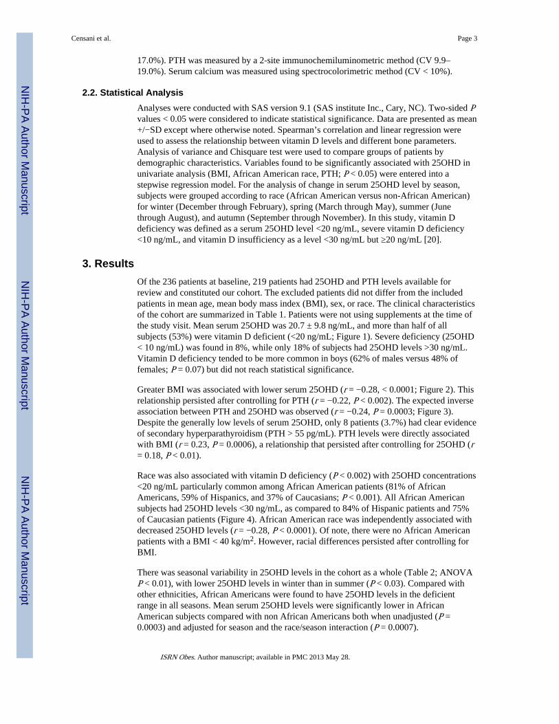
2.2. Statistical AnalysisAnalyses were conducted with SAS version 9.1 (SAS institute Inc., Cary, NC). Two-sided Pvalues < 0.05 were considered to indicate statistical significance. Data are presented as mean+/−SD except where otherwise noted. Spearman’s correlation and linear regression wereused to assess the relationship between vitamin D levels and different bone parameters.Analysis of variance and Chisquare test were used to compare groups of patients bydemographic characteristics. Variables found to be significantly associated with 25OHD inunivariate analysis (BMI, African American race, PTH; P < 0.05) were entered into astepwise regression model. For the analysis of change in serum 25OHD level by season,subjects were grouped according to race (African American versus non-African American)for winter (December through February), spring (March through May), summer (Junethrough August), and autumn (September through November). In this study, vitamin Ddeficiency was defined as a serum 25OHD level <20 ng/mL, severe vitamin D deficiency<10 ng/mL, and vitamin D insufficiency as a level <30 ng/mL but ≥20 ng/mL [20].
3. ResultsOf the 236 patients at baseline, 219 patients had 25OHD and PTH levels available forreview and constituted our cohort. The excluded patients did not differ from the includedpatients in mean age, mean body mass index (BMI), sex, or race. The clinical characteristicsof the cohort are summarized in Table 1. Patients were not using supplements at the time ofthe study visit. Mean serum 25OHD was 20.7 ± 9.8 ng/mL, and more than half of allsubjects (53%) were vitamin D deficient (<20 ng/mL; Figure 1). Severe deficiency (25OHD< 10 ng/mL) was found in 8%, while only 18% of subjects had 25OHD levels >30 ng/mL.Vitamin D deficiency tended to be more common in boys (62% of males versus 48% offemales; P = 0.07) but did not reach statistical significance.
Greater BMI was associated with lower serum 25OHD (r = −0.28, < 0.0001; Figure 2). Thisrelationship persisted after controlling for PTH (r = −0.22, P < 0.002). The expected inverseassociation between PTH and 25OHD was observed (r = −0.24, P = 0.0003; Figure 3).Despite the generally low levels of serum 25OHD, only 8 patients (3.7%) had clear evidenceof secondary hyperparathyroidism (PTH > 55 pg/mL). PTH levels were directly associatedwith BMI (r = 0.23, P = 0.0006), a relationship that persisted after controlling for 25OHD (r= 0.18, P < 0.01).
Race was also associated with vitamin D deficiency (P < 0.002) with 25OHD concentrations<20 ng/mL particularly common among African American patients (81% of AfricanAmericans, 59% of Hispanics, and 37% of Caucasians; P < 0.001). All African Americansubjects had 25OHD levels <30 ng/mL, as compared to 84% of Hispanic patients and 75%of Caucasian patients (Figure 4). African American race was independently associated withdecreased 25OHD levels (r = −0.28, P < 0.0001). Of note, there were no African Americanpatients with a BMI < 40 kg/m2. However, racial differences persisted after controlling forBMI.
There was seasonal variability in 25OHD levels in the cohort as a whole (Table 2; ANOVAP < 0.01), with lower 25OHD levels in winter than in summer (P < 0.03). Compared withother ethnicities, African Americans were found to have 25OHD levels in the deficientrange in all seasons. Mean serum 25OHD levels were significantly lower in AfricanAmerican subjects compared with non African Americans both when unadjusted (P =0.0003) and adjusted for season and the race/season interaction (P = 0.0007).
Censani et al. Page 3
ISRN Obes. Author manuscript; available in PMC 2013 May 28.
NIH
-PA Author Manuscript
NIH
-PA Author Manuscript
NIH
-PA Author Manuscript

Significant predictors of vitamin D deficiency in the univariate analysis included BMI,African American race, and PTH. These variables were entered into a stepwise linearregression model and were found to explain 21% of the overall variance in 25OHD (overallmodel: P < 0.0001). After multivariable adjustment, 25OHD deficiency was significantlyassociated with BMI (β coefficient (standard error (SE)): −0.33 (0.12), P < 0.006) and race(−6.26 (1.96), P < 0.002), with race found to be the strongest predictor of 25OHD. PTH didnot reach statistical significance in the regression model (−0.1 (0.06), P < 0.08). Eachincrease in BMI of 1 kg/m2 was associated with a decrease of 0.35 ng/mL in 25OHD. Age,sex, serum calcium, and 1,25(OH)2D were not found to be significant predictors of 25OHD.
4. DiscussionIn this cohort of over 200 morbidly obese adolescents presenting for bariatric surgery, overhalf of all subjects were frankly vitamin D deficient, and only 20% were clearly vitamin Dreplete. These data extend to the adolescent population that previously reported findings inmorbidly obese adults, where a high prevalence of vitamin D deficiency was documented,and the extent of deficiency was predicted by degree of obesity and race, with those havingthe highest BMI and of African American race most likely to be deficient [13]. Identificationof vitamin deficiencies is particularly important in the bariatric population, as these patientsmay be at greater risk of postoperative vitamin deficiencies. Weight loss has been associatedwith bone loss, and malabsorption may occur with certain bariatric procedures furthercontributing to deficiency risk.
To our knowledge, this study is the first to assess the prevalence of vitamin D deficiency ina cohort of morbidly obese (BMI > 35 kg/m2) adolescent bariatric surgery candidates inrelation to ethnicity, degree of obesity, sex, seasonal variability, and PTH levels. Althoughthe prevalence of low vitamin D status among normal weight, overweight, and obeseadolescents has been addressed in previous studies, [21–23] the effect of increasing obesitylevel on vitamin D status in extreme obesity and the relationship between vitamin D andPTH in this population has not been described in detail. In one study of a population ofmorbidly obese adolescents undergoing laparoscopic gastric banding, vitamin D levels werereported in preoperative adolescents (n = 45), with mean baseline levels slightly higher thanthose reported here [17]. Serum 25OHD rose one year after laparoscopic adjustable gastricbanding (from 22 ± 10 pg/mL to 26 ± 12 pg/mL, P = 0.025) with no change in PTH levelsone year after laparoscopic adjustable gastric banding. However, the study did not reportincidence of insufficient or deficient 25OHD levels, baseline PTH levels, data onpostoperative vitamin D supplementation, or patient characteristics that were associated withhypovitaminosis D in this at risk population.
The BMI guidelines used for determining appropriate adolescent patients to screen forbariatric surgery were consistent with NIH criteria for bariatric surgery in adults [24]. In thiscontext, we had the ability to stratify our population into three different categories of obesity(35–39 kg/m2, 40–49 kg/m2, and >50 mg/m2) and analyze ethnic groups within each BMIcohort. BMI was found to be a strong determinant of serum 25OHD concentrations, withhigher BMI associated with lower 25OHD. In our cohort (mean BMI range 35.3–86.2 ±8.1), an increase in BMI of 1 kg/m2 was associated with a decrease of 0.35 ng/mL in25OHD. Potential explanations for this association have been proposed, including decreasedsunlight exposure and sedentary lifestyle, inadequate dietary intake, and decreasedbioavailability of vitamin D secondary to sequestration of fat soluble vitamin D in excessadipose tissue [18, 25–29].
Our finding that vitamin D deficiency was most common in African Americans is inaccordance with previous literature addressing racial differences in vitamin D metabolism in
Censani et al. Page 4
ISRN Obes. Author manuscript; available in PMC 2013 May 28.
NIH
-PA Author Manuscript
NIH
-PA Author Manuscript
NIH
-PA Author Manuscript

adolescents and adults of varying adiposity [29–32]. The lower serum 25OHD in AfricanAmerican patients has been attributed to increased melanin pigment interfering with theabsorption of ultraviolet B light. This leads to diminished dermal production of vitamin Dand subsequent decreased hepatic synthesis of the major circulating metabolite 25OHD.
There is no consensus concerning the definition of vitamin D deficiency in adults, and datain pediatric literature are sparse [18]. However, studies have reported impaired calciumabsorption and lower bone mineral density at 25OHD levels of <32 ng/mL [33–36]. Holickhas defined vitamin D deficiency as 25OHD <20 ng/mL and vitamin D insufficiency as25OHD of 21 to 29 ng/mL in adults [20]. In adults, serum PTH concentrations rise as25OHD concentrations fall below 31–32 ng/mL [36, 37]. In an adult population presentingprior to bariatric surgery, Stein et al. reported that the association between PTH and BMIwas mediated by 25OHD, suggesting that the hyperparathyroidism seen in extremely obeseadults was largely secondary to vitamin D insufficiency [13]. El-Hajj Fuleihan et al. studiedhealthy children in Lebanon and showed similar findings to the adult literature andrecommended that 25OHD levels are >20–30 ng/mL to maintain serum PTH below theupper limit of normal and avoid complications of secondary hyperparathyroidism [38].However, the relationship between PTH and 25OHD is not as clear in the pediatricpopulation as in adults. Pediatric studies have been unable to clearly identify an inflectionpoint of serum 25OHD for maximal suppression of serum PTH [39]. In a study of 51 obeseAfrican American female adolescents, there was no association between 25OHD and PTH[40].
Although a significant inverse association between PTH and 25OHD was seen in ourmorbidly obese adolescent population, only a small percentage of patients (3.7%) had clearevidence of secondary hyperparathyroidism. Our study confirms findings by Gordon et al.who documented an inverse relationship between 25OHD and PTH levels in 307 normalweight adolescents without increased PTH concentrations [30]. The clinical significance oflow 25OHD concentration without concomitant secondary hyperparathyroidism is stilllargely unknown in children and adolescents. Although the explanation for this finding isunclear, the adolescent vitamin D deficiency detected may be a more acute finding asopposed to long standing vitamin D deficiency in adults with chronic PTH elevation. Thisfinding may also reflect the fact that the laboratory normal range for PTH is likely tosignificantly overestimate normal levels for children and adolescents, as it was calculatedfrom populations that included older individuals with mild renal impairment that is likely tohave led to higher PTH levels. In our cohort, only 8 patients (3.7%) had vitamin D levels<30 ng/mL with clear evidence of secondary hyperparathyroidism based on the laboratorynormal range (PTH > 55 pg/mL). However, a higher percentage of our patients (16.9%) hadPTH levels in the upper third of normal (>40 pg/mL), with 89% of these patients havingvitamin D levels <30, lending support to this hypothesis.
The role of parathyroid hormone in morbid obesity is also unclear. We found that PTHlevels remained positively associated with BMI, even after controlling for 25OHD levels.Snijder et al. found similar results and suggested that PTH may contribute to thedevelopment of adiposity independently of 25OHD. It has been hypothesized that excessPTH could promote further weight gain by enhancing lipogenesis [26].
Our data demonstrate seasonal variability in vitamin D status, with the highest 25OHDconcentrations in subjects enrolled in the summer and lowest concentrations in subjectsenrolled in the winter regardless of race. Of note, the prevalence of vitamin D deficiencywas higher among African American subjects, compared with other subjects, during allseasons: winter (89% versus 70%), spring (89% versus 49%), summer (71% versus 33%),and autumn (75% versus 44%), indicating that African Americans may be at higher risk for
Censani et al. Page 5
ISRN Obes. Author manuscript; available in PMC 2013 May 28.
NIH
-PA Author Manuscript
NIH
-PA Author Manuscript
NIH
-PA Author Manuscript
low vitamin D status throughout the year. The seasonal variation of 25OHD levels isconsistent with previously reported findings in adults and normal weight adolescents andextends these findings to the morbidly obese adolescent population [13, 30, 32]. Althoughwe had no direct measurement of sun UV exposure, the data do support an associationbetween sun exposure and vitamin D levels in morbidly obese adolescents.
This study has some important limitations, including the lack of a community basednonobese adolescent control group. Another limitation is the study’s cross-sectional designwhich prevents the assessment of the temporal associations identified between vitamin Dlevels and risk factors for hypovitaminosis D. Further, we do not have data regarding dietaryintake of vitamin D and calcium or data on the consequences of abnormal vitamin D statuson bone density or quality in these subjects. Despite these limitations, these data provideinsight into prevalence and risk factors for vitamin D deficiency in obese adolescents beforebariatric surgery.
5. ConclusionsIn conclusion, we found a high prevalence of vitamin D deficiency among obese adolescentspresenting for bariatric surgery. Individuals who were African American and had higherBMIs were at greatest risk for vitamin D deficiency. There are currently no establishedguidelines in adult or adolescents undergoing bariatric surgery for key issues pertaining tothe detection and prevention metabolic bone disease, such as the monitoring of biochemicalindices at baseline and after surgery or when to start supplementation. These results supportscreening all morbidly obese adolescents for vitamin D deficiency, and repleting those whoare deficient. This is particularly important prior to bariatric surgery where weight loss anddecreased calcium and vitamin D absorption in some procedures may place these patients atfurther risk. These results also highlight the need for more data in order to determine anoptimal regimen for vitamin D repletion in morbidly obese adolescents prior to bariatricsurgery. Procedure-specific information on the postoperative effects on vitamin D inadolescents is also needed.
AcknowledgmentsDr. I. Fennoy had full access to all of the data in the study and takes responsibility for the integrity of the data andthe accuracy of the data analysis. This work was supported by an NIH NIDDK 5T32 DK 06552-07 in PediatricEndocrinology (PI SE Oberfield). The funding organization had no role in the design and conduct of the study; inthe collection, analysis, and interpretation of the data; or in the preparation, review, or approval of the manuscript.The authors would like to extend their gratitude to Dr. Jeffrey Zitsman, M.D., Columbia University, AssistantProfessor of Surgery, for his dedication to the patients in this study.
References[1]. Xanthakos SA. Bariatric surgery for extreme adolescent obesity: indications, outcomes, and
physiologic effects on the gut-brain axis. Pathophysiology. 2008; vol. 15(no. 2):135–146.[PubMed: 18585904]
[2]. Ogden CL, Carroll MD, Curtin LR, McDowell MA, Tabak CJ, Flegal KM. Prevalence ofoverweight and obesity in the United States, 1999–2004. The Journal of the American MedicalAssociation. 2006; vol. 295(no. 13):1549–1555.
[3]. O’Brien PE, Dixon JB, Laurie C, et al. Treatment of mild to moderate obesity with laparoscopicadjustable gastric banding or an intensive medical program: a randomized trial. Annals ofInternal Medicine. 2006; vol. 144(no. 9):625–633. [PubMed: 16670131]
[4]. Dixon JB, O’Brien PE, Playfair J, et al. Adjustable gastric banding and conventional therapy fortype 2 diabetes: a randomized controlled trial. The Journal of the American Medical Association.2008; vol. 299(no. 3):316–323.
Censani et al. Page 6
ISRN Obes. Author manuscript; available in PMC 2013 May 28.
NIH
-PA Author Manuscript
NIH
-PA Author Manuscript
NIH
-PA Author Manuscript

[5]. Keating CL, Dixon JB, Moodie ML, et al. Cost-effectiveness of surgically induced weight loss forthe management of type 2 diabetes: modeled lifetime analysis. Diabetes Care. 2009; vol. 32(no.4):567–574. [PubMed: 19171720]
[6]. Ingelfinger JR. Bariatric surgery in adolescents. The New England Journal of Medicine. vol.365(no. 15):1365–1367.
[7]. O’Brien PE, Sawyer SM, Laurie C, et al. Laparoscopic adjustable gastric banding in severelyobese adolescents: a randomized trial. The Journal of the American Medical Association. 2010;vol. 303(no. 6):519–526.
[8]. Dolan K, Creighton L, Hopkins G, Fielding G. Laparoscopic gastric banding in morbidly obeseadolescents. Obesity Surgery. 2003; vol. 13(no. 1):101–104. [PubMed: 12630622]
[9]. Dillard BE III, Gorodner V, Galvani C, et al. Initial experience with the adjustable gastric band inmorbidly obese US adolescents and recommendations for further investigation. Journal ofPediatric Gastroenterology and Nutrition. 2007; vol. 45(no. 2):240–246. [PubMed: 17667722]
[10]. Nadler EP, Youn HA, Ren CJ, Fielding GA. An update on 73 US obese pediatric patients treatedwith laparoscopic adjustable gastric banding: comorbidity resolution and compliance data.Journal of Pediatric Surgery. 2008; vol. 43(no. 1):141–146. [PubMed: 18206472]
[11]. Treadwell JR, Sun F, Schoelles K. Systematic review and meta-analysis of bariatric surgery forpediatric obesity. Annals of Surgery. 2008; vol. 248(no. 5):763–776. [PubMed: 18948803]
[12]. Kendrick ML, Dakin GF. Surgical approaches to obesity. Mayo Clinic Proceedings. 2006; vol.81(no. 10):S18–S24. [PubMed: 17036575]
[13]. Stein EM, Strain G, Sinha N, et al. Vitamin D insufficiency prior to bariatric surgery: risk factorsand a pilot treatment study. Clinical Endocrinology. 2009; vol. 71(no. 2):176–183. [PubMed:19018785]
[14]. Gemmel K, Santry HP, Prachand VN, Alverdy JC. Vitamin D deficiency in preoperative bariatricsurgery patients. Surgery for Obesity and Related Diseases. 2009; vol. 5(no. 1):54–59. [PubMed:18848507]
[15]. Flancbaum L, Belsley S, Drake V, Colarusso T, Tayler E. Preoperative nutritional status ofpatients undergoing Rouxen-Y gastric bypass for morbid obesity. Journal of GastrointestinalSurgery. 2006; vol. 10(no. 7):1033–1037. [PubMed: 16843874]
[16]. Carlin AM, Rao DS, Meslemani AM, et al. Prevalence of vitamin D depletion among morbidlyobese patients seeking gastric bypass surgery. Surgery for Obesity and Related Diseases. 2006;vol. 2(no. 2):98–103. [PubMed: 16925330]
[17]. Nadler EP, Reddy S, Isenalumhe A, et al. Laparoscopic adjustable gastric banding for morbidlyobese adolescents affects android fat loss, resolution of comorbidities, and improved metabolicstatus. Journal of the American College of Surgeons. 2009; vol. 209(no. 5):638–644. [PubMed:19854406]
[18]. Misra M, Pacaud D, Petryk A, Collett-Solberg PF, Kappy M. Vitamin D deficiency in childrenand its management: review of current knowledge and recommendations. Pediatrics. 2008; vol.122(no. 2):398–417. [PubMed: 18676559]
[19]. Vieth R. Vitamin D supplementation, 25-hydroxyvitamin D concentrations, and safety. TheAmerican Journal of Clinical Nutrition. 1999; vol. 69(no. 5):842–856. [PubMed: 10232622]
[20]. Holick MF. Medical progress: vitamin D deficiency. The New England Journal of Medicine.2007; vol. 357(no. 3):266–281. [PubMed: 17634462]
[21]. Kumar J, Muntner P, Kaskel FJ, Hailpern SM, Melamed ML. Prevalence and associations of 25-hydroxyvitamin D deficiency in US children: NHANES 2001–2004. Pediatrics. 2009; Vol.124(no. 3):e362–e370. [PubMed: 19661054]
[22]. Smotkin-Tangorra M, Purushothaman R, Gupta A, Nejati G, Anhalt H, Ten S. Prevalence ofvitamin D insufficiency in obese children and adolescents. Journal of Pediatric Endocrinologyand Metabolism. 2007; vol. 20(no. 7):817–823. [PubMed: 17849744]
[23]. Alemzadeh R, Kichler J, Babar G, Calhoun M. Hypovitaminosis D in obese children andadolescents: relationship with adiposity, insulin sensitivity, ethnicity, and season. Metabolism:Clinical and Experimental. 2008; vol. 57(no. 2):183–191. [PubMed: 18191047]
[24]. NIH conference. Gastrointestinal surgery for severe obesity. Consensus development conferencepanel. Annals of Internal Medicine. 1991; vol. 115(no. 12):956–961.
Censani et al. Page 7
ISRN Obes. Author manuscript; available in PMC 2013 May 28.
NIH
-PA Author Manuscript
NIH
-PA Author Manuscript
NIH
-PA Author Manuscript
[25]. Hyppönen E, Power C. Hypovitaminosis D in British adults at age 45 y: nationwide cohort studyof dietary and lifestyle predictors. The American Journal of Clinical Nutrition. 2007; vol. 85(no.3):860–868. [PubMed: 17344510]
[26]. Snijder MB, van Dam RM, Visser M, et al. Adiposity in relation to vitamin D status andparathyroid hormone levels: a population-based study in older men and women. Journal ofClinical Endocrinology and Metabolism. 2005; vol. 90(no. 7):4119–4123. [PubMed: 15855256]
[27]. Wortsman J, Matsuoka LY, Chen TC, Lu Z, Holick MF. Decreased bioavailability of vitamin Din obesity. The American Journal of Clinical Nutrition. 2000; vol. 72(no. 3):690–693. [PubMed:10966885]
[28]. Kamycheva E, Sundsfjord J, Jorde R. Serum parathyroid hormone level is associated with bodymass index. The 5th Tromsø study. European Journal of Endocrinology. 2004; vol. 151(no. 2):167–172. [PubMed: 15296470]
[29]. Rajakumar K, de las Heras J, Chen TC, Lee S, Holick MF, Arslanian SA. Vitamin D status,adiposity, and lipids in black American and Caucasian children. Journal of ClinicalEndocrinology and Metabolism. 2011; vol. 96(no. 5):1560–1567. [PubMed: 21367931]
[30]. Gordon CM, DePeter KC, Feldman HA, Grace E, Emans SJ. Prevalence of vitamin D deficiencyamong healthy adolescents. Archives of Pediatrics and Adolescent Medicine. 2004; vol. 158(no.6):531–537. [PubMed: 15184215]
[31]. Bell NH. Bone and mineral metabolism in African Americans. Trends in Endocrinology andMetabolism. 1997; vol. 8(no. 6):240–245. [PubMed: 18406811]
[32]. Dong Y, Pollock N, Stallmann-Jorgensen IS, et al. Low 25-hydroxyvitamin D levels inadolescents: race, season, adiposity, physical activity, and fitness. Pediatrics. 2010; vol. 125(no.6):1104–1111. [PubMed: 20439594]
[33]. Hollis BW. Circulating 25-hydroxyvitamin D levels indicative of vitamin D sufficiency:implications for establishing a new effective dietary intake recommendation for vitamin D. TheJournal of Nutrition. 2005; vol. 135(no. 2):317–322. [PubMed: 15671234]
[34]. Heaney RP, Dowell MS, Hale CA, Bendich A. Calcium absorption varies within the referencerange for serum 25-hydroxyvitamin D. Journal of the American College of Nutrition. 2003; vol.22(no. 2):142–146. [PubMed: 12672710]
[35]. Bischoff-Ferrari HA, Dietrich T, Orav EJ, Dawson-Hughes B. Positive association between 25-hydroxy vitamin D levels and bone mineral density: a population-based study of younger andolder adults. The American Journal of Medicine. 2004; vol. 116(no. 9):634–639. [PubMed:15093761]
[36]. Vieth R, Ladak Y, Walfish PG. Age-related changes in the 25-hydroxyvitamin D versusparathyroid hormone relationship suggest a different reason why older adults require morevitamin D. Journal of Clinical Endocrinology and Metabolism. 2003; vol. 88(no. 1):185–191.[PubMed: 12519850]
[37]. Chapuy MC, Preziosi P, Maamer M, et al. Prevalence of vitamin D insufficiency in an adultnormal population. Osteoporosis International. 1997; vol. 7(no. 5):439–443. [PubMed: 9425501]
[38]. El-Hajj Fuleihan G, Nabulsi M, Choucair M, et al. Hypovitaminosis D in healthy schoolchildren.Pediatrics. 2001; vol. 107(no. 4) article E53.
[39]. Hill KM, McCabe GP, McCabe LD, Gordon CM, Abrams SA, Weaver CM. An inflection pointof serum 25-hydroxyvitamin D for maximal suppression of parathyroid hormone is not evidentfrom multi-site pooled data in children and adolescents. The Journal of Nutrition. 2010; vol.140(no. 11):1983–1988. [PubMed: 20861214]
[40]. Ashraf A, Alvarez J, Saenz K, Gower B, McCormick K, Franklin F. Threshold for effects ofvitamin D deficiency on glucose metabolism in obese female African-American adolescents.Journal of Clinical Endocrinology and Metabolism. 2009; vol. 94(no. 9):3200–3206. [PubMed:19549742]
Censani et al. Page 8
ISRN Obes. Author manuscript; available in PMC 2013 May 28.
NIH
-PA Author Manuscript
NIH
-PA Author Manuscript
NIH
-PA Author Manuscript
Figure 1.The distribution of 25-hydroxyvitamin D (25OHD) serum concentrations in 219 morbidlyobese adolescents; shaded area indicates 25OHD level below 20 ng/mL.
Censani et al. Page 9
ISRN Obes. Author manuscript; available in PMC 2013 May 28.
NIH
-PA Author Manuscript
NIH
-PA Author Manuscript
NIH
-PA Author Manuscript

Figure 2.The association (Spearman correlation coefficient (r)) between 25-hydroxyvitamin D(25OHD) concentration and body mass index (BMI).
Censani et al. Page 10
ISRN Obes. Author manuscript; available in PMC 2013 May 28.
NIH
-PA Author Manuscript
NIH
-PA Author Manuscript
NIH
-PA Author Manuscript
Figure 3.The association (Spearman correlation coefficient (r)) between parathyroid hormone (PTH)levels and 25-hydroxyvitamin D (25OHD) concentration.
Censani et al. Page 11
ISRN Obes. Author manuscript; available in PMC 2013 May 28.
NIH
-PA Author Manuscript
NIH
-PA Author Manuscript
NIH
-PA Author Manuscript

Figure 4.Prevalence of 25-hydroxyvitamin D deficiency (25OHD < 20 ng/mL) according to race andbody mass index (BMI).
Censani et al. Page 12
ISRN Obes. Author manuscript; available in PMC 2013 May 28.
NIH
-PA Author Manuscript
NIH
-PA Author Manuscript
NIH
-PA Author Manuscript
NIH
-PA Author Manuscript
NIH
-PA Author Manuscript
NIH
-PA Author Manuscript
Censani et al. Page 13
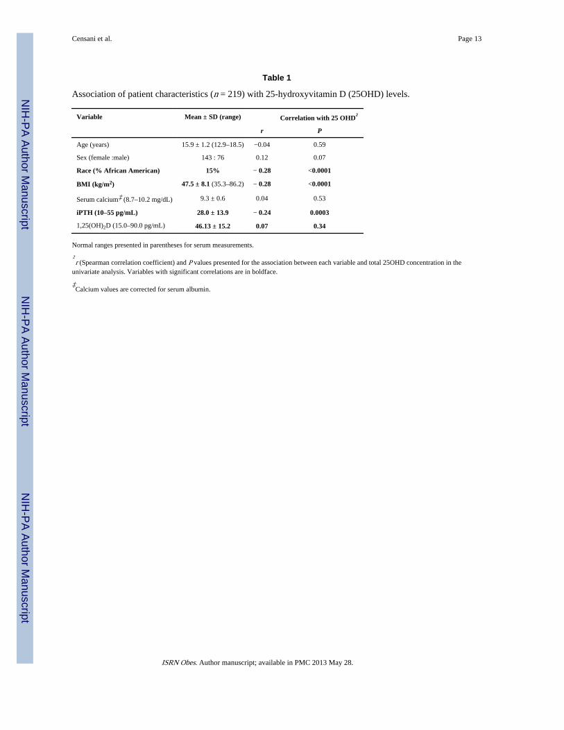
Table 1
Association of patient characteristics (n = 219) with 25-hydroxyvitamin D (25OHD) levels.
Variable Mean ± SD (range) Correlation with 25 OHD†
r P
Age (years) 15.9 ± 1.2 (12.9–18.5) −0.04 0.59
Sex (female :male) 143 : 76 0.12 0.07
Race (% African American) 15% − 0.28 <0.0001
BMI (kg/m2) 47.5 ± 8.1 (35.3–86.2) − 0.28 <0.0001
Serum calcium‡ (8.7–10.2 mg/dL) 9.3 ± 0.6 0.04 0.53
iPTH (10–55 pg/mL) 28.0 ± 13.9 − 0.24 0.0003
1,25(OH)2D (15.0–90.0 pg/mL) 46.13 ± 15.2 0.07 0.34
Normal ranges presented in parentheses for serum measurements.
†r (Spearman correlation coefficient) and P values presented for the association between each variable and total 25OHD concentration in the
univariate analysis. Variables with significant correlations are in boldface.
‡Calcium values are corrected for serum albumin.
ISRN Obes. Author manuscript; available in PMC 2013 May 28.

NIH
-PA Author Manuscript
NIH
-PA Author Manuscript
NIH
-PA Author Manuscript
Censani et al. Page 14
Table 2
Serum 25-hydroxyvitamin D levels (ng/mL): distribution by season and race.
Season† All subjects (N = 219)* African American (n = 33) Non-African American (n = 186)**
N Avg ± SD % <20 N Avg ± SD % <20 N Avg ± SD % <20
Winter 42 16.9 ± 7.4 74 9 13.7 ± 3.8 89 33 17.8 ± 7.9 70
Spring 70 20.7 ± 9.7 54 9 13.6 ± 5.1 89 61 21.7 ± 9.8 49
Summer 56 22.9 ± 11.4 38 7 17.0 ± 5.0 71 49 23.7 ± 11.8 33
Autumn 51 21.3 ± 9.1 49 8 16.5 ± 6.3 75 43 22.2 ± 9.4 44
*ANOVA P value < 0.01;
**P value < 0.02.
†Winter (December through February), spring (March through May), summer (June through August), and autumn (September through November).
ISRN Obes. Author manuscript; available in PMC 2013 May 28.
Copyright © 2022 FDOKUMEN