Вавиловский журнал генетики и селекции - Elpub
118
ВАВИЛОВСКИЙ ЖУРНАЛ ГЕНЕТИКИ И СЕЛЕКЦИИ 2020 • 24 • 2 Print ISSN 2500-0462 Online ISSN 2500-3259 Основан в 1997 г. Периодичность 8 выпусков в год DOI 10.18699/VJ20.605 Научный рецензируемый журнал Учредители Федеральное государственное бюджетное научное учреждение «Федеральный исследовательский центр Институт цитологии и генетики Сибирского отделения Российской академии наук» Межрегиональная общественная организация Вавиловское общество генетиков и селекционеров Сибирское отделение Российской академии наук Главный редактор В.К. Шумный – академик РАН, д-р биол. наук, профессор (Россия) Заместители главного редактора Н.А. Колчанов – академик РАН, д-р биол. наук, профессор (Россия) И.Н. Леонова – д-р биол. наук (Россия) Н.Б. Рубцов – д-р биол. наук, профессор (Россия) Ответственный секретарь Г.В. Орлова – канд. биол. наук (Россия) Редакционная коллегия Т.Г. Амстиславская – д-р биол. наук (Россия) Е.Е. Андронов – канд. биол. наук (Россия) Ю.С. Аульченко – д-р биол. наук (Россия) Д.А. Афонников – канд. биол. наук, доцент (Россия) Е.В. Березиков – канд. биол. наук, проф. (Нидерланды) Н.П. Бондарь – канд. биол. наук (Россия) С.А. Боринская – д-р биол. наук (Россия) П.М. Бородин – д-р биол. наук, проф. (Россия) Т.А. Гавриленко – д-р биол. наук (Россия) В.Н. Даниленко – д-р биол. наук, проф. (Россия) С.А. Демаков – д-р биол. наук (Россия) Е.А. Долгих – д-р биол. наук (Россия) Ю.М. Константинов – д-р биол. наук, проф. (Россия) О. Кребс – д-р биол. наук, проф. (Германия) И.Н. Лаврик – канд. хим. наук (Германия) Д. Ларкин – д-р биол. наук (Великобритания) И.Н. Лебедев – д-р биол. наук, проф. (Россия) Л.А. Лутова – д-р биол. наук, проф. (Россия) В.Ю. Макеев – чл.-кор. РАН, д-р физ.-мат. наук (Россия) М.П. Мошкин – д-р биол. наук, проф. (Россия) Л.Ю. Новикова – канд. техн. наук (Россия) Е. Песцова – д-р биол. наук (Германия) Н.А. Проворов – д-р биол. наук, проф. (Россия) Д.В. Пышный – чл.-кор. РАН, д-р хим. наук (Россия) А.В. Ратушный – канд. биол. наук (США) М.Г. Самсонова – д-р биол. наук (Россия) Е. Туруспеков – канд. биол. наук (Казахстан) М. Чен – д-р биол. наук (Китайская Народная Республика) Ю. Шавруков – д-р биол. наук (Австралия) Редакционный совет Л.И. Афтанас – академик РАН, д-р мед. наук (Россия) В.С. Баранов – чл.-кор. РАН, д-р мед. наук (Россия) Л.А. Беспалова – академик РАН, д-р с.-х. наук (Россия) А. Бёрнер – д-р наук (Германия) М.И. Воевода – академик РАН, д-р мед. наук (Россия) И. Гроссе – д-р наук, проф. (Германия) Г.Л. Дианов – д-р биол. наук, проф. (Великобритания) Ю.Е. Дуброва – д-р биол. наук, проф. (Великобритания) Н.Н. Дыгало – чл.-кор. РАН, д-р биол. наук (Россия) И.К. Захаров – д-р биол. наук, проф. (Россия) И.А. Захаров-Гезехус – чл.-кор. РАН, д-р биол. наук (Россия) С.Г. Инге-Вечтомов – академик РАН, д-р биол. наук (Россия) И.Е. Керкис – д-р наук (Бразилия) А.В. Кильчевский – чл.-кор. НАНБ, д-р биол. наук (Беларусь) С.В. Костров – чл.-кор. РАН, д-р хим. наук (Россия) А.В. Кочетов – чл.-кор. РАН, д-р биол. наук (Россия) Ж. Ле Гуи – д-р наук (Франция) Б. Люгтенберг – д-р наук, проф. (Нидерланды) В.И. Молодин – академик РАН, д-р ист. наук (Россия) В.П. Пузырев – академик РАН, д-р мед. наук (Россия) А.Ю. Ржецкий – канд. биол. наук, проф. (США) И.Б. Рогозин – канд. биол. наук (США) А.О. Рувинский – д-р биол. наук, проф. (Австралия) Е.А. Салина – д-р биол. наук, проф. (Россия) К.Г. Скрябин – академик РАН, д-р биол. наук (Россия) К.В. Славин – д-р наук, проф. (США) В.А. Степанов – чл.-кор. РАН, д-р биол. наук (Россия) И.А. Тихонович – академик РАН, д-р биол. наук (Россия) Е.К. Хлесткина – д-р биол. наук, профессор (Россия) Л.В. Хотылева – академик НАНБ, д-р биол. наук (Беларусь) Э.К. Хуснутдинова – д-р биол. наук, проф. (Россия) М.Ф. Чернов – д-р мед. наук (Япония) С.В. Шестаков – академик РАН, д-р биол. наук (Россия) Н.К. Янковский – академик РАН, д-р биол. наук (Россия)
-
Upload
khangminh22 -
Category
Documents
-
view
0 -
download
0
Transcript of Вавиловский журнал генетики и селекции - Elpub

ВАВИЛОВСКИЙ ЖУРНАЛ ГЕНЕТИКИ И СЕЛЕКЦИИ
2020 • 24 • 2 Print ISSN 2500-0462Online ISSN 2500-3259
Основан в 1997 г.Периодичность 8 выпусков в год DOI 10.18699/VJ20.605
Научный рецензируемый журнал
Учредители Федеральное государственное бюджетное научное учреждение «Федеральный исследовательский центр Институт цитологии и генетики Сибирского отделения Российской академии наук»
Межрегиональная общественная организация Вавиловское общество генетиков и селекционеров
Сибирское отделение Российской академии наук
Главный редакторВ.К. Шумный – академик РАН, д-р биол. наук, профессор (Россия)
Заместители главного редактораН.А. Колчанов – академик РАН, д-р биол. наук, профессор (Россия)И.Н. Леонова – д-р биол. наук (Россия)Н.Б. Рубцов – д-р биол. наук, профессор (Россия)
Ответственный секретарьГ.В. Орлова – канд. биол. наук (Россия)
Редакционная коллегия
Т.Г. Амстиславская – д-р биол. наук (Россия)Е.Е. Андронов – канд. биол. наук (Россия)Ю.С. Аульченко – д-р биол. наук (Россия)Д.А. Афонников – канд. биол. наук, доцент (Россия)Е.В. Березиков – канд. биол. наук, проф. (Нидерланды)Н.П. Бондарь – канд. биол. наук (Россия)С.А. Боринская – д-р биол. наук (Россия)П.М. Бородин – д-р биол. наук, проф. (Россия)Т.А. Гавриленко – д-р биол. наук (Россия)В.Н. Даниленко – д-р биол. наук, проф. (Россия)С.А. Демаков – д-р биол. наук (Россия)Е.А. Долгих – д-р биол. наук (Россия)Ю.М. Константинов – д-р биол. наук, проф. (Россия)О. Кребс – д-р биол. наук, проф. (Германия)И.Н. Лаврик – канд. хим. наук (Германия)Д. Ларкин – д-р биол. наук (Великобритания)И.Н. Лебедев – д-р биол. наук, проф. (Россия)Л.А. Лутова – д-р биол. наук, проф. (Россия)В.Ю. Макеев – чл.-кор. РАН, д-р физ.- мат. наук (Россия)М.П. Мошкин – д-р биол. наук, проф. (Россия)Л.Ю. Новикова – канд. техн. наук (Россия)Е. Песцова – д-р биол. наук (Германия)Н.А. Проворов – д-р биол. наук, проф. (Россия)Д.В. Пышный – чл.-кор. РАН, д-р хим. наук (Россия) А.В. Ратушный – канд. биол. наук (США)М.Г. Самсонова – д-р биол. наук (Россия)Е. Туруспеков – канд. биол. наук (Казахстан)М. Чен – д-р биол. наук (Китайская Народная Республика)Ю. Шавруков – д-р биол. наук (Австралия)
Редакционный совет
Л.И. Афтанас – академик РАН, д-р мед. наук (Россия)В.С. Баранов – чл.-кор. РАН, д-р мед. наук (Россия)Л.А. Беспалова – академик РАН, д-р с.-х. наук (Россия) А. Бёрнер – д-р наук (Германия)М.И. Воевода – академик РАН, д-р мед. наук (Россия)И. Гроссе – д-р наук, проф. (Германия)Г.Л. Дианов – д-р биол. наук, проф. (Великобритания)Ю.Е. Дуброва – д-р биол. наук, проф. (Великобритания)Н.Н. Дыгало – чл.-кор. РАН, д-р биол. наук (Россия)И.К. Захаров – д-р биол. наук, проф. (Россия)И.А. Захаров-Гезехус – чл.-кор. РАН, д-р биол. наук (Россия)С.Г. Инге-Вечтомов – академик РАН, д-р биол. наук (Россия)И.Е. Керкис – д-р наук (Бразилия)А.В. Кильчевский – чл.-кор. НАНБ, д-р биол. наук (Беларусь)С.В. Костров – чл.-кор. РАН, д-р хим. наук (Россия)А.В. Кочетов – чл.-кор. РАН, д-р биол. наук (Россия)Ж. Ле Гуи – д-р наук (Франция)Б. Люгтенберг – д-р наук, проф. (Нидерланды)В.И. Молодин – академик РАН, д-р ист. наук (Россия)В.П. Пузырев – академик РАН, д-р мед. наук (Россия) А.Ю. Ржецкий – канд. биол. наук, проф. (США)И.Б. Рогозин – канд. биол. наук (США)А.О. Рувинский – д-р биол. наук, проф. (Австралия)Е.А. Салина – д-р биол. наук, проф. (Россия)К.Г. Скрябин – академик РАН, д-р биол. наук (Россия)К.В. Славин – д-р наук, проф. (США)В.А. Степанов – чл.-кор. РАН, д-р биол. наук (Россия)И.А. Тихонович – академик РАН, д-р биол. наук (Россия)Е.К. Хлесткина – д-р биол. наук, профессор (Россия)Л.В. Хотылева – академик НАНБ, д-р биол. наук (Беларусь)Э.К. Хуснутдинова – д-р биол. наук, проф. (Россия)М.Ф. Чернов – д-р мед. наук (Япония) С.В. Шестаков – академик РАН, д-р биол. наук (Россия) Н.К. Янковский – академик РАН, д-р биол. наук (Россия)

VAVILOV JOURNALOF GENETICS AND BREEDINGFounded in 1997Published 8 times annually
Scientific Peer Reviewed Journal
Founders Federal State Budget Scientific Institution “The Federal Research Center Institute of Cytology and Geneticsof Siberian Branch of the Russian Academy of Sciences”The Vavilov Society of Geneticists and BreedersSiberian Branch of the Russian Academy of Sciences
Editor-in-ChiefV.K. Shumny, Full Member of the Russian Academy of Sciences, Dr. Sci. (Biology), Russia
Deputy Editor-in-ChiefN.A. Kolchanov, Full Member of the Russian Academy of Sciences, Dr. Sci. (Biology), RussiaI.N. Leonova, Dr. Sci. (Biology), RussiaN.B. Rubtsov, Professor, Dr. Sci. (Biology), Russia
Executive SecretaryG.V. Orlova, Cand. Sci. (Biology), Russia
Editorial council
L.I. Aftanas, Full Member of the RAS, Dr. Sci. (Medicine), RussiaV.S. Baranov, Corr. Member of the RAS, Dr. Sci. (Medicine), RussiaL.A. Bespalova, Full Member of the RAS, Dr. Sci. (Agricul.), RussiaA. Börner, Dr. Sci., GermanyM.F. Chernov, Dr. Sci. (Medicine), JapanG.L. Dianov, Professor, Dr. Sci. (Biology), Great Britain Yu.E. Dubrova, Professor, Dr. Sci. (Biology), Great BritainN.N. Dygalo, Corr. Member of the RAS, Dr. Sci. (Biology), RussiaJ. Le Gouis, Dr. Sci., FranceI. Grosse, Professor, Dr. Sci., GermanyS.G. Inge-Vechtomov, Full Member of the RAS, Dr. Sci. (Biology), RussiaI.E. Kerkis, Dr. Sci., BrazilE.K. Khlestkina, Professor, Dr. Sci. (Biology), RussiaL.V. Khotyleva, Full Member of the NAS of Belarus, Dr. Sci. (Biology), BelarusE.K. Khusnutdinova, Professor, Dr. Sci. (Biology), RussiaA.V. Kilchevsky, Corr. Member of the NAS of Belarus, Dr. Sci. (Biology), BelarusA.V. Kochetov, Corr. Member of the RAS, Dr. Sci. (Biology), RussiaS.V. Kostrov, Corr. Member of the RAS, Dr. Sci. (Chemistry), RussiaB. Lugtenberg, Professor, Dr. Sci., NetherlandsV.I. Molodin, Full Member of the RAS, Dr. Sci. (History), RussiaV.P. Puzyrev, Full Member of the RAS, Dr. Sci. (Medicine), RussiaI.B. Rogozin, Cand. Sci. (Biology), United StatesA.O. Ruvinsky, Professor, Dr. Sci. (Biology), AustraliaA.Yu. Rzhetsky, Professor, Cand. Sci. (Biology), United StatesE.A. Salina, Professor, Dr. Sci. (Biology), RussiaS.V. Shestakov, Full Member of the RAS, Dr. Sci. (Biology), RussiaK.G. Skryabin, Full Member of the RAS, Dr. Sci. (Biology), RussiaK.V. Slavin, Professor, Dr. Sci., United StatesV.A. Stepanov, Corr. Member of the RAS, Dr. Sci. (Biology), RussiaI.A. Tikhonovich, Full Member of the RAS, Dr. Sci. (Biology), RussiaM.I. Voevoda, Full Member of the RAS, Dr. Sci. (Medicine), RussiaN.K. Yankovsky, Full Member of the RAS, Dr. Sci. (Biology), RussiaI.K. Zakharov, Professor, Dr. Sci. (Biology), RussiaI.A. Zakharov-Gezekhus, Corr. Member of the RAS, Dr. Sci. (Biology), Russia
VAV I L O V S K I I Z H U R N A L G E N E T I K I I S E L E K T S I I
2020 • 24 • 2 Print ISSN 2500-0462Online ISSN 2500-3259
Editorial board
D.A. Afonnikov, Associate Professor, Cand. Sci. (Biology), RussiaT.G. Amstislavskaya, Dr. Sci. (Biology), Russia E.E. Andronov, Cand. Sci. (Biology), RussiaYu.S. Aulchenko, Dr. Sci. (Biology), RussiaE.V. Berezikov, Professor, Cand. Sci. (Biology), NetherlandsN.P. Bondar, Cand. Sci. (Biology), RussiaS.A. Borinskaya, Dr. Sci. (Biology), Russia P.M. Borodin, Professor, Dr. Sci. (Biology), RussiaM. Chen, Dr. Sci. (Biology), People’s Republic of ChinaV.N. Danilenko, Professor, Dr. Sci. (Biology), RussiaS.A. Demakov, Dr. Sci. (Biology), RussiaE.A. Dolgikh, Dr. Sci. (Biology), RussiaT.A. Gavrilenko, Dr. Sci. (Biology), RussiaYu.M. Konstantinov, Professor, Dr. Sci. (Biology), RussiaO. Krebs, Professor, Dr. Sci. (Biology), GermanyD. Larkin, Dr. Sci. (Biology), Great BritainI.N. Lavrik, Cand. Sci. (Chemistry), GermanyI.N. Lebedev, Professor, Dr. Sci. (Biology), RussiaL.A. Lutova, Professor, Dr. Sci. (Biology), RussiaV.Yu. Makeev, Corr. Member of the RAS, Dr. Sci. (Physics and Mathem.), RussiаM.P. Moshkin, Professor, Dr. Sci. (Biology), RussiaE. Pestsova, Dr. Sci. (Biology), GermanyL.Yu. Novikova, Cand. Sci. (Engineering), RussiaN.A. Provorov, Professor, Dr. Sci. (Biology), RussiaD.V. Pyshnyi, Corr. Member of the RAS, Dr. Sci. (Chemistry), RussiaA.V. Ratushny, Cand. Sci. (Biology), United StatesM.G. Samsonova, Dr. Sci. (Biology), RussiaY. Shavrukov, Dr. Sci. (Biology), AustraliaE. Turuspekov, Cand. Sci. (Biology), Kazakhstan
DOI 10.18699/VJ20.605

СОДЕРЖАНИЕ • 2020 • 24 • 2
Генетика растений Генетика животных
Генетика человека
Селекция растений на иммунитетФизиологическая генетика
Генетика микроорганизмов
ВАВИЛОВСКИЙ ЖУРНАЛ ГЕНЕТИКИ И СЕЛЕКЦИИ
ОРИГИНАЛЬНОЕ ИССЛЕДОВАНИЕ
Выявление геномного состава аллополиплоидных видов рода Elymus (Poaceae: Triticeae) Азиатской России с помощью CAPS-анализа. А.В. Агафонов, Е.В. Шабанова (Кобозева), С.В. Асбаганов, А.В. Мглинец, В.С. Богданова
ОРИГИНАЛЬНОЕ ИССЛЕДОВАНИЕ
Динамика генетического разнообразия сортов овса в Тюменской области по авенин-кодирующим локусам. А.В. Любимова, Г.В. Тоболова, Д.И. Еремин, И.Г. Лоскутов
ОБЗОР
Современное состояние проблемы сохранения генетических ресурсов сельскохозяйственных птиц in vitro. Ю.Л. Силюкова, О.И. Станишевская, Н.В. Дементьева
ОРИГИНАЛЬНОЕ ИССЛЕДОВАНИЕ
Полногеномные ассоциативные исследования распространения пороков развития и других селекционно значимых качественных признаков у потомства хряков крупной белой породы российской селекции. А.А. Траспов, О.В. Костюнина, А.А. Белоус, Т.В. Карпушкина, Н.А. Свеженцева, Н.А. Зиновьева
ОРИГИНАЛЬНОЕ ИССЛЕДОВАНИЕ
Стеблевая ржавчина в Западной Сибири – расовый состав и эффективные гены устойчивости. В.П. Шаманин, И.В. Потоцкая, С.С. Шепелев, В.Е. Пожерукова, Е.А. Салина, Е.С. Сколотнева, Д. Ходсон, М. Хоумвёллер, М. Патпур, А.И. Моргунов
ОРИГИНАЛЬНОЕ ИССЛЕДОВАНИЕ
Эпифитотический процесс септориоза на сортах яровой пшеницы. Е.Ю. Торопова, О.А. Казакова, В.В. Пискарев
ОРИГИНАЛЬНОЕ ИССЛЕДОВАНИЕ
Коэкспрессия глутаматергических генов и генов аутистического спектра в гиппокампе у самцов мышей с нарушением социального поведения. И.Л. Коваленко, А.Г. Галямина, Д.А. Смагин, Н.Н. Кудрявцева
ОРИГИНАЛЬНОЕ ИССЛЕДОВАНИЕ
У мышей с мутацией lethal yellow (Ay) фармакологические эффекты фактора роста фибробластов 21 зависят от пола. Е.Н. Макарова, Т.В. Яковлева, Н.Ю. Балыбина, К.О. Баранов, Е.И. Денисова, А.Д. Дубинина, Н.А. Феофанова, Н.М. Бажан (на англ. языке)
ОБЗОР
Участие мобильных элементов в нейрогенезе. Р.Н. Мустафин, Э.К. Хуснутдинова
ОБЗОР
Современная классификация и молекулярно-генетические аспекты незавершенного остеогенеза. А.Р. Зарипова, Р.И. Хусаинова
ОБЗОР
Разнообразие и распространение метилотрофных дрожжей, используемых в генной инженерии. А.С. Розанов, Е.Г. Першина, Н.В. Богачева, В. Шляхтун, А.А. Сычев, С.Е. Пельтек
ОРИГИНАЛЬНОЕ ИССЛЕДОВАНИЕ
Перспективы использования Illumina MiSeq для идентификации грибов арбускулярной микоризы. А.А. Крюков, A.O. Горбунова, Э.M. Мачс, Ю.В. Михайлова, A.В. Родионов, П.M. Журбенко, A.П. Юрков (на англ. языке)
ОРИГИНАЛЬНОЕ ИССЛЕДОВАНИЕ
Изменение кишечного микробиома пациентов с язвенным колитом после трансплантации кишечной микробиоты. А.Ю. Тикунов, В.В. Морозов, А.Н. Швалов, А.В. Бардашева, Е.В. Шрайнер, О.А. Максимова, И.О. Волошина, В.В. Морозова, В.В. Власов, Н.В. Тикунова
© ИЦиГ СО РАН, 2020© Вавиловский журнал генетики и селекции, 2020© Сибирское отделение Российской академии наук, 2020
115 176
131191
209
149
123
185
139200
219
158
168

CONTENTS • 2020 • 24 • 2 VAVILOV JOURNAL OF GENETICS AND BREEDING
Plant genetics Animal genetics
Human genetics
Physiological genetics
Microbial genetics
Plant breeding for immunity
115 176
131 191
209149
123
185
139200
219158
168
ORIGINAL ARTICLE
Identification of genome compositions in allopolyploid species of the genus Elymus (Poaceae: Triticeae) in the Asian part of Russia by CAPS analysis. A.V. Agafonov, E.V. Shabanova (Kobozeva), S.V. Asbaganov, A.V. Mglinets, V.S. Bogdanova
ORIGINAL ARTICLE
Dynamics of genetic diversity of oat varieties in the Tyumen region at avenin-coding loci. A.V. Lyubimova, G.V. Tobolova, D.I. Eremin, I.G. Loskutov
REVIEW
The current state of the problem of in vitro gene pool preservation in poultry. Y.L. Silyukova, O.I. Stanishevskaya, N.V. Dementieva
ORIGINAL ARTICLE
Whole-genome association studies of distribution of developmental abnormalities and other breeding-valuable qualitative traits in offspring of the Russian large-white boars. A.A. Traspov, O.V. Kostyunina, A.A. Belous, T.V. Karpushkina, N.A. Svejenceva, N.A. Zinovieva
ORIGINAL ARTICLE
Stem rust in Western Siberia – race composition and effective resistance genes. V.P. Shamanin, I.V. Pototskaya, S.S. Shepelev, V.E. Pozherukova, Е.А. Salina, Е.S. Skolotneva, D. Hodson, M. Hovmøller, M. Patpour, A.I. Morgounov
ORIGINAL ARTICLE
Septoria blotch epidemic process on spring wheat varieties. E.Yu. Toropova, O.A. Kazakova, V.V. Piskarev
ORIGINAL ARTICLE
Co-expression of glutamatergic and autism-related genes in the hippocampus of male mice with disturbances of social behavior. I.L. Kovalenko, A.G. Galyamina, D.A. Smagin, N.N. Kudryavtseva
ORIGINAL ARTICLE
Pharmacological effects of fibroblast growth factor 21 are sex-specific in mice with the lethal yellow (Ay) mutation. E.N. Makarova, T.V. Yakovleva, N.Yu. Balyibina, K.O. Baranov, E.I. Denisova, A.D. Dubinina, N.A. Feofanova, N.M. Bazhan
REVIEW
Involvement of transposable elements in neurogenesis. R.N. Mustafin, E.K. Khusnutdinova
REVIEW
Modern classification and molecular-genetic aspects of osteogenesis imperfecta. A.R. Zaripova, R.I. Khusainova
REVIEW
Diversity and occurrence of methylotrophic yeasts used in genetic engineering. A.S. Rozanov, E.G. Pershina, N.V. Bogacheva, V. Shlyakhtun, A.A. Sychev, S.E. Peltek
ORIGINAL ARTICLE
Perspectives of using Illumina MiSeq for identification of arbuscular mycorrhizal fungi. A.A. Kryukov, A.O. Gorbunova, E.M. Machs, Y.V. Mikhaylova, A.V. Rodionov, P.M. Zhurbenko, A.P. Yurkov
ORIGINAL ARTICLE
Fecal microbiome change in patients with ulcerative colitis after fecal microbiota transplantation. A.Y. Tikunov, V.V. Morozov, A.N. Shvalov, A.V. Bardasheva, E.V. Shrayner, O.A. Maksimova, I.O. Voloshina, V.V. Morozova, V.V. Vlasov, N.V. Tikunova
© Institute of Cytology and Genetics SB RAS, 2020© Vavilov Journal of Genetics and Breeding, 2020© Siberian Branch RAS, 2020

Identification of genome compositions in allopolyploid species of the genus Elymus (Poaceae: Triticeae) in the Asian part of Russia by CAPS analysisA.V. Agafonov1 , E.V. Shabanova (Kobozeva)1, S.V. Asbaganov1, A.V. Mglinets2, V.S. Bogdanova2
1 Central Siberian Botanical Garden of Siberian Branch of the Russian Academy of Sciences, Russia, Novosibirsk, Russia2 Institute of Cytology and Genetics of Siberian Branch of the Russian Academy of Sciences, Novosibirsk, Russia
e-mail: [email protected]
Abstract. The genus Elymus L., together with wheat, rye, and barley, belongs to the tribe Triticeae. Apart from its economic value, this tribe is characterized by abundance of polyploid taxa formed in the course of remote hybridi-zation. Single-copy nuclear genes are convenient markers for identification of source genomes incorporated into polyploids. In the present work, a CAPS-marker is developed to distinguish basic St, H, and Y genomes comprising polyploid genomes of Asiatic species of the genus Elymus. The test is based on electrophoretic analysis of restric-tion patterns of a PCR-amplified fragment of the gene coding for beta-amylase. There are about 50 Elymus species in Russia, and most of them are supposed to possess one of three haplome combinations, StH, StY and StHY. Boreal StH-genomic species endemic for Russia are the least studied. On the basis of nucleotide sequences from public databases, TaqI restrictase was selected, as it produced patterns of restriction fragments specific for St, H, and Y haplomes easily recognizable in agarose gel. A sample of 68 accessions belonging to 32 species was analyzed. In 15 species, the earlier known genomic constitutions were confirmed, but in E. kamoji this assay failed to reveal the presence of H genome. This unusual H genome was suggested to originate from a different Hordeum spe-cies. In 16 species, genomic constitutions were identified for the first time. Fifteen accessions from Asian Russia possessed the genomic constitution StStHH, and E. amurensis, phylogenetically close to the StY-genomic species E. ciliaris, had the genomic constitution StStYY. It is inferred that the center of species diversity of the StH-genomic group is shifted to the north as compared to the center of origin of StY-genomic species, confined to China.Key words: Elymus; taxonomy; allopolyploids; genome constitution; CAPS markers.
For citation: Agafonov A.V., Shabanova (Kobozeva) E.V., Asbaganov S.V., Mglinets A.V., Bogdanova V.S. Identifica-tion of genome compositions in allopolyploid species of the genus Elymus (Poaceae: Triticeae) in the Asian part of Russia by CAPS analysis. Vavilovskii Zhurnal Genetiki i Selektsii = Vavilov Journal of Genetics and Breeding. 2020; 24(2):115-122. DOI 10.18699/VJ20.606
Выявление геномного состава аллополиплоидных видов рода Elymus (Poaceae: Triticeae) Азиатской России с помощью CAPS-анализаА.В. Агафонов1 , Е.В. Шабанова (Кобозева)1, С.В. Асбаганов1, А.В. Мглинец2, В.С. Богданова2
1 Центральный сибирский ботанический сад Сибирского отделения Российской академии наук, Новосибирск, Россия 2 Федеральный исследовательский центр Институт цитологии и генетики Сибирского отделения Российской академии наук,
Новосибирск, Россия e-mail: [email protected]
Аннотация. Род Elymus L. наряду с пшеницей, рожью и ячменем принадлежит к трибе Triticeae. Помимо сво-его хозяйственного значения, эта триба характеризуется широким распространением аллополиплоидных таксонов, которые формируются в ходе межвидовой и межродовой гибридизации и последующих преобра-зований вовлеченных в гибридизацию диплоидных геномов. Для идентификации исходных геномов в соста-ве полиплоидов удобны малокопийные ядерные гены, менее подверженные процессам реорганизации, чем повторенные некодирующие элементы. В настоящей работе разработан удобный CAPS-маркер для различе-ния базисных геномов St, H, Y, входящих в состав азиатских видов рода Elymus, с помощью электрофорети-ческого анализа фрагментов рестрикции ПЦР-амплифицированного участка гена, кодирующего β-амилазу. В России распространено около 50 видов Elymus предположительно трех гапломных комбинаций: StH, StY и StHY. Наименее изученными остаются бореальные StH-геномные виды – эндемики Российской Федера-ции. По результатам анализа ранее изученных разными авторами нуклеотидных последовательностей гена β-амилазы была отобрана эндонуклеаза рестрикции TaqI, которая имела различающиеся по положению сайты узнавания в составе вышеуказанного фрагмента из геномов St, H и Y. В результате расщепления ПЦР-продукта эндонуклеазой TaqI у каждого из исходных гапломов формировался специфичный паттерн фраг-
© Agafonov A.V., Shabanova (Kobozeva) E.V., Asbaganov S.V., Mglinets A.V., Bogdanova V.S., 2020
This work is licensed under a Creative Commons Attribution 4.0 License
ГЕНЕТИКА РАСТЕНИЙОригинальное исследование / Original article
Вавиловский журнал генетики и селекции. 2020;24(2):115-122DOI 10.18699/VJ20.606

A.V. Agafonov, E.V. Shabanova (Kobozeva), S.V. Asbaganov, A.V. Mglinets, V.S. Bogdanova
116
Identification of genome compositions in allopolyploid species of the genus Elymus in the Asian part of Russia by CAPS analysis
Вавиловский журнал генетики и селекции / Vavilov Journal of Genetics and Breeding • 2020 • 24 • 2
ментов рестрикции, легко визуализируемый в агарозном геле. Проанализирована выборка из 68 образцов, принадлежащих 32 видам. У 15 видов была подтверждена ранее известная геномная конституция, у E. kamoji этот метод не позволил выявить присутствие генома H. Предполагается возможное происхождение данного варианта генома H от другого вида Hordeum. У 16 видов геномная конституция определена впервые. Пока-зано, что большинство изученных видов бореальной группы видов из Сибири и Российского Дальнего Вос-тока имеют геномную конституцию StStHH. Исключение составил E. amurensis, филогенетически близкий к StY-геномному виду E. ciliaris и также имеющий геномный состав StStYY. Сделан вывод, что основное видовое разнообразие StH-геномной группы находится севернее центра происхождения большинства StY-геномных видов рода.Ключевые слова: Elymus; таксономия; аллополиплоиды; геномная конституция; CAPS-маркеры.
IntroductionThe genus Elymus L. is the largest in the tribe Triticeae Dum. and, according to different estimates, counts from 150 to 200 species (Dewey, 1984; Barkworth, 2000). It is represented only by alloploid taxa with genome compositions including several basic genomes (haplomes) in different combinations. The genetic basis of the genus Elymus is formed by five haplomes descending from different genera of the tribe Triticeae: (St) Pseudoroegneria, (H) Hordeum, (P) Agropyron, (W) Astralopyrum, (Y) donor unknown. Genome constitution was proposed as a stable genetic criterion for taxonomic classification of Elymus species (Löve, 1984). Within a relatively short span of time, substantial changes occurred in the taxonomy of the tribe Triticeae on the basis of the genomic system of classification suggested by D.R. Dewey (1984). During the next 20 years, six genera were identified according to variants of genome constitution: Douglasdeweya C. Yen, J.L. Yang & B.R. Baum (PPStSt), Roegneria C. Koch (StStYY), Anthosachne Steudel (StStWWYY), Kengylia C. Yen & J.L. Yang (PPStStYY), Campeiostachys Drobow (HHStStYY), and Elymus L. (StStHH, StStStHH, StStHHHH).
However, departing from A. Löve’s principles, many botanists still attribute several genome combinations to the single genus Elymus s. l. With all this, genome constitutions are not yet determined in about 40 % of species (Okito et al., 2009). According to current evidence, 53 species of the genus Elymus subdivided into four sections occur in Russia (Tsvelyov, 2008; Tsvelyov, Probatova, 2010). Two of the sections, Elymus and Goulardia (Husn.) Tzvelev, contain species with different genomic constitutions, which obviously contradicts the phylogenetic principle of their formulation. We suppose that Russia is home to species with only three haplome combinations: StH, StY, and StHY (Agafonov et al., 2015). Boreal StHgenomic endemics of Russia are less studied. According to the taxonomic system based on the genome constitution, the Elymus species should be attributed to three genera: Elymus, Roegneria, and Campeiostachys. However, in our view, the division of the species inhabiting Russia into three genera is impractical due to the difficulties of morphologic identification of these genera. With all this, taxonomic classification within the genus based on genome constitutions is indispensable for the construction of a phylogenetically oriented taxonomy of the genus.
Earlier, Cleaved Amplified Polymorphic Sequences (CAPS) markers were used to distinguish individual genomes in representatives of the tribe Triticeae (Gostimsky et al., 2005; Li et al., 2007; Hu et al., 2014; Shavrukov, 2016). Some advantages of CAPS markers are their codominance, moderate sensitivity to the amount of genomic DNA, and relatively low cost.
We were first to use CAPSmarkers to identify the genomic constitutions of species of the genus Elymus (Kobozeva et al., 2017). For this purpose, primers were designed based on the known sequences of the gene coding for β amylase (MasonGamer, 2013), which included 38 sequences of haplome St, 23 of haplome H, and 15 of haplome Y, belonging to 24 Elymus species. Of them, 14 species had the genomic composition StStHH; 9, StStYY; and 1, StStHHUkUk (Elytrigia repens). Variable positions were sought that would discriminate representatives of an individual genome from the other two. Special attention was paid to those genomespecific sequence variants that resulted in appearance/disappearance of recognition sites for restriction endonucleases. It was found that digestion of the PCR products with TaqI endonuclease resulted in the formation of genomespecific restriction patterns. In the present work, we apply CAPS analysis to a large sample of Elymus species from Asian Russia to reveal their genome constitutions unknown hitherto.
Materials and methodsPlant material included 68 accessions of the species with known (Table 1) and unknown (Table 2) genome constitutions found in Russia. The species nomenclature is given according to N.N. Tsvelyov and N.S. Probatova (2010). The accessions analyzed were received from the scientific collection of biological resources of the Central Siberian Botanic Garden SB RAS “Collections of living plants indoors and outdoors”; their identification numbers are given in Tables 1 and 2. Prefixes correspond to the geographic origin of the accessions.
Total DNA was extracted from 20 mg of dried green matter with the use of NucleoSpin Plant II Kit (MachereyNagel, Germany) according to manufacturer’s recommendations. Amplification of the β amylase gene fragment was made in a C1000 thermocycler (BioRad, USA) with the following primers: El_balg_F4 (5ʹGGTACCATCGTGGACATTGAA 3ʹ) and El_balg_R4 (5ʹCTGTACCACCAGTGAATGCC3ʹ) (Kobozeva et al., 2017). The PCR reaction mixture of 15 μL in volume contained 1× buffer for Taq polymerase, 0.2 mM each dNTP, 1.5 mM MgCl2, 1 µM each of primers, 20 ng of genomic DNA, and 1 U of HS Taq DNA polymerase (Eurogene, RF). The following settings were used: predenaturation at 94 °С for 4 min; 40 cycles: denaturation at 94 °С for 20 s, primer annealing at 60 °C for 25 s, elongation at 72 °С for 90 s; postextension at 72 °С for 5 minutes. CAPSanalysis (Konieczny, Ausubel, 1993) was made as follows: 8 μL of the PCR reaction mixture was mixed with MQH2O and TaqI buffer up to 1× concentration in a volume of 15 μL, and 1 unit of TaqI restrictase (Thermo Scientific, USA) was added. The mixture was incubated at 65 °С for 1 hour and resolved in
Выявление геномного состава аллополиплоидных видов рода Elymus Азиатской России с помощью CAPS-анализа
А.В. Агафонов, Е.В. Шабанова (Кобозева), С.В. Асбаганов, А.В. Мглинец, В.С. Богданова
117
202024 • 2
ГЕНЕТИКА РАСТЕНИЙ / PLANT GENETICS
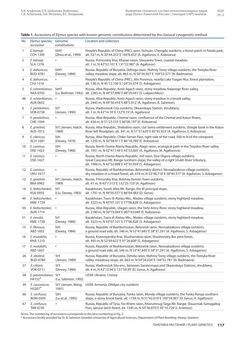
Table 1. Accessions of Elymus species with known genomic constitutions determined by the classical cytogenetic method
No Elymus species;accession
Genomic constitutions
Location and collectors
1 E. kamoji;CCH-1395
StHY (Zhou et al., 1999)
People’s Republic of China (PRC), prov. Sichuan, Chengdu outskirts, a forest patch in Panda park, alt. 527 m, N 30°44.253’ E 104°8.453’ (A. Agafonov, E. Kobozeva)
2 E. kamoji;SLA-1276
» Russia, Primorskiy Krai, Khasan raion, Slavyanka Town, coastal meadow, alt. 1 m, N 42°52.101’ E 131°22.987’ (A. Agafonov)
3 E. dahuricus;BUD-0781
StHY (Dewey, 1984)
Russia, Republic of Buryatia, Dzhirga raion, Nizhniy Torey village outskirts, the Toreyka River valley, meadow slope, alt. 863 m, N 50°34.567’ E 104°52.571’ (N. Badmaeva)
4 E. dahuricus; CHJ-1516
» People’s Republic of China (PRC), Jilin Province, nearby Lake Tsagan-Nur, forest plantation, alt. 138 m, N 45°12.106’ E 124°25.074’ (S. Asbaganov)
5 E. schrenkianus; AKA-0702
StHY (Lu, Bothmer, 1992)
Russia, Altai Republic, Kosh-Agach raion, stony meadow, Kalanegir River valley, alt. 2283 m, N 49°37.896’ E 88°29.441’ (S. Lukjanchikov)
48 E. schrenkianus; AUK-0652
» Russia, Altai Republic, Kosh-Agach raion, stony meadow in a brook valley, alt. 2445 m, N 49°30.418’ E 88°5.012’ (A. Agafonov, B. Salomon)
6 E. pendulinus; VOK-0738
StY (Jensen, 1990)
Russia, Vladivostok City outskirts, Okeanskaya Station, shrubbery; alt. 7 m, N 43°14.10’ E 132°0.19’ (A. Agafonov)
7 E. pendulinus; CHE-1044
» Russia, Altai Republic, Chemal raion, confluence of the Chemal and Katun Rivers, alt. 434 m, N 51°23.533’ E 86°00.197’ (E. Kobozeva)
8 E. gmelinii;AUS-1013
StY (Jensen, Hatch, 1989)
Russia, Altai Republic, Shebalino raion, Ust-Sema settlement outskirts, shingle bank in the Katun River left floodplain, alt. 341 m, N 51°37.620’ E 85°45.923’ (A. Agafonov, E. Kobozeva)
9 E. sibiricus;ACH-1601
StH (Dewey, 1974)
Russia, Altai Republic, Chike-Taman Pass, right side of the road, 500 m N of the viewpoint, alt. 1250 m, N 50°38.911’ E 86°18.789’ (E. Kobozeva)
10 E. caninus;OSE-1423
StH (Dewey, 1968)
Russia, North Osetia-Alania Republic, Alagir raion, ecological path in the Tseydon River valley, alt. 1951 m, N 42°47.139’ E 43°53.605’ (A. Agafonov, M. Agafonova)
11 E. caninus;OSE-1427
» Russia, North Osetia-Alania Republic, Iraf raion, Stur-Digora village outskirts, Great Caucasus Mt. Range northern slope, the valley of a right Urukh River tributary, alt. 1996 m, N 42°52.898’ E 43°35.959’ (S. Asbaganov)
12 E. caninus;UKU-1617
» Russia, Republic of Bashkortostan, Beloretskiy district, Novoabzakovo village outskirts, dry meadow in a mixed forest, alt. 618 m, N 53°48.718’ E 58°40.377’ (A. Agafonov, S. Asbaganov)
13 E. gmelinii;BKA-0962
StY (Jensen, Hatch, 1989)
Russia, Primorskiy Krai, Bolshoy Kamen Town outskirts, alt. 41 m, N 43°7.513’ E 132°25.133’ (A. Agafonov)
14 E. fedtschenkoi;KSA-0935
StY (Liu, Dewey, 1983)
Kazakhstan, South Altai Mt. Range, the W principal slope, alt. 1791 m, N 49°05.077’ E 86°04.483’ (D. Gerus)
49 E. fedtschenkoi;KME-1729
» Kazakhstan, Trans-Ili Alatau Mts., Medeu village outskirts, stony highland meadow, alt. 3223 m, N 43°07.101’ E 77°06.828’ (S. Asbaganov)
50 E. fedtschenkoi;AUR-1714
» Russia, Altai Republic, Ulagan raion, the Yarly-Amry River, stony highland meadow, alt. 2180 m, N 50°19.044’ E 087°43.049’ (E. Kobozeva)
51 E. nevskii;KME-1728
StY (Dewey, 1980)
Kazakhstan, Trans-Ili Alatau Mts., Medeu village outskirts, stony highland meadow, alt. 3223 m, N 43°07.101’ E 77°06.828’ (S. Asbaganov)
15 E. fibrosus;ABZ-1602
StH (Dewey, 1984)
Russia, Republic of Bashkortostan, Beloretsk raion, Novoabzakovo village outskirts, a ground road side, alt. 546 m, N 53°47.845’ E 58°37.291’ (A. Agafonov, S. Asbaganov)
16 E. mutabilis;KHA-1210
» Russia, Krasnoyarskiy Krai, Shushenskoe raion, Shushenskiy Bor pine forest, alt. 495 m, N 52°49.622’ E 91°26.609’ (S. Asbaganov)
17 E. mutabilis;ABZ-1607
» Russia, Republic of Bashkortostan, Beloretsk raion, Novoabzakovo village outskirts, a ground road side, alt. 546 m, N 53°47.845’ E 58°37.291’ (A. Agafonov, S. Asbaganov)
26 E. abolinii;BUD-0780
StY (Jensen, 1989)
Russia, Republic of Buryatia, Dzhida raion, Nizhniy Torey village outskirts, the Toreyka River valley, meadowy slope, alt. 863 m, N 50°34.634’ E 104°52.781’ (N. Badmaeva)
27 E. ciliaris;VOK-0711
StY (Dewey, 1984)
Russia, Vladivostok Sity env., between Sanatornaya and Okeanskaya Stations, shrubbery, alt. 4 m, N 43°13.94’ E 131°59.95’ (D. Gerus, A. Agafonov)
38 E. panormitanus; H4152*
StY (Lu, Salomon, 1992)
USSR: Ukraine, Crimea
39 E. caucasicus;H3207*
StY (Jensen, Wang, 1991)
USSR: Armenia, Dilidjan city outskirts
66 E. confusus;BUM-0505
StH (Lu et al., 1995)
Russia, Republic of Buryatia, Tunka raion, Mondy village outskirts, the Tunka Range southern slope, a stony brook bank, alt. 1738 m, N 51°42.610’ E 100°59.967’ (D. Gerus, A. Agafonov)
67 E. confusus;TAR-0730
» Russia, Republic of Tyva, Tes-Khem raion, Khorumnug-Taiga Mt. Range, Shuurmak-Samagaltay Pass, spruce-larch forest, alt. 1545 m, N 50°36.870’ Е 95°10.729’ (I. Artemov)
Note. The numbering of accessions corresponds to the lane numbering in Fig. 2. * Accessions kindly provided by Dr. B. Salomon (Swedish University of Agricultural Sciences, Department of Plant Breeding, Alnarp, Sweden).

A.V. Agafonov, E.V. Shabanova (Kobozeva), S.V. Asbaganov, A.V. Mglinets, V.S. Bogdanova
118
Identification of genome compositions in allopolyploid species of the genus Elymus in the Asian part of Russia by CAPS analysis
Вавиловский журнал генетики и селекции / Vavilov Journal of Genetics and Breeding • 2020 • 24 • 2
Table 2. Accessions of Elymus species with unknown genomic constitutions collected in Russia
No. Elymus species;accession
Location and collectors
18 E. uralensis;UKU-1617
Republic of Bashkortostan, Beloretsk raion, Novoabzakovo village outskirts, dry meadow in a mixed forest, alt. 618 m, N 53°48.718’ E 58°40.377’ (A. Agafonov, S. Asbaganov)
19 E. viridiglumis;UKU-1618
Republic of Bashkortostan, Beloretsk raion, Novoabzakovo village outskirts, tall herbage meadow in a birch open stand, alt. 619 m, N 53°48.718’ E 58°40.377’ (A. Agafonov, S. Asbaganov)
20 E. transbaicalensis;AKU-0422
Altai Republic, Kosh-Agach raion, 10 km N of Chagan-Uzun village along the Chuya Highway, Kuyaktanar valley, alt. 1815 m, N 50°9.783’ E 88°19.054’ (A. Agafonov, D. Gerus)
21 E. transbaicalensis;GAR-0530
Republic of Buryatia, Oka raion, the road to Orlik town, shingle bank of the Gargan River, alt. 1610 m, N 52°05.947’ E 100°23.005’ (A. Agafonov, D. Gerus)
22 E. margaritae;GUK-1009
Altai Republic, Ust-Koksa raion, Krasnaya Mt., a complex of screes and highland meadows, alt. 2028 m, N 50°4.495’ E 85°13.073’ (D. Nikonova, E. Kobozeva)
23 E. margaritae;AUK-0650
Altai Republic, Kosh-Agach raion, Ukok Plateau, stony meadow in a brook valley, alt. 2438 m, N 49°30.418’ E 88°05.012’ (A. Agafonov, B. Salomon)
24 E. komarovii;AKU-0458
Altai Republic, Kosh-Agach raion, 10 km N of Chagan-Uzun village along the Chuya Highway, Kuyaktanar valley, alt. 1815 m, N 50°9.783’ E 88°19.054’ (A. Agafonov, D. Gerus)
25 E. transbaicalensis;TUV-9697
Republic of Tyva, Todzha raion, Azas State Nature Reserve, Ilgi-Chul ranger post (D. Shaulo)
28 E. komarovii;AKT-0417
Altai Republic, Kosh-Agach raion, North-Chuya Range, Aktry Gorge, ground road edge at forest margin, alt. 2061 m, N 50°6.518’ E 87°48.193’ (A. Agafonov, D. Gerus)
29 E. komarovii;GAR-0501
Republic of Buryatia, Oka raion, the Oka River valley, forest glade 50 m from the Gargan River mouth, alt. 1607 m, N 52°05.947’ E 100°23.005’ (A. Agafonov, D. Gerus)
30 E. komarovii;JPO-1505
Republic of Sakha-Yakutia, Khangalas raion, Pokrovsk Town outskirts, a meadow at the gas station, alt. 131 m, N 61°29.367’ E 129°08.225’ (E. Kobozeva, S. Asbaganov)
31 E. subfibrosus;ANA-1118
Chukotskiy Autonomous district, Anadyr’ Town outskirts (D. Lysenko)
32 E. subfibrosus;LEN-1524
Republic of Sakha-Yakutia, Khangalas raion, the nature reserve “Lenskie Stolby”, alt. 156 m, N 61°6.370’ E 127°21.593’ (E. Kobozeva, S. Asbaganov)
33 E. macrourus;12-0135
Taymyr Peninsula, the shingle floodplain of the Bolshaya Lesnaya Rassokha River at its mouth, alt. 2 m, N 72°37.363’ E 101°17.793’ (E. Pospelova)
34 E. jacutensis;13-0443
Taymyr Peninsula, the Anabar Plateau margin, Eriechka and Nyamakit-Daldyn Rivers, a small meadow below rocks, alt. 218 m, N 71°15.250’ E 105°37.452’ (I. Pospelov)
35 E. sajanensis;ZUN-0502
Republic of Buryatia, Oka raion, Zun-Kholbo village outskirts, alt. 1682 m, N 52°10.092’ E 100°57.581’ (A. Agafonov, D. Gerus)
36 E. sajanensis;ART-0202
Altai Republic, Kosh-Agach raion, Chikhacheva Range, shingle bank of a Buguzun River left tributary, alt. 2254 m, N 50°1.914’ E 89°23.620’ (I. Artemov)
37 E. amurensis;MES-1111
Primorskiy Krai, Khasan raion, Andreevka village outskirts, meadow patch at a ground road edge, alt. 93 m, N 42°37.045’ E 131°8.650’ (E. Kobozeva, A. Agafonov)
40 E. transbaicalensis;AKT-0628
Altai Republic, Kosh-Agach raion, North-Chuya Range, Aktry Gorge, willow thickets at the mountaineering camp, alt. 2118 m, N 50°5.038’ E 87°46.820’ (A. Agafonov, D. Gerus)
41 E. kronokensis;BER-0804
Republic of Buryatia, Eravnoe raion, SE of the temporary settlement Ozernyy, larch forest, alt. 1154 m, N 52°58.625’ E 111°38.166’ (O. Anenkhonov)
42 E. kronokensis;MMA-1103
Magadan oblast, Madaun village outskirts, a burnt area in the Arman’ Rover floodplain, alt. 627 m, N 60°35.861’ E 150°40.862’ (D. Lysenko)
43 E. kronokensis;KES-9603
Kamchatka Krai, Bystraya raion, southern slope of a mountain N of Esso village, alt. 627 m, N 55°55.945’ E 158°41.275’ (A. Agafonov, B. Salomon)
44 E. lenensis;12-0125
Taymyr Peninsula, Bolshaya Rassokha and Novaya Rivers, alt. 39 m, N 72°39.613’ E 101°17.079’ (I. Pospelov)
45 E. kamczadalorum;KSO-9623
Kamchatka Krai, Elizovo raion, Sosnovka village outskirts, alt. 247 m, N 53°5.046’ E 158°17.918’ (A. Agafonov, B. Salomon)
46 E. charkeviczii;KES-9670
Kamchatka Krai, Bystraya raion, Esso village outskirts, ground road margin, alt. 484 m, N 55°55.014’ E 158°42.116’ (A. Agafonov, B. Salomon)
47 E. charkeviczii;MSN-1202
Magadan City, Snezhnyy settlement, path at a forest margin, alt. 145 m, N 59°43.466’ E 150°52.677’ (N. Badmaeva)
52 E. lenensis;LEN-1520
Republic of Sakha (Yakutia), Khangalas raion, meadow slope at the Lena River right bank, alt. 114 m, N 61°6.369’ E 127°21.593’ (E. Kobozeva, S. Asbaganov)

Выявление геномного состава аллополиплоидных видов рода Elymus Азиатской России с помощью CAPS-анализа
А.В. Агафонов, Е.В. Шабанова (Кобозева), С.В. Асбаганов, А.В. Мглинец, В.С. Богданова
119
202024 • 2
ГЕНЕТИКА РАСТЕНИЙ / PLANT GENETICS
Table 2 (end)
No. Elymus species;accession
Location and collectors
53 E. lenensis;ALD-1539-3
Republic of Sakha (Yakutia), Aldan raion, the Aldan River bank, shrubbery at a sandy bank, alt. 228 m, N 58°40.878’ E 128°33.081’ (E. Kobozeva, S. Asbaganov)
54 E. kronokensis;KRT-1611
Krasnoyarsk Kray, Evenk raion, Tura village outskirts, the Nizhnyaya Tunguska River, path side in a larch forest, alt. 169 m, N 64°16.478’ E 100°16.445’ (L. Krivobokov)
55 E. kronokensis;12-0137
Taymyr Peninsula, the Bolshaya Rassokha River bank bluff, alt. 2 m, N 72°35.808’ E 101°15.900’ (E. Pospelova)
56 E. kronokensis;TAL-0602
Altai Republic, Kosh-Agach raion, the Taldura River valley, larch forest on a mountain slope, alt. 2095 m, N 49°57.472’ E 87°57.552’ (D. Gerus, A. Agafonov)
57 E. subfibrosus;KRT-1612
Krasnoyarsk Kray, Evenk raion, Tura village, a ground road edge, alt. 309 m, N 64°16.920’ E 100°14.880’ (L. Krivobokov)
58 E. subfibrosus;JRO-1733
Republic of Sakha (Yakutia), Tompo raion, the Kolyma Riad, Verkhoyanskiy Mt. Range southern spurs, the Rosomakha River valley, alt. 460 m, N 63°2.879’ E 137°52.610’ (N. Badmaeva)
59 E. jacutensis;ALU-1711
Altai Republic, Ulagan raion, herbaceous meadow at the Chulyshman River left bank under Katu-Yaryk Pass, alt. 733 m, N 50°55.497’ E 088°12.226’ (E. Kobozeva)
60 E. jacutensis;GAN-1516
Altai Republic, Chemal raion, Anos village outskirts, slope above the Anos River left bank at the bridge, alt. 380 m, N 51°30.014’ E 85°57.160’ (E. Kobozeva)
61 E. jacutensis;ALD-1541
Republic of Sakha (Yakutia), Aldan raion, the Aldan River bank, shrubbery at a sandy bank, alt. 228 m, N 58°40.878’ E 128°33.081’ (E. Kobozeva, S. Asbaganov)
62 E. macrourus;MTE-1210
Magadan oblast, Tenka raion, roadside at a mixed forest margin, alt. 970 m, N 60°26.034’ E 150°58.558’ (N. Badmaeva)
63 E. macrourus;LEN-1524_1
Republic of Sakha (Yakutia), Khangalas raion, meadow slope at the Lena River right bank, alt. 114 m, N 61°6.369’ E 127°21.593’ (E. Kobozeva, S. Asbaganov)
64 E. turuchanensis;KRE-1440
Krasnoyarsk Kray, Turukhansk raion, Bor village, Yenisey River sandy bank, alt. 30 m, N 61°36.265’ E 90°0.143’ (M. Lomonosova)
65 E. peschkovae;MJA-1106
Magadan oblast, Khasyn raion, Yablonovyy Pass, floodplain meadow at a road, alt. 755 m, N 60°19.467’ E 151°10.540’ (D. Lysenko)
68 E. peschkovae;AMU-8804
Amur oblast, 50 km downstream the Gilyuy River from Tynda City, sandy bank, alt. 445 m, N 54°56.216’ E 125°21.854’ (O. Potemkin)
Note. The numbering of accessions corresponds to the lane numbering in Fig. 2.
H
Length of fragments, after TaqI digestion
St
Y
F4
280 650
650 + 280 + 170
930 + 170
1100
(820 + 280)
1100
170
R4+/–TaqI TaqI
Fig. 1. Map of recognition sites for TaqI endonuclease in the β amylase gene fragment amplified from the basic haplomes constituting the polyploid Elymus genome.
1.7 % agarose gel in ТАЕ buffer. Molecular weight marker: 100+ bp DNA Ladder (Evrogen, RF).
Results and discussionThe comparative analysis of sequences of the β amylase gene published in R. MasonGamer (2013) showed that the studied fragment of Y genome of about 1100 bp in length did not contain recognition sites for TaqI endonuclease, while St genome contained one recognition site in the fragment of interest at a distance of about 170 bp from the primer El_balg_R4. The same site was present in some H genomes; besides, all H genomes contained a recognition site at a distance of about 280 bp from the primer El_balg_F4. Visualized on gels, restriction patterns of the studied genomes were differentiated according to the lengths of the longest fragments: Н genome was distinguished by the presence of a band at about 650 bp; St genome, 930 bp; and Y genome, 1100 bp (Fig. 1).
Restriction patterns of the CAPS marker employed were studied in 68 accessions (see Tables 1, 2). Electrophoretic patterns formed after TaqI digestion are shown in Fig. 2. Based on the results of CAPS analysis, genomic constitutions of the accessions studied were determined. Previously known genomic constitutions were confirmed in 15 species of 16, E. kamoji being the only exception. In 16 species, genomic compositions were determined for the first time: 15 of them had the
genomic constitution StStHH, and one species, E. amurensis, had StStYY (Table 3). However, some limitations of the approach were met. For example, in two accessions of E. kamoji CAPSanalysis revealed only two haplomes, St and Y (Fig. 2, lanes 1 and 2), whereas it is known to be hexaploid according to the number of chromosomes, thus, it should contain three basic genomes (haplomes). It is improbable that the absence of restriction fragments corresponding to haplome H was due to incomplete digestion. Since all representatives of the genus contain St haplome, possessing a recognition site for TaqI endonuclease, the presence of Stspecific fragments serves as an

A.V. Agafonov, E.V. Shabanova (Kobozeva), S.V. Asbaganov, A.V. Mglinets, V.S. Bogdanova
120
Identification of genome compositions in allopolyploid species of the genus Elymus in the Asian part of Russia by CAPS analysis
Вавиловский журнал генетики и селекции / Vavilov Journal of Genetics and Breeding • 2020 • 24 • 2
H
1 10 20
30
50
40
60
11 21
31
51
41
61
12 22
32
52
42
62
13 23
33
53
43
63
14 24
34
54
44
64
15
25 35
55
45
65
M
M
M
M
16
26 36
56
46
66
47
67 68
17
27 37
57
18
28
48
38
58
19
29
49
39
59
2 3 4 5 6 7 8 9
1500
bp
bp
1500
1500
1000
1000
1000
900
900
900
800
800
800
700
700
700
600
600
600
500
500
500
400
400
400
300
300
300
200
200
200
H
H
St
St
St
Y
Y
Y
Fig. 2. Polymorphism of restriction fragment lengths (CAPS) after TaqI digestion of the PCR-amplified fragment of the β amylase gene in species of the genus Elymus. Lane numbers correspond to the accession numbering in Tables 1 and 2. M – molecular weight ladder: 100+bp DNA Ladder (Evrogen).
Table 3. The list of boreal Elymus species in Asian Russia in which genome constitutions (GС) were determined by the CAPS method
No. Elymus species Number of accessions studied
GC No. Elymus species Number of accessions studied
GC
1 E. amurensis 1 StY 9 E. margaritae 2 StH
2 E. charkeviczii 2 StH 10 E. sajanensis 2 StH
3 E. jacutensis 4 StH 11 E. subfibrosus 4 StH
4 E. kamczadalorum 1 StH 12 E. transbaicalensis 4 StH
5 E. komarovii 4 StH 13 E. uralensis 1 StH
6 E. kronokensis 6 StH 14 E. viridiglumis 1 StH
7 E. lenensis 3 StH 15 E. turuchanensis 1 StH
8 E. macrourus 3 StH 16 E. peschkovae 2 StH
internal control for the completeness of hydrolysis. According to the classification system based on genomic compositions, E. kamoji belongs to the genus Campeiostachys (Baum et al., 2011) which embraces species with the genomic composition StHY. In fact, we performed a cytological analysis, which
showed that both accessions of E. kamoji possessed the chromosome number 2n = 42, corresponding to hexaploid. The presence of the H genome lacking two recognition sites for TaqI endonuclease in E. kamoji brings its origin into a question. It is not inconceivable that different representatives of
bp

Выявление геномного состава аллополиплоидных видов рода Elymus Азиатской России с помощью CAPS-анализа
А.В. Агафонов, Е.В. Шабанова (Кобозева), С.В. Асбаганов, А.В. Мглинец, В.С. Богданова
121
202024 • 2
ГЕНЕТИКА РАСТЕНИЙ / PLANT GENETICS
the genus received their H genomes from different ancestor species, which agrees with the assumption of polyphyly of the donors of basic haplomes (MasonGamer, 2013).
An interesting pattern of restriction fragments was observed in two accessions of E. confusus (see Fig. 2, lanes 66 and 67), with the genome constitution formerly determined as StStHH (Lu et al., 1995). In accession TAR0730 (see Fig. 2, lane 67), the longer fragment corresponding to the allele from St genome is truncated, possibly, as the result of a deletion or acquisition of an additional restriction site. The spectrum of restriction fragments in accession BUM0505 (see Fig. 2, lane 66) lacks the fragment of about 930 bp characteristic of St genome, while the smaller fragment of about 170 bp corresponding to this haplome is clearly seen. This phenomenon might be attributed to a mutation in the St genome of the accession, for example, appearance of a recognition site for TaqI. Another possibility is a recombination and/or introgression between genomes St and H in the course of intense microevolutionary processes indirectly confirmed by the high morphologic variability within this species.
According to the CAPS analysis undertaken in the present work, almost all newly studied accessions of the boreal group of species from Siberia and Russian Far East have the StH genomic composition. One exception was E. amurensis, phylogenetically close to the StYgenomic species E. ciliaris and possessing the genome composition StY. This implies that the center of species diversity of the Asiatic StHgenome group is shifted to the north as compared to that of the StYgenome group, which is considered to be situated in China (Lu, Salomon, 1992). In this context, it is worth noting that in North America, the genus Elymus is also represented mainly by StHgenome species (except for Elymus californicus with unclear origin) (MasonGamer, 2001). Besides, in that territory a number of adventive Asiatic StHY and StYgenome species were found (Barkworth et al., 2007).
In general, the applied method showed a high accuracy: in the present work earlier known genome constitutions were confirmed by CAPS analysis in 15 Elymus species of 16. For 10 species, the genomic composition newly determined by CAPS analysis as StH, was independently corroborated by sequencing of a cloned fragment of the GBSS1 (waxy) gene (Kobozeva et al., 2018; Agafonov et al., 2019). It should be noted that the sequencing of DNA from polyploid species has a disadvantage, as it is rather laborious, requiring additional gene cloning manipulations.
ConclusionThe main advantage of CAPS markers is the ease of their methodic implementation, which permits one to analyze many specimens with extensive morphologic and genetic variability from broad ranges. The present work involves CAPS analysis with the use of a fragment of the gene for β amylase and demonstrates rather good predictive power of the method. However, it should be kept in mind that no molecular marker taken by itself can unambiguously identify a genome or species; it serves as a marker, not diagnostic. Therefore, the development of additional simple and accessible approaches for genome identification in new and poorly studied biotypes from local habitats remains vital.
ReferencesAgafonov A.V., Asbaganov S.V., Shabanova (Kobozeva) E.V., Moro
zov I.V., Bondar A.A. Genome constitution and differentiation of subgenomes in Siberian and Far Eastern endemic species of the genus Elymus (Poaceae) according to the sequencing of the nuclear gene waxy. Vavilovskii Zhurnal Genetiki i Selektsii = Vavilov Journal of Genetics and Breeding. 2019;23(7):817826. DOI 10.18699/VJ19.555. (in Russian)
Agafonov A.V., Kobozeva E.V., Asbaganov S.V., Shmakov N.A. Current achievements and prospects of construction of a phylogenetically oriented taxonomy of the genus Elymus (Poaceae: Triticeae). Proceedings of the 14th International Scientific and Practical Conference “Problems of Botany of Southern Siberia and Mongolia”. Barnaul. 2015;314322. (in Russian)
Barkworth M.E. Changing perceptions of the Triticeae. In: Jacobs S.W.L., Everett J. (Eds.). Grasses: Systematics and Evolution. Melbourne: CSIRO, 2000;110120.
Barkworth M.E., Cambell J.J.N., Salomon B. Elymus L. In: Barkworth M.E., Capels K.M., Long S., Anderton L.K., Piep M.B. (Eds.). Flora of North America. New York; Oxford: Oxford University Press, 2007;24:288343.
Baum B.R., Yang J.L., Yen C., Agafonov A.V. A taxonomic revision of the genus Campeiostachys Drobov. J. Syst. Evol. 2011;49(2):146159.
Dewey D.R. Synthetic AgropyronElymus hybrids. III. Elymus canadensis × Agropyron caninum, A. trachycaulum and A. striatum. Amer. J. Bot. 1968;55:11331139.
Dewey D.R. Cytogenetics of Elymus sibiricus and its hybrids with Agropyron tauri, Elymus canadensis and Agropyron caninum. Bot. Gaz. 1974;135:8087.
Dewey D.R. Cytogenetics of Agropyron ugamicum and six of its interspecific hybrids. Bot. Gaz. 1980;141:305312.
Dewey D.R. The genomic system of classification as a guide to intergeneric hybridization with the perennial Triticeae. In: Gustafson J.P. (Ed.). Gene Manipulation in Plant Improvement. New York: Plenum Publ. Corp., 1984;209279.
Gostimsky S.A., Kokaeva Z.G., Konovalov F.A. Studying plant genome variation using molecular markers. Russ. J. Genet. 2005;41(4): 378388. DOI 10.1007/s1117700501011.
Hu C.Y., Tsai Y.Z., Lin S.F. Development of STS and CAPS markers for variety identification and genetic diversity analysis of tea germplasm in Taiwan. Bot. Stud. 2014;55:12. DOI 10.1186/199931105512.
Jensen K.B. Cytology, fertility, and origin of Elymus abolinii (Drob.) Tzvelev and its F1 hybrids with Pseudoroegneria spicata, E. lanceolatus, E. dentatus ssp. ugamicus, and E. drobovii (Poaceae: Triticeae). Genome. 1989;32:468474. DOI 10.1139/g89470.
Jensen K.B. Cytology and morphology of Elymus pendulinus, E. pendulinus ssp. multiculmis, and E. parviglume (Poaceae: Triticeae). Bot. Gaz. 1990;151(2):245251.
Jensen K.B., Hatch S. Genome analysis, morphology, and taxonomy of Elymus gmelinii and E. strictus (Poaceae: Triticeae). Bot. Gaz. 1989;150(1):8492.
Jensen K.B., Wang R.R.C. Cytogenetics of Elymus caucasicus and Elymus longearistatus (Poaceae: Triticeae). Genome. 1991;34:860867.
Kobozeva E.V., Asbaganov S.V., Agafonov A.V. Genome composition and assessment of the divergence between Russian boreal species in the genus Elymus (Poaceae) detected on the basis of sequen cing of the nuclear gene GBSSI. In: Prospects of Development and Challenges of Modern Botany. BIO Web Conf. 2018;11.00023. DOI 10.1051/bioconf/20181100023.
Kobozeva E.V., Mglinets A.V., Agafonov A.V. Identification of the genomic composition in allopolyploid species of the genus Elymus (Poaceae: Triticeae) with CAPSanalysis. In: Proceedings of the 6th International Conference “Issues in the Study of the Vegetation Cover in Siberia”. Tomsk. 2017;155157. DOI 10.17223/ 9785946216371/51. (in Russian)

A.V. Agafonov, E.V. Shabanova (Kobozeva), S.V. Asbaganov, A.V. Mglinets, V.S. Bogdanova
122
Identification of genome compositions in allopolyploid species of the genus Elymus in the Asian part of Russia by CAPS analysis
Вавиловский журнал генетики и селекции / Vavilov Journal of Genetics and Breeding • 2020 • 24 • 2
ORCID IDA.V. Agafonov orcid.org/0000-0002-1403-5867S.V. Asbaganov orcid.org/0000-0002-7482-7495
Acknowledgements. This work was supported by the state project “Estimation of the morphogenetic potential of the North Asian plant population by experimental methods” (state registration number: AAAA-A17-117012610051-5) for the Central Siberian Botanical Garden (CSBG) SB RAS and state project 0324-2019-0039-C-01 for the Institute of Cytology and Genetics SB RAS, the Russian Foundation for Basic Research (project No. 18-04-01030). Materials of the bioresource scientific collection of the CSBG SB RAS “Collections of living plants in open and closed ground”, USU No. 440534 were used. Conflict of interest. The authors declare no conflict of interest.Received December 25, 2018. Revised August 8, 2019. Accepted October 30, 2019.
Konieczny A., Ausubel F.M. A procedure for mapping Arabidopsis mutations using codominant ecotypespecific PCRbased markers. Plant J. 1993;4(2):403410.
Li X.M., Lee B.S., Mammadov A.C., Koo B.C., Mott I.W., Wang R.R.C. CAPS markers specific to Eb, Ee, and R genomes in the tribe Triticeae. Genome. 2007;50:400411.
Liu C.W., Dewey D.R. The genome constitution of Elymus fedtschenkoi. Acta Genet. Sinica. 1983;10:2027.
Löve A. Conspectus of the Triticeae. Feddes Repert. 1984;95:425521.Lu B.R., Bothmer R. von. Interspecific hybridization with Elymus
himalayanus and E. schrenkianus, and other Elymus species (Triticeae: Poaceae). Genome. 1992;35:230237.
Lu B.R., Salomon B. Differentiation of the SY genomes in Asiatic Elymus. Hereditas. 1992;116:121126.
Lu B.R., Salomon B., Bothmer R. von. Interspecific hybridization with Elymus confusus and E. dolichaterus, and their genomic relationships (Poaceae: Triticeae). Plant Syst. Evol. 1995;197:117.
MasonGamer R.J. Origin of North American Elymus (Poaceae: Triticeae) allotetraploids based on granulebound starch synthase gene sequences. Syst. Bot. 2001;26:757768.
MasonGamer R.J. Phylogeny of a genomically diverse group of Elymus (Poaceae) allopolyploids reveals multiple levels of reticulation. PLoS One. 2013;8(11):e78449. DOI 10.1371/journal.pone.0078449.
Okito P., Mott I.W., Wu Y., Wang R.R. A Y genome specific STS marker in Pseudoroegneria and Elymus species (Triticeae: Gramineae). Genome. 2009;52(4):391400.
Shavrukov Y.N. CAPS markers in plant biology. Russ. J. Genet.: Appl. Res. 2016;6(3):279287. DOI 10.1134/S2079059716030114.
Tsvelyov N.N. On the genus Elymus (Poaceae) in Russia. Botanicheskiy Zhurnal = Botanical Journal. 2008;93(10):15871596. (in Russian)
Tsvelyov N.N., Probatova N.S. The genera Elymus L., Elytrigia Desv., Agropyron Gaertn., Psathyrostachys Nevski, and Leymus Hochst. (Poaceae: Triticeae) in the flora of Russia. In: V.L. Komarov Memorial Lectures. Vladivostok: Dalnauka Publ., 2010;57:5102. (in Russian)
Zhou Y.H., Wu B.H., Fu T.H., Zheng Y.L. Morphology, fertility and cytogenetics of intergeneric hybrid between Roegneria kamoji Ohwi and Dasypyrum villosum (L.) Candargy (Poaceae: Triticeae). J. Syst. Evol. 1999;37(2):125130.

Dynamics of the genetic diversity of oat varieties in the Tyumen region at avenin-coding lociA.V. Lyubimova1, 2 , G.V. Tobolova2, D.I. Eremin2, I.G. Loskutov3
1 Scientific Research Institute of Agriculture of the Northern Trans-Ural Region – Branch of the Tyumen Scientific Center of Siberian Branch of the Russian Academy of Sciences, Moskowsky village, Tyumen district, Tyumen region, Russia
2 Northern Trans-Ural State Agricultural University, Tyumen, Russia3 Federal Research Center the N.I. Vavilov All-Russian Institute of Plant Genetic Resources (VIR), St. Petersburg, Russia
e-mail: [email protected]
Abstract. Molecular and biochemical markers are used to analyze the intraspecific genetic diversity of crops. Prolamin- coding loci are highly effective for assessing this indicator. On the basis of the Laboratory of Varietal Seed Identification of the State Agrarian University of the Northern Trans-Urals, 18 varieties of common oat included in the State Register of Selection Achievements in the Tyumen Region from the 1930s to 2019 were studied by electrophoresis in 2018–2019. The aim of the work was to study the dynamics of the genetic diversity of oat va rieties at avenin-coding loci. For the analysis, 100 grains of each variety were used. Electrophoresis was carried out in vertical plates of 13.2 % polyacrylamide gel at a constant vol tage of 500 V for 4.0–4.5 h. It was found that 44.4 % of the varieties are heterogeneous, each consisting of two biotypes. For three loci, 20 alleles were identified, 10 of which were detected for the first time. The allele frequency of avenin-coding loci varied with time. In the process of variety exchange, alleles that are characteristic of varieties of non-Russian origin were re-placed by alleles present in domestic varieties and then in the varieties developed by local breeding institutions. The following alleles had the highest frequency in Tyumen varieties: Avn A4 (50.0 %), A2 (25.0 %), Avn B4 (50.0 %), Bnew6 (37.5 %), Avn C1 (37.5 %), C2 and C5 (25.0 %). These alleles are of great value as markers of agronomically and adaptively important characters for the region in question. The amount of genetic diversity of oats varied with time from 0.33 in 1929–1950 to up to 0.75 in 2019. The high value of genetic diversity in modern breeding varieties of the Scientific Research Institute of Agriculture of the Northern Trans-Urals and an increase in this indicator over the past 20 years are associated with the use of genetically heterogeneous source material in the breeding process. This allowed obtaining varieties with high adaptive potentials in the natural climatic condi-tions of the region.Key words: oat; variety; electrophoresis; storage proteins; avenin; avenin-coding loci; alleles; genetic diversity.
For citation: Lyubimova A.V., Tobolova G.V., Eremin D.I., Loskutov I.G. Dynamics of genetic diversity of oat varieties in the Tyumen region at avenin-coding loci. Vavilovskii Zhurnal Genetiki i Selektsii = Vavilov Journal of Genetics and Breeding. 2020;24(2):123-130. DOI 10.18699/VJ20.607
Динамика генетического разнообразия сортов овса в Тюменской области по авенин-кодирующим локусамА.В. Любимова1, 2 , Г.В. Тоболова2, Д.И. Еремин2, И.Г. Лоскутов3
1 Научно-исследовательский институт сельского хозяйства Северного Зауралья – филиал Федерального исследовательского центра Тюменского научного центра Сибирского отделения Российской академии наук, пос. Московский, Тюменский район, Тюменская область, Россия
2 Государственный аграрный университет Северного Зауралья, Тюмень, Россия3 Федеральный исследовательский центр Всероссийский институт генетических ресурсов растений им. Н.И. Вавилова (ВИР),
Санкт-Петербург, Россия e-mail: [email protected]
Аннотация. Для анализа внутривидового генетического разнообразия сельскохозяйственных культур применяются разнообразные молекулярные и биохимические маркеры. Высокой эффективностью при оценке этого показателя обладают проламин-кодирующие локусы. На базе лаборатории сортовой иден-тификации семян Государственного аграрного университета Северного Зауралья в 2018–2019 гг. методом электрофореза исследованы 18 сор тов овса посевного, включенных в Государственный реестр селекци-онных достижений по Тюменской области с 1930-х гг. до 2019 г. Целью работы было изучить динамику гене-тического разнообразия сортов по авенин-кодирующим локусам. Для анализа использовали по 100 зер-новок каждого сорта. Электрофорез проводили в вертикальных пластинах 13.2 % полиакриламидного геля при постоянном напряжении 500 В в течение 4.0–4.5 ч. Установлено, что 44.4 % сортов гетерогенны и состоят из двух биотипов. Для трех локусов идентифицировано 20 аллелей, 10 из которых выявлены впервые. Частота встречаемости аллелей авенин-кодирующих локусов изменялась с течением времени.
© Lyubimova A.V., Tobolova G.V., Eremin D.I., Loskutov I.G., 2020
ГЕНЕТИКА РАСТЕНИЙОригинальное исследование / Original article
This work is licensed under a Creative Commons Attribution 4.0 License
Вавиловский журнал генетики и селекции. 2020;24(2):123-130DOI 10.18699/VJ20.607
A.V. Lyubimova, G.V. Tobolova D.I. Eremin, I.G. Loskutov
124
Dynamics of genetic diversity of oat varieties in the Tyumen region at avenin-coding loci
Вавиловский журнал генетики и селекции / Vavilov Journal of Genetics and Breeding • 2020 • 24 • 2
Аллели, характерные для сортов иностранного происхождения, в процессе сортосмены заместились ал-лелями, присутствующими в отечественных сортах, а затем в сортах местных селекционных учрежде-ний. Наибольшую час тоту встречаемости в сортах тюменской селекции имели аллели Avn A4 (50.0 %), A2 (25.0 %), Avn B4 (50.0 %), Bnew6 (37.5 %), Avn C1 (37.5 %), C2 и C5 (25.0 %). Эти аллели имеют большую ценность как маркеры хозяйственно ценных и адаптивно значимых признаков. Величина генетического разнообразия в сортах овса изменялась с течением времени от 0.33 в 1929–1950 гг. до 0.75 в 2019 г. Высо-кое значение генетического разнообразия в современных сортах селекции Научно-исследовательского института сельского хозяйства Северного Зауралья, а также увеличение этого показателя на протяжении последних 20 лет связаны с использованием в селекционном процессе генетически разнородного исход-ного материала. Это позволило получить сорта, обладающие высоким адаптивным потенциалом в при-родно-климатических условиях данного региона.Ключевые слова: овес; сорт; электрофорез; запасные белки; авенин; авенин-кодирующие локусы; аллель; генетическое разнообразие.
IntroductionCommon oat (Avena sativa L.) is a valuable agricultural crop used both for food and animal feed (Barsila, 2018). An important factor in increasing the production of oat is the creation of new intensive type varieties characterized by high productivity and environmental sustainability (Goncharenko, 2016). In the Tyumen region, breeding work with this culture is very active. From the first half of the twentieth century to the present, 18 varieties of spring oat have been included in the State Register of Selection Achievements in the region. In 1993, the first variety of local breeding, Megion, was regionalized. The proportion of varieties created by the Scientific Research Institute of Agriculture of the Northern Trans-Urals in the region’s crops has since been constantly increasing. Nowadays, only varieties of local breeding are included in the State Register of Selection Achievements in the region.
However, active breeding can lead to a decrease in the genetic diversity of the species. This is due to the frequent involvement of the same genotypes in the breeding pro-cess to enhance specific agronomic characters. A decrease in genetic diversity negatively affects the resistance of populations to diseases and the populations’ ability to adapt to changing environmental and climatic conditions (Novoselskaya-Dragovich et al., 2007; Afanasenko, No-vozhilov, 2009; Goncharenko, 2016).
A variety of molecular and biochemical markers are used to analyze intraspecific genetic diversity (Konarev et al., 2000; Montilla-Bascón et al., 2013; Shavrukov, 2016; Scheben et al., 2017). Prolamin-coding loci are very effec-tive for assessing this indicator (Che, Li, 2007; Melnikova et al., 2010; Kudryavtsev et al., 2014; Lyalina et al., 2016; Lyubimova, Eremin, 2018a; Zobova et al., 2018; Utebayev et al., 2019). Prolamins of oat (avenins) are inherited as blocks and are controlled by three independent loci: Avn A, Avn B and Avn C, located in three homeologous chromo-somes of group A (Portyanko et al., 1987, 1998). Due to the high level of avenin polymorphism, almost every oat variety, biotype, or line is characterized by a unique component composition of storage proteins (Loscutov,
2007; Lyubimova, Eremin, 2018b). This allows analyzing the individual allele frequency of avenin-coding loci, the dynamics of changes in their occurrence in time and space, and also assessing the genetic transformations that occur under the influence of prolonged artificial selection.
The aim of the work is to study the dynamics of genetic diversity at avenin-coding loci in common oat varieties included in the State Register of Selection Achievements in the Tyumen region from the 1930s to the present for assessing the effectiveness of selection work carried out in the region.
Materials and methodsThe studies were carried out in the Laboratory of Varietal Seed Identification of the Agrobiotechnological Center of the Northern Trans-Urals State Agrarian University in 2018–2019. Eighteen varieties of common oat included in the State Register of Selection Achievements in the Tyumen Region since 1929 were studied (Table 1).
Plant material was provided from the collection of the Federal Research Center N.I. Vavilov All-Russian Institute of Plant Genetic Resources and the institution-originator of varieties, the Scientific Research Institute of Agriculture of the Northern Trans-Urals, a Branch of the Tyumen Sci-entific Center of Siberian Branch of the Russian Academy of Sciences.
For laboratory analysis, 100 grains of each variety selec-ted by random sampling were used. For one-dimensional electrophoresis of avenins, a published technique (Portyan-ko et al., 1998) with modifications was used. Proteins were extracted from individual crushed grains by adding 90 μl of 70 % ethanol. The obtained extract was centrifuged, and 300 μl of methylene green dye was added to it. Protein ex-tract (22 μl) was added to the polyacrylamide gel. Gel com-position: 13.17 g of acrylamide, 0.66 g of N,Nʹ-methylene-bis-acrylamide, 7.17 g of urea, 2.0 mg of iron sulfate (III), 80.0 mg of ascorbic acid, and 0.26 g of aluminum lactate. All reagents were dissolved in 100 ml aluminum-lactate buffer (pH 3.1). Acrylamide polymerization was initiated by adding 25 μl of 15 % hydrogen peroxide to 75 ml of

Динамика генетического разнообразия сортов овса в Тюменской области по авенин-кодирующим локусам
А.В. Любимова, Г.В. Тоболова Д.И. Еремин, И.Г. Лоскутов
125ГЕНЕТИКА РАСТЕНИЙ / PLANT GENETICS
202024 • 2
Table 1. Varieties of common oat included in the State Register of Selection Achievements in the Tyumen region (1929–2019)
VIR catalog number
Variety Origin Year of regionalization
Year of removal from regionalization
Total in regionalization(years)
7965 Seger Sweden 1929 1963 34
7947 Golden Rain 1929 1976 47
8494 Omhafer 1939 1982 43
8256 Udarnik 883 Krasnoyarsk region 1957 1960 3
2874 Nidar Norway 1957 1963 6
11132 Severyanin Arkhangelsk region 1966 1974 8
11717 Skorospelyj Kirov region 1974 1981 7
11122 Narymskij 943 Tomsk region 1975 1996 21
12245 Tayozhnik 1977 2001 24
11379 Astor Netherlands 1978 2000 22
11584 Selma Sweden 1981 1993 12
13478 Perona Netherlands 1985 2018 33
14039 Megion Tyumen region 1993 – 26
14031 Novosibirskij 88 Novosibirsk region 1994 2004 10
14784 Tyumenskij golozyornyj Tyumen region 2000 – 19
14785 Talisman 2002 – 17
15380 Otrada 2014 – 5
15451 Foma 2015 – 4
a gel solution. Electrophoresis was carried out in vertical electrophoretic chambers with dimensions of the formed plates of 17.8 × 17.8 × 0.15 cm (VE-20, Helicon, Russia) for 4.0–4.5 h at a constant voltage of 500 V. To fix and stain the gel, a 10 % solution of trichloroacetic acid with the ad-dition of 0.05 % Coomassie brilliant blue R-250 in ethanol was used. Identification of allelic variants of component blocks controlled by avenin-coding loci was carried out on the basis of a catalog developed by V.A. Portyanko et al. (1987). Astor common oat (Avn A2 B4 C2) were used as a standard. In case the detected block was not in the catalog, it was marked with a “new” mark.
In order to assess the dynamics of the change in the genetic diversity of oat varieties over time, all the studied samples were grouped. One group included varieties cul-tivated in the same ten-year period. The gene diversity at the locus (H) was calculated for each group of varieties separately according to the following formula:
H = nn – 1 × (1 – Σk p2
i = 1 i ),
where pi is the population frequency of the i-th allele; k is the number of locus alleles; n is the sample size (Nei, 1987). To calculate the average gene diversity (H), the num ber
of alleles per locus was averaged over all loci. The calcu-lations were performed using the Arlequin Ver 3.5.2.2 pro-gram (Copyright 2015 L. Excoffier. CMPG, University of Berne).
ResultsAs a result of the studies, it was found that 8 (44.4 %) of the 18 analyzed varieties were heterogeneous in the composi-tion of avenin. Seger, Golden Rain, Omhafer, Severyanin, Narymskij 943, Tayozhnik, Megion and Otrada varieties consisted of two biotypes. These varieties are characterized by the presence of several alleles at one or more avenin-coding loci. In the genetic formula, such states of loci were recorded with the “+” sign (Table 2). In subsequent calcu-lations, each biotype was considered by us as a separate sample. A total of 26 samples were examined.
An analysis of the electrophoretic spectra of avenin al-lowed us to describe the genetic formulas for each of the studied varieties. Altogether, 8 alleles were detected for the Avn A locus; 7, for the Avn B locus; and 5, for the Avn C locus. It should be noted that some of the combinations of avenin components that we found were absent in the catalog of genetic nomenclature. To identify new blocks

A.V. Lyubimova, G.V. Tobolova D.I. Eremin, I.G. Loskutov
126
Dynamics of genetic diversity of oat varieties in the Tyumen region at avenin-coding loci
Вавиловский журнал генетики и селекции / Vavilov Journal of Genetics and Breeding • 2020 • 24 • 2
Table 2. Alleles of avenin-coding loci of common oat varieties included in the State Register of Selection Achievements in the Tyumen region (1929–2019)
Variety Number of biotypes Alleles of the avenin-coding locus
Avn A Avn В Avn С
Seger 2 2 + new9 1 new8
Golden Rain 2 2 1 2 + new8
Omhafer 2 new9 1 2 + new8
Udarnik 883 1 new11 new9 3
Nidar 1 2 1 2
Severyanin 2 new11 + new12 new9 + new10 3
Skorospelyj 1 new12 new10 3
Narymskij 943 2 5 + 2 1 + 4 1 + 2
Tayozhnik 2 2 + 1 new8 2
Astor 1 2 4 2
Selma 1 new9 1 3
Perona 1 4 4 2
Megion 2 2 + new11 new6 5
Novosibirskij 88 1 2 4 2
Tyumenskij golozyornyj 1 2 new6 3
Talisman 1 4 4 2
Otrada 2 new10 + 4 4 1
Foma 1 4 new7 1
Table 3. The allele frequency of avenin-coding loci of common oat varieties, %
Locus Allele Years
1929–1930 1940–1950 1950–1960 1960–1970 1970–1980 1980–1990 1990–2000 2000–2010 2010–2019
Avn A 1 0 0 0 0 8.3 12.5 10.0 12.5 0
2 60.0 60.0 50.0 44.4 41.7 37.5 50.0 50.0 25.0
4 0 0 0 0 0 12.5 10.0 25.0 50.0
5 0 0 0 0 8.3 12.5 10.0 0 0
new9 40.0 40.0 37.5 33.3 16.7 12.5 10.0 0 0
new10 0 0 0 0 0 0 0 0 12.5
new11 0 0 12.5 11.1 8.3 0 10.0 12.5 12.5
new12 0 0 0 11.1 16.7 12.5 0 0 0
Avn В 1 100.0 100.0 87.5 77.8 41.7 25.0 20.0 0 0
4 0 0 0 0 16.7 37.5 40.0 37.5 50.0
new6 0 0 0 0 0 0 20.0 37.5 37.5
new7 0 0 0 0 0 0 0 0 12.5
new8 0 0 0 0 16.7 25.0 20.0 25.0 0
new9 0 0 12.5 11.1 8.3 0 0 0 0
new10 0 0 0 11.1 16.7 12.5 0 0 0
Avn C 1 0 0 0 0 8.3 12.5 10 0 37.5
2 33.3 33.3 37.5 33.3 50.0 62.5 60.0 62.5 25.0
3 0 0 12.5 22.2 25.0 25.0 10.0 12.5 12.5
5 0 0 0 0 0 0 20.0 25.0 25.0
new8 66.7 66.7 50.0 44.4 16.7 0 0 0 0

Динамика генетического разнообразия сортов овса в Тюменской области по авенин-кодирующим локусам
А.В. Любимова, Г.В. Тоболова Д.И. Еремин, И.Г. Лоскутов
127ГЕНЕТИКА РАСТЕНИЙ / PLANT GENETICS
202024 • 2
100
80
60
40
20
0
321
%
Years
1929–1940
1940–1950
1950–1960
1960–1970
1970–1980
1980–1990
1990–2000
2000–2010
2010–2019
1234
1.00
0.80
0.60
0.40
0.20
0
Years
Gen
etic
div
ersi
ty
1929–1940
0.37
8
0.37
8
0.53
6
0.63
0
0.77
8
0.77
4
0.74
1
0.70
2
0.75
0
1940–1950 1950–1960 1960–1970 1970–1980 1980–1990 1990–2000 2000–2010 2010–2019
Fig. 1. The dynamics of the regionalized assortment of common oat in the Tyumen region (1929–2019).Varieties: 1 – of foreign breeding; 2 – of domestic breeding; 3 – of local breeding institutions.
Fig. 2. Genetic diversity of oat varieties by avenin-coding loci.Locuses: 1 – Avn A; 2 – Avn B; 3 – Avn C; 4 – average gene diversity.
of components, it is necessary to conduct a hybridological analysis and assess the nature of the inheritance of avenin components. However, we highlighted the alleged blocks of components, each of which was assigned a number following the blocks previously described in the catalog. A “new” mark was added before the number of each of the proposed blocks.
To assess genetic diversity at different time intervals, we calculated the allele frequency of avenin-coding loci (Table 3).
Different alleles predominate in different groups of va-rieties. For the Avn A locus, only alleles 2 and new9 were found before 1950. However, the frequency of their occur-rence began to decrease with the appearance of domestic varieties and then varieties of local breeding in the crops
of the region (Fig. 1). Alleles 1, 5 and new12 were charac-teristic of varieties cultivated from 1960 to 2010, and are no longer found today. Allele 4 (50.0 %) is currently the most widespread; allele 2 accounts for 25.0 %; new11 and new12, for 12.5 % each.
For the Avn B locus of modern oat varieties, alleles 4 (50.0 %) and new6 (37.5 %) predominate; new7 is found with a frequency of 12.5 %. Alleles 1, new8, new9 and new10, which are characteristic of varieties of foreign and domestic breeding, but not found by us among the varieties of local breeding, are completely eliminated.
A similar situation is observed for the Avn C locus: al-lele new8, which occurred with a frequency of 66.7 % in 1929–1950, is currently replaced by alleles 1 (37.5 %), 5 (25.0 %) and 3 (12.5 %). It is necessary to pay attention to allele 2, the presence of which in varieties has been noted at all periods of cultivation ever since 1929. This allele frequency ranged from 25.0 to 62.5 %. Nowadays, this allele is presented in 25.0 % of the varieties. The same feature was noted for allele 2 of the Avn A locus.
The value of genetic diversity, calculated on the basis of data on the allele frequency, also changed over time (Fig. 2).
This indicator was minimal before 1950 (0.38), when only three varieties of oat were cultivated in the region: Seger, Golden Rain and Omhafer. Subsequently, with the advent of new varieties in the region’s crops, the value of genetic diversity increased, reaching its maximum in the period from 1970 to 1980 (0.78). During this period of time, an active variety exchange was carried out in the region – Seger, Udarnik 883 and Nidar were removed from regionalization, and they were replaced by Skorospelyj, Narymskij 943, Tayozhnik and Astor varieties bearing new alleles of avenin-coding loci. The period 1970–1980 was

A.V. Lyubimova, G.V. Tobolova D.I. Eremin, I.G. Loskutov
128
Dynamics of genetic diversity of oat varieties in the Tyumen region at avenin-coding loci
Вавиловский журнал генетики и селекции / Vavilov Journal of Genetics and Breeding • 2020 • 24 • 2
characterized by the largest variety of allelic variants in varieties – 15 alleles were found at three Avn loci (Table 3). Subsequently, in the process of replacing foreign varieties with domestic ones, the indicator of genetic diversity de-creased to 0.70 by 2010. A decrease in diversity was caused by the exclusion from regionalization of a large number of varieties bearing alleles not found in varieties of local breeding. However, to date, there has been an increase in average gene diversity to 0.75.
DiscussionAs a result of our analysis using multiple alleles of avenin-coding loci, we described the genetic formulas for 18 va - rieties of common oat included in the State Register of Selection Achievements in the Tyumen region. It was established that the heterogeneity of varieties is 44.4 %. The presence of several biotypes increases the adaptive potential of the variety (Metakovsky, 1990; Novoselskaya-Dragovich et al., 2013), which is extremely important in the natural climatic conditions of the Tyumen region, which is a risky farming zone.
In some varieties, identical prolamin spectra were found. Thus, the first and second biotypes of Golden Rain are identical to Seger (I biotype) (2.1.new8) and Nidar (2.1.2), respectively. The second biotype of Seger coincided with the second biotype of Omhafer (new9.1.new8). The first and second biotypes of Severyanin coincide with Udarnik (new11.new9.3) and Skorospelyj (new12.new10.3). The same types of spectra are characteristic of the second bio-type of Narymskij 943 as well as Astor and Novosibirskij 88 (2.4.2); the spectra of Perona and Talisman (4.4.2) co-incide. As a result of the analysis, it was found that only 10 (38.5 %) of the 26 studied genotypes are variety-specific. This is a fairly low rate.
The identity of alleles of prolamin-coding loci in varieties is associated with the involvement of the same genotypes in breeding programs (Portyanko et al., 1998; Melnikova et al., 2010; Novoselskaya-Dragovich et al., 2013). For example, Seger and Golden Rain were bred from the same oat variety Milton (=Propsteier), and Omhafer, too, originated from it. The old oat variety Milton appeared in northern Germany and was widespread in northern Europe (Portyanko et al., 1998). Apparently, this variety possessed outstanding economic characteristics, which led to its frequent inclusion in the breeding process. This was reflected in the matching set of alleles of avenin-coding loci in its descendants. The presence of varieties with the same genetic formulas of prolamins reduces the efficiency of using the method of electrophoresis for their differentia-tion. A number of authors in their studies concluded that the use of avenin-coding loci as the only marker system for distinguishing a large number of oat varieties is insuf-ficient, since the allelic diversity of oat prolamin loci is
characterized as low compared to wheat, barley and rye (Cliff, Cooke, 1984; Souza, Sorrels, 1990; Portyanko et al., 1998). In such cases, there is a need for an additional use of other marker systems (Wight et al., 2010). However, it should be noted that the modern varieties of oat created by the Scientific Research Institute of Agriculture of the Northern Trans-Urals have an individual allelic composi-tion of avenin-encoding loci, which makes it possible to differentiate their genotypes with high accuracy.
An analysis of the frequencies of alleles of avenin-coding loci for all three loci allowed us to note the relationship between the frequency of alleles and a set of cultivated varieties, especially their origin. In the process of variety exchange, the alleles characteristic of varieties of foreign breeding were gradually replaced by alleles present in domestic varieties, and then in the varieties developed of local breeding istitutions. A similar replacement of one allele with another during breeding work was noted by many researchers in the study of prolamin-coding loci of wheat and barley (Novoselskaya-Dragovich et al., 2007; Lyalina et al., 2016). On a large number of examples, the adaptive nature of prolamin polymorphism has been proved. Their connection with adaptive gene complexes allows, based on the spectra of storage proteins, identifying genotypes that are most adapted to specific climatic condi-tions. A.Yu. Novoselskaya-Dragovich and the co-workers (2013) noted that genetic differences between varieties of different geographical origin are determined by natural selection. In this case, the reason for the rather rapid re-placement of the “old” alleles with “new” ones is directed processes associated with new directions in breeding and the involvement of genetically different source material (Novoselskaya-Dragovich et al., 2007). Our data on the allele frequency of avenin-coding loci are in good agree-ment with this statement.
With the beginning of breeding work on oat in the Tyumen region, varieties appeared that possess a set of agronomically and adaptively significant characters for this region. This led to an increase in the frequency of certain alleles of avenin-coding loci, which can be considered markers of such genotypes or characters. At the same time, it caused a decrease in the frequency or even complete disappearance of alleles characteristic of foreign varie-ties. A2 and C2 alleles found in all groups of varieties, pro- bab ly, mark highly competitive gene associations that give their carriers important advantages in the natural climatic conditions of the region.
Monitoring changes in the genetic diversity of varieties over time allows judging the presence or absence of ge-netic erosion. In the works devoted to the assessment of genetic diversity in varieties of other crops, its values were 0.62–0.76 for soft wheat varieties created in Serbia and Italy (Novoselskaya-Dragovich et al., 2007), 0.5–0.6 in soft
Динамика генетического разнообразия сортов овса в Тюменской области по авенин-кодирующим локусам
А.В. Любимова, Г.В. Тоболова Д.И. Еремин, И.Г. Лоскутов
129ГЕНЕТИКА РАСТЕНИЙ / PLANT GENETICS
202024 • 2
wheat varieties of Ukrainian selection (Zayka et al., 2014), and 0.42–0.64 in groups of durum wheat varieties origina-ting from different countries of the world (Kudryavtsev et al., 2014). At the same time, a decrease in the value of this indicator in modern varieties is noted (Kudryavtsev et al., 2014; Lyalina et al., 2016).
The high values of genetic diversity identified as a result of our work and an increase in this indicator since 2000 indicate the absence of genetic erosion. It should be noted that, at different periods of time, the contribution of individual avenin-coding loci to the average gene diversity in varieties of oat in the region was not the same. In the period from 1970 to 2010, the Avn A and Avn B loci played an important role in the formation of genetic diversity. But currently, the maximum genetic diversity is observed at the Avn C locus. In our opinion, this suggests that the alleles of this locus may be important as markers of adaptively significant characters.
ConclusionThe allele frequency of avenin-coding loci in varieties of common oat included in the State Register of Selection Achievements in the Tyumen region from 1929 to 2019 changed over time. The alleles characteristic of the varie-ties of foreign selection were replaced by “new” ones, specific to the varieties of local selection: Avn A4 (50.0 %), A2 (25.0 %), Avn B4 (50.0 %), Bnew6 (37.5 %), Avn C1 (37.5 %), C2 and C5 (25.0 %). These alleles are of great value as markers of agronomically and adaptively signifi-cant characters for the region in question.
Modern regionalized varieties of oat are characterized by high genetic diversity (0.75), which is associated with the use of heterogeneous source material in the breeding process. This allows obtaining varieties with high adaptive potentials in the climatic conditions of Western Siberia.
The high importance of genetic diversity in modern breeding varieties of the Scientific Research Institute of Agriculture of the Northern Trans-Urals and an increase in this indicator over the past 20 years indicate competently organized and effective breeding work with this crop in the Tyumen region.
ReferencesAfanasenko O.S., Novozhilov K.V. Problems of rational use of ge-
netic resources of plant resistance to diseases. Ekologicheskaya Genetika = Ecological Genetics. 2009;7(2):38-43. (in Russian)
Barsila S.R. The fodder oat (Avena sativa) mixed legume forage farming: Nutritional and ecological benefits. J. Agric. Nat. Re-sour. 2018;1(1):206-222. DOI 10.3126/janr.v1i1.22236.
Che Y.H., Li L.H. Genetic diversity of prolamines in Agropyron mongolicum Keng indigenous to northern China. Genet. Resour. Crop. Evol. 2007;54(5):1145-1151. DOI 10.1007/s10722-006-9006-7.
Cliff E.M., Cooke R.J. The characterisation of oat cultivars by elec-trophoresis. J. Natl. Inst. Agric. Bot. 1984;16:415-429.
Goncharenko A.A. Ecological stability of grain crop varieties and tasks of breedeng. Zernovoe Khozjaistvo Rossii = Grain Econo-my of Russia. 2016;3:31-37. (in Russian)
Konarev A.V., Konarev V.G., Gubareva N.K., Peneva T.I. Seed pro-teins as markers in solving problems of plant genetic resources, breeding, and seed production. Tsitologiya i Genetika = Cyto-logy and Genetics. 2000;34(2):91-104. (in Russian)
Kudryavtsev A.M., Dedova L.V., Melnik V.A., Shishkina A.A., Upelniek V.P., Novoselskaya-Dragovich A.Yu. Genetic diver-sity of mo dern Russian durum wheat cultivars at the gliadin-coding loci. Russ. J. Genet. 2014;50(5):483-48. DOI 10.1134/S1022795414050093.
Loscutov I.G. Oat (Avena L.). Distribution, Taxonomy, Evolution and Breeding Value. Saint-Petersburg, 2007. (in Russian)
Lyalina E.V., Boldyrev S.V., Pomortsev A.A. Current state of the ge-netic polymorphism in spring barley (Hordeum vulgare L.) from Russia assessed by the alleles of hordein-coding loci. Russ. J. Genet. 2016;52(6):565-577. DOI 10.1134/S1022795416060077.
Lyubimova A.V., Eremin D.I. Peculiarities of the avenin compo-nent composition in oats cultivated in Western Siberia. Trudy po Pri kladnoy Botanike, Genetike i Selektsii = Proceedings on Applied Botany, Genetics, and Breeding. 2018a;179(2):85-95. DOI 10.30901/ 2227-8834-2018-2-85-95. (in Russian)
Lyubimova A., Eremin D. Variety check as an element of preci-sion far ming in the modern agriculture. In: International Scien-tific and Practical Conference “AgroSMART – Smart Solutions for Agriculture” (AgroSMART 2018). 2018b. Vol. 151. DOI 10.2991/agrosmart-18.2018.91.
Melnikova N.V., Kudryavtsev A.M., Mitrofanova O.P., Liapu-nova O.A. Global diversity of durum wheat Triticum durum Desf. for alleles of gliadin-coding loci. Russ. J. Genet. 2010;46(1): 43-49. DOI 10.1134/S1022795410010072.
Metakovsky E.V. The value of gliadin biotypes in commercial cul-tivars of wheat. In: Proc. 4th Int. Workshop on Gluten Proteins. Winnipeg, 1990.
Montilla-Bascón G., Sánchez-Martín J., Rispail N., Rubiales D., Mur L., Langdon T., Griffiths I., Howarth C., Prats E. Genetic diversity and population structure among oat cultivars and land-races. Plant Mol. Biol. Rep. 2013;31(1305):1305-1314. DOI 10.1007/s11105-013-0598-8.
Nei M. Molecular Evolutionary Genetics. New York: Columbia University Press, 1987.
Novoselskaya-Dragovich A.Yu., Fisenko A.V., Imasheva A.G., Pu khalskiy V.A. Comparative analysis of the genetic diversity dyna mics at gliadin loci in the winter common wheat Triticum aesti vum L. cultivars developed in Serbia and Italy over 40 years of scientific breeding. Russ. J. Genet. 2007;43(11):1236-1242. DOI 10.1134/S1022795407110051.
Novoselskaya-Dragovich A.Y., Fisenko A.V., Puhal’skii V.A. Ge-netic differentiation of common wheat cultivars using multiple alleles of gliadin-coding loci. Russ. J. Genet. 2013;49(5):487-496. DOI 10.1134/S1022795413020087.
Portyanko V.A., Pomortsev A.A., Kalashnik N.A., Bogachkov V.I., Sozinov A.A. The genetic control of avenins and the principles of classification. Genetika = Genetics (Moscow). 1987;23(5): 845-853. (in Russian)
Portyanko V.A., Sharopova N.R., Sozinov A.A. Characterization of European oat germ plasm: allelic variation at complex avenin loci detected by acid polyacrylamide gel electrophoresis. Euphy-tica. 1998;102:15-27. DOI 10.1023/A:1018399919953.

A.V. Lyubimova, G.V. Tobolova D.I. Eremin, I.G. Loskutov
130
Dynamics of genetic diversity of oat varieties in the Tyumen region at avenin-coding loci
Вавиловский журнал генетики и селекции / Vavilov Journal of Genetics and Breeding • 2020 • 24 • 2
ORCID IDA.V. Lyubimova orcid.org/0000-0002-1570-9595D.I. Eremin orcid.org/0000-0002-3672-6060I.G. Loskutov orcid.org/0000-0002-9250-7225
Acknowledgements. The work was carried out according to the state task (Priority area X.10.4, Program X.10.4.148, Project X.10.4.148).Conflict of interest. The authors declare no conflict of interest.Recieved June 23, 2019. Revised October 30, 2019. Accepted October 30, 2019.
Scheben A., Batley J., Edwards D. Genotyping-by-sequencing ap-proaches to characterize crop genomes: choosing the right tool for the right application. Plant Biotechnol. J. 2017;15(2):149-161. DOI 10.1111/pbi.12645.
Shavrukov Y. Comparison of SNP and CAPS markers applica-tion in genetic research in wheat and barley. BMC Plant Biol. 2016;16:11. DOI 10.1186/s12870-015-0689-9.
Souza E., Sorrels M. Inheritance and distribution of variation at four avenin loci in North American oat germplasm. Genome. 1990;33:416-424. DOI 10.1139/g90-063.
Utebayev M., Dashkevich S., Bome N., Bulatova K., Shavru-kov Y. Genetic diversity of gliadin-coding alleles in bread wheat (Triticum aestivum L.) from Northen Kazakhstan. PeerJ. 2019; 7:e7082. DOI 10.7717/peerj.7082.
Wight C.P., Yan W., Fetch J.M., Deyl J., Tinker N.A. A set of new simple sequence repeat and avenin DNA markers suitable for
mapping and fingerprinting studies in oat (Avena spp.). Crop Sci. 2010;50:1207-1218. DOI 10.2135/cropsci2009.09.0474.
Zayka E.V., Kozub N.A., Sozinov I.A., Sozinov A.A., Starichen-ko V.N. Analysis of genotypes of winter soft wheat varieties of the Institute of Farming of the National Academy of Agricul-tural Science according to alleles of storage protein loci. Vest-nik Belorus skoy Gosudarstvennoy Selskohozyaystvennoy Aka-demii = Bulletin of the Belarussian State Agricultural Academy. 2014;4:53-57. (in Russian)
Zobova N.V., Surin N.A., Gerasimov S.A., Chuslin A.A., Onufrie-nok T.V. Spectra of prolamines in agroecological evaluation of the collection barley. Dostizheniya Nauki i Tekhniki APK = Achievements of Science and Technology of AIC. 2018; 32(5):45-47. DOI 10.24411/0235-2451-2018-10511. (in Rus- sian)

Stem rust in Western Siberia – race composition and effective resistance genesV.P. Shamanin1 , I.V. Pototskaya1, S.S. Shepelev1, V.E. Pozherukova1, Е.А. Salina2, Е.S. Skolotneva2, D. Hodson3, M. Hovmøller4, M. Patpour4, A.I. Morgounov5
1 Omsk State Agrarian University named after P.A. Stolypin, Omsk, Russia 2 Institute of Cytology and Genetics of Siberian Branch of the Russian Academy of Sciences, Novosibirsk, Russia3 CIMMYT, Addis Ababa, Ethiopia 4 Aarhus University, Slagelse, Denmark5 CIMMYT-Turkey, Ankara, Turkey
e-mail: [email protected]
Abstract. Stem rust in recent years has acquired an epiphytotic character, causing significant economic damage for wheat production in some parts of Western Siberia. On the basis of a race composition study of the stem rust populations collected in 2016–2017 in Omsk region and Altai Krai, 13 pathotypes in Omsk population and 10 in Altai population were identified. The race differentiation of stem rust using a tester set of 20 North American Sr genes differentiator lines was carried out. The genes of stem rust pathotypes of the Omsk population are aviru-lent only to the resistance gene Sr31, Altai isolates are avirulent not only to Sr31, but also to Sr24, and Sr30. A low frequency of virulence (10–25 %) of the Omsk population pathotypes was found for Sr11, Sr24, Sr30, and for Altai population – Sr7b, Sr9b, Sr11, SrTmp, which are ineffective in Omsk region. Field evaluations of resistance to stem rust were made in 2016–2018 in Omsk region in the varieties and spring wheat lines from three different sources. The first set included 58 lines and spring bread wheat varieties with identified Sr genes – the so-called trap nursery (ISRTN – International Stem Rust Trap Nursery). The second set included spring wheat lines from the Arsenal collec-tion, that were previously selected according to a complex of economically valuable traits, with genes for resistance to stem rust, including genes introgressed into the common wheat genome from wild cereal species. The third set included spring bread wheat varieties created in the Omsk State Agrarian University within the framework of a shuttle breeding program, with a synthetic wheat with the Ae. tauschii genome in their pedigrees. It was estab-lished that the resistance genes Sr31, Sr40, Sr2 complex are effective against stem rust in the conditions of Western Siberia. The following sources with effective Sr genes were selected: (Benno)/6*LMPG-6 DK42, Seri 82, Cham 10, Bacanora (Sr31), RL 6087 Dyck (Sr40), Amigo (Sr24, 1RS-Am), Siouxland (Sr24, Sr31), Roughrider (Sr6, Sr36), Sisson (Sr6, Sr31, Sr36), and Fleming (Sr6, Sr24, Sr36, 1RS-Am), Pavon 76 (Sr2 complex) from the ISRTN nursery; No. 1 BC1F2 (96 × 113) × 145 × 113 (Sr2, Sr36, Sr44), No. 14а F3 (96 × 113) × 145 (Sr36, Sr44), No. 19 BC2F3 (96 × 113) × 113 (Sr2, Sr36, Sr44), and No. 20 F3 (96 × 113) × 145 (Sr2, Sr36, Sr40, Sr44) from the Arsenal collection; and the Omsk State Agrarian University varieties Element 22 (Sr31, Sr35), Lutescens 27-12, Lutescens 87-12 (Sr23, Sr36), Lutescens 70-13, and Lutescens 87-13 (Sr23, Sr31, Sr36). These sources are recommended for inclusion in the breeding process for de-veloping stem rust resistant varieties in the region. Key words: bread wheat; stem rust; pathotype; effective resistance genes; breeding.
For citation: Shamanin V.P., Pototskaya I.V., Shepelev S.S., Pozherukova V.E., Salina Е.А., Skolotneva Е.S., Hodson D., Hovmøller M., Patpour M., Morgounov A.I. Stem rust in Western Siberia – race composition and effective resistance genes. Vavilovskii Zhurnal Genetiki i Selektsii = Vavilov Journal of Genetics and Breeding. 2020;24(2):131-138. DOI 10.18699/VJ20.608
Стеблевая ржавчина в Западной Сибири – расовый состав и эффективные гены устойчивостиВ.П. Шаманин1 , И.В. Потоцкая1, С.С. Шепелев1, В.Е. Пожерукова1, Е.А. Салина2, Е.С. Сколотнева2, Д. Ходсон3, М. Хоумвёллер4, М. Патпур4, А.И. Моргунов5
1 Омский государственный аграрный университет им. П.А. Столыпина, Омск, Россия2 Федеральный исследовательский центр Институт цитологии и генетики Сибирского отделения Российской академии наук,
Новосибирск, Россия3 CIMMYT, Аддис-Абеба, Эфиопия4 Орхусский университет, Слагельсе, Дания5 Представительство CIMMYT, Анкара, Турция
e-mail: [email protected]
Аннотация. Стеблевая ржавчина пшеницы в последние годы приобрела эпифитотийный характер, нанося значительный экономический ущерб производству зерна пшеницы в отдельных областях Западной Сиби-ри. По результатам изучения расового состава популяций стеблевой ржавчины, собранной в 2016–2017 гг.
© Shamanin V.P., Pototskaya I.V., Shepelev S.S., Pozherukova V.E., Salina Е.А., Skolotneva Е.S., Hodson D., Hovmøller M., Patpour M., Morgounov A.I., 2020
This work is licensed under a Creative Commons Attribution 4.0 License
СЕЛЕКЦИЯ РАСТЕНИЙ НА ИММУНИТЕТОригинальное исследование / Original article
Вавиловский журнал генетики и селекции. 2020;24(2):131-138DOI 10.18699/VJ20.608

V.P. Shamanin, I.V. Pototskaya, S.S. Shepelev … M. Hovmøller, M. Patpour, A.I. Morgounov
132 Вавиловский журнал генетики и селекции / Vavilov Journal of Genetics and Breeding • 2020 • 24 • 2
Stem rust in Western Siberia – race composition and effective resistance genes
в Омской области и Алтайском крае, выявлено 13 патотипов в омской популяции и 10 – в алтайской. Диффе-ренцирование рас стеблевой ржавчины проводили с помощью тестерного набора 20 североамериканских линий-дифференциаторов Sr генов. Гены патотипов стеблевой ржавчины омской популяции авирулентны только к гену устойчивости Sr31, алтайские изоляты авирулентны, помимо Sr31, к генам Sr24, Sr30. Низкая частота вирулентности (10–25 %) патотипов омской популяции установлена для Sr11, Sr24, Sr30, а патотипов алтайской – для Sr7b, Sr9b, Sr11, SrTmp, которые неэффективны в Омской области. Полевая оценка устойчи-вости к стеблевой ржавчине проводилась в 2016–2018 гг. в Омской области в динамике в течение вегета-ционного периода у сортов и линий мягкой пшеницы из трех различных источников. Первый набор вклю-чал 58 линий и сортов яровой мягкой пшеницы с идентифицированными генами Sr, условно называемыми « питомник-ловушка» (ISRTN – international stem rust trap nursery). Второй набор включал линии яровой пше-ницы из коллекции «Арсенал», отобранные ранее по комплексу хозяйственно ценных признаков и несущие пирамиду генов устойчивости к стеблевой ржавчине, в том числе интрогрессированных в геном мягкой пшеницы от дикорастущих видов злаков. Третий набор включал сорта яровой мягкой пшеницы, созданные в Омском аграрном университете по программе челночной селекции, имеющие в родословной синтетиче-скую пшеницу с геномом Ae. tauschii. Установлено, что линии с генами Sr31, Sr40, Sr2 complex невосприимчивы к стеб левой ржавчине в условиях Западно-Сибирского региона. Выделены источники с эффективными гена-ми Sr : из питомника ISRTN – (Benno)/6*LMPG-6 DK42 (Sr31), Seri 82 (Sr31), Cham 10 (Sr31), Bacanora (Sr31), RL 6087 Dyck (Sr40), Amigo (Sr24, 1RS-Am), Siouxland (Sr24, Sr31), Roughrider (Sr6, Sr36), Sisson (Sr6, Sr31, Sr36), Fleming (Sr6, Sr24, Sr36, 1RS-Am), Pavon 76 (Sr2 complex); из коллекции «Арсенал» – № 1 BC1F2 (96 × 113) × 145 × 113 (Sr2, Sr36, Sr44), № 14а F3 (96 × 113) × 145 (Sr36, Sr44), № 19 BC2F3 (96 × 113) × 113 (Sr2, Sr36, Sr44), № 20 F3 (96 × 113) × 145 (Sr2, Sr36, Sr40, Sr44); сорта Омского аграрного университета – Элемент 22 (Sr31, Sr35), Лютесценс 27-12, Лютес-ценс 87-12 (Sr23, Sr36), Лютесценс 70-13, Лютесценс 87- 13 (Sr23, Sr31, Sr36). Выделенные источники рекомен-дуются для включения в селекционный процесс при создании сортов, устойчивых к стеблевой ржавчине в условиях региона.Ключевые слова: мягкая пшеница; стеблевая ржавчина; патотип; эффективные гены устойчивости; селекция.
IntroductionStem rust of wheat caused by Puccinia graminis f. sp. tritici Erikss. for a long time had a weak manifestation in the terri-tory of Western Siberia and only in the recent years acquired an epiphytotic nature, causing significant economic damage for wheat production in the region. First of all, this is due to the deterioration of the phytosanitary situation in the region, the general trend of climate warming and cultivation of sus-ceptible wheat varieties on large area (Shamanin et al., 2015, 2016a). The threat of stem rust race Ug99 appearance and the emergence of new pathotypes of this race, affecting varieties with genes Sr24 and Sr36 present a serious threat for wheat production in West Siberian region. Genetic diversity of cul-tivated wheat varieties for resistance to Ug99 and stem rust in general is very limited (Shamanin et al., 2016b).
Enhancement of genetic resistance to pathogens can be solved germplasm exchange, and also cultivation of varieties with different level of resistance to diseases and to different races. Crop protection is necessary to restrain the evolu-tion of pathogens and the emergence of new virulent races. Such programs are widely used in Europe and America. The duration of the variety cultivation in advanced countries is 3–4 years, while in Russia – 7–10 years (Sanin, 2016). In this regard, the breeding of spring wheat varieties, which have a diverse genetic basis of resistance to stem rust, is very relevant.
Since the 1950s, many resistance genes introduced into bread wheat have lost their effectiveness (Singh et al., 2008). The most significant genes for breeding practice are Sr2, Sr23, Sr24, Sr25, Sr31, Sr33, Sr36, Sr38, Sr45, Sr50, SrTmp, Sr1RSAmigo (Singh et al., 2015).
Introgression of resistance genes of wild and cultivated wheat relatives allows to expand the genetic diversity of varieties and contributes to their long-term protection (Leo-
nova et al., 2014). To date, about 86 Sr genes have been iden tified, of which 26 stem rust resistance genes have been transferred into bread wheat from other cereal species ( McIntosh et al., 2013). For example, T. turgidum was the source of the stem rust resistance genes Sr2, Sr9d, Sr9e, Sr9g, Sr11, Sr12, Sr13, Sr14, and Sr17, of which the Sr2, Sr13, and Sr14 genes are effective against Ug99 race; T. monococcum was the source of Sr21, Sr22, and Sr35 genes (Singh et al., 2011).
Genes that caused the resistance to stem rust have been introduced into wheat gene pool from the genome of vari-ous Aegilops L. species: Ae. speltoides – Sr32, Sr39, Sr47; Ae. comosa – Sr34; Ae. ventricosa – Sr38 (Schneider et al., 2008). Ae. tauschii contributed genes Sr33, Sr45, Sr46 (Ker-ber, Dyck, 1979). Direct hybridization of T. aestivum with Ae. tauschii and following backcrosses allowed introduction of new resistance genes SrTA1662, SrTA1017, and SrTA10187 effective against Ug99 race (Olson et al., 2013). The search of new resistance genes in wild wheat relatives continues, for example, G. Yu et al. (2017) identified two new Sr genes in Ae. sharonesis.
One of the objectives of Kazakh-Siberian Spring Wheat Improvement Network (KASIB) is expanding of the genetic polymorphism of new varieties, including resistance to harm-ful diseases (Gomez-Becerra et al., 2006). This is based on shuttle breeding with CIMMYT (Mexico). Varieties and breeding lines developed through shuttle breeding with par-ticipation of Ae. tauschii and T. dicoccum, as well as lines of the “Arsenal” collection, which have wild species in their pedigree are of interest for breeding for resistance to stem rust in the region.
The aim of the research was analysis of the racial composi-tion of the Western-Siberian stem rust population, resistance assessment of spring bread wheat lines and varieties with

Стеблевая ржавчина в Западной Сибири – расовый состав и эффективные гены устойчивости
В.П. Шаманин, И.В. Потоцкая, С.С. Шепелев … М. Хоумвёллер, М. Патпур, А.И. Моргунов
202024 • 2
133СЕЛЕКЦИЯ РАСТЕНИЙ НА ИММУНИТЕТ / PLANT BREEDING FOR IMMUNITY
Table 1. Phenotypic composition and virulence of pathotypes of Puccinia graminis f. sp. tritici in Omsk and Altai regions (2016–2017)
Parameter Experimental field of Omsk SAU, 2016
Experimental field of Altai Breeding Center, 2017
No. of samples 15 12
No. of single pustule isolates 19 20
No. of pathotypes 13 10
The indexes of pathotypes RRGTF, TKRPF, RKRSP, RFRSF, THRTP, RHRTP, TKRTF, QHHSF, RCRTF, SHHSF, RCRTP, QFRSF, RFRTF
SFRSF, NFMSF, QKCSF, MPMTC, LHCSF, LFRSF, LKCSF, LKMSF, LTMSF, QHMSF
High frequency of virulence ≥25 % Sr5, Sr6, Sr7b, Sr8a, Sr9a, Sr9b, Sr9d, Sr9e, Sr9g, Sr10, Sr17, Sr21, Sr36, Sr38, SrMcN, SrTmp
Sr5, Sr8a, Sr9a, Sr9d, Sr9e, Sr9g, Sr10, Sr17, Sr21, Sr36, Sr38, SrMcN
Low frequency of virulence 10–25 % Sr11, Sr24, Sr30 Sr7b, Sr9b, Sr11, SrTmp
Avirulence Sr31 Sr24, Sr30, Sr31
identified resistance genes and identification of the sources with effective Sr genes for breeding under Western Siberian conditions.
Material and methodsThe racial composition of Puccinia graminis f. sp. tritici po-pulations collected in 2016–2017 in Omsk region (15 entries of the nursery KASIB-16, Omsk State Agrarian University (SAU)) and Altai region (12 breeding samples, Altay Breeding Center) were analyzed in the Global Rust Reference Center (GRRC, Denmark; http://agro.au.dk/forskning/internationale-platforme/wheatrust).
Selection of single pustule isolates according to require-ments of GRRC protocols (www.wheatrust.org) was car-ried out. Monopustule isolates were reproducted to identify race Ug99 with usage of the test PCR-Stage 1. A total of 19 single pustule isolates were selected from Omsk population and 20 – from Altai population (Table 1).
Differentiation of stem rust races was performed with use of the set of 20 North American differentiator lines contain-ing Sr genes: Sr5 (ISr5-Ra), Sr21 (CnS_Triticum monoc. Deriv.), Sr9e (Vernstein), Sr7b (ISr7b-Ra), Sr11 (ISr11-Ra), Sr6 (ISr6a-Ra), Sr8a (ISr8a-Ra), Sr9g (CnSr9g), Sr36 (W2691SrTt-1), Sr9b (W2691Sr9b), Sr30 (BtSr30Wst), Sr17+13 (Combination VII), Sr9a (ISr9a-Ra), Sr9d (ISr9d-Ra), Sr10 (W2691Sr10), SrTmp (CnsSrTmp), Sr24 (LcS-r24Ag), Sr31 (Benno Sr31/6*LMPG), Sr38 (VPM-1), SrMcN (McNair 701). Infected plants were evaluated in 14–16 days after inoculation according to modified E.C. Stakman scale (Roelfs, Martens, 1988). Virulence phenotypes were classified according to North American system (Jin et al., 2008).
The varieties and lines of bread wheat from three germplasm sets were evaluated in Omsk at least 4–5 times for reaction to stem rust on scales recommended by Koyshibaev et al. (2014). The type of reaction on E.B. Mains and H.S. Jack son scale (1926) and severity – on modified Peterson scale (Peterson et al.,1948) were considered: 0 – immunity, uredopustules not formed; R (Resistance – high resistance), 1 score, sever-
ity 5–10 %; MR (Moderately resistant – average resistance), 2 score, severity 10–25 %; M (heterogeneous type), pustules of different sizes, surrounded by chlorotic and necrotic spots or without them; MS (Moderately susceptible – average susceptibility), 3 score, severity 40–50 %; S (Susceptible – susceptibility), 4 score, severity more than 60 %.
In 2016–2018, International Stem Rust Trap Nursery with 58 genotypes with identified Sr genes was evaluated to Omsk stem rust population (Table 2). Varieties and lines of nursery-trap were sown manually in 100 cm-long rows with stem rust resistant (Element 22) and susceptible checks (Chernyava 13) alternating every entries.
In 2015, 9 spring wheat lines originating from wide crosses “Arsenal” collection were kindly provided by I.F. Lapoch-kina for evaluation in Omsk. These lines carry a pyramid of stem rust resistance genes (Lapochkina et al., 2017) – No. 1 [BC1F2 (96 × 113) × 145 × 113]; No. 13, 14а [F3 (96 × 113) × 145]; No. 16, 17, 17а [BC1F4 (96 × 113) × 113]; No. 19 [BC2F3 (96 × 113) × 113]; No. 20, 22а [F3 (96 × 113) × 145]. The lines were studied in 2016–2018 in un-replicated trial with the plot size of 2 m2.
Nine spring wheat varieties and breeding lines from ad-vanced yield trial at Omsk SAU developed through utilization of synthetic wheat with the Ae. tauschii genome (Lutes-cens 24-12 (Kasibovskaya), Lutescens 27-12, Lutes cens 87-12, Lutescens 70-13, Lutescens 87-13, Lutescens 88-13 (Silantiy), Lutescens 124-13, Lutescens 53-15, Lutescens 128-15) were evaluated for stem rust resistance and other traits in 2016–2018. The plot size was 25 m2 with four replications. The checks were Pamyati Azieva (early maturing), Duet (me-dium maturing), and Element 22 (late maturing).
Sr genes of Omsk SAU varieties were identified using molecular markers: Xsts638 – Sr15, Xcfa2123 – Sr22, Xgwm210 – Sr23, Xscs73 – Sr24, Xwmc221 – Sr25, BE518379 – Sr26, Xscm09 – Sr31, SCS421 – Sr34, Xcfa2170 – Sr35, Xstm773-2 – Sr36, Ventriup-LN2 – Sr38, Lr34plus – Sr57, according to established protocol (http://maswheat.ucdavis.edu/protocols/StemRust/index.htm). The

V.P. Shamanin, I.V. Pototskaya, S.S. Shepelev … M. Hovmøller, M. Patpour, A.I. Morgounov
134 Вавиловский журнал генетики и селекции / Vavilov Journal of Genetics and Breeding • 2020 • 24 • 2
Stem rust in Western Siberia – race composition and effective resistance genes
Table 2. Results of evaluation of lines and varieties with identified Sr genes on resistance/susceptibility to stem rust, experimental field of Omsk SAU, 2016–2018
No. Variety, line Genes Infection response, %/type2016 2017 2018
1 Element 22 Resistant check R 5MR 5MR2 Chernyava 13 Susсeptible checks 80S 60S 80S3 Morocсo 40S 45S 40S4 ISr5-Ra Cl 14159 Sr5 70S 50S 40S5 Na 101/6*Marquis Sr7a 25S 40MS 30S6 ISr7b-Ra CI 14165 Sr7b – 50S 30S7 CI 14167/9*LMPG-6 DK04 Sr8a 30MS 5M 25MS8 Barleta Benvenuto (CI 14196) Sr8b – 50S 30S9 ISr9a-Ra CI 14169 Sr9a 10MS 65S 40S
10 Prelude*4/2/Marquis*6/Kenya 117A Sr9b 30M 10M 20MS11 Vernstein PI 442914 Sr9e 20S 10M 20MS12 Chinese Spring*7/Marquis 2B Sr9g 20S 10M 40S13 W2691Sr10 CI 17388 Sr10 10S 40MS 60S14 Lee/6*LMPG-6 DK37 Sr11 10М 5М 20MS15 Chinese Spring*5/Thatcher 3B Sr12 10М 5M 40S16 Preude*4/2Marquis*6/Khapstein Sr13 5М 5М 10M17 W2691*2/Khapstein Sr14 5M 5M 30MS18 Preiude*2/Norka Sr15 30MS 10M 30MS19 Thatcher/CS (CI14173) Sr16 20S 5M 30S20 Plelude/8*Marquis*2/2/Esp518/9 Sr17 60S 60S 50S21 Little Club/Sr18Mq Marquis “A” Sr18 20S 70S 40S22 94A 236-1 Marquis “B” Sr19 5MR 20MS 10M23 94A 237-1 Marquis “C” Sr20 40S 30S 5MS24 T. monococcum/8*LMPG-6 DK13 Sr21 10M 10MR 20M25 Einkorn 10MR 3MR 10M26 Mq*6//Stewart*3/RL 5244 Sr22 20M 40M 50S27 Exchange CI 12635 Sr23 10МR 5МR 10МR28 LcSr24Ag + BTSr24Ag Sr24 10МR 10МR 10М29 Agatha (CI 14048)/9*LMPG-6 DK16 Sr25 25MR 15M 10М30 Eagle Sr26 Mclntosh Sr26 15MR 3MR 10М31 WRT 238-5 (1984) Roelfs Sr27 – 20MS 10M32 Kota RL471 Sr28 60S 15MS 10M33 Prelude/8*Marquis/2/Etiole de Choisy Sr29 25M 10M 15S34 Selection from Webster F3:F4#6 Sr30 5M 10M 10М35 Sr31 (Benno)/6*LMPG-6 DK42 Sr31 5MR 10MR 10MR36 Seri 82 R R 5MR37 PBW343=Attila with Sr31 5MR 5MR 10MR38 Cham 10=Kauz//Kauz/star R R 5MR39 Bacanora=Kauz’s’ R R 5MR40 ER5155 S-203 (1995)Roelfs Sr32 – 10MR 10M41 RL 5405 (1192) Kerber Sr33 15MR 10MR 30S42 RL 6098 (1997) Dyck Sr34 – 40MS 50S43 RL 6099 (1995) Dyck Sr35 20M 40MS 30S44 W2691SrTt-1 CI 17385 Sr36 – 10M 10M45 Prelude*4/Line W (W3563) Sr37 10M 5M R46 Trident Sr38 Sr38 5MR R R47 Trident 5MR R R48 RL 5711 Kerber Sr39 10MR 5M 10M49 RL 6087 Dyck Sr40 5MR 10MR 10M50 Amigo Sr24 + 1RS-Am R R R51 Siouxland Sr24 + Sr31 R R R52 Roughrider Sr6 + Sr36 R 5MR R53 Sisson Sr6 + Sr31 + Sr36 R R R54 Bt/Wld SrWld-1 15MR 20M 10M55 Fleming Sr6 + Sr24 + Sr36 + 1RS-Am 10MR 5MR 10MR56 Chris Sr7a + Sr12 + Sr6 – 10MR 30S57 CsSSrTmp SrTmp – 40MS 30S58 Pavon 76 Sr2 complex R R 5M

Стеблевая ржавчина в Западной Сибири – расовый состав и эффективные гены устойчивости
В.П. Шаманин, И.В. Потоцкая, С.С. Шепелев … М. Хоумвёллер, М. Патпур, А.И. Моргунов
202024 • 2
135СЕЛЕКЦИЯ РАСТЕНИЙ НА ИММУНИТЕТ / PLANT BREEDING FOR IMMUNITY
resistance genes of spring bread wheat lines and varieties from nursery-trap and from collection “Arsenal” were identi-fied earlier (McIntosh et al., 2013, 2017; Lapochkina et al., 2017).
In 2016, weather conditions in Omsk region were relatively dry, which contributed to moderate development of stem rust. In 2017, there was an intensive development of the disease, the degree of severity of susceptible accessions varied within 20S–80S. In 2018 high severity of stem rust was observed as the growing season was characterized by cool weather and more precipitation. The degree of severity of susceptible ac-cessions was 30S–80S.
ResultsThe race composition analysis of stem rust populations identified a significant number of pathotypes: in the Omsk population – 13 and in Altai population – 10 (see Table 1). Unlike many regions of the world where stem rust is a harmful disease for decades, for example in Krasnodar region of Russia (Ablova et al., 2016), for Western Siberia this is surprising result considering a short period of time since its appearance. Most of the identified pathotypes of stem rust population in Omsk and Altai regions were not identical in virulence to the pathotypes, which were found in recent years in Asia and Africa (http://wheatrust.org/fileadmin/www). In all studied Western-Siberian populations of P. graminis Ug99 and Sicil-ian races were not identified. Genes of stem rust pathotypes of Omsk population were avirulent only to Sr31 gene, while Altai pathotypes were avirulent to Sr31, Sr24, and Sr30.
Low frequency of virulence (10–25 %) of Omsk population pathotypes was established for Sr11, Sr24, Sr30 genes, for Altai population – for Sr7b, Sr9b, Sr11, SrTmp genes, which
were ineffective in Omsk region. The results of laboratory evaluation of virulence of P. graminis pathotypes collected in Omsk region were confirmed by field of trap nursery with identified Sr genes (see Table 2).
Genotypes with Sr31: Sr31(Benno)/6*LMPG-6 DK42, Seri 82, PBW343=Attila with Sr31, Cham 10=Kauz//Kauz/star, Bacanora=Kauz’s’ showed high level of resistance to Omsk stem rust population in all years of study (2016–2018). Line 28 LcSr24Ag + BTSr24Ag with Sr24 gene was charac-terized by moderate resistance. For some Sr genes, resistant type of reaction under epiphytotic conditions was observed on the stage of adult plants, and susceptible type – on the seedling stage in the laboratory conditions.
For example, variety Trident (entries 46 and 47) with Sr38 gene had high resistance (R–5MR) in the field; variety Einkorn (entry 25) with Sr21 gene, and line W2691SrTt-1 CI 17385 (entry 44) with Sr36 gene had moderate resistance (10M) in the field conditions. In the laboratory conditions the seedlings plants with above mentioned genes were clas-sified as susceptible. Genotypes of ISRTN nursery with a gene pyramid had high resistance to stem rust in all years of research: entry 50 Amigo (Sr24 + 1RS-Am), entry 51 Sioux-land (Sr24 + Sr31), entry 52 Roughrider (Sr6 + Sr36), entry 53 Sisson (Sr6 + Sr31 + Sr36), entry 55 Fleming (Sr6 + Sr24 + Sr36 + 1RS-Am). The results of stem rust resistance evalua-tion of “Arsenal” collection and Omsk SAU germplasm are presented in Table 3.
Lines from “Arsenal” collection are of great interest as sources of resistance to pathogen since they possess the gene pyramid: Sr2 (T. turgidum), Sr36, Sr40 (T. timophee-vii), Sr44 (Th. intermediate). The pedigree of selected lines contains spring wheat line 13/00/i-4 with 7 resistance genes:
Table 3. Results of the assessment for resistance to stem rust of lines and the best varieties of spring bread wheat of Competitive Variety Trial, experimental field of Omsk SAU, 2016–2018
Variety, line %/type Resistance genes
2016 2017 2018
Pamyati Azieva, susceptible standard 80S 40S 70S –
Element 22, resistant standard R 5MR 5MR Sr31, Sr35
Lines from “Arsenal” collection
No. 1 BC1F2 (96 × 113) × 145 × 113 R R 10MR Sr2, Sr36, Sr44
No. 14а F3 (96 × 113) × 145 R R R Sr36, Sr44
No. 19 BC2F3 (96 × 113) × 113 R R – Sr2, Sr36, Sr44
No. 20 F3 (96 × 113) × 145 R R 5MR Sr2, Sr36, Sr40, Sr44
Omsk SAU germplasm
Lutescens 27-12 R R 25MR Sr23, Sr36
Lutescens 87-12 R R 40M Sr23, Sr36
Lutescens 70-13 5MR R 5MR Sr23, Sr31, Sr36
Lutescens 87-13 5M 5MR 10MR Sr23, Sr31, Sr36
Lutescens 88-13 5MR R 25MR Sr23

V.P. Shamanin, I.V. Pototskaya, S.S. Shepelev … M. Hovmøller, M. Patpour, A.I. Morgounov
136 Вавиловский журнал генетики и селекции / Vavilov Journal of Genetics and Breeding • 2020 • 24 • 2
Stem rust in Western Siberia – race composition and effective resistance genes
Sr2, Sr36, Sr39, Sr40, Sr44, Sr47, Sr15, and winter line GT 96/90 with genes Sr15, Sr24, Sr31, Sr36, Sr40, Sr47 (Lapochkina et al., 2017).
In Omsk SAU varieties 3 resistance genes were identified: Sr23, Sr31, Sr36. Variety Element 22, which has winter wheat Aurora in its pedigree also possesses wheat-rye transloca-tion 1BL.1RS with Sr31 gene (Shamanin et al., 2016b). The combination of effective resistance genes Sr31 and Sr35 in this variety results a high level of resistance to stem rust. Ele-ment 22 is one of the few varieties with combined resistance to stem and leaf rust. It was included into State register of breeding achievements in Western Siberian region. This vari-ety is the check of the late maturity group at the State Variety Trials in Omsk region.
Stem rust resistant breeding lines Lutescens 27-12, Lutes-cens 70-13, Lutescens 87-13, Lutescens 88-13 were selected from a cross Lutescens 30-94*2/3/T. dicoccon PI 94625/Ae. squarrosa (372)//3*Pastor involving Kazakhstan spring wheat line Lutescens 30-94 and CIMMYT line developed by hybridization of synthetic wheat with variety Pastor. The line Lutescens 87-12 originated from a cross Kazakhstan-skaya 25/2*Attila/3/T. dicoccon PI 94625/Ae. squarrosa also involving synthetic wheat. Omsk SAU germplasm possessed different combinations of genes Sr23, Sr31, and Sr36.
DiscussionIn modern conditions, stem rust is the most dangerous disease for grain production in Western Siberia. In the epiphytotic years the grain losses of wheat in the region were about 2 mil-lion tons. Unfortunately, stem rust resistant varieties included into the State register occupy about 10–15 % of the total wheat sowing area in the region. In 2015–2016, evaluation of spring wheat varieties at Moskalenskiy State Variety Trial of Omsk region (southern forest-steppe zone) demonstrated that out of 57 varieties tested only Element 22 (Sr31 + Sr35), Omskaya 37, Sigma, Uralosibirskaya (Sr31), and Sigma 2 (Sr31 + Sr25) were resistant to stem rust (5–15MR). The other varieties were affected by pathogen in medium and high de-gree requiring the use of chemical protection (Lapochkina et al., 2017). Previously, Shamanin et al. (2016b) identified the stem rust resistance genes in the germplasm developed by breeding institutions of Western Siberia. High frequency of genes Sr25, Sr31, and their combination was observed. High variability of the race composition of the pathogen population, as shown in our studies, and the uniformity of resistance genes to stem rust in cultivated varieties, threaten grain production stability in Western Siberia.
The breeding strategy should focus on limiting disease development in the region. The study of the populations of P. graminis, formed on wheat in the different regions, is very essential to guide the breeding efforts. There were no clones avirulent to Sr24 gene in Omsk population of P. graminis while in Altai region there were no clones virulent to Sr24, which remains its effectiveness in Novosibirsk region (Skolot-neva et al., 2018). The results of the population composition comparison suggest that Omsk and Altai subpopulations have relatively independent sources of genetic diversity and the
contact zone. Western Siberian population of P. graminis has quite complex structure. Two subpopulations are assumed to exist: Omsk and Altai – with independent sources of genetic diversity, and zone of genotypic exchange on wheat crop in Novosibirsk region (Skolotneva et al., 2020).
Omsk stem rust population analysis showed that the spec-trum of effective resistance genes has narrowed due to losses of some genes to the local population of P. graminis.
Highly resistant varieties and lines of ISRTN nursery were identified: Sr31 (Benno)/6*LMPG-6 DK42, Seri 82, Cham 10, Bacanora (Sr31), RL 6087 Dyck (Sr40), Amigo (Sr24, 1RS-Am), Siouxland (Sr24, Sr31), Roughrider (Sr6, Sr36), Sisson (Sr6, Sr31, Sr36), Fleming (Sr6, Sr24, Sr36, 1RS-Am), Pavon 76 (Sr2 complex). Selected varieties and lines are recommended for using as sources of resistance in breeding programs to create resistant wheat varieties to stem rust. Effective resistance genes Sr31, Sr40, Sr2 complex, and their combinations with ineffective genes are recom-mended for use in breeding, taking into account the constant rotation, combination of genes of nonspecific resistance, as well as the possibility of infection threat from neighboring territory.
The resistance gene Sr2, widely used in breeding for resis-tance to virulent stem rust races, is common in commercial va-rieties in a number of countries around the world, particularly in the United States, Australia, India, and Mexico. This gene is practically absent in the commercial varieties of Russian Federation, however, for effective protection against stem rust, its pyramiding with other resistance genes is recommended (Baranova et al., 2015).
For the development of varieties with long-term resistance, the strategy of combining genes responsible for different types of resistance in one genotype is used. Pyramiding of specific resistance genes (Sr11, Sr24, Sr30, and Sr31) with APR gene Sr2, which causes the slow development of the disease (slow rusting), will provide longer protection of wheat crops from stem rust in Western Siberia in the present phytosanitary situation.
In this regard, the lines from “Arsenal” collection – No. 1 BC1F2 (96 × 113) × 145 × 113 (Sr2, Sr36, Sr44); No. 14а F3 (96 × 113) × 145 (Sr36, Sr44); No. 19 BC2F3 (96 × 113) × 113 (Sr2, Sr36, Sr44); No. 20 F3 (96 × 113) × 145 (Sr2, Sr36, Sr40, Sr44) represent a promising starting material for breeding and creation of varieties with long-term resistance.
It is justified to include resistance sources to stem rust with minimum number of negative traits that reduce their breed-ing value. In this regard, stem rust resistant germplasm from Omsk SAU with identified effective genes Element 22 (Sr31, Sr35), Lutescence 27-12, Lutescence 87-12 (Sr23, Sr36 ), Lutescence 70-13, Lutescence 87-13 (Sr23, Sr31, Sr36 ), Lutescence 88-13 (Sr23) are valuable starting material for breeding in the region.
ConclusionThus, the genetic similarity of spring wheat varieties on stem rust resistance genes cultivated over large areas in Western Siberia, and the predominance of varieties with race specific

Стеблевая ржавчина в Западной Сибири – расовый состав и эффективные гены устойчивости
В.П. Шаманин, И.В. Потоцкая, С.С. Шепелев … М. Хоумвёллер, М. Патпур, А.И. Моргунов
202024 • 2
137СЕЛЕКЦИЯ РАСТЕНИЙ НА ИММУНИТЕТ / PLANT BREEDING FOR IMMUNITY
resistance genes contribute to spreading and high variability of the pathogen. The lines from collection “Arsenal” – No. 1 BC1F2 (96 × 113) × 145 × 113 (Sr2, Sr36, Sr44), No. 14а F3 (96 × 113) × 145 (Sr36, Sr44), No. 19 BC2F3 (96 × 113) × 113 (Sr2, Sr36, Sr44), No. 20 F3 (96 × 113) × 145 (Sr2, Sr36, Sr40, Sr44), varieties of Omsk Agrarian University – Element 22 (Sr31, Sr35), Lutescens 27-12, Lutescens 87-12 (Sr23, Sr36), Lutescens 70-13, Lutescens 87-13 (Sr23, Sr31, Sr36) are recommended for inclusion into breeding process of the creation of resistant to stem rust varieties in the region. Fur-ther monitoring of the virulence of stem rust pathogen and coordination strategy of breeding programs in Western Sibe-ria, and neighboring regions of the Kazakhstan Republic is recommended. Incorporation of effective resistance genes, in particular Sr2 and Sr40, will improve the phytosanitary situa-tion and expand the segment of resistant varieties in the region.
ReferencesAblova I.B., Bespalova L.A., Kolesnikov F.A., Nabokov G.D.,
Kovtunenko V.Ya., Filobok V.A., Davoyan R.O., Khudokormo-va Zh.N., Mokhova L.M., Levchenko Yu.G., Tarkhov A.S. Prin-ciples and methods of wheat breeding on tolerance to diseases in KRIA named after P.P. Lukiyanenko. Zernovoe Khozjai-stvo Rossii = Grain Eco nomy of Russia. 2016;5:32-36. (in Rus- sian)
Baranova O.A., Lapochkina I.F., Anisimova A.V., Gajnullin N.R., Iordanskaya I.V., Makarova I.Yu. Identification of Sr genes in new common wheat sources of resistance to stem rust race Ug99 using molecular markers. Russ. J. Genet.: Appl. Res. 2016;6(3): 344-350.
Gomez-Becerra H., Morgounov A., Abugalieva A. Evaluation of yield grain stability, reliability and cultivar recommendation in spring wheat (Triticum aestivum) from Kazakhstan and Siberia. Central Eur. J. Agriculture. 2006;6:649-660.
Jin Y., Szabo L.J., Pretorius Z.A., Singh R.P., Ward R., Fetch T., Jr. Detection of virulence to resistance gene Sr24 within race TTKS of Puccinia graminis f. sp. tritici. Plant Dis. 2008;92: 923-926.
Kerber E.R., Dyck P.L. Resistance to stem and leaf rust in Aegi-lops squarrosa and transfer of a gene for stem rust resistance to hexaploid wheat. In: Ramanujam S. (Ed.). Proceeding of the 5th International Wheat Genetics Symposium. New Delhi: Ind. Soc. of Genetics and Plant Breeding, Ind. Agric. Res. Institute, 1979;358-364.
Koyshybaev M., Shamanin V.P., Morgunov A.I. Screening of Wheat for Resistance to Major Diseases. Ankara: FAO-SEK, 2014. (in Russian)
Lapochkina I.F., Baranova O.A., Shamanin V.P., Volkova G.V., Gainullin N.R., Anisimova A.V., Galinger D.N., Lazareva E.N., Gladkova E.V., Vaganova O.F. The development of the initial material of spring common wheat for breeding for resistance to stem rust (Puccinia graminis Pers. f. sp. tritici), including the Ug99 race, in Russia. Russ. J. Genet.: Appl. Res. 2017;7(3): 308-317. DOI 10.1134/S207905971703008X.
Leonova I.N., Orlovskaya O.A., Röder M.S., Nesterov M.A., Bu-dashkina E.B. Molecular diversity of common wheat introgres-sion lines (T. aestivum/T. timopheevii). Russ. J. Genet.: Appl. Res. 2015; 5(3):191-197. DOI 10.1134/S2079059715030090.
Mains E.B., Jackson H.S. Physiologic specialization in the leaf rust of wheat, Puccinia triticina Erikss. Phytopathology. 1926;16(2): 89-120.
McIntosh R.A., Dubcovsky J., Rogers W.J., Morris C., Xia X.C. Catalogue of gene symbols for wheat: 2017 Supplement. Avail-able at: http://shigen.nig.ac.jp/wheat/komugi/genes/macgene/supplement 2017.pdf
McIntosh R.A., Yamayaki Y., Dubcovsky J., Rogers W.J., Mor-ris C., Ap pels R., Xia X. Catalogue of Gene Symbols for Wheat: 2013–2014 Supplement. Available at: http://www.shigen.nig.ac. jp/wheat/komugi/genes/macgene/supplement2013.pdf
Olson E.L., Rous M.N., Pumphrey M.O., Bowden R.L., Gill B.S., Poland J.A. Introgression of stem rust resistance genes SrTA10187 and SrTA10171 from Aegilops tauschii to wheat. Theor. Appl. Genet. 2013;126:2477-2484. DOI 10.1007/s00122-013-2148-z.
Peterson R.F., Campbell A.B., Hannah A.E. A diagrammatic scale for estimating rust intensity on leaves and stems of cereals. Can. J. Res. (Sect. C). 1948;26:496-500.
Roelfs A.P., Martens J.W. An international system of nomenclature for Puccinia graminis f. sp. tritici. Phytopathology. 1988;78: 526-533.
Sanin S.S. Problems of phytosanitary of grain production. In: Pro-tection of Cereal Crops Against Diseases, Pests, and Weeds: Progress and Problems: Proc. of the Int. sci. and pract. conf. Bolshye Vya zemy, 5–9 Dec. 2016. Moscow, 2016;4-15. (in Rus-sian)
Schneider A., Molnár I., Molnár-Láng M. Utilisation of Aegilops (goatgrass) species to widen the genetic diversity of cultivated wheat. Euphytica. 2008;163:1-19. DOI 10.1007/s10681-007-9624-y.
Shamanin V.P., Morgunov A.I., Petukhovskiy S.L., Likhenko I.E., Levshunov M.A., Salina E.A., Pototskaya I.V., Trushchenko A.Ju. Breeding of spring bread wheat for resistance to stem rust in West Siberia. Omsk: FGOU VPO OmGAU Publ., 2015. (in Russian)
Shamanin V.P., Pototskaya I.V., Klevakina M.V. Assessment of the Siberian collection of spring wheat on resistance to stem rust in the southern forest-steppe of Western Siberia. Vestnik Kazan-sko go GAU = The Herald of Kazan State Agrarian University. 2016a;2(40):55-59. (in Russian)
Shamanin V., Salina Е., Wanyera R., Zelenskiy Yu., Morgounov A. Genetic diversity of spring wheat from Kazakhstan and Russia for resistance to stem rust Ug99. Euphytica. 2016b;12:287-296. DOI 10.1007/s10681-016-1769-0.
Singh R.P., Hodson D.P., Huerta-Espino J., Jin Y., Njau P., Wan-yera R., Herrera-Foessel S.A., Ward R.W. Will stem rust destroy the world’s wheat crop? Adv. Agron. 2008;98:271-309. DOI 10.1016/S0065-2113(08)00205-8.
Singh R.P., Hodson D.P., Jin Y., Lagudah E.S., Ayliffe M.A., Bha-vani S., Rouse M.N., Pretorius Z.A., Szabo L.J., Huerta-Espi-no J., Basnet B.R., Lan C., Hovmøller M.S. Emergence and spread of new races of wheat stem rust fungus: Continued threat to food security and prospects of genetic control. Phytopatho-logy. 2015;105:872-884.
Singh R.P., Huerta-Espino J., Bhavani S., Herrera-Foessel S.A., Singh D., Singh P.K., Velu G., Mason R.E., Jin Y., Njau P., Cros-sa J. Race non-specific resistance to rust diseases in CIMMYT spring wheats: breeding and advances. Euphytica. 2011;179: 175-186. DOI 10.1007/s10681-010-0322-9.
V.P. Shamanin, I.V. Pototskaya, S.S. Shepelev … M. Hovmøller, M. Patpour, A.I. Morgounov
138 Вавиловский журнал генетики и селекции / Vavilov Journal of Genetics and Breeding • 2020 • 24 • 2
Stem rust in Western Siberia – race composition and effective resistance genes
ORCID IDV.P. Shamanin orcid.org/0000-0003-4767-9957I.V. Pototskaya orcid.org/0000-0003-3574-2875S.S. Shepelev orcid.org/0000-0002-4282-8725V.E. Pozherukova orcid.org/0000-0001-8429-2167Е.А. Salina orcid.org/0000-0001-8590-847XЕ.S. Skolotneva orcid.org/0000-0001-8047-5695M. Hovmøller orcid.org/0000-0002-4432-8898A.I. Morgounov orcid.org/0000-0001-7082-5655
Acknowledgements. The present research has been carried out with the financial support of the Russian Science Foundation (project No. 16-16-10005) and Russian Foundation for Basic Research (project No. 17-29-08018).Conflict of interest. The authors declare no conflict of interest.Received March 24, 2019. Revised December 07, 2019. Accepted December 07, 2019.
Skolotneva E.S., Bukatich E.Ju., Bojko N.I., Piskarev V.V., Sa-lina E.A. Screening of an international stem rust nursery trap for Ug99 in the Priobie forest-steppe in 2017. In: Gene Pool and Plant Breeding: Proc. IV Int. sci. and pract. conf., 4–6 April 2018. Novosibirsk, 2018;313-318. (in Russian)
Skolotneva E.S., Kel’bin V.N., Morgunov A.I., Bojko N.I., Sha-manin V.P., Salina E.A. Races composition of the Novosibirsk population of Puccinia graminis f. sp. tritici. Mikologiya i
Fito patologiya = Mycology and Phytopathology. 2020;54(1): 49-58. (in Russian)
Yu G., Champouret N., Steuernagel B., Olivera P.D., Simmons J., Williams C., Johnson R., Moscou M.J., Hernández-Pinzón I., Green P., Sela H., Millet E., Jones J.D.G., Ward E.R., Steffen-son B.J., Wulff B.B.H. Discovery and characterization of two new stem rust resistance genes in Aegilops sharonensis. Theor. Appl. Genet. 2017;130:1207-1222.

Septoria blotch epidemic process on spring wheat varietiesE.Yu. Toropova1, 2 , O.A. Kazakova1, 2, V.V. Piskarev3
1 Novosibirsk State Agrarian University, Novosibirsk, Russia2 All-Russian Research Institute of Phytopathology, r. p. Bolshie Vyazemy, Odintsovo district, Moscow region, Russia3 Siberian Research Institute of Plant Production and Breeding – Branch of the Institute of Cytology and Genetics of Siberian Branch
of the Russian Academy of Sciences, Novosibirsk, Russia e-mail: [email protected]
Abstract. The Septoria blotch of spring wheat leaves and ears is one of the most economically significant infec-tions in the Siberian region. In the control systems of Septoria blotch the main ecologically safe element is resistant varieties, which are designed to slow down the pathogens reproduction rate and slow down or stop the develop-ment of the epiphytotic process. The purpose of the work was to clarify the species composition of Septoria blotch pathogens for West Siberian regions and spring wheat varieties, to study the epiphytotic process of Septoria dif-ferentially on the leaves and ears of varieties, and to evaluate the activity of seed transmission of Parastagonospora nodorum. Studies were carried out in 2016–2018 according to generally accepted methods. Septoria leaf and ear blotch of spring wheat is widespread in West Siberia and the Trans-Urals, causing a decrease in yield by up to 50 % or more with the deterioration in grain quality. The causative agents of the disease are P. nodorum, Septoria tritici, and P. avenae f. sp. triticae, and the species ratio varied across the regions and varieties, and within plant organs. In Novosibirsk Region, P. nodorum completely dominated; S. tritici was 13.8 times less common; and P. avenae f. sp. triticae was a singleton. In Tyumen Region, the dominance of P. nodorum was disrupted in some geographic locations by S. tritici and P. avenae f. sp. triticae. In Altai Krai, P. nodorum predominated at all points studied; S. tritici and P. avenae f. sp. triticae were found everywhere, but 5.6 and 8.6 times less often, respectively. The study of spring wheat varieties of different origins has not revealed any samples immune to Septoria blotch. A differen tiated mani-festation of resistance to Septoria leaf and ear disease has been established. Some varieties show complex resis-tance, combining reduced susceptibility to Septoria leaf and ear disease. Seed infection with P. nodorum in the regions of Siberia reached 7 thresholds and was largely (52.5 %) determined by the August weather conditions. The study of the collection of spring wheat varieties from three Siberian regions has revealed the following trend. Transmission of P. nodorum with the seeds of varieties was the most active (7.6 %) in Novosibirsk Region and some-what weaker in Omsk Region (5.7 %). The most favorable phytosanitary situation was in Kurgan Region, where varieties transmitted P. nodorum to a low degree (2.1 %), below the threshold.Key words: Septoria leaf and ear blotch; spring wheat; monitoring; Рarastagonospora nodorum; Septoria tritici; P. avenae f. sp. triticae; variety; resistance; seed transmission.
For citation: Toropova E.Yu., Kazakova O.A., Piskarev V.V. Septoria blotch epidemic process on spring wheat va rieties. Vavi lov skii Zhurnal Genetiki i Selektsii = Vavilov Journal of Genetics and Breeding. 2020;24(2):139-148. DOI 10.18699/VJ20.609
Эпифитотический процесс септориоза на сортах яровой пшеницыЕ.Ю. Торопова1, 2 , О.А. Казакова1, 2, В.В. Пискарев3
1 Новосибирский государственный аграрный университет, Новосибирск, Россия2 Всероссийский научно-исследовательский институт фитопатологии, р.п. Большие Вяземы, Одинцовский район,
Московская область, Россия3 Сибирский научно-исследовательский институт растениеводства и селекции – филиал Федерального исследовательского центра
Институт цитологии и генетики Сибирского отделения Российской академии наук, р. п. Краснообск, Новосибирская область, Россия e-mail: [email protected]
Аннотация. Септориоз листьев и колоса яровой пшеницы – одна из наиболее экономически значимых инфекций в Сибирском регионе. В системах контроля септориоза основным экологически безопасным элементом являются устойчивые сорта, которые тормозят или останавливают развитие эпифитотическо-го процесса путем замедления размножения возбудителей септориоза. Цель работы состояла в уточнении видового состава возбудителей септориоза по регионам Западной Сибири и сортам яровой пшеницы, ис-следовании эпифитотического процесса септориоза дифференцированно на листьях и колосьях сортов, а также в оценке активности семенной передачи Рarastagonospora nodorum. Исследования проводили в 2016–2018 гг. по общепринятым методикам. В Западной Сибири септориоз листьев и колоса яровой пшеницы ши-роко распространен: 35 % по показателю развития болезни и 90 % по распространенности. Видовой состав
© Toropova E.Yu., Kazakova O.A., Piskarev V.V., 2020
This work is licensed under a Creative Commons Attribution 4.0 License
СЕЛЕКЦИЯ РАСТЕНИЙ НА ИММУНИТЕТОригинальное исследование / Original article
Вавиловский журнал генетики и селекции. 2020;24(2):139-148DOI 10.18699/VJ20.609

E.Yu. Toropova, O.A. Kazakova V.V. Piskarev
140 Вавиловский журнал генетики и селекции / Vavilov Journal of Genetics and Breeding • 2020 • 24 • 2
Septoria blotch epidemic process on spring wheat varieties
возбудителей септориоза представлен Р. nodorum, Septoria tritici и P. avenae f. sp. triticae, причем соотношение видов изменялось по регионам. В Новосибирской области отмечено полное доминирование Р. nodorum, S. tritici встречался в 13.8 раза реже, а P. avenae f. sp. triticae единично. В Тюменской области доминирование Р. nodorum не было таким безусловным и нарушалось в некоторых географических пунктах S. tritici и P. avenae f. sp. triticae. В Алтайском крае во всех точках учета преобладал Р. nodorum; S. tritici и P. avenae f. sp. triticae встречались повсеместно, но в 5.6 и 8.6 раза реже соответственно. Изучение сортов яровой пшеницы разно-го происхождения не позволило выявить иммунных к септориозу листьев и колоса форм. Установлено диф-ференцированное проявление признаков устойчивости к септориозу листьев и колоса, выделены сорта, сочетающие пониженную восприимчивость к септориозу и листьев, и колоса. Инфицированность семян фитопатогеном в регионах Сибири достигала 7 экономических порогов вредоносности и в значительной степени (52.5 %) определялась погодными условиями августа. Изучение коллекций сортов из трех регио-нов Сибири позволило вы явить следующую тенденцию: наиболее активно передача P. nodorum с семенами сортов шла в Новосибирской области (7.6 %), в меньшей степени – в Омской (5.7 %); самая благополучная фитосанитарная ситуация была в Курганской области: сорта передавали P. nodo rum в слабой степени (2.1 %), ниже экологического порога вредоносности.Ключевые слова: септориоз листьев и колоса; яровая пшеница; мониторинг; Рarastagonospora nodorum; Septoria tritici; P. avenae f. sp. triticae; сорт; устойчивость; семенная передача.
IntroductionSeptoria blotch of leaves and ears has long been one of the most common and damaging diseases of spring wheat in all areas of its cultivation (Eyal, 1999; Robert et al., 2004; Nazarova et al., 2010). When wheat is affected by Septoria blotch, the leaves dry out prematurely, and grain is poured only at the expense of the stem and spike green parts. Grain is formed hollow with a low grain unit and 1000grain mass. The grain productivity of spring wheat falls by 25–60 %. The germination ability and germination energy of seeds are reduced by 7–12 % (Chulkina, 1991; Parker et al., 2004; Robert et al., 2004; Sanin et al., 2015).
The main causative agents of the disease in spring wheat are the fungi Parastagonospora nodorum (Berk.) Quaedvl., Verkley & Crous (syn. Depazea nodorum Berk.) and Septoria tritici Roberge ex Desm. (syn. Zymoseptoria tritici (Roberge ex Desm.) Quaedvl. & Crous.). Of the two species of fungi in spring wheat in Siberia, P. nodorum is predominant, characterized by faster (8–10 times) germination of pycniospores and colonization of the host plant tissue compared to S. tritici (Chulkina, 1991). Also, Phaeosphaeria avenaria f. sp. triticae Shoem. & C.E. Babc. (syn. S. avenae f. sp. triticea) is detected in spring wheat in Siberia (Toropova et al., 2019). The same species cause most damage to winter wheat crops (Kolomiets et al., 2018). Each of the plant pathogens has certain epidemiological features and requirements for environmental conditions, which ensures a greater ecological plasticity of the disease and difficulty in its control. Both species can exist together on the same plant. S. tritici mainly affects the leaves, developing more intensively on young than on old tissues. P. nodorum equally well affects both leaves and ears, and is able to live and multiply on dead tissues (Kolomiets et al., 2018). The highest susceptibility to P. nodorum was noted in the phase of heading and flowering (Cooke, Jones, 1970). The phase of the highest sensitivity of wheat to S. tritici occurs during the tillering–bumping period, which is mainly associated with increased air humidity in the crop folia (Adolf et al., 1993). The representation of the main pathogens in the pathogenic complex of winter wheat Septoria blotch is dependent on the weather. S. tritici predominates in years with low winter temperatures and warmer and rainy conditions in
the first half of summer. S. nodorum prevails in years with wetter autumn, warm winters, and high rainfall in the second half of summer (Sanin et al., 2017).
Septoria leaf and ear blotch is recorded in more than fifty countries, mainly at latitudes with a temperate climate ( Europe, North America, and Australia). In the territory of the former Soviet Union, Septoria is especially prevalent in the North Caucasus, the Urals, the Ukraine, and Belarus (Nazarova et al., 2010).
The germ tubes of plant pathogens are introduced into the leaf tissue of susceptible hosts after the germination of pycniospores in most cases through the stomatal cleft, less often through the epidermis. Several sprouts often penetrate one stoma. After the penetration of pathogens, weak branching and growth of fungi in the intercellular spaces of leaf mesophyll cells is noted, with the majority of hyphae growing along the leaf between the epidermal and mesophyll cells. This, apparently, explains why Septoria blotch spots are often elongated along the leaf veins. Branching of the hyphae gradually occurs, as well as their growth in various directions; the leaf thickness is penetrated several times; and the intercellular spaces are filled with mycelium (Robert et al., 2004; Nazarova et al., 2010). The toxin ochracin, which suppresses the growth of host plants, and septorin, which inhibits oxidative phosphorylation in plant cells, were detected in S. tritici, the Septoria leaf blotch pathogen (Eyal, 1999).
Plant infection with Septoria blotch is particularly successful if the period of drip wetting at the optimum temperature is at least 8 hours and the relative humidity is 98–100 %. Therefore, Septoria blotch most often develops in areas with sufficient moisture. However, there are cases when Septoria blotch is dangerous in dry areas. This is because pathogens can use an intermittent wet period, as a result of regular dewfall (Toropova et al., 2002).
The incubation period of Septoria blotch, depending on hydrothermal conditions, is from 6 to 49 days. The regression analysis showed that 45 % of the variability of the latent period of the pathogen is due to the influence of temperature, 12 % due to its population density, and only 3 % to the duration of hydration (Chulkina, 1991). This indicates that, once in the ecological niche, the pathogen is almost independent of

Эпифитотический процесс септориоза на сортах яровой пшеницы
Е.Ю. Торопова, О.А. Казакова В.В. Пискарев
202024 • 2
141СЕЛЕКЦИЯ РАСТЕНИЙ НА ИММУНИТЕТ / PLANT BREEDING FOR IMMUNITY
moisture. However, in the external environment throughout all the phases of the transmission mechanism (separation from the causative agent source, transmission of propagules with airborne droplets, and germination and incorporation into the tissues of susceptible plants), its life cycle largely depends on the presence of dropletliquid moisture (Nolan et al., 1999; Toropova et al., 2002; Nazarova et al., 2010; Pakholkova, 2015).
The reproductive potential of Septoria blotch pathogens is quite high, amounting to 10–15 thousand spores in one pycnium. A close correlation was established between the numbers of pycnia and spores in them (r = 0.901) (Chulkina, 1991).
For the pycnia formation, high relative humidity (over 98 %) is required. The formation of mature P. nodorum pycnia takes 8–14 days, and S. tritici 14–20 days after inoculation (Pakholkova, 2015). At the end of the growing season of host plants, the pycnia number reaches its maximum value. From 6 to 12 fungi generations develop during the season. At the end of the growing season, pycnia and sacs (fruiting bodies) of different species of Septoria blotch pathogens are formed on the same leaves (Kolomiets et al., 2018).
The Septoria blotch pathogens winter on infected plant debris as mycelium, fruiting bodies, and pycnia; P. nodorum also on or inside seeds as mycelium and pycnia (Toropova et al., 2016; Sanin et al., 2018). The pathogens can survive for 6–18 months on infected plant debris in the surface soil layer or on its surface and until the June–July end in the soil at the depth of the arable layer. At the same time, 1 g of plant residues contains 1.5–6 million pathogen spores in the soil and 52–63 million on the soil surface. The viability of both spores in pycnia and especially ascospores in sacs is high, reaching 100 % in the spring wheat earing phase, when mass plant infection occurs (Chulkina, 1991; Toropova et al., 2002; Sanin et al., 2018).
The ability of pycniospores to spread after release from pycnia is associated with rain. Wind without rain cannot take away spores, as they are covered with an adhesive mass, which in the absence of moisture sticks them to the substrate. As you move away from the infection source, the spore population decreases. Further than 500 m away, spores are usually not detected. In the vertical direction, spores in the mass rise to 75 cm and are absent at a height of 150 cm (Eyal, 1999; Toropova et al., 2002; Robert et al., 2004; Sanin et al., 2018).
Ascospores can be released from perithecia only during rain. This process is extended and can last several months. Ascospores are carried over a few (2–3) kilometers.
The Septoria blotch development is largely dependent on hydrothermal conditions. At low humidity, spores are not released from the pycnia and do not spread. Therefore, the disease outbreaks occur in years with significant rainfall, with a maximum air temperature of no higher than 30 °C and an average daily temperature between 14 and 21 °C. The damage by root rot even in a slight degree (3–10 %) enhances the Septoria blotch severity (Toropova et al., 2002; Toropova, 2005; Nazarova et al., 2010; Sanin et al., 2015).
A decrease in soil cultivation intensity and the accumulation of infected plant residues on the soil surface have led to a 2–2.5fold increase in the frequency of Septoria blotch epiphytotic in the foreststeppe of West Siberia over the past
10 years. Since the seed transmission of the main causative agent P. nodorum has intensified in spring and winter wheat in the last decade, this annually created the prerequisites for the formation of early foci of the disease (Toropova et al., 2018). The epiphytotic development of Septoria blotch foci, in which the infection on the upper leaves reaches the economic threshold (15–20 %), occurs when 3 times more rain falls over the tenday period than the longterm average, at a temperature of 14–22 °C; the disease develops at a rate of up to 2–3 % per day, which necessitates the use of fungicides (Toropova, 2005; Sanin et al., 2015).
The seed transmission of P. nodorum causes the early appearance of Septoria blotch on the coleoptile and basal leaves in the seedling–tillering phase. There is no linear relationship between the infection of seeds and seedlings. The seed infection by 5–10 % can already lead to an epiphytotic of Septoria blotch in favorable weather conditions (Chulkina, 1991; Sanin et al., 2015; Toropova et al., 2018).
The Septoria blotch pathogens have an r life cycle strategy. The rstrategy characteristic features are: numerous (6–12) generations of conidial sporulation (pycnia with spores) under favorable conditions; high pathogen transmission rate; and a polycyclic, variable type of the epiphytotic process dyna mics. The strategy of phytosanitary measures against Septoria blotch is to reduce the rate of pathogen propagation and the development of epiphytotic process to a level below the economic threshold. This is achieved due to the genetic and physiological plant resistance and to the prevention of vertical transmission of P. nodorum with seeds.
The breeding of resistant varieties is the most promising and environmentally friendly component of systems of integrated protection from Septoria leaf and ear blotch of spring wheat. In practice, selection of wheat for resistance to Septoria blotch is difficult, because this trait is unstable, varies in time and space, and is controlled by many mechanisms (Kolomiets et al., 2018). Genotypes with complex resistance are rare: varieties can be resistant to one pathogen type and susceptible to another (Jenkins, Jones, 1981). At one time, it was believed that wheat was generally not resistant to P. nodorum (Scharen, Krupinsky, 1970; Broennimann, 1975). However, further studies showed that the situation is not so obvious (Mullaney et al., 1981; Du et al., 1999).
Stable progress is observed in the selection of wheat varieties resistant to S. tritici. Russian researchers have identified a number of varieties that are recommended for inclusion into the breeding programs as sources and donors of resistance to the pathogen (Kolomiets et al., 2018).
Resistance to Septoria blotch can be either quantitative (horizontal) or isolate-specific (vertical) (Tyryshkin, Ershova, 2004; Kolomiets et al., 2017). Currently, 17 genes of resistance to S. tritici (Stb1–Stb17) have been identified in wheat. Due to genetic analysis in the “wheat–M. graminicola” pathosystem, geneforgene interaction has been proven (Kolomiets et al., 2017). Recent studies have established some biochemical mechanisms of resistance of common wheat to Septoria blotch (Veselova et al., 2018, 2019). Also, Septoriaresistant common wheat forms have been isolated abroad (Van Ginkel, Rajaram, 1999; Simón et al., 2003; Robert et al., 2004). One of the aspects that impede the search for plant forms resistant to Septoria blotch is the underestimation of the multicomponent

E.Yu. Toropova, O.A. Kazakova V.V. Piskarev
142 Вавиловский журнал генетики и селекции / Vavilov Journal of Genetics and Breeding • 2020 • 24 • 2
Septoria blotch epidemic process on spring wheat varieties
species composition of Septoria blotch causative agents and the insufficient knowledge of the regional species representation in the disease pathogenic complex. In addition, when assessing plant resistance, they often do not carry out a differential account of leaf and spike lesions, though they might be determined by different mechanisms.
The purpose of the work was to clarify the species composition of Septoria blotch pathogens for regions of West Siberia and spring wheat varieties, to study the epiphytotic process of Septoria blotch differentially on the leaves and ears of varieties, and to evaluate the activity of seed transmission of P. nodorum.
Materials and methodsThe studies were carried out in 2016–2018 in the West Siberian foreststeppe zone. Septoria leaf and ear blotch was recorded using the international scale (Chulkina et al., 2017) in the technological conditions of the region’s farms. To clarify the species composition of Septoria blotch pathogens, we collected samples of infected wheat plants and plant residues in the fields at the end of the wheat growing season, taking 10–20 infected leaves at each point. To determine the species of fungi, fragments of the diseased tissue with fruiting bodies (pycnia) were placed on glass slides in a water drop, and after 10–15 minutes they were viewed at low magnification. The shape and size of spores emerging from the pycnia determined the species and its percentage in the total number of the pycnia studied (Pyzhikova et al., 1988). The study of Septoria leaf and ear blotch on the spring wheat varieties and variety specimens was carried out on the natural infection background using a collection from the Institute of Cytology and Genetics of SB RAS. The collection consisted of 10 varieties from 5 Russian regions and 13 foreign samples from 8 countries. The area under each variety (variety specimen) ranged from 3 to 10 m2 in triplicate.
According to the degree of damage, the varieties were divided into the following groups: 0–5, highly resistant; 6–20 %, resistant; 21–40 %, poorly susceptible; 41–65 %, susceptible; and 66–100 %, highly susceptible (Sanin et al., 2015).
The seed samples for the analysis were taken from the farms of Novosibirsk, Tomsk, and Tyumen Regions as well as Altai Krai and Krasnoyarsk Krai. Seed analysis for P. nodorum infection was carried out by an original method (Chulkina et al., 2017). Over the years, 258 seed samples of 53 spring wheat varieties have been analyzed totally.
In the northern foreststeppe of the Novosibirsk Region, 2016 was dry (Hydrothermal coefficient = 0.81), whereas 2017 and 2018 were wet (Hydrothermal coefficient = 1.26 and 1.33, respectively), which significantly influenced the intensity of the natural infection background.
Results and discussionThe Septoria blotch monitoring in agrocenoses of winter and spring wheat in Novosibirsk, Tomsk, Kemerovo, Kurgan, and Tyumen Regions and Altai Krai, conducted in 2016–2018, established a widespread distribution of the disease in spring wheat varieties. The disease incidence was from 5 to 35 %, and its severity reached 90 %. By the start of the earing phase, a critical situation aroused in most agrocenoses, which required prompt measures to protect spring wheat from Septoria blotch
of leaves and ears, despite a significant diversity of weather conditions, varieties, and technologies for spring wheat cultivation (Toropova et al., 2019).
The first single foci of Septoria blotch on the lower leaves of spring wheat during transmission of the pathogen from infected plant residues were observed in 2016 and 2017 in the last two ten-day periods of June; in 2018, due to late sowing, in the first two ten-day periods of July. Moreover, P. nodorum appeared earlier (June–early July) than S. tritici (late July–August).
A comparison of weather conditions over the years contrasting in the incidence and severity of Septoria blotch shows that moderate and significant intensity epidemics began when 76.0 to 111.0 mm of precipitation fell at an average air temperature of 16.7 °C. The years favorable to Septoria blotch were distinguished by an increase in precipitation during critical periods for plant infection by an average of 6.7 times and a decrease in air temperature by an average of 2.5 °C.
The climatic trend characterized by warming and increased contrast in the weather conditions during the growing season turned out to be favorable to plant pathogens, leading to an increase in the frequency of Septoria blotch epidemics in spring wheat distribution regions, including West Siberia (Levitin, 2015; Toropova et al., 2016). The results of our studies are consistent with published data on the increase in the spread of Septoria blotch on the winter wheat in the European part of Russia (Sanin et al., 2017; Gultyaeva et al., 2019).
Septoria leaf and ear blotch of spring wheat was encoun-tered as P. nodorum, S. tritici, and P. avenae f. sp. triticae, and the ratio of species varied across the regions (Table 1). The table shows that P. nodorum, S. tritici, and P. avenae f. sp. triticae pycnia were present on the infected leaves of spring wheat varieties cultivated in Siberia; however, their ratio in the regions varied significantly. Thus, according to the averaged data, at 6 sampling points in Novosibirsk Region, an overwhelming dominance of P. nodorum was revealed. S. tritici was 13.8 times less common; P. avenae f. sp. triticae in the Septoria blotch pathogenic complex in Novosibirsk Region was a singleton.
A study of the species composition of Septoria blotch pathogens in Tyumen Region showed a significant diversity at the sampling points. Two sampling points in Tyumen Region were under the dominance of P. nodorum; the second position belonged to P. avenae f. sp. triticae, not to S. tritici. On the wheat leaves from the third point, only P. nodorum was detected. The dominance of S. tritici was revealed on wheat leaves from the fourth point in Tyumen Region, and at 3 points out of five P. avenae f. sp. triticae made a significant contribution to the pathogenic complex of Septoria blotch in spring wheat, which was not observed in Novosibirsk Region.
P. nodorum dominated at all the sampling points in Altai Krai. S. tritici and P. avenae f. sp. triticae were found everywhere, but 5.6 and 8.6 times less often than the main causative agent of the disease, respectively. P. avenae f. sp. triticae was found on spring wheat leaves in Altai agrocenoses 11.3 times more often than in Novosibirsk Region; that is, its contribution to the Septoria blotch pathogenic complex was much more significant.
Thus, significant differences were found in the species composition of Septoria blotch of spring wheat in regions

Эпифитотический процесс септориоза на сортах яровой пшеницы
Е.Ю. Торопова, О.А. Казакова В.В. Пискарев
202024 • 2
143СЕЛЕКЦИЯ РАСТЕНИЙ НА ИММУНИТЕТ / PLANT BREEDING FOR IMMUNITY
Table 1. The species composition of Septoria blotch pathogens on spring wheat leaves in the Siberian regions, 2016–2018, %
Region P. nodorum S. tritici P. avenae f. sp. triticae
Altai Krai 77.2 ± 6.6 13.8 ± 1.9 9.0 ± 1.1
Novosibirsk Region 92.5 ± 8.9 6.7 ± 0.9 0.8 ± 0.1
Tyumen Region 68.0 ± 7.1 24.0 ± 3.2 8.0 ± 1.2
of West Siberia, and this should be taken into account when creating pathogen populations for artificial infection of plants during selection for resistant varieties. A comparison of the data presented above with the results of similar studies in the 1980s (Chulkina, 1991) indicates that the species composition of Septoria blotch has undergone some changes and has become more diverse in the regions. Note the appearance of P. avenae f. sp. triticae in the pathogenic complex of Septoria blotch at all the sampling points, which was not mentioned 40 years ago. The change in the species composition is prob
ably associated with both climatic variations and a change in the spring wheat cultivating technology.
Table 2 shows the differences in the species composition of Septoria blotch on the spring wheat varieties from the Institute of Cytology and Genetics collection in Novosibirsky District of Novosibirsk Region. The table shows that three Septoria blotch pathogens were present on the spring wheat leaves: P. nodorum, S. tritici, and P. avenae f. sp. triticae. The main causative agent of Septoria leaf and ear blotch was P. nodorum, the occurrence of which averaged 85.4 % of the
Table 2. The species composition of causative agents of Septoria blotch in the spring wheat varieties in the full ripeness phase, %
Origin Variety P. nodorum S. tritici P. avenae f. sp. triticae
Russia, Novosibirsk Region Novosibirskaya 15 85 10 5
Novosibirskaya 31 90 10 0
Sibirskaya 17 95 5 0
Obskaya 2 85 15 0
Russia, Orenburg Region Orenburgskaya 23 90 10 0
Russia, Kirov Region Vyatchanka 95 5 0
Russia, Tyumen Region Tyumenochka 80 10 10
Russia, Kurgan Region Ariya 85 10 5
Fora 80 10 10
Zauralochka 90 10 0
Canada NIL Thatcher Lr13 90 10 0
NIL Thatcher Lr2c 85 10 5
China Long Chun 7 Hao 85 15 0
Ke Zhuang 90 10 0
USA UI Alta Blanca 80 5 10
UI Pettit 75 15 10
Kazakhstan Kaiyr 80 20 0
Dostyk 85 15 0
Switzerland Quarna 90 10 0
Syria Mayon 1 85 15 0
Germany KWS Akvilon 70 20 10
Tajikistan К65835 90 10 0
К65834 85 15 0
Average 85.4 11.8 2.8

E.Yu. Toropova, O.A. Kazakova V.V. Piskarev
144 Вавиловский журнал генетики и селекции / Vavilov Journal of Genetics and Breeding • 2020 • 24 • 2
Septoria blotch epidemic process on spring wheat varieties
Table 3. The Septoria blotch incidence in the spring wheat varieties at the beginning of ripening phase by year, %
Origin Variety Flag leaf Ear
2017 2018 Average 2017 2018 Average
Russia
Novosibirsk Region Novosibirskaya 15 10.0 1.0 5.5 5.0 30.0 17.5
Novosibirskaya 31 10.0 1.0 5.5 0 20.0 10.0
Sibirskaya 17 1.0 0 0.5 1.0 20.0 10.5
Obskaya 2 1.0 1.0 1.0 0 20.0 10.0
Orenburg Region Orenburgskaya 23 15.0 5.0 10.0 0 0 0
Kirov Region Vyatchanka 5.0 1.0 3.0 1.0 0 0.5
Tyumen Region Tyumenochka 5.0 5.0 5.0 0 0 0
Kurgan Region Ariya 20.0 5.0 12.5 1.0 0 0.5
Fora 20.0 5.0 12.5 1.0 10.0 5.5
Zauralochka 5.0 5.0 5.0 0 0 0
Foreign countries
Canada NIL Thatcher Lr13 1.0 15.0 8.0 1.0 1.0 1.0
NIL Thatcher Lr2c 5.0 5.0 5.0 1.0 0 0.5
China Long Chun 7 Hao 5.0 5.0 5.0 1.0 1.0 1.0
Ke Zhuang 0 5.0 2.5 0 5.0 2.5
USA UI Alta Blanca 20.0 5.0 12.5 1.0 5.0 3.0
UI Pettit 15.0 5.0 10.0 5.0 0 2.5
Kazakhstan Kaiyr 10.0 5.0 7.5 1.0 0 0.5
Dostyk 5.0 5.0 5.0 1.0 0 0.5
Switzerland Quarna 0 1.0 0.5 1.0 1.0 1.0
Syria Mayon 1 20.0 1.0 10.5 1.0 0 0.5
Germany KWS Akvilon 0 10.0 5.0 1.0 1.0 1.0
Tajikistan К65835 20.0 5.0 12.5 10.0 1.0 5.5
К65834 40.0 10.0 25.0 10.0 5.0 7.5
Average 9.9 4.6 7.2 1.9 5.2 3.6
pathogenic complex in all the varieties. The second place in distribution on the wheat leaves was taken by S. tritici pycnia, 11.8 %, which reached a maximum of 20 % in the varieties ‘Kaiyr’ and ‘KWS Akvilon’. The pathogen of the most limited occurrence was P. avenae f. sp. triticae: it was detected in only 8 varieties, and the average occurrence was 2.8 %. The data obtained indicate a predominantly regional confinement of the pathogen species composition. The varieties of different origins were infected with plant pathogens according to the regional type that is characteristic of Novosibirsk Region.
Assessment of the resistance of 23 spring wheat varieties to Septoria leaf and ear blotch in the northern foreststeppe of the Ob (Novosibirsk) region showed the absence of plant forms immune to Septoria blotch (Table 3).
The formation of Septoria blotch foci each year began on cereal grasses, such as common meadowgrass and cock’s
foot, in which the disease severity by the first detecting date (July 5–7) had already reached 60 %. By that time, on winter wheat, the Septoria blotch symptoms had been detected on the second and third leaves from above and averaged 10 %. On the spring wheat, the Septoria blotch signs were noted only on the lower leaves and reached 3 % in the first ten-day period of July.
The years of research were wet, and the weather was favorable to the disease development. At the beginning of the loading phase, all the studied varieties showed signs of Septoria blotch infection; however, the intensity of the epiphytotic process varied significantly, both by variety and by plant organs in both years of research. For example, in 2017, the disease incidence ranged from 0 to 40 % on the flag leaves of spring wheat varieties and from 0 to 10 % on the spikes. In 2018, flag leaves were affected in the same varieties more

Эпифитотический процесс септориоза на сортах яровой пшеницы
Е.Ю. Торопова, О.А. Казакова В.В. Пискарев
202024 • 2
145СЕЛЕКЦИЯ РАСТЕНИЙ НА ИММУНИТЕТ / PLANT BREEDING FOR IMMUNITY
Table 4. The spring wheat seeds infection with P. nodorum across Siberia regions and by years of seed production, %
Region 2015 2016 2017 2018 Average
Tomsk Region 15.0 ± 2.64 3.3 ± 0.61 4.0 ± 0.53 14.3 ± 2.56 9.2 ± 1.65
Kemerovo Region 6.0 ± 0.92 4.6 ± 0.69 1.6 ± 0.22 5.3 ± 0.82 4.4 ± 0.68
Novosibirsk Region 5.2 ± 0.85 5.2 ± 0.81 1.7 ± 0.20 10.2 ± 2.21 5.6 ± 1.12
Krasnoyarsk Krai 5.0 ± 0.82 3.5 ± 0.52 1.8 ± 0.22 – –
Tyumen Region 4.2 ± 0.71 1.8 ± 0.23 1.8 ± 0.24 12.2 ± 2.40 5.0 ± 0.95
Altai Krai 2.9 ± 0.48 2.7 ± 0.46 0.6 ± 0.13 7.3 ± 1.21 3.4 ± 0.62
Average 6.4 ± 1.12 3.5 ± 0.70 1.9 ± 0.21 9.9 ± 1.89
Note: Influence of the factors: “region” – 15.1 % (level of significance 5 %); “year” – 52.5 % (level of significance 1 %).
evenly, from 0 to 15 %, and the wheat ears, in comparison, showed stark contrast, from 0 to 30 %. The tendency of differentiated manifestation of resistance to Septoria blotch on the leaves and ears of varieties was revealed. The correlation coefficient of Septoria blotch of leaves and spikes by variety was r = 0.414 ± 0.280.
‘Sibirskaya 17’ and ‘Obskaya 2’ (Novosibirsk) were the most resistant to Septoria leaf blotch, with moderate spike damage. In both years of research, the flag leaf was affected at a sporadic level, providing grain loading. However, the resulting grain could become infected with P. nodorum and lead to the appearance of early Septoria blotch foci when sowing seeds in the following year.
Resistance to Septoria spike blotch was shown by ‘Orenburgskaya 23’ (Orenburg Region) and ‘Ariya’ (Kurgan Region), as well as the foreign varieties ‘NIL Thatcher Lr13’ (Canada), ‘Kaiyr’ (Kazakhstan), ‘Mayon 1’ (Syria), and ‘KWS Akvilon’ (Germany), with either unaffected or sporadically affected ears during the loading phase in both years of research. Flag leaves in these varieties were affected by Septoria blotch at the level of 10–20 %.
The domestic varieties ‘Vyatchanka’ (Kirov Region), ‘Tyumenochka’ (Tyumen Region), and ‘Zauralochka’ (Kurgan Region) and the foreign varieties ‘NIL Thatcher Lr2c’ (Canada), ‘Long Chun 7 Hao’ and ‘Ke Zhuang’ (China), ‘Dostyk’ (Kazakhstan), and ‘Quarna’ (Switzerland) showed complex resistance to both Septoria diseases, leaf blotch and ear blotch. This group of varieties was slightly affected by Septoria leaf and ear blotch, and the domestic varieties ‘Tyumenochka’ and ‘Zauralochka’ had a completely healthy spike at the beginning of the filling phase with a weak damage to the flag leaves.
The survey carried out in the phase of milk ripeness showed that the Septoria blotch severity reached 100 % in all varieties. The domestic varieties ‘Orenburgskaya 23’ and ‘Vyatchanka’ as well as ‘Long Chun 7 Hao’ from China showed a complex decreased susceptibility in the phase of milk ripeness. They had moderate, at the level of economic threshold (20 %), leaf and spike damage at the end of the growing season.
The variance analysis showed that the influence of the year conditions on the incidence of Septoria leaf blotch was 17.9–25.4 % (1 % significance level). The influence of the variety factor was 3.5–10 times lower and not always statistically significant.
Considering the ability of P. nodorum to use seeds as a transmission factor in time and to create early disease foci, we evaluated the intensity of seed infection in spring wheat varieties. The results of monitoring seed infection with P. nodorum in the Siberian regions are presented in Table 4.
The maximum infection of seed samples with Septoria blotch in the spring of 2016 (the year of seed production 2015) was noted in Tomsk Region, where it reached 36 %, which is more than 7 economic thresholds (Chulkina et al., 2017). In Krasnoyarsk Krai, seed infection reached 2.5 thresholds; in the remaining regions it was about 2 thresholds. The exception was Altai Krai, where in 2015 there were favorable conditions for obtaining highquality seeds and the maximum infection rate reached 7 %.
The analysis of spring wheat seeds in the spring of 2017 showed that the infection of individual samples with Septoria blotch reached 4.4 thresholds, while the average seed infection only in Novosibirsk Region reached the threshold level. The remaining regions provided moderately infected seeds for analysis, on average below the economic threshold.
According to spring studies in 2018, the infection of spring wheat seeds with P. nodorum reached the economic threshold only in individual samples, while the average in all regions studied did not reach the economic threshold. The highest seed infection was noted in the more humid Tomsk Region. The most favorable situation for Septoria blotch was revealed in Altai Krai, where grain ripening in summer and Sep tember, 2017, in most areas took place under the dry weather. In general, the infection of the spring wheat seeds for sowing in 2018 was insignificant, lower than that in the previous years.
P. nodorum was detected in significant quantities on the spring wheat seeds produced in 2018 in all the regions. The average seed infection exceeded the economic threshold. On the seeds from Tomsk and Tyumen Regions the P. nodorum infection reached 7 thresholds, which should be considered a strong epidemic (Chulkina, 1991; Toropova et al., 2002). In 70 % of the seed samples the Septoria blotch threshold was exceeded, and by 38.5 % it was exceeded more than 2 times. In most West Siberian regions the 2018 growing season was quite moist, characterized by the epidemic severity of Septoria leaf and ear blotch, which provided P. nodorum with favorable conditions for seeds infection. In general, the 2019 spring analysis revealed the highest seed infection in recent
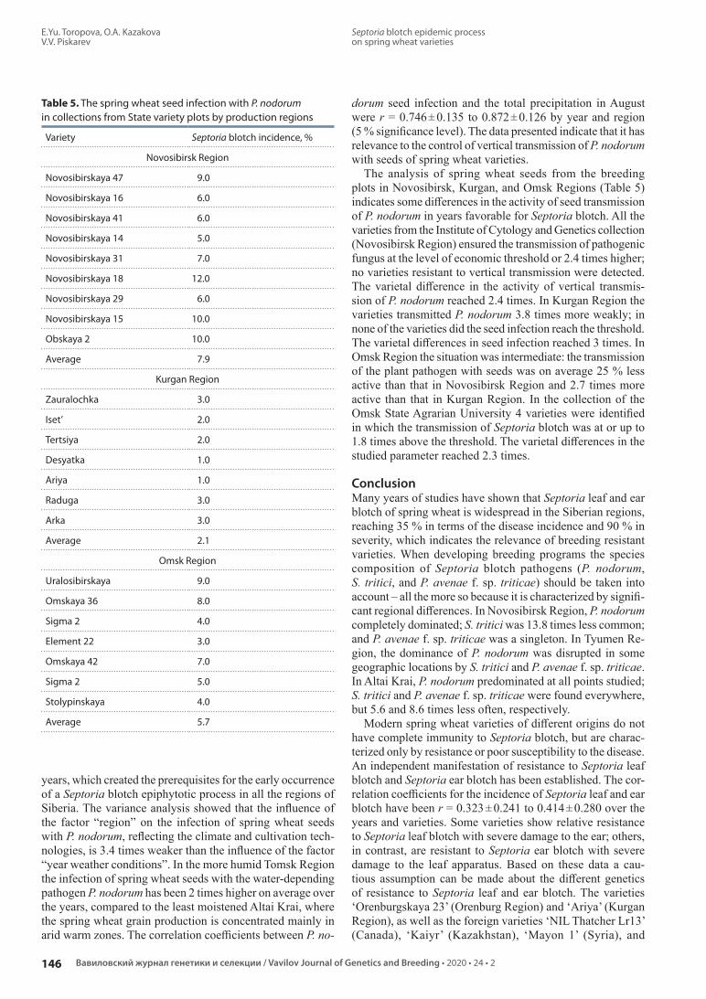
E.Yu. Toropova, O.A. Kazakova V.V. Piskarev
146 Вавиловский журнал генетики и селекции / Vavilov Journal of Genetics and Breeding • 2020 • 24 • 2
Septoria blotch epidemic process on spring wheat varieties
Table 5. The spring wheat seed infection with P. nodorum in collections from State variety plots by production regions
Variety Septoria blotch incidence, %
Novosibirsk Region
Novosibirskaya 47 9.0
Novosibirskaya 16 6.0
Novosibirskaya 41 6.0
Novosibirskaya 14 5.0
Novosibirskaya 31 7.0
Novosibirskaya 18 12.0
Novosibirskaya 29 6.0
Novosibirskaya 15 10.0
Obskaya 2 10.0
Average 7.9
Kurgan Region
Zauralochka 3.0
Iset’ 2.0
Tertsiya 2.0
Desyatka 1.0
Ariya 1.0
Raduga 3.0
Arka 3.0
Average 2.1
Omsk Region
Uralosibirskaya 9.0
Omskaya 36 8.0
Sigma 2 4.0
Element 22 3.0
Omskaya 42 7.0
Sigma 2 5.0
Stolypinskaya 4.0
Average 5.7
years, which created the prerequisites for the early occurrence of a Septoria blotch epiphytotic process in all the regions of Siberia. The variance analysis showed that the influence of the factor “region” on the infection of spring wheat seeds with P. nodorum, reflecting the climate and cultivation technologies, is 3.4 times weaker than the influence of the factor “year weather conditions”. In the more humid Tomsk Region the infection of spring wheat seeds with the waterdepending pathogen P. nodorum has been 2 times higher on average over the years, compared to the least moistened Altai Krai, where the spring wheat grain production is concentrated mainly in arid warm zones. The correlation coefficients between P. no
dorum seed infection and the total precipitation in August were r = 0.746 ± 0.135 to 0.872 ± 0.126 by year and region (5 % significance level). The data presented indicate that it has relevance to the control of vertical transmission of P. nodo rum with seeds of spring wheat varieties.
The analysis of spring wheat seeds from the breeding plots in Novosibirsk, Kurgan, and Omsk Regions (Table 5) indicates some differences in the activity of seed transmission of P. nodorum in years favorable for Septoria blotch. All the varieties from the Institute of Cytology and Genetics collection (Novosibirsk Region) ensured the transmission of pathogenic fungus at the level of economic threshold or 2.4 times higher; no varieties resistant to vertical transmission were detected. The varietal difference in the activity of vertical transmission of P. nodorum reached 2.4 times. In Kurgan Region the varieties transmitted P. nodorum 3.8 times more weakly; in none of the varieties did the seed infection reach the threshold. The varietal differences in seed infection reached 3 times. In Omsk Region the situation was intermediate: the transmission of the plant pathogen with seeds was on average 25 % less active than that in Novosibirsk Region and 2.7 times more active than that in Kurgan Region. In the collection of the Omsk State Agrarian University 4 varieties were identified in which the transmission of Septoria blotch was at or up to 1.8 times above the threshold. The varietal differences in the studied parameter reached 2.3 times.
ConclusionMany years of studies have shown that Septoria leaf and ear blotch of spring wheat is widespread in the Siberian regions, reaching 35 % in terms of the disease incidence and 90 % in severity, which indicates the relevance of breeding resistant varieties. When developing breeding programs the species composition of Septoria blotch pathogens (P. nodorum, S. tritici, and P. avenae f. sp. triticae) should be taken into account – all the more so because it is characterized by significant regional differences. In Novosibirsk Region, P. nodorum completely dominated; S. tritici was 13.8 times less common; and P. avenae f. sp. triticae was a singleton. In Tyumen Region, the dominance of P. nodorum was disrupted in some geographic locations by S. tritici and P. avenae f. sp. triticae. In Altai Krai, P. nodorum predominated at all points studied; S. tritici and P. avenae f. sp. triticae were found everywhere, but 5.6 and 8.6 times less often, respectively.
Modern spring wheat varieties of different origins do not have complete immunity to Septoria blotch, but are characterized only by resistance or poor susceptibility to the disease. An independent manifestation of resistance to Septoria leaf blotch and Septoria ear blotch has been established. The correlation coefficients for the incidence of Septoria leaf and ear blotch have been r = 0.323 ± 0.241 to 0.414 ± 0.280 over the years and varieties. Some varieties show relative resistance to Septoria leaf blotch with severe damage to the ear; others, in contrast, are resistant to Septoria ear blotch with severe damage to the leaf apparatus. Based on these data a cautious assumption can be made about the different genetics of resistance to Septoria leaf and ear blotch. The varieties ‘Orenburgskaya 23’ (Orenburg Region) and ‘Ariya’ (Kurgan Region), as well as the foreign varieties ‘NIL Thatcher Lr13’ (Canada), ‘Kaiyr’ (Kazakhstan), ‘Mayon 1’ (Syria), and

Эпифитотический процесс септориоза на сортах яровой пшеницы
Е.Ю. Торопова, О.А. Казакова В.В. Пискарев
202024 • 2
147СЕЛЕКЦИЯ РАСТЕНИЙ НА ИММУНИТЕТ / PLANT BREEDING FOR IMMUNITY
‘KWS Ak vi lon’ (Germany), show complex decreased susceptibility to the disease. They are weakly affected by Septoria leaf and ear blotch. The domestic varieties ‘Tyumenochka’ (Tyumen Region) and ‘Zauralochka’ (Kurgan Region) have a completely healthy ear at the beginning of the filling phase with weak damage to the flag leaf and can be considered the most promising sources of resistance.
The activity of vertical transmission of P. nodorum with seeds should also be monitored in the selection process, since seed transmission of Septoria blotch increases the infectious load on the plants significantly. Seed infection with the pathogen in the Siberian regions reached 7 thresholds and was largely determined by the weather conditions in August. The study of the collection of spring wheat varieties from three Siberian regions has revealed the following trend: transmission of P. nodorum with the seeds of varieties was the most active (7.6 %) in Novosibirsk Region and somewhat weaker in Omsk Region (5.7 %). The most favorable phytosanitary situation was in Kurgan Region, where varieties transmitted P. nodorum in a weak degree (2.1 %), below the threshold.
ReferencesAdolf В., Schofl U., Verreet J.A. Effect of infection with Septoria tritici
of different growth stages of wheat (GS 25 to GS 59) an dry matter production, nitrogen uptake and yield. Meded. Jc. Landbauw wetensch. 1993;53(3):11671174.
Broennimann A. Contributions to genetics of tolerance towards Septoria nodorum Berk. in wheat (Triticum aestivum L.). J. Plant Breed. 1975;75:138160.
Chulkina V.A. Biological Fundamentals of Plant Epidemiology. Moscow, 1991. (in Russian)
Chulkina V.A., Toropova E.Yu., Stetsov G.Ya., Kirichenko A.A., Marmuleva E.Yu., Grishin V.M., Kazakova O.A., Selyuk M.P. Phytosanitary Diagnosis of Agroecosystems. Toropova E.Yu. (Ed.). Barnaul, 2017. (in Russian)
Cooke B.M., Jones D.G. The epidemiology of S. tritici and S. nodorum. II. Comparative studies of head infection by S. tritici and S. nodorum on spring wheat. Trans. Br. Mycol. Soc. 1970;56:121125.
Du C.G., Nelson L.R., McDaniel M.E. Diallel analysis of gene effects conditioning resistance to Stagonospora nodorum (Berk.) in wheat. Crop Sci. 1999;39(3):686690.
Eyal Z. The Septoria tritici and Stagonospora nodorum blotch diseases of wheat. Eur. J. Plant Pathol. 1999;105(7):629-641.
Gultyaeva E.I., Shaidayuk E.L., Levitin M.M., Maslova I.V., Kolesnikova O.A., Vusatyuk M.P. Phytosanitary state of grain crops in the NorthWest of Russia in 2018. Zashchita i Karantin Rasteniy = Plant Protection and Quarantine. 2019;5:3031. (in Russian)
Jenkins P.D., Jones D.G. The effect of dual inoculation of wheat cultivars with S. tritici and S. nodorum. Phytopathologische Zeitschrift. 1981;101(3):210221.
Kolomiets T.M., Pankratova L.F., Pakholkova E.V. Wheat (Triticum L.) cultivars from GRIN collection (USA) selected for durable resistance to Septoria tritici and Stagonospora nodorum Blotch. Selskokhozyaystvennaya Biologiya = Agricultural Biology. 2017;52(3): 561569. DOI 10.15389/agrobiology.2017.3.561eng.
Kolomiets T.M., Shamanin V.P., Pakholkova E.V., Pankratova L.F., Salnikova N.N., Shepelev S.S., Pototskaya I.V., Abugalieva A., Morgunov A.I. Screening of hexaploid synthetic wheat lines and spring bread wheat variety for resistance to Septoria blotch. Vestnik Omsko go GAU = Bulletin of the Omsk State Agrarian University. 2018;3(31):1326. (in Russian)
Levitin M.M. Microorganisms and global climate change. Selskokhozyaystvennaya Biologiya = Agricultural Biology. 2015;50(5): 641647. DOI 10.15389/agrobiology.2015.5.641eng.
Mullaney E.J., Martin J.M., Scharen A.L. Generation mean analysis to identify and partition the components of genetic resistance to Septoria nodorum in wheat. Euphytica. 1981;31:539545.
Nazarova L.N., Korneva L.G., Zhokhova T.P., Polyakova T.M., Sanin S.S. Epidemiological situation with Septoria disease on wheat in 2001–2009. Zashchita i Karantin Rasteniy = Plant Protection and Quarantine. 2010;10:1820. (in Russian)
Nolan S., Cooke B.M., Monahan F.J. Studies on the interaction between Septoria tritici and Stagonospora nodorum in wheat. Eur. J. Plant Pathol. 1999;105(9):917925.
Pakholkova E.V. The rate of leafstem infections development on cereal crops. Zashchita i Karantin Rasteniy = Plant Protection and Quarantine. 2015;3:3940. (in Russian)
Parker S.R., Welham S., Paveley N.D., Foulkes J., Scott R.K. Tolerance of Septoria leaf blotch in winter wheat. Plant Pathol. 2004;53(1): 110.
Pyzhikova G.V., Sanina A.A., Kurakhtanova T.I. Septoria Blotch on Cereal Crops. Moscow, 1988. (in Russian)
Robert C., Bancal M.O., Nicolas P., Lannou Ch., Ney B. Analysis and modelling of effects of leaf rust and Septoria tritici blotch on wheat growth. J. Exp. Bot. 2004;55(399):1079-1094.
Sanin S.S., Korneva L.G., Polyakov T.M. Forecasting the risk of epiphytotics developing of Septoria leaf and ear on wheat. Zashchita i Karantin Rasteniy = Plant Protection and Quarantine. 2015;3: 3336. (in Russian)
Sanin S.S., Pakholkova E.V., Karlova L.V., Rulyova O.M., Franchuk L.I. Influence of weather factors on S. tritici and S. nodorum representation in a winter wheat Septoria blotch agent population. In: Epidemics of Plant Diseases: Monitoring, Prognosis, Control: Proc. of the Int. conf. Bolshie Vyazemy, 2017;1:8184. (in Rus sian)
Sanin S.S., Pakholkova E.V., Sanina A.A., Karlova L.V., Kor neeva L.G. Epidemiology of wheat leaf blotch: formation of infectious potentials of pathogens. Zashchita i Karantin Rasteniy = Plant Protection and Quarantine. 2018;5:2529. (in Russian)
Scharen A.L., Krupinsky J.M. Cutural and inoculation studies of Septoria nodorum, cause of glume blotch of wheat. Phytopathology. 1970;60(10):14801485.
Simón M.R., Cordo C.A., Perelló A.E., Struik P.C. Influence of nitrogen supply on the susceptibility of wheat to Septoria tritici. J. Phytopathol. 2003;151(5):283289.
Toropova E.Yu. Ecological Fundamentals of Plant Protection Against Diseases in Siberia. Novosibirsk, 2005. (in Russian)
Toropova E.Yu., Kazakova O.A., Piskarev V.V., Porsev I.N., Khristov Yu.A. The role of varieties and fungicides in the control of spring wheat septoriosis. Agrokhimiya = Agrochemistry. 2019;5:6675. DOI 10.1134/S0002188119050107. (in Russian)
Toropova E.Yu., Kazakova O.A., Selyuk M.P. Monitoring of Septoria blight on spring wheat in the foreststeppe of Western Siberia. Dostizheniya Nauki i Tekhniki APK = Achievements of Science and Technology of AIC. 2016;30(12):3335. (in Russian)
Toropova E.Yu., Kazakova O.A., Selyuk M.P., Insebaeva M.K., Kirichenko A.A., Filipchuk O.D., Kvitko A.V. Contamination of wheat seeds with Parastagonospora nodorum Berk. Dostizheniya Nauki i Tekhniki APK = Achievements of Science and Technology of AIC. 2018;32(12):1519. DOI 10.24411/02352451201811204. (in Russian)
Toropova E.Yu., Stetsov G.Ya., Chulkina V.A. Epiphytotiological Grounds of Plant Protection Systems. Chulkina V.A. (Ed.). Novosibirsk, 2002. (in Russian)

E.Yu. Toropova, O.A. Kazakova V.V. Piskarev
148 Вавиловский журнал генетики и селекции / Vavilov Journal of Genetics and Breeding • 2020 • 24 • 2
Septoria blotch epidemic process on spring wheat varieties
ORCID IDV.V. Piskarev orcid.org/0000-0001-9225-5227
Acknowledgements. This work was supported by state budget projects Nos. 0598-2015-0001 (All-Russian Research Institute of Phytopathology) and 0324-2019-0039 (Federal Research Center Institute of Cytology and Genetics SB RAS).Conflict of interest. The authors declare no conflict of interest.Received July 16, 2019. Revised January 18, 2020. Accepted January 21, 2020.
Tyryshkin L.G., Ershova M.A. Inheritance of resistance to Septoria blotch in common wheat sample MN81330. Russ. J. Genet. 2004; 40(4):454457.
Van Ginkel M., Rajaram S. Breeding for resistance to the Septoria/ Stagonospora blights of wheat. In: Van Ginkel M., McNab A., Krupinsky J.M. (Eds.). Septoria and Stagonospora Diseases of Cereals: a Compilation of Global Research: Proc. of the Fifth Int. Septorial Workshop. Mexico, 1999:117126.
Veselova S.V., Burkhanova G.F., Nuzhnaya T.V., Maksimov I.V. Effect of NADPH oxidases cascade on mechanisms regulating defense
common wheat reactions under the disease Stagonospora nodorum blotch. Izvestiya Ufimskogo Nauchnogo Tsentra Rossiy skoy Akademii Nauk = Proceedings of the RAS Ufa Scientific Center. 2018; 3(1):6674. (in Russian)
Veselova S.V., Burkhanova G.F., Nuzhnaya T.V., Rumyantsev S.D., Maksimov I.V. Effect of the host-specific toxin SnTOX3 from Stagonospora nodorum on ethylene signaling pathway regulation and redoxstate in common wheat. Vavilovskii Zhurnal Genetiki i Selektsii = Vavilov Journal of Genetics and Breeding. 2019;23(7): 856864.

Diversity and occurrence of methylotrophic yeasts used in genetic engineeringA.S. Rozanov1 , E.G. Pershina1, N.V. Bogacheva1, V. Shlyakhtun1, A.A. Sychev2, S.E. Peltek1
1 Institute of Cytology and Genetics of Siberian Branch of the Russian Academy of Sciences, Novosibirsk, Russia2 Innovation Centre Biruch-NT, Belgorod region, Russia
e-mail: [email protected]
Abstract. Methylotrophic yeasts have been used as the platform for expression of heterologous proteins since the 1980’s. They are highly productive and allow producing eukaryotic proteins with an acceptable glycosylation level. The first Pichia pastoris-based system for expression of recombinant protein was developed on the basis of the tree-exudate-derived strain obtained in the US southwest. Being distributed free of charge for scientific purposes, this sys-tem has become popular around the world. As methylotrophic yeasts were classified in accordance with biomolecular markers, strains used for production of recombinant protein were reclassified as Komagataella phaffii. Although patent legislation suggests free access to these yeasts, they have been distributed on a contract basis. Whereas their status for commercial use is undetermined, the search for alternative stains for expression of recombinant protein continues. Strains of other species of methylotrophic yeasts have been adapted, among which the genus Ogataea representa-tives prevail. Despite the phylogenetic gap between the genus Ogataea and the genus Komagataella representatives, it turned out possible to use classic vectors and promoters for expression of recombinant protein in all cases. There exist expression systems based on other strains of the genus Komagataella as well as the genus Candida. The potential of these microorganisms for genetic engineering is far from exhausted. Both improvement of existing expression sys-tems and development of new ones on the basis of strains obtained from nature are advantageous. Historically, strains obtained on the southwest of the USA were used as expression systems up to 2009. Currently, expression systems based on strains obtained in Thailand are gaining popularity. Since this group of microorganisms is widely represented around the world both in nature and in urban environments, it may reasonably be expected that new expression sys-tems for recombinant proteins based on strains obtained in other regions of the globe will appear.Key words: methylotrophic yeasts; Pichia pastoris; Ogataea; Komagataella; recombinant enzymes.
For citation: Rozanov A.S., Pershina E.G., Bogacheva N.V., Shlyakhtun V., Sychev A.A., Peltek S.E. Diversity and occurrence of methylotrophic yeasts used in genetic engineering. Vavilovskii Zhurnal Genetiki i Selektsii = Vavilov Journal of Genetics and Breeding. 2020;24(2):149-157. DOI 10.18699/VJ20.602
Разнообразие и распространение метилотрофных дрожжей, используемых в генной инженерииА.С. Розанов1 , Е.Г. Першина1, Н.В. Богачева1, В. Шляхтун1, А.А. Сычев2, С.Е. Пельтек1
1 Федеральный исследовательский центр Институт цитологии и генетики Сибирского отделения Российской академии наук, Новосибирск, Россия
2 ООО «Инновационный центр «Бирюч-НТ», Белгородская область, Россия e-mail: [email protected]
Аннотация. Метилотрофные дрожжи используются в качестве платформы для экспрессии гетерологичных бел-ков с 1980-х гг. Они имеют высокий уровень продукции и позволяют получить белки эукариот с приемлемым уровнем гликозилирования. Первая система для экспрессии рекомбинантного белка на основе Pichia pastoris была разработана на основе штамма, выделенного из сокотечения деревьев на юго-западе США. Система рас-пространялась бесплатно в научных целях и применяется во всем мире. В ходе классификации метилотрофных дрожжей по молекулярно-биологическим маркерам штаммы, используемые для получения рекомбинантного белка, были реклассифицированы как Komagataella phaffii. Они находятся в свободном доступе согласно па-тентному законодательству, однако распространялись на договорной основе. Это делает неопределенным их статус для коммерческого использования и, соответственно, стимулирует поиск альтернативных штаммов для экспрессии рекомбинантного белка. Были адаптированы штаммы других видов метилотрофных дрожжей, среди которых преобладают представители рода Ogataea. Несмотря на филогенетическую удаленность представите-лей рода Ogataea и Komagataella, во всех случаях оказалось возможным использовать классические векторы и промоторы для экспрессии рекомбинантного белка. Существуют системы экспрессии на основе других штаммов рода Komagataella, а также рода Candida. Потенциал этих микроорганизмов для генной инженерии далеко не ис-черпан. Перспективно как усовершенствование имеющихся систем экспрессии, так и создание новых на основе штаммов, выделенных из природных источников. Исторически до 2009 г. в качестве систем экспрессии исполь-
© Rozanov A.S., Pershina E.G., Bogacheva N.V., Shlyakhtun V., Sychev A.A., Peltek S.E., 2020
This work is licensed under a Creative Commons Attribution 4.0 License
ГЕНЕТИКА МИКРООРГАНИЗМОВОбзор / Review
Вавиловский журнал генетики и селекции. 2020;24(2):149-157DOI 10.18699/VJ20.602

A.S. Rozanov, E.G. Pershina, N.V. Bogacheva V. Shlyakhtun, A.A. Sychev, S.E. Peltek
150 Вавиловский журнал генетики и селекции / Vavilov Journal of Genetics and Breeding • 2020 • 24 • 2
Diversity and occurrence of methylotrophic yeasts used in genetic engineering
зовались штаммы, выделенные на юго-западе США. В настоящее время начали развиваться системы экспрессии на основе штаммов, полученных в Таиланде. Поскольку эта группа микроорганизмов широко представлена по всему миру как в природной, так и в городской среде, можно ожидать появления систем экспрессии рекомби-нантных белков, созданных на основе штаммов, выделенных и в других регионах планеты.Ключевые слова: метилотрофные дрожжи; Pichia pastoris; Ogataea; Komagataella; рекомбинантные ферменты.
IntroductionMethylotrophs are a group of microorganisms that can use single-carbon methane compounds, such as methanol, methyl-amine, etc., as the sole source of carbon and energy. The re quirement that all C–C bonds are formed enzymatically during cellular metabolism poses a challenge for the cell. Only some microorganisms are capable of doing so, such as Gram-negative proteobacteria and Gram-positive bacteria (Antony, 1986), as well as yeasts (Wegner, Harder, 1987), which em-ploy metabolic pathways for methanol oxidation to produce energy and form C–C bonds. Both in yeasts and bacteria, C–C bonds are formed through the formation of formaldehyde (Yurimoto et al., 2005), a toxic intermediate product that sub-sequently is either dissimilated into CO2 or assimilated into biomass.
Methylotrophic yeasts were discovered in the late 1960s, when methylotrophic bacteria had already been well known. The difference in times of their discovery was mainly caused by the challenges related to yeast isolation and significant bacterial contamination of samples (Trotsenko, Torgonskaya, 2011). The habitats of methylotrophic yeasts are those where the biomass is degraded to give rise to methoxy groups (soil, fallen trees, rotten fruit, etc.). Methanol is produced naturally during methane oxidation (e. g., by methane-oxidizing bac-teria in the plant rhizosphere) and pectin or lignin degrada-tion (MacDonald, Fall, 1993; Nakagawa et al., 2000). Most natural isolates of methylotrophic yeasts were detected in tree sap (exudates) or rotting wood (Kurtzman, Robnett, 1998; Kurtzman, 2005).
The interest in methylotrophic organisms for bioengineer-ing applications arose in the early 1970s, when methanol was an inexpensive raw material and was considered a virtually inexhaustible fossil feedstock. However, after the 1973 oil crisis its price never dropped back to the pre-crisis level, and Western countries chose to lower their dependence on hydrocarbons. Plant-based proteins (and soybean protein in particular) came to fore and replaces fodder protein derived from unicellular methylotrophs.
In the 1980s, methylotrophic yeasts became widely used again, although in new areas. Today, they are utilized as a plat-form for genetic engineering and commercial-scale production of recombinant proteins. Furthermore, methylotrophic yeasts are a convenient object for studying the features of eukaryotic cell organization. Methods for utilizing methylotrophic yeasts in applied areas (as markers of coastal pollution, for treatment of discharge water at sulfate-cellulose and alcohol-and-liquor manufacturing enterprises, for treatment of formaldehyde-contaminated air, etc.) are currently being elaborated (Kutty, Philip, 2008; Trotsenko, Torgonskaya, 2012; dos Reis et al., 2018).
Pichia pastoris (Komagataella phaffii) is a methylotrophic yeast species that is most frequently used for scientific re-search and commercial purposes. These yeasts can consume
both sugars and methanol to produce proteins with a high yield. Initially, back in the 1970s, Phillips Petroleum sug-gested using P. pastoris as a single-cell protein producer due to their ability to form high-density cultures both on glucose and methanol substrates (Mishra, Baranwal, 2009). The maximum cell density achieved during fermentation is higher than 100 g/L (dry basis) (Wegner, 1981). In the 1980s, the P. pastoris-based heterologous expression system was developed with the use of a strong and strictly-regulated alcohol oxidase 1 (AOX1) promoter (Cregg et al., 1985). In combination with the existing fermentation technologies for animal feed protein manufacturing, AOX1 promoter was en-suring an exceptionally high recombinant proteins expression level. An important benefit of using it in recombinant protein manufacturing is the high expression level in the presence of methanol and robust inhibition of the process in the absence of methanol, which makes it possible to regulate the produc-tion of target proteins, including the autotoxic ones (Kurtz-man, 2009). Production of biomass-derived hydroxynitrile lyase enzyme (the yield being 20 g of recombinant protein per liter of the culture) was one of the first large-scale com-mercial manufacturing processes established in the 1990s (Hasslacher et al., 1997).
Another advantage of P. pastoris is that it ensures the low glycosylation level. Thus, the S. cerevisae yeast species is no longer used in production of recombinant proteins, since it often hyperglycosylates proteins (up to their complete inactivation) (Darby et al., 2012).
The P. pastoris-based expression system has become widely used in basic research. Among methylotrophic yeasts, the P. pastoris species was the first to be developed. It turned out to be a convenient bioengineering system for production of eukaryotic proteins that are not properly expressed in bacteria. Phillips Petroleum made a forward-looking decision to make this expression system available to the scientific community for research purposes, which was a major driver for common application of this yeast species as a platform for recombinant protein expression. Histidine-auxotrophic strain GS115, reconstituted prototrophic strain X-33, aox1 knockout strains KM71 and KM71H, as well as protease-deficient strains SMD1168 and SMD1168H, and ade2 auxotrophic PichiaPink™ strain, are the most frequently utilized commercially available strains. However, their use for commercial purposes is restricted by material distribu-tion policy. Therefore, searching for alternative options that could be used instead of the licensed P. pastoris strains is quite relevant today.
Phylogenic analysisProgress in molecular biology methods as well as mounting evidence on gene sequences for various species made it pos-sible to investigate into phylogeny of methylotrophic yeasts, which was accompanied by a number of surprises.

Разнообразие и распространение метилотрофных дрожжей, используемых в генной инженерии
А.С. Розанов, Е.Г. Першина, Н.В. Богачева В. Шляхтун, А.А. Сычев, С.Е. Пельтек
202024 • 2
151ГЕНЕТИКА МИКРООРГАНИЗМОВ / MICROBIAL GENETICS
Ogataea polymorpha – EF550407*100 9880 78
78 87
74 81
90 95
0.0 0.04
99 95
85 75
99 99
62 50
100 100
Ogataea methanolica – AB018181*
Komagataella pastoris – AB018182*Komagataella phaffii – EF550394*
Komagataella pseudopastoris – EF550393Komagataella ulmi – KF964125
Komagataella populi – KF964124
Candida boidinii – NG_063501*
Ogataea thermomethanolica – FJ914913*
Saccharomyces cerevisiae – NG_063315*
Kluyveromyces marxianus – NG_062653
Priceomyces fermenticarens – NG_063398
Pichia guilliermondii – AB013587
Debaryomyces hansenii var. fabryi – AB013567
Schizosaccharomyces pombe – AY251644*
Pichia farinosa – AB013513
Candida xestobii – AB013517
Candida psychrophila – NG_063399
Yarrowia lipolytica – NG_013120*
Ogataea pini – EU011703
96 50
95 90
Phylogenetic tree built on the basis of sequences of ribosomal 18S gene of Saccharomycetaceae species with the help of maximum likelihood method.SH-Like aLTR and UfBoot support ratios are expressed in percentage on tree branch joints. Figures to the right of species names are identification numbers for relevant sequences in the NCBI data base.
First of all, gene sequence analysis revealed that yeasts known as Pichia are not monophyletic despite the pheno-typic similarity of the majority of species of this genus. First research works discovered that methanol-assimilating species P. pastoris, P. angusta and Hansenula polymorpha are remotely related with one another as well as with Pichia membranifaciens, the typical Pichia species. Yamada et al. (1994, 1995) suggested assigning P. pastoris to a new genus Komagataella and classifying P. angusta as Ogataea polymorpha into a freshly described genus Ogataea. New genera were suggested on the basis of analysis of divergence in par-tial large subunit (LSU) and small subunit (SSU) ribosomal RNA (rRNA) sequences. Since each analysis was limited to a relatively small number of species, it was unclear how close P. pastoris and P. angusta (H. polymorpha) were related to numerous unstudied species, and therefore suggestions were not accepted (Kurtzman, 1998). However, further analysis of D1/D2 regions in the LSU of the rRNA gene sequences for all now-known ascomycetes confirmed that the Komagataella, Ogataea and Pichia classification is correct (Kurtzman, Robnett, 1998), while phylogenetic divergence demonstrated as a result of single-gene sequence analysis was confirmed during the analysis of multi-gene sequences (Kurtzman et al., 2008). Therefore, Komagataella, Ogataea and Pichia are three separate genera. Thus, Komagataella pastoris is the most correct name from the viewpoint of taxonomy. However, due to historical reasons, P. pastoris remains most wide-spread today.
Secondly, it turned out that the name P. pastoris covers at least two yeast species, i. e. Komagataella phaffii and K. pas toris (Cregg et al., 1993; Kurtzman, 2009). The gene sequence analysis also revealed that these are not the only Komagataella representatives. K. pseudopastoris initially described as P. pseudopastoris (Dlauchy et al., 2003) was also classified as part of Komagataella on the basis of LSU and SSU rRNA sequence analysis. Similarly, K. phaffii was classified as Komagataella on the basis of the analysis of the D1/D2 fragment in the LSU rRNA gene (Kurtzman et al., 2008).
Evolution of parallel sequencing methods helped to build up the database on genome sequences of a great number of microorganisms (including methylotrophic yeasts), which allows for a more detailed phylogenetic analysis. 18S rRNA gene sequence is the most wide-spread and conservative marker for analysis of phylogenetic relations among various fungi representatives. Relevant genes were extracted from genomes of methylotrophic yeasts part of the NCBI database and used for the phylogenetic tree design (see the Figure).
Nucleotide sequences were evened with the help of MAFFT algorithm in MAFFT v7.312 program (Katoh, Standley, 2013) with –localpair and –maxiterate 1,000 parameters. Phylogenetic tree was designed on the basis of the maximum likelihood method with the help of IQ-TREE program (Tri-finopoulos et al., 2016). –Auto and +R (FreeRate heteroge-neity) parameters were used to determine the best patterns of nucleotide replacement models, minimum value of the Akaike criterion (AICc) being the key parameter. Two ratios, i. e. SH-like aLRT (1,000 replications) and ultrafast bootstrap (UfBoot, 1,000 replications), were used for statistical support of maximum likelihood in the IQ-TREE program.
As one can see, methylotrophic yeast used for production of recombinant proteins split into three groups: Pichia, Ogataea and Komagataella (see the Figure). Also, one can derive at a conclusion that Ogataea is phylogenetically more heteroge-neous in comparison to Pichia and Komagataella.
Methylotrophic yeasts used for production of recombinant proteins as alternative strains, and their occurrence in natureP. pastoris (K. рhaffii and K. kurtzmanii ). The P. рastoris yeasts group, the majority of representatives of which have been reclassified as Komagataella representatives, is most well-studied among methylotrophic yeasts. Its typical habitat is tree sap (exudate) in regions from moderate to tropical. Initially, P. pastoris was isolated from chestnut exudates in France. Later, it was discovered to occur widely in Hungary and USA (Spencer et al., 1996; Negruta et al., 2010; Kurtz-man, 2011a). Besides, P. pastoris yeasts were discovered in exudates of oil palm in Nigeria (Faparusi, 1974), sap of white algarrobo in Argentina (Spencer et al., 1995), and sap of red oak in Canada (Bowles, Lachance, 2007). The zoning of their occurrence is very curious: this is a predominant species of methylotrophic yeasts in the woods of the Pacific coast of the American Northwest; they are present (although not predominant) in Europe, Africa and South America, and are completely missing in Japan (Lachance et al., 1982).
Although the P. pastoris-based expression system patented by Philips Petroleum is wide-spread, alternative strains of

A.S. Rozanov, E.G. Pershina, N.V. Bogacheva V. Shlyakhtun, A.A. Sychev, S.E. Peltek
152 Вавиловский журнал генетики и селекции / Vavilov Journal of Genetics and Breeding • 2020 • 24 • 2
Diversity and occurrence of methylotrophic yeasts used in genetic engineering
methylotrophic yeasts are being searched for. Those are re-quired for invention of new unlicensed and thus applicable for commercial use platforms for recombinant proteins pro-duction.
P. pastoris (K. phaffii) CBS7435 strain is the closest to those previously suggested by Philips Petroleum. Strains derived from it are unlicensed (Ahmad et al., 2014). At the same time, this strain is a predecessor of patented strains most widely used for production of recombinant proteins these days. On the one hand, this paves the way for use of CBS7435 compounds thanks to the vast knowledge base built up with their help, and on the other hand, not all genome modifica-tions for this strain can be patented taking into consideration their description in academic literature and in patents.
Strain K. kurtzmanii Y-727/KPB 2878/Starmer 75-208.2/CBS 12817/NRRL Y-63667 was patented in the Russian Fe deration. It was isolated by Prof. Starmer from fir sap in Arizona mountains, USA (Naumov et al., 2013), and is one of the closest relatives of the P. pastoris (K. phaffii) CBS7435 strain.
P. guilliermondii (Meyerozyma guilliermondii ). P. guilliermondii sporogenous species can be isolated from a va-riety of sources, i. e. plants, lake water, cow rumen, or oil-contaminated soil. Besides, these yeasts were discovered in elm-dwelling insects, in uncontaminated oil and water (Ne gruta et al., 2010a), in shrimp and other invertebrates, and in low-salinity sea water (Kutty, Philip, 2008). Before, this yeast species was used in gene engineering as a source of genes that were among other things expressed in P. pastoris (Handumrongkul et al., 1998; Zhang et al., 2009), rather than as an expression system.
Since the majority of methylotrophic yeasts have similar methanol-inducible promoter in methanol utilization paths (Hartner, Glieder, 2006), the research group from Malaysia tested the assumption that expression constructs developed on the basis of K. phaffii could be used for expression of re-combinant proteins in other methylotrophic yeasts (Oslan et al., 2015). They isolated the strain called Pichia sp. strain SO from a rotten orange; its SSU sequence demonstrated its 100 % similarity to P. guilliermondii. Then, authors disco-vered that zeocin can be used as a marker for strain SO (Oslan et al., 2012), and conducted work on cloning the recombinant lipase expression construct. The work continued, and in 2017 an article on optimization of expression of T1 lipase isolated earlier from Geobacillus zalihae with use of P. guilliermondii was published. As the result of this work, T1 lipase yielded a 3-fold increase over medium (Abu et al., 2017).
P. (O.) methanolica was suggested by Invitrogen (USA) as a platform for production of recombinant proteins a little later than the same company suggested P. pastoris. Initially, it was isolated from soil sample in Japan in 1974; strains of this species were also isolated in the USA (Sibirny, 1996; Kurtzman, 2011b). In 2008, yeasts of this group (heteroge-neous as P. pastoris) were also isolated on the territory of Russia from willow galls created by slug (Glushakova et al., 2010). P. (O). methanolica didn’t become popular as a platform for production of recombinant proteins although there are single messages about its use, e. g., for expression of human glutamic acid decarboxylase (Raymond et al., 1998). Probably, the popularity of P. (O). methanolica as a plat form
for recombinant proteins is low because Invitrogen has a different, P. pastoris-based platform (K. phaffii).
Hansenula (O.) polymorpha is a thermotolerant methy-lotrophic yeast able to grow at temperatures below 50 °С wide-spread in nature. Besides such media as rotting fruit and other plants typical for methylotrophic yeasts, one of typical habitats for H. polymorpha is organism of insects, including Drosophila melanogaster typically used in research (Spen-cer J., Spencer D., 1997). Since H. polymorpha demonstrates good growth at high temperatures, it could possibly be found around hot springs and in tropical areas.
In science, these yeasts have been used as a model organ-ism for studying peroxisome biogenesis and degradation mechanisms, methanol metabolism control, assimilation of nitrates and reaction to stress (van der Klei et al., 2006). H. polymorpha turned out rather effective for production of recombinant proteins as well (Gellissen, 2005). Recombinant antigen of the hepatitis B virus (HBsAg), that was success-fully commercialized under HepaVax-Gene and AgB trade-marks (Seo et al., 2008), is the most significant therapeutic protein produced with the help of H. polymorpha. Producers of recombinant proteins with high potential for pharmaceuti-cal purposes were developed on the basis of H. polymorpha: hirudin from leech Hirudinaria manillensis (Weydemann et al., 1995) and some human proteins including α1-antitrypsin (Kang et al., 1998), IFNα-2a (Degelmann et al., 2002), serum albumin (Kang et al., 2001), epidermal growth factor (Heo et al., 2002) and parathyroid hormone (Sohn et al., 2012). Besides medical proteins, there were developed producers of food and commercial enzymes: hexose oxidases (Cook, Thygesen, 2003), phytases (Mayer et al., 1999), levansucrase from Zymomonas mobilis (Park et al., 2004), and glucose oxydases from Aspergillus niger (Kim et al., 2004).
P. (O.) thermomethanolica. Besides strains utilized by Invitrogen, there is O. thermomethanolica BCC16875 strain that can be considered one of best-studied methylotrophic yeasts alternatives for production of recombinant proteins. Knowledge of occurrence of O. thermomethanolica is very scarce as this yeast species was discovered only re-cently, in 2005, in soil samples in Thailand (Limtong et al., 2013).
For the first time, information about strain BCC16875 was published by a research group from Thailand in 2012. The research focuses on testing the possibility of using bio-molecular tools for accumulation of protein in this strain. Classical methanol-inducible alcohol oxidase (AOX1) pro-moters and constitutive glyceraldehyde 3-phosphate de-hyd ro genase (GAP) promoters utilized for working with P. pas toris were shown to drive efficient gene expression in this new strain. Recombinant phytase and xylanase were expressed from both promoters as secreted proteins, with the former demonstrating different patterns of N-glycosylation dependent on the promoter and culture medium used. The major glycoprotein oligosaccharide species produced from O. thermomethanolica BCC16875 is Man8-12GlcNAc2 that is similar to that of other methylotrophs. Moreover, manno-sylphosphate and α-1,6- and α-1,2-linked mannose modifica-tions of heterologous secreted protein were also detected. The level of expression of recombinant protein turned out to be equal to the level of expression of commercial strains, which

Разнообразие и распространение метилотрофных дрожжей, используемых в генной инженерии
А.С. Розанов, Е.Г. Першина, Н.В. Богачева В. Шляхтун, А.А. Сычев, С.Е. Пельтек
202024 • 2
153ГЕНЕТИКА МИКРООРГАНИЗМОВ / MICROBIAL GENETICS
makes the suggested platform a good alternative to widely used Invitrogen’s strains (Tanapongpipat et al., 2012).
Studies of the suggested strain O. thermomethanolica BCC16875 continued during next following years. Pro mo-ters typical for this train were studied (Harnpicharnchai et al., 2014; Promdonkoy et al., 2014), as well as methods of high-density cultivation for expression of recombinant proteins (Charoenrat et al., 2016). Besides, work on optimization of strain’s metabolism for increase of target products’ output was started. Thus, the level of expression of auxiliary pro-teins of endoplastic reticulum was increased for this purpose (Roongsawang et al., 2016). Studies of the strain became especially active after 2016, and today there is a significant number of works on updating it to the present-day level as an expression system. In 2018, the CRISPR-Cas9 system was adapted for this strain (Phithakrotchanakoon et al., 2018b), a new sucrose-induction-based expression system was de-veloped (Puseenam et al., 2018; Boonchoo et al., 2019), and studies of metabolism at proteomic and transcriptomic levels continued (Phithakrotchanakoon et al., 2018a).
Candida boidinii is the first described species of methy-lotrophic yeasts. It is also apparently most wide-spread in natural habitat (Ogata et al., 1969). Mainly it’s various plant substrates (tree sap, rotten fruit, some flowers). These yeasts are also abundantly present in naturally fermented olives (Coton et al., 2006). Methylotrophic yeasts are also found in cacti. Their spoiled parts happen to host both C. boidinii, and O. polymorpha, although these species do not prevail among yeasts discovered in these yeast samples. In addition to regular occurrences of methylotrophic yeasts, С. boidinii is also an important marker of seashore contamination. Their lines are predominant in many water and sand samples in Brazil (Kutty, Philip, 2008).
According to the phylogenetic tree of 18S rRNA gene sequences presented in this review, C. boidinii could be clas-sified as Ogataea (see the Figure). Up to 2009, C. boidinii was developed as a platform for production of recombinant proteins alternative to P. pastoris by a group of Japanese scientists (Yurimoto, Sakai, 2009). C. boidinii-based recom-binant protein expression system has some characteristics that can be useful in comparison to other methylotrophic yeasts. Level of expression in C. boidinii varies depending on the source of carbon: AOD1 promoter demonstrates high level of expression in methanol-grown or methanol-glycerol-grown cells, medium level of expression in glycerol-grown cells and zero expression in case glucose- or ethanol-grown cells are used as source of carbon (Sakai et al., 1995; Yuri-moto et al., 2000). The level of expression is significantly higher for C. boidinii than for O. polymorpha. Besides, high level of expression can be ensured in C. boidinii in case of methanol + glycerol medium, which allows shortening the time for high-density cell cultivation. In case of P. pastoris, glycerol suppresses expression of methanol-induced genes, and therefore control over complete eating of glycerol in the culture prior to methanol induction is required. A strain with knocked-out vacuolar proteinase A (PEP4) and proteinase B (PRB1) is available for both P. pastoris and C. boidinii (Komeda et al., 2002). During studies with use of C. boidinii genes expression system, there were developed strains for production of toxic proteins, i. e. membrane-bound peroxi-
some allowing to cumulate toxic proteins (Nishikawa et al., 2000; Yurimoto et al., 2001), as well as effective secretion system for production of active transglutaminase (Yurimoto et al., 2004).
Obtainment of methylotrophic yeasts from natural sourcesYeasts are isolated from water, seawater, atmosphere and ground habitats. They dwell in rotting vegetables and fruit, in moulds, exudates of trees and their barks, in xylophage insects, pig’s intestine, milk of cows suffering from mastitis, in forest, garden and swampy soils, especially drenched with sewage waters, in sea weed and so on (Negruta et al., 2010; Trotsen-ko, Torgonskaya, 2011). These environmental preferences are most likely due to discharge of methoxyl groups during degradation of lignin and pectin (Nakagawa et al., 2005). In addition to fruit juices and soil samples, methylotrophic yeasts are also found in food (Mu et al., 2012; Kozhakhmetov et al., 2016; Syromyatnikov et al., 2018).
Many types of yeast are wide-spread while some are limited only to a certain narrow habitat. They rarely occur in nature in absence of micellar fungi and bacteria. Therefore, to obtain them one must use selective methods allowing yeasts to have advantages in growth speed. When media for selective isola-tion of yeasts are developed, low рН is usually used as in the majority of cases yeasts prevail over bacteria in such condi-tions. Media could also include antibiotics for suppression of bacteria and fungistatic agents for suppression of moulds (Kurtzman et al., 2011).
When yeasts are present in great amounts, they can be isolated by direct application of the material or its suspen-sion on sour agarized medium that can also be enriched with antibiotics or have other selecting properties. Agar hydrolyzes in low-рН medium during autoclaving. Therefore agar and medium are sterilized separately, cooled to around 45 °С, mixed and distributed among Petrie dishes. The majority of yeast species can be isolated at 3.7 рН, but some species such as Schizosaccharomyces species require higher рН ranging from 4.5 to 5.0. If yeasts are present in the sample in low quantities, their population could be increased by preliminary incubation of the sample in liquid medium at рН up to 3.8 (Kurtzman et al., 2011).
To isolate specific physiological groups of yeasts, it’s neces-sary to find additional selecting parameters. Methylotrophic yeasts can be selected with use of methanol as the sole source of carbon and energy in the medium. Thus, when methylo-trophic yeasts were isolated from grape leaves in Thailand, YNB medium with extra 0.5 % of methanol was used to get the enrichment culture. Cultivation continued for 4–5 days at 27 °С, following which enrichment cultures were spread on 0.5 % v/v methanol-YNB agar. As a result, 2 new spe-cies were isolated that classified as Ogataea (Limtong et al., 2013).
ConclusionMethylotrophic yeasts are wide-spread as a platform for production of recombinant protein. Initially, they became of interest to biotech companies as single-cell protein producers. However, due to the 1973 oil crisis, methanol grew in price and isolation of feed protein from it became irrelevant. At

A.S. Rozanov, E.G. Pershina, N.V. Bogacheva V. Shlyakhtun, A.A. Sychev, S.E. Peltek
154 Вавиловский журнал генетики и селекции / Vavilov Journal of Genetics and Breeding • 2020 • 24 • 2
Diversity and occurrence of methylotrophic yeasts used in genetic engineering
the same time, due to discovery of proteins key to molecular biology such as thermotolerant polymerases, lygases and restrictases, various microorganisms modification methods started being developed, including those for production of recombinant proteins.
Production of proteins with use of microorganisms for vari-ous purposes, in the first place, for food and animal feed as well as technological purposes, has been actively developing since 1940s. In the first place, this was connected with the use of natural producers, upgraded in many cases with the help of undirected mutagenesis methods. As molecular biology methods developed, it became possible to develop protein producers untypical for a specific organism (heterologous or recombinant proteins), including with use of yeasts.
Following this trend, Phillips Petroleum developed their own P. pastoris-based expression system for production of recombinant proteins (later renamed into Komagataella phaffii). Thanks to its outstanding properties as well as Phillips Petroleum’s decision to allow its wide-spread utilization for scientific purposes around the world, it got widely popular as a recombinant protein production platform.
Understanding of key properties that made P. pastoris-based expression systems popular is important for further development of protein-expressing platforms. Number one is high protein production level. It’s lower than that of bacteria and micellar fungi but higher than that of other systems, i. e. cells of mammals, plants and insects. Number two: yeasts are eukaryotes, and they have all cell compartments necessary for synthesis and assembly of eukaryotic proteins, which al-lows them to synthesize proteins that cannot be synthesized with use of bacterial expression systems. Number three is extracellular protein’s ability to synthesize, which brings the costs of the production process down. Number four is the low level of glycosylation of proteins in comparison to many other types of yeast.
Other methylotrophic yeasts, except K. phaffii, have also been tried out as a platform for expression of recombinant protein. In the first place, it should be noted that P. pastoris strain CBS7435 initial for the Invitrogen’s system was sug-gested to be used as a patent-independent platform. In Rus-sia, Komagataella kurtzmanii strain of American origin was patented. Alternative Ogataea-based expression platforms were developed in the USA: Hansenula (Ogataea) polymorpha and Pichia (Ogataea) methanolica, the latter belongs to Invitrogen. In Japan, there was developed an alternative plat-form on the basis of Candida boidinii. This system was being developed until 2009, but since then this organism has not been mentioned as a platform used for production of recombinant protein. In Malaysia, strain P. guilliermondii (Meyerozyma guilliermondii) was suggested for production of protein.
One can notice that all yeast strains used for production of heterologous proteins up to 2009 have American origin. This sets a question, whether or not American isolates have unique properties allowing for production of protein in yeasts. More recent works witness that it’s not accurate. First strains were developed by American researchers working in one state. Actually, rapid development of bioscience, including discovery of new methods of molecular biology, took place in the US Southwest. It seems that this fact was the key rea-son for using strains of this region specifically. The Russian
patent is actually the copy of known systems, which requires utilization of the closest species that in the majority of cases have geographically similar habitats.
Overall, alternative methylotrophic-yeasts-based systems haven’t become popular. Reasons are multiple. Lack of interest on part of recombinant proteins market players and disability of new players to enter this market with a new system along with lack of acute need for it is the main reason. For example, Chinese manufacturers actively use strains obtained on the basis of Invitrogen’s strains. It shall also be noted that modern strains may be significantly genetically modified to ensure higher yields of recombinant proteins.
ReferencesAbu M.L., Nooh H.M., Oslan S.N., Salleh A.B. Optimization of physi-
cal conditions for the production of thermostable T1 lipase in Pichia guilliermondii strain SO using response surface methodology. BMC Biotechnol. 2017;17:78. DOI 10.1186/s12896-017-0397-7.
Ahmad M., Hirz M., Pichler H., Schwab H. Protein expression in Pi chia pastoris: recent achievements and perspectives for heterologous pro-tein production. Appl. Microbiol. Biotechnol. 2014;98:5301-5317. DOI 10.1007/s00253-014-5732-5.
Anthony C. Bacterial oxidation of methane and methanol. Adv. Micro bial Physiol. 1986;27:113-210. DOI 10.1016/S0065-2911(08) 60305-7.
Boonchoo K., Puseenam A., Kocharin K., Tanapongpipat S., Roong-sawang N. Sucrose-inducible heterologous expression of phytase in high cell density cultivation of the thermotolerant methylotrophic yeast Ogataea thermomethanolica. FEMS Microbiol. Lett. 2019; 366(5). DOI 10.1093/femsle/fnz052.
Bowles J.M., Lachance M.-A. Patterns of variation in the yeast florae of exudates in an oak community. Can. J. Bot. 2007;61(12):2984-2995. DOI 10.1139/b83-335.
Charoenrat T., Antimanon S., Kocharin K., Tanapongpipat S., Roong-sawang N. High cell density process for constitutive production of a recombinant phytase in thermotolerant methylotrophic yeast Ogataea thermomethanolica using table sugar as carbon source. Appl. Biochem. Biotechnol. 2016;180:1618-1634. DOI 10.1007/s12010-016-2191-8.
Cook M.W., Thygesen H.V. Safety evaluation of a hexose oxidase ex-pressed in Hansenula polymorpha. Food Chem. Toxicol. 2003;41: 523-529. DOI 10.1016/S0278-6915(02)00280-6.
Coton E., Coton M., Levert D., Casaregola S., Sohier D. Yeast ecology in French cider and black olive natural fermentations. Int. J. Food Microbiol. 2006;108:130-135. DOI 10.1016/j.ijfoodmicro.2005. 10.016.
Cregg J.M., Barringer K.J., Hessler A.Y., Madden K.R. Pichia pastoris as a host system for transformations. Mol. Cell. Biol. 1985;(12): 3376-3385. DOI 10.1128/mcb.5.12.3376.
Cregg J.M., Vedvick T.S., Raschke W.C. Recent advances in the ex-pression of foreign genes in Pichia pastoris. Biotechnology (NY). 1993;11:905-910. DOI 10.1038/nbt0893-905.
Darby R.A.J., Cartwright S.P., Dilworth M.V., Bill R.M. Which yeast species shall I choose? Saccharomyces cerevisiae versus Pichia pastoris (Review). In: Bill R.M. (Ed.). Recombinant Protein Production in Yeast: Methods and Protocols. Humana Press, 2012;11-23. DOI 10.1007/978-1-61779-770-5_2.
Degelmann A., Müller F., Sieber H., Jenzelewski V., Suckow M., Stras-ser A.W.M., Gellissen G. Strain and process development for the production of human cytokines in Hansenula polymorpha. FEMS Yeast Res. 2002;2:349-361. DOI 10.1016/S1567-1356(02)00096-X.
Dlauchy D., Tornai-Lehoczki J., Fülöp L., Péter G. Pichia (Komagataella) pseudopastoris sp. nov., a new yeast species from Hun-

Разнообразие и распространение метилотрофных дрожжей, используемых в генной инженерии
А.С. Розанов, Е.Г. Першина, Н.В. Богачева В. Шляхтун, А.А. Сычев, С.Е. Пельтек
202024 • 2
155ГЕНЕТИКА МИКРООРГАНИЗМОВ / MICROBIAL GENETICS
gary. Antonie van Leeuwenhoek. 2003;83:327-332. DOI 10.1023/ A:1023318829389.
dos Reis K.C., Arrizon J., Amaya-Delgado L., Gschaedler A., Schwan R.F., Silva C.F. Volatile compounds flavoring obtained from Brazilian and Mexican spirit wastes by yeasts. World J. Microbiol. Biotechnol. 2018;34(152). DOI 10.1007/s11274-018-2535-3.
Faparusi S.I. Microorganisms from oil palm tree (Elaeis guineensis) tap holes. J. Food Sci. 1974;39(4):755-757. DOI 10.1111/j.1365-2621. 1974.tb17972.x.
Gellissen G. (Ed.). Production of Recombinant Proteins: Novel Micro-bial and Eukaryotic Expression Systems. John Wiley & Sons, 2005.
Glushakova A.M., Maximova I.A., Kachalkin A.V., Yurkov A.M. Ogataea cecidiorum sp. nov., a methanol-assimilating yeast isolated from galls on willow leaves. Antonie van Leeuwenhoek. 2010;98(1): 93-101. DOI 10.1007/s10482-010-9433-5.
Handumrongkul C., Ma D.P., Silva J.L. Cloning and expression of Candida guilliermondii xylose reductase gene (xyl1) in Pichia pastoris. Appl. Microbiol. Biotechnol. 1998;49:399-404. DOI 10.1007/s002530051189.
Harnpicharnchai P., Promdonkoy P., Sae-Tang K., Roongsawang N., Tanapongpipat S. Use of the glyceraldehyde-3-phosphate dehydro-genase promoter from a thermotolerant yeast, Pichia thermo methanolica, for heterologous gene expression, especially at elevated temperature. Ann. Microbiol. 2014;64:1457-1462. DOI 10.1007/s13213-013-0765-z.
Hartner F.S., Glieder A. Regulation of methanol utilisation pathway genes in yeasts. Microb. Cell Fact. 2006;5(39). DOI 10.1186/1475-2859-5-39.
Hasslacher M., Schall M., Hayn M., Bona R., Rumbold K., Lückl J., Griengl H., Kohlwein S.D., Schwab H. High-level intracellular ex-pression of hydroxynitrile lyase from the tropical rubber tree Hevea brasiliensis in microbial hosts. Protein Expr. Purif. 1997;11:61-71. DOI 10.1006/prep.1997.0765.
Heo J.H., Won H.S., Kang H.A., Rhee S.K., Chung B.H. Purification of recombinant human epidermal growth factor secreted from the methylotrophic yeast Hansenula polymorpha. Protein Expr. Purif. 2002;24(1):117-122. DOI 10.1006/prep.2001.1527.
Kang H.A., Kang W., Hong W.K., Kim M.W., Kim J.Y., Sohn J.-H., Choi E.S., Choe K.B., Rhee S.K. Development of expression sys-tems for the production of recombinant human serum albumin using the MOX promoter in Hansenula polymorpha DL-1. Biotechnol. Bioeng. 2001;76(2):175-185. DOI 10.1002/bit.1157.
Kang H.A., Sohn J.-H., Choi E.-S., Chung B.H., Yu M.-H., Rhee S.- K. Glycosylation of human α1-antitrypsin in Saccharomyces cerevisiae and methylotrophic yeasts. Yeast. 1998;14(4):371-381. DOI 10.1002/(SICI)1097-0061(19980315)14:4<371::AID-YEA231> 3.0.CO;2-1.
Katoh K., Standley D.M. MAFFT multiple sequence alignment soft-ware version 7: improvements in performance and usability. Mol. Biol. Evol. 2013;30(4):772-780. DOI 10.1093/molbev/mst010.
Kim M.W., Rhee S.K., Kim J.Y., Shimma Y.I., Chiba Y., Jigami Y., Kang H.A. Characterization of N-linked oligosaccharides assembled on secretory recombinant glucose oxidase and cell wall mannopro-teins from the methylotrophic yeast Hansenula polymorpha. Glyco-biology. 2004;14(3):243-251. DOI 10.1093/glycob/cwh030.
Komeda T., Sakai Y., Kato N., Kondo K. Construction of protease-deficient Candida boidinii strains useful for recombinant protein production: cloning and disruption of proteinase A gene (PEP4) and proteinase B gene… Biosci. Biotechnol. Biochem. 2002;66(3):628-631. DOI 10.1271/bbb.66.628.
Kozhakhmetov S., Tynybayeva I., Baikhanova D., Saduakhasova S., Saduakhasova G., Saduakhasova A., Nurgozhin T., Zhumadilov Z. Metagenomic analysis of koumiss in Kazakhstan. Cent. Asian J. Glob. Health. 2016;3(Suppl.). DOI 10.5195/cajgh.2014.163.
Kurtzman C.P. Description of Komagataella phaffii sp. nov. and the transfer of Pichia pseudopastoris to the methylotrophic yeast genus Komagataella. Int. J. Syst. Evol. Microbiol. 2005;55:973-976. DOI 10.1099/ijs.0.63491-0.
Kurtzman C.P. Pichia E.C. Hansen emend. Kurtzman. In: Kurtz-man C.P., Fell J.W. (Eds.). The Yeasts: A Taxonomical Study. Else-vier, Bv, 1998;273-352. DOI 10.1016/b978-044481312-1/50046-0.
Kurtzman C.P. Biotechnological strains of Komagataella (Pichia) pastoris are Komagataella phaffii as determined from multigene se-quence analysis. J. Ind. Microbiol. Biotechnol. 2009;36:1435-1438. DOI 10.1007/s10295-009-0638-4.
Kurtzman C.P. Komagataella Y. Yamada, Matsuda, Maeda & Mikata (1995). In: Kurtzman C.P., Fell J.W., Boekhout T. (Eds.). The Yeasts: A Taxonomical Study. 5th edn. 2011а;491-495. DOI 10.1016/B978-0-444-52149-1.00037-9.
Kurtzman C.P. Ogataea Y. Yamada, K. Maeda & Mikata (1994). In: Kurtzman C.P., Fell J.W., Boekhout T. (Eds.). The Yeasts: A Taxo-nomical Study. 5th edn. 2011b;645-671. DOI 10.1016/B978-0-444-52149-1.00053-7.
Kurtzman C.P., Fell J.W., Boekhout T., Robert V. Methods for isolation, phenotypic characterization and maintenance of yeasts. In: Kurtz-man C.P., Fell J.W., Boekhout T. (Eds.). The Yeasts: A Taxonomi-cal Study. 5th edn. 2011;87-110. DOI 10.1016/B978-0-444-52149-1.00007-0.
Kurtzman C.P., Robnett C.J. Identification and phylogeny of ascomy-cetous yeasts from analysis of nuclear large subunit (26S) ribosomal DNA partial sequences. Antonie van Leeuwenhoek. 1998;73:331-371. DOI 10.1023/A:1001761008817.
Kurtzman C.P., Robnett C.J., Basehoar-Powers E. Phylogenetic rela-tionships among species of Pichia, Issatchenkia and Williopsis de-termined from multigene phylogenetic analysis and the proposal of Barnettozyma gen. nov., Lindnera gen. nov. and Wickerhamomyces gen. nov. FEMS Yeast Res. 2008;8(6):939-954. DOI 10.1111/j.1567-1364.2008.00419.x.
Kutty S.N., Philip R. Marine yeasts – a review. Yeast. 2008;25:465-483. DOI 10.1002/yea.1599.
Lachance M.A., Metcalf B.J., Starmer W.T. Yeasts from exudates of Quercus, Ulmus, Populus, and Pseudotsuga: new isolations and elucidation of some factors affecting ecological specificity. Microb. Ecol. 1982;8:191-198. DOI 10.1007/BF02010452.
Limtong S., Kaewwichian R., Groenewald M. Ogataea kanchanaburiensis sp. nov. and Ogataea wangdongensis sp. nov., two novel me-thylotrophic yeast species from phylloplane in Thailand. Antonie van Leeuwenhoek. 2013;103:551-558. DOI 10.1007/s10482-012-9837-5.
Limtong S., Srisuk N., Yongmanitchai W., Yurimoto H., Nakase T., Kato N. Pichia thermomethanolica sp. nov., a novel thermotolerant, methylotrophic yeast isolated in Thailand. Int. J. Syst. Evol. Micro-biol. 2005;55:2225-2229. DOI 10.1099/ijs.0.63712-0.
MacDonald R.C., Fall R. Detection of substantial emissions of methanol from plants to the atmosphere. Atmos. Environ. 1993;27(11):1709-1713. DOI 10.1016/0960-1686(93)90233-O.
Mayer A.F., Hellmuth K., Schlieker H., Lopez-Ulibarri R., Oertel S., Dahlems U., Strasser A.W.M., Van Loon A.P.G.M. An expression system matures: a highly efficient and cost-effective process for phy-tase production by recombinant strains of Hansenula polymorpha. Biotechnol. Bioeng. 1999;63(3):373-381. DOI 10.1002/(SICI)1097-0290(19990505)63:3<373::AID-BIT14>3.0.CO;2-T.
Mishra S., Baranwal R. Yeast genetics and biotechnological applica-tions. In: Satyanarayana T., Kunze G. (Eds). Yeast Biotechnology: Diversity and Applications. Springer, 2009;323-355. DOI 10.1007/ 978-1-4020-8292-4_16.
Mu Z., Yang X., Yuan H. Detection and identification of wild yeast in koumiss. Food Microbiol. 2012;31:301-308. DOI 10.1016/j.fm. 2012.04.004.

A.S. Rozanov, E.G. Pershina, N.V. Bogacheva V. Shlyakhtun, A.A. Sychev, S.E. Peltek
156 Вавиловский журнал генетики и селекции / Vavilov Journal of Genetics and Breeding • 2020 • 24 • 2
Diversity and occurrence of methylotrophic yeasts used in genetic engineering
Nakagawa T., Miyaji T., Yurimoto H., Sakai Y., Kato N., Tomizuka N. A methylotrophic pathway participates in pectin utilization by Candida boidinii. Appl. Environ. Microbiol. 2000;66(10):4253-4257. DOI 10.1128/AEM.66.10.4253-4257.2000.
Nakagawa T., Yamada K., Fujimura S., Ito T., Miyaji T., Tomizuka N. Pectin utilization by the methylotrophic yeast Pichia methanolica. Microbiology. 2005;151(6):2047-2052. DOI 10.1099/mic.0.27895-0.
Naumov G.I., Naumova E.S., Tyurin O.V., Kozlov D.G. Komagataella kurtzmanii sp. nov., a new sibling species of Komagataella (Pichia) pastoris based on multigene sequence analysis. Antonie van Leeu-wenhoek. 2013;104:339-347. DOI 10.1007/s10482-013-9956-7.
Negruta O., Csutak O., Stoica I., Rusu E., Vassu T. Methylotrophic yeasts: diversity and methanol metabolism. Rom. Biotechnol. Lett. 2010;15(4):5369-5375.
Nishikawa M., Hagishita T., Yurimoto H., Kato N., Sakai Y., Hatana-ka T. Primary structure and expression of peroxisomal acetylsper-midine oxidase in the methylotrophic yeast Candida boidinii. FEBS Lett. 2000;476:150-154. DOI 10.1016/S0014-5793(00)01708-7.
Ogata K., Nishikawa H., Ohsugi M. A yeast capable of utilizing methanol. Agr. Biol. Chem. 1969;33(10):1519-1520. DOI 10.1080/ 00021369.1969.10859497.
Oslan S.N., Salleh A.B., Rahman R.N.Z.R.A., Basri M., Chor A.L.T. Locally isolated yeasts from Malaysia: identification, phylogenetic study and characterization. Acta Biochim. Pol. 2012;59(2):225-229. DOI 10.18388/abp.2012_2142.
Oslan S.N., Salleh A.B., Rahman R.N.Z.R.A., Leow T.C., Sukamat H., Basri M. A newly isolated yeast as an expression host for recombi-nant lipase. Cell. Mol. Biol. Lett. 2015;20(2):279-293. DOI 10.1515/cmble-2015-0015.
Park B.S., Vladimir A., Kim C.H., Rhee S.K., Kang H.A. Secretory production of Zymomonas mobilis levansucrase by the methylotro-phic yeast Hansenula polymorpha. Enzyme Microb. Technol. 2004; 34:132-138. DOI 10.1016/j.enzmictec.2003.09.005.
Phithakrotchanakoon C., Puseenam A., Phaonakrop N., Roytrakul S., Tanapongpipat S., Roongsawang N. Hac1 function revealed by the protein expression profile of a OtHAC1 mutant of thermotolerant methylotrophic yeast Ogataea thermomethanolica. Mol. Biol. Rep. 2018a;45:1311-1319. DOI 10.1007/s11033-018-4287-4.
Phithakrotchanakoon C., Puseenam A., Wongwisansri S., Eurwilai-chitr L., Ingsriswang S., Tanapongpipat S., Roongsawang N. CRISPR-Cas9 enabled targeted mutagenesis in the thermotolerant methylotrophic yeast Ogataea thermomethanolica. FEMS Micro-biol. Lett. 2018b;365(11). DOI 10.1093/femsle/fny105.
Promdonkoy P., Tirasophon W., Roongsawang N., Eurwilaichitr L., Tanapongpipat S. Methanol-inducible promoter of thermotoler-ant methylotrophic yeast Ogataea thermomethanolica BCC16875 potential for production of heterologous protein at high tempera-tures. Curr. Microbiol. 2014;69:143-148. DOI 10.1007/s00284-014- 0568-x.
Puseenam A., Kocharin K., Tanapongpipat S., Eurwilaichitr L., Ingsri-swang S., Roongsawang N. A novel sucrose-based expression sys-tem for heterologous proteins expression in thermotolerant methylo-trophic yeast Ogataea thermomethanolica. FEMS Microbiol. Lett. 2018;365(20). DOI 10.1093/femsle/fny238.
Raymond C.K., Bukowski T., Holderman S.D., Ching A.F.T., Vana-ja E., Stamm M.R. Development of the methylotrophic yeast Pichia methanolica for the expression of the 65 kilodalton isoform of hu-man glutamate decarboxylase. Yeast. 1998;14:11-23. DOI 10.1002/(SICI)1097-0061(19980115)14:1<11::AID-YEA196>3.0.CO;2-S.
Roongsawang N., Puseenam A., Kitikhun S., Sae-Tang K., Harnpicharn-chai P., Ohashi T., Fujiyama K., Tirasophon W., Tanapongpipat S. A novel potential signal peptide sequence and overexpression of ER-resident chaperones enhance heterologous protein secretion in thermotolerant methylotrophic yeast Ogataea thermomethanolica.
Appl. Biochem. Biotechnol. 2016;178:710-724. DOI 10.1007/s12010-015-1904-8.
Sakai Y., Rogi T., Takeuchi R., Kato N., Tani Y. Expression of Saccharomyces adenylate kinase gene in Candida boidinii under the regula-tion of its alcohol oxidase promoter. Appl. Microbiol. Biotechnol. 1995;42:860-864. DOI 10.1007/BF00191182.
Seo H.S., Park J.S., Han K.Y., Bae K.D., Ahn S.J., Kang H.A., Lee J. Analysis and characterization of hepatitis B vaccine particles syn-thesized from Hansenula polymorpha. Vaccine. 2008;26:4138-4144. DOI 10.1016/j.vaccine.2008.05.070.
Sibirny A.A. Pichia methanolica (Pichia pinus MH4). In: Wolf K. (Ed.). Nonconventional Yeasts in Biotechnology. Springer, 1996;277-291. DOI 10.1007/978-3-642-79856-6_8.
Sohn M.J., Oh D.B., Kim E.J., Cheon S.A., Kwon O., Kim J.Y., Lee S.Y., Kang H.A.H. HpYPS1 and HpYPS7 encode functional as-partyl proteases localized at the cell surface in the thermotolerant methylotrophic yeast Hansenula polymorpha. Yeast. 2012;29:1-16. DOI 10.1002/yea.1912.
Spencer D.M., Spencer J.F.T., Fengler E., de Figueroa L.I. Yeasts as-sociated with algarrobo trees (Prosopis spp.) in northwest Argen-tina: a preliminary report. J. Ind. Microbiol. 1995;14:472-474. DOI 10.1007/BF01573960.
Spencer D.M., Spencer J.F.T., de Figueroa L.I., Garro O., Fengler E. Yeasts associated with pods and exudates of algarrobo trees (Prosopis spp.) and species of columnar cacti in northwest Argentina. Appl. Microbiol. Biotechnol. 1996;44(6):736-739. DOI 10.1007/BF00178611.
Spencer J.F.T., Spencer D.M. Ecology: where yeasts live. In: Spen-cer J.F.T., Spencer D.M. (Eds.). Yeasts in Natural and Artificial Ha-bitats. Springer, 1997;33-58. DOI 10.1007/978-3-662-03370-8_4.
Syromyatnikov M.Y., Kiryanova S.V., Popov V.N. Development and validation of a TaqMan RT-PCR method for identification of mayon-naise spoilage yeast Pichia kudriavzevii. AMB Express. 2018;8(186). DOI 10.1186/s13568-018-0716-y.
Tanapongpipat S., Promdonkoy P., Watanabe T., Tirasophon W., Roong-sawang N., Chiba Y., Eurwilaichitr L. Heterologous protein expres-sion in Pichia thermomethanolica BCC16875, a thermotole rant me-thylotrophic yeast and characterization of N-linked glycosy lation in secreted protein. FEMS Microbiol. Lett. 2012;334:127-134. DOI 10.1111/j.1574-6968.2012.02628.x.
Trifinopoulos J., Nguyen L.T., von Haeseler A., Minh B.Q. W-IQ-TREE: a fast online phylogenetic tool for maximum likelihood analysis. Nucleic Acids Res. 2016;(44):W232-W235. DOI 10.1093/nar/gkw256.
Trotsenko Yu.A., Torgonskaya M.L. Methylotrophic Yeasts. Moscow: TR-Print Publ., 2011. (in Russian)
Trotsenko Yu.A., Torgonskaya M.L. Aerobic methylotrophs – pro-mising objects of modern biotechnology. J. Siberian Federal Univer-sity. Biology. 2012;5(3):243-279. (in Russian)
van der Klei I.J., Yurimoto H., Sakai Y., Veenhuis M. The significance of peroxisomes in methanol metabolism in methylotrophic yeast. Biochim. Biophys. Acta. 2006;1763(12):1453-1462. DOI 10.1016/ j.bbamcr.2006.07.016.
Wegner E.H. Biochemical Conversions by Yeast Fermentation at High Cell Densites. U.S. Pat. 4,414,329. Oct. 29, 1981.
Wegner G.H., Harder W. Methylotrophic Yeasts – 1986. In: van Ver-seveld H.W., Duine J.A. (Eds.). Microbial Growth on C1 Com-pounds: Proc. of the 5th Int. Symp. Dordrecht: Springer Netherlands, 1987;131-138. DOI 10.1007/978-94-009-3539-6_17.
Weydemann U., Keup P., Piontek M., Strasser A.W.M., Schweden J., Gellissen G., Janowicz Z.A. High-level secretion of hirudin by Hansenula polymorpha – authentic processing of three different preprohirudins. Appl. Microbiol. Biotechnol. 1995;4:377-385. DOI 10.1007/BF00169932.
Разнообразие и распространение метилотрофных дрожжей, используемых в генной инженерии
А.С. Розанов, Е.Г. Першина, Н.В. Богачева В. Шляхтун, А.А. Сычев, С.Е. Пельтек
202024 • 2
157ГЕНЕТИКА МИКРООРГАНИЗМОВ / MICROBIAL GENETICS
ORCID IDE.G. Pershina orcid.org/0000-0003-2658-7906S.E. Peltek orcid.org/0000-0002-3524-0456
Acknowledgements. This research is supported by the publically funded project No. 0259-2019-0005 of the Institute of Cytology and Genetics of the Siberian Branch of the Russian Academy of Sciences.Conflict of interest. The authors declare no conflict of interest.Recieved October 08, 2019. Revised November 15, 2019. Accepted December 19, 2019.
Yamada Y., Maeda K., Mikata K. The phylogenetic relationships of the hat-shaped ascospore-forming, nitrate-assimilating Pichia spe-cies, formerly classified in the genus Hansenula Sydow et Sydow, based on the partial sequences of 18S and 26S ribosomal RNAs (Saccharomycetaceae): the proposals of three new genera, Ogataea, Kuraishia, and Nakazawaea. Biosci. Biotechnol. Biochem. 1994; 58(7):1245-1257. DOI 10.1271/bbb.58.1245.
Yamada Y., Matsuda M., Maeda K., Mikata K. The phylogenetic re-lationships of methanol-assimilating yeasts based on the partial sequences of 18S and 26S ribosomal RNAs: the proposal of Koma gataella gen. nov. (Saccharomycetaceae). Biosci. Biotechnol. Bio chem. 1995;59(3):439-444. DOI 10.1271/bbb.59.439.
Yurimoto H., Hasegawa T., Sakai Y., Kato. N. Characterization and high-level production of D-amino acid oxidase in Candida boidinii. Biosci. Biotechnol. Biochem. 2001;65(3):627-633. DOI 10.1271/bbb.65.627.
Yurimoto H., Kato N., Sakai Y. Assimilation, dissimilation, and detoxi-fication of formaldehyde, a central metabolic intermediate of methy-lotrophic metabolism. Chem. Rec. 2005;5:367-375. DOI 10.1002/tcr.20056.
Yurimoto H., Komeda T., Lim C.R., Nakagawa T., Kondo K., Kato N., Sakai Y. Regulation and evaluation of five methanol-inducible promoters in the methylotrophic yeast Candida boidinii. Bio chim. Biophys. Acta. 2000;1493:56-63. DOI 10.1016/S0167-4781(00) 00157-3.
Yurimoto H., Sakai Y. Methanol-inducible gene expression and hete-rologous protein production in the methylotrophic yeast Candida boidinii. Biotechnol. Appl. Biochem. 2009;53:85-92. DOI 10.1042/ba20090030.
Yurimoto H., Yamane M., Kikuchi Y., Matsui H., Kato N., Sakai Y. The pro-peptide of Streptomyces mobaraensis transglutaminase functions in cis and in trans to mediate efficient secretion of active enzyme from methylotrophic yeasts. Biosci. Biotechnol. Biochem. 2004;68(10):2058-2069. DOI 10.1271/bbb.68.2058.
Zhang T., Gong F., Chi Z., Liu G., Chi Z., Sheng J., Li J., Wang X. Cloning and characterization of the inulinase gene from a marine yeast Pichia guilliermondii and its expression in Pichia pastoris. Antonie van Leeuwenhoek. 2009;95:13-22. DOI 10.1007/s10482-008-9281-8.

Perspectives of using Illumina MiSeq for identification of arbuscular mycorrhizal fungiA.A. Kryukov1 , A.O. Gorbunova1, 2, E.M. Machs3, Y.V. Mikhaylova3, A.V. Rodionov2, 3, P.M. Zhurbenko3, A.P. Yurkov1
1 All-Russian Research Institute for Agricultural Microbiology, St. Petersburg, Russia2 Saint Petersburg State University, Biological Faculty, St. Petersburg, Russia3 Komarov Botanical Institute of the Russian Academy of Sciences, St. Petersburg, Russia
e-mail: [email protected]
Abstract. Arbuscular mycorrhiza fungi (AMF) form one of the most common symbiosis with the majority of land plants. AMF supply the plant with various mineral elements, primarily phosphorus, and improve the water supply. The search for the most effective AMF strains for symbiosis and the creation of microbial preparations on that ba-sis is an important task for modern biology. Owing to the difficulties of cultivation without a host plant and their high genetic polymorphism, identifying AMF is very difficult. A high number of cryptic species often makes mor-phological identification unreliable. Recent years have seen a growth in the number of AMF biodiversity studies performed by modern NGS-based methods, Illumina MiSeq in particular. Currently, there are still many questions that remain for the identification of AМF. The most important are whether conservative or variable sequences should be used to select a marker for barcoding and whether universal primers or those specific to AMF should be used. In our work, we have successfully used universal primers ITS3 and ITS4 for the sequencing in Illumina MiSeq of the 5.8S rDNA – ITS2 region of the 35S rRNA genes, which contain both a conservative and variable regions. The molecular genetic approach for AMF identification was quite effective and allowed us to reliably identify eight of nine isolates to the species level: five isolates of Rhizophagus irregularis, and one isolate of R. invermaius, Paraglo-mus laccatum, and Claroideoglomus etunicatum, respectively. For all five R. irregularis isolates, high variability in the ITS region and the absence of ecotopic-related molecular characters in the ITS2 region were demonstrated. The NCBI data is still insufficient for accurate AMF identification of Acaulospora sp. isolates from the genus to the species level. Key words: Glomeromycotina; arbuscular mycorrhiza; Illumina MiSeq; Rhizophagus irregularis; R. invermaius; Para-glomus laccatum; Claroideoglomus etunicatum; Acaulospora.
For citation: Kryukov A.A., Gorbunova A.O., Machs E.M., Mikhaylova Y.V., Rodionov A.V., Zhurbenko P.M., Yurkov A.P. Perspectives of using Illumina MiSeq for identification of arbuscular mycorrhizal fungi. Vavilovskii Zhurnal Genetiki i Selek tsii = Vavilov Journal of Genetics and Breeding. 2020;24(2):158-167. DOI 10.18699/VJ19.38-o
Перспективы использования Illumina MiSeq для идентификации грибов арбускулярной микоризыА.А. Крюков1 , A.O. Горбунова1, 2, Э.M. Мачс3, Ю.В. Михайлова3, A.В. Родионов2, 3, П.M. Журбенко3, A.П. Юрков1
1 Всероссийский научно исследовательский институт сельскохозяйственной микробиологии, Санкт-Петербург, Россия2 Санкт-Петербургский государственный университет, биологический факультет, Санкт-Петербург, Россия3 Ботанический институт им. В.Л. Комарова Российской академии наук, Санкт-Петербург, Россия
e-mail: [email protected]
Аннотация. Грибы арбускулярной микоризы (АМГ) образуют один из наиболее распространенных сим-биозов с большинством наземных растений. АМГ снабжают растение различными минеральными элемен-тами, в первую очередь, фосфором, а также улучшают водоснабжение. Поиск наиболее симбиотически эф-фективных штаммов АМГ и создание на их основе микробных препаратов – важная задача современной биологии. Идентификация AМГ очень сложна. Это связано, прежде всего, с высоким генетическим поли-морфизмом AMГ, а также с трудностями их выращивания без растения-хозяина. Морфологическая иден-тификация AMГ часто ненадежна из-за большого числа криптических видов. В последние годы увеличива-ется число работ по изучению биологического разнообразия AMГ, проводимых современными методами на основе NGS (Next Generation Sequencing), в частности Illumina MiSeq. В настоящее время остается много вопросов по идентификации АМГ. К наиболее важным из них относятся: выбор маркера для генетическо-го штрих-кодирования AMГ – консервативных или вариабельных последовательностей, а также выбор праймеров – специфичных для AMГ или универсальных. В настоящей работе мы успешно использовали универсальные праймеры ITS3 и ITS4 для секвенирования с Illumina MiSeq региона 5.8S рДНК – ITS2, со-держащего как консервативные, так и вариабельные участки. Этот подход для идентификации AMГ ока-зался достаточно эффективным и позво лил достоверно идентифицировать 8 из 9 изолятов до уровня вида: 5 – Rhizophagus irregularis, 1 – R. inver maius, 1 – Paraglomus laccatum, 1 – Claroideoglomus etunicatum. Для всех
© Kryukov A.A., Gorbunova A.O., Machs E.M., Mikhaylova Y.V., Rodionov A.V., Zhurbenko P.M., Yurkov A.P., 2019
ГЕНЕТИКА МИКРООРГАНИЗМОВОригинальное исследование / Original article
Вавиловский журнал генетики и селекции. 2020;24(2):158-167DOI 10.18699/VJ19.38-o
This work is licensed under a Creative Commons Attribution 4.0 License

Перспективы использования Illumina MiSeq для идентификации грибов арбускулярной микоризы
А.А. Крюков, A.O. Горбунова, Э.M. Мачс, Ю.В. Михайлова A.В. Родионов, П.M. Журбенко, A.П. Юрков
159
202024 • 2
ГЕНЕТИКА МИКРООРГАНИЗМОВ / MICROBIAL GENETICS
изолятов R. irregularis показаны высокая вариабельность в области ITS и отсутствие связи c экотопом клад, образуемых ITS на филогенетических деревьях. Для изолята Acaulospora sp., определенного до рода, дан-ные NCBI все еще недостаточны для точной идентификации AMГ рода Acaulospora до вида.Ключевые слова: Glomeromycotina; арбускулярная микориза; Illumina MiSeq; Rhizophagus irregularis; R. invermaius; Paraglomus laccatum; Claroideoglomus etunicatum; Acaulospora.
Introduction Arbuscular mycorrhizal fungi (AMF) belonging to the subdivision Glomeromycotina are a relatively small yet diverse group. Various estimations report from 240 (Stockinger et al., 2014) to 348 species (Öpik et al., 2014). At the same time, AMF form mycorrhizal relationships with more than 200,000 species of land plants (Lee et al., 2013). Identification the AMF strains with the highest symbiosis efficiency is of great value for agricultural applications. To do it, we isolated, identified (Kryukov et al., 2017; Kryukov, Yurkov, 2018; Yurkov et al., 2018b), and evaluated the symbiotic efficiency (Yurkov et al., 2017a, b; Yurkov et al., 2018a) for a number of strains from the collection.
AMF are traditionally identified by more than 20 morphological characters (Schenk, Perez, 1990; Blaszkowski, 2019; INVAM, 2019; Schüßler, 2019). However, in some cases morphological identification fails to discriminate closely related species. A number of AMF species are morphologically indistinguishable. Furthermore, interspecific genetic polymorphism can sometimes be mistakenly treated as intraspecific polymorphism (Savary et al., 2017). Everything mentioned above may result in wrong estimations of AMF species. AMF usually cannot be cultivated on artificial media, thus making the identification very hard. As a result many AMF species are not studied at all, and so have not received the proper morphological description essential to their identification (Bruns et al., 2017). The type genus Glomus is a striking example of the taxonomic problems concerning species and genera identification. In the last twenty years, Glomus taxon has been revised a number of times, so that many Glomus species put under 14 other genera instead: Ambispora, Claroideoglomus, Corym biglomus, Diversispora, Dominikia, Entrophospora, Funneliformis, Kamienskia, Pacispora, Paraglomus, Redeckera, Rhizophagus, Sclerocystis, Septoglomus (Schüßler, 2019). In this key, molecular-genetic identification of Glomeromycotina is very significant as the exponential increase in the number of AMF DNA sequences deposited in databases over the last ten years attests (NCBI, 2018).
The forementioned problems make the choice of the best method to identify Glomeromycotina genera and species very topical (Kryukov et al., 2017; Kryukov, Yurkov, 2018; Yurkov et al., 2018a).
The study of Glomeromycotina by molecular genetic methods is associated with a number of difficulties. One of the main problems is the high degree of genetic polymorphism, including polymorphism at the intraspecific level in the SSU–ITS1–5.8SrRNA–ITS2–LSU region commonly used for molecular-genetic identification (Stockinger et al., 2010; Yurkov et al., 2018a). The reasons for this variability may be: 1) the ability of AMF to form anastomoses and exchange genetic material (Daubois et al., 2016); 2) the formation of a very large number of nuclei – from 576 to 35,000 in one spore (Hosny et al., 1998). Copies of DNA markers independently evolve in different nuclei (Lin et al., 2014).
The ITS region is the main marker for AMF barcoding, it is well represented in different databases, among which are the UNITE (Userfriendly Nordic ITS Ectomycorrhiza) (UNITE, 2017) and MaarjAM (Öpik et al., 2010). It should be noted that the ITS is also often used for the barcoding of vascular plants and the construction of phylogeny (Rodionov et al., 2016). But the main advantage of the ITS region is the possibility of identifying AMF up to the species level. A majority of papers where less variable markers have been used report on identification up to the genus level or even to the order level (Schoch et al., 2012; Tedersoo et al., 2015).
In the case of Illumina MiSeq it is recommended that ITS2 or the full ITS region be used as the fungal barcode (Tedersoo et al., 2015). ITS2 provides a higher taxonomic resolution than SSU or LSU genes, which are suitable for identifying genera and higherlevel taxa. ITS1 in fungi is usually shorter that ITS2, also ITS1 is considered as a hypervariable region and thus less suitable for barcoding fungi (Tedersoo et al., 2015). To increase efficiency of molecular-genetic identification, modifications of the universal primers ITS3 and ITS4 specific for various fungal divisions were proposed. In comparison with other primer combinations, the primer pair ITS3tagmix and ITS4ngs gave a significantly larger number of sequen-cing reads as well as OTUs (Operational Taxonomic Units) (Tedersoo et al., 2015). At the same time, the contribution of AMF in the total OTUs pool was less than 3 %, whereas Agaricomycetes represented half of all OTUs. For AMF we proposed using the slightly modified primer ITS3 – CATC GATGAAGAACGTAG (the modification is in bold) as a direct primer and the primer ITS4 without changes as reverse.
Various aspects of using specific primers for the AMF identification were reviewed earlier (Kryukov et al., 2017; Kryukov, Yurkov, 2018; Yurkov et al., 2018a). Identification using universal primers allows us to investigate the maximal broad range of AMF species and genera. But this then introduces the problem of foreign DNA admixture. Another problem with molecular genetic identification of AMF is the generation of chimeric sequences during the sequencing process (SenésGuerrero et al., 2014). Phusion DNApolymerase, which generates high accuracy PCRproducts, serves to reduce the chances of introducing chimera (SenésGuerrero et al., 2014). At the same time, special software has been developed for detecting chimeric sequences and excluding them from analysis, USEARCH for example (Edgar, 2010).
Before 2015, the main method of AMF moleculargenetic identification was cloning followed by Sanger sequencing. This method applied to AMF demands careful selection of efficient and highly specific primers, as well as the use of nested PCR. In addition, due to the high variability of marker sequences, only multiple sequence cloning can be used (Krü ger et al., 2009), which is very timeconsuming and laborin tensive.
NGS has turned into a powerful and attractive method of AM fungi identification since it can overcome the weak points in the Sanger-based identification. One of the earli

A.A. Kryukov, A.O. Gorbunova, E.M. Machs, Y.V. Mikhaylova A.V. Rodionov, P.M. Zhurbenko, A.P. Yurkov
160
Perspectives of using Illumina MiSeq for identification of arbuscular mycorrhizal fungi
Вавиловский журнал генетики и селекции / Vavilov Journal of Genetics and Breeding • 2020 • 24 • 2
est techniques of NGS was 454 pyrosequencing, which has been employed for AMF identification since 2009. Using the universal fungal primers NS31 and AM1 179,279 sequences were obtained, of which 77.5 % belonged to 47 taxa of AMF isolated from the roots of 10 plant species (Öpik et al., 2009). However, these primers (NS31 and AM1) are not suitable for the analysis of the SSU region in Archaeosporaceae and Paraglomeraceae families (Helgason et al., 1998). Nonetheless, 454 pyrosequencing revealed in the roots of the Hepatica nobilis Mill. 1.5 times more fungal taxa than Sanger sequencing (Öpik et al., 2009). This clearly showed the advantage of NGS methods over cloningSanger sequencing methods. 454 pyrosequencing following nested PCR was successfully used for AMF identification with a barcode in the LSU region (SenésGuerrero, Schüßler, 2015). An interesting result of this work is that about 60 % of the studied plants each formed a symbiosis with at least 10 AMF taxa, and 2 % of plants had more than 25 AMF species in their root system. The authors used the LSU-D1f modified primer (Senés-Guerrero, Schüßler, 2015) together with the LSUmBr primer (Krüger et al., 2009) in the second round of nested PCR. This modification allowed us to obtain 698,297 sequences, of which 0.17 % were the target AMF sequences, 41 taxa were detected, of which 15 are unknown, not registered in the databases (SenésGuerrero, Schüßler, 2015). 454 pyrosequencing has its advantages over cloningSanger sequencing, but this technique is more expensive than the Illumina MiSeq technology that replaced pyrosequencing.
With the development of Illumina technology, Illumina MiSeq has been becoming more and more widely used due to its relative low cost. Illumina MiSeq compared to HiSeq 2000 allows for processing sequences of reads 2.5 times longer, and each sequencing in this case is cheaper. The advantage of HiSeq is more deep sequencing, which allows us to obtain reads at a rate of three orders greater than MiSeq. But this is less significant for fungi identification than sequence read length (Razzauti et al., 2015).
A comparative study of the efficiency of 454 pyrosequen-cing and Illumina MiSeq showed a difference of five times in the diversity of sequences (in favor of the second method), but both approaches revealed the same species composition (Vasar et al., 2017). A comparative study of the Illumina MiSeq and Ion Torrent Personal Genome Machine (PGM) showed that the second method generated a 2–5 fold greater rate of error than Illumina MiSeq (Salipante et al., 2014).
The objectives of this study included the identification of nine strains of AM fungi from the collection of the All Russian Research Institute for Agricultural Microbiology using the Illumina MiSeq approach and universal primers for the ITS2 region, supplemented by the morphological characteristics of the analyzed AMF spores.
Materials and methodsAMF isolation. AMF were isolated in 2015 from samples, collected in various habitats in two different regions (author of the analyzed AMF isolates: A.P. Yurkov). Only isolates with spores with an unambiguously identifiable morphology were used for molecular-genetic identification. Four strains were isolated from samples taken in the Rostov region (1.4 km NW from Zernograd): 46°52ʹ2″ N, 40°16ʹ8″ E, a tree belt
area with oaks, maples and alders: 1) isolate (strain) 01053 was isolated from Ambrosia artemisiifolia roots, 2) isolate (strain) 01056a and, 3) isolate 01056b both were isolated from one ordinary chernozem soil sample, but differentiated via reinoculations of spores with a different morphology, 4) at 46°52ʹ7″ N, 40°16ʹ8″ E, a maize field, the isolate (strain) 02060 was isolated from a Zea mays 282МВ Zelenogradckij hybrid roots. Five isolates were obtained from samples taken in the Moscow region (Lobnya town, academic village in Lugovaya): 1) 56°02ʹ33.80″ N, 37°29ʹ13.70″ E, natural meadow, the isolate (strain) 03097 was isolated from Vicia sepium roots, 2) 56°02ʹ24.30″ N, 37°29ʹ20.00″ E, a Festuca rubra field, isolate (strain) 04-067 was isolated from Festuca rubra roots, 3) isolate (strain) 04068 was isolated from Agrostis vulgaris roots, 4) 56°02ʹ31.40″ N, 37°29ʹ17.60″ E, Medicago × varia field, isolate (strain) 05-077 was isolated from Trifolium pratense roots, 5) isolate 05104 was isolated from a sodpodsol gleyic soil sample.
AMF cultivation. AMF collection at the AllRussian Research Institute for Agricultural Microbiology is cultivated in the Plectranthus australis R. Br. (taxonomical synonyms P. verticillatus (L.f.) Druce, P. nummularius Briq.) line of Swedish ivy. For AMFinoculated plants a soilsand mix was used, described earlier (Yurkov et al., 2015). The substrate had a low level of plantavailable phosphorus (3.0 mg P205/100 g). Plectranthus cuttings 12–15 cm length with two leaves were sterilized in 0.1 % sodium hypochlorite, then germinated in water. On the 7th day Plectranthus plants were inoculated by root fragments, containing AMF vesicles and abruscules. Root fragments of mycorrhizal Plectranthus were selected by visual analysis in stereomicroscope MBS-10 (LZOS, Russia). For further reinoculations (each 6–8 months) sporocarps, or an arrangement of spores in an outroot area, or 5 mm length root fragments with large quantities of observable vesicles were used. Each inoculated plant was cultivated in an individual container with 350 g of sterile substrate at +24–26 °C, 18 h light day. Two luminescent lamp LB-40 (Russia) and OSRAM L36/77 Fluora (Germany) on a 1:1 ratio with output lumen ~4000 were used. Plants were watered every other day by 60 % of soil full water capacity. For the culture purification AMF spores were reinoculated at least three times.
Morphological identification of АМF. More that 20 features were used for morphological identification (Schenck, Pérez, 1990; Blaszkowski, 2003; INVAM, 2019): color, transparency, size and shape of extraradical (outroot) spores; shape of the place of attachment of spores to subtending hypha; number, thickness, density, elasticity or fragility, color in Melzer’s reagent of layers of spore walls and subtending hypha; presence/absence/disappearance/appearance of spore wall layers and subtending hypha during ontogenesis (from juvenile to mature spore); presence/absence of a septum in the place of attachment of the spores to subtending hypha; structural characteristics of AMF and intraradical spores.
In order to assess mycorrhization parameters and the type of mycorrhiza, the roots were macerated and stained by trypan blue according to the method developed by J.M. Phillips and D.S. Hayman (1970). Mycorrhization parameters were determined by light microscopy (Trouvelot et al., 1986) using a special computer program, developed earlier in our research group (Vorob’ev et al., 2016).

Перспективы использования Illumina MiSeq для идентификации грибов арбускулярной микоризы
А.А. Крюков, A.O. Горбунова, Э.M. Мачс, Ю.В. Михайлова A.В. Родионов, П.M. Журбенко, A.П. Юрков
161
202024 • 2
ГЕНЕТИКА МИКРООРГАНИЗМОВ / MICROBIAL GENETICS
DNA extraction, PCR and sequencing. DNA extractions were carried out using the method of J.J. Doyle and J.L. Doyle (1987), with modifications. Micorrhizated roots of P. australis were washed twice in distilled water, placed in 2 ml tubes, dried at +45 °C, and mechanically homogenized with glass beads 2–4 mm in the FastPrep24 (MP Biomedicals, USA), followed by CTAB-protocol. The target region ITS2 was amplified with universal primers ITS3 (5ʹ-GCATCGATGAAGAACG CAGC-3ʹ) and ITS4 (5ʹ-TCCTCCGCTTATTGATATGC-3ʹ) (White et al., 1990). Readymix ScreenMix (Evrogen, Russia) was used for PCR. Amplicons were sliced from agarose gel and purified by the silica approach. Illumina library preparation was made according to MiSeq Reagent Kit Preparation Guide in the Core Center of “Genomic Technologies, Proteomics and Cell Biology” at the AllRussia Research Institute for Agricultural Microbiology (St. Petersburg, Russia). Libraries were sequenced on the Illumina MiSeq platform with MiSeq® Reagent Kit v3 (600 cycle) according to the manufacturer’s instruction (Illumina Inc., USA).
Bioinformatics and fungal OTUs analysis. Two pipelines were used for sequencing data analysis. 1. Illumina reads processing were done by USEARCH software (Edgar, 2010). The key steps for USEARCH data treatment are described on https://www.drive5.com/usearch/. In further paragraphs we are briefly describing these steps. In the case of paired-end sequencing, Illumina sequencer makes sequences from both ends of a fragment and generates two files with forward and reverse reads. This data is written in FASTQ format, where each nucleotide corresponds with its quality score. For further treatment forward and reverse reads are merging using fastq_mergepairs command to give consensus sequences. This step includes resolving any mismatches found in the overlap alignments and calculation the posterior quality scores for the consensus sequences (Edgar, Flyvbjerg, 2014). To discard low-quality reads expected error filtering with fastq_filter command is used.
The next step is a dereplication, which means sorting unique sequences in order of decreasing abundance in the dataset. After this, singletons (sequences that are present exactly once) are discarded, since they are likely to have errors. However the remaining reads can still have errors. So the goal of the final step (denoising) is to identify a set of correct biological sequences. The denoising can be made by UPARSE algorithm which clusters sequences with 97 % or more (Edgar, 2018) similarity and then chooses the most abundance sequence in each cluster. Also chimeric sequences, which occurs by combining parts of two or more biological sequences are detected and deleted at this step.
Raw forward and reverse reads were merged with minimal length parameter (“fastq_minmergelen”) 130 bp and maximal difference parameter (“-fastq_maxdiffs”) 30 bp. Then low-quality read ends including primer sequences were trimmed, reads were filtered based on expected error value (E_max = 1). Singletons were removed from the dataset. Then data were divided on operational taxonomic units (OTUs) with a 97 % similarity cut-off by UPARSE algorithm (Edgar, 2013). Chimeric sequences were removed. For further analysis the most represented sequence from each OTU was chosen. Data were checked for crosstalk errors. Sequences of AMF species were selected by BLAST+ (Altschul et al., 1990).
2. The primers we used are universal for a broad range of species; and after amplification in the ITS2 region the extracted plant DNA prevail over fungi DNA. A second pipeline was made for the selection of rare and unique reads with a high homology of AMF sequences. After quality control (FastQC) forward and reverse reads were trimmed and merged with minimal length parameter 230 bp by trimmomatic (Bolger et al., 2014) and fastqjoin software (Aronesty, 2013). Then sequences were demultiplicated and sorted in the descending order of their frequency. AMF sequences were selected via character for AMF motifs, then aligned and checked via BLAST.
Obtained sequences were submitted to the GenBank database (https://www.ncbi.nlm.nih.gov/). Evolutionary analyses were conducted by using the Maximum Likelihood method in MEGA7 software (Kumar et al., 2016) with implementation of the TamuraNei model (Tamura, Nei, 1993) and 1,000 bootstrap analyses.
ResultsSequencing of 9 isolates yielded approximately 381,249 pair of reads, from 19,236 to 81,054 joined sequences for each isolate. The following OTUs of AMF isolates were identified via BLAST at the genera or species level and submitted to NCBI: MK948362MK948371 (isolate number 01053), MK948403MK948404 (01056a), MK968150 (01056b), MK948427MK948429 (02060), MK948492MK948496 (03097), MK948434MK948436 (04067), MK948447 (04068), MK948486MK948491 (05077), MK948503MK948504 (05104). The length of obtained sequences varied from 340 to 366 bp. Variability in GC content was distinct in different genera: a narrow range was shown in genera Claroideoglomus (38–39 %), Rhizophagus (36–39 %) and Paraglomus (42–46 %), whereas GC content in Acaulospora varied from 31 to 46 %. Owing to the significant variability of the ITS region in AM, alignment of sequences belonging to different genera and orders is ineffective, and in some cases impossible. As a result, four separate phylogenetic trees were constructed for the four genera mentioned above (Fig. 1–4).
Unusual deletion was determined by ITS2 sequence alignment. This deletion of 5–6 bp in alignment positions 97–102 was identified in various species of genus Rhizophagus, and in all OTUs of genus Paraglomus, and in Racocetra weresubiae (FR750135) (NCBI, 2019). This fact can serve to indicate the presence of a deletion-specific site related to secondary RNA structure. However, it can also indicate a relationship of sequences in this region, which is of greater interest because it is well known that AM mycelia contain a significant number of nuclei (Hosny et al., 1998), some of which carry this deletion in ITS.
Some samples (01053, 01056a, 02060, 04068, 05077, and 03097) demonstrated a high similarity with Rhizophagus (see Fig. 1). OTU isolates 01-053, 01-056a, 02-060, 04-068, 05077 fell with high accuracy into the clade formed by Rhizophagus irregularis, while OTUs of 03-097 were placed in one clade with R. invermaius (bootstrap index = 97). This genus includes a small number of species, about 20 according to A. Schüßler (2019), but in the NCBI only 8 species are represented by ITS sequences. Thus one may consider that Rhizophagus is a genus that requires further sequencing
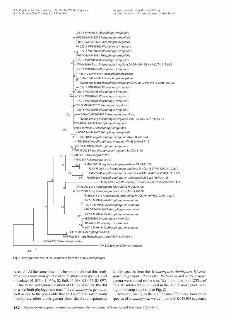
A.A. Kryukov, A.O. Gorbunova, E.M. Machs, Y.V. Mikhaylova A.V. Rodionov, P.M. Zhurbenko, A.P. Yurkov
162
Perspectives of using Illumina MiSeq for identification of arbuscular mycorrhizal fungi
Вавиловский журнал генетики и селекции / Vavilov Journal of Genetics and Breeding • 2020 • 24 • 2
053-6 MK948367 Rhizophagus irregularis
62
65
640
56
87
6154
14
7723
60
98
9787
22
8710
29
9418
899
764342
97
53
99
0.05
053-3 MK948364 Rhizophagus irregularis
053-7 MK948368 Rhizophagus irregularis060-2 MK948428 Rhizophagus irregularis053-1 MK948362 Rhizophagus irregularis
077-1 MK948486 Rhizophagus irregularis053-9 MK948370 Rhizophagus irregularis053-4 MK948365 Rhizophagus irregularis
053-10 MK948371 Rhizophagus irregularis060-1 MK948427 Rhizophagus irregularis
097-3 MK948494 Rhizophagus invermaius
097-5 MK948496 Rhizophagus invermaius097-1 MK948492 Rhizophagus invermaius
097-4 MK948495 Rhizophagus invermaiusHG969390 Rhizophagus invermaius
LN624112 Rhizophagus invermaius
GQ205083 Rhizophagus clarus
HG964399 Rhizophagus melanusAF413089 Funneliformis mosseae
097-2 MK948493 Rhizophagus invermaius
077-4 MK948489 Rhizophagus irregularis
GQ205076 Rhizophagus custosJN847437 Rhizophagus custos
068-1 MK948447 Rhizophagus irregularis
056a-2 MK948404 Rhizophagus irregularis
077-2 MK948487 Rhizophagus irregularis056a-1 MK948403 Rhizophagus irregularis
053-8 MK948369 Rhizophagus irregularis060-3 MK948429 Rhizophagus irregularis
053-5 MK948366 Rhizophagus irregularis077-3 MK948488 Rhizophagus irregularis
077-6 MK948491 Rhizophagus irregularis077-5 MK948490 Rhizophagus irregularisFM865610 R org Rhizophagus irregularis DAOM197198 W3182/Att1192-52
FM865608 R org Rhizophagus irregularis DAOM197198 W3182/Att1192-52
FM865591 org Rhizophagus irregularis BEG195 W5272/Att1485-12
FR750191 org Rhizophagus irregularis from BlaskowskiFR750187 org Rhizophagus irregularis W4682/Att857-12
FR750078 R org Rhizophagus irregularis MUCL43195
FM992402 R org Rhizophagus proliferus MUCL41827FN547500 R org Rhizophagus proliferus MUCL41827 W4728/Att1296-0FM865545 org Rhizophagus intraradices MUCL49410 W5070/Att1102-9
FM865546 org Rhizophagus intraradices MUCL49410 W5070/Att1102-9
FM865541 org Rhizophagus clarus W3776/Att894-7
FR750073 org Rhizophagus fasciculatus MUCL46100FR750071 org Rhizophagus fasciculatus MUCL46100
FM865606 R org Rhizophagus intraradices FL208 W5166/Att4-38FM865602 R org Rhizophagus intraradices FL208 W5166/Att4-38
Fig. 1. Phylogenetic tree of ITS-sequences from the genus Rhizophagus.
research. At the same time, it is beyond doubt that this study provides a molecular genetic identification at the species level of isolates 01053, 01056a, 02060, 04068, 05077, 03097.
Due to the ambiguous position of OTUs of isolate 05-104 on a prebuilt phylogenetic tree of the Acaulospora genus, as well as due to the possibility that OTUs of this isolate could incorporate other close genera from the Acaulosporaceae
fa mily, species from the Archaeospora, Ambispora, Diversispora, Gigaspora, Racocetra, Redeckera and Scutellospora genera were added to the tree. We found that both OTUs of 05104 isolates were included in the Acaulospora clade with high bootstrap support (see Fig. 2).
However, owing to the significant differences from other species of Acaulospora, we define the MK948503 sequence

Перспективы использования Illumina MiSeq для идентификации грибов арбускулярной микоризы
А.А. Крюков, A.O. Горбунова, Э.M. Мачс, Ю.В. Михайлова A.В. Родионов, П.M. Журбенко, A.П. Юрков
163
202024 • 2
ГЕНЕТИКА МИКРООРГАНИЗМОВ / MICROBIAL GENETICS
0.050
7142
68
97
20
70
98
9664
4186
9860
38
32 48
6180
9086
9165
50
46
98
94
45
2199
92
4
1861
54
67
42
99
71
66
37
30
67
2
8
AM076375 org Acaulospora sneverdingii ZS2005
AJ239115 org Acaulospora denticulata CL139-3AJ891114 org Acaulospora paulinae voucher CW4
104-2 MK948504 Acaulospora sp.FM876792 R org Acaulospora sneverdingii WUM18 W2941/Att869-3
FM876790 R org Acaulospora cavernata BEG33 W3293/Att209-37
FM876821 org Acaulospora kentinensis TW111A W5346/Att1499-9FN547523 org Acaulospora kentinensis TW111A W5346/Att1499-9
FR750151 R org Acaulospora spinosa EX-Type W3574/Attnone
FR692348 org Acaulospora cavernata BEG33FR846383 Acaulospora punctata
HE60364 Acaulospora nivalis
HG422732 Acaulospora spinosissima
JF439203 org Acaulospora delicata isolate G11 2JF439090 org Acaulospora mellea isolate G34-6JF439093 org Acaulospora delicata isolate G67-5
KM057070 Acaulospora excavata
LN881564 Acaulospora rugosaAM905249 Acaulospora morrowiae
FR692349 org Acaulospora scrobiculata FO316
AJ891104 org Acaulospora alpina environmental ST2700FN825903 R org Acaulospora brasiliensis W4699/Att1211-0
FR750168 org Acaulospora entreriana W5469/Att1541-1FN547502 R org Acaulospora laevis W3247/Att423-4FN547513 org Acaulospora laevis BEG26
JF439140 org Scutellospora calospora isolate B25-5
HM625899 org Scutellospora pellucida isolate scut1Fi1cons*17 org Racocetra castanea BEG1 ex-type
FR750136 R org Racocetra fulgida W2993/Att-noneFR750134 R org Racocetra weresubiae W2988/Att-noneFN547547 R org Gigaspora margarita BEG34FR750184 R org Gigaspora rosea DAOM194757 W2856/Att1509-20
FN547598 R org Scutellospora gilmorei W5342/Att590-16
AB048683 R org Scutellospora cerradensis MAFF520056FM876837 R org Scutellospora heterogama BEG35 W3214/Att334-16 clone MK029-3FR750013 org Scutellospora dipapillosa W5611/Att1577-4
AM268197 R org Ambispora fennica glomoid W3569/Att200-11 multi spore culture
AB048663 R org Ambispora callosa OK1 MAFF520057 W4769/Att1323-7 single spore isolateAB048654 R org Ambispora leptoticha F3b MAFF520055 W4770/Att315-11 single spore isolate
JF439210 org Ambispora gerdemannii isolate n8 9
FN820282 org Ambispora granatensis JEP-2010FR750022 org Archaeospora schenckii W5673/Att212-4
AM713403 R org Diversispora celata BEG231 FACE234 W4718-19/Att1278-2AY842567 R org Diversispora epigaea BEG47
AY035648 org Claroideoglomus claroideum JJ42 isolate Mr133
AJ849468 org Diversispora aurantia Holotype. Blaszkowski J. 2444 (DPP)FN547637 R org Diversispora spurca ex-type W4119/Att246-18
AM418545 org Redeckera fulva environmentalAM418551 org Redeckera megalocarpa environmental
FR750150 R org Scutellospora spinosissima W3009/Att664-1 pMK024-03+08+06
FR750173 Acaulospora entrerianaAF133768 Acaulospora colossica
LS398085 Acaulospora koskei104-1 MK948503 Acaulospora sp.
FR750063 org Acaulospora colombiana W5795/Att1476-8AJ239117 org Acaulospora colombiana CIAT C18-3
AJ891110 R org Acaulospora lacunosa BEG78LN736025 Acaulospora foveata
FR869691 org Acaulospora minuta
KP191472 Acaulospora ignotaLN810996 Acaulospora baetica
Fig. 2. Common phylogenetic tree of ITS-sequences from Acaulospora, Archaeospora, Ambispora, Diversispora, Gigaspora, Racocetra, Redeckera and Scutellospora genera.

A.A. Kryukov, A.O. Gorbunova, E.M. Machs, Y.V. Mikhaylova A.V. Rodionov, P.M. Zhurbenko, A.P. Yurkov
164
Perspectives of using Illumina MiSeq for identification of arbuscular mycorrhizal fungi
Вавиловский журнал генетики и селекции / Vavilov Journal of Genetics and Breeding • 2020 • 24 • 2
0.050
8799
95
58
91
27 91
94
3977 AF165919 org Paraglomus brasilianum ITH43
FR750046 org Paraglomus brasilianum W5793/Att260-8AF165922 org Paraglomus brasilianum WV224
AF165920 org Paraglomus brasilianum WV224AF165921 org Paraglomus brasilianum WV224
AF005062 org Paraglomus occultum FL703AF005065 org Paraglomus occultum CL383
AF005480 org Paraglomus occultum CL383AF005481 org Paraglomus occultum CL383
FJ769330 org Paraglomus laccatum environmental
FR750196 org Rhizophagus irregularis from Blaszkowski
056b MK968150 Paraglomus laccatumAM295494 org Paraglomus laccatum
0.20
067-2 MK948435 Claroideoglomus etunicatum067-3 MK948436 Claroideoglomus etunicatum
067-1 MK948434 Claroideoglomus etunicatumAY236328.1 Claroideoglomus etunicatum
FN547634 org Claroideoglomus etunicatum W5347/Att1505-8 CA-OT-126-3-2
LT963496 Claroideoglomus lamellosumFR750055 org Claroideoglomus claroideum W5794/Att1063-4
FR750191 org Rhizophagus irregularis from BlaszkowskiAJ972467 org Claroideoglomus walkeri Holotype. Blaszkowski J. 2609 (DPP)
AJ972464 org Claroideoglomus drummondii check paper
AY035653 org Claroideoglomus luteum JJ47 isolate Nr132FM876812 R org Claroideoglomus luteum SA101-3 W3184/Att676-0
AY035648 org Claroideoglomus claroideum JJ42 isolate Nr133AY330597 org Claroideoglomus etunicatum isolate 3-5-4FN547630 org Claroideoglomus etunicatum W5347/Att1505-8 CA-OT-126-3-2
9958
48
968566
65
915999
Fig. 4. Phylogenetic tree of ITS-sequences from Claroideoglomus genus.
Fig. 3. Phylogenetic tree of ITS-sequences from Paraglomus genus.
(05104 isolate) as a virtual taxon. The MK948504 sequence (05104 isolate), though it has a high similarity with the A. sieverdingii and A. paulinae species, did not show similarities with the indicated species according to morphological data, therefore it was also identified as a virtual taxon. At the same time, it can be reliably stated that these two OTUs belong to different taxa, since they have significant differences in GC content (31 and 46 %). But differences did not appear in the case of morphological analysis, highlighting the obstacles of distinguishing among Acaulospora species.
The OTUs of 01-056b isolate formed a well-supported subclade in the Paraglomus genus (bootstrap index = 99) (see Fig. 3). Furthermore, the Paraglomerales order hosts two more genera (Innospora and Pervetustus) in addition to the Paraglomus genus, but the ITS sequences for them are unknown. Thus the outgroup for the phylogenetic tree was taken from another order. The 01-056b isolate was identified as Paraglomus laccatum. The sequences of the P. laccatum 01 056b isolate turned out to be the shortest in comparison with isolates of other genera and ITS. Only three of eight species of this genera have ITS sequences in NCBI (Schüßler, 2019).
The Claroideoglomus genus is the most studied of the above mentioned genera. The ITS data were reported for six out of
eight species (Schüßler, 2019). The OTU of isolate 04-067 with high support belongs to the clade formed by the species Claroideoglomus etunicatum (see Fig. 4).
To verify whether the molecular genetic identification of AMF was effective, we conducted a morphological identification of nine isolates (Supplementary)1. The principal morphological characteristics for comparison were: size, shape, and color of the spores in air by the CMYK standard; shape, thickness and color of spore layers in Melzer’s reagent by the CMYK standard; the shape and thickness of subtending hypha; the wall thickness of subtending hypha; the pre sence of a septum in subtending hypha, type of mycorrhiza in P. aus tralis; mycorrhization parameter (F – frequency of mycorrhizal infection in P. australis roots).
On the basis of morphological analysis of spores, it was concluded that the AM isolates under examination – supported on P. australis culture in the ARRIAM collection – are of the following taxa: 1) 01053 isolate – Rhizophagus irregularis; 2) 01056a – R. irregularis; 3) 01056b – Paraglomus laccatum; 4) 02060 – R. irregularis; 5) 03097 – R. invermaius; 6) 04067 – Claroideoglomus etunicatum; 7) 04068 – R. irre1 Supplementary are available in the online version of the paper: http://www.bionet.nsc.ru/vogis/download/pict-2020-24/appx0.pdf

Перспективы использования Illumina MiSeq для идентификации грибов арбускулярной микоризы
А.А. Крюков, A.O. Горбунова, Э.M. Мачс, Ю.В. Михайлова A.В. Родионов, П.M. Журбенко, A.П. Юрков
165
202024 • 2
ГЕНЕТИКА МИКРООРГАНИЗМОВ / MICROBIAL GENETICS
gularis; 8) 05077 – R. irregularis; 9) 05104 – Acaulospora sp. defined only at the genus level, since it was not possible to clearly determine distinguishing morphological features.
Among the morphological features of the spores formed by various isolates of AMF the following should be highlighted. The spores of all AMF isolates classified as R. irregularis (isolates 01053, 01056a, 02060, 04068 and 05077) have the cylindrical subtending hypha in oblong spores or slightly flared (expanded) in spherical spore at a distance of more than 10 µm from the spores. The size, shape and color of the spores in air in these isolates were similar. Only spores of isolates 01- 053 and 01-056a with a greenish tint, had insignificant differences: color varied from 195540 (pistachio) and 23443 0 (very light yellowgreen) to 2617660 (yellowgreen) and 2925580 (dark khaki). It should be noted that the color of the L3 layer in spores of R. irregularis isolates had significant polymorphism: from 10379910 (cornyellow), 1459125 (deep yellow), 105398 0 (yelloworange), 13 54991 (deep orangeyellow) and 2652972 (deep yellow) to 174899 2 (almost pure orange), 1562980 (deep orangeyellow), 2678258 (brownred), 30809930, 32789925 (saturated redbrown). The spore color of the 01056b isolate, referred to the Paraglomus laccatum species, on the contrary varied in a narrow range: from colorless to 4400 (ghostly white). The second difference in the P. laccatum 01056b isolate was the absence of a septum in the subtending hypha in the presence of narrowing at the site of attachment of the spore as determined by the greater thickness of the L2 layer. The spores of isolate 04067, referred to Claroideoglomus etunicatum, were more yellow: from 5500 (ghostly white) to 21062 5 (orangeyellow Craiol) in air and in Melzer’s reagent L2 had a yellow color from 215602 (orangeyellow Craiol) to 335925 (saturated yellow). The darkest spores were observed in the isolate 03097, referred to the Rhizophagus invermaius species: from 30528851 (deep yellowbrown) to 59676372 (brownolive) in air. The spore morphology of isolate 05104 was significantly different from the other AMF. The spore color of isolate 05104 attributed to the Acaulospora genus, ranged from colorless (transparent) to 315704 (yellow ivory), spores cycatrix (the scarring is a remnant of the connection between the spore wall and the wall of the spore of the saccule during spore synthesis is 5.5–7.1 µm. Since this isolate is poorly maintained in P. australis culture, the morphology of the spores could not be studied in detail. According to molecular genetic analysis data, it may be approximately the same probability as A. paulinae, A. denticulata or A. sieverdingii.
All isolates of the Rhizophagus genus (01053, 01056a, 02060, 03097, 04068 and 05077), collected both in the forest belt and arable land, had a higher activity in P. australis roots (F > 80 %) than isolates of other AMF genera (see Supplementary). All the AMF isolates under examination formed mycorrhiza of the Arumtype, but only seven of nine isolates were able to be isolated into culture: 01053, 01056a, 02060, 03097, 04067, 04068, 05077 isolates should be considered as AMF strains.
Because their mycorrhization activity in P. australis is low (see Supplementary), sustained maintenance of the P. laccatum 01056b isolate and the Acaulospora sp. 05104 isolate will depend on the selection of optimal conditions for their growth (substrate composition and type of host plant).
DiscussionThe Illumina MiSeq approach allowed us to determine eight out of nine AMF isolates from the All Russian Research Institute for Agricultural Microbiology collection. It is very powerful method, which enables the identification of a large number of AMF taxa in fungal communities, especially if the proportion of targeted marker sequences in a sample is small. There are other effective methods of NGS, but several comparative studies indicate that MiSeq in some cases is more efficient as it can provide longer reads and fewer errors in comparison to HiSeq and IonTorrent techniques, respectively (Salipante et al., 2014; Razzauti et al., 2015).
These comparisons show the clear advantage of the se quencetargeted NGS approach as contrasted with the alternative cloning-sequencing method for AMF species identification. However, the main disadvantage of the Illumina MiSeq are relatively short reads (250 bp ×2), which do not allow the use of long markers, such as the entire cloned SSUITS15.8SITS2LSU region that was most often used for AMF barcoding from 2009 to 2012 (Krüger et al., 2009; De Castro et al., 2018). A prerequisite for the correct AMF identification by the MiSeq method is the employment of a short marker region for sequencing (400–500 bp). The most commonly used lengthappropriate marker is ITS2. The advantage of this region is that it provides sufficient variability for identification at the species level. Other less variable regions such as D1–D2 of the LSU allow identification only at the genus level (Krüger et al., 2009).
The use of universal primers for ITS2 region is the optimal choice for the identification of poorly studied taxa, which in recent years has helped to identify a significant number of virtual taxa of AMF (Öpik et al., 2014). These taxa can subsequently receive species names in the presence of individual morphological features and stable maintenance of isolates/strains in culture. For example, we studied the 05 104 sample in which there are two OTUs that belong to two different virtual Acaulospora sp. taxa. Another advantage of using the ITS2 region for identification is that a substantially representative sequence database has been stored in NCBI in comparison to other marker regions, for example, for the SSU region (NCBI, 2018).
It is important to note that OTUs related to one R. irregularis species and collected in different ecotopes (isolates 01-053, 01056a, 02060, 04068 and 05077) did not cluster separately on the phylogenetic tree (see Fig. 1). This suggests that all studied R. irregularis isolates shared one ribotype. Thus, it was not possible to identify the ecotyperelated features of R. irregularis isolates, perhaps they are objectively missing.
Also, it should be noted that NGS methods produce huge data arrays. We paid special attention to choose the correct tools for data treatment. Several pipelines have been developed to process rDNA sequences that are generated using Illumina’s MiSeq platform. Among them mothur (Schloss et al., 2016), QIIME (Caporaso et al., 2010), USEARCH and VSEARCH (Rognes et al., 2016) are the most popular. Generally, all of them have similar steps in sequences treatment. We used USEARCH since it has a large set of tools and detailed documentation. However, two approaches were used to search for AMF OTUs among a great pool of sequences. Both approaches were equally effective since they allowed us to identify the

A.A. Kryukov, A.O. Gorbunova, E.M. Machs, Y.V. Mikhaylova A.V. Rodionov, P.M. Zhurbenko, A.P. Yurkov
166
Perspectives of using Illumina MiSeq for identification of arbuscular mycorrhizal fungi
Вавиловский журнал генетики и селекции / Vavilov Journal of Genetics and Breeding • 2020 • 24 • 2
same set of taxa. At the same time the use of the USEARCH software has significantly reduced the time spent on data processing, which makes it possible to recommend it as the main tool to identify AMF from NGS data.
Combining the results of molecular genetics and morphological identification, we assert that the effectiveness of the Illumina MiSeq method as applied to AMF identification is not inferior to morphological methods that are significantly more labor intensive. However, the NCBI database is still insufficient for identification of some AMF species. The reason for this is that more than half of the known AMF taxa are still absent in the database. For example, according to A. Schüßler (2019), there are up to 56 species in the genus Acaulospora. The NCBI contains 37 species just for Acaulospora sp., and ITS sequences are presented for only 28 species. Thus, the obtained OTUs of the 05-104 isolate may belong to one of Acaulospora species that has yet to be studied.
ConclusionWe have determined that the most effective method for AMF identification is Illumina MiSeq supplemented by application of universal primers for the ITS2 region. Considerable efforts of morphologists in collaboration with molecular geneticists are required to establish a reliable taxonomy of the Glomeromycotina subdivision and to improve the efficiency of the molecular genetic AMF identification as a key method in the future.
ReferencesAltschul S.F., Gish W., Miller W., Myers E.W., Lipman D.J. Basic lo
cal alignment search tool. J. Mol. Biol. 1990;215:403-410. DOI 10.1016/S00222836(05)803602.
Aronesty E. Comparison of sequencing utility programs. TOBioiJ. 2013;7:1-8. DOI 10.2174/1875036201307010001.
Blaszkowski J. Definitions of morphological characters of spores of arbuscular fungi. 2003. Available at http://www.zor.zut.edu.pl/ Glo mero mycota/Definitions%20of%20spore%20characters.htm
Blaszkowski J. Species descriptions and illustrations. 2019. Available at http://www.zor.zut.edu.pl/
Bolger A.M., Lohse M., Usadel B. Trimmomatic: A flexible trimmer for Illumina Sequence Data. Bioinformatics. 2014;30(15):21142120. DOI 10.1093/bioinformatics/btu170.
Bruns T.D., Corradi N., Redecker D., Taylor J.W., Öpik M. Glomeromycotina: what is a species and why should we care? New Phytol. 2017. DOI 10.1111/nph.14913. Available at http://onlinelibrary. wiley.com/doi/10.1111/nph.14913/full
Caporaso J.G., Kuczynski J., Stombaugh J., Bittinger K., Bushman F.D., Costello E.K., Fierer N., Peña A.G., Goodrich J.K., Gordon J.I., Huttley G.A., Kelley S.T., Knights D., Koenig J.E., Ley R.E., Lozupone C.A., Mcdonald D., Muegge B.D., Pirrung M., Reeder J., Sevinsky J.R., Turnbaugh P.J., Walters W.A., Widmann J., Yatsunenko T., Zaneveld J., Knight R. QIIME allows analysis of highthroughput community sequencing data. Nat. Methods. 2010; 7(5):335-336. DOI 10.1038/nmeth.f.303.
Daubois L., Beaudet D., Hijri M., de la Providencia I. Independent mitochondrial and nuclear exchanges arising in Rhizophagus irregularis crossedisolates support the presence of a mitochondrial segregation mechanism. BMC Microbiol. 2016;16(1):1-12. DOI 10.1186/s1286601606275.
De Castro O., Avino M., Di Maio A., Menale B., Guida M. Sanger and next generation sequencing in the characterisation of arbuscular mycorrhizal fungi (AMF) in Pancratium maritimum L. (Amaryllidaceae), a representative plant species of Mediterranean sand dunes. Planta. 2018;248(6):1443-1453. DOI 10.1007/s00425-018-2981-z.
Doyle J.J., Doyle J.L. A rapid DNA isolation procedure for small quantities of fresh leaf tissue. Phytochem. Bull. 1987;19:1115.
Edgar R.C. Search and сlustering orders of magnitude faster than BLAST. Bioinformatics (Oxford, England). 2010;26(19):2460-2461. DOI 10.1093/bioinformatics/btq461.
Edgar R.C. UPARSE: Highly accurate OTU sequences from microbial amplicon reads. Nat. Methods. 2013;10:996-998. DOI 10.1038/nmeth.2604.
Edgar R.C. Updating the 97 % identity threshold for 16S ribosomal RNA OTUs. Bioinformatics. 2018;34(14):2371-2375. DOI 10.1093/bioinformatics/bty113.
Edgar R.C., Flyvbjerg H. Error filtering, pair assembly and error correction for nextgeneration sequencing reads. Bioinformatics. 2015; 31(21):3476-3482. DOI 10.1093/bioinformatics/btv401.
Helgason T., Daniell T.J., Husband R., Fitter A.H., Young J.P.W. Ploughing up the woodwide web? Nature. 1998;394(6692):431431. DOI 10.1038/28764.
Hosny M., GianinazziPearson V., Dulieu H. Nuclear DNA content of 11 fungal species in Glomales. Genome. 1998;41(3):422-428. DOI 10.1139/g98038.
INVAM. Species descriptions from reference cultures. 2019. Available at http://fungi.invam.wvu.edu/thefungi/speciesdescriptions.html
Krüger M., Stockinger H., Krüger C., Schüßler A. DNAbased species level detection of Glomeromycota: one PCR primer set for all arbuscular mycorrhizal fungi. New Phytol. 2009;183(1):212-223. DOI 10.1111/j.14698137.2009.02835.x.
Kryukov A.A., Yurkov A.P. Optimization procedures for molecular-genetic identification of arbuscular mycorrhizal fungi in symbiotic phase on the example of two closely kindred strains. Mikologiya i Fitopatologiya = Mycology and Phytopathology. 2018;52(1):38-48. (in Russian)
Kryukov A.A., Yurkov A.P., Shishova M.F. Features of the molecular phylogeny of arbuscular mycorrhizal fungi based on the analysis of the SSU – ITS1–5.8SrRNA – ITS2 – LSU marker region. Estestvennye i Tekhnicheskie Nauki = Natural and Technical Sciences. 2017; 2:1521. (in Russian)
Kumar S., Stecher G., Tamura K. MEGA7: Molecular Evolutionary Genetics Analysis version 7.0 for bigger datasets. Mol. Biol. Evol. 2016;33:1870-1874. DOI 10.1093/molbev/msw054.
Lee E.H., Eo J.K., Ka K.H., Eom A.H. Diversity of arbuscular mycorrhizal fungi and their roles in ecosystems. Mycobiology. 2013; 41(3):121-125. DOI 10.5941/MYCO.2013.41.3.121.
Lin K., Limpens E., Zhang Z., Ivanov S., Saunders D.G.O., Mu D., Pang E., Cao H., Cha H., Tao Lin T., Qian Zhou Q., Yi Shang Y., Ying Li Y., Trupti Sharma T., van Velzen R., de Ruijter N., Aanen D.K., Win J., Sophien Kamoun S., Ton Bisseling T., Geurts R., Huang S. Single nucleus genome sequencing reveals high similarity among nuclei of an endomycorrhizal fungus. PLoS Genet. 2014;10(1):e1004078. DOI 10.1371/journal.pgen.1004078.
NCBI. National Center for Biotechnology Information. 2018. Available at https://www.ncbi.nlm.nih.gov
Öpik M., Davison J., Moora M., Zobel M. DNAbased detection and identification of Glomeromycota: the virtual taxonomy of environmental sequences. Botany. 2014;92(2):135-147. DOI 10.1139/cjb-20130110.
Öpik M., Metsis M., Daniell T.J., Zobel M., Moora M. Largescale parallel 454 sequencing reveals host ecological group specificity of arbuscular mycorrhizal fungi in a boreonemoral forest. New Phytol. 2009;184(2):424-437. DOI 10.1111/j.1469-8137.2009.02920.x.
Öpik M., Vanatoa A., Vanatoa E., Moora M., Davison J., Kalwij J.M., Reier Ü., Zobel M. The online database MaarjAM reveals global and ecosystemic distribution patterns in arbuscular mycorrhizal fungi (Glomeromycota). New Phytol. 2010;188(1):223-241. DOI 10.1111/j.14698137.2010.03334.x.
Phillips J.M., Hayman D.S. Improved procedures for clearing roots and staining parasitic and vesiculararbuscular mycorrhizal fungi for rapid assessment of infection. Transact. Brit. Mycol. Soc. 1970;55: 158-161. DOI 10.1016/S0007-1536(70)80110-3.

Перспективы использования Illumina MiSeq для идентификации грибов арбускулярной микоризы
А.А. Крюков, A.O. Горбунова, Э.M. Мачс, Ю.В. Михайлова A.В. Родионов, П.M. Журбенко, A.П. Юрков
167
202024 • 2
ГЕНЕТИКА МИКРООРГАНИЗМОВ / MICROBIAL GENETICS
ORCID IDA.A. Kryukov orcid.org/0000-0002-8715-6723E.M. Machs orcid.org/0000-0001-9347-5379Y.V. Mikhaylova orcid.org/0000-0001-9278-0937A.V. Rodionov orcid.org/0000-0003-1146-1622P.M. Zhurbenko orcid.org/0000-0002-2102-4568A.P. Yurkov orcid.org/0000-0002-1072-5166
Acknowledgements. The work was performed using the material and technical base of the Center for Collective Use of Scientific Equipment “Genomic Technologies, Proteomics and Cell Biology” of the All-Russian Research Institute for Agricultural Microbiology and was supported by Russian Founda-tion for Basic Research grants (18-016-00220, 19-29-05275, and a portion of 17-00-00340, 17-00-00337; molecular-genetic identification of arbuscular mycorrhizal fungi). Part of this work was performed in the framework of the state task No. 0664-2019-0026 (morphological identification of arbuscular mycorrhizal fungi). Conflict of interest. The authors declare no conflict of interest.Received June 17, 2019. Revised November 15, 2019. Accepted November 17, 2019. Published online December 27, 2019.
Razzauti M., Galan M., Bernard M., Maman S., Klopp C., Charbonnel N., VayssierTaussat M., Eloit M., Cosson J.F. A comparison between transcriptome sequencing and 16S metagenomics for detection of bacterial pathogens in wildlife. PLoS Negl. Trop. Dis. 2015; 9(8):e0003929. DOI 10.1371/journal.pntd.0003929.
Rodionov A.V., Gnutikov A.A., Kotsinyan A.R. ITS1–5.8S rDNA–ITS2 sequense in the 35S рRNA genes as a marker in the reconstruction of cereal (Poaceae) phylogeny. Uspekhi Sovremennoy Biologii = Advances in Current Biology. 2016;136(5):416. (in Russian)
Rognes T., Flouri T., Nichols B., Quince C., Mahé F. VSEARCH: a versatile open source tool for metagenomics. PeerJ. 2016;4:e2584. DOI 10.7717/peerj.2584.
Salipante S.J., Kawashima T., Rosenthal C., Hoogestraat D.R., Cummings L.A., Sengupta D.J., Harkins T.T., Cookson B.T., Hoffman N.G. Performance comparison of Illumina and ion torrent nextgeneration sequencing platforms for 16S rRNAbased bacterial community profiling. Appl. Environ. Microbiol. 2014;80(24):7583-7591. DOI 10.1128/aem.02206-14.
Savary R., Masclaux F.G., Wyss T., Droh G., Corella J.C., Machado A.P., Morton J.B., Sanders I.R. A population genomics approach shows widespread geographical distribution of cryptic genomic forms of the symbiotic fungus Rhizophagus irregularis. ISME J. 2017;12(1):17-30. DOI 10.1038/ismej.2017.153.
Schenck N.C., Pérez Y. Manual for the Identification of VA Mycor-rhizal Fungi. 3rd ed. Gainesville, FL: Synergistic Publ., 1990.
Schloss P.D. Application of a databaseindependent approach to assess the quality of operational taxonomic unit picking methods. mSystems. 2016;1(2):e0002716.
Schoch C.L., Seifert K.A., Huhndorf S., Robert V., Spouge J.L., Levesque C.A., Chen W., Bolchacova E., Voigt K., Crous P.W., Miller A.N. Nuclear ribosomal internal transcribed spacer (ITS) region as a universal DNA barcode marker for fungi. Proc. Natl. Acad. Sci. USA. 2012;109:6241-6246. DOI 10.1073/pnas.1117018109.
Schüßler A. Glomeromycota species list. 2019. Available at http://www.amfphylogeny.com/amphylo_species.html
SenésGuerrero C., Schüßler A. A conserved arbuscular mycorrhizal fungal corespecies community colonizes potato roots in the Andes. Fungal Divers. 2015;77(1):317-333. DOI 10.1007/s13225-015-0328-7.
Senés-Guerrero C., Torres-Cortés G., Pfeiffer S., Rojas M., Schüßler A. Potatoassociated arbuscular mycorrhizal fungal communities in the Peruvian Andes. Mycorrhiza. 2014;24(6):405-417. DOI 10.1007/s0057201305490.
Stockinger H., Krüger M., Schüssler A. DNA barcoding of arbuscular mycorrhizal fungi. New Phytol. 2010;187(2):461-474. DOI 10.1111/j.14698137.2010.03262.x.
Stockinger H., Peyret-Guzzon M., Koegel S., Bouffaud M.-L., Redecker D. The largest subunit of RNA polymerase II as a new marker gene to study assemblages of arbuscular mycorrhizal fungi in the field. PLoS One. 2014;9(10):e107783. DOI 10.1371/journal.pone.0107783.
Tamura K., Nei M. Estimation of the number of nucleotide substitutions in the control region of mitochondrial DNA in humans and chimpanzees. Mol. Biol. Evol. 1993;10:512-526. DOI 10.1093/oxford journals.molbev.a040023.
Tedersoo L., Anslan S., Bahram M., Põlme S., Riit T., Liiv I., Kõljalg U., Kisand V., Nilsson H., Hildebrand F., Bork P., Abarenkov K. Shotgun metagenomes and multiple primer pairbarcode combinations of amplicons reveal biases in metabarcoding analyses of fungi. MycoKeys. 2015;10:1-43. DOI 10.3897/mycokeys.10.4852.
Trouvelot A., Kough J.L., GianinazziPearson V. Mesure du taux de mycorhization VA d’un système radiculaire. Recherche de méthodes ayant une signification fonctionnelle. In: Gianinazzi-Pearson V., Gianinazzi S. (Eds.). Physiological and Genetical Aspects of Mycorrhizae. Paris: INRAPress, 1986. (in French)
UNITE. Unified system for the DNA based fungal species linked to the classification Ver. 7.2. 2017. Available at https://unite.ut.ee
Vasar M., Andreson R., Davison J., Jairus T., Moora M., Remm M., Young J.P.W., Zobel M., Öpik M. Increased sequencing depth does not increase captured diversity of arbuscular mycorrhizal fungi. Mycorrhiza. 2017;27(8):761-773. DOI 10.1007/s00572-017-0791-y.
Vorob’ev N.I., Yurkov A.P., Provorov N.A. Programma Vychisleniya Indeksov Mikorizatsii Kornei Rastenii = The Program for Calculation of Indexes of Plant Root Mycorrhization. Registration certificate of computer software Svidvo No. 2016612112 ot 12.02.16 o gos. reg. programmy EVM. FGBNU VNIISKHM (RF). Zayavl. 21.12.2015. Opubl. 20.03.16 No. 2016612112, 12.02.16. FSBSI ARRIAM (RU). Appl. December 21, 2015. Publ. March 20, 2016. (in Russian)
White T.J., Bruns T., Lee S., Taylor J. Amplification and direct sequencing of fungal ribosomal RNA genes for phylogenetics. Innis M.A., Gelfand D.H., Sninsky J.J., White T.J. (Eds.). PCR Protocols: a Guide to Methods and Applications. San Diego: Academic Press, 1990.
Yurkov A.P., Jacobi L.M., Gapeeva N.E., Stepanova G.V., Shishova M.F. Development of arbuscular mycorrhiza in highly responsive and mycotrophic host plant – black medick (Medicago lupulina L.). Russian Journal of Developmental Biology. 2015;46(5):263275. DOI 10.1134/S1062360415050082.
Yurkov A.P., Kozhemyakov A.P., Stepanova G.V. The effectiveness of some microbial bioproducts based on bacteria and fungi arbuscular mycorrhiza. Multifunctional Adaptive Fodder Production: Collection of Proceedings (Mnogofunktsional’noe Adaptivnoe Kormoproizvodstvo: Sbornik Trudov). 2018a;19:2028. (in Russian)
Yurkov A.P., Kryukov A.A., Gorbunova A.O., Kozhemyakov A.P., Stepanova G.V., Machs E.M., Rodionov A.V., Shishova M.F. Molecular genetic identification of arbuscular mycorrhiza fungi. Ekologicheskaya Genetika = Ecological Genetics. 2018b;16(2)11-23. DOI 10.17816/ecogen16211-23. (in Russian)
Yurkov A.P., Kryukov A.A., Jacobi L.M., Kozhemyakov A.P., Shishova M.F. The relationship between the activity indicators and the effectiveness of arbuscular mycorrhiza fungi strains of various origin. Tavricheskiy Vestnik Agrarnoy Nauki = Tavrichesky Bulletin of Agrarian Science. 2017a;4(12):3141. (in Russian)
Yurkov A.P., Laktionov Yu.V., Kozhemyakov A.P., Stepanova G.V. Analysis of the symbiotic efficiency of bacterial and fungal preparations on feed crops according to seed yield. Kormoproizvodstvo = Fodder Production. 2017b;3:1621. (in Russian)

Изменение кишечного микробиома пациентов с язвенным колитом после трансплантации кишечной микробиотыА.Ю. Тикунов1, В.В. Морозов1, А.Н. Швалов2, А.В. Бардашева1, Е.В. Шрайнер1, О.А. Максимова3, И.О. Волошина3, В.В. Морозова1, В.В. Власов1, Н.В. Тикунова1
1 Институт химической биологии и фундаментальной медицины Сибирского отделения Российской академии наук, Новосибирск, Россия2 Государственный научный центр вирусологии и биотехнологии «Вектор» Роспотребнадзора Российской Федерации, р. п. Кольцово,
Новосибирская область, Россия3 ООО «Центр персонализированной медицины», Новосибирск, Россия
e-mail: [email protected]
Аннотация. Микробиота кишечника человека – это динамическая система, находящаяся под воздействием организма хозяина и внешних факторов. Возникающие нарушения кишечной микробиоты могут привести к патологическим состояниям, включая воспалительные и онкологические заболевания желудочно-кишечно-го тракта. Одним из возможных способов воздействия на микробиоту кишечника является фекотрансплан-тация (ФТ) – введение кишечной микробиоты от здорового донора в кишечный тракт пациента. В настоящее время в ряде стран этот метод используется для нормализации микробиоты кишечника, в основном при хронических воспалительных заболеваниях кишечника. В России (Новосибирск) уже несколько лет ведутся пилотные исследования эффективности ФТ при язвенном колите. Цель данной работы – оценить изменение микробиома кишечника 20 пациентов с язвенным колитом после однократного проведения ФТ. Основной метод – сравнительный анализ библиотек последовательностей 16S рибосомальной РНК, созданных на осно-ве образцов, полученных от пациентов с язвенным колитом до и после ФТ и секвенированных на платформе Illumina MiSeq. Результаты исследования показали, что ФТ привела к увеличению среднего биоразнообразия последовательностей в образцах, полученных после ФТ, по сравнению с образцами, собранными до ФТ, хотя разница не была статистически достоверной. Доля последовательностей Firmicutes, являющихся доминирую-щей компонентой кишечной микробиоты здоровых людей, уменьшилась (~32 % vs. >70 %), а доля последова-тельностей Proteobacteria увеличилась (>9 % vs. <5 %). В некоторых образцах, собранных до ФТ, были обнару-жены последовательности патогенных представителей Firmicutes и Proteobacteria, включая Acinetobacter spp., Enterococcus spp., Klebsiella pneumoniae, Proteus mirabilis, Staphylococcus aureus, Stenotrophomonas maltophylia, Streptococcus spp. В большинстве случаев после ФТ доля таких последовательностей резко сократилась. Ис-ключение составили последовательности Clostridium difficile, содержание которых в образцах почти половины пациентов составляло менее 0.5 %; после ФТ доля последовательностей C. difficile значительно уменьшилась лишь у трех пациентов. Следует отметить, что после ФТ повысилось на порядок содержание Lactobacillus spp. и существенно расширился их видовой состав. По результатам исследования можно сделать предварительное заключение о том, что даже однократная процедура ФТ приводит к повышению биоразнообразия микробиоты пациентов и оптимизации ее таксономического состава.Ключевые слова: микробиом; язвенный колит; 16S рРНК профилирование; трансплантация кишечной микро-биоты.
Для цитирования: Тикунов А.Ю., Морозов В.В., Швалов А.Н., Бардашева А.В., Шрайнер Е.В., Максимова О.А., Волошина И.О., Морозова В.В., Власов В.В., Тикунова Н.В. Изменение кишечного микробиома пациентов с язвен-ным колитом после трансплантации кишечной микробиоты. Вавиловский журнал генетики и селекции. 2020; 24(2):168-175. DOI 10.18699/VJ20.610
Fecal microbiome change in patients with ulcerative colitis after fecal microbiota transplantationA.Y. Tikunov1, V.V. Morozov1, A.N. Shvalov2, A.V. Bardasheva1, E.V. Shrayner1, O.A. Maksimova3, I.O. Voloshina3, V.V. Morozova1, V.V. Vlasov1, N.V. Tikunova1
1 Institute of Chemical Biology and Fundamental Medicine of Siberian Branch of the Russian Academy of Sciences, Novosibirsk, Russia2 State Research Center of Virology and Biotechnology Vector, Rospotrebnadzor, Koltsovo, Novosibirsk region, Russia3 Limited liability company “Personalized Medicine Center”, Novosibirsk, Russia
e-mail: [email protected]
Abstract. Intestinal human microbiota is a dynamic system that is under the pressures of its host organism and ex-ternal factors. Microbiota disruption caused by these factors can lead to severe diseases including inflammatory and oncological diseases of the gastrointestinal tract. One of the possible approaches in managing the intestinal micro-biota is fecal microbiota transplantation (FT) – transfer of the microbiota from the stool of a healthy donor to the intestinal tract of a recipient patient. Currently, this procedure is recognized as an efficacious method to normalize the
© Тикунов А.Ю., Морозов В.В., Швалов А.Н., Бардашева А.В., Шрайнер Е.В., Максимова О.А., Волошина И.О., Морозова В.В., Власов В.В., Тикунова Н.В., 2020
This work is licensed under a Creative Commons Attribution 4.0 License
ГЕНЕТИКА МИКРООРГАНИЗМОВОригинальное исследование / Original article
Вавиловский журнал генетики и селекции. 2020;24(2):168-175DOI 10.18699/VJ20.610

Изменение кишечного микробиома пациентов с язвенным колитом после трансплантации кишечной микробиоты
А.Ю. Тикунов, В.В. Морозов, А.Н. Швалов … В.В. Морозова, В.В. Власов, Н.В. Тикунова
202024 • 2
169ГЕНЕТИКА МИКРООРГАНИЗМОВ / MICROBIAL GENETICS
intestinal microbiota mainly in inflammatory diseases of the gastrointestinal tract. In Russia, pilot studies of the effec-tiveness of FT in patients with ulcerative colitis have been conducted for several years, and these studies were started in Novosibirsk. The aim of this study was to assess the change of intestinal microbiome in 20 patients with ulcerative colitis after a single FT procedure. The main method is a comparative analysis of 16S ribosomal RNA sequence libraries constructed using fecal samples obtained from patients with ulcerative colitis before and after FT and sequenced on the Illumina MiSeq platform. The obtained results showed that FT led to an increase in average biodiversity in samples after FT compared to samples before FT; however, the difference was not significant. In the samples stud-ied, the proportion of Firmicutes sequences, the major gastrointestinal microbiota of healthy people, was decreased (~32 % vs. >70 %), while the proportion of Proteobacteria sequences was increased (>9 % vs. <5 %). In some samples collected before FT, sequences of pathogenic Firmicutes and Proteobacteria were detected, including Acinetobacter spp., Enterococcus spp., Klebsiella pneumoniae, Proteus mirabilis, Staphylococcus aureus, Stenotrophomonas maltophy-lia, Streptococcus spp. In most cases, the proportion of such sequences after FT substantially decreased in appropri-ate samples. The exception was the Clostridium difficile sequences, which accounted for <0.5 % of the sequences in samples from almost half of the patients and after FT, the share of such C. difficile sequences was significantly reduced only in samples from three patients. It should be noted that the proportion of Lactobacillus spp. increased ten-fold and their species composition significantly expanded. According to the obtained results, a preliminary conclusion can be made that even a single FT procedure can lead to an increase in the biodiversity of the gastrointestinal microbiota in patients and to the optimization of the taxonomic composition of the microbiota.Key words: microbiome; ulcerative colitis; 16S rRNA profiling; fecal microbiota transplantation.
For citation: Tikunov A.Y., Morozov V.V., Shvalov A.N., Bardasheva A.V., Shrayner E.V., Maksimova O.A., Voloshina I.O., Morozova V.V., Vlasov V.V., Tikunova N.V. Fecal microbiome change in patients with ulcerative colitis after fecal microbiota transplantation. Vavilovskii Zhurnal Genetiki i Selektsii = Vavilov Journal of Genetics and Breeding. 2020;24(2):168-175. DOI 10.18699/VJ20.610 (in Russian)
ВведениеШирокое применение технологий NGS (next generation sequencing) обеспечило детальную характеризацию микробных сообществ, преимущественно бактериальных, ас социированных с организмом человека. В настоящее вре мя микробиота кишечника человека рассматривается как динамическая система, находящаяся под воздействием организма хозяина и внешних факторов (Fujimura et al., 2010; Qin et al., 2010). Известно, что основными компонентами нормальной кишечной микробиоты человека являются представители Firmicutes и Bacteroidetes, хотя коро вая группа конкретных видов бактерий не совпадает у разных здоровых индивидуумов (Donaldson et al., 2016), и различные по составу варианты нормальной микробиоты способны обеспечить стабильное функционирование этого сложного микробного сообщества (Lozupone et al., 2012).
Дисбаланс кишечной микробиоты, возникающий под действием внешних или внутренних факторов, может привести к патологическим состояниям, включая не толь ко воспалительные и онкологические заболевания желудочнокишечного тракта, но и нарушения иммунной системы, диабет второго типа, сосудистые заболевания и даже нарушения функций головного мозга (O’Hara, Shanahan, 2006). Одним из возможных способов воздействия на микробиоту кишечника, наряду с использованием антибиотиков, пробиотиков и пребиотиков, является фекотрансплантация (ФТ) – введение кишечной микробиоты от здорового донора в кишечный тракт пациента. Считается, что до нашей эры применение ФТ практиковали в Китае, а в наше время эту процедуру впервые провели в 1958 г. при лечении пациента с энтероколитом (Eiseman et al., 1958). Однако лишь недавно метод стал успешно использоваться для нормализации микробиоты кишечника при различных заболеваниях, включая хронические воспалительные заболевания кишечника (Aas et al., 2003; Khoruts et al., 2010; Angelberger et al., 2013; Pigneur, Sokol,
2016; Vaughn et al., 2016; Kang et al., 2017; Paramsothy et al., 2017; Staley et al., 2017).
Наибольшая результативность применения ФТ показана при псевдомембранозном колите, ассоциированном с Clostridium (Clostridioides) difficile (Drekonja et al., 2015; Khoruts, Sadowsky, 2016; Cheng, Fisher, 2017; Staley et al., 2017; Goldenberg et al., 2018). Быстрое улучшение состояния пациентов и высокая эффективность ФТ (более 80 %) при этих инфекциях была подтверждена в многоцентровых рандомизированных клинических испытаниях (van Nood et al., 2013; Cammarota et al., 2015), и сейчас в США и в европейских странах рекомендуется применять ФТ уже после второго или третьего эпизода C. difficileассоциированного колита (Debast et al., 2014).
Имеются свидетельства того, что ФТ может быть полезной и при язвенном колите (ЯК). Эффективность применения ФТ для лечения пациентов с ЯК в различных исследованиях существенно варьировала – от 20 до 92 % (Angelberger et al., 2013; Kellermayer et al., 2015). В рандомизированных клинических испытаниях ремиссия зарегистрирована на уровне 25–27 %, что, впрочем, статистически значимо превышало эффект плацебо (Moayyedi et al., 2015; Rossen et al., 2015; Paramosthy et al., 2017). Патогенез ЯК не вполне ясен; полагают, что он имеет сложную природу и опосредован нарушениями кишечной микробиоты, генетической предрасположенностью и экологическими факторами (Shen et al., 2018). Как и при C. difficileассоциированном колите, биоразнообразие микробиоты кишечника при ЯК существенно снижено, уменьшено количество представителей Bacteroidetes и Firmicutes. При этом в микробиоте пациентов с ЯК увеличивается количество представителей Proteobacteria и Actinomycetes, выявляются C. difficile, Helicobacter pylori, Salmonella spp., Yersinia spp. и энтероинвазивные Escherichia coli (Okhusa et al., 2003; Saebo et al., 2005; Gradel et al., 2009; Sonnenberg, Genta, 2012; Deshpande et al., 2013; Reddy, Brandt, 2013; Shen et al., 2018). До сих пор не ясно,

A.Y. Tikunov, V.V. Morozov, A.N. Shvalov … V.V. Morozova, V.V. Vlasov, N.V. Tikunova
170 Вавиловский журнал генетики и селекции / Vavilov Journal of Genetics and Breeding • 2020 • 24 • 2
Fecal microbiome change in patients with ulcerative colitis after fecal microbiota transplantation
является ли ЯК результатом нарушенного иммунного ответа на нормальную микробиоту, или это нормальный иммунный ответ на дисбаланс в микрофлоре кишечника (Cheng, Fisher, 2017).
Несмотря на кажущуюся очевидность, механизмы положительного действия ФТ при ЯК не вполне ясны. Полагают, что эффективность ФТ связана с увеличением разнообразия микробиоты кишечника, что приводит к повышению обилия «полезных» бактерий и препятствует колонизации кишечника патогенными бактериями (Broecker et al., 2016; Chehoud et al., 2016; Khoruts, Sadowsky, 2016). Однако неизвестно, вовлечены ли в этот процесс другие механизмы, включая возможное влияние виробиоты, действие иммунной системы пациента, привнесение регуляторных высоко и низкомолекулярных соединений при ФТ; также до сих пор не определен список видов бактерий, обусловливающих нормализацию микробиоты. Цель данного исследования – оценить изменение микробиома кишечника пациентов с ЯК после проведения ФТ на основе профилирования 16S рибосомальной РНК в образцах, полученных до и после лечения.
Материалы и методыВ работе использовали образцы фекалий, полученные от 20 пациентов (27–57 лет) с диагнозом ЯК. Диагноз подтверждали на основании результатов изучения уровня фекального кальпротектина, данных фиброколоноскопии и гистологического исследования биоптатов, взятых из разных отделов толстой и подвздошной кишок. Образцы пациентов, в которых рутинными методами были обнаружены C. difficile, в исследование не вовлекались. Все пациенты предоставили информированное согласие с проводимым исследованием и анонимной обработкой дан ных. Образцы собирали за одиндва дня до ФТ и через 7–12 дней после ФТ. Донорами были молодые здоровые добровольцы (20–39 лет) без хронических заболеваний, не перенесшие инфекции и не подвергавшиеся госпитализации по крайней мере последние два месяца. Все доноры прошли обследование, включающее в себя общий и биохимический анализы крови, а также ИФА крови на наличие лямблий, токсокар, описторхов, аскарид, трихинелл. Кроме того, с использованием стандартных тестси стем подтверждали отсутствие у доноров возбудителя сифилиса, ВИЧ1 и ВИЧ2, вирусов гепатита В и С. Также рутинными методами проводили анализ фекалий на дисбиоз и на отсутствие патогенной микрофлоры (C. difficile, Campilobacter jejuni, Salmonella spp., Shigella spp., энтероинвазивная Escherichia coli, Cryptosporidium spp., Cyclospora spp., Giardia spp., Isospora spp.), ротавирусов А, норовирусов I и II и аденовирусов F, а также гельминтов и их яиц. Пилотное исследование было одобрено Локальным этическим комитетом Автономной некоммерческой организации «Центр новых медицинских технологий в Академгородке».
По 50 мг каждого образца от пациентов суспендировали в 300 мкл 0.9 % NaCl и центрифугировали при 2 тыс. об. в течение 10 мин. Суммарную ДНК очищали из 100 мкл осветленной клеточной суспензии с помощью набора для выделения ДНК из клеток тканей и крови («БиоЛабМикс», Россия) с добавлением лизоцима для повышения
эффективности извлечения ДНК из грамположительных бактерий. С использованием полученной ДНК в качестве матрицы, фьюжнпраймеров (NEBFF 5ʹACACTCTTTC CCTACACGACGCTCTTCCGATCTCTACGGGAGGCA GCAG3ʹ, NEBFR 5ʹGTGACTGGAGTTCAGACGTGT GCTCTTCCGATCTGGACTACCGGGGTATCT3ʹ) и высокоточной полимеразы Q5 (New England Biolabs, США) проводили амплификации фрагмента гена 16S рРНК, со держащего вариабельные участки V3–V4. Продукты ам плификации очищали электрофоретически в геле из лег коплавкой SeaKem GTGагарозы (Lonza, США). Обогащение полученных ампликонов, введение баркодов и служебных последовательностей для дальнейшего секвенирования на платформе MiSeq выполняли, используя полимеразу Q5 и набор олигонуклеотидов Dual index set (New England Biolabs), согласно инструкции производителя. Полученные библиотеки очищали на магнитных частицах AMPure XP (Beckman Coulter, США); концентрацию ДНК измеряли с помощью набора Qubit dsDNA HS (Life Technologies, США). По результатам измерений биб лиотеки объединяли в пул таким образом, чтобы соотношение ДНК библиотек в пуле было приблизительно равным. Секвенировали на высокопроизводитель ном секвенаторе MiSeq с набором реагентов MiSeq reagent kit v2 2 × 250cycles (Illumina, США).
Результаты секвенирования анализировали с использованием пакета программного обеспечения UGENE v.1.32 (Unipro UGENE, Россия). Полученные риды картировали на базу данных 16S рРНК, размещенную на Национальном сервере NSBI (США), с использованием пакета Clark. Предварительно из последовательностей ридов удаляли последовательности адаптеров и проводили фильтрацию ридов по качеству. Риды анализировали двумя метода ми: с помощью генерации операционных таксономических единиц (OTU) с последующим картированием последовательностей на полученные OTU в пакете программ Usearch9.2 и путем классификации ридов алгоритмом Kraken по базе данных известных последовательностей 16S рРНК Silva v.132 (full). В первом случае OTU генерировали алгоритмом unoise2 с отбраковкой химерных последовательностей и учетом ошибок чтения. Таблицы полученных частот встречаемости OTU были обработаны в среде R3.3.3. Во втором случае риды картировали на базу данных 16S рРНК Silva с помощью алгоритма seedkraken с использованием разреженного kмера со специальной решеткой, позволяющей увеличить специфичность классификации. Индекс Шеннона рассчитывали в пакете программ R; достоверность различий между индексами Шеннона определяли с помощью tтеста Хатчесона. Визуализацию результатов анализа библиотек последовательностей методом главных координат PCoA проводили на основе матриц дистанций с использованием пакета программ vegan.
Результаты и обсуждениеНа основе ДНК, выделенной из образцов фекалий от 20 пациентов с ЯК до и после ФТ, было сконструировано 40 библиотек фрагментов гена 16S рРНК. Фрагменты гена включали вариабельные участки V3 и V4, используемые обычно для таксономической классификации бактерий

Изменение кишечного микробиома пациентов с язвенным колитом после трансплантации кишечной микробиоты
А.Ю. Тикунов, В.В. Морозов, А.Н. Швалов … В.В. Морозова, В.В. Власов, Н.В. Тикунова
202024 • 2
171ГЕНЕТИКА МИКРООРГАНИЗМОВ / MICROBIAL GENETICS
(Chak ravorty et al., 2007; Wang, Qian, 2009). В результате секвенирования библио тек, которые были созданы на основе образцов, собранных до ФТ, получено от 106 411 до 1 751 663 ридов (в среднем 141 010). Библиотеки из образцов, собранных после ФТ, содержали от 107 042 до 173 855 ридов (в среднем 142 060). По результатам классификации в библиотеках из первой и второй групп к определенному типу прокариот были таксономически отнесены в среднем 99.7 и 99.6 % ридов соответственно. Лишь незначительная часть последовательностей осталась неклассифицированной. Всего выявлены последовательности 13 типов бактерий, основными из которых были Firmicutes, Bacteroidetes и Proteobacteria. В трех образцах выявлены последовательности архей, принадлежащих к роду Methanobrevibacter (тип Euryarchaeota), однако доля таких последовательностей в соответствующих библиотеках не превышала 0.1 %.
Анализ полученных данных показал, что биоразнообразие бактериальных сообществ в образцах от пациентов до и после ФТ различается. Так, индекс Шеннона для выборки образцов от пациентов после лечения (3.43 ± 0.71) был выше, чем для образцов, взятых до лечения (3.05 ± 0.67), хотя разница статистически недостоверна (рис. 1). Однако при попарном сравнении индексов Шеннона для образцов, полученных от одного пациента до и после ФТ, различия были во всех случаях статистически достоверными (SD, 0.011–0.019); при этом у 15 пациентов биоразнообразие микробных сообществ значимо увеличилось и лишь у 5 пациентов – уменьшилось.
Большее биоразнообразие сообществ в образцах, полученных после ФТ, подтвердилось при анализе данных методом главных координат (рис. 2). Видно, что 60 % образцов, собранных до лечения, находятся в области отрицательных значений первой главной координаты, тогда как 65 % образцов, взятых после лечения, находятся в области положительных значений. Аналогично в области положительных значений второй главной координаты находится
лишь 45 % образцов, полученных до лечения, и 70 % образцов после ФТ. Отметим, что в образцах от 15 пациентов (75 %) после проведения ФТ значение уве личилось в области и первой, и второй главных координат; еще у четырех па циентов значение увеличилось в области хотя бы одной
4.5
4.0
3.5
3.0
2.5
2.0
Before FT After FT
Fig. 1. Shannon index, reflecting the biodiver-sity of sequences in samples obtained from patients before and after FT.
Fig. 2. Visualization of sequence library analysis by the method of principal coordinates PCoA made on matrices of distances: 1, data for libraries from samples obtained before FT; 2, from samples obtained after FT. 3, 4, lines connecting two samples from one patient for which biodiversity (3) increases or (4) decreases after FT. Designations S1–S20 correspond to sample numbers before FT. The values of the first and second principal coordinates are presented on the OX and OY axes, respectively.
S1
S10
S11 S12
PCo 1 [11.5]
PCo
2 [1
0.6]
S13S14
0.10
0
–0.1–0.2–0.3
–0.3
–0.2
–0.1
0.1
0.2
0.2 0.3
S16
S19
S20
S2
1
2
3
4
S3
S4
S5
S6
S7
S8
S9
S15 S18
S17

A.Y. Tikunov, V.V. Morozov, A.N. Shvalov … V.V. Morozova, V.V. Vlasov, N.V. Tikunova
172 Вавиловский журнал генетики и селекции / Vavilov Journal of Genetics and Breeding • 2020 • 24 • 2
Fecal microbiome change in patients with ulcerative colitis after fecal microbiota transplantation
из главных ко ординат; только для пары образцов от одного пациента (№ 18) биоразнообразие уменьшилось после ФТ в области значений обеих главных ко ординат (см. рис. 2). Тем не менее при уменьшении значений в области хотя бы одной главной координаты после ФТ индекс Шеннона для этой же пары образцов статистически значимо уменьшался.
Полученные данные подтверждают возможность повышения биоразнообразия бактериального сообщества кишечника после проведения ФТ. Следует подчеркнуть, что в настоящем исследовании мы сравнивали исходный микробиом пациентов и микробиом после первой процедуры ФТ. Согласно опубликованным сведениям за рубежных авторов, даже после нескольких процедур ФТ статистически значимое повышение биоразнообразия микробиоты регистрируется не у всех пациентов (Angelberger et al., 2013).
Анализ таксономического состава показал, что в образцах до и после ФТ в среднем 55.7 % (от 12.7 до 72.8 %) и 48.3 % (от 15.9 до 92.3 %) выявленных последовательностей соответственно принадлежали к типу Firmicutes (рис. 3), 32.3 % (от 4.4 до 64.4 %) и 33.7 % (от 1.7 до 60.7 %) – к типу Bacteroidetes, а 9.2 % (от 0.4 до 81.3 %) и 6.2 % (от 0.7 до 31.5 %) – к типу Proteobacteria. Четвертыми по встречаемости были последовательности Fusobacteria, в среднем 3.4 % по всем библиотекам, однако их доля значительно увеличилась после ФТ: от <0.1 (0–0.3 %) до 6.7 % (0.1–9.4 %). После ФТ увеличилась также доля последовательностей Actinobacteria, хотя и не так значительно – от 1.9 (0.1–10.7 %) до 3.1 % (0.2–19.4 %). После
довательности Verrucomicrobia присутствовали в каждом образце в небольших количествах, а их встречаемость после ФТ уменьшилась в среднем с 0.4 до 0.2 %. Остальные последовательности встречались лишь в отдельных образцах, не превышая 0.1 % от всех последовательностей в этом образце.
Известно, что микробиота здоровых людей состоит из постоянных и транзиторных видов, относящихся более чем к 17 типам, включая Firmicutes (>70 %), Bacteroidetes (>30 %), протеобактерии (<5 %), актинобактерии (<2 %), Fusobacteria и Verrucomicrobia (<1 %) (Belizário et al., 2018). Полученные нами результаты коррелируют с данными других исследователей, свидетельствующими о пониженном биоразнообразии микробиоты кишечника при ЯК (Manichanh et al., 2012; Machiels et al., 2014; Bajer et al., 2017). Так, в исследуемых образцах от пациентов в среднем присутствовало существенно меньше последо вательностей Firmicutes, что хорошо согласуется с на блюдениями других авторов (Machiels et al., 2014). В основном Firmicutes были представлены последовательностями классов Clostridia (рис. 4), в среднем 47.4 и 40.6 % в библиотеках из образцов, полученных до и после ФТ соответственно. Из них доминировали Faecalibacterium prausnitzii (15.7 и 11.3 % соответственно) и Roseburia hominis (2.3 и 0.5 %), ответственные за расщепление широкого спектра углеводов, включая крахмал и инулин, с образованием бутиратов (Duncan et al., 2007; Machiels et al., 2014). Также были представлены последовательности классов Negativicutes (6.8 и 4.0 %) и Bacilli (1.3 и 2.8 %). Следует отметить, что, несмотря на относительно невы
Firmicutes Not classifiedVerrucomicrobiaActinobacteriaProteobacteriaBacteroidetes Fusobacteria Other
1 10 20 30 4011 21 3116 26 3612 22 3217 27 3713 23 3318 28 3814 24 3419 29 3915 25 352 3 4 5 6 7 8 9
%100
90
80
70
60
50
40
30
20
10
0
Fig. 3. Taxonomic classification of OTUs at the phylum level based on the Silva v.132 (full) database.Here and in Fig. 4 samples 1–20 were obtained from patients before FT (enumeration follows Fig. 2); samples 21–40 were collected after FT. Samples 1 and 21, 2 and 22, etc. were obtained from the first, second, etc. patients, respectively. Phyla with representation exceeding 0.1 % are shown.

Изменение кишечного микробиома пациентов с язвенным колитом после трансплантации кишечной микробиоты
А.Ю. Тикунов, В.В. Морозов, А.Н. Швалов … В.В. Морозова, В.В. Власов, Н.В. Тикунова
202024 • 2
173ГЕНЕТИКА МИКРООРГАНИЗМОВ / MICROBIAL GENETICS
сокую встречаемость Bacillus spp. и Lactobacillus spp., их доля после ФТ повысилась в 3.5 и 11 раз соответственно. При этом существенно расширился видовой состав лактобацилл, которые, как известно, не только участвуют в расщеплении лактозы и других углеводов, но и являются антагонистами по отношению к патогенным микроорганизмам, вытесняя их из микробного сообщества кишечника человека.
Доля последовательностей, принадлежащих к типу Bacteroidetes, в исследуемых образцах от пациентов с ЯК не была снижена (см. рис. 3 и 4) и составила около трети от всех последовательностей, что отличается от наблюдений других исследователей (Machiels et al., 2014). В основном этот тип был представлен последовательностями Bacteroides spp. и Prevotella spp., причем после ФТ доля последовательностей бактероидов уменьшилась в среднем с 19.7 до 9.6 %, а последовательностей, принадлежащих к роду Prevotella, существенно увеличилась, с 3.7 до 14.6 %. Известно, что большинство бактероидов, обитающих в кишечнике человека, способны разлагать разнообразные растительные полисахариды (Flint et al., 2012), причем в кишечной микробиоте жителей западных стран преобладают Bacteroides spp., а в микробиоте населения из стран с преимущественно растительной диетой – Prevotella spp. (Ley, 2016).
Отметим, что доля последовательностей Proteobacteria (см. рис. 3) в образцах, собранных до ФТ (в среднем 9.2 %), превышала таковую, обычно регистрируемую у здоровых людей (<5 %) (Belizário et al., 2018). Это объясняется повышенным содержанием Salmonella spp. в микробных сообществах пациентов с ЯК и наличием в отдельных образцах в большом количестве последовательностей патогенных бактерий, способных вызвать желудочнокишечные заболевания (более 9 % Acinetobacter spp.,
0.5–1 % Klebsiella pneumoniae), что установлено и в других исследованиях (Gradel et al., 2009; Shen et al., 2018). В единичных образцах были обнаружены также Proteus mirabilis и Stenotrophomonas maltophilia, доля которых не превышала 0.1 %. Надо сказать, что после проведения ФТ содержание перечисленных последовательностей патогенных Proteobacteria в образцах существенно уменьшилось, порой до 0.02 % и менее.
Кроме последовательностей патогенных Proteobacteria, в некоторых образцах, полученных от пациентов до ФТ, были обнаружены последовательности патогенных представителей Firmicutes. Так, в девяти образцах найдены последовательности C. difficile, доля которых превышала 0.5 %. Повышенная встречаемость C. difficile в кишечной микробиоте пациентов с ЯК отмечалась и ранее (Deshpande et al., 2013; Reddy, Brandt, 2013). Помимо последовательностей C. difficile, в некоторых образцах обнаружены последовательности Staphylococcus aureus (0.1–0.9 %), Streptococcus spp. и Enterococcus spp. (~0.1 %). Как и в слу чае с патогенными Proteobacteria, доля этих последовательностей после ФТ в соответствующих образцах резко уменьшилась. Исключение составили образцы с C. difficile: доля их последовательностей уменьшилась пос ле ФТ только в трех образцах из девяти, составив <0.2 %.
ЗаключениеТаким образом, исследованы биоразнообразие и таксономический состав последовательностей фрагмента гена 16S рРНК, ассоциированных с кишечной микробиотой у 20 пациентов с ЯК до и после ФТ. Результаты показали, что однократное проведение процедуры ФТ привело к увеличению среднего биоразнообразия последовательностей в образцах, полученных после ФТ, по сравнению с образцами, собранными до ФТ, хотя разница не была
1 10 20 30 4011 21 3116 26 3612 22 3217 27 3713 23 3318 28 3814 24 3419 29 3915 25 352 3 4 5 6 7 8 9
%100
90
80
70
60
50
40
30
20
10
0
Not classifiedOtherVerrucomicrobiae
ActinobacteriaCoriobacteriiaAlphaproteobacteriaGammaproteobacteria Fusobacteriia
BacteroidiaErysipelotrichiaNegativicutesClostridia Bacilli
Fig. 4. Taxonomic classification of OTUs at the class level based on the Silva v.132 (full) database.Classes with representation exceeding 0.1 % are shown.

A.Y. Tikunov, V.V. Morozov, A.N. Shvalov … V.V. Morozova, V.V. Vlasov, N.V. Tikunova
174 Вавиловский журнал генетики и селекции / Vavilov Journal of Genetics and Breeding • 2020 • 24 • 2
Fecal microbiome change in patients with ulcerative colitis after fecal microbiota transplantation
статистически достоверной. Доля последовательностей Fir micutes, являющихся доминирующей компонентой кишечной микробиоты здоровых людей, была снижена (~32 % vs. >70 %), а доля последовательностей Proteobacteria увеличена (>9 % vs. <5 %). В некоторых образцах, собранных до ФТ, обнаружены значимые содержания последовательностей патогенных представителей Firmi cutes и Proteobacteria, включая Acinetobacter spp., Ente rococcus spp., K. pneumoniae, P. mirabilis, S. aureus, St. maltophilia, Streptococcus spp. В большинстве случаев после однократной процедуры ФТ доля таких последовательностей резко сократилась. Исключение составили последовательности C. difficile, которые были обнаружены (около 0.5 % и выше) в образцах почти половины пациентов с ЯК; после ФТ доля последовательностей C. difficile значительно уменьшилась лишь у трех пациентов. Следует отметить, что после ФТ содержание Lactobacillus spp. повысилось на порядок и существенно расширился видовой состав лактобацилл.
Результаты исследования позволяют сделать предварительный вывод, что даже однократная процедура ФТ может привести к повышению биоразнообразия микробиоты и оптимизации ее таксономического состава. Однако для того чтобы сделать заключение об эффективности такого лечения, длительности ремиссии и стабильности изменений микробиоты кишечника у пациентов с ЯК, требуются дальнейшие наблюдения за этими пациентами и анализ кишечного микробиома после последующих процедур ФТ.
Список литературы / ReferencesAas J., Gessert C.E., Bakken J.S. Recurrent Clostridium difficile colitis:
case series involving 18 patients treated with donor stool administered via a nasogastric tube. Clin. Infect. Dis. 2003;36(5):580585.
Angelberger S., Reinisch W., Makristathis A., Lichtenberger C., Dejaco C., Papay P., Novacek G., Trauner M., Loy A., Berry D. Temporal bacterial community dynamics vary among ulcerative colitis patients after fecal microbiota transplantation. Am. J. Gastroenterol. 2013;108(10):16201630. DOI 10.1038/ajg.2013.257.
Bajer L., Kverka M., Kostovcik M., Macinga P., Dvorak J., Stehlikova Z., Brezina J., Wohl P., Spicak J., Drastich P. Distinct gut microbiota profiles in patients with primary sclerosing cholangitis and ulcerative colitis. World J. Gastroenterol. 2017;23(25):45484558. DOI 10.3748/wjg.v23.i25.4548.
Belizário J.E., Faintuch J., GarayMalpartida M. Gut microbiome dysbiosis and immunometabolism: new frontiers for treatment of metabolic diseases. Mediators Inflamm. 2018;2018:112. DOI 10.1155/ 2018/2037838.
Broecker F., Klumpp J., Moelling K. Longterm microbiota and virome in a Zürich patient after fecal transplantation against Clostridium difficile infection. Ann. N.Y. Acad. Sci. 2016;1372(1):2941. DOI 10.1111/nyas.13100.
Cammarota G., Masucci L., Ianiro G., Bibbò S., Dinoi G., Costamagna G., Sanguinetti M., Gasbarrini A. Randomised clinical trial: faecal microbiota transplantation by colonoscopy vs. vancomycin for the treatment of recurrent Clostridium difficile infection. Aliment. Pharmacol. Ther. 2015;41(9):835843. DOI 10.1111/apt.13144.
Chakravorty S., Helb D., Burday M., Connell N., Alland D. A detailed analysis of 16S ribosomal RNA gene segments for the diagnosis of pathogenic bacteria. J. Microbiol. Methods. 2007;69(2):330339.
Chehoud C., Dryga A., Hwang Y., NagySzakal D., Hollister E.B., Luna R.A., Versalovic J., Kellermayer R., Bushman F.D. Transfer of viral communities between human individuals during fecal micro
biota transplantation. MBio. 2016;7(2):e00322. DOI 10.1128/mBio. 0032216.
Cheng Y.W., Fischer M. The present status of fecal microbiota transplantation and its value in the elderly. Curr. Treat. Options Gastroenterol. 2017;15(3):349362. DOI 10.1007/s1193801701431.
Debast S.B., Bauer M.P., Kuijper E.J. European Society of Clinical Microbiology and Infectious Diseases: update of the treatment guidance document for Clostridium difficile infection. Clin. Microbiol. Infect. 2014;20(Suppl.2):126. DOI 10.1111/14690691.12418.
Deshpande A., Pasupuleti V., Pant C., Rolston D.D., Sferra T.J. Diagnostic testing for Clostridium difficile infection in patients with inflammatory bowel disease. J. Clin. Gastroenterol. 2013;47(8):737738. DOI 10.1097/MCG.0b013e318295d4ec.
Donaldson G.P., Lee S.M., Mazmanian S.K. Gut biogeography of the bacterial microbiota. Nat. Rev. Microbiol. 2016;14(1):2032. DOI 10.1038/nrmicro3552.
Drekonja D., Reich J., Gezahegn S., Greer N., Shaukat A., MacDonald R., Rutks I., Wilt T.J. Fecal microbiota transplantation for Clostridium difficile infection: a systematic review. Ann. Intern. Med. 2015;162(9):630638. DOI 10.7326/M142693.
Duncan S.H., Louis P., Flint H.J. Cultivable bacterial diversity from the human colon. Lett. Appl. Microbiol. 2007;44(4):343350.
Eiseman B., Silen W., Bascom G.S., Kauvar A.J. Fecal enema as an adjunct in the treatment of pseudomembranous enterocolitis. Surgery. 1958;44(5):854859.
Flint H.J., Scott K.P., Duncan S.H., Louis P., Forano E. Microbial degradation of complex carbohydrates in the gut. Gut Microbes. 2012; 3(4):289306.
Fujimura K.E., Slusher N.A., Cabana M.D., Lynch S.V. Role of the gut microbiota in defining human health. Expert Rev. Anti. Infect. Ther. 2010;8(4):435454. DOI 10.1586/eri.10.14.
Goldenberg S.D., Batra R., Beales I., DigbyBell J.L., Irving P.M., Kel lingray L., Narbad A., FranslemElumogo N. Comparison of different strategies for providing fecal microbiota transplantation to treat patients with recurrent Clostridium difficile infection in two English hospitals: a review. Infect. Dis. Ther. 2018;7(1):7186. DOI 10.1007/s401210180189y.
Gradel K.O., Nielsen H.L., Schønheyder H.C., Ejlertsen T., Kristensen B., Nielsen H. Increased short and longterm risk of inflammatory bowel disease after salmonella or campylobacter gastroenteritis. Gastroenterology. 2009;137(2):495501. DOI 10.1053/j.gastro. 2009.04.001.
Kang D.W., Adams J.B., Gregory A.C., Borody T., Chittick L., Fasano A., Khoruts A., Geis E., Maldonado J., McDonoughMeans S., Pollard E.L., Roux S., Sadowsky M.J., Lipson K.S., Sullivan M.B., Caporaso J.G., KrajmalnikBrown R. Microbiota transfer therapy alters gut ecosystem and improves gastrointestinal and autism symptoms: an openlabel study. Microbiome. 2017;5(1):10. DOI 10.1186/s4016801602257.
Kellermayer R., NagySzakal D., Harris R.A., Luna R.A., Pitashny M., Schady D., Mir S.A., Lopez M.E., Gilger M.A., Belmont J., Hollister E.B., Versalovic J. Serial fecal microbiota transplantation alters mucosal gene expression in pediatric ulcerative colitis. Am. J. Gastroenterol. 2015;110(4):604606. DOI 10.1038/ajg.2015.19.
Khoruts A., Dicksved J., Jansson J.K., Sadowsky M.J. Changes in the composition of the human fecal microbiome after bacteriotherapy for recurrent Clostridium difficileassociated diarrhea. J. Clin. Gastroenterol. 2010;44(5):354360. DOI 10.1097/MCG.0b013e3181c87e02.
Khoruts A., Sadowsky M.J. Understanding the mechanisms of faecal microbiota transplantation. Nat. Rev. Gastroenterol. Hepatol. 2016; 13(9):508516. DOI 10.1038/nrgastro.2016.98.
Ley R.E. Prevotella in the gut: choose carefully. Nat. Rev. Gastroenterol. Hepatol. 2016;13(2):6970. DOI 10.1038/nrgastro.2016.4.
Lozupone C.A., Stombaugh J.I., Gordon J.I., Jansson J.K., Knight R. Diversity, stability and resilience of the human gut microbiota. Nature. 2012;489(7415):220230. DOI 10.1038/nature11550.
Machiels K., Joossens M., Sabino J., De Preter V., Arijs I., Eeckhaut V., Ballet V., Claes K., Van Immerseel F., Verbeke K., Ferrante M., Ver

Изменение кишечного микробиома пациентов с язвенным колитом после трансплантации кишечной микробиоты
А.Ю. Тикунов, В.В. Морозов, А.Н. Швалов … В.В. Морозова, В.В. Власов, Н.В. Тикунова
202024 • 2
175ГЕНЕТИКА МИКРООРГАНИЗМОВ / MICROBIAL GENETICS
ORCID IDA.Y. Tikunov orcid.org/0000-0001-5613-5447N.V. Tikunova orcid.org/0000-0002-1687-8278
Acknowledgements. This work was supported by the Russian Science Foundation, project 18-74-00082; and the Program of Basic Research for Governmental Academies of Sciences 2013-2020, project АААА-А17-117020210026-2.Conflict of interest. The authors declare no conflict of interest.Received April 22, 2019. Revised June 10, 2019. Accepted June 23, 2019.
haegen J., Rutgeerts P., Vermeire S. A decrease of the butyrateproducing species Roseburia hominis and Faecalibacterium prausnitzii defines dysbiosis in patients with ulcerative colitis. Gut. 2014;63(8): 12751283. DOI 10.1136/gutjnl2013304833.
Manichanh C., Borruel N., Casellas F., Guarner F. The gut microbiota in IBD. Nat. Rev. Gastroenterol. Hepatol. 2012;9(10):599608. DOI 10.1038/nrgastro.2012.152.
Moayyedi P., Surette M.G., Kim P.T., Libertucci J., Wolfe M., Onischi C., Armstrong D., Marshall J.K., Kassam Z., Reinisch W., Lee C.H. Fecal microbiota transplantation induces remission in patients with active ulcerative colitis in a randomized controlled trial. Gastroenterology. 2015;149(1):102109. DOI 10.1053/j.gastro. 2015.04.001.
O’Hara A.M., Shanahan F. The gut flora as a forgotten organ. EMBO Rep. 2006;7(7):688693.
Ohkusa T., Okayasu I., Ogihara T., Morita K., Ogawa M., Sato N. Induction of experimental ulcerative colitis by Fusobacterium varium isolated from colonic mucosa of patients with ulcerative colitis. Gut. 2003;52(1):7983.
Paramsothy S., Kamm M.A., Kaakoush N.O., Walsh A.J., van den Bogaerde J., Samuel D., Leong R.W.L., Connor S., Ng W., Paramsothy R., Xuan W., Lin E., Mitchell H.M., Borody T.J. Multidonor intensive faecal microbiota transplantation for active ulcerative colitis: a randomised placebocontrolled trial. Lancet. 2017;389(10075): 12181228. DOI 10.1016/S01406736(17)3018230184.
Pigneur B., Sokol H. Fecal microbiota transplantation in inflammatory bowel disease: the quest for the holy grail. Mucosal Immunol. 2016;9(6):13601365. DOI 10.1038/mi.2016.67.
Qin J., Li R., Raes J., Arumugam M., Burgdorf K.S., Manichanh C., Nielsen T., Pons N., Levenez F., Yamada T., Mende D.R., Li J., Xu J., Li S., Li D., Cao J., Wang B., Liang H., Zheng H., Xie Y., Tap J., Lepage P., Bertalan M., Batto J.M., Hansen T., Le Paslier D., Linneberg A., Nielsen H.B., Pelletier E., Renault P., SicheritzPonten T., Turner K., Zhu H., Yu C., Li S., Jian M., Zhou Y., Li Y., Zhang X., Li S., Qin N., Yang H., Wang J., Brunak S., Doré J., Guarner F., Kristiansen K., Pedersen O., Parkhill J., Weissenbach J., MetaHIT Consortium, Bork P., Ehrlich S.D., Wang J. A human gut microbial gene catalogue established by metagenomic sequencing. Nature. 2010;464(7285):5965. DOI 10.1038/nature08821.
Reddy S.S., Brandt L.J. Clostridium difficile infection and inflammatory bowel disease. J. Clin. Gastroenterol. 2013;47(8):666671. DOI 10.1097/MCG.0b013e31828b288a.
Rossen N.G., Fuentes S., van der Spek M.J., Tijssen J.G., Hartman J.H., Duflou A., Löwenberg M., van den Brink G.R., MathusVliegen E.M., de Vos W.M., Zoetendal E.G., D’Haens G.R., Ponsioen C.Y. Findings from a randomized controlled trial of fecal transplantation for patients with ulcerative colitis. Gastroenterology. 2015;149(1):110118. DOI 10.1053/j.gastro.2015.03.045.
Saebo A., Vik E., Lange O.J., Matuszkiewicz L. Inflammatory bowel disease associated with Yersinia enterocolitica O:3 infection. Eur. J. Intern. Med. 2005;16(3):176182.
Shen Z.H., Zhu C.X., Quan Y.S., Yang Z.Y., Wu S., Luo W.W., Tan B., Wang X.Y. Relationship between intestinal microbiota and ulcerative colitis: mechanisms and clinical application of probiotics and fecal microbiota transplantation. World J. Gastroenterol. 2018;24(1):514. DOI 10.3748/wjg.v24.i1.5.
Sonnenberg A., Genta R.M. Low prevalence of Helicobacter pylori infection among patients with inflammatory bowel disease. Aliment. Pharmacol. Ther. 2012;35(4):469476. DOI 10.1111/j.13652036. 2011.04969.x.
Staley C., Hamilton M.J., Vaughn B.P., Graiziger C.T., Newman K.M., Kabage A.J., Sadowsky M.J., Khoruts A. Successful resolution of recurrent Clostridium difficile infection using freezedried, encapsulated fecal microbiota; pragmatic cohort study. Am. J. Gastroenterol. 2017;112(6):940947. DOI 10.1038/ajg.2017.6.
van Nood E., Vrieze A., Nieuwdorp M., Fuentes S., Zoetendal E.G., de Vos W.M., Visser C.E., Kuijper E.J., Bartelsman J.F., Tijssen J.G., Speelman P., Dijkgraaf M.G., Keller J.J. Duodenal infusion of donor feces for recurrent Clostridium difficile. N. Engl. J. Med. 2013; 368(5):407415. DOI 10.1056/NEJMoa1205037.
Vaughn B.P., Vatanen T., Allegretti J.R., Bai A., Xavier R.J., Korzenik J., Gevers D., Ting A., Robson S.C., Moss A.C. Increased intestinal microbial diversity following fecal microbiota transplant for active Crohn’s disease. Inflamm. Bowel Dis. 2016;22(9):21822190. DOI 10.1097/MIB.0000000000000893.
Wang Y., Qian P.Y. Conservative fragments in bacterial 16S rRNA genes and primer design for 16S ribosomal DNA amplicons in metagenomic studies. PLoS One. 2009;4:e7401. DOI 10.1371/ journal.pone.0007401.

The current state of the problem of in vitro gene pool preservation in poultryY.L. Silyukova, O.I. Stanishevskaya , N.V. Dementieva
Russian Research Institute of Farm Animal Genetics and Breeding – Branch of the L.K. Ernst Federal Science Center for Animal Husbandry, Pushkin, St. Petersburg, Russia
e-mail: [email protected]
Abstract. This review presents the current progress in and approaches to in vitro conservation of reproductive cells of animals, including birds, such as cryopreservation and freeze-drying, as well as epigenetic conditions for re storing viable spermatozoa and female gametes after conservation. Cryopreservation is an effective way to pre-serve reproductive cells of various species of animals and birds. In vitro gene pool conservation is aimed primarily to the restoration of extinct breeds and populations and to the support of genetic diversity in populations prone to genetic drift. It is the combination of ex situ in vivo and ex situ in vitro methods that can form the basic principles of the strategy of animal genetic diversity preservation. Also, use of cryopreserved semen allows faster breeding in industrial poultry farming. Despite numerous advances in semen cryobiology, new methods that can more ef-ficiently restore semen fertility after cryopreservation are being sought. The mechanisms underlying the effect of cryopreservation on the semen parameters of cocks are insufficiently understood. The review reflects the results of recent research in the field of cryopreservation of female and male germ cells, embryonic cells, the search for new ways in the field of genetic diversity in vitro (the development of new cryoprotective media and new conser-vation technologies: freeze-drying). Molecular aspects of cryopreservation and the mechanisms of cryopreserva-tion influence on the epigenetic state of cells are highlighted. Data on the results of studies in the field of male reproductive cell lyophilization are presented. The freeze-drying of reproductive cells, as a technology for cheaper access to the genetic material of wild and domestic animals, compared to cryopreservation, attracts the attention of scientists in Japan, Israel, Egypt, Spain, and France. There is growing interest in the use of lyophilized semen in genetic engineering technologies. Methods of freeze-drying are developed taking into account the species of birds. Organizational and legal ways of solving the problems of in vitro conservation of genetic resources of farm animals, including birds, are proposed.Key words: semen; spermatozoa; marker proteins; cryopreservation; freeze-drying; poultry; gene pool preserva-tion; cryobank; cryoresistance.
For citation: Silyukova Y.L., Stanishevskaya O.I., Dementieva N.V. The current state of the problem of in vitro gene pool preservation in poultry. Vavilovskii Zhurnal Genetiki i Selektsii = Vavilov Journal of Genetics and Breeding. 2020;24(2):176-184. DOI 10.18699/VJ20.611
Современное состояние проблемы сохранения генетических ресурсов сельскохозяйственных птиц in vitroЮ.Л. Силюкова, О.И. Станишевская , Н.В. Дементьева
Всероссийский научно-исследовательский институт генетики и разведения сельскохозяйственных животных – филиал Федерального научного центра животноводства – ВИЖ им. академика Л.К. Эрнста, Пушкин, Санкт-Петербург, Россия
e-mail: [email protected]
Аннотация. Настоящий обзор представляет современные достижения и подходы по сохранению репро-дуктивных клеток животных in vitro, такие как криоконсервация и лиофилизация, а также эпигенетические предпосылки для получения жизнеспособных сперматозоидов и женских гамет после реконсервации. Криоконсервация – эффективный путь сохранения репродуктивных клеток различных видов сельскохо-зяйственных животных, включая птиц. Метод сохранения генофонда in vitro через поддержание в криоген-ных условиях клеток или тканей в основном направлен на восстановление исчезнувших пород/популяций, на поддержание генетического разнообразия в популяциях, подверженных генетическому дрейфу. Имен-но сочетание методов ex situ in vivo и ex situ in vitro может сформировать основу эффективной стратегии со-хранения генетического разнообразия животных. Кроме того, использование криконсервированного се-мени лучших представителей линии или породы позволяет ускорить прогресс селекции в промышленном птицеводстве. Несмотря на многочисленные достижения в области криобиологии половых клеток, про-должается поиск методов, обеспечивающих более эффективное восстановление жизнеспособности спер-миев после криоконсервации. Механизмы, лежащие в основе влияния процедуры криоконсервации на параметры семени сельскохозяйственных птиц, полностью не изучены. В обзоре отражены результаты со-
© Silyukova Y.L., Stanishevskaya O.I., Dementieva N.V., 2020
This work is licensed under a Creative Commons Attribution 4.0 License
ГЕНЕТИКА ЖИВОТНЫХОбзор / Review
Вавиловский журнал генетики и селекции. 2020;24(2):176-184DOI 10.18699/VJ20.611

Современное состояние проблемы сохранения генетических ресурсов сельскохозяйственных птиц in vitro
Ю.Л. Силюкова, О.И. Станишевская Н.В. Дементьева
202024 • 2
177ГЕНЕТИКА ЖИВОТНЫХ / ANIMAL GENETICS
временных исследований в области проблематики криоконсервации женских и мужских половых клеток, эмбриональных клеток, поиска новых путей решения в области сохранения генетического разнообразия in vitro (разработка новых криопротекторных сред и новых технологий сохранения). Освещены молекуляр-но-генетические аспекты криоконсервации и механизмы влияния криоконсервации на эпигенетическое состояние клеток. Представлены данные по результатам исследований в области лиофильной сушки ре-продуктивных клеток самцов. Интерес к технологии лиофилизации семени как возможности более деше-вого способа сохранения и транспортировки генетического материала диких и домашних животных, по сравнению с криоконсервацией, в мире стремительно растет; исследования ведутся в Японии, Израиле, Египте, Испании, Франции. Растет и интерес к использованию лиофилизированного семени в технологи-ях генной инженерии. Методы лиофильной сушки разрабатываются с учетом видовой принадлежности. В обзоре предложены также организационно-правовые пути решения проблемы сохранения генетических ресурсов сельскохозяйственных животных, включая птиц, in vitro.Ключевые слова: семя; сперматозоид; маркерные белки; криоконсервация; лиофилизация; птицеводство; сохранение генетических ресурсов; криобанк; криорезистентность.
IntroductionThe conservation of genetic resources of farm animals is a global challenge, and it attracts efforts of the world com-munity. The Food and Agriculture Organization (FAO) of the United Nations and its specialized units coordinate these efforts (FAO, 2015).
Conservation programs for genetic resources include the following tasks: economyc (livestock husbandry support, response to changes in the environment, market needs, regula-tory requirements, and the availability of imports and exports); social and cultural issues; conservation of biodiversity; and the maintenance of resources for academic or educational purposes, genetics, genomics, and adaptation to climate and other environmental changes.
The method of ex situ in vitro gene pool preservation through cryogenic maintenance (cryobank) of cells or tissues that can be used for breed/population restoration is recognized a necessary supplement to the in vivo method (FAO, 2015). It is the combination of ex situ in vivo and ex situ in vitro methods that can form grounds for an effective strategy for preserving animal genetic diversity.
Methods developed to freeze reproductive cells of male farm birds can be successfully applied to wild species to pre-serve their genetic diversity: red jungle chicken (Rakhaa et al., 2016), capercaillie (Kowalczyk et al., 2012), and pheasant (Saint Jaime et al., 2003). Due to the significant decrease in genetic diversity in pure lines of industrial crosses (Muir et al., 2008), the use of cryopreserved semen of the best repre-sentatives of a line or breed in artificial insemination under conditions of poultry industry expands the range of variation and accelerates breeding.
Semen preservationAt present, cryopreservation of reproductive cells of males is the most important, practically the only method of preserving the gene pool of farm birds in vitro. Various protocols have been developed for the conservation of poultry semen, and the effectiveness of their use depends on many factors (Tselyutin, Tour, 2013; Thélie et al., 2019). The problem of reduced func-tional ability of the semen after a freeze-thaw cycle has not yet been resolved; the level of the fertilizing abi lity of thawed semen is not satisfactory. According to different authors, depending on the freezing methods, individual and breed characteristics of chickens, egg fertilization varies from 2 to
85 % (Blesbois et al., 2007; Long et al., 2010; Seigneurin, Blesbois, 2010; Çiftci, Aygün, 2018). The average level of fertilization with cryopreserved semen is low, usually less than 30 % (Fulton, 2006); however, some recent publications show 65 % average fertility of frozen-thawed semen (Silyu-kova et al., 2019). The reduced viability of embryos derived from cryopreserved semen caused by DNA fragmentation (Watson, 2000; Liptói, Hidas, 2006; Morris et al., 2012) also compromises the economic feasibility of its use for practical breeding purposes. Therefore, works aimed at the improve-ment of the composition of diluents for cryopreservation, the selection of cryoprotectant and freezing methods (in straws or pellets), the freezing protocols (low/fast), and so on are still under way (Thieu Ngoc Lan Phuong et al., 2014; Svoradová et al., 2017).
Most studies on cryopreservation of avian semen are con-ducted with mixed ejaculates from several males, although it is known that the genetic contributions of males differ due to the effect of selective fertilization (Sakharova, Popov, 2001), and males have different quality indicators of sperm after cryopreservation (Pleshanov et al., 2018, 2019). Therefore, there are concerns that the use of cryopreserved sperm may lead to an increase in inbreeding when using mixed sperm. In order to avoid this problem, when preserving rare and en-dangered breeds of chickens, it is necessary that the cryobank store individual ejaculates.
In Russia, studies in this direction are being conducted at the L.K. Ernst Federal Science Center for Animal Husbandry and its branch, the Russian Research Institute of Farm Animal Genetics and Breeding (Iolchiev et al., 2018; Mavrodina et al., 2018a, b; Pleshanov, Stanishevskaya, 2018; Stanishevskaya, Pleshanov, 2018a–c).
Technologies. Many scientific publications describe differ-ent protocols for cryopreservation of semen of domesticated and wild bird species. Technologies differ in the type of cryoprotectant, the method of packaging (straw, pellets, etc.), the speed of freezing and thawing (fast/slow) and temperature regimes. The effectiveness of the protocols can be assessed by analyzing the semen functional state in the laboratory (determination of sperm concentration, motility, morphology, and live/dead ratio) and assessing the fertilizing capacity of sperm in artificial insemination in vivo (Varadi et al., 2013; Thieu Ngoc Lan Phuong et al., 2014). It was found that high rates of avian semen freezing–thawing were preferable in
Y.L. Silyukova, O.I. Stanishevskaya N.V. Dementieva
178 Вавиловский журнал генетики и селекции / Vavilov Journal of Genetics and Breeding • 2020 • 24 • 2
The current state of the problem of in vitro gene pool preservation in poultry
terms of improving its survival in contrast to the protocol for mammalian semen (Shahverdi et al., 2015; Madeddu et al., 2016). There is significant individual, intrabreed, and intra-specific variation in bird semen cryostability, which demands that different cryopreservation strategies for different species and breeds be developed (Blesbois et al., 2007).
Methods of semen quality assessment. Semen cryo-preservation is very important for ex situ management of avian genetic diversity, but the use of this method is limited due to the high variability of success rates. To calculate the number of sperm doses in the formation of the cryobank, it is necessary to predict the fertilizing capacity of cryopreserved semen. Unfortunately, in determining the effectiveness of their development, many researchers are limited only to the assess-ment of sperm motility. This test is not informative enough in terms of predicting the fertilizing capacity of semen.
A more effective prognosis for the fertilizing capacity of the semen is provided by assessment of morphological dis-orders including fluorescent staining of living and dead cells, flow cytometry, and evaluation of spermatozoon motility parameters using computer-assisted sperm analysis (CASA). The CASA system permits one to estimate the percentage of viable and morphologically normal cells (PVN), mass mobi-lity (MMOT) and various parameters of movement, including the percen tage of motile sperm (PMOT) and biophysical tests (resistance to osmotic stress (OSM), membrane permeability (FLUID)) (Blesbois et al., 2008; Svoradová et al., 2018). However, the set of these tests does not fully reflect the func-tionality of semen.
The functional capacity of thawed semen can be reliably determined in vitro by analyzing the interaction of the sperm with the inner layer of the perivitelline layer of the egg yolk (Robertson et al., 1997; Long et al., 2010). In evaluation of the functional state of spermatozoa in vitro in the laboratory, it is advisable to use the perivitelline membrane of egg yolk. The assessment is based on the number of hydrolysis points (spermatozoon penetrations) per unit area of the inner perivi-telline layer (Robertson et al., 1997). This method, compared to traditional quality assessment, is more informative for predicting the fertilizing capacity of semen.
Preservation of female gametes. By now, no method has been developed to preserve germ cells of female birds. The presence of large amounts of yolk in the eggs of birds hampers the use of existing cryopreservation methods (Fulton, 2006). This is a serious issue in the preservation of a breed/population, as it cannot be fully preserved without the genetic contribu-tion of individuals of both sexes: there is a loss of maternal hereditary material, including the mitochondrial genome. Currently existing methods of preservation of reproductive cells of birds (semen) allow restoration of endangered breeds/populations only by grading.
A relatively new technology is the transplantation of cryopreserved gonad cells from neonatal chickens to adult recipients for reproduction of donor offspring. This method of transplantation can contribute to the conservation of en-dangered bird species and maintain their genetic variability (Benesova, Trefil, 2016). Cryopreservation of ovarian tissue is actually the only effective way to preserve in vitro female germ plasma in birds. A method of vitrification of donor ova rian
tissues in straw is proposed. Ovarian tissue fragments from 1-week old females are transferred to a metal rod, then vitrified in liquid nitrogen using special media. By this method, the ovarian tissue of birds can be stored and transported. Success-ful transplant survival was demonstrated by Liu et al. (2012). In addition, ovarian transplantation can be used for research in genetics and developmental biology (Song, Silversides, 2007; Liptoi et al., 2013). Since this technique involves significant surgery and requires the use of immunosuppressants, today it appears to be costly and technologically challenging in routine implementation.
Preservation of embryonic cells. Chicken primary germ cells (PGCs) can be isolated and cultured in vitro. PGCs act as a valuable source material for cellular genetic engineering, germ plasm production, and genetic conservation of species and populations (Kino et al., 1997). Indeed, bird PGCs can be reproduced in culture and conserved without irreversible alteration of their biological properties (van der Lavoir et al., 2006; Nandi et al., 2016; Tonus et al., 2016). Two main methods are commonly used for cryopreservation of PGCs in birds: slow freezing (SLF) and ultrafast freezing (Vitrif) (Tonus et al., 2017). These cells can be used to repair gonadal tissues with germ cells of the donor line. This method is not currently applicable for the preservation of entire embryos (Fulton, 2006). Both methods require further research, but we can now definitely state that in the future this approach to in vitro preservation of cells will provide grounds for the development of a practical gene bank and systematic genomic bank for birds.
Genetics of cryostability of reproductive cellsIt has been established that semen cryostability is a genetically determined trait (Pleshanov et al., 2019), but the mechanisms of the effect of cryopreservation on the epigenetic state of cells have not yet been fully investigated. Semen freezing–thawing can injure genes, including SNORD116/PWSAS and UBE3A, associated with fertility (Valcarce et al., 2013).
The study of changes in boar semen after cryopreservation revealed differences in 41 proteins (Chen et al., 2014). Pro-teins SOD1, TPI1, ODF2, and AKAP3 have been proposed as markers affecting semen resistance to freezing. In Gallus gallus domesticus, ontology genes were found for SOD1, TPI1, and ODF2. Consider these genes in more detail. The protein encoded by the SOD1 gene (Superoxide dismutase 1) binds copper and zinc ions and promotes the breakdown of superoxide radicals into molecular oxygen and hydrogen peroxide (Bogle et al., 2017; Wu, 2019). Another isozyme of this protein is found in mitochondria, and its functions have not yet been studied. The enzyme TPI1 (Triosephosphate isomerase 1), which consists of two identical proteins, cata-lyzes the isomerization of glyceraldehyde 3-phosphate (G3P) and dihydroxyacetone phosphate (DHAP) in glycolysis and gluconeogenesis (Chen et al., 2014).
It has been found that HSP90 (Heat shock protein 90) proteins are associated with sperm motility, and their pool decreases significantly after freezing–thawing (Huang et al., 2009).
Significant protein changes in human sperm before and after cryopreservation were detected by Wang et al. (2014): mito-
Современное состояние проблемы сохранения генетических ресурсов сельскохозяйственных птиц in vitro
Ю.Л. Силюкова, О.И. Станишевская Н.В. Дементьева
202024 • 2
179ГЕНЕТИКА ЖИВОТНЫХ / ANIMAL GENETICS
chondrial matrix proteins ACO2 (Aconitase 2) and OXCT1 (3-oxoacid CoA-transferase 1); filamentous protein TEKT1 (Tektin 1), which is necessary for the formation of ciliary and flagellar microtubules; glycolytic enzyme ENO1 (Enolase 1), the intermediate filament protein vimentin; and the amino acid tyrosine. These molecules are associated with sperm motility, viability, and acrosome integrity (Wang et al., 2014).
As a result of semen freeze–thawing, the amounts of anti-oxidant proteins such as SOD1, PRDX6 (Peroxiredoxin 6), TXNDC2 (Thioredoxin containing domain 2), GSTM3 (Glu-tathione-S-transferase mu 3), membrane proteins CYB5R2 (Cytochrome b5 reductase 2), pellucid zone proteins ZPBP1 and ZPBP2 (Zona pellucida binding protein), acrosomal pro-teins ACRBP (Acrosin binding protein), and SPACA3 (Sperm acrosome associated 3) were found to decrease. Simultane-ously, the amounts of other proteins whose accumulation is observed in cells under stress – ANX1, ANX3, and ANX4 (An-nexin A); clusterin (CLU Clusterin); importin-1b (KPNB1); Karyopherin subunit beta 1, HIST1H4A (Histone cluster 1 H4 family member a); TUBA1A (Tubulin alpha 1a); and SPAG17 (Sperm associated antigen 17) increased (Bogle et al., 2017).
A study of the effect of cryopreservation on Gallus gallus domesticus spermatozoa has shown an increase in the amounts of 36 proteins and a decrease in 19 proteins after thawing. These proteins are linked to spermatozoa metabolism (Cheng et al., 2015). Proteins such as ACRBP, FN1 (Fibronectin 1), HSP90AA1 (Heat shock protein 90), and VDAC2 (Volt-age dependent anion channel 2) are biomarkers that predict tolerance of cryopreservation in boar semen (Vilagran et al., 2015, 2016).
During fertilization, spermatozoa deliver the paternal mRNA to the egg and thus play an important role in the early development of the embryo. During freezing, transcripts and mRNA–protein interactions in spermatozoa may be lost, which may affect embryo development (Valcarce et al., 2013). Corre-lations between sperm mRNA and early embryo development in humans and some animals were identified in (Hezavehei et al., 2018). Studies by Valcarce et al. (2013) showed a decrease in the expression of the PRM1, PRM2, PEG1/MEST, and ADD1 genes associated with human sperm fertility after cryo-preservation. Some studies confirmed changes in transcripts of some proteins and micro-RNA. Attempts are being made to explain some epigenetic modifications that may occur in spermatozoa during freezing (Hezavehei et al., 2018).
Cryopreservation of semen is a very important method of assisted reproduction, but the freezing–thawing procedure is harmful, because it leads to a decrease in the motility and viability of spermatozoa, premature capacitation, and, as a consequence, poorer effectiveness of artificial fertilization. Therefore, the addition of some proteins normalizes the pro-cess of condensation and accelerates fertilization in vitro. For example, use of TrxA-FNIIx4-His6 is a promising biotech-nological approach for cryopreservation of ram semen and maintenance of spermatozoon viability (Ledesma et al., 2019).
In addition to preserving genetic material by creating a sperm bank, it is possible to create a cryobank of embryos. In cattle, the effect of resveratrol on embryos after cryopreser-vation was evaluated. Its effects on mitochondrial function preservation, DNA integrity, SIRT1 (Sirtuin 1) expression
and embryo development ability have been studied. Embryo survival was significantly improved when embryos were in-cubated in a medium containing 0.5 μM of resveratrol after thawing. Besides, SIRT1 expression and cell-free mtDNA content in the medium were higher in the case of embryos treated with resveratrol. It should be noted that slow freezing affects mitochondrial integrity and function in blastocysts (Hayashi et al., 2019). It is important to improve in vitro maturation (IVM) conditions for immature oocytes after cryopreservation, especially if a limited number of oocytes are collected from specific donors. Culture systems with fresh oocytes significantly accelerate the meiotic development of vitrified oocytes and significantly increase the rate of blasto-cyst formation after parthenogenetic activation and transfer of somatic cell nuclei (Jia et al., 2019).
The understanding of the molecular mechanisms that de-termine epigenetic processes occurring in reproductive cells during freezing–thawing will improve the effectiveness of the technologies used to preserve species, breeds, and populations of rare and endangered animals and birds.
LyophilizationThe preservation of semen by freeze-drying is an innovative method. The advantages of lyophilized semen are that it can be (1) stored at 4 °C for a long time and (2) stored and trans-ported at room temperature without the use of liquid nitrogen or dry ice as cooling agents.
It is expected that sperm lyophilization, rather than cryo-preservation, can become a new simple method of preserving genetic resources and be used, among other things, to produce transgenic animals (Kaneko, 2012). The state of research in the field of freeze-drying of wild and domestic animal semen indicates an increasing interest in this method of preserving genetic resources. Methods of lyophilization in relation to microorganisms and plant cells have been developed and successfully applied. Interest in the lyophilization of repro-ductive cells, as a possibility of a cheaper way to preserve and transport (including space) genetic material of wild and domestic animals, compared with cryopreservation, is growing rapidly in the world; research is underway in Japan, Israel, Egypt, Spain, and France. Methods of lyophilic drying are developed with regard to species features. Promising results have been achieved in mice, rats, hamsters, cattle, sheep, rabbits, chimpanzees, giraffes, jaguars, etc., but it is too early to talk about the problem as solved, since the functional characteristics of sperm are not fully preserved (Hopshi et al., 1994; Foote, 2002; Liu et al., 2004; Kawase et al., 2005; Li et al., 2009; Gil et al., 2014; Kaneko et al., 2014; Shahba et al., 2016; Wakayama et al., 2017; Arav et al., 2018). The main issues are associated with damage of the motility apparatus of spermatozoa, membranes, and DNA. As for birds, including poultry, research on the freeze-drying of their semen has not been carried out, at least not published.
Problems of cryopreservationCryopreservation triggers damage processes not only at the mechanical level of membrane damage, but also chemical and physical processes of denaturation of proteins and lipids of membrane bilayers. These processes result in sublethal freez-

Y.L. Silyukova, O.I. Stanishevskaya N.V. Dementieva
180 Вавиловский журнал генетики и селекции / Vavilov Journal of Genetics and Breeding • 2020 • 24 • 2
The current state of the problem of in vitro gene pool preservation in poultry
ing and the launch of cryocapacitation, generation of reactive oxygen species, and aberrations in sperm proteins, lipids, and sugars (Pini et al., 2018).
It is well known that the tolerance of cold shock and cryo-stability in spermatozoa of different livestock species, includ-ing farm birds, varies greatly. Cryopreserved semen of any animal species has reduced fertility compared to fresh sperm. The causes of fertility loss include the susceptibility to cold shock, cooling rate, diluent composition, and osmotic stress. There are also factors that affect the functional state of frozen/thawed spermatozoa: membrane stability, oxidative damage, membrane receptor integrity, and nucleus structure (Watson, 2000; Iolchiev et al., 2018). In the course of cryopreservation and thawing, sperm can experience both irreversible damage, expressed as the absence of motility and various morpho-logical disorders, and reversible, associated mainly with a temporary injury of the structure and membrane permeability disturbance.
It is believed that the high content of intracellular protein together with osmotic “shrinkage” of the sperm membrane associated with the formation of extracellular ice leads to intracellular vitrification of sperm during cooling. At high cooling rates, sperm damage is the result of osmotic imba-lance occurring during thawing, rather than intracellular ice formation during freezing. Osmotic imbalance occurs at high cooling rates due to limited diffusion of ice crystallization in the extracellular fluid; that is, the amount of ice formed during cooling is less than expected from the phase equilibrium dia-gram (Morris et al., 2012).
A significant interbreeding variability in the cryostability of cock semen, estimated by the activity of thawed semen, is known: the coefficient of variation (Cv ) can be up to 23–25 % (Pleshanov et al., 2018; Stanishevskaya, Pleshanov, 2018a). A greater individual variability in cock spermatozoon activ-ity in the freeze/thaw cycle has been shown in (Pleshanov et al., 2017; Pleshanov, Stanishevskaya, 2018; Stanishevskaya, Pleshanov, 2018a, b). The coefficient of variation (Cv ) of spermatozoon activity was 6.1 % in native sperm and 19.5 % in frozen/thawed, which points to a broad norm of sperm response to low temperatures.
The generally accepted parameters of ejaculate selection for cryopreservation are volume, concentration, and spermato-zoon motility. These criteria do not provide a complete predic-tion of the degree of reproductive cell, cryotolerance, which is largely due to the state of the membranes, as the membranes are first to be damaged in the freezing–thawing process.
One of the ways to assess the degree of cryopreservation of spermatozoon membranes is staining with Sperm VitalStain dye (Nidacon International AB, Sweden), which allows assess-ment of the degree of cryopreservation by changing the color of damaged cells (Pleshnov, Stanishevskaya, 2018). Lipid fractions of membranes, such as glycolipids, phospholipids, sterols, cholesterol, the cholesterol/phospholipids ratio, etc., affect the state of cell membranes; their permeability, mi-croviscosity, and fluidity; molecular mobility of lipids in the membrane; the process of capacitation, the interaction of egg and spermatozoon membranes; and the result of fertilization (Blesbois et al., 2005; Ahmed et al., 2014; Eubaid et al., 2015; Partyka et al., 2016; Pleshanov et al., 2017).
Recent studies of sperm cryostability have established the effect of the amino acid profiles of seminal plasma in different breeds of chickens on DNA fragmentation (Santiago-Moreno et al., 2019). The composition of intracellular spermatozoon protein has been found to be associated with indicators of osmotic imbalance after thawing (Morris et al., 2012). The results of these studies open up new aspects of sperm cryo-biology, which is a prerequisite for the development of new technologies for semen preservation, including vitrification and lyophilization.
Problems of early embryonic mortality. It is well known that the use of frozen/thawed semen reduces not only the percentage of their fertility but also the viability of embryos. The mortality rate of embryos in the early stages of develop-ment can reach 8–17 % (Stanishevskaya, Pleshanov, 2018c). This research area is insufficiently studied, as it is technically difficult to investigate the causes of arrested development, since signs of early embryonic death are not determined. DNA damage is probably a major cause of early embryonic mortality caused by functional damage to sperm nuclear structures (Watson, 2000; Liptói, Hidas, 2006). In addition, the influence of toxic endo/exocellular cryoprotectants used in sperm freezing and their concentrations, which may also cause embryo death at an early stage of development, should not be ruled out (Mosca et al., 2019).
Thus, the genetic diversity of the preserved material is re-duced at different stages of postsingamy due to the elimination of individuals with reduced cryoresistance of reproductive cells.
Cryoprotectants. A necessary condition for successful cryopreservation of reproductive cells is the use of cryopro-tectants. Cryoprotectants acting inside cells penetrate into cells and prevent the formation of intracellular ice, but at high concentrations they exert a damaging effect. Exocellular cryoprotectants act outside the sperm in the extracellular space and protect cells by dehydrating the intracellular space and limiting the action of osmotic shock during thawing.
Glycerol, one of the best known cryoprotectants, is the most effective and less toxic to cock sperm, but unfortunately has a contraceptive effect after insemination of hens and requires re-moval before insemination. The most widely used pene trating cryoprotectants are dimethylsulfoxide, dimethyl acetamide, dimethylformamide and ethyleneglycol. Semen samples can be thawed without further processing, and high fertility levels are obtained with these substances depending on the cooling rate and the type of semen packaging (Santiago-Moreno et al., 2011). Nonpenetrating cryoprotectants, also known as osmo-protectants, are low-molecular-weight hydrophilic nontoxic molecules that stabilize internal solutes under osmotic stress in cells. These cryoprotectants are often used in combination with penetrators (Benesona, Trefil, 2016; Mosca et al., 2016; Svoradová et al., 2017).
Recent approaches to the development of cryoprotectants of fundamentally different actions are based on antifreeze glycoproteins (AFGP) and antifreeze proteins (AFP), found in the blood and tissues of poikilothermic organisms living in freezing environments (insects and marine fish). The obtained substances inhibit the growth of ice crystals in a noncolliga-tive manner. The use of AFP opens up a promising direction

Современное состояние проблемы сохранения генетических ресурсов сельскохозяйственных птиц in vitro
Ю.Л. Силюкова, О.И. Станишевская Н.В. Дементьева
202024 • 2
181ГЕНЕТИКА ЖИВОТНЫХ / ANIMAL GENETICS
for cryopreservation of living tissues and cells. The efficacy of some fish AFP or fish AFGP against hypothermic damage has been reported while preserving swine and cattle oocytes, whole rat liver, and model membranes. To preserve sperm, attempts have been made to develop cryopreservation methods with the addition of fish AFP in different species with differ-ent efficiencies. AFP and AFGP of marine fish have recently been found to improve buffalo sperm cryopreservation results (Qadeer et al., 2016).
There are studies on the use of recombinant AFP-based Dendroides canadensis (DAFP) larvae for cryopreservation. Addition of DAFP to the diluent protects buffalo (Bubalus bubalis) semen during freezing–thawing and increases the fertility of cryopreserved semen (Qadeer et al., 2016).
Cryobanks and their contribution to the conservation of genetic resourcesCollections of genetic banks are of paramount importance in preventing the extinction of breeds due to extreme genetic conditions, such as small breed/population size and high incidence of genetic defects as a result of intensive breeding and genetic drift. Stored material from animals that do not carry undesired or lethal mutations can be used to reduce the frequency of defects to an acceptable level.
Biobanks are a ready-to-use source of genetically diverse and specialized DNA. Conserved materials are used in study-ing genetic diversity, genomic associations, gene functions, and other issues. Importantly, over time, genetic banks can provide samples from different generations, which contributes to the accuracy of genomic selection. These advantages will be easier to implement if the information is catalogued taking into account the phenotype and genotype and the stored samples have genomic certificates (Wildt, 2000; Comizzoli, 2015).
The problem of in vitro preservation of genetic diversity, including farm birds, is being solved in many countries of the world. One of the advantages of preserving genetic diversity in vitro in cryobanks is the economic component (Woelders, 2006; Santiago-Moreno et al., 2011; Silversides et al., 2013; FAO, 2015).
Recently, a new approach to interaction between organiza-tions preserving the gene pool ex situ in vivo and ex situ in vitro has been developed. The goal of the genetic bank is not only to obtain and preserve reserve biological material but also to closely cooperate with collections in live breeding to expand genetic diversity while preserving ex situ in vivo.
Genetic banks for the conservation of farm birds can take into account the experience of the European Union, the European Federation of Animal Science (EAAP), and FAO, which have established European and international consulta-tive forums to discuss and take practical measures for the conservation of genetic resources worldwide. However, the implementation of this idea is a complex process, which requires interdisciplinary cooperation and clear definition of goals (Mara et al., 2013).
The legislation of the Russian Federation provides for a regulatory framework (Strategy) for the conservation of rare and endangered species of animals, plants and fungi (Order No. 212-r of the Government of the Russian Federation of 17.02.2014), including preservation in vitro. As for the prob-
lem of preserving the genetic diversity of farm animals and birds, the Federal law No. 123-FZ “On livestock breeding” of 03.08.1995 does not provide for such regulation. It is necessary to develop and adopt a law and by-laws that would determine the legal status of genetic cryobanks in the overall system of genetic resource conservation.
ReferencesAhmed M., Sattar A., Iqbal S., Shahzad Q., Zahid Tahir M., Ham-
mad Fayyaz M., Ahmad M. Cholesterol loaded cyclodextrin im-proves sperm survival in tris-based extender with less egg yolk used in buffalo bulls semen. Thai J. Vet. Med. Suppl. 2014;44:129-130.
Arav A., Idda A., Nieddu S., Natan Y., Ledda S. High post-thaw survi-val of ram sperm after partial freeze-drying. J. Assist. Reprod. Ge-net. 2018;35(7):1149-1155. DOI 10.1007/s10815-018-1145-1.
Benesova B., Trefil P. Possibilities for preserving genetic resources in birds. World’s Poult. Sci. J. 2016;72(3):628-641. DOI 10.1017/S0043933916000489.
Blesbois E., Grasseau I., Segineurin F. Membrane fluidity and the abil-ity of domestic bird spermatozoa to survive cryopreservation. Re-production. 2005;129(3):371-378. DOI 10.1530/rep.1.00454.
Blesbois E., Grasseau I., Seigneurin F., Mignon-Grasteau S., Saint Jalme M., Mialon-Richard M.M. Predictors of success of semen cryopreservation in chickens. Theriogenology. 2008;69(2):252-261. DOI 10.1016/j.theriogenology.2007.09.019.
Blesbois E., Seigneurin F., Grasseau I., Limouzin C., Besnard J., Gou-richon D., Coquerelle G., Rault P., Tixier-Boichard M. Semen cryo-preservation for ex situ management of genetic diversity in chicken: creation of the French avian cryobank. Poult. Sci. 2007;86(3):555-564. DOI 10.1093/ps/86.3.555-564.
Bogle O.A., Kumar K., Attardo-Parrinello C., Lewis S.E., Esta-nyol J.M., Ballescà J.L., Oliva R. Identification of protein changes in human spermatozoa throughout the cryopreservation process. Andrology. 2017;5:10-22. DOI 10.1111/andr.12279.
Chen X., Zhu H., Hu C., Hao H., Zhang J., Li K., Zhao X., Qin T., Zhao K., Zhu H., Wang D. Identification of differentially expressed proteins in fresh and frozen-thawed boar spermatozoa by iTRAQ-coupled 2D LC-MS/MS. Reproduction. 2014;147(3):321-330. DOI 10.1530/REP-13-0313.
Cheng C.Y., Chen P.R., Chen C.J., Wang S.H., Chen C.F., Lee Y.P., Huang S.Y. Differential protein expression in chicken spermatozoa before and after freezing-thawing treatment. Anim. Reprod. Sci. 2015;152:99-107. DOI 10.1016.anireprosci.2014.11.011.
Çiftci Y., Aygün A. Poultry semen cryopreservation technologies. World’s Poult. Sci. J. 2018;74(4):699-710. DOI 10.1017/s0043933 918000673.
Comizzoli P. Biobanking efforts and new advances in male fertility preservation for rare and endangered species. Asian J. Androl. 2015; 17:640-645. DOI 10.4103/1008-682X.153849.
Eubaid H.J., Al-Haidary B.A., Tawfiq L.J. Possible role for cholesterol in human seminal fluidin relations to other semen parameters & fer-tility. J. Babylon Univ./Pure Appl. Sci. 2015;23(2):654-659.
FAO. 2015. The Second Report on the State of the World’s Animal Genetic Resources for Food and Agriculture. Ed. by B.D. Scherf, D. Pilling. FAO Commission on Genetic Resources for Food and Agriculture Assessments. Rome, 2015. Available at: http://www.fao.org/3/a-i4787e/index.html
Foote R. The history of artificial insemination; selected notes and no-tables. J. Anim. Sci. 2002;80:1-10.
Fulton J.E. Avian genetic stock preservation: an industry perspective. Poult. Sci. 2006;85:227-231.

Y.L. Silyukova, O.I. Stanishevskaya N.V. Dementieva
182 Вавиловский журнал генетики и селекции / Vavilov Journal of Genetics and Breeding • 2020 • 24 • 2
The current state of the problem of in vitro gene pool preservation in poultry
Gil L., Olaciregui M., Luno V., Malo C., González N., Martínez F. Cur-rent status of freeze-drying technology to preserve domestic animals sperm. Reprod. Domest. Anim. 2014;49(4):72-81. DOI 10.1111/rda. 12396.
Hayashi T., Kansaku K., Abe T., Ueda S., Iwata H. Effects of resveratrol treatment on mitochondria and subsequent embryonic development of bovine blastocysts cryopreserved by slow freezing. Anim. Sci. J. 2019;90(7):849-856. DOI 10.1111/asj.13219.
Hezavehei M., Sharafi M., Kouchesfahani H.M., Henkel R., Agar-wal A., Esmaeili V., Shahverdi A. Sperm cryopreservation: a review on current molecular cryobiology and advanced approaches. Re-prod. BioMed. Online. 2018;37(3):327-339. DOI 10.1016/j.rbmo. 2018.05.012.
Hopshi K., Yanagida K., Katayose H., Yazawa H. Pronuclear formation and cleavage of mammalian eggs after microsurgical injection of freeze-dried sperm nuclei. Zigote. 1994;2:237-242. DOI 10.1017/S0967199400002033.
Huang S.Y., Pribenszky C., Kuo Y.H., Teng S.H., Chen Y.H., Chung M.T., Chiu Y.F. Hydrostatic pressure pre-treatment affects the protein profile of boar sperm before and after freezing-thawing. Anim. Reprod. Sci. 2009;112:136-149. DOI 10.1016/j.anireprosci. 2008.04.016.
Iolchiev B.S., Bagirov V.A., Zhilinsky M.A., Volkova N.A., Zino-vieva N.A. Change of biological parameters of poultry semen at cryopreservation. Sel’skohozyaystvennaya Biologiya = Agricultural Biology. 2018;6(53):1230-1237. DOI 10.15389/agrobiology.2018.6. 1230eng.
Jia B.Y., Xiang D.C., Zhang B., Quan G.B., Shao Q.Y., Hong Q.H., Wu G.Q. Quality of vitrified porcine immature oocytes is improved by coculture with fresh oocytes during in vitro maturation. Mol. Re-prod. Dev. 2019;86:1615-1627. DOI 10.1002/mrd.23249.
Kaneko T. New possibilities of sperm freeze-drying. J. Fert. in Vitro. 2012;2:5. DOI 10.4172/2165-7491.1000e119.
Kaneko T., Ito H., Sakamoto H., Onuma M., Inoue-Murayama M. Sperm preservation by freeze-drying for the conservation of wild animals. PLoS One. 2014;9(11):e113381. DOI 10.1371/journal.pone.0113381.g001.
Kawase Y., Araya H., Kamada N., Jishage K., Suzuki H. Possibil-ity of long-term preservation of freeze-dried mouse spermatozoa. Biol. Reprod. 2005;72(3):568-573. DOI 10.1095/biolreprod.104. 035279.
Kino K., Pain B., Leibo S.P., Cochran M., Clark M.E., Etches R.J. Production of chicken chimeras from injection of frozen-thawed blastodermal cells. Poult. Sci. 1997;76(5):753-760. DOI 10.1093/ps/76.5.753.
Kowalczyk A., Lukaszewicz E.T., Rzońca Z. Successful preservation of capercaillie (Tetrao urogallus L.) semen in liquid and frozen states. Theriogenology. 2012;77:899-907. DOI 10.1016/j.theriogenology. 2011.09.015.
Ledesma A., Zalazar L., Buchelly Imbachi F., Pastore J.I., Brown P., Eddy E.M., Hozbor F., Cesari A. Recombinant peptide reverses cryo-capacitation in ram sperm and improves in vitro fertilization. Anim. Reprod. Sci. 2019;207:61-72. DOI 10.1016/j.anireprosci. 2019.05.016.
Li M.-W., Willis B.J., Griffey S.M., Spearow J.L., Lloyd K.C. As-sessment of three generations of mice derived by ICSI using freeze-dried sperm. Zygote. 2009;17(03):239-251. DOI 10.1017/s0967199409005292.
Liptói K., Hidas A. Investigation of possible genetic background of early embryonic mortality in poultry. World’s Poult. Sci. J. 2006; 62(02):326-337. DOI 10.1079/wps2005101.
Liptoi K., Horvath G., Gal J., Varadi E., Barna J. Preliminary results of the application of gonadal tissue transfer in various chicken breeds
in the poultry gene conservation. Anim. Reprod. Sci. 2013;141(1-2): 86-89. DOI 10.1016/j.anireprosci.2013.06.016.
Liu J., Cheng K.M., Silversides F.G. Novel needle-in-straw vitrifica-tion can effectively preserve the follicle morphology, viability, and vascularization of ovarian tissue in Japanese quail (Coturnix japonica). Anim. Reprod. Sci. 2012;134(3-4):197-202. DOI 10.1016/ j.anireprosci.2012.08.002.
Liu J.-L., Kusakabe H., Chang C.-C., Suzuki H., Schmidt D.W., Ju-lian M., Pfeffer R., Bormann C.L., Tian X.C., Yanagimachi R., Yang X. Freeze-dried sperm fertilization leads to full-term develop-ment in rabbits. Biol. Reprod. 2004;70(6):1776-1781. DOI 10.1095/biolreprod.103.025957.
Long J.A., Bongalhardo D.C., Pelaez J., Saxena S., Settar P., O’Sul-livan N.P., Fulton J.E. Rooster semen cryopreservation: effect of pedigree line and male age on post-thaw sperm function. Poult. Sci. 2010;89(5):966-973. DOI 10.3382/ps.2009-00227.
Madeddu M., Mosca F., Abdel Sayed A., Zaniboni L., Mangia-galli M.G., Colombo E., Cerolini S. Effect of cooling rate on the survival of cryopreserved rooster sperm: comparison of differ-ent distances in the vapor above the surface of the liquid nitrogen. Anim. Reprod. Sci. 2016;171:58-64. DOI 10.1016/j.anireprosci.2016. 05.014.
Mara L., Casu S., Carta A., Dattena M. Cryobanking of farm animal gametes and embryos as a means of conserving livestock genetics. Anim. Reprod. Sci. 2013;38(1-2):25-38. DOI 10.1016/j.anireprosci. 2013.02.006.
Mavrodina T., Stanishevskaya O., Cherepanov S., Silyukova Y. In-fluence of sperm quality (cryopreserved and native) on the dura-tion of spermatozoa storage in reproductive tracts of turkeys. Anim. Reprod. Sci. 2018a;194:e13. DOI 10.1016/j.anireprosci.2018. 04.034.
Mavrodina T., Stanishevskaya O., Cherepanov S., Silyukova Y. Influ-ence of osmolality of the media for dilution and cryopreservation of turkey toms’ sperm on fertilization ability of thawed sperm. Reprod. Domest. Anim. 2018b;53(2):164.
Morris G.J., Acton E., Murray B.J., Fonseca F. Freezing injury: the special case of the sperm cell. Cryobiology. 2012;64:71-80. DOI 10.1016/j.cryobiol.2011.12.002.
Mosca F., Madeddu M., Abdel Sayed A., Zaniboni L., Iaffaldano N., Cerolini S. Combined effect of permeant and non-permeant cryo-protectants on the quality of frozen/thawed chicken sperm. Cryobio-logy. 2016;73(3):343-347. DOI 10.1016/j.cryobiol.2016.10.001.
Mosca F., Zaniboni L., Abdel Sayed A., Madeddu M., Iaffaldano N., Cerolini S. Effect of dimethylacetamide and N-methylacetamide on the quality and fertility of frozen/thawed chicken semen. Poult. Sci. 2019;98(11):6071-6077. DOI 10.3382/ps/pez303.
Muir W., Wong G.-K., Zhang Y., Wang J., Groenen M., Crooijmans R., Megens H.-J., Zhang H., Okimoto R., Vereijken A., Jungerius A., Albers G., Lawley C., Delany M., MacEachern S., Cheng H. Genome-wide assessment of worldwide chicken SNP genetic di-versity indicates significant absence of rare alleles in commercial breeds. Proc. Natl. Acad. Sci. USA. 2008;105(45):17312-17317. DOI 10.1073/pnas.0806569105.
Nandi S., Whyte J., Taylor L., Sherman A., Nair V., Kaiser P., McGrew M.J. Cryopreservation of specialized chicken lines using cultured primordial germ cells. Poult. Sci. 2016;95:1905-1911.
Partyka A., Bonarska-Kujawa D., Sporniak M., Strojecki M., Niżań-ski W. Modification of membrane cholesterol and its impact on frozen-thawed chicken sperm characteristics. Zygote. 2016;24(5): 714-723.
Pini T., Leahy T., Graaf S. Sublethal sperm freezing damage: mani-festations and solutions. Theriogenology. 2018;118:172-181. DOI 10.1016/j.theriogenology.2018.06.006.

Современное состояние проблемы сохранения генетических ресурсов сельскохозяйственных птиц in vitro
Ю.Л. Силюкова, О.И. Станишевская Н.В. Дементьева
202024 • 2
183ГЕНЕТИКА ЖИВОТНЫХ / ANIMAL GENETICS
Pleshanov N., Cherepanov S., Stanishevskaya O. Chicken sperm cryo-preservation as a tool of maintenance genetic diversity in small scale populations (Proc. of the XVth Eur. Poultry Conf. Dubrov nik). World’s Poult. Sci. J. 2018:445.
Pleshanov N., Stanishevskaya O. Evaluation of the cocks spermatozoa membranes’ damaging during cryopreservation with use of Sperm VitalStain colorant. Reprod. Domest. Anim. 2018;53(S2):183.
Pleshanov N., Stanishevskaya O., Silyukova Y. Fertilizing ability of the cryopreserved cock sperm depending on cholesterol levels and cho-lesterol impact on the degree of damaging of the membrane struc-tures in spermatozoa. Genetika i Razvedenie Zhivotnyh = Genetics and Breeding of Animals. 2017;3:34-40. (in Russian)
Pleshanov N., Stanishevskaya O., Silyukova Y. Inheritance of cock’s sperm cryostability. Reprod. Domest. Anim. 2019;54(3):133.
Qadeer S., Khan M., Shahzad Q., Azam A., Ansari M., Rakha B., Ejaz R., Husna A., Duman J., Akhter S. Efficiency of beetle (Dendroides canadensis) recombinant antifreeze protein for buffalo se-men freezability and fertility. Theriogenology. 2016;86(7):1662-1669. DOI 10.1016/j.theriogenology.2016.05.028.
Rakhaa B., Ansarib M., Akhterc S., Hussaina I., Blesboisd E. Cryo-preservation of Indian red jungle fowl (Gallus gallusmurghi) semen. Anim. Reprod. Sci. 2016;174:45-55. DOI 10.1016/j.anireprosci. 2016.09.004.
Robertson L., Brown H., Staines H., Wishart G. Characterization and application of an avian in vitro spermatozoa-egg interaction assay using the inner perivitelline layer from laid chicken eggs. J. Reprod. Fertility. 1997;110(2):205-211.
Saint Jalme M., Lecoq R., Seigneurin F., Blesbois E., Plouzeau E. Cryopreservation of semen from endangered pheasants: the first step towards a cryobank for endangered avian species. Theriogenology. 2003;59(3-4):875-888. DOI 10.1016/s0093-691x(02)01153-6.
Sakharova S.A., Popov I.I. Improvement of chicken gene pool conser-vation methods. Ptahivnictvo = Poultry Farming. 2001;51:135-141. (in Ukrainian)
Santiago-Moreno J., Bernal B., Pérez-Cerezales S., Castaño C., Tole-dano-Díaz A., Esteso M.C., Gutiérrez-Adán A., López-Sebastián A., Gil M.G., Woelders H., Blesbois E. Seminal plasma amino acid pro-file in different breeds of chicken: role of seminal plasma on sperm cryoresistance. PLoS One. 2019;14(1):e0209910. DOI 10.1371/journal.pone.0209910.
Santiago-Moreno J., Castaño C., Toledano-Díaz A., Coloma M.A., López-Sebastián A., Prieto M.T., Campo J.L. Semen cryopreser-vation for the creation of a Spanish poultry breeds cryobank: op-timization of freezing rate and equilibration time. Poult. Sci. 2011; 90(9):2047-2053.
Seigneurin F., Blesbois E. Update on semen cryopreservation me-thods in poultry species. In: Proc. of the XIIIth Eur. Poultry Conf. 2010;172. Available at: https://www.visitvalencia.com/sites/default/ files/pdfs/valencia_convention_bureau/candidaturas/epc2022valen ciabid.pdf
Shahba M., El-Sheshtawy R., El-Azab A., Abdel-Ghaffar A., Ziada M., Zaky A. The effect of freeze-drying media and storage temperature on ultrastructure and DNA of freeze-dried buffalo bull spermato-zoa. Asian Pac. J. Reprod. 2016;5(6):524-535. DOI 10.1016/j.apjr. 2016.11.002.
Shahverdi A., Sharafi V., Gourabi H., Yekta A.A., Esmaeili V., Shar-batoghli M., Janzamin E., Hajnasrollahi M., Mostafayi F. Fertility and flow cytometric evaluations of frozen-thawed rooster semen in cryopreservation medium containing low-density lipoprotein. The-riogenology. 2015;83:78-85. DOI 10.1016/j.theriogenology.2014. 07.044.
Silversides F.G., Robertson M., Liu J. Cryoconservation of avian go-nads in Canada. Poult. Sci. 2013;92(10):2613-2617.
Silyukova Y., Pleshanov N., Stanishevskaya O. The influence mem-branes damage andactivity of roosters’ sperm on the fertilization of eggs when using cured cryopreserved sperm. Reprod. Domest. Anim. 2019;54(3):101.
Song X.Y., Silversides F. Offspring produced from orthotopic trans-plantation of chicken ovaries. Poult. Sci. 2007;86:107-111. DOI 10.1093/ps/86.1.107.
Stanishevskaya O., Pleshanov N. Cryotolerance of cocks’ sperm de-pending on their breed and individual properties. Anim. Reprod. Sci. 2018a;194:e1-e27. DOI 10.1016/j.anireprosci.2018.04.031.
Stanishevskaya O., Pleshanov N. Evaluation of the cocks spermatozoa membranes’ damaging during cryopreservation with use of Sperm VitalStain colorant. Reprod. Domest. Anim. 2018b;53(2):183.
Stanishevskaya O., Pleshanov N. Livability of chicken embryos, ob-tained after insemination by frozen/thawed sperm, depending on storage duration of incubation eggs. Reprod. Domest. Anim. 2018c; 53(2):199.
Svoradová A., Kuželová L., Vašíček J., Baláži A., Hanusová E., Chrenek P. In vitro effect of various cryoprotectants on the semen quality of endangered Oravka chicken. Zygote. 2017;26(01):33-39. DOI 10.1017/s0967199417000685.
Svoradová A., Kuželová L., Vašíček J., Olexíková L., Baláži A., Kulíková B., Hrnčár C., Ostro A., Bednarczyk M., Chrenek P. The assessment of cryopreservation on the quality of endangered Oravka rooster spermatozoa using casa and cytometry. CryoLetters. 2018; 39(6):359-365.
Thélie A., Bailliard A., Seigneurin F., Zerjal T., Tixier-Boichard M., Blesbois E. Chicken semen cryopreservation and use for the restora-tion of rare genetic resources. Poult. Sci. 2019;98(1):447-455. DOI 10.3382/ps/pey360.
Thieu Ngoc Lan Phuong, Váradi E., Vegi B., Liptói K., Barna J. Com-parison between low/programmable freezing and fast freezing pro-tocols of Hungarian guinea fowl semen. Athens J. Nat. Formal Sci. 2014;1(3):175-183. DOI 10.13140/2.1.2727.2322.
Tonus C., Cloquette K., Ectors F., Piret J., Gillet L., Antoine N., Des-mecht D., Vanderplasschen A., Waroux O., Grobet L. Long term-cul-tured and cryopreserved primordial germ cells from various chicken breeds retain high proliferative potential and gonadal colonisation competency. Reprod. Fertil. Dev. 2016;28:628-639.
Tonus C., Connan D., Waroux O., Vandenhove B., Wayet J., Gillet L., Desmecht D., Antoine N., Ectors F.J., Grobet L. Cryopreservation of chicken primordial germ cells by vitrification and slow freez-ing: a comparative study. Theriogenology. 2017;88:197-206. DOI 10.1016/j.theriogenology.2016.09.022.
Tselyutin K.V., Tour B.K. Artificial Insemination and Cryopreservation of Sperm (Roosters, Turkeys, Gander, Drakes). St. Peterburg, 2013. (in Russian)
Valcarce D.G., Carton-Garcia F., Riesco M.F., Herraez M.P., Rob les V. Analysis of DNA damage after human sperm cryopreservation in genes crucial for fertilization and early embryo development. An-drology. 2013;1:723-730. DOI 10.1111/j.2047-2927.2013.00116.x.
van der Lavoir M., Diamond J., Leighton P., Mather-Love C., Heyer B., Bradshaw R., Kerchner A., Hooi L., Gessaro T., Swanberg S., De-lany M., Etches R. Germline transmission of genetically modified primordial germ cells. Nature. 2006;441(7094):766-769.
Varadi E., Vegi B., Liptoi K., Barna J. Methods for cryopreservation of Guinea fowl sperm. PLoS One. 2013;8(4):e62759. DOI 10.1371/journal.pone.0062759.
Vilagran I., Castillo-Martín M., Prieto-Martínez N., Bonet S., Yeste M. Triosephosphate isomerase (TPI) and epididymal secretory gluta-thione peroxidase (GPX5) are markers for boar sperm quality. Anim. Reprod. Sci. 2016;165:22-30. DOI 10.1016/j.anireprosci. 2015.12.001.

Y.L. Silyukova, O.I. Stanishevskaya N.V. Dementieva
184 Вавиловский журнал генетики и селекции / Vavilov Journal of Genetics and Breeding • 2020 • 24 • 2
The current state of the problem of in vitro gene pool preservation in poultry
ORCID IDY.L. Silyukova orcid.org/0000-0003-1905-6373O.I. Stanishevskaya orcid.org/0000-0001-9504-3916N.V. Dementieva orcid.org/0000-0003-0210-9344
Acknowledgements. The study was supported by the Russian Science Foundation, project No. 19-16-00009.Conflict of interest. The authors declare no conflict of interest.Received October 10, 2019. Revised November 13, 2019. Accepted December 4, 2019.
Vilagran I., Yeste M., Sancho S., Castillo J., Oliva R., Bonet S. Com-parative analysis of boar seminal plasma proteome from different freezability ejaculates and identification of Fibronectin 1 as sperm freezability marker. Andrology. 2015;3(2):345-356. DOI 10.1111/andr.12009.
Wakayama S., Kamada Y., Yamanaka K., Kohda T., Suzuki H., Shima-zu T., Tada M.N., Osada I., Nagamatsu A., Kamimura S., Naga-tomo H., Mizutani E., Ishino F., Yano S., Wakayama T. Healthy offspring from freeze-dried mouse spermatozoa held on the Interna-tional Space Station for 9 months. Proc. Natl. Acad. Sci. USA. 2017; 14(23):5988-5993. DOI 10.1073/pnas.1701425114.
Wang S., Wang W., Xu Y., Tang M., Fang J., Sun H., Sun Y., Gu M., Liu Z., Zhang Z., Lin F., Wu T., Song N., Wang Z., Zhang W., Yin C.
Proteomic characteristics of human sperm cryopreservation. Pro-teomics. 2014;14:298-310. DOI 10.1002/pmic.201300225.
Watson P.F. The causes of reduced fertility with cryopreserved semen. Anim. Reprod. Sci. 2000;60-61:481-492.
Wildt D. Genome resource banking for wildlife research, manage-ment, and conservation. ILAR J. 2000;41(4):228-234. DOI 10.1093/ilar.41.4.228.
Woelders H. Animal genetic resources conservation in the Netherlands and Europe: poultry perspective. Poult. Sci. 2006;85:216-222.
Wu Y., Guo L., Liu Z., Wei H., Zhou Y., Tan J., Peng J. Microele-ments in seminal and serum plasma are associated with fresh semen quality in Yorkshire boars. Theriogenology. 2019;132:88-94. DOI 10.1016/j.theriogenology.2019.04.002.

Полногеномные ассоциативные исследования распространения пороков развития и других селекционно значимых качественных признаков у потомства хряков крупной белой породы российской селекцииА.А. Траспов , О.В. Костюнина, А.А. Белоус, Т.В. Карпушкина, Н.А. Свеженцева, Н.А. Зиновьева
Федеральный научный центр животноводства – ВИЖ им. академика Л.К. Эрнста, пос. Дубровицы, Подольский городской округ, Московская область, Россия
e-mail: [email protected]
Аннотация. Выявление областей генома, прямо или опосредованно связанных с признаками пороков раз-вития у домашних свиней, может способствовать идентификации генетических мишеней, используемых в ка-честве биомаркеров индивидуальных особенностей формирования экстерьера, их метаболического статуса, а также подверженности генетическим заболеваниям. Такие исследования напрямую связаны с повышением экономической эффективности, поскольку позволяют выявлять и исключать из селекционного процесса жи-вотных-носителей нежелательных генов, фенотип которых может не проявляться. В данной работе проведен поиск подобных целевых генов и геномных регионов с помощью полногеномных ассоциативных исследований (GWAS) с использованием ДНК-чипов PorcineSNP60K BeadChips (Illumina, San Diego, USA). Проанализировано 48 хряков свиней крупной белой породы селекционно-гибридного центра «Знаменский» Орловской области по 21 недостатку экстерьера и дефектам развития у 39 153 их потомков. Расчеты производили по линейной мо-дели смешанного типа в программном пакете GEMMA. Из изначального сета в 61 000 SNP были отобраны 36 704 полиморфных SNP, в которых найдены 24 полиморфизма, входящих в 11 генов (P < 0.1), статистически значимо коррелирующих с признаками аномалий развития в геноме свиней, такими как атрезия анального отверстия (ARMC7, FANCC, RND3, ENSSSCG00000017216), проблемы с конечностями (PAWR, NTM, OPCML, ENSSSCG00000040250, ENSSSCG00000017018) и тремор поросят (RIC3, ENSSSCG00000032665). Также была выявлена коэкспрессия генов NTM, OPCML и RND3, участвующих в регуляции клеточной адгезии. Проведенная работа подтвердила актуаль-ность применения подобного подхода в полногеномно-ассоциативных исследованиях для детектирования единичных SNP, связанных с отдельными признаками, даже для небольших выборок.Ключевые слова: маркер-зависимая селекция; количественные признаки; SNP-чипы; полногеномные ассоциа-тивные исследования; пороки развития свиней.
Для цитирования: Траспов А.А., Костюнина О.В., Белоус А.А., Карпушкина Т.В., Свеженцева Н.А., Зиновьева Н.А. Полногеномные ассоциативные исследования распространения пороков развития и других селекционно зна-чимых качественных признаков у потомства хряков крупной белой породы российской селекции. Вавиловский журнал генетики и селекции. 2020;24(2):185-190. DOI 10.18699/VJ20.612
Whole-genome association studies of distribution of developmental abnormalities and other breeding-valuable qualitative traits in offspring of the Russian large-white boarsA.A. Traspov , O.V. Kostyunina, A.A. Belous, T.V. Karpushkina, N.A. Svejenceva, N.A. Zinovieva
L.K. Ernst Federal Science Center for Animal Husbandry, Dubrovitzy, Podolsk municipal district, Moscow region, Russia e-mail: [email protected]
Abstract. Identifying genome regions that are directly or indirectly associated with developmental defects and mal-formations in domesticated pigs can help identify genomic traits used as biomarkers of the structural and functional composition of the body, their metabolic status and genetic diseases as well. Such studies are directly related to the improvement of the economic efficiency, as they allow identification and exclusion of defect animals, who may carry target genes not appearing phenotypically, from the breeding process. In the current work, we have searched for these kind of target genes and genome regions with conducting the genome-wide association studies using PorcineSNP60K BeadChips (Illumina, San Diego, USA). A total of 48 boars of a large white breed of the nucleus farm “Znamenskoe” were analyzed for 21 traits of indicated shortcomings of the exterior and defects of development in 39,153 their offspring. Calculations were made using a mixed type linear model in package GEMMA. In this study, we selected only 36,704 polymorphic SNPs from an initial 61,000-strong SNP set. After GWAS, we obtained 24 alleles in 11 corresponding genes (P < 0.1) in the genome of pigs, which are significantly correlated with traits of developmental abnormalities such as
© Траспов А.А., Костюнина О.В., Белоус А.А., Карпушкина Т.В., Свеженцева Н.А., Зиновьева Н.А., 2020
This work is licensed under a Creative Commons Attribution 4.0 License
ГЕНЕТИКА ЖИВОТНЫХОригинальное исследование / Original article
Вавиловский журнал генетики и селекции. 2020;24(2):185-190DOI 10.18699/VJ20.612

A.A. Traspov, O.V. Kostyunina, A.A. Belous T.V. Karpushkina, N.A. Svejenceva, N.A. Zinovieva
186 Вавиловский журнал генетики и селекции / Vavilov Journal of Genetics and Breeding • 2020 • 24 • 2
GWAS-analysis of malformations in large-white boars of Russian selection
anal atresia (ARMC7, FANCC, RND3, ENSSSCG00000017216), limb problems (PAWR, NTM, OPCML, ENSSSCG00000040250, ENSSSCG00000017018) and tremor of piglets (RIC3, ENSSSCG00000032665). Also, co-expression of the NTM, OPCML and RND3 genes was revealed. This study confirms the relevance of using the single SNP detection according to the single trait approach in associative studies, even for small sample numbers.Key words: marker assign selection; quantitative trait loci; SNP-chips; genome-wide association studies; mal for- mations.
For citation: Traspov A.A., Kostyunina O.V., Belous A.A., Karpushkina T.V., Svejenceva N.A., Zinovieva N.A. Whole-genome association studies of distribution of developmental abnormalities and other breeding-valuable qualitative traits in offspring of the Russian large-white boars. Vavilovskii Zhurnal Genetiki i Selektsii = Vavilov Journal of Genetics and Breeding. 2020;24(2):185-190. DOI 10.18699/VJ20.612 (in Russian)
ВведениеВ свиноводстве болезни приводят к большим экономи-ческим потерям не только из-за затрат на медикаментоз-ное лечение, но и вследствие снижения продуктивных показателей больных животных. На данный момент у до машних свиней насчитывается более 130 болезней как наследственной, так и инфекционной этимологии (https:// thepigsite.com/disease-guide). Расширение знаний о причинах болезней позволит нивелировать их влияние на ор ганизм благодаря более совершенным программам раз ведения (Boddicker et al., 2012). Отклонения от нор-мального развития могут затрагивать различные органы и системы, ухудшая физическое состояние животного или даже приводя к смерти. Анатомические аномалии или дефекты, вызванные генетическими или экологическими факторами, встречаются по крайней мере у 1 % новорож-денных поросят. В отдельных стадах такие аномалии мо-гут встречаться с достаточно высокой частотой, приводя к значительным экономическим потерям (See et al., 2006).
Одна из стратегий снижения экономических потерь, обусловленных наследственными болезнями, – это выяв-ление и исключение из разведения животных, генетически чувствительных к таким заболеваниям. Например, уже с начала 1990-х гг. селекционеры используют технологию маркерной селекции для выявления нежелательных ал-лелей гена HAL, вызывающего синдром стресса свиней, и гена RN, обусловливающего дефект «кислого мяса» (Sa las, Mingala, 2017). Дополнение индексов племенной ценности (EBV) информацией, полученной на основа-нии анализа непосредственно генотипа животного, дела-ет возможным создание нового типа индекса – GEBV (ge-netic evaluation breeding value), характеризующегося более высокой точностью. Таким образом, дополнение тради-ционных методов оценки молекулярно-генетическими данными стало шагом вперед в направлении повышения интенсивности искусственного отбора (Племяшов, 2014). Выявление молекулярных маркеров, ответственных за желательные фенотипические эффекты, облегчает селек-ционный процесс и ускоряет получение прибыли в про-изводстве (Ernst, Steibel, 2013). Исследования ассоциаций ДНК-маркеров в свиноводстве привлекают внимание уче-ных как в нашей стране (Долматова, Сковородин, 2010), так и за рубежом (Bruun et al., 2006; Ciobanu et al., 2011). Понимание генетических механизмов, ответственных за конкретные генетические аномалии, поможет производи-телям племенной продукции в разработке методов отбора, поскольку разные типы маркеров в геноме отвечают за разные фенотипические признаки. Так, с помощью MAS
(маркер-зависимая селекция) можно проводить не только выбраковку, но и целевой отбор животных, устойчивых к заболеваниям. К примеру, отбор индивидуумов с от-сутствием рецепторов адгезии E. coli на поверхности кишечника (K88) позволяет получить от них потомство, устойчивое к колибактериозу (Nyachoti et al., 2012). Рас-крытие механизмов появления генетических аномалий поможет производителям в разработке методик отбора животных с «желательными» генотипами.
Современные методы полногеномных исследований (детекция SNP, полногеномное секвенирование) находят применение в выявлении генетических факторов и, как следствие, в понимании биологических процессов, ле-жащих в основе развития экстерьера у свиней. С учетом возможного сцепленного наследования и коэкспрессии соседних генов, детекции отдельных SNP может быть недостаточно для детального изучения комплексных признаков заболеваний или резистентности к ним. Вклю-чение в селекционные программы ДНК-маркеров QTL в качестве дополнительного критерия позволяет повысить точность оценки племенной ценности животных в аспекте их продуктивности, с учетом потенциального носитель-ства генетических дефектов или наличия резистентности к ряду заболеваний (Sermyagin et al., 2016, 2018).
Материалы и методыИсследования проводили на хряках крупной белой по-роды и их потомках, разводимых в ООО «Знаменский селекционно-гибридный центр» Орловской области. Было произведено полногеномное генотипирование хряков (n = 48) с использованием ДНК-чипа средней плотности Porcine SNP60BeadChip (Illumina Inc., США). Контроль качества геномных данных выполняли в программном пакете Plink 1.9. Использовались параметры качества ге-нотипирования 90 % для одного SNP (geno 0.1), для одного образца (mind 0.1), а также для частот минорных аллелей не более 0.5 % (maf 0.05) (Purcell et al., 2007). Всего филь-трацию прошли 36 704 полиморфных SNP.
База фенотипов потомков хряков была получена из ООО «Знаменский селекционно-гибридный центр». База данных содержала 31 нежелательный показатель для каж-дого животного. У потомков хряков (n = 39 153) рассчиты-вали частоту встречаемости изучаемых признаков путем деле ния числа носителей фенотипического показателя на общее число потомков. Для проверки гипотезы о нормаль-ном распределении использовали критерий со гласия Пирсона (χ2 для уровня значимости 0.05) с последую щей нормализацией данных в пакете bestNormalize для язы ка R

GWAS-анализ пороков развития у хряков крупной белой породы российской селекции
А.А. Траспов, О.В. Костюнина, А.А. Белоус Т.В. Карпушкина, Н.А. Свеженцева, Н.А. Зиновьева
202024 • 2
187ГЕНЕТИКА ЖИВОТНЫХ / ANIMAL GENETICS
(Peterson, 2017). В итоге мы получили 21 нормализиро-ванный показатель, характеризующий пороки экстерьера и другие нежелательные качественные показатели: крип-торхизм (CR), недоношенность (AF), атрезия ануса (AA), черные и серые пятна на шкуре (BD, GD), недовес при рождении (LW), несоответствие породе (WB), общий вес при рождении (CW), проблемы с пищеварением (DP), гермафродитизм (HM), наличие пупочной и паховой грыж (CH, UH), низкий материнский индекс (LSI), пониженное либидо у хряков (LL), несовершенный эпителиогенез (SL), низкая интенсивность роста (SG), некачественное семя у хряков-производителей (LSQ), синдром спастического тре мора поросят (TP), уродства (UP), искривление конеч-ностей (CL) и аномалии копыт (HA).
Полногеномный анализ ассоциаций (GWAS) выполняли в программном пакете GEMMA, используя линейную модель смешанного типа для частот встречаемости:
y = Wα + xβ + u + ϵ; u ∼ MVNn(0, λτ−1 K), ϵ ∼ MVNn(0, τ−1 In),
где y – ковариантный признак (наличие/отсутствие забо-левания или другого изучаемого качественного признака в виде бинарных значений 0 или 1); W = (w1, … , wc ) – матрица коварианс (фиксированные эффекты), α – пере-хватывающий коэффициент, x – маркерные генотипы; β – эффект маркера, u – случайные эффекты; ϵ – ошибки; τ−1 – дисперсия остаточных ошибок; λ – отношение между двумя компонентами дисперсии; K – матрица родства, соотнесенная с идентификационной матрицей In; MVNn – многомерное нормальное распределение.
Матрица родства рассчитывалась по формуле (в данном случае X – матрица n × P генотипов)
Gc = 1p pƩ
i = 1(xi – 1n xi) (xi – 1n xi)T,
Gc = 1p pƩ
i = 1 1vxi
(xi – 1n xi) (xi – 1n xi)T,
где xi – каждая i-я колонка с генотипами каждого i-го SNP; xi – среднее для образца; vxi – варианса для каждого образца i-го SNP; 1n – вектор для каждого (n × 1) первого образца.
В частности, SNP с меньшей малой частотой аллелей имеют тенденцию оказывать больший эффект (который обратно пропорционален его вариации в генотипе), и в подобном случае выбирается первая модель матрицы (Zhou, Stephens, 2012). Проверка альтернативной гипотезы H1: β ≠ 0 и H0: β = 0 для каждого SNP, в свою очередь, проводилась по трем наиболее распространенным ста-тистическим тестам (Wald, likelihood ratio test или score). В данной работе пакетом GEMMA была автоматически получена оценка максимального правдоподобия пара-метров λ и β (Maximum Likelihood Estimate) для дальней-шего вычисления соответствующего значения P (Zhou, Ste phens, 2012). Фильтрация дисперсионных компонент λ была проведена с пороговым значением P < 1e–10. Для подтверждения достоверного влияния SNP и определе-ния значимых регионов в геноме животных были приме-нены тесты Бонферрони (BFR) с пороговым значением P ≤ 0.1 (P < 2.86 × 10⁻⁶) и ожидаемой долей ложных откло-нений B. Efron по количеству SNP отдельно для каждого
признака (Benjamini, Hochberg, 1995). При вычислении скорректированных индексов Q использовался список P-значений, полученных в результате одновременного тестирования многих гипотез (Wald, likelihood ratio или Score) (Benjamini, Hochberg, 1995). Q-значения измеряли долей ложно-позитивных индексов P в случае прохожде-ния порогового интервала (Storey et al., 2017). В данном исследовании основным критерием был установлен по-роговый интервал P < 0.1.
Для поиска генов, ассоциированных с изучаемыми признаками, использовали данные VEP (variant effect pre-dictor) (McLaren et al., 2016). Для визуализации значений P и геномного контроля λ были построены Manhatten и QQ графики в пакете qqman с помощью языка програм-мирования R (Storey et al., 2017; Turner, 2017). Идентифи-кацию генов и их функциональную аннотацию осущест-вляли по базе взаимосвязей STRING (https://string-db.org/cgi/input.pl). Матрицы гаплотипов были построены посредством программного пакета Haploview (Barrett et al., 2005).
РезультатыПо результатам исследования были установлены зна-чимые (согласно критериям BFR, с пороговым значени-ем P < 0.1) ассоциативные связи для трех из 21 проана-лизированного качественного показателя хряков-произ-водителей: атрезия ануса, AA; синдром спастического тре мора поросят, TP; аномалии копыт, HA. Пять SNP с высокими значениями достоверности обнаружены для признака AA (P = 1.16e–06…3.68e–09), пять – для TP (P = 1.721e–06…1.24e–08) и четырнадцать – для HA (P = 1.766e–06…1.737e–09) (см. таблицу).
Для AA и TP (λ ~ 1) уровень инфляции статистики был на номинальном уровне, а для всех признаков коэффи-циент геномного контроля был близок к единице, как показано на графиках квантиль–квантиль (QQ-график, рис. 1). Однако у HA выявлено наличие близкорасполо-женных SNP, входящих в одинаковые группы генов со значительными превышениями P порога (ASGA0104521, P = 1.737e–09). Значимые нуклеотидные полиморфиз-мы были локализованы внутри отдельных генов. В ходе расчета LD между SNP с самыми высокими значения-ми P были отобраны полиморфизмы со значениями r2, наиболее близкими к 1 по отношению к ALGA0053356 (P = 3.115e–07, Pos 9:64845247). В результате в один блок с ним вошли ALGA0053410, MARC0024097, MARC0051180, DRGA0009397, что подтверждает ожи-даемое функциональное родство отобранных SNP и ге-нов, их включающих (рис. 2).
Анализ SNP, статистически значимо связанных с по-роками развития поросят крупной белой породы, выявил несколько генов, имеющих отношение к различным биологическим процессам. Так, гены ARMC7, FANCC участвуют в репарации ДНК и клеточном цикле. FANCC принимает участие в передаче анемии Фанкони, RND3 выступает как регулятор цитоскелетных структур клетки, препятствующих адгезии. UBAP2 функционирует в про-цессе убиквитинирования и может проявлять повышен-ную экспрессию в надпочечниках и лимфатических узлах. Ген PAWR является опухолевым супрессором, который
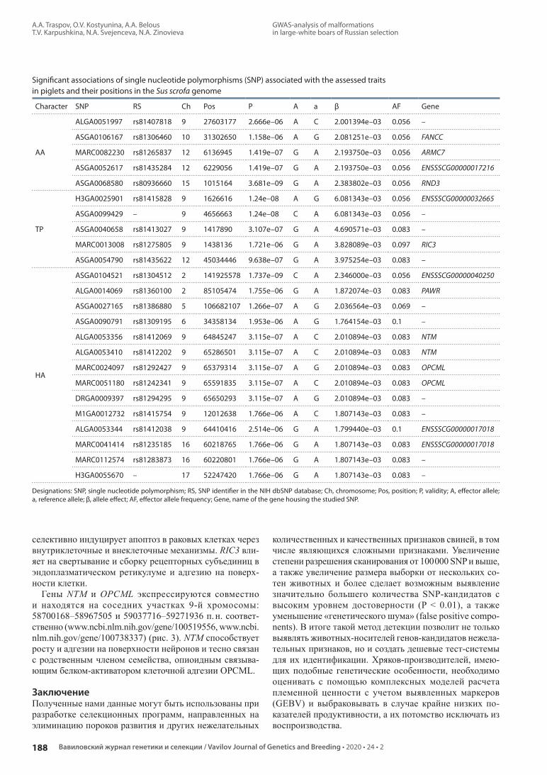
A.A. Traspov, O.V. Kostyunina, A.A. Belous T.V. Karpushkina, N.A. Svejenceva, N.A. Zinovieva
188 Вавиловский журнал генетики и селекции / Vavilov Journal of Genetics and Breeding • 2020 • 24 • 2
GWAS-analysis of malformations in large-white boars of Russian selection
селективно индуцирует апоптоз в раковых клетках через внутриклеточные и внеклеточные механизмы. RIC3 вли-яет на свертывание и сборку рецепторных субъединиц в эндоплазматическом ретикулуме и адгезию на поверх-ности клетки.
Гены NTM и OPCML экспрессируются совместно и находятся на соседних участках 9-й хромосомы: 58700168–58967505 и 59037716–59271936 п. н. соответ-ственно (www.ncbi.nlm.nih.gov/gene/100519556, www.ncbi.nlm.nih.gov/gene/100738337) (рис. 3). NTM способствует росту и адгезии на поверхности нейронов и тесно связан с родственным членом семейства, опиоидным связыва-ющим белком-активатором клеточной адгезии OPCML.
ЗаключениеПолученные нами данные могут быть использованы при разработке селекционных программ, направленных на элиминацию пороков развития и других нежелательных
количественных и качественных признаков свиней, в том числе являющихся сложными признаками. Увеличение степени разрешения сканирования от 100 000 SNP и выше, а также увеличение размера выборки от нескольких со-тен животных и более сделает возможным выявление значительно большего количества SNP-кандидатов с высоким уровнем достоверности (P < 0.01), а также уменьшение «генетического шума» (false positive compo-nents). В итоге такой метод детекции позволит не только выяв лять животных-носителей генов-кандидатов нежела-тельных признаков, но и создать дешевые тест-системы для их идентификации. Хряков-производителей, имею-щих подобные генетические особенности, необходимо оценивать с помощью комплексных моделей расчета племенной ценности с учетом выявленных маркеров (GEBV) и выбраковывать в случае крайне низких по-казателей продуктивности, а их потомство исключать из воспроизводства.
Significant associations of single nucleotide polymorphisms (SNP) associated with the assessed traits in piglets and their positions in the Sus scrofa genome
Character SNP RS Ch Pos P А а β AF Gene
AA
ALGA0051997 rs81407818 9 27603177 2.666e–06 A C 2.001394e–03 0.056 –
ASGA0106167 rs81306460 10 31302650 1.158e–06 A G 2.081251e–03 0.056 FANCC
MARC0082230 rs81265837 12 6136945 1.419e–07 G A 2.193750e–03 0.056 ARMC7
ASGA0052617 rs81435284 12 6229056 1.419e–07 G A 2.193750e–03 0.056 ENSSSCG00000017216
ASGA0068580 rs80936660 15 1015164 3.681e–09 G A 2.383802e–03 0.056 RND3
TP
H3GA0025901 rs81415828 9 1626616 1.24e–08 A G 6.081343e–03 0.056 ENSSSCG00000032665
ASGA0099429 – 9 4656663 1.24e–08 C A 6.081343e–03 0.056 –
ASGA0040658 rs81413027 9 1417890 3.107e–07 G A 4.690571e–03 0.083 –
MARC0013008 rs81275805 9 1438136 1.721e–06 G A 3.828089e–03 0.097 RIC3
ASGA0054790 rs81435622 12 45034446 9.638e–07 G A 3.975254e–03 0.083 –
HA
ASGA0104521 rs81304512 2 141925578 1.737e–09 C A 2.346000e–03 0.056 ENSSSCG00000040250
ALGA0014069 rs81360100 2 85105474 1.755e–06 G A 1.872074e–03 0.083 PAWR
ASGA0027165 rs81386880 5 106682107 1.266e–07 A G 2.036564e–03 0.069 –
ASGA0090791 rs81309195 6 34358134 1.953e–06 A G 1.764154e–03 0.1 –
ALGA0053356 rs81412069 9 64845247 3.115e–07 A C 2.010894e–03 0.083 NTM
ALGA0053410 rs81412202 9 65286501 3.115e–07 A C 2.010894e–03 0.083 NTM
MARC0024097 rs81292427 9 65379314 3.115e–07 A G 2.010894e–03 0.083 OPCML
MARC0051180 rs81242341 9 65591835 3.115e–07 A C 2.010894e–03 0.083 OPCML
DRGA0009397 rs81294295 9 65650293 3.115e–07 A G 2.010894e–03 0.083 –
M1GA0012732 rs81415754 9 12012638 1.766e–06 A C 1.807143e–03 0.083 –
ALGA0053344 rs81412038 9 64410416 2.514e–06 G А 1.799440e–03 0.1 ENSSSCG00000017018
MARC0041414 rs81235185 16 60218765 1.766e–06 G A 1.807143e–03 0.083 ENSSSCG00000017018
MARC0112574 rs81283873 16 60220801 1.766e–06 G A 1.807143e–03 0.083 –
H3GA0055670 – 17 52247420 1.766e–06 G A 1.807143e–03 0.083 –
Designations: SNP, single nucleotide polymorphism; RS, SNP identifier in the NIH dbSNP database; Ch, chromosome; Pos, position; P, validity; A, effector allele; a, reference allele; β, allele effect; AF, effector allele frequency; Gene, name of the gene housing the studied SNP.

GWAS-анализ пороков развития у хряков крупной белой породы российской селекции
А.А. Траспов, О.В. Костюнина, А.А. Белоус Т.В. Карпушкина, Н.А. Свеженцева, Н.А. Зиновьева
202024 • 2
189ГЕНЕТИКА ЖИВОТНЫХ / ANIMAL GENETICS
10
8
6
4
2
0
8
6
4
2
0
8
6
4
2
0
8
6
4
2
0
10
8
6
4
2
0
10
8
6
4
2
0
1 1
1
1
0
0
0
pval
Ppv
alP
pval
P
1
1
–log
10( p
)–l
og10
( p)
–log
10( p
)
10
10
10
11
11
11
12
12
12
13
13
13
14
14
14
15
15
15
16
16
16
17
17
17
18
λ = 1.007
λ = 1.083
λ = 1.018
18
18
2 2
2
2
2
2
3 3
3
3
3
3
4 4
4
4
4
4
5
a
b
c
5
5
6
6
6
7
7
7
8
8
8Chromosome
9
9
9
Fig. 1. Manhattan plot illustrating GWAS (genome-wide association study, top-left) and corresponding probability distributions of confidence P (Q-Q graph) in piglets of the studied populations of large white pigs (Znamenskiy Breeding Center): a, anal atresia (AA); b, hoof problems (HA); and c, spastic tremor (TP). The solid line indicates the Bonferroni level (0.05). Reliable values satisfying the null Bonferroni hypothesis (BFR) are marked with diamonds.
Fig. 2. Haploview LD chart illustrating 17 SNPs with the highest r2 values of coupling between ALGA0053356 and four poly-morphic variants – ALGA0053410, MARC0024097, MARC0051180, and DRGA0009397 to form a 363-kb haploblock in group HA.
AAAA .083
Block 1 (363 kb)1 10 11 12 13 14 15 16 172 3 4 5 6 7 8 9
CCCC .917
ALG
A00
5320
3
M1G
A00
1305
2
DRG
A00
0938
6
ALG
A01
2114
0
ALG
A00
5334
4
ALG
A00
5335
6
MA
RC00
4090
8
ALG
A00
5341
0
MA
RC00
2409
7
MA
RC00
5118
0
DRG
A00
0939
7
DRG
A00
0940
8
MA
RC00
6880
6
ASG
A00
4357
4
ALG
A01
2116
6
ALG
A00
5616
9
MA
RC00
0885
2
08 09 10 11

A.A. Traspov, O.V. Kostyunina, A.A. Belous T.V. Karpushkina, N.A. Svejenceva, N.A. Zinovieva
190 Вавиловский журнал генетики и селекции / Vavilov Journal of Genetics and Breeding • 2020 • 24 • 2
GWAS-analysis of malformations in large-white boars of Russian selection
Проделанная работа иллюстрирует необходимость про-ведения дополнительных исследований с использовани ем методов GWAS в аспекте характеристики популяций сель-скохозяйственных животных по ДНК-маркерам и иденти-фикации комплексных генотипов, ассоциированных с се-лекционно значимыми признаками как пороков развития, так и продуктивных качеств. Данное направление крайне необходимо в современных условиях высокоэффектив-ного воспроизводства сельскохозяйственных животных.
Список литературы / ReferencesДолматова A.B., Сковородин Е.Н. Использование ДНК-поли мор-
физма в селекции свиней. В: Материалы междунар. науч.-практ. конф. «Современные проблемы интенсификации произ водства свинины в странах СНГ», посвященной 75-летнему юби лею заслуженного деятеля науки РФ, профессора В.Е. Уитько, 7–10 июля 2010 г. Ульяновск, 2010;138-143.[Dolmatova A., Skovorodin E. The use of DNA polymorphism in the selection of pigs. In: Proc. of the Int. Sci. and Pract. Conf. “Modern Problems of Intensification of Pork Production in the CIS Coun-tries”, dedicated to the 75th anniversary of the Honored scientist of the Russian Federation, professor V.E. Ulitko, July 7–10, 2010. Ulyanovsk, 2010;138-143. (in Russian)]
Племяшов К.В. Геномная селекция – будущее животноводства. Животноводство России. 2014;5:2-4.[Plemyashov K. Genomic selection – the future of animal husbandry. Zhivotnovodstvo Rossii = Animal Husbandry of Russia. 2014;5:2-4. (in Russian)]
Barrett J., Fry B., Maller J., Daly M. Haploview: analysis and visuali-zation of LD and haplotype maps. Bioinformatics. 2005;21(2):263-265. DOI 10.1093/bioinformatics/bth457.
Benjamini Y., Hochberg Y. Controlling the false discovery rate: a prac-tical and powerful approach to multiple testing. J. R. Statist. Soc. B. 1995;57(1):289-300. DOI 10.2307/2346101.
Boddicker N., Waide E.H., Rowland R.R.R., Lunney J.K., Garrick D.J., Reecy J.M., Dekkers J.C.M. Evidence for a major QTL associated with host response to porcine reproductive and respiratory syndrome virus challenge. J. Anim. Sci. 2012;90(6):1733-1746.
Bruun C.S., Jorgensen C.B., Nielsen V.H., Andersson L., Fredholm M. Evaluation of the porcine melanocortin 4 receptor (MC4R) gene as a positional candidate for a fatness QTL in a cross between Landrace and Hampshire. Anim. Genet. 2006;37(4):359-362. DOI 10.1111/j.1365-2052.2006.01488.x.
Ciobanu D.C., Lonergan S.M., Huff-Lonergan E.J. Genetics of meat quality and carcass traits. In: Rothschild M.F., Ruvinsky A. (Eds.). The Genetics of the Pig. 2nd ed. Wallingford: CAB International, 2011;355-389.
Ernst C.W., Steibel J.P. Molecular advances in QTL discovery and ap-plication in pig breeding. Trends Genet. 2013;29(4):215-224. DOI 10.1016/j.tig.2013.02.002.
McLaren W., Gil L., Hunt S., Riat H., Ritchie G., Thormann A., Flicek P., Cunningham F. The Ensembl Variant Effect Predictor. Genome Biol. 2016;17(1):122. DOI 10.1186/s13059-016-0974-4.
Nyachoti C., Kiarie E., Bhandari S., Zhang G., Krause D. Weaned pig responses to Escherichia coli K88 oral challenge when receiving a lysozyme supplement. J. Anim. Sci. 2012;90(1):252-260. DOI 10.2527/jas.2010-3596.
Purcell S., Neale B., Todd-Brown K., Thomas L., Ferreira M., Ben-der D., Maller J., Sklar P., Bakker P., Daly M., Sham P. PLINK: a toolset for whole-genome association and population-based linkage analysis. Am. J. Hum. Genet. 2007;81:559-575. DOI 10.1086/519795.
Peterson R.A. Estimating normalization transformations with best-Normalize. 2017. Available at: https://github.com/petersonR/best Normalize
Salas R., Mingala C. Genetic factors affecting pork quality: halothane and rendement napole genes. Anim. Biotechnol. 2017;28(2):148-155. DOI 10.1080/10495398.2016.1243550. Epub 2016 Nov. 17.
See T., Max F., Rothschild C., Christains J. Swine Genetic Abnormali-ties. Pork Information Gateway. 2006; PIG 06-06-01.
Sermyagin А., Gladyr E., Plemyashov K., Kudinov A., Dotsev A., Deniskova T., Zinovieva N. Genome-wide association studies for milk production traits in Russian population of Holstein and black-and-white cattle. In: Anisimov K.V. et al. (Eds.). Proc. of the Sci.-Pract. Conf. “Research and Development – 2016”, 14–15 Dec. 2016, Moscow, Russia. Springer Open, 2018;591-599. DOI 10.1007/978-3-319-62870-7_62.
Sermyagin A., Kovalyuk N., Ermilov A., Yanchukov I., Satsuk V., Do tsev A., Deniskova T., Brem G., Zinovieva N.A. Associations of Bola-drb3 genotypes with breeding values for milk produc-tion traits in Russian dairy cattle population. Selskokhozyaystven-naya Biologiya = Agricultural Biology. 2016;51(6):775-781. DOI 10.15389/agrobiology.2016.6.775eng.
Storey J., Bass A., Dabney A., Robinson D. qvalue: Q-value estima-tion for false discovery rate control. R package version 2.10.1. 2017. http://github.com/StoreyLab/qvalue
Turner S. qqman: Q-Q and Manhattan Plots for GWAS Data. R package version 0.1.4. 2017. https://CRAN.R-project.org/package = qqman
Zhou X., Stephens M. Genome-wide efficient mixed-model analysis for association studies. Nat. Genet. 2012;44:821-824. DOI 10.1038/ng.2310.
OPCML
NTM
RIC3
ARMC7
FANCC
RND3PAWR
Fig. 3. String diagram (https://string-db.org) illustrating the strong pro-tein-mediated interaction of the coexpressed OPMCL and NTM genes, as well as their indirect effect on RND3.
Acknowledgements. This work was supported by the Russian Science Foundation, project 18-76-00034.Conflict of interest. The authors declare no conflict of interest.Received April 24, 2019. Revised November 05, 2019. Accepted November 06, 2019.

Коэкспрессия глутаматергических генов и генов аутистического спектра в гиппокампе у самцов мышей с нарушением социального поведенияИ.Л. Коваленко , А.Г. Галямина, Д.А. Смагин, Н.Н. Кудрявцева
Федеральный исследовательский центр Институт цитологии и генетики Сибирского отделения Российской академии наук, Новосибирск, Россия
e-mail: [email protected]
Аннотоция. В настоящее время существует представление о вовлеченности глутаматергической системы (ГГ) в механизмы развития аутизма. В предыдущих исследованиях нами было показано, что негативный социаль-ный опыт, приобретенный в ежедневных межсамцовых конфронтациях, приводит к нарушениям в социальном поведении: снижению коммуникативности, нарушению социализации, появлению стереотипных форм пове-дения, которые могут рассматриваться как симптомы аутистического спектра. В связи с этим целью нашей ра-боты было изучение с помощью транскриптомного анализа изменений экспрессии генов, кодирующих белки, вовлеченные в функционирование глутаматергической системы, и генов, связанных с патологией аутизма (ГА), в гиппокампе. В эксперименте использовали животных с нарушениями социального поведения, вызванными повторным опытом социальных побед или поражений в ежедневных агонистических взаимодействиях. Для формирования групп животных с контрастными типами поведения использовали модель сенсорного контакта (хронического социального стресса). Полученные образцы мозга были секвенированы в ЗАО «Геноаналитика» (http://genoanalytica.ru/,, Россия, Москва). Транскриптомный анализ показал, что у агрессивных животных снижа-ется экспрессия генов Shank3, Auts2, Ctnnd2, Nrxn2, для которых показано участие в развитии аутизма, а также глу-таматергического гена Grm4. В то же время у животных с негативным социальным опытом экспрессия ГА Shank2, Nlgn2, Ptcdh10, Reln, Arx возрастает. При этом ГГ (Grik3, Grm2, Grm4, Slc17a7, Slc1a4, Slc25a22), за исключением гена Grin2a, повышают свою экспрессию. Корреляционный анализ выявил статистически значимую взаимо связь из-мененной экспрессии ГГ и ГА. Полученные результаты, с одной стороны, могут служить подтверждением участия ГГ в патофизиологии развития симптомов аутистического спектра, с другой – свидетельствовать о коэкспрессии ГГ и ГА в гиппокампе, развивающейся под влиянием социальной среды. Так как большинство ГА, изменивших экспрессию в настоящем исследовании, являются генами, связанными с клеточным скелетом и внеклеточным матриксом, в частности участвующими в формировании синапсов, а ГГ, изменившие свою экспрессию, – генами, кодирующими субъединицы рецепторов, то можно предположить, что вовлечение ГГ в патофизиологию аутиз-ма происходит на уровне рецепторов.Ключевые слова: RNA-seq; аутизм; гиппокамп; гены аутизма; глутаматергические гены; социальный опыт.
Для цитирования: Коваленко И.Л., Галямина А.Г., Смагин Д.А. , Кудрявцева Н.Н. Коэкспрессия глутаматергических генов и генов аутистического спектра в гиппокампе у самцов мышей с нарушением социального поведения. Вавиловский журнал генетики и селекции. 2020;24(2):191-199. DOI 10.18699/VJ20.42-o
Co-expression of glutamatergic and autism-related genes in the hippocampus of male mice with disturbances of social behaviorI.L. Kovalenko , A.G. Galyamina, D.A. Smagin, N.N. Kudryavtseva
Institute of Cytology and Genetics of Siberian Branch of the Russian Academy of Sciences, Novosibirsk, Russia e-mail: [email protected]
Abstract. There is a hypothesis of the involvement of the glutamatergic system in the development of autism. It has been shown that the chronic experience in daily intermale confrontations leads to disturbances in social behavior: a decrease in communicativeness, disturbances of socialization, emergence of stereotypical behaviors that can be con-sidered as symptoms of the autistic spectrum disorders. So, the aim of this study was to investigate changes in the expression of glutamatergic (GG) and autism-related (GA) genes in the hippocampus of animals with impaired social behavior caused by repeated experience of social defeat or aggression in daily agonistic confrontations. To form groups of animals with contrasting behaviors, a model of sensory contact (chronic social stress) was used. The collected brain samples were sequenced at JSC Genoanalytica (http://genoanalytica.ru/, Moscow, Russia). Transcriptomic analysis re-
© Коваленко И.Л., Галямина А.Г., Смагин Д.А. , Кудрявцева Н.Н., 2020
This work is licensed under a Creative Commons Attribution 4.0 License
ФИЗИОЛОГИЧЕСКАЯ ГЕНЕТИКАОригинальное исследование / Original article
Вавиловский журнал генетики и селекции 2020;24(2):191-199DOI 10.18699/VJ20.42-o

I.L. Kovalenko, A.G. Galyamina D.A. Smagin, N.N. Kudryavtseva
192
Co-expression of glutamatergic and autism-related genes in the hippocampus of male mice
Вавиловский журнал генетики и селекции / Vavilov Journal of Genetics and Breeding • 2020 • 24 • 2
vealed a down-regulation of autism-related (Shank3, Auts2, Ctnnd2, Nrxn2) and glutamatergic (Grm4) genes in aggres-sive mice. At the same time, the expression of GA-related genes (Shank2, Nlgn2, Ptcdh10, Reln, Arx) and GG genes (Grik3, Grm2, Grm4, Slc17a7, Slc1a4, Slc25a22) excluding Grin2a was increased in defeated mice. Correlative analysis revealed a statistically significant association between GG and GA expression. These results can serve as a confirmation of the participation of the glutamatergic system in the pathophysiology of the autistic spectrum disorder. Key words: RNA-seq; autism; hippocampus; glutamatergic genes; autism-related genes; social experience.
For citation: Kovalenko I.L., Galyamina A.G., Smagin D.A., Kudryavtseva N.N. Co-expression of glutamatergic and autism-related genes in the hippocampus of male mice with disturbances of social behavior. Vavilovskii Zhurnal Genetiki i Selektsii = Vavilov Journal of Genetics and Breeding. 2020;24(2):191-199. DOI 10.18699/VJ20.42-o (in Russian)
ВведениеПолагают, что выраженные нарушения в социальном поведении могут свидетельствовать об аутизме, который проявляется в детском возрасте и представляет собой нарушения развития нервной системы (Bauman, Kemper, 2005; Zablotsky et al., 2015). Согласно DSM-IV (American Psychiatric Association…, 1993), диагностические кри-терии аутизма включают триаду основных признаков: ухудшение социализации, под которой можно понимать способность индивида адекватно встраиваться в социум в новой обстановке, низкий уровень общительности и повторяющееся/стереотипное поведение. Хотя изучение близнецов свидетельствует о высокой наследуемости аутизма (Hallmayer et al., 2011), ни один ген не определен как единственная причина развития этого заболевания. Недавние геномные и генетические исследования по-казали, что сотни генетических вариантов, включающие общие и редкие взаимодействия генов, способствуют воз-никновению аутизма (Miles, 2011). Согласно различным базам данных (http://omim.org/, http://www.genecards.org/, http://autism.mindspec.org/autdb/Welcome.dohttp://www.malacards.org), в базе генов аутизма насчитывается около 1500 генов, которые в той или иной мере вовлечены в механизмы аутизма. Многие гены, связанные с развити-ем головного мозга, – потенциальные гены-кандидаты аутизма, к ним относятся гены и семейства генов Shank (1–3), Nlgn, Reln, Arx, Pcdh, Mecp2, Auts2 (Kleijer et al., 2014; Liu et al., 2015).
Литературные данные свидетельствуют об участии раз личных нейромедиаторных систем (например, серо-тонергической, дофаминергической) в развитии аутизма (Pa văl, 2017). В последнее время большое внимание уде-ляется глутаматергической гипотезе аутизма (Rojas, 2014). В пользу этой теории говорит то, что глутамат является од-ним из наиболее распространенных нейротрансмиттеров в мозге млекопитающих, его рецепторы сосредоточены в областях мозга (мозжечок, гиппокамп, префронтальная кора), в которых обнаружены нейропатологические изме-нения при аутизме. Во взрослом мозге глутаматные рецеп-торы участвуют в обучении и памяти (Riedel et al., 2003; Simonyi, 2010). Глутамат-опосредованные межнейронные взаимодействия также играют роль в формировании эмо-ционального поведения (Morgane et al., 2005; Faure et al., 2010). Показано, что для больных аутизмом характерен повышенный уровень глутамата в плазме крови, который может даже служить одним из биомаркеров этого заболе-вания (Zheng et al., 2016). У пациентов с аутизмом обна-руживаются и молекулярно-генетические повреждения глутаматергической системы (ГГ). Так, например, есть
сообщения о мутациях в генах глутаматного рецептора GluR6 (Jamain et al., 2002), а также митохондриального переносчика глутамата (AGC1, ген Slc25a12) (Ramoz et al., 2004). Изменения наблюдаются на уровне м-РНК, белков-транспортеров и рецепторов ГГ в посмертных образцах мозга больных аутизмом (Purcell et al., 2001). В целом некоторые исследователи связывают аутизм с дефицитом глутаматергической системы в мозге (Сarlsson, 2015).
Ранее нами было показано, что в условиях хрониче-ского социального конфликта, вызванного повторным опытом побед или поражений в ежедневных межсамцо-вых конфронтациях (Kudryavtseva, 1991), у самцов мы-шей формируются не только повышенная тревожность, агрессивность или депрессивноподобное поведение, но и нарушения коммуникативного поведения и социального взаимодействия. Такие животные демонстрировали сни-женную коммуникативность, а также стереотипные фор-мы поведения (аутогруминг, вставание на задние лапы, кружение, разрывание и разбрасывание подстилки, пово-роты в прыжке и др.), что позволяет рассматривать данную модель как модель, воспроизводящую некоторые черты аутистического поведения (Коваленко, Кудрявцева, 2010), формирующиеся под влиянием социальной среды. При этом наши предыдущие исследования показали, что в этот процесс были вовлечены гены аутистического спектра, экспрессия которых изменялась под влиянием агонисти-ческих взаимодействий (Kudryavtseva et al., 2017).
В связи с этим целью настоящей работы было изучить у животных с альтернативным опытом социального пове-дения, позитивным и негативным, взаимосвязь изменения экспрессии генов, кодирующих белки, вовлеченные в функционирование ГГ, и генов, связанных с патологией аутизма (ГА) в гиппокампе. Выбор этой структуры голов-ного мозга основан на многочисленных литературных данных о том, что гиппокамп непосредственно участвует в патогенетических про цессах аутизма (DeLong, 1992), в развитии тревожных расстройств (Irle et al., 2010) и депрессии (Savitz, Drevets, 2009). Гиппокамп отвечает за эмоциональную саморегуляцию, обучаемость и память. А, как известно, нарушение именно этих функций часто наблюдается у людей, демонстрирующих симптомы аути-стического спектра.
Материалы и методыЖивотные. Эксперименты проводили на самцах мышей линии С57BL/6J в возрасте 2.5 мес. и массой тела 26–28 г. Животные были привезены из питомника лабораторных животных Института биоорганической химии Российской академии наук (Пущино, Московская область). Экс пе-

Коэкспрессия глутаматергических генов и генов аутистического спектра в гиппокампе у самцов мышей
И.Л. Коваленко, А.Г. Галямина Д.А. Смагин, Н.Н. Кудрявцева
193ФИЗИОЛОГИЧЕСКАЯ ГЕНЕТИКА / PHYSIOLOGICAL GENETICS
202024 • 2
римент проводили в виварии конвенциональных живот - ных Института цитологии и генетики Сибирского отде-ления РАН. Воду и корм (гранулы) животные получали в достаточном количестве. Световой режим был 12С : 12Т. Все процедуры осуществляли в соответствии с между-народными правилами проведения экспериментов с жи-вотными (Directive 2010/63/EU of the European Parliament and of the Council on the Protection of Animals Used for Scientific Purposes). Применяемые методики для изучения поведения у мышей были одобрены Научной комиссией (№ 9) Института цитологии и генетики СО РАН (Протокол № 613 от 24.03.2010).
Поведенческие исследования. Для формирования альтернативного опыта социального поведения у самцов мышей использовали модель сенсорного контакта (Kud-ryavtseva et al., 2014). Животных попарно помещали в экспериментальные клетки, разделенные пополам про-зрачной перегородкой с отверстиями, позволявшей мы-шам видеть, слышать, воспринимать запахи друг друга (сенсорный контакт), но предотвращавшей физическое взаимодействие. Ежедневно во второй половине дня (15:00–17:00) убирали перегородку, что приводило к агонистическим взаимодействиям. В течение первых двух-трех дней тестов выявляли победителей (агрессоров/агрессивных животных) и особей, терпящих поражения (жертв) при взаимодействии с одним и тем же партне ром. В дальнейшем ежедневно после теста побежденного самца пересаживали в новую клетку к незнакомому агрессивно-му партнеру, сидящему за перегородкой. Взаимодействие самцов прекращали, если интенсивные атаки со стороны нападающей особи во время агрессивных столкновений длились более трех минут, устанавливая между ними перегородку. В исследование были взяты аг рессивные животные с 20-дневным опытом побед (агрес соры) и с 20-дневным опытом поражений (жертвы). В качестве контроля использовали самцов, не имевших последова-тельного опыта агонистических взаимодействий. В каж-дой группе было по 14–16 животных.
Количественную оценку реакции экспериментальных животных на незнакомого партнера на нейтральной тер-ритории проводили при помощи теста «социальные взаи-модействия» (Kudryavtseva et al., 2017). Как параметры социального поведения мы рассматривали избегание незнакомого партнера или же замирание при его подходе и приближение к партнеру: подходы, обнюхивания, сле-дование за партнером; как параметры индивидуального поведения выделяли стойки – вставания на задние лапы, являющиеся показателем исследовательской активно-сти, аутогруминг, служащий показателем смещенной активности и неадекватного стереотипного поведения, и двигательную активность, оценивающую интенсивность передвижений по клетке.
Статистическую обработку полученных данных вы-полняли с использованием пакета программ STATISTICA (ver. 8.0; StatSoft, Inc., 2001). Проверка нормальности рас-пределения количественных признаков была проведена с использованием критерия Шапиро-Уилка (Shapiro-Wilk’s W-test). Поскольку выборки исследованных параметров поведения удовлетворяли гипотезе о нормальном рас-пределении, были использованы методы параметриче-
ской статистики: однофакторный дисперсионный анализ (ANOVA) с фактором «группа» (контроль, агрессивные самцы, жертвы); последующее попарное сравнение по-казателей осуществляли с помощью LSD-теста Фишера.
При помощи корреляционного анализа по методу Пир-сона мы исследовали взаимосвязь экспрессии ГГ и ГА. В экспериментальных поведенческих группах было по 10–12 животных.
RNA-Seq-анализ проводили с помощью ЗАО «Гено-аналитика» (http://genoanalytica.ru, Москва). Методика анализа подробно описана ранее (Галямина и др., 2017).
Мы провели проверку результатов, сравнив их с дан-ными B.M. Kadakkuzha (Kadakkuzha et al., 2015), пред-ставившими полный транскриптомный анализ генов в гиппокампе интактных мышей. Обнаружен высокий уровень корреляции (0.74 по Спирмену) между экспрес-сией генов аутизма и ГГ у контрольных особей в нашем эксперименте и у интактных животных в работе (Kadda-kuzha et al., 2015), что может быть дополнительным до-казательством адекватности применяемого метода. Кро- ме того, осуществляли кросс-верификацию результатов (Ba benko et al., 2017) с данными, полученными в Стэнд-фордском университете (Zhang et al., 2014), и обнаружили значительную корреляцию между ними. Это доказывает, что метод транскриптомного анализа позволяет выявить происходящие в мозге молекулярные процессы.
В качестве верификации данных настоящего экспери-мента использовали результаты, полученные нами ранее при помощи RT-PCR (Smagin et al., 2013), экспрессия ко-торых, по методу RNA-Seq, была изменена. Было показано совпадение направленности и выраженности изменений экспрессии для генов, кодирующих белки серотонерги-ческой системы в ядрах шва среднего мозга, полученных при применении этих методов, что позволяет говорить о высокой надежности результатов этого исследования и о стабильности примененного метода.
Декапитацию всех трех групп экспериментальных животных проводили одновременно на следующий день после последней конфронтации. Гиппокамп извлекался одним исследователем в соответствии с анатомическим атласом мозга (Allen Mouse Brain Atlas; http://mouse.brain map.org/static/atlas). Все образцы помещали в рас-твор RNAlater (LifeTechnologies, США) и хранили при температуре –70 °С до секвенирования. Проводили два типа сравнения: контроль–агрессоры и контроль–жертвы.
Для изучения изменений глутаматергической системы в гиппокампе животных с нарушенным социальным по-ведением были исследованы: гены переносчиков глутама-та: Slc17a6, Slc17a7 и Slc17a8; гены, кодирующие 1–4-ю субъединицы ионотропного AMPA-рецептора: Gria1, Gria2, Gria3, Gria4; гены, кодирующие 1–5-ю субъеди-ницы ионотропного каинатного глутаматного рецептора: Grik1, Grik2, Grik3, Grik4, Grik5; гены, кодирующие 1, 2а, 2b, 2c, 2d, 3a, 3b субъединицы ионотропного NMDA-рецептора: Grin1, Grin2a, Grin2b, Grin2c, Grin2d, Grin3a, Grin3b; гены, кодирующие метаботропные рецепторы 1–8-го подтипов: Grm1, Grm2, Grm3, Grm4, Grm5, Grm6, Grm7, Grm8; гены, кодирующие 1 и 2-ю субъединицы глу-таматного ионотропного рецептора дельта Grid1 и Grid2; GRID2IP – белок, взаимодействующий с Grid2; гены

I.L. Kovalenko, A.G. Galyamina D.A. Smagin, N.N. Kudryavtseva
194
Co-expression of glutamatergic and autism-related genes in the hippocampus of male mice
Вавиловский журнал генетики и селекции / Vavilov Journal of Genetics and Breeding • 2020 • 24 • 2
фермента глутаматдекарбоксилазы, метаболизирующей глутамат в ГАМК: Gad1 и Gad2.
По генетическим базам данных OMIM (http://omim.org/), GeneCards (http://www.genecards.org/), MalaCards (http://www.malacards.org/) из 1.5 тыс. аннотированных генов было выбрано около 80 основных генов-кандидатов аутизма, которые в дальнейшем были просмотрены в гип-покампе у контрольных особей и животных с нарушенным социальным поведением. При анализе сравнивали по три пробы от каждой группы животных.
РезультатыИсследование нарушений социального поведения у самцов мышей под влиянием хронического социаль-ного стресса. ANOVA выявил достоверное влияние фак-тора «группа» (контроль, агрессоры, жертвы) на избе га-ние партнера (F(2.29) = 52.30, p < 0.001), приближение (F(2.28) = 1097.4, p < 0.001), время стоек (F(2.29) = 661.7, p < 0.001), время двигательной активности (F(2.29) = 2549, p < 0.001). Сравнение групп LSD-тестом Фишера (рис. 1) выявило увеличение времени избегания у жертв, по срав-нению с контролем и агрессивными животными ( p < 0.001 для обеих групп). По сравнению с контролем у агрессоров и жертв также было показано снижение времени стоек ( p < 0.001 для обеих групп) и времени приближения к партнеру ( p < 0.001 для обеих групп). Кроме того, у жертв время приближения к партнеру было снижено и по сравнению с агрессивными животными ( p < 0.006). Выявлены увеличение времени двигательной активности у агрессоров ( р < 0.001) и снижение времени у жертв ( р < 0.042), по сравнению с контрольными животными. Время двигательной активности было значительно ниже у жертв, по сравнению с агрессорами ( р < 0.001). Кроме того, у жертв были увеличены число и время аутогру-минга, по сравнению с контролем ( p < 0.041, p < 0.034 соответственно).
Таким образом, мы видим, что в тесте «социальные взаимодействия» жертвы активно избегали незнакомого партнера (тестера) на нейтральной территории. Они редко первыми подходили и проявляли интерес к незнакомому партнеру, в отличие от контроля. Контрольные животные целенаправленно следовали за партнером, обнюхивали его. У жертв также было снижено время стоек, рассмат-
риваемое нами как показатель исследовательской актив-ности. Кроме того, у них было увеличено время демон-страции аутогруминга, который может оцениваться как признак стереотипного поведения. При этом отмечено, что у агрессивных животных были снижены параметры коммуникативности, что может свидетельствовать о на-рушениях социального поведения. Большую часть 10-ми-нутного теста (около 5–6 мин) агрессоры хаотично пере-мещались по клетке, не обращая внимания на партнера: общее время двигательной активности было значительно больше, чем у жертв, что может отражать развитие гипер-активности и, по-видимому, дефицит внимания. Ранее нами было показано, что в агонистических взаимодей-ствиях агрессоры часто демонстрируют быстрые пово-роты в прыжке или же повторяющиеся повороты вокруг оси тела в тесте «перегородка» (Kudryavtseva, 2006), т. е. сте реотипии у агрессивных мышей этой линии могут про являться в другом тесте и другой форме. Таким об-разом, и у агрессоров, и у жертв в результате 20-дневных агонистических взаимодействий развиваются симптомы аутистического спектра.
Исследование экспрессии ГГ и ГА у самцов мышей с контрастными типами социального поведения. В ре-зультате анализа данных RNA-Seq в гиппокампе обна-ружено изменение экспрессии девяти генов-кандидатов аутизма (рис. 2, табл. 1). Так, у жертв выявлено увеличение экспрессии генов Shank2 ( p < 0.040), Nlgn2 ( p < 0.047), Pcdh10 ( p < 0.011), Reln ( p < 0.026) и Arx ( p < 0.0002), по сравнению с уровнем экспрессии у контрольных жи-вотных. У агрессивных мышей в гиппокампе под влия ни-ем повторного опыта агрессии в межсамцовых конфрон- тациях обнаружено снижение экспрессии генов Shank3 ( p < 0.010), Auts2 ( p < 0.023), Ctnnd2 ( p < 0.020), Nrxn2 ( p < 0.010).
Были проанализированы изменения уровня экспрессии генов, кодирующих белки, вовлеченные в функциони-рование ГГ в гиппокампе мышей (см. табл. 1, рис. 3). Показано, что у мышей в гиппокампе под влиянием по-вторного опыта агрессии в межсамцовых конфронтациях снижена экспрессия гена Grm4 ( p < 0.023), кодирующего метаботропный рецептор 4-го подтипа. У жертв было обнаружено снижение экспрессии гена Grin2a ( p < 0.01), кодирующего субъединицу 2а ионотропного NMDA-ре-
Fig. 1. Behavior of the aggressors and losers in the social interactions test.* p < 0.05; *** p < 0.001 vs control; ++ p < 0.01; +++ p < 0.001 – losers vs aggressors.
ControlAggressorsLosers
Avoidance, s Approach, s Rearings, s Self-grooming, s Locomotion, s
+++***
******
***
***
*
++***
*+++
150
100
50
0
150
100
50
0
150
100
50
0
350
280
210
140
70
0
50
40
30
20
10
0

Коэкспрессия глутаматергических генов и генов аутистического спектра в гиппокампе у самцов мышей
И.Л. Коваленко, А.Г. Галямина Д.А. Смагин, Н.Н. Кудрявцева
195ФИЗИОЛОГИЧЕСКАЯ ГЕНЕТИКА / PHYSIOLOGICAL GENETICS
202024 • 2
Shank3
FPKM
Shank2Ctnnd2 Pcdh10Auts2 Nlgn2Nrxn2 Reln Arx
120
100
80
60
40
20
0
**
*****
**
*
*
*
*
*
Grm4
FPKM
Grm4Grik3 Slc25a22 Slc17a7Grin2a Slc1a4Grm2
30
25
20
15
10
5
0
120
100
80
60
40
20
0
700
600
500
400
300
200
100
0
**
**
*
****
**
*
Fig. 2. Change in GA expression in the hippocampus of aggressors and losers. * р < 0.05; ** p < 0.01; *** p < 0.001 – vs control.
Fig. 3. Change in expression of GG in the hippocampus in mice with a disturbances of social behavior. * р < 0.05, ** p < 0.01 – vs control.
цеп тора, при этом повышалась экспрессия генов Grm2 ( p < 0.004) и Grm4 ( p < 0.02), кодирующих метаботроп-ные рецепторы 2 и 4-го подтипов, гена Grik3 ( p < 0.003), кодирующего субъединицу 3 ионотропного каинатного глутаматного рецептора, генов переносчиков глутамата Slc17a7 ( p < 0.051), Slc1a4 ( p < 0.01), Slc25a22 ( p < 0.028).
Результаты корреляционного анализа продемонстри-ровали высокий уровень корреляционной взаимосвязи между экспрессией ГГ и ГА у животных исследуемых групп (см. табл. 1 и 2). Корреляция между уровнями экс-
прессии ГА и ГГ показывает возможное участие глута-матэргической системы в механизмах этого заболевания. Из ГА наибольшее число корреляций обнаружено для генов Nlgn2, Pcdh10, Arx, Ctnnd2, Nrxn2. В то же время уровень экспрессии гена Reln коррелирует только с уров-нем экспрессии гена Slc1a4.
Значения экспрессии некоторых ГГ и ГА в единицах FPKM, между которыми установлена статистически зна-чимая коррелятивная взаимосвязь, показаны на рис. 4. Большая часть генов демонстрирует положительную кор-
Table 1. Differential expression GG and GA in hippocampus of aggressors and losers
Genes Agressors Losers
Associated with autistic spectrum disorders
Shank3, SH3 and Multiple Ankyrin Repeat Domains 3
Auts2, Activator of Transcription and Developmental Regulator
Ctnnd2, Catenin Delta 2
Nrxn2, Neurexin 2
Shank2, SH3 and Multiple Ankyrin Repeat Domains 2
Nlgn2, Neuroligin 2
Pcdh10, Protocadherin 10
Reln, Reelin
Arx, Aristaless Related Homeobox
Involved in the functioning of the glutaminergic system
Grm4, Glutamate Metabotropic Receptor 4
Grin2a, Glutamate Ionotropic Receptor NMDA Type Subunit 2A
Grik3, Glutamate Ionotropic Receptor Kainate Type Subunit 3
Grm2, Glutamate Metabotropic Receptor 2
SLc17a7, Solute Carrier Family 17 Member 7
Slc1a4, Solute Carrier Family 1 Member 4
Slc25a22, Solute Carrier Family 25 Member 22
Note: Decreased expression: – p < 0.05; – p < 0.01. Increased expression: – p < 0.05; – p < 0.01; – p < 0.001 vs control.
ControlAggressorsLosers
ControlAggressorsLosers

I.L. Kovalenko, A.G. Galyamina D.A. Smagin, N.N. Kudryavtseva
196
Co-expression of glutamatergic and autism-related genes in the hippocampus of male mice
Вавиловский журнал генетики и селекции / Vavilov Journal of Genetics and Breeding • 2020 • 24 • 2
реляцию друг с другом. Например, экспрессия Grm4, един-ственного гена, который изменил экспрессию и у жертв, и у агрессоров, положительно коррелирует с экспресси-ей ГА: Shank3 (r = 0.810, p < 0.01) и Arx (r = 0.862, p < 0.01) (см. рис. 4, а). Экспрессия гена Grin2a демонстрирует отрицательную корреляцию с экспрессией ГА, например генов Nlgn2 (r = –0.823, p < 0.01) и Arx (r = –0.823, p < 0.01) (см. рис. 4, б ). Единственный ген, с которым коррелирует уровень экспрессии гена Reln, – это Slc1a4 (r = 0.696, p < 0.05) (см. рис. 4, в).
ОбсуждениеВ наших предыдущих работах было обнаружено, что пос-ле 20 дней агонистических взаимодействий и проживания в условиях хронического социального конфликта у экс-периментальных самцов нарушаются многие параметры социального поведения (Коваленко, Кудрявцева, 2010), схожие по симптоматике с заболеваниями аутистического спектра. Мы считаем, что в данном случае коморбидные аутистические симптомы развиваются на фоне других за болеваний. Это может быть тревожно-депрессивное со стояние, возникающее вследствие хронического со-циального стресса у самцов мышей С57Bl/6 (Berton et al., 2006; Kudryavtseva et al., 2006) или каталепсии, раз-вивающейся у животных СВА/Lac (Kudryavtseva et al., 2006), или же патологической агрессии (Kudryavtseva, 2006), формирующейся под влиянием повторного опыта агрессии. В литературе существуют данные, свидетель-ствующие, что коморбидность аутистических симптомов и расстройств настроения может быть следствием работы
одних и тех же генов, продукты которых вовлекаются в патофизиологические механизмы развития этих заболе-ваний (Ragunath et al., 2011).
В связи с этим помимо генов, для которых показано участие в развитии симптомов аутизма, мы исследовали также изменения экспрессии ГГ (транспортеров, рецеп-торов, ферментов катаболизма), так как известно, что эта нейромедиаторная система вовлечена в развитие данной патологии (Сarlsson, 2015).
Мы выявили в гиппокампе изменение экспрессии семи ГГ: ген Grin2a кодирует NR2А-субъединицу NMDA-рецептора; Grm2 и Grm4 – метаботропные глутаматные рецепторы, mGluR2 и mGluR4 соответственно; Grik3 – субъединицу каинатного рецептора, Slc1a4 и Slc17a7 – транспортеры глутамата Vglut1; Slc25a22 – белок мито-хондриального переносчика глутамата. Изменили свою экспрессию 9 генов, ассоциированных с заболеваниями аутистического спектра: Shank2, Shank3, Nlgn2, Reln, Arx, Nrxn2, Auts2, Ctnnd2, Pcdh10.
Белки семейства SHANK, известные также как ProSAP, являются строительными белками на возбуждающих глутаматергических синапсах. Так, ген Shank2 кодирует белок, взаимодействующий на постсинаптической мем-бране с NMDA-рецептором, участвуя в синаптической глутаматергической передаче (Сarlsson, 2015). Вовлечение в процесс развития аутизма генов Shank было впервые описано для Shank3. Например, было отмечено, что мыши, гетерозиготные по белку Shank3 (Shank3+/delta-C), демон-стрировали низкий уровень социального распознавания и коммуникативности (Wang et al., 2011). В литературе было
Fig. 4. Correlating GG and GA. K1–3 – control animals; A1–3 – aggressors; L1–3 – losers. a, The right axis of ordinates is for the Shank3 gene, the left one is Arx and Grm4.
Table 2. Co-expression of GG and GA (Pearson coefficient value)
Gene Nlgn2 Pcdh10 Reln Shank2 Shank3 Arx Auts2 Ctnnd2 Nrxn2
Grm2 0.761 0.581 0.138 0.644 0.657 0.538 0.547 0.692 0.668
Grm4 0.894 0.915 0.537 0.827 0.810 0.862 0.602 0.868 0.823
Grin2a –0.825 –0.870 –0.397 –0.643 –0.521 –0.823 –0.581 –0.733 –0.867
Grik3 0.847 0.946 0.597 0.851 0.708 0.956 0.693 0.854 0.862
Slc1a4 0.677 0.640 0.696 0.515 0.437 0.714 0.178 0.632 0.561
Slc17a7 0.878 0.696 0.231 0.738 0.691 0.727 0.679 0.864 0.875
Slc25a22 0.922 0.918 0.457 0.906 0.792 0.940 0.802 0.933 0.935
Shank3Nlgn2
Reln
Arx
Arx
Grm4
Grin2a
Slc1a4
FPKM
FPKM
FPKM
K1
a b c
K1K1 K2 K2K2 K3 K3K3 A1 A1A1 A2 A2A2 A3 A3A3 L1 L1L1 L2 L2L2 L3 L3L3
10
8
6
4
2
0
16
12
8
4
0
90
60
30
0
100
80
60
40
20
0

Коэкспрессия глутаматергических генов и генов аутистического спектра в гиппокампе у самцов мышей
И.Л. Коваленко, А.Г. Галямина Д.А. Смагин, Н.Н. Кудрявцева
197ФИЗИОЛОГИЧЕСКАЯ ГЕНЕТИКА / PHYSIOLOGICAL GENETICS
202024 • 2
показано, что для аутистических симптомов характерно снижение функции Shank2 (Won et al., 2012). Нарушения в социальном поведении характерны как для пониженного (Peça et al., 2011), так и для повышенного (Moessner et al., 2007) уровня белка SHANK3. В нашей работе также об-наружены повышенный уровень экспрессии гена Shank2 и сниженный уровень гена Shank3. Вероятно, нарушения в социальном поведении могут быть связаны с любыми изменениями в функционировании этих белков. Известно, что ProSAP2/Shank3 влияет на работу глутаматергических синапсов, взаимодействуя с внеклеточным С-концом нейролигинов (Meyer et al., 2004). Мутации в этом гене ухудшают синаптическую передачу (Arons et al., 2012). Следовательно, можно предположить, что влияние белков SHANK на развитие аутистических симптомов осуществ-ляется через изменение функционирования глутаматер-гической системы.
Значительную часть ГА, изменивших свой уровень экс прессии в нашей работе, составляют гены клеточной адгезии: Nlgn2 – нейролигин 2-го типа; Pcdh10 – прото-кадерин; Ctnnd2 – δ-катенин; Nrxn3 – нейрексин 3. Для всех этих белков многократными исследованиями было показано участие в формировании социального поведения и расстройств аутистического спектра. Известно, что из-менение уровня белка NLGN2 может влиять на социаль-ное и эмоциональное поведение (Maćkowiak et al., 2014). У аутистических больных обнаружены мутации в генах Pcdh10 (Anitha et al., 2013), Ctnnd2 (Turner et al., 2015), Nrxn3 (Vaags et al., 2012). Кроме того, продукты этих генов взаимодействуют с глутаматергической системой. Так, например, нейролигины вызывают кластеризацию Vglut-положительных синаптических пузырьков (Graf et al., 2004), без чего невозможно созревание и функционирова-ние синапса. Правда, там же отмечено, что это характерно для нейролигина 1, а нейролигин 2 не располагается на одном синапсе с транспортером глутамата. Обнаружено, что в отсутствие нейролигинов снижается число Vglut1-положительных терминалей (Chih et al., 2005). Можно предположить, что на самом деле нейролигин взаимодей-ствует непосредственно с NMDA-рецепторами, а перенос-чик Vglut1 выступает только в качестве марке ра этих ре-цепторов. Что касается продукта гена Pcdh10, то установ- лено, что протокадерины локализуются на ней ронах, фор-мирующих глутаматергические AMPA и каинатные рецеп-торы (Puller, Haverkamp, 2011). Кроме того, показано, что у мышей, нокаутных по генам прото кадеринов, снижено число глутаматных транспортеров Vglut 1–2 (Chen et al., 2012), что также свидетельствует о взаимосвязи между протокадеринами и глутаматергической системой.
Продукты генов Arx и Reln (белок рилин) играют важ-ную роль в механизмах пре- и постнатального нейрогене-за. Участие этих генов в патофизиологии аутиз ма является спорным: часть авторов приводят данные в пользу этого факта (Wall et al., 2009), в то время как другие опровергают его (Persico et al., 2001). В то же время обнаружена связь этих генов с функционированием глутаматергической системы. Так, известно, что мутации в гене Arx связаны с изменениями в глутаматергической системе у больных эпилепсией (Beguin et al., 2013), а бе лок рилин способен повышать мобильность рецепторов, содержащих NR2B-
субъединицу, снижая время ее пребывания в синапсе, таким образом, изменяя состав рецепторов в сторону преобладания NR2А-субъединицы (Groc et al., 2007).
Ген Auts2 кодирует экспрессирующийся в головном мозге белок с неизвестной функцией, хотя предполагают, что он вовлечен в механизмы развития нервной системы (Oksenberg et al., 2013). Мутации в этом гене обнаружены у пациентов с аутистическими расстройствами (Liu et al., 2015). Показано, что Auts2 экспрессируется на глутаматер-гических нейронах гиппокампа (Hori et al., 2014).
Так как большинство ГА, изменивших экспрессию в нашем исследовании, – гены, связанные с клеточным скелетом и внеклеточным матриксом, в частности уча-ствующие в формировании синапсов, а ГГ – гены, коди-рующие субъединицы рецепторов, то можно полагать, что изменение активности глутаматергической системы проявляется в аутистических симптомах.
Корреляционный анализ наших экспериментальных данных в целом выявил высокий уровень корреляции между экспрессией ГГ и ГА. Исключение составил ген Reln, коррелирующий только с геном Slc1a4. Характерно также, что везде обнаруживается положительная корре-ляция между генами, в то время как ген Grin2a отрица-тельно коррелирует с ГА. Однако снижение экспрес сии Grin2a, продукт которого является субъединицей NMDA-рецептора, хорошо согласуется с литературными данны-ми, связывающими развитие аутистических симптомов с дефицитом этих рецепторов (Lee et al., 2015). Экспрессия гена Nlgn2 коррелирует с экспрессией всех ГГ, экспрес-сия генов Pcdh10, Arx, Cttnd2, Nrxn2 – с экспрессией практически всех генов. Это позволяет предположить, что именно при помощи данных генов ГГ вовлекается в механизмы развития аутистического поведения. Можно видеть, что все наиболее коррелирующие гены, кроме Arx, – гены клеточной адгезии. Мы предполагаем, что продукты этих генов участвуют в соединении синапсов, в том числе глутаматергических, с клеточной мембраной. Таким образом, они вовлекаются в функционирование глутаматергической системы. Следовательно, изменение экспрессии этих генов изменяет уровень активности ГГ, что, в свою очередь, приводит к изменениям в социальном поведении, хотя нельзя исключить и обратный вариант взаимодействия.
ЗаключениеПроведенное исследование подтвердило полученные ра нее результаты, свидетельствующие о том, что под влиянием хронического социального конфликта у сам-цов мышей могут развиваться нарушения социального поведения, причем в этом процессе участвуют как гены, кодирующие белки, вовлеченные в функционирование глутаматергической системы, так и гены, связанные с патологией аутизма. Коэкспрессия этих генов позволяет говорить о вовлечении глутаматергической системы головного мозга в развитие патологии социального по-ведения. Полученные результаты могут служить дока-зательством того, что аутистические симптомы могут не только быть следствием генетических нарушений, но и развиваться в течение жизни индивидуума под влиянием стрессорных воздействий.

I.L. Kovalenko, A.G. Galyamina D.A. Smagin, N.N. Kudryavtseva
198
Co-expression of glutamatergic and autism-related genes in the hippocampus of male mice
Вавиловский журнал генетики и селекции / Vavilov Journal of Genetics and Breeding • 2020 • 24 • 2
Список литературы / ReferencesГалямина А.Г., Коваленко И.Л., Смагин Д.А., Кудрявцева Н.Н.
Изменение экспрессии генов нейромедиаторных систем в вен-тральной тегментальной области депрессивных мышей: данные RNA-Seq. Журн. высш. нервн. деят. им. И.П. Павлова. 2017; 67(1):113-128. DOI 10.7868/S0044467717010063.[Galyamina A.G., Kovalenko I.L., Smagin D.A., Kudryavtseva N.N. Altered expression of neurotransmitters systems’ genes in the ven-tral tegmental area of depressive male mice: data of RNA-Seq. Zhur-nal Vysshey Nervnoy Deyatel’nosti imeni I.P. Pavlova = I.P. Pav-lov Journal of Higher Nervous Activity. 2017;67(1):113-128. DOI 10.7868/S0044467717010063. (in Russian)]
Коваленко И.Л., Кудрявцева Н.Н. Развитие симптомов аутисти-ческого спектра под влиянием хронического социального стрес-са у тревожных самцов мышей: эффекты диазепама. Психофар-макол. биол. наркология. 2010;10(1-2):2624-2635.[Kovalenko I.L., Kudryvtseva N.N. Development of autistic spec-trum symptoms under chronic social defeat stress in anxious male mice: effects of diazepam. Psikhopharmakologiya and Biologiche-skaya Narkologiya = Psychopharmacology and Biological Narco-logy. 2010;10(1-2):2624-2635.
American Psychiatric Association. DSM-IV Draft Criteria. Washing-ton, DC, American Psychiatric Press, 1993.
Anitha A., Thanseem I., Nakamura K., Yamada K., Iwayama Y., Toy-ota T., Iwata Y., Suzuki K., Sugiyama T., Tsujii M., Yoshikawa T., Mori N. Protocadherin α (PCDHA) as a novel susceptibility gene for autism. J. Psychiatry Neurosci. 2013;38(3):192-198. DOI 10.1503/jpn.120058.
Arons M.H., Thynne C.J., Grabrucker A.M., Li D., Schoen M., Chey-ne J.E., Boeckers T.M., Montgomery J.M., Garner C.C.J. Autism-associated mutations in ProSAP2/Shank3 impair synaptic trans-mission and neurexin-neuroligin-mediated transsynaptic signaling. J. Neurosci. 2012;32(43):14966-14978. DOI 10.1523/JNEUROSCI. 2215-12.2012.
Babenko V.N., Smagin D.A., Kudryavtseva N.N. RNA-Seq mouse brain regions expression data analysis: Focus on ApoE functional network. J. Integr. Bioinform. 2017;14(3):20170024. DOI 10.1515/jib-2017-0024.
Bauman M.L., Kemper T.L. Neuroanatomic observations of the brain in autism: a review and future directions. Int. J. Dev. Neurosci. 2005; 23(2-3):183-187. DOI 10.1016/j.ijdevneu.2004.09.006.
Beguin S., Crépel V., Aniksztejn L., Becq H., Pelosi B., Pallesi-Po-cachard E., Bouamrane L., Pasqualetti M., Kitamura K., Cardoso C., Represa A. An epilepsy-related ARX polyalanine expansion modi-fies glutamatergic neurons excitability and morphology without affecting GABAergic neurons development. Cereb. Cortex. 2013; 23(6):1484-1494. DOI 10.1093/cercor/bhs138.
Berton O., McClung C.A., Dileone R.J., Krishnan V., Renthal W., Russo S.J., Graham D., Tsankova N.M., Bolanos C.A., Rios M., Monteggia L.M., Self D.W., Nestler E.J. Essential role of BDNF in the mesolimbic dopamine pathway in social defeat stress. Science. 2006;311:864-886. DOI 10.1126/science.1120972.
Сarlsson M. Autism and glutamate. In: Fatemi S.H. (Ed.). The Molecu-lar Basis of Autism. New York: Springer, 2015;243-256.
Chen W.V., Alvarez F.J., Lefebvre J.L., Friedman B., Nwakeze C., Geiman E., Smith C., Thu C.A., Tapia J.C., Tasic B., Sanes J.R., Maniatis T. Functional significance of isoform diversification in the protocadherin-gamma gene cluster. Neuron. 2012;75(3):402-409. DOI 10.1016/j.neuron.2012.06.039.
Chih B., Engelman H., Scheiffele P. Control of excitatory and inhibito-ry synapse formation by neuroligins. Science. 2005;307:1324-1328. DOI 10.1126/science.1107470.
DeLong G.R. Autism, amnesia, hippocampus, and learning. Neurosci. Biobehav. Rev. 1992;16:63-70.
Faure A., Richard J.M., Berridge K.C. Desire and dread from the nuc-leus accumbens: cortical glutamate and subcortical GABA differen-tially generate motivation and hedonic impact in the rat. PLoS One. 2010;5(6):e11223. DOI 10.1371/journal.pone.0011223.
Graf E.R., Zhang X., Jin S.X., Linhoff M.W., Craig A.M. Neurexins induce differentiation of GABA and glutamate postsynaptic spe-cializations via neuroligins. Cell. 2004;119(7):1013-1026. DOI 10.1016/j.cell.2004.11.035.
Groc L., Choquet D., Stephenson F.A., Verrier D., Manzoni O.J., Cha-vis P. NMDA receptor surface trafficking and synaptic subunit com-position are developmentally regulated by the extracellular matrix protein reelin. J. Neurosci. 2007;27(38):10165-10175. DOI 10.1523/JNEUROSCI.1772-07.2007.
Hallmayer J., Cleveland S., Torres A., Phillips J., Cohen B., Torigoe T., Miller J., Fedele A., Collins J., Smith K., Lotspeich L., Croen L.A., Ozonoff S., Lajonchere C., Grether J.K., Risch N. Genetic herita bi-lity and shared environmental factors among twin pairs with autism. Arch. Gen. Psychiatry. 2011;68(11):1095-1102. DOI 10.1001/arch-genpsychiatry.2011.76.
Hori K., Nagai T., Shan W., Sakamoto A., Taya S., Hashimoto R., Hayashi T., Abe M., Yamazaki M., Nakao K., Nishioka T., Saki-mura K., Yamada K., Kaibuchi K., Hoshino M. Cytoskeletal regula-tion by AUTS2 in neuronal migration and neuritogenesis. Cell Rep. 2014;9(6):2166-2179. DOI 10.1016/j.celrep.2014.11.045.
Irle E., Ruhleder M., Lange C., Seidler-Brandler U., Salzer S., De-chent P., Weniger G., Leibing E., Leichsenring F. Reduced amyg-dalar and hippocampal size in adults with generalized social phobia. J. Psychiatry Neurosci. 2010;35:126-131.
Jamain S., Betancur C., Quash H. Linkage and association of the gluta-mate receptor 6 gene with autism. Paris autism research international sibpair [PARIS] study. Mol. Psychiatry. 2002;7(3):302-310.
Kadakkuzha B.M., Liu X.A., McCrate J., Shankar G., Rizzo V., Afinogenova A., Young B., Fallahi M., Carvalloza A.C., Raveen-dra B., Puthanveettil S.V. Transcriptome analyses of adult mouse brain reveal enrichment of lncRNAs in specific brain regions and neuronal populations. Front. Cell. Neurosci. 2015;9:63. DOI 10.3389/fncel.2015.00063.
Kleijer K.T., Schmeisser M.J., Krueger D.D., Boeckers T.M., Scheif-fele P., Bourgeron T., Brose N., Burbach J.P. Neurobiology of autism gene products: towards pathogenesis and drug targets. Psychophar-macology (Berl.). 2014;231(6):1037-1062. DOI 10.1007/s00213-013-3403-3.
Kudryavtseva N.N. A sensory contact model for the study of aggres-sive and submissive behavior in male mice. Aggress. Behav. 1991; 17(5):285-291. DOI 10.1007/s00210-018-1580-7.
Kudryavtseva N.N. The psychopathology of repeated aggression: a neurobiological aspect. In: Morgan J.P. (Ed.). Perspectives on the Psychology of Aggression. New York: Nova Science Publishers, 2006;35-64.
Kudryavtseva N.N., Avgustinovich D.F., Bakshtanovskaya I.V., Korya-kina L.A., Alekseyenko O.V., Lipina T.V., Bondar N.P. Experimental studies of hereditary predisposition to the development of depres-sion. In: Kalueff A. (Ed.). Animal Models of Biological Psychiatry. New York: Nova Science Publishers, 2006;75-95.
Kudryavtseva N.N., Kovalenko I.L., Smagin D.A., Galyamina A.G., Babenko V.N. Abnormality of social behavior and dysfunction of autism related gene expression developing under chronic social de-feat stress in male mice. Eur. Neuropsychopharm. 2017;27(4):S678-S679.
Kudryavtseva N.N., Smagin D.A., Kovalenko I.L., Vishnivet skaya G.B. Protocol for repeated positive fighting experience in male mice: the tool for the study of movementdisorders. Nature Protoc. 2014;9(11): 2705-2717.
Lee E.J., Choi S.Y., Kim E.NMDA receptor dysfunction in autism spec-trum disorders. Curr. Opin. Pharmacol. 2015;20:8-13. DOI 10.1016/ j.coph.2014.10.007.
Liu Y., Zhao D., Dong R., Yang X., Zhang Y., Tammimies K., Ud-din M., Scherer S.W., Gai Z. De novo exon1 deletion of AUTS2 gene in a patient with autism spectrum disorder and developmental delay: a case report and a brief literature review. Am. J. Med. Genet. A. 2015;167(6):1381-1385. DOI 10.1002/ajmg.a.37050.

Коэкспрессия глутаматергических генов и генов аутистического спектра в гиппокампе у самцов мышей
И.Л. Коваленко, А.Г. Галямина Д.А. Смагин, Н.Н. Кудрявцева
199ФИЗИОЛОГИЧЕСКАЯ ГЕНЕТИКА / PHYSIOLOGICAL GENETICS
202024 • 2
Maćkowiak M., Mordalska P., Wędzony K. Neuroligins, synapse ba-lance and neuropsychiatric disorders. Pharmacol. Rep. 2014;66: 830-835. DOI 10.1016/j.pharep.2014.04.011.
Meyer G., Varoqueaux F., Neeb A., Oschlies M., Brose N. The comple-xity of PDZ domain-mediated interactions at glutamatergic synap-ses: a case study on neuroligin. Neuropharmacology. 2004;47:724-733. DOI 10.1016/j.neuropharm.2004.06.023.
Miles J.H. Autism spectrum disorders, a genetics review. Genet. Med. 2011;13(4):278-294. DOI 10.1097/GIM.0b013e3181ff67ba.
Moessner R., Marshall C.R., Sutcliffe J.S., Skaug J., Pinto D., Vin-cent J., Zwaigenbaum L., Fernandez B., Roberts W., Szatmari P., Scherer S.W. Contribution of SHANK3 mutations to autism spec-trum disorder. Am. J. Hum. Genet. 2007;81:1289-1297. DOI 10.1086/522590.
Morgane P.J., Galler J.R., Mokler D.J. A review of systems and net-works of the limbicforebrain/limbicmidbrain. Prog. Neurobiol. 2005;75(2):143-160. DOI 10.1016/j.pneurobio.2005.01.001.
Oksenberg N., Stevison L., Wall J.D., Ahituv N. Function and regulation of AUTS2, a gene implicated in autism and human evolution. PLoS Genet. 2013;9(1):e1003221. DOI 10.1371/journal.pgen.1003221.
Pavăl D.A. Dopamine hypothesis of autism spectrum disorder. Dev. Neurosci. 2017;39(5):355-360. DOI 10.1159/000478725.
Peça J., Feliciano C., Ting J.T., Wang W., Wells M.F., Venkatra-man T.N., Lascola C.D., Fu Z., Feng G. Shank3 mutant mice dis-play autistic-like behaviours and striatal dysfunction. Nature. 2011; 472(7344):437-442. DOI 10.1038/nature09965.
Persico A.M., D’Agruma L., Maiorano N., Totaro A., Militerni R., Bra-vaccio C., Wassink T.H., Schneider C., Melmed R., Trillo S., Mon-tecchi F., Palermo M., Pascucci T., Puglisi-Allegra S., Reichelt K.L., Conciatori M., Marino R., Quattrocchi C.C., Baldi A., Zelante L., Gasparini P., Keller F.; Collaborative Linkage Study of Autism. Ree-lin gene alleles and haplotypes as a fact or predisposing to auti stic disorder. Mol. Psychiatry. 2001;6(2):150-159. DOI 10.1038/sj.mp. 4000850.
Puller C., Haverkamp S. Cell-type-specific localization of protocad-herin β16 at AMPA and AMPA/Kainate receptor-containing synap-ses in the primate retina. J. Comp. Neurol. 2011;519(3):467-479. DOI 10.3233/DMA-2012-0917.
Purcell A.E., Jeon O.H., Pevsner J. The abnormal regulation of gene ex-pression in autistic brain tissue. J. Autism Dev. Disord. 2001;31(6): 545-549.
Ragunath P.K., Chitra R., Mohammad S., Abhinand P.A. A systems bio-logical study on the comorbidity of autism spectrum disorders and bipolar disorder. Bioinformation. 2011;7(3):102-106.
Ramoz N., Reichert J.G., Smith C.J., Silverman J.M., Bespalova I.N., Davis K.L., Buxbaum J.D. Linkage and association of the mito-chondrial aspartate/glutamate carrier SLC25A12 gene with autism. Am. J. Psychiatry. 2004;161(4):662-669. DOI 10.1176/appi.ajp.161. 4.662.
Riedel G., Platt B., Micheau J. Glutamate receptorfunction in learning and memory. Behav. Brain Res. 2003;140(1-2):1-47.
Røjas D.C. The role of glutamate and its receptors in autism and the use of glutamate receptor antagonists in treatment. J. Neural. Transm. (Vienna). 2014;121(8):891-905. DOI 10.1007/s00702-014-1216-0.
Savitz J.B., Drevets W.C. Imaging phenotypes of major depressive disorder: genetic correlates. Neuroscience. 2009;164:300-330. DOI 10.1016/j.neuroscience.2009.03.082.
Simonyi A., Schachtman T.R., Christoffersen G.R. Metabotropic gluta-mate receptor subtype 5 antagonism in learning and memory. Eur. J. Pharmacol. 2010;639(1-3):17-25. DOI 10.1016/j.ejphar.2009.12.039.
Smagin D.A., Boyarskikh U.A., Bondar N.P., Filipenko M.L., Kudryav-tseva N.N. Reduction of serotonergic gene expression in the mid-brain raphe nuclei under positive fighting experience. Adv. Biosci. Biotechnol. 2013;4(10B):36-44. DOI 10.4236/abb.2013.410A3005.
Turner T.N., Sharma K., Oh E.C., Liu Y.P., Collins R.L., Sosa M.X., Auer D.R., Brand H., Sanders S.J., Moreno-De-Luca D., Pihur V., Plona T., Pike K., Soppet D.R., Smith M.W., Cheung S.W., Mar-tin C.L., State M.W., Talkowski M.E., Cook E., Huganir R., Kat-sanis N., Chakravarti A. Loss of δ-catenin function in severe autism. Nature. 2015;520(7545):51-56. DOI 10.1038/nature14186.
Vaags A.K., Lionel A.C., Sato D., Goodenberger M., Stein Q.P., Cur-ran S., Ogilvie C., Ahn J.W., Drmic I., Senman L., Chrysler C., Thompson A., Russell C., Prasad A., Walker S., Pinto D., Mar-shall C.R., Stavropoulos D.J., Zwaigenbaum L., Fernandez B.A., Fombonne E., Bolton P.F., Collier D.A., Hodge J., Roberts W., Szat-mari P., Scherer S.W. Rare deletions at the neurexin 3 locus in autism spectrum disorder. Am. J. Hum. Genet. 2012;90(1):133-141. DOI 10.1016/j.ajhg.2011.11.025.
Wall D.P., Esteban F.J., Deluca T.F., Huyck M., Monaghan T., Velez de Mendizabal N., Goñí J., Kohane I.S. Comparative analysis of neu-rological disorders focuses genome-wide search for autism genes. Genomics. 2009;93(2):120-129. DOI 10.1016/j.ygeno.2008.09.015.
Wang X., McCoy P.A., Rodriguiz R.M., Pan Y., Je H.S., Roberts A.C., Kim C.J., Berrios J., Colvin J.S., Bousquet-Moore D., Lorenzo I., Wu G., Weinberg R.J., Ehlers M.D., Philpot B.D., Beaudet A.L., Wetsel W.C., Jiang Y.H. Synaptic dysfunction and abnormal beha-viors in mice lacking major isoforms of Shank3. Hum. Mol. Genet. 2011;20:3093-3108. DOI 10.1093/hmg/ddr212.
Won H., Lee H.R., Gee H.Y., Mah W., Kim J.I., Lee J., Ha S., Chung C., Jung E.S., Cho Y.S., Park S.G., Lee J.S., Lee K., Kim D., Bae Y.C., Kaang B.K., Lee M.G., Kim E. Autistic-like social behaviour in Shank2-mutant mice improved by restoring NMDA receptor func-tion. Nature. 2012;486:261-265. DOI 10.1038/nature11208.
Zablotsky B., Black L.I., Maenner M.J., Schieve L.A., Blumberg S.J. Estimated prevalence of autism and other developmental disabilities following questionnaire changes in the 2014 national health inter-view survey. Natl. Health Stat. Report. 2015;87:1-20.
Zhang Y., Chen K., Sloan S.A., Bennett M.L., Scholze A.R., O’Keef-fe S., Phatnani H.P., Guarnieri P., Caneda C., Ruderisch N., Deng S., Liddelow S.A., Zhang C., Daneman R., Maniatis T., Barres B.A., Wu J.Q. An RNA-sequence and splicing database of glia, neurons, and vascular cells of the cerebral cortex. J. Neurosci. 2014;34(36): 11929-11947. DOI 10.1523/JNEUROSCI.1860-14.2014.
Zheng C., Zhu Q., Liu X., Huang X., He C., Jiang L., Quan D., Zhou X., Zhu Z. Effect of platelet-rich plasma (PRP) concentration on pro-li feration, neurotrophic function and migration of Schwann cells in vitro. J. Tissue Eng. Regen. Med. 2016;10(5):428-436. DOI 10.1002/term.175.
ORCID IDI.L. Kovalenko orcid.org/0000-0002-6672-1287A.G. Galyamina orcid.org/0000-0001-6101-2521D.A. Smagin orcid.org/0000-0002-4017-7287N.N. Kudryavtseva orcid.org/0000-0003-2418-1750
Acknowledgements. This work was supported by the Russian Foundation for Basic Research, project 17-04-00140, and State Budgeted Projects 0324- 2019-0041 and 0324-2019-0041-С-01.Conflict of interest. The authors declare no conflict of interest.Received July 5, 2019. Revised October 17, 2019. Accepted November 8, 2019. Published online February 19, 2020.

Pharmacological effects of fibroblast growth factor 21 are sex-specific in mice with the lethal yellow (Ay) mutationE.N. Makarova1 , T.V. Yakovleva1, N.Yu. Balyibina2, K.O. Baranov3, E.I. Denisova1, A.D. Dubinina1, N.A. Feofanova4, N.M. Bazhan1, 2
1 Institute of Cytology and Genetics of the Siberian Branch of the Russian Academy of Sciences, Novosibirsk, Russia2 Novosibirsk State University, Novosibirsk, Russia 3 The Institute of Molecular and Cellular Biology of the Siberian Branch of the Russian Academy of Sciences, Novosibirsk, Russia4 Research Institute of Fundamental and Clinical Immunology, Novosibirsk, Russia
e-mail: [email protected]
Abstract. Hypothalamic melanocortin 4 receptors (MC4R) regulate energy balance. Mutations in the MC4R gene are the most common cause of monogenic obesity in humans. Fibroblast growth factor 21 (FGF21) is a promising anti-obesity agent, but its effects on melanocortin obesity are unknown. Sex is an important biological variable that must be considered when conducting preclinical studies; however, in laboratory animal models, the pharmacological effects of FGF21 are well documented only for male mice. We aimed at investigating whether FGF21 affects metabolism in male and female mice with the lethal yellow (Ay) mutation, which results in MC4R blockage and obesity development. Obese C57Bl-Ay male and female mice were administered subcutaneously for 10 days with vehicle or FGF21 (1 mg per 1 kg). Food intake (FI), body weight (BW), blood parameters, and gene expression in the liver, muscles, brown adipose tissue, subcutaneous and visceral white adipose tissues, and hypothalamus were measured. FGF21 action strongly depended on the sex of the animals. In the males, FGF21 decreased BW and insulin blood levels without affecting FI. In the females, FGF21 increased FI and liver weight, but did not affect BW. In control Ay-mice, expression of genes involved in lipid and glucose metabolism (Ppargc1a, Cpt1, Pck1, G6p, Slc2a2) in the liver and genes involved in lipogenesis (Pparg, Lpl, Slc2a4) in visceral adipose tissue was higher in females than in males, and FGF21 administration inhibited the ex-pression of these genes in females. FGF21 administration decreased hypothalamic POMC mRNA only in males. Thus, the pharmacological effect of FGF21 were significantly different in male and female Ay-mice; unlike males, females were resistant to catabolic effects of FGF21. Key words: FGF21; Ay-mice; melanocortin obesity; sex differences; liver; hypothalamus.
For citation: Makarova E.N., Yakovleva T.V., Balyibina N.Yu., Baranov K.O., Denisova E.I., Dubinina A.D., Feofanova N.A., Bazhan N.M. Pharmacological effects of fibroblast growth factor 21 are sex-specific in mice with the lethal yellow (Ay) mutation. Vavilovskii Zhurnal Genetiki i Selektsii = Vavilov Journal of Genetics and Breeding. 2020;24(2):200-208. DOI 10.18699/VJ20.40-o
У мышей с мутацией lethal yellow (Ay) фармакологические эффекты фактора роста фибробластов 21 зависят от полаЕ.Н. Макарова1 , Т.В. Яковлева1, Н.Ю. Балыбина2, К.О. Баранов3, Е.И. Денисова1, А.Д. Дубинина1, Н.А. Феофанова4, Н.М. Бажан1, 2
1 Федеральный исследовательский центр Институт цитологии и генетики Сибирского отделения Российской академии наук, Новосибирск, Россия2 Новосибирский национальный исследовательский государственный университет, Новосибирск, Россия3 Институт молекулярной и клеточной биологии Сибирского отделения Российской академии наук, Новосибирск, Россия4 Научно-исследовательский институт фундаментальной и клинической иммунологии, Новосибирск, Россия
e-mail: [email protected]
Аннотация. Гипоталамические меланокортиновые рецепторы 4-го типа (МК4Р) принимают участие в поддержа-нии баланса энергии. Мутации в гене, кодирующем МК4Р, – наиболее распространенная причина монолокусного ожирения у людей. Фактор роста фибробластов 21 (FGF21) рассматривают в качестве перспективного кандидата для медикаментозного лечения ожирения, однако неизвестно, влияет ли он на меланокортиновое ожирение. Пол особей необходимо учитывать при проведении доклинических исследований, но у лабораторных животных фармакологические эффекты FGF21 изучали только на самцах. В настоящей работе исследовано влияние FGF21 на метаболизм у самцов и самок мышей с мутацией lethal yellow в локусе агути (Ay), которая приводит к блокаде МК4Р в гипоталамусе и развитию ожирения. Самцам и самкам мышей Ay линии C57Bl с развитым ожирением вво-дили подкожно в течение 10 дней физиологический раствор или FGF21 (1 мг/кг). Измеряли потребление пищи (ПП), массу тела (МТ), показатели крови и экспрессию генов в печени, мышцах, бурой жировой ткани, подкожной и висцеральной белой жировой ткани и гипоталамусе. Эффекты FGF21 зависели от пола животных. У самцов FGF21 снижал МТ и уровень инсулина в крови и не влиял на ПП. У самок FGF21 увеличивал ПП и массу печени, но не влиял на МТ. У контрольных самок Ay экспрессия генов углеводно-жирового обмена (Ppargc1a, Cpt1, Pck1, G6p, Slc2a2) в печени и генов липогенеза (Pparg, Lpl, Slc2a4) в висцеральной жировой ткани была выше, чем у самцов,
© Makarova E.N., Yakovleva T.V., Balyibina N.Yu., Baranov K.O., Denisova E.I., Dubinina A.D., Feofanova N.A., Bazhan N.M., 2020
This work is licensed under a Creative Commons Attribution 4.0 License
ФИЗИОЛОГИЧЕСКАЯ ГЕНЕТИКАОригинальное исследование / Original article
Вавиловский журнал генетики и селекции 2020;24(2):200-208DOI 10.18699/VJ20.40-o

У мышей с мутацией lethal yellow (Ay) фармакологические эффекты FGF21 зависят от пола
Е.Н. Макарова, Т.В. Яковлева, Н.Ю. Балыбина … А.Д. Дубинина, Н.А. Феофанова, Н.М. Бажан
201
202024 • 2
ФИЗИОЛОГИЧЕСКАЯ ГЕНЕТИКА / PHYSIOLOGICAL GENETICS
и введение FGF21 снижало экспрессию этих генов только у самок. Введение FGF21 уменьшало уровень мРНК ПОМК в гипоталамусе только у самцов. Полученные результаты демонстрируют, что фармакологический эффект FGF21 значительно различается у самцов и самок мышей с мутацией Ay: в отличие от самцов, самки проявляют устойчивость к катаболическому действию FGF21.Ключевые слова: FGF21; мыши Ay; меланокортиновое ожирение; половые различия; печень; гипоталамус.
IntroductionObesity is a serious problem in modern society, this being the reason why various methods of combating obesity (medicinal, non-medicinal, preventive, etc.) are under intensive investi-gation. Hypothalamus plays a critical role in coordination of energy homeostasis, and mutations in various hypothalamic genes responsible for controlling appetite and metabolism lead to obesity (Singh et al., 2017). Melanocortin (MC) obesity, caused by mutations in the melanocortin system of the brain, is the most common genetic form of obesity in humans (Farooqi et al., 2003; Girardet, Butler, 2014). Melanocortin system regulates energy intake and expenditure. Activation of type 4 melanocortin receptors (MC4R) in the hypothalamic neurons reduces food consumption and increases energy expenditure, while their blockade or loss (knockout) is associated with hy per phagia, gradual development of obesity, and insulin resistance (Tao, 2010). In humans, the loss of MC4R func-tions causes severe obesity (Farooqi et al., 2003), but intensive search for therapeutic options of MC obesity correction has yet not identified an efficient drug (Fani et al., 2014).
Fibroblast Growth Factor 21 (FGF21) is assumed to be one of the most promising candidates for obesity treatment, because administration of FGF21 or its analogs was shown to reduce body weight in laboratory rodents, monkeys, and humans (Jackson et al., 2015). In rodents, it is efficient against both diet-induced and genetic forms of obesity (leptin ob/ob or its receptor db/db deficiency) (Kharitonenkov et al., 2005; Coskun et al., 2008). FGF21 is an atypical member of the fibroblast growth factor family; it possesses a hormone-like activity and is involved in maintaining energy homeostasis, regulation of carbohydrate and lipid metabolism, and adapta-tion to various stresses, including metabolic, such as nutri-tional deficiencies and calorie overload (Xie, Leung, 2017). FGF21 induces weight loss through its effects on the central nervous system (Lan et al., 2017). It is not known whether melanocortin system is involved in signal transmission from FGF21 to the CNS. If melanocortin signaling pathways are involved in the central action of FGF21, loss of function of the melanocortin system could reduce or eliminate the beneficial effect of FGF21 on metabolism and body weight. However, the pharmacological effects of FGF21 have not been studied in melanocortin obesity models.
Most animal studies of physiological and pharmacological effects of FGF21 have been made on males (Kharitonenkov et al., 2005; Coskun et al., 2008; Xu et al., 2009; Camporez et al., 2013; Markan et al., 2014). However, sex steroids have such a significant effect on the regulation of metabolic processes that National Institutes of Health (NIH) recognized sex as an important biological variable that must be considered when conducting preclinical studies (Mauvais-Jarvis et al., 2017; Clayton, 2018). In a few studies performed on rats and mice of both sexes, sex differences in the expression of FGF21 in liver (Lee et al., 2016; Chukijrungroat et al., 2017) and other tissues (Gasparin et al., 2018) were observed, exhibiting differential
manifestation in obesity and starvation (Bazhan et al., 2019). These data suggest that the physiological and pharmacological effects of FGF21 may vary in individuals of different sexes.
The objective of this study was to investigate the phar-macological effects of FGF21 in male and female mice with melanocortin obesity. As a model of melanocortin obesity, we used mice with the lethal yellow mutation at the agouti locus (Ay). In mice, Ay mutation causes ectopic overexpression of the agouti gene (Bultman et al., 1992). Ay-mice have yellow coat color and develop obesity and non-insulin-dependent diabetes with age (Wolff et al., 1999), due to ectopic expression of agouti gene in the hypothalamus, which evokes chronic block-age of MC4Rs by the agouti protein (Michaud et al., 1997).
We found that therapeutic effects of FGF21 in Ay-mice strongly depended on the sex. In male Ay-mice, the blockage of MC4Rs did not prevent anti-obesity effect of FGF21, and its administration resulted in weight loss and decreased blood insulin levels. In females, FGF21 administration increased food intake without reducing body weight and glucose and insulin concentrations in blood, but inhibited the expression of genes related to glucose and lipid turnover in liver and increased liver weight. Thus, female Ay-mice were resistant to anti-obesity effects of FGF21.
Materials and methodsEthical approval. All experiments were performed accord-ing to the Guide for the Care and Use of Laboratory Animals (1996) and the Russian National Instructions for the Care and Use of Laboratory Animals. The protocols were approved by the Independent Ethics Committee of the Institute of Cyto-logy and Genetics (Siberian Branch of the Russian Academy of Sciences).
Animals. C57Bl and C57Bl-Ay mice were bred in the vi-varium of the Institute of Cytology and Genetics in reciprocal crosses. The mice were separated from their mothers at the age of 4 weeks and housed in groups of 5–6 per cage. At the age of 30 weeks, each mouse was placed into a separate cage and housed individually until the beginning of the experiment. The mice were housed under a 12/12-h light-dark regime (light from 07:30 to 19:30) at an ambient temperature of 22–24 °C. The mice were provided ad libitum access to commercial mouse chow (Assortiment Agro, Turakovo Village, Moscow region, Russia) and water.
FGF21 (1 mg per 1 kg) or PBS were administered sub-cutaneously at the end of the light period (17:00–17:30) for 10 days. We have chosen this dose based on literature data. T. Coskun et al. (2008) showed that daily FGF21administra-tion at this dose reduced body weight and blood glucose concentrations in male mice. To reveal the effect of FGF21 on glycaemia, fasted blood glucose was measured before and during the experiment. The mice were fasted overnight for two days before the first injection and at the seventh day of the experiment (after seven injections of FGF21 or PBS), and blood glucose was measured at the end of fasting. Glucose

E.N. Makarova, T.V. Yakovleva, N.Yu. Balyibina … A.D. Dubinina, N.A. Feofanova, N.M. Bazhan
202
Pharmacological effects of FGF21 are sex-specific in mice with the lethal yellow (Ay) mutation
Вавиловский журнал генетики и селекции / Vavilov Journal of Genetics and Breeding • 2020 • 24 • 2
concentrations were measured using a Lifescan One Touch Basic Plus glucometer. Body weight and food intake were measured daily for 6 days prior to fasting and within 24 h of refeeding after fasting.
On the last day, the animals were sacrificed by decapitation (an hour after the injection), and samples of trunk blood were collected; liver, brown adipose tissue (BAT), and subcutaneous and abdominal white adipose tissues (WAT) were weighed, and the tissues were collected and snap-frozen in liquid nitro-gen to evaluate gene expression. Seven male and six female mice received PBS (control); six male and five female mice received FGF21.
Plasma assays. Concentrations of insulin, leptin, and adi-po nectin were measured, respectively, using Rat/Mouse In-sulin ELISA Kit, Mouse Leptin ELISA Kit (EMD Millipore, St. Charles, Missouri, USA), and Mouse Adiponectin ELISA Kit (EMD Millipore, Billerica, MA, USA). Concentrations of glucose, triglycerides and cholesterol were measured co-lorimetrically using, respectively, Fluitest GLU, Fluitest TG, and Fluitest CHOL (Analyticon® Biotechnologies AG Am Mühlenberg 10, 35104 Lichtenfels, Germany). Concentrations of free fatty acids were measured using NEFA FS DiaSys kits (Diagnostic Systems GmbH, Holzheim, Germany).
Expression and purification of mouse FGF21. Mouse FGF21 coding sequence (aa 29 to 210) was optimized for Escherichia coli expression and synthesized by Genewiz (South Plainfield, NJ, USA). This DNA sequence was sub-cloned into the expression vector pE-SUMOpro (LifeSensors Inc., USA). This construct was used for induction of fusion 6xHis-SUMO-fgf21 protein in E. coli BL21 (DE3) cells. The purified 6xHis-SUMO-fgf21 was cleaved using SUMO protease 1 and loaded onto a column with Ni-NTA resin. The FGF21 protein (aa 29 to 210) was in the flow-through fractions. Size exclusion chromatography on a Superdex 200 10/300 GL column was used as a final purification step. The absence of bacterial endotoxins in FGF21 protein sample was confirmed by LAL-test (< 0.2 U/µg protein).
Relative quantitation real-time PCR. Total RNA was isolated from tissue samples using ExtractRNA kit (Evrogen, Moscow, Russia) according to the manufacturer’s instructions. First-strand cDNA was synthesized using Moloney murine leukemia virus (MMLV) reverse transcriptase (Evrogen) and oligo(dT) as a primer. TaqMan gene expression assays (Applied Biosystems) listed in Table 1 were used for relative quantitation real-time PCR with β-actin as an endogenous control according to manufacturer’s manual. Sequence am-plification and fluorescence detection were performed on an Applied Biosystems ViiA 7 Real-Time PCR System. Relative quantification was performed by the comparative threshold cycle (∆∆CT) method.
Statistical analysis. Each result is presented as an arithme-tic mean ± SE for a sample size (i. e., number of mice) indi-cated. Three-way ANOVA with factors “sex” (male, female), “experimental group” (PBS, FGF21 administration), and “day of experiment” (1–6) was used to analyze FGF21 effects on food intake and body weight; with factors “sex”, “experimen-tal group”, and “eating” (daily FI before and after fasting), to analyze FGF21 effect on FI after fasting. Two-way ANOVA with factors “sex” and “experimental group” was used to ana-lyze FGF21 effects on blood parameters and gene expression
with multiple comparisons using the post hoc Duncan test. Significance was determined as p ≤ 0.05. The STATISTICA 6 software package (StatSoft) was used for analysis.
ResultsBiochemical characteristics of blood and plasma. No dif-ferences between male and female Ay-mice were observed in concentrations of blood glucose, either before or during the experiment. Only the plasma level of adiponectin was higher in females than in males. FGF21 administration significantly decreased plasma insulin specifically in male mice. Both male and female mice tended to respond to FGF21 admini-stration by decreased plasma levels of free fatty acids (FFA) (Table 2).
Body weight (BW), weights of fat and liver, and food intake (FI). FGF21 administration exerted differential effects on BW in male and female mice ( p < 0.001, F1.118 ± 12.2, “sex” × “experimental group”, three-way ANOVA, Fig. 1). In males, FGF21 contributed to weight loss, and significant differences in BW between male mice treated with PBS and FGF21 were observed from day 5 of the experiment (see Fig. 1). In females, FGF21 administration did not affect BW.
The weights of both subcutaneous and abdominal WAT were higher in female mice, whereas no such differences could be detected in the case of BAT, FGF21 administration did not affect fat weights in either males or females (Fig. 2). Liver weight was lower in females, and FGF21 administra-tion increased this parameter in females, without affecting it in males (see Fig. 2).
The effect of FGF21 on FI was also sex-dependent ( p < 0.0001, F1.113 ± 20.7, “sex” × “experimental group”, three-way ANOVA, Fig. 3, a). FGF21 administration did not affect FI in males, but significantly increased it in females from the first day of the experiment (see Fig. 3, a).
We also assessed the effect of FGF21 on FI during the 24-h period of refeeding after overnight fasting. Fasting taken alone stimulated FI, and FGF21 administration further increased it during refeeding in both males and females ( p < 0.001, F1.39 ± 14.3, “experimental group”, three-way ANOVA, see Fig. 3, b).
Gene expression. In mice that received PBS, liver expres-sion of most of the genes studied was sex-dependent (Fig. 4).
Higher levels of mRNA of genes involved in beta-oxidation (Ppara, Pgc1, Cpt1), glucose metabolism (Insr, Slc2a2), gly-colysis (G6pc, Pklr), and gluconeogenesis (Gck, Pck1), as well as lipolysis (Pnpla2) and lipogenesis (Lpl, Acaca, Acacb) were observed in females, as compared to males. FGF21 administra-tion inhibited liver expression of the genes, and this inhibition was more pronounced in females than in males. Thus, FGF21 administration effectively eliminated sex differences in liver expression of the genes, observed in the control (see Fig. 4). Liver expression of Fgf21, exhibiting no differences between males and females, was reduced in response to exogenous FGF21. Sex differences in gene expression were also observed in abdominal fat. In mice treated with PBS, mRNA levels of genes encoding PPARg, LPL, and GLUT4 were higher in females than in males (Fig. 5). FGF21 administration reduced the expression of these genes in females to a greater extent than in males, thereby eliminating the sex-dependent differen ces observed in the control (see Fig. 5).

У мышей с мутацией lethal yellow (Ay) фармакологические эффекты FGF21 зависят от пола
Е.Н. Макарова, Т.В. Яковлева, Н.Ю. Балыбина … А.Д. Дубинина, Н.А. Феофанова, Н.М. Бажан
203
202024 • 2
ФИЗИОЛОГИЧЕСКАЯ ГЕНЕТИКА / PHYSIOLOGICAL GENETICS
Table 1. Gene expression assays used for relative quantitation real-time PCR
Protein Gene Gene expression assay
Acetyl-coenzyme A carboxylase alpha Acaca Mm01304285_m1
Acetyl-coenzyme A carboxylase beta Acacb Mm01204683_m1
Agouti related neuropeptide Agrp Mm00475829_g1
Carnitine palmitoyltransferase 1a Cpt1a Mm01231183_m1
Carnitine palmitoyltransferase 1b Cpt1b Mm00487191_g1
Deiodinase, iodothyronine, type II Dio2 Mm00515664_m1
Fatty acid synthase Fasn Mm00662319_m1
Fibroblast growth factor 21 Fgf21 Mm00840165_g1
Glucose-6-phosphatase, catalytic G6pc Mm00839363_m1
Glucokinase Gck Mm00439129_m1
Insulin receptor Insr Mm01211875_m1
Klotho beta Klb Mm00473122_m1
Leptin receptor Lepr Mm00440181_m1
Lipase, hormone sensitive Lipe Mm00495359_m1
Lipoprotein lipase Lpl Mm00434764_m1
Neuropeptide Y Npy Mm01410146_m1
Patatin-like phospholipase domain containing 2 (adipocyte triglyceride lipase (ATGL)) Pnpla2 Mm00503040_m1
Peroxisome proliferative activated receptor, gamma, coactivator 1 alpha Ppargc1a Mm01208835_m1
Peroxisome proliferator activated receptor alpha Ppara Mm0040939_m1
Peroxisome proliferator activated receptor gamma Pparg Mm00440940_m1
Phosphoenolpyruvate carboxykinase 1, cytosolic Pck1 Mm01247058_m1
Pro-opiomelanocortin Pomc Mm00435874_m1
Pyruvate kinase liver and red blood cell Pklr Mm00443090_m1
Solute carrier family 2 (facilitated glucose transporter), member 1 (GLUT1) Slc2a1 Mm00441480_m1
Solute carrier family 2 (facilitated glucose transporter), member 2 (GLUT2) Slc2a2 Mm00446229_m1
Solute carrier family 2 (facilitated glucose transporter), member 4 (GLUT4) Slc2a4 Mm00436615_m1
Uncoupling protein 1 (mitochondrial, proton carrier) Ucp1 Mm01244861_m1
Uncoupling protein 3 Ucp3 Mm01163394_m1
Beta-actin Actb Mm00607939_s1
Table 2. Effects of FGF21 administration on biochemical parameters of blood
Biochemical parameter Males Females p (ANOVA)
PBS n = 7
FGF21 n = 6
PBS n = 6
FGF21 n = 5
Glucose fasted initial, mM 8.17 ± 0.51 8.08 ± 0.75 7.90 ± 1.28 8.30 ± 1.02 NS
Glucose fasted day 7, mM 7.73 ± 1.51 6.68 ± 0.70 5.73 ± 1.11 8.30 ± 0.93 NS
Glucose, mM 9.64 ± 0.71 8.64 ± 0.30 9.27 ± 0.50 10.48 ± 0.49 NS
FFA, mM 0.19 ± 0.09 0.05 ± 0.04 0.27 ± 0.13 0.05 ± 0.05 0.07, E
Triglycerides, mM 1.04 ± 0.10 0.99 ± 0.07 0.99 ± 0.07 0.79 ± 0.11 NS
Cholesterol, mM 3.06 ± 0.12 3.25 ± 0.41 2.70 ± 0.22 2.83 ± 0.17 NS
Insulin, ng/ml 12.06 ± 0.89 7.56 ± 1.29* 8.60 ± 1.48 7.42 ± 1.45 < 0.05, E
Adiponectin, µg/ml 3.90 ± 0.37 4.12 ± 0.46 5.44 ± 0.54 5.67 ± 0.67 < 0.01, S
Leptin, ng/ml 21.75 ± 3.13 22.28 ± 6.86 21.15 ± 5.85 23.08 ± 4.72 NS
Male and female Ay-mice were administered with FGF21 (1 mg per 1 kg) or PBS for 10 days, and blood samples to measure plasma parameters were collected 1 h after the last injection. Fasted glucose was measured in blood before (initial) and on day 7 of the experiment using a glucometer. *p < 0.05, males, FGF21 vs. PBS, post hoc Duncan test. NS – non-significant; S – “sex”; E – “experimental group”.

E.N. Makarova, T.V. Yakovleva, N.Yu. Balyibina … A.D. Dubinina, N.A. Feofanova, N.M. Bazhan
204
Pharmacological effects of FGF21 are sex-specific in mice with the lethal yellow (Ay) mutation
Вавиловский журнал генетики и селекции / Vavilov Journal of Genetics and Breeding • 2020 • 24 • 2
For the majority of the genes studied, the inhibitory effects of FGF21 administration on the expression were more pro-nounced in females. This was confirmed for genes involved in lipogenesis (Pparg, Lpl, Acacb), lipolysis (Pnpla2), and transcription coactivation (Ppargc1a). In a single case of Acaca gene, the pattern was reversed: FGF21 administration decreased the level of mRNA in males while increasing it in females. The expression of Insr and Lipe was higher in fe-males, and FGF21 administration did not affect their mRNA levels (see Fig. 5).
In subcutaneous fat of the controls, there were no sex-dependent expression differences for any of the genes studied. FGF21 administration affected only Cpt1b, decreasing its expression both in males and females.
In brown fat of the controls, there were no sex-dependent expression differences for any of the genes studied, and FGF21 administration did not affect the expression.
In muscle tissue of the controls, there were no sex-de-pendent expression differences for any of the genes studied.
Wei
ght l
oss
(% in
itial
wei
ght)
Females
Day Day
PBSFGF21
1 12 23 34 45 5
* *
6 6
Males2
0
–2
–4
–6
–8
Fig. 1. Effect of FGF21 administration on BW in male and female Ay-mice. Weight loss was calculated as the difference between the weight on the day of injection and the initial weight, related to the initial weight (%). *p < 0.05, post hoc Duncan test.
% B
W
WAT subcutaneous
p < 0.001, F1.20 = 18.4 p < 0.001, F1.20 = 50.7 p < 0.001, F1.20 = 26.9
WAT abdominal Liver
14
12
10
8
6
4
2
0
12
10
8
6
4
2
0
6
5
4
3
2
1
0Female
# # # #
#
#*
Female FemaleMale Male Male
8
6
4
2
0
Fl/B
W
Dai
ly F
I (g)
Females
Day Day1 12 23 34 45 56 6
Malesa b
0.13
0.12
0.11
0.10
0.09
0.08
0.07
*
*
Before fasting After fasting
**
**
*
*PBSFGF21
Males, PBSMales, FGF21Females, PBSFemales, FGF21
p < 0.001, F1.39 = 27.1
Fig. 2. FGF21 influence on weights of fat tissues and liver in male and female Ay-mice. Organ weights were calculated as percentages of BW. Sex dependence (two-way ANOVA) is indicated in the graph. #p < 0.05 for males vs. females; *p < 0.05 for FGF21 vs. PBS in females, post hoc Duncan test.
Fig. 3. FGF21 influence on FI ad libitum (a) and after fasting (b) in male and female Ay-mice.a – FI/BW ratio; b – mice were fasted overnight after seven injections of FGF21 or PBS. FI “before fasting” was calculated as an arithmetic mean of six daily FIs measured for 6 days prior to fasting. FI “after fasting” represents the amount of food consumed during 24 h of refeeding. The effect of fasting on FI (three-way ANOVA) is indicated in the graph. *p ≤ 0.05 for FGF21 vs. PBS in females, post hoc Duncan test.
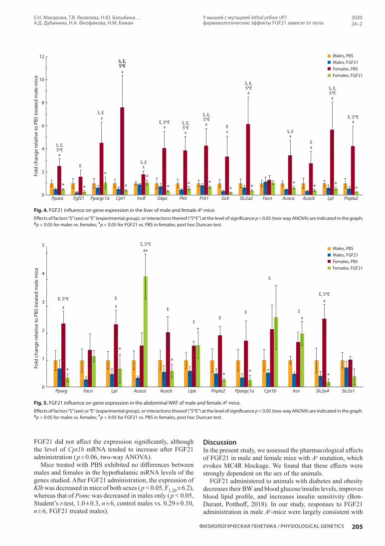
У мышей с мутацией lethal yellow (Ay) фармакологические эффекты FGF21 зависят от пола
Е.Н. Макарова, Т.В. Яковлева, Н.Ю. Балыбина … А.Д. Дубинина, Н.А. Феофанова, Н.М. Бажан
205
202024 • 2
ФИЗИОЛОГИЧЕСКАЯ ГЕНЕТИКА / PHYSIOLOGICAL GENETICS
FGF21 did not affect the expression significantly, although the level of Cpt1b mRNA tended to increase after FGF21 administration ( p ± 0.06, two-way ANOVA).
Mice treated with PBS exhibited no differences between males and females in the hypothalamic mRNA levels of the genes studied. After FGF21 administration, the expression of Klb was decreased in mice of both sexes ( p < 0.05, F1.20 ± 6.2), whereas that of Pomc was decreased in males only ( p < 0.05, Student’s t-test, 1.0 ± 0.3, n ± 6, control males vs. 0.29 ± 0.10, n ± 6, FGF21 treated males).
DiscussionIn the present study, we assessed the pharmacological effects of FGF21 in male and female mice with Ay mutation, which evokes MC4R blockage. We found that these effects were strongly dependent on the sex of the animals.
FGF21 administered to animals with diabetes and obesity decreases their BW and blood glucose/insulin levels, improves blood lipid profile, and increases insulin sensitivity (Bon-Durant, Potthoff, 2018). In our study, responses to FGF21 administration in male Ay-mice were largely consistent with
12
10
8
6
4
2
0
Fold
cha
nge
rela
tive
to P
BS tr
eate
d m
ale
mic
e
* *
*
*
*
* * ** * * * *
*
S, E, S*E
S, E, S*E
S, E, S*E
S, E, S*E S, E,
S*E
E, S*EE, S*E
S, E, S*ES, E, S*E
S, E
S, E
S, E
E
E
E
Pnpla2LplGck AcacbPck1Cpt1 AcacaPklrPpargc1a FacnG6pcFgf21 Slc2a2InsRPpara
#
#
#
##
#
#
#
#
#
#
##
Males, PBSMales, FGF21Females, PBSFemales, FGF21
Males, PBSMales, FGF21Females, PBSFemales, FGF21
5
4
3
2
1
0
Fold
cha
nge
rela
tive
to P
BS tr
eate
d m
ale
mic
e
*
*
#*
*
* * *
E, S*EE, S*E
S
S
EE S
S, S*E
E
E
Lpl Cpt1bAcaca Acacb Pnpla2 Ppargc1aFacn Lipe Slc2a4 Slc2a1InsrPparg
# #
#
#
#
Fig. 4. FGF21 influence on gene expression in the liver of male and female Ay-mice. Effects of factors “S” (sex) or “E” (experimental group), or interactions thereof (“S*E”) at the level of significance p < 0.05 (two-way ANOVA) are indicated in the graph; #p < 0.05 for males vs. females; *p < 0.05 for FGF21 vs. PBS in females; post hoc Duncan test.
Fig. 5. FGF21 influence on gene expression in the abdominal WAT of male and female Ay-mice. Effects of factors “S” (sex) or “E” (experimental group), or interactions thereof (“S*E”) at the level of significance p < 0.05 (two-way ANOVA) are indicated in the graph. #p < 0.05 for males vs. females; *p < 0.05 for FGF21 vs. PBS in females, post hoc Duncan test.

E.N. Makarova, T.V. Yakovleva, N.Yu. Balyibina … A.D. Dubinina, N.A. Feofanova, N.M. Bazhan
206
Pharmacological effects of FGF21 are sex-specific in mice with the lethal yellow (Ay) mutation
Вавиловский журнал генетики и селекции / Vavilov Journal of Genetics and Breeding • 2020 • 24 • 2
its expected impact: their BWs and blood insulin decreased, blood levels of FFA tended to decrease, and blood glucose and adiponectin were not affected. Our observation that FGF21 decreased blood insulin without affecting glucose, which retained normal levels, suggests that insulin sensitiv-ity in male Ay-mice was possibly improved. Metabolic effect of FGF21 administration was not associated with changes in blood adiponectin, since adiponectin is not required for the chronic effects of FGF21 to reduce body weight or its effects on glucose homeostasis (BonDurant et al., 2017).
Although we did not monitor energy expenditure, it was most likely increased, since weight loss in male mice took place in the absence of changes in FI. The ability of FGF21 to induce weight loss without affecting FI was reported pre-viously for male mice with diet-induced and genetic obesity (db/db, ob/ob) (Kharitonenkov et al., 2005; Xu et al., 2009; Camporez et al., 2013). This effect was arguably due to increased energy expenditure caused by intense locomotor activity (Xu et al., 2009) and elevated metabolic rate (Coskun et al., 2008; Xu et al., 2009). FGF21-induced growth of metabolic rates is associated with augmentation of fatty acid oxidation in liver and adipose tissue (BAT and WAT), caused in turn by increased expression of genes encoding CPT-1 (in liver only), PGC-1 (in liver and adipose tissue), and UCP-1 (in liver and adipose tissue) (Camporez et al., 2013). No such changes in gene expression have been observed in this study; on the contrary, FGF21 administration to males reduced the expression of Ppargc1a and Cpt1 in liver and Ppargc1a in abdominal WAT ( p < 0.05, two-way ANOVA).
Our results are consistent with those reported by (Coskun et al., 2008), demonstrating that weight loss and improved glucose metabolism in FGF21-treated C57Bl males were accompanied by a decrease in liver expression of Cpt1 (with no changes of Ppargc1a expression). However, T. Coskun et al. (2008) found increased expression of genes related to (1) thermogenesis (Ucp1) in WAT and BAT, (2) lipolysis (Lipe, Pnpla2) in WAT, and (3) lipogenesis (Acaca, Acacb) in WAT, suggesting that energy expenditure is achieved via thermo-genesis and induction of a state of increased futile cycling. We have not observed induction of Ucp1 expression either in BAT or WAT; likewise, there have been no increase in the expression of lipolysis/lipogenesis genes in liver or adipose tissue. Thus, our experimental data provide no indication of futile cycling activation. FGF21-induced activation of energy expenditure involves, in addition to central mechanisms, the sympathetic nervous system (Owen et al., 2014; Lan et al., 2017). Of note, the latter also mediates regulation of energy expenditure by the melanocortin system (Rossi et al., 2011; Berglund et al., 2014). Therefore, FGF21 may act, at least in certain cases, via melanocortin signaling pathways. If so, the blockade of MC4R in Ay-mice can interfere with the activating effect of FGF21 on the expression of the genes we studied. Weight loss in male Ay-mice is indicative of yet other mecha-nisms whereby energy expenditure may be increased, which remain to be explored.
In females with Ay mutation, the effect of FGF21 differed dramatically from that observed in males. In females unlike males, FGF21 administration did not induce weight loss, but increased liver weight. Ay-mice are hyperphagic (Wolff et al., 1999), and FGF21 exacerbated hyperphagia specifically in
females. It is not known whether this difference is due to Ay mutation or, rather, we are dealing with a general sex-linked discrepancy in responses of female and male mice to FGF21 treatment (the pharmacological effects of FGF21 have thus far not been studied in female mice).
Resistance of female Ay-mice to catabolic effects of FGF21 can be explained by increase of food intake. FGF21 influence on FI in female rodent was not studied, but in males, FGF21 was shown previously to increase FI in rats (Recinella et al., 2017), mice with diet-induced obesity (Coskun et al., 2008), and to increase protein intake while reducing carbohydrate intake in normal mice, and the later effect was mediated via CNS (Larson et al., 2019). However, FGF21-dependent sig-naling pathways that regulate eating behavior have not been identified. The melanocortin system is involved in the regu-lation of the response to protein deficiency, and FGF21 may act as the sensor triggering that response. In our experiment, male mice demonstrated no increase in FI, although FGF21 administration decreased hypothalamic expression of Pomc. POMC is a precursor of the anorexigenic neuropeptide MSH, and a decrease in Pomc expression (limiting MC4R activation by MSH) is associated with increased FI. Ay mutation leads to MC4R blockade, which disrupts MSH regulation of FI and may be the reason why males did not respond to FGF21 by increased FI.
In females, FGF21 administration caused a considerable increase in FI without affecting the expression of the mela-nocortin system genes. In Ay-females, FGF21 influence on FI may be mediated via estradiol-sensitive mechanism that is not altered by impaired melanocortin signaling (Morton et al., 2004). It is possible that the orexigenic effects of FGF21 in males and females involve distinct neuronal signaling pathways.
According to the data obtained in the control groups, a different metabolic response to exogenous FGF21 in males and females was induced at the background of significant sex differences in the mass of the liver and adipose tissue and in the expression of the liver and fat genes. Sex differen-ces in gene expression in adipose tissue differed depending on localization; in our experiment, we found them only in abdominal adipose tissue. These data are consistent with the results obtained when assessing gene expression in adipose tissue in mice: the number of differentially expressed genes in males and females was significantly higher in abdominal than in subcutaneous fat (Grove et al., 2010). Increased weight of abdominal fat in control Ay-females, compared to males, was associated with increased expression of genes upregulating lipogenesis in adipose tissue: Ppar, Lpl, Insr and Slc2a4. FGF21 administration eliminated differences in the expression of these genes, however, it did not lead to significant changes in the mass of adipose tissue. It is possible that more prolonged FGF21 administration is required to detect changes at the level of adipose tissue weight.
The most striking sex differences in gene expression in the control groups were observed in liver. In controls, the expres-sion of most genes studied (except for Fgf21 and Fasn) was higher in females than in males. These results are consistent with transcriptome analysis data demonstrating differential expression rates for 72 % active liver genes in male and female mice (Yang et al., 2006). Sex differences in expression of genes

У мышей с мутацией lethal yellow (Ay) фармакологические эффекты FGF21 зависят от пола
Е.Н. Макарова, Т.В. Яковлева, Н.Ю. Балыбина … А.Д. Дубинина, Н.А. Феофанова, Н.М. Бажан
207
202024 • 2
ФИЗИОЛОГИЧЕСКАЯ ГЕНЕТИКА / PHYSIOLOGICAL GENETICS
related to lipid metabolism were found in the liver of mice and rats on high-calorie diet: compared to males, females had enhanced mRNA levels of Acc1, Pparg, Cpt1 and other genes in mice (Gasparin et al., 2018), and enhanced protein levels of ACC, PPARs and FAS in rats (Chukijrungroat et al., 2017). As shown by S. Della Torre et al. (2017), liver is the major target for estrogens, and estrogen receptor alpha (ERα) has a direct effect on the regulation of the hepatic genes relevant for energy metabolism.
Increased expression of genes involved in fatty acid β-oxidation (Ppara, Ppargc1a, Cpt1), glycolysis (G6p, Pklr), gluconeogenesis (Pck1, Gck), glucose transport (Slc2a2), li-pogenesis and lipolysis (Acaca, Acacb, Lpl, Pnpla2) possibly indicates that the rate of glucose and fat metabolism is higher in the liver of Ay-females than Ay-males. FGF21 administra-tion reduced liver expression of all the genes mentioned, thereby eliminating entirely any sex-dependent differences, and this effect was associated with the increase of female liver weight. Enlarged liver under the action of FGF21 in female Ay-mice may indicate the development of steatosis. Simultaneous decrease in catabolic (lipolysis, glycolysis) and anabolic (lipogenesis, gluconeogenesis) processes can suggest decreased metabolism in the liver of Ay-females. Decreased liver metabolism in females could reduce liver energy expen-diture and contribute, together with increased food intake, to liver weight gain. Additional morphological and biochemical studies are needed to find out whether FGF21 initiates (or promotes) steatosis in Ay-female mice.
Taken together, our findings indicate that therapeutic effects of FGF21 in mice with disrupted melanocortin signaling are strongly sex-dependent. In male Ay-mice, the blockage of MC4Rs does not prevent the anti-obesity effect of FGF21: its administration results in weight loss and blood insulin decrease. However, obese Ay-females exerted resistance to catabolic and antidiabetic effects of FGF21. In females, exo-ge nous FGF21 stimulates FI without reducing BW and blood glucose/insulin, inhibits liver expression of genes related to glucose and lipid turnover, and increases liver weight. The contribution of estrogens and the melanocortin system to those effects remains to be elucidated.
ConclusionThe pharmacological effect of FGF21 may depend on the animal sex and etiology of obesity. Although an immediate translation to humans of findings obtained in experiments with mice is not possible, our results suggest that detailed preclinical studies of the pharmacological effects of FGF21 are required, taking into account the sex of individuals under study and the genesis of obesity.
ReferencesBazhan N., Jakovleva T., Balyibina N., Dubinina A., Denisova E., Feo-
fanova N., Makarova E. Sex dimorphism in the Fgf21 gene expres-sion in liver and adipose tissues is dependent on the metabolic con-dition. Online J. Biol. Sci. 2019;19(1):28-36. DOI 10.3844/ojbsci. 2019.28.36.
Berglund E.D., Liu T., Kong X., Sohn J.-W., Vong L., Deng Z., Lee C.E., Lee S., Williams K.W., Olson D.P., Scherer P.E., Lowell B.B., Elm-quist J.K. Melanocortin 4 receptors in autonomic neurons regulate thermogenesis and glycemia. Nat. Neurosci. 2014;17(7):911-913. DOI 10.1038/nn.3737.
BonDurant L.D., Ameka M., Naber M.C., Markan K.R., Idiga S.O., Acevedo M.R., Walsh S.A., Ornitz D.M., Potthoff M.J. FGF21 regulates metabolism through adipose-dependent and -independent mechanisms. Cell Metab. 2017;25(4):935-944.e4. DOI 10.1016/j.cmet.2017.03.005.
BonDurant L.D., Potthoff M.J. Fibroblast growth factor 21: a versatile regulator of metabolic homeostasis. Annu. Rev. Nutr. 2018;38(1): 173-196. DOI 10.1146/annurev-nutr-071816-064800.
Bultman S.J., Michaud E.J., Woychik R.P. Molecular characterization of the mouse agouti locus. Cell. 1992;71(7):1195-1204.
Camporez J.P.G., Jornayvaz F.R., Petersen M.C., Pesta D., Gui-gni B.A., Serr J., Zhang D., Kahn M., Samuel V.T., Jurczak M.J., Shulman G.I. Cellular mechanisms by which FGF21 improves in-sulin sensitivity in male mice. Endocrinology. 2013;154(9):3099-3109. DOI 10.1210/en.2013-1191.
Chukijrungroat N., Khamphaya T., Weerachayaphorn J., Songserm T., Saengsirisuwan V. Hepatic FGF21 mediates sex differences in high-fat high-fructose diet-induced fatty liver. Am. J. Physiol. Endocrinol. Metab. 2017;313(2):E203-E212. DOI 10.1152/ajpendo.00076.2017.
Clayton J.A. Applying the new SABV (sex as a biological variable) policy to research and clinical care. Physiol. Behav. 2018;1872-1875. DOI 10.1016/j.physbeh.2017.08.012.
Coskun T., Bina H.A., Schneider M.A., Dunbar J.D., Hu C.C., Chen Y., Moller D.E., Kharitonenkov A. Fibroblast growth factor 21 corrects obesity in mice. Endocrinology. 2008;149(12):6018-6027. DOI 10.1210/en.2008-0816.
Della Torre S., Lolli F., Ciana P., Maggi A. Sexual dimorphism and estrogen action in mouse liver. Adv. Exp. Med. Biol. 2017;1043: 141-151.
Fani L., Bak S., Delhanty P., van Rossum E.F.C., van den Akker E.L.T. The melanocortin-4 receptor as target for obesity treatment: a sys-tematic review of emerging pharmacological therapeutic options. Int. J. Obes. (Lond.) 2014;38(2):163-169. DOI 10.1038/ijo.2013.80.
Farooqi I.S., Keogh J.M., Yeo G.S.H., Lank E.J., Cheetham T., O’Rahilly S. Clinical spectrum of obesity and mutations in the mela-nocortin 4 receptor gene. N. Engl. J. Med. 2003;348(12):1085-1095. DOI 10.1056/NEJMoa022050.
Gasparin F.R.S., Carreño F.O., Mewes J.M., Gilglioni E.H., Pagadi-gorria C.L.S., Natali M.R.M., Utsunomiya K.S., Constantin R.P., Ouchi da A.T., Curti C., Gaemers I.C., Elferink R.P.J.O., Constan-tin J., Ishii-Iwamoto E.L. Sex differences in the development of hepatic steatosis in cafeteria diet-induced obesity in young mice. Biochim. Biophys. Acta Mol. Basis Dis. 2018;1864(7):2495-2509. DOI 10.1016/j.bbadis.2018.04.004.
Girardet C., Butler A.A. Neural melanocortin receptors in obesity and related metabolic disorders. Biochim. Biophys. Acta. 2014;1842(3): 482-494. DOI 10.1016/j.bbadis.2013.05.004.
Grove K.L., Fried S.K., Greenberg A.S., Xiao X.Q., Clegg D.J. A mi-croarray analysis of sexual dimorphism of adipose tissues in high-fat-diet-induced obese mice. Int. J. Obes. (Lond.). 2010;34(6):989-1000. DOI 10.1038/ijo.2010.12.
Jackson V.M., Breen D.M., Fortin J.-P., Liou A., Kuzmiski J.B., Loo-mis A.K., Rives M.-L., Shah B., Carpino P.A. Latest approaches for the treatment of obesity. Expert Opin. Drug Discov. 2015;10(8): 825-839. DOI 10.1517/17460441.2015.1044966.
Kharitonenkov A., Shiyanova T.L., Koester A., Ford A.M., Mica-novic R., Galbreath E.J., Sandusky G.E., Hammond L.J., Moy-ers J.S., Owens R.A., Gromada J., Brozinick J.T., Hawkins E.D., Wroblewski V.J., Li D.-S., Mehrbod F., Jaskunas S.R., Shanafelt A.B. FGF-21 as a novel metabolic regulator. J. Clin. Invest. 2005;115(6): 1627-1835. DOI 10.1172/JCI23606.
Lan T., Morgan D.A., Rahmouni K., Sonoda J., Fu X., Burgess S.C., Holland W.L., Kliewer S.A., Mangelsdorf D.J. FGF19, FGF21, and an FGFR1/β-Klotho-activating antibody act on the nervous system to regulate body weight and glycemia. Cell Metab. 2017;26(5):709-718.e3. DOI 10.1016/j.cmet.2017.09.005.
Larson K.R., Chaffin A.T.-B., Goodson M.L., Fang Y., Ryan K.K. Fi-broblast growth factor-21 controls dietary protein intake in male

E.N. Makarova, T.V. Yakovleva, N.Yu. Balyibina … A.D. Dubinina, N.A. Feofanova, N.M. Bazhan
208
Pharmacological effects of FGF21 are sex-specific in mice with the lethal yellow (Ay) mutation
Вавиловский журнал генетики и селекции / Vavilov Journal of Genetics and Breeding • 2020 • 24 • 2
ORCID IDE.N. Makarova orcid.org/0000-0002-6417-9893T.V. Yakovleva orcid.org/0000-0001-7628-5856N.Yu. Balyibina orcid.org/0000-0001-7168-1438K.O. Baranov orcid.org/0000-0002-4029-6518E.I. Denisova orcid.org/0000-0001-8696-8781A.D. Dubinina orcid.org/0000-0003-3907-3488N.A. Feofanova orcid.org/0000-0002-2591-1623N.M. Bazhan orcid.org/0000-0002-7246-4758
Acknowledgements. This research was funded by the Russian Science Foundation, Grant No. 17-15-01036.Conflict of interest. The authors declare no conflict of interest.Received October 7, 2019. Revised November 28, 2019. Accepted November 29, 2019. Published online February 3, 2020.
mice. Endocrinology. 2019;160(5):1069-1080. DOI 10.1210/en. 2018-01056.
Lee Y.-H., Kim S.H., Kim S.-N., Kwon H.-J., Kim J.-D., Oh J.Y., Jung Y.-S. Sex-specific metabolic interactions between liver and adi-pose tissue in MCD diet-induced non-alcoholic fatty liver di sease. Oncotarget. 2016;7(30):46959-46971. DOI 10.18632/oncotarget. 10506.
Markan K.R., Naber M.C., Ameka M.K., Anderegg M.D., Mangels-dorf D.J., Kliewer S.A., Mohammadi M., Potthoff M.J. Circulat-ing FGF21 is liver derived and enhances glucose uptake during refeeding and overfeeding. Diabetes. 2014;63(12):4057-4063. DOI 10.2337/db14-0595.
Mauvais-Jarvis F., Arnold A.P., Reue K. A guide for the design of pre-clinical studies on sex differences in metabolism. Cell Metab. 2017;25(6):1216-1230. DOI 10.1016/j.cmet.2017.04.033.
Michaud E.J., Mynatt R.L., Miltenberger R.J., Klebig M.L., Wilkin-son J.E., Zemel M.B., Wilkison W.O., Woychik R.P. Role of the agouti gene in obesity. J. Endocrinol. 1997;155(2):207-209.
Morton G.J., Mystkowski P., Matsumoto A.M., Schwartz M.W. In-creased hypothalamic melanin concentrating hormone gene expres-sion during energy restriction involves a melanocortin-independent, estrogen-sensitive mechanism. Peptides. 2004;25(4):667-674. DOI 10.1016/j.peptides.2004.02.007.
Owen B.M., Ding X., Morgan D.A., Coate K.C., Bookout A.L., Rah-mouni K., Kliewer S.A., Mangelsdorf D.J. FGF21 acts centrally to induce sympathetic nerve activity, energy expenditure, and weight loss. Cell Metab. 2014;20(4):670-677. DOI 10.1016/j.cmet.2014. 07.012.
Recinella L., Leone S., Ferrante C., Chiavaroli A., Di Nisio C., Marti-notti S., Vacca M., Brunetti L., Orlando G. Effects of central fibro-
blast growth factor 21 (FGF21) in energy balance. J. Biol. Regul. Homeost. Agents. 2017;31(3):603-613.
Rossi J., Balthasar N., Olson D., Scott M., Berglund E., Lee C.E., Choi M.J., Lauzon D., Lowell B.B., Elmquist J.K. Melanocortin-4 receptors expressed by cholinergic neurons regulate energy balance and glucose homeostasis. Cell Metab. 2011;13(2):195-204. DOI 10.1016/j.cmet.2011.01.010.
Singh R.K., Kumar P., Mahalingam K. Molecular genetics of human obesity: A comprehensive review. C. R. Biol. 2017;340(2):87-108. DOI 10.1016/j.crvi.2016.11.007.
Tao Y.-X. The melanocortin-4 receptor: physiology, pharmacology, and pathophysiology. Endocr. Rev. 2010;31(4):506-543. DOI 10.1210/er.2009-0037.
Wolff G.L., Roberts D.W., Mountjoy K.G. Physiological consequen-ces of ectopic agouti gene expression: the yellow obese mouse syndrome. Physiol. Genomics. 1999;1(3):151-163. DOI 10.1152/ physiolgenomics.1999.1.3.151.
Xie T., Leung P.S. Fibroblast growth factor 21: a regulator of metabolic disease and health span. Am. J. Physiol. Endocrinol. Metab. 2017; 313(3):E292-E302. DOI 10.1152/ajpendo.00101.2017.
Xu J., Lloyd D.J., Hale C., Stanislaus S., Chen M., Sivits G., Vonder-fecht S., Hecht R., Li Y.-S., Lindberg R.A., Chen J.-L., Jung D.Y., Zhang Z., Ko H.-J., Kim J.K., Véniant M.M. Fibroblast growth fac-tor 21 reverses hepatic steatosis, increases energy expenditure, and improves insulin sensitivity in diet-induced obese mice. Diabetes. 2009;58(1):250-259. DOI 10.2337/db08-0392.
Yang X., Schadt E.E., Wang S., Wang H., Arnold A.P., Ingram-Drake L., Drake T.A., Lusis A.J. Tissue-specific expression and regulation of sexually dimorphic genes in mice. Genome Res. 2006;16(8):995-1004. DOI 10.1101/gr.5217506.

Involvement of transposable elements in neurogenesisR.N. Mustafin1 , E.K. Khusnutdinova2
1 Bashkir State Medical University, Ufa, Russia2 Institute of Biochemistry and Genetics – Subdivision of the Ufa Federal Research Centre of the Russian Academy of Sciences, Ufa, Russia
e-mail: [email protected]
Abstract. The article is about the role of transposons in the regulation of functioning of neuronal stem cells and mature neurons of the human brain. Starting from the first division of the zygote, embryonic development is governed by regular activations of transposable elements, which are necessary for the sequential regulation of the expression of genes specific for each cell type. These processes include differentiation of neuronal stem cells, which requires the finest tuning of expression of neuron genes in various regions of the brain. Therefore, in the hippocampus, the center of human neurogenesis, the highest transposon activity has been identified, which causes somatic mosai cism of cells during the formation of specific brain structures. Similar data were obtained in studies on experimental animals. Mobile genetic elements are the most important sources of long non-coding RNAs that are coexpressed with important brain protein-coding genes. Significant activity of long non-coding RNA was detected in the hippocampus, which confirms the role of transposons in the regulation of brain func-tion. MicroRNAs, many of which arise from transposon transcripts, also play an important role in regulating the differentiation of neuronal stem cells. Therefore, transposons, through their own processed transcripts, take an active part in the epigenetic regulation of differentiation of neurons. The global regulatory role of transposons in the human brain is due to the emergence of protein-coding genes in evolution by their exonization, duplica-tion and domestication. These genes are involved in an epigenetic regulatory network with the participation of transposons, since they contain nucleotide sequences complementary to miRNA and long non-coding RNA formed from transposons. In the memory formation, the role of the exchange of virus-like mRNA with the help of the Arc protein of endogenous retroviruses HERV between neurons has been revealed. A possible mechanism for the implementation of this mechanism may be reverse transcription of mRNA and site-specific insertion into the genome with a regulatory effect on the genes involved in the memory. Key words: brain; differentiation; noncoding RNA; retroelements; neuronal stem cells; transposable elements.
For citation: Mustafin R.N., Khusnutdinova E.K. Involvement of transposable elements in neurogenesis. Vavilovskii Zhurnal Genetiki i Selektsii = Vavilov Journal of Genetics and Breeding. 2020;24(2):209-218. DOI 10.18699/VJ20.613
Участие мобильных элементов в нейрогенезеР.Н. Мустафин1 , Э.К. Хуснутдинова2
1 Башкирский государственный медицинский университет, Уфа, Россия2 Институт биохимии и генетики – обособленное структурное подразделение Уфимского федерального исследовательского центра
Российской академии наук, Уфа, Россия e-mail: [email protected]
Аннотация. В обзоре представлены накопленные в научной литературе данные об участии мобильных генетических элементов в регуляции дифференцировки нейрональных стволовых клеток и функциони-рования зрелых нейронов головного мозга. Начиная с первого деления зиготы, эмбриональное развитие управляется закономерными активациями транспозонов, необходимыми для последовательного изме-нения экспрессии специфических для каждого типа клеток генов. Частным отражением этих процессов может быть дифференцировка нейрональных стволовых клеток – процесс, в ходе которого необходима наиболее тонкая настройка экспрессии генов в нейронах различных областей головного мозга. Доказа-тельствами этого предположения являются данные о высокой активности транспозонов в центре нейро-генеза, зубчатой извилине гиппокампа. Кроме того, мобильные элементы – источники возникновения и эволюции длинных некодирующих РНК, которые коэкспрессируются с необходимыми для работы голов-ного мозга белок-кодирующими генами. Наибольшая активность длинных некодирующих РНК, так же как и транспозонов, обнаружена в центре нейрогенеза человека, что позволяет предположить их участие в управлении работой головного мозга. В регуляции дифференцировкой нейрональных стволовых клеток используются также микроРНК, многие из которых возникают из транскриптов мобильных элементов. Транспозоны посредством собственных процессированных транскриптов играют роль в эпигенетической регуляции дифференцировки нейронов. Объяснением глобальной регуляторной функции мобильных эле-ментов в головном мозге человека может служить их значение в возникновении белок-кодирующих генов в эволюции путем экзонизации, дупликации и доместикации. Эти гены вовлечены в эпигенетическую регу-
© Mustafin R.N., Khusnutdinova E.K., 2020
ГЕНЕТИКА ЧЕЛОВЕКАОбзор / Review
This work is licensed under a Creative Commons Attribution 4.0 License
Вавиловский журнал генетики и селекции. 2020;24(2):209-218DOI 10.18699/VJ20.613

R.N. Mustafin E.K. Khusnutdinova
210
Involvement of transposable elements in neurogenesis
Вавиловский журнал генетики и селекции / Vavilov Journal of Genetics and Breeding • 2020 • 24 • 2
ляторную сеть с участием транспозонов, так как содержат нуклеотидные последовательности, комплемен-тарные микроРНК и длинным некодирующим РНК, образуемым из транскриптов мобильных элементов. В формировании памяти выявлена роль обмена вирусоподобными частицами мРНК при помощи белка Arc эндогенных ретровирусов HERV между нейронами. Возможными способами реализации этого механизма могут быть обратная транскрипция мРНК и сайт-специфическая интеграция в геном с регуляторным воз-действием на гены, участвующие в консолидации информации. Ключевые слова: головной мозг; дифференцировка; некодирующие РНК; ретроэлементы; стволовые нерв-ные клетки; транспозоны.
IntroductionTransposable elements (TE) make up 69 % of the human ge-nome (de Koning et al., 2011). In the course of evolution, many protein-coding genes (Joly-Lopez, Bureau, 2018), regulatory nucleotide sequences (Ito et al., 2017; Schrader, Schmitz, 2018), and telomeres (Kopera et al., 2011), non-coding RNAs (ncRNAs), including microRNAs (Piriyapongsa et al., 2007; Yuan et al., 2010, 2011; Qin et al., 2015) and long human ncRNAs (Johnson, Guigo, 2014) originating from TE. Over millions of years of evolution, cells have developed various defense systems against TE insertion into their genomes, including DNA methylation, heterochromatin formation, and RNA interference (RNAi). These epigenetic mechanisms have made a significant contribution to the regulation of spe-cific gene expression and cell differentiation (Habibi et al., 2015).
Transposable elements are divided into two main classes, in accordance with the mechanisms of their transposition. DNA-TEs are transposed by “cut and paste” or “rolling circle”. Retroelements (REs) are integrated into new genome sites using “copy and paste”. REs are classified into those contain-ing long terminal repeats (LTR REs) (Fig. 1) and those not containing them (non-LTR REs) (Fig. 2). The latter are divided into autonomous (LINE, long interspersed nuclear elements) and non-autonomous (SINE, short interspersed nuclear ele-ments) and SVA (SINE-VNTR-Alu) (Fig. 3) (Klein, O’Neill, 2018).
The human genome contains more than 500,000 copies of LINE1 (L1), which make up 17 % of all nucleotide sequences. Among them, only about 100 L1 are active, since they contain the full length of 6000 bp. Among non-autonomous REs, the human genome contains more than 2700 copies of SVA (Hancks, Kazazian, 2012). One of the important factors for the development of the human brain is considered the waves of L1 retrotranspositions, as well as the birth of new TEs, such as SINE, Alu and SVA in the evolution of primates (Linker et al., 2017).
Human endogenous retroviruses (HERV) belong to LTR-RE. They occupy about 8 % of the entire genome and serve as sources of a huge number (794,972) of binding sites with specific transcription factors (TFs), the activation of which plays a role in embryogenesis. For example, in the mesoderm, LTRs interact with SOX17, FOXA1, GATA4; in pluripotent cells, with SOX2, NANOG, POU5F1; in hematopoietic cells, with TAL1, GATA1, PU1 (Ito et al., 2017). Mammalian-wide interspersed repeats (MIRs), which belong to the ancient SINE family descended from tRNA, are also associated with tissue-specific gene expression (Jjingo et al., 2014).
Transposable elements are characterized by nonrandom activation, depending on the tissue and stage of develop-
ment. High-throughput profiling of integration sites by next-generation sequencing, combined with large-scale genomic data mining and cellular or biochemical approaches, has revealed that the insertions are usually non-random (Sultana et al., 2017). Programmed activation of TE in individual cells during neurogenesis leads to a change in the expression of certain genes necessary for differentiation into specific types of neurons for the formation and functioning of brain structures (Coufal et al., 2009; Bailie et al., 2011; Thomas, Muotri, 2012; Richardson et al., 2014; Evrony et al., 2015; Upton et al., 2015; Muotri, 2016; Suarez et al., 2018). In accordance with this, somatic mosaicism of neurons detected by insertions of TEs (Richardson et al., 2014; Upton et al., 2015; Bachiller et al., 2017; Paquola et al., 2017; Rohrback et al., 2018; Suarez et al., 2018) can reflect the programmed regulatory pattern of the genome necessary for the maturation of specific structures of the central nervous system (Paquola et al., 2017; Rohrback et al., 2018). Somatic mosaicism means the presence, in the same organism, of cells with different genomes as a result of de novo DNA changes. These structural variations may be due to CNV, insertions of REs, deletions under the influence of TEs, and SNV (Paquola et al., 2017). This means that in different cells of one organism, not only the genotype, but also the whole genome changes. This is due to the occurrence of mutations in exons of protein-coding genes, intergenic regulatory regions and introns, which is accompanied by a specific expression of certain genes specific for each cell type.
The role of transposable elements in neuronal differentiationThe human brain contains an average of 86.1 billion neurons. Moreover, each of the neurons forms from 5,000 to 20,000 synaptic connections, creating a complex network with a variety of cell types and subtypes. The number of subtypes of neurons is so large that it does not lend itself to modern methods for their description. There must be mechanisms to ensure such a diversity of neurons with their specific temporal and spatial features of functioning (Thomas, Muotri, 2012). The sources of these mechanisms can be TEs, combinations of movements of which can become sources of countless variety. An example of this is the molecular mechanism for generat-ing antibodies by the mammalian immune system (V(D)J recombination), derived from TEs (Lapp, Hunter, 2016). TEs played a role in the development of the central nervous system. In evolution, they turned out to be sources of the formation of regulatory structures and genes involved in the formation of the brain. Non-autonomous TEs MER130 were preserved in the genomes during evolution due to their location near the neocortex genes as a necessary link for their regulation. The experiments showed the activation of MER130 in mouse

Участие мобильных элементов в нейрогенезе
Р.Н. Мустафин Э.К. Хуснутдинова
211ГЕНЕТИКА ЧЕЛОВЕКА / HUMAN GENETICS
202024 • 2
ERV and Retrovirus superfamily
Gypsy and Bel-Pao superfamily
Copia superfamily
Capsid protein gene
Aspartic protease gene
Ribonuclease H gene
Reverse transcriptase gene
Integrase gene
Envelope protein geneLTR
LTR
LTR
LTR
LTR
LTR GAG
AP
RH
RT
INT
ENVGAG
GAG
GAG
AP
AP
AP
RT
RT
RT
RH
RH
RH
INT
INT
INT ENV
Fig. 1. Scheme of the structure of the genes of LTR-containing retroelements.
Fig. 2. Scheme of the structure of the gene of non-LTR retroelements (LINE-1).UTR – untranslated region; ORF – open reading frame; CC – coiled-coiled; RRM – RNA recognition motif; CTD – С-terminal domain; EN – endonuclease; Z – Z-domain; RT – reverse transcriptase; С – cysteine-rich domain.
Fig. 3. Scheme of the structure of SVA elements.
embryos on the fourteenth day of development as gene en-hancers for the development of the neocortex (Notwell et al., 2015). Among 11 sushi-ichi-specific placental animal genes derived from REs, the SIRH11/ZCCHC16 gene encoding zinc finger CCHC protein contributed to the evolution of the brain. This domesticated gene is involved in the development of cognitive functions of placental animals (Irie et al., 2016).
In 2009, in neuronal stem cells isolated from the brain of a human embryo, L1s retrotranspositions were detected, as well as an increase (in comparison with the liver and heart of the same individual) in the number of copies of endogenous L1s in the adult hippocampus (Coufal et al., 2009). In addition to L1 (7743 insertions), a large number of somatic transposi-tions Alu (13,692 insertions) and SVA (1350 insertions) were found in the hippocampus of adults (Bailie et al., 2011). These de novo integrations can affect the expression of certain genes, creating unique transcriptomes of individual neurons (Muotri, 2016). This may be due to the genome-programmed TE ability for their regular site-specific insertions (Sultana et al., 2017).
In 2009, of 19 retrotranspositions, 16 were found at a distance of less than 100 kilobases from genes expressed in neurons (Coufal et al., 2009). In 2015, in a study of the somatic mosai-cism of the human hippocampus K.R. Upton et al. revealed, out of 20 identified L1 transpositions, 2 functionally significant insertions into the introns of the ZFAND3 and USP33 genes functioning in the brain (Upton et al., 2015). A.A. Kurnosov et al., when studying human brain samples, showed that out of 3100 transpositions of L1 in neurons of the dentate gyrus of the hippocampus, 50.26 % of insertions are located in the genes, and out of 2984 Alu, 49.1 % (Kurnosov et al., 2015). In 2016, J.A. Erwin et al. revealed that in the brain of healthy people 44–63 % of neurons undergo somatic mosaicism at the loci of genes that are important for the functioning of the nervous system. For example, a high insertion frequency of L1-RE is shown for the DLG2 gene, which affects cognitive flexibility, attention, and learning. Mutations in DLG2 are associated with the development of schizophrenia (Erwin et al., 2016).
ORF15’
3’ORF2
UTR UTRFLANK
LINE-1(non-LTR)
AAATAA
AATAAA AATAAA
AAAnRT CEN ZCTDRRMCC
FLANK
Flanking of target site duplication
Flanking of target site duplication
Antisense fragment,
Alu homologous
Tandem repeat locus
Long terminal repeat
envelope (env) gene part
FLANKSINE-R
3’
AATAAAAATAAA
AAAnCCCTCTn env LTRAlu-like
VNTR

R.N. Mustafin E.K. Khusnutdinova
212
Involvement of transposable elements in neurogenesis
Вавиловский журнал генетики и селекции / Vavilov Journal of Genetics and Breeding • 2020 • 24 • 2
Somatic retrotranspositions, unlike germinal ones, cannot be inherited by future generations. However, the programmed ability for specific insertions, depending on the composition and location of TEs in the genome, can be inherited. An ex-planation of the ability of TEs to be inserted in a site-specific manner in the region of genes expressed in the brain may be the evolutionary relationship of protein-coding genes and their regulatory sequences with TEs (Gianfrancesco et al., 2017; Ito et al., 2017; Joly-Lopez, Bureau, 2018). The insertions specific for humans and chimpanzees were revealed near the promoters of the tachycin receptor genes TACR3, cation channels TRPV1 and TRPV3, oxytocin OXT. These genes are associated with the functioning of neuropeptides. Analysis of the genomes of various mammals showed that the neural enhancer nPE2, which regulates the expression of the POMC gene in the hypothalamus, evolved from SINE in evolution (Gianfrancesco et al., 2017).
Transpositions and expression of TEs can vary depending on the area of the brain and change under environmental in-fluences, as they can perform a number of adaptive functions (Lapp, Hunter, 2016). More active are L1, which retained the ability to transpose, causing somatic mosaicism (Suarez et al., 2018). In 2005, A.R. Muotri et al. suggested that L1 using somatic transpositions can actively create mosaicism of neuron genomes (Muotri et al., 2005). In the brain, somatic mosaicism plays an important role in the regulation of cogni-tion and behavior. The consequences of somatic mosaicism encompass vast changes – from a variant at a single locus, to genes in neuronal networks (Paquola et al., 2017; Rohrback et al., 2018). Moreover, the features of somatic mosaicism differ between neurons of various regions of the brain. For example, in the cerebral cortex, only 0.6 insertions of L1-RE are observed, while in the hippocampus, from 80 to 800 inserts per neuron (Lapp, Hunter, 2016). Somatic mosaicism due to retrotranspositions is a source of phenotypic diversity between neurons during development. In the brain of an adult under the influence of various environmental factors, L1 expression can affect the functioning of neurons during the formation of long-term memory (Bachiller et al., 2017).
The hippocampus is the center of human neurogenesis, where many insertions affect transcriptional expression, creat-ing unique transcriptomes in neurons. In addition, transcrip-tional activation of L1 is similar to that for the NeuroD1 gene. This may indicate the effect of L1 expression on neurogenesis, since stimulation of Wnt3a in neuronal stem cells increases L1 expression 10-fold along the beta-catenin pathway, similarly activating transcription of the NeuroD1 gene. This gene en-codes the transcription factor that activates the genes involved in neurogenesis. The NeuroD1 promoter region contains a Sox/LEF site similar to the 5ʹUTR of the L1 element, and the pattern of time expression of the NeuroD1 and L1 genes during differentiation of neurons is similar (Thomas, Muotri, 2012).
Genetic variations between neurons due to L1 retrotranspo-sitions may be associated with specific enrichment of neuronal stem cell enhancers. It was shown that specific enhancers for certain types of neurons (determined using FANTOM5) correspond to the coordinates in the genome for insertions L1, which are within 100 bp from the enhancer. These pat-terns have not been identified for astrocytes and hepatocytes
(Upton et al., 2015). When studying the features of L1 retro-transpositions in more than 30 regions of the brain, a lot of L1 insertion-specific cell lines were found (Evrony et al., 2015). In experiments on mice, specific L1 expression was also shown depending on the area of the brain and the age of the animal (Cappucci et al., 2018).
In addition to L1 elements, LTR-REs are also involved in the regulation of neurogenesis. For example, in mice, the region where the full-length ERVmch8 on chromosome 8 was located was comparatively less methylated in the cerebellum, due to its specific expression depending on the stage of development (Lee et al., 2011). In accordance with these data, it can be as-sumed that the features of TEs activation observed in neuronal stem cells can naturally alter the expression of specific genes necessary for differentiation of neurons during the formation of specific brain structures. The reason for the activation of TEs in the neuronal stem cells of the hippocampus and the reason for their importance in memory consolidation may be the sensitivity of TEs to stressful environmental influences (Mustafin, Khusnutdinova, 2019). These mechanisms are a particular reflection of the general pattern of epigenetic control of the development of the whole organism, starting from the first division of the zygote, under the regulatory influence of TEs (Mustafin, Khusnutdinova, 2018). To understand the role of TEs in these processes, it is necessary to consider their participation in embryogenesis.
The role of transposable elements in embryogenesisTo initiate the development of the body after fertilization, gametes are reprogrammed to totipotency. During this re-programming, TEs activation is observed. Previously, this phenomenon was believed to be a side effect of extensive chromatin remodeling at the basis of epigenetic reprogram-ming of gametes. However, a targeted epigenomic approach has been performed to determine whether TEs directly affect chromatin organization and body development. It was found that silencing of L1 elements reduces the availability of chro-matin, and prolonged activation of L1 prevents its gradual compaction, which occurs naturally during development. That is, L1 activation is an integral part of the development program (Jachowicz et al., 2017). In experiments on mice, the role of LTR-REs as a necessary control element for early embryogenesis was proved (Wang et al., 2016).
For the cis-regulatory activity of the LTR retroelements ERVK, MERVL and GLN, a complex of RNA and proteins is required, formed using the long ncRNA LincGET. Artificial silencing of LincGET expression in the embryo at the bi-cellular stage leads to a complete halt to further development due to disruption of cis-regulation of the genes necessary for proliferation under the influence of LTR-REs driven by LincGET (Wang et al., 2016). It has also been shown that HERVs are activated in all types of human cells with charac-teristic features for certain tissues and organs (Seifarth et al., 2005). In the study of the association of 112 TE families in 24 human tissues, tissue-specific enrichment of active regions of LTR-REs was noted, which indicates the involvement of LTE-REs in the regulation of gene expression for differentia-tion of cells depending on their functional purpose in onto-genesis. This is due to the presence, in the TEs sequences,

Участие мобильных элементов в нейрогенезе
Р.Н. Мустафин Э.К. Хуснутдинова
213ГЕНЕТИКА ЧЕЛОВЕКА / HUMAN GENETICS
202024 • 2
of transcription factors binding sites (TFBSs) that regulate the development of the corresponding tissue. TE enrichment characteristic of certain cells in intron enhancers correlates with tissue-specific variations in the expression of nearby genes (Trizzino et al., 2018).
The genetic program in the 2-cell stage of embryogenesis in mice and humans is largely controlled by transcription fac-tors of the DUX family, which are key inducers of zygote ge-nome activation in placental mammals (De Laco et al., 2017). L1 transcripts in embryos are necessary for Dux silencing, rRNA synthesis and exit from the 2-cell stage. M. Per charde et al. in their article showed that L1 expression is required for preimplantation development (Percharde et al., 2018). In embryonic cells, L1 transcripts act as a nuclear RNA scaf-fold that recruits Nucleolin and Kap1/Trim28 factors for Dux repression. In parallel, L1 products mediate the binding of Nucleolin and Kap1 to rDNA, contributing to the synthesis of rRNA and self-renewal of embryonic stem cells (Percharde et al., 2018). The role of L1 in the repression of the transcrip-tional program of a 2-cell embryo indicates their participation in the development-specific regulation of gene expression necessary for cell differentiation and body development (Jachowicz et al., 2017). It can be assumed that the activity of REs in neuronal stem cells indicates their use as switches of transcription programs in the specific functionalization of neurons. That is, TEs are involved in the management of both the differentiation of embryonic cells and postnatal stem cells. Regulation is carried out by implementing information encoded in the features of the composition and distribution of TEs in the genome, through the sequential activation of strictly defined TEs in each new cell, specific for the tissue and stage of development. The greatest role is played by this species-specific “coding” in the brain, where neurons are distinguished by higher activity of REs. This is reflected in the structural and functional complexity of the brain compared to other organs. The use of TEs as sources of ncRNAs plays an important role in these processes.
The relationship of transposable elements with non-coding RNAs in the brainAccording to recent data, from 75 to 85 % of the human ge-nome is transcribed into primary transcripts, while only 1.2 % of the genome is translated into proteins. Most transcripts are registered as ncRNAs that are involved in the regulation of the genome (Djebali et al., 2012). In humans, 13,000 genes of long ncRNAs have been identified, for the occurrence of which HERVs are responsible by insertion of promoters. HERV-stimulated long ncRNAs are characterized by specific transcription in different types of pluripotent cells, which is consistent with the over-expression of these HERVs in human embryonic stem cells (Johnson, Guigo, 2014). Transcription of most long ncRNAs is associated with the expression of protein coding genes according to the type of neurons and a specific region of the brain. For example, according to Allen Brain Atlas in situ hybridization data, out of 1328 known long mouse ncRNAs, 849 are expressed in their brain and are associated with cell types and subcellular structures. The biological significance of these ncRNAs in the functioning of neurons and their relationship with protein-coding genes has been shown (Mercer et al., 2008).
Long ncRNAs expressed in the brain, such as Miat, Rmst, Gm17566, Gm14207, Gm16758, 2610307P16Rik, C230034O21Rik, 9930014A18Rik, share a similar expression model with neurogenesis genes and overlap these genes, which proves the role of long ncRNAs in neurogenesis (Aprea et al., 2013). These data are consistent with the role of TEs in neuro-genesis (Coufal et al., 2009; Kurnosov et al., 2015; Erwin et al., 2016; Muotri, 2016) and regulation of brain function (Tho-mas, Muotri, 2012; Upton et al., 2015; Rohrback et al., 2018). This is because TEs are the main sources of the emergence and evolution of long ncRNAs, forming their functional domains and making up more than 2/3 of their mature transcripts in humans (Kapusta, Feschotte, 2014). REs can serve as genes for long ncRNAs (Lu et al., 2014). L1s have a function similar to lncRNA in regulating the expression of genes necessary for self-renewal of stem cells and for preimplantation develop-ment (Honson, Macfarlan, 2018).
In a number of studies, the role of miRNAs in controlling the differentiation of neurons, switching expression profiles of genes important for cell function in time and space has been proved (Stappert et al., 2015). About 40 % of all known hu-man miRNAs are expressed in the human brain. The specific expression of many of them differs in different types of cells and is important in the regulation of differentiation, which is necessary for a huge variety of phenotypes of neurons in the brain (Smirnova et al., 2005). The accumulation of certain miRNAs in various structures of neurons (axons, dendrites, synapses) was revealed. For example, in experiments in mice, the role of miR-134 in the regulation of specific mRNAs of the LIMK1 gene for the growth of dendritic spins was shown, and the accumulation of miR-99a, 124a1-3, 125b1, 125b2, 134, 339 was noted in synaptosomes (Lugli et al., 2008). The for-mation of neurites is promoted by miR-21 (the target is the mRNA of the SPRY2 gene), miR-431 is involved in the rege-neration of axons (the target is the Kremen-1 gene), differen-tiation of neurons occurs under the influence of miR- 34a (the targets are Tap73, synaptotagmin-1, syntaxin-1A) and miR-137 (targets are the Mib1, Ezh2 genes). Enhanced ex-pression of miR-9 promotes branching and reduced axon growth by repressing microtubule-associated Map1b protein. Axon growth depends on the effect of miR-431, as well as miR-17-92, which interacts with PTEN (phosphate tensin homolog) in neurons of the cerebral cortex of the embryo. The regulatory role of differential expression of miR-221 and miR-222 in neurogenesis has also been proven (Nampoothiri, Rajanikant, 2017).
In 2007, J. Piriyapongsa et al. found that in humans TEs can be sources of microRNAs (Piriyapongsa et al., 2007), which was confirmed by other researchers (Yuan et al., 2010, 2011; Qin et al., 2015). The key role of TEs in the formation of microRNAs and long ncRNAs (Johnson, Guigo, 2014; Ka- pusta, Feschotte, 2014) indicates that the maximum activity of TEs at the center of human neurogenesis (Kurnosov et al., 2015) as a natural phenomenon is necessary for epigenetic control of differentiation of neuronal stem cells. Another mechanism of TE participation in the regulation of gene ex-pression necessary for the specific work of neurons is the cis- and trans-effects of TEs (Garcia-Perez et al., 2016). This con-firms the nonrandom activations of TEs as sources of hetero-geneous subpopulations of neurons (Fig. 4) (Faulkner, 2011).

R.N. Mustafin E.K. Khusnutdinova
214
Involvement of transposable elements in neurogenesis
Вавиловский журнал генетики и селекции / Vavilov Journal of Genetics and Breeding • 2020 • 24 • 2
Transposable elements
Primary transcripts
Differentiation into specific types of neurons
Transcription
Processing
Processing
ProcessingSelf
regulation
Splicing regulation Emergence
of new insulators
Chromatin remodeling
Emergence of new neuron-specific
transcription factor binding sites
Expression of neuron-specific protein coding genes
Expression of neuron-specific protein
coding genes
Non-random neuron-specific an site-specific transposition
Splicing regulation
Processing
mRNAsmicroRNAs
Long noncoding RNAs
cis-
regu
latio
n
tran
s-re
gula
tion
Fig. 4. Scheme of TE involvement in neurogenesis.
The role of retroelements in interactions between neuronsFor the development and functioning of the brain, intercel-lular interactions are necessary, the study of the regulation mechanisms of which is promising for therapeutic targeted exposure to the work of the brain. For this, it is important to identify drivers for gene expression and post-transcriptional epigenetic regulation of the structural components of neurons. Based on the analysis of the accumulated data on the role of TEs in controlling the functioning of the genome in embryonic development (Garcia-Perez et al., 2007; Van den Hurk et al., 2007; Macia et al., 2011; Kurnosov et al., 2015; Percharde et al., 2018) and the physiological functioning of the human brain (Coufal et al., 2009; Bailie et al., 2011; Thomas, Muotri, 2012; Richardson et al., 2014; Evrony et al., 2015; Upton et al., 2015; Muotri, 2016; Suarez et al., 2018), it was concluded that TEs are regulators of epigenetic control for gene function in ontogenesis (Mustafin, Khusnutdinova, 2017, 2018). Despite the lack of mitotic activity of mature neurons, the specific expression of TEs in them is important in controlling both interneuronal interactions and the structural and functional characteristics of neurons (Bailie et al., 2011; Richardson et al., 2014; Erwin et al., 2016). These properties may be due to processing from transcripts of transposons of specific long ncRNAs (Lu et al., 2014; Honson, Macfarlan, 2018) and microRNAs (Piriyapongsa et al., 2007; Yuan et al., 2010, 2011; Qin et al., 2015). Indeed, in experiments on laboratory animals, the enrichment of specific miRNAs in certain structures and regions of neurons was revealed. For example, an abundance of miR-15b, miR-16, miR-204, miR-221 was found in the distal axons compared to neuron bodies (Natera-Naranjo et al., 2010). Enrichment of specific miRNAs in synapses was detected. This suggests a local post-transcriptional regulation
of the expression of neuron-specific genes (Lugli et al., 2008). The role of miRNAs in intercellular interactions in the brain was shown, as well as the value of the electrical activity of neurons for the secretion of miR-124 and miR-9, which can penetrate microglia and change the phenotype of its cells (Veremeyko et al., 2019).
Transposable elements regulate brain function through expression into specific microRNAs that regulate gene expres-sion in neurons and in intercellular interactions in the brain. The role of ERV in transferring information between neurons for memory consolidation has also been identified. In the human genome, the full-length HERV-K (about 10,000 bp) consists of the remains of ancient retroviruses and includes LTR-flanked regions, including three retroviral ORFs: pol-pro (encodes protease, RT and integrase enzymes), env (encodes horizontal transfer proteins) and gag (encodes structural pro-teins of the retroviral capsid) (Klein, O’Neill, 2018). In the course of evolution, the specific ERV Ty3/gypsy has become the source of Arc protein. This protein is similar in biologi-cal properties to the gag retroviral gene expression product (Pastuzyn et al., 2018).
Since domestication and use for the needs of the host, the Arc gene has become highly conserved for vertebrates, playing a role in the functioning of their brain. Expression of Arc is highly dynamic in the brain in accordance with the encoding of information in neural networks. Arc gene transcript is trans-ported to dendrites and accumulates in areas of local synaptic activity, where translation into protein occurs (Shepherd, 2018). In neurons, the Arc protein forms spatial structures resembling viral capsids that encapsulate cell mRNA. The re-sulting virus-like elements in the composition of extracellular vesicles are transmitted to neighboring neurons, where they are able to translate. This phenomenon is used to consolidate

Участие мобильных элементов в нейрогенезе
Р.Н. Мустафин Э.К. Хуснутдинова
215ГЕНЕТИКА ЧЕЛОВЕКА / HUMAN GENETICS
202024 • 2
long-term memory (Pastuzyn et al., 2018), in the formation of which the hippocampus is involved, where the maximum activity of TEs is detected (Coufal et al., 2009; Bailie et al., 2011; Thomas, Muotri, 2012; Bachiller et al., 2017).
Based on the data listed above, it can be concluded that the observed phenomenon of intercellular neuronal interconnec-tion using Arc has developed in evolution as a reflection of the adaptive value of the TE transcript transfer phenomenon between postmitotic cells. It is possible that when neurons ex-change virus-like mRNA particles between neurons, the ability of TEs to be integrated in a site-specific manner (Sultana et al., 2017) with a change in the expression of neuron-specific genes is used to form long-term memory. As a result, the functioning of neurons and the storage of information in the brain change (Bachiller et al., 2017).
Other functions of transposable elementsTransposable elements transpositions affect gene expression in various ways. Insertions within a gene can cause frameshift mutations, premature stop codons, or exon skipping. In the transcribed portion of the gene, TEs can reduce mRNA levels by slowing transcription due to the high A/T content in ORF2 of TEs such as L1 RE (Thomas, Muotri, 2012). However, despite the potentially mutagenic effect TEs play a role in the evolution of the genomes of all eukaryotes through the use of TE sequences to form host adaptive abilities (Mustafin, Khus-nutdinova, 2019). TEs are involved in controlling the expres-sion of protein-coding genes, many of which (Joly-Lopez, Bureau, 2018), including transcription factors (Ito et al., 2017), originated from TEs. In addition to the direct domestication of TEs, new protein-coding genes were formed due to exoni-zation and duplication of genes using TEs (Thomas, Muotri, 2012; Joly-Lopez, Bureau, 2018; Mustafin, Khusnutdinova, 2018).
Mechanisms derived from TEs are used by the mammalian immune system to generate antibodies using the V(D)J recom-bination system. TEs are the source of most steroid receptors, participating in the global regulation of cell function by the hormonal system (Lapp, Hunter, 2016). Regulatory sequences, silencers, and insulators evolved from TEs (Jjingo et al., 2014; Ito et al., 2017; Schrader, Schmitz, 2018). If TEs are inserted into non-coding regions of genomes, they are used as alter-native promoters, enhancers, and polyadenylation signals of genes. For example, L1s are found in non-coding regions of 80 % of human genes, the expression pattern of which depends on the density of these REs (Klein, O’Neill, 2018).
About 60 % of all SVAs in the human genome are located in the genes or flank them within 10 kb. These SVAs are characterized as mobile CpG islands capable of upstream or downstream regulation of gene expression by recruiting transcription factors. In addition, due to the high GC content, SVAs can form alternative DNA structures, such as the G- qua-druplex (characteristic of promoters of 40 % of human genes), which affects transcription (Gianfrancesco et al., 2017). Many transcription factors are immediately directed to the relation-ship with TEs, forming and maintaining heterochromatin (Lapp, Hunter, 2016). TEs serve as sources of cis- and trans-regulatory elements that coordinate the expression of groups of genes. In addition to acting as promoters that control the expression of alternative host gene isoforms, TFBS within TEs
can act as enhancers in certain tissues and at certain stages of development (Garcia-Perez et al., 2016).
In evolution, ТЕs were the sources of a significant part of the specific sequences of the genome, as well as transcripts and proteins interacting with them. This indicates a global regula-tory role of TEs, necessary for both mitosis and meiosis, and for controlling the work of cells in interphase. For example, not only spliceosomal introns (Kubiak, Makalowska, 2017), but also the Prp8 spliceosome component originated from TEs (Galej et al., 2013). Splicing enhancers and silencers are 10-nucleotide-long ncRNAs that interact with SR proteins and snRNAs. They are formed by processing transcripts of Alu retroelements (Pastor et al., 2009). TEs turned out to be sources of satellites due to the capability of site-specific inser-tions (McGurk, Barbash, 2018) and illegitimate recombina-tion, followed by amplification by gene conversion (Han et al., 2016). In evolution, TEs have become sources of telomerase and telomeres (Kopera et al., 2011), as well as centromeres (Cheng, Murata, 2003; Sharma et al., 2013; Han et al., 2016) and the protein CENP/CENH3 interacting with them (Lo- pez-Flores et al., 2004; Volff, 2006). Small ncRNAs formed upon transcription of centromeric REs are involved in the regulation of these interactions (Carone et al., 2013).
ConclusionLess than 1.2 % of the human genome is responsible for the coding of proteins. The remaining non-coding part of the genome was largely formed due to TEs. The data on the par-ticipation of TEs in the regulation of gene switching during cell differentiation in embryogenesis, starting with the first zygote division, suggest that somatic mosaicism observed in neurons reflects the active role of TEs in neurogenesis. A number of papers have been published proving the participation of TEs in the control of differentiation of neurons. Transposable elements are sources of ncRNA, which are also important in gene switching in brain cells. The revealed role of LTR-containing REs in the exchange of transcripts between neurons may reflect the general principle of the participation of TEs in the regulation of gene expression for the development and maintenance of brain function. The use of Arc protein for the formation of virus-like particles in the transfer of informa-tion between cells indicates the evolutionary mechanisms of TE conversion into viruses for the formation of adaptive functions. This mechanism is associated with the use of TEs to ensure the dynamism of the genomes of postmitotic cells with the possibility of their adaptive changes in response to environmental influences. The realization of this phenomenon is possible due to reverse transcription of mRNA transported between cells with site-specific insertions, the formation of somatic mosaicism of mature neurons, and a change in gene expression for memory consolidation.
Since somatic mosaicism cannot be inherited, the func-tional role of TE insertions in neurogenesis is difficult to prove. Moreover, these changes can be characterized as random events that are more important for the development of neurological disorders. However, the data presented in the review prove the importance of TE transpositions into functionally significant regions of the genome, which are necessary for differentiation of neuronal stem cells and the response to environmental influences. The explanation of this
R.N. Mustafin E.K. Khusnutdinova
216
Involvement of transposable elements in neurogenesis
Вавиловский журнал генетики и селекции / Vavilov Journal of Genetics and Breeding • 2020 • 24 • 2
regular phenomenon is the capability of TEs to be inserted in a site-specific manner programmed by their own posi-tion in the genome. These nonrandom events are selected during the evolution of multicellular organisms, promoting regulatory regulation of gene expression during cell differen- tiation.
The results obtained on the importance of TE transpositions in neurogenesis reflect one of the stages of regulation of gene expression in successive cell divisions during differentiation of tissues and organs of the whole organism. Somatic mosaicism in neurons and stem cells is in favor of this assumption, since the brain is characterized by a pronounced variety of cell types, for the specific tuning of gene expression of which universal combinatorial units, such as TEs, are required.
ReferencesAllen Brain Atlas. Available at: www.brain-map.orgAprea J., Prenninger S., Dori M., Ghosh T., Monasor L.S., Wessen-
dorf E., Zocher S., Massalini S., Alexopoulou D., Lesche M., Dahl A., Groszer M., Hiller M., Calegari F. Transcriptome sequenc-ing during mouse brain development identifies long non-coding RNAs functionally involved in neurogenic commitment. EMBO J. 2013;32(24):3145-3160.
Bachiller S., Del-Pozo-Martin Y., Carrio A.M. L1 retrotransposition alters the hippocampal genomic landscape enabling memory forma-tion. Brain. Behav. Immun. 2017;64:65-70.
Bailie J.K., Barnett M.W., Upton K.R., Gerhardt D.J., Richmond T.A., De Sapio F., Brennan P.M., Rizzu P., Smith S., Fell M., Tal-bot R.T., Gustinicich S., Freeman T.C., Mattick J.S., Hume D.A., Heutink D.A., Carninci P., Jeddeloh J.A., Faulkner G.J. Somatic retrotransposition alters the genetic landscape of the human brain. Nature. 2011;479:534-537.
Cappucci U., Torromino G., Casale A.M., Camon J., Capitano F., Ber-loco M., Mele A., Pimpinelli S., Rinaldi A., Piacentini L. Stress-induced strain and brain region-specific activation of LINE-1 trans-posons in adult mice. Stress. 2018;21:575-579.
Carone D.M., Zhang C., Hall L.E., Obergfell C., Carone B.R., O’Neill M.J., O’Neill R.J. Hypermorphic expression of centromeric retroelement-encoded small RNAs impairs CENP-A loading. Chro-mosome Res. 2013;21:49-62.
Cheng Z.J., Murata M. A centromeric tandem repeat family originat-ing from a part of Ty3/gypsy-retroelement in wheat and its relatives. Genetics. 2003;164:665-672.
Coufal N.G., Garcia-Perez J.L., Peng G.E., Yeo G.W., Mu Y., Lov-ci M.T., Morell M., O’Shea K.S., Moran J.V., Gage F.H. L1 retro - transposition in human neural progenitor cells. Nature. 2009;460: 1127-1131.
De Koning A.P., Gu W., Castoe T.A., Batzer M.A., Pollock D.D. Repeti-tive elements may comprise over two-thirds of the human genome. PLoS Genet. 2011;7:e1002384.
De Laco A., Planet E., Coluccio A., Verp S., Cuc J., Trono D. DUX-family transcription factors regulate zygotic genome activation in placental mammals. Nat. Genet. 2017;49:941-945.
Djebali S., Davis C.A., Merkel A., Dobin A., Lassmann T., Mortaza-vi A., Tanzer A., Lagarde J., Lin W., Schlesinger F., Xue C., Mari-nov G.K., Khatun J., Williams B.A., Zaleski C., Rozowsky J., Ro-der M., Kokocinski F., Abdelhamid R.F., Alioto T., Antoshechkin I., Carninci P., Guigo R., Gingeras T.R. Landscape of transcription in human cells. Nature. 2012;489(7414):101-108.
Erwin J.A., Paquola A.C., Singer T., Gallina I., Novotny M., Quayle C., Bedrosian T., Ivanio F., Butcher C.R., Herdy J.R., Sarkar A., Las-
ken R.S., Muotri A.R., Gage F.H. L1-associated genomic regions are deleted in somatic cells of the healthy human brain. Nat. Neurosci. 2016;19:1583-1591.
Evrony G.D., Lee E., Mehta B.K., Benjamini Y., Johnson R.M., Cai X., Yang L., Haseley P., Lehmann H.S., Park P.J., Walsh C.A. Cell li-neage analysis in human brain using endogenous retroelements. Neuron. 2015;85:49-59.
Faulkner G.J. Retrotransposons: mobile and mutagenic from concep-tion to death. FEBS Lett. 2011;585(11):1589-1594.
Galej W.P., Oubridge C., Newman A.J., Nagai K. Crystal structure of Prp8 reveals active site cavity of the spliceosome. Nature. 2013; 493:638-643.
Garcia-Perez J.L., Marchetto M.C., Muotri A.R., Coufal N.G., Gage F.H., O’Shea K.S., Moran J.V. LINE-1 retrotransposition in human embryonic stem cells. Hum. Mol. Genet. 2007;16(13):1569-1577.
Garcia-Perez J.L., Widmann T.J., Adams I.R. The impact of transpo-sable elements on mammalian development. Development. 2016; 143:4101-4114.
Gianfrancesco O., Bubb V.J., Quinn J.P. SVA retrotransposons as po-tential modulators of neuropeptide gene expression. Neuropeptides. 2017;64:3-7.
Habibi L., Pedram M., AmirPhirozy A., Bonyadi K. Mobile DNA ele-ments: the seeds of organic complexity on earth. DNA Cell Biol. 2015;34:597-609.
Han J., Masonbrink R.E., Shan W., Song F., Zhang J., Yu W., Wang K., Wu Y., Tang H., Wendel J.F., Wang K. Rapid proliferation and nu-cleolar organizer targeting centromeric retrotransposons in cotton. Plant J. 2016;88:992-1005.
Hancks D.C., Kazazian H.H. Jr. Active human retrotransposons: varia-tion and disease. Curr. Opin. Genet. Dev. 2012;22(3):191-203. DOI 10.1016/j.gde.2012.02.006.
Honson D.D., Macfarlan T.S. A lncRNA-like role for LINE1s in deve-lopment. Dev. Cell. 2018;46:132-134.
Irie M., Koga A., Kaneko-Ishino T., Ishino F. An LTR retrotransposon-derived gene displays lineage-specific structural and putative spe-cies-specific functional variations in eutherians. Front. Chem. 2016; 4:26.
Ito J., Suqimoto R., Nakaoka H., Yamada S., Kimura T., Hayano T., Inoue I. Systematic identification and characterization of regula-tory elements derived from human endogenous retroviruses. PLoS Genet. 2017;13(7):e1006883.
Jachowicz J.W., Bing X., Pontabry J., Boskovic A., Rando O.J., Torres-Padilla M.E. LINE-1 activation after fertilization regulates global chromatin accessibility in the early mouse embryo. Nat. Genet. 2017;49:1502-1510.
Jjingo D., Conley A.B., Wang J., Marino-Ramirez L., Lunyak V.V., Jordan I.K. Mammalian-wide interspersed repeat (MIR)-derived en-hancers and the regulation of human gene expression. Mob. DNA. 2014;5:5-14.
Johnson R., Guigo R. The RIDL hypothesis: transposable elements as functional domains of long noncoding RNAs. RNA. 2014;20(7): 959-976.
Joly-Lopez Z., Bureau T.E. Exaptation of transposable element coding sequences. Curr. Opin. Genet. Dev. 2018;49:34-42.
Kapusta A., Feschotte C. Volatile evolution of long noncoding RNA repertoires: mechanisms and biological implications. Trends Genet. 2014;30(10):439-452.
Klein S.J., O’Neill R.J. Transposable elements: genome innovation, chromosome diversity, and centromere conflict. Chromosome Res. 2018;26:5-23.
Kopera H.C., Moldovan J.B., Morrish T.A., Garcia-Perez J.L., Mo-ran J.V. Similarities between long interspersed element-1 (LINE-1)

Участие мобильных элементов в нейрогенезе
Р.Н. Мустафин Э.К. Хуснутдинова
217ГЕНЕТИКА ЧЕЛОВЕКА / HUMAN GENETICS
202024 • 2
reverse transcriptase and telomerase. Proc. Natl. Acad. Sci. USA. 2011;108:20345-20350.
Kubiak M.R., Makalowska I. Protein-coding genes’ retrocopies and their functions. Viruses. 2017;9(4):pii: E80.
Kurnosov A.A., Ustyugova S.V., Nazarov V., Minervina A.A., Kom-kov A.Y., Shugay M., Pogorelyy M.V., Khodosevich K.V., Mame-dov I.Z., Lebedev Y.B. The evidence for increased L1 activity in the site of human adult brain neurogenesis. PLoS One. 2015;10(2): e0117854.
Lapp H.E., Hunter R.G. The dynamic genome: transposons and environ-mental adaptation in the nervous system. Epigenomics. 2016;8:237.
Lee K.H., Horiuchi M., Itoh T., Greenhalgh D.G., Cho K. Cerebellum-specific and age-dependent expression of an endogenous retrovirus with intact coding potential. Retrovirology. 2011;8:82.
Linker S.B., Marchetto M.C., Narvaiza I., Denli A.M., Gage F.H. Exa-mining non-LTR retrotransposons in the context of the evolving pri-mate brain. BMC Biol. 2017;15:68.
Lopez-Flores I., de la Herran R., Garrido-Ramos M.A., Boudry P., Ruiz-Rejon C., Ruiz-Rejon M. The molecular phylogeny of oysters based on a satellite DNA related to transposons. Gene. 2004;339: 181-188.
Lu X., Sachs F., Ramsay L., Jacques P.E., Goke J., Bourque G., Ng H.H. The retrovirus HERVH is a long noncoding RNA required for hu-man embryonic stem cell identity. Nat. Struct. Mol. Biol. 2014; 21(4):423-425.
Lugli G., Torvik V.L., Larson J., Smalheiser N.R. Expression of mi-croRNAs and their precursors in synaptic fractions of adult mouse forebrain. J. Neurochem. 2008;106:650-661.
Macia A., Munoz-Lopez M., Cortes J.L., Hastings R.K., Morell S., Lu-cena-Aguilar G., Marchal J.A., Badge R.M., Garcia-Perez J.L. Epi-genetic control of retrotransposons expression in human embryonic stem cells. Mol. Cell. Biol. 2011;31(2):300-316.
McGurk M.P., Barbash D.A. Double insertion of transposable elements provides a substrate for the evolution of satellite DNA. Genome Res. 2018;28(5):714-725.
Mercer T.R., Dinger M.E., Sunkin S.M., Mehler M.F., Mattick J.S. Spe-cific expression of long noncoding RNAs in the mouse brain. Proc. Natl. Acad. Sci. USA. 2008;105(2):716-721.
Muotri A.R. L1 retrotransposition in neural progenitor cells. Methods Mol. Biol. 2016;1400:157-163.
Muotri A.R., Chu V.T., Marchetto M.C., Deng W., Moran J.V., Gage F.H. Somatic mosaicism in neuronal precursor cells mediated by L1 retrotransposition. Nature. 2005;435:903-910.
Mustafin R.N., Khusnutdinova E.K. Non-coding parts of genomes as the basis of epigenetic heredity. Vavilovskii Zhurnal Genetiki i Sele-ktsii = Vavilov Journal of Genetics and Breeding. 2017;21(6): 742-749. DOI 10.18699/VJ17.30-o. (in Russian)
Mustafin R.N., Khusnutdinova E.K. The role of transposons in epigen-etic regulation of ontogenesis. Russian Journal of Developmental Biology. 2018;49(2):61-78.
Mustafin R.N., Khusnutdinova E.K. The role of transposable ele-ments in the ecological morphogenesis under the influence of stress. Vavilovskii Zhurnal Genetiki i Selektsii = Vavilov Journal of Genet-ics and Breeding. 2019;23(4):380-389. DOI 10.18699/VJ19.506. (in Russian)
Nampoothiri S.S., Rajanikant G.K. Decoding the ubiquitous role of microRNAs in neurogenesis. Mol. Neurobiol. 2017;54:2003-2011.
Natera-Naranjo O., Aschrafi A., Gioio A.E., Kaplan B.B. Identification and quantitative analyses of microRNAs located in the distal axons of sympathetic neurons. RNA. 2010;16:1516-1529.
Notwell J.H., Chung T., Heavner W., Bejerano G. A family of transpo-sable elements co-opted into developmental enhancers in the mouse neocortex. Nat. Commun. 2015;6:6644.
Paquola A.C.M., Erwin J.A., Gage F.H. Insight into the role of somatic mosaicism in the brain. Curr. Opin. Syst. Biol. 2017;1:90-94.
Pastor T., Talotti G., Lewandowska M.A., Pagani F. An Alu-derived intronic splicing enhancer facilitates intronic processing and modu-lates aberrant splicing in ATM. Nucleic Acids Res. 2009;37:7258-7267.
Pastuzyn E.D., Day C.E., Kearns R.B., Kyrke-Smith M., Taibi A.V., McCormick J., Yoder N., Belnap D.M., Erlendsson S., Morado D.R., Briggs J.A.G., Feschotte C., Shephered J.D. The neuronal gene Arc encodes a repurposed retrotransposon Gag protein that mediates in-tercellular RNA transfer. Cell. 2018;172:275-278.
Percharde M., Lin C.J., Yin Y., Guan J., Peixoto G.A., Bulut-Rarslio-glu A., Biechele S., Huang B., Shen X., Ramalho-Santos M. A LINE1-nucleolin partnership regulates early development and ESC identity. Cell. 2018;174:391.
Piriyapongsa J., Marino-Ramirez L., Jordan I.K. Origin and evolution of human microRNAs from transposable elements. Genetics. 2007; 176(2):1323-1337.
Qin S., Jin P., Zhou X., Chen L., Ma F. The role of transposable ele-ments in the origin and evolution of microRNAs in human. PLoS One. 2015;10(6):e0131365.
Richardson S.R., Morell S., Faulkner G.J. L1 retrotransposons and so-matic mosaicism in the brain. Annu. Rev. Genet. 2014;48:1-27.
Rohrback S., Siddoway B., Liu C.S., Chun J. Genomic mosaicism in the developing and adult brain. Dev. Neurobiol. 2018;78:1026-1048.
Schrader L., Schmitz J. The impact of transposable elements in adaptive evolution. Mol. Ecol. 2018(6):1537-1549. DOI 10.111/mec.14794.
Seifarth W., Frank O., Zeilfelder U., Spiess B., Greenwood A.D., Hehl-mann R., Leib-Mosch C. Comprehensive analysis of human en-dogenous retrovirus transcriptional activity in human tissues with a retrovirus-specific microarray. J. Virol. 2005;79(1):341-352.
Sharma A., Wolfgruber T.K., Presting G.G. Tandem repeats derived from centromeric retrotransposons. BMC Genomics. 2013;14:142.
Shepherd J.D. Arc – An endogenous neuronal retrovirus? Semin. Cell Dev. Biol. 2018;77:73-78.
Smirnova L., Grafe A., Seiler A., Schumacher S., Nitsch R., Wul-czyn F.G. Regulation of miRNA expression during neural cell speci-fication. Eur. J. Neurosci. 2005;21:1469-1477.
Stappert L., Roese-Koerner B., Brustle O. The role of microRNAs in human neural stem cells, neuronal differentiation and subtype speci-fication. Cell Tissue Res. 2015;359:47-64.
Suarez N.A., Macia A., Muotri A.R. LINE-1 retrotransposons in healthy and diseased human brain. Dev. Neurobiol. 2018;78:434-455.
Sultana T., Zamborlini A., Cristofari G., Lesage P. Integration site se-lection by retroviruses and transposable elements in eukaryotes. Nat. Rev. Genet. 2017;18(5):292-308.
Thomas C.A., Muotri A.R. LINE-1: creators of neuronal diversity. Front. Biosci. (Elite Ed). 2012;4:1663-1668.
Trizzino M., Kapusta A., Brown C.D. Transposable elements gener-ate regulatory novelty in a tissue-specific fashion. BMC Genomics. 2018;19:468.
Upton K.R., Gerhardt D.J., Jesuadian J.S., Richardson S.R., Sanchez-Luque F.J., Bodea G.O., Ewing A.D., Salvador-Palomeque C., van der Knaap M.S., Brennan P.M., Vanderver A., Faulkner G.J. Ubi-quitous L1 mosaicism in hippocampal neurons. Cell. 2015;161(2): 228-239.
Van den Hurk J.A., Meij I.C., Seleme M.C. L1 retrotransposition can occur early in human embryonic development. Hum. Mol. Genet. 2007;16(13):1587-1592.
Veremeyko T., Kuznetsova I.S., Dukhinova M., Yung A., Kopeikina E., Barteneva N.S., Ponomarev E.D. Neuronal extracellular microRNAs miR-124 and miR-9 mediate cell-cell communication between neu-rons and microglia. J. Neurosci. Res. 2019;97(2):162-184.

R.N. Mustafin E.K. Khusnutdinova
218
Involvement of transposable elements in neurogenesis
Вавиловский журнал генетики и селекции / Vavilov Journal of Genetics and Breeding • 2020 • 24 • 2
ORCID IDR.N. Mustafin orcid.org/0000-0002-4091-382XE.K. Khusnutdinova orcid.org/0000-0003-2987-3334
Conflict of interest. The authors declare no conflict of interest.Received June 19, 2019. Revised September 22, 2019. Accepted October 2, 2019.
Volff J.N. Turning junk into gold: domestication of transposable ele-ments and the creation of new genes in eukaryotes. BioEssays. 2006; 28:913-922.
Wang J., Li X., Wang L., Li J., Zhao Y., Bou G., Li Y., Jiao G., Shen X., Wei R., Liu S., Xie B., Lei L., Li W., Zhou Q., Liu Z. A novel long intergenic noncoding RNA indispensable for cleavage of mouse two-cell embryos. EMBO Rep. 2016;17:1452-1470.
Yuan Z., Sun X., Jianq D., Ding Y., Lu Z., Gong L., Liu H., Xie J. Origin and evolution of a placental-specific microRNA family in the human genome. BMC Evol. Biol. 2010;10:346-358.
Yuan Z., Sun X., Liu H., Xie J. MicroRNA genes derived from re-petitive elements and expanded by segmental duplication events in mammalian genomes. PLoS One. 2011;6(3):e17666.

Modern classification and molecular-genetic aspects of osteogenesis imperfectaA.R. Zaripova1 , R.I. Khusainova1, 2
1 Institute of Biochemistry and Genetics – Subdivision of the Ufa Federal Research Centre of the Russian Academy of Sciences, Ufa, Russia2 Republican Medical-Genetic Center, Ufa, Russia
e-mail: [email protected]
Abstract. Osteogenesis imperfecta (imperfect osteogenesis in the Russian literature) is the most common he-reditary form of bone fragility, it is a genetically and clinically heterogeneous disease with a wide range of clinical severity, often leading to disability from early childhood. It is based on genetic disorders leading to a violation of the structure of bone tissue, which leads to frequent fractures, impaired growth and posture, with the develop-ment of characteristic disabling bone deformities and associated problems, including respiratory, neurological, cardiac, renal impairment, hearing loss. Osteogenesis imperfecta occurs in both men and women, the disease is inherited in both autosomal dominant and autosomal recessive types, there are sporadic cases of the disease due to de novo mutations, as well as X-linked forms. The term “osteogenesis imperfecta” was coined by W. Vrolick in the 1840s. The first classification of the disease was made in 1979 and has been repeatedly reviewed due to the identification of the molecular cause of the disease and the discovery of new mechanisms for the development of osteogenesis imperfecta. In the early 1980s, mutations in two genes of collagen type I (COL1A1 and COL1A2) were first associated with an autosomal dominant inheritance type of osteogenesis imperfecta. Since then, 18 more genes have been identified whose products are involved in the formation and mineralization of bone tissue. The degree of genetic heterogeneity of the disease has not yet been determined, researchers continue to identify new genes involved in its pathogenesis, the number of which has reached 20. In the last decade, it has become known that autosomal recessive, autosomal dominant and X-linked mutations in a wide range of genes, encoding proteins that are involved in the synthesis of type I collagen, its processing, secretion and post-translational modi-fication, as well as in proteins that regulate the differentiation and activity of bone-forming cells, cause imperfect osteogenesis. A large number of causative genes complicated the classical classification of the disease and, due to new advances in the molecular basis of the disease, the classification of the disease is constantly being improved. In this review, we systematized and summarized information on the results of studies in the field of clinical and genetic aspects of osteogenesis imperfecta and reflected the current state of the classification criteria for diag-nosing the disease.Key words: osteogenesis imperfecta; collagen; bone fragility; bisphosphonates; multiple fractures.
For citation: Zaripova A.R., Khusainova R.I. Modern classification and molecular-genetic aspects of osteoge-nesis imperfecta. Vavilovskii Zhurnal Genetiki i Selektsii = Vavilov Journal of Genetics and Breeding. 2020;24(2): 219-227. DOI 10.18699/VJ20.614
Современная классификация и молекулярно-генетические аспекты незавершенного остеогенезаА.Р. Зарипова1 , Р.И. Хусаинова1, 2
1 Институт биохимии и генетики – обособленное структурное подразделение Уфимского федерального исследовательского центра Российской академии наук, Уфа, Россия
2 Республиканский медико-генетический центр, Уфа, Россия e-mail: [email protected]
Аннотация. Незавершенный остеогенез (несовершенный остеогенез в русскоязычной литературе) – наи-более распространенная наследственная форма ломкости костей, генетически и клинически гетерогенное заболевание с широким спектром клинической тяжести, основное клиническое проявление которого – множественные переломы начиная с натального периода жизни, зачастую приводящие к инвалидизации с детского возраста. К основным клиническим признакам незавершенного остеогенеза относятся голубые склеры, потеря слуха, аномалия дентина, повышенная ломкость костей, нарушение роста и осанки с раз-витием характерных инвалидизирующих деформаций костей и сопутствующих проблем, включающих ды-хательные, неврологические, сердечные, почечные нарушения. Незавершенный остеогенез встречается и у мужчин, и у женщин, заболевание наследуется как по аутосомно-доминантному, так и аутосомно-ре-
© Zaripova A.R., Khusainova R.I., 2020
This work is licensed under a Creative Commons Attribution 4.0 License
ГЕНЕТИКА ЧЕЛОВЕКАОбзор / Review
Вавиловский журнал генетики и селекции. 2020;24(2):219-227DOI 10.18699/VJ20.614

A.R. Zaripova R.I. Khusainova
220 Вавиловский журнал генетики и селекции / Vavilov Journal of Genetics and Breeding • 2020 • 24 • 2
Modern classification and molecular-genetic aspects of osteogenesis imperfecta
цессивному типам, существуют спорадические случаи заболевания, обусловленные мутациями de novo, а также обнаружены Х-сцепленные формы. Термин «незавершенный остеогенез» был введен W. Vrolick в 1840-х гг. Первая классификация заболевания сделана в 1979 г. и неоднократно пересматривалась из-за идентификации молекулярной причины заболевания и открытия новых механизмов развития незавер-шенного остеогенеза. В начале 1980-х гг. мутации в двух генах коллагена типа I (COL1A1 и COL1A2) впервые были ассоциированы с аутосомно-доминантным типом наследования незавершенного остеогенеза. С тех пор идентифицированы еще 18 генов, продукты которых участвуют в процессах формирования и минера-лизации костной ткани. До сих пор не определена степень генетической гетерогенности заболевания, ис-следователи продолжают идентифицировать новые гены, вовлеченные в его патогенез, число которых до-стигло 20. В последнее десятилетие стало известно, что аутосомно-рецессивные, аутосомно-доминантные и Х-связанные мутации в широком спектре генов, кодирующих белки, участвующие в синтезе коллагена типа I, его процессинге, секреции и посттрансляционной модификации, а также в белках, регулирующих дифференцировку и активность костеобразующих клеток, вызывают несовершенный остеогенез. Большое количество причинных генов усложнило классическую классификацию заболевания, и в связи с новыми достижениями в области молекулярных основ незавершенного остеогенеза постоянно совершенствуется и классификация. В этом обзоре мы систематизировали и обобщили информацию о результатах исследо-ваний в области изучения клинико-генетических аспектов незавершенного остеогенеза и отразили совре-менное состояние классификационных критериев диагностики заболевания.Ключевые слова: незавершенный (несовершенный) остеогенез; коллаген; хрупкость костей; бисфосфо-наты; множественные переломы.
IntroductionOsteogenesis imperfecta (OI), also known as brittle bone disease, is a clinically and genetically heterogeneous heredi-tary disease of connective tissue, the main cause of which is a genetically determined violation of the quality of bone tissue, leading to frequent fractures with the development of disabling bone deformities and a complex of concomitant problems on the part of the respiratory, cardiovascular, neu-romuscular systems.
Worldwide osteogenesis imperfecta occur with a frequency approximately of 1 in every 30,000 births. The desease affects both men and women. In Russia osteogenesis imperfecta is the most common genetic bone disease – one case per 10– 20 thou-sand newborns. According to the ministry of health in 2014 in Russia there are 556 adults and children with osteogenesis imperfecta (Kruchkova, Kruglov, 2014). In the past decade, (mostly) recessive, dominant and X-linked defects in a wide variety of genes encoding proteins involved in type I collagen synthesis, processing, secretion and post-translational modi-fication, as well as in proteins that regulate the differentiation and activity of bone-forming cells have been shown to cause osteogenesis imperfecta (Marini et al., 2017). Also sporadic cases of osteogenesis imperfecta are affected by de novo muta-tions, which frequency is necessary to find out.
Nowadays, 20 genes are responsible for the development of different types of osteogenesis imperfecta and the search of new genes that take part in pathogenesis of the disease is still continuing. In past five years 6 new genes, which take part in pathogenesis of osteogenesis imperfecta, were identified. The last gene was identified in 2018 and it is not still known if the disease clinically and genetically heterogenious. Genetic defects, that lead to OI, are transformed into the defects of collagen synthesis, structures of its chains, post-translational modification of collagen, proper twisting into a triple helix and stitching (Nadyrshina et al., 2012). Also there are defects of bone tissue mineralization and osteoblasts differentiation. Due to the identification of new molecular causes of the disease,
continuous improvement of diagnostic criteria and revision of classification of OI is carried out.
The aim of this article is the review of current state of clini-cal and genetic aspects of OI and the generalization of the results of molecular pathogenesis of the disease.
Evolution of classification criteria of osteogenesis imperfectaThe existance of clinical features, which are corresponded to osteogenesis imperfecta, had been known from ancient times. The earliest case of the disease was identified in 1000 BC in the study of a partially mummified skeleton of an infant from ancient Egypt (Lowenstein, 2009; Ramachandran, Jones, 2018). Also exist a story about Ivar The Boneless was a Viking leader who invaded Anglo-Saxon England. According to the Tale of Ragnar Lodbrok, Ivar’s bonelessnes was the result of a curse. He was born with weak bones. While the sagas describe Ivar’s physical disability, they also emphase his wisdom, cun-ning, and mastery of strategy and tactics in battle (Mahoney, 2017). Different publications of brittle bones and hearing loss studies have been appearing in medical literature since 1600. J.F. Lobstein и W. Vrolik were one of the first people, who could get the etiology of osteogenesis imperfecta. In 1825 J.F. Lobstein got some information about 3 sick children of different age. They had fractures of tubular bones without any reason. Author decided to name this disease as “ostepsathy-rosys” and in his treatise on pathological anatomy devoted an entire chapter of it.
In 1849 W. Vrolik described “Osteogenesis Imperfecta” as syndrome of brittle bone with a lot of fractures which hap-pened in prenatal period or immediately after birth. Search-ing the literature, we can see how gradually congenital bone fragility stood out from the concept of rickets. Since 1900 the authors began to point out the genetic nature of osteogenesis imperfecta.
J. Spurway in 1896 reported the first instance in which the condition of fragile bones (fragilitas ossium) was asso ciated

Современная классификация и молекулярно- генетические аспекты незавершенного остеогенеза
А.Р. Зарипова Р.И. Хусаинова
202024 • 2
221ГЕНЕТИКА ЧЕЛОВЕКА / HUMAN GENETICS
Table 1. Expanded F.H. Glorieux classification of osteogenesis imperfecta
Type Clinical severity Typical features Inheritance Associated mutations
I Mild nondeforming Normal height or mild short stature; blue sclera AD COL1A1, COL1A2
II Perinatal lethal Multiple rib and long bone fractures at birth AD Rarely AR
COL1A1, COL1A2
III Severely deforming Very short; triangular face; grayish sclera AD COL1A1, COL1A2
IV Moderately deforming Moderately short; grayish or white sclera AD COL1A1, COL1A2
V Moderately deforming Hyperplastic callus; white sclera AD Unknown
VI Moderately to severely deforming
White sclera AR Unknown
VII Moderately deforming White sclera AR CRTAP
VIII Severely deforming, perinatal lethal
White sclera AR LEPRE1
Note. AD – autosomal dominance; AR – autosomal recessivity; COL1A1 and COL1A2 – genes encoding type I collagen; CRTAP – cartilage-associated protein; LEPRE1, also known as P3H1, prolyl-3-hydroxylase 1.
with blue scleras. E. Bronson in 1917 and J. Hoeve and A. Kleyn in 1918 added to syndrome the third feature, deaf-ness. J.A. Key in 1926 reffered to the syndrome as “hereditary hypoplasia of the mesenchyme” and called attention to the hypotonicity of the ligaments with hypermobile joints. The first classification of osteogenesis imperfecta was made by E. Looser, in 1906 who divided the condition on two forms, osteogenesis imperfecta congenita (also known as Vrolik disease) and osteogenesis imperfecta tarda (also known as Ekman-Lobstein disease) to distinguish the early and late forms of the disease.
In the 1970s, Dr. D. Sillence and his team of researchers in Australia developed the system of categorization using “Types” that is currently in use. His original four classifica-tions (Type I, Type II, Type III and Type IV) combine clinical symptoms with genetic components. This listing is based on the number of people in the study who had similar symptoms. The types do not go from mildest to most severe. This clas-sification system has been generally accepted world wide since 1979 OI continues to evolve as new information is discovered (Yakhyayeva et al., 2015b). Later this classification was sup-plemented by M. Ramachandran et al. (Pigarova et al., 2017), which also took into account the violation of dentinogenesis, OI the IV type of OI was subdivided into subtype B, which is accompanied by defects of dentinogenesis, and subtype A, which does not have these violations.
In 2000, F.H. Glorieux presented a classification of os-teogenesis imperfecta, in which, in addition to the already known types, four more types of OI (V, VI, VII, VIII) were identified that are not associated with the pathology of type I collagen. In this classification, modern advances in the field of molecular genetic studies of the disease were taken into account (Table 1).
In less than 5 % of patients diagnosed with OI, type V oc-curs, which is inherited in an autosomal dominant type. The clinical phenotype of OI type V differs from other types of
OI and is characterized by calcification of the interosseous membrane of the forearm and the formation of hyperplastic callus. OI–V has a wide spectrum of disease severity.
Type VI OI is clinically similar to types II and IV but has different characteristic histological picture – forming osteoid due to a violation of mineralization (Glorieux et al., 2002).
Type VII is manifested by deformations of long bones, shortening of the proximal limbs, coxa vara (varus deformi-ties of the femoral neck), accompanied by normal dentino-genesis and the usual color of the sclera. It is characterized by an autosomal recessive type of inheritance. VII type OI is caused by a gene mutation in the chromosome 3p22-24.1, which encodes a protein associated with cartilage (CRTAP). CRTAP is a co-factor for post-translational modification of type I collagen. The severity of the disease depends on the degree of CRTAP deficiency. In the complete absence of CRTAP protein, prenatal death occurs, or the baby is born with severe OI (Ward et al., 2002).
Type VIII– a severe type of the desease, clinically similar to type II of OI, characterized by an autosomal recessive type of inheritance, associated with the mutation in LEPRE1. Diag-nosed at perinatal age. Severe bone deformities, white sclera, are characteristic, accompanied by normal dentinogenesis (Fratzl-Zelman et al., 2016).
Types I–V are predominantly autosomal dominant inheri-tance, VI–XVIII are autosomal recessive. When new genes were discovered, the classification expanded, and by 2015, the number of forms of the disease reached 18.
Osteogenesis imperfecta type I is characterized by the pre sence of a defect in the COL1A1 gene, which leads to a decrease in the amount of type I collagen produced; in types II–IV, due to mutations in the COL1A1 and COL1A2 genes, type V is due to mutations in the IFITM5 gene and dysregulation of bone mineralization, type VI occurs due to a mutation in the SERPINF1 gene, which leads to a defect in bone mineralization; types VII (CRTAP gene), VIII (LEPRE1

A.R. Zaripova R.I. Khusainova
222 Вавиловский журнал генетики и селекции / Vavilov Journal of Genetics and Breeding • 2020 • 24 • 2
Modern classification and molecular-genetic aspects of osteogenesis imperfecta
Table 2. Modern classification of osteogenesis imperfecta
Type Type name Gene Inheritance type
I Non-deforming type with blue sclera COL1A1, COL1A2, SP7, BMP1, P3H1, PLS3 AD, X-linked
II Perinatally fatal, severe COL1A1, COL1A2, CRTAP, P3H1, CREB3L1, PPIB, BMP1 AD, AR
III Progressively deforming, moderately severe COL1A1, COL1A2, BMP1, CRTAP, FKBP10, P3H1, PLOD2, PPIB, SERPINF1, SERPINH1, TMEM38B, WNT1, CREB3L1, FAM46A
AD, AR
IV Variable OI with blue sclera, medium heavy COL1A1, COL1A2, WNT1, CRTAP, PPIB, SP7, PLS3, TMEM38B, FKBP10, SPARC
AD, AR, X-linked
V Moderate OI with ossification of the interosseous membrane of the forearm
IFITM5 AD
Note. AD is an autosomal dominant type of inheritance; AR is an autosomal recessive type of inheritance.
gene, also known as P3H1) and IX (PPIB gene) are the re-sult of a defect in the collagen 3-hydroxylation process. The cause of osteogenesis imperfecta of the X and XI types is a violation of the processing and cross-linking of collagen due to mutations in the SERPINH1 and FKBP10 genes, re-spectively. Mutations in the PLOD2 and BMP1 genes lead to incomplete type XII osteogenesis. These genes are involved in post-translational modification, processing, folding, secretion, and crosslinking of type I procollagen. Types XIII–XVIII of osteogenesis imperfecta are characterized by a violation of the differentiation of osteoblasts: mutations in the SP7 gene lead to the manifestation of the XIII type, in the TMEM38B gene – the XIV type, in WNT1 – the XV type, in CREB3L1 – the XVI type, in SPARC – the XVII type, in MBTPS2 – XVIII type (Marini et al., 2017).
The classification of the disease, taking into account the molecular pathogenesis of the disease, complicated the work of clinical doctors and in 2016 the International committee of nomenclature of constitutional disorders of the skele ton, INCDS) reduced the classification to 5 forms, retaining 4 types, which were originally described by silence and add-ing a 5th type. In total, 5 groups of the disease were identified using the Arabic digital system, which indicates the unifying phenotypic characteristics, and individual (characteristic for a particular type) changes still retained their original Roman designation (Table 2) (Ignatovich et al., 2018). This charac-teristic leaves room for the inclusion of new genes found as the cause of osteogenesis imperfecta until the degree of heterogeneity of the disease is identified.
Thus, the classification of osteogenesis imperfecta has undergone a number of fundamental changes associated with advances in the study of the molecular pathogenesis of the disease. The degree of heterogeneity of the disease has not yet been determined, the incidence of de novo cases has not been estimated, and therefore, it will probably continue to improve the classification criteria for the diagnosis of osteo-genesis imperfecta.
Modern views on the etiology and pathogenesis of osteogenesis imperfectaOsteogenesis imperfecta is characterized by wide clinical and genetic heterogeneity; earlier, the disease was referred to as
collagenopathies, because in most cases, the structure and function of the main protein of bone tissue – type I collagen, as well as its stability are disturbed. Later, in patients with osteogenesis imperfecta, mutations were revealed in genes that do not participate in the formation of collagen structure and folding (Tournis, Dede, 2017).
To date, 20 genes responsible for the development of OI have been identified. The autosomal dominant type of ND inheritance in most cases is caused by defects in the COLIA1 or COLIA2 genes of type I collagen chains encoding α1 (I) and α2 (I) peptide type I collagen chains, respectively (Ignato- vich et al., 2018). Autosomal dominant disease inheritance options have also been described in several patients with mutations in the IFITM5 (MIM: 614757) and P4HB (MIM: 176790) genes.
P4HB encodes the beta subunit of prolyl 4-hydroxylase, which is involved in prolyl hydroxylation and folding of pro-collagen (Li et al., 2019), and IFITM5 is a gene specific for osteoblasts associated with matrix mineralization (Glorieux et al., 2000). The IFITM5 gene is located on chromosome 11 (p15.5) in a cluster of related genes (IFITM1, 2, 3, and 10) and belongs to the family of genes encoding proteins containing two transmembrane domains that perform various significant cellular functions (Yakhyayeva et al., 2014).
Osteogenesis imperfecta is also transmitted in an autosomal recessive manner of inheritance, which is caused by mutations in the following genes: BMP1 (MIM: 112264) (Asharani et al., 2012), CRTAP (MIM: 605497) (Morello et al., 2006), FKBP10 (MIM: 607063) (Barnes et al., 2012), P3H1 (MIM: 610339) (Cabral et al., 2007), PLOD2 (MIM: 601865) (Puig-Hervás et al., 2012), PPIB (MIM: 123841) (VanDijk et al., 2009), SEC24D (MIM: 607186) (Zhang et al., 2017), SERPINH1 (MIM: 600943) (Christiansen et al., 2010) and TMEM38B (MIM: 611236) (Rubinato et al., 2014), which are involved in post-translational modifications, processing, coagulation, secretion and cross-linking of procollagen (I). However, there is another group of OI loci with AR type of inheritance, which are not recognized as directly involved in the biosynthesis of type I collagen, but play a role in the mineralization or deve-lopment of osteoblasts. This second group of genes includes CREB3L1 (MIM: 616215) (Symoens et al., 2013), SERPINF1 (MIM: 172860) (Becker et al., 2011), SP7 (MIM: 606633)

Современная классификация и молекулярно- генетические аспекты незавершенного остеогенеза
А.Р. Зарипова Р.И. Хусаинова
202024 • 2
223ГЕНЕТИКА ЧЕЛОВЕКА / HUMAN GENETICS
(Lapunzina et al., 2010), SPARC (MIM: 182120) (Mendoza-Londono et al., 2015) and WNT1 (MIM: 164820) (Laine et al., 2013; Pyott et al., 2013). Finally, mutations in the PLS3 (MIM: 300131) genes (Costantini et al., 2018) and MBTPS2 (MIM: 300294) were associated with two different forms of X-linked forms of OI.
It is known that there are two genes that encode proteins that are part of the metabolic chain that regulate intramembrane proteolysis (RIP) in osteoblasts, leading to the formation of the phenotype of osteogenesis imperfecta. During the intra-membrane proteolysis, endopeptidases S1P (encoded by the MBTPS1 gene) and S2P (encoded by the MBTPS2 gene) in the Golgi membrane sequentially cleave regulatory proteins transported from the endoplasmic reticulum during stress endoplasmic reticulum or sterol metabolite deficiency. In patients with mutations in the MBTPS2 gene, lysine hydroxy-lation of the α1 (I) chain and α2 (I) chain is reduced, collagen crosslinking is altered, and bone tissue strength is impaired. One of the transcription factors activated by RIP is a specific astrocyte-induced substance (OASIS; encoded by CREB3L1). A deficiency of this substance has been reported in association with a family with severe osteogenesis imperfecta. OASIS is a stress transducer of the endoplasmic reticulum, which regu-lates the transcription of genes involved in the development, differentiation and maturation of osteoblasts. In mice with the knocked out CREB3L1 gene, severe osteopenia was observed with spontaneous fractures and a decrease in the production of type I collagen in the bone (Lindert et al., 2016).
In 2018, another gene was discovered – FAM46, which also leads to osteogenesis imperfecta with an autosomal recessive type of inheritance. FAM46A is a member of the superfamily of nucleotidyl transferase folded proteins, but its exact func-tion is currently unknown. However, there is some evidence pointing to the corresponding role of FAM46A in bone de- velopment. Using RT-PCR analysis, specific FAM46A exp-ression was detected in human osteoblasts and, interesting ly, a nonsense mutation in FAM46A was recently discovered in a mouse model derived from ENU (N-ethyl-N-nitrosourea), characterized by a decrease in body length, limbs, deformation of the ribs, pelvis and skull and a decrease in the thickness of the cortex in long bones (Doyard et al., 2018) (Table 3).
About 90 % of the 3,000 people from the incomplete os-teogenesis database (http://www.le.ac.uk/ge/collagen/) have changes in either the COL1A1 gene or COL1A2, and the re-maining 10 % show homozygous or heterozygous mutations in other genes involved in the pathogenesis of OI. However, major sequencing centers that offer a panel of causal muta-tions associated with incomplete osteogenesis identify a lower frequency of structural mutations in the COL1A1 and COL1A2 genes in patients with a moderate to severe clinical presenta-tion of the disease. For example, heterozygous mutations in the COL1A1 or COL1A2 genes were identified in 77 % of 598 patients with ND from the Shreiners Clinic (Montreal, Canada), 9 % had one mutation in the IFITM5 gene, and the rest had homozygous or heterozygous mutations in other genes causing incomplete osteogenesis. Lethal mutations in the col-lagen gene could be lost in this study. In populations with a high level of blood relationship, the frequency of incomplete osteogenesis is higher, for example, among African Ameri-
cans in the United States of America, the frequency of the mutant variant in the P3H1 gene (previously called LEPRE1 encoding shedding 3-hydroxylase 1) is about 1 in 240 people. Homozygosity for this so-called West African allele accounts for 25 % of all cases of incomplete lethal osteogenesis in this population, which may be clinically erroneously classified as type II OI. Among the West Africans of Ghana and Nige-ria, the frequency of occurrence of this allele is 1.5 %, which can lead to a frequency of lethal recessive incomplete osteo-genesis equal to the frequency of de novo mutations in type I collagen.
Despite the large number of mutations recorded in the da-tabase on incomplete osteogenesis (https://oi.gene.le.ac.uk), each population has its own spectrum consisting of a small number of mutations, with each researcher finding previously undescribed in mutation literature.
As in the case of other recessive diseases, in some popula-tions there are isolated cases of mutations in rare genes that are not found in other populations: an exon deletion in the TMEM38B gene was found in a family from Saudi Arabia; reading frame offset in the FKBP10 gene was found in pa-tients from Turkey; missense mutations in the WNT1 gene in the Hmong ethnic group from Vietnam and China (Marini et al., 2017). Among the population of northern Ontario (Canada), the intron variant destabilizes the mRNA of the CRTAP gene (which encodes a protein associated with carti-lage) and develops the phenotype of incomplete osteogenesis type VII.
The clinical picture of OI and the severity of the disease are diverse, they can be manifested by lethal variants, obvi-ous abnormalities of the skeleton in children, or have an easy manifestation in people of mature age. The severity of the disease is determined by the frequency of fractures, progres-sive deformity, chronic bone pain and loss of mobility. Due to the clinical heterogeneity of the disease, there are difficulties in diagnosing and verifying the diagnosis; in children with OI, a delay in physical development, scoliosis, progres-sive deformations of long bones, hearing loss, pathology of teething are revealed, therefore only the identification of the molecular cause of the disease allows an accurate diagnosis to be established.
Thus, significant progress has been made in the study of the molecular pathogenesis of incomplete osteogenesis, but the degree of heterogeneity of the disease remains to be deter-mined. With the development of genotyping technologies and the widespread adoption of deep sequencing and full-exomic sequencing methods, it has become possible to identify not only new mutations in known genes, but also to identify new genes involved in the development of the disease.
Prospects for the treatment of incomplete osteogenesisCurrently, active research is being conducted on the possibili-ties of targeted therapy for patients with hereditary diseases, taking into account the molecular defect. Encouraging results were obtained with the pathogenetic treatment of cystic fibrosis.
Bisphosphonates (BP) are the main drug for the treatment of both children and adult patients with OI. It is believed that

A.R. Zaripova R.I. Khusainova
224 Вавиловский журнал генетики и селекции / Vavilov Journal of Genetics and Breeding • 2020 • 24 • 2
Modern classification and molecular-genetic aspects of osteogenesis imperfecta
Table 3. Characterization of genes and their protein products responsible for the development of OI
Genes and their protein products Localization Type OI Inheritance type
Function Exons Mutations
COL1A1 – collagen α1(I) 17q21.33 I, II, III, IV Autosomal dominant
Part of collagen type I 52 1035
COL1A2 – collagen α2(I) 27q21.3 I, II, III, IV » Part of collagen type I 52 604
CRTAP – cartilage-associated protein 3p22.3 III, IV Autosomal recessive
Participates in post-translational modification of collagen I
7 32
FKBP10 – 65kDa FK506-binding protein 17q21.2 III, IV » Serves as collagen chaperones 11 39
IFITM5 – bone-restricted interferon-induced transmembrane protein-like protein (BRIL; also known as IFM5)
11p15.5 V Autosomal dominant
Expressed in skeletal tissue and is involved in bone formation
2 2
P3H1 – prolyl-3-hydroxylase 1 1p34.2 III Autosomal recessive
Participates in post-translational modification of collagen I
16 69
SP7 – transcription factor SP7 (also known as osterix)
12q13.13 III » Involved in the regulation of bone cell differentiation
5 2
TMEM38B – trimeric intracellular cation channel type B (TRIC B; also known as TM38B)
9q31.2 IV » Involved in the transfer of divalent Ca 6 6
WNT1 – proto-oncogene Wnt-1 (WNT1) 12q13.12 IV » Involved in the functioning of osteoblasts and bone development
4 36
BMP1 – bone morphogenetic protein 1 8p21.3 I, III, IV » Participates in the C-terminal processing of both procollagen protein chains
20 11
PPIB – peptidyl-prolyl cis–trans isomerase B (PPIase B)
15q22.31 III » Participates in post-translational modification of type I collagen
5 17
SERPINF1 – pigment epithelium-derived factor (PEDF)
17p13.3 III, IV » Involved in bone mineralization 9 38
SERPINH1 – serpin H1 (also known as HSP47)
11q13.5 III, IV » Is a collagen chaperone 7 9
PLS3 (plastin 3) – plastin 3 Xq23 I X-linked type
The molecular function of plate-3 is not fully understood, may play a role in the differentiation of bone cells
21 11
CREB3L1 – old astrocyte specifically induced substance (OASIS; also known as CR3L1)
11p11.2 II Autosomal recessive
Regulates the formation of type I collagen in the process of bone formation
13 4
P4HB – prolyl 4-hydroxylase, subunit beta
17q25.3 III Autosomal dominant
Participates in the hydroxylation of lysine residues in collagen fibers; catalyzes the hydroxylation of proline residues in X-Pro-Gly repeats in the spiral domain of procollagen
11 2
PLOD2 – lysyl hydroxylase 2 (LH2) 3q24 III, IV Autosomal recessive
Participates in the hydroxylation of lysine residues in collagen fibers
23 10
SEC24D – SEC24 homolog D, COPII coat complex component
4q26 III, IV » Function is not fully understood 25 7
SPARC – osteonectin 5q33.1 IV » Regulates the proliferation and interaction of cells and matrix by binding calcium ions with hydroxyapatite
10 2
FAM46A – family of similar sequences 46A 6q14.1 III » Function is not fully understood 3 3
Современная классификация и молекулярно- генетические аспекты незавершенного остеогенеза
А.Р. Зарипова Р.И. Хусаинова
202024 • 2
225ГЕНЕТИКА ЧЕЛОВЕКА / HUMAN GENETICS
BP can be less effective or even lead to adverse consequences in cases of insufficient calcium intake and/or vitamin D defi-ciency (Weaver et al., 2016).
There are also preclinical and a small number of clinical studies in adult patients with OI regarding denosumab, a monoclonal antibody targeted at RANKL (receptor activator of the nuclear factor kappa-B ligand). Regarding anabolic therapy, teriparatide, currently the only available anabolic agent, has shown promising results in adult patients with type I OI. Preclinical studies show that inhibition of TGF-beta signaling, as well as inhibition of sclerostin, can also play a role in treating bone fragility. In addition to pharmacological interventions, the multidisciplinary approach provided by experienced orthopedic surgeons, dental care specialists, physiotherapists and kinesitherapy specialists is of paramount importance for providing the best possible medical care.
Currently, bisphosphonates are widely used to treat chil-dren with OI. It has been shown that both oral (alendronate, risedronate) and intravenous administration of BP (pamidro-nate, zoledronate, neridronate) improve the level of BMD, especially in the spine. However, data from a randomized, placebo-controlled trial regarding fracture response, pain re-lief, and motor activity improvement are still missing. Recent studies have not found a consistent decrease in the frequency of fractures and an improvement in the clinical status of pa-tients in the treatment of BP (Dwan et al., 2014).
Concerning the effect of BF in adults with OI, there is li mited evidence that tested the effect of various BFs on BMD levels. Almost all studies reported a beneficial effect on the level of BMD of the lower spine (an increase of up to 13.9 %) with less pronounced effects on the total level of BMD of the thigh (an increase of up to 4.3 %) (Lindahl et al., 2014). Recently, a number of reports have been published about atypical hip fractures in adult patients with OI receiving treatment with BF.
A number of studies have evaluated the effect of denosu-mab in patients with OI caused by a mutation in SERPINF1, characterized by a weak response to BP, as well as in patients with OI I/IV (n = 8) and OI III (n = 2) types (Hoyer-Kuhn et al., 2016). The dose used was 1 mg/kg subcutaneously every 3 months. All studies reported a significant increase in BMD and the absence of significant side effects of treatment over a two-year period.
Sclerostin inhibition may be another treatment option for bone fragility in OI. Recently published studies have shown that administering romososumab (a sclerostin-binding mono-clonal antibody) within one year reduces the incidence of spinal fractures and osteoporosis in postmenopausal women with osteoporosis (Sinder et al., 2015; Grafe et al., 2016).
In a mouse model with OI, it was shown that increased TGF-β signaling is involved in the OI phenotype, while inhi-bition of TGF-β improves bone mass and strength. Phase 1 of the study verifies the safety of fresolumumab, a high-affinity neutralizing antibody that targets all 3 TGF-β isoforms, in adults with a mild clinical presentation of OI. Combination therapy with antiresorptive and anabolic agents is another po-tential treatment option for bone fragility in patients with OI. Other treatments, such as bone marrow transplantation and
gene therapy, are in the process of evaluating the effectiveness of treating severe forms of OI (Marini et al., 2017).
Thus, despite the progress made in understanding the pathophysiology of OI, additional research is still needed to determine the best therapeutic approach to this heterogeneous disease.
ConclusionSummarizing the foregoing, we can conclude that there has been a breakthrough in the identification of the molecular pathogenesis of incomplete osteogenesis, which is due to the introduction of modern next-generation sequencing tech-nologies (NGS). However, questions about the prevalence of the disease as a whole and its individual clinical forms in various world populations are still far from over. Also, a final determination of the degree of molecular heterogeneity of OI has not been achieved; the identification of new pathogenetic mechanisms of the formation of the phenotype of the disease continues on the basis of identifying new genes involved in the pathogenesis of OI. Currently, attempts are being made to develop targeted therapy for the disease, taking into account new knowledge about the clinical and genetic aspects of OI, but there are still many conflicting results and the solution to the problem of treating the disease is far from complete.
ReferencesAsharani P.V., Keupp K., Semler O., Wang W., Li Y., Thiele H.,
Yigit G., Pohl E., Becker J., Frommolt P., Sonntag C., Altmül-ler J., Zimmermann K., Greenspan D.S., Akarsu N.A., Netzer C., Schönau E., Wirth R., Hammerschmidt M., Nürnberg P., Wollnik B., Carney T.J. Attenuated BMP1 function compromises osteogenesis, leading to bone fragility in humans and zebrafish. Am. J. Hum. Genet. 2012;90(4):661-674. DOI 10.1016/j.ajhg.2012.02.026.
Barnes A.M., Cabral W.A., Weis M., Makareeva E., Merta E.L., Lei-kin S., Eyre D., Trujillo C., Marini J.C. Absence of FKBP10 in re-cessive type XI OI leads to diminished collagen cross-linking and reduced collagen deposition in extracellular matrix. Hum. Mutat. 2012;33(11):1589-1598. DOI 10.1002/humu.22139.
Becker J., Semler O., Gilissen C., Li Y., Bolz H.J., Giunta C., Berg-mann C., Rohrbach M., Koerber F., Zimmermann K. Exome se-quencing identifies truncating mutations in human SERPINF1 in autosomal-recessive osteogenesis imperfecta. Am. J. Hum. Genet. 2011;88(3):362-371. DOI 10.1016/j.ajhg.2011.01.015. Belgian Bone Club. 2019. Available at: http://www.bbcbonehealth.org/osteogenesis-imperfecta.
Cabral W.A., Chang W., Barnes A.M., Weis M.A., Scott M.A., Lei-kin S., Makareeva E., Kuznetsova N.V., Rosenbaum K.N., Tifft C.J., Bulas D.I., Kozma C., Smith P.A., Eyre D.R., Marini J.C. Prolyl 3-hydroxylase 1 deficiency causes a recessive metabolic bone dis-order resembling lethal/severe osteogenesis imperfecta. Nat. Genet. 2007;39(3):359-365. DOI 10.1038/ng1968.
Christiansen H.E., Schwarze U., Pyott S.M., AlSwaid A., Al Balwi M., Alrasheed S., Pepin M.G., Weis M.A., Eyre D.R., Byers P.H. Ho-mozygosity for a missense mutation in SERPINH1, which encodes the collagen chaperone protein HSP47, results in severe recessive osteogenesis imperfecta. Am. J. Hum. Genet. 2010;86(3):389-398. DOI 10.1016/j.ajhg.2010.01.034.
Costantini A., Krallis P.Ν., Kämpe A., Karavitakis E.M., Taylan F., Mäkitie O., Doulgeraki A. A novel frameshift deletion in PLS3 causing severe primary osteoporosis. J. Hum. Genet. 2018;63(8): 923-926. DOI 10.1038/s10038-018-0472-5.

A.R. Zaripova R.I. Khusainova
226 Вавиловский журнал генетики и селекции / Vavilov Journal of Genetics and Breeding • 2020 • 24 • 2
Modern classification and molecular-genetic aspects of osteogenesis imperfecta
Doyard M., Bacrot S., Huber C., Di Rocco M. FAM46A mutations are responsible for autosomal recessive osteogenesis imperfecta. J. Med. Genet. 2018;55(4):278-284. DOI 10.1136/jmedgenet-2017-104999. Epub 2018. Jan 22.
Dwan K., Phillipi C.A., Steiner R.D. Bisphosphonate therapy for osteo-genesis imperfecta. Cochrane Database Syst. Rev. 2014;7:Cd005088.
Fratzl-Zelman N., Barnes A.M., Weis M., Carter E., Hefferan T.E., Perino G., Chang W., Smith P.A., Roschger P., Klaushofer K., Glo-rieux F.H., Eyre D.R., Raggio C., Rauch F., Marini J.C. Non-lethal type VIII osteogenesis imperfecta has elevated bone matrix mine-ralization. J. Clin. Endocrinol. Metab. 2016;101(9):3516-3525. DOI 10.1210/jc.2016-1334.
Glorieux F.H., Rauch F., Plotkin H., Ward L., Travers R., Roughley P., Lalic L., Glorieux D.F., Fassier F., Bishop N.J. Type V osteogenesis imperfecta: a new form of brittle bone disease. J. Bone Miner. Res. 2000;15(9):1650-1658. DOI 10.1359/jbmr.2000.15.9.1650.
Glorieux F.H., Ward L.M., Rauch F., Lalic L., Roughley P.J., Travers R. Osteogenesis imperfecta type VI: a form of brittle bone disease with a mineralization defect. J. Bone Miner. Res. 2002;17(1):30-38. DOI 10.1359/jbmr.2002.17.1.30.
Grafe I., Alexander S., Yang T., Lietman C., Homan E.P., Munivez E., Chen Y., Jiang M.M., Bertin T., Dawson B., Asuncion F., Ke H.Z., Ominsky M.S., Lee B. Sclerostin antibody treatment improves the bone phenotype of Crtap(–/–) mice, a model of recessive Osteoge-nesis Imperfecta. J. Bone Miner. Res. 2016;31(5):1030-1040.
Hoyer-Kuhn H., Franklin J., Allo G., Kron M., Netzer C., Eysel P., Hero B., Schoenau E., Semler O. Safety and efficacy of denosumab in children with osteogenesis imperfect – a first prospective trial. J. Musculoskelet Neuronal. Interact. 2016;16(1):24-32.
Ignatovich O.N., Namazova-Baranova L.S., Margieva T.V., Yakhya-eva G.T., Zhurkova N.V., Savostyanov K.V., Pushkov A.A., Kro-tov I.A. Osteogenesis imperfecta: diagnostic feature. Pediatriche-skaya Pharmacologiya = Pediatric Pharmacology. 2018;15(3): 224-232. DOI 10.15690/pf.v15i3.1902. (in Russian)
Kruchkova O.A., Kruglov S.V. Treatment of osteogenesis imperfecta. Symptoms of osteogenesis imperfecta. 2014. Available at: https://ymkababy.ru/pregnancy/nesovershennyi-osteogenez-lechenie-ne-sovershennyi-osteogenez-simptomy.html. (in Russian)
Laine C.M., Joeng K.S., Campeau P.M., Kiviranta R., Tarkkonen K., Grover M., Lu J.T., Pekkinen M., Wessman M., Heino T.J., Nie-minen-Pihala V., Aronen M., Laine T., Kröger H., Cole W.G., Le-hesjoki A.E., Nevarez L., Krakow D., Curry C.J., Cohn D.H., Gibbs R.A., Lee B.H., Mäkitie O. WNT1 mutations in early-onset osteoporosis and osteogenesis imperfecta. N. Engl. J. Med. 2013; 368:1809-1816. DOI 10.1056/NEJMoa1215458.
Lapunzina P., Aglan M., Temtamy S., Caparrós-Martin J.A., Valen-cia M., Letón R., Martinez-Glez V., Elhossini R., Arm K., Vilaboa N., Ruiz-Perez V.L. Identification of a frameshift mutation in Osterix in a patient with recessive osteogenesis imperfecta. Am. J. Hum. Genet. 2010;87(1):110-114. DOI 10.1016/j.ajhg.2010.05.016.
Li L., Zhao D., Zheng W., Wang O., Jiang Y., Xia W., Xing X., Li M. A novel missense mutation in P4HB causes mild osteogenesis im-perfecta. Biosci. Rep. 2019;39(4). DOI 10.1042/BSR20182118.
Lindahl K., Langdahl B., Ljunggren O., Kindmark A. Treatment of osteogenesis imperfecta in adults. Eur. J. Endocrinol. 2014;171(2): R79-R90. DOI 10.1530/EJE-14-0017.
Lindert U., Cabral W.A., Ausavarat S., Tongkobpetch S. MBTPS2 mutations cause defective regulated intramembrane proteolysis in X- linked osteogenesis imperfecta. Nat. Commun. 2016;7:11920. DOI 10.1038/ncomms11920.
Lowenstein E.J. Osteogenesis imperfecta in a 3,000-year-old mummy. Childs Nerv. Syst. 2009;25(5):515-516. DOI 10.1007/s00381-009-0817-7.
Mahoney M. Ivar the Boneless. 2017. Available at: www.englishmon archs.co.uk/vikings_10.html.
Marini J.C., Forlino A., Bächinger H.P., Bishop N.J., Byers P.H., De Paepe A., Fassier F., Fratzl-Zelman N., Kozloff K.M., Krakow D., Montpetit K., Semler O. Osteogenesis imperfecta. Nat. Rev. Dis. Primers. 2017;3:1-19. DOI 10.1038/nrdp.2017.52.
Mendoza-Londono R., Fahiminiya S., Majewski J. Care4Rare Cana-da Consortium; Tétreault M., Nadaf J., Kannu P., Sochett E., Ho- ward A., Stimec J., Dupuis L., Roschger P., Klaushofer K., Palo-mo T., Ouellet J., Al-Jallad H., Mort J.S., Moffatt P., Boudko S., Bächinger H.P., Rauch F. Recessive osteogenesis imperfecta caused by missense mutations in SPARC. Am. J. Hum. Genet. 2015;96(6): 979-985. DOI 10.1016/j.ajhg.2015.04.021.
Morello R., Bertin T.K., Chen Y., Hicks J., Tonachini L., Monticone M., Castagnola P., Rauch F., Glorieux F.H., Vranka J., Bachinger H.P., Pace J.M., Schwarze U., Byers P.H., Weis M.A., Fernandes R.J., Eyre D.R., Yao Z., Boyce B.F., Lee B. CRTAP is required for prolyl 3-hydroxylation and mutations cause recessive osteogenesis imper-fecta. Cell. 2006;127(2):291-304. DOI 10.1016/j.cell.2006.08.039.
Nadyrshina D.D., Khusainova R.I., Khusnutdinova E.K. Studies of type I collagen (COL1A1) α1 chain in patients with osteogenesis imperfecta. Russ. J. Genet. 2012;48(3):321-328.
Pigarova E.A., Sheremeta M.S., Kulikova K.S., Belovalova I.M., Tul-pakov A.N., Rumiantsev P.O. Osteogenesis imperfecta in combina-tion with Graves disease. Ozhirenie i Metabolism = Obesity and Metabolism. 2017;14(4):77-82. DOI 10.14341/OMET2017477-82. (in Russian)
Puig-Hervás M.T., Temtamy S., Aglan M., Valencia M., Martínez-Glez V., Ballesta-Martínez M.J., López-González V., Ashour A.M., Amr K., Pulido V., Guillén-Navarro E., Lapunzina P., Caparrós-Martín J.A., Ruiz-Perez V.L. Mutations in PLOD2 cause autoso-mal-recessive connective tissue disorders within the Bruck syn-drome-osteogenesis imperfecta phenotypic spectrum. Hum. Mutat. 2012;33(10):1444-1449. DOI 10.1002/humu.22133.
Pyott S.M., Tran T.T., Leistritz D.F., Pepin M.G., Mendelsohn N.J., Temme R.T., Fernandez B.A., Elsayed S.M., Elsobky E., Verma I., Nair S., Turner E.H., Smith J.D., Jarvik G.P., Byers P.H. WNT1 mu-tations in families affected by moderately severe and progressive recessive osteogenesis imperfecta. Am. J. Hum. Genet. 2013;92(4): 590-597. DOI 10.1016/j.ajhg.2013.02.009.
Ramachandran M., Jones D. Osteogenesis imperfecta. 2018. Available at: https://emedicine.medscape.com/article/1256726-overview.
Rubinato E., Morgan A., D’Eustacchio A., Pecile V., Gortani G., Gas-parini P. A novel deletion mutation involving TMEM38B in a pa-tient with autosomal recessive osteogenesis imperfecta. Gene. 2014; 545(2):290-292. DOI 10.1016/j.gene.2014.05.028.
Sinder B.P., Salemi J.D., Ominsky M.S., Caird M.S., Marini J.C., Koz-loff K.M. Rapidly growing Brtl/+ mouse model of osteogenesis im-perfecta improves bone mass and strength with sclerostin antibody treatment. Bone. 2015;71:115-123.
Symoens S., Malfait F., D’hondt S., Callewaert B., Dheedene A., Steyaert W. Deficiency for the ER-stress transducer OASIS causes severe recessive osteogenesis imperfecta in humans. Orphanet J. Rare Dis. 2013;8:154. DOI 10.1186/1750-1172-8-154.
Tournis S., Dede A.D. Osteogenesis imperfecta – a clinical update. Me-tabolism. 2018;80:27-37. DOI 10.1016/j.metabol.2017.06.001.
VanDijk F.S., Nesbitt I.M., Zwikstra E.H., Nikkels P.G.J., Piers-ma S.R., Fratantoni S.A., Jimenez C.R., Huizer M., Morsman A.C., Cobben J.M., van Roij M.H.H., Elting M.W., Verbeke M.I.J.L., Wijnaendts L.C.D., Shaw N.J., Högler W., McKeown C., Sister-mans E.A., Dalton A., Meijers-Jeijboer H., Pals G. PPIB mutations cause severe osteogenesis imperfecta. Am. J. Hum. Genet. 2009; 85(4):521-527. DOI 10.1016/j.ajhg.2009.09.001.
Ward L.M., Rauch F., Travers R., Chabot G., Azouz E.M., Lalic L., Roughley P.J., Glorieux F.H. Osteogenesis imperfecta type VII: an autosomal recessive form of brittle bone disease. Bone. 2002;31(1): 12-18.

Современная классификация и молекулярно- генетические аспекты незавершенного остеогенеза
А.Р. Зарипова Р.И. Хусаинова
202024 • 2
227ГЕНЕТИКА ЧЕЛОВЕКА / HUMAN GENETICS
ORCID IDR.I. Khusainova orcid.org/0000-0002-8643-850X
Acknowledgements. This research was supported by the Russian Foundation for Basic Research grant No. 19-015-00489_a.Conflict of interest. The authors declare no conflict of interest.Received June 11, 2019. Revised August 6, 2019. Accepted October 17, 2019.
Weaver C.M., Alexander D.D., Boushey C.J., Dawson-Hughes B., Lappe J.M., LeBoff M.S., Liu S., Looker A.C., Wallace T.C., Wang D.D. Calcium plus vitamin D supplementation and risk of fractures: an updated meta-analysis from the National Osteoporosis Foundation. Osteoporos. Int. 2016;27:367-376.
Yakhyayeva G.T., Margieva T.V., Namazova-Baranova L.S., Savosty-anov K.V., Pushkov A.А., Zhurkova N.V., Zherdev K.V., Vashak-madze N.D., Gevorkyan A.K. Clinical case of rare type V osteo-genesis imperfecta. Pediatricheskaya Pharmakologiya = Pediatric Pharmacology. 2015a;12(1):79-84. (in Russian)
Yakhyayeva G.T., Namazova-Baranova L.S., Margieva T.V. New aspects of the genetic basis, classification and treatment of osteo-ge nesis imperfecta: a literature review. Pediatricheskaya Pharma-kolo giya = Pediatric Pharmacology. 2015b;12(5):579-588. DOI 10.15690/pf.v12i5.1461. (in Russian)
Zhang H., Yue H., Wang C., Gu J., He J., Fu W., Hu W., Zhang Z. Novel mutations in the SEC24D gene in Chinese families with auto-somal recessive osteogenesis imperfecta. Osteoporos. Int. 2017; 28(4):1473-1480. DOI 10.1007/s00198-0163866-2.

Прием статей через электронную редакцию на сайте http://vavilov.elpub.ru/index.php/jour Предварительно нужно зарегистрироваться как автору, затем в правом верхнем углу страницы выбрать «Отправить рукопись». После завершения загрузки материалов обязательно выбрать опцию «Отправить письмо», в этом случае редакция автоматически будет уведомлена о получении новой рукописи.
Отпечатано в типографии ФГУП «Издательство СО РАН», Морской проспект, 2, Новосибирск, 630090.
«Вавиловский журнал генетики и селекции»/“Vavilov Journal of Genetics and Breeding” до 2011 г. выходил под названием «Информационный вестник ВОГиС»/ “The Herald of Vavilov Society for Geneticists and Breeding Scientists”.
Регистрационное свидетельство ПИ № ФС77-45870 выдано Федеральной службой по надзору в сфере связи, информационных технологий и массовых коммуникаций 20 июля 2011 г.
«Вавиловский журнал генетики и селекции» включен ВАК Минобрнауки России в Перечень рецензируемых научных изданий, в которых должны быть опубликованы основные результаты диссертаций на соискание ученой степени кандидата наук, на соискание ученой степени доктора наук, Российский индекс научного цитирования, ВИНИТИ, базы данных Emerging Sources Citation Index (Web of Science), Zoological Record (Web of Science), Scopus, Ebsco, DOAJ, Ulrich’s Periodicals Directory, Google Scholar, Russian Science Citation Index на платформе Web of Science, каталог научных ресурсов открытого доступа ROAD.
Открытый доступ к полным текстам: на сайте ИЦиГ СО РАН – bionet.nsc.ru/vogis/ платформе Elpub – vavilov.elpub.ru/index.php/jour платформе Научной электронной библиотеки – elibrary.ru/title_about.asp?id=32440
Подписку на «Вавиловский журнал генетики и селекции» можно оформить в любом почтовом отделении России. Индекс издания 42153 по каталогу «Пресса России».
При перепечатке материалов ссылка на журнал обязательна.
e-mail: [email protected]
Издатель: Федеральное государственное бюджетное научное учреждение «Федеральный исследовательский центр Институт цитологии и генетики Сибирского отделения Российской академии наук», проспект Академика Лаврентьева, 10, Новосибирск, 630090.Адрес редакции: проспект Академика Лаврентьева, 10, Новосибирск, 630090. Секретарь по организационным вопросам С.В. Зубова. Тел.: (383)3634977. Издание подготовлено информационно-издательским отделом ИЦиГ СО РАН. Тел.: (383)3634963*5218. Начальник отдела: Т.Ф. Чалкова. Редакторы: В.Д. Ахметова, И.Ю. Ануфриева. Дизайн: А.В. Харкевич. Компьютерная графика и верстка: Т.Б. Коняхина, О.Н. Савватеева.
Подписано в печать 10.04.2020. Выход в свет 30.04.2020. Формат 60 × 84 1/8. Усл. печ. л. 13.72. Уч.-изд. л. 16.2. Тираж 150 экз. (1-й завод 1–50 экз.). Заказ № 310. Цена свободная.