Simultaneous influence of Zn2+/Mg2+ on the luminescent behaviour of La2O3:Tm3+–Yb3+ phosphors

Valence states of europium in CaAl2O4:Eu
phosphors
Nursen Avci,1,2
Katleen Korthout,1,2
Mark A. Newton,3 Philippe F. Smet,
1,2,*
and Dirk Poelman1,2
1LumiLab, Dept. Solid State Sciences, Ghent University, Krijgslaan 281-S1, 9000 Gent, Belgium
2Center for Nano- and Biophotonics (NB-Photonics), Ghent University, Belgium 3European Synchrotron Radiation Facility, F-38043 Grenoble, France
Abstract: Persistent luminescent CaAl2O4:Eu2+
,Nd3+
powders were
prepared by a non-aqueous sol-gel technique. The crystallization of calcium
aluminate by heat-treatment of the sols is described in detail. After heat
treatment in air, the europium dopant ions are mainly in a trivalent state.
For the reduction to the divalent state post-annealing in a reducing
nitrogen-hydrogen atmosphere is used. The reduction of europium ions is
monitored by photoluminescence and x-ray absorption (XANES)
spectroscopy. The degree of reduction is strongly dependent on the
annealing temperature. Although for high temperature a strong
enhancement of the Eu2+
emission is observed, this also leads to powders
with a gray body color.
© 2012 Optical Society of America
OCIS codes: (160.5690) Rare-earth-doped materials; (160.6060) Solgel; (300.6280)
Spectroscopy, fluorescence and luminescence; (160.2900) Optical storage materials; (250.5230)
Photoluminescence; (300.6560) Spectroscopy, x-ray.
References and links
1. A. Gaki, T. Perraki, and G. Kakali, “Wet chemical synthesis of monocalcium aluminate,” J. Eur. Ceram. Soc.
27(2-3), 1785–1789 (2007).
2. S. Iftekhar, J. Grins, G. Svensson, J. Loof, T. Jarmar, G. A. Botton, C. M. Andrei, and H. Engqvist, “Phase
formation of CaAl2O4 from CaCO3-Al2O3 powder mixtures,” J. Eur. Ceram. Soc. 28(4), 747–756 (2008).
3. J. M. R. Mercury, A. H. De Aza, and P. Pena, “Synthesis of CaAl2O4 from powders: Particle size effect,” J. Eur.
Ceram. Soc. 25(14), 3269–3279 (2005).
4. B. M. Mohamed and J. H. Sharp, “Kinetics and mechanism of formation of monocalcium aluminate, CaAl2O4,”
J. Mater. Chem. 7(8), 1595–1599 (1997).
5. V. Singh, V. Natarajan, and D. K. Kim, “Characterisation and luminescence investigations of Mn doped
CaAl2O4 phosphor prepared by combustion,” Int. J. Mod. Phys. B 22(13), 2095–2099 (2008).
6. X. J. Wang, D. D. Jia, and W. M. Yen, “Mn2+ activated green, yellow, and red long persistent phosphors,” J.
Lumin. 102-103, 34–37 (2003).
7. S. Janáková, L. Salavcova, G. Renaudin, Y. Filinchuk, D. Boyer, and P. Boutinaud, “Preparation and structural
investigations of sol-gel derived Eu3+-doped CaAl2O4,” J. Phys. Chem. Solids 68(5-6), 1147–1151 (2007).
8. K. Van den Eeckhout, P. F. Smet, and D. Poelman, “Persistent Luminescence in Eu2+-Doped Compounds: A
Review,” Materials 3(4), 2536–2566 (2010).
9. J. Hölsä, T. Laamanen, M. Lastusaari, M. Malkamaki, E. Welter, and D. A. Zajac, “Valence and environment of
rare earth ions in CaAl2O4:Eu2+,R3+ persistent luminescence materials,” Spectrochim. Acta, B At. Spectrosc.
65(4), 301–305 (2010).
10. T. Aitasalo, J. Hölsä, H. Jungner, M. Lastusaari, and J. Niittykoski, “Sol-gel processed Eu2+-doped alkaline earth
aluminates,” J. Alloy. Comp. 341(1-2), 76–78 (2002).
11. A. Douy and M. Gervais, “Crystallization of amorphous precursors in the calcia-alumina system: A differential
scanning calorimetry study,” J. Am. Ceram. Soc. 83(1), 70–76 (2000).
12. S. W. Choi and S. H. Hong, “Size and morphology control by planetary ball milling in CaAl2O4:Eu2+ phosphors
prepared by Pechini method and their luminescence properties,” Mater. Sci. Eng. B 171(1-3), 69–72 (2010).
13. V. Singh, J. J. Zhu, M. K. Bhide, and V. Natarajan, “Synthesis, characterisation and luminescence investigations
of Eu activated CaAl2O4 phosphor,” Opt. Mater. 30(3), 446–450 (2007).
14. C. K. Chang, J. Xu, L. Jiang, D. L. Mao, and W. J. Ying, “Luminescence of long-lasting CaAl2O4: Eu2+,Nd3+
phosphor by co-precipitation method,” Mater. Chem. Phys. 98(2-3), 509–513 (2006).
#160742 - $15.00 USD Received 3 Jan 2012; revised 14 Feb 2012; accepted 18 Feb 2012; published 23 Feb 2012(C) 2012 OSA 1 March 2012 / Vol. 2, No. 3 / OPTICAL MATERIALS EXPRESS 321

15. W. Y. Jia, H. B. Yuan, L. Z. Lu, H. M. Liu, and W. M. Yen, “Crystal growth and characterization of Eu2+, Dy3+:
SrAl2O4 and Eu2+, Nd3+: CaAl2O4 by the LHPG method,” J. Cryst. Growth 200(1-2), 179–184 (1999).
16. T. Katsumata, T. Nabae, K. Sasajima, and T. Matsuzawa, “Growth and characteristics of long persistent
SrAl2O4- and CaAl2O4-based phosphor crystals by a floating zone technique,” J. Cryst. Growth 183(3), 361–365
(1998).
17. T. Aitasalo, J. Hölsä, H. Jungner, M. Lastusaari, J. Niittykoski, M. Parkkinen, and R. Valtonen, “Eu2+ doped
calcium aluminates prepared by alternative low temperature routes,” Opt. Mater. 26(2), 113–116 (2004).
18. M. Murayama, N. Takeuchi, Y. Aoki, and T. Matsuzawa, “Phosphorescent phosphor,” US Patent 5424006
(1995).
19. J. Hölsä, H. Jungner, M. Lastusaari, and J. Niittykoski, “Persistent luminescence of Eu2+ doped alkaline earth
aluminates, MAl2O4:Eu2+,” J. Alloy. Comp. 323-324, 326–330 (2001).
20. Y. H. Lin, Z. L. Tang, Z. T. Zhang, and C. W. Nan, “Influence of co-doping different rare earth ions on the
luminescence of CaAl2O4-based phosphors,” J. Eur. Ceram. Soc. 23(1), 175–178 (2003).
21. X. Y. Chen, Z. Li, S. P. Bao, and P. T. Ji, “Porous MAl2O4:Eu2+ (Eu3+), Dy3+ (M = Sr, Ca, Ba) phosphors
prepared by Pechini-type sol-gel method: The effect of solvents,” Opt. Mater. 34(1), 48–55 (2011).
22. D. Ravichandran, S. T. Johnson, S. Erdei, R. Roy, and W. B. White, “Crystal chemistry and luminescence of the
Eu2+-activated alkaline earth aluminate phosphors,” Displays 19(4), 197–203 (1999).
23. T. Aitasalo, J. Hölsä, H. Jungner, M. Lastusaari, and J. Niittykoski, “Comparison of sol-gel and solid-state
prepared Eu2+ doped calcium aluminates,” Mater. Sci. 20, 15–20 (2002).
24. C. Ronda, “(Y,Gd)2O3:Eu3+,” in Luminescence From Theory to Applications (Wiley-VCH, Weinheim, 2008).
25. G. Kaindl, G. Schmiester, E. V. Sampathkumaran, and P. Wachter, “Pressure-induced changes in LIII x-ray-
absorption near-edge structure of CeO2 and CeF4: Relevance to 4f-electronic structure,” Phys. Rev. B Condens.
Matter 38(14), 10174–10177 (1988).
26. Y. Takahashi, G. R. Kolonin, G. P. Shironosova, T. Kupriyanova II, T. Uruga, and H. Shimizu, “Determination
of the Eu(II)/Eu(III) ratios in minerals by X-ray absorption near-edge structure (XANES) and its application to
hydrothermal deposits,” Mineral. Mag. 69(2), 179–190 (2005).
27. K. Korthout, K. Van den Eeckhout, J. Botterman, S. Nikitenko, D. Poelman, and P. F. Smet, “Luminescence and
x-ray absorption measurements of persistent SrAl2O4:Eu,Dy powders: Evidence for valence state changes,”
Phys. Rev. B 84(8), 085140 (2011).
1. Introduction
Calcium aluminates are the most widely used members of the alkaline earth aluminates
MxAl2yOx + 3y (M = Ca, Sr and Ba) [1,2]. They are commonly used as cements in a wide range
of applications, from construction to dental restoration [1–4]. In addition to this, rare earth
doped CaAl2O4 shows interesting luminescence properties [5–16]. More specifically,
CaAl2O4:Eu is one of the few materials exhibiting a long-lasting afterglow after excitation
has ended, a phenomenon known as persistent luminescence [8,9,12,17]. The afterglow
duration can be improved by co-doping with other rare earths. The best co-dopant has been
reported to be neodymium, which increases the afterglow intensity more than an order of
magnitude compared to non-co-doped CaAl2O4:Eu [18]. Afterglow durations of over 5 hours
have been reported in literature [14,19,20].
Europium can be incorporated into the CaAl2O4 lattice in its trivalent (Eu3+
) state, divalent
(Eu2+
) state, or both of them together [7]. Different kinds of techniques have been used to
prepare Eu2+
doped CaAl2O4 such as solid state reaction, co-precipitation, microwave,
Pechini, combustion and sol-gel synthesis [9–16,21,22]. Comparing these methods, sol-gel
synthesis possesses some benefits, namely, relatively low preparation temperature, easy
control of the stoichiometry, high levels of product homogeneity, and no need for the use of
expensive equipment. In most of the techniques a heat treatment under a reducing atmosphere
is necessary to obtain Eu2+
[10,17,23].
It was reported that both hexagonal and monoclinic CaAl2O4:Eu2+
can be synthesized by
sol-gel methods [17], in contrast to solid state methods which normally yield only the
monoclinic phase. The hexagonal CaAl2O4:Eu2+
phase shows a single photoluminescent (PL)
emission band at 448 nm, at slightly longer wavelength than that of the monoclinic one (440
nm) [17]. Nevertheless, the monoclinic phase is preferred for luminescence application due to
its higher emission intensity. The minimum reported temperature to obtain monoclinic
CaAl2O4 is around 1050°C [7]. In the present work, structural and luminescence properties of
CaAl2O4:Eu(,Nd) powders prepared by a non-aqueous sol-gel method were studied.
#160742 - $15.00 USD Received 3 Jan 2012; revised 14 Feb 2012; accepted 18 Feb 2012; published 23 Feb 2012(C) 2012 OSA 1 March 2012 / Vol. 2, No. 3 / OPTICAL MATERIALS EXPRESS 322

Post-annealing under H2/N2 (10%/90%) atmosphere was performed to reduce the
europium ions. To assess the valence state of the europium dopant ions, two techniques were
used and compared. Based on photoluminescence emission and excitation spectroscopy it is
straightforward to assess the presence of Eu2+
and Eu3+
emission centers, due to their distinct
luminescence properties. However, it is far from obvious to determine the fraction of both
species from PL spectroscopy. Therefore x-ray absorption spectroscopy (XAS) was applied
as it is a powerful tool to investigate the valence states of the rare earth dopant, i.e. europium,
in CaAl2O4:Eu. The white lines of the Eu LIII XANES spectra (x-ray absorption near-edge
structure), assigned to 2p3/2 → 5d transitions in europium, are separated according to the
valence state of the rare earth ion. Furthermore, the area of the absorption peaks allows to
calculate the relative concentration of Eu2+
and Eu3+
. To be able to separate the effects of the
reducing atmosphere on the valence state of the europium ions from the formation of the
polycrystalline CaAl2O4 out of the sols, we first performed an heat treatment in air. Then the
influence of the temperature during the post-annealing step in reducing atmosphere was
assessed. Consequently, the emission spectrum of the Eu ions, which already showed their
typical luminescence after the heat treatment in air, could be used for determining the valence
state, also at low post-annealing temperatures (e.g. from 500°C onwards).
2. Experimental
CaAl2O4:Eu2+
,Nd3+
luminescent powders were prepared via a non-aqueous sol-gel method.
First, we investigated the structural properties and the phase formation for undoped CaAl2O4
powder. In the second stage, luminescent powders were synthesized by adding appropriate
rare earth doping during the preparation process.
Undoped calcium aluminum oxide (CaAl2O4) powder was synthesized using 2mmol
calcium nitrate tetrahydrate (Ca(NO3)2.4H2O) (Alfa Aesar, 99%) and 4mmol aluminum sec-
butoxide (Al[O(CH3)CHC2H5]3) (Alfa Aesar, 95%) as precursor, 0.4mol n-butyl alcohol (n-
BuOH) (Alfa Aesar, 99.4%) as solvent and 4mmol acetylacetone (AcAcH) (Alfa Aesar, 99%)
as chelating agent. Initially, 2/3 of the n-butyl alcohol and acetylacetone were mixed, and
aluminum sec-butoxide was added to this mixture. Then the solution was stirred for 4 hours
at 40°C. At the same time calcium nitrate tetrahydrate was dissolved in the remaining 1/3 of
n-butyl alcohol at 40°C, which took around 20 minutes. Finally, calcium nitrate tetrahydrate
solution was added to the aluminum sec-butoxide solution and the mixture was stirred for 4
hours at 40°C. Stable and transparent solutions were readily obtained via this route. CaAl2O4
powders were obtained by heat treatment in air for 1 hour at different temperatures (800 to
1200°C), with a heating rate of 5°C/min.
In the second stage, europium and/or neodymium were incorporated in CaAl2O4. These
powders were prepared in the same way as in the first stage except from adding hydrated
europium nitrate and/or neodymium(III) nitrate hexahydrate (Alfa Aesar, 99.9%) into the
calcium nitrate tetrahydrate solution and dissolving at 40°C.
In order to reduce Eu3+
to Eu2+
in CaAl2O4 powders, which were already heat treated in
air, annealing under reducing H2/N2 (10%/90%) atmosphere was used. Powders were
annealed for 1 hour at different temperatures (500 to 1000°C) with a heating rate of 5°C/min
under H2/N2 (10%/90%).
X-ray diffraction measurements (XRD, Bruker D8-Discovery, Cu Kα radiation) were
employed to acquire information about the crystal structure of the undoped and doped
CaAl2O4 powders. Photoluminescence emission and excitation were measured with a FS920
luminescence spectrometer (Edinburgh Instruments). XANES spectra at the europium LIII
edge were recorded in fluorescence mode at the BM23 beamline of the 6 GeV ESRF
synchrotron (Grenoble, France) during a uniform filling mode, giving a typical storage ring
current of 200 down to 160 mA within one synchrotron injection. The synchrotron radiation
emitted by the bending magnet was monochromated with a double crystal Si(111)
#160742 - $15.00 USD Received 3 Jan 2012; revised 14 Feb 2012; accepted 18 Feb 2012; published 23 Feb 2012(C) 2012 OSA 1 March 2012 / Vol. 2, No. 3 / OPTICAL MATERIALS EXPRESS 323

monochromator. Reference measurements were performed using EuS and Eu2O3 powder
samples, for the divalent and trivalent state, respectively.
3. Results and discussion
3.1 Influence of heat treatment temperature
In previous investigations it was suggested that the strongest luminescence in CaAl2O4 is
obtained for the monoclinic phase [23]. Janakova et al. reported the start of the transition
from the hexagonal phase to the monoclinic phase at 1050°C (for a dwell time of one hour).
Higher temperatures or longer heating times were required to have a full conversion [7]. The
X-ray diffraction (XRD) spectra of undoped CaAl2O4 sols heat-treated at various
temperatures are shown in Fig. 1. Crystallization starts between 800 and 900°C with the
formation of the hexagonal CaAl2O4 phase, as reported by Aitasalo et al. [23]. The similarity
between the diffraction patterns for the hexagonal and the monoclinic phase, in combination
with the relatively broad diffraction peaks, makes it difficult to confidently deduce the
crystallographic structure from the x-ray diffraction patterns (Fig. 1). At 1100°C, the phase
transition to the monoclinic CaAl2O4 phase sets in. For 1100°C or higher, some minority
phases are observed as well. Doping with europium (up to 3% substitution of calcium) does
not affect the crystallization temperature (not shown).
Besides structural consideration, the luminescence properties upon doping with Eu should
be assessed, in terms of the emission intensity and spectrum, which relates to the degree of
incorporation into the lattice and the valence state of the Eu ions, respectively. As mentioned
before, europium ions can show luminescence in CaAl2O4 in divalent or trivalent state, or
even with both valence states present in the material, leading to simultaneous 5d-4f broad
band and 4f-4f line emission [7,13,23]. In order to identify the oxidation state of europium in
CaAl2O4, two different excitation wavelengths were used (260 and 330 nm), as in this way
Eu3+
and Eu2+
can be preferentially excited.
Fig. 1. XRD spectra of undoped CaAl2O4 sols heat treated at various temperatures, with part of
the spectra enlarged in the figure on the right. The reference spectrum for monoclinic CaAl2O4
is based on data from ICDD file no. 70-0134. The green triangles represent the peak positions
of Ca12Al14O33 (data from ICDD file no. 09-0413) and blue squares indicate the peak positions
of CaO(Al2O3)2 (data from ICDD file no. 89-3851). The reference spectrum for the hexagonal
phase is based on [7].
Figure 2 shows the excitation and emission spectra of CaAl2O4: Eu (1%), which was heat-
treated at 1000°C. The excitation spectrum in Fig. 2(a) was recorded at an emission
wavelength of 616 nm (i.e. at the main Eu3+
emission line). It contains a broad band related to
#160742 - $15.00 USD Received 3 Jan 2012; revised 14 Feb 2012; accepted 18 Feb 2012; published 23 Feb 2012(C) 2012 OSA 1 March 2012 / Vol. 2, No. 3 / OPTICAL MATERIALS EXPRESS 324

the Eu3+
-O2−
charge-transfer transition, and narrow peaks originating from transitions within
the 4f6 electronic configuration [7]. For the emission spectrum recorded at an excitation
wavelength of 260 nm, we can distinguish two different wavelength regions, connected to
Eu2+
and Eu3+
. A broad emission peak at 440 nm, which is hardly visible, is related to the
5d4f6-4f
7 transition of Eu
2+. In the Eu
3+ emission region, the peak with a maximum at around
578 nm is originating from the 5D0 to
7F0 transition and the peaks at 587 and 598 nm are
associated with 5D0 to
7F1 transitions. The transitions from
5D0 to
7F2 levels in Eu
3+ are
associated with the peaks located at 611 and 616 nm [24]. The last two peaks at 649 and 655
nm are assigned to 5D0 to
7F3 transitions. The presence of two kinds of transitions ∆J = 1
(magnetic-dipole) and ∆J = 2 (electric-dipole) suggests at least two different crystallographic
sites occupied by Eu3+
[7], although the peaks related to sites without inversion symmetry (∆J
= 2) are dominating the spectrum.
For the same sample, excitation and emission spectra are presented in Fig. 2(b) for λem =
440nm and λexc = 330nm, respectively. The two peaks at 275 nm and 330 nm in the excitation
spectrum are due to transitions from the 4f7 ground state to the crystal field split excited
levels of the 4f65d state of the Eu
2+ ions [13]. As seen in Fig. 2, we can divide the emission
spectra into two regions. Eu2+
emission clearly dominates over Eu3+
emission when the
sample is excited at 330 nm, contrary to when the emission spectrum is recorded at an
excitation wavelength of 260nm (Fig. 2(a)). Therefore, it is not straightforward to determine
the ratio between the number of Eu2+
and Eu3+
luminescence centers for the emission spectra,
as the relative excitation efficiency of both ions depends on the wavelength of the excitation
light.
The emission spectra of CaAl2O4: Eu (1%) heat-treated at different temperatures are
shown in Fig. 3. The emission properties of Eu2+
are significantly more affected by the
environment, in contrast to those of Eu3+
. Therefore, if there is a small change in the structure
of CaAl2O4 with annealing temperature, this change is likely to be reflected in the emission
spectrum of Eu2+
. As observed in Fig. 3, the Eu2+
emission spectrum is similar for all
annealing temperatures (emission peak at 440nm), apart from an additional shoulder around
390 nm for the higher temperature, pointing at the formation of impurity phases. The
photoluminescence measurements reveal that the optimum annealing temperature is 1000°C,
although crystallization already occurs at 900°C, as observed from the x-ray diffraction data
(Fig. 1).
#160742 - $15.00 USD Received 3 Jan 2012; revised 14 Feb 2012; accepted 18 Feb 2012; published 23 Feb 2012(C) 2012 OSA 1 March 2012 / Vol. 2, No. 3 / OPTICAL MATERIALS EXPRESS 325
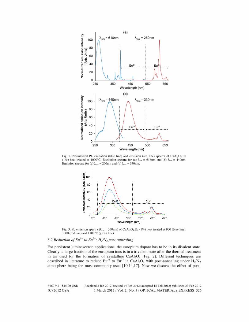
Fig. 2. Normalized PL excitation (blue line) and emission (red line
(1%) heat treated at 1000°C. Excitation spectra for (a)
Emission spectra for (a) λ
Fig. 3. PL emission spectra (
1000 (red line) and 1100°C (green line).
3.2 Reduction of Eu3+
to Eu2+
For persistent luminescence applications, the europium dopant has to be in its divalent state.
Clearly, a large fraction of the europium ions is in a
in air used for the formation of crystalline CaAl
described in literature to reduce Eu
atmosphere being the most commonl
Fig. 2. Normalized PL excitation (blue line) and emission (red line) spectra of CaAl2O4:Eu
(1%) heat treated at 1000°C. Excitation spectra for (a) λem = 616nm and (b) λem = 440nm.
Emission spectra for (a) λexc = 260nm and (b) λexc = 330nm.
Fig. 3. PL emission spectra (λexc = 330nm) of CaAl2O4:Eu (1%) heat treated at 900 (blue line),
1000 (red line) and 1100°C (green line).
2+: H2/N2 post-annealing
For persistent luminescence applications, the europium dopant has to be in its divalent state.
Clearly, a large fraction of the europium ions is in a trivalent state after the thermal treatment
in air used for the formation of crystalline CaAl2O4 (Fig. 2). Different techniques are
described in literature to reduce Eu3+
to Eu2+
in CaAl2O4 with post-annealing under H
atmosphere being the most commonly used [10,14,17]. Now we discuss the effect of post
:Eu
= 440nm.
(blue line),
For persistent luminescence applications, the europium dopant has to be in its divalent state.
trivalent state after the thermal treatment
). Different techniques are
annealing under H2/N2
]. Now we discuss the effect of post-
#160742 - $15.00 USD Received 3 Jan 2012; revised 14 Feb 2012; accepted 18 Feb 2012; published 23 Feb 2012(C) 2012 OSA 1 March 2012 / Vol. 2, No. 3 / OPTICAL MATERIALS EXPRESS 326
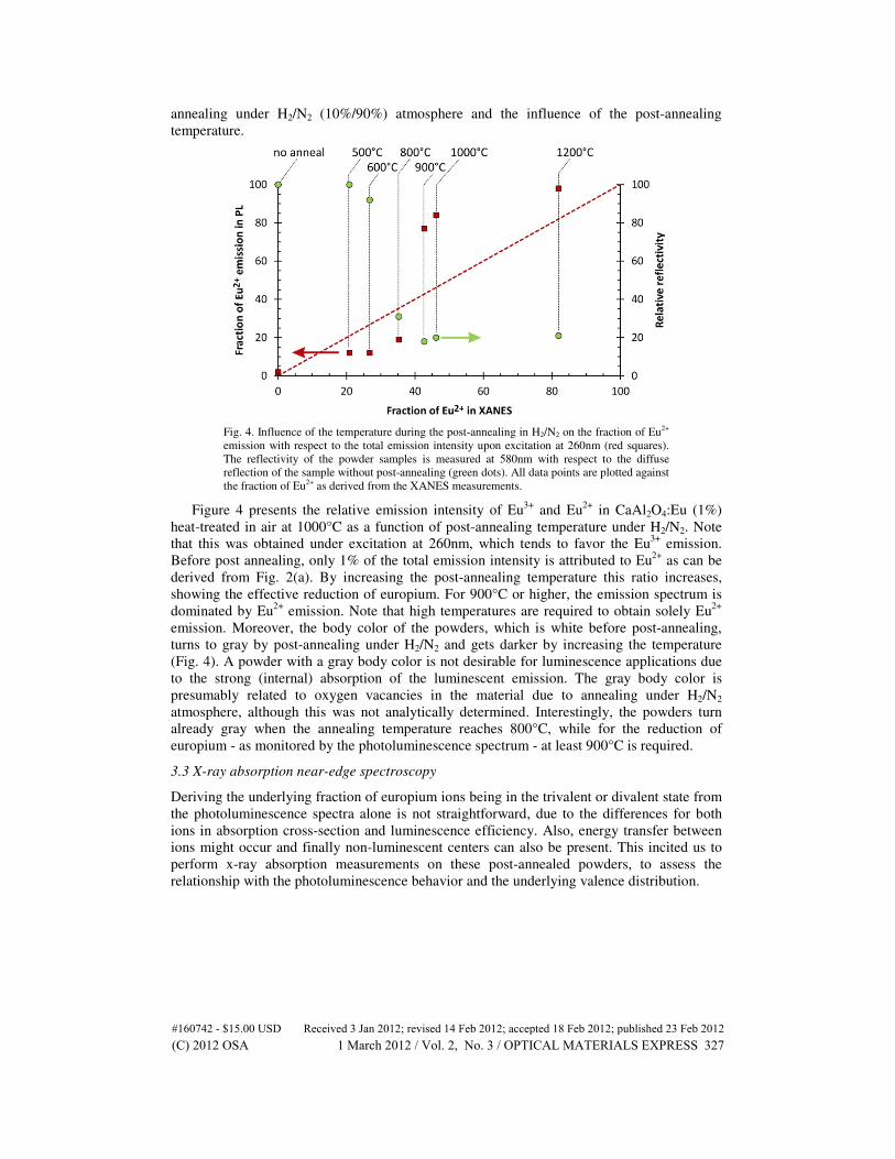
annealing under H2/N2 (10%/90%) atmosphere and the influence of the post-annealing
temperature.
Fig. 4. Influence of the temperature during the post-annealing in H2/N2 on the fraction of Eu2+
emission with respect to the total emission intensity upon excitation at 260nm (red squares).
The reflectivity of the powder samples is measured at 580nm with respect to the diffuse
reflection of the sample without post-annealing (green dots). All data points are plotted against
the fraction of Eu2+ as derived from the XANES measurements.
Figure 4 presents the relative emission intensity of Eu3+
and Eu2+
in CaAl2O4:Eu (1%)
heat-treated in air at 1000°C as a function of post-annealing temperature under H2/N2. Note
that this was obtained under excitation at 260nm, which tends to favor the Eu3+
emission.
Before post annealing, only 1% of the total emission intensity is attributed to Eu2+
as can be
derived from Fig. 2(a). By increasing the post-annealing temperature this ratio increases,
showing the effective reduction of europium. For 900°C or higher, the emission spectrum is
dominated by Eu2+
emission. Note that high temperatures are required to obtain solely Eu2+
emission. Moreover, the body color of the powders, which is white before post-annealing,
turns to gray by post-annealing under H2/N2 and gets darker by increasing the temperature
(Fig. 4). A powder with a gray body color is not desirable for luminescence applications due
to the strong (internal) absorption of the luminescent emission. The gray body color is
presumably related to oxygen vacancies in the material due to annealing under H2/N2
atmosphere, although this was not analytically determined. Interestingly, the powders turn
already gray when the annealing temperature reaches 800°C, while for the reduction of
europium - as monitored by the photoluminescence spectrum - at least 900°C is required.
3.3 X-ray absorption near-edge spectroscopy
Deriving the underlying fraction of europium ions being in the trivalent or divalent state from
the photoluminescence spectra alone is not straightforward, due to the differences for both
ions in absorption cross-section and luminescence efficiency. Also, energy transfer between
ions might occur and finally non-luminescent centers can also be present. This incited us to
perform x-ray absorption measurements on these post-annealed powders, to assess the
relationship with the photoluminescence behavior and the underlying valence distribution.
#160742 - $15.00 USD Received 3 Jan 2012; revised 14 Feb 2012; accepted 18 Feb 2012; published 23 Feb 2012(C) 2012 OSA 1 March 2012 / Vol. 2, No. 3 / OPTICAL MATERIALS EXPRESS 327

Fig. 5. XANES spectra for CaAl2O4:Eu heat-treated at 1000°C as a function of the temperature
of the post-annealing in H2/N2. The inset shows the XANES spectra for the reference
compounds EuS and Eu2O3.
Figure 5 shows the europium LIII XANES spectra as a function of the post-annealing
temperature. The peaks located at 6.975 keV and 6.983 keV are assigned to the 2p3/2 → 5d
transitions in divalent, resp. trivalent europium ions (inset of Fig. 5). The peak at 6.975 keV
becomes apparent upon increasing the temperature of the annealing process, indicating that
Eu3+
ions are reduced to Eu2+
due to the annealing process in H2/N2.
The Eu LIII XANES spectra are then analyzed by fitting procedures in which the white
line transitions are approximated with Lorentzian curves and the absorption step by an
arctangent function [25,26]. The resulting percentage of Eu2+
as a function of the post-
annealing temperature is given in Fig. 4. A clear one-to-one relationship between the valence
distributions derived from photoluminescence measurements and from the XANES
measurements is not observed, as this would imply data points on the dotted line in Fig. 4.
When the fraction of Eu2+
is 40% or above, as derived from the XANES data, the
photoluminescence spectrum starts to be dominated by Eu2+
emission.
3.4. Afterglow in CaAl2O4:Eu,Nd
Given that CaAl2O4:Eu only shows a limited afterglow, sols were also prepared by adding
appropriate amounts of Nd. CaAl2O4:Eu,Nd powders were then prepared by a heat treatment
at 1000°C in air, followed by a post-annealing in reducing H2/N2 atmosphere at 1000°C.
Similar to CaAl2O4:Eu powders which received the same synthesis and annealing conditions,
the steady state emission spectrum is dominated by Eu2+
emission. Figure 6 shows the
afterglow emission intensity (monitored at 445nm) after excitation at 350nm during 6
minutes for CaAl2O4:Eu(1%),Nd(1%). Samples were kept in the dark for a sufficiently long
time prior to the experiment, so that all traps can be assumed to be empty.
The emission intensity during the excitation is not constant, but smoothly increases, until
saturation is reached. This build-up is associated to the competitive process of filling the traps
responsible for the afterglow. When the excitation ends, a considerable afterglow is observed,
with respect to the steady-state photoluminescence intensity. The shape of the decay profile is
typical for CaAl2O4:Eu,Nd, with a relatively slow afterglow after an initial faster decay. In
absolute terms, the afterglow intensity one minute after the excitation (for five minutes with
1000lx of unfiltered Xe arc light) is only 0.25mcd/m2, which is about two orders of
magnitude lower than commercially available CaAl2O4:Eu,Nd (GloTech Int.). This much
lower afterglow intensity is mainly related to the dark gray body color of the post-annealed
samples, which leads not only to a strong absorption of the excitation light by defects (instead
of by the europium ions) but also hampers the emitted light to leave the sample.
#160742 - $15.00 USD Received 3 Jan 2012; revised 14 Feb 2012; accepted 18 Feb 2012; published 23 Feb 2012(C) 2012 OSA 1 March 2012 / Vol. 2, No. 3 / OPTICAL MATERIALS EXPRESS 328

Fig. 6. Emission intensity monitored at 445nm for CaAl2O4:Eu,Nd powder post-annealed in
H2/N2 at 1000°C. The sample was excited at 350nm for 360s. The excitation ended at time t =
0s.
4. Discussion
It is clear that the proposed non-aqueous sol-gel method allows the synthesis of crystalline
and luminescent CaAl2O4:Eu powder phosphors. A heat treatment at 900°C in air is sufficient
to crystallize the powders, and at 1100°C the expected monoclinic phase of CaAl2O4 starts to
be formed. However it is not a priori clear from the x-ray diffraction analysis what phase is
formed below 1100°C. Considering the work by Aitasalo et al. [10] and Janakova et al. [7],
the transition from hexagonal to monoclinic phase occurs around 1050°C, although it is
dependent on the dopant concentration and the duration of the heat treatment [7]. Also, both
phases can be detected simultaneously over a quite broad temperature range. For our
synthesis method, it appears that the largest crystalline fraction consists of the hexagonal
phase, based on the absence of splitting of the diffraction peak at a 2θ value of 35.5° (Fig. 1).
More information on the phase formation can be derived from the photoluminescence
measurements. In the low-temperature range, the Eu2+
emission band peaks invariably at
440nm, which is characteristic for the monoclinic phase [23]. The strongest emission peak
within the 5D0-
7F2 transition of Eu
3+ lies at 616.5nm, with a second less-intense one at 611nm
(Fig. 2). Again, this points at the Eu3+
emission mainly originating from the monoclinic
phase, based on the spectra described in [7]. Also the excitation spectrum for the Eu3+
emission, with a strong CT-based excitation band, is typical for the monoclinic phase [7].
Hence we can conclude that even for heat treatments at relatively low temperature, i.e. 900 to
1000°C, the monoclinic phase for CaAl2O4:Eu3+
is already obtained to a considerable extent.
The absence of the monoclinic phase in the x-ray diffraction patterns could be due to a
limited long range order of the monoclinic phase.
In a second part of this work, we evaluated the reduction of Eu3+
by a post-annealing in a
reducing H2/N2 atmosphere. Already at a post-annealing temperature of 500°C, about one
fifth of the europium ions are reduced to Eu2+
, which is also reflected in the
photoluminescence spectrum (Fig. 4). Increasing the post-annealing temperature further
increases the fraction of Eu2+
. At 900°C, the emission spectrum is dominated by Eu2+
, even
for an excitation wavelength which preferentially excites Eu3+
, although XANES indicates
that less than half of the europium ions are reduced to Eu2+
. For the highest annealing
temperatures used, the emission spectrum hardly contains Eu3+
-related emission, while
according to the XANES results there is still a significant fraction of Eu3+
ions. This behavior
is not untypical for Eu-doped materials [27]. During a XANES study on monoclinic rare-
#160742 - $15.00 USD Received 3 Jan 2012; revised 14 Feb 2012; accepted 18 Feb 2012; published 23 Feb 2012(C) 2012 OSA 1 March 2012 / Vol. 2, No. 3 / OPTICAL MATERIALS EXPRESS 329

earth co-doped CaAl2O4:Eu, Hölsä et al. observed only the presence of Eu2+
in CaAl2O4:Eu
and small fractions of Eu3+
if co-dopants were used [9]. These powders were formed by
heating for 10h at 1350°C in a reducing atmosphere (N2 + 12% H2). The result on
CaAl2O4:Eu appears to be compatible with our results (Fig. 4).
As mentioned above, the main change in the emission spectrum occurs between 800°C
and 900°C. At slightly lower temperature, the body color of the powders has already turned
gray, presumably indicating the formation of oxygen vacancies. This blackening is
detrimental for the afterglow emission intensity of the CaAl2O4:Eu,Nd powder. However, the
shape of the decay profile and the afterglow intensity relative to the steady-state
photoluminescence intensity (Fig. 6) allows us to state that many relatively deep traps have
been created in the CaAl2O4:Eu,Nd powder. Further research is required to evaluate whether
oxygen vacancies formed during the reducing post-annealing are playing a role in the
trapping mechanism, as has been suggested [8,9]. In any case, the post-annealing step should
be modified to avoid this blackening of the powder.
As a follow-up of this research, alternative post-annealing techniques will be explored,
such as reducing atmosphere with different composition (e.g. lower hydrogen content) or
vacuum annealing. In this way it should be possible to keep a white body color, while still
efficiently reducing the europium ions, and thus enhancing the afterglow intensity in
CaAl2O4:Eu,Nd.
5. Conclusions
In this work we have shown that crystalline CaAl2O4:Eu powders can be obtained at
relatively low temperature by a non-aqueous sol-gel method. After a heat-treatment of the
sols in air, the emission spectrum is dominated by 4f-4f emission peaks originating from
transitions within Eu3+
ions. XANES analysis confirmed that in these powders the trivalent
state is dominant for the europium ions. By performing a post-annealing in a reducing H2/N2
atmosphere, a large fraction of the Eu3+
ions can be reduced to Eu2+
. On the one hand, a
relatively low post-annealing temperature of 900°C is sufficient to have mainly Eu2+
emission. On the other hand, for an annealing temperature of 800°C or more, the body color
of the powders turns gray. Finally, the ratio between both valence states of europium derived
from photoluminescence measurements deviates strongly from the ratio derived from x-ray
absorption spectroscopy. A considerable fraction of Eu3+
ions can be present in the powders,
although this is not necessarily reflected in the photoluminescence properties.
Acknowledgments
KK thanks FWO-Vlaanderen for a PhD research grant. This research is partially supported by
the Interuniversity attraction poles programme IAP/VI-17 (INANOMAT) financed by the
Belgian State, Federal science policy office. We gratefully thank Iolanda Cimieri, Manuel
Claeys Boùùaert, Jonas Botterman and Koen Van den Eeckhout for assisting in the
synchrotron data collection.
#160742 - $15.00 USD Received 3 Jan 2012; revised 14 Feb 2012; accepted 18 Feb 2012; published 23 Feb 2012(C) 2012 OSA 1 March 2012 / Vol. 2, No. 3 / OPTICAL MATERIALS EXPRESS 330
Copyright © 2022 FDOKUMEN