
Utilization of nitrophenylphosphates and oxime-based ligation for the development of nanomolar...
11
This article not subject to U.S. Copyright. Published XXXX by the American Chemical Society A dx.doi.org/10.1021/jm200022g | J. Med. Chem. XXXX, XXX, 000–000 ARTICLE pubs.acs.org/jmc Utilization of Nitrophenylphosphates and Oxime-Based Ligation for the Development of Nanomolar Affinity Inhibitors of the Yersinia pestis Outer Protein H (YopH) Phosphatase †,‡ Medhanit Bahta, § George T. Lountos, || Beverly Dyas, ^ Sung-Eun Kim, § Robert G. Ulrich, ^ David S. Waugh, || and Terrence R. Burke, Jr.* ,§ § Chemical Biology Laboratory, Molecular Discovery Program, Center for Cancer Research, National Cancer Institute, National Institutes of Health, NCI—Frederick, Frederick, Maryland 21702, United States ) Macromolecular Crystallography Laboratory, Center for Cancer Research, National Cancer Institute, National Institutes of Health, NCI—Frederick, Frederick, Maryland 21702, United States ^ Laboratory of Molecular Immunology, United States Army Medical Research Institute of Infectious Diseases, Frederick, Maryland 21702, United States b S Supporting Information ’ INTRODUCTION Maintaining proper levels of tyrosyl phosphorylation through the reversible actions of protein tyrosine kinases (PTKs) and protein tryrosine phosphatases (PTPs) is vital for cellular processes ranging from growth and metabolism to adhesion and differentiation. 1,2 Deregulation of PTPs can be linked to diseases such as diabetes and cancer, and accordingly, this class of enzymes represents a new source of potential drug targets. 36 The Gram-negative enterobacterium Yersinia pestis (Y. pestis ) has played an important role in human history as the causative agent of plague, 7 and more recently it has gained attention because of its possible use as a biological warfare agent. For pathogenicity, Y. pestis requires the virulence factor Yersinia pestis outer protein H “YopH”, a highly active PTP. 8 Accordingly, potent and selective YopH inhibitors could provide a basis for new antiplague therapeutics. One di fficulty encountered in the development of PTP inhibitors is a high incidence of “false positives” that can arise through inhibition of enzyme function by “promiscuous” mechanisms attributable to nonspecific factors such as protein aggregation. 9,10 It is generally believed that promiscuous inhibitors do not represent valid leads, and avoiding promiscuous mechanisms is an important compo- nent of current drug development. 11 In theory, avoiding promiscuous behavior could be achieved through the use of substrates as templates for inhibitor design. This is because substrates must interact with their enzyme hosts in nonpromiscuous fashions in order for productive catalysis to occur. Employing small nonpeptidic arylphosphates to identify potential leads for PTP inhibitor design has been known for some time. 1215 However, the explicit application of “substrate activity screening” for the purpose of minimizing misleading promiscuous inhibition has only more recently been proposed by Ellman for protease 1620 and PTP targets. 21 This approach consists of first identifying substrates that exhibit high affinity, structurally enhancing these substrates, and then converting the optimized substrates to inhibitors by replacement of their labile phosphoryl groups with suitable nonhydrolyzable phosphoryl mimetics. Additional structural variations can then be performed to further increase inhibitory potency. In the identification of high affinity substrates for the devel- opment of PTP inhibitors, advantage can be taken of the hydrolytic action of a PTP on an arylphosphate, which produces Received: January 10, 2011 ABSTRACT: Our current study reports the first K M optimiza- tion of a library of nitrophenylphosphate-containing substrates for generating an inhibitor lead against the Yersinia pestis outer protein phosphatase (YopH). A high activity substrate identi- fied by this method (K M = 80 μM) was converted from a substrate into an inhibitor by replacement of its phosphate group with difluoromethylphosphonic acid and by attachment of an aminooxy handle for further structural optimization by oxime ligation. A cocrystal structure of this aminooxy-containing platform in complex with YopH allowed the identification of a conserved water molecule proximal to the aminooxy group that was subsequently employed for the design of furanyl-based oxime derivatives. By this process, a potent (IC 50 = 190 nM) and nonpromiscuous inhibitor was developed with good YopH selectivity relative to a panel of phosphatases. The inhibitor showed significant inhibition of intracellular Y. pestis replication at a noncytotoxic concentration. The current work presents general approaches to PTP inhibitor development that may be useful beyond YopH.
Transcript of Utilization of nitrophenylphosphates and oxime-based ligation for the development of nanomolar...
This article not subject to U.S. Copyright.Published XXXX by the American Chemical Society A dx.doi.org/10.1021/jm200022g | J. Med. Chem. XXXX, XXX, 000–000
ARTICLE
pubs.acs.org/jmc
Utilization of Nitrophenylphosphates and Oxime-Based Ligation forthe Development of Nanomolar Affinity Inhibitors of the Yersiniapestis Outer Protein H (YopH) Phosphatase†,‡
Medhanit Bahta,§George T. Lountos,|| Beverly Dyas,^ Sung-Eun Kim,§ Robert G. Ulrich,^David S. Waugh,||
and Terrence R. Burke, Jr.*,§
§Chemical Biology Laboratory, Molecular Discovery Program, Center for Cancer Research, National Cancer Institute, NationalInstitutes of Health, NCI—Frederick, Frederick, Maryland 21702, United States
)Macromolecular Crystallography Laboratory, Center for Cancer Research, National Cancer Institute, National Institutes of Health,NCI—Frederick, Frederick, Maryland 21702, United States^Laboratory of Molecular Immunology, United States Army Medical Research Institute of Infectious Diseases, Frederick,Maryland 21702, United States
bS Supporting Information
’ INTRODUCTION
Maintaining proper levels of tyrosyl phosphorylation throughthe reversible actions of protein tyrosine kinases (PTKs) andprotein tryrosine phosphatases (PTPs) is vital for cellularprocesses ranging from growth and metabolism to adhesionand differentiation.1,2 Deregulation of PTPs can be linked todiseases such as diabetes and cancer, and accordingly, this class ofenzymes represents a new source of potential drug targets.3�6 TheGram-negative enterobacterium Yersinia pestis (Y. pestis) has playedan important role in human history as the causative agent of plague,7
andmore recently it has gained attention because of its possible use asa biological warfare agent. For pathogenicity, Y. pestis requires thevirulence factorYersinia pestis outer proteinH “YopH”, a highly activePTP.8 Accordingly, potent and selective YopH inhibitors couldprovide a basis for new antiplague therapeutics. One difficultyencountered in the development of PTP inhibitors is a highincidence of “false positives” that can arise through inhibition ofenzyme function by “promiscuous” mechanisms attributable tononspecific factors such as protein aggregation.9,10 It is generallybelieved that promiscuous inhibitors do not represent valid leads,and avoiding promiscuous mechanisms is an important compo-nent of current drug development.11
In theory, avoiding promiscuous behavior could be achievedthrough the use of substrates as templates for inhibitor design.This is because substrates must interact with their enzyme hostsin nonpromiscuous fashions in order for productive catalysis tooccur. Employing small nonpeptidic arylphosphates to identifypotential leads for PTP inhibitor design has been known forsome time.12�15 However, the explicit application of “substrateactivity screening” for the purpose of minimizing misleadingpromiscuous inhibition has only more recently been proposed byEllman for protease16�20 and PTP targets.21 This approachconsists of first identifying substrates that exhibit high affinity,structurally enhancing these substrates, and then converting theoptimized substrates to inhibitors by replacement of their labilephosphoryl groups with suitable nonhydrolyzable phosphorylmimetics. Additional structural variations can then be performedto further increase inhibitory potency.
In the identification of high affinity substrates for the devel-opment of PTP inhibitors, advantage can be taken of thehydrolytic action of a PTP on an arylphosphate, which produces
Received: January 10, 2011
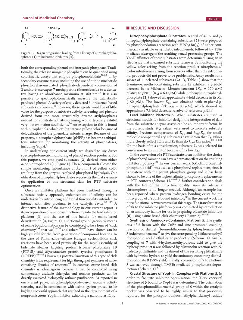
ABSTRACT: Our current study reports the first KM optimiza-tion of a library of nitrophenylphosphate-containing substratesfor generating an inhibitor lead against the Yersinia pestis outerprotein phosphatase (YopH). A high activity substrate identi-fied by this method (KM = 80 μM) was converted from asubstrate into an inhibitor by replacement of its phosphategroup with difluoromethylphosphonic acid and by attachmentof an aminooxy handle for further structural optimization byoxime ligation. A cocrystal structure of this aminooxy-containing platform in complex with YopH allowed the identification of aconserved water molecule proximal to the aminooxy group that was subsequently employed for the design of furanyl-based oximederivatives. By this process, a potent (IC50 = 190 nM) and nonpromiscuous inhibitor was developed with good YopH selectivityrelative to a panel of phosphatases. The inhibitor showed significant inhibition of intracellular Y. pestis replication at a noncytotoxicconcentration. The current work presents general approaches to PTP inhibitor development that may be useful beyond YopH.
B dx.doi.org/10.1021/jm200022g |J. Med. Chem. XXXX, XXX, 000–000
Journal of Medicinal Chemistry ARTICLE
both the corresponding phenol and inorganic phosphate. Tradi-tionally, the released inorganic phosphate can be quantified usingcolorimetric assays that employ phosphomolybdate22,23 or bysecondary enzyme assays, including the use of purine nucleotidephosphorylase-mediated phosphate-dependent conversion of2-amino-6-mercapto-7-methylpurine ribonucleoside to a deriva-tive having an absorbance maximum at 360 nm.24 It is alsopossible to spectrophotometrically measure the catalyticallyproduced phenol. A variety of easily detected fluorescence-basedsubstrates are known;25 however, these agents would be of littlevalue for the purpose of substrate activity screening and phenolsderived from the more structurally diverse arylphosphatesneeded for substrate activity screening would typically exhibitvery low extinction coefficients.26 An exception to this is foundwith nitrophenols, which exhibit intense yellow color because ofdelocalization of the phenolate anionic charge. Because of thisproperty, p-nitrophenylphosphate (pNPP) has become a ubiqui-tous substrate for monitoring the activity of phosphatases,including YopH.8
In undertaking our current study, we desired to use directspectrophotometric monitoring of phenol reaction products. Forthis purpose, we employed substrates (2) derived from eithero- or p-nitrophenols (1, Figure 1). These compounds allowed thesimple monitoring (absorbance at λ405 nm) of yellow colorresulting from the enzyme-catalyzed phosphoryl hydrolysis. Ourutilization of nitrophenylphosphates represents the first systema-tic application of this structural class for PTP substrateoptimization.
Once an inhibitor platform has been identified through asubstrate activity approach, enhancement of affinity can beundertaken by introducing additional functionality intended tointeract with sites proximal to the catalytic cavity.27�33 Adistinctive feature of the methodology in our current report isits incorporation of aminooxy functionality into the lead inhibitorplatform (3) and the use of this handle for oxime-basedderivatization (4, Figure 1). Functional group ligation by meansof oxime bond formation can be considered to be a form of “clickchemistry”34 that we35�38 and others39�42 have shown can behighly useful for the facile generation of compound libraries. Inthe case of PTPs, azide�alkyne Huisgen cycloaddition clickreactions have been used previously for the rapid assembly ofbidentate libraries targeting protein tyrosine phosphatase 1B(PTP1B) and Mycobacterium protein tyrosine phosphatase B(mPTPB).43�45 However, a potential limitation of this type of clickchemistry is the requirement for high throughput syntheses of azide-containing libraries of reactants.46 In contrast, oxime-based clickchemistry is advantageous because it can be conducted usingcommercially available aldehydes and reaction products can bedirectly evaluated biologically without purification. As reported inour current paper, nitrophenylphosphate-based substrate activityscreening used in combination with oxime ligation proved to behighly a successful approach that resulted in the development of anonpromiscuous YopH inhibitor exhibiting a nanomolar IC50.
’RESULTS AND DISCUSSION
Nitrophenylphosphate Substrates. A total of 48 o- and p-nitrophenylphosphate-containing substrates (2) were preparedby phosphorylation (reaction with HPO3(Bn)2) of either com-mercially available or synthetic nitrophenols, followed by TFA-mediated cleavage of the resulting benzyl protecting groups. TheYopH affinities of these substrates were determined using an invitro assay that measured substrate turnover by monitoring theyellow color arising from the reaction product nitrophenols.8
Color interference arising from sources other than the nitrophe-nol products did not prove to be problematic. Assay results for asubset of 11 selected substrates (2a�k, Table 1) show that the3-aminooxymethyl-containing substrate 2e exhibited a 3.5-folddecrease in its Michaelis�Menten constant (KM = 170 μM)relative to pNPP (KM = 600 μM) while p-phenyl-o-nitrophenyl-phosphate (2j) showed an approximate 4-fold decrease in its KM
(150 μM). The lowest KM was obtained with m-phenyl-p-nitrophenylphosphate (2k, KM = 80 μM), which showed anapproximate 7.5-fold decrease relative to reference pNPP.Lead Inhibitor Platform 5. When substrates are used as
structural models for inhibitor design, the interpretation of datafrom the substrate enzyme assays can be an important factor. Inthe current study, KM values were used to indicate substrateaffinity. Previous comparisons of KM and kcat/KM for small-molecule non-peptidyl aryl substrates have shown thatKM valuesmore closely reflect IC50 and Ki than do kcat/KM ratios.12�14,21
On the basis of this consideration, substrate 2k was selected forconversion to an inhibitor because of its low KM.In the conversion of a PTP substrate to an inhibitor, the choice
of phosphoryl mimetic can have a dramatic effect on the resultinginhibitory potency.21 In our current work R,R-difluoromethyl-phosphonic acid47 was used as a phosphoryl replacement, since itis isosteric with the parent phosphate group and it has beenshown to be one of the highest affinity phosphoryl replacementsin PTP contexts (Scheme 1).48,49 A further consideration dealswith the fate of the nitro functionality, since its role as achromophore is no longer needed. Although an example hasbeen reported where protein hydrogen bonding exists for thenitro group of a YopH-bound inhibitor,50 in the current work thenitro functionality was removed at this stage. The transformationof 2k to the inhibitor platform 5 was completed by introductionof an aminooxy handle for use in preparing bidentate inhibitors(6) using oxime-based click chemistry (Figure 2).34�42
Synthesis of Aminooxy-Containing Platform 5. The synth-esis of 5 began with the CuBr and zinc promoted couplingreaction of diethyl (bromodifluoromethyl)phosphonate with3-iodobromobenzene51 to give the corresponding (difluoromethyl)phosphonic acid diethyl ester product 7 (Scheme 1). Suzukicoupling of 7 with 4-hydroxymethylboronic acid to give thebiphenyl product 8 was followed by Mitsunobu reaction with N-hydroxyphthalimide and treatment of the resulting phthalimidewith hydrazine hydrate to yield the aminooxy-containing diethyl-phosphonate 9 (79% yield). Finally, conversion of 9 to platform5 was achieved through TMSBr-mediated phosphonate depro-tection (Scheme 1).52
Crystal Structure of YopH in Complex with Platform 5. Inorder to facilitate inhibitor optimization, the X-ray cocrystalstructure of 5 bound to YopH was determined. The orientationof the phosphonodifluormethyl group of 5 within the catalyticpocket was observed to be highly similar to that previouslyreported for the phosphonodifluormethylphenylalanyl residue
Figure 1. Design progression leading from a library of nitrophenylpho-sphates (1) to bidentate inhibitors (4).
C dx.doi.org/10.1021/jm200022g |J. Med. Chem. XXXX, XXX, 000–000
Journal of Medicinal Chemistry ARTICLE
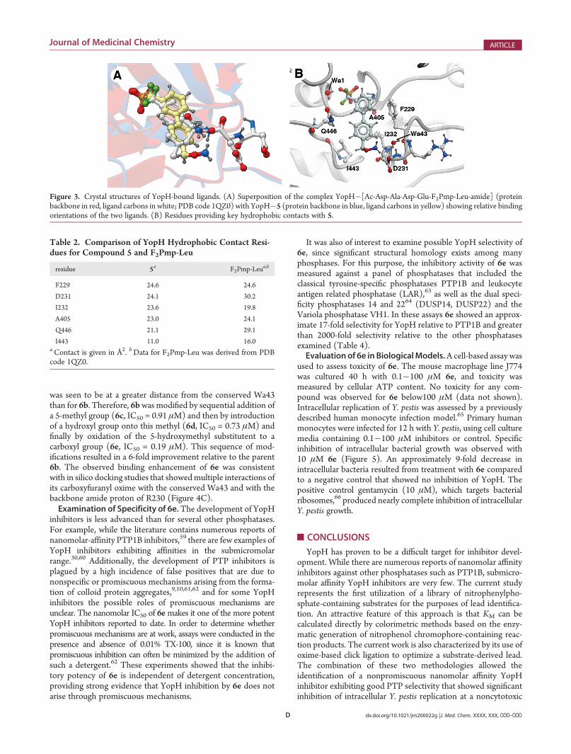
(F2Pmp) of the hexapeptide Ac-Asp-Ala-Asp-Glu-F2Pmp-Leu-amide (PDB code 1QZ0) (Figure 3A).53 The protein backbonesof the two structures are nearly superimposable, and in bothstructures the flexible “WPD loops” (residues 354�356) are heldin the “closed” conformation. For both structures the phosphor-yl-mimicking difluoromethylphosphonic acid group is coordi-nated within the conserved (H/V)CX5R(S/T) signature motif“P loop” (residues 404�410) by six hydrogen bonds,54�56 whilethe guanidine group of R409 forms two salt bridges with twophosphonic acid oxygen atoms and indirect hydrogen bonds withresidues Q357, Q450, and Q446 are made through a conservedwater residue (designated as “Wa1” for ligand 5 and “cw” in PDB1QZ0). The 1-phenyl ring of 5 is pivoted about the difluoro-methylene carbon so that it is offset, yet within the same planerelative to the F2Pmp aryl ring in the 1QZ0 structure. This allowsthe 3-(40-methylphenyl) moiety of 5 to interact, similar to theLeu side chain of the dipeptide unit F2Pmp-Leu in the 1QZ0structure (Figure 3A). The aryl rings of 5 form extensivehydrophobic contacts with residues F229, D231, I232, A405,
Q446, and I443, similar to what is observed in the 1QZ0structure (Figure 3 and Table 2).Introduction of Oxime Functionality into 5. In the cocrystal
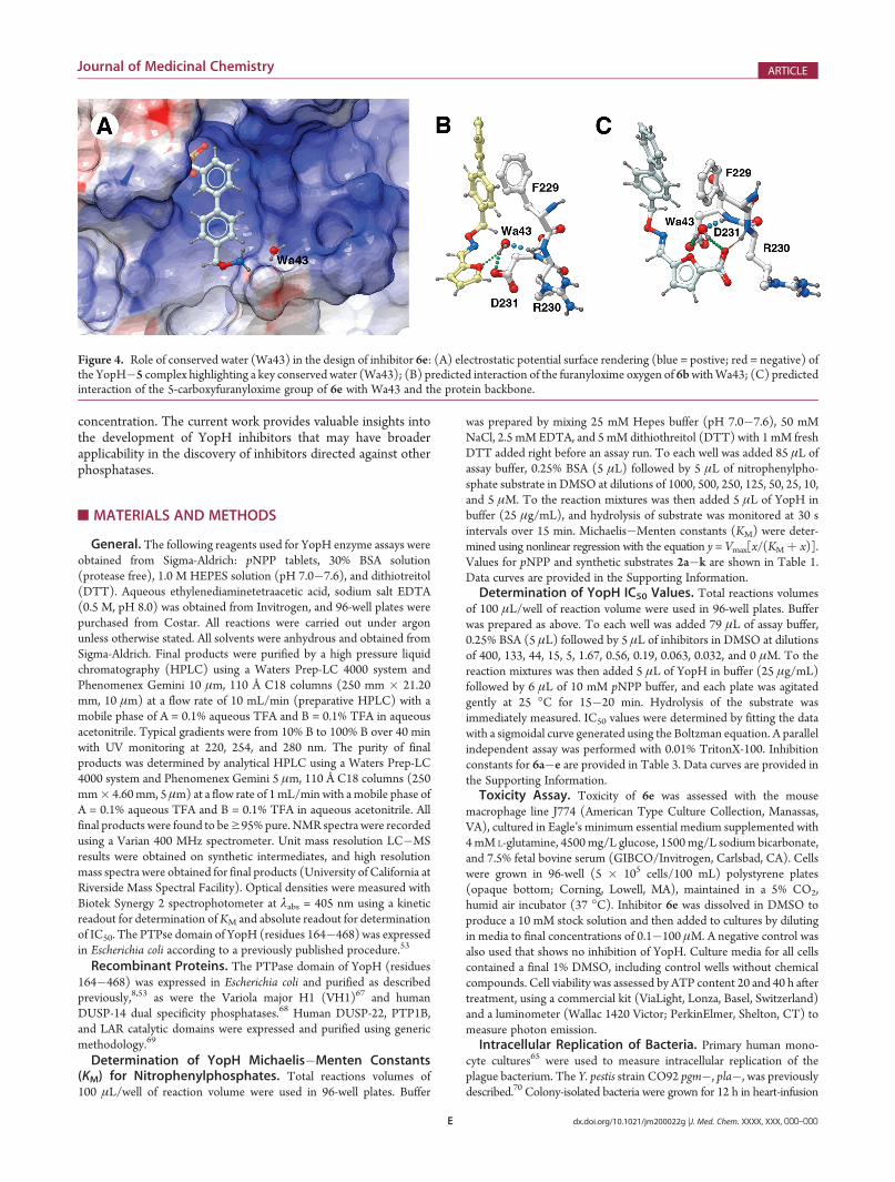
structure the aminooxyamine of 5 forms hydrogen bonds withthe side chain carboxyl of D231 and a water molecule (Wa43),which also hydrogen-bonds to the D231 residue (Figure 4A).The importance of this latter water is indicated by its presencein the absence of inhibitor (designated as “Wa87” in PBD code1LYV), suggesting that it could be used for inhibitor design.In silico docking studies performed using the cocrystal structureof 5 with the inclusion of Wa4357,58 identified furanyl-basedoximes as providing favorable interactions with the D231 residuethrough the intermediacy of the conserved water (Figure 4B).Syntheses of a series of furanyl-based oxime inhibitors was
performed in DMSO by reacting 5 (24 mM) with commerciallyavailable furanyl aldehydes and AcOH in the ratio (1:1:2). Theoxime products (6), which were typically of >90% purity asshown by random HPLC analysis, were used directly forbiological evaluation. Inhibitory potencies (IC50) were obtainedspectrophotometrically in an in vitro YopH assay using pNPP assubstrate.38
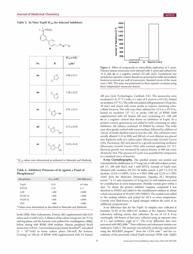
The 3-furanyloxime (6a) showed an IC50 of 3.69 μM,whereas the2-furanyloxime (6b) was approximately 3-fold more potent (IC50 =1.20 μM) (Table 3). In modeling studies, the furanyl oxygen in 6a
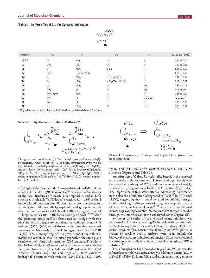
Table 1. In Vitro YopH KM for Selected Substrates
substrate R1 R2 R3 R4 KM ( SE (mM)a
pNPP H NO2 H H 0.60 ( 0.10
2a NO2 t-Bu H H 0.47 ( 0.06
2b H NO2 t-Bu H 1.14 ( 0.19
2c NO2 �CH2ONH2 H H 1.13 ( 0.15
2d H NO2 �CH2ONH2 H 0.41 ( 0.05
2e H NO2 CH2ONdCHMe H 0.17 ( 0.02
2f H NO2 H Me 0.65 ( 0.11
2g NO2 H H Me no activity
2h cyclohexyl NO2 H H 0.26 ( 0.02
2i NO2 H H cyclohexyl no activity
2j NO2 Ph H H 0.15 ( 0.03
2k H NO2 Ph �H 0.08 ( 0.01a KM values were determined as indicated in the Materials and Methods.
Scheme 1. Synthesis of Inhibitor Platform 5a
aReagents and conditions: (i) Zn, diethyl (bromodifluoromethyl)-phosphonate, CuBr, DMF, 60 �C to room temperature (96% yield);(ii) 4-(hydroxymethyl)phenylboronic acid, Pd(PPh3)4, sat. K2CO3,EtOH, PhMe, 70 �C (45% yield); (iii) (a) N-hydroxyphthalimide,PPh3, DIAD, THF, room temperature; (b) NH2NH2 3H2O, EtOH,room temperature (75% yield); (iv) TMSBr, CH2Cl2, room tempera-ture (72% yield).
Figure 2. Development of oxime-containing inhibitors (6) startingfrom platform 2k.
D dx.doi.org/10.1021/jm200022g |J. Med. Chem. XXXX, XXX, 000–000
Journal of Medicinal Chemistry ARTICLE
was seen to be at a greater distance from the conserved Wa43than for 6b. Therefore, 6bwas modified by sequential addition ofa 5-methyl group (6c, IC50 = 0.91 μM) and then by introductionof a hydroxyl group onto this methyl (6d, IC50 = 0.73 μM) andfinally by oxidation of the 5-hydroxymethyl substitutent to acarboxyl group (6e, IC50 = 0.19 μM). This sequence of mod-ifications resulted in a 6-fold improvement relative to the parent6b. The observed binding enhancement of 6e was consistentwith in silico docking studies that showedmultiple interactions ofits carboxyfuranyl oxime with the conserved Wa43 and with thebackbone amide proton of R230 (Figure 4C).Examination of Specificity of 6e.The development of YopH
inhibitors is less advanced than for several other phosphatases.For example, while the literature contains numerous reports ofnanomolar-affinity PTP1B inhibitors,59 there are few examples ofYopH inhibitors exhibiting affinities in the submicromolarrange.30,60 Additionally, the development of PTP inhibitors isplagued by a high incidence of false positives that are due tononspecific or promiscuous mechanisms arising from the forma-tion of colloid protein aggregates,9,10,61,62 and for some YopHinhibitors the possible roles of promiscuous mechanisms areunclear. The nanomolar IC50 of 6emakes it one of the more potentYopH inhibitors reported to date. In order to determine whetherpromiscuous mechanisms are at work, assays were conducted in thepresence and absence of 0.01% TX-100, since it is known thatpromiscuous inhibition can often be minimized by the addition ofsuch a detergent.62 These experiments showed that the inhibi-tory potency of 6e is independent of detergent concentration,providing strong evidence that YopH inhibition by 6e does notarise through promiscuous mechanisms.
It was also of interest to examine possible YopH selectivity of6e, since significant structural homology exists among manyphosphases. For this purpose, the inhibitory activity of 6e wasmeasured against a panel of phosphatases that included theclassical tyrosine-specific phosphatases PTP1B and leukocyteantigen related phosphatase (LAR),63 as well as the dual speci-ficity phosphatases 14 and 2264 (DUSP14, DUSP22) and theVariola phosphatase VH1. In these assays 6e showed an approx-imate 17-fold selectivity for YopH relative to PTP1B and greaterthan 2000-fold selectivity relative to the other phosphatasesexamined (Table 4).Evaluationof 6e in BiologicalModels.A cell-based assay was
used to assess toxicity of 6e. The mouse macrophage line J774was cultured 40 h with 0.1�100 μM 6e, and toxicity wasmeasured by cellular ATP content. No toxicity for any com-pound was observed for 6e below100 μM (data not shown).Intracellular replication of Y. pestis was assessed by a previouslydescribed human monocyte infection model.65 Primary humanmonocytes were infected for 12 h with Y. pestis, using cell culturemedia containing 0.1�100 μM inhibitors or control. Specificinhibition of intracellular bacterial growth was observed with10 μM 6e (Figure 5). An approximately 9-fold decrease inintracellular bacteria resulted from treatment with 6e comparedto a negative control that showed no inhibition of YopH. Thepositive control gentamycin (10 μM), which targets bacterialribosomes,66 produced nearly complete inhibition of intracellularY. pestis growth.
’CONCLUSIONS
YopH has proven to be a difficult target for inhibitor devel-opment. While there are numerous reports of nanomolar affinityinhibitors against other phosphatases such as PTP1B, submicro-molar affinity YopH inhibitors are very few. The current studyrepresents the first utilization of a library of nitrophenylpho-sphate-containing substrates for the purposes of lead identifica-tion. An attractive feature of this approach is that KM can becalculated directly by colorimetric methods based on the enzy-matic generation of nitrophenol chromophore-containing reac-tion products. The current work is also characterized by its use ofoxime-based click ligation to optimize a substrate-derived lead.The combination of these two methodologies allowed theidentification of a nonpromiscuous nanomolar affinity YopHinhibitor exhibiting good PTP selectivity that showed significantinhibition of intracellular Y. pestis replication at a noncytotoxic
Figure 3. Crystal structures of YopH-bound ligands. (A) Superposition of the complex YopH�[Ac-Asp-Ala-Asp-Glu-F2Pmp-Leu-amide] (proteinbackbone in red, ligand carbons in white; PDB code 1QZ0) with YopH�5 (protein backbone in blue, ligand carbons in yellow) showing relative bindingorientations of the two ligands. (B) Residues providing key hydrophobic contacts with 5.
Table 2. Comparison of YopH Hydrophobic Contact Resi-dues for Compound 5 and F2Pmp-Leu
residue 5a F2Pmp-Leua,b
F229 24.6 24.6
D231 24.1 30.2
I232 23.6 19.8
A405 23.0 24.1
Q446 21.1 29.1
I443 11.0 16.0aContact is given in Å2. bData for F2Pmp-Leu was derived from PDBcode 1QZ0.
E dx.doi.org/10.1021/jm200022g |J. Med. Chem. XXXX, XXX, 000–000
Journal of Medicinal Chemistry ARTICLE
concentration. The current work provides valuable insights intothe development of YopH inhibitors that may have broaderapplicability in the discovery of inhibitors directed against otherphosphatases.
’MATERIALS AND METHODS
General. The following reagents used for YopH enzyme assays wereobtained from Sigma-Aldrich: pNPP tablets, 30% BSA solution(protease free), 1.0 M HEPES solution (pH 7.0�7.6), and dithiotreitol(DTT). Aqueous ethylenediaminetetraacetic acid, sodium salt EDTA(0.5 M, pH 8.0) was obtained from Invitrogen, and 96-well plates werepurchased from Costar. All reactions were carried out under argonunless otherwise stated. All solvents were anhydrous and obtained fromSigma-Aldrich. Final products were purified by a high pressure liquidchromatography (HPLC) using a Waters Prep-LC 4000 system andPhenomenex Gemini 10 μm, 110 Å C18 columns (250 mm � 21.20mm, 10 μm) at a flow rate of 10 mL/min (preparative HPLC) with amobile phase of A = 0.1% aqueous TFA and B = 0.1% TFA in aqueousacetonitrile. Typical gradients were from 10% B to 100% B over 40 minwith UV monitoring at 220, 254, and 280 nm. The purity of finalproducts was determined by analytical HPLC using a Waters Prep-LC4000 system and Phenomenex Gemini 5 μm, 110 Å C18 columns (250mm� 4.60mm, 5 μm) at a flow rate of 1mL/min with amobile phase ofA = 0.1% aqueous TFA and B = 0.1% TFA in aqueous acetonitrile. Allfinal products were found to beg95% pure. NMR spectra were recordedusing a Varian 400 MHz spectrometer. Unit mass resolution LC�MSresults were obtained on synthetic intermediates, and high resolutionmass spectra were obtained for final products (University of California atRiverside Mass Spectral Facility). Optical densities were measured withBiotek Synergy 2 spectrophotometer at λabs = 405 nm using a kineticreadout for determination ofKM and absolute readout for determinationof IC50. The PTPse domain of YopH (residues 164�468) was expressedin Escherichia coli according to a previously published procedure.53
Recombinant Proteins. The PTPase domain of YopH (residues164�468) was expressed in Escherichia coli and purified as describedpreviously,8,53 as were the Variola major H1 (VH1)67 and humanDUSP-14 dual specificity phosphatases.68 Human DUSP-22, PTP1B,and LAR catalytic domains were expressed and purified using genericmethodology.69
Determination of YopH Michaelis�Menten Constants(KM) for Nitrophenylphosphates. Total reactions volumes of100 μL/well of reaction volume were used in 96-well plates. Buffer
was prepared by mixing 25 mM Hepes buffer (pH 7.0�7.6), 50 mMNaCl, 2.5 mM EDTA, and 5 mM dithiothreitol (DTT) with 1 mM freshDTT added right before an assay run. To each well was added 85 μL ofassay buffer, 0.25% BSA (5 μL) followed by 5 μL of nitrophenylpho-sphate substrate in DMSO at dilutions of 1000, 500, 250, 125, 50, 25, 10,and 5 μM. To the reaction mixtures was then added 5 μL of YopH inbuffer (25 μg/mL), and hydrolysis of substrate was monitored at 30 sintervals over 15 min. Michaelis�Menten constants (KM) were deter-mined using nonlinear regression with the equation y = Vmax[x/(KMþ x)].Values for pNPP and synthetic substrates 2a�k are shown in Table 1.Data curves are provided in the Supporting Information.Determination of YopH IC50 Values. Total reactions volumes
of 100 μL/well of reaction volume were used in 96-well plates. Bufferwas prepared as above. To each well was added 79 μL of assay buffer,0.25% BSA (5 μL) followed by 5 μL of inhibitors in DMSO at dilutionsof 400, 133, 44, 15, 5, 1.67, 0.56, 0.19, 0.063, 0.032, and 0 μM. To thereaction mixtures was then added 5 μL of YopH in buffer (25 μg/mL)followed by 6 μL of 10 mM pNPP buffer, and each plate was agitatedgently at 25 �C for 15�20 min. Hydrolysis of the substrate wasimmediately measured. IC50 values were determined by fitting the datawith a sigmoidal curve generated using the Boltzman equation. A parallelindependent assay was performed with 0.01% TritonX-100. Inhibitionconstants for 6a�e are provided in Table 3. Data curves are provided inthe Supporting Information.Toxicity Assay. Toxicity of 6e was assessed with the mouse
macrophage line J774 (American Type Culture Collection, Manassas,VA), cultured in Eagle’s minimum essential medium supplemented with4mM L-glutamine, 4500mg/L glucose, 1500mg/L sodium bicarbonate,and 7.5% fetal bovine serum (GIBCO/Invitrogen, Carlsbad, CA). Cellswere grown in 96-well (5 � 105 cells/100 mL) polystyrene plates(opaque bottom; Corning, Lowell, MA), maintained in a 5% CO2,humid air incubator (37 �C). Inhibitor 6e was dissolved in DMSO toproduce a 10 mM stock solution and then added to cultures by dilutingin media to final concentrations of 0.1�100 μM. A negative control wasalso used that shows no inhibition of YopH. Culture media for all cellscontained a final 1% DMSO, including control wells without chemicalcompounds. Cell viability was assessed by ATP content 20 and 40 h aftertreatment, using a commercial kit (ViaLight, Lonza, Basel, Switzerland)and a luminometer (Wallac 1420 Victor; PerkinElmer, Shelton, CT) tomeasure photon emission.Intracellular Replication of Bacteria. Primary human mono-
cyte cultures65 were used to measure intracellular replication of theplague bacterium. The Y. pestis strain CO92 pgm�, pla�, was previouslydescribed.70 Colony-isolated bacteria were grown for 12 h in heart-infusion
Figure 4. Role of conserved water (Wa43) in the design of inhibitor 6e: (A) electrostatic potential surface rendering (blue = postive; red = negative) ofthe YopH�5 complex highlighting a key conserved water (Wa43); (B) predicted interaction of the furanyloxime oxygen of 6bwithWa43; (C) predictedinteraction of the 5-carboxyfuranyloxime group of 6e with Wa43 and the protein backbone.
F dx.doi.org/10.1021/jm200022g |J. Med. Chem. XXXX, XXX, 000–000
Journal of Medicinal Chemistry ARTICLE
broth (HIB; Difco Laboratories, Detroit, MI) supplemented with 0.2%xylose and 2.5mMCaCl2. A dilution of the culture was grown (26 �C) tomid-log phase, and the bacteria were pelleted by centrifugation (600g)before rinsing with RPMI 1640 medium. Human peripheral bloodmonocytes (CD14þ) were isolated as previously described65 and added(2 � 105/well) to tissue culture plates (96-well, flat bottom;Corning) in 100 μL of RPMI 1640 supplemented with 5% human
AB sera (Life Technologies, Carlsbad, CA). The monocytes wereincubated (1 h, 37 �C) with a 1:1 ratio of Y. pestis in a 5% CO2, humidair incubator (37 �C). Thewells were pulsedwith gentamycin (10μg/mL,20 min) and rinsed with warm media to remove remaining extra-cellular bacteria. The cells were then cultured for 12 h in a 5% CO2,humid air incubator (37 �C) in media (100 μL of RPMI 1640supplemented with 5% human AB sera) containing 0.1�100 μM6e or a negative control that shows no inhibition of YopH. As apositive control, gentamycin was added to wells containing no otherinhibitors. All cultures contained 1% DMSO by volume. The wellswere then gently washed with warm medium, followed by addition of120 μL of sterile distilled water to lyse the cells. The cell lysates wereserially diluted (1:5) in HIB, and 200 mL of each dilution was placedinto duplicate wells of a culture plate (Honeycomb; Growth CurvesUSA, Piscataway, NJ) and placed in a growth-monitoring incubator(Bioscreen; Growth Curves USA) with constant agitation (37 �C).Bacterial growth was measured by optical density at 600 nm every20 min for 16 h. The amount of Y. pestiswas quantified by comparisonto a standard curve of bacteria.X-ray Crystallography. The purified protein was pooled and
concentrated by diafiltration to 17.6 mg/mL in 100 mM sodium acetate,pH 5.7, 100 mM NaCl, and 1 mM EDTA. Crystals of YopH wereobtained with condition D8 (0.1 M buffer system 2, pH 7.5, 0.12 Malcohols, 12.5% v/v MPD, 12.5% w/v PEG 1000, and 12.5% w/v PEG3350) from the Molecular Dimensions (Apopka, FL) Morpheusscreen.71 A 1:1 ratio of protein (17.6 mg/mL) to well solution was usedfor crystallization at room temperature. Platelike crystals grew within 3days. To obtain the protein�inhibitor complex, compound 5 wasdissolved in DMSO and added to the crystallization solution to obtaina final concentration of 10 mM (10% DMSO). The crystals were addedto the soaking solution and soaked for 48 h at room temperature.Crystals were flash-frozen in liquid nitrogen without the need of anadditional cryoprotectant.
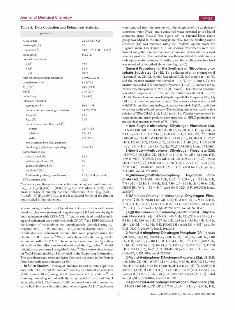
X-ray diffraction data for the YopH�5 complex were collected atbeamline 22-ID of the SER-CAT facilities at the Argonne NationalLaboratory utilizing remote data collection. By use of 1.0 Å X-raywavelength, 180 frames of data were collected using an exposure timeof 3 s and oscillation angle of 1�. The X-ray diffraction data wereprocessed with HKL3000.72 Data collection and refinement statistics areoutlined in Table 5. The structure was solved by molecular replacementusing the MOLREP program73 from the CCP4 suite74 and the co-ordinates of the previously solved YopH structure (PDB code 1QZ0)
Table 3. In Vitro YopH IC50 for Selected Inhibitors
a IC50 values were determined as indicated in Materials and Methods.
Figure 5. Effect of compounds on intracellular replication of Y. pestis.Primary human monocytes were infected with Y. pestis and cultured for12 h with 6e or a negative control (10 μM each). Gentamycin wasincluded as a positive control. Results are presented as viable intracellularbacteria recovered per well of monocytes. Standard errors of the meanwere <10%. The assay was performed on three separate occasions usingthree independent monocyte donors.
Table 4. Inhibitory Potencies of 6e against a Panel ofPhosphatasesa
phosphatase IC50 (μM) fold difference
YopH 0.19 ref value
PTP1B 2.23 11.7
LAR >400 >2000
DUSP-14 >400 >2000
DUSP-22 >400 >2000
VH1 >400 >2000aValues were determined as described in Materials and Methods.
G dx.doi.org/10.1021/jm200022g |J. Med. Chem. XXXX, XXX, 000–000
Journal of Medicinal Chemistry ARTICLE
after removing all solvent and ligand atoms. Cross-rotation and transla-tional searches were performed using data up to 3.0 Å followed by rigid-body refinement with REFMAC5.75 Iterative rounds of model rebuild-ing and refinement were performed with COOT76 and REFMAC5, andthe location of the inhibitor was unambiguously identified using σA-weighted 2mFo � DFc and mFo � DFc electron density maps.77 Thecoordinates and refinement restraint files were prepared using theDundee PRODRG server.78Watermolecules were located usingCOOTand refined with REFMAC5. The refinement was monitored by settingaside 5% of the reflections for calculation of the Rfree value.
79 Modelvalidation was performed usingMolProbity.80 The electron density mapfor YopH-bound inhibitor 5 is included in the Supporting Information.The coordinates and structure factor files were deposited in the ProteinData Bank with accession code 2Y2F.In Silico Studies. Docking of inhibitors 6a and 6e onto YopH was
done with ICM Chemist Pro software57 running on a MacIntosh computer(OSX, version 10.5.8) using default parameters and procedures.58 Insummary, modeling started with the X-ray crystal structure of YopHin complex with 5. The “convert PDB” command was used to convert tonative ICM format, with optimization of hydrogens. All H2O molecules
were removed from the enzyme with the exception of the catalyticallyconserved water (Wa1) and a conserved water proximal to the ligandaminooxy group (Wa43) (see Figure 4A). A 2-furanyl-based oximegroup was added to the aminooxyamine of 5, and the resulting oximestructure (6a) was redocked using the “re-dock” option under the“Ligand” menu (see Figure 4B). All docking experiments were per-formed using the standard “re-dock” command, which utilizes a rigidreceptor protocol. The docked 6a was then modified by addition of acarboxyl group to the furanyl 5-position, and the resulting structure (6e)was redocked as described above (see Figure 4C).General Procedure for the Synthesis of Nitrophenylpho-
sphate Substrates (2a�k). To a solution of o- or p-nitrophenol(1.0 mmol) in CH2Cl2 (5 mL) was added CCl4 (5.0 mmol) at�15 �C,and the reaction mixture was stirred at �15 �C (5�10 min). To themixture was addedN,N-diisopropylethylamine (DIEA) (2.0 mmol) andN,N-dimethylaminopyridine (DMAP) (0.1 mmol). Then dibenzyl phosphitewas added dropwise at �15 �C, and the mixture was stirred at �15 �C(1.5 h). The reaction was quenched by stirring with 0.5M aqueous KH2PO4
(20 mL) at room temperature (5 min). The aqueous phase was extractedwithEtOAc, and the combined organic extractwas dried (MgSO4) and takento dryness under reduced pressure. The resulting residue was stirred with asolution of TFA/CH2Cl2 (1:1, 5 mL) for 2�3 h. Volatiles were removed byevaporation and crude products were subjected to HPLC purification toprovide final products in yields of 75�100%.4-(tert-Butyl)-2-nitrophenyl Dihydrogen Phosphate (2a).
1H NMR (400 MHz, CD3OD): δ 7.86 (d, J = 2.0 Hz, 1H), 7.67 (dd, J =2.4 Hz, J = 8.8 Hz, 1H), 7.44 (d, J = 8.8 Hz, 1H), 1.31 (s, 9H). 13C NMR(400MHz, CD3OD): δ 149.99 (1C), 143.19 (1C), 143.04 (1C), 132.27(1C), 123.66 (1C), 123.06 (1C), 35.69 (1C), 31.49 (3C). HRMS-ESI(m/z): [M � H]� calcd for C10H14NO6P, 274.0486; found, 274.0490.3-(tert-Butyl)-4-nitrophenyl Dihydrogen Phosphate (2b).
1H NMR (400 MHz, CD3OD): δ 7.42�7.46 (m, 2H), 7.18 (m, 1H),1.39 (s, 9H). 13C NMR (400 MHz, CD3OD): δ 154.17 (1C), 149.26(1C), 144.82 (1C), 126.89 (1C), 121.66 (1C), 119.79 (1C), 35.28 (1C),29.36 (3C). HRMS-ESI (m/z): [M � H]� calcd for C10H14NO6P,274.0486; found, 274.0490.4-((Aminooxy)methyl)-2-nitrophenyl Dihydrogen Phos-
phate (2c). 1H NMR (400 MHz, D2O): δ 8.09 (d, J = 2.2 Hz, 1H),7.78 (dd, J = 2.4 Hz, J = 8.8 Hz, 1H), 7.59 (d, J = 8.8 Hz, 1H), 5.09 (s, 2H).HRMS-ESI (m/z): [M � H]� calcd for C7H8N2O7P, 263.0075; found,263.0077.3-((Aminooxy)methyl)-4-nitrophenyl Dihydrogen Phos-
phate (2d). 1H NMR (400 MHz, D2O): δ 8.27 (d, J = 9.2 Hz, 1H),7.53 (s, 1H), 7.40 (d, J = 9.2 Hz, 1H), 5.41 (s, 2H). HRMS-ESI (m/z):[M � H]� calcd for C7H8N2O7P, 263.0075; found, 263.0067.(3-(((Ethylideneamino)oxy)methyl)-4-nitrophenyl Dihydro-
gen Phosphate (2e). 1H NMR (400 MHz, CD3OD): δ 8.16 (d, J =9.2 Hz, 1H), 7.36 (m, 1H), 7.27 (m, 1H), 6.98 (q, J = 5.6 Hz, 1H), 5.40 (s,2H), 1.88 (d, J = 5.6 Hz, 3H). HRMS-ESI (m/z): [M þ H]þ calcd forC9H12N2O7P, 291.0377; found, 291.0374.2-Methyl-4-nitrophenyl Dihydrogen Phosphate (2f). 1HNMR
(400MHz,CD3OD):δ 8.06 (m, J=4.0Hz, 1H), 8.00 (dd, J=4.0Hz, J=8.0Hz, 1H), 7.42 (d, J = 8.0 Hz, 1H), 2.34 (s, 3H). 13C NMR (400 MHz,CD3OD): δ 156.26 (1C), 145.43 (1C), 132.75 (1C), 127.32 (1C), 123.68(1C), 121.33 (1C), 16.65 (1C). HRMS-ESI (m/z): [M � H]� calcd forC7H8NO6P, 232.0016; found, 232.0011.2-Methyl-6-nitrophenylDihydrogenPhosphate (2g). 1HNMR
(400MHz, CD3OD):δ 7.67 (dd, J= 1.2Hz, J = 8.0Hz, 1H), 7.50 (d, J= 9.5Hz, 1H), 7.20 (td, J = 1.2 Hz, J = 8.0 Hz, 1H), 2.41 (s, 3H). 13C NMR (400MHz, CD3OD): δ 145.15 (1C), 143.43 (1C), 136.75 (1C), 135.56 (1C),126.02 (1C), 124.10 (1C), 17.04 (1C).HRMS-ESI (m/z): [MþH]þ calcdfor C7H8NO6P, 234.0162; found, 234.0160.2-Cyclohexyl-4-nitrophenyl Dihydrogen Phosphate (2h).
1H NMR (400 MHz, CD3OD): δ 7.68 (dd, J = 1.6 Hz, J = 8.0 Hz, 1H),
Table 5. Data Collection and Refinement Statistics
parameter
X-ray source 22-ID, SER-CAT
wavelength (Å) 1.0
resolution (Å) 50.0�1.78 (1.80�1.78)a
space group P 212121unit cell dimensions
a (Å) 49.2
b (Å) 55.5
c (Å) 100.1
total reflections/unique reflections 148922/25241
completeness (%) 92.8/74.8
Rsym (%)b 10.6 (49.8)
I/σ(I) 21.9 (2.3)
redundancy 5.9 (4.1)
refinement statisticsc
resolution (Å) 50.0�1.78
no. of reflections working set/test set 23875/25241
Rwork (%) 16.5
Rfree (%) 21.0
no. of atoms, mean B-factor (Å2)
protein 2237/15.5
inhibitor 22/15.7
water 274/29.3
rms deviation from ideal geometry
bond length (Å)/bond angle (deg) 0.014/1.5
Ramachandran plot
most favored (%) 93.2
additionally allowed (%) 6.4
generously allowed (%) 0.4
disallowed (%) 0
MolProbity protein geometry score 1.53 (92nd percentile)
PDB accession code 2Y2FaValues in parentheses are for reflections in the highest resolution shell.b Rsym = ∑hkl∑i|Ii(hkl) � ÆI(hkl)æ|/∑hkl∑iIi(hkl), where ÆI(hkl)æ is themean intensity of multiply recorded reflections. c R = ∑|Fobs(hkl) �Fcalc(hkl)|/∑ |Fobs(hkl)|. Rfree is the R calculated for 5% of the data setnot included in the refinement.
H dx.doi.org/10.1021/jm200022g |J. Med. Chem. XXXX, XXX, 000–000
Journal of Medicinal Chemistry ARTICLE
7.61 (dd, J = 1.2 Hz, J = 8.0 Hz, 1H), 7.30 (td, J = 1.2 Hz, J = 8.0 Hz, 1H),3.09 (m, 1H), 1.75�1.88 (m, 5H), 1.30�1.53 (m, 5H). 13C NMR (400MHz, CD3OD): δ 155.29 (1C), 145.93 (1C), 141.78 (1C), 123.91(1C), 123.41 (1C), 121.50 (1C), 38.59 (1C), 34.19 (2C), 27.90 (2C),27.20 (1C). HRMS-ESI (m/z): [M þ H]þ calcd for C12H16NO6P,300.0642; found, 300.0642.2-Cyclohexyl-6-nitrophenyl Dihydrogen Phosphate (2i).
1HNMR (400MHz, CD3OD): δ 8.15 (m, 1H), 8.07 (dd, J = 2.8 Hz, J =8.8 Hz, 1H), 7.52 (dd, J = 1.2 Hz, J = 8.8 Hz, 1H), 3.30 (m, 1H), 1.77�1.90 (m, 5H), 1.32�1.51 (m, 5H). 13C NMR (400 MHz, CD3OD): δ145.11 (1C), 144.68 (1C), 142.13 (1C), 133.31 (1C), 126.30 (1C), 123.85(1C), 38.10 (1C), 34.77 (2C), 27.89 (2C), 27.20 (1C).HRMS-ESI (m/z):[M þ H]þ calcd for C12H16NO6P, 300.0642; found, 300.0642.3-Nitro-[1,10-biphenyl]-4-yl Dihydrogen Phosphate (2j).
1H NMR (400 MHz, CD3OD): δ 8.07 (dd, J = 0.8 Hz, J = 2.4 Hz,1H), 7.85 (dd, J = 2.0 Hz, J = 8.4 Hz, 1H), 7.58�7.60 (m, 3H),7.41�7.45 (m, 2H), 7.35 (m, 1H). 13C NMR (400 MHz, CD3OD): δ155.48 (1C), 144.51 (1C), 139.62 (1C), 139.38 (1C), 130.29 (2C), 129.47(1C), 128.01 (2C), 124.44 (1C), 124.41 (1C), 124.36 (1C). HRMS-ESI(m/z): [M þ H]þ calcd for C12H11NO6P, 296.0319; found, 296.0315.6-Nitro-[1,10-biphenyl]-3-yl Dihydrogen Phosphate (2k).
1H NMR (400 MHz, CD3OD): δ 7.95 (d, J = 8.8 Hz, 1H), 7.40�7.43(m, 3H), 7.34�7.38 (m, 2H), 7.30�7.32 (m, 3H). 13C NMR (400MHz, CD3OD): δ 155.48 (1C), 146.76 (1C), 139.79 (1C), 138.63(1C), 129.76 (2C), 129.49 (1C), 128.90 (2C), 127.35 (1C), 129.32(1C), 120.89 (1C). HRMS-ESI (m/z): [M þ H]þ calcd forC12H11NO6P, 296.0319; found, 296.0316.Diethyl ((3-Bromophenyl)difluoromethyl)phosphonate
(7). A suspended solution of Zn dust (1.27 g, 19.4 mmol) in 3 mL ofDMF was purged with argon. To this solution diethyl (bromodifluo-romethyl)phosphonate (3.4 mL, 19.4 mmol) in 2 mL of DMF wasadded dropwise by maintaining reaction temperature at 50�60 �C(reaction is exothermic). The reaction mixture was stirred at roomtemperature for over 3 h. CuBr (2.79 g, 19.4 mmol) was added, and themixture was stirred for 30 min at room temperature. A solution ofbromo-3-iodobenzene (2.00 g, 7.1 mmol) in 1 mL of DMF was addeddropwise and was stirred for over 24 h at room temperature. Water(10 mL) and ether (10 mL) were added, and mixture was passedthrough Celite. The layers were separated, and the aqueous layer wasextracted by ether. The organic extract was dried over MgSO4, filtered,and solvent was removed. The crude material was purified via silica gelcolumn chromatography (9:1 to 2:1 hexanes/EtOAc) to give pale yellowoil product (2.3 g, 96%). 1H NMR (400 MHz, CDCl3): δ 7.71 (s, 1H),7.58 (m, 1H), 7.52 (m, 1H), 7.29 (m, 1H), 4.19 (m, 4H), 1.29 (m, 6H).13C NMR (400 MHz, CDCl3): δ 133.86 (1C), 129.99 (1C), 129.97(1C), 129.85 (1C), 129.25 (1C), 129.97 (1C), 122.40 (1C), 64.92 (1C),64.85 (1C), 16.29 (1C), 16.24 (1C). APCI-MS (m/z): calcd forC11H14BrF2O3P, 342.0 and 344.0; found, 343.0 and 345.0 [M þ H]þ.Diethyl (Difluoro(40-(hydroxymethyl)-[1,10-biphenyl]-3-
yl)methyl)phosphonate (8). Amixture of 7 (500.0 mg, 1.46mmol),(4-(hydroxymethyl)phenyl)boronic acid (332.0 mg, 2.19 mmol), andPd(PPh3) (84.0 mg, 0.07 mmol) in 5 mL of a saturated solution of K2CO3,2 mL of EtOH, and 5 mL of toluene was purged with argon and stirred at70 �C overnight. Water (20 mL) was added upon cooling. The aqueouslayer was extracted by EtOAc, and organic extract was dried over MgSO4,filtered. The solvent was removed under reduced pressure. Crude materialwas purified via silica gel chromatography (1.5:1 to 1:3 hexanes/EtOAc) togive 8 as a colorless oil (308mg, 57% yield). 1HNMR (400MHz, CDCl3):δ 7.83 (m, 1H), 7.79 (m, 1H), 7.58 (m, 3H), 7.52 (m, 1H), 7.43 (m, 2H),4.71 (s, 2H), 4.21 (m, 4H), 2.42 (s, 1H), 1.31 (m, 6H). 13C NMR (400MHz, CDCl3): δ 141.15 (1C), 140.78 (1C), 139.12 (1C), 133.11 (1C),132.98 (1C), 129.36 (1C), 128.90 (1C), 127.40 (2C), 127.16 (2C), 124.95(1C), 124.73 (1C), 64.90 (1C), 64.83 (1C), 16.31 (1C), 16.26 (1C). ESI -MS (m/z): calcd for C18H21F2O4P, 370.11; found, 393.20 [M þ Na]þ.
Diethyl ((40-((Aminooxy)methyl)-[1,10-biphenyl]-3-yl)difluoro-methyl)phosphonate (9).To amixture of 8 (184.0 mg, 0.50mmol),N-hydroxyphthalimide (98.1 mg, 0.60 mmol), and PPh3 (170.2 mg, 0.65mmol) in anhydrous THF (5 mL) was added diisopropyl azodicarbox-ylate (DIAD) (0.13 mL, 0.65 mmol). The mixture was stirred at roomtemperature overnight. The reaction mixture was partitioned (H2O/EtOAc), and the organic layer was dried over MgSO4 and solvent wasremoved under reduced pressure. To a solution of the resultant product(168 mg, 0.33 mmol) in CH2Cl2 (5 mL) and ethanol was added 50%aqueous hydrazine hydrate (80 μL, 1.30 mmol). The mixture was stirredat room temperature for 4 h. The resulting precipitate was removed byfiltration, and solvent was removed from the filtrate. The crude productwas purified by silica column chromatography (50�100% EtOAc inhexanes) to yield 9 as an amorphous white solid (100 mg, 79% yield).1H NMR (400 MHz, CD3OD): δ 7.81 (m, 2H), 7.59 (m, 4H), 7.46 (m,2H), 4.73 (s, 2H), 4.20 (m, 4H), 1.30 (m, 6H). 13C NMR (400 MHz,CD3OD): δ 141.17 (1C), 139.31 (1C), 137.26 (1C), 132.90 (1C), 132.76(1C), 129.29 (1C), 129.27 (1C), 128.63 (2C), 128.23 (1C), 126.64 (2C),124.63 (1C), 124.24 (1C), 76.98 (1C), 65.14 (1C), 65.07 (1C), 15.27(1C), 15.21 (1C). APCI-MS (m/z): calcd for C18H22F2NO4P, 385.13;found, 386.10 [M þ H]þ.((40-((Aminooxy)methyl)-[1,10-biphenyl]-3-yl)difluoromethyl)
phosphonic Acid (5). To a solution of 9 (100 mg, 0.26 mmol) inanhydrous CH2Cl2 (5 mL) under argon was added trimethylsilylbromide (0.13 mL, 0.93 mmol), and the mixture was stirred at roomtemperature for 3 h. Solvent was removed, and HPLC purification wasperformed as described in the general synthetic methods section(retention time of 15.6 min) to provide 5 as an amorphous white solid(44.4 mg, 52% yield). Analytical HPLC gave 99% purity. 1H NMR(400 MHz, DMSO-d6): δ 7.69�7.73 (m, 2H), 7.56 (m, 2H), 7.49 (m,2H), 7.36 (m, 2H), 4.70 (s, 2H). HRMS-ESI (m/z): [MþH]þ calcd forC14H15NO4F2P, 330.0701; found, 330.0694.(E)-5-((((30-(Difluoro(phosphono)methyl)-[1,10-biphenyl]-
4-yl)methoxy)imino)methyl)furan-2-carboxylic acid (6e). Toa solution of 5 (8.2 mg, 0.025 mmol) and 5-formylfuran-2-carboxylicacid (4.2 mg, 0.030 mmol) in 2 mL of DMSOwas added AcOH (2.9 μL,0.050 mmol). The reaction mixture was agitated at room temperatureovernight. Product was purified via HPLC with a retention time of 18.4min to give a white solid product (7.8 mg, 69%). 1H NMR (400 MHz,CD3OD): δ 8.07 (s, 1H), 7.77 (s, 1H), 7.67 (d, J = 7.2 Hz, 1H),7.56�7.59 (m, 2H), 7.41�7.52 (m, 5H), 7.22, 7.16 (d, J = 3.6 Hz, 1H),7.18, 6.76 (d, J = 3.6 Hz, 1H), 5.26, 5.17 (s, 2H). 13C NMR (400 MHz,CDCl3): δ 159.78 (1C), 150.55 (1C), 147.54 (1C), 145.59 (1C),140.75 (1C), 139.89 (1C), 139.02 (1C), 136.89 (1C), 135.45 (1C),128.55 (2C), 128.46 (1C), 128.44 (1C), 126.75 (1C), 126.66 (2C),124.52 (1C), 118.78 (1C), 112.77 (1C), 76.01 (1C). HRMS-ESI (m/z):[M � H]� calcd for C20H16F2NO7P, 450.0560; found, 450.0562.
’ASSOCIATED CONTENT
bS Supporting Information. Curves for determination ofsubstrate YopH Michaelis�Menten constants (KM); YopH inhibi-tion curves for oxime-containing inhibitors 6a�e; electron densitymap for YopH-bound inhibitor 5 and NMR spectra. This material isavailable free of charge via the Internet at http://pubs.acs.org.
Accession Codes†Coordinates and structure factor files have been deposited in theProtein Data Bank with accession code 2Y2F.
’AUTHOR INFORMATION
Corresponding Author*Phone: (301) 846-5906. Fax: (301) 846-6033. E-mail: [email protected].
I dx.doi.org/10.1021/jm200022g |J. Med. Chem. XXXX, XXX, 000–000
Journal of Medicinal Chemistry ARTICLE
’ACKNOWLEDGMENT
Appreciation is expressed to Afroz Sultana (LMI) for technicalsupport and to Joseph Tropea and Scott Cherry (MCL) forassistance with the production of phosphatases. Electrospraymass spectrometry experiments were conducted on the LC/ESMS instrument maintained by the Biophysics Resource in theStructural Biophysics Laboratory, Center for Cancer Research,National Cancer Institute at Frederick. X-ray diffraction datawere collected at the Southeast Regional Collaborative AccessTeam (SER-CAT) beamline 22-ID at the Advanced PhotonSource, Argonne National Laboratory. Supporting institutionsmay be found at http://www.ser-cat.org/members.html. Use ofthe Advanced Photon Source was supported by the U.S. Depart-ment of Energy, Office of Science, Office of Basic EnergySciences, under Contract No. W-31-109-Eng-38. This workwas supported in part by the Intramural Research Program ofthe NIH, Center for Cancer Research, NCI—Frederick and theNational Cancer Institute, National Institutes of Health, and theJoint Science andTechnologyOffice of theDepartment of Defense.The content of this publication does not necessarily reflect the viewsor policies of the Department of Health and Human Services, nordoes mention of trade names, commercial products, or organiza-tions imply endorsement by the U.S. Government.
’ABBREVIATIONS USED
Y. pestis, Yersinia pestis; YopH, Yersinia pestis outer protein H; PTP,protein tyrosine phosphatase; PTK, protein tyrosine kinasepNPP, p-nitrophenylphosphate; PTP1B, protein tyrosinephosphatase 1B;mPTPB, Mycobacterium protein tyrosinephosphatase B; TFA, trifluoroacetic acid; F2Pmp, difluoro-phosphonomethylphenyl; LAR, leukocyte antigen related;DUSP 14, dual specificity phosphatase 14; DUSP 22, Dualspecificity phosphatase 22; BSA, bovine serum albumin;HEPES,4-(2-hydroxyethyl)-1-piperazineethanesulfonic acid; EDTA,ethylenediaminetetraacetic acid, sodium salt
’ADDITIONAL NOTE‡A preliminary account of this work has been reported: Bahtaet al. Application of Substrate Activity Screening in the Devel-opment of Inhibitors of the Yersinia pestis Protein TyrosinePhosphatase, YopH. Presented at the 238th National Meetingof the American Chemical Society, Washington, DC, August16�20, 2009; MEDI-178.
’REFERENCES
(1) Soulsby, M.; Bennett, A. M. Physiological signaling specificity byprotein tyrosine phosphatases. Physiology 2009, 24, 281–289.(2) Hunter, T. Tyrosine phosphorylation: thirty years and counting.
Curr. Opin. Cell Biol. 2009, 21, 140–146.(3) Zhang, Z.-Y.; Lee, S.-Y. PTP1B inhibitors as potential therapeu-
tics in the treatment of type 2 diabetes and obesity. Expert Opin. Invest.Drugs 2003, 12, 223–233.(4) Ostman, A.; Hellberg, C.; Bohmer, F. D. Protein-tyrosine
phosphatases and cancer. Nat. Rev. Cancer 2006, 6, 307–320.(5) Jiang, Z.-X.; Zhang, Z.-Y. Targeting PTPs with small molecule
inhibitors in cancer treatment.Cancer Metastasis Rev. 2008, 27, 263–272.(6) Vidovic, D.; Schurer, S. C. Knowledge-based characterization of
similarity relationships in the human protein-tyrosine phosphatasefamily for rational inhibitor design. J. Med. Chem. 2009, 52, 6649–6659.(7) Titball, R. W.; Leary, S. E. Plague. Br. Med. Bull. 1998, 54, 625–
633.
(8) Zhang, Z. Y.; Clemens, J. C.; Schubert, H. L.; Stuckey, J. A.; Fischer,M.W.; Hume, D.M.; Saper,M. A.; Dixon, J. E. Expression, purification, andphysicochemical characterization of a recombinant Yersinia protein tyrosinephosphatase. J. Biol. Chem. 1992, 267, 23759–23766.
(9) McGovern, S. L.; Caselli, E.; Grigorieff, N.; Shoichet, B. K. Acommon mechanism underlying promiscuous inhibitors from virtualand high-throughput screening. J. Med. Chem. 2002, 45, 1712–1722.
(10) McGovern, S. L.; Helfand, B. T.; Feng, B.; Shoichet, B. K. A specificmechanism of nonspecific inhibition. J. Med. Chem. 2003, 46, 4265–4272.
(11) Seidler, J.; McGovern, S. L.; Doman, T. N.; Shoichet, B. K.Identification and prediction of promiscuous aggregating inhibitorsamong known drugs. J. Med. Chem. 2003, 46, 4477–4486.
(12) Zhang, Z.-Y. Kinetic and mechanistic characterization of amammalian protein-tyrosine phosphatase, PTP1. J. Biol. Chem. 1995,270, 11199–11204.
(13) Montserat, J.; Chen, L.; Lawrence, D. S.; Zhang, Z.-Y. Potentlowmolecular weight substrates for protein-tyrosine phosphatase. J. Biol.Chem. 1996, 271, 7868–7872.
(14) Chen, L.; Montserat, J.; Lawrence, D. S.; Zhang, Z.-Y. VHR andPTP1 protein phosphatases exhibit remarkably different active sitespecificities toward low molecular weight nonpeptidic substrates. Bio-chemistry 1996, 35, 9349–9354.
(15) Taylor, S. D.; Kotoris, C. C.; Dinaut, A. N.; Wang, Q.;Ramachandran, C.; Huang, Z. Potent non-peptidyl inhibitors of proteintyrosine phosphatase 1B. Bioor. Med. Chem. 1998, 6, 1457–1468.
(16) Wood, W. J. L.; Patterson, A. W.; Tsuruoka, H.; Jain, R. K.;Ellman, J. A. Substrate activity screening: a fragment-based method forthe rapid identification of nonpeptidic protease inhibitors. J. Am. Chem.Soc. 2005, 127, 15521–15527.
(17) Salisbury, C. M.; Ellman, J. A. Rapid identification of potentnonpeptidic serine protease inhibitors. ChemBioChem 2006, 7, 1034–1037.
(18) Patterson, A. W.;Wood, W. J. L.; Ellman, J. A. Substrate activityscreening (SAS): a general procedure for the preparation and screeningof a fragment-based non-peptidic protease substrate library for inhibitordiscovery. Nat. Protoc. 2007, 2, 424–433.
(19) Inagaki, H.; Tsuruoka, H.; Hornsby, M.; Lesley, S. A.; Sprag-gon, G.; Ellman, J. A. Characterization and optimization of selective,nonpeptidic inhibitors of cathepsin S with an unprecedented bindingmode. J. Med. Chem. 2007, 50, 2693–2699.
(20) Brak, K.; Doyle, P. S.; McKerrow, J. H.; Ellman, J. A. Identifica-tion of a new class of nonpeptidic inhibitors of cruzain. J. Am. Chem. Soc.2008, 130, 6404–6410.
(21) Soellner, M. B.; Rawls, K. A.; Grundner, C.; Alber, T.; Ellman,J. A. Fragment-based substrate activity screening method for theidentification of potent inhibitors of the Mycobacterium tuberculosisphosphatase PTPB. J. Am. Chem. Soc. 2007, 129, 9613–9615.
(22) Jenkins, W. T.; Marshall, M. M. A modified direct phosphateassay for studying ATPases. Anal. Biochem. 1984, 141, 155–160.
(23) Black, M. J.; Jones, M. E. Inorganic phosphate determination inthe presence of a labile organic phosphate: assay for carbamyl phosphatephosphatase activity. Anal. Biochem. 1983, 135, 233–238.
(24) Webb, M. R. A continuous spectrophotometric assay forinorganic phosphate and for measuring phosphate release kinetics inbiological systems. Proc. Natl. Acad. Sci. U.S.A. 1992, 89, 4884–487.
(25) Tautz, L.; Mustelin, T. Stategies for developing protein tyrosinephosphatase inhibitors. Methods 2007, 42, 250–260.
(26) Zhang, Z.-Y. Pre-steady-state and steady-state kinetic analysisof the low molecular weight phosphotyrosyl protein phosphatase frombovine heart. J. Biol. Chem. 1991, 266, 1516–1525.
(27) Puius, Y. A.; Zhao, Y.; Sullivan, M.; Lawrence, D. S.; Almo,S. C.; Zhang, Z.-Y. Identification of a second aryl phosphate-binding sitein protein-tyrosine phosphatase 1B: a paradigm for inhibitor design.Proc. Natl. Acad. Sci. U.S.A. 1997, 94, 13420–13425.
(28) Guo, X.-L.; Shen, K.; Wang, F.; Lawrence, D. S.; Zhang,Z.-Y. Probing the molecular basis for potent and selective protein-tyrosine phosphatase 1B inhibition. J. Biol. Chem. 2002, 277, 41014–41022.
J dx.doi.org/10.1021/jm200022g |J. Med. Chem. XXXX, XXX, 000–000
Journal of Medicinal Chemistry ARTICLE
(29) Chen, Y. T.; Seto, C. T. Divalent and trivalent alpha-ketocar-boxylic acids as inhibitors of protein tyrosine phosphatases. J. Med.Chem. 2002, 45, 3946–3952.(30) Chen, Y. T.; Seto, C. T. Parallel synthesis of a library of
bidentate protein tyrosine phosphatase inhibitors based on the alpha-ketoacid motif. Bioor. Med. Chem. 2004, 12, 3289–3298.(31) Xie, J.; Seto, C. T. Investigations of linker structure on the
potency of a series of bidentate protein tyrosine phosphatase inhibitors.Bioorg. Med. Chem. 2005, 13, 2981–2991.(32) Liu, G.; Xin, Z.; Pei, Z.; Hajduk, P. J.; Abad-Zapatero, C.;
Hutchins, C. W.; Zhao, H.; Lubben, T. H.; Ballaron, S. J.; Haasch, D. L.;Kaszubska, W.; Rondinone, C. M.; Trevillyan, J. M.; Jirousek, M. R.Fragment screening and assembly: a highly efficient approach to aselective and cell active protein tyrosine phosphatase 1B inhibitor.J. Med. Chem. 2003, 46, 4232–4235.(33) Comeau, A. B.; Critton, D. A.; Page, R.; Seto, C. T. A focused
library of protein tyrosine phosphatase inhibitors. J. Med. Chem. 2010,53, 6768–6772.(34) Kolb, H. C.; Finn, M. G.; Sharpless, K. B. Click chemistry:
diverse chemical function from a few good reactions. Angew. Chem., Int.Ed. 2001, 40, 2004–2021.(35) Liu, F.; Stephen, A. G.; Fisher, R. J.; Burke, T. R., Jr. Protected
aminooxyprolines for expedited library synthesis: application to Tsg101-directed proline-oxime containing peptides. Bioorg. Med. Chem. Lett.2008, 18, 1096–1101.(36) Liu, F.; Stephen, A. G.; Waheed A., A.; Aman, M. J.; Freed,
E. O.; Fisher, R. J.; Burke, T. R., Jr. SAR by oxime-containing peptidelibraries: application to Tsg101 ligand optimization. ChemBioChem2008, 9, 2000–2004.(37) Liu, F.; Thomas, J.; Burke, T. R., Jr. Synthesis of a homologous
series of side-chain-extended orthogonally protected aminooxy-contain-ing amino acids. Synthesis 2008, 2432–2438.(38) Liu, F.; Hakami, R. M.; Dyas, B.; Bahta, M.; Lountos, G. T.;
Waugh, D. S.; Ulrich, R. G.; Burke, T. R., Jr. A rapid oxime linker-basedlibrary approach to identification of bivalent inhibitors of the Yersiniapestis protein-tyrosine phosphatase, YopH. Bioor. Med. Chem. Lett. 2010,20, 2813–2816.(39) Maly, D. J.; Choong, I. C.; Ellman, J. A. Combinatorial target-
guided ligand assembly: identification of potent subtype-selective c-Srcinhibitors. Proc. Natl. Acad. Sci. U.S.A. 2000, 97, 2419–2424.(40) Jiang, Y. L.; Krosky, D. J.; Seiple, L.; Stivers, J. T. Uracil-
directed ligand tethering: an efficient strategy for uracil DNAglycosylase (UNG) inhibitor development. J. Am. Chem. Soc. 2005,127, 17412–17420.(41) Johnson, S. M.; Petrassi, H. M.; Palaninathan, S. K.; Moha-
medmohaideen, N. N.; Purkey, H. E.; Nichols, C.; Chiang, K. P.;Walkup, T.; Sacchettini, J. C.; Sharpless, K. B.; Kelly, J. W. Bisaryloximeethers as potent inhibitors of transthyretin amyloid fibril formation.J. Med. Chem. 2005, 48, 1576–1587.(42) Chung, S.; Parker, J. B.; Bianchet, M.; Amzel, L.M.; Stivers, J. T.
Impact of linker strain and flexibility in the design of a fragment-basedinhibitor. Nat. Chem. Biol. 2009, 5, 407–413.(43) Srinivasan, R.; Uttamchandani, M.; Yao, S. Q. Rapid assembly
and in situ screening of bidentate inhibitors of protein tyrosinephosphatases. Org. Lett. 2006, 8, 713–716.(44) Tan, L. P.;Wu,H.; Yang, P. Y.; Kalesh, K. A.; Zhang, X.; Hu,M.;
Srinivasan, R.; Yao, S. Q. High-throughput discovery of Mycobacteriumtuberculosis protein tyrosine phosphatase B (MptpB) inhibitors usingclick chemistry. Org. Lett. 2009, 11, 5102–5105.(45) Zhou, B.; He, Y.; Zhang, X.; Xu, J.; Luo, Y.;Wang, Y.; Franzblau,
S. G.; Yang, Z.; Chan, R. J.; Liu, Y.; Zheng, J.; Zhang, Z.-Y. Targetingmycobacterium protein tyrosine phosphatase B for antituberculosisagents. Proc. Natl. Acad. Sci. U.S.A. 2010, 107, 4573–4578.(46) Srinivasan, R.; Tan, L. P.; Wu, H.; Yang, P. Y.; Kalesh, K. A.; Yao,
S. Q. High-throughput synthesis of azide libraries suitable for direct “click”chemistry and in situ screening. Org. Biomol. Chem. 2009, 7, 1821–1828.(47) Smyth, M. S.; Ford, H., Jr.; Burke, T. R., Jr. A general method
for the preparation of benzylic alpha,alpha-difluorophosphonic acids;
non-hydrolyzable mimetics of phosphotyrosine. Tetrahedron Lett. 1992,33, 4137–4140.
(48) Burke, T. R., Jr.; Kole, H. K.; Roller, P. P. Potent inhibition ofinsulin receptor dephosphorylation by a hexamer peptide containing thephosphotyrosyl mimetic F2Pmp. Biochem. Biophys. Res. Commun. 1994,204, 129–134.
(49) Yao, Z. J.; Ye, B.; Wu, X. W.; Wang, S. M.; Wu, L.; Zhang, Z. Y.;Burke, T. R., Jr. Structure-based design and synthesis of small moleculeprotein-tyrosine phosphatase 1B inhibitors. Bioorg. Med. Chem. 1998,6, 1799–1810.
(50) Sun, J.-P.; Wu, L.; Fedorov, A. A.; Almo, S. C.; Zhang, Z.-Y.Crystal structure of the Yersinia protein-tyrosine phosphatase YopHcomplexed with a specific small molecule inhibitor. J. Biol. Chem. 2003,278, 33392–33399.
(51) Yokomatsu, T.; Murano, T.; Suemune, K.; Shibuya, S. Facilesynthesis of aryl(difluoromethyl)phosphonates through CuBr-mediatedcross coupling reactions of [(diethoxyphosphinyl)difluoromethyl]zincbromide with aryl iodides. Tetrahedron 1997, 53, 815–822.
(52) Li, Z.; Yeo, S. L.; Pallen, C. J.; Ganesan, A. Solid-phasesynthesis of potential protein tyrosine phosphatase inhibitors via theUgi four-component condensation. Bioorg. Med. Chem. Lett. 1998,8, 2443–2446.
(53) Phan, J.; Lee, K.; Cherry, S.; Tropea, J. E.; Burke, T. R., Jr.;Waugh, D. S. High-resolution structure of the Yersinia pestis proteintyrosine phosphatase YopH in complex with a phosphotyrosyl mimetic-containing hexapeptide. Biochemistry 2003, 42, 13113–13121.
(54) Kolmodin, K.; Aqvist, J. The catalytic mechanism of proteintyrosine phosphatases revisited. FEBS Lett. 2001, 498, 208–213.
(55) Denu, J. M.; Dixon, J. E. Protein tyrosine phosphatases:mechanisms of catalysis and regulation. Curr. Opin. Chem. Biol. 1998,2, 633–641.
(56) Zhang, Z.-Y. Protein-tyrosine phosphatases: biological func-tion, structural characteristics, and mechanism of catalysis. Crit. Rev.Biochem. Mol. Biol. 1998, 33, 1–52.
(57) ICM Chemist Pro Software, version 3.7-1f/MacOSX; MolSoftLLC: La Jolla, CA; https://www.molsoft.com/icm-chemist-pro.html.
(58) Abagyan, R.; Orry, A.; Raush, E.; Totrov, M. ICM User Guide3.6; MolSoft LLC: La Jolla, CA; https://www.molsoft.com/icm/index.html.
(59) Zhang, S.; Zhang, Z.-Y. PTP1B as a drug target: recentdevelopments in PTP1B inhibitor discovery. Drug Discovery Today2007, 12, 373–381.
(60) Tautz, L.; Bruckner, S.; Sareth, S.; Alonso, A.; Bogetz, J.;Bottini, N.; Pellecchia, M.; Mustelin, T. Inhibition of Yersinia tyrosinephosphatase by furanyl salicylate compounds. J. Biol. Chem. 2005,280, 9400–9408.
(61) Feng, B. Y.; Shelat, A.; Doman, T.N.; Guy, R. K.; Shoichet, B. K.High-throughput assays for promiscuous inhibitors. Nat. Chem. Biol.2005, 1, 146–148.
(62) Ryan, A. J.; Gray, N. M.; Lowe, P. N.; Chung, C. W. Effect ofdetergent on “promiscuous” inhibitors. J. Med. Chem. 2003,46, 3448–3451.
(63) Streuli, M.; Krueger, N. X.; Hall, L. R.; Schlossman, S. F.; Saito,H. A new member of the immunoglobulin superfamily that has acytoplasmic region homologous to the leukocyte common antigen.J. Exp. Med. 1988, 168, 1523–1530.
(64) Patterson, K. I.; Brummer, T.; O’Brien, P. M.; Daly, R. J. Dual-specificity phosphatases: critical regulators with diverse cellular targets.Biochem. J. 2009, 418, 475–489.
(65) Saikh, K. U.; Khan, A. S.; Kissner, T.; Ulrich, R. G. IL-15-induced conversion of monocytes to mature dendritic cells. Clin. Exp.Immunol. 2001, 126, 447–455.
(66) Tangy, F.; Moukkadem, M.; Vindimian, E.; Capmau, M. L.;Le Goffic, F. Mechanism of action of gentamicin components. Char-acteristics of their binding to Escherichia coli ribosomes. Eur. J. Biochem.1985, 147, 381–386.
(67) Tropea, J. E.; Phan, J.; Waugh, D. S. Overproduction, purifica-tion, and biochemical characterization of the dual specificity H1 protein
K dx.doi.org/10.1021/jm200022g |J. Med. Chem. XXXX, XXX, 000–000
Journal of Medicinal Chemistry ARTICLE
phosphatase encoded by Variola major virus. Protein Expression Purif2006, 50, 31–36.(68) Lountos, G. T.; Tropea, J. E.; Cherry, S.; Waugh, D. S.
Overproduction, purification and structure determination of humandual-specificity phosphatase 14. Acta Crystallog., Sect. D 2009,65, 1013–1020.(69) Tropea, J. E.; Cherry, S.; Nallamsetty, S.; Bignon, C.; Waugh,
D. S. A generic method for the production of recombinant proteins inEscherichia coli using a dual hexahistidine-maltose-binding proteinaffinity tag. Methods Mol. Biol. (Totowa, NJ, U. S.) 2007, 363, 1–19.(70) Welkos, S.; Pitt, M. L.; Martinez, M.; Friedlander, A.; Vogel, P.;
Tammariello, R. Determination of the virulence of the pigmentation-deficient and pigmentation-/plasminogen activator-deficient strains ofYersinia pestis in non-human primate and mouse models of pneumonicplague. Vaccine 2002, 20, 2206–2214.(71) Gorrec, F. The MORPHEUS protein crystallization screen.
J. Appl. Cryst. 2009, 42, 1035–1042.(72) Minor, W.; Cymborowski, M.; Otwinowski, Z.; Chruszcz, M.
HKL-3000: the integration of data reduction and structure solution—from diffraction images to an initial model in minutes. Acta Crystallogr.,Sect. D: Biol. Crystallogr. 2006, 62, 859–866.(73) Vagin, A.; Teplyakov, A. Molecular replacement with MOL-
REP. Acta Crystallogr., Sect. D: Biol. Crystallogr. 2010, 66, 22–25.(74) The CCP4 suite: programs for protein crystallography. Acta
Crystallogr., Sect. D: Biol. Crystallogr. 1994, 50, 760�763.(75) Murshudov, G. N.; Vagin, A. A.; Dodson, E. J. Refinement of
macromolecular structures by the maximum-likelihood method. ActaCrystallogr., Sect. D: Biol. Crystallogr. 1997, 53, 240–255.(76) Emsley, P.; Cowtan, K. Coot: model-building tools for molec-
ular graphics. Acta Crystallogr., Sect. D: Biol. Crystallogr. 2004,60, 2126–2132.(77) Read, R. J. Model phases: probabilities and bias. Methods
Enzymol. 1997, 277, 110–128.(78) Schuttelkopf, A. W.; van Aalten, D. M. PRODRG: a tool for
high-throughput crystallography of protein�ligand complexes. ActaCrystallogr., Sect. D: Biol. Crystallogr. 2004, 60, 1355–1363.(79) Brunger, A. T. Free R value: a novel statistical quantity for
assessing the accuracy of crystal structures. Nature 1992, 355, 472–475.(80) Davis, I. W.; Leaver-Fay, A.; Chen, V. B.; Block, J. N.; Kapral,
G. J.; Wang, X.; Murray, L. W.; Arendall, W. B., 3rd; Snoeyink, J.;Richardson, J. S.; Richardson, D. C. MolProbity: all-atom contacts andstructure validation for proteins and nucleic acids. Nucleic Acids Res.2007, 35, W375–W383.