Urgent Need for change in classification to protect the public, especially children!
26
Pathophysiology 20 (2013) 85–110 Use of mobile phones and cordless phones is associated with increased risk for glioma and acoustic neuroma Lennart Hardell a,∗ , Michael Carlberg a , Kjell Hansson Mild b a Department of Oncology, University Hospital, SE-701 85 Örebro, Sweden b Department of Radiation Physics, Umeå University, SE-901 87 Umeå, Sweden Received 11 October 2012; received in revised form 14 November 2012; accepted 15 November 2012 Abstract The International Agency for Research on Cancer (IARC) at WHO evaluation of the carcinogenic effect of RF-EMF on humans took place during a 24–31 May 2011 meeting at Lyon in France. The Working Group consisted of 30 scientists and categorised the radiofrequency electromagnetic fields from mobile phones, and from other devices that emit similar non-ionising electromagnetic fields (RF-EMF), as Group 2B, i.e., a ‘possible’, human carcinogen. The decision on mobile phones was based mainly on the Hardell group of studies from Sweden and the IARC Interphone study. We give an overview of current epidemiological evidence for an increased risk for brain tumours including a meta-analysis of the Hardell group and Interphone results for mobile phone use. Results for cordless phones are lacking in Interphone. The meta-analysis gave for glioma in the most exposed part of the brain, the temporal lobe, odds ratio (OR) = 1.71, 95% confidence interval (CI) = 1.04–2.81 in the ≥10 years (>10 years in the Hardell group) latency group. Ipsilateral mobile phone use ≥1640 h in total gave OR = 2.29, 95% CI = 1.56–3.37. The results for meningioma were OR = 1.25, 95% CI = 0.31–4.98 and OR = 1.35, 95% CI = 0.81–2.23, respectively. Regarding acoustic neuroma ipsilateral mobile phone use in the latency group ≥10 years gave OR = 1.81, 95% CI = 0.73–4.45. For ipsilateral cumulative use ≥1640 h OR = 2.55, 95% CI = 1.50–4.40 was obtained. Also use of cordless phones increased the risk for glioma and acoustic neuroma in the Hardell group studies. Survival of patients with glioma was analysed in the Hardell group studies yielding in the >10 years latency period hazard ratio (HR) = 1.2, 95% CI = 1.002–1.5 for use of wireless phones. This increased HR was based on results for astrocytoma WHO grade IV (glioblastoma multiforme). Decreased HR was found for low-grade astrocytoma, WHO grades I–II, which might be caused by RF-EMF exposure leading to tumour-associated symptoms and earlier detection and surgery with better prognosis. Some studies show increasing incidence of brain tumours whereas other studies do not. It is concluded that one should be careful using incidence data to dismiss results in analytical epidemiology. The IARC carcinogenic classification does not seem to have had any significant impact on governments’ perceptions of their responsibilities to protect public health from this widespread source of radiation. © 2012 Elsevier Ireland Ltd. All rights reserved. Keywords: Brain tumour; Glioma; Meningioma; Acoustic neuroma; Wireless phones; Incidence; Adolescent risk; CEFALO; Danish cohort 1. Introduction On 31 May 2011 the International Agency for Research on Cancer (IARC) at WHO categorised the radiofrequency elec- tromagnetic fields (RF-EMF) from mobile phones, and from other devices that emit similar non-ionising electromagnetic fields, as a Group 2B, i.e., a ‘possible’, human carcinogen [1,2]. Nine years earlier IARC had also classified extremely ∗ Corresponding author. Tel.: +46 19 602 10 00; fax: +46 19 10 17 68. E-mail addresses: [email protected] (L. Hardell), [email protected] (M. Carlberg), [email protected] (K. Hansson Mild). low frequency (ELF) magnetic field as Group 2B carcinogen [3]. The IARC evaluation of the carcinogenic effect of RF- EMF on humans took place during a 24–31 May 2011 meeting at Lyon in France. The Working Group consisted of 30 scientists representing four areas: ‘animal cancer stud- ies’, ‘epidemiology’, ‘exposure’ and ‘mechanistic and other relevant data’. The expert groups initially prepared a written draft prior to the IARC meeting. Further work was done in the expert groups and a final agreement, sentence by sen- tence, was obtained during plenary sessions with all experts participating. 0928-4680/$ – see front matter © 2012 Elsevier Ireland Ltd. All rights reserved. http://dx.doi.org/10.1016/j.pathophys.2012.11.001
-
Upload
independent -
Category
Documents
-
view
0 -
download
0
Transcript of Urgent Need for change in classification to protect the public, especially children!

A
de2aaT(9RcnlWbirp©
K
1
Ctofi[
mk
0h
Pathophysiology 20 (2013) 85–110
Use of mobile phones and cordless phones is associated with increasedrisk for glioma and acoustic neuroma
Lennart Hardell a,∗, Michael Carlberg a, Kjell Hansson Mild b
a Department of Oncology, University Hospital, SE-701 85 Örebro, Swedenb Department of Radiation Physics, Umeå University, SE-901 87 Umeå, Sweden
Received 11 October 2012; received in revised form 14 November 2012; accepted 15 November 2012
bstract
The International Agency for Research on Cancer (IARC) at WHO evaluation of the carcinogenic effect of RF-EMF on humans took placeuring a 24–31 May 2011 meeting at Lyon in France. The Working Group consisted of 30 scientists and categorised the radiofrequencylectromagnetic fields from mobile phones, and from other devices that emit similar non-ionising electromagnetic fields (RF-EMF), as GroupB, i.e., a ‘possible’, human carcinogen. The decision on mobile phones was based mainly on the Hardell group of studies from Swedennd the IARC Interphone study. We give an overview of current epidemiological evidence for an increased risk for brain tumours includingmeta-analysis of the Hardell group and Interphone results for mobile phone use. Results for cordless phones are lacking in Interphone.he meta-analysis gave for glioma in the most exposed part of the brain, the temporal lobe, odds ratio (OR) = 1.71, 95% confidence interval
CI) = 1.04–2.81 in the ≥10 years (>10 years in the Hardell group) latency group. Ipsilateral mobile phone use ≥1640 h in total gave OR = 2.29,5% CI = 1.56–3.37. The results for meningioma were OR = 1.25, 95% CI = 0.31–4.98 and OR = 1.35, 95% CI = 0.81–2.23, respectively.egarding acoustic neuroma ipsilateral mobile phone use in the latency group ≥10 years gave OR = 1.81, 95% CI = 0.73–4.45. For ipsilateralumulative use ≥1640 h OR = 2.55, 95% CI = 1.50–4.40 was obtained. Also use of cordless phones increased the risk for glioma and acousticeuroma in the Hardell group studies. Survival of patients with glioma was analysed in the Hardell group studies yielding in the >10 yearsatency period hazard ratio (HR) = 1.2, 95% CI = 1.002–1.5 for use of wireless phones. This increased HR was based on results for astrocytoma
HO grade IV (glioblastoma multiforme). Decreased HR was found for low-grade astrocytoma, WHO grades I–II, which might be causedy RF-EMF exposure leading to tumour-associated symptoms and earlier detection and surgery with better prognosis. Some studies showncreasing incidence of brain tumours whereas other studies do not. It is concluded that one should be careful using incidence data to dismiss
esults in analytical epidemiology. The IARC carcinogenic classification does not seem to have had any significant impact on governments’erceptions of their responsibilities to protect public health from this widespread source of radiation.2012 Elsevier Ireland Ltd. All rights reserved.
eywords: Brain tumour; Glioma; Meningioma; Acoustic neuroma; Wireless phones; Incidence; Adolescent risk; CEFALO; Danish cohort
l[
Em
. Introduction
On 31 May 2011 the International Agency for Research onancer (IARC) at WHO categorised the radiofrequency elec-
romagnetic fields (RF-EMF) from mobile phones, and from
ther devices that emit similar non-ionising electromagneticelds, as a Group 2B, i.e., a ‘possible’, human carcinogen1,2]. Nine years earlier IARC had also classified extremely∗ Corresponding author. Tel.: +46 19 602 10 00; fax: +46 19 10 17 68.E-mail addresses: [email protected] (L. Hardell),
[email protected] (M. Carlberg),[email protected] (K. Hansson Mild).
oirdttp
928-4680/$ – see front matter © 2012 Elsevier Ireland Ltd. All rights reserved.ttp://dx.doi.org/10.1016/j.pathophys.2012.11.001
ow frequency (ELF) magnetic field as Group 2B carcinogen3].
The IARC evaluation of the carcinogenic effect of RF-MF on humans took place during a 24–31 May 2011eeting at Lyon in France. The Working Group consisted
f 30 scientists representing four areas: ‘animal cancer stud-es’, ‘epidemiology’, ‘exposure’ and ‘mechanistic and otherelevant data’. The expert groups initially prepared a writtenraft prior to the IARC meeting. Further work was done in
he expert groups and a final agreement, sentence by sen-ence, was obtained during plenary sessions with all expertsarticipating.
8 physiol
oopaap
svgo
aiccTbwD
CsT(1raTamStarmfar
SfiTtssmsRchs
fip
1aEtoddihcr
ciddlcd
brrsi7f
rlogsotooii2
2
fatswari
6 L. Hardell et al. / Patho
The IARC decision on mobile phones was based mainlyn two sets of case-control human studies; the Hardell groupf studies from Sweden and the IARC Interphone study. Bothrovided complementary and supportive results on positivessociations between two types of brain tumours; glioma andcoustic neuroma, and exposure to RF-EMF from wirelesshones.
The final IARC decision was confirmed by voting of 29cientists (one not present). A large majority of participantsoted to classify RF-EMF radiation as ‘possibly carcino-enic’ to humans, Group 2B. The decision was also basedn occupational studies.
In this paper an up-to-date review of the evidence of anssociation between use of wireless phones and brain tumourss presented. The Nordic countries were among the firstountries in the world to widely adopt wireless telecommuni-ations technology. Analogue phones (NMT; Nordic Mobileelephone System) were introduced in the early 1980s usingoth 450 and 900 Megahertz (MHz) frequencies. NMT 450as used in Sweden from 1981 but closed down on 31ecember 2007, NMT 900 operated during 1986–2000.The digital system (GSM; Global System for Mobile
ommunication) using dual band, 900 and 1800 MHz,tarted to operate in 1991 and dominates now the market.he third generation of mobile phones, 3G or UMTS
Universal Mobile Telecommunication System), using900/2100 MHz RF fields has been introduced worldwide inecent years, in Sweden in 2003. Currently the fourth gener-tion, 4G (Terrestrial 3G), operating at 800/2600 MHz andrunked Radio Communication (TETRA 380–400 MHz)re being established in Sweden and elsewhere. Nowadaysobile phones are used more than landline phones inweden (http://www.pts.se/upload/Rapporter/Tele/2011/sv-
elemarknad-halvar-2011-pts-er-2011-21.pdf). Worldwide,n estimate of 5.9 billion mobile phone subscriptions waseported at the end of 2011 by the International Telecom-unication Union (ITU; http://www.itu.int/ITU-D/ict/
acts/2011/material/ICTFactsFigures2011.pdf). Many usersre children and adolescents, which is of special concernegarding potential health effects.
Desktop cordless phones (DECT) have been used inweden since 1988, first using analogue 800–900 MHz RFelds, but since early 1990s using a digital 1900 MHz system.he cordless phones are becoming more common than tradi-
ional landlines. Also these phones emit RF-EMF radiationimilar to that of mobile phones. Thus, it is also neces-ary to consider the usage of cordless phones along withobile phones, when human health risks are evaluated. It
hould be noted that the usual cordless base stations emitF-EMF continuously. They are often installed in officeslose to the person using a cordless phone handset or inomes even in bedrooms next to the head of a sleeping per-
on.The real increase in use and exposure to electromagneticelds from wireless phones (mobile phones and cordlesshones) in most countries has occurred since the end of the
tas
ogy 20 (2013) 85–110
990s. When used they emit RF-EMFs. The GSM phonesnd to a lesser extent the cordless phones emit also ELF-MF from the battery when used [4,5]. The brain is the main
arget organ during use of the handheld phone [6]. Thus, fearf an increased risk for brain tumours has dominated theebate during the last one or two decades. While RF-EMFso not have sufficient energy to break chemical bonds likeonising radiation, at least not directly, they can neverthelessave harmful effects on biological tissues. Plausible biologi-al mechanisms for these effects include impairment of DNAepair mechanisms and epigenetic changes to DNA.
Primary brain tumours (central nervous system; CNS)onstitute of a heterogeneous group of neoplasms dividednto two major groups; malignant and benign. They are ofifferent histological types depending on tissue of origin withifferent growth patterns, molecular markers, anatomicalocalisations, and age and gender distributions. The clini-al appearance, treatment and prognosis are quite differentepending on tumour type.
Ionising radiation is an established risk factor for primaryrain tumours [7], but there are no well-established envi-onmental causes. Higher socio-economic status tends to beelated to higher incidence and some rare inherited canceryndromes account for a small fraction of tumours [7]. Famil-al aggregation of glioma has been reported. In a large study7% more glioma cases than expected were reported amongamily members [8].
The purpose of this article is to give a comprehensiveeview of the association between use of mobile and cord-ess phones and brain tumours, primarily based on the resultsf the major publications in this field. We include the Hardellroup papers and the WHO Interphone study [9–11]. Alsoome additional analyses of the risk for brain tumours basedn these results are given. Some early studies not part of thesewo major study groups are also included. More discussionf the results and responses, agreements and disagreementsf the findings for the Hardell group and Interphone stud-es can be found elsewhere [12]. In addition, this reviewncludes studies published after the IARC evaluation in May011.
. Materials and methods
The PubMed database (www.ncbi.nlm.nih.gov) was usedor an up-dated search of published studies in thisrea using mobile/cellular/cordless telephone and brainumour/neoplasm/acoustic neuroma/meningioma/glioma asearching terms. Personal knowledge of published studiesas also used in order to get as comprehensive a review
s possible. All of the authors have long experience in thisesearch area and have published the pioneer studies indicat-ng an association between use of wireless phones and certain
ypes of brain tumours. They represent different supportivereas of competence such as oncology, cancer epidemiology,tatistics and physics.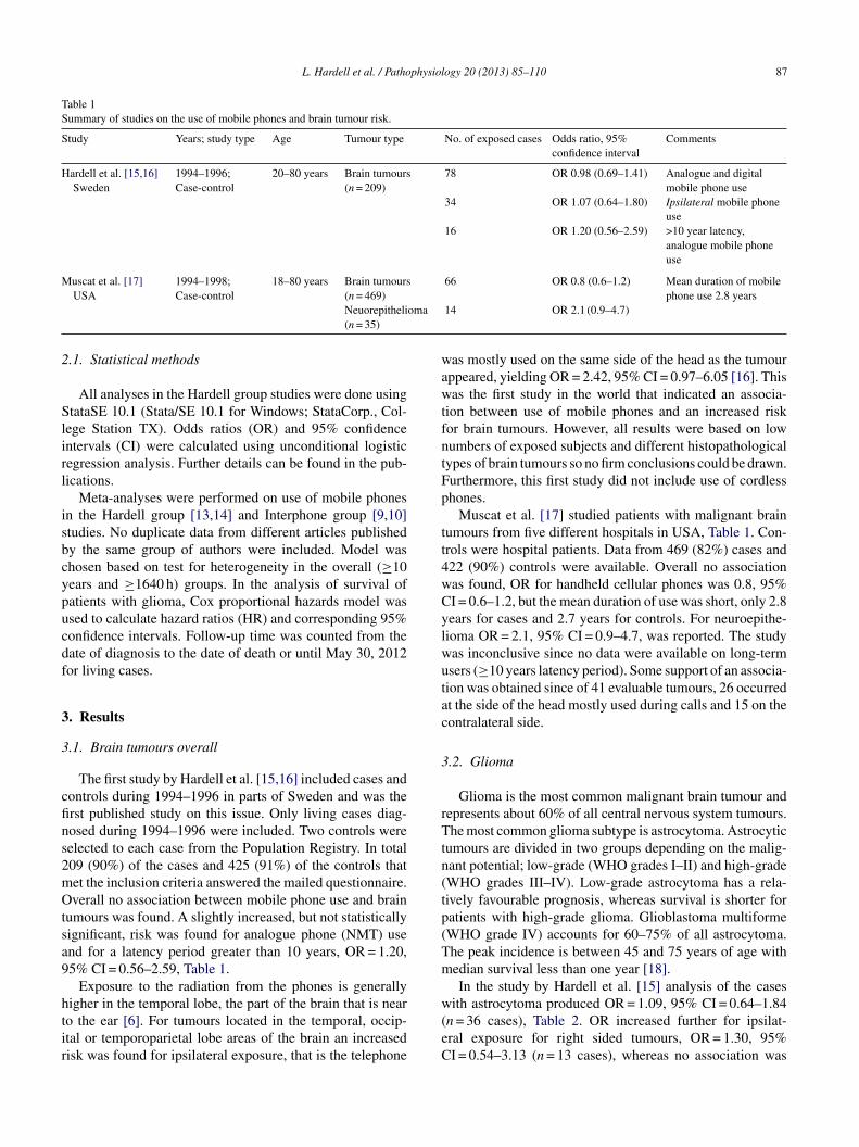
L. Hardell et al. / Pathophysiology 20 (2013) 85–110 87
Table 1Summary of studies on the use of mobile phones and brain tumour risk.
Study Years; study type Age Tumour type No. of exposed cases Odds ratio, 95%confidence interval
Comments
Hardell et al. [15,16]Sweden
1994–1996;Case-control
20–80 years Brain tumours(n = 209)
78 OR 0.98 (0.69–1.41) Analogue and digitalmobile phone use
34 OR 1.07 (0.64–1.80) Ipsilateral mobile phoneuse
16 OR 1.20 (0.56–2.59) >10 year latency,analogue mobile phoneuse
Muscat et al. [17]USA
1994–1998;Case-control
18–80 years Brain tumours(n = 469)
66 OR 0.8 (0.6–1.2) Mean duration of mobilephone use 2.8 years
oma
2
Slirl
isbcypucdf
3
3
cfins2mOtsa9
htir
wawtfntFp
tt4wCylwutac
3
rTtn(tp(Tm
Neuorepitheli(n = 35)
.1. Statistical methods
All analyses in the Hardell group studies were done usingtataSE 10.1 (Stata/SE 10.1 for Windows; StataCorp., Col-
ege Station TX). Odds ratios (OR) and 95% confidencentervals (CI) were calculated using unconditional logisticegression analysis. Further details can be found in the pub-ications.
Meta-analyses were performed on use of mobile phonesn the Hardell group [13,14] and Interphone group [9,10]tudies. No duplicate data from different articles publishedy the same group of authors were included. Model washosen based on test for heterogeneity in the overall (≥10ears and ≥1640 h) groups. In the analysis of survival ofatients with glioma, Cox proportional hazards model wassed to calculate hazard ratios (HR) and corresponding 95%onfidence intervals. Follow-up time was counted from theate of diagnosis to the date of death or until May 30, 2012or living cases.
. Results
.1. Brain tumours overall
The first study by Hardell et al. [15,16] included cases andontrols during 1994–1996 in parts of Sweden and was therst published study on this issue. Only living cases diag-osed during 1994–1996 were included. Two controls wereelected to each case from the Population Registry. In total09 (90%) of the cases and 425 (91%) of the controls thatet the inclusion criteria answered the mailed questionnaire.verall no association between mobile phone use and brain
umours was found. A slightly increased, but not statisticallyignificant, risk was found for analogue phone (NMT) usend for a latency period greater than 10 years, OR = 1.20,5% CI = 0.56–2.59, Table 1.
Exposure to the radiation from the phones is generally
igher in the temporal lobe, the part of the brain that is nearo the ear [6]. For tumours located in the temporal, occip-tal or temporoparietal lobe areas of the brain an increasedisk was found for ipsilateral exposure, that is the telephonew(eC
14 OR 2.1 (0.9–4.7)
as mostly used on the same side of the head as the tumourppeared, yielding OR = 2.42, 95% CI = 0.97–6.05 [16]. Thisas the first study in the world that indicated an associa-
ion between use of mobile phones and an increased riskor brain tumours. However, all results were based on lowumbers of exposed subjects and different histopathologicalypes of brain tumours so no firm conclusions could be drawn.urthermore, this first study did not include use of cordlesshones.
Muscat et al. [17] studied patients with malignant brainumours from five different hospitals in USA, Table 1. Con-rols were hospital patients. Data from 469 (82%) cases and22 (90%) controls were available. Overall no associationas found, OR for handheld cellular phones was 0.8, 95%I = 0.6–1.2, but the mean duration of use was short, only 2.8ears for cases and 2.7 years for controls. For neuroepithe-ioma OR = 2.1, 95% CI = 0.9–4.7, was reported. The studyas inconclusive since no data were available on long-termsers (≥10 years latency period). Some support of an associa-ion was obtained since of 41 evaluable tumours, 26 occurredt the side of the head mostly used during calls and 15 on theontralateral side.
.2. Glioma
Glioma is the most common malignant brain tumour andepresents about 60% of all central nervous system tumours.he most common glioma subtype is astrocytoma. Astrocytic
umours are divided in two groups depending on the malig-ant potential; low-grade (WHO grades I–II) and high-gradeWHO grades III–IV). Low-grade astrocytoma has a rela-ively favourable prognosis, whereas survival is shorter foratients with high-grade glioma. Glioblastoma multiformeWHO grade IV) accounts for 60–75% of all astrocytoma.he peak incidence is between 45 and 75 years of age withedian survival less than one year [18].In the study by Hardell et al. [15] analysis of the cases
ith astrocytoma produced OR = 1.09, 95% CI = 0.64–1.84n = 36 cases), Table 2. OR increased further for ipsilat-ral exposure for right sided tumours, OR = 1.30, 95%I = 0.54–3.13 (n = 13 cases), whereas no association was

88L
.Hardelletal./Pathophysiology
20(2013)
85–110
Table 2Summary of studies on the use of wireless phones and glioma risk.
Study Years; study type Age Tumour type No. ofexposedcases
Odds ratio, 95%confidence interval
Comments
Hardell et al. [15] Sweden 1994–1996;Case-control
20–80 years Astrocytoma WHOgrade I–IV (n = 94)
36 OR 1.09 (0.64–1.84) Analogue and digital mobile phone use
13 OR 1.30 (0.54–3.13) Ipsilateral mobile phone use, right sided tumours3 OR 0.35 (0.07–1.81) Ipsilateral mobile phone use, left sided tumours
Inskip et al. [19] USA 1994–1998;Case-control
≥18 years Glioma (n = 489) 11 OR 0.6 (0.3–1.4) ≥5 years of mobile phone use
Auvinen et al. [20] Finland 1996; Case-control,register based
20–69 years Glioma (n = 198) Not given OR 1.5 (1.0–2.4) Analogue and digital mobile phone “ever” use
25 OR 2.1 (1.3–3.4) Analogue mobile phone “ever” used11 OR 2.4 (1.2–5.1) Analogue mobile phone use, 1–2 years11 OR 2.0 (1.0–4.1) Analogue mobile phone use, >2 years
Hardell et al. [26–28] Carlberg,Hardell [29] Sweden
1997–2003;Case-control
20–80 years Glioma (n = 1148) 123 OR 2.5 (1.8–3.3) >10 year latency, mobile phone
57 OR 2.9 (1.8–4.7) >10 year latency, mobile phone, ipsilateral, only living50 OR 2.6 (1.7–4.1) >10 year latency, mobile phone only45 OR 1.7 (1.1–2.6) >10 year latency, cordless phone20 OR 3.8 (1.8–8.1) >10 year latency, cordless phone, ipsilateral, only living9 OR 1.2 (0.5–2.9) >10 year latency, cordless phone only; >5–10 year latency
OR 1.9 (1.3–2.9; n = 55)150 OR 2.1 (1.6–2.8) >10 year latency, wireless phone (mobile and cordless
phone)
Astrocytoma, highgrade (n = 820)
102 OR 3.0 (2.1–4.2) >10 year latency, mobile phone
47 OR 3.9 (2.3–6.6) >10 year latency, mobile phone, ipsilateral, only living37 OR 2.8 (1.7–4.6) >10 year latency, mobile phone only36 OR 2.0 (1.2–3.2) >10 year latency, cordless phone15 OR 5.5 (2.3–13) >10 year latency, cordless phone, ipsilateral, only living6 OR 0.9 (0.3–2.6) >10 year latency, cordless phone only; >5–10 year latency
OR 2.4 (1.6–3,7; n = 44)121 OR 2.5 (1.8–3.4) >10 year latency, wireless phone (mobile and cordless
phone)
Interphone Study Group [9] 13countries; Australia, Canada,Denmark, Finland, France,UK, Germany, Israel, Italy,Japan, New Zealand, Norway,Sweden
2000–2004, 2–4 yearsdepending on studyregion. Case-control
30–59 years Glioma (n = 2708) 1666 OR 0.81 (0.70–0.94) Regular use of mobile phone in the past ≥1 year

L. Hardell et al. / Pathophysiol
210
OR
1.40
(1.0
3–1.
89)
Cum
ulat
ive
hour
sm
obile
phon
e≥1
640
h78
OR
1.87
(1.0
9–3.
22)
Cum
ulat
ive
hour
sm
obile
phon
e≥1
640
h,tu
mou
rsin
tem
pora
llob
e10
0O
R1.
96(1
.22–
3.16
)C
umul
ativ
eho
urs
mob
ileph
one
≥164
0h,
ipsi
late
ral
mob
ileph
one
use
Inte
rpho
neSt
udy
Gro
up[9
]A
ppen
dix
220
00–2
004,
2–4
year
sde
pend
ing
onst
udy
regi
on.C
ase-
cont
rol
30–5
9ye
ars
Glio
ma
(n=
1211
)46
0O
R1.
68(1
.16–
2.41
)R
estr
icte
dto
ever
regu
lar
use
time
sinc
est
art2
–4ye
ars;
1–1.
9ye
ars
asre
fere
nce
entit
y
468
OR
1.54
(1.0
6–2.
22)
Res
tric
ted
toev
erre
gula
rus
etim
esi
nce
star
t5–9
year
s;1–
1.9
year
sas
refe
renc
een
tity
190
OR
2.18
(1.4
3–3.
31)
Res
tric
ted
toev
erre
gula
rus
etim
esi
nce
star
t10+
year
s;1–
1.9
year
sas
refe
renc
een
tity
160
OR
1.82
(1.1
5–2.
89)
Res
tric
ted
toev
erre
gula
rus
e≥1
640
h,<
5h
asre
fere
nce
entit
y
se
twrbwnv8acdwRvs
gabd2cOlpwfo
bd1wnTcp
diiSm(ocpdasdam
ogy 20 (2013) 85–110 89
een for astrocytoma in the left hemisphere and ipsilateralxposure, OR = 0.35, 95% CI = 0.07–1.81 (n = 3 cases).
The study by Inskip et al. [19] from USA had few long-erm users of mobile phones. Only 11 cases with glioma, 6ith meningioma and 5 with acoustic neuroma had ≥5 years
egular use. No subject had ≥10 years use. Of the hospital-ased cases 92% participated. The study comprised 489 casesith glioma, 197 with meningioma and 96 with acousticeuroma, and 799 (86%) hospital-based controls. Proxy inter-iews were necessary for 16% of the patients with glioma,% of the patients with meningioma, 3% of the patients withcoustic neuroma, and 3% of the controls. Overall no statisti-ally significant associations were found, Table 2. Regardingifferent types of glioma OR = 1.8, 95% CI = 0.7–5.1as found for anaplastic astrocytoma (WHO grade III).egarding hospital-based interviews and use of proxy inter-iews, see discussion below in relation to the Interphonetudy.
A register based case-control study on brain and salivaryland tumours was performed in Finland [20]. All casesged 20–69 years diagnosed in 1996 were included; 398rain tumour cases and 34 salivary gland tumour cases. Theuration of mobile phone use was short, for analogue users–3 years and for digital users less than one year. No asso-iation was found for salivary gland tumours. For gliomaR = 2.1, 95% CI = 1.3–3.4 was calculated for use of ana-
ogue phones, but no association was found for digital mobilehones, Table 2. When duration of use of analogue phonesas used as a continuous variable an increased risk was
ound for glioma with OR = 1.2, 95% CI = 1.1–1.5 per yearf use.
The Hardell group in Sweden studied the associationetween use of mobile and cordless phones and brain tumoursiagnosed during 1997–2003. First, cases diagnosed duringJanuary 1997 to 30 June 2000 were included. These resultsere published separately [21,22]. This was followed by theext study period, 1 July 2000 to 31 December 2003 [23,24].he methods were the same including the same inclusionriteria and an identical questionnaire in both studies; see theublications for further details.
Both men and women aged 20–80 years at the time ofiagnosis were included and all were alive at the time ofnclusion in the study. They were reported from cancer reg-stries with a brain tumour verified by histopathology. Thewedish Population Registry was used for identification ofatched controls. The study included use of wireless phones
mobile and cordless phones), as well as asking questionsn e.g., occupational exposures. Use of wireless phones wasarefully assessed by a self-administered questionnaire sup-lemented over the phone. The ear that had mostly been useduring calls with mobile phone and/or cordless phone wasssessed by separate questions; >50% of the time for one
ide, or equally for both sides. This information was checkeduring the supplementary phone calls and finally also byseparate letter with good agreement between these threeethods.
9 physiol
mmwc(rp
tSdAcircw
hcsq
at
y1r[o8
lifBot9Cl9
lCCac
cfluO
lc
tgtpc(s
i2rIfHhntp
sefAw
bbirm
fgimhCtslt
rp9oC
a
0 L. Hardell et al. / Patho
Tumour localisation for the cases was defined by usingedical records including computer tomography (CT) and/oragnetic resonance imaging (MRI). The matched controlas assigned the same side as the tumour of the respective
ase. Use of the wireless phone was defined as ipsilateral≥50% of the time), or contralateral (<50% of the time) inelation to tumour side. Further details can be found in theublications.
In a review commissioned by the former Swedish Radia-ion Protection Agency (now called the Swedish Radiationafety Authority) it was suggested that the exclusion ofeceased cases was a source of bias in our studies [25].s a response to that critique we performed a study on the
ases with a malignant brain tumour that had died beforenclusion in the case-control studies 1997–2003. These casesepresented patients with a poor prognosis, mostly with astro-ytoma WHO grade IV (glioblastoma multiforme). Controlsere selected from the Death Registry in Sweden.The study encompassed 464 cases and 464 controls that
ad died from a malignant disease and 463 controls with otherauses of death. Exposure was assessed by a questionnaireent to the next of kin to each deceased case and control. Theuestionnaire was similar as in previous studies.
This investigation confirmed the previous results of anssociation between mobile phones and malignant brainumours [26].
The Hardell group has previously published pooled anal-sis of malignant brain tumours diagnosed during the period997–2003 [27]. These results were updated including alsoesults for deceased cases with malignant brain tumours28,29]. The results on use of wireless phones were basedn 1251 cases with malignant brain tumour (response rate5%) and 2438 controls (response rate 84%).
Most cases had glioma (n = 1148) so we present in the fol-owing results for that type of tumour. Latency was dividedn three categories, >1–5 years, >5–10 years, and >10 yearsrom first use of a wireless phone until diagnosis of glioma.oth use of mobile and cordless phone gave an increased riskverall, highest in the latency group >10 years, increasing fur-her for ipsilateral use yielding for mobile phone OR = 2.9,5% CI = 1.8–4.7 and for cordless phone OR = 3.8, 95%I = 1.8–8.1, Table 2. Highest ORs were found in the >10 year
atency group for total wireless phone use as well, OR = 2.1,5% CI = 1.6–2.8 or a doubling of glioma risk.
OR increased statistically significant for glioma for cumu-ative use of wireless phones per 100 h; OR = 1.014, 95%I = 1.008–1.019, and per year of latency; OR = 1.056, 95%I = 1.037–1.075 [29]. Separate calculations of mobile phonend cordless phone use yielded similar results with statisti-ally significant increasing risks.
It is common for a person to use both a mobile and aordless phone. For only use of mobile phone OR increased
or glioma with time since first use yielding for >10 yearsatency OR = 2.6, 95% CI = 1.7–4.1. For only cordless phonese highest risk was obtained in the >5–10 years latency time;R = 1.9, 95% CI = 1.3–2.9. However, the calculations in thedtm“
ogy 20 (2013) 85–110
ongest latency period were based on few subjects regardingordless phone.
In Table 2 results are presented for high-grade astrocy-oma (n = 820). The results are similar as for the whole gliomaroup. Low-grade glioma is less common and the results inhis study were based on 132 cases. Ipsilateral use of mobilehone yielded in total OR = 1.8, 95% CI = 1.02–3.1 (n = 39ases) and cordless phone OR = 1.7, 95% CI = 0.98–3.1n = 34 cases, data not in Table). Further results and discus-ion may be found elsewhere [29].
The Interphone study was conducted at 16 research centresn 13 countries during varying time periods between 2000 and004. It was an international collaboration on brain tumourisk and mobile phone use conducted under the guidance ofARC. The investigation was initiated by recommendationsrom several expert groups including one of the authors, Kjellansson Mild as a member of the EU group, to study possibleealth effects of exposure to RF-EMF [30,31]. It should beoted that there was no overlap of cases or controls betweenhe Hardell group studies and the Swedish part of Interphoneerformed by another research group.
Some of the separate country analyses of the Interphonetudy produced contradictory results, as we have discussedlsewhere [13,32]. An increased risk for brain tumour wasound in some studies and decreased risk in other studies.fter several years of delay the overall Interphone resultsere finally published in May 2010 [9].The study included 4301 glioma cases and the results were
ased on 2708 participating cases (response rate 64%, rangey centre 36–92%). In total 14,354 potential controls weredentified and interviews were completed with 7658 (53%,ange 42–74%). The low participation rates in some centresay have created selection bias, see Hardell et al. [32].Regular use of mobile phone in the past ≥1 year gave
or glioma OR = 0.81, 95% CI = 0.70–0.94, Table 2. Sub-roup analyses showed statistically significant increased riskn the highest exposure group, i.e., those with cumulative
obile phone use ≥1640 h, which corresponds to aboutalf an hour of use per day for ten years, OR = 1.40, 95%I = 1.03–1.89. The risk increased further for glioma in the
emporal lobe yielding OR = 1.87, 95% CI = 1.09–3.22. In theame exposure category, cumulative use ≥1640 h and ipsi-ateral exposure produced OR = 1.96, 95% CI = 1.22–3.16 inotal (no data given for temporal lobe).
In Appendix 2, available on the web [9] analysis wasestricted to ever-regular users of mobile phones in the Inter-hone study. Cumulative call time ≥1640 h gave OR = 1.82,5% CI = 1.15–2.89 compared with use <5 h. Time since startf regular use (latency) ≥10 years produced OR = 2.18, 95%I = 1.43–3.31; reference entity 1–1.9 years.
The Interphone study group concluded: “However, biasesnd errors limit the strength of the conclusions we can
raw from these analyses and prevent a causal interpreta-ion.” In an editorial accompanying the Interphone results theain conclusion of the Interphone results was described asboth elegant and oracular. . .(which) tolerates diametrically

physiol
omlllygis
ba2uoctiHqdtHipltath
tsmdmopaiftbRlscrrTsha[
p
tpTmoat2wcbbph
pribblitSpcrsoor2aridart
tpitnilItcfe
L. Hardell et al. / Patho
pposite readings” [33]. They also pointed out severalethodological reasons why the Interphone results were
ikely to have underestimated the risks, such as the shortatency period since first exposures became widespread;ess than 10% of the Interphone cases had more than 10ears exposure. “None of the today’s established carcino-ens, including tobacco, could have been firmly identified asncreasing risk in the first 10 years or so since first expo-ure”.
As has pointed our elsewhere [32] there were differencesetween the Hardell group studies and Interphone. Regardingge group the Hardell group studies included subjects aged0–80 years, versus 30–59 years in Interphone. Furthermorese of cordless phones was not properly assessed, analysedr reported in Interphone. These differences have been dis-ussed in detail by Hardell et al. [14]. Thus, it could be shownhat restricting the age group to 30–59 years and consider-ng subjects that used a cordless phone as unexposed in theardell group studies reduced the OR and produced resultsuite similar to Interphone, Table 3; see also Table 11 asiscussed below. Latency time >10 years for glioma in theemporal lobe yielded OR = 1.40, 95% CI = 0.70–2.81 in theardell group studies and OR = 1.36, 95% CI = 0.88–2.11
n Interphone (latency ≥10 years). Unfortunately the Inter-hone study did not give results for glioma in the temporalobe in the analyses in Appendix 2. Thus, excluding exposureo RF-EMFs from cordless phones as in the Interphone study,s well as excluding the younger and older subjects biasedhe ORs towards unity, which likely dilutes the ability to seeealth risks.
Most mobile phone users have not been using one singleelephone. It is likely that they have changed their handseteveral times if they have been using a mobile phone forore than a few years. Many users have also been using
ifferent phone systems, such as analogue and digital, andany of them have also been using a cordless phone at home
r at work. It is not clear how to combine the use of differenthones with different power outputs, systems, frequenciesnd anatomical specific absorption rate (SAR) distributionsnto one exposure and dose measure. The difficulties lie in theact that there is no generally accepted mechanism(s) betweenhe electromagnetic fields emitted from the phone and theiological organism. This includes a mechanism by whichF-EMF exposure produces changes in DNA. The energy
evel associated with exposure is too low to cause direct DNAtrand breaks and DNA cross links. However, DNA damagesan be caused by cellular biochemical activities such as freeadicals. Several studies indicate that RF-EMFs increase freeadical activity in cells, as reviewed by Phillips et al. [34].his process is probably mediated via the Fenton reaction. Ithould also be noted that possible biological effects might notave linear dose–response as indicated in some studies [35]
nd that the effects are depending on the carrier frequencies36].The different types of phones have different outputower. We applied different weighting factors according to
ibas
ogy 20 (2013) 85–110 91
he mean output power of the phones using for analoguehones (NMT) = 1, GSM = 0.1 and cordless phones = 0.01.he cumulative time for use of the different phone types wasultiplied with the respective weighting factor added into
ne score. The median score among the controls was useds the cut-off in the dose–response calculations. We appliedhis method for the study period 1 January 1997 to 30 June000 [21,22]. Somewhat higher ORs were obtained using theeighting factor, especially with a >10-year latency period,
ompared with calculations based on cumulative use only,ut overall the results were similar [37]. This was explainedy the fact that most subjects had used an analogue mobilehone with the weighting factor = 1, thus the weighting factorad little impact on the results.
A further issue is that there is a difference in the out-ut power level from mobile phones between urban andural areas. This is caused by adaptive power control (APC)n the cellular telephone and is regulated by the distanceetween base stations. Thus, in areas with a long distanceetween base stations, usually rural areas, the output powerevel is higher than in more densely populated areas; thats, urban areas, with a shorter distance between base sta-ions. To further explore these circumstances we used thewedish population register that contains information onresent municipality for all residents. The municipalities arelassified by Statistics Sweden into so called homogeneityegions, six categories depending on the population den-ity, and the number of inhabitants in the nearest vicinityf the main city in that municipality. Thus, we used thesefficial statistics for grouping of the subjects in urban orural areas for the study period 1 January 1997 to 30 June000. For use of digital mobile phones (GSM) we foundclear effect of urban versus rural areas [38]. Living in
ural areas yielded OR = 1.4, 95% CI = 0.98–2.0, increas-ng to 3.2, 95% CI = 1.2–8.4 with >5 year latency time forigital phones. The corresponding ORs for living in urbanreas were 0.9, 95% CI = 0.8–1.2 and 0.9, 95% CI = 0.6–1.4,espectively. This effect was most obvious for malignant brainumours.
Estimated RF-EMF dose from mobile phone use in theumour area was associated with an increased risk of glioma inarts of the Interphone study [11]. OR increased with increas-ng total cumulative dose of specific energy (J/kg) absorbed athe estimated tumour centre for more than 7 years before diag-osis giving OR = 1.91, 95% CI = 1.05–3.47 (p trend = 0.01)n the highest quintile of exposure. A similar study based oness sound methods was later published by another part of thenterphone study group [39]. The results seemed to contradicthe findings of Cardis et al. [11]. However, a different, lesslear method was used. Only 42 cases had used mobile phoneor more than 10 years and no analysis was made of the mostxposed group with longest duration of use. Thus, this study
s much less informative and less sophisticated than the oney Cardis et al. [11]. It should have been of great value topply the method by Cardis et al. for the whole Interphonetudy.
92L
.Hardelletal./Pathophysiology
20(2013)
85–110
Table 3Comparison between Hardell group and Interphone using the same age group 30–59 years and excluding use of cordless phones.
Study Years; study type Age Tumour type No. ofexposed cases
Odds ratio, 95%confidence interval
Comments
Hardell et al. [14] 1997–2003;Case-control
30–59 years Glioma (n = 490) 56 OR 1.79 (1.19–2.70) >10 year latency, cordless phoneamong unexposed, age 30–59 years
29 OR 1.75 (1.02–3.00) Cumulative use ≥1640 h, cordlessphone among unexposed, age 30–59years
20 OR 2.18 (1.09–4.35) Cumulative use ≥1640 h, cordlessphone among unexposed, age 30–59years, ipsilateral
8 OR 1.48 (0.57–3.87) Cumulative use ≥1640 h, cordlessphone among unexposed, age 30–59years, contralateral
Interphone Study Group [9] 13countries; Australia, Canada,Denmark, Finland, France, UK,Germany, Israel, Italy, Japan, NewZealand, Norway, Sweden
2000–2004, 2–4 yearsdepending on studyregion. Case-control
30–59 years Glioma (n = 2708) 252 OR 0.98 (0.76–1.26) Regular use of mobile phone in thepast ≥1 year, latency ≥10 years
210 OR 1.40 (1.03–1.89) Cumulative hours mobile phone≥1640 h
100 OR 1.96 (1.22–3.16) Cumulative hours mobile phone≥1640 h, ipsilateral
39 OR 1.25 (0.64–2.42) Cumulative hours mobile phone≥1640 h, contralateral
160 OR 1.82 (1.15–2.89) Restricted to ever regular use≥1640 h, <5 h as reference entity,Appendix 2. Results for ipsilateraland contralateral use not reported.

L. Hardell et al. / Pathophysiology 20 (2013) 85–110 93
Table 4Use of mobile phones and glioma risk, meta-analysis of Hardell et al. [14] and Interphone [9]. Numbers of exposed cases (Ca) and controls (Co) are given.
Hardell et al. Interphone Meta-analysis
Ca/Co OR, CI Ca/Co OR, CI Ca/Co OR, CI
Latency ≥10 years-all 88/99 2.26 (1.60–3.19) 252/232 0.98 (0.76–1.26) 340/331 1.48 (0.65–3.35)-ipsilateral 57/45 2.84 (1.82–4.44) 108/82 1.21 (0.82–1.80) 165/127 1.84 (0.80–4.25)-contralateral 29/29 2.18 (1.24–3.85) 49/56 0.70 (0.42–1.15) 78/85 1.23 (0.40–3.73)-temporal lobe 28/99 2.26 (1.32–3.86) 94/69 1.36 (0.88–2.11) 122/168 1.71 (1.04–2.81)
Cumulative use ≥1640 h-all 42/43 2.31 (1.44–3.70) 210/154 1.40 (1.03–1.89) 252/197 1.74 (1.07–2.83)-ipsilateral 29/21 2.94 (1.60–5.41) 100/62 1.96 (1.22–3.16) 129/83 2.29 (1.56–3.37)-contralateral 12/12 2.10 (0.90–4.90) 39/31 1.25 (0.64–2.42) 51/43 1.52 (0.90–2.57)-temporal lobe 14/43 2.44 (1.21–4.95) 78/47 1.87 (1.09–3.22) 92/90 2.06 (1.34–3.17)
R eneity i
3
pGfgprry≥Orp9eg
3
dndm
m9pu
m9aHb
t
m9Cli
bampl99yarh
duT≥Ccl
3
mauiiet
andom-effects model used for all meta-analyses, based on test for heterog
.3. Meta-analysis glioma
We performed a meta-analysis of glioma on use of mobilehones based on Hardell et al. [14] and Interphone Studyroup [9]. Random-effects model was used based on test
or heterogeneity in the overall (≥10 years and ≥1640 h)roups. The analysis was based on published results in Inter-hone since we do not have access to their database. Ouresults were recalculated to these groups of exposure. Thus,esults can be found in Table 4 for latency ≥10 years, (>10ears in Hardell et al.), and cumulative use of mobile phone1640 h. The meta-analysis yielded for mobile phone useR = 1.71, 95% CI = 1.04–2.81 for glioma in the tempo-
al lobe in the ≥10 years latency group. Ipsilateral mobilehone use ≥1640 h in total gave the highest risk, OR = 2.29,5% CI = 1.56–3.37. Certainly the meta-analysis strength-ns a causal association between use of mobile phones andlioma.
.4. Meningioma
Meningioma is the most common benign brain tumour. Itevelops from the pia and arachnoid that covers the centralervous system. Meningioma is an encapsulated and well-emarked tumour. It is rarely malignant. More women thanen develop meningioma.In the first study by Hardell et al. [15] only 46 cases had
eningioma. No increased risk was found overall; OR = 1.05,5% CI = 0.49–2.27, Table 5. Only 16 cases had used a mobilehone. There was no pattern of increased risk for ipsilateralse, although the results were based on low numbers.
The US study by Inskip et al. [19] included 197 cases witheningioma. Regular mobile phone use produced OR = 0.8,
5% CI = 0.4–1.3, Table 5. The risk did not increase withverage daily use, cumulative use, or duration of regular use.
owever, results for duration of regular use ≥5 years wasased on only 6 exposed cases.The Finnish register based case-control study on brainumours by Auvinen et al. [20] included 129 cases with
Typm
n the overall (≥10 years and ≥1640 h) groups.
eningioma. Ever use of mobile phone gave OR = 1.1,5% CI = 0.5–2.4, analogue phone use OR = 1.5, 95%I = 0.6–3.5, Table 5. As discussed above the study was
imited by short latency and exposure based on subscriptionnformation.
The Hardell group made a pooled analysis of benignrain tumours from the two case-control studies 1997–2003s discussed above [40,41]. Regarding meningioma use ofobile phone gave OR = 1.1, 95% CI = 0.9–1.3, and cordless
hone OR = 1.1, 95% CI = 0.9–1.4, Table 5. Using >10 yearatency period OR increased; for mobile phone to OR = 1.5,5% CI = 0.98–2.4, and for cordless phone to OR = 1.8,5% CI = 1.01–3.2. Ipsilateral mobile phone use in the >10ears latency group yielded OR = 1.6, 95% CI = 0.9–2.9,nd cordless phone OR = 3.0, 95% CI = 1.3–7.2. Theseesults were based on rather low numbers of exposed cases,owever.
In the Interphone study [9] a statistically significantecreased risk was found for meningioma for regularse of mobile phone, OR = 0.79, 95% CI = 0.68–0.91,able 5. The risk increased somewhat with cumulative use1640 h and ipsilateral mobile phone use to OR = 1.45, 95%I = 0.80–2.61. The overall pattern of no association did nothange if analysis was restricted to tumours in the temporalobe or only to the group of ever-regular use.
.5. Meta-analysis meningioma
Similarly as for glioma we performed meta-analysis ofeningioma for use of mobile phone on the Hardell group
nd Interphone results, Table 6. Random-effects model wassed in the ≥10 years group based on test for heterogeneityn the overall group. For analyses of ≥1640 h no heterogene-ty was found in the heterogeneity test; random- and fixedffects models produced identical results. In summary no sta-istically significant decreased or increased risks were found.
hese results support the conclusion that up to latency ≥10ears or cumulative use ≥1640 h there is not a consistentattern of an association between use of mobile phones andeningioma.
94L
.Hardelletal./Pathophysiology
20(2013)
85–110
Table 5Summary of studies on the use of wireless phones and meningioma risk.
Study Years; study type Age Tumour type No. ofexposed cases
Odds ratio, 95%confidence interval
Comments
Hardell et al. [15] Sweden 1994–1996;Case-control
20–80 years Meningioma (n = 46) 16 OR 1.05 (0.49–2.27) Analogue and digital mobile phone use
Inskip et al. [19] USA 1994–1998;Case-control
≥18 years Meningioma (n = 197) 32 OR 0.8 (0.4–1.3) Regular use
6 OR 0.9 (0.3–2.7) ≥5 years of mobile phone use
Auvinen et al. [20] Finland 1996; Case-control,register based
20–69 years Meningioma (n = 129) Not given OR 1.1 (0.5–2.4) Analogue and digital mobile phone “ever” use
8 OR 1.5 (0.6–3.5) Analogue mobile phone “ever” used3 OR 1.6 (0.4–6.1) Analogue mobile phone use, 1–2 years2 OR 1.0 (0.2–4.4) Analogue mobile phone use, >2 years
Hardell et al. [40], Hardell, Carlberg[41] Sweden
1997–2003;Case-control
20–80 years Meningioma (n = 916) 347 OR 1.1 (0.9–1.3) >1 year latency, mobile phone use
38 OR 1.5 (0.98–2.4) >10 years latency of mobile phone use18 OR 1.6 (0.9–2.9) >10 years latency of ipsilateral mobile phone use294 OR 1.1 (0.9–1.4) >1 year latency, cordless phone use23 OR 1.8 (1.01–3.2) >10 years latency of cordless phone use11 OR 3.0 (1.3–7.2) >10 years latency of ipsilateral cordless phone
use
Interphone Study Group [9] 13countries; Australia, Canada,Denmark, Finland, France, UK,Germany, Israel, Italy, Japan, NewZealand, Norway, Sweden
2000–2004, 2–4 yearsdepending on studyregion. Case-control
30–59 years Meningioma(n = 2409)
1262 OR 0.79 (0.68–0.91) Regular use of mobile phone in the past ≥1 year
130 OR 1.15 (0.81–1.62) Cumulative hours mobile phone ≥1640 h21 OR 0.94 (0.31–2.86) Cumulative hours mobile phone ≥1640 h,
tumours in temporal lobe46 OR 1.45 (0.80–2.61) Cumulative hours mobile phone ≥1640 h,
ipsilateral mobile phone use
Interphone [9] Appendix 2 Meningioma (n = 842) 362 OR 0.90 (0.62–1.31) Restricted to ever regular use time since start2–4 years; 1–1.9 years as reference entity
288 OR 0.75 (0.51–1.10) Restricted to ever regular use time since start5–9 years; 1–1.9 years as reference entity
76 OR 0.86 (0.51–1.43) Restricted to ever regular use time since start10+ years; 1–1.9 years as reference entity
96 OR 1.10 (0.65–1.85) Restricted to ever regular use ≥1640 h, <5 h asreference entity

L. Hardell et al. / Pathophysiology 20 (2013) 85–110 95
Table 6Use of mobile phones and meningioma risk, meta-analysis of Hardell, Carlberg [41] and Interphone [9]. Numbers of exposed cases (Ca) and controls (Co) aregiven.
Hardell et al. Interphone Meta-analysis
Ca/Co OR, CI Ca/Co OR, CI Ca/Co OR, CI
Latency ≥10 years-all 38/99 1.52 (0.98–2.37) 110/112 0.83 (0.61–1.14) 148/211 1.10 (0.61–1.99)-ipsilateral 18/45 1.59 (0.86–2.95) 40/42 0.88 (0.52–1.47) 58/87 1.16 (0.65–2.06)-contralateral 12/29 1.57 (0.75–3.31) 20/25 0.58 (0.29–1.16) 32/54 0.95 (0.36–2.51)-temporal lobe 10/99 2.46 (1.08–5.60) 12/12 0.60 (0.22–1.62) 22/111 1.25 (0.31–4.98)
Cumulative use ≥1640 h-all 10/43 0.85 (0.41–1.75) 130/107 1.15 (0.81–1.62) 140/150 1.09 (0.80–1.49)-ipsilateral 6/21 1.11 (0.42–2.88) 46/35 1.45 (0.80–2.61) 52/56 1.35 (0.81–2.23)-contralateral 3/12 0.98 (0.26–3.61) 28/28 0.62 (0.31–1.25) 31/40 0.69 (0.37–1.27)-temporal lobe 1/43 0.52 (0.07–3.95) 21/14 0.94 (0.31–2.86) 22/57 0.82 (0.31–2.17)
R r heterow
3
tfulptaTacalwat
1mwc
ifuwou
bruitc
t
ppr9IlpO
iawcqacdsuwefyidrof
ctc1fe9
andom-effects model used for meta-analyses of ≥10 years, based on test foas found; random- and fixed effects models produced identical results.
.6. Acoustic neuroma
Acoustic neuroma or Vestibular Schwannoma is a benignumour that is located in the eighth cranial nerve that leadsrom the inner ear to the brain. This tumour type does notndergo malignant transformation. It tends to be encapsu-ated and grows in relation to the auditory and vestibularortions of the nerve. It is a slow growing tumour in the audi-ory canal but grows gradually out into the cerebellopontinengle with potential compression of vital brain stem centres.innitus and hearing problems are usual first symptoms ofcoustic neuroma. Although neuroma is a benign tumour itauses persistent disabling symptoms after treatment suchs loss of hearing and tinnitus that severely affect the dailyife. The eighth cranial nerve is located close to the handheldireless phone when used, so there is particular concern of
n increased risk for neuroma development due to exposureo RF-EMF emissions during use of these devices.
In the first study by Hardell et al. [15] in Sweden only3 cases had acoustic neuroma. Five cases reported use ofobile phone, only one with ipsilateral use. The numbersere too low to make meaningful interpretation of an asso-
iation, Table 7.Inskip et al. [19] included 96 cases with acoustic neuroma
n their US case-control study. No increased risk was foundor regular use of mobile phone, Table 7. Duration of regularse ≥5 years gave OR = 1.9, 95% CI = 0.6–5.9. This resultas based on only 5 exposed cases and there were no resultsn long-term use. Furthermore only 1 case had cumulativese >500 h.
Muscat et al. [42] presented results from a hospitalased case-control study on acoustic neuroma on 90 (100%esponse rate) patients and 86 (100%) controls. Mobile phonese 1–2 years gave OR = 0.5, 95% CI = 0.2–1.3 (n = 7 cases),ncreasing to OR = 1.7, 95% CI = 0.5–5.1 (n = 11 cases), inhe group with 3–6 years use, Table 7. Average use among
ases was 4.1 years and among controls 2.2 years.The pooled analysis of the Hardell group studies yielded inotal OR = 2.9, 95% CI = 2.0–4.3 for use of analogue mobile
pg
geneity in the overall group. For meta-analyses of ≥1640 h no heterogeneity
hone and OR = 1.5, 95% CI 1.1–2.1 for use of digital mobilehone [40]. Use of mobile phones gave for acoustic neu-oma OR = 1.7, 95% CI = 1.2–2.3 increasing to OR = 2.9,5% CI = 1.6–5.5 with >10 years latency period, Table 7.psilateral use increased the risk further; in the >10 yearsatency group to OR = 3.0, 95% CI = 1.4–4.2 [41]. Cordlesshone use gave OR = 1.5, 95% CI = 1.04–2.0 increasing toR = 1.7, 95% CI = 1.2–2.5 for ipsilateral use.A case-case study on acoustic neuroma was conducted
n Japan [43]. The cases were identified during 2000–2006t 22 participating neurosurgery departments. The diagnosisas based on histopathology or CT/MRI imaging. Of 1589
ases 816 (51%) agreed to participate and answered a maileduestionnaire. A total of 787 cases were included in the finalnalysis. Two datasets were analysed, one consisted of 362ases without any tumour related symptoms 1 year beforeiagnosis, and another consisted of 593 cases without anyymptoms 5 years before diagnosis. Cases with ipsilateralse were regarded as exposed and those with contralateral useere assumed to be unexposed and were used as the refer-
nce category. Overall no increased risk was found. However,or average daily call duration >20 min with reference date 1ear Risk Ratio (RR) = 2.74, 95% CI = 1.18–7.85 was foundncreasing to RR = 3.08, 95% CI = 1.47–7.41 with referenceate 5 years before diagnosis, Table 7. Unfortunately noesults were given for cumulative number of hours for usever the years. For cordless phones no increased risk wasound but the analysis was not very informative.
In the Interphone study [10] 1121 (82%) acoustic neuromaases participated, range 70–100% by centre. Of the con-rols 7658 (53%) completed the interviews, range 35–74% byentre. The final matched analysis (1:1 or 1:2) consisted of105 cases and 2145 controls. Overall no increased risk wasound censoring exposure at one year or at 5 years before ref-rence date, OR = 0.85, 95% CI = 0.69–1.04 and OR = 0.95,5% CI = 0.77–1.17, respectively, Table 7.
Cumulative number of hours of ipsilateral mobilehone use ≥1640 h up to 1 year before reference dateave OR = 2.33, 95% CI = 1.23–4.40 and contralateral use

96L
.Hardelletal./Pathophysiology
20(2013)
85–110
Table 7Summary of studies on the use of wireless phones and acoustic neuroma risk.
Study Years Study Type Age Tumour type No. of exposed cases Odds ratio, 95%confidence interval
Comments
Hardell et al. [15] Sweden 1994–1996; Case-control 20–80 years Acousticneuroma(n = 13)
5 OR 0.78 (0.14–4.20) >1 year latency of mobile phone use
Inskip et al. [19] USA 1994–1998; Case-control ≥18 years Acousticneuroma(n = 96)
22 OR 1.0 (0.5–1.9) Regular mobile phone use
5 OR 1.9 (0.6–5.9) ≥5 years of mobile phone use
Muscat et al. [42] USA 1997–1999; Case-control ≥18 years Acousticneuroma(n = 90)
11 OR 1.7 (0.5–5.1) 3–6 years of mobile phone use
Hardell et al. [40], Hardell, Carlberg[41] Sweden
1997–2003; Case-control 20–80 years Acousticneuroma(n = 243)
130 OR 1.7 (1.2–2.3) >1 year latency of mobile phone use
20 OR 2.9 (1.6–5.5) >10 years latency of mobile phone use13 OR 3.0 (1.4–6.2) >10 years of ipsilateral mobile phone use4 OR 1.3 (0.4–3.8) >10 years latency of cordless phone use3 OR 2.3 (0.6–8.8) >10 years latency of ipsilateral cordless phone
use
Sato et al. [43] Japan 2000–2006; Case-case All ages Acousticneuroma(n = 787)
97 RR 1.08 (0.93–1.28) Mobile phone, reference date 1 year beforediagnosis, ipsilateral
86 RR 1.14 (0.96–1.40) Mobile phone, reference date 5 years beforediagnosis, ipsilateral
18 RR 2.74 (1.18–7.85) Mobile phone, reference date 1 year beforediagnosis, average daily call duration >20 min,ipsilateral
28 RR 3.08 (1.47–7.41) Mobile phone, reference date 5 years beforediagnosis, average daily call duration >20 min,ipsilateral
45 RR 0.93 (0.79–1.14) Cordless phone, reference date 1 year beforediagnosis, ipsilateral; mobile phone non-users
125 RR 1.02 (0.91–1.17) Cordless phone, reference date 5 years beforediagnosis, ipsilateral; mobile phone non-users
Interphone Study Group [10] 13countries; Australia, Canada,Denmark, Finland, France, UK,Germany, Israel, Italy, Japan, NewZealand, Norway, Sweden
2000–2004, 2–4 yearsdepending on studyregion. Case-control
30–59 years Acousticneuroma(n = 1105)
643 OR 0.85 (0.69–1.04) Mobile phone regular use up to 1 year beforereference date

L.H
ardelletal./Pathophysiology20
(2013)85–110
97
Interphone [10] 13 countries;Australia, Canada, Denmark,Finland, France, UK, Germany,Israel, Italy, Japan, New Zealand,Norway, Sweden
2000–2004, 2–4 yearsdepending on studyregion. Case-control
30–59 years Acousticneuroma(n = 1105)
304 OR 0.95 (0.77–1.17) Mobile phone regular use up to 5 years beforereference date
77 OR 1.32 (0.88–1.97) Cumulative hours mobile phone ≥1640 h up to 1year before reference date
36 OR 2.79 (1.51–5.16) Cumulative hours mobile phone ≥1640 h up to 5years before reference date
47 OR 2.33 (1.23–4.40) Cumulative hours mobile phone ≥1640 h up to 1year before reference date; ipsilateral use
27 OR 3.53 (1.59–7.82) Cumulative hours mobile phone ≥1640 h up to 5years before reference date; ipsilateral use
37 OR 1.93 (1.10–3.38) Cumulative hours mobile phone ≥1640 h in thepast start ≥10 years before reference date
28 OR 3.74 (1.58–8.83) Cumulative hours mobile phone ≥1640 h in thepast start ≥10 years before reference date,ipsilateral
225 OR 1.41 (0.82–2.40) Restricted to ever regular use time since start2–4 years; 1–1.9 years as reference entity
209 OR 1.38 (0.80–2.39) Restricted to ever regular use time since start5–9 years; 1–1.9 years as reference entity
64 OR 1.08 (0.58–2.04) Restricted to ever regular use time since start10+ years; 1–1.9 years as reference entity
72 OR 1.74 (0.90–3.36) Restricted to ever regular use ≥1640 h, <5 h asreference entity

9 physiol
O[pOOiw9Oe9≥≥
[l9≥hf9u
3
aHw(auoTimo
3
gt(sOtilufosl9n
co
a2o(tdyonst(it
w[Ftte6gtlc9f
3
atttocRbbm
gwo5u
8 L. Hardell et al. / Patho
R = 0.72, 95% CI = 0.34–1.53 for acoustic neuroma, Table 710]. For cumulative number of hours of ipsilateral mobilehone use ≥1640 h up to 5 years before reference dateR = 3.53, 95% CI = 1.59–7.82, and for contralateral useR = 1.69, 95% CI = 0.43–6.69 were obtained. The risk
ncreased further for cumulative ipsilateral use ≥1640 hith start ≥10 years before reference date to OR = 3.74,5% CI = 1.58–8.83. Contralateral use in that group yieldedR = 0.48, 95% CI = 0.12–1.94, however based on only 4
xposed cases and 9 exposed controls. Overall OR = 1.93,5% CI = 1.10–3.38 was obtained for long-term use with start10 years before reference date and cumulative call time1640 h.Similar analyses of the data as in Appendix 2 for glioma
9], yielded highest OR for acoustic neuroma in the shortestatency group, 2–4 years before reference date, OR = 1.41,5% CI = 0.82–2.40 [10]. Lower OR was calculated in the10 years group, OR = 1.08, 95% CI = 0.58–2.04. Somewhat
igher risk than in total, OR = 1.32, 95% CI = 0.88–1.97, wasound for cumulative mobile phone use ≥1640 h; OR = 1.74,5% CI = 0.90–3.36, in this analysis restricted to only regularsers. No results were given for ipsilateral use.
.7. Meta-analysis acoustic neuroma
Table 8 shows results for use of mobile phone and thessociation with acoustic neuroma based on results by theardell group and Interphone study. Random-effects modelas used based on test for heterogeneity in the overall
≥10 years and ≥1640 h) groups. The same exposure groupss in the meta-analyses of glioma and meningioma weresed. For the latency group ≥10 years highest risk wasbtained for ipsilateral use, OR = 1.81, 95% CI = 0.73–4.45.he risk increased further for cumulative use ≥1640 h yield-
ng OR = 2.55, 95% CI = 1.50–4.40 for ipsilateral use. Theeta-analysis strengthens a causal association between use
f mobile phones and acoustic neuroma.
.8. Other types of brain tumours
Results for other types of brain tumours from the Hardellroup diagnosed during 1997–2003 included medulloblas-oma (n = 6), ependymoma (n = 19) and other malignant typesn = 46). In total using >1 year latency time no statisticallyignificant increased risk was found for mobile phone use,R = 1.2, 95% CI = 0.7–2.1 for these tumour types grouped
ogether [41]. However, with >10 years latency the riskncreased to OR = 3.2, 95% CI = 1.2–8.8 in total; for ipsi-ateral use OR = 4.1, 95% CI = 1.03–16. For cordless phonese no statistically significant decreased or increased risk wasound (data not in Table). For pituitary adenoma (n = 34) andther types of benign brain tumours (n = 62) no statistically
ignificant associations were found overall. In the >10 yearatency group ipsilateral mobile phone use gave OR = 4.7,5% CI = 1.1–21 for benign tumours other than pituitary ade-oma (central location in the brain and not included in these2O9m
ogy 20 (2013) 85–110
alculations) but based on only 4 exposed cases. Thus, severalf the calculations were based on low numbers.
Takebayashi et al. [44] included 102 cases with pituitarydenoma in the Japanese part of Interphone from December000 to November 2004. The response rate was 76%; 102ut of 135 cases. Of the individually matched controls 20849%) of 421 participated. In the statistical analysis 161 con-rols were used to 101 cases; one case was excluded since notiagnosed within study period. Regular mobile phone useielded OR = 0.90, 95% CI = 0.50–1.61. Cumulative lengthf use in years or cumulative call time in hours producedo pattern of an association and there was no statisticallyignificant trend. The cut off for highest quartile of cumula-ive use was 560 h producing OR = 1.33, 95% CI = 0.58–3.09n = 21 cases, 27 controls exposed). Since pituitary adenomas a centrally located tumour in the pituitary gland in sellaurcica there was no laterality analysis.
In parallel with the Interphone study, pituitary tumoursere studied in Southeast England using the same protocol
45]. The inclusion period was from December 2000 untilebruary 2005. In total 506 eligible cases were identified. Of
hem 317 (63%) were interviewed and 291 (58%) included inhe final analysis. Eligible controls from patient lists at gen-ral practitioners in the study region were 1464 subjects, and30 (43%) were interviewed. Regular use of mobile phoneave OR = 0.9, 95% CI = 0.7–1.3. No statistically significantrend for the risk was found for lifetime use in years or cumu-ative use in hours. For ≥10 years since first use and ≥51 h ofumulative use (median number in that category) OR = 1.6,5% CI = 0.8–3.6 (n = 16 cases, 23 controls exposed) wasound.
.9. Risks to children and adolescents
Children have smaller head and thinner skull bone thandults. Their brain tissue has also higher conductivity andhese circumstances give higher absorption from RF-EMFhan for adults [6,46,47]. The developing brain is more sensi-ive to toxins [48] and it is still developing until about 20 yearsf age [49]. Use of wireless phones is widespread amonghildren and adolescents [50,51]. The greater absorption ofF energy per unit of time, the greater sensitivity of theirrains, and their longer lifetimes with the risk to develop arain tumour leaves children at a higher risk than adults fromobile phone radiation.The Hardell group has published results for different age
roups at the time of diagnosis [52] or age at first use ofireless phones [12,13,28]. Three age groups for first usef a wireless phone were used: <20 years, 20–49 years and0–80 years. Highest risk for glioma was found for firstse of mobile phone or cordless phone before the age of
0 years, Table 9. Thus, mobile phone yielded for gliomaR = 3.1, 95% CI = 1.4–6.7 and cordless phone OR 2.6,5% CI = 1.2–5.5. The risk increased further for ipsilateralobile phone use in the youngest age group to OR = 4.4,
L. Hardell et al. / Pathophysiology 20 (2013) 85–110 99
Table 8Use of mobile phones and acoustic neuroma risk, meta-analysis of Hardell, Carlberg [41] and Interphone [10]. Numbers of exposed cases (Ca) and controls(Co) are given.
Hardell et al. Interphone Meta-analysis
Ca/Co OR, CI Ca/Co OR, CI Ca/Co OR, CI
Latency ≥10 years-all 20/99 2.93 (1.57–5.46) 68/141 0.76 (0.52–1.11) 88/240 1.46 (0.39–5.47)-ipsilateral 13/45 2.97 (1.42–6.21) 44/52 1.18 (0.69–2.04) 57/97 1.81 (0.73–4.45)-contralateral 6/29 2.38 (0.89–6.35) 17/30 0.69 (0.33–1.42) 23/59 1.22 (0.37–4.11)
Cumulative use ≥1640 h-all 10/43 2.86 (1.33–6.14) 77/107 1.32 (0.88–1.97) 87/150 1.81 (0.86–3.81)-ipsilateral 7/21 3.10 (1.21–7.95) 47/46 2.33 (1.23–4.40) 54/67 2.55 (1.50–4.40)- 6
R eneity i
9C
yoibca
dii[amp
fa9c
[ltrsns
sNWmc[ilpe
TO[a
W
M
C
contralateral 3/12 2.28 (0.60–8.71) 16/2
andom-effects model used for all meta-analyses, based on test for heterog
5% CI = 1.3–15 for mobile phone use and to OR = 4.3, 95%I = 1.4–13 for cordless phone use.
Also for acoustic neuroma the risk was highest in theoungest age group with OR = 5.0, 95% CI = 1.5–16 for usef mobile phone increasing to OR = 6.8, 95% CI = 1.4–34 forpsilateral use. Only one case had first use of cordless phoneefore the age of 20, so no conclusions could be drawn forordless phones. Regarding meningioma no clear pattern ofge-dependent increased risk was seen.
There are few other studies on brain tumour risk for chil-ren from use of wireless phones. Mobikids is one study thats on-going. A multi-centre case-control study was conductedn Denmark, Sweden, Norway, and Switzerland, CEFALO53]. It included children and adolescents aged 7–19 yearsnd has been commented elsewhere in detail since seriousethodological problems exist in the study design and inter-
retation of the results [54].In CEFALO a statistically non-significant increased risk
or brain tumours among regular users (one call per week for
t least 6 months) of mobile phones was found; OR = 1.36,5% CI = 0.92–2.02. This OR increased somewhat withumulative duration of subscriptions and duration of callstbt
able 9dds ratio (OR) and 95% confidence interval (CI) for glioma, meningioma and ac
26–28,40]. Numbers of exposed cases (Ca) and controls (Co) are given. Adjustmdjustment was also made for vital status.
Glioma (n = 1148)
Ca/Co OR, CI
ireless phone (mobile and cordless phone) 670/1267 1.3 (1.1–1.5)<20 years old 25/27 2.3 (1.3–4.3)20–49 years old 377/746 1.3 (1.1–1.6)≥50 years old 268/494 1.3 (1.1–1.6)
obile phone 529/963 1.3 (1.1–1.6)<20 years old 17/14 3.1 (1.4–6.7)20–49 years old 315/581 1.4 (1.1–1.7)≥50 years old 197/368 1.3 (1.01–1.6)
ordless phone 402/762 1.3 (1.1–1.6)<20 years old 16/16 2.6 (1.2–5.5)20–49 years old 206/437 1.2 (0.9–1.5)≥50 years old 180/309 1.4 (1.1–1.7)
0.72 (0.34–1.53) 19/38 1.12 (0.37–3.34)
n the overall (≥10 years and ≥1640 h) groups.
53]. No data for long-term use were given; the longestatency period was 5 years. Interestingly, further support of arue association was found in the results based on operator-ecorded use for 62 cases and 101 controls, which for timeince first subscription >2.8 years yielded a statistically sig-ificant OR of 2.15, 95% CI = 1.07–4.29, with a statisticallyignificant trend (p = 0.001).
Use of cordless phones was not well assessed. The authorstated that such use was covered only in the first 3 years of use.o explanation was given for this most peculiar definition.ireless phone use was not considered, that is use of bothobile phones and cordless phones as the relevant exposure
ategory, as used by the Hardell group and adopted by IARC1]. Instead Aydin et al. [53] included use of cordless phonesn the ‘unexposed’ category when risk estimates were calcu-ated for mobile phone use. Similarly, when use of cordlesshones was analysed mobile phone use was regarded as ‘noxposure’. Thus, an increased risk was potentially concealed.
The authors summarised that they “did not observe
hat regular use of a mobile phone increased the risk forrain tumors in children and adolescents.” An editorial inhe same journal accompanied that conclusion by statingoustic neuroma in different age groups for first use of the wireless phoneent was made for age, gender, SEI-code, year of diagnosis. For glioma
Meningioma (n = 916) Acoustic neuroma (n = 243)
Ca/Co OR, CI Ca/Co OR, CI
461/1172 1.0 (0.9–1.2) 155/1172 1.5 (1.1–2.0)6/27 1.0 (0.4–2.6) 5/27 2.4 (0.8–7.3)276/711 1.3 (1.02–1.6) 103/711 1.8 (1.2–2.6)179/434 0.9 (0.7–1.2) 47/434 1.3 (0.9–1.9)
347/900 1.1 (0.9–1.3) 130/900 1.7 (1.2–2.3)5/14 1.9 (0.6–5.6) 5/14 5.0 (1.5–16)210/555 1.3 (0.99–1.6) 86/555 2.0 (1.3–2.9)132/331 1.0 (0.8–1.3) 39/331 1.4 (0.9–2.2)
294/701 1.1 (0.9–1.4) 96/701 1.5 (1.04–2.0)2/16 0.5 (0.1–2.2) 1/16 0.7 (0.1–5.9)167/416 1.3 (0.98–1.6) 65/416 1.7 (1.1–2.5)125/269 1.1 (0.8–1.4) 30/269 1.3 (0.8–2.1)

1 physiol
tiutrjHriadnowofca
3
beeooAmSRDao
r2DoaischrotwlcTwc
uw
mwTtwe9
lcmrrs
maw1ltrd
owcrasoblfeo
tiia
(Trdc1hp(a
00 L. Hardell et al. / Patho
hat the study showed “no increased risk of brain tumorsn children and adolescents who are regular cell phonesers” [55]. This was echoed by a news release fromhe Karolinska Institute in Stockholm claiming that theesults of no increased risk were ‘reassuring’ (http://ki.se/ki/sp/polopoly.jsp?d=130&a=125250&l=en&newsdep=130).owever, these statements go far beyond what the study
eally showed. In fact, the results indicate a moderatelyncreased risk, in spite of low exposure, short latency periodnd limitations in study design and analyses. Aydin et al.iscussed recall bias – that people tend to overestimate theirumber of calls – and interestingly they showed that controlsverestimated their number of calls more than cases [56]. Itas concluded that it was unlikely that a false positive resultccurred in CEFALO and that the OR was underestimatedor heavy users. Certainly the results in the article [53]annot be used as reassuring evidence against an association,s discussed in our commentary [54].
.10. Danish cohort study on mobile phone users
Ideally a cohort study on wireless phone users woulde of substantial value. However, several problems exist tostablish a cohort with high quality assessed exposure. Forxample use of both mobile phones and cordless phones varyver time and exposure to RF-EMF emissions also dependsn several physical characteristics for different phone types.n attempt to establish a cohort of mobile phone users wasade in Denmark in co-operation between the Danish Cancerociety and the International Epidemiology Institute (IEI),ockville, MD, USA. It was financed by grants from twoanish telecom operation companies (TeleDenmark Mobil
nd Sonofon), IEI, and the Danish Cancer Society. The sourcef money for IEI has not been disclosed.
The first results from the Danish study on brain tumourisk among mobile phone subscribers were published in001 [57]. It included subjects from January 1, 1982 untilecember 31, 1995 identified from the computerised filesf the two Danish operating companies, TeleDenmark Mobilnd Sonofon. A total of 723,421 subscribers were initiallydentified but the final cohort consisted of only 58% of theseubjects. Due to lack of names of individual users 200,507orporate users were excluded. They were expected to be theeaviest users and such exclusion would underestimate anyisk estimates. It should be noted that duration of subscriptionf a digital phone was at most ≥3 years (n = 9) and that twohirds of the subscriptions began in 1994 and 1995. In otherords, the majority of the cohort members had two years or
ess of subscription time. This and other shortcomings in thisohort study have been discussed elsewhere in detail [58].he Danish study was part of the IARC evaluation but itas concluded that the methods used could have resulted in
onsiderable misclassification in exposure assessment [1].The first update of the Danish study gave follow-up data
ntil 2002 [59]. The median time since first subscriptionas this time 8.0 years. It was now stated that the cohort
e
ew
ogy 20 (2013) 85–110
embers were excluded from the reference population,hich seems not to have been the case in the first publication.he Standardised Incidence Ratio (SIR) for glioma was close
o unity, SIR = 1.01, 95% CI = 0.89–1.14. The highest SIRas found for glioma in the temporal lobe where RF-EMF
xposure from a mobile phone would be highest, SIR = 1.21,5% CI = 0.91–1.58 (n = 54 cases).
After the outcome of the IARC-evaluation was made pub-ic in June 2011 [1] two additional reports on the Danishohort were soon published. Both were new up-dates ofobile phone subscribers and included more information on
isk related to longer follow-up. One focused on acoustic neu-oma [60] while the other gave results both for all cancers andeparately for glioma and meningioma [61].
Approximately 2.9 million of the Danish population of 5.5illion in total was included in the record linkage study on
coustic neuroma [60]. Of the 2.9 million subjects 420,095ere mobile phone subscribers that started their subscription987–1995 and in accordance with the aim of the study hadasted for ≥11 years, i.e., 1998–2006 during which period theumour cases were ascertained. No evidence of an increasedisk was found for ≥11 years of subscription; adjusted Inci-ence Rate Ratio (IRR) was 0.87, 95% CI = 0.52–1.46.
The analysis of long-term exposure (≥11 years) was basedn only 15 exposed cases with acoustic neuroma all of whichere men. Analysis of tumour size was based on even fewer
ases; 8 had a subscription for ≥11 years. As for the riskelated to laterality Schüz et al. [60] compared the location ofcoustic neuroma in long-term mobile phone subscribers withhorter use (<11 years) and non-subscribers to see if tumoursccurred more frequently on the side which was assumed toe the mostly exposed. This assumption was based on eco-ogical data from the prospective study, COSMOS, as proxyor laterality [62]. Due to these facts the argument of no lat-rality risk is not very impressive, especially when applied tonly 15 exposed cases.
The fourth report on the Danish mobile phone cohort onumours of the central nervous system showed no overallncreased risk [61]. This was true also when restricted to thendividuals with the longest mobile phone use, ≥13 years ofssumed subscription.
This time the number of the cohort was reduced to 358,40349.5%) of the initially identified subscribers (n = 723,421).his number was also used in the study on acoustic neu-
oma [60]. The major additional exclusion (n = 54,350) wasue to record linkage with the Danish so-called CANULIohort on socioeconomic factors [63]. That register started990 and included subjects from the age of 30. Subscriptionolders aged 18–29 years were excluded from the mobilehone cohort; this was also the case for the third publicationacoustic neuroma), see above. Follow-up of cancer startedt January 1, 1990, or at the age of 30 if occurred later, and
nded December 31, 2007.The study period was 1990–2007 [61] but the cohort wasstablished during 1982–1995. Cancer cases before 1990ere disregarded since the CANULI cohort started in 1990.

physiol
TtthTdhgywhyftautrp
slpcpoTocai
[cwmstbimpitucbamsstar2w
bh
osttslp1p‘
(odb
sooc
3g
tsalssbtcif
aC1mlaat
ug
L. Hardell et al. / Patho
he authors did not discuss the impact of the exclusion ofhese subscribers on the results. This exclusion would includehe early users of analogue phones, which seem to have hadigher emissions of RF-EMF than the later digital system.he authors themselves also stated the following in theiriscussion: “. . .we found indications that early subscriptionolders before 1995 were in fact heavier users (based on out-oing calls) compared with all subscription holders in theears 1996–2002.” Analysis of any early effect in the groupho used phones with the highest emissions was most likelyampered. Moreover, also the youngest users, aged 18–29ears that had previously been included, were now excludedrom the cohort. The fully adjusted model had no substan-ial effect on the risk estimates, so results adjusted for agend calendar period should be possible also for the youngestsers. The exclusion of young subscribers could be of impor-ance since as discussed above studies have indicated highestisk in subjects that started the use of a mobile or cordlesshone before the age of 20 [28,41].
Some of the many shortcomings of the Danish cohorttudy include: (a) no individual exposure data (e.g. on cumu-ative exposure, side of head mostly used, and use of cordlesshones); including users of cordless phones in the referenceategory; (b) no control for use of mobile phones in theopulation after the establishment of the cohort; and (c) noperator-verified data on years of subscription was available.hese limitations are likely to have led to an underestimatef any risk in this study. One would expect considerable mis-lassification of mobile phone use both among subscribersnd the reference population since no new subscribers werencluded in the exposed cohort after 1995.
The publication of the latest update of the Danish study61] was accompanied by an editorial by Ahlbom and Fey-hting from the Karolinska Institute in Sweden [64]. It beganith the statement: “Evidence is reassuring, but continuedonitoring of health registers and prospective cohorts is
till warranted.” They pointed out methodological advan-ages, such as elimination of non-response and selectionias, but did forget to mention that less than 50% of thenitial cohort remained for analysis. However, they were
ore lenient on the methodological limitations that they hadreviously pointed out as serious. In a letter to the Editorn 2007 on an earlier publication of the same cohort [59]hey pointed out that several methodological shortcomingsndermined the authors’ conclusion that “any large asso-iation of risk of cancer and cellular telephone use cane excluded” [65]. Although more long-term data was nowvailable and adjustment for socioeconomic factors could beade, the update by Frei et al. [61] suffers from basically the
ame methodological limitations – mainly related to expo-ure assessment – as the first one did. Instead of addressinghe limitations of the Danish cohort study in full, Ahlbom
nd Feychting [64] used their space to selectively report onesults in the Hardell group studies choosing the time period000–2003 [23,24] although the whole investigation periodas 1997–2003 [27,40]. They discussed incidence data onfcuu
ogy 20 (2013) 85–110 101
rain tumours in Sweden instead of Denmark, which wouldave been more appropriate regarding a Danish cohort study.
The authors of the Danish study have themselves pointedut the main causes of such considerable exposure misclas-ifications [61]: mobile phone subscription holders not usinghe phone were classified as ‘exposed’, non-subscribers usinghe mobile phone were classified as ‘unexposed’; corporateubscribers of mobile phones (200,507 people), which areikely to have been heavy users, were classified as ‘unex-osed’; persons with a mobile phone subscription later than995 were classified as ‘unexposed’ and users of cordlesshones not using a mobile phone were also classified asunexposed’.
Other limitations are the absence of analysis by lateralitythe side of head where the phone is used in relation to the sidef the tumour) and the complete absence of actual exposureata. These and other shortcomings in the cohort study haveeen discussed elsewhere in more detail [58,65].
It is clear from these limitations that the authors’ conclu-ion that: “In this update of a large nationwide cohort studyf mobile phone use, there were no increased risks of tumoursf the central nervous system, providing little evidence for aausal association” is not soundly based [61].
.11. Hazard ratio (HR) for survival of patients withlioma
A poorer survival among children with acute lymphoblas-ic leukaemia exposed to ELF-EMF has been reported in twotudies [66,67]. These findings certainly strengthen a causalssociation between exposure to ELF-EMF and childhoodeukaemia. Thus, a carcinogenic effect of RF-EMF emis-ions would be strengthened if exposure might correlate withurvival of glioma patients. To further elucidate that possi-ility we analysed survival of all cases with malignant brainumour (n = 1251) in our case-control studies [26–28]. Mostases were diagnosed with glioma (n = 1132 in this study) son the following results for glioma are presented in short, forurther details see Hardell and Carlberg [68].
Hazard ratio (HR) for survival was close to unity forll glioma cases for use of wireless phones, HR = 1.1, 95%I = 0.9–1.2. However, latency >10 years increased HR to.2, 95% CI = 1.002–1.5. Increased ratio was found for bothobile phone use, HR = 1.3, 95% CI = 1.0005–1.6, and cord-
ess phone use, HR = 1.3, 95% CI = 0.9–1.9. HR increasedlso with cumulative number of hours of use of mobile phonend cordless phone with statistically significant trend for ter-iles (p = 0.01) of use of both phone types.
Regarding different types of astrocytoma wireless phonese gave a decreased HR = 0.5, 95% CI = 0.3–0.9 for low-rade astrocytoma, WHO grades I–II. Similar results were
ound for both mobile and cordless phones. Latency did nothange these results. Also cumulative numbers of hours forse yielded decreased HR for both mobile and cordless phonese.
1 physiol
nlg>H9
vuiOtgla
3
ttote[SR
r(9tasasaPriaggNea
NCicb1++
aha
tgeSTCe
dwirct(s(
seoLdwn[
tiuge6t
ratptl[dThTga
02 L. Hardell et al. / Patho
For anaplastic astrocytoma, WHO grade III, there waso clear pattern of an association for latency or cumu-ative number of hours for use. On the contrary, forlioblastoma multiforme, WHO grade IV, long-term use10 years latency of mobile phone increased the ratio,R = 1.3, 95% CI = 0.9–1.7, and cordless phone, HR = 1.8,5% CI = 1.2–2.8.
This study showed elevated HR, indicating decreased sur-ival of all glioma cases with long-term and high cumulativese of wireless phones. For astrocytoma WHO grade IV anncreased HR was found indicating a survival disadvantage.n the other hand HR was decreased for low-grade astrocy-
oma, WHO grades I–II, indicating a survival benefit in thatroup of cases. This could be caused by RF-EMF exposureeading to tumour-associated symptoms and earlier detectionnd surgery with better prognosis in that patient group [69].
.12. Brain tumour incidence
It has been suggested that overall incidence data on brainumours for countries may be used to qualify or disqualifyhe association between mobile phones and brain tumoursbserved in the case-control studies [53,64,70,71]. As men-ioned above, in support of the cohort findings that Freit al. [61] presented for Denmark, Ahlbom and Feychting64] refer to data on overall brain tumour incidence from thewedish Cancer Registry rather than from the Danish Canceregistry, which would have been more relevant.
In Denmark a statistically significant increase in incidenceate per year for brain and central nervous system tumourscombined) was seen during 2000–2009; in men +2.7%,5% CI = +1.1 to 4.3% and in women +2.9%, 95% CI = +0.7o 5.2% (http://www-dep.iarc.fr/NORDCAN/english/frame.sp). Updated results for brain and central nervousystem tumours have been released in Denmark. Thege-standardised incidence of brain and central nervousystem tumours increased with 40% among men and 29%mong women during 2001–2010 (http://www.sst.dk/publ/ubl2011/DAF/Cancer/Cancerregisteret2010.pdf). A moreecent news release based on the Danish Cancer Reg-ster stated that during the last 10 years there has beenn increasing number of cases with the most malignantlioma type, glioblastoma multiforme (astrocytoma WHOrade IV), especially among men (http://www.cancer.dk/yheder/nyhedsartikler/2012kv4/Kraftig+stigning+i+hjern-
svulster.htm). So far these incidence data are not generallyvailable.
Also in the CEFALO study including Denmark, Sweden,orway and Switzerland [53] only data from the Swedishancer Registry were used on time trends for brain tumour
ncidence. As we have displayed elsewhere [54] annualhange in incidence in the age group 5–19 years differs
etween the Nordic countries. Thus, for the time period990–2008 in Norway a yearly increase in incidence with3.3%, 95% CI +0.8 to 5.9% in boys and +2.5%, 95% CI0.2 to 4.9% in girls was seen, whereas in Sweden there wasitba
ogy 20 (2013) 85–110
decline in boys and slight increase in girls. Thus, it wouldave been more appropriate in CEFALO to discuss trends inll included countries.
The quality of the Swedish Cancer Registry for repor-ing central nervous system tumours, particularly high-gradelioma, has been seriously questioned [72,73]. In the Deltourt al. [70] article on cancer incidence in the Nordic countriesweden accounted for about 40% of the population and cases.hus, under-reporting of brain tumour cases to the Swedishancer Register would make the conclusions of the Deltourt al. study less valid.
Little et al. [71] studied the incidence rates of gliomauring 1992–2008 in the United States and comparedith ORs for glioma associated with mobile phone use
n the 2010 Interphone publication [9] and our pooledesults published in 2011 [28]. Since our results are dis-ussed and questioned by Little et al., their study needso be reviewed in more detail. Our response to the journalBMJ) was never accepted for publication in paper ver-ion and cannot be found via PubMed, only on the webhttp://www.bmj.com/content/344/bmj.e1147/rr/578564).
First, one important methodological issue that was nottated in the abstract or in Figs. 2–4 in the article by Littlet al. [71], but can be found in the web appendix, is thatbserved rates were based on men aged 60–64 years from theos Angeles SEER registry as the baseline category. Theseata were used to estimate rates in the entire dataset, men andomen aged ≥18 years and all 12 SEER registries. Therebyumerous assumptions were made as pointed out by Kundi74] and Davis et al. [75].
Using only men, as Little et al. [71] did, ignores the facthat women had less frequent use of mobile phones than menn our studies, Table 10. Overall 31% of women reported suchse versus 57% of men. Furthermore, use varies with ageroup with a large difference according to age, as we havexplored in our publications [28,41]. Thus, the age group0–64 year old men is not valid to use for these calcula-ions.
Little et al. [71] do not explain how they obtained differentesults on incidence trends based on the Hardell group resultsnd Interphone on the risk for mobile phone use. They ignoredhat the Hardell group assessed also use of cordless desktophones in contrast to Interphone. As pointed out by IARC andhe Hardell group the appropriate exposure category for wire-ess phone RF-EMF is use of both mobile and cordless phones1]. We have compared our results with Interphone regardingifferent age groups and exposure categories in these studies.hereby the results are similar for both study groups [14]. Weave now updated the results based on our 2011 publication,able 11 [14]. We restricted cases and controls to the ageroup 30–59 years and disregarded use of cordless phoness in Interphone. Odds ratios are in fact somewhat lower
n our study than in Interphone. It is thus remarkable thathe projected incidence rates by Little et al. are so differentased on our results compared with Interphone although ORsre similar. It should be added that Little et al. [71] present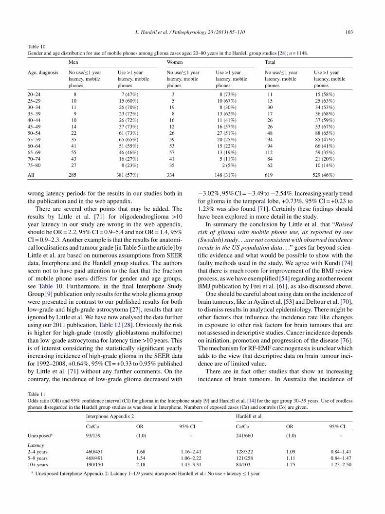
L. Hardell et al. / Pathophysiology 20 (2013) 85–110 103
Table 10Gender and age distribution for use of mobile phones among glioma cases aged 20–80 years in the Hardell group studies [28]; n = 1148.
Men Women Total
Age, diagnosis No use/≤1 yearlatency, mobilephones
Use >1 yearlatency, mobilephones
No use/≤1 yearlatency, mobilephones
Use >1 yearlatency, mobilephones
No use/≤1 yearlatency, mobilephones
Use >1 yearlatency, mobilephones
20–24 8 7 (47%) 3 8 (73%) 11 15 (58%)25–29 10 15 (60%) 5 10 (67%) 15 25 (63%)30–34 11 26 (70%) 19 8 (30%) 30 34 (53%)35–39 9 23 (72%) 8 13 (62%) 17 36 (68%)40–44 10 26 (72%) 16 11 (41%) 26 37 (59%)45–49 14 37 (73%) 12 16 (57%) 26 53 (67%)50–54 22 61 (73%) 26 27 (51%) 48 88 (65%)55–59 35 65 (65%) 59 20 (25%) 94 85 (47%)60–64 41 51 (55%) 53 15 (22%) 94 66 (41%)65–69 55 46 (46%) 57 13 (19%) 112 59 (35%)70–74 43 16 (27%) 41 5 (11%) 84 21 (20%)75–80 27 8 (23%) 35 2 (5%) 62 10 (14%)
A
wt
rysCcLdsosGwliuitiifbc
−f1h
r(ttftpB
btoinoT
TOp
U
L251
ll 285 381 (57%) 334
rong latency periods for the results in our studies both inhe publication and in the web appendix.
There are several other points that may be added. Theesults by Little et al. [71] for oligodendroglioma >10ear latency in our study are wrong in the web appendix,hould be OR = 2.2, 95% CI = 0.9–5.4 and not OR = 1.4, 95%I = 0.9–2.3. Another example is that the results for anatomi-al localisations and tumour grade [in Table 5 in the article] byittle et al. are based on numerous assumptions from SEERata, Interphone and the Hardell group studies. The authorseem not to have paid attention to the fact that the fractionf mobile phone users differs for gender and age groups,ee Table 10. Furthermore, in the final Interphone Studyroup [9] publication only results for the whole glioma groupere presented in contrast to our published results for both
ow-grade and high-grade astrocytoma [27], results that aregnored by Little et al. We have now analysed the data furthersing our 2011 publication, Table 12 [28]. Obviously the risks higher for high-grade (mostly glioblastoma multiforme)han low-grade astrocytoma for latency time >10 years. Thiss of interest considering the statistically significant yearly
ncreasing incidence of high-grade glioma in the SEER dataor 1992–2008, +0.64%, 95% CI = +0.33 to 0.95% publishedy Little et al. [71] without any further comments. On theontrary, the incidence of low-grade glioma decreased withad
i
able 11dds ratio (OR) and 95% confidence interval (CI) for glioma in the Interphone studhones disregarded in the Hardell group studies as was done in Interphone. Numbe
Interphone Appendix 2
Ca/Co OR 95% CI
nexposeda 93/159 (1.0) –
atency–4 years 460/451 1.68 1.16–2.4–9 years 468/491 1.54 1.06–2.20+ years 190/150 2.18 1.43–3.3a Unexposed Interphone Appendix 2: Latency 1–1.9 years; unexposed Hardell et
148 (31%) 619 529 (46%)
3.02%, 95% CI = −3.49 to−2.54%. Increasing yearly trendor glioma in the temporal lobe, +0.73%, 95% CI = +0.23 to.23% was also found [71]. Certainly these findings shouldave been explored in more detail in the study.
In summary the conclusion by Little et al. that “Raisedisk of glioma with mobile phone use, as reported by oneSwedish) study. . .are not consistent with observed incidencerends in the US population data. . .” goes far beyond scien-ific evidence and what would be possible to show with theaulty methods used in the study. We agree with Kundi [74]hat there is much room for improvement of the BMJ reviewrocess, as we have exemplified [54] regarding another recentMJ publication by Frei et al. [61], as also discussed above.
One should be careful about using data on the incidence ofrain tumours, like in Aydin et al. [53] and Deltour et al. [70],o dismiss results in analytical epidemiology. There might bether factors that influence the incidence rate like changesn exposure to other risk factors for brain tumours that areot assessed in descriptive studies. Cancer incidence dependsn initiation, promotion and progression of the disease [76].he mechanism for RF-EMF carcinogenesis is unclear which
dds to the view that descriptive data on brain tumour inci-ence are of limited value.There are in fact other studies that show an increasingncidence of brain tumours. In Australia the incidence of
y [9] and Hardell et al. [14] for the age group 30–59 years. Use of cordlessrs of exposed cases (Ca) and controls (Co) are given.
Hardell et al.
Ca/Co OR 95% CI
241/660 (1.0) –
1 128/322 1.09 0.84–1.412 121/258 1.11 0.84–1.471 84/103 1.75 1.23–2.50
al.: No use + latency ≤ 1 year.

104 L. Hardell et al. / Pathophysiology 20 (2013) 85–110
Table 12Odds ratio (OR) and 95% confidence interval (CI) for mobile phone use and astrocytoma, cf. Hardell et al. [28].
>1–5 year latency >5–10 year latency >10 year latency Total, >1 year latency
OR 95% CI OR 95% CI OR 95% CI OR 95% CI
AA
poapc2tAi5T−
nrAwbccviw
b[staTad
micTdtat+(iiona
wrfZdc
iNiwncilsfdsobdI9
4
agptmgst
rHswtR
strocytoma, high grade (n = 820) 1.2 0.9–1.5 1.5strocytoma, low grade (n = 132) 1.4 0.8–2.2 1.3
rimary brain tumours was studied in two areas, the statef New South Wales and Australian Capital Territory, withbout 7 million inhabitants [77]. The study covered the timeeriod 2000–2008 and all diagnoses had a histopathologi-al verification. It included 13 pathology databases servicing4 neurosurgical centres. Adults aged ≥65 years recordedhe largest proportion of malignant brain tumours, 52%. Thennual Percentage Change (APC) for malignant tumours
ncreased statistically significant +3.9%, 95% CI +2.4 to.4%. An increase was seen among both men and women.he APC for benign tumours increased with +1.7%, 95% CI1.4 to +4.9%, thus not statistically significant.From urban Shanghai an increasing incidence of brain and
ervous system tumours for the time period 1983–2007 waseported with APC +1.2%, 95% CI +0.4 to 1.9% in males andPC +2.8%, 95% CI +2.1 to 3.4% in females [78]. No resultsere given for different tumour types, e.g. malignant andenign brain tumours, or anatomical site. The authors con-luded that “The study did not support an association betweenellular telephone use and increased risk of brain and ner-ous tumours.” However, that statement goes far beyond whats scientifically justified from this register based study andhat was actually investigated.Certainly it is more informative to analyse incidence trends
y anatomical site and histology of the tumour. de Vocht et al.79] reported in England for the time period 1998–2007 atatistically significant increasing incidence of brain tumours,he majority glioma, in the temporal lobe for men (p < 0.01)nd women (p < 0.01), and frontal lobe for men (p < 0.01).he incidence increased also for women in the frontal lobe,lthough not statistically significant (p = 0.07). The incidenceecreased in other parts of the brain.
Zada et al. [80] studied incidence trends of primaryalignant brain tumours in the Los Angeles area dur-
ng 1992–2006. APC was calculated for microscopicallyonfirmed histological subtypes and anatomic sub sites.he overall incidence of primary malignant brain tumoursecreased over the time period with the exception of glioblas-oma multiforme (astrocytoma WHO grade IV). The annualge adjusted incidence rate of that tumour type increased sta-istically significant in the frontal lobe with APC +2.4% to3.0% (p ≤ 0.001) and temporal lobe APC +1.3% to +2.3%p ≤ 0.027) across all registries. In the California Cancer Reg-stry the incidence of glioblastoma multiforme increased alson cerebellum, APC +11.9% (p < 0.001). In the parietal and
ccipital lobes or in overlapping lobes no statistically sig-ificant changes in incidence were seen. For lower gradestrocytoma decreases of annual age adjusted incidence ratesaTti
1.1–1.9 3.0 2.1–4.2 1.5 1.2–1.80.7–2.4 1.7 0.7–4.0 1.4 0.9–2.2
ere observed. The authors concluded that there was aeal increase in the incidence of glioblastoma multiforme inrontal and temporal lobes and cerebellum. These results byada et al. [80] are of interest since the highest absorbedose of RF-EMF emissions from mobile phones has beenalculated to occur in these parts of the brain [6].
It should be noted that also Deltour et al. [70] reportedncreasing glioma incidence rates in Denmark, Finland,orway, and Sweden for the time period 1979–2008. APC
ncreased for men with +0.4%, 95% CI +0.1 to 0.6% and foromen with +0.3%, 95% CI +0.1 to 0.5%. Unfortunatelyo data were given for subtypes of glioma and anatomi-al sites of the tumours, which would certainly have beennformative. The authors did not consider these and otherimitations when they conclude that “Our data indicate that,o far, no risk associated with mobile phone use has mani-ested in adult glioma incidence trends...many increased orecreased risks reported in case-control studies are implau-ible, implying that biases and errors in the self-reported usef mobile phone have likely distorted the findings.” It shoulde noted that regarding Sweden we reported increasing inci-ence of astrocytoma WHO grades I–IV during 1970–2007.n the age group >19 years the annual change was +2.16%,5% CI +0.25 to 4.10% during 2000–2007 [41].
. Discussion
The most comprehensive results on use of wireless phonesnd the association with brain tumours come from the Hardellroup in Sweden and the international Interphone study. Asointed out by IARC [1] other studies as discussed above areoo small with short latency times, usually in the range of at
ost 5 years. Both the Hardell group studies and Interphoneive results for latency time of 10 years or more. Thus, aummary evaluation will mainly be based on results fromhese two study groups.
Both were case-control studies and the cases wereecruited during similar time periods, 1997–2003 in theardell group and during 2000–2004 in Interphone, with
omewhat different years in the varying study regions. Thereas no overlapping of cases in the Hardell group studies and
he Swedish part of Interphone. Cases were ascertained fromegional Cancer Registries in the Hardell group studies and
ll diagnoses were based on histopathological verification.hus, all cases had been operated or undergone biopsy of theumour for diagnosis. In contrast, in Interphone cases weredentified from neurological or neurosurgical facilities in the

physiol
sTrsaaewceltrgmgfilIttmfm
wait6iisttIuEa
ift5cmp
iwcuffs
mwIic(tnoa
uciatPaoisFfcsotm
ttsotbDasccii
aiidmltwe
L. Hardell et al. / Patho
tudy regions; in some centres also from cancer registries.he diagnoses of glioma, meningioma and acoustic neu-
oma were based on histopathology or diagnostic imaging. Ithould be pointed out that the diagnosis of both meningiomand acoustic neuroma has a rather high precision using CTnd/or MRI. Regarding glioma it is certainly more difficult tostablish a valid diagnosis without histopathology, especiallyhen it comes to subgroups such as different grades of astro-
ytoma (WHO grades I–IV). In the publication by Lahkolat al. [81] most glioma diagnoses were based on histopatho-ogy, whereas this has not been published for Interphone inotal. It is notable that Interphone [9] has not presented sepa-ate results for astrocytoma in total in contrast to the Hardellroup. Especially results for high-grade glioma including theost common glioma type, glioblastoma multiforme (WHO
rade IV), would be of value since the highest risk was foundor that subtype by Hardell et al., Table 12 [27,28]. It is also ofnterest that we found higher risk for use of mobile and cord-ess phones for astrocytoma grades III–IV than for grades–II [82]. Some results were published for glioblastoma mul-iforme from the 5 North European countries [81]. Certainlyhe total result for glioma and >10 years since first ipsilateral
obile phone use with OR = 1.39, 95% CI = 1.01–1.92 (por trend 0.04) would have been of interest for glioblastomaultiforme separately in Lahkola et al. [81].The Hardell group included cases aged 20–80 years
hereas eligible cases in Interphone were aged 30–59 yearst diagnosis. This difference is important since the highestncidence of astrocytoma WHO grade IV (glioblastoma mul-iforme) is found in the age group 45–75 years with mean age1 years and 80% older than 50 years [83]. As can be seenn Table 10, the highest prevalence of use of mobile phonesn the Hardell group studies was up to the age of 54 years,o limiting the age to 59 years as in Interphone diminisheshe possibility to find an increased risk taking a reasonableumour induction period. It seems as if the age distribution innterphone was more decided by prevalence of mobile phonese in the population than age distribution for glioma cases.xcluding the age group 20–29 years, as in Interphone, makeslso an evaluation of young users more difficult, see Table 9.
Meningioma is a slow growing benign tumour with a peakncidence in the sixth and seventh decade of life with a 3:2–2:1emale:male ratio [84]. As pointed out by Interphone [10]he incidence peak of acoustic neuroma is in the age group0–65 years. Thus, again limiting upper age to 59 years forases in Interphone excluded a large proportion of cases witheningioma or acoustic neuroma taking a reasonable latency
eriod.One control subject matched on age, gender and geograph-
cal area (region) to each case in the Hardell group studiesas drawn from the national population register. The register
overs the whole population and each person is assigned a
nique id-number making it possible to trace current addressor all inhabitants. In Interphone one control was selectedor each case from a ‘locally appropriate population-basedampling frame’. In Germany the centres used individualadwr
ogy 20 (2013) 85–110 105
atching or frequency matching. The matching variablesere age within 5 years, gender and region of residence; in
srael also ethnic origin. When stratified matching was usedndividual matching was made afterwards from the wholeontrol sample with cases being assigned one control subjecttwo in Germany) interviewed as close as possible in timeo the case [9]. Regarding the Interphone study on acousticeuroma some centres sampled special controls to the cases,ther draw controls from the pool of controls in the gliomand meningioma studies, or used a mixture of both methods.
The Nordic countries have population registers that weresed in Denmark, Norway and Finland for recruitment ofontrols in Interphone. Also Germany used a population reg-ster [85]. However, UK used general practioners’ lists [86]nd in Japan random digit dialling was used [44,87]. Certainlyhe methods used in Interphone may introduce selection bias.atient lists are usually selective to use for drawing of controlsnd do not represent the whole population which is the sourcef the cases. Also random digit dialling has the potential tontroduce selection bias since persons that are registered toubscribe a phone are usually wealthier than non-subscribers.urthermore, it seems not to be the most appropriate methodor selection of controls in a study on mobile phone use, andertainly not regarding cordless phones, since phone sub-cribers are selected as controls. Furthermore, later selectionf controls from a pool with individual matching may givehe possibility for selection bias if this is not done in a blinded
anner as to exposure status.These methods contrast to the Hardell group where con-
rols were drawn consequently to the cases and all controlshat answered the questionnaire were included in the analy-es. In Interphone proxy interviews were performed for 13%f glioma cases but only 1% of controls [9]. This is in contrasto the Hardell group study on deceased cases with malignantrain tumours [26]. Deceased controls were drawn from theeath Registry in Sweden. Relatives to both deceased cases
nd deceased controls were interviewed, thereby creating theame condition for assessment of exposure among cases andontrols. Although using proxy interviews for both cases andontrols is the more appropriate method exclusion of proxynterviews in Interphone had little impact on the overall resultn the sensitivity analysis.
Use of wireless phones was carefully assessed by a self-dministered questionnaire in the Hardell et al. studies. Thenformation was supplemented over the phone by trainednterviewers thereby using a structured protocol. This wasone blinded as to case or control status. The ear that hadostly been used during calls with mobile phone and/or cord-
ess phone was assessed by separate questions; >50% of theime for one side, or equally for both sides. This informationas checked during the supplementary phone calls. Moreover
very person that had used a mobile phone received after that
letter asking them again to specify the ear that had been useduring phone calls and to what extent that side of the headas mostly used. There was a very good agreement of theesults using these three methods to assess these data. Also

1 physiol
oirtTw
aRqfiiscd
wpwnticliipcmq
IqfdsvfriOtplwsrobittc
a
vffScvmewIf9‘Ho14tdvetvmpii2e
piinba5wOsjd
cgwbL67r5
06 L. Hardell et al. / Patho
ther exposures were assessed in the questionnaire. After thenterviews all personal data like names and addresses wereemoved from the questionnaires so that only an id-numberhat did not disclose if it was a case or a control was shown.hus, coding of the data for statistical analysis was performedithout personal data on the individual.We investigated in more detail the possibility of recall
nd observational bias in our second case-control study [21].eporting a previous cancer or if a relative helped to fill in theuestionnaire did not change the results, i.e., were no con-ounding factors. Potential observational bias during phonenterviews was analysed by comparing change of exposuren cases and controls after these interviews. No statisticallyignificant differences were found, showing that our resultsould not be explained by observational bias, for furtheretails see discussion in that publication [21].
On the contrary information on past mobile phone useas mostly collected during face-to-face interviews in Inter-hone obviously disclosing if it was a case or a control thatas interviewed. These interviews were performed by a largeumber of interviewers at different participating centres. Inhe personal interviews a computer program that guided thenterview with questions read by the interviewer from a laptopomputer screen was used. The interviews in the Swedish partasted for about 45 min. The answers were entered directlynto the computer by the interviewer. Cards were shown tof possible identify the model of the mobile phone [88]. Theurpose of the study was thereby obviously disclosed to theases and controls. This was in contrast to the Hardell groupailed questionnaire that contained a large number of other
uestions without special attention to wireless phones.We regard hospital based interviews of cases, as in the
nterphone study, to be a major disadvantage and ethicallyuestionable. At that time the patient has not fully recoveredrom e.g. surgery, may not have been fully informed about theiagnosis, treatment and prognosis and may even be underedation by drugs. Using computer based face-to-face inter-iews may also be a stressful situation for the patient. Inact patients scored significantly lower than controls due toecalling of words (aphasia), problems with writing and draw-ng due to paralysis in the Danish part of Interphone [89].bviously observational bias could have been introduced by
he interview methods in Interphone. Only Finland used aaper version of the questionnaire, but Finland has never pub-ished country specific results on the different tumour types,hich would certainly have been of interest. For unclear rea-
ons the results on glioma were only included as part of theesults for the 5 North European countries [81] and as partf the whole Interphone study [9]. Furthermore, it has noteen disclosed how the personal interviews were performedn sparsely populated areas, e.g. in the Northern Sweden. Didhe interviewers travel long distances for interviews of con-
rols in rural areas or were all controls living in the largestities thereby creating selection bias?It should be noted that the number of participating casesnd controls from each centre in Interphone was quite low. It
ttct
ogy 20 (2013) 85–110
aried for glioma from 60 (Japan) to at most 421 (UK North),or meningioma from 52 (New Zealand) to 350 (Israel) andor acoustic neuroma from 18 (New Zealand) to 152 (UKouth). Similarly the number of controls varied according toentre [9,10]. It is obvious that with so low number of inter-iewed subjects by many different interviewers the qualityay have been hampered in Interphone by low training and
xperience of certain interviewers. Experienced interviewersere defined as those who conducted at least 20 interviews.
n fact, in the sensitivity analysis the risk increased for gliomaor cumulative mobile phone use ≥1640 h from OR = 1.40,5% CI = 1.03–1.89 to OR = 1.50, 95% CI = 1.10–2.06 ifexperienced interviewers only’ were considered. In theardell group studies few persons conducted all interviewsf the 1251 participating cases with malignant brain tumour,254 cases with benign brain tumour, and 2438 controls (total942; note one case had both a malignant and a benign brainumour). All interviewers were first educated; they used aefined protocol and gained considerable experience as inter-iewers. In fact, they were obliged to carry out the interviewsxtensively to fulfil the quality in data assessment accordingo the structured protocol. It is obvious that the few inter-iewers in the Hardell group study must have been muchore experienced than the diversity of interviewers in Inter-
hone. The higher risk restricting analysis to ‘experiencednterviewers’ in Interphone indicates observational bias dur-ng assessment of exposure decreasing the risk. Furthermore,0 interviews as the definition was in Interphone to be anxperienced interviewer, is after all a very low number.
Several other sensitivity analyses were performed in Inter-hone without any major impact on the results. It is discussedn the Interphone study [9] that the increased risk for glioman the highest decile of cumulative exposure was caused by aumber of subjects reporting >5 h call time per day. This num-er may be real in e.g. certain occupations using the phones a working tool. Furthermore, if call time was truncated toh per day no statistically significant difference of the riskas found, OR = 1.38, 95% CI = 1.02–1.87 for glioma andR = 3.03, 95% CI = 1.62–5.67 for acoustic neuroma (expo-
ure up to 5 years before reference date). Certainly it is notustified to exclude these subjects from the analysis as wasone in some of the calculations in Interphone [9,10].
It is always essential to have a high response rate in case-ontrol studies to get as valid results as possible. In the Hardellroup studies the response rate was 85% (n = 1251) for casesith malignant brain tumour, 88% (n = 1254) for cases withenign brain tumour, and 84% (n = 2438) for controls [29,40].ower response rates were obtained in the Interphone study,4%, range by centre 36–92%, (n = 2765) for glioma cases,8%, range 56–92%, (n = 2425) for meningioma cases, 82%,ange 70–100% (n = 1121) for acoustic neuroma cases, and3%, range 42–74%, (n = 7658) for controls [9,10]. Certainly
hese low response rates, less than half of the cases and con-rols in some centres, may have created the possibility ofonsiderable selection bias and are examples of the mul-iple methodological problems in Interphone. As has been
physiol
dti
s3Halyacasas2atpt
bwwtcgWcfeegaditbctw
chirosta[ssie
tghmuteebcolbtifsli
5
aamIgasawaasSotrbir
wspSBadw
L. Hardell et al. / Patho
iscussed elsewhere not responding controls in Interphoneended to be less frequent users of mobile phone than partic-pating controls leading to underestimation of the risk [32].
There are other differences between the Hardell grouptudies and Interphone study such as restricting age to0–59 years in Interphone compared with 20–80 years in theardell-group studies and considering use of cordless phones
s no exposure to RE-EMF in Interphone. Even if the preva-ence of mobile phone use is highest in the age group 30–59ears, excluding older cases diminishes the possibility to findn increased risk, assuming a reasonable latency time. As dis-ussed above the peak incidence of most brain tumours is athigher age. In a case series from Canada all brain tumours
howed a bimodal age distribution with one peak in the 0–4ge group and the other in the 60–69 age group [90]. Ashown elsewhere [14] step-wise exclusion of the age group0–29 years, 60–80 years and including cordless phone usemong unexposed reduced OR in the Hardell-group studieso similar results as in Interphone [see Tables 1 and 2 in theublication]. Thus, Interphone seems to have underestimatedhe risk also for these reasons.
Survival of patients with glioma has only been presentedy the Hardell group [68]. Decreased survival of glioma casesith long-term and high cumulative use of wireless phonesas found. We found a survival disadvantage for astrocy-
oma WHO grade IV among cases using mobile phone orordless phone indicating a worse prognosis in that patientroup. On the contrary, a survival benefit for astrocytomaHO grades I–II was observed. The fact that there was no
lear trend with intensity or duration of wireless phone useor low-grade astrocytoma does not speak in favour of anffect of RF-EMF from such use. The exposure might, how-ver, produce awareness bias in these cases. RF-EMF mayive tumour promotion [91] inducing disease related person-lity disturbances and habit changes leading to earlier tumouriagnosis than among unexposed patients. This would resultn earlier treatment with a better prognosis after surgery inhis patient group [69]. These findings indicate a complexiological effect from RF-EMF exposure and strengthen aausal association between these tumour types, e.g. astrocy-oma WHO grade IV (glioblastoma multiforme), and use ofireless phones.By placing a strong emphasis on incidence data an asso-
iation between use of wireless phones and brain tumoursas been challenged [92]. The authors considered that, if thencreased risks seen in case-control studies reflect a causalelationship, there would already be an increase in incidencef brain and central nervous system tumours, for which thereeemed to be little evidence. This belief is unfounded forwo reasons. The first relates to latent periods for gliomand acoustic neuroma development, typically 10–40 years93,94]. The results on long-term use of wireless phones are
canty and at most latency period of 10+ years have beentudied. Furthermore, we know little about the earliest eventsn the genesis of glioma in humans for obvious reasons. How-ver, progression of glioma has been studied in large series ofctlfi
ogy 20 (2013) 85–110 107
umours of different malignancy grades. Patients with low-rade glioma have been followed with later progression toigh-grade glioma [95]. Thus, since the natural history ofost glioma from earliest events to clinical manifestation is
nknown, but most likely several decades, the exposure dura-ion in most studies is incompatible with a tumour initiatingffect. An initiating effect is what would have the most directffect on the incidence. The other reason concerns the possi-ility of an effect on tumour development (promotion) and itsonsequences on the increase in incidence that can possiblyccur. If the exposure acts as a promoter, this would decreaseatency time for already existing tumours, giving a temporaryut not a continuous increase in incidence. In addition it haso be pointed out that any such effect on tumour developments limited by the magnitude of the shift of the age-incidenceunction and its slope for the respective tumour type [91]. Ithould be noted that studies on tumour type and anatomicalocalisation indicate by now an effect from RF-EMF on thencidence of brain tumours [71,77,79,80].
. Conclusions
There is a consistent pattern of increased risk of gliomand acoustic neuroma associated with use of mobile phonesnd cordless phones. The epidemiological evidence comesainly from two study centres, the Hardell group and the
nterphone study group. In the same studies by the Hardellroup and Interphone study group no consistent pattern ofn increased risk was found for meningioma. These resultstrengthen the other findings, i.e., increased risk for gliomand acoustic neuroma, since a systematic bias in the studiesould also have been inherit for meningioma. Furthermore,causal association between use of mobile phone and gliomand acoustic neuroma comes from the meta-analyses as pre-ented in this publication and also reviewed elsewhere [96].upportive evidence comes also from anatomical localisationf the tumour to the most exposed area of the brain, cumula-ive exposure and latency time that all add to the biologicalelevance of an increased risk. In addition risk calculationased on estimated absorbed dose gives strength to the find-ngs as well as the impact on survival of glioma patientselating to their use of mobile and cordless phones.
Evidence is increasing that workers with heavy use ofireless phones who develop glioma or acoustic neuroma
hould be compensated. In fact, the first case with such com-ensation has now been established in court. The Italianupreme Court affirmed a previous ruling that the Insuranceody for Work (INAIL) must grant worker’s compensation tobusinessman who had used wireless phones for 12 years andeveloped a neuroma in the brain (www.applelettrosmog.it;ww.microwavenews.com). He had used both mobile and
ordless phones for five to six hours per day preferably onhe same side as the tumour developed. The neuroma wasocated in the trigeminal Gasser’s ganglion in the brain. Thisfth cranial nerve controls facial sensations and muscles. It is

1 physiol
tci1otwt
Riglbgecas
A
mlgrr
A
CF
R
[
[
[
[
[
[
[
[
[
[
[
[
[
[
08 L. Hardell et al. / Patho
he same type of tumour as the acoustic neuroma in the eighthranial nerve located in the same area of the brain. The Ital-an case fulfils the criteria for a causal association; more than0 years use of wireless phones, high cumulative exposuren the same side as the tumour appeared, and a tumour typehat would be predicted based on previous research on use ofireless phones and brain tumour risk. No further appeal of
he Supreme Court decision is possible.In summary there is reasonable basis to conclude that
F-EMFs are bioactive and have a potential to cause healthmpacts. There is a consistent pattern of increased risk forlioma and acoustic neuroma associated with use of wire-ess phones (mobile phones and cordless phones) mainlyased on results from case-control studies from the Hardellroup and Interphone Final Study results. Epidemiologicalvidence gives that RF-EMF should be classified as a humanarcinogen. The current safety limits and reference levelsre not adequate to protect public health. New public healthtandards and limits are needed.
uthors’ contributions
Lennart Hardell was responsible for drafting of theanuscript and Michael Carlberg made all statistical calcu-
ations. Michael Carlberg and Kjell Hansson Mild read andave valuable comments on the manuscript. All authors haveead and approved the final version. No conflicts of interesteported.
cknowledgements
Supported by grants from Cancer-och Allergifonden,ancerhjälpen, and Örebro University Hospital Cancerund.
eferences
[1] R. Baan, Y. Grosse, B. Lauby-Secretan, F. El Ghissassi, V.Bouvard, L. Benbrahim-Tallaa, et al., Carcinogenicity of radiofre-quency electromagnetic fields, Lancet Oncology 12 (2011)624–626.
[2] IARC Monographs on the Evaluation of Carcinogenic Risks toHumans, Volume 102, Non-Ionizing radiation, Part II: RadiofrequencyElectromagnetic Fields [includes mobile telephones], IARC Press2011, Lyon, France.
[3] IARC Monographs on the Evaluation of Carcinogenic Risks toHumans, Volume 80, Non-Ionizing Radiation, Part I: Static andExtremely Low-Frequency (ELF) Electric and Magnetic Fields, IARCPress 2002, Lyon, France.
[4] T. Linde, K. Hansson Mild, Measurement of low frequency magneticfields from digital cellular telephones, Bioelectromagnetics 18 (1997)
184–186.[5] K. Jokela, L. Puranen, A.P. Sihvonen, Assessment of the magnetic fieldexposure due to the battery current of digital mobile phones, HealthPhysics 86 (2004) 56–66.
[
ogy 20 (2013) 85–110
[6] E. Cardis, I. Deltour, S. Mann, M. Moissonnier, M. Taki, N. Varsier,et al., Distribution of RF energy emitted by mobile phones in anatomicalstructures of the brain, Physics in Medicine and Biology 53 (2008)2771–2783.
[7] S. Preston-Martin, R. Munir, I. Chakrabarti, Nervous system, in: D.Schottenfeld, J.F. Fraumeni Jr. (Eds.), Cancer Epidemiology and Pre-vention, Oxford University Press, 2006, pp. 1173–1195.
[8] M.E. Scheurer, C.J. Etzel, M. Liu, J. Barnholtz-Sloan, F. Wik-lund, B. Tavelin, et al., Familial aggregation of glioma: apooled analysis, American Journal of Epidemiology 172 (2010)1099–1107.
[9] Interphone Study Group, Brain tumour risk in relation to mobiletelephone use: results of the INTERPHONE international case-control study, International Journal of Epidemiology 39 (2010)675–694.
10] Interphone Study Group, Acoustic neuroma risk in relation to mobiletelephone use: results of the INTERPHONE international case-controlstudy, Cancer Epidemiology 35 (2011) 453–464.
11] E. Cardis, B.K. Armstrong, J.D. Bowman, G.G. Giles, M. Hours, D.Krewski, et al., Risk of brain tumours in relation to estimated RF dosefrom mobile phones: results from five Interphone countries, Occupa-tional and Environmental Medicine 68 (2011) 631–640.
12] L. Hardell, M. Carlberg, D. Gee, Mobile phone use and brain tumourrisk: early warnings, early actions? In: Late Lessons from Early War-nings, part 2, European Environment Agency, Copenhagen, Denmark2013, in press.
13] L. Hardell, M. Carlberg, K. Hansson Mild, Epidemiological evidencefor an association between use of wireless phones and tumour diseases,Pathophysiology 16 (2009) 113–122.
14] L. Hardell, M. Carlberg, K. Hansson Mild, Re-analysis of risk forglioma in relation to mobile telephone use: comparison with the resultsof the Interphone international case-control study, International Journalof Epidemiology 40 (2011) 1126–1128.
15] L. Hardell, Å. Näsman, A. Påhlson, A. Hallquist, K. Hansson Mild,Use of cellular telephones and the risk for brain tumours: a case-controlstudy, International Journal of Oncology 15 (1999) 113–116.
16] L. Hardell, K. Hansson Mild, A. Påhlson, A. Hallquist, Ionizing radi-ation, cellular telephones and the risk for brain tumours, EuropeanJournal of Cancer Prevention 10 (2001) 523–529.
17] J.E. Muscat, M.G. Malkin, S. Thompson, R.E. Shore, S.D. Stell-man, D. McRee, et al., Handheld cellular telephone use and risk ofbrain cancer, Journal of the American medical Association 284 (2000)3001–3007.
18] H. Ohgaki, P. Kleihues, Population-based studies on incidence, survivalrates, and genetic alterations in astocytic and oligodendroglial gliomas.Review, Journal of Neuropathology and Experimental Neurology 64(2005) 479–489.
19] P.D. Inskip, R.E. Tarone, E.E. Hatch, T.C. Wilcosky, W.R. Shapiro, R.G.Selker, et al., Cellular-telephone use and brain tumours, New EnglandJournal of Medicine 344 (2001) 79–86.
20] A. Auvinen, M. Hietanen, R. Luukkonen, R.S. Koskela, Brain tumoursand salivary gland cancers among cellular telephone users, Epidemiol-ogy 13 (2002) 356–359.
21] L. Hardell, A. Hallquist, K. Hansson Mild, M. Carlberg, A. Påhlson, A.Lilja, Cellular and cordless telephones and the risk for brain tumours,European Journal of Cancer Prevention 11 (2002) 377–386.
22] L. Hardell, K. Hansson Mild, M. Carlberg, Further aspects on cellularand cordless telephones and brain tumours, International Journal ofOncology 22 (2003) 399–407.
23] L. Hardell, M. Carlberg, K. Hansson Mild, Case-control study of theassociation between the use of cellular and cordless telephones andmalignant brain tumours diagnosed during 2000-2003, EnvironmentalResearch 100 (2006) 232–241.
24] L. Hardell, M. Carlberg, K. Hansson Mild, Case-control study on cel-
lular and cordless telephones and the risk for acoustic neuroma ormeningioma in patients diagnosed 2000-2003, Neuroepidemiology 25(2005) 120–128.
physiol
[
[
[
[
[
[
[
[
[
[
[
[
[
[
[
[
[
[
[
[
[
[
[
[
[
[
[
[
[
[
[
[
[
[
[
[
[
[
L. Hardell et al. / Patho
25] J.D. Jr. Boice, J.K. McLaughlin, Epidemiologic Studies ofCellular Telephones and Cancer Risk—A Review. SSI Publica-tion 2002:16, accessed at http://www.stralsakerhetsmyndigheten.se/Publikationer/Rapport/Stralskydd/2002/200216/
26] L. Hardell, M. Carlberg, K. Hansson Mild, Mobile phone use and therisk for malignant brain tumours: a case-control study on deceased casesand controls, Neuroepidemiology 35 (2010) 109–114.
27] L. Hardell, M. Carlberg, K. Hansson Mild, Pooled analysis of twocase-control studies on use of cellular and cordless telephones and therisk for malignant brain tumours diagnosed in 1997-2003, InternationalArchives of Occupational Environmental Health 79 (2006) 630–639.
28] L. Hardell, M. Carlberg, K. Hansson Mild, Pooled analysis of case-control studies on malignant brain tumours and the use of mobile andcordless phones including living and deceased subjects, InternationalJournal of Oncology 38 (2011) 1465–1474.
29] M. Carlberg, L. Hardell, On the association between glioma, wirelessphones, heredity and ionising radiation, Pathophysiology 19 (2012)243–252.
30] A. McKinlay, Possible health effects related to the use ofradiotelephones—recommendations of a European Commission ExpertGroup, Radiological Protection Bullentin 187 (1997) 9–16.
31] E. Cardis, L. Richardson, I. Deltour, B. Armstrong, M. Feychting, C.Johansen, et al., The INTERPHONE study: design, epidemiologicalmethods, and description of the study population, European Journal ofEpidemiology 22 (2007) 647–664.
32] L. Hardell, M. Carlberg, K. Hansson, Mild, methodological aspects ofepidemiological studies on the use of mobile phones and their asso-ciation with brain tumours, Open Environmental Sciences 2 (2008)54–61.
33] R. Saracci, J. Samet, Commentary: call me on my mobile phone.orbetter not?—a look at the INTERPHONE study results, InternationalJournal of Epidemiology 39 (2010) 695–698.
34] J.L. Phillips, N.P. Singh, H. Lai, Electromagnetic fields and DNA dam-age, Pathophysiology 16 (2009) 79–88.
35] H. Nittby, A. Brun, J. Eberhardt, L. Malmgren, B.R. Persson, L.G.Salford, Increased blood-brain barrier permeability in mammalian brain7 days after exposure to the radiation from a GSM-900 mobile phone,Pathophysiology 16 (2009) 103–112.
36] E. Markova, L.O.G. Malmgren, I.Y. Belyaev, Microwaves from mobilephones inhibit 53BP1 focus formation in human stem cells morestrongly than in differentiated cells: possible mechanistic link to cancer,Environmental Health Perspectives 118 (2010) 394–399.
37] K. Hansson Mild, M. Carlberg, J. Wilén, L. Hardell, How to combinethe use of different mobile and cordless telephones in epidemiologicalstudies on brain tumours? European Journal of Cancer Prevention 14(2005) 285–288.
38] L. Hardell, M. Carlberg, K. Hansson Mild, Use of cellular telephonesand brain tumour risk in urban and rural areas, Occupational and Envi-ronmental Medicine 62 (2005) 390–394.
39] S. Larjavaara, J. Schüz, A. Swerdlow, M. Feychting, C. Johansen, S.Lagorio, et al., Location of gliomas in relation to mobile telephone use:a case-case and case-specular analysis, American Journal of Epidemi-ology 174 (2011) 2–11.
40] L. Hardell, M. Carlberg, K. Hansson Mild, Pooled analysis of two case-control studies on the use of cellular and cordless telephones and therisk of benign brain tumours diagnosed during 1997-2003, InternationalJournal of Oncology 28 (2006) 509–518.
41] L. Hardell, M. Carlberg, Mobile phones, cordless phones and the riskfor brain tumours, International Journal of Oncology 35 (2009) 5–17.
42] J.E. Muscat, M.G. Malkin, R.E. Shore, S. Thompson, A.I. Neugut,S.D. Stellman, et al., Handheld cellular telephones and risk of acousticneuroma, Neurology 58 (2002) 1304–1306.
43] Y. Sato, S. Akiba, O. Kubo, N. Yamaguchi, A case-case study of mobile
phone use and acoustic neuroma risk in Japan, Bioelectromagnetics 32(2011) 85–93.44] T. Takebayashi, N. Varsier, Y. Kikuchi, K. Wake, M. Taki, S. Watanabe,et al., Mobile phone use, exposure to radiofrequency electromagnetic
[
ogy 20 (2013) 85–110 109
field, and brain tumour: a case-control study, British Journal of Cancer98 (2008) 652–659.
45] M.J. Schoemaker, A.J. Swerdlow, Risk of pituitary tumours in cellularphone users: a case-control study, Epidemiology 20 (2009) 348–354.
46] A. Christ, M.C. Gosselin, M. Christopoulou, S. Kühn, N. Kuster, Age-dependent tissue-specific exposure of cell phone users, Physics inMedicine and Biology 55 (2010) 1767–1783.
47] O.P. Gandhi, L.L. Morgan, A.A. de Salles, Y.Y. Han, R.B. Herber-man, D.L. Davis, Exposure limits: the underestimation of absorbedcell phone radiation, especially in children, Electromagnetic Biologyand Medicine 31 (2012) 34–51.
48] L. Kheifets, M. Repacholi, R. Saunders, E. van Deventer, The sen-sitivity of children to electromagnetic fields, Pediatrics 116 (2005)303–313.
49] N.U. Dosenbach, N. Nardos, A.L. Cohen, D.A. Fair, J.D. Power, J.A.Church, et al., Prediction of individual brain maturity using fMRI,Science 329 (2010) 1358–1361.
50] F. Söderqvist, L. Hardell, M. Carlberg, K. Hansson Mild, Ownershipand use of wireless telephones: a population-based study of Swedishchildren aged 7-14 years, BMC Public Health 7 (2007) 105.
51] F. Söderqvist, M. Carlberg, L. Hardell, Use of wireless telephonesand self-reported health symptoms: a population-based study amongSwedish adolescents aged 15–19 years, Environmental Health 7 (2008)18.
52] L. Hardell, K. Hansson Mild, M. Carlberg, A. Hallquist, Cellular andcordless telephone use and the association with brain tumours in dif-ferent age groups, Archives of Occupational and Environmental Health59 (2004) 132–137.
53] D. Aydin, M. Feychting, J. Schüz, T. Tynes, T.V. Andersen, L.S.Schmidt, et al., Mobile phone use and brain tumours in children andadolescents: a multicenter case-control study, Journal of the NationalCancer Institute 103 (2011) 1264–1276.
54] F. Söderqvist, M. Carlberg, K. Hansson Mild, L. Hardell, Childhoodbrain tumour risk and its association with wireless phones: a commen-tary, Environmental Health 10 (2011) 106.
55] J.D. Boice Jr., R.E. Tarone, Cell phones, cancer, and children, Journalof the National Cancer Institute 16 (2011) 1211–1213.
56] D. Aydin, M. Feychting, J. Schüz, T.V. Andersen, A. Harbo Poulsen,M. Prochazka, et al., Impact of random and systematic recall errorsand selection bias in case-control studies on mobile phone use andbrain tumors in adolescents (CEFALO Study), Bioelectromagnetics 32(2011) 396–407.
57] C. Johansen, J.D. Boice Jr., J. McLaughlin, J. Olsen, Cellular telephonesand cancer—a nationwide cohort study in Denmark, Journal of theNational Cancer Institute 93 (2001) 203–207.
58] F. Söderqvist, M. Carlberg, L. Hardell, Review of four publica-tions on the Danish cohort study on mobile phone subscribers andrisk of brain tumours, Reviews Environmental Health 27 (2012)51–58.
59] J. Schüz, R. Jacobsen, J.H. Olsen, J.D. Boice Jr., J.K. McLaughlin, C.Johansen, Cellular telephone use and cancer risk: update of a nation-wide Danish cohort, Journal of the National Cancer Institute 98 (2006)1707–1713.
60] J. Schüz, M. Steding-Jessen, S. Hansen, S.E. Stangerup, P. Cayé-Thomasen, A.H. Poulsen, et al., Long-term mobile phone use andthe risk of vestibular schwannoma: a Danish nationwide cohort study,American Journal of Epidemiology 174 (2011) 416–422.
61] P. Frei, A. Harbo Poulsen, C. Johansen, J.H. Olsen, M. Steding-Jessen, J. Schüz, Use of mobile phones and risk of brain tumours:update of Danish cohort study, British Medical Journal 343 (2011)d6387.
62] J. Schüz, P. Elliott, A. Auvinen, H. Kromhout, A. Harbo Poulsen, C.Johansen, et al., An international prospective cohort study of mobile
phone users and health (Cosmos): design considerations and enrolment,Cancer Epidemiology 35 (2011) 37–43.63] S.O. Dalton, M. Steding-Jessen, M. Gislum, K. Frederiksen, G.Engholm, J. Schüz, Social inequality and incidence of and survival

1 physiol
[
[
[
[
[
[
[
[
[
[
[
[
[
[
[
[
[
[
[
[
[
[
[
[
[
[
[
[
[
[
[
[
10 L. Hardell et al. / Patho
from cancer in a population-based study in Denmark, 1994-2003: back-ground, aims, material and methods, European Journal of Cancer 44(2008) 1938–1949.
64] A. Ahlbom, M. Feychting, Mobile telephones and brain tumours,British Medical Journal 343 (2011) d6605.
65] A. Ahlbom, M. Feychting, E. Cardis, P. Elliott, Re: Cellular telephoneuse and cancer risk: update of a nationwide Danish cohort study, Journalof the National Cancer Institute 99 (2007) 655.
66] D.E. Foliart, B.H. Pollock, G. Mezei, R. Iriye, J.M. Silva, K.L. Ebi,et al., Magnetic field exposure and long-term survival among childrenwith leukaemia, British Journal of Cancer 94 (2006) 161–164.
67] A.-L. Svendsen, T. Weihkopf, P. Kaatsch, J. Schüz, Exposure to mag-netic fields and survival after diagnosis of childhood leukemia: aGerman cohort study, Cancer Epidemiology Biomarkers Prevention16 (2007) 1167–1171.
68] L. Hardell, M. Carlberg, Use of mobile and cordless phones and survivalof patients with glioma, Neuroepidemiology 40 (2012) 101–108.
69] F.T. Vertosick Jr., R.G. Selker, V.C. Arena, Survival of patients withwell-differentiated astrocytomas diagnosed in the era of computedtomography. Review, Neurosurgery 28 (1991) 496–501.
70] I. Deltour, A. Auvinen, M. Feychting, C. Johansen, L. Klaeboe, R.Sankila, et al., Mobile phone use and incidence of glioma in theNordic countries 1997-2008: consistency check, Epidemiology 23(2012) 301–307.
71] M.P. Little, P. Rajaraman, R.E. Curtis, S.S. Devesa, P. Inskip, D.P.Check, et al., Mobile phone use and glioma risk: comparison of epi-demiological study results with incidence trends in the United States,British Medical Journal 344 (2012) e1147.
72] T. Bergenheim, A. Malmström, H. Bolander, A. Michanek, G.Stragliotto, L. Damber, et al., Registration on regional basis of patientswith primary brain tumours. Regional differences disclosed, Läkartid-ningen 104 (2007) 332–338, 340–341.
73] L. Barlow, K. Westergren, L. Holmberg, M. Talbäck, The completenessof the Swedish Cancer Register: a sample survey for year 1998, ActaOncologica 48 (2009) 27–33.
74] M. Kundi, Study of mobile phone use and glioma risk was fatallyflawed, British Medical Journal 344 (2012) e3078.
75] D.L. Davis, A.B. Miller, A. Philips, Association of mobile phone usewith adult brain cancer remains plausible, British Medical Journal 344(2012) e3083.
76] W.D. Hazelton, M.S. Clements, S.H. Moolgavkar, Multistage car-cinogenesis and lung cancer mortality in three cohorts, CancerEpidemiology Biomarkers and Prevention 14 (2005) 1171–1181.
77] M. Dobes, B. Shadbolt, V.G. Khurana, S. Jain, S.F. Smith, R. Smee,et al., A multicenter study of primary brain tumour incidence inAustralia (2000-2008), Neuro Oncology 13 (2011) 783–790.
78] L.-X. Ding, Y.-X. Wang, Increasing incidence of brain and nervoustumours in urban Shanghai, China, 1983-2007, Asian Pacific Journalof Cancer Prevention 12 (2011) 3319–3322.
79] F. de Vocht, I. Burstyn, J.W. Cherrie, Time trends (1998-2007) in braincancer incidence rates in relation to mobile phone use in England,
Bioelectromagnetics 32 (2011) 334–339.80] G. Zada, A.E. Bond, Y.-P. Wang, S.L. Giannotta, D. Deapan, Incidencetrends in the anatomic location of primary malignant brain tumours inthe United States: 1992-2006, World Neurosurgery 77 (2012) 518–524.
[
ogy 20 (2013) 85–110
81] A. Lahkola, A. Auvinen, J. Raitanen, M.J. Schoemaker, H.C. Chris-tensen, M. Feychting, et al., Mobile phone use and risk of glioma in 5North European countries, International Journal of Cancer 120 (2007)1769–1775.
82] K. Hansson Mild, L. Hardell, M. Carlberg, Pooled analysis of twoSwedish case-control studies on the use of mobile and cordlesstelephones and the risk of brain tumours diagnosed 1997-2003, Inter-national Journal of Occupational Safety and Ergonomics 13 (2007)63–71.
83] H. Ohgaki, P. Dessen, B. Jourde, S. Horstmann, T. Nishikawa, P.L.Di Patre, et al., Genetic pathways of glioblastoma: a population-basedstudy, Cancer Research 64 (2004) 6892–6899.
84] C. Marosi, M. Hassle, K.R. Ssler, Guidelines to the treatment of menin-gioma, Forum 13 (2003) 76–89.
85] B. Schlehofer, K. Schlaefer, M. Blettner, G. Berg, E. Böhler, I. Het-tinger, et al., Environmental risk factors for sporadic acoustic neuroma(Interphone Study Group, Germany), European Journal of Cancer 43(2007) 1741–1747.
86] S.J. Hepworth, M.J. Schoemaker, K.R. Muir, A.J. Swerdlow, M.J. vanTongeren, P.A. McKinney, Mobile phone use and risk of glioma inadults: case-control study, British Medical Journal 332 (2006) 883–887.
87] T. Takebayashi, S. Akiba, Y. Kikuchi, M. Taki, K. Wake, S. Watan-abe, et al., Mobile phone use and acoustic neuroma risk in Japan,Occupational Environmental Medicine 63 (2006) 802–807.
88] S. Lönn, A. Ahlbom, P. Hall, M. Feychting, the Swedish InterphoneStudy Group, Long-term mobile phone use and brain tumour risk,American Journal of Epidemiology 161 (2005) 526–535.
89] H.C. Christensen, J. Schüz, M. Kosteljanetz, H.S. Poulsen, J.D. BoiceJr., J.K. McLaughlin, et al., Cellular telephones and risk for braintumours: a population-based, incident case-control study, Neurology64 (2005) 1189–1195.
90] G.R. Sutherland, R. Florell, D. Louw, N.W. Choi, A.A. Sima, Epi-demiology of primary intracranial neoplasms in Manitoba, Canada,Canadian Journal of Neurological Sciences 14 (1987) 586–592.
91] M. Kundi, Essential problems in the interpretation of epidemiologic evi-dence for an association between mobile phone use and brain tumours,Comptes Rendus Physique 11 (2010) 556–563.
92] A.J. Swerdlow, M. Feychting, A.C. Green, L. Kheifets, D.A. Savitz,International commission for non-ionizing radiation protection stand-ing committee on epidemiology. Mobile phones, brain tumours, andthe interphone study: where are we now? Environmental Health Per-spectives 119 (2011) 1534–1538.
93] M. Kranzinger, N. Jones, O. Rittinger, P. Pilz, W.P. Piotrowski, M.Manzl, et al., Malignant glioma as a secondary malignant neoplasmafter radiation therapy for craniopharyngioma: report of a case andreview of reported cases, Onkologie 24 (2001) 66–72.
94] A. Mohyuddin, E.A. Vokurka, D.G. Evans, R.T. Ramsden, A. Jack-son, Is clinical growth index a reliable predictor of tumour growth investibular schwannomas? Clinical Otolaryngology and Allied Sciences28 (2003) 85–90.
95] V.P. Collins, Gliomas, Cancer Survival 32 (1998) 37–51.
96] A.G. Levis, N. Minicuci, P. Ricci, V. Gennaro, S. Garbisa, Mobilephones and head tumours. The discrepancies in cause-effect rela-tionships in the epidemiological studies—how do they arise?Environmental Health 10 (2011) 59.