Updates on the Management of Non-Melanoma Skin Cancer ...
24
healthcare Review Updates on the Management of Non-Melanoma Skin Cancer (NMSC) Artur Fahradyan 1,2 , Anna C. Howell 1 , Erik M. Wolfswinkel 1 , Michaela Tsuha 2 , Parthiv Sheth 3 and Alex K. Wong 1, * 1 Division of Plastic and Reconstructive Surgery, Keck School of Medicine, University of Southern California, Los Angeles, CA 90033, USA; [email protected] (A.F.); [email protected] (A.C.H.); [email protected] (E.M.W.) 2 Division of Plastic and Maxillofacial Surgery, Children’s Hospital of Los Angeles, Los Angeles, CA 90027, USA; [email protected] 3 Keck School of Medicine of University of Southern California, Los Angeles, CA 91001, USA; [email protected] * Correspondence: [email protected]; Tel.: +1-323-442-7920 Academic Editor: Sampath Parthasarathy Received: 8 September 2017; Accepted: 24 October 2017; Published: 1 November 2017 Abstract: Non-melanoma skin cancers (NMSCs) are the most common malignancy worldwide, of which 99% are basal cell carcinomas (BCCs) and squamous cell carcinomas (SCCs) of skin. NMSCs are generally considered a curable diseases, yet they currently pose an increasing global healthcare problem due to rising incidence. This has led to a shift in emphasis on prevention of NMSCs with development of various skin cancer prevention programs worldwide. This article aims to summarize the most recent changes and advances made in NMSC management with a focus on prevention, screening, diagnosis, and staging. Keywords: non-melanoma skin cancer; basal cell carcinoma; cutaneous squamous cell carcinoma; management of non-melanoma skin cancer; surgical margins of non-melanoma skin cancer; sentinel lymph node biopsy in non-melanoma skin cancer 1. Introduction Non-melanoma skin cancers (NMSC), also known as keratinocyte cancers, are the most common malignancy worldwide [1,2]. The incidence rate is the highest in Australia with more than 1000 per 100,000 person-years, followed by Europe with 98 per 100,000 person-years [1–4]. The number of people with NMSC increased from 2.4 million to 3.3 million from 2006 to 2012 in the USA [5]. Basal cell carcinoma (BCC) and squamous cell carcinoma (SCC) of the skin make up 99% of all NMSC, with BCC being 3 to 5 times more common than SCC [1,4–7]. Other much less common forms of NMSC include: Merkel cell carcinoma (MCC), primary cutaneous B-cell lymphoma, Kaposi sarcoma, carcinosarcoma, and dermatofibrosarcoma [6,8]. Although NMSCs are generally considered to be curable, they pose a vast problem for healthcare worldwide due to rising incidence [1,9–11]. Some NMSCs are associated with fatal outcomes. In particular, SCC is responsible for the majority of NMSC-related deaths [9]. The purpose of this paper is to discuss the updates on current management of NMSC and review the latest American Joint Committee on Cancers (AJCC) and National Comprehensive Cancer Network (NCCN) guidelines. Healthcare 2017, 5, 82; doi:10.3390/healthcare5040082 www.mdpi.com/journal/healthcare
-
Upload
khangminh22 -
Category
Documents
-
view
0 -
download
0
Transcript of Updates on the Management of Non-Melanoma Skin Cancer ...
healthcare
Review
Updates on the Management of Non-MelanomaSkin Cancer (NMSC)
Artur Fahradyan 1,2, Anna C. Howell 1, Erik M. Wolfswinkel 1, Michaela Tsuha 2, Parthiv Sheth 3
and Alex K. Wong 1,*1 Division of Plastic and Reconstructive Surgery, Keck School of Medicine, University of Southern California,
Los Angeles, CA 90033, USA; [email protected] (A.F.); [email protected] (A.C.H.);[email protected] (E.M.W.)
2 Division of Plastic and Maxillofacial Surgery, Children’s Hospital of Los Angeles, Los Angeles, CA 90027,USA; [email protected]
3 Keck School of Medicine of University of Southern California, Los Angeles, CA 91001, USA; [email protected]* Correspondence: [email protected]; Tel.: +1-323-442-7920
Academic Editor: Sampath ParthasarathyReceived: 8 September 2017; Accepted: 24 October 2017; Published: 1 November 2017
Abstract: Non-melanoma skin cancers (NMSCs) are the most common malignancy worldwide,of which 99% are basal cell carcinomas (BCCs) and squamous cell carcinomas (SCCs) of skin. NMSCsare generally considered a curable diseases, yet they currently pose an increasing global healthcareproblem due to rising incidence. This has led to a shift in emphasis on prevention of NMSCs withdevelopment of various skin cancer prevention programs worldwide. This article aims to summarizethe most recent changes and advances made in NMSC management with a focus on prevention,screening, diagnosis, and staging.
Keywords: non-melanoma skin cancer; basal cell carcinoma; cutaneous squamous cell carcinoma;management of non-melanoma skin cancer; surgical margins of non-melanoma skin cancer; sentinellymph node biopsy in non-melanoma skin cancer
1. Introduction
Non-melanoma skin cancers (NMSC), also known as keratinocyte cancers, are the most commonmalignancy worldwide [1,2]. The incidence rate is the highest in Australia with more than 1000 per100,000 person-years, followed by Europe with 98 per 100,000 person-years [1–4]. The number ofpeople with NMSC increased from 2.4 million to 3.3 million from 2006 to 2012 in the USA [5]. Basal cellcarcinoma (BCC) and squamous cell carcinoma (SCC) of the skin make up 99% of all NMSC, with BCCbeing 3 to 5 times more common than SCC [1,4–7]. Other much less common forms of NMSC include:Merkel cell carcinoma (MCC), primary cutaneous B-cell lymphoma, Kaposi sarcoma, carcinosarcoma,and dermatofibrosarcoma [6,8]. Although NMSCs are generally considered to be curable, they pose avast problem for healthcare worldwide due to rising incidence [1,9–11]. Some NMSCs are associatedwith fatal outcomes. In particular, SCC is responsible for the majority of NMSC-related deaths [9].The purpose of this paper is to discuss the updates on current management of NMSC and review thelatest American Joint Committee on Cancers (AJCC) and National Comprehensive Cancer Network(NCCN) guidelines.
Healthcare 2017, 5, 82; doi:10.3390/healthcare5040082 www.mdpi.com/journal/healthcare
Healthcare 2017, 5, 82 2 of 24
2. Brief Overview of Etiology, Risk Factors and Staging
2.1. Etiology and Risk Factors
The hypothesis that sunlight leads to skin cancer has existed for more than a century, however,the mathematical relationship between ultraviolet (UV) exposure and the risk of skin cancers wasfirst described by Fears et al. in 1977 [12,13]. The authors suggested that while melanoma wasprimarily caused by intermittent sun exposure, keratinocyte cancers were related to cumulative dosesof ultraviolent light [14,15]. Additional risk factors for development of NMSC include radiation therapy,lower Fitzpatrick skin types 1–4, prolonged immunosuppression, human immunodeficiency virus(HIV), human papilloma virus (HPV), and a diagnosis of certain syndromes or genetic disorders [16–34].Unusual changes in peristomal skin of ileostomy and gastrostomy, burn scars, chronic inflammatorydermatologic conditions, and non-healing ulcers should also raise a suspicion for SCC, particularlyMajolin’s ulcers [35–42]. Moreover, it has been well documented that male gender, tumor location inthe trunk and extremities, superficial histologic subtype, younger age at first diagnosis of BCC, and redhair phenotype are associated with a higher risk of having multiple lesions [43–48].
Immunosuppressed patients have a particularly increased risk of developing NMSC whencompared to the general population. Patients with HIV have a nearly 3-fold increased risk ofdeveloping primary NMSC as compared to the general population and a 44% increased risk ofsubsequent NMSC [49–52]. Additionally, the estimated standardized incidence rate for invasive NMSCis up to 82 times greater in patients who have received a kidney transplant when compared tonon-transplanted patients [53]. Furthermore, immunosuppressed patients have an increased risk forrare types of NMSC, such as KS and MCC [54,55].
Lastly, the causal link between some risk factors and NMSC has been debated. Some evidencesuggests a possible increased risk of NMSC in patients that are chronically exposed to certainphotoreactive medications such as tetracycline, although further studies are needed to investigatethese relationships [56]. Additionally, there are reported cases of NMSC occurring in the tattooed areasof the skin, but direct association between tattoos and skin cancers has not been established. As such,the FDA does not consider NMSCs complications of tattoo application [57]. Nevertheless, some stillsuggest that skin cancers should be included in the list of potential complications of tattooing giventhe actual incidence may be underreported [58].
2.2. Staging
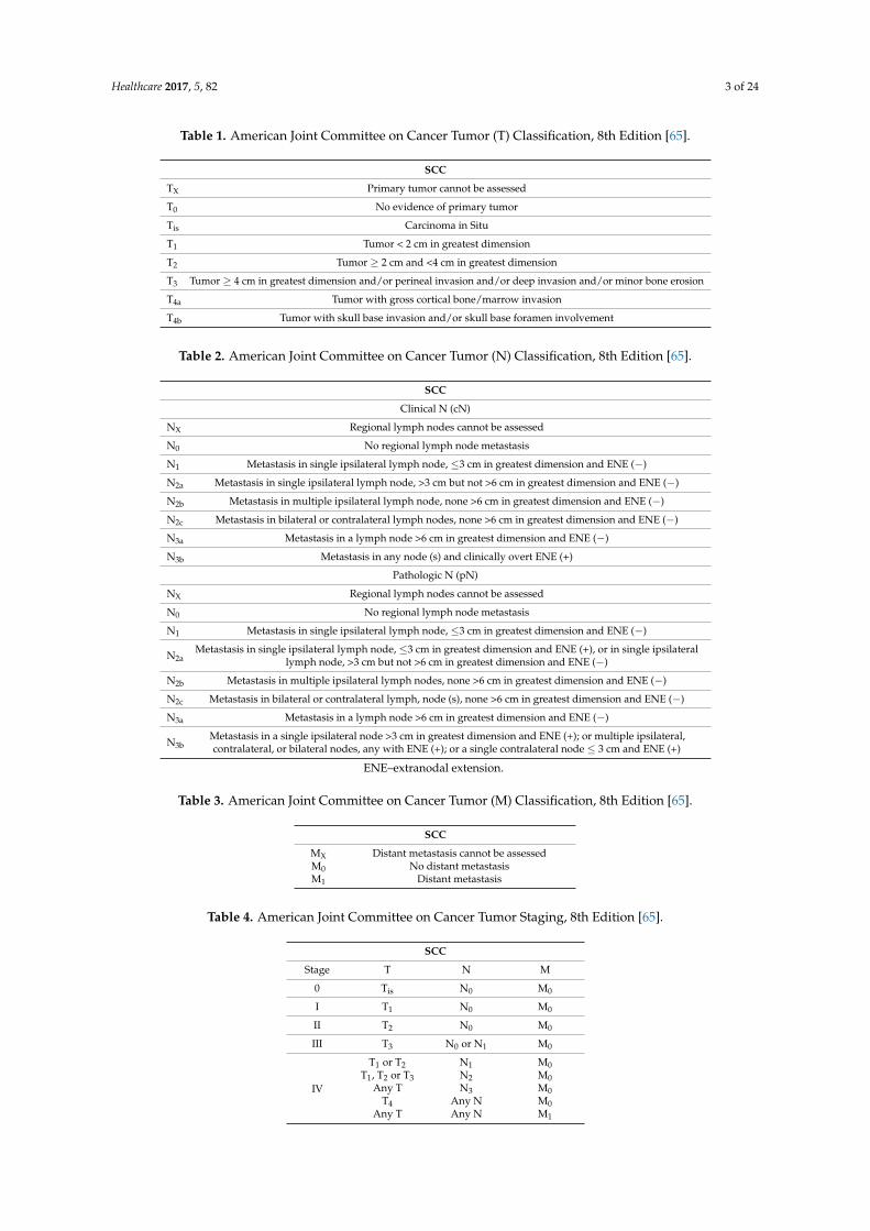
BCCs rarely require staging given their minimal potential for metastasis. However, cutaneous SCChas a 4% annual incidence of metastasis, so staging is vital to its management and treatment [59–61].The staging system of SCC is continuously being updated to meet current data. The staging systempublished in 2010 by the American Joint Committee on Cancers (AJCC) and the International UnionAgainst Cancer (UICC) had some limitations. For example, T3/T4 tumors were reserved for bonyinvasion, which was very rare at 0.3–3.0%, while the rest were T1/T2 [62]. Thus, 70–86% of pooroutcomes occurred in patients with T1/T2 tumors resulting in heterogeneous outcomes [62–64].Whereas, in Brigham and Women’s Hospital (BWH) tumor staging, 5% of tumors were high stage butthey accounted for 60% of poor outcomes, indicating superior homogeneity [62]. In 2017, the AJCCpublished the 8th edition of the Cancer Staging Manual. The staging of cutaneous SCC was revised toreflect recent evidence concerning high-risk clinicopathologic features to improve the overall stagingsystem, which can be seen in Tables 1–4 [63,65]. It should be noted that because the majority of SCCsof skin occur in the head and neck area, both of the AJCC’s 7th and 8th guidelines apply to tumorsfrom the head and neck [63–66].
Healthcare 2017, 5, 82 3 of 24
Table 1. American Joint Committee on Cancer Tumor (T) Classification, 8th Edition [65].
SCC
TX Primary tumor cannot be assessed
T0 No evidence of primary tumor
Tis Carcinoma in Situ
T1 Tumor < 2 cm in greatest dimension
T2 Tumor ≥ 2 cm and <4 cm in greatest dimension
T3 Tumor ≥ 4 cm in greatest dimension and/or perineal invasion and/or deep invasion and/or minor bone erosion
T4a Tumor with gross cortical bone/marrow invasion
T4b Tumor with skull base invasion and/or skull base foramen involvement
Table 2. American Joint Committee on Cancer Tumor (N) Classification, 8th Edition [65].
SCC
Clinical N (cN)
NX Regional lymph nodes cannot be assessed
N0 No regional lymph node metastasis
N1 Metastasis in single ipsilateral lymph node, ≤3 cm in greatest dimension and ENE (−)
N2a Metastasis in single ipsilateral lymph node, >3 cm but not >6 cm in greatest dimension and ENE (−)
N2b Metastasis in multiple ipsilateral lymph node, none >6 cm in greatest dimension and ENE (−)
N2c Metastasis in bilateral or contralateral lymph nodes, none >6 cm in greatest dimension and ENE (−)
N3a Metastasis in a lymph node >6 cm in greatest dimension and ENE (−)
N3b Metastasis in any node (s) and clinically overt ENE (+)
Pathologic N (pN)
NX Regional lymph nodes cannot be assessed
N0 No regional lymph node metastasis
N1 Metastasis in single ipsilateral lymph node, ≤3 cm in greatest dimension and ENE (−)
N2aMetastasis in single ipsilateral lymph node, ≤3 cm in greatest dimension and ENE (+), or in single ipsilateral
lymph node, >3 cm but not >6 cm in greatest dimension and ENE (−)
N2b Metastasis in multiple ipsilateral lymph nodes, none >6 cm in greatest dimension and ENE (−)
N2c Metastasis in bilateral or contralateral lymph, node (s), none >6 cm in greatest dimension and ENE (−)
N3a Metastasis in a lymph node >6 cm in greatest dimension and ENE (−)
N3bMetastasis in a single ipsilateral node >3 cm in greatest dimension and ENE (+); or multiple ipsilateral,contralateral, or bilateral nodes, any with ENE (+); or a single contralateral node ≤ 3 cm and ENE (+)
ENE–extranodal extension.
Table 3. American Joint Committee on Cancer Tumor (M) Classification, 8th Edition [65].
SCC
MX Distant metastasis cannot be assessedM0 No distant metastasisM1 Distant metastasis
Table 4. American Joint Committee on Cancer Tumor Staging, 8th Edition [65].
SCC
Stage T N M
0 Tis N0 M0
I T1 N0 M0
II T2 N0 M0
III T3 N0 or N1 M0
IV
T1 or T2 N1 M0T1, T2 or T3 N2 M0
Any T N3 M0T4 Any N M0
Any T Any N M1
Healthcare 2017, 5, 82 4 of 24
3. Prevention of NMSC
The incidence of NMSC has been increasing worldwide [1–4,67]. The rising incidence hasbeen associated with several factors, including raised awareness in the general population andamong physicians, an increased number of patients undergoing surgical treatment with confirmedhistopathology instead of cryotherapy or electrodessication, improved registration, an agingpopulation, and increased exposure to ultraviolent (UV) radiation [13–15,68]. These have led tothe development of various skin cancer prevention measures and programs worldwide, highlightingthe importance of skin cancer prevention.
Prevention strategies are divided into primary and secondary strategies. Primary preventionmeasures target behavioral changes to reduce sun exposure, reinforce the use of adequatesun protection and discourage intensive tanning, while the secondary measures facilitate earlydetection [4,69]. Public education is one of the key aspects of primary prevention, which is aimedto increase group-oriented awareness. It is known that among teen girls, correlating sun damagewith premature wrinkling is an effective way to raise awareness [10,70]. In teen boys, emphasizingthe protection of painful burns rather than the long-term risks of NMSC is more efficient [10,70].Introduction of sun protection policy guidelines for schools and implementation of mass mediacampaigns of public education may also have significant impact on behavioral modifications [71,72].Increasing evidence suggests that some pharmacologic agents may also be effective in prevention ofNMSC. There is now strong evidence that vitamin D plays an important role in photocarcinogenesisand progression of NMSC. Hence, pharmacologic modulation by vitamin D, 1,25(OH)2D3 and itsanalogs represents a promising new strategy for prevention of NMSC [73]. Nicotinamide (Vitamin B3)is another agent that has been proven to decrease the incidence of NMSC, most likely by its abilityto enhance DNA repair, modulate the inflammation produced by UVR and reduce UV inducedimmunosuppression [74,75]. Preventing and treating actinic keratosis (AKs) with minimal side effectsand a low cost may be a promising alternative for skin cancer prevention in immunosuppressedpatients [76].
Secondary prevention strategies include screening of high-risk populations, skin self-examinationand physician surveillance, which aim to reduce morbidity and mortality by detecting cancer in itsearly stages [77].
4. Diagnosis and Types of Biopsy Techniques
The gold standard for diagnosing NMSC includes a thorough physical examination followedby conventional biopsy of the lesion for histopathologic examination [78,79]. A thorough lymphnode exam is essential for diagnosis of SCC due to the risk for metastasis. Patients at high-risk fordeveloping NMSC require special attention, particularly for individuals who work outside and thushave chronic exposure to UV radiation, as well as immunocompromised individuals. Some features,such as vaguely margined, white indurated, scar-like plaques, or the recently described “candle wax”sign may indicate an aggressive form of BCC [80–83].
When a suspicious lesion is identified, the diagnosis needs be confirmed with histopathologicanalysis, which requires invasive intervention to obtain a tissue sample. There are several non-invasivemedical technologies available that may be used to decide what lesions need to be biopsied asto potentially avoid unnecessary invasive procedures. Such technology includes: dermoscopy,confocal microscopy, cross-polarized light and fluorescence photography, as well as optical coherencetomography with high-frequency ultrasound [78]. There are also newer options, such as multiphotonmicroscopy and Raman spectroscopy that are emerging as state-of-the-art tools [78]. These adjunctscreening tools may be used to assess skin lesions to find characteristic features of NMSC beforeproceeding with formal biopsy. The use of dermoscopy in the US is increasing with a recent surveyshowing up to 81% of US dermatologists using dermoscopy [84]. While it was mainly used fordifferentiation of melanocytic lesions, it is now widely utilized to screen superficial BCC and SCCsas well [85–87]. Most of these diagnostic modalities require additional training, which is one of the
Healthcare 2017, 5, 82 5 of 24
limiting factors for their use in clinic. Digitally stained multimodal confocal mosaic imaging wasrecently compared with standard frozen histopathologic specimens, thus showing 90% sensitivity and79% specificity before training and 99% sensitivity and 93% specificity after training [88]. The longerprocessing time and the lack of reimbursement by the insurance companies are other limitingfactors [89]. However, with the conduction of more research to justify their use, assignment ofreimbursement values, development of more training courses for dermatologists, incorporating theminto residency programs and continued improvement in speed and accuracy of these techniqueswill allow physicians to overcome some of the barriers and potentially increase the role of thesetechnologies in the management of NMSC [89].
4.1. Biopsy Techniques
Despite tremendous advances in medical care and the shift to evidence-based medicine overthe past several decades, there are no specific diagnostic guidelines for biopsy techniques that arespecific to the lesion type or characteristics for one of the most common cancers worldwide. Thereare several types of biopsy techniques that are commonly used in the diagnosis of NMSC, yet notechnique has been deemed the gold standard. These techniques include: excisional, incisional, shave,and punch biopsy. Although punch and shave biopsies are the most commonly performed techniquesfor initial sampling of NMSC [90], no well-powered studies in current literature or guidelines exist tohelp the physician decide which biopsy technique should be employed. Most studies are retrospectivereviews and are not able to provide evidence-based proof that one technique is superior over the others,but rather demonstrate that each technique has advantages and disadvantages. It has been shownthat although the intent of a shave biopsy is to obtain a diagnostic sample, 15–40% of patients whounderwent secondary excisions for BCC had no residual cancer [91–94]. While some argue that those15–40% of patients may have undergone unnecessary secondary procedures, final histopathologicexaminations may be inaccurate. Kimyai-Asadi and Goldberg showed that bread-loafing at 4 mmintervals of elliptical excision specimens from facial basal cell carcinomas excised with 2 mm surgicalmargins is only 44% sensitive in detecting residual tumor at the surgical margins [95]. Another studydemonstrated that 33% of cases with the initial biopsy results of actinic keratosis had additionalfindings on deep sections, including 13% SCC, 4% BCC, and 3% invasive SCC [96]. In the same context,a recent study brings up another critical point while questioning the value of reporting margin statusin biopsies of NMSC. Based on their findings, 23% of cases with negative margins on initial biopsydemonstrated positive margins upon deeper-level examination [97] to further highlight the significanceof proper interpretation of biopsy results when formulating a treatment plan.
With technology emerging in all aspects of the healthcare field, many look for new solutions toNMSC diagnosis. A recent study suggests that the reflectance confocal microscopy (RCM) may havea comparable accuracy to punch biopsy with regards to diagnosing and subtyping BCC [96,98,99].It may also have a role in monitoring nonsurgical skin cancer therapies or assessment of tumor marginsprior to surgery or intraoperatively [100–103].
In summary, there are currently no guidelines available to direct the biopsy techniques for thediagnosis of NMSCs. As a result, the clinician may decide what technique is the most appropriatein each scenario by considering not only the characteristics of the lesions, but also the intent of thebiopsy. We are however hopeful that this issue will be addressed soon as the American Academy ofDermatology plans on releasing such guidelines in 2017 [104,105].
4.2. Low-Risk versus High-Risk Lesions
The current treatment of NMSC is driven by a risk assessment of the lesions. These assessmentsare based on the recurrence rates for BCC and recurrence and metastasis for SCC. According toNCCN 2017 guidelines, NMSC are classified as low- and high-risk lesions [106]. The lesions thatare located on the trunk and extremities, excluding pretibia, hands, feet, nails, ankles, and are lessthan 20 mm in size are considered low-risk, whereas greater than 20 mm lesions are considered
Healthcare 2017, 5, 82 6 of 24
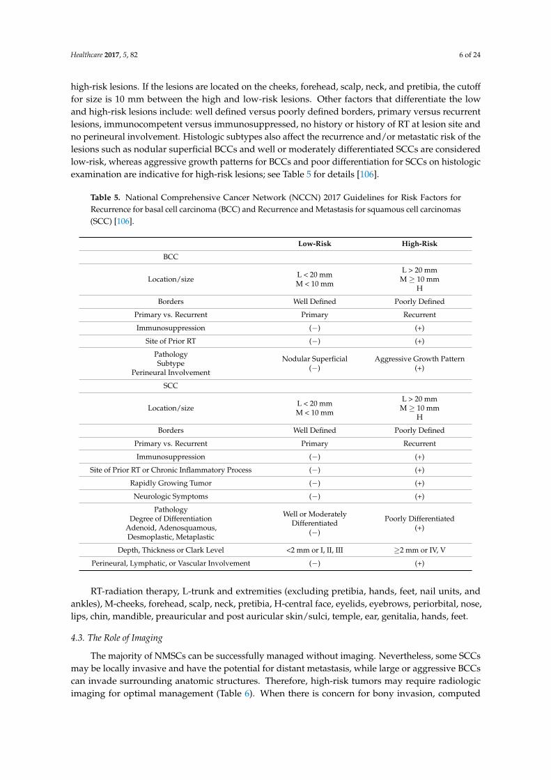
high-risk lesions. If the lesions are located on the cheeks, forehead, scalp, neck, and pretibia, the cutofffor size is 10 mm between the high and low-risk lesions. Other factors that differentiate the lowand high-risk lesions include: well defined versus poorly defined borders, primary versus recurrentlesions, immunocompetent versus immunosuppressed, no history or history of RT at lesion site andno perineural involvement. Histologic subtypes also affect the recurrence and/or metastatic risk of thelesions such as nodular superficial BCCs and well or moderately differentiated SCCs are consideredlow-risk, whereas aggressive growth patterns for BCCs and poor differentiation for SCCs on histologicexamination are indicative for high-risk lesions; see Table 5 for details [106].
Table 5. National Comprehensive Cancer Network (NCCN) 2017 Guidelines for Risk Factors forRecurrence for basal cell carcinoma (BCC) and Recurrence and Metastasis for squamous cell carcinomas(SCC) [106].
Low-Risk High-Risk
BCC
Location/size L < 20 mmM < 10 mm
L > 20 mmM ≥ 10 mm
H
Borders Well Defined Poorly Defined
Primary vs. Recurrent Primary Recurrent
Immunosuppression (−) (+)
Site of Prior RT (−) (+)
PathologySubtype
Perineural Involvement
Nodular Superficial(−)
Aggressive Growth Pattern(+)
SCC
Location/size L < 20 mmM < 10 mm
L > 20 mmM ≥ 10 mm
H
Borders Well Defined Poorly Defined
Primary vs. Recurrent Primary Recurrent
Immunosuppression (−) (+)
Site of Prior RT or Chronic Inflammatory Process (−) (+)
Rapidly Growing Tumor (−) (+)
Neurologic Symptoms (−) (+)
PathologyDegree of Differentiation
Adenoid, Adenosquamous,Desmoplastic, Metaplastic
Well or ModeratelyDifferentiated
(−)
Poorly Differentiated(+)
Depth, Thickness or Clark Level <2 mm or I, II, III ≥2 mm or IV, V
Perineural, Lymphatic, or Vascular Involvement (−) (+)
RT-radiation therapy, L-trunk and extremities (excluding pretibia, hands, feet, nail units, andankles), M-cheeks, forehead, scalp, neck, pretibia, H-central face, eyelids, eyebrows, periorbital, nose,lips, chin, mandible, preauricular and post auricular skin/sulci, temple, ear, genitalia, hands, feet.
4.3. The Role of Imaging
The majority of NMSCs can be successfully managed without imaging. Nevertheless, some SCCsmay be locally invasive and have the potential for distant metastasis, while large or aggressive BCCscan invade surrounding anatomic structures. Therefore, high-risk tumors may require radiologicimaging for optimal management (Table 6). When there is concern for bony invasion, computed
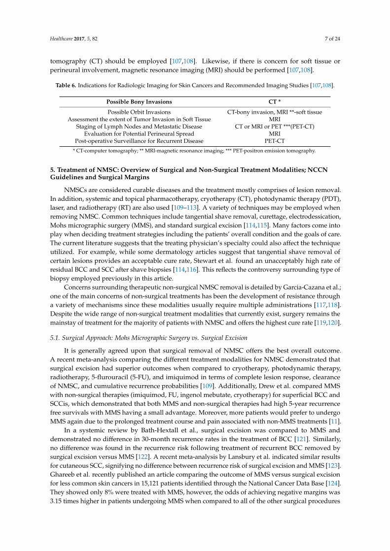
Healthcare 2017, 5, 82 7 of 24
tomography (CT) should be employed [107,108]. Likewise, if there is concern for soft tissue orperineural involvement, magnetic resonance imaging (MRI) should be performed [107,108].
Table 6. Indications for Radiologic Imaging for Skin Cancers and Recommended Imaging Studies [107,108].
Possible Bony Invasions CT *
Possible Orbit Invasions CT-bony invasion, MRI **-soft tissueAssessment the extent of Tumor Invasion in Soft Tissue MRI
Staging of Lymph Nodes and Metastatic Disease CT or MRI or PET ***(PET-CT)Evaluation for Potential Perineural Spread MRI
Post-operative Surveillance for Recurrent Disease PET-CT
* CT-computer tomography; ** MRI-magnetic resonance imaging; *** PET-positron emission tomography.
5. Treatment of NMSC: Overview of Surgical and Non-Surgical Treatment Modalities; NCCNGuidelines and Surgical Margins
NMSCs are considered curable diseases and the treatment mostly comprises of lesion removal.In addition, systemic and topical pharmacotherapy, cryotherapy (CT), photodynamic therapy (PDT),laser, and radiotherapy (RT) are also used [109–113]. A variety of techniques may be employed whenremoving NMSC. Common techniques include tangential shave removal, curettage, electrodessication,Mohs micrographic surgery (MMS), and standard surgical excision [114,115]. Many factors come intoplay when deciding treatment strategies including the patients’ overall condition and the goals of care.The current literature suggests that the treating physician’s specialty could also affect the techniqueutilized. For example, while some dermatology articles suggest that tangential shave removal ofcertain lesions provides an acceptable cure rate, Stewart et al. found an unacceptably high rate ofresidual BCC and SCC after shave biopsies [114,116]. This reflects the controversy surrounding type ofbiopsy employed previously in this article.
Concerns surrounding therapeutic non-surgical NMSC removal is detailed by Garcia-Cazana et al.;one of the main concerns of non-surgical treatments has been the development of resistance througha variety of mechanisms since these modalities usually require multiple administrations [117,118].Despite the wide range of non-surgical treatment modalities that currently exist, surgery remains themainstay of treatment for the majority of patients with NMSC and offers the highest cure rate [119,120].
5.1. Surgical Approach: Mohs Micrographic Surgery vs. Surgical Excision
It is generally agreed upon that surgical removal of NMSC offers the best overall outcome.A recent meta-analysis comparing the different treatment modalities for NMSC demonstrated thatsurgical excision had superior outcomes when compared to cryotherapy, photodynamic therapy,radiotherapy, 5-flurouracil (5-FU), and imiquimod in terms of complete lesion response, clearanceof NMSC, and cumulative recurrence probabilities [109]. Additionally, Drew et al. compared MMSwith non-surgical therapies (imiquimod, FU, ingenol mebutate, cryotherapy) for superficial BCC andSCCis, which demonstrated that both MMS and non-surgical therapies had high 5-year recurrencefree survivals with MMS having a small advantage. Moreover, more patients would prefer to undergoMMS again due to the prolonged treatment course and pain associated with non-MMS treatments [11].
In a systemic review by Bath-Hextall et al., surgical excision was compared to MMS anddemonstrated no difference in 30-month recurrence rates in the treatment of BCC [121]. Similarly,no difference was found in the recurrence risk following treatment of recurrent BCC removed bysurgical excision versus MMS [122]. A recent meta-analysis by Lansbury et al. indicated similar resultsfor cutaneous SCC, signifying no difference between recurrence risk of surgical excision and MMS [123].Ghareeb et al. recently published an article comparing the outcome of MMS versus surgical excisionfor less common skin cancers in 15,121 patients identified through the National Cancer Data Base [124].They showed only 8% were treated with MMS, however, the odds of achieving negative margins was3.15 times higher in patients undergoing MMS when compared to all of the other surgical procedures
Healthcare 2017, 5, 82 8 of 24
combined when controlled for tumor size, location and histology. When MMS was compared with fourdifferent surgical techniques individually, it was still superior to primary excision and biopsy followedby narrow excision, however, no statistically significant difference was identified when comparingMMS with wide excision and re-excision with >1 cm margins [124].
The anatomic location of the tumor may pose unique challenges in its surgical excision. It shouldbe noted that head and neck skin cancers are particularly challenging to treat given the difficulty indetermining the amount of tissue that should be excised to provide adequate cancer-free marginswith the best functional and cosmetic outcome. Moncrief et al. showed that the common practice ofintraoperative frozen section analysis (IFSA) of margins had unacceptably high false negatives for headand neck BCC (28.7%) and SCC (27.5%). This resulted in the abandonment of IFSA in their practice andsupport for intraoperative MMS, even in advanced cases [125]. In light of this, Walker et al. investigatedthe utility of quenched activity-based probe imaging to discriminate cancerous versus normal skintissue for NMSC [126]. They validated the activation of the probe with hematoxylin-eosin-confirmedcancerous tissue with 0.989 and 0.894 sensitivity and specificity, respectively. However, further studiesare needed to confirm if this new, rapid, and easy to interpret technology would provide actualcost-effective increased cure rates by reducing re-excision and recurrence rates [126].
Although surgical excision remains the most commonly performed procedure in the treatmentof NMSC, adequate surgical margins have not yet been defined. In an attempt to address this issue,Shel et al. suggested surgical margins for low and high-risk BCC and SCC based on a retrospectivereview of margins on 495 lesions as defined by MMS [127]. They concluded that lesions located on theface are best treated with MMS but in situations when standard surgical excision is the only option,a minimum 5 mm margin for low-risk lesions and about a 1 cm margin for high-risk lesions arerequired to completely excise the lesions in 95% of cases [127].
NCCN currently recommends standard excision as a primary treatment choice for low-risk BCCsthat can be excised with 4 mm clinical margins as well as low-risk SCCs that can be excised with4–6 mm clinical margins [106]. In case of positive margins, MMS or resection with complete marginassessment or standard re-excision in the trunk and extremity area are recommended [106]. In high-riskBCCs and local high-risk SCCs, MMS or resection with a complete margin assessment is recommended.Standard excision with wider surgical margins with linear or delayed repair may also be performedfor high-risk lesions; however, there are no specific recommendations for margin size, and if marginsare positive, then MMS or resection with complete margin assessment can be performed [106].
5.2. Curettage and Electrodessication (C&E), Cryosurgery
Curettage and electrodessication (C&E) is a commonly performed procedure to remove variousskin lesions by scraping off the lesion with a curate followed by cauterization. It can be used forlow-risk BCC and SCC, but it is not recommended for high-risk lesions due to an unacceptably highrecurrence rate of up to 27% [128–133]. The overall efficacy of the C&E has been shown to be dependenton physicians’ experience as well. Silverman et al. showed decreased average recurrence rate of BCCsfrom 17.0% to 7.3%, when comparing the rates following procedures performed in the years of1955–1963 and 1973–1982 by the same group, which was the attributed to physicians’ experience,specifically increased thoroughness in the technique [134]. Curettage alone may be used for selectedlow-risk lesions and can have similar cure rates (96% 5-year cure rate) but better healing compared toC&E [130]. Overall, C&E is considered a reasonable treatment choice for small low-risk NMSCs. It isalso the least expensive and fastest method among all treatment methods of NMSCs [130,131].
Cryotherapy may also be considered for small, low-risk BCCs and SCCs, and can be combinedwith curettage, but it is not recommended for high-risk lesions [132,133]. It is associated with low cost,yet given the high overall recurrence rate of 7.5% following the treatment of primary lesions and 13%for recurrent lesions, it is rarely utilized [135,136].
Healthcare 2017, 5, 82 9 of 24
According to current NCCN guidelines, C&E is recommended as a primary treatment choicefor low-risk BCCs and local low-risk SCCs excluding terminal hair-bearing areas, such as the scalp,pubic and axillary region, beard area in men, and if adipose tissue is reached [106].
5.3. Photodynamic Therapy (PDT)
Photodynamic therapy (PDT) utilizes light, oxygen, and photosensitizers. 5-Aminolevulinicacid (5-ALA) and methyl 5-aminolevulinate (MAL) are commonly used pro-drugs for PDT, whichare administered locally or systemically and convert to protoporphyrin IX (PPIX), which is thenfurther converted to heme inside the cells. However, the PPIX conversion to heme is usually aslower process which results in its accumulation in cancer cells. Upon light absorption, the PPIX thenundergoes excitation, resulting in production of oxygen free radicals, ultimately leading to cancercell death. [137–160]. PDT has shown to be effective in treating superficial BCC, SCCis, and actinickeratosis (AK) with excellent cosmesis, though, it is currently not recommended for invasive SCC andaggressive BCC subtypes such as basosquamous or morphoeic infiltrating types [110,111]. A recentretrospective review demonstrated excellent response of superficial BCC to MAL-PDT and thick BCCto meta-tetrahydroxyphenylchlorin-PDT (mTHPC-PDT), with a 95% complete response rate after oneround of PDT, which was maintained at 3-year follow-up and slightly decreased to 92% at 5-yearfollow-up [161]. PDT has also proven effective in reducing the incidence of AKs and SCCis in patientswith organ transplant on long-term immunosuppressive therapy [110,111]. A randomized, multi-centerstudy of 81 organ transplant recipients with 881 NMSCs (mainly AKs) as compared MAL-PDT tostandard treatment (curettage, cryotherapy, surgery, or laser) and demonstrated significantly fewernew AKs in the MAL-PDT treated area at 15 months post-treatment [143]. These results indicatethat pre-treatment of “field cancerization” in immunosuppressed patients with PDT is a promisingalternative in terms of skin cancer prevention in immunosuppressed patients.
Significant research is being conducted to enhance the efficacy of PDT to treat more advancedstages of skin cancers. A recently developed microneedle-assisted PDT is aimed to delivertargeted therapy, which would allow treatment of deeper lesions [112,144]. In a small case series,the combination of a 630 nm laser with intralesional 5-aminolevulinic acid allowed treatment ofdeeper lesions [145]. A recent clinical trial studying the administration of 5-FU as a neoadjuvant for5-aminolevulinic acid-PDT (ALA-PDT) has shown increased tumor selective protoporphyrin IX levelsand enhanced cell death in a mouse mode [146]. Another clinical trial has compared the efficacy ofALA-PDT with surgery versus surgery alone for SCC of all stages, demonstrating a lower recurrencerisk in the experimental group (16.6% versus 30.0%, p < 0.005) [147]. New agents are also developedwith higher photosensitizing efficacy and better pharmacokinetics to provide higher tumor uptakeand potentially long-term tumor cure. Patel et al. developed a near infrared bacteriochlorin 3 and thecorresponding stereoisomers to be used in PDT with higher photosensitizing efficacy and limited skinphototoxicity when compared to porphyrin-based PDT [148].
PDT has also been studied as a potential modality for the prevention of NMSC. Studiesdemonstrated an equal preventative effect of daylight-PDT (dl-PDT) and conventional-PDT (c-PDT)in high-risk patients; however, all of the patients reported a burning sensation and pain duringc-PDT while dl-PDT was reported to be almost pain free, which is why dl-PDT is preferred [149–151].The authors postulate that PDT can induce premature senescence and kill senescent cells inducedby itself [151]. A large retrospective review in a high volume dermatologic clinic investigated thedevelopment of SCC at one year following the PDT treatment of 1404 patients with AKs and 45with NMSCs [152]. They found that 11% of patients developed SCC, while the rest remained SCCfree at 1-year follow-up. Factors associated with developing SCC were older age, SCC history,Fitzpatrick skin-type 1, and sixty-minute or less incubation time, which is the time period between theadministration of photosensitizing agent and exposure of the skin area to the light source. Therefore,it was concluded that PDT may be more effective in younger patients and with greater than 60 min ofincubation time [152]. In addition, PDT has a tolerable side effect profile and low cost, which makes
Healthcare 2017, 5, 82 10 of 24
it more appealing in the treatment and prevention of skin cancers [153]. The main side effect is pain,which increases in intensity with the number of sessions and if the lesions are located in the head andneck area [153].
5.4. Laser Therapy
Treatment of NMSC with lasers causes light absorption by blood vessels of the targeted area,resulting in thermal distraction that then leads to tumor regression [154,155]. This targeted vascularphotothermal destruction preserves the normal surrounding area and with treatment of dermatologicconditions, can lead to excellent cosmesis [155,156].
There are four major laser types used in the treatment of skin cancers: solid-state, diode, dye,and gas lasers [157]. A recent review article by Soleymani et al. discusses the different types of lasertreatments in detail [158]. This study demonstrated that vascular targeting pulse dye lasers (PDL) haveshown promising results in different studies with a complete clinical response in up to 95% of patients;nevertheless, poor results were also seen. Most incomplete responders were large tumors. Lowerenergy and small spot size also contributed to a poor cure rate. The 595 nm wavelength lasers weresuperior to the 585 nm wavelength lasers in terms of treating BCC. They concluded that the 595 nmwavelength vascular-targeted lasers had promising clinical efficacy in the treatment of BCC; however,data for consistent long-term outcome is not yet available. CO2 lasers allow very thin tissue ablationin the range of 20 µm with each pass [159–163]. Several studies have shown a different efficacy ofCO2 lasers for treatment of BCC with 85–100% cure rates, excellent cosmetic outcomes, and minimalcomplications [158]. Lasers have been less commonly used and studied for the treatment of SCC,with only a handful of studies demonstrating the efficacy of CO2 lasers in treatment of SCCis. In thelargest study of CO2 laser therapy, 44 of the 48 patients with SCCis were treated with CO2 lasers andhad a total clearance rate of 97.7% and a recurrence rate of 6.8% at a mean 18-month follow-up [164].
Laser therapies have also been used in combination with PDT and the results have been mixed.One study investigated the efficacy of MAL-PDT with and without ablative (CO2) laser for thetreatment of microinvasive SCC [165]. They found a greater efficacy rate when treated with thePDT-laser combination when compared to PDT alone at three months (84.2% versus 52.4%, p-0.03),and the differences in efficacy remained significant at 24 months. They also found a significantlylower recurrence rate in the PDT-laser group compared to the PDT alone group (12.5% versus 63.6%,p-0.006) [165]. In a case series, all patients with solitary superficial BCC treated with a combination offractional CO2 laser and PDT achieved pathologically confirmed resolution of BCC with no seriousadverse effects [152]. PDT combined with 2940 nm ER:YAG (solid-state) laser treatment has also shownimproved relative efficacy in a few studies, demonstrating a lower recurrence risk of BCC and Bowendisease as compared to laser alone therapy, however, the esthetic outcomes were superior in laser alonetreatment patients and the patients seem to prefer it due to its simplicity [157,166–169] Other studiesdispute the benefits of PDT-laser combined therapy. One study compared the efficacy of PDT combinedwith pulse dye laser (PDL) with PDT alone for the treatment of 62 BCCs in 15 patients [170] and foundno difference in complete tumor regression between the two groups. Additionally, this study foundthat the recurrence rate was higher in the combined treatment group [170].
Diode lasers, also called near-infrared lasers, function through temperature increases of targettissue and kill tumor cells directly. They have shown some efficacy in the treatment of small superficialSCC [157].
In summary, laser therapy has demonstrated promising results in the treatment of superficialNMSCs; however, the overall efficacy remains inferior compared to more traditional treatmentmodalities. It appears that the most beneficial aspects of laser treatment are the minimal side effectsand excellent cosmetic outcome. While its role may be expanded in the future for the treatment ofNMSCs, its use is currently limited. More long-term follow-up studies are needed to validate theirefficacy and determine possible indications of laser therapy in the management of NMSCs.
Healthcare 2017, 5, 82 11 of 24
5.5. Radiation Therapy
Although radiation therapy has shown control rates of 75–100% in early stage BCC and SCC,the role of radiotherapy has significantly decreased in the treatment of NMSCs with the emergenceof MMS [113,171–175]. Now, it is mainly recommended as a primary treatment method if surgery iscontraindicated, if lesions are located in cosmetically sensitive areas, or as an adjunct to surgery inpatients with advanced disease [113].
There are several techniques of radiotherapy, each having advantages and disadvantages. A recentsurvey study completed by 16 members of American Brachytherapy Society indicated that the majorityof them prefer brachytherapy over external beam radiation therapy given the shorter treatment course,conformity of treatment for irregular or curved targets and shallow dose deposition [176]. Manyof them routinely use ultrasound to estimate the depth and lateral extension of the lesion beforeinitiating treatment, which has shown to be helpful for more accurate treatment planning [176,177].The advantage of superficial radiotherapy (SRT) is that the maximum dose is at skin’s surface, and thereis less penetration through eye shields, thus making it easier to use around the eye [178]. It has shownto provide 93–100% tumor control depending on the lesion size, whereas electron beam radiationprovided 72–88% tumor control in the same study [179]. Another study demonstrated a recurrencerisk of 5% at five years in patients with low-risk NMSCs that are treated with SRT [180]. Electronbeam radiotherapy (EBRT) has the advantage of treating superficial lesions without causing significantdamage to deeper structures and may be useful when treating skin cancers over bony prominences orcartilaginous structures [181]. Interstitial brachytherapy is another subtype that involves the insertionof a radioactive catheter within the tumor bed and is used to treat skin cancers in difficult areas suchas eyelids [182].
Some of the common complications of radiation therapy include alopecia, pigmentary changes,telangiectasias, fibrosis, atrophy, buccal mucositis, gingivitis, tooth loss, and a loss of salivary glandfunction. In addition, more severe complications such as soft tissue or bone necrosis, cataracts,conjunctival scarring, or eyelid deformity may occur [113,183].
NCCN currently recommends radiotherapy as a primary treatment modality for non-surgicalcandidates in the treatment of low- and high-risk BCCs and local low- and high-risk SCCs (in high-riskSCCs, RT may be supplemented with chemotherapy) [106]. However, it is often reserved for patientsover 60 years of age due to concerns for long-term sequelae. Conversely, it is contraindicated inpatients with genetic conditions predisposing to skin cancer and connective tissue diseases [106]. RT isalso recommended in high-risk lesions in cases of positive margins after surgical excision and/or if anegative margin is unachievable with MMS. It may also be considered if there is extensive perineuralor large nerve involvement [106]. NCCN recommends RT with or without chemotherapy in patientswith SCCs with positive lymph nodes on FNA or core biopsy who are not surgical candidates orpossible adjuvant therapy following surgical resection of lesions [107].
5.6. Chemotherapy and Immunotherapy
Systemic chemotherapy has an important role in the management of advanced NMSC. The term“advanced NMSC” usually refers to metastatic disease and/or inoperable lesions due to sizeand location that would otherwise cause unacceptable functional impairment if treated withexcision [21,113]. Platinum based preparations such as cisplatin, are the most commonly usedchemotherapeutic agents for NMSC; however, other agents, including cyclophosphamide, bleomycin,doxorubicin, methotrexate, and 5-FU, may also be used [23–31,121,122,184–189]. A recent publicationclaims that monotherapy of patients with stage I and II lip SCC via superficial temporal arteryadministration of bleomycin derivative peplomycin is a highly effective treatment method for achievinga cure in 70.8% of patients [113]. It was reported though, that 12.5% of patients developed interstitialpneumonia, 12.5% had occurrence of metastasis, and 16.7% had local recurrence [113].
Certain immune modulators and monoclonal antibodies have demonstrated promising results.In this concept, epidermal growth factor receptor (EGFR) appears to be of particular interest. EGFR is
Healthcare 2017, 5, 82 12 of 24
involved in the pathogenesis of SCC [190], hence its inhibitors, such as cetuximab and panitumumab,may be used in the treatment of SCC [23]. In one study, cetuximab has shown 69% efficacy in diseasecontrol rate at 6 weeks when administered as a single agent in patients with advanced SCC [191].In another study, 95% of patients became operable following administration of cetuximab combinedwith a chemotherapeutic agent [192]. Cetuximab’s reasonable tolerability makes it a potential agent forneo-adjuvant therapy [23]. In contrast, panitumumab did not show a superior efficacy in combinationwith chemotherapy over chemotherapy alone in CONSERT-1 phase II trial or in combination withradiotherapy over chemotherapy alone in CONSERT-2 phase II trial [193,194]. In both studies, grade 3,grade 4, and serious adverse effects were more common in the group receiving panitumumab, whichmakes the future of this agent questionable in the treatment of NMSCs.
NCCN currently recommends chemotherapy as a possible supplement to radiation therapy inlocal, high-risk SCCs for patients who are not surgical candidates [106]. It is also recommended as aprimary treatment concurrent with RT in patients with FNA/core biopsy positive lymph nodes whohave inoperable diseases [106].
5.7. The Hedgehog Pathway Inhibitors
The ability of some antifungal agents to inhibit the Hedgehog (HH) pathway has also beenexplored in the treatment of NMSC. While itraconazole has shown some efficacy in exploratory phaseII trial, the toxicity remains a major limiting factor in terms of its use in clinical practice. Posaconazoleis another antifungal with better safety profile, though further validation is still needed [23,195].
Vismodegib is a newer agent that acts as an inhibitor of the sonic hedgehog (SHH) pathway andwas approved for adult patients with advanced BCC in 2012 [21,196]. Despite some controversialreports of Vismodegib being associated with development of new SCC, it still appears to have greatinterest among physicians; however, its significant adverse effects pose a challenge [21,197–199].Sonidegib is another SHH inhibitor recently approved for treatment of advanced BCC in the UnitedStates of America (USA) with slightly better efficacy than Vismodegib [200]. They are both currentlyrecommended by NCCN to be considered in high-risk BCCs if positive margins are present followingsurgical excision and/or negative margins are not achievable with MMS [106].
5.8. Topical Agents
Topical agents may be used to treat NMSC in instances when patients are not candidates forstandard approach. Factors that may favor the use of topical pharmacotherapy include extensive,multifocal or multiple tumors, indistinct lesion boundaries, localization in cosmetically sensitive ordifficult to treat areas, and a history of hypertrophic scars and keloid [201]. There are several topicalagents that are currently used in the treatment of NMSC, some of which are approved by FDA. Topical5-FU has been approved for treatment of AK and superficial BCC for many years, although its use fortreatment of SCCis is off-label [202–204]. Some studies suggest a 90–96% cure rate of AK and superficialBCC with topical 5-FU [205,206]. Imiquimod is another agent that is approved by FDA for treatment ofAK and superficial BCC, while diclofenac and ingenol mebutate are FDA approved for AK only [202].Currently, there are many other topical agents still in the investigational stage, including: resiquimod,piroxicam, calcium dobesilate, and potassium dobesilate containing formulations, betulinin acid andtopical retinoids that may have some efficacy for the treatment of skin cancers [202].
6. Utility of Sentinel Lymph Node Biopsy (SLNB) and Regional Lymphadenectomy
Sentinel lymph node biopsy (SLNB) is usually not considered for BCC given the very rare chanceof metastasis. In contrast, SLNB may play a significant role in the management of SCC. An associationof positive SNL and poor prognosis of cutaneous SCC has been previously demonstrated in severalstudies, showing a 96% 5-year survival with no regional lymph node involvement [206–215]. The 5-yearsurvival decreases to 72% with adequate treatment, and 25–35% with no treatment [207–216]. A recentstudy showed that patients with no micrometastasis to SNL developed no local and distant disease at
Healthcare 2017, 5, 82 13 of 24
an average follow-up period of 27.5 months [217]. Another recently published report indicated thatpositive results were seen in 13.4% of 364 SNLBs in patients with cutaneous SCC, which is similar to thatof melanoma [218]. They also found that positive SNL was associated with poor prognosis; hence theirfindings further support the utility of SLNB in the management of SCC. However, while it appears thatconsensus exists in terms of positive rate of SNL and poor prognosis associated with it [219,220], manystill question the utility of SNLB due to inadequate evidence that supports it actually improves theprognosis the disease. Maryuma et al. showed that despite an 18.4% positive rate of micrometastasisto SNL in patients who underwent SNLB, there was no difference in terms of metastasis-free anddisease-specific survival rates between the groups who did and did not undergo SNLB, regardless ofT-staging [221]. Silberstein et al. investigated the rate of metastasis to lymph nodes in 572 patientswith 725 head and neck cutaneous T1 and T2 SCC with 1.09% and 5.46% positive results, respectively,and recommended no SNLB for clinically N0 patients based on these findings [222]. While there isno indication for SLNB in the current NCCN guidelines, some authors still recommend it given thatSNLB may potentially improve patients’ prognosis if enough data is available [106,217].
When palpable regional lymph nodes are present or abnormal lymph nodes are identified onimaging studies, FNA or core biopsy is warranted [106]. In the case of positive results, excisionof primary tumor and proper nodal dissection followed by chemoradiation therapy is a standardof care; however, prophylactic regional nodal dissection in clinically N0 disease still remainscontroversial [106,222].
7. Conclusions
Despite the emergence of novel non-surgical treatment modalities, surgical resection remains themost common treatment method for NMSCs with 4 mm clinical margins in low-risk BCCs and 4–6 mmin local low-risk SCCs. However, NCCN offers no clear guidelines in terms of margins in high-risklesions. In high-risk BCCs and local high-risk SCCs, MMS, or surgical resection with complete marginassessment is recommended. RT is usually utilized for local lesions if the patient is not a surgicalcandidate or in metastatic disease with or without chemotherapy. Several topical agents, such as5-FU and imiquimod, are FDA approved for superficial BCCs and AKs, while diclofenac and ingenolmebutate are FDA approved for AKs only. However, the off-label use of topical agents for treatment ofSCC is also reported. PDT and laser therapy are relatively newer treatment modalities for NMSCs,with a reasonable success rate in low-risk lesions, though, more research is required to study theirlong-term outcomes, which may allow for the expansion of their utility in the management of skincancers. Finally, the sonic hedgehog pathway inhibition is another emerging area of the treatmentNMSCs with vismodegib and sonidegib already being approved for high-risk BCCs.
There appears to be no consensus in terms of recommended skin biopsy techniques for definitivediagnosis, as well as the role of sentinel lymph node biopsy. Though sentinel lymph node biopsy iswarranted in cases of palpable lymphadenopathy, current data on regional lymph node dissection aresparse and more research is needed for any recommendation.
Author Contributions: Artur Fahradyan, Anna Howell, Erik Wolfswinkel, Michaela Tsuha and Parthiv Shethmade substantial contributions and design, and/or acquisition of data, and/or analysis and interpretation ofdata. Artur Fahradyan, Anna Howell, Erik Wolfswinkel and Alex K. Wong participated in drafting the articleor revising it critically for important intellectual content. Alex K. Wong gave final approval of the version to besubmitted and any revised version.
Conflicts of Interest: The authors declare no conflict of interest.
Financial Disclosure Statement: The authors have no financial interest to declare in relation to the content ofthis manuscript.
Healthcare 2017, 5, 82 14 of 24
References
1. Lomas, A.; Leonardi-Bee, J.; Bath-Hextall, F. A systematic review of worldwide incidence of nonmelanomaskin cancer. Br. J. Dermatol. 2012, 166, 1069–1080. [CrossRef] [PubMed]
2. Rogers, H.W.; Weinstock, M.A.; Harris, A.R.; Hinckley, M.R.; Feldman, S.R.; Fleischer, A.B.; Coldiron, B.M.Incidence estimate of nonmelanoma skin cancer in the United States, 2006. Arch. Dermatol. 2010, 146, 283–287.[CrossRef] [PubMed]
3. Leiter, U.; Eigentler, T.; Garbe, C. Epidemiology of skin cancer. Adv. Exp. Med. Biol. 2014, 810, 120–140.[PubMed]
4. Apalla, Z.; Lallas, A.; Sotiriou, E.; Lazaridou, E.; Ioannides, D. Epidemiological trends in skin cancer.Dermatol. Pract. Concept. 2017, 7, 1–6. [CrossRef] [PubMed]
5. Katalinic, A.; Kunze, U.; Schafer, T. Epidemiology of cutaneous melanoma and non-melanoma skin cancer inSchleswig-Holstein, Germany: Incidence, clinical subtypes, tumour stages and localization (epidemiology ofskin cancer). Br. J. Dermatol. 2003, 149, 1200–1206. [CrossRef] [PubMed]
6. Losquadro, W. Anatomy of the Skin and the Pathogenesis of Nonmelanoma Skin Cancer. Facial Plast. Surg.Clin. N. Am. 2017, 25, 283–289. [CrossRef] [PubMed]
7. Eide, M.J.; Weinstock, M.A. Epidemiology of skin cancer. In Cancer of the Skin, 2nd ed.; Rigel, D.S.,Robinson, J.K., Ross, M., Friedman, R.J., Cockerell, C.J., Lim, H.W., Stockfleth, E., Kirkwood, J.M., Eds.;Elsevier: Amsterdam, The Netherlands, 2011; pp. 44–55.
8. Wollina, U. Carcinosarcoma of skin (sarcomatoid carcinoma)—A rare non-melanoma skin cancer (CaseReview). Georgian Med. News 2017, 263, 7.
9. Barton, V.; Armeson, K.; Hampras, S.; Ferris, L.K.; Visvanathan, K.; Rollison, D.; Alberg, A.J. Nonmelanomaskin cancer and risk of all-cause and cancer-related mortality: A systematic review. Arch. Dermatol. Res. 2017,309, 243–251. [CrossRef] [PubMed]
10. Lewis, K.; Weinstock, M. Nonmelanoma Skin Cancer Mortality (1988–2000). Arch. Dermatol. 2004, 140,837–842. [CrossRef] [PubMed]
11. Guy, G.; Machlin, S.; Ekwueme, D.; Yabroff, K. Prevalence and Costs of Skin Cancer Treatment in the U.S.,2002−2006 and 2007−2011. Am. J. Prev. Med. 2015, 48, 183–187. [CrossRef] [PubMed]
12. Urbach, F.; Forbes, P.D.; Davies, R.E.; Berger, D. Cutaneous photobiology: Past, present and future. J. Investig.Dermatol. 1976, 67, 209–224. [CrossRef] [PubMed]
13. Fears, T.; Scotto, J.; Schneiderman, M. Mathematical models of age and ultraviolet effects on the incidence ofskin cancer among whites in the United States. Am. J. Epidemiol. 1977, 105, 420–427. [CrossRef] [PubMed]
14. Fears, T.; Scotto, J.; Schneiderman, M. The Authors Reply. Am. J. Epidemiol. 1978, 107, 260–262. [CrossRef]15. Armstrong, B.; Cust, A. Sun exposure and skin cancer, and the puzzle of cutaneous melanoma. Cancer
Epidemiol. 2017, 48, 147–156. [CrossRef] [PubMed]16. Hampras, S.; Locke, F.; Chavez, J.; Patel, P.S.; Giuliano, A.R.; Miller, K.; Gheit, T.; Tommasino, M.;
Rollison, D.E. Prevalence of cutaneous viral infections in incident cutaneous squamous cell carcinomadetected among chronic lymphocytic leukemia and hematopoietic stem cell transplant patients.Leuk. Lymphoma 2017, 1–7. [CrossRef] [PubMed]
17. Hiesse, C.; Rieu, P.; Kriaa, F.; Larue, J.R.; Goupy, C.; Neyrat, N.; Charpentier, B. Malignancy after renaltransplantation: Analysis of incidence and risk factors in 1700 patients followed during a 25-year period.Transplant. Proc. 1997, 29, 831–833. [CrossRef]
18. Greenberg, J.N.; Zwald, F.O. Management of Skin Cancer in Solid-organ Transplant Recipients:A Multidisciplinary Approach. Dermatol. Clin. 2011, 29, 231–241. [CrossRef] [PubMed]
19. Penn, I. Post-transplant malignancy: The role of immunosuppression. Drug Saf. 2000, 23, 101–113. [CrossRef][PubMed]
20. Pollard, J.D.; Hanasono, M.M.; Mikulec, A.A.; Le, Q.T.; Terris, D.J. Head and neck cancer in cardiothoracictransplant recipients. Laryngoscope 2000, 110, 1257–1261. [CrossRef] [PubMed]
21. Marzuka, A.G.; Book, S.E. Basal cell carcinoma: Pathogenesis, epidemiology, clinical features, diagnosis,histopathology, and management. Yale J. Biol. Med. 2015, 88, 167–179. [PubMed]
22. Fogel, A.; Sarin, K.; Teng, J. Genetic diseases associated with an increased risk of skin cancer development inchildhood. Curr. Opin. Pediatr. 2017, 29, 426–433. [CrossRef] [PubMed]
Healthcare 2017, 5, 82 15 of 24
23. Amaral, T.; Garbe, C. Non-melanoma skin cancer: New and future synthetic drug treatments. Exp. Opin.Pharmacother. 2017, 18, 689–699. [CrossRef] [PubMed]
24. Perkins, J.L.; Liu, Y.; Mitby, P.A.; Neglia, J.P.; Hammond, S.; Stovall, M.; Meadows, A.T.; Hutchinson, R.;Dreyer, Z.E.; Robinson, L.L.; Mertens, A.C. Nonmelanoma skin cancer in survivors of childhood andadolescent cancer: A report from the childhood cancer survivor study. J. Clin. Oncol. 2005, 23, 3733–3741.[CrossRef] [PubMed]
25. Karagas, M.R.; Nelson, H.H.; Zens, M.S.; Linet, M.; Stukel, T.A.; Spencer, S.; Applebaum, K.M.; Mott, L.;Mabuchi, K. Squamous cell and basal cell carcinoma of the skin in relation to radiation therapy and potentialmodification of risk by sun exposure. Epidemiol. Camb. Mass 2007, 18, 776–784. [CrossRef] [PubMed]
26. Watt, T.C.; Inskip, P.D.; Stratton, K.; Smith, S.A.; Kry, S.F.; Sigurdson, A.L.; Stovall, M.; Leisenning, W.;Robison, L.L.; Mertens, A.C. Radiation-related risk of basal cell carcinoma: A report from the ChildhoodCancer Survivor Study. J. Natl. Cancer Inst. 2012, 104, 1240–1250. [CrossRef] [PubMed]
27. Schwartz, J.L.; Kopecky, K.J.; Mathes, R.W.; Leisenring, W.M.; Friedman, D.L.; Deeg, H.J. Basal cell skincancer after total-body irradiation and hematopoietic cell transplantation. Radiat. Res. 2009, 171, 155–163.[CrossRef] [PubMed]
28. Khalesi, M.; Whiteman, D.C.; Tran, B.; Kimlin, M.G.; Olsen, C.M.; Neale, R.E. A meta-analysis of pigmentarycharacteristics, sun sensitivity, freckling and melanocytic nevi and risk of basal cell carcinoma of the skin.Cancer Epidemiol. 2013, 37, 534–543. [CrossRef] [PubMed]
29. Bernat Garcia, J.; Morales Suarez-Varela, M.; Vilata, J.J.; Marguina, A.; Pallardo, L.; Crespo, J. Risk factorsfor non-melanoma skin cancer in kidney transplant patients in a Spanish population in the Mediterraneanregion. Acta Derm. Venereol. 2013, 93, 422–427. [CrossRef] [PubMed]
30. Zilinska, Z.; Sersenova, M.; Chrastina, M.; Breza, J., Sr.; Bena, L.; Baltesova, T.; Jurcina, A.; Roland, R.;Lackova, E.; Cellar, M.; Laca, L.; Dedinska, I. Occurrence of malignancies after kidney transplantation inadults: Slovak multicenter experience. Neoplasma 2017, 64, 311–317. [CrossRef] [PubMed]
31. Parren, L.J.; Frank, J. Hereditary tumour syndromes featuring basal cell carcinomas. Br. J. Dermatol. 2011,165, 30–34. [CrossRef] [PubMed]
32. Karagas, M.R.; Nelson, H.H.; Sehr, P.; Waterboer, T.; Stukel, T.A.; Andrew, A.; Green, A.C.; Bavinck, J.N.;Perry, A.; Spencer, S.; et al. Human papillomavirus infection and incidence of squamous cell and basal cellcarcinomas of the skin. J. Natl. Cancer Inst. 2006, 98, 389–395. [CrossRef] [PubMed]
33. Patel, A.S.; Karagas, M.R.; Perry, A.E.; Nelson, H.H. Exposure profiles and human papillomavirus infectionin skin cancer: An analysis of 25 genus beta-types in a population-based study. J. Investig. Dermatol. 2008,128, 2888–2893. [CrossRef] [PubMed]
34. Nikolaou, V.; Stratigos, A.J.; Tsao, H. Hereditary nonmelanoma skin cancer. Semin. Cutan. Med. Surg. 2012,31, 204–210. [CrossRef] [PubMed]
35. Cerdan Santacruz, C.; Diaz del Arco, C.; Rubio Herrera, M.; Sanchez-Pernaute, A.; Torres Garcia, A.Squamous Cell Carcinoma of the Peristomal Skin of a Gastrostomy. J. Wound Ostomy Cont. Nurs. 2017, 44,384–386. [CrossRef] [PubMed]
36. Oh, P.; Gill, K.; Lynch, L.; Cowles, R. Primary squamous cell carcinoma arising at a gastrostomy tube site.J. Pediatr. Surg. 2011, 46, 756–758. [CrossRef] [PubMed]
37. Arons, M.S.; Lynch, J.R.; Lewis, S.R.; Blocker, T.G., Jr. Scar tissue carcinoma, part I: A clinical study withspecial reference to burn scar carcinoma. Ann. Surg. 1965, 161, 170–188. [CrossRef] [PubMed]
38. Ramanujam, P.; Venkatesh, K.S. An unusual case of squamous cell carcinoma arising at the stomal site:Case report and review of the literature. J. Gastrointest. Surg. 2002, 6, 630–631. [CrossRef]
39. Losanoff, J.E.; Sochaki, P.; Khoury, N.; Levi, E.; Salwen, W.A.; Basson, M.D. Squamous cell carcinomacomplicating chronic suppurative hydradenitis. Am. Surg. 2011, 77, 1449–1453. [PubMed]
40. Lavogiez, C.; Delaporte, E.; Darras-Vercambre, S.; Martin De Lassalle, E.; Castillo, C.; Mirabel, X.; Laurent, F.;Patenotre, P.; Gheit, T.; Talmant, J.C.; et al. Clinicopathological study of 13 cases of squamous cell carcinomacomplicating hidradenitis suppurativa. Dermatology 2010, 220, 147–153. [CrossRef] [PubMed]
41. Cruickshank, A.H.; Gaskell, E. Jean-Nicolas Marjolin: Destined to be forgotten? Med. Hist. 1963, 7, 383–384.[CrossRef] [PubMed]
42. Metwally, I.; Roshdy, A.; Saleh, S.; Ezzat, M. Epidemiology and predictors of recurrence of Marjolin’s ulcer:Experience from Mansoura Universityxs. Ann. R. Coll. Surg. Engl. 2017, 99, 245–249. [CrossRef] [PubMed]
Healthcare 2017, 5, 82 16 of 24
43. Bartos, V.; Kullova, M. Basal Cell Carcinoma Multiplicity–A Retrospective Analysis of 899 Biopsy-provenPatients from a Single Institute. Klinicka Onkol. 2017, 30, 197–201. [CrossRef] [PubMed]
44. Ramachandran, S.; Fryer, A.A.; Smith, A.; Lear, J.; Bowers, B.; Jones, P.W.; Strange, R.C. Cutaneous basal cellcarcinomas: Distinct host factors are associated with the development of tumors on the trunk and on thehead and neck. Cancer 2001, 92, 354–358. [CrossRef]
45. Karagas, M.R.; Greenberg, E.R. Unresolved issues in the epidemiology of basal cell and squamous cell skincancer. In Skin Cancer: Mechanisms and Human Relevance; Mukhtar, H., Ed.; CRC Press: Boca Raton, FL, USA,1995; pp. 79–86.
46. Kiiski, V.; de Vries, E.; Flohil, S.C.; Bill, M.J.; Hofman, A.; Stricker, B.H.; Nijsten, T. Risk factors for single andmultiple basal cell carcinomas. Arch. Dermatol. 2010, 146, 848–855. [CrossRef] [PubMed]
47. Flohil, S.C.; Koljenovic, S.; de Haas, E.R.; Overbeek, L.I.; de Vries, E.; Nijsten, T. Cumulative risks and ratesof subsequent basal cell carcinomas in Netherlands. Br. J. Dermatol. 2011, 165, 874–881. [CrossRef] [PubMed]
48. Lovatt, T.J.; Lear, J.T.; Bastrilles, J.; Wong, C.; Griffiths, C.E.; Samarasinghe, V.; Roebuck, J.; Ramachandran, S.;Smith, A.G.; Jones, P.W.; et al. Associations between ultraviolet radiation, basal cell carcinoma site andhistology, host characteristics, and rate of development of further tumors. J. Am. Acad. Dermatol. 2005, 52,468–473. [CrossRef] [PubMed]
49. Weistenhofer, W.; Hiller, J.; Drexler, H.; Kiesel, J. Retrospective evaluation of exposure to natural UV radiation:Experiences with the online UV history tool in a field study. JDDG J. Dtsch. Dermatol. Ges. 2017, 15, 610–619.[CrossRef] [PubMed]
50. Schaart, F.; Garbe, C.; Orfanos, C. Disappearance of the ozone layer and skin cancer: Attempt at riskassessment. Hautarzt 1993, 44, 63–68. [PubMed]
51. Silverberg, M.J.; Leyden, W.; Warton, E.M.; Quesenberry, C.P., Jr.; Engels, E.A.; Asgari, M.M. HIV infectionstatus, immunodeficiency, and the incidence of non-melanoma skin cancer. J. Natl. Cancer Inst. 2013, 105,350–360. [CrossRef] [PubMed]
52. Asgari, M.; Ray, G.; Quesenberry, C.; Katz, K.; Silverberg, M. Association of Multiple Primary Skin Cancerswith Human Immunodeficiency Virus Infection, CD4 Count, and Viral Load. JAMA Dermatol. 2017, 153,892–896. [CrossRef] [PubMed]
53. Moloney, F.; Comber, H.; O’Lorcain, P.; O’Kelly, P.; Conlon, P.; Murphy, G. A population-based study of skincancer incidence and prevalence in renal transplant recipients. Br. J. Dermatol. 2006, 154, 498–504. [CrossRef][PubMed]
54. Lindelof, B.; Sigurgeirsson, B.; Gabel, H.; Stern, R. Incidence of skin cancer in 5356 patients following organtransplantation. Br. J. Dermatol. 2000, 143, 513–519. [PubMed]
55. Buell, J.; Trofe, J.; Hanaway, M.; Beebe, T.M.; Gross, T.G.; Alloway, R.R.; First, M.R.; Woodle, E.S.Immunosuppression and merkel cell cancer. Transpl. Proc. 2002, 34, 1780–1781. [CrossRef]
56. Vakharia, P.; Nardone, B.; Schlosser, B.; Lee, D.; Serrano, L.; West, D. Chronic exposure to tetracyclines andsubsequent diagnosis for non-melanoma skin cancer in a large Midwestern U.S. patient population. J. Eur.Acad. Dermatol. Venereol. 2017. [CrossRef] [PubMed]
57. FDA. Tattoos & Permanent Makeup: Fact Sheet. Available online: https://www.fda.gov/cosmetics/productsingredients/products/ucm108530.htm (accessed on 24 July 2017).
58. Junqueira, A.; Wanat, K.; Farah, R. Squamous neoplasms arising within tattoos: Clinical presentation,histopathology and management. Clin. Exp. Dermatol. 2017, 42, 601–606. [CrossRef] [PubMed]
59. Burton, K.; Ashack, K.; Khachemoune, A. Cutaneous Squamous Cell Carcinoma: A Review of High-Riskand Metastatic Disease. Am. J. Clin. Dermatol. 2016, 17, 491–508. [CrossRef] [PubMed]
60. Ting, P.T.; Kasper, R.; Arlette, J.P. Metastatic basal cell carcinoma: Report of two cases and literature review.J. Cutan. Med. Surg. 2005, 9, 10–15. [CrossRef] [PubMed]
61. Toll, A.; Margalef, P.; Masferrer, E.; Ferrandiz-Pulido, C.; Gimeno, J.; Pujol, R.M.; Bigas, A.; Espinosa, L.Active nuclear IKK correlates with metastatic risk in cutaneous squamous cell carcinoma. Arch. Dermatol.Res. 2015, 307, 721–729. [CrossRef] [PubMed]
62. Karia, P.; Jambusaria-Pahlajani, A.; Harrington, D.; Murphy, G.; Qureshi, A.; Schmults, C. Evaluation ofAmerican Joint Committee on Cancer, International Union Against Cancer, and Brigham and Women’sHospital Tumor Staging for Cutaneous Squamous Cell Carcinoma. J. Clin. Oncol. 2014, 32, 327–334.[CrossRef] [PubMed]
Healthcare 2017, 5, 82 17 of 24
63. Motaparthi, K.; Kapil, J.; Velazquez, E. Cutaneous Squamous Cell Carcinoma. Adv. Anatomic Pathol. 2017, 24,171–194. [CrossRef] [PubMed]
64. Edge, S.B.; Byrd, D.R.; Compton, C.C.; Fritz, A.G.; Greene, F.L.; Trotti, A. Cutaneous Squamous CellCarcinoma and Other Cutaneous Carcinomas American Joint Committee on Cancer. In AJCC Cancer StagingHandbook, 7th ed.; Springer: New York, NY, USA, 2010; Chapter 29.
65. Califano, J.A.; Lydiatt, W.M.; Nehal, K.S.; O’Sullivan, B.; Schmults, C.; Seethala, R.R.; Weber, R.S.; Shah, J.P.Chapter 15: Cutaneous Squamous Cell Carcinoma of the Head and Neck AJCC Cancer Staging Manual, 8th ed.;Springer: New York, NY, USA, 2017; pp. 171–181.
66. McCormack, C. Differences in age and body site distribution of the histological subtypes of basal cellcarcinoma. A possible indicator of differing causes. Arch. Dermatol. 1997, 133, 593–596. [CrossRef] [PubMed]
67. Muzic, J.; Schmitt, A.; Wright, A.; Alniemi, D.T.; Zubair, A.S.; Olazagasti Lourido, J.M.; Sosa Seda, I.M.;Weaver, A.L.; Baum, C.L. Incidence and Trends of Basal Cell Carcinoma and Cutaneous Squamous CellCarcinoma. Mayo Clin. Proc. 2017, 92, 890–898. [CrossRef] [PubMed]
68. Verkouteren, J.; Ramdas, K.; Wakkee, M.; Nijsten, T. Epidemiology of basal cell carcinoma: Scholarly review.Br. J. Dermatol. 2017, 177, 359–372. [CrossRef] [PubMed]
69. Olson, A.L.; Gaffney, C.A.; Starr, P.; Dietrich, A.J. The impact of an appearance-based educational interventionon adolescent intention to use sunscreen. Health Educ. Res. 2008, 23, 763–769. [CrossRef] [PubMed]
70. Reeder, A.I.; Jopson, J.A.; Gray, A. Sun protection policies and practices in New Zealand primary schools.N. Z. Med. J. 2012, 125, 70–82. [PubMed]
71. Doran, C.M.; Ling, R.; Byrnes, J.; Crane, M.; Shakeshaft, A.P.; Searles, A.; Perez, D. Benefit Cost Analysis ofThree Skin Cancer Public Education Mass-Media Campaigns Implemented in New South Wales, Australia.PLoS ONE 2016, 11, e0147665. [CrossRef] [PubMed]
72. Robinson, J.K.; Mallett, K.A. The duty to inspect the skin and counsel those at risk to develop melanoma.JAMA 2009, 301, 1702–1704. [CrossRef] [PubMed]
73. Reichrath, J.; Saternus, R.; Vogt, T. Endocrine actions of vitamin D in skin: Relevance for photocarcinogenesisof non-melanoma skin cancer, and beyond. Mol. Cell. Endocrinol. 2017, 453, 96–102. [CrossRef] [PubMed]
74. Chen, A.; Martin, A.; Choy, B.; Fernandez-Penas, P.; Dalziell, R.A.; McKenzie, C.A.; Scolyer, R.A.;Dhillon, H.M.; Vardy, J.L.; Kricker, A.; et al. A Phase 3 Randomized Trial of Nicotinamide for Skin-CancerChemoprevention. N. Engl. J. Med. 2015, 373, 1618–1626. [CrossRef] [PubMed]
75. Minocha, R.; Damian, D.; Halliday, G. Melanoma and non-melanoma skin cancer chemoprevention: A rolefor nicotinamide? Photodermatol. Photoimmunol. Photomed. 2017. [CrossRef] [PubMed]
76. Drago, F.; Ciccarese, G.; Cogorno, L.; Calvi, C.; Marsano, L.A.; Parodi, A. Prevention of non-melanoma skincancers with nicotinamide in transplant recipients: A case-control study. Eur. J. Dermatol. 2017, 27, 382–385.[PubMed]
77. Cummings, S.R.; Trip, M.K.; Herrmann, N.B. Approaches to the prevention and control of skin cancer.Cancer Metastasis Rev. 1997, 16, 309–327. [CrossRef]
78. Malvehy, J.; Pellacani, G. Dermoscopy, Confocal Microscopy and other Non-invasive Tools for the Diagnosisof Non-Melanoma Skin Cancers and Other Skin Conditions. Acta Derm. Venereol. 2017. [CrossRef] [PubMed]
79. Werner, R.N.; Stockfleth, E.; Connolly, S.M.; Correia, O.; Erdmann, R.; Foley, P.; Gupta, A.K.; Jacobs, A.;Kerl, H.; Lim, H.W.; et al. Evidence- and consensus-based (S3) Guidelines for the treatment of actinickeratosis–International League of Dermatological Societies in cooperation with the European DermatologyForum–short version. J. Eur. Acad. Dermatol. Venereol. 2015, 29, 2069–2079. [CrossRef] [PubMed]
80. Dourmishev, L.; Rusinova, D.; Botev, I. Clinical variants, stages, and management of basal cell carcinoma.Indian Dermatol. Online J. 2013, 4, 12. [CrossRef] [PubMed]
81. Walling, H.; Fosko, S.; Geraminejad, P.; Whitaker, D.; Arpey, C. Aggressive basal cell carcinoma: Presentation,pathogenesis, and management. Cancer Metastasis Rev. 2004, 23, 389–402. [CrossRef] [PubMed]
82. Rubin, A.I.; Chen, E.H.; Ratner, D. Basal-cell carcinoma. N. Engl. J. Med. 2005, 353, 2262–2269. [CrossRef][PubMed]
83. O’Donnell, B.; Duarte, C. A prospective evaluation of the candle wax sign: A visual clue to diagnoseaggressive basal cell carcinoma. J. Am. Acad. Dermatol. 2017, 77, 163–164. [CrossRef] [PubMed]
84. Murzaku, E.; Hayan, S.; Rao, B. Methods and rates of dermoscopy usage: A cross-sectional survey of USdermatologists stratified by years in practice. J. Am. Acad. Dermatol. 2014, 71, 393–395. [CrossRef] [PubMed]
Healthcare 2017, 5, 82 18 of 24
85. Katz, B.J.; Oliviero, M.; Rabinovitz, H. Dermoscopy and its impact on skin cancer diagnostics. J. DrugsDermatol. 2010, 9, 129–130. [PubMed]
86. Felder, S.; Rabinovitz, H.; Oliviero, M.; Kopf, A. Dermoscopic differentiation of a superficial basal cellcarcinoma and squamous cell carcinoma in situ. Dermatol. Surg. 2006, 32, 423–425.
87. Amjadi, M.; Coventry, B.; Greenwood, A.M.J. Non-Invasive Tools for Improving Diagnosis of Non-MelanomaSkin Cancer: A Review. Int. J. Plast. Surg. 2010, 7, 1–5.
88. Mu, E.; Lewin, J.; Stevenson, M.; Meehan, S.; Carucci, J.; Gareau, D. Use of Digitally Stained MultimodalConfocal Mosaic Images to Screen for Nonmelanoma Skin Cancer. JAMA Dermatol. 2016, 152, 1335.[CrossRef] [PubMed]
89. Que, S.; Grant-Kels, J.; Longo, C.; Pellacani, G. Basics of Confocal Microscopy and the Complexity ofDiagnosing Skin Tumors. Dermatol. Clin. 2016, 34, 367–375. [CrossRef] [PubMed]
90. Russell, E.; Carrington, P.; Smoller, B. Basal cell carcinoma: A comparison of shave biopsy versus punchbiopsy techniques in subtype diagnosis. J. Am. Acad. Dermatol. 1999, 41, 69–71. [CrossRef]
91. Gurunluoglu, R.; Kubek, E.; Arton, J.; Olsen, A.; Bronsert, M. Nonpersistence of Basal Cell Carcinoma afterDiagnostic Shave Biopsy. Ann. Plast. Surg. 2015, 74, 695–698. [CrossRef] [PubMed]
92. Alcalay, J.; Alkalay, R.; Hazaz, B. Residual skin cancer after preoperative biopsy: Evaluation by Mohsmicrographic surgery. Int. J. Dermatol. 2004, 43, 456–458. [CrossRef] [PubMed]
93. Alcalay, J.; Alkalay, R. Histological evaluation of residual basal cell carcinoma after shave biopsy prior toMohs micrographic surgery. J. Eur. Acad. Dermatol. Venereol. 2010, 25, 839–841. [CrossRef] [PubMed]
94. Grelck, K.; Sukal, S.; Rosen, L.; Suciu, G. Incidence of Residual Nonmelanoma Skin Cancer in Excisions afterShave Biopsy. Dermatol. Surg. 2013, 39, 374–380. [CrossRef] [PubMed]
95. Kimyai-Asadi, A.; Goldberg, L.; Jih, M. Accuracy of serial transverse cross-sections in detecting residualbasal cell carcinoma at the surgical margins of an elliptical excision specimen. J. Am. Acad. Dermatol. 2005,53, 468–473. [CrossRef] [PubMed]
96. Roozeboom, M.; Mosterd, K.; Winnepenninckx, V.; Nelemans, P.; Kelleners-Smeets, N. Agreement betweenhistological subtype on punch biopsy and surgical excision in primary basal cell carcinoma. J. Eur. Acad.Dermatol. Venereol. 2012, 27, 894–898. [CrossRef] [PubMed]
97. Schnebelen, A.; Gardner, J.; Shalin, S. Margin Status in Shave Biopsies of Nonmelanoma Skin Cancers: Is ItWorth Reporting? Arch. Pathol. Lab. Med. 2016, 140, 678–681. [CrossRef] [PubMed]
98. Sun, M.; Wu, A.; Huilgol, S.; Selva, D. Accuracy of Biopsy in Subtyping Periocular Basal Cell Carcinoma.Ophthalmic Plast. Reconstr. Surg. 2015, 31, 449–451. [CrossRef] [PubMed]
99. Kadouch, D.; Leeflang, M.; Elshot, Y.; Longo, C.; Ulrich, M.; van der Wal, A.C.; Wolkerstorfer, A.;Bekkenk, M.W.; de Rie, M.A. Diagnostic accuracy of confocal microscopy imaging versus punch biopsyfor diagnosing and subtyping basal cell carcinoma. J. Eur. Acad. Dermatol. Venereol. 2017, 31, 1641–1648.[CrossRef] [PubMed]
100. Que, S.; Grant-Kels, J.; Rabinovitz, H.; Oliviero, M.; Scope, A. Application of Handheld Confocal Microscopyfor Skin Cancer Diagnosis. Dermatol. Clin. 2016, 34, 469–475. [CrossRef] [PubMed]
101. Cinotti, E.; Perrot, J.L.; Labeille, B.; Douchet, C.; Mottet, N.; Cambazard, F. Laser photodynamic treatmentfor in situ squamous cell carcinoma of the glans monitored by reflectance confocal microscopy. Australas. J.Dermatol. 2014, 55, 72–74. [CrossRef] [PubMed]
102. Kai, A.C.; Richards, T.; Coleman, A.; Mallipeddi, R.; Barlow, R.; Craythorne, E.E. Five-year recurrence rate oflentigo maligna after treatment with imiquimod. Br. J. Dermatol. 2016, 174, 165–168. [CrossRef] [PubMed]
103. Chen, C.S.; Sierra, H.; Cordova, M.; Rajadhyaksha, M. Confocal microscopy-guided laser ablation forsuperficial and early nodular basal cell carcinoma: A promising surgical alternative for superficial skincancers. JAMA Dermatol. 2014, 150, 994–998. [CrossRef] [PubMed]
104. Phelps, R.; Lebwohl, M. Biopsy Techniques. JAMA Dermatol. 2014, 150, 12. [CrossRef] [PubMed]105. American Academy of Dermatology|Association. Clinical Guidelines. Available online: https://www.aad.
org/practicecenter/quality/clinical-guidelines#undefined (accessed on 20 July 2017).106. National Comprehensive Cancer Network. Available online: https://www.nccn.org/professionals/
physician_gls/pdf/nmsc_blocks.pdf (accessed on 18 July 2017).107. Humphreys, T.; Shah, K.; Wysong, A.; Lexa, F.; MacFarlane, D. The role of imaging in the management
of patients with nonmelanoma skin cancer: When is Imaging Necessary? J. Am. Acad. Dermatol. 2017, 76,591–607. [CrossRef] [PubMed]
Healthcare 2017, 5, 82 19 of 24
108. MacFarlane, D.; Shah, K.; Wysong, A.; Wortsman, X.; Humphreys, T. The role of imaging in the managementof patients with nonmelanoma skin cancer: Diagnostic Modalities and Applications. J. Am. Acad. Dermatol.2017, 76, 579–588. [CrossRef] [PubMed]
109. Lv, R.; Sun, Q. A Network Meta-Analysis of Non-Melanoma Skin Cancer (NMSC) Treatments: Efficacy andSafety Assessment. J. Cell. Biochem. 2017, 118, 3686–3695. [CrossRef] [PubMed]
110. Griffin, L.; Lear, J. Photodynamic Therapy and Non-Melanoma Skin Cancer. Cancers 2016, 8, 98. [CrossRef][PubMed]
111. Cohen, D.; Lee, P. Photodynamic Therapy for Non-Melanoma Skin Cancers. Cancers 2016, 8, 90. [CrossRef][PubMed]
112. Hamdan, I.; Donnelly, R. Microneedle-assisted photodynamic therapy: Delivery of a NIR photosensitiser forthe treatment of skin cancers. Photodiagnosis Photodyn. Ther. 2017, 17, A63. [CrossRef]
113. Cheraghi, N.; Cognetta, A.; Goldberg, D. Radiation Therapy in Dermatology: Non-Melanoma Skin Cancer.J. Drugs Dermatol. 2017, 16, 464–469. [PubMed]
114. Abramson, A.; Krasny, M.; Goldman, G. Tangential Shave Removal of Basal Cell Carcinoma. Dermatol. Surg.2013, 39, 387–392. [CrossRef] [PubMed]
115. Taheri, A.; Mansoori, P.; Laffer, M.; Feldman, S. Tangential Shave Removal of Basal Cell Carcinoma. Dermatol.Surg. 2013, 39, 1945–1946. [CrossRef] [PubMed]
116. Stewart, C.; Garlick, J.; Mcmullin, J.; Siddigi, F.; Crombie, C.; Rockwell, W.B.; Gociman, B. Surgical Excisionof Non–Melanoma Skin Cancer in an Elderly Veteran’s Affairs Population. Plast. Reconstr. Surg. Glob. Open2014, 2, e277. [CrossRef] [PubMed]
117. Gracia-Cazana, T.; Gonzalez, S.; Gilaberte, Y. Resistance of Nonmelanoma Skin Cancer to NonsurgicalTreatments. Part I: Topical Treatments. Actas Dermo-Sifiliogr. 2016, 107, 730–739. [CrossRef]
118. Gracia-Cazana, T.; Salazar, N.; Zamarron, A.; Mascaraque, M.; Lucena, S.; Juarranz, Á. Resistance ofNonmelanoma Skin Cancer to Nonsurgical Treatments. Part II: Photodynamic Therapy, Vismodegib,Cetuximab, Intralesional Methotrexate, and Radiotherapy. Actas Dermo-Sifiliogr. 2016, 107, 740–750.[CrossRef] [PubMed]
119. Telfer, N.R.; Colver, G.B.; Morton, C.A. Guidelines for the management of basal cell carcinoma. Br. J. Dermatol.2008, 159, 35–48. [CrossRef] [PubMed]
120. Motley, R.; Kersey, P.; Lawrence, C. Multiprofessional guidelines for the management of the patient withprimary cutaneous squamous cell carcinoma. Br. J. Dermatol. 2002, 146, 18–25. [CrossRef] [PubMed]
121. Bath-Hextall, F.J.; Perkins, W.; Bong, J.; Williams, H.C. Interventions for basal cell carcinoma of the skin.Cochrane Database Syst. Rev. 2007. [CrossRef]
122. Smeets, N.W.; Krekels, G.A.; Ostertag, J.U.; Essers, B.A.; Dirksen, C.D.; Nieman, F.H.; Neuman, H.A. Surgicalexcision vs. Mohs’ micrographic surgery for basal-cell carcinoma of the face: Randomised controlled trial.Lancet 2004, 364, 1766–1772. [CrossRef]
123. Lansbury, L.; Bath-Hextall, F.; Perkins, W.; Stanton, W.; Leonardi-Bee, J. Interventions for nonmetastaticsquamous cell carcinoma of the skin: Systematic review and pooled analysis of observational studies. BMJ2013, 347, f6153. [CrossRef] [PubMed]
124. Ghareeb, E.; Dulmage, B.; Vargo, J.; Balasubramani, G.; Beriwal, S. Underutilization of Mohs MicrographicSurgery for Less Common Cutaneous Malignancies in the United States. Dermatol. Surg. 2016, 42, 653–662.[CrossRef] [PubMed]
125. Moncrieff, M.; Shah, A.; Igali, L.; Garioch, J. False-negative rate of intraoperative frozen section marginanalysis for complex head and neck nonmelanoma skin cancer excisions. Clin. Exp. Dermatol. 2015, 40,834–838. [CrossRef] [PubMed]
126. Walker, E.; Mann, M.; Honda, K.; Vidimos, A.; Schluchter, M.D.; Straight, B.; Bogyo, M.; Popkin, D.;Basilion, J.P. Rapid visualization of nonmelanoma skin cancer. J. Am. Acad. Dermatol. 2017, 76, 209–216.[CrossRef] [PubMed]
127. Schell, A.; Russell, M.; Park, S. Suggested Excisional Margins for Cutaneous Malignant Lesions Based onMohs Micrographic Surgery. JAMA Facial Plast. Surg. 2013, 15, 337. [CrossRef] [PubMed]
128. McDaniel, W.E. Therapy for basal cell epitheliomas by curettage only: Further study. Arch. Dermatol. 1983,119, 901–903. [CrossRef] [PubMed]
129. Reymann, F. 15 years’ experience with treatment of basal cell carcinomas of the skin with curettage. Acta Derm.Venereol. Suppl. Stockh 1985, 120, 56–59. [PubMed]
Healthcare 2017, 5, 82 20 of 24
130. Barlow, J.O.; Zalla, M.J.; Kyle, A.; DiCaudo, D.J.; Lim, K.K.; Yiannias, J.A. Treatment of basal cell carcinomawith curettage alone. J. Am. Acad. Dermatol. 2006, 54, 1039–1045. [CrossRef] [PubMed]
131. Kauvar, A.; Cronin, T., Jr.; Roenigk, R.; Hruza, G.; Bennett, R. Consensus for Nonmelanoma Skin CancerTreatment; basal cell carcinoma, including a cost analysis of treatment methods. Dermatol. Surg. 2015, 41,550–571. [CrossRef] [PubMed]
132. Kauvar, A.; Arpey, C.; Hruza, G.; Olbricht, S.; Bennett, R. Consensus for Nonmelanoma Skin CancerTreatment, Part II; Squamous Cell Carcinoma, Including a Cost Analysis of Treatment Methods. Dermatol.Surg. 2015, 41, 1214–1240. [CrossRef] [PubMed]
133. Blixt, E.; Nelsen, D.; Stratman, E. Recurrence rates of aggressive histologic types of basal cell carcinomaafter treatment with electrodesiccation and curettage alone. Dermatol. Surg. 2013, 39, 719–725. [CrossRef][PubMed]
134. Silverman, M.K.; Kopf, A.W.; Grin, C.M.; Bart, R.S.; Levenstein, M.J. Recurrence rates of treated basal cellcarcinomas. Part 2: Curettage-electrodesiccation. J. Dermatol. Surg. Oncol. 1991, 17, 720–726. [CrossRef][PubMed]
135. Rowe, D.E.; Carroll, R.J.; Day, C.L., Jr. Mohs surgery is the treatment of choice for recurrent (previouslytreated) basal cell carcinoma. J. Dermatol. Surg. Oncol. 1989, 15, 424–431. [CrossRef] [PubMed]
136. Rowe, D.E.; Carroll, R.J.; Day, C.L., Jr. Long-term recurrence rates in previously untreated (primary) basalcell carcinoma: Implications for patient follow-up. J. Dermatol. Surg. Oncol. 1989, 15, 315–328. [CrossRef][PubMed]
137. Kennedy, J.C.; Pottier, R.H.; Pross, D.C. Photodynamic therapy with endogenous protoporphyrin IX: Basicprinciples and present clinical experience. J. Photochem. Photobiol. B Biol. 1990, 6, 143–148. [CrossRef]
138. Grant, W.E.; Hopper, C.; Speight, P.M.; Macrobert, A.J.; Brown, S.G. Photodynamic therapy of malignantand premalignant lesions in patients with ‘field cancerization’ of the oral cavity. J. Laryngol. Otol. 1993, 107,1140–1145. [CrossRef] [PubMed]
139. Grant, W.E.; Speight, P.M.; Hopper, C.; Brown, S.G. Photodynamic therapy: An effective, but non-selectivetreatment for superficial cancers of the oral cavity. Int. J. Cancer 1997, 71, 937–942. [CrossRef]
140. Lou, P.J.; Jager, H.R.; Jones, L.; Theodossy, T.; Brown, S.G.; Hopper, C. Interstitial photodynamic therapy assalvage treatment for recurrent head and neck cancer. Br. J. Cancer 2004, 91, 441–446. [CrossRef] [PubMed]
141. Jager, H.R.; Taylor, M.N.; Theodossy, T.; Hopper, C. MR imaging-guided interstitial photodynamic lasertherapy for advanced head and neck tumors. AJNR Am. J. Neuroradiol. 2005, 26, 1193–1200. [PubMed]
142. Jerjes, W.; Upile, T.; Betz, C.S.; el Maaytah, M.; Abbas, S.; Wright, A.; Hopper, C. The application ofphotodynamic therapy in the head and neck. Dent. Update Lond. 2007, 34, 478. [CrossRef]
143. Wennberg, A.M.; Keohane, S.; Lear, J.T.; Jemec, G.; Mørk, C.; Christensen, E.; Kapp, A.; Sølvsten, H.;Talm, T.; Berne, B.; et al. Results from a 15-month update of a multicentre study of methyl aminolaevulinatephotodynamic therapy in immunocompromised organ transplant recipients with nonmelanoma skin cancer.Br. J. Dermatol. 2006, 155, 57.
144. Jain, A.; Lee, C.; Gill, H. 5-Aminolevulinic acid coated microneedles for photodynamic therapy of skintumors. J. Control. Release 2016, 239, 72–81. [CrossRef] [PubMed]
145. Suarez-Valladares, M.; Rodriguez-Prieto, M.; Serra-Llusa, R. Penetration of 630 nm laser and 5-aminolevulinicacid in tissue with intralesional photodynamic therapy. Photodiagnosis Photodyn. Ther. 2016, 16, 166–168.[CrossRef] [PubMed]
146. Anand, S.; Rollakanti, K.; Brankov, N.; Brash, D.; Hasan, T.; Maytin, E. Fluorouracil Enhances PhotodynamicTherapy of Squamous Cell Carcinoma via a p53-Independent Mechanism that Increases Protoporphyrin IXlevels and Tumor Cell Death. Mol. Cancer Ther. 2017, 16, 1092–1101. [CrossRef] [PubMed]
147. Peng, J.; Feng, W.; Luo, X.; Wang, T.; Xiang, W.; Dai, Y.; Zhu, J.; Zheng, J. A Clinical Trial Using AttritionCombined with 5-Aminolevulinic Acids Based Photodynamic Therapy in Treating Squamous Cell Carcinoma.Med. Sci. Monit. 2017, 23, 1347–1354. [CrossRef] [PubMed]
148. Patel, N.; Pera, P.; Joshi, P.; Dukh, M.; Tabaczynski, W.A.; Siters, K.E.; Kryman, M.; Cheruku, R.R.; Durrani, F.;Missert, J.R.; et al. Highly Effective Dual-Function Near-Infrared (NIR) Photosensitizer for FluorescenceImaging and Photodynamic Therapy (PDT) of Cancer. J. Med. Chem. 2016, 59, 9774–9787. [CrossRef][PubMed]
Healthcare 2017, 5, 82 21 of 24
149. Sotiriou, E.; Apalla, Z.; Vrani, F.; Lazaridou, E.; Vakirlis, E.; Lallas, A.; Ionnides, D. Daylight photodynamictherapy vs. Conventional photodynamic therapy as skin cancer preventive treatment in patients with facesand scalp cancerization: An intra-individual comparison study. J. Eur. Acad. Dermatol. Venereol. 2017.[CrossRef] [PubMed]
150. Apalla, Z.; Sotiriou, E.; Chovarda, E.; Lefaki, I.; Devliotou-Panagiotidou, D.; Ioannides, D. Skin cancer:Preventive photodynamic therapy in patients with face and scalp cancerization. A randomizedplacebo-controlled study. Br. J. Dermatol. 2010, 162, 171–175. [CrossRef] [PubMed]
151. Grigalavicius, M.; Juraleviciute, M.; Kwitniewski, M.; Juzeniene, A. The influence of photodynamic therapywith 5-aminolevulinic acid on senescent skin cancer cells. Photodiagnosis Photodyn. Ther. 2017, 17, 29–34.[CrossRef] [PubMed]
152. Orbuch, D.E.; Penn, L.; Bloom, B.S.; Brauer, J.A.; Shin, D.B.; Greenbaum, J.; Bernstein, L.J.; Weiss, E.T.;Anolik, R.T.; Geronemus, R.G. The Photodynamic Therapy Experience of a High Volume Laser andDermatologic Surgery Center. J. Drugs Dermatol. 2016, 15, 1420–1426. [PubMed]
153. Blanco, K.C.; Inada, N.M.; Silva, A.P.; Stringasci, M.D.; Buzza, H.H.; Ramirez, D.P.; Salvio, A.G.;Moriyama, L.T.; Kurachi, C.; Bagnato, V.S. A Multicenter Clinical Study of Expected and UnexpectedSide Reactions during and after Skin Cancer Treatment by Photodynamic Therapy. Skinmed 2017, 15, 113–118.[PubMed]
154. Anderson, R. Lasers for Dermatology and Skin Biology. J. Investig. Dermatol. 2013, 133, E21–E23. [CrossRef][PubMed]
155. Jalian, H.; Avram, M.; Stankiewicz, K.; Shofner, J.; Tannous, Z. Combined 585 nm pulsed-dye and 1064nm Nd:YAG lasers for the treatment of basal cell carcinoma. Lasers Surg. Med. 2013, 46, 1–7. [CrossRef][PubMed]
156. Anderson, R.R.; Parrish, J.A. Selective photothermolysis: Precise microsurgery by selective absorption ofpulsed radiation. Science 1983, 220, 524–527. [CrossRef] [PubMed]
157. Mirza, F.; Khatri, K. The use of lasers in the treatment of skin cancer: A review. J. Cosmet. Laser Ther. 2017.[CrossRef] [PubMed]
158. Soleymani, T.; Abrouk, M.; Kelly, K. An Analysis of Laser Therapy for the Treatment of Nonmelanoma SkinCancer. Dermatol. Surg. 2017, 43, 615–624. [CrossRef] [PubMed]
159. Zachary, C.B.; Rofagha, R. Laser therapy. In Dermatology, 3rd ed.; Bolognia, J.L., Jorizzo, J.L., Schaffer, J.V.,Eds.; Mosby: London, UK, 2012.
160. Omi, T.; Numano, K. The role of the CO2 laser and fractional CO2 laser in dermatology. Laser Ther. 2014, 23,49–60. [CrossRef] [PubMed]
161. Humphreys, T.R.; Malhotra, R.; Scharf, M.J.; Marcus, S.M.; Starkus, L.; Calegari, K. Treatment of superficialbasal cell carcinoma and squamous cell carcinoma in situ with a high-energy pulsed carbon dioxide laser.Arch. Dermatol. 1998, 134, 1247–1252. [CrossRef] [PubMed]
162. Trelles, M.; David, L.; Rigau, J. Penetration depth of ultrapulsed carbon dioxide laser in human skin. Dermatol.Surg. 1996, 22, 863–865. [CrossRef] [PubMed]
163. Adams, E.L.; Price, N.M. Treatment of basal-cell carcinomas with a carbon-dioxide laser. J. Dermatol. Surg.Oncol. 1979, 5, 803–806. [CrossRef] [PubMed]
164. Martinez-Gonzalez, M.C.; Pozo, J.D.; Paradela, S.; Fernández-Jorge, B.; Fernández-Torres, R.; Fonseca, E.Bowen’s disease treated by carbon dioxide laser. A series of 44 patients. J. Dermatol. Treat. 2008, 19, 293–299.[CrossRef] [PubMed]
165. Choi, S.; Kim, K.; Song, K. Effect of Methyl Aminolevulinate Photodynamic Therapy With and WithoutAblative Fractional Laser Treatment in Patients with Microinvasive Squamous Cell Carcinoma. JAMADermatol. 2017, 153, 289. [CrossRef] [PubMed]
166. Smucler, R.; Vlk, M. Combination of Er:YAG laser and photodynamic therapy in the treatment of nodularbasal cell carcinoma. Lasers Surg. Med. 2008, 40, 153–158. [CrossRef] [PubMed]
167. Ko, D.Y.; Kim, K.H.; Song, K.H. A randomized trial comparing methyl aminolaevulinate photodynamictherapy with and without Er:YAG ablative fractional laser treatment in Asian patients with lower extremityBowen disease: Results from a 12-month follow-up. Br. J. Dermatol. 2014, 170, 165–172. [CrossRef] [PubMed]
168. Smucler, R.; Kriz, M.; Lippert, J.; Vlk, M. Ultrasound guided ablative-laser assisted photodynamic therapy ofbasal cell carcinoma (US-aL-PDT). Photomed. Laser Surg. 2012, 30, 200–205. [CrossRef] [PubMed]
Healthcare 2017, 5, 82 22 of 24
169. Choi, S.H.; Kim, K.H.; Song, K.H. Er:YAG ablative fractional laser-primed photodynamic therapy withmethyl aminolevulinate as an alternative treatment option for patients with thin nodular basal cell carcinoma:12-month follow-up results of a randomized, prospective, comparative trial. J. Eur. Acad. Dermatol. Venereol.2016, 30, 783–788. [CrossRef] [PubMed]
170. Carija, A.; Puizina-Ivic, N.; Vukovic, D.; Miric Kovacevic, L.; Capkun, V. Single treatment of low-riskbasal cell carcinomas with pulsed dye laser-mediated photodynamic therapy (PDL-PDT) compared withphotodynamic therapy (PDT): A controlled, investigator-blinded, intra-individual prospective study.Photodiagnosis Photodyn. Ther. 2016, 16, 60–65. [CrossRef] [PubMed]
171. Kwan, W.; Wilson, D.; Moravan, V. Radiotherapy for locally advanced basal cell and squamous cellcarcinomas of the skin. Int. J. Radiat. Oncol. Biol. Phys. 2004, 60, 406e411. [CrossRef] [PubMed]
172. Locke, J.; Karimpour, S.; Young, G.; Lockett, M.A.; Perez, C.A. Radiotherapy for epithelial skin cancer. Int. J.Radiat. Oncol. Biol. Phys. 2001, 51, 748–755. [CrossRef]
173. Schulte, K.W.; Lippold, A.; Auras, C.; Bramkamp, G.; Breitkopf, C.; Elsmann, H.J.; Habenicht, E.M.;Jasnoch, V.; Muller-Pannes, H.; Rupprecht, R.; et al. Soft X-ray therapy for cutaneous basal cell andsquamous cell carcinomas. J. Am. Acad. Dermatol. 2005, 53, 993–1001. [CrossRef] [PubMed]
174. Van Hezewijk, M.; Creutzberg, C.L.; Putter, H.; Chin, A.; Schneider, I.; Hoogeveen, M.; Willemze, R.;Marijnen, C.A. Efficacy of a hypofractionated schedule in electron beam radiotherapy for epithelial skincancer: Analysis of 434 cases. Radiother. Oncol. 2010, 95, 245–249. [CrossRef] [PubMed]
175. Guix, B.; Finestres, F.; Tello, J.; Palma, C.; Martinez, A.; Guix, J.; Guix, R. Treatment of skin carcinomas of theface by high-dose-rate brachytherapy and custom-made surface molds. Int. J. Radiat. Oncol. Biol. Phys. 2000,47, 95–102. [CrossRef]
176. Likhacheva, A.; Devlin, P.; Shirvani, S.; Barker, C.A.; Beron, P.; Bhatnagar, A.; Doggett, S.W.; Hochman, L.;Hsu, C.; Kasper, M.; et al. Skin surface brachytherapy: A survey of contemporary practice patterns.Brachytherapy 2017, 16, 223–229. [CrossRef] [PubMed]
177. Goyal, U.; Kim, Y.; Tiwari, H.; Witte, R.; Stea, B. A pilot study of ultrasound-guided electronic brachytherapyfor skin cancer. J. Contemp. Brachyther. 2015, 5, 374–380. [CrossRef] [PubMed]
178. Mendenhall, W.M.; Amdur, R.J.; Hinerman, R.W.; Cognetta, A.B.; Mendenhall, N. Radiotherapy for cutaneoussquamous and basal cell carcinomas of the head and neck. Laryngoscope 2009, 119, 1994–1999. [CrossRef][PubMed]
179. Lovett, R.D.; Perez, C.A.; Shapiro, S.J.; Garcia, D.M. External Irradiation of epithelial skin cancer. Int. J.Radiat. Oncol. Biol. Phys. 1990, 19, 235–242. [CrossRef]
180. Cognetta, A.B.; Howard, B.M.; Heaton, H.P.; Stoddard, E.R.; Hong, H.G.; Green, H.W. Superficial X-ray in thetreatment of basal and squamous cell carcinomas: A viable option in select patients. J. Am. Acad. Dermatol.2012, 67, 1235–1241. [CrossRef] [PubMed]
181. Miller, R.A.; Spittle, M.F. Electron beam therapy for difficult cutaneous basal and squamous cell carcinoma.Br. J. Dermatol. 1982, 106, 429–435. [CrossRef] [PubMed]
182. Mareco, V.; Bujor, L.; Abrunhosa-Branquinho, A.N.; Ferreira, M.R.; Ribeiro, T.; Vasconcelos, A.L.;Ferreira, C.R.; Jorge, M. Interstitial high-dose-rate brachytherapy in eyelid cancer. Brachytherapy 2015,14, 554–564. [CrossRef] [PubMed]
183. Cho, M.; Gordon, L.; Remblelak, A.; Woo, T.C. Utility of radiotherapy for treatment of basal cell carcinoma:A review. Br. J. Dermatol. 2014, 171, 968–973. [CrossRef] [PubMed]
184. Pfeiffer, P.; Hansen, O.; Rose, C. Systemic cytotoxic therapy of basal cell carcinoma. A review of the literature.Eur. J. Cancer 1990, 26, 73–77. [CrossRef]
185. Carneiro, B.A.; Watkin, W.G.; Mehta, U.K.; Brockstein, B.E. Metastatic basal cell carcinoma: Completeresponse to chemotherapy and associated pure red cell aplasia. Cancer Investig. 2006, 24, 396–400. [CrossRef][PubMed]
186. Khandekar, J.D. Complete response of metastatic basal cell carcinoma to cisplatin chemotherapy: A reporton two patients. Arch. Dermatol. 1990, 126, 1660. [CrossRef] [PubMed]
187. Wysong, A.; Aasi, S.Z.; Tang, J.Y. Update on metastatic basal cell carcinoma: A summary of published casesfrom 1981 through 2011. JAMA Dermatol. 2013, 149, 615–616. [CrossRef] [PubMed]
188. Espeli, V.; Ruegg, E.; Hottinger, A.F.; Modarressi, A.; Dietrich, P.Y. Weekly multi-agent chemotherapy (CMF-b)for advanced non-melanoma skin cancer. Anticancer Res. 2016, 36, 2359–2364. [PubMed]
Healthcare 2017, 5, 82 23 of 24
189. Sadek, H.; Azli, N.; Wendling, J.L.; Cvitkovic, E.; Rahal, M.; Mamelle, G.; Guillaume, J.C.; Armand, J.P.;Avril, M.F. Treatment of advanced squamous cell carcinoma of the skin with cisplatin, 5-fluorouracil, andbleomycin. Cancer 1990, 66, 1692–1696. [CrossRef]
190. Krahn, G.; Leiter, U.; Kaskel, P.; Udart, M.; Utikal, J.; Bezold, G.; Peter, R.U. Coexpression patterns of EGFR,HER2, HER3 and HER4 in non-melanoma skin cancer. Eur. J. Cancer 2001, 37, 251–259. [CrossRef]
191. Maubec, E.; Petrow, P.; Scheer-Senyarich, I.; Duvillard, P.; Lacroix, L.; Gelly, J.; Certain, A.; Duval, X.;Crickx, B.; Buffard, V.; et al. Phase II study of cetuximab as first-line single-drug therapy in patients withunresectable squamous cell carcinoma of the skin. J. Clin. Oncol. 2011, 29, 3419–3426. [CrossRef] [PubMed]
192. Reigneau, M.; Robert, C.; Routier, E.; Mamelle, G.; Moya-Plana, A.; Tomasic, G.; Mateus, C. Efficacy ofneoadjuvant cetuximab alone or with platinum salt for the treatment of unresectable advanced nonmetastaticcutaneous squamous cell carcinomas. Br. J. Dermatol. 2015, 173, 527–534. [CrossRef] [PubMed]
193. Mesia, R.; Henke, M.; Fortin, A.; Minn, H.; Yunes Ancona, A.C.; Cmelak, A.; Markowitz, A.B.; Hotte, S.J.;Singh, S.; Chan, A.T.; et al. Chemoradiotherapy with or without panitumumab in patients with unresected,locally advanced squamous-cell carcinoma of the head and neck (CONCERT-1): A randomised, controlled,open-label phase 2 trial. Lancet Oncol. 2015, 16, 208–220. [CrossRef]
194. Giralt, J.; Trigo, J.; Nuyts, S.; Ozsahin, M.; Skladowski, K.; Hatoum, G.; Daisne, J.F.; Yunes Ancona, A.C.;Cmelak, A.; Mesia, R.; et al. Panitumumab plus radiotherapy versus chemoradiotherapy in patients withunresected, locally advanced squamous-cell carcinoma of the head and neck (CONCERT-2): A randomized,controlled, open-label phase 2 trial. Lancet Oncol. 2015, 16, 221–232. [CrossRef]
195. Kim, D.J.; Kim, J.; Spaunhurst, K.; Montoya, J.; Khodosh, R.; Chandra, K.; Fu, T.; Gilliam, A.; Molgo, M.;Beachy, P.A.; et al. Open-label, exploratory Phase II trial of oral itraconazole for the treatment of basal cellcarcinoma. J. Clin. Oncol. 2014, 32, 745–751. [CrossRef] [PubMed]
196. Bakshi, A.; Chaudhary, S.; Rana, M.; Elmets, C.; Athar, M. Basal cell carcinoma pathogenesis and therapyinvolving hedgehog signaling and beyond. Mol. Carcinog. 2017. [CrossRef] [PubMed]
197. Alarcon, I.; Pasquali, P.; Malvehy, J.; Puig, S. Tumor regrowth and development of keratinocytic neoplasmsin patients under smoothened inhibition: In vivo assessment with reflectance confocal microscopy. Skin Res.Technol. 2016, 23, 283–288. [CrossRef] [PubMed]
198. Gjersvik, P. Study on the risk of cutaneous squamous cell carcinoma after vismodegib therapy for Basal cellcarcinoma. JAMA Dermatol. 2016, 152, 1172. [CrossRef] [PubMed]
199. Zargari, O.; Azimi, S.; Geranmayeh, S. Inoperable infiltrative basal cell carcinoma successfully treated withvismodegib. Dermatol. Ther. 2017, 30, e12509. [CrossRef] [PubMed]
200. Odom, D.; Mladsi, D.; Purser, M.; Kaye, J.A.; Palaka, E.; Charter, A.; Jensen, J.A.; Sellami, D.A Matching-Adjusted Indirect Comparison of Sonidegib and Vismodegib in Advanced Basal Cell Carcinoma.J. Skin Cancer 2017, 2017, 1–7. [CrossRef] [PubMed]
201. Micali, G.; Lacarrubba, F.; Nasca, M.R.; Ferraro, S.; Schwartz, R.A. Topical pharmacotherapy for skin cancer:Part II. Clinical applications. J. Am. Acad. Dermatol. 2014, 70, 979.e1–979.e12. [CrossRef] [PubMed]
202. Micali, G.; Lacarrubba, F.; Nasca, M.R.; Schwartz, R.A. Topical pharmacotherapy for skin cancer: Part I.Pharmacology. J. Am. Acad. Dermatol. 2014, 70, 965.e1–965.e12. [CrossRef] [PubMed]
203. Bargman, H.; Hochman, J. Topical treatment of Bowen’s disease with 5-fluorouracil. J. Cutan. Med. Surg.2003, 7, 101–105. [CrossRef] [PubMed]
204. Salim, A.; Leman, J.A.; McColl, J.H.; Chapman, R.; Morton, C.A. Randomized comparison of photodynamictherapy with topical 5-fluorouracil in Bowen’s disease. Br. J. Dermatol. 2003, 148, 539–543. [CrossRef][PubMed]
205. Gross, K.; Kircik, L.; Ricorian, K.G. 5% 5-Fluorouracil cream for the treatment of small superficial basalcell carcinoma: Efficacy, tolerability, cosmetic outcome, and patient satisfaction. Dermatol. Surg. 2007, 33,433–439. [CrossRef] [PubMed]
206. Ishioka, P.; Maia, M.; Rodrigues, S.B.; Marta, A.C.; Hirata, S.H. Evaluation of the therapeutic results of actinickeratosis treated with topical 5% fluorouracil by reflectance confocal laser microscopy: Preliminary study.An. Bras Dermatol. 2015, 90, 426–429. [CrossRef] [PubMed]
207. Kwon, S.; Dong, Z.M.; Wu, P.C. Sentinel lymph node biopsy for high-risk cutaneous squamous cell carcinoma:Clinical experience and review of literature. World J. Surg. Oncol. 2011, 9, 80. [CrossRef] [PubMed]
208. Alam, M.; Ratner, D. Cutaneous squamous-cell carcinoma. N. Engl. J. Med. 2001, 344, 975–983. [CrossRef][PubMed]
Healthcare 2017, 5, 82 24 of 24
209. Ross, A.S.; Schmults, C.D. Sentinel lymph node biopsy in cutaneous squamous cell carcinoma: A systematicreview of the English literature. Dermatol. Surg. 2006, 32, 1309–1321. [CrossRef] [PubMed]
210. Dona, E.; Veness, M.J.; Cakir, B.; Morgan, G.J. Metastatic cutaneous squamous cell carcinoma to the parotid:The role of surgery and adjuvant radiotherapy to achieve best outcome. ANZ J. Surg. 2003, 73, 692–696.[CrossRef] [PubMed]
211. Reschly, M.J.; Messina, J.L.; Zaulyanov, L.L.; Cruse, W.; Fenske, N.A. Utility of sentinel lymphadenectomy inthe management of patients with high-risk cutaneous squamous cell carcinoma. Dermatol. Surg. 2003, 29,135–140. [PubMed]
212. Martinez, J.C.; Cook, J.L. High-risk cutaneous squamous cell carcinoma without palpable lymphadenopathy:Is there a therapeutic role for elective neck dissection. Dermatol. Surg. 2007, 33, 410–420. [CrossRef] [PubMed]
213. Garcia-Zuazaga, J.; Olbricht, S.M. Cutaneous squamous cell carcinoma. Adv. Dermatol. 2008, 24, 33–57.[CrossRef] [PubMed]
214. Kyrgidis, A.; Tzellos, T.G.; Kechagias, N.; Patrikidou, A.; Xirou, P.; Kitikidou, K.; Bourlidou, E.;Vahtsevanos, K.; Antoniades, K. Cutaneous squamous cell carcinoma (SCC) of the head and neck: Risk factorsof overall and recurrence-free survival. Eur. J. Cancer 2010, 46, 1563–1572. [CrossRef] [PubMed]
215. Samsanavicius, D.; Kaikaris, V.; Norvydas, S.; Liubauskas, R.; Valiukeviciene, S.; Makstiene, J.; Maslauskas, K.;Rimdeika, R. Sentinel lymph node biopsy for high-risk cutaneous squamous cell carcinoma: Analysis ofrecurrence-free survival. Medicina 2016, 52, 276–282. [CrossRef] [PubMed]
216. Durham, A.B.; Lowe, L.; Malloy, K.M.; McHugh, J.B.; Bradford, C.R.; Chubb, H.; Johnson, T.M.; McLean, S.A.Sentinel Lymph Node Biopsy for Cutaneous Squamous Cell Carcinoma on the Head and Neck. JAMAOtolaryngol. Head Neck Surg. 2016, 142, 1171–1176. [CrossRef] [PubMed]
217. Ahadiat, O.; Higgins, S.; Sutton, A.; Ly, A.; Wysong, A. SLNB in cutaneous SCC: A review of the currentstate of literature and the direction for the future. J. Surg. Oncol. 2017, 116, 344–350. [CrossRef] [PubMed]
218. Navarrete-Dechent, C.; Veness, M.; Droppelmann, N.; Uribe, P. High-risk cutaneous squamous cell carcinomaand the emerging role of sentinel lymph node biopsy: A literature review. J. Am. Acad. Dermatol. 2015, 73,127–137. [CrossRef] [PubMed]
219. Gore, S.; Shaw, D.; Martin, R.; Kelder, W.; Roth, K.; Uren, R.; Gao, K.; Davies, S.; Ashford, B.G.; Ngo, Q.; et al.Prospective study of sentinel node biopsy for high-risk cutaneous squamous cell carcinoma of the head andneck. Head Neck 2015, 38, E884–E889. [CrossRef] [PubMed]
220. Maruyama, H.; Tanaka, R.; Fujisawa, Y.; Nakamura, Y.; Ito, S.; Fujimoto, M. Availability of sentinel lymphnode biopsy for cutaneous squamous cell carcinoma. J. Dermatol. 2016, 44, 431–437. [CrossRef] [PubMed]
221. Silberstein, E.; Sofrin, E.; Bogdanov-Berezovsky, A.; Nash, M.; Segal, N. Lymph Node Metastasis in CutaneousHead and Neck Squamous Cell Carcinoma. Dermatol. Surg. 2015, 41, 1126–1129. [CrossRef] [PubMed]
222. Martinez, J.; Cook, J. High-Risk Cutaneous Squamous Cell Carcinoma without Palpable Lymphadenopathy:Is There a Therapeutic Role for Elective Neck Dissection? Dermatol. Surg. 2007, 33, 410–420. [CrossRef][PubMed]
© 2017 by the authors. Licensee MDPI, Basel, Switzerland. This article is an open accessarticle distributed under the terms and conditions of the Creative Commons Attribution(CC BY) license (http://creativecommons.org/licenses/by/4.0/).


![Menschenhaut [Human skin]](https://static.fdokumen.com/doc/165x107/6326d24f24adacd7250b1364/menschenhaut-human-skin.jpg)